High density DNA methylation array with single CpG site resolution
8
High density DNA methylation array with single CpG site resolution Marina Bibikova ⁎, Bret Barnes, Chan Tsan, Vincent Ho, Brandy Klotzle, Jennie M. Le, David Delano, Lu Zhang, Gary P. Schroth, Kevin L. Gunderson, Jian-Bing Fan, Richard Shen Illumina, Inc. 9885 Towne Centre Drive, San Diego, CA 92121, USA abstract article info Article history: Received 19 May 2011 Accepted 26 July 2011 Available online 2 August 2011 Keywords: DNA methylation Methylation profiling Genome-wide Epigenetics Array Infinium We have developed a new generation of genome-wide DNA methylation BeadChip which allows high- throughput methylation profiling of the human genome. The new high density BeadChip can assay over 480K CpG sites and analyze twelve samples in parallel. The innovative content includes coverage of 99% of RefSeq genes with multiple probes per gene, 96% of CpG islands from the UCSC database, CpG island shores and additional content selected from whole-genome bisulfite sequencing data and input from DNA methylation experts. The well-characterized Infinium® Assay is used for analysis of CpG methylation using bisulfite- converted genomic DNA. We applied this technology to analyze DNA methylation in normal and tumor DNA samples and compared results with whole-genome bisulfite sequencing (WGBS) data obtained for the same samples. Highly comparable DNA methylation profiles were generated by the array and sequencing methods (average R 2 of 0.95). The ability to determine genome-wide methylation patterns will rapidly advance methylation research. © 2011 Elsevier Inc. All rights reserved. 1. Introduction DNA methylation is one of the most studied epigenetic modifica- tions in human cells. Changes in DNA methylation patterns play a critical role in development, differentiation and diseases such as multiple sclerosis, diabetes, schizophrenia, aging, and multiple forms of cancer. Over the past decade, interest in DNA methylation has grown rapidly and expanded across new areas of research. Conse- quently, DNA methylation analysis methods have undergone dramat- ic changes. Many microarray and next-generation sequencing based technologies have emerged, and analyses that were previously re- stricted to specific loci in a limited number of genes can now be performed on a genome-wide scale [1–10]. Several recent reviews compared these approaches, and discussed the strengths and weak- nesses associated with microarray and next-generation sequencing- based methods for DNA methylation profiling [11–14]. The increasing affordability and throughput of sequencing-based methylation analysis promises to revolutionize study designs in the coming years, but price and throughput still remain rate-limiting for many researchers, especially in the context of large sample size studies. Methylation analysis based on Illumina's Infinium technology was first introduced with the Infinium HumanMethylation27 Bead- Chip [8,15]. Infinium chemistry enables the reliable measurement of methylation status with single base resolution and without the requirement for a methylated DNA capture step, which bypasses the challenges associated with capture-dependent coverage bias and allows free access to most genomic target sites. Here we describe the development of a microarray that combines the benefits of Infinium chemistry with substantially expanded genome coverage to provide high quality, genome-wide content with target selection guided by researchers' needs rather than technical limitations. CpG site selection was defined by a set of content categories identified by a Consortium of epigenetics researchers. Each category was represented with either publicly-available data or experimentally-validated sites identified internally or contributed by members of the Consortium. An emphasis was placed on gene and CpG island regions, for which 99% and 96% coverage, respectively, were achieved. In addition, 12-sample per array format provides a throughput capacity for cost and time efficient analysis of large sample cohorts. The array data show strong repro- ducibility between replicates and high correlation with whole ge- nome bisulfite sequencing data generated on the same samples. By providing a unique combination of high quality content, throughput and affordability, the Infinium HumanMethylation450 provides the research community with a powerful tool for assessing epigenetic changes across a wide range of study designs. 2. Results 2.1. Infinium methylation probe design The key advantage of Infinium technology is that the assay com- plexity is limited only by the number of beads which are assembled on the slide section. The Infinium methylation array uses beads with long target-specific probes designed to interrogate individual CpG sites. Genomics 98 (2011) 288–295 ⁎ Corresponding author. Fax: + 1 858 202 4680. E-mail address: [email protected] (M. Bibikova). 0888-7543/$ – see front matter © 2011 Elsevier Inc. All rights reserved. doi:10.1016/j.ygeno.2011.07.007 Contents lists available at ScienceDirect Genomics journal homepage: www.elsevier.com/locate/ygeno
-
Upload
independent -
Category
Documents
-
view
5 -
download
0
Transcript of High density DNA methylation array with single CpG site resolution

Genomics 98 (2011) 288–295
Contents lists available at ScienceDirect
Genomics
j ourna l homepage: www.e lsev ie r.com/ locate /ygeno
High density DNA methylation array with single CpG site resolution
Marina Bibikova ⁎, Bret Barnes, Chan Tsan, Vincent Ho, Brandy Klotzle, Jennie M. Le, David Delano, Lu Zhang,Gary P. Schroth, Kevin L. Gunderson, Jian-Bing Fan, Richard ShenIllumina, Inc. 9885 Towne Centre Drive, San Diego, CA 92121, USA
⁎ Corresponding author. Fax: +1 858 202 4680.E-mail address: [email protected] (M. Bibiko
0888-7543/$ – see front matter © 2011 Elsevier Inc. Aldoi:10.1016/j.ygeno.2011.07.007
a b s t r a c t
a r t i c l e i n f oArticle history:Received 19 May 2011Accepted 26 July 2011Available online 2 August 2011
Keywords:DNA methylationMethylation profilingGenome-wideEpigeneticsArrayInfinium
We have developed a new generation of genome-wide DNA methylation BeadChip which allows high-throughput methylation profiling of the human genome. The new high density BeadChip can assay over 480KCpG sites and analyze twelve samples in parallel. The innovative content includes coverage of 99% of RefSeqgenes with multiple probes per gene, 96% of CpG islands from the UCSC database, CpG island shores andadditional content selected from whole-genome bisulfite sequencing data and input from DNA methylationexperts. The well-characterized Infinium® Assay is used for analysis of CpG methylation using bisulfite-converted genomic DNA. We applied this technology to analyze DNA methylation in normal and tumor DNAsamples and compared results with whole-genome bisulfite sequencing (WGBS) data obtained for the samesamples. Highly comparable DNA methylation profiles were generated by the array and sequencing methods(average R2 of 0.95). The ability to determine genome-wide methylation patterns will rapidly advancemethylation research.
va).
l rights reserved.
© 2011 Elsevier Inc. All rights reserved.
1. Introduction
DNA methylation is one of the most studied epigenetic modifica-tions in human cells. Changes in DNA methylation patterns play acritical role in development, differentiation and diseases such asmultiple sclerosis, diabetes, schizophrenia, aging, and multiple formsof cancer. Over the past decade, interest in DNA methylation hasgrown rapidly and expanded across new areas of research. Conse-quently, DNA methylation analysis methods have undergone dramat-ic changes. Many microarray and next-generation sequencing basedtechnologies have emerged, and analyses that were previously re-stricted to specific loci in a limited number of genes can now beperformed on a genome-wide scale [1–10]. Several recent reviewscompared these approaches, and discussed the strengths and weak-nesses associated with microarray and next-generation sequencing-based methods for DNA methylation profiling [11–14].
The increasing affordability and throughput of sequencing-basedmethylation analysis promises to revolutionize study designs in thecoming years, but price and throughput still remain rate-limitingfor many researchers, especially in the context of large sample sizestudies. Methylation analysis based on Illumina's Infinium technologywas first introduced with the Infinium HumanMethylation27 Bead-Chip [8,15]. Infinium chemistry enables the reliable measurementof methylation status with single base resolution and without therequirement for a methylated DNA capture step, which bypasses the
challenges associated with capture-dependent coverage bias andallows free access to most genomic target sites. Here we describe thedevelopment of a microarray that combines the benefits of Infiniumchemistry with substantially expanded genome coverage to providehigh quality, genome-wide content with target selection guided byresearchers' needs rather than technical limitations. CpG site selectionwas defined by a set of content categories identified by a Consortiumof epigenetics researchers. Each category was represented with eitherpublicly-available data or experimentally-validated sites identifiedinternally or contributed bymembers of the Consortium. An emphasiswas placed on gene and CpG island regions, for which 99% and 96%coverage, respectively, were achieved. In addition, 12-sample perarray format provides a throughput capacity for cost and time efficientanalysis of large sample cohorts. The array data show strong repro-ducibility between replicates and high correlation with whole ge-nome bisulfite sequencing data generated on the same samples. Byproviding a unique combination of high quality content, throughputand affordability, the Infinium HumanMethylation450 provides theresearch community with a powerful tool for assessing epigeneticchanges across a wide range of study designs.
2. Results
2.1. Infinium methylation probe design
The key advantage of Infinium technology is that the assay com-plexity is limited only by the number of beadswhich are assembled onthe slide section. The Infiniummethylation array uses beads with longtarget-specific probes designed to interrogate individual CpG sites.
289M. Bibikova et al. / Genomics 98 (2011) 288–295
DNA methylation is measured using quantitative “genotyping” ofbisulfite-converted genomic DNA. The previously developed Human-Methylation27 array [8] employed an Infinium I methylation-specificassay design consisting of two probes per CpG locus: one “unmethy-lated” and one “methylated” query probe (Fig. 1A). The 3′ terminus ofthe probe is designed to match either the protected cytosine (meth-ylated design) or the thymine base resulting from bisulfite conversionand whole-genome amplification (unmethylated design). For targetloci with flanking CpG sites, we assumed that methylation would beregionally correlated and resolved underlying CpG sites to be in phasewith the ‘methylated’ (C) or ‘unmethylated’ (T) query sites. The co-methylation assumption is based on the study by Eckhardt et al. inwhich they bisulfite sequenced chromosomes 6, 20, and 22, and foundover 90% of CpG sites within 50 bases had the samemethylation status[16]. A recent investigation of correlation of methylation statesbetween adjacent CpG sites conducted by Shoemaker et al.[17] alsoshowed that in general methylation status at adjacent sites tends to becorrelated, though suggested that the correlation may depend on thecell types or nearby polymorphic sites. Our probes have a span of 50bases and within this distance methylation state is expected to behighly correlated.
To maximize the utilization of the new array's capacity, we testedInfinium II assay design which requires one probe per locus for CpGsites located in regions of low CpG density. The underlying CpG sitesare represented by a “degenerate” R-base, allowing multiple combi-
U CA
M CGx
GT
GT
Unmethylated locus
Unmethylated bead type
Methylated bead type
Bisulfite converted DNA
CpG locus
CpG locus
Bisulfite converted DNA5’
5’
AC
TG
AC
TG
CGT
Unmethylated locus
Infinium II bead type
Bisulfite converted DNA
CpG locus 5’
TC
G
A
A
B
Fig. 1. Infinium Methylation Assay scheme. 1A. Infinium I assay. Two bead types correspunmethylated (T) state of the CpG site. Probe design assumes same methylation status for adof labeled nucleotide, determined by the base preceding the interrogated “C” in the CpG locbead type corresponds to each CpG locus. Probe can contain up to 3 underlying CpG sitesdetected by single-base extension. Each locus will be detected in two colors. In the current vunmethylated query site (“T”), and “G” is incorporated at methylated query site (“C”).
nations of oligos attached to the bead. The 3′ terminus of the probecomplements the base directly upstream of the query site while asingle base extension results in the addition of a labeled G or A base,complementary to either the ‘methylated’ C or ‘unmethylated’ T(Fig. 1B). We demonstrated that Infinium II probes can have up tothree underlying CpG sites within the 50-mer probe sequence (i.e. 23
possible combinations overall) without compromising data quality.This feature enables the methylation status at a query site to beassessed independently of assumptions on the status of neighboringCpG sites.
2.2. Array content selection
We included 485,577 assays (482,421 CpG sites, 3091 non-CpGsites and 65 random SNPs) representing content categories selectedwith the guidance of a Consortium comprised of 22 methylationresearchers representing 19 institutions worldwide. The Consortiumidentified a series of content categories including RefSeq genes(http://www.ncbi.nlm.nih.gov/RefSeq/), CpG islands, CpG islandshores [18–20], Hidden Markov Model-defined CpG islands [21,22],FANTOM 4 promoters (http://fantom.gsc.riken.jp/4/) [23,24], MHCregions [25], informatically-identified enhancers [26–28] and others.The numbers of sites represented for each content category are listedin Table 1.
CAx
CG
GC
GC
Methylated locus
Unmethylated bead type
Methylated bead type
Bisulfite converted DNA
CpG locus
CpG locus
Bisulfite converted DNA5’
5’
U
M
AC
TG
AC
TG
CGC
Methylated locus
Infinium II bead type
Bisulfite converted DNACpG locus 5’
T
AC
G
ond to each CpG locus: one bead type — to methylated (C), another bead type — tojacent CpG sites. Both bead types for the same CpG locus will incorporate the same typeus, and therefore will be detected in the same color channel. 1B. Infinium II assay. One, with degenerate R base corresponding to C in the CpG position. Methylation state isersion of the Infinium II methylation assay design, labeled “A” is always incorporated at

Table 1HumanMethylation450 array content.
Feature type Included on array
Total number of sites 485,577RefSeq genes 21,231 (99%)CpG islands 26,658 (96%)CpG island shores (0–2 kb from CGI) 26,249 (92%)CpG island shelves (2–4 kb from CGI) 24,018 (86%)HMM islandsa 62,600FANTOM 4 promoters (High CpG content)a 9426FANTOM 4 promoters (Low CpG content)a 2328Differentially methylated regions (DMRs)a 16,232Informatically-predicted enhancersa 80,538DNAse hypersensitive sites 59,916Ensemble regulatory featuresa 47,257Loci in MHC region 12,334HumanMethylation27 loci 25,978Non-CpG loci 3091
a Features may contain multiple assay probes. One probe may belong to severalcontent categories.
290 M. Bibikova et al. / Genomics 98 (2011) 288–295
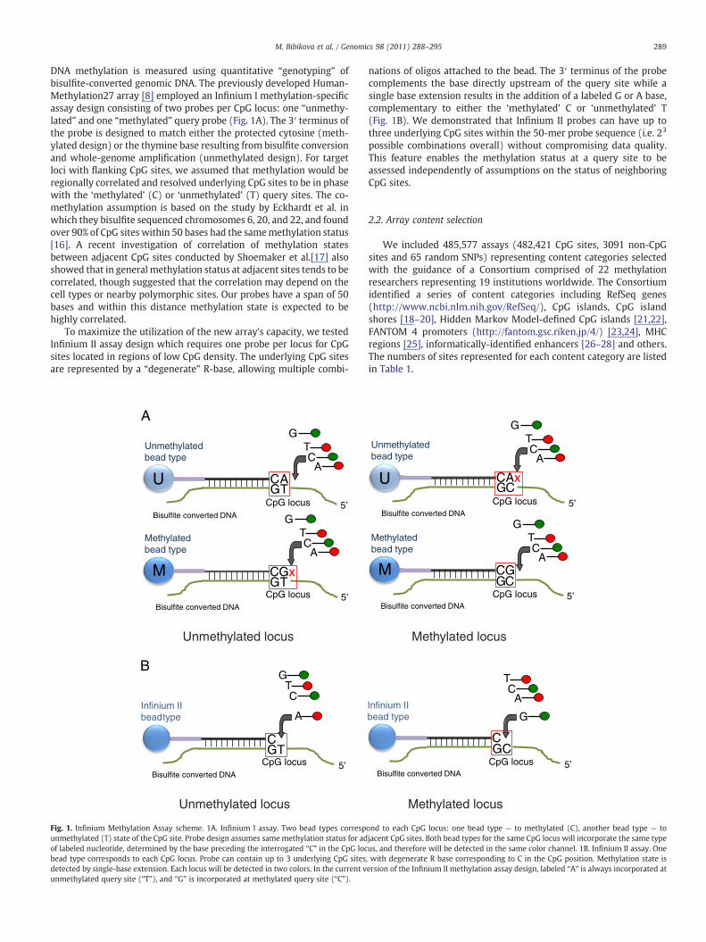
Per the Consortium's recommendations, the highest priority wasplaced on providing comprehensive coverage across the complete geneand CpG island regions. Toward this end, both gene and CpG islandregions were subdivided according to UCSC classifications [29,30](Fig. 2) and each subcategory was targeted individually (Table 2).Coverage of CpG island regionswas further enhanced by including the 2kb regions flanking CpG island shores (referred to here as “CpG islandshelves”) (Table 3 and Fig. 2B) aswell as HiddenMarkovModel-definedCpG islands [21].
Also included are sites that were shown to be biologicallysignificant/informative based on data that were generated internallyor by the members of the Consortium. Other categories representedwere non-CpG sites [9,31], DNase hypersensitive sites [32,33] anddifferentially methylated regions [18,34]. Detailed information on thiscontent is available in the Infinium HumanMethylation450 UserGuide and HumanMethylation450 manifest (www.illumina.com). Arepresentative example of gene coverage by assay probes is shown onFig. 2C.
2.3. Gene coverage
The array provides coverage of a total of 21,231 out of 21,474 UCSCRefGenes (NM and NR) (98.9%) with a global average of 17.2 probesper gene region (Table 2). Multiple transcripts of RefSeq genes areincluded, plus additional genes and transcripts not covered by theUCSC database (total of 29,246 transcripts). In order to achieve acomprehensive assessment of gene region methylation, probescovering gene regions were designed across multiple sub-regions.Promoter regions were divided into two, mutually exclusive bins of200 bp and 1500 bp blocks upstream of the transcription start site(designated TSS200 and TSS1500, respectively). The 5′ and 3′UTR,firstexon and gene body were independently targeted as well (Fig. 2A).Details regarding the number of RefSeq genes and sub-regions, and theaverage number of CpG sites per gene locus represented on the arrayare given in Table 2.
2.4. CpG island coverage
CpG islands were defined based on UCSC annotation and as per thecriteria previously described [35,36]. We employed a NCBI ‘strict’definition for CpG islands (CGI) as DNA sequences (500 base win-dows; excluding most repetitive Alu-elements) with a GC base com-position greater than 50% and a CpG observed/expected ratio of morethan 0.6 [35,36]. As described by Takai and Jones [35], regions of DNAof greater than 500 bp with GC composition equal to or greater than55% and observed CpG/expected CpG of 0.65 were more likely to beassociated with the 5′ regions of genes. Using this definition, 60% of
RefSeq genes contain one or more CGI and 40% contained no CGI.26,658 CpG islands were covered overall with an average of 5.63 siteseach. 28,249 “north” or upstream and 25,761 “south,” or downstreamCpG island shores, immediately outside of the CpG islands, weretargeted with averages of 2.93 and 2.81 sites, respectively. The 2 kbregions upstream and downstream of the CpG island shores, referredto here as “CpG island shelves,” were also targeted with global av-erages of 2.07 and 2.03 sites each (“north” and “south,” respectively)(Table 3 and Fig. 2B).
2.5. Methylation controls
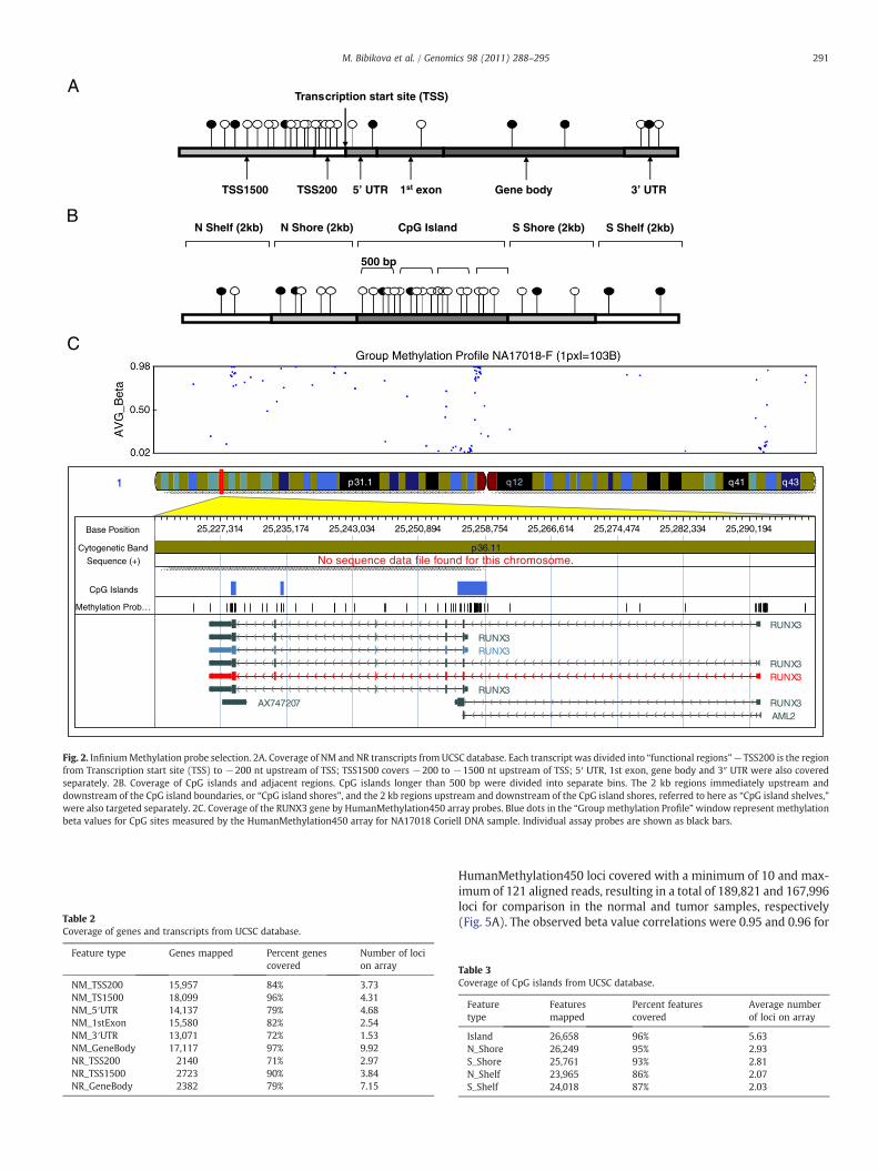
To assess the overall functionality of the individual CpG assays onHumanMethylation450, we created three human genomic DNAmeth-ylation reference standards: unmethylated (U, 0%), hemi-methylated(H, 50%) and methylated (M, 100%) controls. These three referencestandards were created by in vitro de-methylation (U) and subse-quent in vitromethylationwith SssI methylase (M) of standard Coriellgenomic DNA. The hemi-methylated reference was a mixture of U andM in a 1:1 ratio. These three reference standards were run on theInfinium HumanMethylation450 methylation array, and the corre-sponding methylation beta-value (β=intensity of the Methylatedallele (M)/(intensity of the Unmethylated allele (U)+intensity of theMethylated allele (M)+100) was calculated for each of the N480KCpG sites. The distribution of beta-values is consistent with the threereference standards with the unmethylated (U) standard showinglow beta-values, the hemi-methylated (H) standard showing inter-mediate beta-values, and the methylated (M) standard having highbeta-values (Fig. 3). We noticed slightly different performance ofInfinium I and Infinium II assays in terms of the beta-value distri-butions they produced. Infinium II assays demonstrate more pro-nounced off-axis behavior, resulting in an average upward shift inbeta value of 0.02 for the unmethylated standard and an averagedownward shift of 0.08 for the methylated standard (Fig. 3). Thesedifferences do not significantly affect differential methylation detec-tion; we can detect a delta beta of |0.2| with 99% confidence, a resultsimilar to that for the HumanMethylation27 array in which all CpGsites were interrogated using Infinium I assays [8].
2.6. Assay reproducibility
To gage the technical performance of the assay, we assessed datareproducibility between technical replicates using lymphoblastoidcell lines NA17105 and NA17018, cancer cell lineMCF7 and tumor andnormal lung tissues (see Materials and methods section). The averagecorrelation R2 of beta-values for technical replicates was 0.992 (datanot shown).
2.7. Correlation with HumanMethylation27 array
Over 94% of loci present on HumanMethylation27 array wereincluded in the HumanMethylation450 array content. All loci whichsatisfied Infinium II design criteria were re-designed using one beadper locus. To confirm accurate methylation measurement across twoplatformswe compared the correlation between 450K and 27K arrays,showing an R2 of N0.95. An example of beta value correlation for25,978 loci in MCF7 cell line is shown in Fig. 4.
2.8. Correlation with whole-genome bisulfite sequencing data
We evaluated the correlation of methylation beta-values mea-sured by the Infinium Methylation assay with results from wholegenome bisulfite sequencing (WGBS) data generated on a HiSeq2000(Illumina) using next-generation sequencing technology. Two com-parisons were run, one with a normal lung tissue and the other with alung tumor sample.WGBS data were filtered to include corresponding
RTU’3RTU’5TSS1500 TSS200 1st exon Gene body
Transcription start site (TSS)
N Shelf (2kb) N Shore (2kb) S Shore (2kb) S Shelf (2kb) CpG Island
500 bp
1
Base Position 25,227,314 25,235,174 25,243,034 25,250,894 25,258,754 25,266,614 25,274,474 25,282,334 25,290,194
Cytogenetic Band p36.11Sequence (+) No sequence data file found for this chromosome.
CpG Islands
Methylation Prob…
RUNX3RUNX3RUNX3
RUNX3RUNX3
RUNX3AX747207 RUNX3
AML2
A
B
C
p31.1 q12 q41 q43
Fig. 2. InfiniumMethylation probe selection. 2A. Coverage of NM and NR transcripts from UCSC database. Each transcript was divided into “functional regions” — TSS200 is the regionfrom Transcription start site (TSS) to −200 nt upstream of TSS; TSS1500 covers −200 to −1500 nt upstream of TSS; 5′ UTR, 1st exon, gene body and 3″ UTR were also coveredseparately. 2B. Coverage of CpG islands and adjacent regions. CpG islands longer than 500 bp were divided into separate bins. The 2 kb regions immediately upstream anddownstream of the CpG island boundaries, or “CpG island shores”, and the 2 kb regions upstream and downstream of the CpG island shores, referred to here as “CpG island shelves,”were also targeted separately. 2C. Coverage of the RUNX3 gene by HumanMethylation450 array probes. Blue dots in the “Group methylation Profile”window represent methylationbeta values for CpG sites measured by the HumanMethylation450 array for NA17018 Coriell DNA sample. Individual assay probes are shown as black bars.
Table 2Coverage of genes and transcripts from UCSC database.
Feature type Genes mapped Percent genescovered
Number of locion array
NM_TSS200 15,957 84% 3.73NM_TS1500 18,099 96% 4.31NM_5′UTR 14,137 79% 4.68NM_1stExon 15,580 82% 2.54NM_3′UTR 13,071 72% 1.53NM_GeneBody 17,117 97% 9.92NR_TSS200 2140 71% 2.97NR_TSS1500 2723 90% 3.84NR_GeneBody 2382 79% 7.15
291M. Bibikova et al. / Genomics 98 (2011) 288–295
HumanMethylation450 loci covered with a minimum of 10 and max-imum of 121 aligned reads, resulting in a total of 189,821 and 167,996loci for comparison in the normal and tumor samples, respectively(Fig. 5A). The observed beta value correlations were 0.95 and 0.96 for
Table 3Coverage of CpG islands from UCSC database.
Featuretype
Featuresmapped
Percent featurescovered
Average numberof loci on array
Island 26,658 96% 5.63N_Shore 26,249 95% 2.93S_Shore 25,761 93% 2.81N_Shelf 23,965 86% 2.07S_Shelf 24,018 87% 2.03
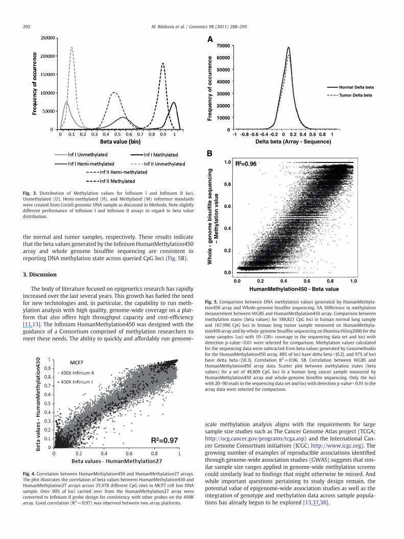
Fig. 3. Distribution of Methylation values for Infinium I and Infinium II loci.Unmethylated (U), Hemi-methylated (H), and Methylated (M) reference standardswere created from Coriell genomic DNA sample as discussed in Methods. Note slightlydifferent performance of Infinium I and Infinium II assays in regard to beta valuedistribution.
0.0
0.2
0.4
0.6
0.8
1.0
Wh
ole
- g
eno
me
bis
ulf
ite
seq
uen
cin
g
–
Met
hyla
tio
n v
alu
e
0.0 0.2 0.4 0.6 0.8 1.0
HumanMethylation450 - Beta value
R2=0.96
70000
60000
50000
40000
30000
20000
10000
0-1 -0.8 -0.6 -0.4 -0.2 0 0.2 0.4 0.6 0.8 1
Fre
qu
ency
of
occ
urr
ence
Delta beta (Array - Sequence)
B
A
Normal Delta beta
Tumor Delta beta
Fig. 5. Comparison between DNA methylation values generated by HumanMethyla-tion450 array and Whole-genome bisulfite sequencing. 5A. Difference in methylationmeasurement between WGBS and HumanMethylation450 array. Comparison betweenmethylation states (beta values) for 189,821 CpG loci in human normal lung sampleand 167,996 CpG loci in human lung tumor sample measured on HumanMethyla-tion450 array and by whole-genome bisulfite sequencing on Illumina HiSeq2000 for thesame samples. Loci with 10–120× coverage in the sequencing data set and loci with
292 M. Bibikova et al. / Genomics 98 (2011) 288–295
the normal and tumor samples, respectively. These results indicatethat the beta values generated by the InfiniumHumanMethylation450array and whole genome bisulfite sequencing are consistent inreporting DNA methylation state across queried CpG loci (Fig. 5B).
3. Discussion
The body of literature focused on epigenetics research has rapidlyincreased over the last several years. This growth has fueled the needfor new technologies and, in particular, the capability to run meth-ylation analysis with high quality, genome-wide coverage on a plat-form that also offers high throughput capacity and cost-efficiency[11,13]. The Infinium HumanMethylation450 was designed with theguidance of a Consortium comprised of methylation researchers tomeet these needs. The ability to quickly and affordably run genome-
R2=0.97
Fig. 4. Correlation between HumanMethylation450 and HumanMethylation27 arrays.The plot illustrates the correlation of beta values between HumanMethylation450 andHumanMethylation27 arrays across 25,978 different CpG sites in MCF7 cell line DNAsample. Over 90% of loci carried over from the HumanMethylation27 array wereconverted to Infinium II probe design for consistency with other probes on the 450Karray. Good correlation (R2=0.97) was observed between two array platforms.
detection p-valueb0.01 were selected for comparison. Methylation values calculatedfor the sequencing data were subtracted from beta values generated by GenomeStudiofor the HumanMethylation450 array. 88% of loci have delta betab |0.2|, and 97% of locihave delta betab |\0.3|. Correlation R2=0.96. 5B. Correlation between WGBS andHumanMethylation450 array data. Scatter plot between methylation states (betavalues) for a set of 48,809 CpG loci in a human lung cancer sample measured byHumanMethylation450 array and whole-genome bisulfite sequencing. Only the lociwith 20–90 reads in the sequencing data set and loci with detection p-valueb0.01 in thearray data were selected for comparison.
scale methylation analysis aligns with the requirements for largesample size studies such as The Cancer Genome Atlas project (TCGA;http://ocg.cancer.gov/programs/tcga.asp) and the International Can-cer Genome Consortium initiatives (ICGC; http://www.icgc.org). Thegrowing number of examples of reproducible associations identifiedthrough genome-wide association studies (GWAS) suggests that sim-ilar sample size ranges applied in genome-wide methylation screenscould similarly lead to findings that might otherwise be missed. Andwhile important questions pertaining to study design remain, thepotential value of epigenome-wide association studies as well as theintegration of genotype and methylation data across sample popula-tions has already begun to be explored [13,37,38].
293M. Bibikova et al. / Genomics 98 (2011) 288–295
The utility of Infinium HumanMethylation450 will be furtherextended by its applicability to FFPE samples, which was recentlydemonstrated using a new restoration kit (data not shown).
In summary, the HumanMethylation450 array should provide apowerful tool for investigators to fuel the continued, rapid evolution ofepigenetic research [11,39] by offering simple and rapid genome-widemethylation analysis of hundreds of thousands of CpG sites across largenumbers of samples. While additional sites of interest will continue tobe identified, this arraywas designed to provide an efficient, robust andaffordable discovery solution targeting core content of commoninterest within the epigenetics research community.
4. Materials and methods
4.1. Array design
Probe performance assessment experiments were run to determineoptimal probe design parameters for both Infinium I and II designs.Assay probes were selected with the goal of providing the mostcomplete coverage possible across the content categories identified bythe Consortium. Among these content categories, gene regions and CpGislands were given top priority. Each target region was allocated amaximum loci count which was inversely related to its level of priority(e.g. gene promoter regions and CpG islands were allotted a highernumber of loci than other target regions). Large regions such as largeCpG islands were subdivided into separate sub-regions to ensure evencoverage. After each round of target selection and probe design,empirical analytical testing removed poorly-performing probes. Subse-quent rounds of selection were then run until the pool size wasexhausted. This approach ensured strong probe performance as well asan optimal balance between coverage density in the highest priorityregions and breadth of coverage across remaining targeted regions.
4.2. DNA samples
DNA samples NA17105 and NA17018 were purchased from theCoriell Institute for Medical Research (NJ, USA). DNA from normal andtumor lung tissues and MCF7 cell line were purchased from BioChainInstitute (Hayward, CA).
4.3. Bisulfite conversion of genomic DNA
DNA samples for Infinium Methylation assay were bisulfite con-verted using EZ DNA methylation kit (Cat. #D5001) from ZymoResearch (CA, USA). 500 ng of gDNA was denatured by addition ofZymo M-Dilution buffer (contains NaOH) and incubated for 15 min at37 °C. CT-conversion reagent (bisulfite-containing) was added to thedenatured DNA and incubated for 16 h at 50 °C in a thermocycler anddenatured every 60 min by heating to 95 °C for 30 s. DNA samples forthewhole-genome bisulfite sequencingwere bisulfite converted usingEpiTect Bisulfite conversion kit (Cat. #59104) from QIAGEN (Valencia,CA) following manufacturer's recommendations with modifications[40].
4.4. Methylation reference samples preparation
Methylation reference standards for assessment of the Infiniumprobes quality were prepared as described previously [8]. 50 ng ofCoriell gDNA NA18105 was amplified with the REPLI-g Mini Kit(QIAGEN Cat. #150025) following manufacturer's recommendations.We used male genomic DNA in order to assess quality of the Y-chromosomal loci. The WGA amplified DNA was subjected to Mungbean nuclease treatment to remove single-stranded DNA. Theresultant unmethylated DNA (U) was treated with SssI methylase,which globally methylates all double-stranded CpG sites, to create anearly completely methylated reference standard (M). The hemi-
methylated reference (H)was created bymixing U andM samples in a1:1 stoichiometric ratio.
4.5. Infinium methylation assay
The assay was carried out as described previously [8]. In brief, 4 μlof bisulfite-converted DNA (~150 ng) was used in the whole-genomeamplification (WGA) reaction. After amplification, the DNA wasfragmented enzymatically, precipitated and re-suspended in hybrid-ization buffer. All subsequent steps were performed following thestandard Infinium protocol (User Guide part #15019519 A). Frag-mented DNA was dispensed onto the HumanMethylation450 Bead-Chips, and hybridization performed in hybridization oven for 20 h.After hybridization, the array was processed through a primerextension and an immunohistochemistry staining protocol to allowdetection of a single-base extension reaction [41–43]. Finally,BeadChips were coated and then imaged on an Illumina iScan.
Methylation level of each CpG locus was calculated in GenomeStu-dio® Methylation module as methylation beta-value (β=intensity ofthe Methylated allele (M)/(intensity of the Unmethylated allele (U)+intensity of the Methylated allele (M)+100).
4.6. Whole-genome bisulfite sequencing
For the whole-genome DNA methylation analysis at single nucleo-tide resolution, 2–5 μgof lungnormal and lung tumor genomicDNAwasfragmented using Covaris shearing. The fragmented DNA was end-polished, and a single ‘A’nucleotidewasadded to the3′ ends of thebluntfragments. The fragmentswere ligatedwith Illuminamethylated forkedadaptors, and 200–400 bp fragments were selected by gel electropho-resis andpurified using aQIAquickGel ExtractionKit (QIAGEN). PurifiedDNA fragmentswere treatedwith bisulfite using the EpiTect Bisulfite Kit(QIAGEN) for approximately 14 h to ensure maximal conversion rate[40]. The bisulfite-treated DNAwas enriched by 4 cycles of PCRwith PfuTurbo Cx DNA polymerase (Stratagene Products, Agilent, La Jolla, CA).The libraries were sequenced on Illumina HiSeq2000 sequencinginstrument according to standard Illumina cluster generation andsequencing protocols with a 2×75 bp read length.
4.7. Data analysis
Infinium methylation data was processed with MethylationModule of GenomeStudio software using HumanMethylation450manifest v1.1. Whole genome bisulfite sequencing data was analyzedusing pipeline developed at the Salk Institute [9]. Briefly, rawsequencing data was processed using Illumina pipeline and FastQoutput data was generated and aligned to the human genome (hg19)using Bowtie alignment algorithm. Methylation status for eachaligned site was calculated at a minimum of 10× coverage per site.
All CpG sites with a p-value less than 0.01 on the 450k array weremapped toWGBS data on the same strand and coordinate. For each ofthese mapped sites a beta value was calculated by taking the numberof methylated tags (C) and dividing by the sum of methylated (C)and unmethylated (T) tag counts. WGBS sites with less than 10× andgreater than120× coveragewere removed. Scatter plots and r-squaredstatistics were then calculated by comparing array vs. sequencing betavalues for the remaining matching sites.
Acknowledgments
We sincerely thank all Consortium members who contributed tothe development of the HumanMethylation450 array: Devin Absher(HudsonAlpha Institute for Biotechnology), Hiroyuki Aburatani (Uni-versity of Tokyo), Stephen Baylin (the JohnsHopkins University Schoolof Medicine), Stephan Beck (University College London), BenjaminBerman (University of Southern California), Andrew P. Feinberg (the
294 M. Bibikova et al. / Genomics 98 (2011) 288–295
Johns Hopkins University School of Medicine), François Fuks (FreeUniversity of Brussels), Michel Georges (University of Liège), Ivo Gut(Centre National de Génotypage), Thomas Hudson (Ontario Institutefor Cancer Research), Peter W. Laird (University of SouthernCalifornia), Louise Laurent (The Scripps Research Institute), JeanneLoring (The Scripps Research Institute), Alexander Meissner (HarvardUniversity/Broad Institute), Vardhman Rakyan (Queen Mary Univer-sity of London), Brian Reed (FredHutchinson Cancer Research Center),Bing Ren (University of California San Diego), Tim Spector (King'sCollege London), John Stamatoyannopoulos (University of Washing-ton), Chia-Lin Wei (Genome Institute of Singapore), and MartinWidschwendter (University College London).
We would like to thank Juying Yan and Carmen Xiao for technicalassistance with generation of the WGBS data, and Ryan Lister and JoeEcker (Salk Institute) for help with initial WGBS data analysis.
References
[1] A. Schumacher, P. Kapranov, Z. Kaminsky, J. Flanagan, A. Assadzadeh, P. Yau, C.Virtanen, N. Winegarden, J. Cheng, T. Gingeras, A. Petronis, Microarray-based DNAmethylation profiling: technology and applications, Nucleic Acids Res. 34 (2006)528–542.
[2] M. Weber, J.J. Davies, D. Wittig, E.J. Oakeley, M. Haase, W.L. Lam, D. Schubeler,Chromosome-wide and promoter-specific analyses identify sites of differentialDNA methylation in normal and transformed human cells, Nat. Genet. 37 (2005)853–862.
[3] J.M. Ordway, J.A. Bedell, R.W. Citek, A. Nunberg, A. Garrido, R. Kendall, J.R. Stevens,D. Cao, R.W. Doerge, Y. Korshunova, H. Holemon, J.D. McPherson, N. Lakey, J. Leon,R.A. Martienssen, J.A. Jeddeloh, Comprehensive DNA methylation profiling in ahuman cancer genome identifies novel epigenetic targets, Carcinogenesis 27(2006) 2409–2423.
[4] A. Meissner, A. Gnirke, G.W. Bell, B. Ramsahoye, E.S. Lander, R. Jaenisch, Reducedrepresentation bisulfite sequencing for comparative high-resolution DNA meth-ylation analysis, Nucleic Acids Res. 33 (2005) 5868–5877.
[5] T. Rauch, H. Li, X. Wu, G.P. Pfeifer, MIRA-assisted microarray analysis, a newtechnology for the determination of DNA methylation patterns, identifiesfrequent methylation of homeodomain-containing genes in lung cancer cells,Cancer Res. 66 (2006) 7939–7947.
[6] S.Q. Kuang, W.G. Tong, H. Yang, W. Lin, M.K. Lee, Z.H. Fang, Y. Wei, J. Jelinek, J.P.Issa, G. Garcia-Manero, Genome-wide identification of aberrantly methylatedpromoter associated CpG islands in acute lymphocytic leukemia, Leukemia 22(2008) 1529–1538.
[7] N. Omura, C.P. Li, A. Li, S.M. Hong, K. Walter, A. Jimeno, M. Hidalgo, M. Goggins,Genome-wide profiling of methylated promoters in pancreatic adenocarcinoma,Cancer Biol. Ther. 7 (2008) 1146–1156.
[8] M. Bibikova, J. Le, B. Barnes, S. Saedinia-Melnyk, L. Zhou, R. Shen, K.L. Gunderson,Genome-wide DNA methylation profiling using Infinium® assay, Epigenomics 1(2009) 177–200.
[9] R. Lister, M. Pelizzola, R.H. Dowen, R.D. Hawkins, G. Hon, J. Tonti-Filippini, J.R.Nery, L. Lee, Z. Ye, Q.M. Ngo, L. Edsall, J. Antosiewicz-Bourget, R. Stewart, V.Ruotti, A.H. Millar, J.A. Thomson, B. Ren, J.R. Ecker, Human DNA methylomes atbase resolution show widespread epigenomic differences, Nature 462 (2009)315–322.
[10] Y. Ruike, Y. Imanaka, F. Sato, K. Shimizu, G. Tsujimoto, Genome-wide analysis ofaberrant methylation in human breast cancer cells using methyl-DNA immuno-precipitation combined with high-throughput sequencing, BMC Genomics 11(2010) 137.
[11] S. Beck, Taking the measure of the methylome, Nat. Biotechnol. 28 (2010)1026–1028.
[12] Y.W. Huang, T.H. Huang, L.S. Wang, Profiling DNAmethylomes frommicroarray togenome-scale sequencing, Technol. Cancer Res. Treat. 9 (2010) 139–147.
[13] P.W. Laird, Principles and challenges of genome-wide DNA methylation analysis,Nat. Rev. Genet. 11 (2010) 191–203.
[14] M. Bibikova, J.B. Fan, Genome-wide DNA methylation profiling, Wiley Interdiscip.Rev. Syst. Biol. Med. 2 (2010) 210–223.
[15] C. Bock, J. Walter, M. Paulsen, T. Lengauer, CpG island mapping by epigenomeprediction, PLoS Comput. Biol. 3 (2007) e110.
[16] F. Eckhardt, J. Lewin, R. Cortese, V.K. Rakyan, J. Attwood, M. Burger, J. Burton, T.V.Cox, R. Davies, T.A. Down, C. Haefliger, R. Horton, K. Howe, D.K. Jackson, J. Kunde,C. Koenig, J. Liddle, D. Niblett, T. Otto, R. Pettett, S. Seemann, C. Thompson, T. West,J. Rogers, A. Olek, K. Berlin, S. Beck, DNA methylation profiling of humanchromosomes 6, 20 and 22, Nat. Genet. 38 (2006) 1378–1385.
[17] R. Shoemaker, J. Deng, W.Wang, K. Zhang, Allele-specific methylation is prevalentand is contributed by CpG-SNPs in the human genome, Genome Res. 20 (2010)883–889.
[18] A. Doi, I.H. Park, B. Wen, P. Murakami, M.J. Aryee, R. Irizarry, B. Herb, C. Ladd-Acosta, J. Rho, S. Loewer, J. Miller, T. Schlaeger, G.Q. Daley, A.P. Feinberg,Differential methylation of tissue- and cancer-specific CpG island shoresdistinguishes human induced pluripotent stem cells, embryonic stem cells andfibroblasts, Nat. Genet. 41 (2009) 1350–1353.
[19] A.F. Rubin, P. Green, Mutation patterns in cancer genomes, Proc. Natl. Acad. Sci. U.S.A. 106 (2009) 21766–21770.
[20] R.A. Irizarry, C. Ladd-Acosta, B. Wen, Z. Wu, C. Montano, P. Onyango, H. Cui, K.Gabo, M. Rongione, M. Webster, H. Ji, J.B. Potash, S. Sabunciyan, A.P. Feinberg,The human colon cancer methylome shows similar hypo- and hypermethyla-tion at conserved tissue-specific CpG island shores, Nat. Genet. 41 (2009)178–186.
[21] R.A. Irizarry, H. Wu, A.P. Feinberg, A species-generalized probabilistic model-based definition of CpG islands, Mamm. Genome 20 (2009) 674–680.
[22] F. Hsieh, S.C. Chen, K. Pollard, A nearly exhaustive search for CpG islands on wholechromosomes, Int. J. Biostat. 5 (2009) 1.
[23] J. Severin, A.M. Waterhouse, H. Kawaji, T. Lassmann, E. van Nimwegen, P.J.Balwierz, M.J. de Hoon, D.A. Hume, P. Carninci, Y. Hayashizaki, H. Suzuki, C.O.Daub, A.R. Forrest, FANTOM4 EdgeExpressDB: an integrated database of pro-moters, genes, microRNAs, expression dynamics and regulatory interactions,Genome Biol. 10 (2009) R39.
[24] FANTOM4, Functional Annotation of the Mammalian Genome, RIKEN OmicsScience Center, Yokohama City, 2009.
[25] E.M. Tomazou, V.K. Rakyan, G. Lefebvre, R. Andrews, P. Ellis, D.K. Jackson, C.Langford, M.D. Francis, L. Backdahl, M. Miretti, P. Coggill, D. Ottaviani, D. Sheer, A.Murrell, S. Beck, Generation of a genomic tiling array of the human majorhistocompatibility complex (MHC) and its application for DNA methylationanalysis, BMC Med. Genomics 1 (2008) 19.
[26] N.D. Heintzman, R.K. Stuart, G. Hon, Y. Fu, C.W. Ching, R.D. Hawkins, L.O. Barrera, S.Van Calcar, C. Qu, K.A. Ching, W. Wang, Z. Weng, R.D. Green, G.E. Crawford, B. Ren,Distinct and predictive chromatin signatures of transcriptional promoters andenhancers in the human genome, Nat. Genet. 39 (2007) 311–318.
[27] E. Birney, J.A. Stamatoyannopoulos, A. Dutta, R. Guigo, T.R. Gingeras, E.H.Margulies, Z. Weng, M. Snyder, E.T. Dermitzakis, R.E. Thurman, M.S. Kuehn, C.M.Taylor, S. Neph, C.M. Koch, S. Asthana, A. Malhotra, I. Adzhubei, J.A. Greenbaum,R.M. Andrews, P. Flicek, P.J. Boyle, H. Cao, N.P. Carter, G.K. Clelland, S. Davis, N.Day, P. Dhami, S.C. Dillon, M.O. Dorschner, H. Fiegler, P.G. Giresi, J. Goldy, M.Hawrylycz, A. Haydock, R. Humbert, K.D. James, B.E. Johnson, E.M. Johnson, T.T.Frum, E.R. Rosenzweig, N. Karnani, K. Lee, G.C. Lefebvre, P.A. Navas, F. Neri, S.C.Parker, P.J. Sabo, R. Sandstrom, A. Shafer, D. Vetrie, M. Weaver, S. Wilcox, M. Yu,F.S. Collins, J. Dekker, J.D. Lieb, T.D. Tullius, G.E. Crawford, S. Sunyaev, W.S.Noble, I. Dunham, F. Denoeud, A. Reymond, P. Kapranov, J. Rozowsky, D. Zheng,R. Castelo, A. Frankish, J. Harrow, S. Ghosh, A. Sandelin, I.L. Hofacker, R.Baertsch, D. Keefe, S. Dike, J. Cheng, H.A. Hirsch, E.A. Sekinger, J. Lagarde, J.F.Abril, A. Shahab, C. Flamm, C. Fried, J. Hackermuller, J. Hertel, M. Lindemeyer, K.Missal, A. Tanzer, S. Washietl, J. Korbel, O. Emanuelsson, J.S. Pedersen, N.Holroyd, R. Taylor, D. Swarbreck, N. Matthews, M.C. Dickson, D.J. Thomas, M.T.Weirauch, J. Gilbert, et al., Identification and analysis of functional elements in1% of the human genome by the ENCODE pilot project, Nature 447 (2007)799–816.
[28] N.D. Heintzman, G.C. Hon, R.D. Hawkins, P. Kheradpour, A. Stark, L.F. Harp, Z. Ye, L.K.Lee, R.K. Stuart, C.W. Ching, K.A. Ching, J.E. Antosiewicz-Bourget, H. Liu, X. Zhang, R.D.Green, V.V. Lobanenkov, R. Stewart, J.A. Thomson, G.E. Crawford, M. Kellis, B. Ren,Histone modifications at human enhancers reflect global cell-type-specific geneexpression, Nature 459 (2009) 108–112.
[29] P.A. Fujita, B. Rhead, A.S. Zweig, A.S. Hinrichs, D. Karolchik, M.S. Cline, M. Goldman,G.P. Barber, H. Clawson, A. Coelho, M. Diekhans, T.R. Dreszer, B.M. Giardine, R.A.Harte, J. Hillman-Jackson, F. Hsu, V. Kirkup, R.M. Kuhn, K. Learned, C.H. Li, L.R.Meyer, A. Pohl, B.J. Raney, K.R. Rosenbloom, K.E. Smith, D. Haussler, W.J. Kent, TheUCSC genome browser database: update 2011, Nucleic Acids Res. 39 (2011)D876–D882.
[30] B. Rhead, D. Karolchik, R.M. Kuhn, A.S. Hinrichs, A.S. Zweig, P.A. Fujita, M.Diekhans, K.E. Smith, K.R. Rosenbloom, B.J. Raney, A. Pohl, M. Pheasant, L.R. Meyer,K. Learned, F. Hsu, J. Hillman-Jackson, R.A. Harte, B. Giardine, T.R. Dreszer, H.Clawson, G.P. Barber, D. Haussler, W.J. Kent, The UCSC genome browser database:update 2010, Nucleic Acids Res. 38 (2010) D613–D619.
[31] L. Laurent, E. Wong, G. Li, T. Huynh, A. Tsirigos, C.T. Ong, H.M. Low, K.W. Kin Sung,I. Rigoutsos, J. Loring, C.L. Wei, Dynamic changes in the human methylome duringdifferentiation, Genome Res. 20 (2010) 320–331.
[32] H. Lian, W.A. Thompson, R. Thurman, J.A. Stamatoyannopoulos, W.S. Noble, C.E.Lawrence, Automated mapping of large-scale chromatin structure in ENCODE,Bioinformatics 24 (2008) 1911–1916.
[33] H. Xi, H.P. Shulha, J.M. Lin, T.R. Vales, Y. Fu, D.M. Bodine, R.D. McKay, J.G.Chenoweth, P.J. Tesar, T.S. Furey, B. Ren, Z. Weng, G.E. Crawford, Identification andcharacterization of cell type-specific and ubiquitous chromatin regulatorystructures in the human genome, PLoS Genet. 3 (2007) e136.
[34] V.K. Rakyan, T.A. Down, N.P. Thorne, P. Flicek, E. Kulesha, S. Graf, E.M. Tomazou, L.Backdahl, N. Johnson, M. Herberth, K.L. Howe, D.K. Jackson, M.M. Miretti, H.Fiegler, J.C. Marioni, E. Birney, T.J. Hubbard, N.P. Carter, S. Tavare, S. Beck, Anintegrated resource for genome-wide identification and analysis of human tissue-specific differentially methylated regions (tDMRs), Genome Res. 18 (2008)1518–1529.
[35] D. Takai, P.A. Jones, Comprehensive analysis of CpG islands in humanchromosomes 21 and 22, Proc. Natl. Acad. Sci. U.S.A. 99 (2002) 3740–3745.
[36] M. Gardiner-Garden, M. Frommer, CpG islands in vertebrate genomes, J. Mol. Biol.196 (1987) 261–282.
[37] J.T. Bell, A.A. Pai, J.K. Pickrell, D.J. Gaffney, R. Pique-Regi, J.F. Degner, Y. Gilad, J.K.Pritchard, DNA methylation patterns associate with genetic and gene expressionvariation in HapMap cell lines, Genome Biol. 12 (2011) R10.
[38] V.K. Rakyan, T.A. Down, D.J. Balding, S. Beck, Epigenome-wide association studiesfor common human diseases, Nat. Rev. Genet. 12 (2011) 529–541.

295M. Bibikova et al. / Genomics 98 (2011) 288–295
[39] J. Sandoval, H.A. Heyn, S. Moran, J. Serra-Musach, M.A. Pujana, M. Bibikova, M.Esteller, Validation of a DNA methylation microarray for 450,000 CpG sites in thehuman genome, Epigenetics 6 (2011) 692–702.
[40] F. Boellmann, L. Zhang, H.J. Clewell, G.P. Schroth, E.M. Kenyon, M.E. Andersen, R.S.Thomas, Genome-wide analysis of DNAmethylation and gene expression changesin the mouse lung following subchronic arsenate exposure, Toxicol. Sci. 117(2010) 404–417.
[41] K.L. Gunderson, F.J. Steemers, H. Ren, P. Ng, L. Zhou, C. Tsan, W. Chang, D. Bullis, J.Musmacker, C. King, L.L. Lebruska, D. Barker, A. Oliphant, K.M. Kuhn, R. Shen,Whole-genome genotyping, Methods Enzymol. 410 (2006) 359–376.
[42] F.J. Steemers, K.L. Gunderson, Whole genome genotyping technologies on theBeadArray platform, Biotechnol. J. 2 (2007) 41–49.
[43] K.L. Gunderson, Whole-genome genotyping on bead arrays, Methods Mol. Biol.529 (2009) 197–213.