Validation of a DNA methylation microarray for 450,000 CpG sites in the human genome
11
Epigenetics 6:6, 692-702; June 2011; © 2011 Landes Bioscience RESEARCH PAPER 692 Epigenetics Volume 6 Issue 6 *Correspondence to: Manel Esteller; Email: [email protected] Submitted: 04/29/11; Accepted: 04/29/11 DOI: 10.4161/epi.6.6.16196 Introduction The disruption of the DNA methylation profile is a major hall- mark of human cancer. 1,2 Among the aberrant DNA methyla- tion signatures of transformed cells there are two key events in tumorigenesis: the promoter CpG island hypermethylation- associated inactivation of tumor suppressor genes and the global hypomethylation of the cancer genome, which is probably linked to chromosomal instability and the reactivation of endoparasitic sequences. 1,2 The large body of data concerning DNA methyla- tion landscapes in human tumors whose clinical relevance has already been recognized 3 occurs in a scientific context where information about the altered DNA methylation patterns in other common disorders, 4,5 such as neurological 6 and cardiovas- cular 7 diseases, is still very scarce. Part of the explanation might relate to the distinct biological processes involved and the ease with which sample material can be acquired in the oncology arena, but the explanation may also concern technical aspects. In this regard, it is necessary to have standard and common meth- ods for obtaining comprehensive DNA methylation profiles from different types of cells and tissues with user-friendly bioanalytical DNA methylation is the most studied epigenetic mark and CpG methylation is central to many biological processes and human diseases. Since cancer has highlighted the contribution to disease of aberrant DNA methylation patterns, such as the presence of promoter CpG island hypermethylation-associated silencing of tumor suppressor genes and global DNA hypomethylation defects, their importance will surely become apparent in other pathologies. However, advances in obtaining comprehensive DNA methylomes are hampered by the high cost and time-consuming aspects of the single nucleotide methods currently available for whole genome DNA methylation analyses. Following the success of the standard CpG methylation microarrays for 1,505 CpG sites and 27,000 CpG sites, we have validated in vivo the newly developed 450,000 (450K) cytosine microarray (Illumina). The 450K microarray includes CpG and CNG sites, CpG islands/shores/shelves/open sea, non-coding RNA (microRNAs and long non-coding RNAs) and sites surrounding the transcription start sites (-200 bp to -1,500 bp, 5'-UTRs and exons 1) for coding genes, but also for the corresponding gene bodies and 3'-UTRs, in addition to intergenic regions derived from GWAS studies. Herein, we demonstrate that the 450K DNA methylation array can consistently and significantly detect CpG methylation changes in the HCT-116 colorectal cancer cell line in comparison with normal colon mucosa or HCT-116 cells with defective DNA methyltransferases (DKO). The provided validation highlights the potential use of the 450K DNA methylation microarray as a useful tool for ongoing and newly designed epigenome projects. Validation of a DNA methylation microarray for 450,000 CpG sites in the human genome Juan Sandoval, 1 Holger Heyn, 1 Sebastian Moran, 1 Jordi Serra-Musach, 2 Miguel A. Pujana, 3 Marina Bibikova 4 and Manel Esteller 1,5,6, * 1 Cancer Epigenetics and Biology Program (PEBC); 2 Translational Research Laboratory; Catalan Institute of Oncology; Bellvitge Biomedical Research Institute (IDIBELL); 3 Biomarkers and Susceptibility Unit; Spanish Biomedical Research Centre Network for Epidemiology and Public Health; Catalan Institute of Oncology; Bellvitge Biomedical Research Institute (IDIBELL); L‘Hospitalet de Llobregat; Barcelona, Catalonia, Spain; 4 Illumina, Inc.; San Diego, CA USA; 5 Department of Physiological Sciences II; School of Medicine; University of Barcelona; Barcelona, Catalonia, Spain; 6 Institucio Catalana de Recerca i Estudis Avançats (ICREA)l Barcelona; Catalonia, Spain Key words: DNA methylation, epigenetics, microarray, CpG sites, DNA methyltransferase, colon cancer, validation tools that allow data to be shared among scientists with distinct areas of expertise. Looking at DNA methylation analysis technologies it is evi- dent that a wide range of options exists. The bisulfite genomic sequencing of multiple clones is a useful approach for the detailed study of the CpG methylation status of a given sequence in a few samples, whilst methylation-specific PCR or pyrosequencing techniques are of great value for studying a few CpG sites in a greater number of samples. If we go to the global scale, available approaches include the isolation of methylated fractions of the genome by methylation-sensitive enzymes, 8-10 immunoprecipita- tion with a methyl-CpG binding domain antibody 11,12 and meth- ylcytosine antibody. 13-16 New approaches include representation bisulphite sequencing (RRBS), 17 and sequencing-by-synthesis (MethylC-Seq) technology. 18 Using whole-genome bisulfite sequencing, an analysis of the DNA methylome of peripheral blood mononuclear cells from a single case has also been reported recently. 19 However, these techniques require a high level of spe- cialization and are still expensive and time-consuming. Thus, they are a largely unavailable to a large part of the biomedical community who wish to examine the relevance of extensive
-
Upload
independent -
Category
Documents
-
view
0 -
download
0
Transcript of Validation of a DNA methylation microarray for 450,000 CpG sites in the human genome

©2011 Landes Bioscience.Do not distribute.
Epigenetics 6:6, 692-702; June 2011; © 2011 Landes Bioscience
RESEARCH PAPER
692 Epigenetics Volume 6 Issue 6
*Correspondence to: Manel Esteller; Email: [email protected]: 04/29/11; Accepted: 04/29/11DOI: 10.4161/epi.6.6.16196
Introduction
The disruption of the DNA methylation profile is a major hall-mark of human cancer.1,2 Among the aberrant DNA methyla-tion signatures of transformed cells there are two key events in tumorigenesis: the promoter CpG island hypermethylation-associated inactivation of tumor suppressor genes and the global hypomethylation of the cancer genome, which is probably linked to chromosomal instability and the reactivation of endoparasitic sequences.1,2 The large body of data concerning DNA methyla-tion landscapes in human tumors whose clinical relevance has already been recognized3 occurs in a scientific context where information about the altered DNA methylation patterns in other common disorders,4,5 such as neurological6 and cardiovas-cular7 diseases, is still very scarce. Part of the explanation might relate to the distinct biological processes involved and the ease with which sample material can be acquired in the oncology arena, but the explanation may also concern technical aspects. In this regard, it is necessary to have standard and common meth-ods for obtaining comprehensive DNA methylation profiles from different types of cells and tissues with user-friendly bioanalytical
DNA methylation is the most studied epigenetic mark and CpG methylation is central to many biological processes and human diseases. Since cancer has highlighted the contribution to disease of aberrant DNA methylation patterns, such as the presence of promoter CpG island hypermethylation-associated silencing of tumor suppressor genes and global DNA hypomethylation defects, their importance will surely become apparent in other pathologies. However, advances in obtaining comprehensive DNA methylomes are hampered by the high cost and time-consuming aspects of the single nucleotide methods currently available for whole genome DNA methylation analyses. Following the success of the standard CpG methylation microarrays for 1,505 CpG sites and 27,000 CpG sites, we have validated in vivo the newly developed 450,000 (450K) cytosine microarray (Illumina). The 450K microarray includes CpG and CNG sites, CpG islands/shores/shelves/open sea, non-coding RNA (microRNAs and long non-coding RNAs) and sites surrounding the transcription start sites (-200 bp to -1,500 bp, 5'-UTRs and exons 1) for coding genes, but also for the corresponding gene bodies and 3'-UTRs, in addition to intergenic regions derived from GWAS studies. Herein, we demonstrate that the 450K DNA methylation array can consistently and significantly detect CpG methylation changes in the HCT-116 colorectal cancer cell line in comparison with normal colon mucosa or HCT-116 cells with defective DNA methyltransferases (DKO). The provided validation highlights the potential use of the 450K DNA methylation microarray as a useful tool for ongoing and newly designed epigenome projects.
Validation of a DNA methylation microarray for 450,000 CpG sites in the human genome
Juan Sandoval,1 Holger Heyn,1 Sebastian Moran,1 Jordi Serra-Musach,2 Miguel A. Pujana,3 Marina Bibikova4 and Manel Esteller1,5,6,*
1Cancer Epigenetics and Biology Program (PEBC); 2Translational Research Laboratory; Catalan Institute of Oncology; Bellvitge Biomedical Research Institute (IDIBELL); 3Biomarkers and Susceptibility Unit; Spanish Biomedical Research Centre Network for Epidemiology and Public Health; Catalan Institute of Oncology;
Bellvitge Biomedical Research Institute (IDIBELL); L‘Hospitalet de Llobregat; Barcelona, Catalonia, Spain; 4Illumina, Inc.; San Diego, CA USA; 5Department of Physiological Sciences II; School of Medicine; University of Barcelona; Barcelona, Catalonia, Spain; 6Institucio Catalana de Recerca i Estudis Avançats (ICREA)l Barcelona; Catalonia, Spain
Key words: DNA methylation, epigenetics, microarray, CpG sites, DNA methyltransferase, colon cancer, validation
tools that allow data to be shared among scientists with distinct areas of expertise.
Looking at DNA methylation analysis technologies it is evi-dent that a wide range of options exists. The bisulfite genomic sequencing of multiple clones is a useful approach for the detailed study of the CpG methylation status of a given sequence in a few samples, whilst methylation-specific PCR or pyrosequencing techniques are of great value for studying a few CpG sites in a greater number of samples. If we go to the global scale, available approaches include the isolation of methylated fractions of the genome by methylation-sensitive enzymes,8-10 immunoprecipita-tion with a methyl-CpG binding domain antibody11,12 and meth-ylcytosine antibody.13-16 New approaches include representation bisulphite sequencing (RRBS),17 and sequencing-by-synthesis (MethylC-Seq) technology.18 Using whole-genome bisulfite sequencing, an analysis of the DNA methylome of peripheral blood mononuclear cells from a single case has also been reported recently.19 However, these techniques require a high level of spe-cialization and are still expensive and time-consuming. Thus, they are a largely unavailable to a large part of the biomedical community who wish to examine the relevance of extensive

©2011 Landes Bioscience.Do not distribute.
www.landesbioscience.com Epigenetics 693
RESEARCH PAPER RESEARCH PAPER
Results and Discussion
Distribution and classification of the 450,000 cytosine sites in the human genome. The 450K DNA Methylation array (Infinium HumanMethylation450 BeadChip) includes 485,764 cytosine positions of the human genome. From these cytosine sites, 482,421 positions (99.3%) are CpG dinucleotides, whilst 3,343 sites (0.7%) correspond to CNG targets. Thus, hereafter, we will use the term CpG to refer to all of these, except when we refer specifically to putative CNG methylation. The incor-porated CpG sites are distributed among all 22 autosomal and 1 sex chromosome pairs of humans. Chromosome 1 harbors the most positions (46,867, 9.6%) and chromosome Y the fewest (416, 0.08%). According to their associated RNA transcripts, 361,766 CpGs (74.4%) correspond to classic coding messenger RNA genes, 4,168 (0.85%) are linked to non-coding RNAs (3,440 for microRNAs and 728 for long non-coding RNAs). For 119,830 (24.6%) sites there are no annotated transcripts associ-ated with the described CpG location. From the CpG content and neighborhood context, 150,254 (30.9%) CpGs are in CpG
DNA methylation changes in the origin and progression of many human diseases.
Mirroring the extensive use of well annotated DNA microar-rays for single-nucleotide polymorphisms with massive genome coverage, DNA methylation microarrays with a user-friendly com-ponent have begun to become available. Among these, the 1,505 CpG (Illumina GoldenGate DNA Methylation BeadArray),20-23 and 27,000 CpG (Illumina Infinium HumanMethylation27 BeadChip),24-27 microarrays have proved to be useful for addressing various biological and medical questions in a range of independent studies in areas such as cancer,23,24 aging21,25-27 and tissue-specific-ity.21,22 The launch of a new 450,000 CpG site platform for DNA methylation studies (Illumina Infinium HumanMethylation450 BeadChip) appears to be a promising tool in the field. Herein, we have validated the 450K DNA methylation microarray from biological, functional and technical standpoints using colorectal cancer and DNA methylation models. The data obtained indicate that this new platform is a robust and reliable approach that may advance our understanding of the contribution of DNA methyla-tion to human biology and its disorders.
Figure 1. Description of the 450K DNA methylation array. (A) Functional genomic distribution (FGD) classified in different groups: promoter, body, 3'UTR and intergenic. (B) CpG content and neighborhood context classified in: island, shore, shelf and other. (C) Associated RNA transcription classified in: coding, non-coding and intergenic. (D) Chromosome location.

©2011 Landes Bioscience.Do not distribute.
694 Epigenetics Volume 6 Issue 6
distribution standpoint, 200,339 CpGs (41%) are located in proximal promoters, defined as the sum of CpG sites located within 200 bp (62,625) or 1,500 bp (77,379) upstream of the
islands, 112,072 (23%) in CpG shores,10 47,161 (9.7%) in CpG shelves10 and 176,277 (36.3%) are isolated CpGs in the genome that we define as “Open Sea”. From the functional genome
Figure 2. Normal colon mucosa 1 vs. colorectal cancer cell line HCT-116: A 450K DNA methylation portrait. (A) Graphic showing percentage of differentially methylated CpG sites in Normal colon 1 respect to HCT-116. (B) Percentage of Hypermethylation and hypomethylation. (C) Distribution of hypermethylated CpGs in HCT-116 according to functional genomic distribution; CpG content and neighborhood context; associated RNA transcrip-tion and chromosome location. (D) Distribution of hypomethylated CpGs in HCT-116 according to functional genomic distribution; CpG content and neighborhood context; associated RNA transcription and chromosome location.

©2011 Landes Bioscience.Do not distribute.
www.landesbioscience.com Epigenetics 695
Figure 3. Normal colon mucosa 2 vs. colorectal cancer cell line HCT-116: A 450K DNA methylation portrait. (A) Graphic showing percentage of differentially methylated CpG sites in normal colon 2 respect to HCT-116. (B) Percentage of hypermethylation and hypomethylation. (C) Distribution of hypermethylated CpGs in HCT-116 according to functional genomic distribution; CpG content and neighborhood context; associated RNA transcrip-tion and chromosome location. (D) Distribution of hypomethylated CpGs in HCT-116 according to functional genomic distribution; CpG content and neighborhood context; associated RNA transcription and chromosome location.

©2011 Landes Bioscience.Do not distribute.
696 Epigenetics Volume 6 Issue 6
Validation of the 450K DNA methylation microarray in colorectal tumorigenesis. We decided to test the ability of the 450K array to detect CpG methylation changes in a well defined model of colon cancer and DNA methylation.28-35 We did this in two ways. First, we compared the DNA methylation profiles of two human primary normal colorectal mucosa samples with that observed in a widely used colorectal cancer cell line, HCT-116. Second, we took advantage of the fact that homologous recombination has been used in HCT-116 to disrupt DNMT1 and DNMT3b in this system.29 Most importantly, the HCT-116 double-knockout cells for DNMT1 and DNMT3b (DKO cells) have minimal DNA methyltransferase activity, a 95% reduc-tion in 5-methylcytosine content, demethylation of repeated sequences and loss of imprinting at the insulin-like growth factor II locus.29 Interestingly, DKO cells also display abrogation of the methylation-mediated silencing of many tumor suppressor cod-ing genes29-32,34 and non-coding RNAs with growth inhibitory functions such as microRNAs33 and transcribed ultraconserved regions (T-UCRs).35
Before analyzing the CpG methylation data, we excluded possible sources of technical bias that could have influenced the results. Every b value in the Infinium HumanMethylation450 BeadChip platform is accompanied by a detection p-value. We found that a threshold p-value above 0.01 indicated unreliable b values in 796 CpGs (0.16%) of the 485,764 sites analyzed. Thus, using this filter, 484,968 CpGs proved to be reliable and were used subsequently in the study.
described transcription start site and in the 5'-untranslated region (49,525) and exon 1 (10,810); moreover, 15,383 (3.16%), 150,212 (30.9%) and 119,830 (24.6%) CpG sites correspond to 3'-untranslated regions, gene body and intergenic-Open Sea sequences, respectively. If we combine the two classifications described above, we can determine that for the 200,339 CpG sites located in proximal promoters, 92,374 CpGs (46.1%) are in CpG islands, 56,672 (28.3%) in CpG shores and 7,548 (3.8%) in CpG shelves, whilst 43,745 (21.8%) are in other regions of the genome without any enrichment of CpG content (Open Sea). Figure 1 summarizes the genomic environment of the 485,764 CpGs.
Figure 4. Scatter plot for CpG methylation values between normal colon mucosa 1 and 2.
Figure 5. Genomic environment of the DNA methylation differences observed in the proximal promoters. (A) CpG content and neighborhood context of the CpG sites included in the promoters of the Infinium HumanMethylation 450K array. (B) CpG hypermethylation and hypomethylation events observed in HCT-116 cells in comparison to normal colon mucosa 1 and 2 according to the promoter type (island, shore, shelf or others-open sea).

©2011 Landes Bioscience.Do not distribute.
www.landesbioscience.com Epigenetics 697
genome (including intergenic regions) with a value of 67–70% (448 and 483 CpGs) (Figs. 2 and 3). For those few hypometh-ylation events observed in the proximal promoters of cancer cells (165 and 174 CpGs), most of them (61%) occurred in non-CpG island-associated promoters (Fig. 5). There was not any special association between the DNA methylation events and the type of related transcript or the chromosomal location: both hyper- and hypomethylation changes occurred in mRNA, ncRNA and non-RNA-associated CpG sites (Figs. 2 and 3) and all human chro-mosomes presented the two types of DNA methylation events (Figs. 2 and 3). CNG methylation of the 3,343 sites included in the 450K microarray was not observed in the normal colon mucosa samples or in HCT-116 cells.
Our approach to biological validation of the robustness of the 450K microarray data revealed promoter CpG island hypermethylation of sequences previously described as densely methylated in the HCT-116 cancer cell line using candidate, genomic and transcriptional approaches and further confirmed by bisulfite genomic sequencing of multiple clones and other techniques.29-32,34 For example, this was the case for the 5'-CpG islands of the tumor suppressor genes SFRP2 and SFRP5,30 CHFR,36 GATA-5,37 IGFBP7,38 PRDM2 (RIZ1),39 PCDH8,40 PAX6,11 and RASGRF2.32 DNA methylation status was also confirmed for CpG islands encompassing the transcription start sites of microRNAs with growth inhibitory functions, such as miR-124a,33 miR-34b/c,41,42 miR-129-2,43 miR-152,44 miR-375.45 Further biological validation of the new platform was provided by the DNA methylation status of imprinted and germline-specific genes in both normal colon mucosas. We observed that the 450K
450K DNA methylation analysis of normal mucosa and colon cancer cells. The first 450K DNA methylation compari-sons were made between the two normal colon mucosa samples (NC1 and NC2) and the colorectal cancer cell line HCT-116. The results are summarized for NC1/HCT-116 in Figure 2 and for NC2/HCT-116 in Figure 3. The data obtained from both comparison pairs are extremely similar, being partly associated with the high concordance of the 450K DNA methylation pat-tern between the two normal colon tissues (Pearson correlation coefficient R2 = 0.9532 and Spearman’s rank correlation coef-ficient rho = 0.9739) (Fig. 4). Of the 484,968 CpGs studied, significant DNA methylation differences were observed between normal colon mucosa and colorectal cancer cells at 3–6% of the sites (17,884 and 29,784 CpGs) (Figs. 2 and 3). Most impor-tantly, 96–97% (17,190 and 29,112 CpGs) of the observed DNA methylation changes corresponded to a CpG hypermethylation event in the cancer cells, whilst hypomethylation changes only accounted for 2–4% (672 and 694 CpGs) of the total observed in HCT-116 (Figs. 2 and 3). The difference between gain or loss of DNA methylation was not only quantitative, but also quali-tative. CpG hypermethylation events in the colorectal cancer cells mostly occurred in CpG islands (66–68%; 11,784 and 19,372 CpGs) and proximal promoters (54–56%; 9,720 and 15,792 CpGs) (Figs. 2 and 3). If we combine both concepts, 69–71% (6,894 and 10,843 CpGs) of the hypermethylated CpG site events in cancer cells occurred in proximal promoters that contained a CpG island (Fig. 5). On the other hand, the DNA hypomethylation events mainly occurred in the body of the genes (38–40%; 253 and 277 CpGs) and in the Open Sea areas of the
Figure 6. A comparison of the 450K DNA methylation microarray with the 1,505 CpGs and 27K microarray data in HCT-116 cells. (A) Correlation of Infinium HumanMethylation 27K vs. 450K array. (B) Correlation of Infinium HumanMethylation Golden Gate (1.5K) vs. 450K array.

©2011 Landes Bioscience.Do not distribute.
698 Epigenetics Volume 6 Issue 6
regulation (p = 1.5 x 10-45). Conversely, CpGs hypomethylated in HCT-116 cells relative to normal colon involved 336 genes. These HCT-116 hypomethylated genes were significantly enriched in pathways associated with the regulation of cell communication (p = 0.0055) and system development (p = 0.0260).
Finally, we provide additional technical validation of the 450K DNA methylation microarray data in HCT-116 cells by comparing them with results from the well established and fully standardized 1,505 CpGs (GoldenGate),20-22 and 27,000 CpG (Illumina Infinium HumanMethylation27 BeadChip),26,27 methylation microarrays. The HumanMethylation450 BeadChip includes 95% and 90% of the content contained on the Golden Gate and HumanMethylation27 BeadChip, respectively. We observed that DNA methylation data obtained with the new 450K microarray is correlated with the methylation levels detected at each CpG site using the 1,505 CpGs (Pearson correlation coefficient R2 = 0.8721 and Spearman’s rank correlation coef-ficient rho = 0.8615) and 27,000 CpG (Pearson correlation
CpGs located in the differentially methylated regions (DMRs) of imprinted genes, such as H19, KCNQ1, SNRPN and MEST, presented 50% CpG methylation, as expected from the parental allele-specific methylation.46 In addition, 450K CpGs located in the proximal promoter regions of male germline-specific genes, such as the MAGE and GAGE families, showed 100% CpG methylation in normal colon mucosa, as has been previously described for non-testicular tissues.47
We were also interested in the overall type of genes that undergo DNA methylation changes in HCT-116 in comparison to normal colon mucosa. In order to identify functions associ-ated with these differentially methylated CpGs and genes, we performed gene ontology (GO) analysis using the DAVID pro-gram (BROAD). The CpGs identified as being hypermethylated in the colon cancer cell line were associated with 4,763 genes. These genes were highly significantly enriched in cell and organ morphogenesis (p = 1.2 x 10⁻²⁹ and p = 1.3 x 10⁻³⁴, respectively), regulation of RNA metabolic processes and transcriptional
Figure 7. HCT-116 vs. DKO cells: A 450K DNA methylation portrait. (A) Total number of hypermethylated CpG sites in HCT-116 and DKO cells over a basal Beta value of 0.66. (B) Graphic showing percentage of differentially methylated CpG sites in HCT-116 respect to DKO. (B) Percentage of hyper-methylation and hypomethylation. (C) Distribution of hypomethylated CpGs in HCT-116 according to functional genomic distribution; CpG content and neighborhood context; associated RNA transcription and chromosome location.

©2011 Landes Bioscience.Do not distribute.
www.landesbioscience.com Epigenetics 699
CpGs), promoters (33.2%, 36,718 CpG) and intergenic regions (30.2%, 33,368 CpGs) (Fig. 7). With respect to the CpG con-tent and neighborhood context, the over-representation of hypo-methylated CpG sites in the shore regions (25.7%, 28,464 CpGs) (Fig. 7) is particularly noteworthy. As an additional internal con-trol, the 450K microarray also detected the loss of promoter CpG island hypermethylation of the previously reported hypermeth-ylated tumor suppressor coding genes in DKO cells29-32,34 and non-coding RNAs.33,35 Interestingly, there were 191,000 (74.3%) CpG sites that had a methylation loss below the threshold of the 0.33-fold change in b values. Several explanations for this observa-tion can be proposed, such as the suggested hypomorphic nature of the disrupted DNMT1 locus in DKO cells48,49 or the role of other DNA methyltransferases (such as DNMT3a) in maintain-ing the DNA methylation status of these CpG sites.50
coefficient R2 = 0.9379 and Spearman’s rank correlation coef-ficient rho = 0.8770) microarrays (Fig. 6). These results highlight the reliability of this novel DNA methylation platform.
450K DNA methylation analysis of HCT-116 and DKO cells. The in vivo validation of the 450K DNA methylation microarray was confirmed using the aforementioned isogenic cell line with the double defect in DNMT1 and DNMT3B (DKO cell line).29 HCT-116 cells had 301,067 CpGs methylated over a basal b value of 0.66, while DKO cells showed a 51% reduction (146,834 CpGs retained the described methylation values) (Fig. 7). When we ana-lyzed HCT-116 in comparison with DKO for each CpG position, taking a 0.33-fold change in b values as the cut-off, we observed that 110,611 (37%) CpGs sites were hypomethylated in DKO cells (Fig. 7). These demethylated CpG sites were widely distributed throughout the genome, affecting gene bodies (33.3%, 36,870
Figure 8. A comparison of the 450K DNA methylation data with the expression microarray information in HCT-116 and DKO cells. (A) Gene Set Enrich-ment Analysis (GSEA) comparing upregulated genes (>2-fold) in HCT116-DKO with ranked methylation differences of both cell lines. Methylation values >0 display hypo- and values <0 represent hypermethylation in HCT116-DKO. (B) Methylation level of CpGs associated to upregulated genes represented by b values. (C) Methylation level of CpGs within the gene body associated to upregulated genes. (D) Methylation level of CpGs within the promoter region associated with upregulated genes. (E) Methylation level of CpG island related promoter probes associated with upregulated genes. (F) Methylation level of low density promoters associated with upregulated. TSS, transcription start site; CGI, CpG island.
©2011 Landes Bioscience.Do not distribute.
700 Epigenetics Volume 6 Issue 6
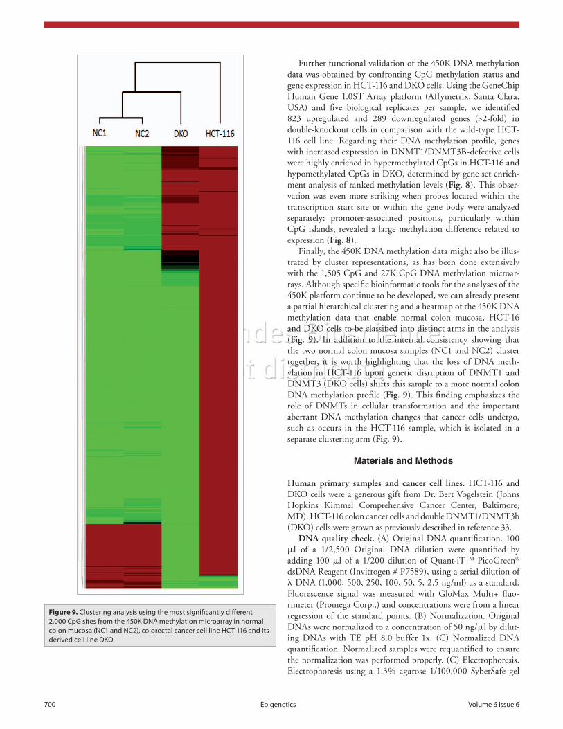
Further functional validation of the 450K DNA methylation data was obtained by confronting CpG methylation status and gene expression in HCT-116 and DKO cells. Using the GeneChip Human Gene 1.0ST Array platform (Affymetrix, Santa Clara, USA) and five biological replicates per sample, we identified 823 upregulated and 289 downregulated genes (>2-fold) in double-knockout cells in comparison with the wild-type HCT-116 cell line. Regarding their DNA methylation profile, genes with increased expression in DNMT1/DNMT3B-defective cells were highly enriched in hypermethylated CpGs in HCT-116 and hypomethylated CpGs in DKO, determined by gene set enrich-ment analysis of ranked methylation levels (Fig. 8). This obser-vation was even more striking when probes located within the transcription start site or within the gene body were analyzed separately: promoter-associated positions, particularly within CpG islands, revealed a large methylation difference related to expression (Fig. 8).
Finally, the 450K DNA methylation data might also be illus-trated by cluster representations, as has been done extensively with the 1,505 CpG and 27K CpG DNA methylation microar-rays. Although specific bioinformatic tools for the analyses of the 450K platform continue to be developed, we can already present a partial hierarchical clustering and a heatmap of the 450K DNA methylation data that enable normal colon mucosa, HCT-16 and DKO cells to be classified into distinct arms in the analysis (Fig. 9). In addition to the internal consistency showing that the two normal colon mucosa samples (NC1 and NC2) cluster together, it is worth highlighting that the loss of DNA meth-ylation in HCT-116 upon genetic disruption of DNMT1 and DNMT3 (DKO cells) shifts this sample to a more normal colon DNA methylation profile (Fig. 9). This finding emphasizes the role of DNMTs in cellular transformation and the important aberrant DNA methylation changes that cancer cells undergo, such as occurs in the HCT-116 sample, which is isolated in a separate clustering arm (Fig. 9).
Materials and Methods
Human primary samples and cancer cell lines. HCT-116 and DKO cells were a generous gift from Dr. Bert Vogelstein (Johns Hopkins Kimmel Comprehensive Cancer Center, Baltimore, MD). HCT-116 colon cancer cells and double DNMT1/DNMT3b (DKO) cells were grown as previously described in reference 33.
DNA quality check. (A) Original DNA quantification. 100 μl of a 1/2,500 Original DNA dilution were quantified by adding 100 μl of a 1/200 dilution of Quant-iTTM PicoGreen® dsDNA Reagent (Invitrogen # P7589), using a serial dilution of λ DNA (1,000, 500, 250, 100, 50, 5, 2.5 ng/ml) as a standard. Fluorescence signal was measured with GloMax Multi+ fluo-rimeter (Promega Corp.,) and concentrations were from a linear regression of the standard points. (B) Normalization. Original DNAs were normalized to a concentration of 50 ng/μl by dilut-ing DNAs with TE pH 8.0 buffer 1x. (C) Normalized DNA quantification. Normalized samples were requantified to ensure the normalization was performed properly. (C) Electrophoresis. Electrophoresis using a 1.3% agarose 1/100,000 SyberSafe gel
Figure 9. Clustering analysis using the most significantly different 2,000 CpG sites from the 450K DNA methylation microarray in normal colon mucosa (NC1 and NC2), colorectal cancer cell line HCT-116 and its derived cell line DKO.

©2011 Landes Bioscience.Do not distribute.
www.landesbioscience.com Epigenetics 701
GenomeStudio normalizes data using different internal con-trols that are present on the HumanMethylation 450 BeadChip. It also normalizes data depending on internal background proves. CpG methylation differences were considered significant above a cut-off of a 0.6-fold change in the β value, unless specifically indicated otherwise.
DNA methylation clustering. We used the GenomeStudio (2010.3) Methylation module (1.8.5) software to generate heat-maps of clustered CpGs. Unsupervised hierarchical clustering of the 2,000 most variable B values (standard deviation >0.37) with a detection p value > 0.01 was performed, calculating Euclidian distances.
Conclusions
In the near future it will be possible to obtain a greater number of complete human DNA methylomes at a reasonable cost using the new emerging sequencing technologies. However, this is still an extremely challenging process and currently there are only a very few whole methylomes available in which every CpG has been counted.18,19 However, if researchers in the pure genome sequencing area are encountering difficulties in obtaining a mas-sive number of completed human genomes in a cost-effective and user-friendly manner, those of us in the epigenetic field are even further behind—by several years. However, the development of a standardized comprehensive DNA methylation array that can be shared by different laboratories and agencies, employing a common methodology and bioinformatic package would be an important step in the right direction. Herein we have dem-onstrated that a newly available DNA methylation microarray (HumanMethylation450 BeadChip) might be an appropriate tool for this purpose. The 450K analyses of a colorectal tumori-genesis model that allows the study of normal and transformed cells in a variety of DNA methylation settings show that the data obtained are confirmed by the results of previously published studies in the area and provide biological, functional and techni-cal validation of the new microarray. In the coming years, the application of the 450K DNA methylation platform in a user-friendly 12-sample per array format that requires only 500 ng of bisulfite-converted DNA per sample will provide extensive infor-mation about the DNA methylation landscape of human tumors. Most importantly, it might also be decisive in establishing that DNA methylation defects have a central role in other common human diseases and in finding answers to many currently unre-solved biological questions.
Acknowledgments
This work was supported by the Fondo de Investigaciones Sanitarias Grant PI08-1345, Consolider Grant MEC09-05, Dr. Josef Steiner Cancer Research Foundation Award, Lilly Foundation Biomedical Research Award, European Research Council Advanced Grant EPINORC, the Health Department of the Catalan Government (Generalitat de Catalunya) and Fundació Cellex. J.S. is a “Juan de la Cierva” Researcher. M.E. is an Institucio Catalana de Recerca i Estudis Avançats (ICREA) Research Professor.
was performed to check for high molecular weight DNA frag-ments. 50 ng of DNA were loaded onto the gel using GeneRuler 1 kB as a ladder (Fermentas Cat. No. SM0314). The gel was run for 60 min at 120 mA. Samples showing bands or smears smaller than 10,000 bp were discarded. (D) DNA purity. 1 ul of normal-ized DNA was employed to measure the 260/280 and 260/230 ratios using a Nanodrop-1000. Samples showing RNA contami-nation (260/280 ratio >2) were discarded, and no samples with a 260/230 ratio less than 1 were accepted.
Bisulfite conversion. Bisulfite conversion of 600 ng of each sample was performed according to the manufacturer’s recom-mendations for the Illumina Infinium Assay (EZ DNA meth-ylation kit. Zymo Research. Cat. No. D5004). The incubation profile was 16 cycles at 95°C for 30 s, 50°C for 60 min and a final holding step at 4°C. The effectiveness of the bisulphite conversion was checked for three controls that were converted simultaneously with the samples. Enzymatically methylated Jurkat genomic DNA (New England Biolabs Cat. No. N4002S), genomic Jurkat DNA (New England Biolabs Cat. No. N4001S), and whole genome amplified genomic DNA were used as total methylated, intermediate-methylated and non-methylated con-trols. After bisulfite treatment, methylation-specific PCR51 was performed on the control samples using a set of primers to amplify a 301 bp region of the 28s ribosomal DNA. (5'-AAA ATT CTT TTC AAC TTT CCC T-3' and 5'-GAG TGA ATA GGG AAG AGT TTA G-3'). PCR conditions were: 2 mM MgCl
2 at 95°C
for 1 min, followed by 30 cycles at 95°C for 30 s, 58°C for 30 s and 72°C for 45 s, with a final step at 72°C for 10 min. Results were checked by loading 5 μl of the PCR control onto a 1.3% agarose gel using 1:100,000 dilution of SyberSafe and electro-phoresing at 80 mA for 30 min.
DNA methylation assay. 4 μl of bisulfite-converted DNA were used for hybridization on Infinium HumanMethylation 450 BeadChip, following the Illumina Infinium HD Methylation protocol. This consisted of a whole genome amplification step followed by enzymatic end-point fragmentation, precipita-tion and resuspension. The resuspended samples were hybrid-ized on HumanMethylation 450 BeadChips at 48°C for 16 h. Then unhybridized and non-specifically hybridized DNA were washed away, followed by a single nucleotide extension using the hybridized bisulfite-treated DNA as a template. The nucleotides incorporated were labeled with biotin (ddCTP and ddGTP) and 2,4-dinitrophenol (DNP) (ddATP and ddTTP). After the single-base extension, repeated rounds of staining were performed with a combination of antibodies that differentiated DNP and biotin by fixing them different fluorophores. Finally the BeadChip was washed and protected in order to scan it.
Scanning beadchips. The Illumina HiScan SQ scanner is a two-color laser (532 nm/660 nm) fluorescent scanner with a 0.375 μm spatial resolution capable of exciting the fluorophores generated during the staining step of the protocol. The intensi-ties of the images were extracted using GenomeStudio (2010.3) Methylation module (1.8.5) software. The methylation score for each CpG was represented as a b value according to the fluores-cent intensity ratio. B values may take any value between 0 (non-methylated) and 1 (completely methylated).

©2011 Landes Bioscience.Do not distribute.
702 Epigenetics Volume 6 Issue 6
References1. Jones PA, Baylin SB. The epigenomics of cancer. Cell
2007; 128:683-92.2. Berdasco M, Esteller M. Aberrant epigenetic landscape
in cancer: how cellular identity goes awry. Dev Cell 2010; 19:698-711.
3. Rodríguez-Paredes M, Esteller M. Cancer epigenetics reaches mainstream oncology. Nat Med 2011; 17:330-9.
4. Portela A, Esteller M. Epigenetic modifications and human disease. Nat Biotechnol 2010; 28:1057-68.
5. Feinberg AP. Epigenomics reveals a functional genome anatomy and a new approach to common disease. Nat Biotechnol 2010; 28:1049-52.
6. Urdinguio RG, Sanchez-Mut JV, Esteller M. Epigenetic mechanisms in neurological diseases: genes, syndromes and therapies. Lancet Neurol 2009; 8:1056-72.
7. Ordovás JM, Smith CE. Epigenetics and cardiovascular disease. Nat Rev Cardiol 2010; 7:510-9.
8. Lippman Z, Gendrel AV, Colot V, Martienssen R. Profiling DNA methylation patterns using genomic tiling microarrays. Nat Methods 2005; 2:219-24.
9. Irizarry RA, Ladd-Acosta C, Carvalho B, Wu H, Brandenburg SA, Jeddeloh JA, et al. Comprehensive high-throughput arrays for relative methylation (CHARM). Genome Res 2008; 18:780-90.
10. Irizarry RA, Ladd-Acosta C, Wen B, Wu Z, Montano C, Onyango P, et al. The human colon cancer methy-lome shows similar hypo- and hypermethylation at conserved tissue-specific CpG island shores. Nat Genet 2009; 41:178-86.
11. Ballestar E, Paz MF, Valle L, Wei S, Fraga MF, Espada J, et al. Methyl-CpG binding proteins identify novel sites of epigenetic inactivation in human cancer. EMBO J 2003; 22:6335-45.
12. Rauch TA, Wu X, Zhong X, Riggs AD, Pfeifer GP. A human B cell methylome at 100-base pair resolution. Proc Natl Acad Sci USA 2009; 106:671-8.
13. Weber M, Davies JJ, Wittig D, Oakeley EJ, Haase M, Lam WL, et al. Chromosome-wide and promoter-specific analyses identify sites of differential DNA methylation in normal and transformed human cells. Nat Genet 2005; 37:853-62.
14. Keshet I, Schlesinger Y, Farkash S, Rand E, Hecht M, Segal E, et al. Evidence for an instructive mechanism of de novo methylation in cancer cells. Nat Genet 2006; 38:149-53.
15. Weber M, Hellmann I, Stadler MB, Ramos L, Paabo S, Rebhan M, et al. Distribution, silencing potential and evolutionary impact of promoter DNA methylation in the human genome. Nat Genet 2007; 39:457-66.
16. Down TA, Rakyan VK, Turner DJ, Flicek P, Li H, Kulesha E, et al. A Bayesian deconvolution strategy for immunoprecipitation-based DNA methylome analysis. Nat Biotechnol 2008; 26:779-85.
17. Meissner A, Mikkelsen TS, Gu H, Wernig M, Hanna J, Sivachenko A, et al. Genome-scale DNA methylation maps of pluripotent and differentiated cells. Nature 2008; 454:766-70.
18. Lister R, Pelizzola M, Dowen RH, Hawkins RD, Hon G, Tonti-Filippini J, et al. Human DNA methylomes at base resolution show widespread epigenomic differ-ences. Nature 2009; 462:315-22.
19. Li Y, Zhu J, Tian G, Li N, Li Q, Ye M, et al. The DNA methylome of human peripheral blood mononuclear cells. PLoS Biol 2010; 8:1000533.
20. Bibikova M, Lin Z, Zhou L, Chudin E, Garcia EW, Wu B, et al. High-throughput DNA methylation profiling using universal bead arrays. Genome Res 2006; 16:383-93.
21. Christensen BC, Houseman EA, Marsit CJ, Zheng S, Wrensch MR, Wiemels JL, et al. Aging and environ-mental exposures alter tissue-specific DNA methylation dependent upon CpG island context. PLoS Genet 2009; 5:1000602.
22. Byun HM, Siegmund KD, Pan F, Weisenberger DJ, Kanel G, Laird PW, et al. Epigenetic profiling of somatic tissues from human autopsy specimens identi-fies tissue- and individual-specific DNA methylation patterns. Hum Mol Genet 2009; 18:4808-17.
23. Martinez R, Martin-Subero JI, Rohde V, Kirsch M, Alaminos M, Fernandez AF, et al. A microarray-based DNA methylation study of glioblastoma multiforme. Epigenetics 2009; 4:255-64.
24. Kanduri M, Cahill N, Göransson H, Enström C, Ryan F, Isaksson A, et al. Differential genome-wide array-based methylation profiles in prognostic subsets of chronic lymphocytic leukemia. Blood 2010; 115:296-305.
25. Bork S, Pfister S, Witt H, Horn P, Korn B, Ho AD, et al. DNA methylation pattern changes upon long-term culture and aging of human mesenchymal stromal cells. Aging Cell 2010; 9:54-63.
26. Teschendorff AE, Menon U, Gentry-Maharaj A, Ramus SJ, Weisenberger DJ, Shen H, et al. Age-dependent DNA methylation of genes that are sup-pressed in stem cells is a hallmark of cancer. Genome Res 2010; 20:440-6.
27. Rakyan VK, Down TA, Maslau S, Andrew T, Yang TP, Beyan H, et al. Human aging-associated DNA hyper-methylation occurs preferentially at bivalent chromatin domains. Genome Res 2010; 20:434-9.
28. Rhee I, Jair KW, Yen RW, Lengauer C, Herman JG, Kinzler KW, et al. CpG methylation is maintained in human cancer cells lacking DNMT1. Nature 2000; 404:1003-7.
29. Rhee I, Bachman KE, Park BH, Jair KW, Yen RW, Schuebel KE, et al. DNMT1 and DNMT3b cooperate to silence genes in human cancer cells. Nature 2002; 416:552-6.
30. Suzuki H, Gabrielson E, Chen W, Anbazhagan R, van Engeland M, Weijenberg MP, et al. A genomic screen for genes upregulated by demethylation and histone deacetylase inhibition in human colorectal cancer. Nat Genet 2002; 31:141-9.
31. Paz MF, Wei S, Cigudosa JC, Rodriguez-Perales S, Peinado MA, Huang TH, et al. Genetic unmasking of epigenetically silenced tumor suppressor genes in colon cancer cells deficient in DNA methyltransferases. Hum Mol Genet 2003; 12:2209-19.
32. Jacinto FV, Ballestar E, Ropero S, Esteller M. Discovery of epigenetically silenced genes by methylated DNA immunoprecipitation in colon cancer cells. Cancer Res 2007; 67:11481-6.
33. Lujambio A, Ropero S, Ballestar E, Fraga MF, Cerrato C, Setién F, et al. Genetic unmasking of an epigeneti-cally silenced microRNA in human cancer cells. Cancer Res 2007; 67:1424-9.
34. Schuebel KE, Chen W, Cope L, Glöckner SC, Suzuki H, Yi JM, et al. Comparing the DNA hypermethylome with gene mutations in human colorectal cancer. PLoS Genet 2007; 3:1709-23.
35. Lujambio A, Portela A, Liz J, Melo SA, Rossi S, Spizzo R, et al. CpG island hypermethylation-associated silencing of non-coding RNAs transcribed from ultra-conserved regions in human cancer. Oncogene 2010; 29:6390-401.
36. Toyota M, Sasaki Y, Satoh A, Ogi K, Kikuchi T, Suzuki H, et al. Epigenetic inactivation of CHFR in human tumors. Proc Natl Acad Sci USA 2003; 100:7818-23.
37. Akiyama Y, Watkins N, Suzuki H, Jair KW, van Engeland M, Esteller M, et al. GATA-4 and GATA-5 transcription factor genes and potential downstream antitumor target genes are epigenetically silenced in colorectal and gastric cancer. Mol Cell Biol 2003; 23:8429-39.
38. Lin J, Lai M, Huang Q, Ma Y, Cui J, Ruan W. Methylation patterns of IGFBP7 in colon cancer cell lines are associated with levels of gene expression. J Pathol 2007; 212:83-90.
39. Du Y, Carling T, Fang W, Piao Z, Sheu JC, Huang S. Hypermethylation in human cancers of the RIZ1 tumor suppressor gene, a member of a histone/pro-tein methyltransferase superfamily. Cancer Res 2001; 61:8094-9.
40. Yu JS, Koujak S, Nagase S, Li CM, Su T, Wang X, et al. PCDH8, the human homolog of PAPC, is a candidate tumor suppressor of breast cancer. Oncogene 2008; 27:4657-65.
41. Lujambio A, Calin GA, Villanueva A, Ropero S, Sánchez-Céspedes M, Blanco D, et al. A microRNA DNA methylation signature for human cancer metas-tasis. Proc Natl Acad Sci USA 2008; 105:13556-61.
42. Toyota M, Suzuki H, Sasaki Y, Maruyama R, Imai K, Shinomura Y, et al. Epigenetic silencing of microRNA-34b/c and B-cell translocation gene 4 is associated with CpG island methylation in colorectal cancer. Cancer Res 2008; 68:4123-32.
43. Huang YW, Liu JC, Deatherage DE, Luo J, Mutch DG, Goodfellow PJ, et al. Epigenetic repression of microRNA-129-2 leads to overexpression of SOX4 oncogene in endometrial cancer. Cancer Res 2009; 69:9038-46.
44. Lehmann U, Hasemeier B, Christgen M, Müller M, Römermann D, Länger F, et al. Epigenetic inactiva-tion of microRNA gene hsa-mir-9-1 in human breast cancer. J Pathol 2008; 214:17-24.
45. Furuta M, Kozaki KI, Tanaka S, Arii S, Imoto I, Inazawa J. miR-124 and miR-203 are epigenetically silenced tumor-suppressive microRNAs in hepatocel-lular carcinoma. Carcinogenesis 2010; 31:766-76.
46. Monk D. Deciphering the cancer imprintome. Brief Funct Genomics 2010; 9:329-39.
47. Sigalotti L, Covre A, Zabierowski S, Himes B, Colizzi F, Natali PG, et al. Cancer testis antigens in human melanoma stem cells: expression, distribution and methylation status. J Cell Physiol 2008; 215:287-91.
48. Robert MF, Morin S, Beaulieu N, Gauthier F, Chute IC, Barsalou A, et al. DNMT1 is required to main-tain CpG methylation and aberrant gene silencing in human cancer cells. Nat Genet 2003; 33:61-5.
49. Chen T, Hevi S, Gay F, Tsujimoto N, He T, Zhang B, et al. Complete inactivation of DNMT1 leads to mitotic catastrophe in human cancer cells. Nat Genet 2007; 39:391-6.
50. Hermann A, Gowher H, Jeltsch A. Biochemistry and biology of mammalian DNA methyltransferases. Cell Mol Life Sci 2004; 61:2571-87.
51. Herman JG, Graff JR, Myöhänen S, Nelkin BD, Baylin SB. Methylation-specific PCR: a novel PCR assay for methylation status of CpG islands. Proc Natl Acad Sci USA 1996; 93:9821-6.