A Microarray Related Study - Karger Publishers
15
Cell Physiol Biochem 2018;48:618-632 618 Cellular Physiology and Biochemistry Cellular Physiology and Biochemistry Original Paper Accepted: April 30, 2018 This article is licensed under the Creative Commons Attribution-NonCommercial-NoDerivatives 4.0 Interna- tional License (CC BY-NC-ND) (http://www.karger.com/Services/OpenAccessLicense). Usage and distribution for commercial purposes as well as any distribution of modified material requires written permission. DOI: 10.1159/000491890 Published online: July 18, 2018 © 2018 The Author(s) Published by S. Karger AG, Basel www.karger.com/cpb Abnormal Expression of Long Noncoding RNAs in Primary Immune Thrombocytopenia: A Microarray Related Study Tengda Li a Mingli Gu b Peng Liu a Yun Liu b Jie Guo b Weiwei Zhang b Anmei Deng a,c,d Cheng Qian e a Center of Clinical Experiments, Changhai Hospital, Second Military Medical University, Shanghai, b Department of Laboratory Diagnosis, Changhai Hospital, Second Military Medical University, Shanghai, c North Sichuan Medical College, Sichuan, d Shanxi University of Chinese Medicine, Xianyang, e The 100th Hospital of PLA, Suzhou, China Key Words Long noncoding RNA • Primary immune thrombocytopenia • Microarray • Quantitative real- time polymerase chain reaction • Co-expression network construction. Abstract Background/Aims: Long noncoding RNAs (lncRNAs) are important regulators of biological processes and they contribute to the pathological developments of various diseases, including autoimmune diseases. To gain the further understanding, we estimate the expression of lncRNAs in primary immune thrombocytopenia (ITP). Methods: In this study, microarray studies were performed to characterize expression profiles of various lncRNAs and mRNAs in blood samples collected from ITP patients. Quantitative real-time PCR (qRT-PCR) was performed to confirm the results, Kyoto Encyclopedia of Genes and Genomes (KEGG) analysis and gene ontology analysis were used to provide functional annotations, co-expression network construction (CNC) analysis was made to reveal the relations between lncRNAs and their targeted genes. Results: A total of 1177 and 632 lncRNAs were significantly up-regulated or down-regulated, respectively, in “newly diagnosed ITP” patients versus healthy individuals. In addition, 1182 genes and 737 genes were up-regulated or down-regulated, respectively, in “chronic recurrent ITP” patients versus healthy individuals. In a KEGG analysis, “TNF signaling pathway-Homo sapiens (human)” was a key result. In a gene ontology analysis, “Granulocyte macrophage colony-stimulating factor production (GO:0032604, ontology: Biological process, P = 1.69577E-05)” and “coreceptor activity (GO: 0015026, ontology: molecular function, P = 4.67594E-06)” were the two most critical results. Data from qRT-PCR and receiver operating characteristic curves further demonstrated that ENST00000440492, ENST00000528366, Cheng Qian and Anmei Deng The 100th Hospital of PLA, Suzhou 215007 (China) and Changhai Hospital, Shanghai 200433(China) E-Mail [email protected], [email protected] T. Li and M. Gu contributed equally to this work.
-
Upload
khangminh22 -
Category
Documents
-
view
4 -
download
0
Transcript of A Microarray Related Study - Karger Publishers

Cell Physiol Biochem 2018;48:618-632DOI: 10.1159/000491890Published online: July 18, 2018 618
Cellular Physiology and Biochemistry
Cellular Physiology and Biochemistry
© 2018 The Author(s). Published by S. Karger AG, Baselwww.karger.com/cpb
Li et al.: Abnormal Long Noncoding RNAs in Primary Immune Thrombocytopenia
Original Paper
Accepted: April 30, 2018
This article is licensed under the Creative Commons Attribution-NonCommercial-NoDerivatives 4.0 Interna-tional License (CC BY-NC-ND) (http://www.karger.com/Services/OpenAccessLicense). Usage and distribution for commercial purposes as well as any distribution of modified material requires written permission.
DOI: 10.1159/000491890Published online: July 18, 2018
© 2018 The Author(s) Published by S. Karger AG, Baselwww.karger.com/cpb
Abnormal Expression of Long Noncoding RNAs in Primary Immune Thrombocytopenia: A Microarray Related StudyTengda Lia Mingli Gub Peng Liua Yun Liub Jie Guob Weiwei Zhangb
Anmei Denga,c,d Cheng Qiane
aCenter of Clinical Experiments, Changhai Hospital, Second Military Medical University, Shanghai,bDepartment of Laboratory Diagnosis, Changhai Hospital, Second Military Medical University, Shanghai, cNorth Sichuan Medical College, Sichuan, dShanxi University of Chinese Medicine, Xianyang, eThe 100th Hospital of PLA, Suzhou, China
Key WordsLong noncoding RNA • Primary immune thrombocytopenia • Microarray • Quantitative real-time polymerase chain reaction • Co-expression network construction.
AbstractBackground/Aims: Long noncoding RNAs (lncRNAs) are important regulators of biological processes and they contribute to the pathological developments of various diseases, including autoimmune diseases. To gain the further understanding, we estimate the expression of lncRNAs in primary immune thrombocytopenia (ITP). Methods: In this study, microarray studies were performed to characterize expression profiles of various lncRNAs and mRNAs in blood samples collected from ITP patients. Quantitative real-time PCR (qRT-PCR) was performed to confirm the results, Kyoto Encyclopedia of Genes and Genomes (KEGG) analysis and gene ontology analysis were used to provide functional annotations, co-expression network construction (CNC) analysis was made to reveal the relations between lncRNAs and their targeted genes. Results: A total of 1177 and 632 lncRNAs were significantly up-regulated or down-regulated, respectively, in “newly diagnosed ITP” patients versus healthy individuals. In addition, 1182 genes and 737 genes were up-regulated or down-regulated, respectively, in “chronic recurrent ITP” patients versus healthy individuals. In a KEGG analysis, “TNF signaling pathway-Homo sapiens (human)” was a key result. In a gene ontology analysis, “Granulocyte macrophage colony-stimulating factor production (GO:0032604, ontology: Biological process, P = 1.69577E-05)” and “coreceptor activity (GO: 0015026, ontology: molecular function, P = 4.67594E-06)” were the two most critical results. Data from qRT-PCR and receiver operating characteristic curves further demonstrated that ENST00000440492, ENST00000528366,
Cheng Qianand Anmei Deng
The 100th Hospital of PLA, Suzhou 215007 (China) and Changhai Hospital, Shanghai 200433(China)E-Mail [email protected], [email protected]
T. Li and M. Gu contributed equally to this work.

Cell Physiol Biochem 2018;48:618-632DOI: 10.1159/000491890Published online: July 18, 2018 619
Cellular Physiology and Biochemistry
Cellular Physiology and Biochemistry
© 2018 The Author(s). Published by S. Karger AG, Baselwww.karger.com/cpb
Li et al.: Abnormal Long Noncoding RNAs in Primary Immune Thrombocytopenia
NR_038920, and ENST00000552576 can efficiently distinguish different stages of ITP, especially NR_038920 and ENST00000528366. In a CNC analysis, four lncRNAs were emphasized, and NR_038920 and ENST00000528366 were both associated with proteins with important roles in autoimmune diseases. Conclusions: These results suggest that lncRNAs act through targeted genes to mediate their functions and to mediate their functions and affect the pathogenesis of ITP.
Introduction
Primary immune thrombocytopenia (ITP), also known as idiopathic thrombocytopenic purpura, is an autoimmune disorder characterized by a decrease in platelets due to autoantibody-mediated destruction of platelets or compromised platelet production [1, 2]. It has been reported that pathological autoantibodies produced by B cells, aberrant T lymphocytes, and especially the turbulent developed megakaryocytes, significantly contribute to ITP[3-6]. Previous studies have demonstrated that most immune disorders are caused by dysregulation of gene expression [7, 8], and changes in phenotypes are often manifestations of changes in gene regulation [9-11]. In addition to coding genes, noncoding genes such as microRNAs and lncRNAs have increasingly gained attention [12-14]. LncRNAs have recently been discovered to be noncoding RNAs with lengths greater than 200 nucleotides [15]. Based on proximal protein-coding mRNAs, lncRNAs have been classified as sense lncRNAs, antisense lncRNAs, intronic lncRNAs, bidirectional lncRNAs, and long intergenic ncRNAs (lincRNAs). LncRNAs have been shown to affect every stage of a gene’s life cycle, including chromosome reconstruction, transcription, post-transcription, and intracellular metabolism [15-18]. In a recent study, the role of lncRNAs in the development of autoimmune diseases was highlighted [19]. For example, the lncRNAs, Gas5, Hotair and H19, and Tmevg1, have roles in systemic lupus erythematosus (SLE)[20, 21], rheumatoid arthritis (RA)[22, 23], and Sjögren syndrome (SS)[24], respectively. However, few studies have investigated lncRNAs in ITP. Among these studies, the lncRNA, TMEVPG1, has been shown to be related to Th1-type transcription factors, T-bet, STAT1, and STAT4, thereby indicating the importance of lncRNAs in the pathology of ITP[25]. In the present study, a microarray analysis of lncRNAs and mRNAs in blood samples collected from newly diagnosed ITP patients (n = 3), chronic recurrent ITP patients (n = 3), and healthy individuals (n = 3) was conducted (Fig. 1). The goal was to identify differentially expressed lncRNAs which may participate in the development of ITP. In addition, quantitative real-time PCR (qRT-PCR) was performed to detect selected lncRNAs in a larger number of patient samples, and bioinformatics methods were applied to reveal potential functions.
Materials and Methods
Patient selectionAll of the subjects enrolled in this study received
treatment at No. 100 Hospital of PLA (Suzhou, Jiangsu, China) or Changhai Hospital (Shanghai, China) between October 2014 and January 2015. There were
Fig. 1. Hierarchical clustering for lncRNAs(a) and mRNAs(b) detected in healthy individuals(N1-3), newly diagnosed ITP cases(P1a-c), and chronic recurrent ITP patients(P2a-c).
Figures
a b
Figure 1
© 2018 The Author(s)Published by S. Karger AG, Basel

Cell Physiol Biochem 2018;48:618-632DOI: 10.1159/000491890Published online: July 18, 2018 620
Cellular Physiology and Biochemistry
Cellular Physiology and Biochemistry
© 2018 The Author(s). Published by S. Karger AG, Baselwww.karger.com/cpb
Li et al.: Abnormal Long Noncoding RNAs in Primary Immune Thrombocytopenia
no statistically significant differences among the patients in regard to gender or age. Patients referred to as having “newly diagnosed ITP” (termed P1 below) included diagnosed patients without reliable predictive clinical or laboratory parameters of disease duration. The patients referred to as having “chronic recurrent ITP” (termed P2 below) were previously diagnosed with ITP that persisted for more than 12 months, as previously detailed [26]. Healthy individuals composed the N group and they were examined for the same period of time as the P1 and P2 patients. Table 1 summarizes the characteristics of the P1 (n = 30) and P2 (n = 30) patients. Pregnant patients and those with diabetic complications, cardiovascular disease, an active infection, hypertension, or other autoimmune diseases were excluded. The Ethics Committee of the People’s Liberation Army 100th Hospital and Changhai Hospital approved this research. The procedures performed were carefully explained to the patients and informed consents were signed by all of the participating patients. All experiments in the methods section were performed in accordance with approved international guidelines and ethical standards.
Patient specimensPeripheral blood (6 mL) was collected from each subject. To isolate peripheral blood mononuclear cells
(PBMCs), the collected blood samples were diluted with PBS (pH 7.2–7.4, Jiru Biology Company, Shanghai, China) and then slowly added to Ficoll-Hypaque (Thermo Fisher Scientific, USA). After centrifugation at 2500 rpm for 20 min, the obtained PBMCs were washed twice with PBS, were added into 1 ml Trizol reagent (Invitrogen, USA) and then were stored at -80 °C
RNA isolationPrepared blood samples were thawed at room temperature (RT) before chloroform was added to
each sample. After a brief vortexing step, the samples were centrifuged (12000 × g, 5 min, 4 °C) and the supernatants were transferred to RNase-free eppendorf tubes and mixed with isopropyl alcohol. After an incubation at RT for 30 min and another centrifugation step (12000 × g,15 min, 4 °C), the supernatants were precipitated with cold 75% alcohol and centrifuged (12000 × g, 5 min, 4 °C). The pellets were dried at RT for 2–5 min and then 20 ul of RNase-free water was added to each. From each sample, a 1 ul aliquot was analyzed for RNA concentration and purity with a Nanodrop 2000 instrument (Thermo Scientific, USA).
Microarray to obtain expression profiles for lncRNAs and mRNAsAn Arraystar Human LncRNA Microarray V3.0 (Arraystar Inc., USA) was used based on its capacity
to evaluate 30, 586 lncRNAs and 26, 109 coding transcripts. To prepare samples for analysis, total RNA was isolated as described above. Both RNA integrity and genomic DNA contamination were assayed by denaturing agarose gel electrophoresis performed with an Agilent Bioanalyzer 2100 instrument (Agilent Technologies, USA) by KangChen (Shanghai, China). Total RNA was purified with RNeasy Mini Kits (Qiagen, Germany) and Baseline-ZERO DNase (EPICENTRE, USA) according to the manufacturers’ protocols. A One-Color Quick Amp Labeling Kit (Agilent, USA) was used to label samples. The labeled samples were subsequently purified, along with labeled cRNA as a quality control, with RNeasy Mini Kits (Qiagen, Germany) according to the manufacturer’s protocol. Specific activity (pmol dye per g cRNA) was calculated as follows: specific activity = (pmol per ul dye)/(ug per ul cRNA). Array hybridization was performed according to the manufacturer’s protocol for the Agilent Gene Expression Hybridization Kit (Agilent, USA). The hybridized
Table 1. Characteristics of the patients with ITP. Abbreviations: ITP: immune thrombocytopenia; PLT: platelet; IVIG: intravenous immunoglobulin; GP: glycoprotein; GC: glucocorticoid; P1: newly diagnosed ITP; P2: chronic ITP
Variables P1 No. patients(%) P2 No. patients(%) Age of the ITP patients (y)
20 4(13.3%) 2(6.7%) 20-40 10(33.3%) 14(46.7%) 40-60 8(26.7%) 12(40.0%)
60 8(26.7%) 2(6.7%) Male 4(13.3%) 16(53.3%) Course(mo)
1(P1) 3-100(P2) 26(86.7%) 22(73.3%) 1-3(P1) 100(P2) 4(13.3%) 8(26.7%)
PLT count 10109/L 12(40.0%) 18(60.0%) Anti-platelet antibodies GPIIb+ 12(40.0%) 16(53.3%) GPIIIa+ 18(60.0%) 14(46.7%)
GPIb/IX+ 14(46.7%) 8(26.7%) GPIb+ 14(46.7%) 16(53.3%)
Therapy GC 4(13.3%) 30(100.0%) IVIG 2(6.7%) 16(53.3%)

Cell Physiol Biochem 2018;48:618-632DOI: 10.1159/000491890Published online: July 18, 2018 621
Cellular Physiology and Biochemistry
Cellular Physiology and Biochemistry
© 2018 The Author(s). Published by S. Karger AG, Baselwww.karger.com/cpb
Li et al.: Abnormal Long Noncoding RNAs in Primary Immune Thrombocytopenia
array was washed with Gene Expression Wash Buffer 1 and Gene Expression Wash Buffer 2 (Agilent, USA) before being scanned by an Agilent Microarray Scanner. Data were subsequently extracted and analyzed with Agilent Feature Extraction Software (V11.0.1.1).
Data analysisRaw data were imported into GeneSpring GX v12.1 software (Agilent, USA) for quantile standardization.
Differentially expressed LncRNAs and mRNAs with statistically significant differences between each set of comparisons were identified with P-value/ false discovery rate filtering. For the differently expressed lncRNAs or mRNAs, we selected them according to the following standards: 1) the fold change was ≥ 2.0 and P-value ≤ 0.05; 2) the raw intensity was ≥200 to make the background intensity as small as possible; 3) the Xhyb column was blank to make sure that there was not non-specific detection. Hierarchical clustering and combined analysis were performed with homemade scripts. To better understand the functions of the identified lncRNAs, correlations between enhancer lncRNAs, antisense lncRNAs, lincRNAs, and nearby mRNAs were analyzed. A gene ontology (GO) analysis was performed and the categories included: Molecular Function (MF), Biological Process (BP), and Cellular Component (CC). P-values were obtained according to Fisher’s exact test and were used to measure GO term enrichment for differentially expressed lncRNAs and mRNAs. To calculate fold enrichment of a GO term, the number of differentially expressed genes and those with an assigned GO term (“Count”), the number of genes annotated in the database (“Pop.Nits”), the number of different genes with GO annotations (“List.Total”), and the total number of genes in the database (background) with a GO annotation (“Pop.Total”), were used as follows: (Count / Pop.Nits) / (List.Total / Pop.Total), Enrichment Score = -log(P-value). A Kyoto Encyclopedia of Genes and Genomes (KEGG) Pathway analysis showed every gene/protein or small molecule in each Pathway Map it generated. Histograms presented the enrichment scores of the pathway terms. A Volcano plot was used to select differentially expressed mRNAs and lncRNAs (for all online suppl. material, see www.karger.com/doi/10.1159/000491890, Fig. S3a-c). A scatter plot was generated to present repeatability among the microarray data and groups (see online suppl. material, Fig. S3d-f). Probe distribution in the microarray is presented in a box plot, with changes before and after standardization shown (see online suppl. material, Fig. S3g-h). To display similar biological properties in multiple samples, hierarchical clustering was performed with Aligent GeneSpring GX software (version 11.5.1) (Fig. 1).
Fig. 2. CNC analysis results for 12 lncRNAs (a) and a subset of 4 immune-related lncRNAs [ENST00000528366, and ENST00000440492, NR_038920, and ENST00000552576] and their related genes (b). (a) The blue-green nodes represent coding genes, the red nodes represent lncRNAs. Solid lines indicate positive relationships between the lncRNAs and coding genes, and the dotted lines indicate negative relationships. (b) In this second CNC network, there were 47 nodes representing mRNAs, as well as related genes. The related genes were presented in colors that indicate their level of significance in immunity or autoimmunity (significance: blue-green > green > yellow-green > yellow).
PPP1R21
ZBED4
ZNF540
C14orf149JARID2 ZNF322
ASCC3
NOD2
ATP6V0A1
CYTH4 IER3ZNF283
C9orf69
S100PBP
TYSND1BICD2
SLC8A1
SGOL2
PID1
SLC31A1CLHC1
EXOC4
IRAK4ICA1 ARSATHEMIS
ENST00000412084
OR5B21 SLAMF6
EN1
TRIM49L1
TFB1M
DGKG
FGL2
KLK15
TRAF3IP3SZRD1
RGS3
NAT8B
HBA2
C1orf63
SLC4A1
AHSA2
MCFD2
TTLL11RAMP1
TMEM244
NDUFV3
MAPRE1GOLGA7
RAP2B
GBP4
RABGEF1
ECE1
TNFRSF10B ICAM1
TNFAIP8
CD300LFC20orf196
TACC3CNTLNAMICA1
PTEN
PLXNB2RHOBTB2
DNAJA3
UTY
ACYP1
MRPS7KYNUTRAF1
ARL8BHSPA5
KANSL2TRNT1
S100A9
NAIPHBBBRD8
MGAT4A
C6orf99
APBB1IPCHST2
RELBAKR1C4
IFIT3
TRAF3IP2 USP12
EZH2
GSTO1DTD1 FARP2KDM5D RAB27BFCRL5 CPSF2GLS
CLEC7ATMEM131
MS4A14OR7D2
CEACAM21
FUT3CCDC58
COG8
EGLN1KRTAP21-1
CPNE8
TWISTNB
GRAMD3
NHLRC1RIPK2
LIMS2
FAM26F
OASL
MSS51
SRC
HESX1SERPINB8
RAB3B
RIN2IFIT5
DOT1L
FRAT2 SPINT2
CMPK2
MAST3
ARL13B
ARRDC1EIF2A
LEPROTL1
ATG4C
KLRC4CDC42SE2 TMCO3
USP47TOMM5
TNFRSF10CLCN2
CD79B
TMLHE
CTSG
HBA1NLRC4
RPS6KA2ANKRD12
HBD
RGS16
NSA2
CCR7
ENST00000552576
PILRA
ME2
LIPN
TRAF3
B4GALT1
LYN
PRRG4
CBWD3
CD86
HLA-DRB1
CDV3
KIAA1919
IL1RN
CSNK1G3
PIM3SYTL3
LILRA3PPP1R17
FAM188A
GPR137B
ANKLE2 RNF144B
GBP1
TIMM10
ISG15FOS NFKB1
IDO1 HIST1H2BG
DBIDENND4A
CASP7
ENST000004 40492
GALR2ZC3HAV1
IRAK2NEK4
FGR
CAMP
DCAF12
KIAA0907
LCE2C
FIG4
ZNF268
C1orf43
TMEM2
EREGCXCR1
SRD5A1
ST14
SNCA
DDX6ZFYVE26
ARRDC2
DTNB
MGAT1APAF1ILF3
PRDM1
PLEKHM1
PYGL
IL8
SHBG
ARID4B
RIC8BSHOX
EML4
KPNA5
SEC14L4
CPA3
TTLL6
C3orf33
VMP1DERL2
MS4A6AITGA6
C19orf48
IGJ
B9D2
ZNF432
PLXDC2
NLRP12
NOL6
RNMT
FAM133A
SULT1A4
ACAA1
HCAR2PLXNB3
CDPF1
NOP56
AHSP
PPHLN1
NOS1AP
NR_038920
SULT1A3
MTX1
NFKBIA
NT5C3
ZNF404
LIN7A
DOCK8
DCAF12L2FGFBP1 CHCHD4
NSUN5
RND2 CSTF3
FBXO22
CTNNAL1
STAG2 ERAP2
NFKBIE
CD8A
MCM6
TRIM27RGS19
ASPRV1
CASP4BPTF
FCGR1A
F13A1
ALAS1
PHTF2
ERICH1
CSADRPS4Y1 ZFP36L1
H1FX
CAPN3
IRS2
NXT1
CH25H
GADD45A
PMS1
ATXN3L
ELK4
APOBEC3D
CFH
NCF4
C15orf48MLF1IP
CD63
NGLY1
YES1
TMEM66
AUTS2
FBXO33
CTSZ
HLA-DRB5
KRCC1B3GNT5
IL1B
NR4A3
ZNF350 SPTY2D1
ODC1
AGTPBP1
ZNF331
NRARP
TFDP2
SNX9
LTBP1
SAMHD1
ADPGK SLC16A5
CD55
METTL3
RECK
MAPK6
PLEKHG3
TBC1D2B
GIMAP8
UBE3A
CRY1
CXCL3DDIT3
LGALS2
SLC22A7
ADRB2
RILPL2
PHF21A
SLC22A15AHSA1
NGFRAP1
SH2D1A
NRG1
RELL1
SH2D2A
TMEM41B
ENST00000417932
SPOCK2
TYW5
POLR1C
SETD9
IFT46 ACOT1
CD68
NFAT5DDIT4
FBXO3
ABCA1
GUCY1A3
NUDT16
UBAP1
SLC35A1
OMA1
CYP51A1
ZNF410
TLR10
FAM160B1
ATP2B1
FCGR2A
SLC7A1
OR51E1
SERPINF1
HBG2NUDT7
MEGF9 CLCN3
ABCD3
CIRBP
IL17RA
SUV420H1
CRIM1
RCL1
FOSL2
HSPA9RBM38
TRIQK
HIST1H1C
RAB31
SAP30
ST20
TBC1D22A
SCN5A
SYNRG
RHBDD2
THUMPD3
VSTM1
FAM111ANCBP2
PVRL1
WDR48
ZNF281
SLC5A2
NANS
LTA
CBWD1
PNMAL1
GBP5
CD33
SLC6A6
PINX1
ARL3
PDE4B
H2AFJ
METTL7AFAM173B
MPP2
PNLIPRP2
PADI4
TET2
CXCR4
MFSD6L
CD28
NUDT17
BRWD1
BPGM
COA3
MOSPD2PTPN6
PRX
SPTLC2MSRB1
DENND2D
EAF1
CDC37L1
SNAPC3
TCEANC2CCDC99
PIAS2
SPSB1
SDR39U1
TMEM154
CDKN2B
SMAP2
CD5
CXCL2
DAB2
FEM1C
KIF3C
ACP1
EDDM3B
NRG3
KIAA1429
ARMC10
RAB13
GPR132
GALK2
CRY2
AKR1C3
LILRA5
NACA
OPRM1
CHST12
GNAL
DNAJB9
CDH22
ETV3
TEN1
HIVEP1
AGAP9TNFSF8
CTSW PDE6BSFXN1KCNJ2
KLHDC4
ZC3H12A
SPAG9LDHA
GTPBP4
CTTN
ENST00000436506
SLC39A8
ZNF175
SLC11A2
C1orf162
PNO1
TEX30
YPEL3
ENST00000528366
MYZAP
AQP9
C17orf96HNMT
PTS
TIMM23
NRP1
SOD2
IFNE
TAF9B
EIF2AK2
ZNF674 CDC42EP3
CCL18IFIT1
ACVR2A
EIF1B
RHOH
FBLIM1SERPINB9
CD36CYCSRLN2
CD44 LACTB
PELI1
ANGPTL2
CCDC50
IL23AMTM1
REL
C1orf21
KCTD12
ETS2
ZBTB10
uc010kun.2
MAP3K8
MTMR14
MAGOH
KLRC3MED6
STX11
TP73
FFAR2
FLCN
LRRC25
TIPARP
GMCL1TANK
PLK2
MAFFSRRM5
SCIMP
BAZ2B
SELK
OAZ2
WTAP
FOXD4L3
ZCCHC6
GPR182 PEX6
PTPN1
PRSS23
PELI2
CLP1SPRY2PLOD2
FYN CTSH
FRS2
RAB30
uc002ubt.1
SP2
G0S2
CALCA
FAM177A1
ZNF267
MRGPRX3
TP53BP2
SOCS1
ANXA2
FAM105BSGTB
CLDN22
TIMM22
PSMB7
PLAUR
CDK14
HNRNPDRNASEH1
NCK1
ENST00000440436
HECA
HAL
EZR
SCGB1C1
ANKRD22PRKAR1A
TSFM
CIRH1A
JMJD6
SKP1
INSIG1
PFKFB3
CCDC152
LRRC59PHACTR1
C11orf82
U2AF1
PANK3HIF1AUAP1
TRIM8NUP98
CBR1 ECDIL6R
KHDRBS1
CCDC18
AGAP1
NFYB
BACE1
HERPUD1
SH2D3C
P2RY13
CHST15
RAB22A
ZNF320
CUL4A
CCDC121
FAM136A
DNAJB6
CYorf15A PTPRO
CREM TNFAIP6
CYLDCCL3L3NOL7
ULBP2CASZ1
USP43CXCL1CEP290
LIMK2
ETNK1
CEP170
CARD16
IFI44
CDC42
CCL19
SAR1A UEVLD
ABT1
EID2B
ROCK2
SEC14L2
SLAMF7
KANSL1L HLA-DRAGCOM1
YME1L1 ZNF638
CBWD6CSDE1
ACAT2
SPTLC1
WDR67
PTGER3
FAM48A
BIRC3
EIF2B3
NAP1L5
PFKFB4
GADD45B
GBP7
COMMD2
SOS1
SLC9A7GTF3C4KCNA2
PLK3
STARD4TNFAIP3 UPF3B
HAUS2
VAPAHSF2DDX47 TM2D2
ZNF83
SAT1
IFNB1
ELL2
CCRN4L
CYTIP
CDK17
G3BP2
ARMC8
SLC25A3
VDAC1CD96
CKAP4
EGR3
USP32
ZBTB43
DNAJB11
RBM3
FAM103A1
RASGEF1B
ARID3C
FAM115C
FAM20B
ARID5B
TREM1
APLP2
BCL2A1TMPOANKRD37
CLEC4E
CCDC132
CES1
LCORL
ANKRD28
WDR73DNAJC18
C6orf225
SAMD9
SOWAHD
MNDA
MMP25
BCL2L1
MICU1BIRC2
ERCC1
NFE2
FBXL4
NFATC3
ATP13A3
LPCAT3
CNOT7
ABHD3
IER2
RAD51C
IL1R2
ZNF295PRMT2
TSPORAD50
PLGLB2
RHOT1
BPI
CR1
CAPNS2TRPM6
SUCLG1
LYSTZFAND5
NQO2
FKBP15
ABHD5
BRCA1 FAM82A1
HDAC8
ZNF668
GGCT
KIAA0319
SKP2
CXCR7
SP110
ATP6V0A4TK2
ZNF454
STX3ERN1
SNX10CDA
CDK19 KIAA0196
DEFA1B
LARP4
AREG
FBXL3
MYO1F
S100A8
TIMMDC1
DEXI
LCE1CZNF467
ADAM28DYSF
PICALM
JAK2FAM198B
ZFHX3
RMI1PARVG
HEATR3ENST00000521756
PLG
ZNF701
GHDC
MIS18BP1
HS3ST3B1
WIPF1 KLK3HIAT1
NCOA4
CFLARRAB12
MRPS18A
RGS2
GPR52
C18orf1EIF2C4
RASGRP4
DET1HIBCH
SP140
UBFD1
CLEC4D
PGK1
SIAH1
ZNF607
KIAA0754
RBM41
KIF13A
CR1L
MITF
CCL21
NEDD9
KLHL6
NCR3
UBR2DUSP5
ZNF860 PSMD12
HCAR3
APOL3CLMN
PCSK5
FOXC1
VPS35
ACOT9
FAM53B
MAGT1
ALYREF
GABPB1
ENSA
ZNFX1
GIT2
CBWD2
MKKS
CCRL2
LRP5L
ICOSPPP1R16B
FCAR
ADNP2
CCL3
AKAP5
CTNNB1
IL18
MX1
BMP2K
ZNF768
RSAD2
SLC46A2
ORC4
ZC2HC1A
SGMS1
ENST00000509463
ZMYM1
CCDC112TBC1D2
PQLC1
LCE3D
DRAM1
ANXA5
SMPDL3A
CCR2
C11orf9
RNF19B
MYCL1
RAPGEF2
PTPRJ
GK5CFB
ARFGAP3
RYBP ZFX
RIMBP3C
PGBD2
PQBP1
PMAIP1
SRGN
ZNF438
ETF1
TRIP11
POLR2M
JMY
TLR5
EIF5A CD274
CD69
FBXO25
CETN3
RAB3D
ARL5B
HNRNPH2
BTG3
BCL2L12
QKI
IFI44L
CXCL11
ZNF720
OSBPL11
PARD6G
ATP13A2
GTPBP1SUFU
FAM129C
IFIT2
DUSP10
RDBP
KLHL7
TMEM39A
SLU7
TMEM68
RPN2
GLT25D1
OXER1
CDK5R1
NRAS
RAB21XAGE1ARPE
a
CEA76
WARS FAM96A
Cell Physiol Biochem 2018;48:618-632DOI: 10.1159/000491890Published online: July 18, 2018 622
Cellular Physiology and Biochemistry
Cellular Physiology and Biochemistry
© 2018 The Author(s). Published by S. Karger AG, Baselwww.karger.com/cpb
Li et al.: Abnormal Long Noncoding RNAs in Primary Immune Thrombocytopenia
Synthesis of cDNA and qRT-PCRFor the synthesis of cDNA, an annealing mixture composed of 1.5 ug RNA, 1 ul oligo(dT)18 (0.5 ug/ul),
1.6 ul dNTPs mix (2.5 mM) (HyTest Ltd., China), and 10.4 ul RNA-free H2O was used. After the samples were incubated in the annealing mixture in a water bath at 65 °C for 5 min, and then in an ice bath for 2 min, the samples were subjected to a short centrifugation. The following reagents were then added in this order: 4 ul 5X First-Strand Buffer (Invitrogen, USA), 1 ul 0.1M DTT, 0.3 ul RNase Inhibitor (Epicentre, USA), and 0.2 ul SuperScript III RT (Invitrogen, USA). The samples were incubated at 37 °C for 1 min, at 50 °C for 60 min, and then at 70 °C 15 min in a thermostatic water tank (DK-8D, Senxin Instruments Inc., Shanghai, China). For immediate use, the samples were placed on ice, while samples were stored at -20 °C for later use.
PCR reactions were performed with 2X PCR Master Mix (Arraystar, USA) according to the manufacturer’s protocol. See online suppl. material, Table S2 provides a list of the primers used. β-Actin was detected as a reference gene and relative quantification was performed to measure gene expression levels. PCR amplification was subsequently performed to verify selected genes from the lncRNA microarray analysis in 30 patients with “newly diagnosed ITP” (P1) and 30 patients with “chronic recurrent ITP” (P2) (Table 1). ROC curves and areas under the ROC curves (AUCs) for the qRT-PCR results of the selected genes were generated with GraphPad Prism 6.0 software (GraphPad, USA).
Co-expression network constructionNormalized intensities of coding genes were calculated to perform a co-expression network
construction (CNC) analysis. When there were varying intensity values for different transcripts of the same gene, the median value of these intensities was used as the expressed value of the gene. The Pearson correlation coefficient (pcc) of the normalized intensities of the selected lncRNAs and all of their related genes was also calculated. When [abs(pcc)] was > 0.998 and false discovery rate (FDR) was <0.05 , the record was acquired. All of the records were plotted with Cytoscape (version 3.4.0) to construct a co-expression network that is shown in Fig. 2. Considering the immune functions demonstrated for the related genes in previous reports [27-42], 12 lncRNAs were selected for CNC analysis (Fig. 2a). Subsequently, the four most important lncRNAs (ENST00000440492, ENST00000528366, ENST00000552576, and NR_038920) and related genes according to their immunological function were selected for a CNC analysis (Fig. 2b).
Results
Differentially expressed lncRNAsCompared with healthy controls (the N group), a total of 7878 lncRNAs were found to be
up-regulated in newly diagnosed ITP patients (the P1 group), among which only 451 lncRNAs exhibited significant differences according to the standards as described above. The lncRNA that exhibited the greatest fold-change was NR_037166 (fold-change: 18.2). Meanwhile, a total of 7692 lncRNAs (312 of which exhibited significant differences) were down-regulated between the P1 and N groups. The most remarkable decrease in expression was exhibited by TCONS_00003870 (fold change: 93.6).
The total number of up-regulated lncRNAs between the chronic recurrent group (P2) and the N group was 9313 (with 376 lncRNAs exhibiting significant differences). The total number of down-regulated lncRNAs was 6257 (with 393 of these exhibiting significant differences) between the P2 and N groups. The most notably up-regulated lncRNA was TCONS_00000918 (fold-change: 13.9), while TCONS_00003870 (fold-change: 186.4) was the most down-regulated lncRNA.
Between the P2 and P1 groups, a total of 9392 up-regulated lncRNAs (with 100 exhibiting significant differences) and 6178 down-regulated lncRNAs (with 191 exhibiting significant differences) and their related genes were analyzed. The antisense lncRNAs that exhibited the greatest change between the P1 and N groups, between the P2 and N groups, and between the P2 and P1 groups were: uc010kun.2, NST00000559553, and ENST00000559553, respectively. Detailed information regarding these analysis is presented (see online suppl. material,) in Table S1 (a, b, and e). A hierarchical clustering analysis of lncRNAs detected in three healthy controls, three newly diagnosed ITP patients, and three chronic recurrent ITP

Cell Physiol Biochem 2018;48:618-632DOI: 10.1159/000491890Published online: July 18, 2018 623
Cellular Physiology and Biochemistry
Cellular Physiology and Biochemistry
© 2018 The Author(s). Published by S. Karger AG, Baselwww.karger.com/cpb
Li et al.: Abnormal Long Noncoding RNAs in Primary Immune Thrombocytopenia
patients is shown in Fig. 1a. We further analyzed the chromosomal derivation of the top10 lncRNAs in each group and found that in up-regulated groups (including P1 vs N, P2 vs N, P2 vs P1), those lncRNAs were focused on chr8 and chr11, while in down-regulated groups, they were mainly on chr2, chr12.
Differentially expressed mRNAsAn mRNA analysis of the P1 versus N samples, the P2 versus N samples, and the P2
versus P1samples identified 9519 up-regulated mRNAs (with 363 of these mRNAs exhibiting significant differences), 9795 up-regulated mRNAs (with 234 of these mRNAs exhibiting significant differences), and 9997 up-regulated mRNAs (with 40 of these mRNAs exhibiting significant differences), respectively. The most up-regulated mRNAs in each analysis were: NM_001008 (fold-change: 68.2), NM_001008 (fold-change: 84.0), and ENST00000361255 (fold-change: 5.4), respectively. Conversely, there were 10779 mRNAs that were down-regulated in the P1 group versus the N group (with 353 of these mRNAs exhibiting significant differences), 10503 mRNAs down-regulated in the P2 versus N group (with 516 of these mRNAs exhibiting significant differences), and 10301 mRNAs down-regulated in the P2 versus P1 groups (with 183 of these mRNAs exhibiting significant differences). The most-downregulated mRNAs in each group were: NM_002090 (fold-change: 48.4), NM_002090 (fold-change: 84.4), and NM_003246(fold-change: 8.4), respectively. Details of these analyses are summarized (see online suppl. material) in Table S1 (c,d).
GO analysisA GO analysis of the P1 group versus the N group data showed that the greatest fold
enrichment of GO terms for the up-regulated transcripts included: long-chain fatty acid binding (GO:0036041, ontology: MF, P = 0.000686924), response to bacterium (GO:0009617, ontology: BP, P = 9.31494E-09), and retromer complex (GO:0030904, ontology: CC, P = 0.000642907). The highest fold enrichment of GO terms for the down-regulated transcripts were: granulocyte macrophage colony-stimulating factor production (GO:0032604, ontology: BP, P = 1.69577E-05), coreceptor activity (GO:0015026, ontology: MF, P = 4.67594E-06), and T cell receptor complex (GO:0042101, ontology: CC, P = 0.012832272). Detailed information regarding these analyses are provided (see online suppl. material) in Fig. S1.
A GO analysis of the P2 group versus the N group data showed that the greatest fold enrichment of GO terms for the up-regulated transcripts included: oxygen transporter activity (GO:0005344, ontology: MF, P = 1.60566E-06), hemoglobin complex (GO:0005833, ontology: CC, P = 3.78385E-07), and oxygen transport (GO:0015671, ontology: BP, P = 3.65271E-06). For the down-regulated transcripts, the greatest fold enrichment of GO terms included: mRNA cap binding complex (GO:0005845, ontology: CC, P = 0.000243535), fever generation (GO:0001660, ontology: BP, P = 0.000102935), and activin binding (GO:0048185, ontology: MF, P = 0.003340754).
Finally, a GO analysis of the P2 group versus the P1 group showed that the greatest fold enrichment of GO terms for the up-related transcripts included: hemoglobin complex (GO:0005833, ontology: CC, P = 9.60704E-10), oxygen transport (GO:0015671, ontology: BP, P = 6.65012E-07), and oxygen transporter activity (GO:0005344, ontology: MF, P = 4.09006E-07). For the most down-regulated transcripts, the most enriched GO terms were: extracellular region (GO:0005576, ontology: CC, P = 6.75631E-09), cGMP-mediated signaling (GO:0019934, ontology: BP, P = 7.52627E-05), and IgG binding (GO:0019864, ontology: MF, P = 0.003234109).
Pathway analysisFor up-regulated transcripts, the highest enrichment score pathways included:
Legionellosis-Homo sapiens (human) (Fisher P-value = 2.51334E-05) in the P1 versus N analysis, Legionellosis-Homo sapiens (human) (Fisher P-value = 0.001259538) in the P2 versus N analysis, and Glycine, serine, and threonine metabolism - Homo sapiens (human) (Fisher P-value = 0.00173499) in the P2 versus P1 analysis (see online suppl. material, Fig.
Cell Physiol Biochem 2018;48:618-632DOI: 10.1159/000491890Published online: July 18, 2018 624
Cellular Physiology and Biochemistry
Cellular Physiology and Biochemistry
© 2018 The Author(s). Published by S. Karger AG, Baselwww.karger.com/cpb
Li et al.: Abnormal Long Noncoding RNAs in Primary Immune Thrombocytopenia
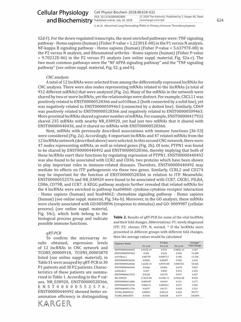
S2d-f). For the down-regulated transcripts, the most enriched pathways were: TNF signaling pathway - Homo sapiens (human) (Fisher P-value = 1.22301E-08) in the P1 versus N analysis, NF-kappa B signaling pathway - Homo sapiens (human) (Fisher P-value = 5.63797E-08) in the P2 versus N analysis, and Rheumatoid arthritis - Homo sapiens (human) (Fisher P-value = 9.70212E-06) in the P2 versus P1 analysis (see online suppl. material, Fig. S2a-c). The two most common pathways were the “NF APPA signaling pathway” and the “TNF signaling pathway” (see online suppl. material, Fig. S2, g and h).
CNC analysisA total of 12 lncRNAs were selected from among the differentially expressed lncRNAs for
CNC analysis. There were also nodes representing mRNAs related to the lncRNAs (a total of 912 different mRNAs) that were analyzed (Fig. 2a). Many of the mRNAs in the network were shared by two or more lncRNAs, yet the relationships were distinct. For example, CXCL11 was positively related to ENST00000528366 and uc010kun.2 (both connected by a solid line), yet was negatively related to ENST00000509463 (connected by a dotted line). Similarly, CD69 was positively related to ENST00000528366 and negatively related to ENST00000509463. More proximal lncRNAs shared a greater number of mRNAs. For example, ENST00000417932 shared 235 mRNAs with nearby NR_038920, yet had just two mRNAs that it shared with ENST00000440436, and it shared no mRNAs with ENST00000528366.
Next, mRNAs with previously described associations with immune functions [36-53] were considered (Fig. 2a). Accordingly, 4 important lncRNAs and 47 related mRNAs from the 12 lncRNAs network (described above) were selected. In this second CNC network, there were 47 nodes representing mRNAs, as well as related genes (Fig. 2b). Of note, PTPN1 was found to be shared by ENST00000440492 and ENST00000528366, thereby implying that both of these lncRNAs exert their functions by regulating expression of PTPN1. ENST00000440492 was also found to be associated with CCR2 and CD36, two proteins which have been shown to play important roles in immune-related diseases. Therefore, ENST00000440492 may mediate its effects on ITP pathogenesis via these two genes. Similarly, CCRL2 and CD274 may be important for the function of ENST00000528366 in relation to ITP. Meanwhile, ENST00000552576 and NR_038920 were found to be associated with CCR7, CXCR1, PILRA, CD86, CD79B, and CCR7. A KEGG pathway analysis further revealed that related mRNAs for the 4 lncRNAs were enriched in pathway hsa04060: cytokine-cytokine receptor interaction - Homo sapiens (human) and hsa04062: chemokine signaling pathway - Homo sapiens (human) (see online suppl. material, Fig. S4a-b). Moreover, in the GO analysis, these mRNAs were closely associated with GO:0050896 (response to stimulus) and GO: 0009987 (cellular process) (see online suppl. material, Fig. S4c), which both belong to the biological process group and indicate possible immune functions.
qRT-PCRTo confirm the microarray re-
sults obtained, expression levels of 12 lncRNAs in CNC network and TCONS_00000918, TCONS_00003870 listed (see online suppl. material), in Table S1 were assayed by qRT-PCR in 30 P1 patients and 30 P2 patients. Charac-teristics of these patients are summa-rized in Table 1. According to the P val-ues, NR_038920, ENST00000528366, E N S T 0 0 0 0 0 5 5 2 5 7 6 , ENST00000440492 showed better ex-amination efficiency in distinguishing
Table 2. Results of qRT-PCR for some of the vital lncRNAs and their fold changes. Abbreviations: P1: newly diagnosed ITP; P2: chronic ITP; N, normal. * If the lncRNAs were presented in different groups with different fold changes, then the average values would be calculated
Sequence Name P-Value Average* Fold Change P1 vs N P2 vs N P2 vs P1
ENST00000440492 4.915E-19 0.376 6.985E-13 3.187 ENST00000509463 0.581 0.626 0.466 13.468 uc010kun.2 0.00739 0.000717 0.186 12.296 ENST00000436506 0.0402 0.00387 0.582 2.634 ENST00000528366 1.623E-15 5.097E-08 0.000765 18.546 ENST00000440436 0.0466 0.0401 0.678 9.855 uc002ubt.1 0.447 0.828 0.416 2.493 ENST00000417932 0.0220 0.0170 0.597 2.457 NR_038920 2.781E-08 3.636E-11 2.655E-08 8.943 ENST00000412084 0.000187 0.0419 0.121 5.271 ENST00000552576 0.000111 0.000261 0.231 3.503 ENST00000521756 0.0337 0.0127 0.620 2.521 TCONS_00000918 0.00815 0.00133 0.302 30.577 TCONS_00003870 0.0106 0.00258 0.579 140.009

Cell Physiol Biochem 2018;48:618-632DOI: 10.1159/000491890Published online: July 18, 2018 625
Cellular Physiology and Biochemistry
Cellular Physiology and Biochemistry
© 2018 The Author(s). Published by S. Karger AG, Baselwww.karger.com/cpb
Li et al.: Abnormal Long Noncoding RNAs in Primary Immune Thrombocytopenia
P1 from N, P2 from N, and P2 from P1 than other lncRNAs (Table 2). In detail, expres-sion of NR_038920 significantly differed between the P1 and N groups, between the P2 and N groups, and between the P2 and P1 groups. ENST00000528366 also significantly differed between these three groups. In addition, ENST00000552576 distinguished the P1 samples from the N samples and the P2 samples from the N samples, while ENST00000440492 sig-nificantly differed between the P1 and N groups and between the P2 and P1 groups (Fig. 3a-d, P-values were shown in Table 2). AUCs of the ROC curves for the four lncRNAs ranged from 0.531 to 0.997, implying that these lncRNAs can distinguish different stages of ITP and the presence or absence of ITP, particu-larly NR_038920 (AUC: 0.857–0.958) and ENST00000528366 (AUC: 0.758–0.962) (Fig. 3e-h).
Furthermore, we also examined the gene expression of CCR7, CXCR1, CD274, CCRL2 by qRT-PCR as above, the results showed that the gene expressions of CXCR1 and CD274 were significantly different in P1 vs N, P2 vs N, P2 vs P1 groups. For CXCR1, the P-values were 0.000409, 1.11E-08, 0.000309, separately; for CD274, 0.00444, 3.696E-07, 0.0109, separately. CCR7 and CCRL2 were statistically different in P1 vs N, and P2 vs N groups. For CCR7, P-values were 0.00316, 2.529E-05, individually; for CCRL2, 0.00160, 3.86E-05, separately (see online suppl. material, Fig. S5).
Discussion
ITP is an autoimmune disorder that is characterized by pathologically produced lymphocytes, abnormally developed megakaryocytes, and/or biologically disordered platelets [5]. With the development of second generation sequence analysis (especially RNA-seq), an increasing number of lncRNAs are being identified, as well as their roles in various types of diseases, including cancers (e.g., hepatocellular carcinoma, prostate cancer, etc.) and autoimmune diseases (e.g., SLE, SS, etc.)[54-56]. Previous studies have shown that lncRNAs potentially influence the functions of dendritic cells (DCs) and T lymphocytes, and also affect the differentiation of Th1/Th2 cells which play an important role in the development of ITP[57-59]. For example, targeting of STAT3 by lnc-DC involves the differentiation of DCs, and this in turn can induce the activation of an immune response [57]. In addition, linc-MAF-4 influences the differentiation of CD4+ T lymphocytes by targeting the chromatin modifiers, EZH2 and LSD1[60]. Considering the potential roles that lncRNAs may have in autoimmune disorders, they have received greater attention in recent years. As a result, specific roles for lncRNAs in autoimmune diseases such as SLE, RA, and SS have been elucidated [20-22].
In the present study, lncRNAs were profiled in three newly diagnosed ITP patients, in three chronic recurrent ITP patients, and in three healthy individuals by microarray (Fig. 1). The lncRNAs which exhibited the greatest fold-changes were NR_047631, ENST00000417071, and uc010idt.2 (see online suppl. material, Table S1a-b). We hypothesize that these lncRNAs may critically affect ITP pathology. To gain insight into the biological functions of these lncRNAs, bioinformatics were applied. As a result, ENST00000559553 was found to be related to “normal mucosa of esophagus-specific gene 1 protein”. Previously, normal mucosa
Fig. 3. Box plots (a-d) and ROC curves (e-h) for gene expression of ENST00000440492, ENST00000528366, NR_038920, and ENST00000552576. Blue, P1 vs N, Green, P2 vs N, Red, P2 vs P1, ns:not significant, *P<0.001, **P<0.0001.
b
Figure 2

Cell Physiol Biochem 2018;48:618-632DOI: 10.1159/000491890Published online: July 18, 2018 626
Cellular Physiology and Biochemistry
Cellular Physiology and Biochemistry
© 2018 The Author(s). Published by S. Karger AG, Baselwww.karger.com/cpb
Li et al.: Abnormal Long Noncoding RNAs in Primary Immune Thrombocytopenia
of esophagus-specific gene 1 protein was overexpressed in an esophageal squamous cell carcinoma cell line and cell motility was found to be affected [61].
In the GO analysis of down-regulated lncRNAs identified from a comparison of P1 versus N group data, three terms were predominant: “regulation of granulocyte macrophage colony-stimulating factor (GM-CSF) production”, “coreceptor activity”, and “T cell receptor complex” (see online suppl. material, Fig. S1d-f). These results suggest that differentially expressed lncRNAs may contribute to the pathology of ITP by influencing production of GM-CSF, coreceptor activity, and/or T cell receptors, and these correlations are consistent with the results of previous studies which have characterized the pathogenesis of autoimmune diseases [62]. For example, in patients with multiple sclerosis (MS), an increase in GM-CSF-expressing B cells has been detected, and this increase could activate myeloid cells in a GM-CSF-dependent manner [63]. In relation to B cells, CD22 is a co-receptor of these cells and is reported to control the B cell response to antigens. Thus, dysfunction of CD22 could lead to the development of autoimmune diseases such as SLE. Correspondingly, epratuzumab, a monoclonal antibody recognizing CD22, is a potential treatment for SLE[64]. T cells and their receptors have also been characterized as critical immune factors in immune-related diseases, with T cell receptors (TCRs) being able to recognize restricted antigenic peptide fragments and mediate signaling via transduction pathways which are critical for T cell activation [65]. Regarding the latter, decreased tyrosine phosphorylation of the TCR zeta-chain has been linked to defective TCR signaling that may underlie the hyporesponsiveness of synovial T cells in RA[66].
Metabolism-associated GO terms were also identified in the present study. These included: “long-chain fatty acid binding”, “oxygen transporter activity”, “oxygen transport”, and inflammation-related terms such as “response to bacterium” and “fever generation”. It is well established that ITP represents a complex disease that is accompanied by an inflammatory reaction mediated by a variety of immune cytokines including IL-17, IFN-γ, and TNF-α, among others. It has also been observed that the metabolism of these cytokines can change over the course of this disease, including their oxidant and antioxidant states, and these changes can potentially influence the development of ITP[33, 67-69]. It is possible that differentially expressed lncRNAs exert their functions by influencing inflammatory or metabolism processes, and these effects could influence the development of ITP. In the pathway analysis conducted in the present study, the “TNF signaling pathway - Homo sapiens (human)” was markedly enriched in a comparison of the down-regulated lncRNAs between the P1 and N groups (see online suppl. material, Fig. S2a). Previous studies have shown that TNFR1 and TNFR2 are two important receptors that mediate signaling downstream of TNF activation and binding [70]. In general, signaling via TNF-TNFR1 involves the MAPK and NF-κβ pathways. In contrast, signaling via TNF-TNFR2 is limited to CD4+ T cells, CD8+ T cells, and endothelial cells, and signaling via NF-κβ can be stimulated as well [70-74]. Between the P2 and N group data, the most enriched pathway that was identified was the, “NF-kappa B signaling pathway - Homo sapiens (human)” (see online suppl. material, Fig. S2g); and between the P2 and P1 group data, the “Rheumatoid arthritis - Homo sapiens (human)” pathway was identified (see online suppl. material, Fig. S2b-c). A role for the NF-κβ signaling pathway in maintaining immune tolerance in the body has previously been demonstrated, including its roles in central and peripheral tolerance which include an inhibition of regulatory T cells [75, 76]. Thus, NF-κβ signaling is an important mediator in regulating chronic inflammatory diseases and autoimmune diseases [77]. In brain tissues affected by MS, NF-κβ has also been shown to localize to astrocytes and infiltrating macrophages in or near lesions in the central nervous system [77, 78]. Furthermore, genetic studies have shown that abnormal levels of NF-κβ-related genes may be related to MS relapses [79, 80]. Regarding the RA pathway that was identified in a comparison of the P2 and P1 groups, enrichment of this pathway may be due to similarities in the underlying mechanisms of these two diseases, which include the involvement of CD4+ T cells, the NF-κβ signaling pathway, and aberrant expression of cytokines such as IFN-γ. Moreover, clinically, patients with RA are often complicated by

Cell Physiol Biochem 2018;48:618-632DOI: 10.1159/000491890Published online: July 18, 2018 627
Cellular Physiology and Biochemistry
Cellular Physiology and Biochemistry
© 2018 The Author(s). Published by S. Karger AG, Baselwww.karger.com/cpb
Li et al.: Abnormal Long Noncoding RNAs in Primary Immune Thrombocytopenia
immune thrombocytopenic purpura, thereby further supporting the relationship between these two diseases [81-84].
For the CNC analysis, we initially selected 12 lncRNAs. Then, four of these 12 lncRNAs were selected for further analysis (Fig. 2, a and b). As a result, NR_038920 appear to share related genes with ENST00000552576, such as CCR7 and CXCR1 (Fig. 2b). CCR7 has been shown to play an important role in adaptive immune activities as part of a CCR7-CCL19/CCL21 signaling axis. This axis has been shown to affect T lymphocyte function, DCs, and T cell priming or adaptive immunity in lymph nodes [85, 86]. Furthermore, CCR7 has also been shown to promote T cell development and lymphorganogenesis, two processes which are significant in the progression of autoimmune diseases [85, 86]. Thus, inappropriate regulation of the CCR7-CCL19/CCL21 axis may lead to immune disorders, including the development of autoimmune diseases [85]. Regarding CXCR1, it has always been found to function in combination with CXCR2. The CXCR1/2 complex has been detected on neutrophils, macrophages, and lymphocytes, and these cell types are critical in inflammatory processes and autoimmune diseases [50, 87, 88]. Inhibition of CXCR1/2 has been shown to prevent islet damage due to immune disorders, and this inhibition also promotes autoimmune throditis [50]. Alternatively, the CXCR1/CXCL8 axis has been associated with the accumulation of NK cells in the liver of patients with primary biliary disease, thereby contributing to the pathological development of this disease [89]. ENST00000528366 is shown to be related with CD274 and CCRL2. CD274, also known as ligand for programmed cell death 1 (PD-L1), is expressed on the surface of lymphocytes, macrophages or the dendritic cells [48]. After interacting with PD-1, it can regulate the proliferation or the immune-tolerance of T cells, it is emphasized in the T cell-mediated autoimmune diseases such as SLE, MS, et al [90, 91]. In patients with active SLE, PD-L1 is found to be overexpressed in monocytes and negatively related with the level of TGF-β, while interestingly when patients are in the recovery stage, PD-L1 is showed to be positively related to TNF-α, implying that PD-L1 may involve in the pathogenesis of SLE by influencing the expression of TGF-β or TNF-α[90]. Chemokine (CC motif) receptor-like 2 (CCRL2) is a 7-transmembrane protein related to the family of the typical chemokine receptors that participate in the inflammation process, for example, in the murine models of experimental autoimmune encephalitis, those short of CCRL2 develop exacerbated nonresolving disease and recurrent inflammatory reactions, suggesting that it may take part in the control of multiple sclerosis [46]. Based on the roles CCRL2 and CD274 played in the process of autoimmune diseases, in ITP ENST00000528366 may also involve in its pathogenesis through the pathways as described above.
According to the literature regarding ITP, ENST00000440492, ENST00000528366, NR_038920, and ENST00000552576 may act through similar pathways. For example, when the pathways or GO terms associated with the mRNAs in the CNC network were searched to reveal their related lncRNAs’ functions, hsa04060: cytokine-cytokine receptor interaction - Homo sapiens (human) (see online suppl. material, Fig. S4b) and hsa04062: chemokine signaling pathway - Homo sapiens (human) (see online suppl. material, Fig. S4a) pathways were identified. The GO results further suggest that these lncRNAs may mediate functions associated with stimulus response (GO:0050896) and cellular processes (GO: 0009987) (see online suppl. material, Fig. S4c).
To further verify the microarray results of the present study, qRT-PCR was performed to detect ENST00000440492, ENST00000528366, NR_038920, and ENST00000552576 in 30 patients with newly diagnosed ITP (P1) and 30 patients with chronic recurrent ITP (P2). These results (Fig. 3a-d), in combination with the ROC curves data (Fig. 3e-h), confirm that these four lncRNAs can efficiently distinguish different stages of ITP, especially NR_038920 and ENST00000528366 which can distinguish P1, P2 from N and P1 from P2 (ENST00000440492 cannot distinguish P2 from N, and ENST00000552576 cannot distinguish P2 from P1). Consequently, these lncRNAs may represent potential targets for monitoring, diagnosing, and/or treating ITP.
To our knowledge, this is the first microarray-based study to investigate lncRNAs in ITP. The identification of differentially expressed lncRNAs in this study provides support

Cell Physiol Biochem 2018;48:618-632DOI: 10.1159/000491890Published online: July 18, 2018 628
Cellular Physiology and Biochemistry
Cellular Physiology and Biochemistry
© 2018 The Author(s). Published by S. Karger AG, Baselwww.karger.com/cpb
Li et al.: Abnormal Long Noncoding RNAs in Primary Immune Thrombocytopenia
for further analyses regarding their functions in this disease process. As described above, lncRNAs may exert their functions by influencing other molecules or via actions through certain pathways. Accordingly, the latter has been demonstrated for a subset of lncRNAs in relation to the development of certain autoimmune diseases. Thus, it appears that lncRNAs have the capacity to facilitate monitoring of ITP progression and to serve as targets for the treatment of this disease, and further studies of lncRNAs are warranted.
Acknowledgements
This research was supported by grants from the National 973 Foundation (2013CB531606), the National Science Foundation of China (81701608, 81671556, 81601406, 81501397, 31500721, 81501398, 81471605, 81401358, 81302579, 81273282 and 81202353), Shanghai Shenkang (SHDC22014014), Shanghai Educational Science (D14017), Army Scientific Research (BWS14J023, 15ZD009,15XD007), China Postdoctoral Science Foundation (2017M623440) and MJD Founding (MJR20150019).
Disclosure Statement
The authors have no competing interests to declare.
References
1 Neunert C, Lim W, Crowther M, Cohen A, Solberg L, Jr., Crowther MA: The American Society of Hematology 2011 evidence-based practice guideline for immune thrombocytopenia. Blood 2011;117:4190-4207.
2 Schipperus M, Fijnheer R: New therapeutic options for immune thrombocytopenia. Neth J Med 2011;69:480-485.
3 Ji X, Zhang L, Peng J, Hou M: T cell immune abnormalities in immune thrombocytopenia. J Hematol Oncol 2014;7:72.
4 Kuwana M, Kaburaki J, Ikeda Y: Autoreactive T cells to platelet GPIIb-IIIa in immune thrombocytopenic purpura. Role in production of anti-platelet autoantibody. J Clin Invest 1998;102:1393-1402.
5 Gernsheimer T: Chronic idiopathic thrombocytopenic purpura: mechanisms of pathogenesis. Oncologist 2009;14:12-21.
6 Zhou Z, Li X, Li J, Su C, Zhuang L, Luo S, Zhang L: Direct B-cell stimulation by peripheral blood monocyte-derived dendritic cells in idiopathic thrombocytopenic purpura patients. J Clin Immunol 2010;30:814-822.
7 Jernas M, Nookaew I, Wadenvik H, Olsson B: MicroRNA regulate immunological pathways in T-cells in immune thrombocytopenia (ITP). Blood 2013;121:2095-2098.
8 Hrdlickova B, Kumar V, Kanduri K, Zhernakova DV, Tripathi S, Karjalainen J, Lund RJ, Li Y, Ullah U, Modderman R, Abdulahad W, Lahdesmaki H, Franke L, Lahesmaa R, Wijmenga C, Withoff S: Expression profiles of long non-coding RNAs located in autoimmune disease-associated regions reveal immune cell-type specificity. Genome Med 2014;6:88.
9 Bodenreider O, Burgun A: A framework for comparing phenotype annotations of orthologous genes. Stud Health Technol Inform 2010;160:1309-1313.
10 Harper M, Gronenberg L, Liao J, Lee C: Comprehensive detection of genes causing a phenotype using phenotype sequencing and pathway analysis. PLoS One 2014;9:e88072.
11 Luo J, Liang S: Prioritization of potential candidate disease genes by topological similarity of protein-protein interaction network and phenotype data. J Biomed Inform 2015;53:229-236.
12 Gernapudi R, Wolfson B, Zhang Y, Yao Y, Yang P, Asahara H, Zhou Q: MicroRNA 140 Promotes Expression of Long Noncoding RNA NEAT1 in Adipogenesis. Mol Cell Biol 2016;36:30-38.
13 Shi Q, Yang X: Circulating MicroRNA and Long Noncoding RNA as Biomarkers of Cardiovascular Diseases. J Cell Physiol 2016;231:751-755.

Cell Physiol Biochem 2018;48:618-632DOI: 10.1159/000491890Published online: July 18, 2018 629
Cellular Physiology and Biochemistry
Cellular Physiology and Biochemistry
© 2018 The Author(s). Published by S. Karger AG, Baselwww.karger.com/cpb
Li et al.: Abnormal Long Noncoding RNAs in Primary Immune Thrombocytopenia
14 Sui W, Lin H, Peng W, Huang Y, Chen J, Zhang Y, Dai Y: Molecular dysfunctions in acute rejection after renal transplantation revealed by integrated analysis of transcription factor, microRNA and long noncoding RNA. Genomics 2013;102:310-322.
15 Song X, Cao G, Jing L, Lin S, Wang X, Zhang J, Wang M, Liu W, Lv C: Analysing the relationship between lncRNA and protein-coding gene and the role of lncRNA as ceRNA in pulmonary fibrosis. J Cell Mol Med 2014;18:991-1003.
16 Kazemzadeh M, Safaralizadeh R, Orang AV: LncRNAs: emerging players in gene regulation and disease pathogenesis. J Genet 2015;94:771-784.
17 Li X, Wu Z, Fu X, Han W: lncRNAs: insights into their function and mechanics in underlying disorders. Mutat Res Rev Mutat Res 2014;762:1-21.
18 Wu Z, Liu X, Liu L, Deng H, Zhang J, Xu Q, Cen B, Ji A: Regulation of lncRNA expression. Cell Mol Biol Lett 2014;19:561-575.
19 Sigdel KR, Cheng A, Wang Y, Duan L, Zhang Y: The Emerging Functions of Long Noncoding RNA in Immune Cells: Autoimmune Diseases. J Immunol Res 2015;2015:848790.
20 Haywood ME, Rose SJ, Horswell S, Lees MJ, Fu G, Walport MJ, Morley BJ: Overlapping BXSB congenic intervals, in combination with microarray gene expression, reveal novel lupus candidate genes. Genes Immun 2006;7:250-263.
21 Wu GC, Li J, Leng RX, Li XP, Li XM, Wang DG, Pan HF, Ye DQ: Identification of long non-coding RNAs GAS5, linc0597 and lnc-DC in plasma as novel biomarkers for systemic lupus erythematosus. Oncotarget 2017;8:23650-23663.
22 Song J, Kim D, Han J, Kim Y, Lee M, Jin EJ: PBMC and exosome-derived Hotair is a critical regulator and potent marker for rheumatoid arthritis. Clin Exp Med 2015;15:121-126.
23 Zhou JZ, Li JJ, Hua DJ, Huang SC, Sun QQ, Huang H, Xin XF, Cen H: A study on associations of single-nucleotide polymorphisms within H19 and HOX transcript antisense RNA (HOTAIR) with genetic susceptibility to rheumatoid arthritis in a Chinese population. Inflamm Res 2017;66:515-521.
24 Wang J, Peng H, Tian J, Ma J, Tang X, Rui K, Tian X, Wang Y, Chen J, Lu L, Xu H, Wang S: Upregulation of long noncoding RNA TMEVPG1 enhances T helper type 1 cell response in patients with Sjogren syndrome. Immunol Res 2016;64:489-496.
25 Li H, Hao Y, Zhang D, Fu R, Liu W, Zhang X, Xue F, Yang R: Aberrant expression of long noncoding RNA TMEVPG1 in patients with primary immune thrombocytopenia. Autoimmunity 2016;49:496-502.
26 Rodeghiero F, Stasi R, Gernsheimer T, Michel M, Provan D, Arnold DM, Bussel JB, Cines DB, Chong BH, Cooper N, Godeau B, Lechner K, Mazzucconi MG, McMillan R, Sanz MA, Imbach P, Blanchette V, Kuhne T, Ruggeri M, George JN: Standardization of terminology, definitions and outcome criteria in immune thrombocytopenic purpura of adults and children: report from an international working group. Blood 2009;113:2386-2393.
27 Cuker A: ITP and TTP: interpreting evidence in light of patient values. Blood 2015;125:1514-1515.28 Culic S, Salamunic I, Konjevoda P, Dajak S, Pavelic J: Immune thrombocytopenia: serum cytokine levels in
children and adults. Med Sci Monit 2013;19:797-801.29 Del Vecchio GC, Giordano P, Tesse R, Piacente L, Altomare M, De Mattia D: Clinical significance of serum
cytokine levels and thrombopoietic markers in childhood idiopathic thrombocytopenic purpura. Blood Transfus 2012;10:194-199.
30 Feng X, Scheinberg P, Samsel L, Rios O, Chen J, McCoy JP, Jr., Ghanima W, Bussel JB, Young NS: Decreased plasma cytokines are associated with low platelet counts in aplastic anemia and immune thrombocytopenic purpura. J Thromb Haemost 2012;10:1616-1623.
31 Jernas M, Hou Y, Stromberg Celind F, Shao L, Nookaew I, Wang Q, Ju X, Mellgren K, Wadenvik H, Hou M, Olsson B: Differences in gene expression and cytokine levels between newly diagnosed and chronic pediatric ITP. Blood 2013;122:1789-1792.
32 Jernas M, Hou Y, Stromberg Celind F, Shao L, Wang Q, Ju X, Mellgren K, Wadenvik H, Hou M, Olsson B: Altered cytokine levels in pediatric ITP. Platelets 2015;26:589-592.
33 Leontyev D, Neschadim A, Branch DR: Cytokine profiles in mouse models of experimental immune thrombocytopenia reveal a lack of inflammation and differences in response to intravenous immunoglobulin depending on the mouse strain. Transfusion 2014;54:2871-2879.
34 Pourcelot E, Trocme C, Mondet J, Bailly S, Toussaint B, Mossuz P: Cytokine profiles in polycythemia vera and essential thrombocythemia patients: clinical implications. Exp Hematol 2014;42:360-368.

Cell Physiol Biochem 2018;48:618-632DOI: 10.1159/000491890Published online: July 18, 2018 630
Cellular Physiology and Biochemistry
Cellular Physiology and Biochemistry
© 2018 The Author(s). Published by S. Karger AG, Baselwww.karger.com/cpb
Li et al.: Abnormal Long Noncoding RNAs in Primary Immune Thrombocytopenia
35 Talaat RM, Elmaghraby AM, Barakat SS, El-Shahat M: Alterations in immune cell subsets and their cytokine secretion profile in childhood idiopathic thrombocytopenic purpura (ITP). Clin Exp Immunol 2014;176:291-300.
36 Antonelli A, Ferrari SM, Ruffilli I, Fallahi P: Cytokines and HCV-related autoimmune disorders. Immunol Res 2014;60:311-319.
37 Holdsworth SR, Gan PY: Cytokines: Names and Numbers You Should Care About. Clin J Am Soc Nephrol 2015;10:2243-2254.
38 Storan ER, O’Gorman SM, McDonald ID, Steinhoff M: Role of cytokines and chemokines in itch. Handb Exp Pharmacol 2015;226:163-176.
39 Venkatesha SH, Dudics S, Acharya B, Moudgil KD: Cytokine-modulating strategies and newer cytokine targets for arthritis therapy. Int J Mol Sci 2014;16:887-906.
40 Postal M, Appenzeller S: The importance of cytokines and autoantibodies in depression. Autoimmun Rev 2015;14:30-35.
41 Wang K, Grivennikov SI, Karin M: Implications of anti-cytokine therapy in colorectal cancer and autoimmune diseases. Ann Rheum Dis 2013;72 Suppl 2:ii100-103.
42 Yang CA, Chiang BL: Inflammasomes and human autoimmunity: A comprehensive review. J Autoimmun 2015;61:1-8.
43 Gu D, Chen Z, Zhao H, Du W, Xue F, Ge J, Sui T, Wu H, Liu B, Lu S, Zhang L, Yang R: Th1 (CXCL10) and Th2 (CCL2) chemokine expression in patients with immune thrombocytopenia. Hum Immunol 2010;71:586-591.
44 Schultz DR, Arnold PI, Jy W, Valant PA, Gruber J, Ahn YS, Mao FW, Mao WW, Horstman LL: Anti-CD36 autoantibodies in thrombotic thrombocytopenic purpura and other thrombotic disorders: identification of an 85 kD form of CD36 as a target antigen. Br J Haematol 1998;103:849-857.
45 Xu X, Ye X, Xia W, Liu J, Ding H, Deng J, Chen Y, Shao Y, Wang J, Fu Y, Santoso S: Studies on CD36 deficiency in South China: Two cases demonstrating the clinical impact of anti-CD36 antibodies. Thromb Haemost 2013;110:1199-1206.
46 Mazzon C, Zanotti L, Wang L, Del Prete A, Fontana E, Salvi V, Poliani PL, Sozzani S: CCRL2 regulates M1/M2 polarization during EAE recovery phase. J Leukoc Biol 2016;99:1027-1033.
47 Salvi V, Sozio F, Sozzani S, Del Prete A: Role of Atypical Chemokine Receptors in Microglial Activation and Polarization. Front Aging Neurosci 2017;9:148.
48 Dai S, Jia R, Zhang X, Fang Q, Huang L: The PD-1/PD-Ls pathway and autoimmune diseases. Cell Immunol 2014;290:72-79.
49 Wichner K, Fischer A, Winter S, Tetzlaff S, Heimesaat MM, Bereswill S, Rehm A, Lipp M, Hopken UE: Transition from an autoimmune-prone state to fatal autoimmune disease in CCR7 and RORgammat double-deficient mice is dependent on gut microbiota. J Autoimmun 2013;47:58-72.
50 Citro A, Cantarelli E, Piemonti L: The CXCR1/2 Pathway: Involvement in Diabetes Pathophysiology and Potential Target for T1D Interventions. Curr Diab Rep 2015;15:68.
51 Citro A, Valle A, Cantarelli E, Mercalli A, Pellegrini S, Liberati D, Daffonchio L, Kastsiuchenka O, Ruffini PA, Battaglia M, Allegretti M, Piemonti L: CXCR1/2 inhibition blocks and reverses type 1 diabetes in mice. Diabetes 2015;64:1329-1340.
52 Badawi AH, Kiptoo P, Siahaan TJ: Immune Tolerance Induction against Experimental Autoimmune Encephalomyelitis (EAE) Using A New PLP-B7AP Conjugate that Simultaneously Targets B7/CD28 Costimulatory Signal and TCR/MHC-II Signal. J Mult Scler (Foster City) 2015;2:
53 Dumitriu IE: The life (and death) of CD4+ CD28(null) T cells in inflammatory diseases. Immunology 2015;146:185-193.
54 Wu GC, Pan HF, Leng RX, Wang DG, Li XP, Li XM, Ye DQ: Emerging role of long noncoding RNAs in autoimmune diseases. Autoimmun Rev 2015;14:798-805.
55 Li C, Chen J, Zhang K, Feng B, Wang R, Chen L: Progress and Prospects of Long Noncoding RNAs (lncRNAs) in Hepatocellular Carcinoma. Cell Physiol Biochem 2015;36:423-434.
56 Luo G, Wang M, Wu X, Tao D, Xiao X, Wang L, Min F, Zeng F, Jiang G: Long Non-Coding RNA MEG3 Inhibits Cell Proliferation and Induces Apoptosis in Prostate Cancer. Cell Physiol Biochem 2015;37:2209-2220.
57 Wang P, Xue Y, Han Y, Lin L, Wu C, Xu S, Jiang Z, Xu J, Liu Q, Cao X: The STAT3-binding long noncoding RNA lnc-DC controls human dendritic cell differentiation. Science 2014;344:310-313.

Cell Physiol Biochem 2018;48:618-632DOI: 10.1159/000491890Published online: July 18, 2018 631
Cellular Physiology and Biochemistry
Cellular Physiology and Biochemistry
© 2018 The Author(s). Published by S. Karger AG, Baselwww.karger.com/cpb
Li et al.: Abnormal Long Noncoding RNAs in Primary Immune Thrombocytopenia
58 Hu G, Tang Q, Sharma S, Yu F, Escobar TM, Muljo SA, Zhu J, Zhao K: Expression and regulation of intergenic long noncoding RNAs during T cell development and differentiation. Nat Immunol 2013;14:1190-1198.
59 Spurlock CF, 3rd, Tossberg JT, Guo Y, Collier SP, Crooke PS, 3rd, Aune TM: Expression and functions of long noncoding RNAs during human T helper cell differentiation. Nat Commun 2015;6:6932.
60 Zhang F, Liu G, Wei C, Gao C, Hao J: Linc-MAF-4 regulates Th1/Th2 differentiation and is associated with the pathogenesis of multiple sclerosis by targeting MAF. FASEB J 2017;31:519-525.
61 Arai M, Imazeki F, Sakai Y, Mikata R, Tada M, Seki N, Shimada H, Ochiai T, Yokosuka O: Analysis of the methylation status of genes up-regulated by the demethylating agent, 5-aza-2’-deoxycytidine, in esophageal squamous cell carcinoma. Oncol Rep 2008;20:405-412.
62 Wang L, Wang FS, Gershwin ME: Human autoimmune diseases: a comprehensive update. J Intern Med 2015;278:369-395.
63 Li R, Rezk A, Miyazaki Y, Hilgenberg E, Touil H, Shen P, Moore CS, Michel L, Althekair F, Rajasekharan S, Gommerman JL, Prat A, Fillatreau S, Bar-Or A: Proinflammatory GM-CSF-producing B cells in multiple sclerosis and B cell depletion therapy. Sci Transl Med 2015;7:310ra166.
64 Dorner T, Shock A, Smith KG: CD22 and autoimmune disease. Int Rev Immunol 2012;31:363-378.65 Sakaguchi S, Benham H, Cope AP, Thomas R: T-cell receptor signaling and the pathogenesis of autoimmune
arthritis: insights from mouse and man. Immunol Cell Biol 2012;90:277-287.66 Maurice MM, Lankester AC, Bezemer AC, Geertsma MF, Tak PP, Breedveld FC, van Lier RA, Verweij CL:
Defective TCR-mediated signaling in synovial T cells in rheumatoid arthritis. J Immunol 1997;159:2973-2978.
67 Jin CQ, Dong HX, Cheng PP, Zhou JW, Zheng BY, Liu F: Antioxidant status and oxidative stress in patients with chronic ITP. Scand J Immunol 2013;77:482-487.
68 Li HY, Zhang DL, Zhang X, Liu XF, Xue F, Yang RC: Interleukin-7 is decreased and maybe plays a pro-inflammatory function in primary immune thrombocytopenia. Platelets 2015;26:243-249.
69 McKenzie CG, Guo L, Freedman J, Semple JW: Cellular immune dysfunction in immune thrombocytopenia (ITP). Br J Haematol 2013;163:10-23.
70 Rossol M, Meusch U, Pierer M, Kaltenhauser S, Hantzschel H, Hauschildt S, Wagner U: Interaction between transmembrane TNF and TNFR1/2 mediates the activation of monocytes by contact with T cells. J Immunol 2007;179:4239-4248.
71 Deckert-Schluter M, Bluethmann H, Rang A, Hof H, Schluter D: Crucial role of TNF receptor type 1 (p55), but not of TNF receptor type 2 (p75), in murine toxoplasmosis. J Immunol 1998;160:3427-3436.
72 Eugster HP, Frei K, Bachmann R, Bluethmann H, Lassmann H, Fontana A: Severity of symptoms and demyelination in MOG-induced EAE depends on TNFR1 Eur J Immunol 1999;29:626-632.
73 Funk JO, Walczak H, Voigtlander C, Berchtold S, Baumeister T, Rauch P, Rossner S, Steinkasserer A, Schuler G, Lutz MB: Cutting edge: resistance to apoptosis and continuous proliferation of dendritic cells deficient for TNF receptor-1 J Immunol 2000;165:4792-4796.
74 Gane JM, Stockley RA, Sapey E: TNF-alpha Autocrine Feedback Loops in Human Monocytes: The Pro- and Anti-Inflammatory Roles of the TNF-alpha Receptors Support the Concept of Selective TNFR1 Blockade In Vivo. J Immunol Res 2016;2016:1079851.
75 Oh H, Ghosh S: NF-kappaB: roles and regulation in different CD4(+) T-cell subsets. Immunol Rev 2013;252:41-51.
76 Ruan Q, Chen YH: Nuclear factor-kappaB in immunity and inflammation: the Treg and Th17 connection. Adv Exp Med Biol 2012;946:207-221.
77 Sun SC, Chang JH, Jin J: Regulation of nuclear factor-kappaB in autoimmunity. Trends Immunol 2013;34:282-289.
78 Bonetti B, Stegagno C, Cannella B, Rizzuto N, Moretto G, Raine CS: Activation of NF-kappaB and c-jun transcription factors in multiple sclerosis lesions. Implications for oligodendrocyte pathology. Am J Pathol 1999;155:1433-1438.
79 Lindsey JW, Agarwal SK, Tan FK: Gene expression changes in multiple sclerosis relapse suggest activation of T and non-T cells. Mol Med 2011;17:95-102.
80 Satoh J, Misawa T, Tabunoki H, Yamamura T: Molecular network analysis of T-cell transcriptome suggests aberrant regulation of gene expression by NF-kappaB as a biomarker for relapse of multiple sclerosis. Dis Markers 2008;25:27-35.

Cell Physiol Biochem 2018;48:618-632DOI: 10.1159/000491890Published online: July 18, 2018 632
Cellular Physiology and Biochemistry
Cellular Physiology and Biochemistry
© 2018 The Author(s). Published by S. Karger AG, Baselwww.karger.com/cpb
Li et al.: Abnormal Long Noncoding RNAs in Primary Immune Thrombocytopenia
81 Gal I, Toth L, Szegedi L, Kiss GG: Gastric bleeding in a patient with rheumatoid arthritis complicated by immune thrombocytopenic purpura. Joint Bone Spine 2008;75:350-352.
82 Horino T, Sasaoka A, Takao T, Taguchi T, Maruyama H, Ito H, Takemoto S, Taguchi H, Hashimoto K: Immune thrombocytopenic purpura associated with rheumatoid arthritis: case report. Clin Rheumatol 2005;24:641-644.
83 Komoglu S, Silte D, Sertbas M, Sertbas Y, Ozdemir A: Methorexate therapy in a patient with rheumatoid arthritis complicated by idiopathic thrombocytopenic purpura. Eur J Rheumatol 2015;2:39-40.
84 Glant TT, Mikecz K, Rauch TA: Epigenetics in the pathogenesis of rheumatoid arthritis. BMC Med 2014;12:35.
85 Hauser MA, Legler DF: Common and biased signaling pathways of the chemokine receptor CCR7 elicited by its ligands CCL19 and CCL21 in leukocytes. J Leukoc Biol 2016;99:869-882.
86 Comerford I, Harata-Lee Y, Bunting MD, Gregor C, Kara EE, McColl SR: A myriad of functions and complex regulation of the CCR7/CCL19/CCL21 chemokine axis in the adaptive immune system. Cytokine Growth Factor Rev 2013;24:269-283.
87 Diana J, Lehuen A: Macrophages and beta-cells are responsible for CXCR2-mediated neutrophil infiltration of the pancreas during autoimmune diabetes. EMBO Mol Med 2014;6:1090-1104.
88 Kerstetter AE, Padovani-Claudio DA, Bai L, Miller RH: Inhibition of CXCR2 signaling promotes recovery in models of multiple sclerosis. Exp Neurol 2009;220:44-56.
89 Maghazachi AA: Role of chemokines in the biology of natural killer cells. Curr Top Microbiol Immunol 2010;341:37-58.
90 Ou JN, Wiedeman AE, Stevens AM: TNF-alpha and TGF-beta counter-regulate PD-L1 expression on monocytes in systemic lupus erythematosus. Sci Rep 2012;2:295.
91 Pittet CL, Newcombe J, Prat A, Arbour N: Human brain endothelial cells endeavor to immunoregulate CD8 T cells via PD-1 ligand expression in multiple sclerosis. J Neuroinflammation 2011;8:155.