Dokumentation und Evaluation der Weiterbildung - Karger ...
55
© 2003 S. Karger GmbH, Freiburg Accessible online at: www.karger.com/tmh Fax +49 761 4 52 07 14 E-mail [email protected] www.karger.com B 1.04 Cloning and Expression of a Soluble b2m-HLA-A*0201-Heavy Chain Fusion Protein B. Eiz-V esper , U. Holtkamp, R. Blasczyk Department of Transfusion Medicine, Hannover Medical School, Hannover, Germany Recombinant soluble HLA molecules may be useful tools for the detec- tion of HLA-specific alloantibodies in the sera of patients awaiting transplantation or peptide-specific T-cell recognition. The production of heterotrimeric HLA in procaryotic expression systems consists of sever- al steps: expression of the heavy chain, expression of β2-microglobulin (β2M), isolation of the recombinant proteins, synthesis of specific pep- tide-ligands and a final refolding step to assemble the trimeric construct. In order to facilitate these expression and isolation procedures a β2M- HLA-A*0201 fusion protein was constructed and expressed in Es- cherichia coli. Therefore, the extracellular domains (α1, α2 and α3 do- mains) of HLA-A*0201, were cloned and physically connected to the C-terminus of β2M by a glycine-rich linker. For the detection of the re- combinant protein by western blot and direct ELISA a V5 tag was con- nected to the C-terminus, followed by a histidine-rich sequence for isola- tion purposes. The proteins were expressed in the inclusion body com- partment of E. coli. After isolation by immobilized metal affinity chro- matography via the 6xHis-tag, they were finally refolded. An immunological in vitro refold assay involving the monoclonal antibody W6/32 was used to gauge refolding yielding a similar refolding behavior compared to the heterotrimeric protein. Thus, this straight forward strat- egy has the potential to simplify considerably large scale production of soluble HLA. The chimeric proteins can be used for the induction of epitope-specific CTL responses, as well as for the detection of HLA-spe- cific alloantibodies in the sera of patients. B 1.05 Anti-HLA-Antibody-Screening by LCT and ELISA-Technique as a Supplementing Strategy T . Lor enzen , S. Rummler, D. Barz Institut für Transfusionsmedizin, Thuringia, Universitätsklinikum Jena, Germany Purpose: Finding HLA-specific antibodies is of uttermost importance in transplantation of solid organs, transplantation of bone marrow, diag- nostics of immunthrombocytopenia, and transfusions of platelets. Meth- ods: We analyzed 1081 sent in samples from Nov. 2001 to Nov. 2002, which were examined for HLA-specific antibodies because of a variety of diagnostic questions. The routine strategies of our laboratory include testing for HLA-Antibodies in patients on the kidney transplantation lists, routinely checks of patients who have reacted on transfusion of blood products, patients with suspected HIT and other causes of throm- bocytopenia techniques used at our laboratory for screening and differ- entiation are commercially available LCT-kits (Biotest lymphoscreen ABC 60 and DR 30*2), ELISA-kits (GTI Quikscreen, GTI B screen, Quik ID) and the MAIPA-technique (platelets from HLA-typed donors of our blood donation service). Results: 173 sample proofed positive in screening of which 82 were positive in LCT and ELISA, 48 negative in LCT and positive in ELISA, 31 positive in LCT but negative in ELISA and in 12 samples LCT and ELISA were negative, but non-HLA-anti- bodies were found. The non HLA-antibodies were specific for platelet antigens and were found by ELISA (GTI PAK LE and PAK plus) and MAIPA-technique. The case of 48 year old patient who developed vas- cular rejection of his second transplanted kidney as an example of the relevance of non-complement activating antibodies, which are negative in LCT-technique but positive in ELISA-technique. Immunadsorption by therapeutic apheresis resulted in improvement of kidney functions. In this case HLA-specific antibodies were only detectable in the eluate of therapeutic apheresis. The antibodies apparently were bound to the en- dothelium and therefore not detectable in the patients serum. These an- B 1: Transfusion- and Transplantation-Related Immunogenectics B 1.02 HLA-B*35SRE: A Null Allele with a Disrupted Disulfide Bond H.-A. Elsner 1 , C. Schoenemann 2 , R. Blasczyk 1 1 Department of Transfusion Medicine, Hannover Medical School, Hannover; 2 Department of Transfusion Medicine, Humboldt University Berlin Charité, Campus Virchow Klinikum, Berlin, Germany The identification of expression variants is a challenge in HLA diagnos- tics. We here describe the identification of the novel blank allele HLA- B*35SRE which was detected in a cadaveric organ donor. The serologi- cal HLA class I type, determined by a lymphocytotoxicity test was A11,24; B38; Bw4; Cw-; whereas the molecular genetic type as identified by PCR-SSP, was A*11,*24, B*35,*38; Bw4, Bw6; Cw*12, thus indicating the presence of a non-expressed B*35 allele. To clarify the lack of sero- logical HLA-B35 reactivity, exons 2 and 3 were sequenced following haplotype-specific amplification. At position 564 from the beginning of the coding region (exon 3), a transversion (C→G) was observed, which at the amino acid level results in a substitution from cysteine to trypto- phane at position 164 of the mature polypeptide. Since this position is es- sential for the formation of a disulfide bond linking the cysteine residues at positions 101 and 164, which is strongly conserved in functional class I molecules of vertebrates, the disruption of this bond is very likely to be the reason for the lack of serological detectability. As translation into a polypeptide would result in a complete though non-functional B35 mol- ecule, peptides derived from it might be presented via the indirect al- lorecognition pathway, and thus impair the outcome of hematopoietic stem cell transplantation. B 1.03 Rating of HLA Mismatches Using the HistoCheck Software H.-A. Elsner , D. DeLuca, J. Strub, R. Blasczyk Department of Transfusion Medicine, Hannover Medical School, Hannover, Germany HLA polymorphism is a major barrier for hematopoietic stem cell and solid organ transplantation. To estimate the allogeneic potential be- tween HLA mismatched stem cell donor/recipient pairs, we recently proposed a matching score (dissimilarity index), that is based on struc- tural data of HLA class I molecules, and on functional similarity of amino acids. This first approach revealed new features about presump- tive subtype allogenicities within the HLA-A*23 and A*24 groups. We now have developed an internet-based software tool (‘HistoCheck’) that is capable to assess the allogenicity (matching score) between any pair of HLA class I and also class II alleles. Newly described HLA sequences will be regularly integrated into the database according to the updates of the nomenclature for factors of the HLA system. The software is intend- ed to be a first step for estimating the allogenicity of HLA mismatches in peculiar clinical settings, as long as there are no reliable in vitro or clini- cal studies available. The clinical data of the 13th IHWC are currently under evaluation using the HistoCheck algorithm. The algorithm can later be modified according to functional data, e.g. peptide binding specificities. With the extension of the sequence similarity concept to all relevant HLA class I and II loci, the HistoCheck software may con- tribute to prevent HLA mismatching being a matter of chance. 1 Dokumentation und Evaluation der Weiterbildung Abstracts Transfus Med Hemother 2003;30(Sonderheft 1):1–56 Wednesday, September 17, 2003
-
Upload
khangminh22 -
Category
Documents
-
view
0 -
download
0
Transcript of Dokumentation und Evaluation der Weiterbildung - Karger ...

© 2003 S. Karger GmbH, Freiburg
Accessible online at: www.karger.com/tmh
Fax +49 761 4 52 07 14E-mail [email protected]
B 1.04Cloning and Expression of a Soluble b2m-HLA-A*0201-HeavyChain Fusion Protein
B. Eiz-Vesper, U. Holtkamp, R. BlasczykDepartment of Transfusion Medicine, Hannover Medical School,Hannover, Germany
Recombinant soluble HLA molecules may be useful tools for the detec-tion of HLA-specific alloantibodies in the sera of patients awaitingtransplantation or peptide-specific T-cell recognition. The production ofheterotrimeric HLA in procaryotic expression systems consists of sever-al steps: expression of the heavy chain, expression of β2-microglobulin(β2M), isolation of the recombinant proteins, synthesis of specific pep-tide-ligands and a final refolding step to assemble the trimeric construct.In order to facilitate these expression and isolation procedures a β2M-HLA-A*0201 fusion protein was constructed and expressed in Es-cherichia coli. Therefore, the extracellular domains (α1, α2 and α3 do-mains) of HLA-A*0201, were cloned and physically connected to theC-terminus of β2M by a glycine-rich linker. For the detection of the re-combinant protein by western blot and direct ELISA a V5 tag was con-nected to the C-terminus, followed by a histidine-rich sequence for isola-tion purposes. The proteins were expressed in the inclusion body com-partment of E. coli. After isolation by immobilized metal affinity chro-matography via the 6xHis-tag, they were finally refolded. Animmunological in vitro refold assay involving the monoclonal antibodyW6/32 was used to gauge refolding yielding a similar refolding behaviorcompared to the heterotrimeric protein. Thus, this straight forward strat-egy has the potential to simplify considerably large scale production ofsoluble HLA. The chimeric proteins can be used for the induction ofepitope-specific CTL responses, as well as for the detection of HLA-spe-cific alloantibodies in the sera of patients.
B 1.05Anti-HLA-Antibody-Screening by LCT and ELISA-Technique asa Supplementing Strategy
T. Lorenzen, S. Rummler, D. Barz Institut für Transfusionsmedizin, Thuringia, UniversitätsklinikumJena, Germany
Purpose: Finding HLA-specific antibodies is of uttermost importance intransplantation of solid organs, transplantation of bone marrow, diag-nostics of immunthrombocytopenia, and transfusions of platelets. Meth-ods: We analyzed 1081 sent in samples from Nov. 2001 to Nov. 2002,which were examined for HLA-specific antibodies because of a varietyof diagnostic questions. The routine strategies of our laboratory includetesting for HLA-Antibodies in patients on the kidney transplantationlists, routinely checks of patients who have reacted on transfusion ofblood products, patients with suspected HIT and other causes of throm-bocytopenia techniques used at our laboratory for screening and differ-entiation are commercially available LCT-kits (Biotest lymphoscreenABC 60 and DR 30*2), ELISA-kits (GTI Quikscreen, GTI B screen,Quik ID) and the MAIPA-technique (platelets from HLA-typed donorsof our blood donation service). Results: 173 sample proofed positive inscreening of which 82 were positive in LCT and ELISA, 48 negative inLCT and positive in ELISA, 31 positive in LCT but negative in ELISAand in 12 samples LCT and ELISA were negative, but non-HLA-anti-bodies were found. The non HLA-antibodies were specific for plateletantigens and were found by ELISA (GTI PAK LE and PAK plus) andMAIPA-technique. The case of 48 year old patient who developed vas-cular rejection of his second transplanted kidney as an example of therelevance of non-complement activating antibodies, which are negativein LCT-technique but positive in ELISA-technique. Immunadsorptionby therapeutic apheresis resulted in improvement of kidney functions. Inthis case HLA-specific antibodies were only detectable in the eluate oftherapeutic apheresis. The antibodies apparently were bound to the en-dothelium and therefore not detectable in the patients serum. These an-
B 1: Transfusion- and Transplantation-Related Immunogenectics
B 1.02HLA-B*35SRE: A Null Allele with a Disrupted Disulfide Bond
H.-A. Elsner1, C. Schoenemann2, R. Blasczyk1
1Department of Transfusion Medicine, Hannover Medical School,Hannover; 2Department of Transfusion Medicine, HumboldtUniversity Berlin Charité, Campus Virchow Klinikum, Berlin,Germany
The identification of expression variants is a challenge in HLA diagnos-tics. We here describe the identification of the novel blank allele HLA-B*35SRE which was detected in a cadaveric organ donor. The serologi-cal HLA class I type, determined by a lymphocytotoxicity test wasA11,24; B38; Bw4; Cw-; whereas the molecular genetic type as identifiedby PCR-SSP, was A*11,*24, B*35,*38; Bw4, Bw6; Cw*12, thus indicatingthe presence of a non-expressed B*35 allele. To clarify the lack of sero-logical HLA-B35 reactivity, exons 2 and 3 were sequenced followinghaplotype-specific amplification. At position 564 from the beginning ofthe coding region (exon 3), a transversion (C→G) was observed, whichat the amino acid level results in a substitution from cysteine to trypto-phane at position 164 of the mature polypeptide. Since this position is es-sential for the formation of a disulfide bond linking the cysteine residuesat positions 101 and 164, which is strongly conserved in functional class Imolecules of vertebrates, the disruption of this bond is very likely to bethe reason for the lack of serological detectability. As translation into apolypeptide would result in a complete though non-functional B35 mol-ecule, peptides derived from it might be presented via the indirect al-lorecognition pathway, and thus impair the outcome of hematopoieticstem cell transplantation.
B 1.03Rating of HLA Mismatches Using the HistoCheck Software
H.-A. Elsner, D. DeLuca, J. Strub, R. BlasczykDepartment of Transfusion Medicine, Hannover Medical School,Hannover, Germany
HLA polymorphism is a major barrier for hematopoietic stem cell andsolid organ transplantation. To estimate the allogeneic potential be-tween HLA mismatched stem cell donor/recipient pairs, we recentlyproposed a matching score (dissimilarity index), that is based on struc-tural data of HLA class I molecules, and on functional similarity ofamino acids. This first approach revealed new features about presump-tive subtype allogenicities within the HLA-A*23 and A*24 groups. Wenow have developed an internet-based software tool (‘HistoCheck’) thatis capable to assess the allogenicity (matching score) between any pair ofHLA class I and also class II alleles. Newly described HLA sequenceswill be regularly integrated into the database according to the updates ofthe nomenclature for factors of the HLA system. The software is intend-ed to be a first step for estimating the allogenicity of HLA mismatches inpeculiar clinical settings, as long as there are no reliable in vitro or clini-cal studies available. The clinical data of the 13th IHWC are currentlyunder evaluation using the HistoCheck algorithm. The algorithm canlater be modified according to functional data, e.g. peptide bindingspecificities. With the extension of the sequence similarity concept to allrelevant HLA class I and II loci, the HistoCheck software may con-tribute to prevent HLA mismatching being a matter of chance.
1
Dokumentation und Evaluation der WeiterbildungAbstracts
Transfus Med Hemother 2003;30(Sonderheft 1):1–56
Wed
nesd
ay, S
ep
tem
ber
17, 2003

tibodies were donor-specific against HLA-antigens of the first and thesecond transplanted kidney. Conclusion: We consider the ELISA-tech-nique an useful and necessary addition but not a replacement of theLCT-technique for the investigation of HLA-specific antibodies.
A 2: Therapeutic Hemapheresis
A 2.03Protein A Immunoadsorption (PA-IA): Anticoagulation withrHirudin
S. Rummler, D. BarzInstitute for Transfusion Medicine, Jena, Germany
Introduction: As a severe side effect of heparinisation, some patients de-velop an antibody-mediated heparin-induced thrombocytopenia type II(HIT II). For these patients an alternative anticoagulant is required. Re-combinant Hirudin (rHirudin) are increasingly used in the managementof HIT II patients. Heparin in combination with sodium citrate is oftenused for anticoagulation during PA-IA. Experience with rHirudin anti-coagulation for PA-IA is very limited. Material and Methods: We per-formed a total of 23 PA-IA sessions (Immunosorba, Fresenius Hemo-Care), using rHirudin (Lepirudin) as a secondary anticoagulant for twoHIT II patients with renal impairment. Patient A: Vascular rejectionafter 2nd kidney transplant with serum creatinine level of 11.2 mg/dl andremaining diuresis of 180 ml/24h, 90 kg body weight (bw). Patient B:SLE, Lupus nephritis, serum creatinine level of 6.57 mg/dl, normal di-uresis, 50 kg bw. Patients received a bolus of 1.875 mg Lepirudin; ECTwas measured 2 hours later. Plasmapheresis: Cobe Spectra cell separator(blood flow 30–45 ml/min, ACD-A: WB 1:20–1:25). Plasma flow rate forPA-IA: 20–25 ml/min. Results: Patient A: Bolus of 1.875 mg Lepirudinwas given at the beginning of the PA-IA, when Lepirudin pre PA-IAwas already <100 ng/ml (Lepirudin was used for hemodialysis too; alevel <100 ng/ml was reached at <5.44 mg/dl serum creatinine). After 5PA-IA and 5 dialysis treatments, the level of serum creatinine was de-creased from 11.2.mg/dl to 5.44 mg/dl. After 8 PA-IA a second bolus ofLepirudin was necessary because the level of Lepirudin was <100 ng/mlafter 2 hours of PA-IA with a serum creatinine of >3.25 mg/dl and <3.62mg/dl. Diuresis improved from an initial level of 180 ml/24h to 3700ml/24h. Thrombin time was constant at >180 sec throughout the treat-ment. Patient B: Bolus of 1.875 mg Lepirudin was given at the beginningof the PA-IA. Lepirudin level measured after 2 hours varied in a rangeof 409 ng/ml to 135 ng/ml and correlated positively with the level ofserum creatinine of 6.57 mg/dl to 1.94 mg/dl. Thrombin time was con-stantly >180 sec and aPTT ranged from 50.2 sec to 71.4 sec. Conclusion:rHirudin seems to be an effective anticoagulant for PA-IA. Clottingproblems or other side effects did not occur. The Lepirudin dose de-pends on kidney function and half-life. The clinical results of the PA-IAwith Immunosorba were very satisfactory.
A 2.04Autologous Dendritic Cell Vaccination in Refractory AcuteMyeloid Leukemia: Results in Immunological Response
P. Reinhardt, M. Schmitt*, L. Li*, J. Greiner*, M. Ringhoffer*, H. Schrezenmeier, H. Döhner*, M. WiesnethDept. of Transfusion Medicine and Dept. of Internal Medicine III*,University of Ulm, Germany
Purpose: To assess the ability of circulating myeloid blasts/monocytes todifferentiate into antigen presenting cells (APC) i.e. dendritic cells(DC), their expression of leukemia-associated antigens (LAA) and theeffect of autologous DC-treatment in AML patients (pts.). Methods: Au-tologous DC were generated from circulating blasts in GM-CSF, IL-4and TNF-α containing medium. The expression of surface antigens es-sential for APC / T-cell interaction was assessed by flow cytometry.Leukemic blasts and the generated DC were analyzed for WT-1,PRAME and RHAMM. After successful generation of DC, pts. with re-fractory AML, in stable condition and adequate increment to transfusedblood products were included in an ongoing monocenter trial on an out-
patient basis. MNC were collected by a single leukapheresis with subse-quent GMP-conform generation of APC/DC in the clean room. Aftercryopreservation and stringent quality controls, 5 × 10E6 cells werethawed and injected s.c. in the vicinity of the inguinal lymph nodes everyother week, for 4 times. Results: DC from 19/25 pts. with refractoryAML could be generated, expressing HLA-ABC, -DR, CD40, CD80,CD83 and CD86. Preservation of at least one LAA, e.g. WT-1, PRAMEor RHAMM, was detectable by RT-PCR in all DC. Till now 3 pts. in re-lapse of disease underwent leukapheresis for DC generation. 5 × 10E6cells were injected s.c. according to the study protocol. No serious ad-verse events were observed after DC vaccination. However, 2 pts. suc-cumbed to an intracranial hemorrhage due to AML-related thrombocy-topenia prior to the second DC vaccination. An allosensitization againstplatelets or HLA-antigens could not be detected. The third patient re-quired repeated blood transfusions and remained in stable condition forseveral months, but died from pneumonia 13 months after DC vaccina-tion. After completion of DC vaccination ELISPOT analysis for IFN-γdetected a twofold increase of T-cells recognizing primary autologousleukemic blasts. Conclusions: DC can be generated from AML blasts in75% of AML pts. and preserve LAA profile. In vivo DC vaccination re-sults in a demonstrable immunological anti-leukemic response in vitro.
A 2.05Further Experience with Protein A Immunoadsorption inAutoimmune Haemophilia
B. Mansouri Taleghani, S. Fontana, L. Alberio, K. Peter, B. Lämmle,C. Marbacher, E. Klinker, A. Opitz, R. Grossmann, D. Wiebecke Central Hematology Laboratory, Inselspital, University Hospital,Bern, Switzerland; Transfusion Medicine, University Hospital,Würzburg, Germany
Purpose: Extracorporeal immunoadsorption to staphylococcal ProteinA (PAIA) is a powerful tool in reducing circulating IgG and immunecomplexes. According to preliminary results, its implementation in thetreatment strategy of several severe autoantibody mediated haemato-logical disorders (e.g. catastrophic antiphospholipid-syndrome andthrombotic thrombocytopenic purpura) should be useful. Here we sum-marize our data on 6 non-haemophiliac patients (Ps) with factor VIII au-toantibodies (FVIII-auto-AB), which is one of the most promising indi-cations. Methods: We use a combination of PAIAs, cyclophosphamide(CY), corticosteroids (CST) and immunoglobulins i.v. as induction cy-cles (IC). These are repeated every 3–4 weeks until consistent loweringof the FVIII-auto-AB / increase of FVIII activity. This is followed by amaintenance treatment (MT) consisting of further single PAIAs (on de-mand) and oral CY or CST. Adjusted individually, the MT is tapered.For PAIA we utilize a cell separator (Spectra, Gambro BCT, USA),staphylococcal protein A columns and a plasma flow monitor (Im-munosorba and Citem 10, Fresenius, Germany). In every single PAIA2.0–2.5 plasma volumes of the P were processed. We started with 3 dailyPE and switched to PAIA on day 4–6. Results: So far (April 2003), 6 Pswith severe bleedings caused by F-VIII-AB were treated. Main resultsare summarized in the table.
Age (y) Sex Inhibitor IC (n) MT Remission (BU/mL) (months) (months)*
27 f 76 3 6 78+63 f 123 4 12 85+54 m 138 3 10 71+67 m 3 1 3 50+77 f 3 1 3 3**75 f 18 1 2 2**
* Elapsed time since disappearance of inhibitor + normalization F VIII values
** Died from different underlying diseases
Conclusion: So far at least 7 other groups reported on 17 further Ps, whowere treated with similar protocols with generally excellent results inacute bleeding episodes and convincing long term outcomes in 15/17.Our results are in agreement with these reports and offer further sup-
2 Transfus Med Hemother 2003;30(Sonderheft 1):1–56 Abstracts
Wed
nesd
ay, S
ep
tem
ber 1
7, 2
003

port that a combination of PAIA with immunosuppression is an effi-cient, safe and cost-effective treatment strategy for non-haemophiliac Pswith FVIII-auto-AB. However, these results should be confirmed byprospective controlled trials.
A 2.06Treatment of Chronic Graft Versus Host Disease with Extra-corporeal Photopheresis
K. Hölig1, A. Haack1, K. Zimmer1, M. Bornhäuser2, G. Ehninger2
1Transfusionsmedizin, 2Medizinische Klinik und Poliklinik I,Klinikum der Technischen Universität, Dresden, Germany
Purpose: Extracorporeal photopheresis (ECP) is reported to be an ef-fective treatment for skin and liver manifestations of chronic graft versushost disease (cGvHD) by many groups. We report on 12 patients withchronic GvHD who were treated with ECP in our institution between1998 and 2003. Methods: In a retrospective study 9 male and 3 femalepatients with extensive chronic GvHD after allogenic blood stem celltransplantation were included who completed at least 4 weeks of ECP-treatment. The stem cell source was bone marrow in 3 and peripheralblood stem cells in 9 cases, 5 of the transplants were from related, 7 fromunrelated donors. Manifestation of cGvHD was generalised skin andmucosal involvement in 7, localised skin involvement in 1 and liver in-volvement in all cases. One female and 1 male patient suffered from se-vere pulmonary GvHD. Standard immunosuppression with Cy-closporine A, FK506 or Mycophenolat Mofetil and Corticosteroids wereadministered additionally. ECP treatments were performed with theUVAR-XTS-System (Therakos Company, Exton) according to differentschedules. We added soluble 8-methoxypsoralen to the buffy coat priorphotoactivation. Heparin or ACD-A were used as anticoagulant. Re-sults: The procedures were well tolerated by all patients, no severe sideeffects were observed. The cutaneous and mucosal manifestations re-solved remarkably in 4/8 patients, in 2 patients the skin status stabilised.Liver enzymes improved in 9/12 patients, in 2 of them only temporary.The steroid dosage could be reduced in all cases. The 2 patients withpulmonary manifestation showed a substantial reduction of their respi-ratory symptoms. Technical problems of the ECP-procedures were most-ly related to difficult venous access. Conclusions: Our findings supportthat ECP is a feasible and safe therapeutic modality in cGvHD patientsnot responding to conventional treatment. The efficacy has to be con-firmed by multicenter random clinical studies.
B 2: Molecular Genetics of Blood Types I – Rhesus
B 2.02D Epitope 6 Expression by RHCE(R154T)
W.A. Flegel, F.F. WagnerInstitute for Clinical Transfusion Medicine and ImmunogeneticsUlm, Germany
Purpose: Previously known RHCE alleles expressing D epitopes, likeR0
Har, encoded at least one RhD specific amino acid. We investigatedthe molecular basis of samples expressing D epitopes without any RhDspecific amino acid. Methods: Blood samples were referred for unex-plained agglutination by commercial monoclonal anti-D. Antigen D de-termination was evaluated in tube and column techniques. The RHCEsequence was determined by RHCE-specific sequencing of the tenexons. The presence of RHD specific nucleotides was examined by RHDexon specific PCR-SSP and RHD/RHCE bi-specific sequencing. APCR-SSP was developed to detect an RHCE(R154T) allele. Results: NoRHD specific sequence was detected in the red blood cell (RBC) sampleinvestigated. In its RHCE gene, a 641G>C substitution causing anRHCE(R154T) allele was present in heterozygous form associated witha cde haplotype. The phenotype was dubbed ceRT and confirmed in 7more samples. ceRT RBCs were agglutinated by several commercialmonoclonal anti-D at 20 °C, including BS226, BS232, HM16, MS201,and RUM-1, the reactivities of which were diminished or lost at 37 °C.Testing with a panel of monoclonal anti-D indicated the partial presence
of D epitope 6. D antigen could also be confirmed by adsorption/elutionwith a polyclonal anti-D. ceRT was not found in 314 random ccddeedonors, indicating a frequency of less than 1% of ccddee phenotypes(upper limit of 95% confidence interval). Conclusion: A single Arg toThr substitution at position 154 in RhCE caused D epitope expression.Interestingly, this R154T substitution does not represent an RHCE geneconversion to RHD. Hence, the involved D epitopes are of conforma-tional nature. Weak reactions of anti-D at 20°C that are diminished at 37°C may represent ceRT, the carriers of which should be transfused Dnegative.
B 2.03Molecular Genetic RHD Characterization of 577 Cases withSerologic Suspect for Weak D
A. Döscher, B. Ladewig*, C. Das Gupta*, S. Gnoth, S. Janßen, I. Grambart, T.H. Müller, F. Schunter, E.K. Petershofen Molecular Diagnostics and Immunohematology, Institute Oldenburg,Red Cross Blood Transfusion Service N.S.T.O.B.; *Biotest, Dreieich, Germany
Background: Blood samples are routinely analysed with 2 monoclonalsera for rhesus D antigen. To further elucidate the D-antigen status, redblood cells (rbc) are analysed with other IgM and IgG sera. In cases withweak positive results patients may be phenotypically weak D or partialD. Methods: Here we present data on 577 cases that were RHD-geno-typed by a multiplex-fluorescent assay or sequenced within D exons1–10 (dye-terminator cycle-sequencing in an ABI 310) and comparemolecular genetic and serologic data [sera (Biotest ‘blend sera’ and Dia-gast ‘totem sera’, 20 & 37 °C); IgMs: BS226, BS 232, P3x61,P3x21223B10; IgGs: P3x35, P3x290, BS221]. Results and Discussion: (A)577 samples were analysed in total, 246 samples (43%) were genotypedweak-D; 80 (14%) were a D-category; 242 (43%) had a wild type RHDbut weak serologic result in the first analysis; (e.g. a positive DAT). (B)28 weak-D types are known so far; we found 55% of samples to be type1, 23% type 2, 11% type 3; 3% type 4, 2% type 5, 2% type 15, and 2%other types (2 × 14, 1 × 17, 2 × 18). Six new polymorphisms or combina-tions were found. (C) D-categories: 9 × D-VII, 33 × D-VI (1 type III, 28type II, 4 type I), 3 × D-HMi, 1 × D-IIIb and 1 × D-Va, 19 × D-HAR(Rh33). (D) Weak-D type-1: 114 samples (84%) were Ce/ce, 19 (14%)were Ce/Ce; D-type-2: 53 (95%) with cE/ce, 3 (5%) with cE/Ce; D-types4: 7 (100%) were ce/ce. (E) serologic results within a single weak-Dgroup were variable for both, complete and incomplete sera, typing byserology was problematic since an assessment between weak & partial Dcould not be done solely by serology.
B 2.04Six New RHD-Alleles with Previously UnknownPolymorphisms
A. Döscher, B. Ladewig*, I. Gerdes, C. Das Gupta*, S. Gnoth, F. Schunter, E.K. Petershofen Molecular Diagnostics, Institute Oldenburg, Red Cross BloodTransfusion Service N.S.T.O.B.; *Biotest, Dreieich, Germany
Background: Blood samples were routinely analysed with monoclonalsera for rhesus D antigens (BS 226, BS 232, BS ‘blend serum’; Biotest).After phenotypical characterization DNA from these samples was ex-tracted with standard methods to determine the RHD-status (weak orpartial?) using molecular genetic methods. While genotyping 246 weakD-samples for polymorphisms (PM) with multiplex-PCR methods andsequencing, we found six weak-D samples that could not be classified ac-cording to the weak D RHD-nomenclature (‘Rhesus Base’: www.uni-ulm.de/~fwagner/RH/RB/.htm). Here we present data of six new RHDalleles with so far unpublished polymorphisms. Methods: DNA was firstanalysed with a fluorescent multiplex-PCR methods in an ABI 310(screening for fluorescence and size) for the occurrence of RHD exons2–7, 9 and 10, weak D polymorphisms of type 1–5, D-VII, D-HMii andknown RHCE-polymorphisms. In certain cases dye-terminator cycle-se-quencing was performed to find point mutations within RHD exons1–10.
Transfus Med Hemother 2003;30(Sonderheft 1):1–56Abstracts 3
Wed
nesd
ay, S
ep
tem
ber
17, 2003

Results: PM=polymorphism; range of serologic results: 0 4+, m=mi-croscopy(1=BS226 20 °C; 2=BS232 20 °C; 3=blend sera 20 °C; 4=blend sera-IAT37 °C; Biotest Dreieich)Allele-A: PM-1 exon 6 (C851T); SER (1:—/2:—) (3:+ / 4: ++)Allele-B: PM-1 exon 2 (G260A), SER (1:—/2:—) (3:+ / 4: ++)Allele-C: PM-1 exon 4 (C602G),) SER (1:—/2:—) (3:—/ 4: +)Allele-D: PM-1 exon 10 (T1250C), SER (1:—/2:—) (3:((+) / 4: ++)Allele-E: PM-1 exon 5 (T667G), SER (1:—/2:—) (3:+ / 4: ++):
PM-2 exon 5 (G676C); phenotype similar to D-Cat. VaAllele-F: PM-1 exon 6 (T842G) SER (1:—/2:—) (3:— / 4: +++)All of these alleles have been found without any irregular anti-D anti-bodies.These data were reported to the Rhesus weak D database in Ulm.
B 2.05New Weak D Type Allele Found in a Donor’s Sample ofPreviously RhD Negative Phenotype
E. Foerstemann1, C. Huebler1, M. Kettel1, U.-M. Liebscher1, C. Gassner2, G. Koermoeczi31Red Cross Blood Donor Service Sachsen, Institute for TransfusionMedicine Dresden, Germany; 2Central Institute for Blood Trans-fusion and Immunological Department, General Hospital andUniversity Clinics Innsbruck; 3Clinical Department for Blood GroupSerology and Transfusion Medicine, University of Vienna, Austria
Purpose: The high variety of RhD has received much attention duringthe past years. Currently, more than 30 different partial-D alleles andmore than 20 weak-D types have been described. The expression of par-tial D is caused by hybrid alleles or mutations leading to amino acid ex-changes in extra cellular parts of the RhD-protein. Contrary, in weak-Dtypes single or multiple point mutations cause amino acid exchanges intransmembraneous or intracellular regions of the gene segments. Casereport: Manuel routine control of a female blood donor sample follow-ing a fully automated blood pre-transfusion test (Olympus PK 7200)yielded incongruent results. The primary test with two different mono-clonal IgM-anti-D antibodies confirmed the formula as found previous-ly: D-negative. However, an Indirect Antiglobulin Test (IAT) with twodifferent IgG-anti-D antibodies showed D-positive results. The suspect-ed weak D could not be confirmed using RhD PCR-SSP or weak DPCR-SSP although the tests were performed in duplicate. All tests werealso performed with blood samples obtained from the two daughters ofthe donor and the children’s father. One daughter had the same test re-sults whereas the second daughter and the children’s father showed clearresults of ccddee. Finally, using DNA sequencing a point-mutation inExon 1 was found: The nucleotide C at position 17 was replaced by Tleading to an amino acid exchange at peptide position 6 from prolin toleucin in the observed allele. The analysis of the antigen-density yielded131 D-antigen sites per cell and confirmed the weak D. The new allele isnamed: weak D type 31. Conclusion: It is known that serological nega-tive D-donors with single C or E may have altered RhD-alleles. Our casesupports the suggestion from other authors to perform a DNA typing ona regular base in donor blood with isolated C or E. Thus, D alleles wouldbe further distinguished and possible anti-D antibody formation inblood transfusion recipients minimized.
B 2.06RHD Positives among Phenotypically D Negatives with either C,or E – the Innsbruck Experience
C. Gassner, M. Bodner, B. Egger, H.P. Spötl, C. Leitner, D. Köll, N. Konzett, E. Rainer, D. SchönitzerCentral Institute for Blood Transfusion and Immunological Dept.,Innsbruck, Austria
Purpose: It has been demonstrated, that among D negative samples witheither C, or E a certain percentage of RHD positive haplotypes has to beexpected. The respective RHD genes are (1) large D negativeRHD(CE)D hybrid, (2) unexpressed D negative RHD alleles caused bysingle point mutations, (3) Dels, detectable by adsorption elution tech-
niques and (4) weakly expressed D alleles. Methods: Over a time periodof 18 months, a total of 737 respective samples were initially investigatedby 3 different PCR-SSPs specific for RHD exon 1, 3, and 10. Positivesamples for at least one of the reactions were consequently investigatedby ‘exon-screening’ (8 PCR-SSPs) and weak D typing (8 PCR-SSPs), yetunresolved samples by RHD DNA-sequencing. One new allele was rein-vestigated by serological methods. Results: Among 737 samples, includ-ing 530 Ccddee, 188 ccddEe, 9 CCddee, 7 CcddEe and 3 ccddEE, a totalof 41 RHD positive samples could be identified.
n Phen D– RHD alleles % D–
29 Ccee RHD-CE(2-9)-D 70.71 ccEe RHD-CE(4-7)-D 2.41 Ccee Del: RHD(IVS3+1G>A) 2.41 Ccee Dneg: RHD(ins1253T) 2.4
32 . SUM D negative 77.9
n Phen D+ RHD alleles % D+
1 ccEe D Cat VI type I 2.45 Ccee weak D type 11 12.22 ccEe weak D type 5 4.91 CCee weak D type 26 2.4
9 . SUM D positive 21.9
Among the 41 cases investigated, 78% are true D negative ones, 22%(or 9 of 737 D negative samples with either C, or E = 1.2%) were D pos-itive and could consequently lead to allo-D-immunization in recipients,if transfused. All D positive samples do have a very low antigen density,ranging in between 183 (weak D type 11) and 1050 (Cat VI type I), com-pared to 13.283 D sites per cell of a regular CcDdee sample. Our newlyidentified D negative RHD allele shows an exon 10 insertion of T at1253, resulting in a frame shift leading to a prolonged D protein. Con-clusions: Routine serological D testing fails to detect some weakly ex-pressed D positive samples. Using standard RHD-DNA typing methods,these samples can be identified and potential allo-D-immunization in re-cipients prevented. Therefore we suggest routine DNA typing of RHDof D negative samples with either C, or E.
B 2.07A Novel Partial RhD with Highly Retained Epitope Compositionin an Individual with Alloanti-D
G.F. Körmöczi, T.J. Legler, G.L. Daniels, C. Green, R. Struckmann, S. Moser, D. Schönitzer, S. Panzer, C. GassnerDept. of Blood Group Serology, University of Vienna, Austria; Dept.of Transfusion Medicine, University of Göttingen, Germany; BristolInstitute for Transfusion Sciences, UK; Central Institute for BloodTransfusion and Immunological Department, General Hospital andUniversity Clinics Innsbruck, Austria
Purpose: The majority of RhD negative individuals mount an anti-D an-tibody response upon challenge with RhD positive red blood cells(RBCs). Moreover, anti-D is the leading cause of hemolytic disease ofthe newborn (HDN). Weak and/or partial RhD variants may pose typ-ing problems and require particular consideration because of differingimmunogenic potential for anti-D induction. We report on a novel par-tial RhD, DWI, in an Austrian female typed RhD positive by routineserology with alloanti-D in her serum. Methods and Results: For RHDand RHCE genotyping a polymerase chain reaction (PCR) with se-quence-specific priming (SSP) was performed using a commerciallyavailable kit. RhD zygosity was determined by PCR-SSP and PCR-re-striction fragment length polymorphism. The DWI proposita was DdC-cee. Genomic DNA sequencing of RHD exons 1-10 disclosed a singlenucleotide exchange at the last position of RHD exon 7 (1073T>C) ac-counting for a Met358Thr substitution in the predicted sixth extracellu-lar loop of the RhD polypeptide. A PCR-SSP directed at this polymor-phism revealed that also two relatives exhibited the DWI allele. RhDantigen density was found to be slightly reduced compared to control
4 Transfus Med Hemother 2003;30(Sonderheft 1):1–56 Abstracts
Wed
nesd
ay, S
ep
tem
ber 1
7, 2
003

CcDee RBCs as assessed by flow cytometry. Since single point muta-tions may offer particularly valuable insights in the molecular basis ofthe RhD antigen, epitope mapping studies were performed which re-vealed weakening of RhD epitopes 1.1, 9.1 and 16.1. All other epitopestested appeared normal. Conclusions: Our findings suggest a possibleclinical significance of the novel ‘high grade’ partial RhD DWI. Despitethe largely retained RhD antigen integrity, the DWI proposita had de-veloped alloanti-D from an immunization event at least 25 years earlier.Consequently, such individuals should preferably be transfused withRhD negative blood. Moreover, pregnant DWI individuals should betreated as RhD negative to minimize the risk of HDN.
B 2.08RHD Allele Distribution in Africans of Mali
F.F. Wagner1, J.M. Moulds2, A. Tounkara3, B. Kouriba3, W.A. Flegel11Institute for Clinical Transfusion Medicine and ImmunogeneticsUlm, Germany; 2Department of Microbiology and Immunology,Drexel University College of Medicine, Philadelphia, USA; 3CentreNational de Transfusion Sanguine, Bamako, Mali
Purpose: Aberrant and non-functional RHD alleles are much more fre-quent in Africans than in Europeans. The DAU cluster of RHD allelesexemplified that alleles frequent in Africans have evaded recognitionuntil recently. A comprehensive survey of RHD alleles was lacking inany African population. Methods: We determined the RHD alleles pre-sent in 58 blood donors from Mali by RHD PCR-SSP, sequencing of the10 RHD exons from genomic DNA and PCR-RFLP detection of theRHD deletion. Because RHD specific sequencing of exon 1 and exon 2by standard methods failed in Ccdes samples, information for these sam-ples remained partially incomplete. Results: Among 116 haplotypesevaluated, only 71% were associated with standard RHD (n=65) or theRHD deletion (n=17). The aberrant RHD allele DAU-0 was observed in20 haplotypes, RHDψ in 7 and Ccdes-like alleles in 5. DAU-3 and thenew RHD allele RHD(L207F), dubbed DMA, were found in one haplo-type each. A PCR-RFLP for the detection of the hybrid Rhesus box di-agnostic for the RHD deletion in Europeans was false positive in 8 indi-viduals, including all carriers of RHDψ. Including silent mutations, atotal of 9 different alleles could be differentiated. Conclusion: In addi-tion to standard RHD and to the RHD deletion, DAU-0, RHDψ andCcdes-like alleles are frequent in Mali. Our survey indicated that manyof the more prevalent alleles of West-Africans have been recognizedtoday allowing to devise reliable genotyping and phenotyping strategiesfor this population and their descendents.
A 3: Preparative Hemapheresis
A 3.02Implementation of Concurrent Red Blood Cell and PlateletCollection in a University Hemapheresis Unit
R. Moog Institute for Transfusion Medicine, University Clinics Essen,Germany
Purpose: New technological developments make it possible to collectred blood cells (RBCs) by apheresis which provides standardized prod-ucts and has the potential for improved RBC quality. Concurrent collec-tion of RBCs and platelets (PLTs) allows for an increase of the bloodsupply and reduces costs of laboratory tests. The present study analysesthe number of concurrently collected RBC units in plateletpheresisdonors and the reasons why donors were deferred from multicompo-nent collection. Methods: Donors fulfilling inclusion criteria for multi-component donation underwent concurrent collection of RBCs andPLTs with the single needle procedure of the Amicus blood cell separa-tor. The hemoglobin value prior to RBC collection and of the follow updonation as well as the reasons for deferral were retrospectively evaluat-ed for a period of one year. Results: A total of 404 RBC units were con-currently collected with PLTs. An average of 1.8 RBC units was collect-ed from each donor. The baseline haemoglobin value was almost equal
for the first (n=221), the second (n=117), the third (n=54) and the fourthdonation (n=12). Concurrent PLT and RBC collections were well toler-ated by most donors. An RBC unit was not collected in 190 aphereses.39 donors (20.5%) were not accepted for RBC collection due to a dona-tion interval of less than three months. Hematoma and blood flow prob-lems occurred in 36 (18.9%) requiring a new venipuncture. 33 RBC(17.4%) units were not collected due to low pre-hemoglobin values, 17donors (8.9%) were excluded because of low body weight. 24 donors(12.6%) were unwilling to donate an additional RBC unit and 21 RBC(11.1%) units were not collected due to problems with logistics. Conclu-sions: RBC supply was increased by the implementation of concurrentRBC and PLT collection. The donor eligibility for the procedure has tobe taken into account and the logistics of RBC processing (filtration)should be optimized for a further increase of blood supply.
A 3.03Quality Control in Apheresis Platelet Concentrates: VariousWBC Reduction Technologies Lead to Different LeukocyteDepletion
G. Stiegler, G. Leitner, S. Jurko, K. Gerhartl, P. HöckerClinic for Blood Group Serology and Transfusion Medicine, ViennaUniversity Hospital, Austria
Objective: The guidelines of the Council of Europe require the amountof rWBCs in a leukocyte depleted platelet concentrate (PC) <1 × 106.WBC reduction of apheresis PCs can be performed by preparative tech-nologies (fluidized particle bed technology, elutration principle, inter-face detector) or by filtration. Materials and Methods: At least four PCsper month of each cell separator are randomly assigned to our QC labo-ratory. Measurement of rWBCs is performed with the Leuco-Count kit(B&D). If a PC contains >1 × 10⁶ WBCS, the next two PCs produced bythis cell separator also undergo QC. If a second PC >1× 10⁶ rWBCs isdetected, the cell separator is eliminated from production till a technicalservice has been performed. The first PC after reparation is controlledagain. Results: A total number of 4965 PCs have been produced in 2002.QC was performed in 962 PCs (19.4%):
Cell separator # PCs # QCs r WBC (× 106) # >1 × 10⁶
median (range)
Amicus SN 803 319 0.04 (<0.02–99.9) 34 (11%)Cobe Spectra DN 411 70 0.04 (<0.02–29.3) 1 (1.5%)Cobe Trima 425 53 0.04 (<0.02–6.9) 2 (4%)ComTec 295 97 0.03 (<0.02–3.9) 11 (11%)MCS+ 3030 423 <0.02 (<0.02–364) 38 (9%)
Conclusion: The interface detector which is responsible for WBC reduc-tion with the Amicus (Baxter) and the ComTec (Fresenius) is very sensi-tive and high rWBCs are found in 11% of the PCs. PCs from the MCS+(Haemonetics) show the lowest leukocyte contamination in median, butthe incidence of filtration failures is 9%. The participle bed technologyused with the Cobe has constantly good rWBC results. In some casesfailed WBC reduction is donor related: these donors are identified, andthey are assigned to another type of cell separator.
A 3.04Leukapheresis Products as a Source for Culturing DendriticCells from a Qualitiy Management Aspect
C.E. Wolf1, J. Riggert1, D.R. Lorenzen2, J.H. Peters2, M. Köhler1
1Dept. of Transfusion Medicine, 2Immunology, University ofGöttingen, Germany
Purpose: In tumour vaccination strategies, leukapheresis products fromhealthy donors are required as source for culturing dendritic cells. Inthis prospective study, we analysed and compared the blood count,leukocyte subpopulations and amount of platelet-factor-4 (PF-4) and ß-thromboglobulin (ß-TG) of unstimulated healthy donors who under-went collection with the Auto-PBSC technique before and after aphere-sis as well as in the concentrates. These preparations were also charac-
Transfus Med Hemother 2003;30(Sonderheft 1):1–56Abstracts 5
Wed
nesd
ay, S
ep
tem
ber
17, 2003

terised for the viability of leukocytes and free hemoglobin. The yield ofthe dendritic cells in culture was compared with the content of the con-centrates prepared by apheresis. Methods: The procedures were per-formed using a Cobe spectra (Gambro BCT, Planegg-Martinsried, Ger-many). The discrimination of the subpopulations of leukocytes was donewith flow-cytometric methods and the following antibodies against: CD3, 7, 19, 20, 14, 15, 16, 45. For identification of dendritic cells antibodiesagainst CD 1a, 11c, 80, 83, 86 and 209 were used. Cell viabilities weremeasured using trypanblue exclusion and/or propidium iodide incorpo-ration. The amount of PF-4 and ß-TG was tested using EIA (Diagnosti-ca Stago). Results: Mean values in peripheral blood before and after do-nation were 14.9 ± 1.1 and 14.0 ± 1.0 g/dl for hemoglobin, 243 ± 41 and213 ± 43 × 103/µl for platelets, 5.8 ± 1.6 and 5.15 ± 1.5 × 10³/µl for whiteblood cells (WBC). With the A-PBSC we were processing 5478 ± 879 mlof blood within 90 minutes and gained 36 ml of concentrate after 3 cyclesof harvest. The products contained 98 ± 36 × 103 WBC/µl (3.63 ×10⁹/unit) with 59.2% of lymphocytes (2.5 × 10⁹/unit), 25.8% of mono-cytes (0.9 × 10⁹/unit) and 2.2% of granulocytes (0.1 × 10⁹/unit) with99.2% viability. Hematocrit was 3.2 ± 1.6%, free hemoglobin 0.31 g/dland platelet count 1552 ± 407 × 103/µl (5.5 × 10¹⁰/unit). The amount ofdendritic cells, that could be produced by cell culturing was 1.23 × 10⁸
with a range of 0.44 to 3.12 × 10⁸. Conclusion: Leukapheresation of not-mobilised donors is an easy procedure to collect a great amount of WBCwith a strong enrichment of mononuclear subpopulations as source forgenerating dendritic cells.
A 3.05Reduced Platelet Loss in Therapeutic Plasma Exchange Usingthe Blood Cell Separator Fresenius COM.TEC
M. Sassi1, G. Bernuzzi1, E. Talarico1, G.A. Sauer2
1Dept. of Immunohematoloy & Transfusion Medicine, Az. Osp.ofParma, Italy; 2Fresenius TransfusionsGmbH, Friedberg, Germany
Purpose: Pathologic substances which are diluted in the plasma can ef-fectively be removed by Therapeutic Plasma Exchange (TPE), e.g. inautoimmune or neurologic disorders. After separation of patient’s wholeblood the platelet poor plasma is removed and replaced by colloidal orchristalloid fluids like albumin, fresh frozen plasma or electrolyte solu-tions. The blood cell contents are retransfused to the patient. Theplatelet loss due to removing plasma should be kept as low as possible.The aim of this study is to examine the performance of TPE treatmentwith the blood cell separator Fresenius COM.TEC with a special viewon platelet loss. Methods: The blood cell separator Fresenius COM.TECis used with the program ‘TPE’ (software version 02.03.05). The cellblood counts of the patient were determined before and after the treat-ment. The number of platelets (PLT) in the patient before TPE is calcu-lated by the concentration of PLT multiplied by the total blood volume.The PLT concentration in the plasma waste bag is measured. The num-ber of PLT in the waste bag is calculated by this concentration multipliedby the volume of the wasted plasma. The plasma collection efficiency isthe relation of wasted pathogenic plasma to processed plasma. Results: 8patients (2 pediatric patients) with different disorders underwent 11treatments. The plasma volume exchanged was 0.8 times the total plas-ma volume of the patient in 8 treatments, whereas in the others therange was from 0.6 to 1.2 times. The whole blood flow was between 20and 40 ml/min. The plasma collection efficiency is 86.6% ± 3%. The me-dian number of PLT in the plasma waste bag is 626*109 (min=195*109,max=1251*109).The PLT loss related to the treatment is calculated bythe relation of total body PLT number in patient before TPE and thenumber of PLT in the waste bag. The total body PLT number (accordingto Perdue et al., Journal of Clin Apheresis 16:55–60) takes into accountthat PLT are also sequestered in the spleen. To obtain the total bodyPLT number, the number of PLT in the patient before TPE is multipliedby 1.5. The median PLT loss is 3.3% (min=1%, max = 22%). Conclu-sions: TPE performed with the blood cell separator FreseniusCOM.TEC is safe and efficient. No moderate or severe side effects wereobserved. The median PLT loss in TPE using COM.TEC is about 5.5times lower than with the predecessor AS104 (Perdue et al.: Journal ofClin Apheresis 16:55–60).
A 3.06AUTO PBSC Program for Automated Lymphocyte Apheresisof High Purity Products for Adoptive Immunotherapy
P. Reinhardt, J. Kalinova, E. Krug, D. Riedel, P. Schauwecker, H. Schrezenmeier, M. WiesnethInstitute for Clinical Transfusion Medicine and ImmunogeneticsUlm and Department of Tranfusion Medicine, University of Ulm,Germany
Purpose: To improve the purity and collection efficiency of lymphocyte-leukapheresis by using the AUTO PBSC (peripheral blood stem cells)protocol of the COBE Spectra cell separator (Gambro BCT, Martin-sried, Germany). Methods: Many allogeneic transplant protocols foreseethe use of increasing doses of donor lymphocyte infusions as part of theadoptive cellular immunotherapy. In an attempt to standardize collec-tion efficiency and cell purity we employed the AUTO PBSC version ofthe COBE Spectra cell separator for the collection of lymphocytes fromunstimulated healthy donors. Two-times body blood volume wasprocessed in 150–180 min., with 12–15 collections for a total of 200 ml ofthe lymphocyte concentrates. Depending on the default settings in eachmachine, some minor changes may be required to render lymphocytecollections of high purity and low platelet loss. Results: Using AUTOPBSC median leukocyte yield was 1.4 (1.0–1.8) × 10E10 with a purity ofabove 88% lymphocytes and 7% contaminating granulocytes as well as5% monocytes. The collected hematocrit was between 6 and 8% withless than 3.5 × 10E11 platelets harvested. No severe adverse events ofthe donors were observed, citrate related hypocalcemia was rare. Minormodifications suffice to adjust the AUTO PBSC version of the COBESpectra cell separator to allow for very efficient and pure lymphocytecollections, compared to our historical controls collected with COBESpectra cell separator V4 manual program (Median NC 1.5 × 10E10 with75% lymphocytes, Hkt 16% and 5 × 10E11 platelets respectively). Con-clusions: In our hands the consistently low amount of co-collected gran-ulocytes, erythrocytes and platelets make the AUTO-PBSC modus idealfor lymphocyte collections with a high cell count and purity enabling se-quential therapeutic regimens, immunomagnetic cellsorting and/or kry-opreservation.
A 3.07A Comparative Study on Adverse Reactions Occurring withGlykosylated G-CSF and Dexamethasone, Given Separately orin Combination, for the Mobilization of Neutrophils in Periph-eral Blood of Normal Donors
H.G. Heuft, L. Goudeva, R. BlasczykDepartment of Transfusion Medicine, Hannover Medical School,Hannover, Germany
Objective: To evaluate the rate, the severity and clinical significance ofdonor adverse reactions attributable to different regimen for the mobi-lization and collection of granulocytes for transfusion. Donors andMethods: We compared three donor groups who received either oraldexamethasone (DXM) alone (8 mg; n=25) or glykosylated G-CSF(Lenograstim, 5 µg/kg, n=24) alone or both Lenograstim and DXM (5µg/kg, 8 mg; n=23) and performed a standard granulocyte apheresisusing the PMN program of the Spectra cell separator. The severity andclinical significance of donor adverse reactions were assessed by a ques-tionnaire. Results: Based on PMN counts in mobilized peripheral bloodof 7.0 (3.6–20.4) × 109/L (DXM alone), 25.2 (15.5–49.7) × 109/L (G-CSFalone), and 31.6 (20.0–43.0) × 109/L (G-CSF + DXM) PMN apheresisyields of 13 (8–43) × 109/U (DXM alone), 56 (34–118) × 109/U (G-CSFalone) and 83 (33–117) × 109/U (G-CSF + DXM) were obtained. Whilethe percentage of donors with at least one symptom was similar (varyingfrom 75 to 80%) between the groups, the grade of severity, the distribu-tion and clinical significance of the adverse reactions were remarkablydifferent. Generally, the G-CSF containing regimen were experiencedto be more toxic, as proven by higher percentages of donors with mod-erate or even severe reactions and overall severity scores of 2.31 and2.24 as compared to 1.33 in DXM alone donors (p ≤0.001). Differentpain symptom complexes were more often noted and more severe withG-CSF application and triggered Paracetamol requests (9/47 donors;
6 Transfus Med Hemother 2003;30(Sonderheft 1):1–56 Abstracts
Wed
nesd
ay, S
ep
tem
ber 1
7, 2
003

19%) and unwillingness to further neutrophil donations (2/47 donors;4%). The combination of DXM to G-CSF was associated with weaken-ing of some symptoms, particularly bone pain and headache and theneed for Paracetamol requests. The predominant symptoms in DXMalone donors were mild gastroenterological complaints and clinically in-significant general symptoms such as weakness and fatigue. Conclusions:The improved neutrophil mobilization and apheresis results from donorsafter G-CSF stimulation are obtained at the expense of a reduced donortolerability. However, the combination of DXM to G-CSF does not re-sult in additional donor toxicity.
A 3.08Preoperative Autologous Blood Donation in Patients withRheumatoid Arthritis
G. Singbartl1, M. Schnelle2, A. Quoss2
1AIT – ENDO-Klinik Hamburg GmbH, Hamburg; 2Anaesthesieabteilung Rheumaklinik Bad Bramstedt, Germany
Purpose: Data concerning efficacy (+RBC) of preoperative autologousblood donation (PABD) in pat. with rheumatoid arthritis (RA) are verysparse; data comparing its efficacy due to different PABD-concepts areeven lacking. Methods: Prospective preferential study in RA-pat. sched-uled for joint surgery. Pat. donated either twice one RBC-U on twoPABD sessions (2SCU) or the equivalent of two RBC-U on one PABD(DD). Statistical analysis (mean ± SD) by t- / U-test, and Chi2-test; p<0.05. Results: Tab. 1 summarises the relevant data.
parameter 2 SCU (n=24) DD (n=50) p-valuegender (f / m) 17 / 7 39 / 11 0.147hct init (%) 40.5 ± 4 37.4 ± 4 0.025pat.’s RBC-mass init (liter) 1.6 ± 0.3 1.4 ± 0.3 0.334RBC coll 1 (ml) 174 ± 16 322 ± 32 <0.000RBC coll 2 (ml) 162 ± 15 - -RBC collected tot. (ml) 334 ± 25 322 ± 32 0.415hct preop (%) 35.8 ± 5 35.3 ± 4 0.959pat.’s RBC-mass preop. (liter) 1.4 ± 0.3 1.3 ± 0.3 0.913PABD 1 – surgery (days) 27 ± 1 27 ± 3 0.214PABD 1 – PABD 2 (days) 14 ± 1 - -PABD 2 – surgery (days) 13 ± 1 - -+RBC 1 (ml) 60 ± 77 - -+RBC 2 (ml) 89 ± 120 - -+RBC total (ml) 149 ± 152 238 ± 112 0.039
Conclusion: +RBC in RA-pats. is higher with DD than with 2SCU. Thus,mimicking the physiologic basics of erythropoiesis (i.e. long time inter-val, strong decline in hct) improves the efficacy of PABD in RA-pat.
A3.09RBC Quality of Banked Blood stored up to 21 Days and Sub-sequently Washed in a Closed System
C. Grabmer1, W. Mayer, H.Schennach1, D. Schönitzer1, J. Holmberg2, S. Falaize2, H. Hanske2, E. Pages2, M. Popovsky2, W. Nussbaumer1
1Dept of Transfusion Medicine-University Hospital, Innsbruck,Austria; 2Haemonetics Corporation, Braintree, MA, USA
Background: Washing banked blood is often necessary for some clinicalsituations to remove protein allergens, isoagglutinins and metabolites ofRBC storage. The ACP® 215 System (Haemonetics) was recently Coun-cil of Europe (CE) marked for washing 6 day old blood and storing thewashed cells at 4 °C for up to 14 days when the RBC are resuspended inSAG-M solution. The purpose of this study was to evaluate bankedblood up to 21 days and extended post-wash storage of saline washedRBC resuspended in SAG-M. Methods: This study involved the evalua-tion of in vitro RBC quality for leukoreduced RBC units derived from450 mL of whole blood, collected in CPD and resuspended in SAG-M,stored at 4 °C for 14 days and 21 days. Ten (10) units were evaluated foreach group of banked blood. In vitro quality parameters included pH,
lactate, supernatant potassium, product hemolysis, hematocrit, ATP, pro-tein, and ABO isoagglutinins. Results: Recovery rate for the red cellsafter the washing process was 89.45% ± 2.47% vs. 86.67% ± 1.98% (14days stored blood vs. 21 days stored blood). The residual amount of totalprotein per unit(mg) could be lowered to 80.23 ± 20.28 vs. 76.55 ± 20.22(14 days stored vs. 21 days stored), Ig-A and Isoagglutinins were not de-tectable after washing. Potassium (mmol/L) was decreased to 1.15 ± 0.35vs. 1.38 ± 0.67 after washing, increased to 11.05 ± 2.47 vs. 10.10 ± 2.26after one week of storage but was significantly lower than the pre-washvalue of 44.15 ± 9.14 vs. 36.96 ± 4.97 (14 days stored vs. 21 days stored).Hemolysis (%) after one week storage was 0.23 ± 0.9 vs. 0.29 ± 0.13 (14days stored vs. 21 days stored). Conclusion: These in vitro data confirmbanked blood up to 21 days old can be effectively washed by the ACP215 and subsequently stored at 4 °C for up to 14 days. These data pro-vide blood centers and hospitals with logistical alternatives to 24 hourwashed RBC products through the use of a closed system and additivesolutions.
B 3: Molecular Genetics of Blood Types II – Other Blood Types
(Prenatal)
B 3.02Prenatal Genotyping of ABO, Rh and Kell Blood Group versusSerological Typing Postpartum: A Retrospective Case-ControlStudy
G. Maccagno1, M. Kunz-Kostomanolakis1, S. Runkel1, F. Bahlmann2,W. Hitzler1
1Transfusion Center, 2Department of Obstetrics and Gynecology,Johannes Gutenberg-University of Mainz, Germany
Background: In sensitised pregnant women, where the father’s redblood cell type is unknown or the father is heterozygous for the incom-patible antigen, establishing the blood type of the fetus is useful to pre-dict the risk for HDN. PCR-based assays for blood group typing, usingDNA isolated from amniocytes, are very effective techniques, but theyare not without limitations. Particularly, results of genotyping assays arenot always concordant with serological typing, due to the existence ofdysfunctional silent alleles or to new allelic variants or to typing errorsbecause of contamination with maternal cells. Purpose and Methods:With the aim of evaluating the rate of concordance between genotypingand serotyping in our laboratory, we compared the results of the prena-tal molecular typing with the serological typing postpartum. 54 pregnantwomen underwent amniocentesis between the 14th and the 17th gesta-tional week and a culture of amniotic fluid cells was performed accord-ing to standard method. DNA was isolated from cultured amniocyteswith the QIAamp DNA Blood Mini Kit (Qiagen, Hilden, Germany)and a sequence-specific polymerase chain reaction (PCR-SSP) per-formed with the ABO-SSP-, CDE-SSP- and KKD-SSP-kits (Inno-TrainDiagnostik GmbH, Kronberg, Germany) according to the manufactur-er’s instructions. Serological typing was conducted after birth using astandard test-tube method and the ID Micro-Typing- System (DiaMedAG Diagnostic and Medical Products, Cressier, Switzerland) on cord orperipheral blood. Results: A concordance rate of 100 percent was ob-served between serotyping and PCR-SSP detection of the ABO and Kellblood group. For the Rh system we observed a concordance in all butone case which was genotyped as CcDEe and serotyped as CcDee. Be-cause the mother’s Rh phenotype was ccDEe, we interpreted this dis-crepancy as a contamination of the amniocytes culture with maternalcells during the collection of the sample. Conclusion: In our experiencePCR-SSP genotyping of ABO, Rh CDE and Kk system appears as areliable tool for identifying fetuses at risk for HDN, allowing for appro-priate monitoring, early intervention and improved outcome. The possi-bility of contamination with maternal cells must always be consideredwhen interpreting discrepant results. Importantly in our series no falsenegative results (potentially more dangerous in prenatal diagnosis) wereobserved.
Transfus Med Hemother 2003;30(Sonderheft 1):1–56Abstracts 7
Wed
nesd
ay, S
ep
tem
ber
17, 2003

B 3.03Microarray Hybridisation Based Genotyping of Human PlateletAlloantigens as a Model for Multiplex SNP Analysis
P. Bugert, A. Rankin*, W. Ouwehand*, H. KlüterInstitute of Transfusion Medicine and Immunology, Mannheim,Germany; *National Blood Service, Cambridge, UK
Purpose: Most alloantigens (HLA, blood group antigens or HPA) arebased on single nucleotide polymorphisms (SNPs) in the correspondinggene sequences. Techniques for rapid genotyping of SNPs such as PCR-SSP or PCR-RFLP are commonly in use. However, the multiplex capac-ity of the methods is limited. In contrast, the hybridisation of microar-rays with allele specific oligos may fulfill the criteria of a technique withhigh complexity and specification. Methods: Aim of this study was to es-timate optimal conditions for sample generation, hybridisation and dataevaluation for SNP-typing by oligoarray hybridization. We used theSNPs for platelet antigens HPA-1, -2, -3, -5 and -15 as a model system.Up to 9 oligonucleotides of various lengths specific for one allele werespotted onto specialised glass slides. The samples were generated in amultiplex PCR reaction for all 5 loci with Cy-3 labelled primers. Afterhybridisation and stringent washing the arrays were scanned by using alaser scanner (GMS418, Affymetrix). In the first validation we used ref-erence DNA samples representing the different genotypes. In the sec-ond validation routine samples were typed by perfoming the microarray-based technique and standard PCR-SSP. The results obtained from bothanalyses were compared to genotyping by TaqMan PCR assay. Results:Hybridisation signals could be detected for most allele-specific oligonu-cleotide probes. Only some of the probes were suitable to discriminatebetween the alleles and were included in computer-assisted evaluationof signal intensities. Based on the results obtained from the referenceDNA samples we could define the mean signal intensity ranges for thedifferent genotypes (aa, ab and bb). Samples from the daily routine weretyped for the HPA SNPs using micorarray hybridisation. The array datafitted to the results obtained by PCR-SSP and TaqMan PCR assays fromall samples. Conclusions: Multiplex genotyping of platelet-specific al-loantigens can be performed by microarray hybridization analysis. Weprovide a standard protocol for sample generation, hybridisation anddata evaluation which may be applied for other SNP-based alloantigensas well. The microarray technique is suitable for simultaneous analysis oflarge numbers of SNPs (100 or more). Important characteristics of blooddonors such as blood groups, granulocyte and platelet alloantigens, maybe typed by a single analysis.
B 3.04The 505G>A Mutation in Exon 1 of the IGnT3 Gene Is Respon-sible for the Rare Adult I Phenotype in White Germans
A. Seltsam and R. Blasczyk Department of Transfusion Medicine, Hannover Medical School,Hannover, Germany
Background: Three different genes, IGnT1, IGnT2 and IGnT3, have re-cently been shown to encode the N-acetylglucosaminyltransferases thatare responsible for the formation of I antigens on the surface of mosthuman cells and on soluble glycoproteins in various body fluids. All 3isoforms consist of 3 exons and share the second and third exons. Theexpression of the blood group I antigen is determined by the IGnT3gene. Whereas missense mutations in exons 2 and 3 were exclusivelyfound in Asians and associated with congenital cataracts, two point mu-tations (505G>A and 683G>A) in exon 1 of the IGnT3 gene were re-cently reported to be responsible for the adult i phenotype in healthywhite individuals. Methods: Two unrelated healthy individuals fromGermany (donor no. 1 and no. 2) diagnosed as having Anti-I in theserum and two relatives of donor no. 1 were subjected to extended sero-logic red blood cell typing using standard techniques. Molecular geneticanalysis of the I loci were performed by PCR-SSP and sequence analysisof the coding sequences and the adjacent splice-sites of all three IGnTisoforms. Results: The red blood cells of donors no. 1 and no. 2 wereserologically typed I negative, while the relatives of donor no. 1 bothstrongly expressed blood group I antigens. Sequence analysis of theIGnT1, IGnT2 and IGnT3 genes of the two I negative donors revealed
that they were homozygous for a G>A substitution at the nucleotide po-sition 505 of the IGnT3 gene. Exons 2 and 3 as wells as exons 1 of IGnT1and IGnT2 showed wild-type sequences. Genotyping of the two rela-tives using primers specific for the mutation or the consensus nucleotideat position 505 indicated heterozygosity for the variant IGnT3 allele.The 505G>A mutation in exon 1 of IGnT3 resulted in amino acid ex-change from alanine to threonine at position 169. Conclusions: The datagave further evidence that the 505G>A mutation in exon 1 of the IGnT3gene could be correlated with the lack of I antigens on red blood cells.The data strongly suggest that this mutation may be also predominant ingenerating adult i phenotype in white Germans.
B 3.05Blood Group AX Genotyping by Using PCR with Sequence-Specific Primers
I. Bruchmüller, L. Rütten, H. Klüter, M. Kerowgan, P. BugertInstitute of Transfusion Medicine and Immunology, Mannheim,Germany
Purpose: The blood group Ax is characterized by a low density of the Aantigen molecules on erythrocytes. Presumably, the activity of the glyco-syl transferase encoded by the ABO gene is reduced due to point muta-tions. So far, two mutations (C502G and T646A) were identified in Ax
individuals. The T to A substitution at position 646 is also present in theO1v-1 allele. In addition to serologic testing information about the geno-type may be useful and we intend to establish PCR techniques with se-quence-specific primers (PCR-SSP) for rapid typing of the Ax mutations.Methods: The exon 6–intron 6–exon 7 region of the ABO gene of 5 indi-viduals with weak blood group A phenotype was previously character-ized by sequencing. One of the samples revealed an Ax allele with G atposition 502. Four samples showed the T646A mutation in combinationwith a O1 or O1v-1 allele. The samples were used as reference to establishPCR-SSP techniques. Concerning the T646A mutation in order to dis-tinguish between A1, O1, O1-v1 and Ax alleles the forward primer was de-signed to detect the G261del polymorphism and the reverse primer wasspecific for the T646A polymorphism. The PCR-SSP techniques wereused to screen individuals with blood group A, B or O for the preva-lence of the two mutations. Results: The C502G mutation could be de-tected in the reference sample by using the PCR-SSP technique. Screen-ing of 214 non-Ax individuals revealed absence of the mutation in thispopulation. For genotyping of the T646A polymorphism we used twoallele-specific primers per reaction and were able to distinguish betweenO1-v1 and Ax alleles both carrying the T646A mutation. Three of the fourAx individuals had the Ax allele in combination with the O1-v1 allele and,thus, were homozygous for the A at position 646. Typing of 34 individu-als with blood group A or O revealed 17 samples heterozygous for theT646A mutation and 1 sample homozygous. Conclusions: Using PCR-SSP we were able to detect Ax specific mutations in the ABO gene. Theknown C502G mutation was exclusively present in one Ax individual,whereas, the T646A mutation was identified in 4 of 5 Ax individuals andin approximately 50% of individuals with regular blood group A or O.The non-Ax individuals showed the mutation always in combinationwith the G-deletion at position 261 representing the O1-v1 allele. Wefound a higher prevalence of the O1-v1 allele than the O1 allele.
B 3.06KEL1/2 (Kk) DNA Typing as an Example for Quality Testing ofSerology
S. Gülce, M. Flexer, D. Schönitzer, C. GassnerCentral Institute for Blood Transfusion and Immunological Dept.,Innsbruck, Austria
Purpose: Nowadays, beside other advantages, blood group DNA-typingmethods can also be used as a independent quality testing tool for bloodgroup serology, especially for reliable detection of even weakly ex-pressed alleles. With this guideline, we decided to reinvestigate a part ofour total KK blood donor pool. Methods: Our donor data base (db) de-livered data for KK blood donors living in, or close to Innsbruck. Sam-ples of 73 donors (=25% of all KK blood in our db) were serologically
8 Transfus Med Hemother 2003;30(Sonderheft 1):1–56 Abstracts
Wed
nesd
ay, S
ep
tem
ber 1
7, 2
003
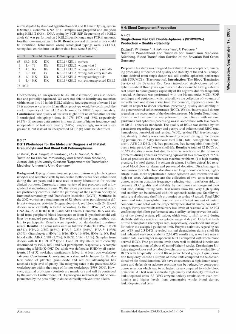
reinvestigated by standard agglutination test and ID micro typing system(Diamed). Genomic DNA of all samples was prepared and analysedusing KEL1/2 (Kk) – DNA typing by PCR-SSP. Sequencing of a KEL2allele (k) was performed on 2 KEL2 specific long range PCR fragments,together covering exons 1 to 10. Results: Several different errors couldbe identified. Total initial wrong serological typings were 3 (4.1%),wrong data entries into our donor data base were 5 (6.8%).
n % Ser-old Ser-new DNA-typing Conclusion
63 86.3 KK KK KEL1 / KEL1 correct1 1.4 ?? Kk KEL1 / KEL2 wrong what ?3 4.1 Kk Kk KEL1 / KEL2 wrong data entry into db2 2.7 kk kk KEL2 / KEL2 wrong data entry into db3 4.1 KK Kk KEL1 / KEL2 wrong serology old*1 1.4 KK KK KEL1 / KEL2 correct, unexpressed KEL2
73 100.0
Unexpectedly, an unexpressed KEL2 allele (Cellano) was also identi-fied and partially sequenced. We were not able to identify any mutationwithin exons 1 to 10 in this KEL2 allele so far, sequencing of exons 11 to19 is underway currently. If an allelic genotype would be confirmed, theallele frequency of this KEL2 allele would be 0.00016, approximately.Conclusions: Poor quality of test sera might offer an explanation for the3 serological mistypings* done in 1976, 1978 and 1988, respectively(4.1%). Erroneous data entries into our db are of higher frequency andindependent of test sera quality (6.8%). Surprisingly, no weakly ex-pressed k, but instead an unexpressed KEL2 (k) could be identified.
B 3.07DGTI Workshops for the Molecular Diagnosis of Platelet,Granulocyte and Red Blood Cell Polymorphisms
H. Kroll1, W.A. Flegel2, S. Santoso1, U.J.H. Sachs1, G. Bein1
1Institute for Clinical Immunology and Transfusion Medicine,Justus Liebig University Giessen; 2Department for TransfusionMedicine, University Ulm, Germany
Background: Typing of immunogenic polymorphisms on platelets, gran-ulocytes and red blood cells by molecular methods has been establishedduring the last years and is now used in many laboratories for routineclinical purposes. Currently, a large variety of test protocols and a lowgrade of standardisation exist. We therefore performed a series of exter-nal proficiency controls under the patronage of the German Society ofImmunohaematology and Transfusion Medicine (DGTI). Methods: Inthe 2002 workshop a total number of 32 laboratories participated in dif-ferent categories: platelets 24, granulocytes 4, red blood cells 24. Blooddonors were carefully selected according to their HPA-1, -2, -3, -5;HNA-1a, -b, -c; RHD, RHCE and ABO alleles. Genomic DNA was iso-lated from peripheral blood leukocytes or from B-lymphoblastoid celllines by standard procedures. The selection of the typing method wasleft to participants. Results were reported on standardised question-naires. Results: The error rates were as follows: Platelets: HPA-1: 1/368(0.3%), HPA-2: 2/352 (0.6%), HPA-3: 2/336 (0.6%), HPA-5: 11/368(3.0%). Granulocytes: HNA-1a: 0/16, HNA-1b: 0/16, HNA-1c: 0/8. Redblood cells: ABO: 5/184 (2.7%), RHCE: 5/160 (3.1%). Samples fromdonors with RHD, RHDVI type III and RHDψ alleles were correctlydetermined by 19/21, 18/21 and 3/21 participants, respectively. A samplecontaining a RHD(K409K) Del allele was defined as RHD by all partic-ipants. 13 of 32 workshop participants failed in at least one workshopcategory. Conclusions: Genotyping as a standard technique for the de-termination of platelet, granulocyte and red cell alloantigens hasreached a high level of quality. The mistyping rates in the presented 2002workshop are within the range of former workshops (1998–2001). How-ever, external proficiency controls are mandatory and will be continuedby the authors. Furthermore, RHD genotyping methods should be com-plemented by the possibility to detect clinically relevant rare alleles.
A 4: Blood Component Preparation
A 4.01Single-Donor Red Cell Double-Apheresis (SDR/MCS+)Production – Quality – Stability
W. Illert1, W. Sänger2, H. Jahn-Jochem2, F. Weinauer2
1Institute Wiesentheid and 2Institute for Transfusion Medicine,Nürnberg, Blood Transfusion Service of the Bavarian Red Cross,Germany
Purpose: This study was designed to evaluate donor acceptance, emerg-ing processing difficulties and quality and stability of the red cell compo-nents derived from single-donor red cell double-apheresis performedwith SDR/MCS+ (Haemonetics). Introduction: The Blood TransfusionService of the Bavarian Red Cross introduced single-donor red cellapheresis about three years ago to recruit donors and to have greater di-rect access to blood groups, especially of Rh negative donors, frequentlyneeded. Apheresis was performed with the Haemonetics MCS+/SDRtechnique and equipment which also allows the collection of two units ofred cells from one donor at one time. Furthermore, experience should bemade in respect to donor selection, processing, quality and stability ofthe generated red cell concentrates (RCC). We also interrogated donorsregarding the acceptance of the donation process. Methods: Donor qual-ification and examination was peformed in compliance with nationalguidelines and apheresis processing was in accordance with Haemonet-ics MCS+ apheresis standards. The quality of RCCs was defined by testparameters regarding potency and purity: total volume, total RBC, totalhemoglobin, hematokrit and residual WBC, residual PLT, free hemoglo-bin, sterility. Stability was characterized by repeat testing of the follow-ing parameters in 7-day intervals: total RBC, total hemoglobin, hema-tokrit, ATP, 2,3-DPG, pH, free potassium, free hemoglobin (hemolysis)over a total period of 6 weeks shelf-life. Results: A total of 32 RCCs outof 623 donations were lost due to adverse reactions of the donor orproblems during apheresis processing equivalent to a recovery of 97.5%.Loss of products due to apheresis machine problems (1 × high startingpressure, 1 × bowl defect, 1 × system air alarm, 1 × filter defect) led to re-duced blood flow or alarm and prevented donation. Disadvantages incomparison to whole blood donations are longer donation times, highercitrate loads, more sophisticated donor selection and information andhigh set costs. Advantages are the collection of two units from onedonor, reducing donation frequency, reducing viral transfusion risk, in-creasing RCC quality and stability by continuous anticoagulant flowand, also, cutting testing costs. Test results show that very high qualityand stability can be achieved with this apheresis technique. Stability re-sults reveal adequate shelf-life properties up to day 43 tested. Total RBCcount and total hemoglobin demonstrate sufficient amount of potentcompounds and total volume, respectively hematokrit enable consistentdosage. Purity test results reveal very low levels of residual WBC or PLTconfirming high filter performance and sterility testing proves the valid-ity of the closed system. pH values, which tend to shift to acid duringshelf-life still stay inside an acceptable range at day 43. Only low levelsof free hemoglobin (hemolysis rate, respectively) developed and stayedfar below the accepted guideline limit. Enzyme activities, regarding redcell ATP and 2,3-DPG revealed normal degradation during shelf-lifeand indicated very good stability. 2,3-DPG results are, as we have seen inearlier data, even higher in apheresis RCCs compared with whole bloodderived RCCs. Free potassium levels show well established kinetics andreach concentrations of about 60 mmol/l after 6 weeks. Conclusions: Uti-lizing single-donor red cell double-apheresis supports the availability ofRCCs with frequently needed Rh negative blood groups. Equal dona-tion frequency leads to a surplus of these units compared to the conven-tional whole blood donation. We have encountered a high donor accep-tance and deferrals or adverse reactions can be reduced by consequentdonor selection which lead to no higher losses compared to whole blooddonations. All test results indicate high quality and stability levels of allleukodepleted units. 2,3-DPG enzyme activity results show even pro-longing and higher levels than comparable whole blood derivedleukodepleted red cells.
Transfus Med Hemother 2003;30(Sonderheft 1):1–56Abstracts 9
Wed
nesd
ay, S
ep
tem
ber
17, 2003

A 4.02Low Propidium Iodide Intensity in Flow Cytometric LeukocyteCounting as a Marker of Cell Destruction?
T. Wagner, M. Stubenrauch, G. LanzerDept. of Blood Group Serology and Transfusionmedicine,University of Graz, Austria
Background: To determine residual leukocytes in blood products flowcytometry (FC) with propidium iodide (PI) staining is commonly used.Frequently two distinct populations with different PI intensity can befound using FC. The high intensity area is known to contain the leuko-cytes and the aim of our study was to specify the population with low PIintensity. Material and Methods: EDTA blood was diluted 1:100 and thegate set on leukocytes using LeucoCount-kit (Becton Dickinson, Aus-tria). Packed red cells were tested for leukocytes before and after filtra-tion using LCR5® in-line filtration set after 4 hours (LCR5/4hours) and16 hours (LCR5/16hours) storage and LST1® (LST1/4hours) wholeblood filtration set (MacoPharma, France). High quality DNA was ob-tained using the salting out method. Results: Leukocytes found in thepreviously defined gate correlated well with the Sysmex 9500 (Toa,Japan). The absolute count obtained in the low PI intensity area beforeand after filtration was significantly different comparing LCR5/4hours(44.1 ± 18.9/µl and 0.43 ± 0.24/µl) and LCR5/16hours (170.5 ± 107.7 and1.71 ± 1.54; p <0.002 and p <0.01, respectively). Using LST1/4hours nodifference was found compared to LCR5/4hours after filtration (0.43 ±0.24 vs. 0.19 ± 0.28), but a significant difference when comparing the re-sults before filtration (2.62 ± 1.37 vs. 44.1 ± 18.9; p <0.009). High qualityDNA was found predominantly in the low PI intensity area. Subse-quently, it could be demonstrated that DNA was retained in the filters.Conclusions: Events found in the low PI intensity area are not leuko-cytes but DNA coming from ongoing cell destruction during storage,centrifugation and preparation of donated blood. Their absolute countcorrelated well with the degree of manipulation. Therefore, our resultsprovide evidence that the amount of DNA found in the low PI intensityarea can be used as a semiquantitative marker of leukocyte destructionin blood products.
A 4.03Analysis of Cell Binding to Inline Filters: Leukocyte Subpopulations and Platelets Act in Distinct Waysand Influence Each Other
R. Henschler1, B. Rüster1, W. Walker2, T. Montag-Lessing3, E. Seifried1
1Institute for Transfusion Medicine and Immune Hematology,University of Frankfurt; 2MacoPharma Deutschland GmbH, 3Paul-Ehrlich-Institute, Langen, Germany
Purpose: Since limited knowledge exists on the mechanisms which regu-late cell binding to filter surfaces, we investigated interaction patterns oflymphocytes (LY), monocytes (MO), granulocytes (GR) and platelets(PLT) with inline filter layers. Methods: Whole blood inline filters (LST-1) were either analyzed intact after filtration of one unit of whole blood,or individual filter layers were pre-cut and incubated with individuallyprepared preparations of GR, LY, MO, PLT. Cells were counted in su-pernatants by Cell-Dyn differential, by flow cytometry or visualized insitu by immunohistochmistry. Results: We found avid binding of allleukocyte types and of PLT to isolated filter layers, with saturation bind-ing occurring faster (<10 min) in PLT than in LY, MO and GR (0.5-6h).PLT showed >2 fold higher binding in the presence of Ca2+, whereasCa2+ was of only marginal significance for the binding of LY, MO or GR.Also, in the absence of Ca2+, MO and to some extent LY competed withPLT for filter binding, suggesting interaction of the two cell types,whereas GR bound with unaltered efficiency in the presence and ab-sence of PLT. Immunohistological analysis showed that both PLT andleukocytes mainly bound in foci. PLT were found either in immediatecontact with leukocytes, or formed aggregates in the absence of leuko-cytes. Preincubation of leukocytes with substances blocking cytoskeletalactivation, the actin inhibitor cytochalasin D and the Rho GTPase in-hibitor Toxin B from Clostridium botulinum, and their subsequent inlinefiltration in reconstituted blood units showed unaltered distribution of
the leukocytes between filter layers, indicating a passive nature of filterbinding by leukocytes. This was also supported by the absence of focalaccumulation of the actin-interacting proteins, vinculin and paxillinwhen assayed microscopically. Filter fibers precoated with the majorplasma constitutents, hyaluronic acid, fibrinogen, and fibronectin indi-cated that they contribute to varying degrees to cell binding in inline fil-ters. Conclusions: The binding of leukocytes to inline filters occurs most-ly passively, involves interaction of subpopulations with PLT, and is stim-ulated by plasma constituents. Closer analysis of these phenomena willbe required when new selective enrichment or depletion strategies areenvisaged.
A 4.04Microbial Contamination of Apheresis Bags before and afterDecontamination Procedures during Introduction in a Class ACleanroom Area
A. Humpe1, B. Christiansen2, M. Kneba1, H. Horst1
1Stem cell laboratory of the second Department of Medicine, 2Department of Hygiene, University of Schleswig-Holstein CampusKiel, FRG
Purpose: Manipulation of cellular therapeutics carried out in an opensystem need to be performed in a class A environment. Contaminationcontrol consists of appropriate clothing of the personal and effective de-contamination procedures for material introduced in the critical area.We examined the extent of contamination on apheresis bags containinghematopoietic progenitor cells for PBSCT before and after decontami-nation on introduction in the cleanroom area. Methods: In a first series,34 transplant bags were examined on arrival in the production laborato-ry. Two random contact plate (diameter 55mm) examinations, one onthe margin and one in the area of the label of the bag, were performed.After an overnight storage at +6 °C, the bags were introduced in thecleanroom area A after a decontamination step before introduction intothe material air lock and an additional decontamination step in thegrade A area. Decontamination was performed by an alcoholic disinfec-tant spray and wiping with a non-sterile, one-way, class 100 suitabledrapery. After the second decontamination step, the examination for mi-crobial contamination was repeated. In a second series of 10 additionalapheresis products, the examinations were repeated but in the seconddecontamination step, a reusable sterile class 100 suitable drapery wasapplied. Results: Before decontamination procedures microbial contam-inants consisting of gram-positive skin bacteria or fungal germs were de-tected in both series. For all 44 bags, the median contamination was sig-nificantly (p=0.023) higher on the margin compared to the label with 3(0–31) and 2 (0–43) colonies per plate, respectively. In the first series, 6of 68 plates still exhibited bacterial growth after both decontaminationsteps with a median number of 2 colonies (1–8) per plate. In the secondseries, none of the 20 plates showed bacterial growth after the deconta-mination steps although this result is not statistically different from thefirst series. All 17 patients transplanted with grafts from this series en-grafted successfully with typical engraftment parameters. Conclusions:Microbial contaminants on bags containing cellular products need to becontrolled before introduction in cleanroom areas. Effective decontami-nation procedures that do not influence the quality of the product needto be established, validated, and controlled according to the EU guide toGood Manufacturing Practice.
A 4.05Results of Routine Integrity Testing of TSCD Tube Connections
W.F. Boecker1, G. Walther-Wenke1, A. Bexte1, M. Oehler1, G. DiDio2,U. Demmer3
German Red Cross Bloodtransfusion Service West, 1Muenster,2Breitscheid, 3Hagen
Background: In April 1999 the advisory board of the German Ministy ofHealth (Arbeitskreis Blut) proposed 100% integrity testing of TSCDtube connections as part of requirements for using this method in bloodcomponent preparation. Material and Methods: Top and bottom bloodbag systems of Fresenius Hemocare (T 2814, T 2729, T 2930), Ma-
10 Transfus Med Hemother 2003;30(Sonderheft 1):1–56 Abstracts
Wed
nesd
ay, S
ep
tem
ber 1
7, 2
003

copharma (LQT 7240 XC) and other blood bag systems for specialpreparations were used. There were certificates of conformity for alltubes. Sterile tube connections were made by Terumo TSCD tubewelders SC-201 AH. Integrity testing was performed as 100% inprocesscontrol using a tube stripping device test procedure. Results: Betweenthe years 2000 and 2002, 609,440 connections were performed for thepreparation of 121,420 blood components: 90,477 pooled, filtered PCs,19,273 pediatric RBCs, 627 washed, filtered RBCs and 11,043 specialblood preparations. 1 out of 541 tube connections turned out to be leak.1 out of 109 products had to be discarded (1098 pooled, filtered PCs, 4washed, filtered RBCs, 5 pediatric and 9 special blood preparations). In-accuracies were caused by incorrect positioning of tubes in the sealinghead, residuals of plastic on the moving parts of the sterile docking de-vice and deviations from tube diameter. In 2002 a manufacturing defec-tive of Terumo’s cutting blades caused the majority of cases. Conclu-sions: Routine integrity testing of TSCD tube connections is mandatoryto identify sources of bacterial contamination. A huge number of data isneeded to find leakages. Only well trained critical staff members, ongo-ing maintenance of the devices and the tube stripping device test proce-dure allows us to gain all benefits of sterile docking in blood banking.
A 4.06Application of the Temperature Indicator CHECK-SPOT™ forthe Monitoring of Packed Red Blood Cells (RBC)
H. Schennacha, E.M. Amanna, M. Meyera, H. Temmelb, D. Schönitzera
aCentral Institute for Blood Transfusion, University Clinic Innsbruck;bMessrs. Harald H. Temmel KEG, Gleisdorf, Austria
Purpose: The temperature indicator (CHECK-SPOT™) was developedfor the control of storage and transport temperatures of RBCs. The indi-cator provides reliable and definite information whether a single RBCwas adequately stored and transported at blood temperatures lowerthan 10 °Celsius. The purpose of this study is to show first results afterimplementation of the indicator in the Univ. Hospital Innsbruck. Meth-ods: A labelling device (SPOT-GUN™) for quick and easy activationand fixation of the indicator was established. The pneumatic tube trans-port system of the hospital was evaluated on its influence on the indica-tor. Medical and nursing staff of the Central Institute for Blood Transfu-sion and of the wards of the Univ. Hospital Innsbruck were informedabout the orderly handling of the indicator and the packed RBCs inorder to obtain adequate transport temperatures. To improve constanttemperature conditions during the transport of RBCs new thermoinsu-lation bags were introduced. Results: The pneumatic tube transport sys-tem showed no negative influence on the indicator. By using the ther-moinsulation bags the transport period could be prolonged to appr. 60min without additional cooling. In April 2003 we started to adjust the in-dicator on each RBC. Labeling also of large batches of RBCs (>500/day)could be easily performed. So far appr. 3500 RBCs have been labelled.74 (11.8%) of the non transfused 624 RBCs, which were returned to theCentral Institute for Blood Transfusion, showed inadequate transportconditions. However the main part of RBCs showed no exceeding of theallowed temperature and could be provided for further patients. Conclu-sion: The temperature indicator (CHECK-SPOT™) is an easy to handlesurveillance system which can be well used to monitor storage and ship-ping conditions of RBCs. RBCs that show no color change of the indica-tor can be provided for transfusion for another patients.
B 4: Genetics of Immunoglobulins
B 4.01Library of Iso-, Allo- & Idiotypic Antigens on Antibodies: A Diversity Bundled to Targeted Immune Response
U.E. NydeggerHead, Research & Development, University Clinic for Cardiovascu-lar Surgery, Inselspital, Bern, Schweiz
Technological advances in protein analysis, such as proteomics, cristal-lography and better understanding of structure-function relationship inaddition to improvements in carbohydrate chemistry have allowed toclosely define antigenic epitopes on the antibody molecules themselves.Since the work by Kunkel, Mannik and others in the 1960ies, observa-tions with monoclonal antibodies directed at human immunoglobulins(Ig) have allowed to confirm that isolated antibodies possess individualspecificity as antigens: antibody isotypes include IgM, IgD, IgG with sub-classes 1–4, IgA and IgE whereby the H-chain C-regions of all antibodymolecules of one isotype or subtype have the same amino acid sequence.Antigens that constitute different inherited alleles of Ig molecules arecalled allotopes, antibodies sharing the same allotopes belonging to thesame allotype; finally the unique antigenic determinants of individualantibody clones are called idiotypes that arise both from inheritedgermline diversity and from somatic events. Unlike allotopes, idiotopesare functionally significant because they are involved in regulation of Bcell function and are targets of anti-idiotypic antibodies in i.v. im-munoglobulin preparations. Clonal characteristics of ABO antibodies in18 paired samples of serum and breast milk for IgM showed a uniform,polyclonal spectrotype with more than 20 bands and minimal interindi-vidual differences. In contrast, IgG spectroptypes were oligoclonal (<12bands) and individually distinct, a finding that was interpreted throughabsence of somatic mutations for IgM and differences in isotype-switchregulation for IgG. We also studied clonal characteristics of Galalpha1-3Gal (anti-Gal), and confirmed our observations with ABO antibodies.In addition the study with anti-Gal suggested the predominant use ofthe VH2f gene family to produce antibodies against bacterial pathogens.The induction of donor chimaerism in erythroid burst forming units thatfollows ABO-incompatible stem cell transplantation, clinically constitut-ing no barrier, might become more clearly elucidated when the ensuingchange in anti-A/B antibodies at the level of iso-, allo- and idiotopes ismore precisely analyzed. For survival advantage, nature apparentlydraws the right mix from a rich library of genetic background.
B 4.03ABO-Incompatible Hematopoietic Stem Cell Transplantationand Pregnancy: in vivo Models for B Cell Tolerance
J.D. SeebachLaboratory for Transplantation Immunology, Department of InternalMedicine, University Hospital Zurich, Switzerland
The current organ shortage in transplantation medicine stimulates theexploration of new strategies to expand the donor pool including theutilization of living donors, ABO blood group incompatible grafts, andxenotransplantation. Similar to hemolysis in ABO-incompatible transfu-sion, preformed immunoglobulins, alleged natural antibodies, lead to hy-peracute rejection and graft failure of ABO-incompatible solid organsand xenotransplants. Corresponding to the rules of transfusion medicine,matching of the ABO blood groups has therefore been thought to beprerequisite for successful solid organ transplantation. In contrast, theABO system seems to be of minor importance for the clinical outcomein hematopoietic stem cell transplantation (SCT). ABO-incompatibleSCT is routinely performed and may be viewed as a human in-vivo studymodel for the immunological mechanisms of antigen mismatched trans-plantation. Moreover, pregnancy can also be viewed as a model for im-munological tolerance: ABO incompatible pregnancies are thereforevaluable to analyze the protective mechanisms avoiding immunglobu-lin-mediated damage as well as their limitations. The purpose of this lec-ture is (1) to give a short overview of the role of anti-A/B blood groupimmunoglobulins in SCT, solid organ transplantation and pregnancy; (2)
Transfus Med Hemother 2003;30(Sonderheft 1):1–56Abstracts 11
Wed
nesd
ay, S
ep
tem
ber
17, 2003
to present our recent data on the time course, levels and isotypes of anti-A/B immunoglobulins during ABO mismatched SCT and pregnanciesobtained by a new quantitative method based on flow cytometry; (3) todiscuss the potential mechanisms responsible for the apparent success ofminor ABO-incompatible SCT including in vivo absorption of im-munoglobulins, the generation of anti-idiotypic immunoglobulins, and Bcell tolerance resulting from either deletion and/or anergy of B-lympho-cytes; and (4) to explain the role of anti-A/B immunoglobulins in he-molysis, delayed engraftment and pure red cell aplasia in the setting ofmajor ABO-incompatible SCT. In conclusion, by increasing the currentknowledge of ABO incompatibility we hope to develop a concept of theunderlying immunological mechanisms and to facilitate new strategies toovercome the ABO barrier in solid organ transplantation as well as thehyperacute rejection of xenogeneic organs.
A 5: Stem Cells
A 5.02Progenitor Cells in RA CD34, CD133 and STRO-1 PositiveBone Marrow Derived Precursor Cells in the Synovial Tissueof Patients with RA
B. Rüger, P. Höcker, M.B. Fischer Department of Transfusion Medicine, University of Vienna, Austria
Bone marrow derived precursor cells were found in the synovial tissueof 18 patients with RA expressing either CD34, STRO-1 or CD133.CD34 positive CD31 negative precursor cells formed cell clusters in thesublining area of synovial membranes with STRO-1 positive and CD133positive progenitor cells. Within the cluster, CD34 positive cells were lo-cated at the inside, surrounded by STRO-1 positive cells and CD133positive cells at the outside. These clusters of precursor cells for imma-ture vessels that have not formed a lumen yet. Further, morphometricanalysis revealed that within the sublining area single STR0-1 positivecells were spotted around at a density of 3–5 cells/mm2 and at the basisof the synovial lining single CD133 positive cells were found at a densi-ty of 1–2 cells/mm2. CD34 positive precursor cells expressed CXCR4the receptor for stromal cell derived factor-1 (SDF-1), while VEGFR-2was expressed on CD34 positive and CD133 positive cells. Alphasmooth muscle actin was expressed by STRO-1 positive cells. The pres-ence of bone marrow derived endothelial precursor cells found in thesynovial tissue of patients with RA give evidence for vasculogenesis andSTRO-1 cells for tissue regeneration by cells from the mesenchymallineage.
A 5.03Influence of Donor Characteristics and G-CSF-AdministrationSchedule on the Efficacy of Peripheral Blood Progenitor CellMobilisation
K. Hölig1, A. Haack1, K. Zimmer1, F. Kroschinsky2, M. Bornhäuser2, G. Ehninger2
1Transfusionsmedizin, 2Medizinische Klinik und Poliklinik I,Klinikum der Technischen Universität, Dresden, Germany
Purpose: CD 34+ cell mobilisation in healthy donors varies to a widescale. Defining predictive factors of mobilisation efficacy is of great in-terest to optimise protocols for allogenic stem cell donors. Methods:1474 healthy donors (986 men, 488 women) underwent G-CSF applica-tion and PBPC collection at our department between 1/1996 and 8/2002.G-CSF administration was performed in 3 dosages: filgrastim 10µg/kg/day on 5 days; lenograstim 7.5 µg/kg/day on 5 days; lenograstim 7.5µg/kg/day on day 1 and 2, 12.5 µg/kg/day on the following 3 days. Leuka-pheresis was performed at day 5 (and 6, if necessary). CD34 -concentra-tion in peripheral blood (× 106/L) at day 5 before 1st leukapheresis wasanalysed for correlation with the following parameters: leukocyte andplatelet counts before G-CSF administration, sex, age, body mass index(BMI), nicotine and alcohol consumption of the donors, G-CSF doseand mode of G-CSF application (single dose versus split dose). Results:The median concentration of CD 34+ cells in peripheral blood at day 5was 56/µl in male versus 42/µl in female donors (p <0.0001). A 2nd
apheresis had been performed in 34% of male and 53% of femaledonors. A significantly positive correlation of CD 34+ concentration wasfound with BMI (p <0.001) and the schedule of G-CSF application (splitversus single dose; p <0.0001). In a multivariate analysis, the schedule ofG-CSF application had the most significant influence on the efficacy ofperipheral blood progenitor cell (PBPC) mobilisation. No significantcorrelation was found with G-CSF-dose, donor age, alcohol consump-tion and smoking status. Conclusions: In our donor population PBSCmobilisation worked best in male donors with higher BMI. The scheduleof G-CSF-administration seems to be very important for the mobilisa-tion efficacy in healthy volunteer donors. Dose splitting of G-CSFshould be performed, whenever possible.
A 5.04Optimization and Results of a Flow Cytometry Method forEvaluation of Viability of Peripheral Blood Stem Cell Graftsafter Cryopreservation
A. Humpe, C. Beck, R. Schoch, M. Kneba, H. HorstSecond Department of Medicine, Stem cell laboratory, Universityclinic Schleswig-Holstein Campus Kiel, Germany
Purpose: Quality control of peripheral blood stem cell (PBSC) graftsafter cryopreservation is still controversial. Viability assessment by try-pan blue exclusion staining and microscopic evaluation is an acceptedmethod (I) although the number of analyzed cells is small and a differ-entiation of cell type is not possible. We established a flow cytometry-based method (II), analyzed the influence of incubation time and lysison the results and compared the results with method I. Methods: In afirst series, viability was assessed in 22 autologous apheresis productsfrom 8 patients by method I directly after thawing and by method II(Stem-Kit, Beckman Coulter) after 20 minutes of antibody staining and10 minutes of lysis and after halving the times. In a second series, viabil-ity was assessed in 21 autologous apheresis products from 10 patientsand in 1 allogeneic product by method I directly after thawing and bymethod II after 10 minutes of antibody staining with and without lysis. Ina third series, viability was assessed in 65 autologous apheresis productsfrom 19 patients and in 7 allogeneic apheresis products of 3 donors bymethod I directly after thawing and after 10 minutes and by method IIafter 10 minutes of antibody staining. Results: In the first series, shorten-ing of the time of staining and of lysis did not influence the viability ofthe CD45+ cells, the viability of the CD34+ cells, and the number of de-tectable CD34+ cells. In the second series, the median viability of CD45+
cells was significantly (p=0.022) higher without than with lysis (71.0 and75.8%, respectively). The median viability as well as the number ofCD34+ cells were also higher without lysis but these differences werenot significant. In the third series, the median viability of CD34+ cells(97.8%) was significantly (p <0.0001) higher compared with the viabilityof CD45+ cells (73.0) and the viability determined by method I (85.0%)in the freshly thawed sample. Conclusions: The viability of CD34+ cellswas significantly higher compared with the viability of all leukocytesmeasured by method I or II. The presented cytometry-based method issuperior to the standard trypan blue method regarding the number ofanalyzed cells, reproducibility, and documentation. As the presentedmethod can easily been established and allows the measurement of thecells of interest, it should be the method of choice regarding quality con-trol of PBSC grafts after cryopreservation.
A 5.05Potassium Channel Openers Depolarise Mitochondria ofCD34+ Cell Line KG-1a: Implications for Modulation ofGrowth of CD34+ Cells?
S. Körper, F. Nolte, M. Rojewski, H. SchrezenmeierDepartment for Transfusionmedicine, University of Ulm and Institutefor clinical Tranfusionmedicine and Immunognenetics, Ulm, Germany
Purpose: It has been shown that hemopoietic stem cells (HSC) withlow rhodamine125 (a probe for mitochondrial membrane potential)staining engraft faster and with a more complete chimerism than rho-damine 125 high cells in mice. This implicates that cells with a lower
12 Transfus Med Hemother 2003;30(Sonderheft 1):1–56 Abstracts
Th
urs
day, S
ep
tem
ber 1
8, 2
003

mitochondrial membrane potential have a higher engraftment poten-tial. Diazoxide an opener of the mitochondrial ATP dependent potas-sium channel is currently under investigation as a cytoprotective agentin heart and brain. This agent has the potential to depolarise mitochon-dria. The big Ca2+ dependent K+ channel is also expressed in mito-chondria. A specific opener of this channel is NS1619. In this study weinvestigated if K+ channel openers have the capacity to depolarise mi-tochondria of CD34+ cells. Methods: Mitochondria were investigatedwith the dye JC-1 by flow cytometry. Cells can be divided in threegroups: JC-1 high, and two depolarised states: JC-1 intermediate andJC-1 low. KG-1a cells express glycoprotein P. To exclude artefactsthrough dye exclusion we used a dye loading protocol with verapamil.Cells were incubated for one hour with diazoxide or NS1619. To ex-clude caspase activation as a reason for mitochondrial depolarisationwe used the pan-caspase inhibitor BocD(OMe)-Fmk (50 µM). A con-tribution of plasmacellular channels to the depolarisation of mitochon-dria was excluded with an extracellular high K+ solution (135mM). Re-sults: Diazoxide (200–800 µM) and NS1619 (5–50 µM) induced signifi-cant mitochondrial depolarisations (p ≤0.05) in solutions containingnormal or high K+. At a concentration of 400 µM diazoxide and at 25µM NS1619 the agents depolarised mitochondria in 20% of cells. Atthis concentration NS1619 induced 9.4 ±5.8% of cells to JC-1 lowwhereas diazoxide induced only 0.7 ± 0.2% to JC-1 low (P=0.005, n=7).The pan-caspase inhibitor BocD(OMe)-Fmk was not able to inhibitmitochondrial depolarisations. Conclusion: NS1619 and diazoxide in-duce depolarisation of mitochondria in CD34+ cell line KG1a. This isindependent from caspase activation or potassium currents over theplasmamebrane. According to the single channels conductances of thebig potassium channel and the ATP-dependent K+ channel NS1619 in-duce stronger depolarisations than diazoxide. Both substances can nowbe further investigated for their potential to modify hemopoiesis andfor their utility in ex-vivo stem cell expansion.
A 5.06Mesenchymal Stem and Progenitor Cells (MSC): Isolation, ExVivo Expansion, Differentiation and Potential for ChemotacticActivation of Homing and Migration by Blood Plasma
O. Seitz, B. Rüster, R. Henschler Institute for Transfusion Medicine and Immune Hematology,University of Frankfurt, Germany
Purpose: Cells which can give rise to a multitude of mesenchymal lin-eages (MSC) have been widely characterized, and in clinical studiescould be successfully re-infused into patients. However, their homingand migration behaviour is largely unknown, and its regulation is un-clear. Methods: Bone marrow samples were seeded onto plastic dishesin various serum-containing and serum-free culture media, with andwithout previous enrichment for CD45- cells. Immunohistochemistyand RT-PCR analyses were established to characterize differentiationmarkers. A modified Boyden Chamber assay was developed to ana-lyze chemotactic migration. Results: We found reproducible and effi-cient expansion of MSC with multilineage differentiation capabilityover 3–4 log within culture periods of 21-28 days. Preselected fetal calfserum proved superior to commercially available expansion supple-ment for MSC, or serum-free medium to promote optimal expansionrates. MSC could be differentiated into either osteoblasts (assayed byexpression of bone sialoprotein, osteocalcin, or alkaline phosphatase),chondrocytes (by staining with toluidine blue), myocytes (by expres-sion of myogenin) or fat cells (assayed by oil red O staining) at differ-ent time points of the expansion culture. However, activation of MSCmigration proved difficult. Platelet Derived Growth Factor (PDGF)and Fibroblast Growth Factor (FGF)-2, which were shown to inducechemotaxis of mature fibroblasts and osteocytes, stimulated MSC mi-gration only to limited degrees and under selected conditions. In con-trast, blood plasma efficiently induced the transmigration of MSCthrough 8 µm micropores within 4 h. When immunodeficientNOD/SCID mice were infused i.v. with 1 × 10⁶ MSC, human cells de-tected at 24h were still contained in the lungs, but very little signal wasfound in other organs. Conclusions: This study describes conditions forefficient expansion and differentiation of MSC from human bone mar-row. We show that activation of MSC migration may follow very differ-
ent mechanisms compared with those known for hematopoietic stemcells. It may be underlying a multicomponent regulation and requireprevious activation.
A 5.07The Homing of Transplanted Hematopoietic Progenitor Cells(HPC) Into the Blood-Forming Organs is Selective, but Occurswith Low Efficiency
R. Henschler, Z. Fehervizyova, E. Seifried, R. BistrianInstitute for Transfusion Medicine and Immune Hematology,German Red Cross Blood Donation Service, Johann-Wolfgang-Goethe University, Frankfurt, Germany
Purpose: Empirical, but so far not well explained data have indicatedthat critical threshold doses are required for appropriate hematopoieticrecovery after stem cell transplantation. To better elucidate the process-es occurring early after stem cell infusion and to characterize the fate ofinfused progenitor cells, we investigated early steps of their homing andengraftment. Methods: We have transplanted C57/BL6 mice with fluo-rescence-marked primitive lin-Sca-1+ hematopoietic progenitor cells(HPC, FDCP-mix) or primary bone marrow cells, and analyzed theirfate during the first 24 h after i.v. injection. Homed cells were analyzedflow-cytometrically or microscopically in tissue sections. A specific tech-nique using the erythrocyte-specific marker TER119 was used to distin-guish HPC contained in blood vessels from those HPC which had mi-grated into tissues. Results: HPC (5–15 × 106/mouse) rapidly entered avariety of tissues including muscle, lung, liver, gut, fat and connectivetissue, but not brain and heart, at incidences between 100 and 800HPC/mg tissue 2 h after transplantation. In particular, the liver con-tained a majority of the retrieved cells. Higher incidences of HPC wererecorded in the bone marrow and spleen (3000–5000 HPC/mg), yetgiven the much greater total mass of non-hematopoietic organs com-pared with hematopoietic organs, HPC located only at a relatively smallpercentage to the hematopoietic organs (3–5%). At 24 h, the totalamount of HPC detected per mouse decreased to 5–10% of the cellsfound at 2 h, with very few cells left in non-hematopoietic tissues and,also, about 8 to 10 fold lower absolute HPC numbers in the bone mar-row and spleen. This shows that >95% of all transplanted HPC were lostduring the first 24 h, most likely by death of apoptosis. Conclusions: Therapid migration of HPC into tissues and their massive destruction withinorgans explain the requirement of threshold doses of HPC per trans-plant. Engineering of HPC homing therefore has a significant potentialto modulate stem cell engraftment and influence the clinical success ofhematopoietic transplantation.
A 5.08In vitro Erythropoiesis Induced from Human CD34 PositiveCells
I. Dorn, D. Hartwig, K. Weber, H. Kirchner, P. SchlenkeInstitute of Immunology & Transfusion medicine, University ofSchleswig-Holstein, Campus Luebeck, Germany
Purpose: Up to now, the regulation of human erythropoiesis is only part-ly understood. Thus, an in vitro erythropoiesis model might be very use-ful to investigate, which factors and molecular mechanisms are involvedin proliferation an differentiation of human erythroid precursor cells.Methods: Peripheral blood mononuclear cells were collected by leuka-pheresis. Isolation of CD34+ cells using an immunomagnetic separationtechnique (MACS) followed. Purified cells were cultured over 16 daysin a two phase liquid assay. To induce proliferation, cells were incubatedin the presence of SCF, EPO, Flt3-L and insulin like growth factor. Bychanging culture conditions on day 8 (EPO and Insulin), differentiationof erythroid precursor cells was initiated. Cell growth and differentiationwas evaluated using the following methods: 1) Cell viability was deter-mined by trypan blue staining. 2) To measure cell proliferation, cellswere quantified by a flow cytometric procedure using fluorescent mi-croparticles. 3) Cell surface expression of CD34, CD45, CD71, CD117and glycophorin A was analyzed by flow cytometry 4) For analysis ofcell morphology, cytospin preparations were made and cells were stained
Transfus Med Hemother 2003;30(Sonderheft 1):1–56Abstracts 13
Th
urs
day, S
ep
tem
ber
18, 2003

by May-Grünwald-Giemsa. Haemoglobin content was determined byneutral benzidine staining. Results: Cell purity of CD34+ cells after im-munomagnetic separation was always greater 95%. During the prolifer-ation phase (d1–d8) absolute cell numbers increased 103 fold. On day 8,~60% of cells showed high expression of CD71 and ~50% were gly-cophorin A positive. Only few cells expressed CD117 and CD34. About50% showed morphological characteristics of polychromatic erythrob-lasts and also a weak staining for haemoglobin. After changing cultureconditions, a large increase in the number of glycophorin A and haemo-globin positive cells could be observed. On day 16 more than 90% ex-pressed glycophorin A and were strongly haemoglobin positive. Thesecells showed morphological characteristics of normoblasts, enucleatedreticulocytes and erythrocytes. Conculsions: The established in vitro ery-thropoiesis model shows the different stages of normal human erythro-poiesis from CD34+ stem cells up to enucleated erythrocytes. It might bea tool for further investigations in the field of erythropoiesis and its reg-ulation. By varying culture conditions or inhibiting signal pathways, theinfluence of special factors or molecular mechanisms involved in ery-thropoiesis could be investigated.
B 5: Immuno-Hematology and Auto-Immunity I
B 5.01Selection of Natural Self-Reactive Antibody Repertoires byImmunoglobulins Critically Depends on ImmunoglobulinIsotypes
D. Stahl1, M. Hoemberg1, U. Cassens1, U. Pachmann2, W. Sibrowski11Institute for Transfusion Medicine, University of Muenster; 2Center for Transfusion Medicine, Bayreuth, Germany
Purpose: Current understanding of immune network interactions medi-ated by immunoglobulins focuses on the role of idiotypes expressed onantibody variable regions. Idiotype interactions account significantly forthe functional integrity of natural self-reactive antibody repertoires. Im-munoglobulin isotypes are not considered to affect natural self-reactiveantibody repertoires. We used autoimmune thrombocytopenic purpura(AITP) as a model to investigate the impact of isotype differences ofdisease-associated autoantibodies on the selection of natural self-reac-tive antibody repertoires in humans. Methods: We investigated antibodyrepertoires of 7 patients with IgM-, and 7 patients with IgG-mediatedAITP by quantitative immunoblotting of immunoglobulins on a panel ofself antigens. Data were evaluated by multiparametric statistical analysisincluding principal component analysis and linear discriminant analysis.Binding of anti-platelet antibodies to disease-specific self-antigens wasanalysed by an affinity biosensor system (IAsys, Thermobiosciences,England). Results: Patients with IgM- and with IgG-mediated AITPwere discriminated by both their IgM- (p <0.0023) and IgG-antibodyrepertoire (p <0.006). Alterations of antibody reactivities were not re-stricted to platelet antigens. Autoantibody glycoprotein specificity didnot contribute to differences in self-reactive antibody repertoires (p>0.05). Binding assays of anti-platelet autoantibodies confirmed that theisotype of disease-associated autoantibodies in AITP is of higher rele-vance for selection of natural self-reactive antibody repertoires than gly-coprotein specificity. Conclusion: Selection of disease-, i.e. platelet-asso-ciated and of natural, i.e. not platelet-associated autoantibody reper-toires of AITP patients is altered in dependence of the isotype of dis-ease-associated autoantibodies. Our data suggest that antibody idiotypesregulate natural autoreactivity at the single-cell level, whereas antibodyisotypes direct natural autoreactivity at the supraclonal level. They maythus offer a conceptual framework to understand how an organ-specific,clonally restricted disease-associated autoantibody is related to a broad,non-organ specific shift in self-reactive antibody repertoires.
B 5.02External Quality Assessment on Platelet Antibody Testing inGermany
V. Kiefel1, J. Bux2
1Department of Transfusion Medicine, Rostock University, Rostock,Germany; 2Blood Transfusion Service SRK, Berne, Switzerland
Purpose: In 2002, an exercise for ‘Granulocyte and Platelet Antibodies’has been added to the External Quality Assessment Scheme of IN-STAND e.V. This report summarizes experience on the platelet antibodypart of this first quality assessment exercise (QAE). Material and Meth-ods: Three serum samples were shipped to the participants: A serumwith a high-titered anti-HPA-1a and HLA-antibodies (sample 1), aserum with HLA-antibodies detectable in both the lymphocytotoxic testand antiglobulin binding tests (sample 3) and a serum from a male sub-ject without platelet reactive antibodies (sample 2). Participants wereasked to use their methods currently in use for diagnostic investigationof patients’ samples and to communicate results with numeric codes. Re-sults: Twenty five participants sent results of their analysis: 18/25 (72%)correctly diagnosed sample 1, HLA antibodies in sample 3 were identi-fied by 14/25 (56%) and sample 2 was diagnosed as negative control by23/25 (92%) of participants. Correct results on all three samples were re-ported by only 12/25 (48%) of laboratories. Conclusion: at present a con-siderable percentage of laboratories fails to correctly diagnose the mostcommon platelet reactive antibodies.
B 5.03Recombinant Platelet Alloantigens Are Suitable Tools for theCharacterization of Platelet-Specific Alloantibodies
H. Kroll, S. Santoso Institute for Clinical Immunology and Transfusion Medicine, JustusLiebig University, Giessen, Germany
Fetal and neonatal alloimmune thrombocytopenia (FNAIT), posttrans-fusion purpura (PTP) and platelet transfusion refractoriness (PTR) areinduced by alloimmunization against platelet alloantigens. The detectionand differentiation of the responsible antibodies are usually done by gly-coprotein (GP)-specific assays (MAIPA), with a panel of phenotypedplatelets. Since the availability of certain donors having rare phenotypesis often limited we analysed the suitability of recombinant platelet al-loantigens for alloantibody detection. Full length cDNAs from GPs IIb,IIIa and Ia encoding for platelet alloantigens (HPA-1a, HPA-5, HPA-13)were generated by site-directed mutagenesis and were cloned into ex-pression vectors. Subsequently, Chinese hamster ovarian (CHO) cellswere transfected with the respective constructs and subcloned for stableantigen expression. A panel of alloantibodies against the HPA-1a, -1b, -5a, -5b and -13bW antigens from patients with FNAIT, PTP and PTRwas analysed by a modified MAIPA protocol using stable transfectantsand reference platelets. All sera showed corresponding reactions withcell lines expressing recombinant alloantigens and with platelets. Fur-thermore, recombinant alloantigens (HPA-5a and -5b) were purified byaffinity chromatography and were applied for detection of HPA-5 al-loantibodies in dot-blot analysis. In this solid phase system, allele-specif-ic reaction could be observed as well.Our study demonstrates that stable transfectants expressing or purifiedplatelet alloantigens are suitable reagents for the characterization ofplatelet alloantibodies. In the near future, purified recombinant plateletantigens will facilitate the detection of frequent antibodies (e.g. anti-HPA-1a, -5b), whereas stable cell lines may serve as reference tools forthe characterization of rare antibodies (e.g. anti-HPA-13bW). Recombi-nant platelet alloantigens, together with recombinant platelet antibodieswill allow the development of well standardized techniques for the labo-ratory diagnosis of alloimmune thrombocytopenic syndromes.
14 Transfus Med Hemother 2003;30(Sonderheft 1):1–56 Abstracts
Th
urs
day, S
ep
tem
ber 1
8, 2
003

16 Transfus Med Hemother 2003;30(Sonderheft 1):1–56 Abstracts
Th
urs
day, S
ep
tem
ber 1
8, 2
003
B 5.04Diclofenac-Induced Red Blood Cell Antibodies Are Heteroge-neous and Recognize Various Epitopes on Different TargetProteins
U.J.H. Sachs1, S. Santoso1, E. Smart2, L. Röder1, G. Bein1, H. Kroll11Institute for Clinical Immunology and Transfusion Medicine,Justus-Liebig-University, Giessen, Germany; 2South AfricanNational Blood Service, East Coast Region, Pinetown, Republic ofSouth Africa
Purpose: Diclofenac (DCF) is a common anti-inflammatory drug thathas been reported to induce immune-mediated hemolytic anemia in sev-eral patients. Although c/E-specificity has been suspected, no profounddata have been published. Patients and Methods: Sera from 12 patientsadmitted to various German hospitals because of hemolytic anemiawere sent to our laboratory for serological evaluation during the lastyears. In all patients, hemoglobin levels were below 9.0 g/dL, white celland platelet counts were normal, and lactate dehydrogenase levels wereelevated. In the direct antiglobulin test, C3d, but not IgG, was detectablein all samples. Five known metabolites were provided from Dr. A.R.Mackenzie, Novartis (Basel, Switzerland). DCF was purchased from No-vartis. Urine containing ex vivo metabolites was collected from healthyvolunteers receiving 150 mg DCF. All serologic studies were performedusing a routine gel technique. Results: Nine sera agglutinated red bloodcells (RBCs) in the presence of DCF, 3 additional sera agglutinatedRBCs in the presence of ex vivo metabolites only. When chemically de-fined metabolites were tested, five sera were reactive with all of them.The remaining seven sera showed diverse reactions with no commonpattern. Interestingly, no metabolite was recognized by all twelve sera,and no metabolite remained unrecognized. When sera were incubatedwith Rh null cells in the presence of DCF or ex vivo metabolites, fivesera did not show differences. However, 3 lost reactivity in the presence fDCF, two in the presence of ex vivo metabolites, and 2 lost both. Whentested with metabolites, six sera displayed different recognition patternswhen compared to their reactivity with Rh negative test cells. One serumwhich remained reactive with Rh null cells in the presence of ex vivometabolites was not able to agglutinate Rh null cells in the presence ofany defined metabolite. Conclusion: There is evidence that patients withDCF-induced immune hemolysis produce a broad spectrum of anti-DCF/RBC antibodies. First, DCF-induced antibodies can bind severaland distinguishable epitopes on the Rh protein. Second, many DCF-in-duced antibodies are not Rh dependent. In fact, most antisera still reactwith RBCs in the absence of the Rh protein. Their binding partners re-main to be described.
B 5.05Demonstration of Diclofenac-Induced Antibodies Reactive toRBCs and Platelets
O. Meyer1, T. Hoffmann2, T. Aslan1, N. Ahrens1, H. Kiesewetter1, A. Salama1
1Institut für Transfusionsmedizin, Universitätsklinikum Charité,Berlin; 2Institut für Hämostaseologie und Transfusionsmedizin,Heinrich-Heine-Universität, Düsseldorf, Germany
Purpose: Diclofenac is a widely used nonsteroidal anti-inflammatorydrug that has been frequently implicated as the cause of immune he-molytic anemia (IHA), and less frequently of immune thrombocytope-nia. The causative antibodies have only been demonstrated in patientswith IHA, but not yet in patients with immune thrombocytopenia. Inthis report, we describe two patients with detectable antibodies to RBCsand platelets. Patient and Methods: Patient no. 1, a 74-year-old female,developed severe hemolysis and thrombocytopenia 14 days after cardio-vascular surgery. The second patient, a 37-year-old female, developedthrombocytopenia but no hemolysis. The serological testing was carriedout using standard techniques. Serum samples were tested in the pres-ence as well as in the absence of diclofenac and its metabolites. Results:Diclofenac was discontinued and patients were treated with corticos-teroids. Both patients recovered within a few days. In both patients thedirect antiglobulin test was positive for anti-C3d but negative for anti-IgG, anti-IgM, and anti-IgA. In patient no 1, the indirect antiglobulin
test (IAT) was positive. However, this reactivity became weak followingserum dialysis and could be restored by adding diclofenac or its metabo-lites. The IAT in the second patient was only positive in the presence ofdiclofenac or its metabolites. The autologous platelets of patient no. 1could not be investigated. The platelets of the second patient were coat-ed with autoantibodies against GPIIb-IIIa and GPIb-IX. The serumsamples of both patients failed to react with GPIIb-IIIa, GPIb-IX, andGPIa-II. In the presence of diclofenac, the serum of patient no. 1 re-mained still unreactive, whereas the serum of the second patient reactedweakly with GPIIb-IIIa and GPIb-IX. In contrast, the serum samples ofboth patients reacted strongly with GPIIb-IIIa and GPIb-IX in the pres-ence of diclofenac metabolites. Conclusion: (1) Diclofenac may lead tothe production of antibodies against RBCs and/or platelets. (2) Di-clofenac may lead to the production of antibodies against RBCs that donot invariably cause significant hemolysis.
B 5.06Immune Hemolysis Following Intramuscular DiclofenacAdministration
N. Ahrens, A. Pruß, R. Genth, A. Herziger, H. Kiesewetter, A. SalamaInstitute for Transfusion Medicine, Charité Campus Virchow-Klinikum, Humboldt-University, Berlin, Germany
Purpose: Diclofenac is a widely used NSAR that is available for oral, in-travenous, and intramuscular application. It may lead to the productionof drug-dependent antibodies (ddab) and drug-independent autoanti-bodies to red blood cells (RBC). The resulting hemolysis is frequentlylife threatening and abolishes after withdrawal of the drug. A relation-ship between the route of administration and clinical manifestation ofhemolysis is not known. Patient and Methods: A 66-year-old female witha history of mild hypertension, anamnestic allergy to penicillin, and lum-bar pain had frequently ingested diclofenac for several years. She had re-ceived diclofenac as injection only once six years ago. One day prior toadmission, she received 75 mg diclofenac i.m. because of a sudden onsetof pain in her leg. One day later, she was admitted with jaundice andnausea. A relationship to a cholecystektomy that was carried out 5weeks ago could be ruled out by ultrasound investigation, CT-scan ofthe abdomen, and ERC. Immunohematological investigations were car-ried using the standard gel cards for direct and indirect antiglobulin test-ing (IAT). Cross matching was performed by the tube technique. Re-sults: The patient became anuric on the 4th day. Her hemoglobin-con-centration fell from 11.6 g/dl (day 1) to a minimum of 3.3 g/dl on the 7thday. Both direct and indirect antiglobulin tests were strongly positive ingel card technique (4+ and 1:128, respectively). Initially, autoimmunehemolytic anemia of warm-type was suggested, and treatment withsteroids was started. Unexpectedly, cross matching was negative usingthe tube technique. Since washing procedures may remove drugs, ddabwere suspected. In fact, the addition of diclofenac to the test procedureresulted in agglutinations up to a maximum titer of 256,000. The patientwas treated with RBC transfusions, dialysis, and plasma exchange forseven weeks. Conclusion: The patient seemed to have developed di-clofenac-dependent antibodies that did not appear to have caused signif-icant hemolysis under oral administration, but following intramusculardrug injection. The reason for this phenomenon is unclear.
C 5: Blood Safety, Surrogate Markers, Pathogen Inactivation I
C 5.03West Nile, Hepatitis C and Other Flaviviruses Are Highly Sensitive to Treatment by Phenothiazine Dyes and Light
H. Mohr, J. Knüver-Hopf, A. Redecker-Klein Blood Center of the German Red Cross Chapters of NSTOB,Springe, Gemany
Purpose: One of the main arguments to justify the use of virus inactiva-tion procedures for therapeutic blood products is that ‘new’ viruses mayemerge for which no routine assay is applied. That this is not only specu-lative became evident very recently when it was found that West Nile

Transfus Med Hemother 2003;30(Sonderheft 1):1–56Abstracts 17
Th
urs
day, S
ep
tem
ber
18, 2003
Virus (WNV) may be transmitted by blood products. In this study it wasinvestigated whether WNV and other flaviviruses including hepatitis Cvirus (HCV), bovine viral diarrhea virus (BVDV) and classical swinefever virus (CSFV) are sensitive to photodynamic treatment in the pres-ence of the phenothiazine dyes methylene blue (MB) or thionine (Th).Methods: Human plasma was isolated from blood donations. In the caseof BVDV, CSFV and WNV, samples were spiked with one of the viruses.In the case of HCV, PCR-positive plasma was used. The samples weretreated with MB/light or Th/light at photosensitizer concentrations be-tween 0.2 and 1 µM. Their virus content was estimated by infectivityassay (BVDV, CSFV and WNV) and in addition by real-time(RT)-PCR(BVDV, WNV and HVC). Illumination was with fluorescent tubes emit-ting white light or with low pressure sodium lamps emitting monochro-matic yellow light at a wavelength of 595 nm. Results: At MB concentra-tions of 0.8 and 1 µM and a light intensity of 30,000–45,000 Lux (whitelight) complete inactivation of WNV was achieved within 2 min, i.e. bymore than 5 log10 steps. At a dye concentration of 0.5 µM, 5 min wereneeded and at 0.2 µM 10 min. Even at an ambient light (approx. 250Lux) WNV was almost completely inactivated within 60 min. The lightsensitivity of BVDV and CSFV was similar to that of WNV. The sameresults were found when instead of MB its demethylated derivative Thwas used. The kinetics of virus inactivation were even accelerated whenthe samples were illuminated with high intensity yellow instead of whitelight. As was estimated by RT-PCR, at least one of the main target struc-tures of MB/light or Th/Light is the viral genome: in each case (HCV,BVDV and CSFV were tested) the PCR-signal was almost completelyabrogated within 5–10 min, indicating that virus RNA had been de-stroyed. Conclusion: Flavi viruses like HCV, WNV, BVDV and CSFVare highly sensitive to photodynamic treatment in the presence of MB orTh. Therapeutic plasma and also platelet concentrates may easily be de-contaminated using this approach.
C 5.04The Quality of Intercept™ Treated Amicus™ ApheresisPlatelets as Compared to Untreated Platelets
G. Stiegler, S. Prikoszovits, P. Strohmeyer, U. Tesar, A. Zehentbauer*,P. HöckerClinic for Blood Group Serology and Transfusion Medicine; ViennaUniversity Hospital; Baxter / Transfusion Therapies, Vienna, Austria
Objective: We compared the quality of Intercept™ treated and untreat-ed apheresis platelet concentrates (PC) and evaluated the in vitro ef-fects of storage. Materials and Methods: 20 double dose PCs have beencollected with the Amicus™ and split immediately after apheresis. Theplatelet additive solution factor was set to 0.57% leading to an Inter-Sol™ fraction of median 60% (range 53–65). 4 PCs had to be dischargedbecause of visible aggregates. The half of each PC underwent Inter-cept™ treatment the day after production, the other half was left un-treated. Results: The platelet yield of the double dose PC was 7.18 × 10¹¹
(5.35–8.07), and the amount of rWBC was 0.03 × 10⁶ (<0.02–0.45).Platelet loss after treatment was 7% (3–14) (Table). Conclusion: The Intercept™ treatment causes a platelet loss of appr. 7%which has to be considered for reaching a satisfying platelet dose for
transfusion. pH levels are significantly reduced after Intercept™ treat-ment due to the addition of 15 mL Amotosalen™ (pH=4.5). The contin-uing effect of the Intercept™ treatment is reflected by the further de-crease of the pH level, higher platelet activation, and larger platelet vol-umes at the end of storage as compared to untreated platelets. Platelets’metabolism and their ability to respond to hypotonic shock are not af-fected by the Intercept™ treatment. The formation of visible aggregateswhich did not dissolve during storage over night maybe due to the sepa-ration technology of the Amicus™ and/or to the addition of InterSol™.Platelets suspended in InterSol™ should not be stored >5 days becauseglucose and pH levels are very low at day 7 due to the contents of 60%InterSol™.
C 5.05Solvent/Detergent-Treated Plasma (SD/P) May be Safer andMore Cost-Effective than Quarantine Fresh Frozen Plasma(FFP)
G.F. Riedler1, A.R. Haycox2, A.K. Duggan3, H.A. Dakin3
1Regionales Blutspendezentrum, Lucerne, Switzerland; 2Department of Pharmacology, University of Liverpool; 3Abacus International, Bicester, UK
Introduction: The problem of non-infective complications seems to bemore important then transfusion-related infections. A dangerous one istransfusion-related acute lung injury (TRALI) with a reported incidenceof 1/2000 to 1/ 5000 plasma-containing transfusions (1/625 to 1/1200 re-cipients) and an acute mortality of 5–14%. TRALI treatment requiresintensive care for 4–8 days. HLA antibodies and biologically active lipidsof cellular origin have been implicated in the pathogenesis of TRALI.SD/P (Octaplas®) is a pathogen inactivated alternative to FFP. No SD/P-related TRALI cases have been reported. Methods: We looked at pub-lished data referring to TRALI and statistics of transfused blood prod-ucts in the United Kingdom (UK) and Switzerland (SUI) to estimatethe expected morbidity, mortality and costs of TRALI caused by thetransfusion of FFP. Results: 1) Yearly use of FFP: UK 375’000, SUI57’000 units. 2) Prices: UK: SD/P costs 1 17 more per unit than FFP;SUI both 1 100. 3 ) Expected TRALI-morbidity: UK 132 (75–188); SUI17 (11–29) and mortality: UK 13 (8–19); SUI 2 (1–4). Discussion: Esti-mations of yearly TRALI costs (incidence rate 1/3500, mortality rate10%) for SUI are 1 120’000. Because SD/P is 1 17/unit more expensivethan FFP in the UK, costs to prevent one death raise to 1 610’000. Costsper life-year saved for a hypothetical patient (life expectancy of 20years) are 1 30’000. Net incremental cost would be less than 1 6.6 mil-lion/year, but would prevent 8–19 deaths due to TRALI in UK. Conclu-sions: Besides elimination of most infectious complications through theprocess of SD treatment involved in the production, SD/P reduces therisk of non-infectious complications of which TRALI is the third fre-quent complication and the most common cause of transfusion relateddeath. SD/P also fulfils future EC requirements (Council of settingstandards of quality and safety for the collection, testing, processing,storage and distribution of human blood and blood components andamending Directive 2001/83/EC) concerning pathogen inactivation ofblood products.
Intercept™ treated PC’s Untreated PC’s
Day 2 Day 2 Day 5 Day 7 Day 5 Day 7before treatment after treatment
MPV (fL) 6. 65 (0.74) 6.82 (0.74)* 7.03 (0.73) 7.47 (0.71)* 6.75 (0.67) # 6.77 (0.65) #
pH (22 °C) 7.03 (0.09) 6.84 (0.07)* 6.77 (0.11)* 6.65 (0.18)* 6.94 (0.13) # 6.85 (0.13)*#
Glucose (mg/dL) 115 (19.9) 113 (17.5) 54 (25.4)* 4 (14.1)* 51 (25.1)* 11 (18.1)*Lactate (mmol/L) 4.5 (1.2) 4.7 (1.2) 10.3 (2.0)* 13.7 (2.0)* 11.3 (2.0)* 14.0 (1.9)*LDH (U/L) 115 (56) 150 (58) 192 (116)* 214 (152)* 205 (144)* 277 (179)*CD62p (%) 14.5 (3.1) 16.9 (4.0) 15.0 (3.6) 17.7 (4.5)* 13.3 (3.7) 13.3 (4.3) #
HSR (%) 59 (19) 61 (17) 54 (16) 55 (20) 56 (14) 55 (14)
Results are given in median and (standard deviation), * p <0.02 versus previous day, # p <0.02 versus treated group same day.

C 5.06Neopterin Screening in Blood Donations and Infectious Risks
H. Schennach, D. SchönitzerCentral Institute for Blood Transfusion, University Clinic Innsbruck,Innsbruck, Austria
Purpose: Today there are two major problems transfusion medicine isfacing with respect to viral transmission: One is how to reduce the diag-nostic window phase of diseases where specific screening tests are avail-able and performed. The second problem is how to detect newly devel-oping diseases where no specific tests exist or diseases where specifictests are not performed routinely. The first problem can be solved intro-ducing specific tests with higher sensitivity (nucleic amplifying tech-niques), the second problem maybe by using so-called surrogate markers(ALT, neopterin). In 1986 we introduced neopterin screening of blooddonations as an additional screening test to improve the safety of bloodtransfusions. Purpose of this study is to summarize the results of previ-ous studies of the last decade and evaluate the impact of neopterin onreducing the infectious risks of blood transfusions. Methods: The studieswere analyzed for: Prevalence of Parvo B19-, Epstein-Barr-, CMV- in-fections in blood donor groups with elevated neopterin concentrationscompared to groups with neopterin values within the reference range,O.R. and p-values were determined. In another study we investigatedwhether CMV-seroconversions were indicated by elevated neopterinconcentrations. Results: 1) We found statistically significant higherprevalence of transfusion relevant infectious diseases (indicated by IgMantibodies or by viremia) in the groups with elevated neopterin levels:Parvo B19-IgM: O.R. 3.34, p=0.0003; Parvo B19 viremia: O.R. >20, p<0.001; EBV-IgM: O.R.=2.85, p=0.002; CMV-IgM: O.R. =20.2, p<0.0001; Hepatitis C viremia: O.R. =5.9, p <0.001. 2) In the CMV-sero-conversion study all acute CMV-infections were indicated by significant-ly increased neopterin concentrations. 3) Including all infections we in-vestigated so far for associations to neopterin, appr. 25% of elevatedneopterin concentrations in blood donations could be associated withan infectious agent. Conclusions: Neopterin as an additional marker forthe screening of blood donations reduces the risk of transmission of viralinfections by blood donations. This could be confirmed retrospectivelyfor hepatitis C but was also demonstrated for infections with ParvovirusB19, EBV and Cytomegalovirus. Considering these results screeningwith the non-specific neopterin may also serve as a firewall againstnewly emerging viral diseases, e.g. West Nile virus.
A 6: Dendritic Cells
A 6.02Comparison of Flasks and Bag-Systems in Cultivation ofGenetically Unmodified and Modified Dendritic Cells forClinical Applications
L. Mackea, W. Meyeringa, H. Garritsend, J. Lauberb, H. Hannigd,M. Rohdec, B. Wörmannd, W. Lindenmaiera, K.E.J. Dittmara
aDepartment of Molecular Biotechnology,bMucosal Immunity andcDepartment of Microbiology, GBF German Research Centre ofBiotechnology, Braunschweig; dDepartments of Transfusionmedicine and of Haematology and Oncology, Klinikum der StadtBraunschweig, Germany
Purpose: Dendritic cells (DC) for clinical applications can be generatedwith high yield from leukapheresis cells either after positive selection ornegative selection. We compared both techniques in combination withculture flasks and closed bag-systems (Baxter, sbwRVIEW-Pack).Closed-bag systems can easily transformed in a production process ac-ceptable under GMP-conditions. Results: Advantage of the positive se-lection technique was a higher grade and yield of pure monocytes, thedisadvantage was the lower yield of mature dendritic cells. The negativeselection procedure (removal of CD2/CD19 cells) showed a highergrade of contaminations with red blood cells (ratios of leukocytes perRBC of 1/4 and 1/10, respectively). The viability of the cultured and dif-ferentiated monocytes into mature dendritic cells was 2 to 4 times high-
er. Bag systems resulted in lower yield of viable cells. No major differ-ence in the migratory activity of mature DC was found. Immature den-dritic cells were infected with different recombinant adenoviruses onday 4. Maturation of DC was induced with maturation mix (TNFβ, IL1ß,prostaglandin E2, Il-6) on day 5. Immature dendritic cells expressedCD14hiCD32hiMan-RhiCD86loHLA–ABCmedHLA-DRmedCD83neg andmature dendritic expressed CD14lo/negCD32lo/negMan-Rlo/negCD86hi
HLA–ABChiHLA-DRhiCD83pos. Both, peptide-loaded immature andmature DC strongly stimulated specific T-cells. Despite of variable DCyields adenoviruses efficiently infected DC derived from healthy donors.Adenovirus infection showed no influence on maturation of DC.Tyrosi-nase expressing DC stimulated tyrosinase specific T-cell lines examinedvia Elispot assays. Preliminary data of DNA-chip analysis (AffymetrixHu95A-chip) showed that most of the more than 500 regulated geneshad a similar expression profile in uninfected and adenovirus infectedDC.
A 6.03Sanglifehrin A, a Novel Cyclophilin-Binding Immunosuppres-sant Blocks Bioactive IL-12 Production by Human DendriticCells
C. Steinschulte1, T. Taner2, A.W. Thomson2, G. Bein1, H. Hackstein1
1Institute for Clinical Immunology and Transfusion Medicine, University of Giessen, Germany; 2Thomas E. Starzl TransplantationInstitute and Department of Immunology , University of Pittsburgh,USA
Sanglifehrin A is a novel cyclophilin-binding immunosuppressant with anunknown mechanism of action. IL-12p70 plays a critical role in the patho-genesis of inflammation and autoimmune diseases. We discovered thatSanglifehrin A abrogates bioactive IL-12p70 production by human den-dritic cells, the major producers of this cytokine. In direct comparison tothe related calcineurin inhibitor cyclosporin A and the mTOR inhibitorrapamycin, Sanglifehrin A acts uniquely within 1h to inhibit (80–95%) IL-12p70 production by differentiated DC. Experiments with TLR3 andTLR4 ligands show a stimulus-independent suppression. Competitive ex-periments with a molar excess of cyclosporin A indicate a cyclophilin A-independent blockade of IL-12p70 production. We confirm potent inhibi-tion of IL-12p70 production by Sanglifehrin A using purified human pe-ripheral blood DC. Real-time RT-PCR reveals 84–94% suppression of IL-12p40, IL-12p35 and IL-23 specific p19 transcription. Given the clinicalimportance of cyclosporin A and rapamycin in therapy of transplant re-jection and autoimmune diseases, these novel findings are likely to im-pact on the introduction of Sanglifehrin A into clinical therapy
A 6.04Histone Deacetylase Inhibition Improves Dendritic Cell Differ-entiation of Leukemic Blasts With an AML1-DependentTranslocation
A. Moldenhauer1,2, R.C. Frank1, N. Schnoy3, K. Seeger4, A. Salama2,M.A.S. Moore1, S. D. Nimer1
1Memorial Sloan-Kettering Cancer Center, Sloan-Kettering Institute,New York, NY, USA; 2Institute of Transfusion Medicine, 3Departmentof Pathology, 4Department of Pediatric Oncology, Charité, CampusVirchow-Klinikum, Humboldt-University Berlin, Germany
Purpose: Stimulation of leukemic cells with a known oncoprotein to dif-ferentiate into immunopotent dendritic cells (DCs) that can elicit a spe-cific immune response. Methods: The following leukemic cell lines wereinvestigated: Kasumi-1, an AML cell line containing the (8;21)-chromo-somal translocation and the leukemia-specific protein AML1/ETO, andREH, an ALL cell line that contains the (12;21)-chromosomal transloca-tion and expresses TEL/AML1. Generation of DCs was achieved usinga cytokine cocktail containing TNFα, GM-CSF, and kit ligand, yet, addi-tion of either soluble CD40 ligand or the histone deacetylase inhibitortrichostatin A enhanced DC differentiation. Flow cytometry and allo-geneic mixed lymphocyte reaction studies proved the maturity of the
18 Transfus Med Hemother 2003;30(Sonderheft 1):1–56 Abstracts
Th
urs
day, S
ep
tem
ber 1
8, 2
003

Transfus Med Hemother 2003;30(Sonderheft 1):1–56Abstracts 19
Th
urs
day, S
ep
tem
ber
18, 2003
leukemic DCs. Retention of fusion transcripts in the leukemic DCs wasassessed by Western blots. Cytotoxicity assays were performed to deter-mine whether the DCs induced a specific immune response. Results: Theleukemic DCs showed high-level CD83 and HLA-DR expression, re-tained expression of the AML1/ETO or TEL/AML1 fusion proteins,and had a relatively high allostimulatory potential. DCs differentiatedafter inhibition of histone deacetylation induced blast-specific cytotoxicT-cell responses in HLA-A matched T-cells resulting in cytolysis of 47%of parental Kasumi cells and 82% of parental REH cells, respectively,with no lysis of HLA-mismatched control cells. Conclusion: This modelsystem suggests that the inhibition of histone deacetylation has a signifi-cant effect on the in vitro generation of DCs from leukemic blasts con-taining AML1 fusion proteins.
B 6: Immuno-Hematology and Auto-Immunity II
B 6.01Probability of Anti-D Development in D-Negative PatientsReceiving D-Positive Packed Red Cells
C. Frohn, L. Dümbgen, J.-M. Brand, S. Görg, J. Luhm, H. KirchnerInstitute of Immunology and Transfusion Medicine University ofLübeck, Germany
Purpose: In some situations, the administration of Rh-D-positive redpacked cells (RPC’s) to Rh-D-negative patients is necessary. The proba-bility of a subsequent anti-D-formation is assumed to be around 80%, afigure based primarily on studies in healthy volunteers. We hypothesisedthat patients requiring blood transfusion have a much lower probabilityof developing antibodies. Methods: A retrospective analysis was per-formed whereby 78 D-negative patients were evaluated for the develop-ment of erythrocyte antibodies after administration of Rh-D-positiveRPC’s. For the statistical analysis of the cross-sectional observations weused parametric models for interval-censored data. Results: Anti-D wasdetected in 16/78 patients. Considering the individual patient’s inspec-tion times, the calculated probability of developing antibody followingD-positive RPC supply was shown to be below 41.7% (upper 95% confi-dence bound) and estimated as 30.4%. The data hinted towards an in-verse correlation between the number of transfused units and the proba-bility of antibody formation. Interestingly, 6 of these 16 patients devel-oped additional IgG-autoantibody. In 3 of those cases we found evi-dence for prolonged haemolysis. Conclusion: The actual frequency ofantibody formation in our patients is much lower than assumed. On theother hand, prolonged haemolysis probably induced by additional au-toreactive antibodies might occur. This possible complication has not yetbeen addressed. Further studies might reveal whether or not a less re-stricted transfusion policy with respect to D-matching is justified in se-lected patients.
B 6.02Comparison of the Performance of Seven Different TestSystems in the Detection of Red Cell Alloantibodies
V. Weisbach, T. Kohnhäuser, E. Strasser, J. Zingsem, R. Zimmermann,R. EcksteinDepartment of Transfusion Medicine and Hemostaseology,University Erlangen-Nuremberg, Erlangen, Germany
Purpose: To compare the performance of currently available test sys-tems in the detection of red cell alloantibodies. Methods: We tested inparallel 453 sera (84 sera contained two or more antibodies) which werepreviously demonstrated to contain red cell alloantibodies: in the stan-dard tube spin low-ionic-strength solution indirect antiglobulin test(LISS-IAT) [A], three microtube column agglutination techniques (Di-aMed-ID [B], Ortho BioVue [C] and BIO-RAD Scangel [D]), one affin-ity adherence test system (CLB / Mast CellBind Screen [E]) and twosolid phase tests (Biotest Solidscreen II [F] and Immucor Capture-RReady-Screen [G]). Additionally, 27 auto controls, previously known tobe weak positive without clinical relevance, were done in all test sys-
tems. The Solidscreen II was performed by means of the Tango device,all other tests were done manually. A four-cell screening set was usedwith the Capture-R, in all other systems the same three-cell screeningset was used using the appropriate diluent. Results: Table 1 shows thereactivity of clinically significant alloantibodies and of the autocontrols.Results of antibodies of minor clinical significance are not shown. Con-clusions: The sensitivity of the microcolumn, affinity adherence and solidphase test systems in the detection of clinically significant red cell al-loantibodies was similar and was markedly superior compared to theconventional tube LISS-IAT. The disadvantage of a higher rate of un-wanted positive autocontrols compared to the tube LISS-IAT was leastpronounced in the Capture-R Ready-Screen, the test system whichshowed the best sensitivity, too.
Table 1: Reactivity of sera in 7 different test systems
specificity sera tested positive in system (test codification see above)
A B C D E F G
D (iatrogenic?) ( n=75) 18 73 70 73 69 73 74D (n=108) 68 106 105 106 105 105 107 E (n=80) 31 74 67 73 72 59 79C (n=48) 31 45 46 46 47 42 47c (n=20) 9 19 18 19 18 18 20e (n=2) 0 2 2 2 2 2 2K (n=47) 23 39 40 41 43 40 45Jk(a) (n=14) 8 11 11 12 12 13 13Jk(b) (n=1) 0 1 1 1 1 1 1Fy(a) (n=24) 14 22 24 22 24 23 24Fy(b) (n=3) 2 3 2 3 3 2 3S (n=17) 6 17 16 17 17 16 17autocontrol (n=27) 0 25 27 27 24 22 17
B 6.03Unclear Positive Red Cell Antibody Screen Tests in the GelTechnique are Mostly Caused by HLA Class I Antibodies
E.A. Scharberg, M. Boniek, S. Roth, J. Senne, A. Ernst, K. Huck, E. RichterDRK-Blutspendedienst Baden-Württemberg/Hessen, Institut Baden-Baden, Germany
Purpose: It is known that some HLA class I antigens, called the Bg(Bennett-Goodspeed) antigens, can be expressed on red cells. The mostknown of them are Bga concordant with HLA-B7, Bgb correlated withHLA-B17, and Bgc correlated with HLA-A28. Using sensitive methods,like the gel technique, for red cell antibody screening and identificationthere are sometimes troublesome, unclear, positive reactions showingno correlation with the listed antigen typing of the antibody identifica-tion panel sheets. This study evaluates how often these unclear reactionsare caused by HLA class I antibodies. Methods: Antibody identificationusing an 11-cell identification panel in the gel technique (DiaMed, LISS-Coombs) was performed in 7,411 patients with a positive antibodyscreen test. Samples showing unclear, positive reactions were tested with2 additional identification panels in the gel test containing 22 test cells. Ifthe antibody specificity still could not be determined, HLA antibody de-tection was performed using a complement dependent microlymphocy-totoxic technique (LCT1W60, One Lambda). Results: 242 of 7,411 pa-tients (3.3%) with a positive antibody screen test showed unclear, posi-tive reactions in the antibody identification panels. 144 of these samples(60%) contained HLA-antibodies of different specificity. 74 samples(51%) contained HLA- and red cell allo- and/or auto-antibodies; 70samples (49%) contained HLA-antibodies alone. 70% of the patientsreceived former blood transfusions and 87% of them were female. Theaverage age was 63 years for women and 66 years for men. Conclusions:HLA class I antibodies are the most common reason for unclear positivereactions in red cell antibody screen tests. Most patients with HLA anti-bodies have been multiply transfused. The HLA antibodies show differ-ent specificity. They are not only the classical anti-Bga, -Bgb and -Bgc.

20 Transfus Med Hemother 2003;30(Sonderheft 1):1–56 Abstracts
Th
urs
day, S
ep
tem
ber 1
8, 2
003
The rate of HLA antibodies in women is 7 times higher than in men;many female patients are obviously immunized by pregnancies. Al-though HLA antibodies do not cause hemolytic transfusion reactions itis useful to identify them in order to exclude the clinical significance ofthe unclear reactions in the red cell antibody identification tests.
B 6.04Altered IgM-IgG Complexes in Plasma Characterize WarmAutoimmune Hemolytic Anemia
D. Stahl, W. Sibrowski University of Muenster, Institute for Transfusion Medicine,Muenster, Germany
Purpose: Natural IgM critically contributes to self-tolerance. Our previ-ous work revealed altered reactivities of natural plasma IgM of patientswith warm autoimmune hemolytic anemia (WAIHA), and suggested al-tered IgM-IgG interactions in WAIHA patients. We here characterizeIgM-IgG interactions in plasma of WAIHA patients. Methods: IgM waspurified by FPLC from plasma of 10 WAIHA patients, 10 patients withcold agglutinin disease (CAD), 10 healthy blood donors, and 10 other-wise healthy individuals with high leukocyte counts due to a commoncold. Purified IgM as well as total IgG and IgG bound to IgM within thepurified IgM fractions were quantified by ELISA. Molecular weight, in-trinsic viscosity and hydrodynamic radius of IgM molecules wereanalysed exemplarily for IgM of 2 WAIHA patients and 2 healthy indi-viduals by triple detection size exclusion chromatography (TD-SEC)(Triple Detector Array, Viscotek Europe, UK). IgG subclasses in IgM-IgG complexes were quantified by ELISA (The Binding Site, UK). In-teraction of B cells of WAIHA patients and of healthy individuals withIgM-IgG complexes was analysed using an affinity biosensor system(IAsys, Thermobiosciences, UK). Results: IgG is enhanced in the IgMfraction of plasma of WAIHA patients by factor 10, as compared to theIgM fraction of plasma of all of the other individuals. The enhanced IgGconcentration in the IgM fraction of WAIHA patients is due to IgGbound to IgM (IgM-IgG complexes). TD-SEC confirms a high amountof IgM-IgG complexes in plasma of WAIHA patients. IgG subclasses inplasma of all of the groups of individuals were distributed normally(IgG1 > IgG2 > IgG3 > IgG4). The predominant IgG subclass in IgM-IgG complexes in plasma of WAIHA patients is IgG3, whereas it is IgG2in case of all of the other groups of individuals (WAIHA: 0.28 µg IgG2and 0.40 µg IgG3 / 10 µg IgM; CAD: 0.29 µg IgG2 and 0.09 µg IgG3 / 10µg IgM; healthy individuals: 0.14 µg IgG2 and 0.10 µg IgG3 / 10 µg IgM;individuals with common cold: 0.22 µg IgG2 and 0.03 µg IgG3 / 10 µgIgM). Binding affinities of B lymphocytes of WAIHA patients andhealthy individuals toward IgM-IgG complexes indicate that the IgGsubclass within the immune complexes influences B cell–immunoglobu-lin interactions. Conclusion: Quantitatively and qualitatively alteredIgM-IgG complexes in plasma characterize autoimmune hemolytic ane-mia of warm type. Our data suggest a functional role of IgM-IgG com-plexes for B lymphocyte activation in WAIHA.
B 6.05Changes of the Frequency of Red Cell IgG Alloantibodies inPretransfusion Screening
R. Lynen, S. Vossmann, M. Meyer, T.J. Legler, M. KöhlerDept. of Transfusion Medicine, University Clinics, Göttingen,Germany
Purpose: Statistical analyses of the frequency of red cell IgG alloanti-bodies in recent years were performed in order to optimise transfusionstrategies. Methods: Blood group determinations were performed usingthe PK 7200 robot from Olympus. For antibody screening and identifica-tion the gel centrifugation test (Liss/Coombs version) was used. The fre-quencies of different antibodies were compared with historical data ob-tained with tube test. The antibody frequencies were additionally classi-fied according to age, gender, transfusion history and the underlying di-agnoses. Results: Blood group determinations including antibodyscreenings were performed in 25054 patients (m/f [%] = 52/48) during
the years 2000 and 2001. We found 525 irregular red cell alloantibodiesin 444 patients (1.75%). Anti-K was the most frequent antibody(n=121), followed by anti-D (n=118) and anti-E (n=107). IgG alloanti-bodies were more frequently produced by females (m/f [%] = 42/58), es-pecially concerning anti-D (m/f [%] = 13/87). The following table com-pares the number of antibodies found per year in different periods oftime:
Time period anti-K anti-D anti-E anti-Fy(a) Others
1985–1987 18 25 16 4 82000–2001 61 59 51 19 32
Data concerning hemolytic transfusion reactions were scarce in both pe-riods. A correlation other than with gender and a history of transfusionswas not found. Conclusions: According to gel centrifugation testing, thenumber of antibodies found per year has risen about threefold as com-pared to a tube test period 16 years ago. The clinical significance of thefindings should be further investigated.
B 6.06Reproduction of Antibody-Mediated TRALI in an Ex Vivo RatLung Model
J. Bux1, O. Hardt1, N. Weissmann2, U. Sibelius2
1Institute for Clinical Immunology and Transfusion Medicine,2Department of Internal Medicine, University of Giessen, Germany
Purpose: Although granulocyte antibodies have been frequently detect-ed in association with the occurrence of TRALI, the pathophysiologicalrelevance of antibody-mediated granulocyte activation remained un-clear. Since TRALI has been frequently associated with the presence ofantibodies directed against the granulocyte-specific alloantigen HNA-2a(NB1 GP, CD177), we studied whether TRALI could be induced by ananti-HNA-2a-specific antibody in an ex vivo lung rat model using typedhuman neutrophils. Material and Methods: Ex vivo isolated rat lungswere perfused with buffer containing human neutrophils. Two groups ofneutrophils which differed in HNA-2a expression (<30% vs >70%) wereused. The HNA-2a-specific monoclonal antibody 7D8 was added to theperfusate 30 min after admixing of neutrophils to the perfusate. The cap-illary filtration coefficient, the total vascular compliance and the fluid re-tention were repeatedly determined during a period of 3 h. Results: Asignificant increase in the capillary filtration coefficient and the fluid re-tention as well as a decrease of the compliance could be shown in neu-trophils with high HNA-2a expression compared to neutrophils with lowHNA-2a expression. No differences were observed when the HNA-2a-specific monoclonal antibody was used without neutrophils in the per-fusate or a control monoclonal antibody which did not bind to neu-trophils. Neutrophils primed with the bacterial peptide formyl-me-thionyl-leucyl-phenylalanine (fMLP) in subliminal doses resulted in ac-celerated and amplified alteration of the parameter measured.Conclusion: In an ex vivo lung model we reproduced TRALI. Weshowed that granulocyte-reactive antibodies are able to induce lungedema by granulocyte activation without complement. Priming of neu-trophils accelerated the development of lung edema. Our results indi-cate that plasma from donors with granulocyte-reactive alloantibodiesshould not be transfused to patients.

C 6: Blood Safety, Surrogate Markers, Pathogen Inactivation II
C 6.01Detection of CMV DNA by TaqMan PCR in Blood Donors inthe Context of CMV Seroconversion
S. Krüger, A.B. Maier, A. Reimer, D. Hartwig, H. Kirchner, H. HennigInstitute of Immunology and Transfusion Medicine, University ofLübeck, Germany
Purpose: In spite of multiple efforts in the development of antiviral ther-apy, infection with CMV remains life-threatening in immunocompro-mised patients. Testing of blood donations for anti-CMV IgG was imple-mented to reduce the risk of transfusion-associated CMV disease inthose patients. Virus detection by nucleic acid amplification techniques(NAT) has greatly contributed to transfusion safety where HCV or HIVis concerned. For early diagnosis of CMV disease in patients, the NAThas been widely used. We questioned whether a CMV TaqMan PCRcould be useful in early detection of CMV infection in blood donors.Methods: 146 healthy blood donors who had been seronegative forCMV so far and showed anti-CMV IgG for the first time were includedin the study. Seroconversion was defined as repeatedly detectable IgGby the routine ELISA (Biotest) and subsequent positive results in a sec-ond ELISA (Abbott). The positive specimens were then tested for CMVDNA by a newly developed TaqMan PCR on the ABI Prism 7700 SDS(Applied Biosystems) at the time point of seroconversion and in allavailable samples before and hereafter. Results: After repetition of theinitial reactive routine ELISA, 30 samples were anti-CMV-negative. Thesecond ELISA revealed 53 seronegative specimens; therefore, 63 serawere considered to be CMV-IgG-positive in both tests. Of these 63 sero-converted blood donors, viral DNA was repeatedly detected in plasmaof 24 donors. In the plasma of 3 donors, DNA was also found either be-fore (1) or after (2) seroconversion. Conclusions: The detection of CMVDNA was closely related to the first detection of CMV IgG antibodies in38% of our seroconverted donors. In one case, we found a viremicseronegative window-period donation. Further investigations are neces-sary to characterise the duration of viremia in relation to the serocon-version.
C 6.02Follow up of Allogeneic Recipients of Bacterially ContaminatedHematopoietic Stem Cell Products
A. Platz, U. Kiesel, P. Wernet, J. FischerInstitute for Transplantation Diagnostic and Cell Therapeutics (ITZ),University Hospital Duesseldorf, Germany
Purpose: Bacterial contamination (BC) of blood products represents aserious risk especially for immunosuppressed allogeneic recipients. Thisstudy should give information about the risk of clinical infection due toBC hematopoietic stem cell products. Methods: All allogeneic peripher-al blood progenitor cell (PBPC) and bone marrow (BM) products of theITZ are regularly tested for BC. In case of a positive result the corre-sponding transplant centre is informed about specifity and antibioticsensitivity. All transplant centres are appealed to send back a question-naire to the ITZ 30 and 90 days after transplantation. Results: 823 PBPCand 87 BM produced between 1997 and 2002 in the ITZ were analyzed.17/823 (2.0%) PBPC and 13/87 (14.6%) BM were tested positive for BC.Specifity was for coagulase-negative staphylococci (CNS) (n=8), staph.aureus (SA) (n=3), peptococci (n=2), enterococci, micrococci,corynebacteria, streptococcus viridans, enterococcus faecium (each 1) inthe PBPC group and cns (n=10), microaerophile streptococci, peptococ-ci, SA (each 1) in the BM group. All donors were free of infection attime of collection. The results were communicated to the transplant cen-tres. The follow up data showed that 14/17 od PBPC (82.3%) and 9/11 ofBM recipients (81.8%) had no signs of systemical infection or sepsis.Cause of infection was aspergillosis, CMV, staphylococci, no specifity(each 1) in the PBPC group and aspergillosis, no specifity (each 1) in theBM group, leading to a state of sepsis in between 30 days after trans-plantation. In no case the existence of a previous communicated mi-croorganism could be proved. Conclusion: Clinical infection after trans-
plantation of products with BC is not inevitable. Reasons may be thelow number of organisms, short storage-time and prophylaxis with an-tibiotics at time of transplantation. Most of the bacteria found are classi-fied as part of the skin flora and are suspicious of secondary contamina-tion. Therefore it is to be claimed that skin disinfection, diversion of theinitial volume after phlebotomy and any product manipulation must beperformed accurately to minimize contamination.
C 6.03Prevalence of Pathological Findings in Blood Donors withElevated ALT Levels
S. Runkel, W. Hitzler Transfusion Center, J. Gutenberg University, Mainz, Germany
Purpose: Serum alanine-aminotransferase (ALT) is mainly being used asa surrogate marker for preventing post-transfusion viral hepatitis. Afterthe introduction of HCV nucleic acid screening of blood donations, therelevance of ALT screening mainly targets at the detection of earlyHAV infection. ALT elevation is influenced by many factors, thus fur-ther use of this surrogate marker is currently discussed. The aim of thisstudy was, to find out more about the cause of ALT elevation in blooddonors. Methods: 191,804 donations (32,543 donors) were screened forALT. All donations with ALT >45 U/L from female donors and >68 U/Lfrom male donors (limits according to the German guidelines) were ad-ditionally tested for the following markers: Aspartate-aminotransferase(AST), Gamma-glutamyl-transferase (GGT), Carbohydrate-deficienttransferrin (CDT), anti-HAV, anti-HAV IgM, HAV-RNA, HCV-RNA(highly sensitive), HBsAg, Anti-HBc, Anti-CMV, Anti-CMV IgM. Re-sults: Over a period of 3.5 years 120 (0.063%) out of 191,804 donationshad ALT levels >68 U/L respectively >45 U/L. These donations werefrom 118 donors (0.36%), 57 female and 61 male. One donor had acutehepatitis C infection, which was also identified by routine HCV PCR.One 23 years-old male donor had an acute CMV infection as confirmedby anti-CMV IgM, CMV DNA and clinical symptoms. 14 donors (11%)had elevated CDT levels most likely due to alcohol consumption. AcuteHAV infection was not observed. 33 donations were anti-HAV positivebut anti-HAV IgM and HAV-RNA could not be found in any of the do-nations with elevated ALT. Conclusions: Elevation of ALT levels inblood donors (according to the German guidelines) is rare. In these rarecases alcohol consumption is the most frequent reason for ALT eleva-tion. As is known ALT activity is also influenced by HCV and CMV in-fection. With regard to acute hepatitis A infection, which anyway is sel-dom in blood donors, ALT screening does not lead to a higher safety ofblood components and therefore could be neglected.
C 6.04Rapid and Reliable Sterility Testing of Platelet Concentratesby Flow Cytometry
H. Mohr1, H.-P. Spengler2, B. Lambrecht1, T. Montag-Lessing3
1DRK-Blutspendedienst NSTOB; 2Fa. Becton Dickinson Heidelberg;3Paul-Ehrlich-Institut Langen, Germany
Purpose: Assuming the assay is sensitive enough, sterility testing ofplatelet concentrates (PC) by flow cytometry has potential advantagesover other methods applied. For example,. it is less labour intensive andthe results are available within minutes. In this study a flow cytometricsterility test was established and used to estimate the proportion of notsterile PC occurring in routine production. Methods: PC were preparedfrom pools of 4 buffy coats which were isolated from routinely collectedblood donations. They were spiked with different bacteria, stored at20–24 °C under agitation and subjected to sterility testing after 3–6 days.Spiked PC aliquots were incubated at 37 °C and assayed after 15–48 h.Outdated PC (which had been stored for more than 5 days) from routineproduction were tested without prior spiking. Those which were identi-fied as not sterile were retested using the BactAlert system. In addition,the bacteria were classified. Results: The sensitivity level of the sterilitytest based on flow cytometry is about 50,000 bacteria per ml. When PCwere spiked with low levels (approx. 0.1–10 colony forming units/ml) ofStaphylococcus (S.) epidermidis or other bacteria and then stored at
Transfus Med Hemother 2003;30(Sonderheft 1):1–56Abstracts 21
Th
urs
day, S
ep
tem
ber
18, 2003

20–24 °C, the bacteria generally became detectable after 3–5 days; withaliquots incubated at 37 °C, detection was after 15–48 h. The assay wasused to screen approx. 2,500 outdated PC which were produced betweenMarch 2002 and March 2003. Nine of these (0.36%) were found to benot sterile. In 7 cases the bacteria were identified as S. epidermidis, in 2cases as Bacillus cereus. Conclusion: Flow cytometry may be a usefultool for routine sterility testing of PC.
C 6.05Evaluation of Real-Time RT-PCR for Implementation of BloodDonor Screening of Human Immunodeficiency Virus Type 1
J. Dreier, C. Götting, K. Kleesiek Institut für Laboratoriums- und Transfusionsmedizin, Herz- undDiabeteszentrum Nordrhein-Westfalen, Universitätsklinik der Ruhr-Universität Bochum, Bad Oeynhausen, Germany
The implementation of NAT for the detection of human immunodeficien-cy virus type 1 (HIV-1) RNA in blood donations is currently a matter ofdebate. In Germany, the Paul Ehrlich Institute decided on the use of HIV-1 NAT for the cellular blood products starting from March 1, 2004. Therequired minimum sensitivity, defined for HIV-1 RNA in the single blooddonation, is 10,000 IU per mL.Here we describe the development and evaluation of a real-time RT-PCRfor the LightCycler and Rotorgene 3000 instruments. The RT-PCR assayis based on hybridization probe and 5´-nuclease PCR technology. A one-step RT-PCR that targets the polymerase genes of all known HIV-1 groupM and some O isolates was developed. It allows a closed-tube HIV-1RNA detection without risk of cross-contamination by PCR products.This assay uses intact MS2 coliphages, which were added during thepreparation of pools as a control for pool handling and RNA extraction,amplification and detection stages. RNA isolation from sample volumesup to 1 mL was performed using a new ultrasensitive nucleic acid extrac-tion procedure without prior enrichment by centrifugation. The RNAphage MS2 was shown to be a stable control material, which can be pro-duced with definitive titres and can be stored over years protected fromRNA degradation. The 95-percent detection limit related to the WHO HIV-1 RNA standardpreparation was calculated by probit regression analysis to be 78 IU permL of plasma of the single blood donation (95-percent confidence inter-val 63 to 112 IU per mL). The novel non-commercial HIV-1 NAT wasused successfully for blood donor screening with plasma pools up to 48samples. Our new rapid cycle PCR allows the cost-effective screening ofsamples within less than one hour.
C 6.06Simultaneous NAT Screening of HIV-1, HCV and HBV in BloodDonations on a Fully Automated, High-Throughput System
D. Kolk, A. Martinez, S. Ahern, A. Binder, J. Knight, J. Tidd, G. Sun,B. Coffman, B. Eaton A. McElroy, M. Park, J. Linnen, J. Dockter, S.S. McDonough, L.T. Mimms, C. Giachetti, J. MacioszekGen-Probe Incorporated, San Diego, CA, USA
Background: Simultaneous nucleic acid testing (NAT) of blood dona-tions for HIV-1, HCV and HBV on a high throughput, fully automatedinstrument would greatly increase the efficiency and reduce labor costsof blood screening. We are developing a fully automated system, calledTIGRIS™, and a Transcription-Mediated Amplification (TMA) nucleicacid test, the HIV-1 / HCV / HBV assay, that detects HIV-1, HCV andHBV with high sensitivity. We evaluated throughput, specificity, sensi-tivity and carryover contamination of the HIV-1 / HCV / HBV assay onthe TIGRIS instrument. Methods: To evaluate assay performance, weconducted instrument runs of 500–1000 samples. Throughput, sensitivityand specificity were determined from a run composed of negative speci-mens (n= 870) and samples spiked with HIV-1 (20 replicates at 100copies/mL and at 30 copies/mL each), HCV (20 replicates at 100copies/mL and at 30 copies/mL each), and HBV (20 replicates at 15IU/mL). Carryover contamination on the instrument was evaluated in a500-tube run of negative reactions interspersed with samples containinghigh-titer HBV (>108 IU/mL). Results: With the current assay protocol,
the TIGRIS instrument processed 1000 tubes in approximately 15 hrs. Inthe HIV-1 / HCV / HBV assay we obtained 100% detection of HBV at15 IU/mL, 100% detection at both 100 and 30 copies/mL of HCV and100% detection at 100 and 30 copies/mL of HIV-1. Specificity wasgreater than 99.5% (862/865) for the assay on the fully automated sys-tem. We observed no carryover target contamination on the TIGRIS in-struments. Conclusion: Compared to the semi- automated NAT assaysystem currently used by blood banks, the TIGRIS system offers com-plete automation and increased efficiency of nucleic acid testing ofblood products. These preliminary data indicate that the prototype HIV-1 / HCV / HBV assay on TIGRIS show good performance and through-put (Partially funded by NHLBI grant no. HB-07148). *The Ultrio™Assay and the TIGRIS Instrument are both under devel-opment at Gen-Probe.
A 7: NK Cells (Immuntherapy, Gene Therapy)
A 7.02Use of Endothelial Progenitor Cells from Cord Blood CD34+Cells
T. Tonn1, C. Herder1,2, R. Oostendorp3, S. Becker1, U. Keller3, M. Behrmann4, C. Peschel3, M. Grez2, E. Seifried1
1Institute for Transfusion Medicine and Immunohematology, Red Cross Blood Donor Service Baden-Württemberg/Hessen;2Georg-Speyer-Haus, Institute for Biomedical Research; Frankfurtam Main; 3III. Medizinische Klinik, Klinikum Rechts der Isar,Munich; 4 Biotest AG, Dreieich, Germany
Purpose: Hemophilia A is a monogenetic X-linked inherited bleedingdisorder caused by a deficiency in blood coagulation factor VIII (FVIII).While hemophilia seems particularly suitable for gene therapy becauseeven low amounts of plasma FVIII provide a significant clinical benefitto the patients, the ideal target cell for gene therapy approaches remainsto be identified. In this study, we tested the capacity of cord blood-de-rived endothelial cells (CBEC) for recombinant expression of FVIII.Methods: CD34+ endothelial progenitor cells (EPC) were enriched fromcord blood and differentiated into CBEC by a novel cytokine cocktail.Endothelial phenotype was characterized by expression of cell surfacemarkers and tube formation. Results: Lentiviral transduction of ‘earlypassage’ EPC with a vector encoding FVIII and EGFP did not alter thefunctional properties and proliferative potential of CBEC. Moreover,the cells could be expanded by five to nine orders of magnitude, thus al-lowing the expansion of up to 1015 FVIII-secreting CBEC starting fromas little as 106 CD34+ cells. CBEC proved to be highly suitable for FVIIIsecretion with 0.35–0.39 IU FVIII:C/5 × 104 cells/48h (corresponding to7.0–7.8 IU FVIII:C/106 cells/48 h), which remained stable over the ex-pansion period. Conclusion: Our data indicate that endothelial cells areattractive target cells for hemophilia A gene therapy. Furthermore, weshow that cord blood represents a valuable source of endothelial targetcells with very high proliferative potential.
A 7.03Kinetics and Organ Distribution of Allogeneic NK Lympho-cytes Transfused Into Patients Suffering from Renal CellCarcinoma
J.-M. Brand; B. Meller*; K. von Hof*; J. Luhm; M. Bähre*; H. Kirchner; C. FrohnInstitute of Immunology and Transfusion Medicine and *Clinic ofRadiotherapy and Nuclear Medicine University of Lübeck School ofMedicine, Lübeck, Germany
Purpose: The transfusion of NK lymphocytes into patients sufferingfrom malignant diseases is an approach of current interest in the field ofimmunotherapy. Little is known about the organ distribution, survivaland clearance of donor immune effector cells in cellular therapy, and noreports exist on these important parameters considering NK cells in par-ticular or any other type of allogeneic lymphocytes in humans. Methods:In the context of a clinical phase I/II-study we examined the distribution
22 Transfus Med Hemother 2003;30(Sonderheft 1):1–56 Abstracts
Th
urs
day, S
ep
tem
ber 1
8, 2
003
of transfused allogeneic NK cells in patients suffering from renal cellcarcinoma. The NK cells were ex-vivo cultivated and activated beforetransfusion. To assess the circulation of the transfused cells in the pe-ripheral blood we used a nested PCR technique to detect HLA DRB1alleles of the NK cell donors. Results/Conclusion: Post-transfusion, allpatients showed evidence of circulating donor cells for up to 3 days.After 7 days, all donor cells were cleared from the blood to undetectablelevels. In order to assess organ distribution, 111In-labelled NK cells wereinjected and monitored by whole body scintiscans. A distribution to thewhole body with preference for liver, spleen and bone marrow was ob-served after a short initial uptake in the lungs. No activity was observedin lymphatic tissue. 2/4 evaluable metastases showed a clear accumula-tion of transfused NK cells. The half-life corrected activity in all bodycompartments remained almost constant over the 6-day observation pe-riod in concordance with the absence of any excretion of radioactivity.This may indicate an extended survival of the transfused cells, despitetheir foreign nature, in the host organism.
A 7.04Autologous Lymphocytes Opsonized with TrifunctionalBispecific Antibodies for Immunotherapy of EpCAM-PositveCarcinoma Patients
M. Wiesneth1, M. Schmitt2, S. Gronau3, P. Reinhardt1, A. Schmitt1,H. Schrezenmeier1, H. Riechelmann3
1Dept. of Transfusion Medicine, 2Dept. of Internal Medicine III,3Dept. of Otorhinolaryngology, University of Ulm, Germany
Purpose: To induce a specific cellular immune response to EpCAM pos-itive tumor cells by lymphocytes opsonized with a trifunctional bispecif-ic antibody (tbAb) which bridges CD3+ T-cells and EpCAM+ tumor cellsand binds with its Fc fragment to antigen presenting cells. Methods: Ap-propriate conditions for tumor cell culture were established on thechorioallantois membrane (CAM) of chicken eggs. Specific T-cell re-sponse of tbAb opsonized lymphocytes was investigated againstEpCAM+ tumor cells i.e. HCT8 (colon carcinoma) and BHY (larynxcarcinoma) cell lines as well as SCC (squamous cell carcinoma) cells of20 patients. The activation of lymphocytes was analysed by ELISPOTand FACS and the lysis of tumor cells precultured on the CAM wasevaluated by viability staining and inverse microscopy at 24 and 48 hoursafter coincubation with tbAb / lymphocytes. Results: In comparison withuntreated lymphocytes, both tbAb coincubation and incubation withtbAb opsonized allogeneic and autologous lymphocytes resulted in– activation of CD8+ cytotoxic T-cells and CD83+ dendritic cells,– significant (5–10 fold) enhancement of IFN gamma secretion mea-
sured by ELISPOT,– significant lysis of EpCAM+ tumor cells similar to the cytotoxic effect
of cisplatin,– enhanced lysis of tumor cells of SSC patients also by autologous lym-
phocytes opsonized with tbAb.Conclusions: Autologous lymphocytes opsonized with tbAb lead to anenhanced lysis of EpCAM+ tumor cells by activation of CD8+ andCD83+ cells and might induce in vivo a similar specific T-cell responseagainst carcinoma cells. Thus, a clinical study is intended for this newapproach of anti-tumor vaccination by cellular immunotherapy.
A 7.05Natural Killer Cell Immunoglobulin-Like Receptor Gene Analy-sis in Patients with Psoriatic Arthritis
C. Wild, F. Behrens, T. Tonn, D. Thaci, W.H. Boehncke, B. Möller, E. Seifried, J.P. Kaltwasser, C. SeidlInstitute of Transfusion Medicine and Immunohematology, MedicalDeparmtent of Rheumatology, J.W. Goethe University Hospital,and Centre for Rheumatic Diseases, Frankfurt/Main, Germany
Purpose: Natural killer (NK) cell mediated cytolysis is regulated throughthe interaction of distinct human leukocyte antigen (HLA) class I mole-cules on target cell with specific killer cell immunoglobulin-like recep-tors (KIRs). While inhibitory (DL) KIRs are controlled by direct bind-ing to HLA-molecules, activating (DS) KIRs can also bind to non-
MHC-molecules, such as foreign or microbial antigens. Dysregulation ofthese receptors may lead to a break down in the immune regulationleading to autoimmune disease. Methods: We have therefore studied thegenetic distribution of inhibitory (DL) and stimulatory (DS) KIRsamong 30 patients with psoriasis and different subtypes of psoriaticarthritis. As controls 99 healthy volunteers were included in this study.Analysis of KIRs was performed as described previously (S. Becker et.al, Human Immunol 64, 2003) using PCR amplification with sequence-specific primers for KIR gene segments. Results: Our results indicatethat psoriatic arthritis patients are characterized by an altered KIR generepertoire. The inhibitory receptor KIR3DL2 is reduced among psoriat-ic arthritis patients in comparison to controls(87% versus 90%, p<0.01,RR 0.03, EF 0.97), while the variant receptor type 2DL1v is increasedamong patients with a respective decrease of 2DL1 (2DL1v: 27% versus12% and 2DL1: 70% versus 91%, p<0.01). Furthermore, psoriatic arthri-tis patients were characterized by lower proportions of stimulatory re-ceptors (2DS1, 2DS4 and 3DS1). Conclusions: Variation in KIRs couldconsiderably alter the NK and/or T cell mediated immune response andmay contribute to the disease pathogenesis.
B 7: Automated Data Processing
B 7.03Eurocode Blood Labelling System – Uniform Identification andClassification of Blood Products Improves Transfusion Safety
A. Redecker-Klein1, G. Becker2, S. Göbel3, J. Kardoeus4, S. Mörsdorf5, D. Roos6, R. Knels7
1DRK-BSD NSTOB, 2Amtl. BSD der Landeshauptstadt München,3Universitätsklinikum Leipzig, 4Klinik und Poliklinik fürAnästhesiologie und operative Intensivmedizin Münster, 5Bliestal-Kliniken Blieskastel, 6Universitätsklinikum HamburgEppendorf, 7DRK-BSD Sachsen, Germany
The importance of the introduction of a unique labelling system forblood products has been emphasized by the Directive 2002/98/EG of theEuropean Parliament and of the European Council, which demands anunique bag number and a full traceability from donor to production andtransfusion. The Eurocode International Blood Labelling System e.V. (IBLS) has de-veloped a basic code system for unique bag numbers, product and bloodgroup codes. During production the use of individual internal productcodes identified by the precursors !q is possible. This labelling systemdoes not only simplify the cooperation between the hospitals and theblood transfusion services in the cases of adverse effects. It also allowseasy statistical analysis of consumption as required in Article 21 of theGerman Transfusion Law. The German regulatory authority for bloodproducts, Paul Ehrlich Institute (PEI), has assured the IBLS that theproduct specifications offered by the Eurocode product codes fulfill themarketing authorization requirements. Recently the application for reg-istration of Eurocode Labelling system as a DIN Standard has been sub-mitted by the IBLS.In connection with the introduction of transponder technology (RFID)for blood product labelling a general use of the Eurocode System wouldavoid problems that can occur when several different code systems aresimultaneously used. The oral presentation of this subject matter will deal with the structureand the possibilities of the Eurocode System and new developmentsthrough its introduction.At the moment Eurocode is used by the German Red Cross Blood Cen-ters West (Nordrhein-Westfalen and Rheinland-Pfalz) and Sachsen. TheBlood Centers Baden-Würtemberg/Hessen and Bayern will adopt thenew system when the CTS Computer System is implemented in 2–3years.A management decision for the introduction of the Eurocode LabellingSystem exists at the German Red Cross Blood Centers NSTOB, Nord,Berlin and Brandenburg.
Transfus Med Hemother 2003;30(Sonderheft 1):1–56Abstracts 23
Th
urs
day, S
ep
tem
ber
18, 2003

B 7.04Computerised Monitoring of Blood Collection and Processingin the DRK BSD Mecklenburg-Vorpommern (MV), Germany
K. Schnurstein1, M. Schmidt2, J.Schnabl11DRK Blutspendedienst Mecklenburg-Vorpommern; 2MartinSchmidt Softwareentwicklung, Wachtberg/Bonn, Germany
Purpose: Implementation of GMP Guidance requirements via importinto BBIS and integrity checks of data related to the collection and pro-cessing of whole blood as well as apheresis donations. Methods: 1. Gath-ering whole blood data: In a first step, data import from donor scales andseparators into the PC-BLUT BBIS host was implemented in the loca-tions of DRK BDS MV. In the next stage, shock freezer temperaturerecords as well as inline filtration timing data were collected by pro-grammable mobile barcode scanners. Finally, data migration from theHERANET centrifuge data system was implemented. 2. Plasma andplatelet apheresis data are stored in an intermediate database main-tained by HAEMONET (the apheresis device manufacturer) and subse-quently extracted into the PC-BLUT BBIS. 3. Extensive process datachecks are based on criteria such as data integrity, correctsequencing/timing of preparation procedures, and value range limits.Products failing to meet these criteria will receive no clearance for fur-ther use. Results: Centralized availability of all product and donationdata for evaluation, product clearance and lookback are among the ad-vantages of the data management policy thus adopted. Transmission er-rors are minimized by automatization. Manual records otherwise keptby operators during donation/preparation procedures are rendered es-sentially unnecessary. Realtime production monitoring becomes muchmore feasible. Conclusions: During app. one year of successful operationsince its implementation, the data policy described above has proved tobe reliable and transparent. Overall product safety and transparency willbe further enhanced by the introduction in DRK BDS MV of a BBIShost-based blood recipient set management module in 2004.
B 7.05A Hemovigilance Messaging Application: Developing a Web-Based Software Solution for Creating, Sending, and ManagingTransfusion Related Incidents Messages
E. Schloegl1, R. Reisner1, I. Sormaz2, H. Goldenits2, G. Schwondra2,M. Bernhart11Hanusch Krankenhaus, 3rd Medical Department (Hematology,Oncology and Blood Depot), 2Siemens AG, Program and SystemEngineering, Vienna, Austria
Reporting about Transfusion Related Incidents (TRIs) comprises theobservation of the TRI, the creation of a report, sending it to authoritiesand managing the submitted forms. The clerical work load is consider-able, so easy management of messages and forms would be crucial to theacceptance of a hemovigilance system and would certainly entail a rise inthe count of reported TRIs.In our pilot project, «Blutprodukt UAW Meldewesen», we tried to cre-ate a soft ware application to easily present the relevant messagingforms to the observer of a TRI, facilitate the information exchange withthe local blood bank/depot, the blood product (BP) producer, the localmedical authorities, and the national hemovigilance authority and en-able the user to keep submitted forms at timely disposal in versionizedform. Hardware investment and teaching requirement should be keptto a minimum. TRIs in the strict sense, product flaws and near miss events (NMEs)were targetted in this project according to the model currently employedin the Austrian hemovigilance set up (http://www.oebig/at ). Messagesare created in Portable Document Format (PDFs; http://www.adobe.com), presented visually equivalent to the forms established bythe authorities. Forms can be viewed, modified and saved using stan-dardized browser based techniques over the internet. Each save createsa version of the message form that can be reviewed and retrieved easily,especially as message/form retrieval is endorsed by a database of inci-dents/observations, products and patients, respectively. The information created by filling out the forms can be submitted in var-ious ways via the internet (via eMail, as attachment, or data strings in
various formats). Depending on the type of incident observed the re-spective follow up forms can be managed in the same way. Tools for user administration and the management of target addressesfor information routing was also developed. For the user one servermanaging the data base is necessary, users can use the application byvisiting the messaging system URL, logging in and following the user in-terface. There is no software installation on the client and no patientdata is kept on the local PC.In our pilot project we are able to manage information about TRIs suffi-ciently. A benefit for the medical staff as well as relief from bureaucraticprocedures can be expected from using this form of electronic documen-tation. By using a web based technique the demand for minimal hard-ware, speedy software maintenance and management of report formsare considered to have been met.
B 8: Hemostaseology, Diagnostics and Therapy of Allo- and
Auto-Immunothrombocytopenia I
B 8.02Incidence and Relevance of Different Immunoglobulin Classesin Patients with Heparin-Induced Thrombocytopenia
D. Juhl, P. Eichler, D. Albrecht, A. GreinacherDepartment of Transfusion Medicine, Ernst-Moritz-Arndt UniversityGreifswald, Germany
Purpose: In patients with heparin-induced thrombocytopenia (HIT) an-tibodies of the immunoglobulin classes G, A and M directed to PF4/he-parin complexes are detectable. In contrast to the IgG-antibodies theimpact of IgA and IgM in the pathogenicity of HIT is unclear. Recentreports have shown inconsistent results (Amiral; 1996, Lindhoff-Last;2000 and 2001, Suh; 1997). The purpose of our study was to determinethe incidence and relevance of IgM- and IgA-antibodies in patients withHIT. Methods: Sera of 93 consecutive patients with clinical symptoms ofHIT and with positive results in a polyvalent PF4/heparin-ELISA mea-suring all 3 immunoglobulin classes and/or positive results in the HIPAassay were investigated retrospectively in a monovalent PF4/heparin-ELISA measuring IgG,-A and -M antibodies separately. Furthermore,we correlated the occurrence of the single immunoglobulin classes withclinical symptoms of HIT. Results: 39 patients were female, 54 male. Themedian age for all patients was 66.8 years. 49/93 (52.7%) sera showedpositive results in the HIPA assay and in the polyvalent PF4/heparin-ELISA, 44 sera (47.3%) were positive only in the polyvalent ELISA,and 2 sera (2.2%) were positive only in the HIPA assay. Of 91 ELISA-positive sera 23 were positive for IgG/M/A, 20 for IgG/M, 10 for IgG/A,6 for IgM/A, 15 for IgG only, 17 for IgM only, 0 for IgA only. A correla-tion of the ELISA results to clinical symptoms of HIT was possible in55/93 patients. In the remaining 38 patients clinical information weresparse. Of 18/55 patients with TEC´s 5 patients showed HIT-antibodiesof the IgM-class with or without HIT-IgA but no HIT-IgG-antibodies.Of 34/55 patients with isolated thrombocytopenia 11 showed IgM-anti-bodies with or without IgA but no HIT-antibodies of the IgG-class. Con-clusions: HIT can occur in the absence of IgG against PF4/heparin com-plexes but currently it remains unclear, whether additional IgG-antibod-ies to other chemokines like NAP-2 or IL-8 are responsible for clinicalsymptoms of HIT.
24 Transfus Med Hemother 2003;30(Sonderheft 1):1–56 Abstracts
Frid
ay, S
ep
tem
ber 1
9, 2
003

B 8.03Fibrinogen (Fb) HOMBURG VII – a New Case of CongenitalDysfibrinogenemia γ 354 Coinciding with β-Fb 455G>A,Heparin Induced Antibodies, and a Clinical History Suspiciousfor HIT
J.F. Schenk1, P. Hellstern2, F.H. Herrmann3, M. Meyer4
1Department of Clinical Hemostaseology and TransfusionMedicine, University of Saarland, Homburg/S; 2Institute ofTransfusion Medicine and Hemostaseology, Municipal Hospital ofLudwigshafen; 3Institute of Human Genetics, University of Greifs-wald; 4Department of Biomedical Engineering, University ofApplied Sciences (FH), Jena, Germany
Background: In patients with heparin induced thrombocytopenia the ev-idence of more or less specific antibodies might influence the patient’soutcome and increase the thrombophilic risk. Thus data about the roleof ‘HIT-antibodies’ as a potentially thrombophilic trigger mechanism areup to date lacking. Despite the assay is frequently present and more andmore per-formed this laboratory phenomenon has to be critically re-vealed. Defects of the fibrinogen molecule might either interfere withbleeding diathesis or thrombosis. Thus about 50% of cases with congen-ital dysfibrinogenemia are asymptomatic. These patients regularly be-come obvious by the measurement of a prolonged thrombin time anddiscrepancy between fibrinogen function and antigen. Patients andMethods: Genomic DNA was isolated from frozen EDTA whole bloodby standard procedures in the laboratories of Jena (3) and Ludwigshafen(2). All coding sequences and exon-intron boundaries from the fibrino-gen genes (FGA, FGB, FGG) were amplified by polymerase chain reac-tion (PCR) using the Big Dye kit from PE Applied Biosystems (4). Se-quences were read on an automated ABI 310 Genetic Analyzer. Besidesgenetic analyses thrombophilic coagulation parameters were evaluatedin a 40 years old female patient with HIT-related thrombosis, a defect inthe gamma subdomain of fibrinogen D, and b-Fb 455 G>A abnormality.In addition the asymptomatic relatives were screened for hemostatic de-fects. Results: In the index patient tested positive for HIT-antibodies fib-rinogen (derived) was found to be 151 mg/dl (60mg/dl acc. to Clauss)whereas normal fb-values were found in the asymptomatic parents. Be-sides fb Homburg VII (γ 354) a further mutant defect (b-fibrinogen455G>A) was found in this family (homozygous in the symptomatic pa-tient, heterozygous in the asymptomatic relatives). Thrombin generation(Nycocard D-dimer concentration) was not found to be abnormal inany case. In contrast to the described abnormal genotypes, no patholog-ic results were obtained by laboratory testings. Conclusions: We first de-scribe the coincidence of two rare defects in the fibrinogen molecule (fbHomburg VII (γ 354), (β-fibrinogen –455G>A)) in a high risk patientwith heparin induced thrombocytopenia type II. We suggest screeningfor thrombophilia to be indicated ei-ther in symptomatic HIT patients orin patients suspicious for a fb variant. This should be mandatory sincesufficient data are to date lacking. Our study provides a new combina-tion of fibrinogen polymorphisms useful to further investigate gene-geneinteractions.
B 8.04HPA-1 Polymorphism of αIIbβ3 Modulates Platelet Adhesiononto Immobilized Fibrinogen in Arterial EnvironmentalConditions
R. Loncar, V. Stoldt, S. Hellmig, R.B. Zotz, R.E. ScharfInstitut für Hämostaseologie und Transfusionsmedizin Heinrich-Heine-Universität, Düsseldorf, Germany
Purpose: Adhesion of platelets onto immobilized fibrinogen is impor-tant in development of arterial thrombosis. The platelet integrin αIIbβ3is primarily responsible for the interaction of platelets with fibrinogen.In this study, we evaluated the influence of the β3 polymorphism of inte-grin αIIbβ3 on platelet adhesion onto immobilized fibrinogen using anin vitro system simulating arterial flow. Material and Methods: Antico-agulated blood was obtained from 43 healthy blood donors previouslygenotyped for HPA-1. None of the donors had taken any medication inthe preceding 14 days. Platelets in whole blood were labelled with thefluorescence dye Mepacrine. Glass cover slips coated with fibrinogen so-
lution were incubated in an humid environment for 60 min at 37 °C.Specificity of binding of platelets to immobilized fibrinogen was testedin whole blood preincubated with Abciximab (0.4 µg/ml) and with BSA-coated glass cover slips. Platelet adhesion onto coated glass coverslips was assessed in a rectangular chamber (shear rates of 500 s-1 and1500 s-1). A fluorescence laser-scan microscope was used for visualisationand quantification of platelet adhesion at 15 seconds, 1, and 5 minutesafter the start of the perfusion. Results: During perfusion, the plateletadhesion rate linearly increased with regard to exposition time and shearrate. Perfusion of blood preincubated with Abciximab over fibrinogen-coated cover-slips showed reduced platelet adherence (absolute fluores-cence: 168 ± 35 U vs. 53400 ± 22225 U at control experiments, p<0.05), aswell as perfusion over BSA-coated glass coverslips. Platelet with the1a1a genotype exhibited initially better adhesion but also higher detach-ment compared to the 1b1b platelets. At a shear rate of 1500 s-1, relativeadhesion of platelets with 1b1b genotype was significantly higher thanthe relative adhesion of platelets with the 1a1a genotype ( 4.42 ± 1.21 vs.3.35 ± 1.17, p<0.05). Conclusions: We provide experimental evidencethat the platelet adhesion rate onto fibrinogen in an arterial environ-ment is dependent on the β3 polymorphism of integrin αIIbβ3. Eventhough platelets with 1a1a genotype expressed higher initial adherence,stronger cohesion force of 1b1b platelets to immobilized fibrinogen di-minished this initial effect. Our data provide experimental support tothe epidemiological association of the β3 polymorphism with arterialthrombosis.
B 8.05A Platelet Adhesion in a Microtiter-Plate System Applicablefor the Assessment of Differences in Platelet AdhesionAssociated with the GPIIb-IIIa and GPIa-IIa Polymorphisms
L.E. Carlsson, B. Großjohann, J. Freier, S. Santoso, A. GreinacherDept. of Immunology and Transfusion Medicine, Greifswald; Dept. of Clinical Immunology and Transfusion Medicine, Giessen,Germany
Purpose: Platelet adhesion to subendothelial matrix is an important stepduring primary haemostasis. To study platelet adhesion we developed amicrotiter-plate based assay and assessed the platelet adhesion depend-ing on different platelet glycoprotein polymorphisms. Methods: Mi-crotiter wells were coated with bovine collagen type I and fibrinogenand blocked using bovine serum albumin (BSA). Washed platelets werelabelled with calcein and allowed to adhere for 30 minutes. Boundplatelets were quantified by measuring the fluorescence before and afterwashing, giving the adherence in percent. Sequence-specific-primer(SSP) -PCR was used to determine the HPA-1, HPA-3, HPA-5, andGPIa-C807T genotypes of the platelets. Statistical analysis was per-formed using Mann-Whitney test comparing homozygous genotypes anda regression model. Results: Adhesion to BSA was consistently low(0.6%, range 0.0–3.0%). Adhesion to fibrinogen ranged between 5.1 and11.7% (mean 8.2%) and to collagen between 7.1 and 14.7% (mean11.2%). Platelet adhesion to collagen was enhanced by the GPIa-807TTgenotype (p=0.032). Binding to fibrinogen was dependent on the HPA-1(p=0.025) and the HPA-5 genotype (p=0.041), where 1bb and 5bb con-ferred decreased binding of platelets. Conclusions: Platelet adhesion tocollagen is enhanced by the GPIa-807TT genotype, confirming previousfindings. Unexpectedly, adhesion to fibrinogen is decreased by the HPA-1bb and HPA-5bb genotype. This implies a different role for the HPA-1bpolymorphism in adhesion than that suggested for aggregation. The as-sociation between fibrinogen binding and HPA-5 genotype needs fur-ther elucidation. The HPA-3 polymorphism did not influence the adhe-sion to neither collagen nor fibrinogen. The microtiter-plate adhesionmodel is suitable to determine differences in platelet adhesion. Themodel is especially useful when multiple samples from the same plateletdonor can be used.
Transfus Med Hemother 2003;30(Sonderheft 1):1–56Abstracts 25
Fri
day, S
ep
tem
ber
19, 2003

26 Transfus Med Hemother 2003;30(Sonderheft 1):1–56 Abstracts
Frid
ay, S
ep
tem
ber 1
9, 2
003
B 8.06The Efficiency of Maternal Platelet Concentrates versusHighdose-Donor-Platelet-Concentrates in Fetal AlloimmuneThrombocytopenia (FAIT)
G. Giers1, A.M. Ludwig1, H. Bauer1, R. Bald2, H. Kroll31Institut für Haemostasiologie und Transfusionsmedizin, Univer-sitätsklinik Düsseldorf; 2Klinik und Poliklinik für Frauenheilkundeund Geburtshilfe, Medizinische Einrichtungen der Universität zu Köln; 3Institut für Klinische Immunologie und Transfusions-medizin, Universitätsklinik Giessen, Germany
Purpose: Fetal alloimmune thrombocytopenia (FAIT) is caused by ma-ternal alloimmunization to a fetal (paternal) platelet antigen not presenton maternal platelets surface. FAIT is the platelet equivalent of the clas-sic RH haemolytic disease of the newborn (HDN). The thrombocytope-nia results in maternofetal transfer of the human platelet IgG-antibodies(HPA), most frequently (~80%) anti-HPA-1a (Zwa, PlA1) into fetal cir-culation and removal of the antibody-bound fetal platelets by the reticu-lo-endothelial system (RES).The most common presentation in thosefetal and neonatal cases of severe thrombocytopenia is the presence ofpetechiae, haematoma (e.g. intracranial hemorrhage) or severe bleedingcomplications. Unfortunately the choice of antenatal therapy for FAIT isalmost contentious. Our purpose was to show the advantage of high-dose-selected-donor platelet concentrates in opposite to maternal‘washed’ platelet concentrates to obtain mother’s and fetuses maximumsafety and avoid intracranial hemorrhage (ICH), which occurs in10–20% of all cases. Patients, materials and methods: Two investigationsin cooperation with the prenatal medicine, University of Köln have beenmade between 1994 and 2002. The total number of 64 pregnancies af-fected by FAIT was analyzed. 18 fetuses were treated by nearly once aweek maternal intrauterine HPA-compatible platelet transfusions, 46 fe-tuses by selected-donor-platelet transfusions. At the beginning of thepregnancies maternal HPA-antibodies were detected by monoclonal-an-tibody-specific-immobilization of platelet antigen (MAIPA, Kiefel). Be-sides the genetic typing of paternal and maternal platelet antigens wasmade by oligonucleotide ligation assay (OLA, Zotz). At a mean gesta-tional age of 23 weeks the fetal thrombocytopenia was validated by um-bilical blood sampling (cordocentesis) under continuous ultrasoundguidance and generally combined with intrauterine platelet transfusion.All selected donor platelets were collected by automated hemapheresisprocedure, whereas some maternal platelets by manual harvest. Results:Both platelet concentrates raised reliable the fetal post-transfusionplatelet count to >100,000/mm3 unlike to alternative non-invasive meth-ods (e.g. IvIgG-treatment). Highdose-donor-platelet transfused fetuseshave shown significant higher posttransfusion platelet counts on an av-erage of 18%. Depending on prae- and posttransfusion fetal plateletcounts the interval-distances between cordocenteses were settled. All fe-tuses had normal findings on ultrasound scans and no signs of cutaneousbleeding or intracranial hemorrhage (ICH) occurred after delivery most-ly done by caesarean section. In this study a procedure-related loss ratewasn’t registered. Conclusion: Maternal collected platelets are mostcompatible for any HLA-antibodies (class I) and have less additional in-fectious risk, but before transfusing into fetal circulation the incompati-ble-antibodies should be removed. Also gentle ‘washing’ of maternalplatelets may effect their function or even survival. That could be thereason of lower fetal posttransfusion platelet count by using platelets ofmaternal origin. The other important point is the possible exsanguationfrom the puncture site on the cord as described in some publications.Our study confirms that the risk of fetal bleeding from transfusion pro-cedure is lower than the risk of intracranial hemorrhage from a low fetalplatelet count. Of course, the skill of the operator is decisive.
A 9: Transfusion Management in Desasters and Large-Scale
Accidents
A 9.04Transfusion Medicine and Flooding – Demands and Conse-quences for the Collection, Processing and Distribution ofBlood Products
R. Knels1, K. Hölig2, A. Löffler3, S. Vogel4, D. Klemm5, M. Sieber6
U.-M. Liebscher1
1DRK-Blutspendedienst Sachsen gGmbH; 2Universitätsklinikumder TU Dresden; 3Städtisches Klinikum Dresden-Friedrichstadt;4Städtisches Krankenhaus Dresden-Neustadt; 4KrankenhausSt.Josephstift Dresden; 6Diakonissenkrankenhaus Dresden
As an initial result of the extremely heavy downpours in the course ofaugust 12th in 2002, the flood waters from the little river Weißeritz burstinto the central Altstadt area and surrounding areas of the city of Dres-den. A primary casualty was a comprehensive-care hospital around 900beds which had to be completely evacuated. Thanks to the efforts ofstaff and helpers no patients came to serious harm, but little time couldbe spent in securing valuable equipment. It was only through personalintervention that the pharmacy and blood depot could be saved fromdestruction. The valuable experiences thereby won will be presented inthe first part of the lecture.Following on heavy rainfall further upstream, the level of the Elbe rosecontinuously to reach a new record in Dresden of 9.36 m by Saturdaymorning. Large parts of the Dresden Institute of the German Red CrossBlood Donation Service as well as an additional 4 blood depots (includingin the University Hospital) had to be evacuated. This placed great logisti-cal demands on the correct production, storage and transport of bloodproducts. The previous close collaboration between individual institutesover many years proved to be of advantage in such an extreme situation.A further important aspect in this kind of situation is the cooperation bet-ween the regional media and the catastrophe committee in order to avoidunnecessary waves of donors and panic reactions in the public.The second part of the presentation will deal mainly with experiencesgained while tackling the effects of a large-scale natural catastrophe inrespect to blood donation, the production, storage and transport ofblood products as well as the question of maintaining clinic supplies. Aseries of more minor problems will also be outlined which arose, in part,only after the events. Also to be treated are particular features of large-scale natural catastrophes in contrast to the large numbers of injuredfollowing accidents or terrorist attacks of a localised area.
B 9: Hemostaseology, Diagnostics and Therapy of Allo- and
Auto-Immunothrombocytopenia II
B 9.01New Insights into Megakaryocyte Gene Functions Based onCharacterisation of the Platelet Transcriptome
P. Bugert, A. Dugrillon, A. Günaydin, H. Eichler, H. KlüterInstitute of Transfusion Medicine and Immunology, University ofHeidelberg, Faculty of Clinical Medicine Mannheim, Germany
Purpose: Platelets are cell fragments derived from megakaryocytes inthe bone marrow and are free of cell nuclei. The main biological func-tions of platelets have been implicated in hemostatic, wound healing andinflammatory processes. The presence of mRNA in platelets is wellknown and the transcripts represent active genes in megakaryocytes atthe time of platelet release. The mRNA seem to be stable during thewhole lifetime of platelets in the peripheral blood. Methods: Minimiza-tion of leukocyte contamination of platelet concentrates prior to RNAisolation was achieved by three filtration procedures and was monitoredby a genomic PCR approach. Platelet RNA was isolated using Trizolprotocol and further purified after DNase I treatment using phenol/chlo-roform extraction. Direct mRNA labeling was performed using reversetranscription and Cy3-dCTP. Labeled cDNA was hybridized on oligomi-croarrays representing 28,683 human genes. After stringent washing the
Transfus Med Hemother 2003;30(Sonderheft 1):1–56Abstracts 27
Fri
day, S
ep
tem
ber
19, 2003
arrays were laser scanned and the raw data images were evaluated usingspecialized computer software. Twelve genes were investigated by RT-PCR amplification to confirm the microarray results. Results: In total weidentified 5,310 (18.5%) positive genes and the data were confirmed infour individual experiments. RT-PCR analyses of selected genes con-firmed presence or absence of specific mRNA. Genes specific for nucle-ated blood cells, like CD4, CD15, CD45 and others were negative andverified the purity of platelet mRNA. The absence of glycophorin A andblood group genes also excluded contamination by erythrocyte RNA.Gene transcripts encoding RANTES, PF4, GRO-α, MIP-1α and MIP-1ßas well as interleukin receptors were found at high levels of signal inten-sity, whereas, interleukin genes were all negative. Interestingly, coagula-tion factor genes VIIIB and X were positive, but FV, FVIIIA, XI werenegative. Conclusions: Microarray analysis of platelet RNA revealeddata about the platelet and megakaryocyte transcriptome. We couldachieve new insights into gene functions such as coagulation factor genesand interleukin receptors in platelets and megakaryocytes. The knowl-edge about gene transcripts may also have an impact on the characteri-zation of novel proteins and their functions in platelets.
B 9.02Epitheliotrophic Capacity of Platelet Releasate for CornealEpithelium
D. Hartwig1, S. Harloff2, P. Schlenke1, H. Kirchner1, G. Geerling2
1Institute of Immunology and Transfusion Medicine, 2Dep. of Ophtalmology, University Hospital Schleswig-Holstein,Lübeck, Germany
Purpose: Accelerated healing of ocular surface disorders was reportedusing autologous serum for local application. It is supposed that growthfactors, fibronectin and vitamins in serum support the proliferation of ep-ithelial cells. Platelets are a main source of growth factors and easily avail-able as platelet concentrates (PC) from blood banks. In this study, afterstimulation with thrombin to release platelet-stored growth factors, thecell-free supernatant (platelet releasate) of washed PC was analysed forcomposition of epitheliotrophic factors and epithelial cell-growth support-ing capacity in comparison to serum. Methods: Washed PC were stimulat-ed with thrombin and, after centrifugation, the cell-free platelet releasatewas used for the experiments. Serum was obtained from 10 healthy volun-teers. In these preparations the content of EGF, FGF, HGF, PDGF-AB,TGF-beta1, fibronectin and vitamin A and E was determined by ELISA orHPLC. Immortalised human corneal keratinocytes were grown in fully de-fined culture medium. At 30% confluence, culture medium was exchangedfor platelet releasate or serum and intracellular ATP as a measure forgrowth was quantified with a luminescence-based assay in time- and dose-response experiments. Results: Platelet releasate contains more EGF,PDGF and TGF-beta, but less HGF, fibronectin and vitamins than serum.
PDGF EGF FGF HGF (ng/ml) (ng/ml) (ng/ml) (ng/ml)
Platelet 95.1 +/- 28.9 1.5 +/- 0.3 0.04 +/- 0.02 0.2 +/- 0.2 releasate Serum 15.4 +/- 5.6 0.7 +/- 0.5 0.02 +/- 0.01 0.7 +/- 0.3
TGF-b Fibronectin Vit.A Vit.E (ng/ml) (ng/ml) (nMol/ml) (nMol/ml)
Platelet 309.8 +/- 127.0 6.4 +/- 1.7 0.06 +/- 0.04 0.4 +/- 0.2 releasate Serum 46.1 +/- 3.2 438.6 +/- 67.6 3.5 +/- 0.9 33.5 +/- 8.6
Proliferation of human corneal keratinocytes was significantly enhanced(~ 7-fold) by incubation with platelet releasate in comparison withserum. This effect was dose-dependent. Conclusions: In comparison toserum, platelet releasate has a superior epitheliotrophic capacity oncorneal keratinocytes, possibly due to its high content of growth factors,and could therefore be a novel treatment option for ocular surface dis-orders. The benefit of these preparations have to be determined by fu-ture clinical trials.
B 9.03Concurrent Determination of von Willebrand Factor Antigenand Collagen Binding Activity
M. Behrmann, M. Kloft, R. Kotitschke, M. GermerBiotest Pharma GmbH, Research and Development, Dreieich, Germany
Injury of the vessel wall leads to exposure of extracellular matrix and col-lagen fibers to the circulating blood. Platelets adhere to these structuresand initiate arrest of blood flow. von Willebrand Factor (VWF) binds tocollagen in the subendothelium and mediates adhesion via the GPIb/IXcomplex on the platelet. This biological activity can be examined directlyin vitro using immobilised collagen on plates by means of an enzyme-linked immunosorbent assay. Although the first collagen-binding(VWF:CB) assay was described almost twenty years ago, this test systemhas not yet found its way into routine analysis because the test results de-pend very much on the type and pre-treatment of collagen and the assayperformance. Taking the proposed European Pharmacopoeia method as astarting point we established an optimised VWF:CB assay as an alterna-tive method for the quantification of the activity in blood clotting factorVIII and VWF concentrates for the therapy of von Willebrand disease. The assay is based on the following principle: 1. collagen fibrils (equine,type I) are immobilised on a microtiter plate, 2. serial dilutions of a ref-erence preparation and VWF containing samples are prepared andbound to the precoated microtiter plate, 3. bound VWF is detected witha polyclonal antibody conjugate, 4. the TMB substrate reaction is fol-lowed photometrically with an ELISA reader. VWF:CB test is highly specific and sensitive. Its broad working range forVWF (25 to 0.1 U/ml) under standard conditions is a prerequisite for itsapplication for concentrates employing modern statistical procedures.Repeatability and intermediate precision are high (CV < 2 and 13% re-spectively). Assay procedures for VWF antigen (VWF:Ag) and risto-cetin co-factor activity (VWF:RCo) were analysed in parallel. For theVWF:Ag an immuno-turbidimetric method with a STA compact andSTA LiatestTM VWF reagents was used. For VWF:RCo, platelet aggre-gation was followed by turbidimetry in an APACT 2TM. The latter iscurrently the standard method for the evaluation of the VWF activity invitro although the test requires the non-physiological mediator risto-cetin, is cumbersome and not always reproducible. The VWF:CBmethod appears not only to be easier to carry out than the vWF:RComethod but also to have a higher repeatability and intermediate preci-sion and to allow better standardisation. Different commercial FVIIIconcentrates were tested for VWF:Ag, VWF:RCo and VWF:CB in par-allel. The concentrates in this analysis gave different patterns of reactiv-ity in theses three test systems. Correlation between vWF:RCo andVWF:CB was excellent whereas the ratio of VWF:AG and VWF:CB de-pends on the type of product and may well serve as a measure for con-centrate quality. Since collagen I fibrils are known to predominantlybind high VWF multimers this is likely to account for these differences. The new VWF:CB assay is characterized by its wide range, safety, ro-bustness, avoidance of a non-physiological activator and the option todetermine the VWF:Ag simultaneously. Its high sensitivity may make ituseful for the measurement of one functional activity of VWF in VWFconcentrates and factor VIII products having VWF and for the clinicaldiagnosis of von Willebrand disease.
B 9.04Quantitative Real Time PCR Using Fluorogenic PCR-Primers toEvaluate Platelet Transfusions by Donor Specific mtDNA Poly-morphisms
J.B. Warner1, H. Hannig2, E.J. Bruin1, G. van der Steege1, L.F.M.H. de Leij1, H.S.P. Garritsen3
1Department of Medical Biology, PAT-LABG, University ofGroningen, The Netherlands; 2Center for molecular diagnosticsand 3Department of Transfusion Medicine, Städtisches KlinikumBraunschweig, Germany
Background: Molecular methods using mitochondrial DNA polymor-phisms represent a future avenue for evaluating (micro-) chimerism aftertransplantation and evaluation of transfusions of homologous blood com-ponents. The advantage of such methods is that there is no need for addi-

tional labeling of cells. We developed a sequence-specific primer systemenabling us to detect 20 frequent single nucleotide polymorphisms in thehypervariable regions (HVR1 and 2) of human mitochondrial DNA.However this molecular tool is only useful if a quantification of theamount of donor specific mtDNA is possible. We therefore evaluated anew Q-PCR system (invitrogen) which does not need additional expen-sive probes (like taqman probes or molecular beacons) included in thePCR-reaction. Study design and methods: MtDNA of mononuclear cellsand platelets were extracted (Qiagen extraction). Tenfold dilution serieswere made 1–10-4 (Starting point: 30 pmol of extracted DNA). Q-PCR(light cycler) was performed with fluorogenic primers (FAM or JOE la-beled). Results: Using modified fluorogenic primers we were able toquantify the amount of mtDNA in a large dynamic range in all triplicatesamples tested (platelets and leukocytes). The new Q-PCR-kit enablesdual color Q-PCR, therefore internal control primers can be included inthe system. The new Q-PCR system has a high sensitivity and sequencespecificity. Amplicons upto 500 basepairs can be analysed. Conclusions:PCR-SSP analysis targeting HVR1 and HVR2 is a promising newmethod to potentially identify donor and recipient cells on the basis ofmtDNA polymorphisms. By using fluorogenic primers this method canbe optimized to quantify the amount of either allogeneic thrombocytes orleukocytes after transfusion or transplantation. Work sponsored by EU ‘Interreg-IIIA-Project: Euregio Business Sup-port‘: J.B.W, E.J.B. and H.S.P.G.
B 9.05Enhanced Discriminatory Power of Donor and RecipientPlatelets by Inclusion of mtDNA Polymorphisms of the ThirdHypervariable Region (HVR3)
J.B. Warner1, H. Hannig2, E.J. Bruin1,G. van der Steege1, L.F.M.H. de Leij1, H.S.P. Garritsen3
1Department of Medical Biology, PAT-LABG, University of Gronin-gen, The Netherlands; 2Center for molecular diagnostics and 3Department of Transfusion medicine, Städtisches KlinikumBraunschweig, Germany
Background: Human mtDNA polymorphisms can be used to detect allo-genic transfused platelets. To increase the number of informative poly-morphisms we investigated three hypervariable regions (HVR1, HVR2,and HVR3) within the displacement loop region of the mitochondrialDNA. Methods: Mitochondrial DNA was obtained from 67 plateleta-pherese donors. Forward and reverse primers were designed and condi-tions optimized to amplify the template DNA by dye terminator cyclesequencing. Results: We were able to establish a specific sequencing pro-tocol for all three hypervariable regions. By using the sequencing infor-mation of all three HVR’s we were able to increase the discriminatorypower significantly. Conclusions: The D-loop region of mtDNA containsa wealth of informative molecular markers for survival studies afterplatelet transfusions. It is helpful to increase the discriminatory power ofthis approach by including the third hypervariable region. Work sponsored by EU ‘Interreg-IIIA-Project: Euregio Business Sup-port‘: J.B.W, E.J.B. and H.S.P.G.
B 9.06Mutations in the g-Glutamyl Carboxylase Gene and the Vita-min K Epoxidase Reductase Complex are Responsible for thePhenotype of Hereditary Combined Deficiency of Vitamin K-Dependent Clotting Factors
J. Oldenburg1,2, S. Rost2, C. Geisen1, A. Fregin2, D. Koch3, M. Compes4, W. Eberl5, E. Seifried1, C. Mueller-Reible2
1Institute of Transfusion Medicine and Immuno Haematology,Blood Donor Service Baden-Württemberg/Hessen, Frankfurt; 2Institute of Human Genetics, Würzburg; 3Kliniken der Stadt Köln,Childrens Hospital, Köln; 4Kliniken der Stadt Köln, KrankenhausMerheim, Transfusionsmedizin, Köln; 5Städtische Kliniken,Children Hospital, Braunschweig, Germany
Background: Familial deficiency of multiple vitamin K dependent coag-ulation factors (FMFD) is a rare bleeding disorder that may result eitherfrom a mutation within the gamma-carboxylase gene or a mutation af-
fecting the vitamin K 2,3 epoxidase reductase (VKOR) complex. Whiletwo different mutations have been reported in consanguineous familiesfor the γ-glutamyl carboxylase gene, the responsible gene(s) of theVKOR complex are still unknown. Results and Discussion: In the pre-sent study we report on the genetic findings in four families with anFMFD phenotype. In two consanguineous families we found increasedlevels of vitamin K epoxide, thus suggesting a defect in one of the sub-units of the VKOR complex. A genome wide linkage analysis revealedsignificant linkage (maximum two-point LOD score 3.40) of FMFD tothe pericentric region of chromosome 16 in the interval between mark-ers D16S3131 on 16p12 and D16S419 on 16q21, thus proving evidencefor a new gene locus for the FMFD phenotype. Further evidence frommapping studies of warfarin resistance in rat and mice indicate, that thisgene locus probably encodes the protein target of the anticoagulant drugwarfarin. In the third consanguineous family with increased levels of vit-amin K epoxid neither linkage to chromosome 16 nor a mutation in thegamma-glutamyl carboxylase gene was found, thus indicating a thirdgene locus leading to FMFD. In the fourth family, sequence analysis ofthe gamma-glutamyl carboxylase gene showed a splice site mutation ofexon 3 (IV2, G>T at nt-1) and a missense mutation in exon 11 (R485P).Because of the mutation type and especially the nature of the missensemutation R485P (highly conserved residue among different species andcompletely different characteristics of amino acids R and P) both muta-tions are regarded to be causative. Thus this patients is the first one witha compound heterozygous mutation of the γ-glutamyl carboxylase gene.Both mutations have not been published before and represent the thirdand the fourth mutations described so far within the γ-glutamyl carboxy-lase gene.
B 9.07Do We Need New Laboratory Parameters to Assess S/D PlasmaQuality?
M. Heiden, U. Salge, S. Breitner-Ruddock, A. Hunfeld, H. König, R. Seitz
Purpose: Thromboembolic complications following SD plasma transfu-sion in patients undergoing liver transplantation [Coignard et al: 11.Annual Meeting SHEA 2001, platform presentation No. 117] and inpatients with TTP [Flamholz et al: J Clin Apheresis 2000;15:169–172]have been reported only for SD plasma from the USA. Conversely, ahigher incidence of hyperfibrinolytic complications especially followingSD plasma transfusion in liver transplantation has been reported forSD plasma from Europe [de Jonge et al: Anesth Analg 2002;94:1127-1131]. Methods: In order to look for differences SD plasma prepara-tions from the US manufacturer were compared with those of two dif-ferent manufacturers from Europe. Clotting factor activities, antigencontent and activity of plasma proteinase inhibitors, complement andclotting activation markers, and the oxidative state were determined.α2plasmin inhibitor (α2PI) and α1proteinase inhibitor (α1PI) were alsoanalysed by immunoblotting. Results: No differences could be ob-served for the activities of clotting factors V, VII, VIII, IX, and X, forC1 inhibitor and protein C, as well as thrombin-antithrombin complex-es. Differences were found for TFPI activity, which was moderately re-duced, and for protein S activity, which was almost completely lost inUS plasma and therefore may explain the thromboembolic complica-tions only described for US plasma. Furthermore, diminished activitiesof α2PI and α1PI were found in all SD plasma, lowest values in the USplasma. Plasminogen activator inhibitor 1 (PAI-1) activity was slightlybelow normal values in the European and absent in the US plasma.The likewise higher content of TNBP and oxygen radicals may becausative for the observed decrease in the plasma proteinase inhibitoractivities, which are sensitive to oxidative alterations. Complement ac-tivation marker C3a-des-Arg, fibrin monomers, a marker for clottingactivation, as well as factor XII activity and XIIa concentration werehigher in the US plasma. In view of the higher fibrin monomer content,the missing PAI-1, and the lower α2PI capacity in addition to the high-er concentration of factor XII, an initiator of fibrinolysis, one would ex-pect an even higher tendency to hyperfibrinolysis of SD plasma fromthe USA compared to the European plasma. Conclusions: In conse-quence of our data we intend to examine additional parameters during
28 Transfus Med Hemother 2003;30(Sonderheft 1):1–56 Abstracts
Frid
ay, S
ep
tem
ber 1
9, 2
003
batch release, especially protein S activity, the capacity of plasma pro-teinase inhibitors and the oxidative state. Depending on these results itshould be discussed if additional parameters are necessary for qualitycontrol of SD-treated plasma.
B 9.08Mutation Profiling in Congenital FXIIIA Deficiency: Detectionof 6 Novel Mutations
V. Ivaskevicius1, R. Hilgenfeld3, T. Sicker3, H.H. Brackmann2, W. Eberl4, K. Kurnik5, U. Göbel6, E. Seifried1, J. Oldenburg1,2
1Institute of Transfusion Medicine, Frankfurt; 2Institute of Transfusion Medicine, Bonn; 3Institute of Molecular Biotechnology,Jena; 4Braunschweig Clinic; 5University Clinic, Munich; 6UniversityClinic, Düsseldorf, Germany
Introduction: Mutations of the Factor XIII A-gene (FXIIIA) have beenfound in patients with FXIII deficiency, a rare autosomal recessivebleeding disorder. FXIII A-subunit gene is located on chromosome6p24-25 and contains 15 exons. The corresponding protein stabilizes thefibrin clot and increasing its resistance to fibrinolysis. Materials andMethods: The FXIIIA gene of 11 patients (5 Turkish, 3 German, 1French, 1 British and 1 Lithuanian origin) were analysed on denaturinghigh performance liquid chromatography (DHPLC) and sequencing.The three – dimensional structure of the mutant amino acides wereanalysed by a molecular protein model based on the X-ray sructure ofFXIIIA. Results and Discussion: A total of twelve FXIIIA gene muta-tions in 11 unrelated patients (9 homozygous and 2 compound heterozy-gous) comprising 5 splice site, 3 missense, 3 small deletions and onesmall insertion could be elucidated. The same splice site mutation(IVS5–1 G to A) was found in 3 families originated from Germany, U.K.and Turkey. In one of 2 compound heterozygous patients the second mu-tation remains unknown. Six mutations were described for the first time.The protein modelling of the newly detected homozygous missense mu-tation in exon 5 (G215R) demonstrated that R215 is located at the inter-face of two beta strands and the barrel 1 and barrel 2 domains. Substitu-tion of the small Glycin by the large Arginin obviously affects the threedimensional configuration substantially, however does not completelyknock out the function of the protein, since the patient still has a resid-ual activity of >5%. Two novel splice site mutations (heterozygousIVS12+1 G>A and homozygous IVS14-2A>G) cause splicing error ofexon 6 and exon 15 respectively, while two novel homozygous smalldeletions (Cdel in codon 249, GAdel in codon 489) result in prematuretermination of FXIIIA protein. A novel heterozygous TCCTGAAT-Gdel in exon 5 (codons 205-208) results in frame deletion of amino acidsV205, L206 and N207. Conclusions: (1) DHPLC has proven a fast andhighly sensitive method for the mutation analysis of the FXIIIA gene.(2) The identified novel mutations will help better to understand thefunctionally important sites of the FXIIIA protein.
B 9.09FVII Gene Profiling in 21 Patients with FVII Deficiency and 200Blood Donors by dHPLC and Sequence Analysis: Detection of7 Novel Mutations
C. Geisen1, B. Freyaldenhoven2, D. Söhngen3, A. Schulz3, L. Daugela1, M. Watzka1, V. Ivaskevicius1, A. Pavlova1, E Seifried1, J. Oldenburg1
1Institute of Transfusion Medicine and Immune Haematology,Blood Donor Service Baden-Württemberg/Hessen, Frankfurt; 2Institute of Medical Microbiology, Aachen; 3Department I ofInternal Medicine, Cologne, Germany
Introduction: Hereditary Factor VII (FVII) deficiency is transmitted asan autosomal recessive disorder. The incidence has been estimated inthe literature at 1 in 500,000. Clinical presentation is highly variable andcorrelates poorly with laboratory phenotype and also with genotype.Methods: A total of 21 unrelated FVII deficient patients with FVII ac-tivities (FVII:C) ranging from <1% to 58% of normal and 200 blooddonors were investigated. The nine exons, the 5’flanking region and thepoly A region of the FVII gene were amplified and the PCR samples
loaded on the WAVETM DNA Fragment Analysis System (Transge-nomics, San Jose, USA). To evaluate the sensitivity of dHPLC all pa-tients were also analysed by direct sequencing. Previously describedpolymorphisms which modulate FVII levels were also determined. Forall patients FVII Antigen concentrations were evaluated. Results: As-suming an additive gene dosage effect on FVII:C mutation analysis re-sults in the identification of 5 homozygous, 8 compound heterozygousand 7 heterozygous patients. 5 of the 8 heterozygous individuals withFVII levels ranging from 27%–58% and 1 individual without detectablemutations (FVII:C 55%) showed polymorphisms which are known tocontribute to reduced levels of FVII:C. In two patients with FVII:C 18%and 11% respectively only one heterozygous mutation could be identi-fied, which does not explain the phenotype, thus pointing to a furthernot detected defect of the factor VII gene. The 34 detected mutations in-clude 20 different mutations comprising 13 missense mutations, 4 splicesite mutation, 2 small deletions and 1 stop mutation. 7 of the mutations(4 missense, 2 splice site and 1 stop mutation) have not been reportedbefore (IV3+4 A>G, IVS6+1 G>A, A191V, T238I, W284X, G331I,D343Y). In 2 of 200 blood donors the known missense mutation A294Vwas detected in a heterozygous state. Conclusion: DHPLC allowed us todetect all mutations that were identified by direct sequencing, thus beingequally efficient. Despite a number of frequent polymorphisms in thefactor VII-gene dHPLC has proven to be a highly sensitive and fastmethod for genetic analysis of F VII deficiency. Analysis of sequencevariations in the FVII gene in 200 blood donors suggests an unexpectedhigh frequency of homozygous FVII deficiency of 1:40.000 in the Ger-man population.
C 9: Quality Control
C 9.03Improved Quality and Risk Assessment in Blood Banking andDiagnostics Following DIN EN ISO 9001, DIN EN 13845 andDIN EN 15189
C. Seidl, S. Findhammer, S. Hake, G. Soedel, W. Sireis, E. SeifriedInstitute of Transfusion Medicine and Immunohematology, RCBDSBaden-Württemberg/Hessen, Frankfurt/Main, Germany
Product safety is an essential requisite in transfusion medicine to min-imise detrimental side effects in hemotherapy and to improve therapeu-tic intervention by novel cellular techniques including stem cell biology.Quality safety and in particular quality management systems havegained considerable importance in controlling safety and also economicaspects in modern transfusion medicine. Beside the risk of viral infec-tions there is considerable interest in reducing the risk of bacterial infec-tion. This is in particular of importance on the national legislative basisin Germany and has been also addressed by European union directivesto guarantee equivalent levels of product safety in the increasing num-ber of European countries. The Council of Europe is recommending aquality management system in blood transfusion based on current GMPand ISO9001 principles. We have established a novel improved qualitymanagement system for blood banks combining certification of a qualitymanagement system according to ISO EN 9001 / EN 46001 and accredi-tation of laboratory test parameters according to ISO EN 45001 / EN17025 since two years. This system has been further improved to includenormative regulation of the novel ISO EN 13845 and ISO EN 15189standards. Based on the ISO 9001 quality elements (QE) it fulfils highdemands in the control of producing blood components and diagnosticresults by risk assessment and validation of computer software (ISO EN13845) as well as the control of pre-analytic conditions of samples (ISOEN 15189). The quality management handbook and the supplementing25 procedures provide in detail process control in blood component pro-duction, laboratory testing and autologous and homologous blood trans-fusion. The system further includes measurements of GMP and GLPstandards and undergoes external audits at regular intervals by the certi-fication and accreditation authorities as well as the governmental bodies.It is ideally suited to ensures a high quality level for blood components,diagnostic procedures and tests provided to hospitals and patients.
Transfus Med Hemother 2003;30(Sonderheft 1):1–56Abstracts 29
Fri
day, S
ep
tem
ber
19, 2003
C 9.04Bacterial Contamination Rate in Peripheral Blood ProgenitorCell Concentrates (PBPC) Could Be Successfully Reducedafter Installation of a Labaratory According to GMP
K. Rosskopf, S. Sipurzynski, R. Gilli, U. Posch, W. Olipitz1, W. Linkesch1, M. Gehrer2, T. Wagner, G. LanzerDpt. of blood group serology and transfusion medicine, 1Div. ofhematology, 2Inst. of Microbiology, Univ. Hospital Graz, Austria
In the Austrian Guidelines for stem cell transplantation edited by thenational societies of hematology and of transfusion medicine and theministry of health a clean area according to the Good ManufacturingPractice (GMP) Guidelines is prescribed in case of preparing PBPC inan opening way to contribute to higher transplantation safety. The goalof our investigation was to evaluate the efficacy of the implementationof the new GMP labaratory regarding the sterility of the processed com-ponents.The results of the microbial contamination of PBPC before and afterpreparation in the time-span of 28 months in our new labaratory (period2) were compared with 18 months before the implementation, whenPBPC were prepared only in a clean bench (period 1). Componentswere autologous and allogeneic stem cell concentrates and donor leuko-cytes. Preparations were cryopreservation and CD34 positive, positive-negative and negative selections. Specimen were inoculated and culturedat the institute of microbiology of our hospital.Altogether 1116 tests of 776 components were performed, 30 showedpositive results (2.7%). At period 1, 4/344 (1.2%) before preparationand 10/81 (12.3%) after preparation were positive. At period 2, 10/420(2.4%) and 6/271 (2.2%) were positive (p <0.05 for the latter). If 4 pos.components from 2 patients, which were confirmed carriers of the samemicrobes at the time of collection, are not taken into consideration, thepositive rate in period 2 after preparation is reduced to 0.7%. No signif-icant difference was seen between selected and non-selected cryopre-served components. Bacterial contamination rates of PBPC after preparation could be signif-icantly reduced after installation of a labaratory which fits GMP criteria.It is not clear whether this improvement is due to particle filtered airsupply or the higher demands on personnel hygiene and dressing proce-dures.
C 9.05Validation of a Novel Simple and Rapid Flow CytometricAssay for the Simultaneous Enumeration of Residual Leuko-cytes and Red Blood Cells in Platelet Concentrates
H.P. Spengler1, B. Lambrecht2, D. Hilbig3, F. Nauwelaers1, H. Mohr2,U. Bauerfeind2, W. Sireis3, E. Seifried3, T. Müller 2, T. Tonn3
1BD Biosciences Europe, Erembodegem-Aalst, Belgium; 2BloodCenters of the German Red Cross Chapters of NSTOB, InstituteSpringe; 3Institute for Transfusion Medicine and Immunhematology,Red Cross Blood Donor Service Baden-Württemberg/Hessen,Frankfurt/Main, Germany
Purpose: European recommendations and the German national guide-line define the quality control (QC) specifications of platelet concen-trates (PC) prepared from fresh whole blood or collected by plateletapheresis. The German guideline recommends the limits for residualcells per unit of leukocyte-depleted PC to be less than 1 × 10⁶ residualwhite blood cells (rWBC) and not to exceed 6 × 10⁹ residual red bloodcells (rRBC) cells per liter. Methods: We developed a rapid one-stepstaining procedure in a single BD TruCOUNT tube. This method de-livers an absolute count for the residual cells by automated flow cytome-try. The nucleic acid dye propidium iodide stains the rWBC, and the PE-conjugated monoclonal antibody CD235a (anti-glycophorin-A) labelsthe rRBC. Extra fixation, permeabilizing or washing steps are not re-quired. Assay validation, using samples of PC spiked with WBC andRBC, was based on ICH and NCCLS EP10-A guidelines completedover a minimum of 5 days using a BD FACSCalibur flow cytometer. Ex-tended validation data was collected by parallel assays performed along-side routine QC of PC (samples n=100). Results were compared withcell counts obtained by Nageotte (rWBC) and Neubauer (rRBC) count-
ing chambers or by flow cytometry analysis with the BDLeucoCOUNT assay (rWBC). Results: Validation showed no carry-over or drift. Unspecific background was <0.02 cells/µl for WBC and <34cells/µl for RBC (mean of n=21). Linear regression analysis and impreci-sion analysis were determined for both WBC (R2=0.992; CV <33–12%,0.6–6.0 WBC/µl) and RBC (R2=0.999; CV <12–6%, 800–8,000 RBC/µl).Regression analysis of rRBC in the QC samples enumerated by flow cy-tometry vs. manual counting showed a correlation of R2=0.902 (75–4,900rRBC/µl). For rWBC, cell counts were below 0.2 rWBC/µl in 99% of theQC samples, confirmed with the LeucoCOUNT assay (97%). Manualcounting gave higher values. In summary, more than 95% of the QCsamples were within the specification range. Conclusion: The novel BDflow cytometric test was successfully validated for enumeration of resid-ual cells (rRBC and rWBC) in PC. This test is a rapid, simple and reli-able single tube assay that has been demonstrated to be a potential al-ternative for the existing manual microscopic counting procedures thatare both time consuming and labor intensive.
C 9.06Implementation of a Haemovigilance System in Austria
J. Kurz*, A. Parr***Bundesministerium für Gesundheit und Frauen, **Österreichis-ches Bundesinstitut für Gesundheitswesen (Austrian Health Insti-tute), Wien, Austria
Purpose and Methods: Haemovigilance describes a set of surveillanceprocedures relating to untoward and unexpected effects in patients anddonors. All information provided by haemovigilance may contribute toimproving the quality and safety of blood transfusion. In Austria the re-port of serious adverse events and serious adverse reactions concerningmedicinal products is mandatory according to the Austrian MedicinesAct and a reporting decree based on it. On the European level the newlyenacted directive (2002/98/EC) and a guide of the Council of Europedefine that member states shall ensure a system of notification concern-ing serious adverse events and reactions to the competent authority.Since 1st January 2003 the Austrian haemovigilance register has beenestablished at the Austrian Health Institute and the Poisons ControlCentre on behalf of the Federal Ministry of Social Security and Genera-tions. At the preliminary stage pertinent preparatory work has beenmade. A list of adverse events and reactions in recipients after transfu-sion that have to be compulsorily reported has been drawn up. In this re-spect an immediate report and a report on a yearly basis are to be differ-entiated. Furthermore a form for the report of ‘near-miss-events’ wasdeveloped. At the end of 2002 all hospital blood banks and blood estab-lishments and their legal entities were informed about this system. Re-sults and Conclusions: By the end of March 2003 22 reports were madeto the Austrian haemovigilance register, 11 of these concerned compul-sory reports. Thus in comparison to preceding years, the number of re-ports could be significantly raised signalling a successful implementationof this haemovigilance system. Continuous improvements will include acomputer-aided reporting system and facilitating user-friendly reportingforms. In the future this scope of haemovigilance shall be extended toevents related to donor selection and preparation of blood componentsas well as to serious reactions of the donors and serious events duringand after donation.
30 Transfus Med Hemother 2003;30(Sonderheft 1):1–56 Abstracts
Frid
ay, S
ep
tem
ber 1
9, 2
003

Transfus Med Hemother 2003;30(Sonderheft 1):1–56Abstracts 31
Po
ste
rs
P 1: Alternatives on Blood Transfusion and Allogeneic Blood
Sparing Procedures
P 1.01Lymphocyte Subset Composition after Preoperative Auto-logous Blood Donation
R. Karger, J. Schmidt, C. Weber, V. KretschmerInstitute for Transfusion Medicine and Hemostaseology, UniversityHospital, Philipps-University, Marburg, Germany
Purpose: There is increasing but still inconclusive evidence that bloodtransfusion might lead to immunomodulation. One approach to identifythe underlying mechanisms of this phenomenon is to study patients afterautologous transfusions compared to patients receiving allogeneic trans-fusions. However, one main difference between the groups is that theformer has donated 2–6 units of blood prior to surgery which might itselfinfluence the reactivity of the immune system of these patients. Meth-ods: The composition of the lymphocyte subsets of 28 patients undergo-ing PABD prior to elective hip replacement surgery were determinedusing two colour FACS analysis. CD4-positive (T-helper cell), CD8-pos-itive (T-cytotoxic cell), CD3-negative, CD19-positive (B cells) and CD3-negative, CD16/56-positive (NK cells) lymphocytes were analysed at twotimes: immediately prior to the first donation (pre donation), and on theday of admission to the hospital, usually one day prior to operation (preoperation). Patients donated 2 units of autologous blood, 500 ml each, intwo successive weeks, 2 to 5 weeks prior to operation. Results: The results are depicted in the table as mean ± SD.
Pre donation Pre operation P-value
Lymphocytes x103/µL 1.81 (0.53) 1.78 (0.64) 0.87T-helper cells (%) 43.93 (9.23) 42.57 (10.15) 0.59T-cytotoxic cells (%) 28.14 (9.35) 27.54 (9.59) 0.80B cells (%) 11.68 (4.77) 11.18 (4.07) 0.63NK cells (%) 15.93 (9.14) 17.50 (8.91) 0.43
No significant differences in the T-helper cell, T-cytotoxic cell, B-cell,NK-cell proportions of the patients’ lymphocytes could be established(paired t test). Conclusions: Autologous blood donation of up to 1000 mlof whole blood does not lead to a change of lymphocyte subsets in pa-tients prior to surgery. However, further studies investigating differentaspects of immunomodulation taking its complexity into account arenecessary in order to exclude any immunomodulatory effects caused bythe donation process itself.
P 1.02Intraoperative Use of ‘Cell Saver’-Blood for Patients withMalignancies
T. Krueger1, K. Selleng1, T. Wenzel2, S. Gruendling2, A. Greinacher1
Ernst-Moritz-Arndt University, 1Dept. of Transfusion Medicine,2Dept. of Anaesthesiology, Greifswald, Germany
Purpose: Intraoperative use of the cell saver in patients with malignan-cies is controversial, because of the fear of disseminating tumor cells.
However, in some of these patients severe bleeding complications occurduring surgery, often leading to extreme shortage especially of rh-nega-tive blood. We report a 9 months experience of using a cell saver in pa-tients with malignancies and severe intraoperative bleeding. Methods:In case of severe intraoperative bleeding the Transfusion Medicine De-partement provides the anesthetist with a prepared kit to obtain bloodfrom the cell saver (5 empty 500 mL bags, clamps, forceps, documenta-tion protocols, labeling stickers, standard operating procedure). Theanesthetist transfers the blood from the collecting bag of the cell saver(Fresenius CATS; Medtronic Sequestra 1000) into an empty 500 mLbag, wich is closed by clamps, irradiated with 60 Gy (2 × 30 Gy) in theTransfusion Medicine Department and retransfused to the patient with-in 4 hours. Results: Up to now we used this procedure on 9 patientswith malignancies (one each metastatic glucagonoma, metastatic pan-creatic carcinoma, pancreatic pseudocyst with suspected malignancy,bile duct tumor, metastatic cholecystic carcinoma, metastatic renal cellcarcinoma, metastatic testicular tumor, metastatic lung carcinoma,metastatic rectal carcinoma). In all of these patients blood loss was >2L. 330 mL to 1535 mL RBCs from the cell saver were retransfused. In-traoperative or postoperative transfusion related complications werenot observed. Conclusions: The intraoperative use of a cell saver in pa-tients with malignancies and irradiation of cell saver blood before re-transfusion is feasible if the Anaesthesiology and the Transfusion Med-icine Department collaborate closely. Hereby a shortage of blood espe-cially rh-negative RBCs and a possible immunisation of the patient areavoided.
P 1.03Mechanically Processed Autologous Transfusion – Impact of RBC-Mass to Be Processed on the Resulting FinalProduct Quality
G. SingbartlENDO-Klinik Hamburg, Soltau, Germany
Purpose: Due to theoretical considerations concerning processing ofsalvaged wound blood, only completely-filled discontinuously pro-cessing Latham-bowls (DPS) will result in a high-quality final RBC-product. Data comparing both incompletely (I) with completely (II)filled DPS, and continuously processing systems (CPS) processingcomparable RBC-masses (III, IV) are lacking. Methods: In-vitrostudy (n=10) of DPS (I, II) and CPS (III, IV). Filling (112 [I, III] or225 [II, IV] ml of RBC, respectively), washing (1,000 ml), processing(‘Better Wash-Quality’ – BWQ), and emptying of the DPS (DidecoCompact A) was each performed with 5,600 RPM, and a processingspeed of 300 ml p. min. In CPS (Fresenius CATS), ‘Quality-Wash’(QW) was applied; it chooses automatically the processing parame-ters according to the hct of salvaged blood entering CPS. Followingparameters were analysed before and after processing: Hct, WBC,platelets, total protein content, plasma-hb, heparin, D-Dimers, F1+2,IL-6, PMN-Elastase. Elimination-rate (ER) and transfusion-rate(TR) was calculated. Statistical analysis (mean ± SD) by ANOVAwith Scheffé-test / H-test; statistical significance with p <0.01 (a,b,c,d,e)with Bonferroni correction. Results: Tab. 1 gives the results.
Tab. 1 DPS (Dideco Compact A – BWQ) CPS (Fresenius CATS – QW)
(n=10) I II III IV
Hct proc. 0.3 (0.04) a,b,c, 0.54 (0.03) a,d,e 0.63 (0.06) b,d 0.68 (0.04) c,e
ER TR ER TR ER TR ER TR
WBC 62(12) 11(4) 64(16) 10(4) 74(11) 8(5) 66(14) 8(5)Plt 91(10) 54(69) 92(7) 51(59) 88(10) 76(85) 84(10) 84 (74)TPC 93(9) 1(2) 96(5) 1(0.3) 98(1) 0.4(0.2) 98(1) 0.4(0.1)plasma-hb 93(4) 315(200) 96(3) 218(165) 96(2) 143(63) 95(2) 144(45)Hep. 99(2) 0.3(0.2) 99(1) 0.1(0.1) 99(0.3) 0.3(0.1) 99(0.5) 0.3(0.2)D-D 94(10) 1104(574) 98(2) 435(207) 95(3) 1948(1676) 93(7) 1480(1185)F1+2 98(3) 28(27) 99(1) 10(12) 99(1) 22(12) 99(1) 21(10) Elastase 95(2) 348(167) 97(2) 280(224) 95(7) 235(192) 97(2) 203(197)
Conclusion: 1. No differences in ER and TR were demonstrated betweenspecial ‘BWQ’ in Compact A (DPS I, II) and standard ‘QW’ in CATS(CPS III, IV). 2. No differences in ER and TR were demonstrated be-tween DPS I and II. The reason for that might be due to either applyingspecial ‘BWQ’ in Compact A, or RBC-mass of DPS I might have beentoo high to reveal differences in ER and TR compared with DPS II.
P 1.04Evaluation of a Method to Better Decide the Usefulness ofAutologous Blood Donation – a Retrospective Analysis
S. Sipurzynski-Budrass, S. Fruhwald, R. Gilli, H.J. Steitzer, G. Lanzer Department of Blood Group Serologie and Transfusionsmedicine,Graz, Austria
Purpose: Autologous blood donation (ABD) should lead to a decrease inallogeneic transfusion, on the other side we have to avoid the discard ofdonated units. In this retrospective analysis we compared the usefulnessof a standardized program versus an individual program with respect toindividual patient data. Methods: We compared conventional ABD of 2units (group 1) with a more individual program with respect to sex andhemoglobin (Hb) (group 2). In group 2 patients with a Hb-value >14.5g/dldonated only 1 unit, in women with a Hb value <13.0 g/dl we preferred aswell the donation of only 1 unit to avoid preoperative anemia. We includ-ed 137 orthopedic patients in group 1 and 58 orthopedic patients in group2. ABD was started 3 weeks prior to surgery. Hb-levels were measured byan automated cell counter (Sysmex SE-9500, TOA Japan). Results: Ingroup 1 a total of 266 units were donated (1.94 units/patient), 13.9% werediscarded, whereas 58 allogeneic transfusions were given in 27 (20%) pa-tients. In group 2 a total of 80 units were donated (1.37 units/patient),21.2% were discarded, whereas 10 allogeneic transfusions were given in 7(12%) patients preferring female patients as well as in group 1. The dis-carded units were units donated mainly by young people with a Hb-valueat the start >14.5 g/dl. Conclusions: In ABD a standardized program is notany more to recommend. Consideration of Hb can help to reduce allo-geneic transfusions. In future we have to use more individual parameterslike total blood volume and tolerated blood loss as recommended in Mer-curiali algorithm, to make ABD sufficient and efficient.
P 2: Blood Safety, Including Surrogate Markers
P 2.01Sensitivity and Specificity of the Procleix Ultrio™ Assay forSimultaneous Screening of HIV-1, HCV and HBV in BloodDonations
J.M. Linnen, J. Dockter, A. Broulik, A. Umali, E. Peterson, W. Schneider, D. Cox, D. Kolk, S. McDonough, L. Mimms, C. GiachettiGen-Probe Incorporated, San Diego, CA, USA
To assess the analytical sensitivity and specificity of the Procleix UltrioAssay*, a Transcription-mediated Amplification (TMA) triplex assay, wetested standard panels and negative donor specimens. Design: We are de-veloping the Procleix Ultrio Assay for simultaneously screening blood do-nations for HIV-1, HCV, and HBV. The Ultrio Assay uses the same semi-automated instrumentation and assay procedures as the Procleix HIV-1/HCV Assay, which recently gained US FDA approval. In this study, wetested serial dilutions of the WHO International Standards and geneticvariants of HIV-1, HCV, and HBV. To examine assay specificity, we testednegative donor specimens with the Ultrio Assay and discriminatory assaysfor the three viruses. Methods: WHO Standards were serially diluted innegative human serum. HBV genotypes A–F, HCV genotypes 1–6, andHIV subtypes A–G, groups O and N (quantitated with commerciallyavailable assays) were tested at 100 copies/mL. Negative specimens(N=500 for each assay) were obtained from the Community Blood Centerof Greater Kansas City. Each negative specimen was tested in the ProcleixUltrio Assay and the three discriminatory assays; reactive and invalid re-sults were re- tested. Results: Analysis of analytical sensitivity results indi-cate that that the 95% detection level with Ultrio Assay for HIV-1 is atabout 13 IU/mL, and at about 2 and 6 IU/mL for HCV and HBV, respec-
tively. All HIV-1, HCV and HBV genetic variant samples were detectedat 100 copies/mL. Results from the specificity study indicate that the Pro-cleix Ultrio assays have similar specificity as the Procleix HIV-1/HCVAssay and discriminatory assays, with >99% initial specificity and 100%resolved specificity. Conclusion: These results indicate that the sensitivityand specificity of the Procleix Ultrio Assay are very similar to those of theFDA- cleared Procleix HIV-1/HCV Assay. The analytical sensitivitydemonstrated for HBV is sufficient to detect low titers of HBV typicallypresent prior to detection of HBsAg in the seroconversion window. (Partially funded by NHLBI grant HB-07148). *currently under devel-opment.
P 2.02Evaluation of a Blood Screening Assay for the Detection ofHCV-RNA, HIV-1-RNA and HBV-DNA in Plasma Minipools
L. Pichl*, V. SchottstedtGerman Red Cross Blood Transfusion Service West, Central Labo-ratory, Hagen, Germany
Purpose: Aim of the study, that will be conducted from May 2003 is toevaluate, whether the transcription-mediated amplification(TMA)-based Ultrio test (Chiron/Gen-Probe) is suitable to simultaneouslyscreen pooled blood donations for HCV-RNA, HIV-1-RNA and HBV-DNA. This semi-automated triplex assay uses the same procedures asthe established Procleix HCV-/HIV-RNA assay. As our five institutescollect more than 1,000,000 donations per year we examine if a maxi-mum pool size of 96 plasma samples would fit optimal with the demandsof sensitivity, feasibility and throughput. Methods: Appropriate NAT-reference material, calibrated against WHO standards, will be seriallydiluted in pooled NAT-negative human plasma. Spiked pools will betested including discriminatory assays for the three viruses. Downstreamtesting via x-y intersection approach and confirmation of the relatedpositive donation will follow. Results: The 95% detection level will becalculated by statistical analysis. Specificity will be evaluated by testing100 minipools under routine conditions. Data will be shown for a poolsize of n ≤ 96 single donations. Conclusions: Data obtained from theevaluation studies will reveal whether the Ultrio TMA-based assayperforms well in high-throughput NAT screening.
P 2.03Transfusion-Related Acute Lung Injury after the Transfusion ofCross-Match Positive Granulocytes
U.J.H. Sachs1, J. Bux2
1Institute for Clinical Immunology and Transfusion Medicine,Justus-Liebig-University, Giessen, Germany; 2Swiss Red CrossBlood Service, Bern, Switzerland
Purpose: Transfusion-associated lung injury (TRALI) is a hazardous compli-cation of hemotherapy. In its classic form, TRALI is clinically identical to theadult respiratory distress syndrome (ARDS). Granulocyte-specific antibodiesin the donor’s or recipient’s blood play a crucial role in the pathogenesis ofTRALI. In approximately 80% of all TRALI syndromes, granulocyte-specif-ic antibodies are detectable in the plasma of the applied blood product. In-fusing antineutrophil antibodies leads to the activation of the recipient’s ownneutrophils (PMN), which then cause damage of the endothelium, capillaryleak, and tissue injury. In less than 20%, antineutrophil antibodies are de-tected in the recipient. Here, the recipient’s antibodies activate the contami-nating PMNs transfused with the blood product. As a consequence, TRALIis a dreaded adverse effect of granulocyte transfusions, where high numbersof PMNs (>2×10¹⁰ per unit) are transmitted. However, pre-transfusion com-patibility testing is not generally performed. Case Report: We report a patientwith haematological malignancy and neutropenia-associated pneumonitiswho received two units of granulocytes despite a positive serological cross-match. She developed severe TRALI. Results: Her serum was found to bereactive with immobilized HLA class I antigens from donor cells in a glyco-protein-specific assay. Using an absorption-elution technique, anti-HLA-A2could be identified. Conclusion:. We advocate leukocyte cross-matching pro-cedures prior to granulocyte transfusions, at least by lymphocytotoxicity test-ing, to prevent life-threatening complications.
32 Transfus Med Hemother 2003;30(Sonderheft 1):1–56 Abstracts
Po
ste
rs

P 2.04Bone Marrow Collection – Reducing Bacterial ContaminationRate through Introduction of a Closed Collection System
A. Platz, U. Kiesel, P. Wernet, J. FischerInstitute for Transplantation Diagnostic and Cell Therapeutics (ITZ),University Hospital Duesseldorf, Germany
Purpose: Bone marrow from volunteer allogenous donors represents afrequently used source of hematopoietc stem cells. Bacterial contamina-tion of bone marrow may lead to severe infection of the recipient. Tominimize this risk we introduced a closed collection system. Data shallshow possible differences in frequency of contaminations. Methods:Using the open system marrow was drawn from the donor’s spina iliacapost. sup. into a syringe which was emptied in a bowl with anticoagulant,followed by open filtration and accumulation in another bowl. Finallythe marrow was transferred into a bag in which it was delivered. Usingthe closed system (Baxter/Fenwal bone marrow collection set EFR4R2107) bone marrow is transferred directly into a bag connected withfilters and the definite bag for delivery. Results: Between 1997 and 199951 bone marrow collections were performed with the open system. From2000 to 2002 36 collections were done with the closed system. The rate ofbacterial contamination was 19.6% (10/51) for the open system and8.3% (3/36) for the closed system. In 2002 no contamination was found(0/24). Conclusions: After introducing the closed bone marrow collec-tion set in 2000 bacterial contamination explicitly decreased in bonemarrow transplants produced in the ITZ. Furthermore we saw a time-dependent decrease which we consider to be caused by staff skills. Usingclosed system for marrow collection, filtration and transfer increasesproduct safety and therefore lowers the risk of transfusion transmittedinfection for the recipient.
P 2.05Bacterial Contamination of Blood Products: A 4-YearsExperience of a University Based Transfusion Service
K. Selleng, P. Eichler, A. GreinacherErnst-Moritz-Arndt-University, Institute of Immunology and Trans-fusion Medicine, Department of Transfusion Medicine, Greifswald,Germany
Purpose: The risk of virus transmission by blood products is low (HCV<1:13,000,000; HIV <1:1,100,000). Currently the safety discussion is fo-cused on bacterial contamination of platelet concentrates with an as-sumed risk for developing clinical symptoms of sepsis between 1:4,000and 1:15,000 due to bacterial infection. We evaluated all transfusion re-actions over the last 4 years in our hospital and the results of routinequality control sterility testing to investigate the incidence of bacterialcontaminations in our blood products. Methods: Red blood cell concen-trates (RBCs), fresh frozen plasmas (FFPs) and platelet concentrates(PCs) were tested for bacterial and fungal growth. RBCs were testedafter 42 ± 3 days storage at 4 °C, FFPs after 12 months storage at –30 °Cand PCs after 5 days storage at 22 °C in bacterial culture systems (BBLSepti-CHECKTM, Becton Dickinson) at 36 °C and on Chrom-Agar(Becton Dickinson) at 36 °C and 20 °C for 14 days. Furthermore allblood products which had been reported to be associated with any trans-fusion reaction (shiver sensations, fever, urticaria, circulation sensations,bronchospasms) during or after transfusion were tested. Results: Duringthe observation period 71,996 RBCs, 31,785 FFPs, 6,652 PCs have beentransfused. None of the products in the quality control process (1344RBCs, 720 FFPs, 1632 PCs) showed any microbial contamination. In the281 blood products involved in transfusion reactions bacterial growthoccurred in 2 PCs – Staphylococcus epidermidis (1), cohnii (1). Conclu-sion: Over a period of 4 years 110,433 blood products have been trans-fused. None of the routine sterility tests was positive. Bacterial growthwas found in 2 PCs cultured after clinical transfusion reactions(<1:55,000, 95% CI 18,000–180,000). Both PCs caused allergic reactionsbut no symptoms compatible with bacterial infection. Both PCs hadbeen opened for transfusion and handled under uncontrolled conditionsuntil microbiological testing. As all products tested under controlledconditions in the qualitiy control process were sterile it is likely that thebacterial contamination occurred after opening of the blood bag. We
conclude that our procedure of careful donor examination for risks ofbacteraemia and a strict skin disinfection regimen before venepunctureare sufficient, safe and cost effective to prevent the risk of bacterial con-tamination of blood products.
P 2.06Comparison of Leukocyte Contamination in Red Blood CellConcentrates and in Platelet Concentrates
P. Eichler1, A. Greinacher1, V. Kiefel2, U. Cassens3
1Ernst-Moritz-Arndt-University, Department of Transfusion Medi-cine, Greifswald; 2University Rostock, Department of TransfusionMedicine, Rostock; 3Westfälische-Wilhelms-University Münster, In-stitute of Transfusion Medicine, Münster, Germany
Purpose: Leukocyte-reduced blood products are increasingly used as asubstitute for provision of cytomegalovirus(CMV)- antibody negativeblood products. Recently, Nichols et al. [Blood 2003] reported a higherincidence of transfusion-transmitted CMV disease in patients who re-ceived leukocyte-depleted but not CMV antibody tested RBCs in au-tologous and allogeneic stem-cell / bone-marrow transplanted patientsin comparison to a historical control group of patients treated withproducts from CMV antibody negative donors only. This effect couldnot be demonstrated for platelet concentrates (PC). To assess whetherleukocyte-depleted RBCs may contain a higher contamination ofleukocytes as PCs, we compared leukocyte depleted RBCs and PCsproduced by our transfusion medicine units. Methods: Leukocyte cont-amination of blood products was determined by manual counting inthe Nageotte chamber or by flow cytometry. Leukocyte contaminationrates were compared by Wilcoxon test. Results: In this multicentricevaluation the following blood products were assessed, with leukocytecontaminations (LC[× 106 per unit]) given in parenthesis: 669 RBCs:median 0.025; 95% quantil 0.376; 976 PCs (pooled PCs and apheresis-PCs combined): median 0.021; 95% quantil 0.281. There was no differ-ence in leukocyte counts in RBCs versus PCs (p=0.27). Conclusion:The amount of contaminating leukocytes in RBCs is below 1 × 10⁶ anddoes not differ from leukocyte counts in PCs. Whereas platelet aphere-sis machines of a single manufacturer follow the same protocol all overthe world, there seem to be major differences in protocols for produc-tion of leukocyte depleted RBCs. This has to be taken into accountwhen interpreting the cohort study of Nichols et al. which is based on a leukocyte reduction rate with a cut off of 5 × 10⁶ per unit which is 5 times higher than the threshold recommended in the Europeanguidelines.
P 2.07Continous CMV Seroconversion in a Large Group of BloodDonors
M. Hecker1, D. Qiu2, K.-H. Marquardt2, G. Bein1, H. Hackstein1
1Institute for Clinical Immunology and Transfusion Medicine, 2Department for Medical Computing, University of Giessen, Germany
Transmission of cytomeglovirus (CMV) to seronegative, immunocom-promised recipients can cause serious and fatal diseases such as retinitisand interstitial pneumonitis. Although CMV seroprevalence is high, therisk of CMV seroconversion among healthy adults has not beenanalysed in a large population during an extended observation periodyet. We developed an algorithm to determine the rate of CMV serocon-version and seroprevalence in an overall cohort of n=24,260 subjects thathave donated blood (n >170,000 donations) during an 11-year observa-tion period (1992–2002). We detected CMV seroconversion in all agegroups (18–60 years) with an overall seroconversion rate of 0.57% peryear. CMV seroconversion and CMV seroprevalence both occurredmore frequently in female blood donors (p=0.02 and p <0.001, respec-tively). We identified blood donors 30–35 years old as the group with thehighest rate of CMV seroconversion per year (1.36% vs. 0.57%; p<0.001). We conclude that the risk of primary CMV infection is a contin-uous lifelong event and correlates with age and female gender.
Transfus Med Hemother 2003;30(Sonderheft 1):1–56Abstracts 33
Po
ste
rs

P 2.08Profile of the Blood Donors in Rural Area of Congo: Experienceof IME/Kimpese’s Hospital
A.E. Bafende*, N.O. Kiabanzawoko***Physician director of hospital I.M.E KIMPESE; **Lab technician,Responsible of blood bank I.M.E KIMPESE, DRC
Objective: To present the profile of the blood donors at the bank bloodof IME/kimpese’s hospital located in rural area of Congo. Methodolo-gy: The statistical data on the distribution of blood group, the types ofblood donors,the prevalence HIV antibody and Australia antigenamong blood donors are mentioned. Results: During a period of 5months from November 2002 to march 2003, 484 blood donors werepresented at the blood bank of IME/kimpese hospital; among them 394(81.6%) belong to the blood group O, sixty (12.4%) belong to theblood group A, twenty-eight (5.6%) belong to the group B and two(0.4%) to the group AB. The proportion of familial blood donors,benevolent blood donors and those who sell their blood is respectively79.1% (324), 18.7% (84) and 2.2% (8). The prevalence of HIV anti-body and Hbs Antigen is respectively 5.5% and 1.4% among the famil-ial blood donors, 0.4% and 1.5% among benevolent blood donor. Con-clusion: There is still a low rate of benevolent blood donors atIME/kimpese Hospital which is located in rural area of Lower Congo.This is due to the difficulty to set up a campaign for the recruitment ofbenevolent blood donors with a great fidelity. In fact there is a lowprevalence of HIV antibody and HBs antigen among the benevolentblood donors.
P 2.09In-Vitro Assessment of Apheresis Platelet Concentrates andBuffy Coat Derived Platelet Concentrates, Treated with Inter-cept Blood System
G. Matthes, R. Müller, M. HirschfeldInstitute of Transfusion Medicine, University Hospital Leipzig,Germany
Purpose: The INTERCEPT Blood System was applied to apheresisand buffy coat derived platelet concentrates (PC) to evaluate in-vitrofunctional parameters during storage for submission of PEI registra-tion files. Methods: PC (n=14), collected by single-donor, double unitplatelet apheresis (PLT target 6.5 × 10¹¹) with Amicus CRESCEN-DO, suspended in additive solution InterSol (residual plasma volume37%), were divided into two equal PC (control=C, test unit=T). Fur-thermore, 16 PC, buffy coat pooled (prepared from 5 units of wholeblood), also suspended in Intersol solution, resulting in a residual plas-ma volume of 35%, were used as test units. Within 24 hours all testunits were treated with INTERCEPT Blood system. All units havebeen stored for at least five days at 22 °C under continuous agitation.In-vitro platelet function parameters used were PLT count, pH, MPV,HSR, aggregation (collagen, ADP), pO2, and pCO2, measured afterapheresis, on day 1, and 5. Results: Data of both test units are shown inthe table. For apheresis platelets, no significant difference is observedin initial platelet units between T and C; at day 5, a significant variationin pH, pO2, and pCO2; and no significant difference for MPV, HSR,Aggregation Coll and ADP were calculated. Treated buffy coat de-rived platelets are comparable in functional parameters to apheresisderived platelets.
PI treated PC: Apheresis PC Buffy coat PC
Storage day 0 1 5 0 1 5Volume mL 287 276 276 331 320 320PLT *10E11 3.42 3.20 3.06 3.50 3.22 3.10 pH 22 °C 7.17 6.95 6.91 7.32 7.18 7.05 MPV fL 6.95 7.17 7.79 7.46 7.34 8.05HSR % 74.1 82.6 77.8 81.6 86.1 73.5Aggr. Coll % 71.3 51.8 44.5 87.9 81.4 70.0Aggr. ADP % 29.0 17.2 10.2 73.5 51.0 34.3pO2 mm Hg 110 150 142 131 166 126pCO2 mm Hg 33 35 20 39 35 27
Conclusions: Both apheresis PC and buffy coat derived PC treated withINTERCEPT Blood System fit at storage day 5 the German and Euro-pean guidelines and prove the application of INTERCEPT inactivationprocess for clinical use in order to increase the safety of platelet transfu-sions.
P2.10The INTERCEPT Blood System for Platelets Inactivates theCausative Agent of SARS
K Dupuis, A Sampson-Johannes and L CorashCerus Corporation, Concord, CA, USA
Purpose: The INTERCEPT Blood System for Platelets utilizes Helinxtechnology to inactive a broad array of viruses (e.g., HIV, HBV, HCV,CMV), Gram positive and negative bacteria, protozoa and leukocytes inplatelet concentrates (PC). Severe acute respiratory syndrome (SARS)is caused by a previously unknown human coronavirus (HCoV). Cur-rently, SARS has infected approximately 8,000 people worldwide, with amortality rate of 15 percent and up to 50 percent in elderly patients. TheSARS virus has been detected in the blood of infected patients duringthe acute phase of the illness, generating concern that it could be trans-mitted by blood transfusion. The objective of this study was to evaluatethe inactivation of SARS coronavirus in PC treated with the INTER-CEPT Blood System for Platelets. Methods: 30 mL PC aliquots, contain-ing approximately 3.7 × 10¹⁰ platelets, were prepared from single-donorapheresis platelets collected in 37% plasma/63% platelet additive solu-tion (Intersol). Platelets were inoculated with the Urbani strain of HCoVto a final concentration of approximately 106 pfu/mL then treated with150 µM amotosalen HCl and 3.0 J/cm2 UVA treatment. Samples takenprior to illumination were used to determine input titer and immediatelyafter illumination to detect any residual viable virus. Titers were deter-mined by plaque assay on Vero E6 cells. Results: A mean inactivation of>6.2 logs of SARS virus was demonstrated in two replicates (see table).
Pre-Treatment Titer Post-Treatment Titer Log (Log pfu/mL) (Log pfu/mL)a Reductionb
Replicate #1 5.4 <-0.5 >5.9Replicate #2 5.5 <-1.1 >6.6Mean >6.2
a Negative log titers are the result of >1 mL test volumes. Where noplaques were detected, the titer was defined as <1 pfu in the volumeassayed and a theoretical 1 pfu/volume assayed was used to calculatethe log reduction.
bLog reduction is calculated as Log (Pre-Treatment titer ÷ Post-Treat-ment titer) where titers are expressed as 10x pfu/mL.
Conclusions: The SARS agent was inactivated in platelet concentratesto below the limit of detection, >6.2 logs, by treatment with the IN-TERCEPT Blood System for Platelets.
P 3: Data Processing and Electronic Data Transfer
P 3.01Possibilities and Problems of ‘Cordless’ Electronic Data Trans-fer in Transfusion Medicine
R. Knels, U. Müller, M. WohsmannDRK-Blutspendedienst Sachsen gGmbH, Germany
The production and release of blood and blood products is subject to aseries of legal regulations in countries of the European Union, and Ger-many in particular.These stipulations refer time and again to the importance of reliableidentification at each stage (blood-donator, donator to donation, test-tube to donation, product to recipient). At the same time the require-ment of clearly documenting the ‘origin’ and ambient temperature of ablood product arises; and this costs much effort under the current collec-tion and production regulations.
34 Transfus Med Hemother 2003;30(Sonderheft 1):1–56 Abstracts
Po
ste
rs

Transfus Med Hemother 2003;30(Sonderheft 1):1–56Abstracts 35
Po
ste
rs
There are already a number of data-management solutions to supportthese various tasks of documentation, and these solutions are primarilybased on the technology of fixed cabling. At various important points ofintersection there does not exist the possibility of uninterrupted datamanagement. In view of continuous developments in radio technologyin the last years and its wide-ranging implementation in several branchesof industry, the question can be posed as to how far such technology canalso be utilised in transfusion medicine. The first pieces of equipment arefinding use in the area of collection (mixing-scales, haemoglobin pho-tometer). Prototypes of passive RFID transponders for identification,or active transponders for additional monitoring of temperature, arebeing offered by the first manufacturers. The cheap mass production ofsuch smart labels can now be realised.The lecture will first turn to the possibilities and limits of radio-basedtechnology and then present some experiments in the use of activetransponders. Here the problems in connection with centrifuging andperformance in temperatures of ≤ 20 °C will be addressed. Anothermajor issue is the ‘temperature history’ of erythrocyte-concentrates andthe consequences resulting from continuous monitoring.
P 3.02EDV-gestütztes Controlling mit Kosten- und Leistungsrechnung(KLR) in der Transfusionsmedizin
O. Henker
Im Fach Transfusionsmedizin und den angrenzenden Bereichen Blut-spende, Blutproduktherstellung und -lagerung einschl. der labormedi-zinischen Prüfung und Überwachung herrschen bekanntlich strenge,gesetzlich verankerte Qualitätsvorschriften. Beim Gegenstück zur Leistungserbringung, d.h. bei den dafür entste-henden Kosten sowie deren Entwicklung und Struktur sind dagegen invielen transfusionsmedizinischen Einrichtungen noch klare Defizite beider Transparenz zu verzeichnen. Der Autor hat in einem großen transfusionsmedizinischen Institut mitangeschlossenen Blutspendediensten einschl. Blutproduktherstellungund -lagerung ein EDV-gestütztes System entwickelt und eingeführt, dasfolgende Hauptfunktionen abdeckt: – Absatz-, Umsatz-, Kosten- und Ergebnisrechnung des Gesamt-Instituts, – Kostenarten- und Kostenstellenrechnung (cost center-Betrachtung), – Kostenträgerstück- und -zeitrechnung mit detaillierter Ist-Kalkulation
der Selbstkosten jedes Blutprodukts bzw. jeder transfusionsmedizinis-chen Leistung.
– Durch Gegenüberstellung der Selbstkosten mit den erzielten Erlösengibt es eine so genannte Deckungsbeitragsrechnung in mehrerenStufen (DB 1–DB 4):
• für jede Leistung/jedes Produkt und• für die verschiedenen Leistungs-/Produktgruppen (profit center-
Controlling). • Die DB-Rechnung liefert auch als wichtige Information die so genan-
nte relative und absolute Preisuntergrenze pro Leistung/Produkt undist damit ein wichtiges Hilfsmittel für die Kalkulation von Ange-boten/Preisen.
– In einem so genannten Einsender-/Kundeninformationssystem (EIS)gibt es analog auch eine DB-Rechnung, die genau ausweist, beiwelchen Einsendern/Kunden man Erträge erwirtschaftet und beiwelchem man draufzahlt. Schließlich werden die Daten des KLR- undEIS-Systems zu Führungsinformationen verdichtet und in einem sogenannten Management- bzw. Chef-Informationssystem (MIS)fokussiert auf das Wesentliche dargestellt. Dieses System ist leichtverständlich (auch für Nicht-Betriebswirte), «chef-sicher» und einfachzu bedienen. Es ist zugleich ein dezentrales Planungs- undFührungsinstrument, das dem verantwortlichen Leiter die Wahr-nehmung seiner wirtschaftlichen Mitverantwortung spürbar erle-ichtert und ihn auch zum betriebswirtschaftlich kompetentenGesprächspartner macht.
P 4: Immunohematology, Including Autoimmunity
P 4.01Quantification of IgG, IgM, IgA Anti-A, B with a CompetitiveEnzyme-Linked Immunoassay (CELIA)
V. Kiefel, K. EbertDepartment of Transfusion Medicine, Rostock University, Rostock,Germany
In current transfusion practice, concentration of rbc antibodies is estimat-ed by titration using different rbc agglutination techniques. Determinationof rbc antibody concentration is of interest for prediction of severity ofthe hemolytic disease of the fetus and newborn, for assessment of transfu-sion reactions due to ABO incompatibility and in the clinical setting ofABO-incompatible bone marrow/stem cell transplantation. In the case ofisoagglutinins (anti-A, anti-B) mixtures of antibodies of different im-munoglobulin classes have to be analyzed. Data on specific IgG, IgM, IgAanti-A, B concentrations in healthy individuals are currently not avail-able. Methods: Red cells were incubated with plasma (200, 600 and 1000µl) from healthy subjects (blood group A: n=18, blood group B: n=17,blood group 0: n=17). After washing, red cells were incubated in rabbitanti-IgG, anti-IgM or IgA. Unbound rabbit anti-Ig was allowed to reactwith serum coated microtiter plates and detected with enzyme-labelledgoat anti-rabbit IgG. In order to measure cell bound Igs quantitatively, acalibration curve (diluted serum processed instead of Ig-coated rbc) wasincluded. Agglutination titers and scores were assessed in saline and in in-direct antiglobulin test and correlated with IgM- and IgG concentrations.Results: Median value/range of IgG concentrations in subjects with bloodgroup O anti-A [µg/ml]: 2.0/0.072–17.2, anti-B: 1.14/0.03–6.2; blood groupA anti-B: 0.18/0.03–5.3; blood group B anti-A: 0.49/0.02–2.1. IgM concen-trations in subjects with blood group O anti-A: 1.07/0.36–4.1, anti-B:0.46/0.09–2.4; blood group A anti-B: 0.6/0.078–2.07; blood group B anti-A:0.9/0.37–8.04. IgA concentrations in subjects with blood group O anti-A:0.1/0.015–0.44, anti-B: 0.069/0.025–0.36 blood group A anti-B: 0.056/0.02–0.26 blood group B anti-A: 0.046/0.01–0.34. Conclusion: A methodfor quantitative measurement of IgG, IgG, IgA anti-A, anti-B is nowavailable for reference purposes and for clinical investigations.
P 4.02Flow Cytometric Differentiation between HLA-B27 MolecularVariants
M. Böttcher1, H.-A. Elsner2, U. Dahmen3, R. Blasczyk2
1Arztpraxis für medizinische Mikrobiologie, Labordiagnostik u.Hygiene, Dessau; 2Department of Transfusion Medicine,Hannover Medical School Hannover; 3Department of General andTransplantation Surgery, University Hospital of Essen, Germany
HLA-B27 is an important marker in ankylosing spondylitis. In this study,we have quantitated the binding of molecular equivalents of soluble fluo-rochrome (MESF) in 257 HLA-B27 positive individuals by flow cytome-try (FCM) using the FITC-conjugated mAb FD705 (Medac, Hamburg,Germany). All samples were typed for HLA-B by standard lymphocyto-toxicity; the B*27 allelic type was determined by sequencing. 251individuals were heterozygous (second allele of non-B*27 type), whereasthree individuals possessed a B*27 subtype heterozygosity (B*2702,*270502/270504/2713) and three were homozygous for B*270502/270504/2713. In the FCM analysis, for lymphocytes a bimodal normal dis-tribution of MESF was observed, in which 31 individuals (12%), hadabout three times lower MESF values than the remaining individuals. Ofthese 31 samples with low MESF values, 29 (94%) were identified asB*2702, one as B*270502/270504/2713, and one as B*2710. Of the 226 re-maining samples with high MESF values, 222 were identified as B*270502/270504/2713 (including the three samples with B*27 heterozygosity), andone was identified as B*270503, B*2707, B*2709, and B*2716 each. Ac-cordingly, the quantitation of MESF by FCM reveals previously unknowndifferences in the serological reactivity of B*27 allelic variants. Thismethod thus proves to be highly sensitive and specific for the differentia-tion between B*2702 and B*2705 which might be based on different levelsof expression rather than different antibody binding kinetics.

36 Transfus Med Hemother 2003;30(Sonderheft 1):1–56 Abstracts
Po
ste
rs
P 4.03Case Report of a Rare HLA-DRB1*-DQB1* Association
H. Alber1, U.-M. Liebscher1, M. Füssel2,1DRK-Blutspendedienst Sachsen gGmbH, Institut Dresden; 2Institut für Immunologie, Medizinische Fakultät Carl Gustav Carusder TU Dresden, Germany
We report the case of a rare HLA-DRB1*-DQB1* association.Scanning the German Bone Marrow Donor Registry yielded the HLA-DRB*0301-DQB1*0302 of the potential donor A.B.: HLA-A1, HLA-B8, HLA-DRB1*0301, HLA-DQB1*0302
HLA-A3, HLA-B27, HLA-DRB1*1103, HLA-DQB1*0301 This very rare association was confirmed by family testing.HLA Class I antigen testing was performed by serology typing usingNIH-methods (Biotest). HLA Class II alleles were obtained throughmolecular genetic testing (PCR-SSP-method; Dynal, GenoVision).These alleles were sequestered by allel-specific primers (PharmaciaBiotec).
P 4.04First Experience with a New Test Kit (DiaMed ID) for Heparin-Induced Platelet Antibodies
B. Schnabel, H. Alber, K. Malberg, U.-M. LiebscherDRK-Blutspendedienst Sachsen gGmbH, Institut Dresden, Germany
Introduction: Heparin-induced thrombocytopenia type II (HIT II) isseen in approximately 3% of patients receiving heparin for thrombopro-phylaxis. Early diagnosis of HIT II is crucial in these patients since thepossible complications may be life-threatening. The treating physicianneeds fast and reliable test results because therapeutic consequencesmust be undertaken without delay. Methods: We compared the new ID-heparin/FP4 antibody test for rapid detection of heparin-induced anti-bodies (DiaMed ID) with a commercially available Elisa-test (Stago).Only patients with a suspected HIT II were included in this study. TheHIPA-test (Greinacher) was used to confirm the results. Results: Thetotal number of patients included in this study was 1600. The HIPA-testand the Elisa-test were positive in 3.6% of the patients. In 0.25% of thepatients, only the HIPA-test showed a positive result. The Elisa-testyielded positive results in 62% and the DiaMed ID-test in 88% of thesamples with a positive HIPA-test. The sensitivities of the Elisa-test andthe DiaMed ID-test were comparable but the DiaMed ID-test had ahigher specifity. The Elisa-test was five times more time consuming toperform than the DiaMed ID-test. Discussion: The DiaMed ID-test ap-pears to be a useful tool for rapid detection of heparin-induced antibod-ies. It has a comparable sensitivity with increased specifity when com-pared with an Elisa-test. Additionally, the test results can be obtainedwithin shorter time.
P 4.05Diagnosis of Graft Versus Host Disease (GVHD) after LiverTransplantation with HLA PCR SSP Typing of the Skin Biopsy
G. Maccagno1, M. Kunz-Kostomanolakis1, S. Runkel1, A.W. Lohse2,W. Hitzler1
1Transfusion Center, Johannes Gutenberg-University of Mainz,2First Department of Medicine, Johannes Gutenberg-University ofMainz, Germany
Background: GVHD is a well known complication after bone marrowtransplantation but is very rare after solid organ transplantation. A firmdiagnosis is difficult because skin rush, marrow failure and diarrhoeacan be indistinguishable from drug reaction or viral infection and thehistology of skin lesions is often non-specific. Moreover the outcome isoften fatal, maybe in relation to the delay of treatment. We describe thediagnosis of a case of severe GVHD after liver transplantation in a 66-year-old female patient presenting with generalised skin rush. Methods:A 11.5 mg skin biopsy was cut into small pieces and incubated overnightat 56 °C with Proteinase K. The DNA purification was performed withthe Qiagen method (QIAamp Tissue) using spin columns in a standard
microcentrifuge. The procedure involves 3 steps: adsorption of DNA toa silica-gel membrane during a brief centrifugation, removal of residualcontaminants with two different wash buffers, elution of pure nucleicacids in distilled water. An HLA class I and II typing was then per-formed by polymerase chain reaction-SSP (Dynal). Results: The donor and recipient tissue types are shown in the table.
A A B B Cw Cw
Recipient *01 *02 *08 *58 *07Donor 3 29 7 7
DR DR DR / DQ DQB3, 4, 5
Recipient B1*13 B1*14 DRB3* B1*05 B1*06Donor 15 10 DR51 (corres- 5 6
ponds DRB5*)
The PCR-SSP for the HLA A-locus gave the following results: A*01,A*02, A*03, A*29. The PCR-SSP for the HLA DRB-locus gave the fol-lowing results: DRB1*15, DRB1*10, DRB1*13, DRB1*14, DRB*3,DRB*5. We were able to demonstrate in the skin biopsy both recipientand donor HLA class I and class II alleles. The direct demonstration ofdonor HLA alleles at the diseased epidermis indicates an active im-munological attack on host tissue and strongly suggests the diagnosis ofGVHD. Conclusions: PCR examination in the target tissues, as soon asthe diagnosis is suspected clinically, may allow earlier treatment and thusimprove the poor prognosis of this complication.
P 4.06Use of the Enzyme Method for Antibody Identification
E. StrobelInstitut für Medizinische Mikrobiologie, Immunologie und Kranken-haushygiene, Städtisches Krankenhaus München-Schwabing,Munich, Germany
Purpose: Enzyme methods for red blood cell (RBC) antibody (ab) test-ing may have two different goals: detection of weak antibodies by in-creasing the strength of the reactions and differentiation of antibodiesby abolishing the reaction of antibodies against enzyme-labile antigensin an ab mixture. Materials and methods: We analysed the phenotypelisting sheets of all the panels of one year (expiry date in 2002) from sev-eral manufacturers. It was our aim to find out, how often some antibod-ies could only be detected after enzyme treatment, when there would beanother antibody against one of the following enzyme-labile antigens: Fya, Fy b, M, N, S, s. Results: For example the analysis of 13 panels (eachwith 11 RBC suspensions) of one manufacturer showed, that there wasthe following number of panels, in which an antibody against an enzyme-labile antigen might cover an additional antibody against another anti-gen (if both ab would be reactive under the same reaction conditions):
Ab might cover in the following number of panels another anti-against body against
D C E c e Cw K Kp a Jk a Jk b Le a Le b P1 Lu a
Fy a 1 7 8 2 10Fy b 4 2 8 6 9 1 6 4M 1 2 7 4 7 1 2 9N 3 2 4 7 2 9 1 2 1 5S 1 1 7 7 2s 4 4 9 4 7 3 4 1 10
Conclusion: As in some antibody identification panels irregular antibod-ies against some enzyme-stable antigens might be covered by antibodiesagainst enzyme-labile antigens, the enzyme method can be helpful in an-tibody recognition and differentiation in some sera with multiple anti-bodies.

P 5: Infection Risk and Insurance Coverage
P 5.01Detection of a HAV-Positiv Blood Donation by PCR RoutineDonor Screening
G. Goldmann1, L. Pichl1, C. Jimenez1, H. Spallek1, W.K. Roth2, V. Brixner2, V. Schottstedt1
1Red Cross Blood Transfusion Service West, Central Laboratory,Department Hagen; 2Red Cross Blood Transfusion Service Baden-Württemberg/Hessen, Department Frankfurt, Germany
Purpose: HAV-RNA-PCR has been implemented in our blood transfu-sion centre as routine donor screening since June 2000. Pool size is atmost 96 single blood donations. More than 2,000,000 donations havebeen screened so far. Methods: In January 2003 one plasma pool testedrepeatedly positive for HAV-RNA performed by PCR lab in Frankfurt.Confirmation testing was done by analyzing subpools via chessboardpooling. 2 subpools and the corresponding single donation were positivein HAV-RNA-PCR. Plasma aliquots from the related FFP unit weresent to 5 different PCR laboratories for testing. 2 of those also screenedfor antibodies. A summary of the PCR-results obtained is given in thetable below. Results: The donation showed no IgM and IgG antibodiesagainst HAV. A second sample recovered from the donor 14 days latershowed IgM and IgG antibodies, indicating early phase of seroconver-sion. ALT-Level of the PCR positive donor was 135 U/l at the day of do-nation (approx. 1.5 weeks after supposed infection by sea food). Aboutseven days post donation the donor showed clinical symptoms of anacute Hepatitis A infection with icterus and an ALT-Level of 4626 U/L.
Table: Results of HAV-RNA-PCR testing in 5 different laboratories
Qualitative result: Quantitative result:
Laboratory 1: positive not testedLaboratory 2: negative not testedLaboratory 3: positive not testedLaboratory 4: positive 5 × 10⁶ I.U.NIBSC/mlLaboratory 5: positive 1.3 × 10⁶ I.U.NIBSC/ml
Conclusion: HAV-RNA-PCR is suitable to identify viremic donations byroutine plasma minipool screening. We identified one donation withhigh viral level and elevated ALT. Seroconversion of the donor has beenconfirmed by testing a second sample 14 days after first donation. Se-quencing of the viral RNA from the infected donation is ongoing to helpexplaining the divergent results from the different laboratories.
P 5.02Seroprevalence and Risk Factors Associated with T. CruziInfection in Blood Donors from The Mexico General Hospital
J. Rojo*, E. Alvarez+, V. Chavez+, J.M. Ramsey+*Hospital General de México, Medical School, UNAM, Mexico City;+Centro de Investigaciones sobre enfermedades infecciosas.Cuernavaca, Mor, México
T. cruzi seroprevalence in mexican transfusión donors averages 1.5%(0.2–2.8%), although point prevalence in specific donors centers indi-cates a wider range. In order to study risk factors associated with donorseropositivity and develop appropriate questionnaires for donor bloodscreening for potential T. cruzi infection, blood from a representativesubsample of 1,539 donors (28,500 in 2 yrs) attending at Mexico City’sGeneral Hospital were tested by IHA and IFA assays. Antigens usedfor both assays were from mexican T. cruzi strains. The donor populationranged between 18 and 63 yrs old (mean of 29 yrs), 84.5% were men and15.5% women, with 2.1% of these having received a previous bloodtransfusion and 2.3% having been bitten by a triatomine. 45.1% werenot residents, while 42.6% were not born in Mexico City. Among the re-maining, 15.5% were born in and 39.6% were residents of Mexico Statewhich borders the City. The rest of the donors was classified into 2 prin-cipal groups, those being born or residing in a state from the high plainsor the Pacific coast. Seroprevalence was 0.9% (positive for both assays),
with 42.9% of those reporting having been born and continually residingin Mexico City. None of the seropositive donors had received a bloodtransfusion. Donors born or residing in a high plain state had 2.0 and 3.5times the risk of being seropositive, respectively, and those reporting thepresence of triatomines in or around their domicile at any time of theirlife had 1.84 times the risk of being seropositive. Other risk factors asso-ciated with T. cruzi infection were flooring or walls constructed fromnatural materials (1.94 and 2.83 greater risk in seropositives). It is clearthat those seropositive donors born and residing in Mexico City couldnot have been infected by vectors in this area, and therefore either thevalidity of responses or the lack of further queries regarding activitiesoutside the City must be considered. There is a very high probabilitythat all seropositive donors were infected via the vector, and thereforesuggests that this method of T. cruzi transmission, and not blood transfu-sion, remains of greatest importance in Mexico.
P 6: Component Production and Storage
P 6.01The Effect of ASS on Platelet Activation during Apheresis
T. Zeiler, D. Gritzka, R. Karger, V. KretschmerInstitute for Transfusion Medicine and Hemostaseology, UniversityHospital, Philipps-University Marburg, Germany
Purpose: Blood donation using continuous-flow apheresis techniquesmight lead to circulation of activated platelets in the donor. We investi-gated whether taking of acetylsalicylic acid (ASS), a well-known in-hibitor of platelet function, has any influence on platelet activation andmodulation of platelet function during apheresis. Material and Methods:10 voluntary donors underwent two plateletpheresis procedures each,using a continuous-flow cell separator (COMTEC, Fresenius Hemo-care), once after taking 500 mg ASS 12 hours prior to donation and oncewithout having taken ASS in the previous 4 weeks. Platelet function wasanalysed before and immediately after apheresis. The expression ofplatelet antigens (CD41, CD62) was determined by flow cytometry (withand without stimulation with TRAP). Platelet aggregation was mea-sured after stimulation with ADP, collagen and arachidonic acid. Re-sults: ASS resulted in a reduced platelet aggregation after stimulationwith ADP, collagen and arachidonic acid (p <0.005) before and afterapheresis. However, there were no significant changes of platelet aggre-gation due to the apheresis procedure itself in both groups. Apheresisresulted in a reduced expression of CD41 in both groups (p <0.001 in thenon ASS group; p <0.05 in the ASS group) though the difference be-tween the ASS and the non ASS group was not statistically significant.In addition, there were no significant differences in CD62 expressionbefore and after apheresis in both groups and CD62 expression afterTRAP stimulation was not affected by apheresis in both groups. Conclu-sion: Plateletpheresis, using a modern continuous-flow cell separator, didnot result in a significant activation of platelets or any impairment ofthe donor’s platelet function. Thus, a significant protective effect of ASS(‘platelet anesthesia’) could not be detected. However, the decrease ofCD41 occurring during apheresis, which is probably due to antigen shed-ding, might probably be reduced by the intake of ASS. However, the po-tential clinical relevance of this observation is unclear.
P 6.02Stability of Thawed Packed Red Blood Cells Using the ACP215® of Haemonetics for Freezing and Thawing
G. Leitner, A. Wagner, G. Stiegler, G. Weigel*; P. HöckerClinical department for transfusion medicine, *Department ofcardiothoracic surgery, University Hospital, Vienna, Austria
Background: In the freeze and thaw technology of packed red bloodcells (PRBCs) commonly open systems are used. Therefore the shelf lifeof thawed PRBCs is limited to 24 hours. In this study we evaluated anautomatic and functionally closed system for both the glycerolizationand deglycerolization process which allows a postthawing storage of 7days. Materials and Methods: Seventeen PRBCs were collected from
Transfus Med Hemother 2003;30(Sonderheft 1):1–56Abstracts 37
Po
ste
rs

38 Transfus Med Hemother 2003;30(Sonderheft 1):1–56 Abstracts
Po
ste
rs
healthy male donors using the MCS+ device. All donors met the nation-al and European guidelines for eglibility for cytapheresis donors andgave written informed consent to participate in the study. PRBCs wereglycerolized immediately after donation (without adding SAGM) usingthe high glycerol method. Glycerolization was done with the ACP® de-vice (Haemonetics) at a final concentration of glycerol of 37%. ThesePRBCs were stored at –80° for 14 days. Quality was assesed by the mea-suring of cell counts, free hemoglobin (fHb), K+, LDH, pH, lactate, glu-cose and intracellular ATP, ADP and AMP immediately after collection,postthawing, after 24 hours, on day 4 and 7. Results: Rate of hemolysisas determined by fHb, LDH, Hct as well as the increase of K+ content inthe supernatant, lactate and consumption of glucose remained withinthe ranges of conventionally stored PRBCs during their shelf life. Overthe storage period pH remained stable above 6.5 and the recovery of in-tracellular ATP was 60% on day 7. However, we observed a loss of redblood cells and RBC mass after the thawing procedure of 33% and 40%respectively. Conclusion: With ACP 215® glycerolized and deglyc-erolized PRBCs meet the quality requirements for transfusion of the eu-ropean guidelines, but due to the loss of RBC transfusion of 3 PRBCs isnecessary to achieve the effect of 2 conventionally stored PRBCs.
P 6.03Biochemical and Functional Characterization of Octagene SF,a Novel, Human, Recombinant Factor VIII Produced underSerum-Free Culture Conditions
J. Buschko, E. Casademunt, S. Hengst, B. Hienz, M. Lehnerer, Y. Wang, C. HauserOctagene GmbH Biomedical Laboratories, Martinsried, Germany
Octagene SF is a novel, human, recombinant B-domain deleted factorVIII (FVIII) produced by a stably transfected human cell line in serum-free, suspension culture conditions. The molecule is purified from cellculture supernatants following a four-step chromatography procedurewhich includes cation exchange, immuno-affinity, anion exchange andsize exclusion. Here we present a detailed characterization of the bio-chemical and physico-chemical properties of Octagene SF in comparisonto those of other FVIII products. Quantitative scanning densitometryshows that the most prominent protein (>75%) in Octagene SF prepara-tions corresponds to the properly processed, FVIII heavy and lightchains, and a minor (~6%) component is the uncleaved 170 KDa pro-tein. Although some partially proteolysed forms of the protein are pre-sent in minimal amounts, these data indicate that Octagene SF is cor-rectly processed in our cell culture system. The functionality of Octa-gene SF has been characterized by quantification of its binding activityto von Willebrand factor (vWF), phospholipids, thrombin and factor Xa.Surface plasmon resonance studies indicate that in all cases, the bindingkinetics displayed by Octagene SF are comparable to those of otherFVIII preparations both of plasma and recombinant origin. We concludefrom these results that Octagene SF is a properly processed FVIII mole-cule which displays physico-chemical and biochemical properties com-parable to those of full-length FVIII. It is therefore a highly interestingcandidate to be further investigated in pre-clinical and clinical studies inorder to explore its therapeutic use in hemophilia patients.
P 6.04Detection of Blood Cells, Including Cell Fragments, in HumanPlasma of Various Sources
K. Wurm, S. Rummler, G. Dornheim, A. Vornwald, T. Burkhardt, D. BarzInstitute for Transfusion Medicine FSU Jena, Institute for Trans-fusion Medicine Suhl, BSD Berlin, BSD Sachsen
Introduction: In Germany there are two kinds of fresh frozen plasma(FFP): quarantine plasma (QP) and cell-free Solvent-Detergent (SD)-Plasma. In the case of thrombotic microangiopathy repeated plasma ex-change is necessary. In order to avoid immunization of patients by resid-ual blood cells cell-free plasma is required in addition to the safetyagainst infections and a balanced coagulation potential. Material andMethod: The content of residual blood cells including the detection ofblood cell antigens was analysed in plasma (n=197) produced by aphere-sis named firms and resulting from whole blood separation (Maco-Phar-ma) in using immunoassays based on the test principle of MAIPA. Allplasma was tested with monoclonal antibodies against erythrocytic anti-gens (CD42a/CD44), against HLA-Class I (β2-microglobulin)-, HPA(CD61)- and HNA (CD16) antigens. Following production methodswere examined:
1) Baxter APC 200 (n=26)2) Dideco Excel Pro (n=16)3) Fresenius Comtec (n=13)4) Gambro bct. Spectra (n=21)5) Gambro bct. Trima (n=24)6) Haemonetics MCS+ (Latham bowl) (n=22)7) Haemonetics BMB - Filter Core (n=27)8) Maco Pharma – Leucoflex (whole blood filtration) (n=23)9) Maco Pharma – Leucoflex and Plasmaflex (whole blood filtration)
(n=25)SD-Plasma [Barz D: Anaesthesiol. Reanimation 1994;19/6:155–158] hasbeen tested mentioned methods.Results: See table above.Conclusions: For plasma treatment cell-free plasma must be used. InGermany now available cell-free plasma are SD-Plasma, plasma fromwhole blood filtration by Leucoflex/Plasmaflex and from apheresis byBMB - Filter Core. Plasma resulting from APC 200 is not completelyfree of cells. All other tested products contains more or less blood cellsand in this form unsuitable for plasma treatment.
P 6.05Bacterial Contamination of Pooled Platelet Concentrates1999–2001 and after Inclusion of Pre-Donation Sampling in2002
T. Wagner1, M. Gehrer2, A. Bogiatzis2, G. Lanzer1
1Department of Blood Group Serology and Transfusionmedicine,2Institute of Hospital Hygiene and Microbiology, UniversityHospital Graz, Austria
Objective: Bacterial contamination of pooled platelets are reported witha wide range. In order to exclude a possible contamination during sam-ple handling we considered two conditions of major importance with re-spect to true contamination and therefore clinical significance: bacterialgrowth in both aerob and anaerob cultures and a detection time <20hours in an automated system. Pre-donation sampling was introduced inJanuary 2002 and its efficacy evaluated. Material and methods: Wholeblood donations are kept at RT overnight prior to preparation. 4 buffy
Comtec Excel MCS+ Spectra Trima APC200 Core Leukf Plasmaf SD
HLA Cl. +++ +++ + + + - - - - -I (x=2.097) (x=1.368) (x=0.475) (x=0.381) (x=0.430) (x=0.200) (x=0.169) (x=0.122) (x=0.110) -
HPA +++ +++ ++ ++ ++ + - - - -(x=2.761) (x=1.767) (x=0.870) (x=0.943) (x=0.597) (x=0.211) (x=0.165) (x=0.088) (x=0.090)
All plasma showed no rh- and granulocytic antigens. Positive results are considered extinctions ≥0.200.

Transfus Med Hemother 2003;30(Sonderheft 1):1–56Abstracts 39
Po
ste
rs
coats are pooled using a sterile connecting device. An aseptical sampleof 20-30 ml for both aerobic and anaerobic cultures is taken using thesatellite bag. These bags are stored for additional 12-16 hours prior to in-oculation in the BACT-ALERT 3D system. Cultures are incubated for 7days and checked automatically. If positive, confirmation tests are car-ried out on agar plates. Results: In total 23.716 units were tested and 239(1.0%) were found positive. From 1999–2001 197 (1.09%) out of 18,010and in 2002 42 (0.74%) out of 5706 were found positive, respectively. In-terestingly, we found a marked reduction over time of samples positivein both culture milieus (aerob/anaerob) as shown in detail in table 1.Additionally, taking the detection time in the BACT-ALERT into ac-count a decrease was seen after introduction of pre-donation sampling.Discussion: Pre-donation sampling was found at least in our hands bene-ficial with respect to decrease of bacterial contamination especially inthose cases we defined as most likely of being clinically significant.
2002 2001 2000 1999
Number of platelet 5706 5557 6400 6053poolsOverall positive 42 (0.74%) 77 (1.38%) 68 (1.1%) 52 (0.86%)aerob/anaerob 2 (4.7%) 11 (14.3%) 15 (22.1%) 19 (36.5%)<20 hours 2 (4.7%) 19 (24.7%) 17 (25%) 22 (42.3%)<20 h + ae/anae 0 7 (9.1%) 10 (14.7%) 10 (19.2%)
P 6.06Release of Frequent Plasmapheresis Donors by the Physician:Its Effect on Donor- and Product Safety. A Multicenter Retro-spective and Prospective Observational Study
U. Diekamp, for 11 participating centres of ARGE Plasmapheresis e.V.Humanplasma Erfurt GmbH, Erfurt, Germany
German regulations emphasize the need for donor release by a physi-cian for every donation, regardless of the kind or the interval betweendonations. The efficacy of this procedure is well established for wholeblood donations but not for frequent plasma donations by apheresis. TheARGE-Plasmapheresis e.V. addresses this information deficit.We entered into a multicenter study to measure the physician’s input ofreleasing every plasma donor prior to donation, with particular interestto those donors not scheduled for a qualifying or re-qualifying examina-tion. We want to analyse the results of the physician’s efforts for– new donor qualification prior to a first donation,– donor re-qualification (every 15th donation or every 6 months,
whichever comes first),– donor eligibility at a donation without scheduled physician’s examina-
tion,– donor monitoring during donation,– donor contacts outside of donation visits.Retrospectively, 11 participating centres reviewed their 3-months donorrecords from October through December, 2002, for– donor deferrals,– untoward events during and after plasma donation.Prospectively, the centres are documenting from February throughApril, 2003,– donor deferrals according to donor status (applicant or new donor,
donor for re-qualification, qualified donor without scheduled physi-cian visit, donor for laboratory control without donation): parametersof physician’s input towards product- and donor safety,
– untoward events during and after donation according to donor status:parameters of physician’s input towards safety of the donation.
To harmonize data collection, we categorized reasons for donor deferraland grouped them according to the work steps where they were elicited.Similarly, we categorized untoward events during donation.We expect some 5% applicant/new donors and 4% donors for re-qualifi-cation. Thus, more than 90% of donors will come to donate without aqualifying/re-qualifying examination by the physician. It is this group ofdonors, among whom we expect the physician’s input to be the lowestwith respect to donor- and product safety. More than 50% of all dona-tions may come from the one third of donors donating 10 or more timeswithin a 3-month period. We anticipate data on about 100,000 donationsfor each of the two study periods.
At the abstract deadline, data collection is still in progress. It is beingmonitored by two independent study monitors. Results will be presentedat the meeting.
P 6.07Evaluation of CPP Devices for the Extended 9-Day-Storage ofSingle Donor Apheresis Platelet Concentrates
S. Peinert*, H. Schrezenmeier, G. Otto, K. Schlatterer, A. Nierenheim-Ulrich, E. Thiel, M. Notter Department of Hematology, Oncology, and Transfusion Medicine,Benjamin Franklin Free University Medical Center, Berlin, FRG
Purpose: The shelf life of platelet concentrates is limited to 5 days due tothe risk of bacterial contamination and loss of platelet function. Thisleads to wastage rates as high as 50% with a major impact on healthcosts. This study asks if storage time can be extended to 9 days usingCPP containers provided with the MCS+ apheresis device (Haemonet-ics®). Methods: 24 leukodepleted platelet concentrates produced in 12apheresis procedures were stored for 9 days under standard conditions.They were evaluated on days 1, 3, 5, 7, and 9 with respect to plateletcount, metabolic stability, and microbiological sterility testing. Plateletfunction was tested by flow cytometry using monoclonal antibodies todetect expression of CD41 and CD62P on platelets in response to ADP± adrenalin (A). Annexin V binding was used as a measure of mem-brane asymmetry. Results: Platelet concentrations remained constantover time (996–1612/nL). pH values on day 9 were within the requiredrange (6.754–7.350). This was associated with a decline of pCO2, bicar-bonate, and glucose with a corresponding mirror-like rise in lactate con-centration. pO2 increased until day 3 followed by a plateau through day9. The functional reserve of units as reflected by significantly increasedexpression levels of CD62P and CD41 on platelets exposed to ADP ± Awas preserved throughout the entire storage period. The difference ofCD41-expression (MFI) between constitutive and induced expressiondecreased over time by 63.5% compared to the day 1 baseline indicatingloss of platelet function during storage. Inducibility of CD62P, however,did not change significantly over time. This is consistent with loss ofmembrane integrity occurring on only 15.3 ± 10.3% of platelets by day 9as indicated by Annexin V binding. Microbiological analysis of plateletsamples obtained on days 5, 7, and 9 revealed sterility with the exceptionof 1 out of 24 units growing staph. coag.-neg. and propioni bacteria, re-spectively, in day 5 and 9 but not day 7 cultures. Conclusions: Timecourse analysis of metabolic parameters as well as functional evaluationof platelet physiology indicate that platelet concentrates collected withthe MCS+ apheresis device can be stored for extended periods as long as9 days. The risk of bacterial contamination, however, continues to be apractical problem that needs to be addressed by further refinements ofproduction and storage technology such as pathogen inactivation.
P 6.08Pros and Cons of Whole Blood Filtration versus Red CellConcentrate Filtration
S. Wegener1, J. Schnabl2, H.-P. Geisen3
1Red Cross Blood Transfusion Institute Rostock; 2Red Cross BloodTransfusion Institute Neubrandenburg; 3Diakonie-Krankenhaus,Schwaebisch Hall, Germany
Purpose: After seven years of experience with red cell concentrate filtra-tion (RCCF; LCR4/5) we compared the results of whole blood filtration(WBF; T/T-system) with the results of RCCF (T/B-system) and discusspros and cons for routine praxis. Methods: 30 leukodepleted (LD) RCCfrom 500 ml whole blood were prepared with 3 different blood bag filtersystems, respectively (WBF: LST-2, LST-1; RCCF: LCR-5, Maco Pharma).Whole blood donations were immediately cooled at RT using coolingplates and prepared at RT 16–20 h after donation. RCC quality was evalu-ated by in vitro red cell properties on day 1, 35 and 42 of storage. WBCwere counted using a Nageotte hemocytometer. Additionally, we testedC3a-desArg and PMN elastase of FFP before and after filtration with LST-2. Results: See Table. Conclusions: All systems proved to be in accordancewith our own specifications and the German and EU guidelines. Leukode-

40 Transfus Med Hemother 2003;30(Sonderheft 1):1–56 Abstracts
Po
ste
rs
pletion by RCCF with LCR-5 outmatched the WBF with LST-2 and LST-1approach regarding efficiency (0.22 versus 0.34 and 0.62 × 10e6/U). Butdue to the influence of the T/T system hemoglobin loss is significantlysmaller after LST-2 and LST-1 filtration than after LCR-5 filtration [9 and14 versus 33 (27)% respectively]. The advantage of WBF with LST-2 T/T-system is the effective leukodepletion at RT with very small hemoglobinloss, simple handling and filtration time of about 45 minutes. The smallervolume of FFP after WBF is a disadvantage compared to RCCF (about7% in our production).In LD FFP we registered no alteration in elastaserelease and complement activation by LST-2 filtration.
Parameter* LST-2 LST-1 LCR-5n=30 n=30 n=30
Filtration time, min 45 37 25Volume, ml 329 314 257Hematocrit,% 0.57 0.55 0.53Hemoglobin, g/U 64 60 47 (51**)Hemoglobin loss***,% 9 14 33 (27**)WBCx10e6/U 0.34 0.62 0.22Units with >10e6WBC 0 0 0Hemolysis 35d,% 0.33 0.28 0.21Potassium 35d, mmol/l 47 47 43ATP 35d, µmol/gHb 2.59 2.77 2.87Hemolysis 42d,% 0.43 0.38 0.31
*median values**median values from routine praxis***proceeding from 500 ml whole blood with ~ 70 gHb
P 6.09First Experience with the Improved BSF350R-System for Cryo-genic Storage of Human Haematopoietic Stem Cell Grafts
A. Dada, M. Klouche, G. SchmitzInstitute for Clinical Chemistry and Laboratory Medicine, Regens-burg, Germany
Purpose: National and international rules oblige GMP-conditions forpreparation and storage of stem cell grafts, yet until now most of the sys-tems for cryogenic storage of stem cells do not allow a continuous docu-mentation of the storage conditions, as required. Therefore, there are ef-forts to modify and to improve the storage conditions of cryoconservedstem cells according to the GMP-regulations. BSF350R-liqued nitrogenvaults (Consarctic GmbH) have been modified and integrated with a newsoftware system, to provide one of the most secure cryogenic storageavailable today. Here we first report about our experience with this newcryogenic storage system. Methods: 122 G-CSF-mobilized peripheralblood stem cells were stored, after controlled-rate freezing using the newsoftware NRT2010 (Consarctic GmbH). This software is controlled by acentral computer that integrated in the BSF350R-liqued nitrogen vaults.We evaluate the handling of this system for the storage of human stemcells and we checked whether this new system fulfills the GMP-demandsof the regulations authorities. Results: The use of this software is veryeasy to follow and uses only few steps. The NRT2010 system both con-trols and documents all storage phases, which are performed on the liq-uid nitrogen tank. All failures that may occur during storage, such aselectric power failure or other technical defects, can be documented. Fur-thermore, the levels of the liquid nitrogen are continuously controlledand documented in a visual diagram, including the automated filling ofstorage tanks with liquid nitrogen. In addition, the storage of stem cells inthis new system occurs in the vapor phase to avoid possible microbialcross-contamination that may happen with liquid nitrogen immersion.Furthermore, continues documentation of temperature is possible, whichcan be printed any time. Conclusions: The modified BSF350R-liqued ni-trogen storage tanks with the integrated new software NRT2010 allows abetter total GMP-Quality management and continues improvement ofthe safety of cryoconserved stem cell grafts.
P 6.10The in vitro Quality of Washed, Pre Storage Leukocyte DepletedRed Blood Cell Concentrates
V. Weisbach, W. Riego, E. Strasser, J. Zingsem, R. EcksteinDepartment of Transfusion Medicine and Hemostaseology,University Erlangen-Nuremberg, Erlangen, Germany
Purpose: One of the few accepted indications to wash red blood cellconcentrates (RBCs) is the massive transfusion of neonates, especially ifthey are of low birth weight and fresh RBCs are not available. Up tonow, no data are available on the quality of washed pre storage leuko-cyte depleted RBCs. Methods: Eight groups of five RBCs were investi-gated: the groups differed in the age of RBCs (2–5 days or 11–15 daysold), the temperature during the washing procedure (removal of super-natant, two washing steps, resuspendation in washing solution) and a 6hour post-washing storage period (4 °C or room temperature) and thewashing solution (saline, additive solution SAG-M or albumine 5%). Wemeasured ATP and 2,3-DPG, hemolysis, blood picture, Na+, K+, extra-cellular pH, pO2, pCO2 and lactate before and after the washing proce-dure and after each hour of the 6 hour storage period. Results: The pHof the washing solutions was 5.47 (saline), 5.59 (SAG-M) and 6.85 (albu-mine 5%).The extracellular pH of RBCs was maximally decreased by0.10 during the washing procedure in all groups. Free hemoglobin, K+
and lactate were widely removed in all RBCs. Erythrocyte ATP in-creased between 2% and 13% of the baseline value during the washingprocedure in all groups of RBCs studied A further 18% increase in ATPduring the 6 hour storage period could be observed in two week oldRBCs which were washed in SAG-M. 2,3-DPG decreased between 15%and 35% of the baseline value in RBCs between 2 and 6 days of age andbetween 30% and 40% in RBCs between 11 and 15 days of age duringthe washing procedure. In RBCs which were washed and stored at roomtemperature and in RBCs with an age of about two weeks a further de-crease in 2,3-DPG of up to 40% of the baseline value could be observedduring the 6 hour post-washing period. Conclusion: Washing of RBCsresults in a considerable loss of erythroctye 2,3-DPG, especially in olderRBCs. This loss increases in the 6 hour post-washing storage period,even at 4 °C. The ability of erythrocytes to deliver oxygen to tissues issubstantially diminished, therefore. This loss of erythrocyte qualitymight well outweigh the potential benefits of washed RBCs in neonatalmassive transfusion.
P 6.11Evaluation of Rotation Thrombelastography (Roteg®) vs.Platelet Aggregometry in Stored Platelet Concentrates
K. Schallmoser, T. Wagner, G. LanzerDept for Blood Group Serology and Transfusion Medicine,University Graz, Austria
Purpose: Buffy coat derived platelet concentrates were evaluated usingrotation thrombelastography (ROTEG®) as a new device comparedwith conventional platelet aggregometry. Methods: 15 buffy coat pooledplatelet concentrates (PC) were tested on days 0, 1, 3, 5 and 7 of storage.Using ROTEG® (Pentapharm GmbH) different assays were performedand various parameters such as Clot Formation Time (CFT), MaximumClot Firmness (MCF) and Maximum Clot Elasticity (MCE) wereanalysed. Inclusion of abciximab (ReoPro®) in one test allows to calcu-late MCE-diff which determines platelet function. For platelet aggre-gometry (BCT, Dade Behring) platelets were activated with collagenand Maximum Aggregation (MA) was measured after 600 seconds. Ad-ditionally, metabolic parameters such as pH, potassium, glucose and lac-tate (AVL Omni) were measured on day 5, only. Results: The mean con-centration ± SD of platelets in PCs was 972.3 ± 297.9 ×10E9/L. Mean val-ues ± SD for pH were 7.233 ± 0.084, potassium 3.68 ± 0.18 mmol/L, glu-cose 257.41 ± 20.08 mmol/L and lactate 13.61 ± 2.74 mmol/l, respectively.Concerning CFT, MCF, MCE no statistically significant differences wereobserved during observation time. Using platelet aggregometry signifi-cant differences were found when comparing day 0 with day 3 and day 3with day 7, respectively. As shown in detail in table 1, platelets were ableto contribute to clot formation independently of storage time.

Transfus Med Hemother 2003;30(Sonderheft 1):1–56Abstracts 41
Po
ste
rs
Table 1: Results of ROTEG and aggregometry are shown as mean val-ues ± SD.
Day 0 Day 1 Day 3 Day 5 Day 7
MCE-diff 148.8 ± 27.5 122.3 ± 22.4 149.8 ± 35.0 162.2 ± 50.5 138.7 ± 23.7MA (%) 85.0 ± 9.3 87.3 ± 5.9 71.4 ± 5.2 62.6 ± 11.7 52.1 ± 10.5
Conclusions: We conclude that platelet function with respect to clot for-mation in vitro is well maintained although platelet aggregometry showsa significant decrease in ability to aggregate following collagen expo-sure. Additional studies are necessary to evaluate the usefulness of rota-tion thrombelastography as a new tool for quality control of plateletconcentrates.
P 6.12Quality of FFP Produced by Plasmapheresis with DifferentTechniques after Storage at –30 °C
K. Frank, A. Karl, Th. Burkhardt DRK-BSD Sachsen ITM Plauen, Germany
Introduction: The introduction of new plasmapheresis methods requiresthe examination of stability of FFP while storage. For sufficient effect ofthe plasmas it is necessary to have activity values of coagulationfactorsand -inhibitors >0.7 I U/ml at the end of expiration time. Material andMethods: Plasmapools were analysed in activity of coagulation factors V,VII, VIIa, VIII, XI. Also content of Fibrinigen, ATIII, TAT and Pro-thrombin fragments F1/F2 were analysed. The measurements were doneuntil the end of storage time every three months for 27 months (BMB-Bowl, Filter Core Bowl) resp. 18 months (High Separation Bowl). Theplasmapools were produced using the BMB-Bowl, High SeparationBowl and the Filter Core Bowl from Haemonetics. Results: Followingtable is showing the means at the day of production and end of storagetime:See table below.The statistical analysis of the data do not show significant differences.All results are >0.7 U/ml at the end of storage. Summary: Therapeuticeffective doses of coagulation factors and -inhibitors are seen in the pro-duced and stored FFP after 27 months (Filter Core Bowl, BMB-Bowl)resp. 18 months (High Separation Bowl). The results for plasma pro-duced with the High Separation Bowl while storage so far available an-ticipate good results also after 27 months. After four months of storageto quarantine the plasmas can be used after clearance for 20 months andutilized for therapeutic use without loss of activity.
P 6.13Dynamics and Fate of Platelets during Preparation of PooledPlatelet Concentrates from Whole Blood with DifferentContents of Additive Solution
W. Sireis, H. Pfeiffer, A. Sakhi, E. Seifried, R. HenschlerInstitute for Transfusion Medicine, German Red Cross Blood Center,Johann-Wolfgang-Goethe University, Frankfurt, Germany
Objective: Since the introduction of inline leukocyte depletion, the useof pooled platelet concentrates from whole blood (PPC) has steadily in-creased, and in many locations has outnumbered the use of platelets iso-lated by apheresis. In an effort to further standardize and optimize themethodology of PPC preparation, we have analyzed the fate of plateletsat various phases of the 4 buffy coat pool method initially introduced byGulliksson. Materials and Methods: In standard size blood bags, the con-tents of 4 buffy coats (BC) were collected in 200 or 300 ml T-SolR. PPCwere prepared by sterile connecting of 4 buffy coats and subsequent cen-trifugation. Results: The PPC contained an average 3.09 × 10¹¹ platelets(200 ml –Sol group), or 3.33 × 10¹¹ platelets (300 ml –Sol group). Qualityparameters according to the national and European guidelines wereequally fulfilled with products from both preparation groups. We deter-mined the numbers of platelets in 500 ml of peripheral blood of donors(n=36) with 5.25 ± 0.58 × 10¹¹ platelets (mean ± SD) per whole blood do-nation. This indicates a loss of >35% of platelets during the donationand preparation process. In an effort to locate the origin of the plateletlosses, the yields of platelets after individual preparation steps were de-termined. Whereas we the overall yield of the preparation process was57.7 ± 13.1% in 200 ml T-Sol preparations and 65.0 ± 14.7% with 300 mlT-Sol, the yield between pool-buffy coat and the final PPC product was71.2 ± 6.1% in 200 ml T-Sol preparations and 77.2 ± 6.5% in 300 ml T-Sol, respectively (stat. significant difference, p <0.05). This indicated sig-nificant platelet loss very early during the preparation procedure.Whereas the loss of platelet was low (on average, 4.0 + 1.1%) duringwhole blood centrifugation process, a higher loss of platelets was foundduring the pooling procedure, amounting up to 10.4%, equally with bothpreparations. The higher overall yield in 300 ml T-Sol products thus wasassociated with a higher yield during the second centrifugation step, andcorrelated with a statistically significant difference in hematocrit levels(23.8 ± 1.9%, 200 ml T-Sol, versus 19.81 ± 1.3%, 300 ml T-Sol). Interest-ingly, we found that the overall loss of platelets into the residual lowerlayer of the pooled buffy coats after the second centrifugation was veryconstant, in the range of 0.9–1.2 × 10¹¹ (absolute number of platelets),even in products with very low initial platelet contents. Conclusion: Wepropose that the systematic investigation of platelet contents duringmultiple preparation steps of PPC can reveal potential critical steps to-wards further improved routine platelet products.
P 6.14Effects of γ-Irradiation on WBC-Depleted RBC Units
P. Schlenke, M. Müller-Steinhardt, H. KirchnerInstitute of Immunology & Transfusion Medicine, University ofSchleswig-Holstein Campus Luebeck, Germany
Purpose: Transfusion-associated graft versus host disease (TA-GVHD)is a rare complication of allogeneic blood transfusion. TA-GVHD iscaused by immunocompetent donor T lymphocytes proliferating and en-
FC bowl BMB bowl HS bowlParameter result result result result result result
before freezing 27 months before freezing 27 months before freezing 18 months
Faktor V in% 105 110 88 94 92 94Faktor VII in% 135 131 115 121 117 111Faktor VIII in% 125 126 146 129 107 101Faktor XI in% 107 120 101 107 100 115Fibrinogen in mg/dl 284 299 275 281 240 240AT III in% 102 89 106 101 84 83TAT in µg/l 2.34 2.5 2.86 2.98 2.44 3.1F1/F2 µmol/l 0.58 0.59 0.27 0.74 0.69 0.61

42 Transfus Med Hemother 2003;30(Sonderheft 1):1–56 Abstracts
Po
ste
rs
grafting in susceptible recipients. The use of γ-irradiation to inactivateleukocytes in blood components is the key strategy for preventing TA-GVHD. In this in vitro study we evaluated the effects of γ-irradiation onbuffy coat removed, leukocyte depleted RBC units. National recommen-dations preferred the irradiation of ‘fresh’ (<14 d) RBC units. With firstpriority we investigated the feasibility of γ-irradiation of RBC units afterprolonged storage period (up to 30 days). Methods: Whole blood dona-tions (450 ml, n=12) were performed using CPD (63 ml) and SAG-M(100 ml) in a closed quadruple blood container system with integratedsoft-shell filter (HGR8437B Baxter). After hard-spin centrifugation theRBC units were immediately leukocyte filtered at 22 ± 2 °C and after-wards stored (4 ± 2 °C) for different time intervals (7, 14, 21, 30 days). γ-irradiation was performed using caesium 137 as source (33 ± 2 Gy). Im-mediately before and after the γ-irradiation and after an additional stor-age of 48 h, the biochemistry of erythrocytes and the quality of RBCunits were assessed using a hematology analyser (Hb content), bloodgas analyser (pH) and photometry (ATP, free hemoglobin and potassi-um). Results: The table summarizes the most relevant results (super-natant Hb (sHb; mg/L), supernatant potassium (sPot; mmol/L) and ATP(µmol/gHb) for RBC units after different storage periods (day 7, 14, 21and 30) before/after and 48 h after γ-irradiation:
Pre Rad. Post Rad. 48h Rad.
sHb 7 434 389 416sHb 14 451 542 423sHb 21 623 625 537sHb 30 769 648 630
sPot 7 18 18 32sPot 14 26 26 46sPot 21 36 37 49sPot 30 40 40 50
ATP 7 4.28 4.28 4.31ATP 14 5.14 5.14 5.01ATP 21 4.93 4.91 4.67ATP 30 3.51 3.53 3.21
Conclusions: The objective of this study was to assess the feasibility of γ-irradiation on RBC units stored longer than 14 days. We did not observeany significant alterations of RBC units which were irradiated day 30and durable on additional 48 hours. The parameters investigated meetthe criteria of national and European requirements.
P 6.15The Impact of ABO Blood Group on the Quality of FreshFrozen Plasma during Storage
T. Schulzki, B. Mansouri TaleghaniInstitute of Hemostaseology and Transfusion Medicine, AcademicCity Hospital, Ludwigshafen, Germany, Central Hematology Labo-ratory, Inselspital, University Hospital, Bern, Switzerland
Purpose: The lower levels of coagulation factor F VIII (F VIII) and vonWillebrand factor (vWF) in group O fresh frozen plasma (FFP) may beof clinical concern, because their endogenous levels correlate withhaemorrhagic/thrombotic diathesis. Furthermore, half-life time of in-fused F VIII is shorter in haemophiliac patients with blood group O thanin those with group A. In Germany it is recommended to use small sizedplasma pools for standard quality control procedures of FFP. In contrastwe used single FFP units stored for definite time spans, which is a pre-requisite for this kind of analysis. With this study (and to our knowledgefor the first time) it was feasible to trace the ABO specific differences inFFP during frozen storage in order to detect possible variations in stor-age stability. Methods: Standard FFP units (n=699) were stored for dif-ferent time spans and retrospectively analysed, including prothrombintime, activated partial thrombin time (aPTT), fibrinogen, F VIII (routinecoagulometric methods) and anti-thrombin III (routine chromogenicmethod). We compared cohorts of distinct ABO groups by analysis ofvariance and non-parametric tests. Results: Selected results (mean +/-SD) are summarized in the table (aPTT [sec], F VIII [%]).
Storage months <12 12–17 18–23 ≥24
aPTT A, B, AB 32.6 ± 3.40 31.9 ± 2.77 32.6 ± 4.18 33.5 ± 4.15aPTT 0 34.5 ± 4.40 33.6 ± 3.70 34.7 ± 4.32 35.3 ± 3.87F VIII A, B, AB 105.3 ± 9.91 106.0 ± 9.66 104.7 ± 11.75 102.9 ± 12.34F VIII 0 96.6 ± 14.96 97.7 ± 13.28 93.2 ± 16.55 92.9 ± 15.99
Conclusions: The well known ABO blood group specific differences ofaPTT and F VIII are also observed in the corresponding FFP units (p<0.05). The kinetics of FFP storage stability reveal only minor differ-ences of the distinct ABO groups with a possibly accentuated shorterhalf-life time of F VIII in group O units (p >0.05).
P 6.16Extended Storage of Buffy-Coat Platelets with PlateletAdditive Solution in a New Elx Platelet Storage Bag
S. Holme1, M. Donart1, K. Wilkins2
1Pall Medical, Covina, CA, USA; 2Pall Medical, Portsmouth, UK
Purpose: The extension of platelet storage may offer important logisticaland economical benefits. The purpose of the study was to evaluate anewly developed platelet storage bag (Pall ELX). Methods: Pooledbuffy coat platelets were produced from 450 ml whole blood donationsin two different centers (n=4 each centre). In centre A, buffy coats wereprepared after storage of whole blood overnight at RT with subsequenthold of the buffy coat (bc) for 2 hours before pooling of 4 bc with 300 mlT-Sol (Baxter). In centre B, buffy coats were prepared from whole bloodstored for 2–6 hours at RT with subsequent storage of bc overnight. 6 bcwere pooled with 300 ml T-Sol (Baxter). All platelet units were leuko-cyte depleted with Pall ATSBC filter. Platelet units were stored at 22 °Cfor 9days at centre A and for 7 days at centre B. Results: In vitro para-meters of platelet metabolic and functional integrity were well main-tained during extended storage. Centre A had mean platelet count of2.96 × 10E11/unit on day 2. Mean ± SD data for day 9 storage as follows:pH, 7.03 ± 0.03; pO2, 15.9 ± 0.84 kPa; pCO2, 3.5 ± 0.22 kPa; bicarbonate,7.25 ± 0.39 mmol/L; glucose, 1.90 ± 0.40 mmol/L; lactate, 12.85 ± 0.66mmol/L; and spontaneous CD62% positive cells, 38.65 ± 5.29. Centre Bhad mean platelet count of 3.51 × 10E11/unit on day 2. Mean ± SD datafor day 7 storage as follows: pH, 7.24 ± 0.06; pO2, 20.75 ± 0.34 kPa;pCO2, 3.45 ± 0.28 kPa; bicarbonate, 6.73 ± 0.43 mmol/L; glucose, 3.75 ±0.31 mmol/L; lactate, 9.80 ± 0.47 mmol/L; HSR, 38 ± 2%; ATP, 4.3 ±0.2umoles/10E11plts. Conclusions: The results of this study with the newPall ELX platelet storage bag support the use of this bag for extendedstorage of platelets prepared from buffy coats in platelet additive solu-tion. Together with bacterial detection (e.g. Pall BDS), the logistical ad-vantage of 7 day storage of platelets may be achievable.
P 6.17Evaluation of a New Platelet Storage Bag for Extended Storageof Platelets
J. Knüver-Hopf*, K. Wilkins°, H. Mohr**Blood Center of the German Red Cross NSTOB, Springe,Germany; °Pall Medical, Portsmouth, United Kingdom
Purpose: A new platelet storage bag (ELX) has been developed by PallMedical. Purpose of the study was to evaluate this new bag with buffycoat platelets stored in plasma. Methods: Platelets were prepared fromwet buffy coats and leukocyte depleted with Pall ATSBC filter. Nineplatelet units of mean 350 ml volume containing a mean of 3.3 × 10E11platelets per unit were stored for up to 9 days at 22 °C. Samples foranalysis were taken on day 1, 5, 7 and 9. Results: Mean pH values re-mained in the range of 7.29 to 7.08 over the 9 day storage period. MeanpCO2 was 5.97 kPa (day 1) and 3.31 kPa (day 9) while mean pO2 was15.48 kPa (day1) and 17.35 kPa (day 9). Mean collagen induced aggrega-tion and HSR were 87.5% and 66.3% respectively on day 1 and 46.0%and 51.9% respectively on day 9. Mean platelet volume was 8.76 fl (day1) and 8.49 fl (day 9). Mean glucose decreased from 18.6 mmol/l (day 1)to 11.9 mmol/l (day 9), while mean lactate increased from 5.6 mmol/l(day1) to 17.0 mmol/l (day 9). Lactate dehydrogenase increased from

343 U/l (day1) to 385 U/l (day 9). Swirling was good in all platelet unitsthrough to day 9. Conclusions: The in-vitro storage parameter data ofplatelets in plasma confirm the suitability of the new ELX bag for longterm storage of platelets. The in-vitro quality of platelets, even after 9day storage, in conjunction with measures to reduce the bacterial conta-mination risk of platelets may offer the option and logistic advantage toextend platelet storage beyond the currently established 5 days.
P 6.18Qualification of Shock Freezers
F. ReinhardBlutspendedienst Sachsen, Plauen, Germany
Background: The validation of correct shock freezing of plasma includ-ing the qualification of freezing systems/equipments according to thenumber of units which can be frozen concurrently at the same time with-in 60 minutes at a minimal core temperature of –30 °C is a key require-ment for the use of such techniques in the manufacturing process forblood and blood derivatives. Material and Methods: Often, the numberof positions to freeze plasma units is so large that statistical methods arerequired to define the freezing course for all possible positions. For theevaluation of the freezing progression at different positions, a statisticaldeclaration about the possible number of units which can be frozen si-multaneously within a distinct time period is given at the current poster.Results: The evaluation of freezing courses in different plasma contain-ers for different control positions with regard to their equality of thevariances demonstrated in the ‘Fischer-Test’ no differences. For normal-ly distributed data it could be concluded, that with respect for the freez-ing time a confidence interval can be calculated for all positions of thefreezer. Conclusion: By using at least 3 control positions for temperaturesensors per equipment and with respect to distinct quality defining para-meters of freezers like plasma container type, container size, content,positioning of the sensors, pressure parameters in plate freezers, a statis-tical analysis for the freezing course can be carried out on a valid basefor shock freezing down to –30 °C. It is not required to register tempera-ture data of all temperature sensors at all positions.
P 7: Component Therapy and Hemostaseology
P 7.01Clinical Considerations on Hereditary Thrombocytopenia
J.F. Schenk Department of Clinical Hemostaseology and Transfusion Medicine,University of Saarland, Homburg/S., Germany
Background: Patients with hereditary thrombocytopenia are oftenasymptomatic but may also suffer from bleeding complications. Thusplatelet function might be pathologic or not. For diagnostic conforma-tion coagulation and platelet function has to be evaluated besideslight/electron microscopic examinations. In our study we aimed to deliv-er an approach in diagnostic/therapeutic possibilities and elucidate he-mostatic abnormalities in a family suspicious for Fechtner Syndrome(FS). Patients and Methods: We investigated a family with comprisingthree generations. Platelet count in EDTA-whole blood and citratedblood, bleeding time (according to Mielke/Ivy) and platelet aggregationwas measured (photometrically). Platelet spreading was performed ac-cording to the method of Marx [1960], modified by Breddin in 1962.Blood smears were stained with Wright´s Giemsa. For electron mi-croscopy platelet buttons were fixed according the method of Stockinger[1996]. Besides global coagulation parameters, v. Willebrand specific pa-rameters (F VIIIR: Ag, Rcof) was also controlled. Beyond it protein Zwas measured using an enzyme-immunoassay by Diagnostica Stago/As-nières sur Seine. Results: 6/8 examined individuals were affected(macrothrombocytopenia, leukocyte inclusions). Bleeding time varied –even intra-individually – between normal and extremely prolonged val-ues (>15´). Platelet aggregation induced by a) ADP (10-6 M – 1 µl), b)collagen (1 µg/ml), and c) ristocetin (1 µl) was moderately reduced(angle α 62 ° (a), 64 °(b), 45 ° (c)) coinciding with platelet counts of
30–50 × 103/µl. Platelet spreading demonstrated giant platelets (10 to 25fl). Ultrastructural examinations demonstrated cytoplasmatic inclusionsrepresenting ribosome rich granular zones of endoplasmatic reticulum(gER). F VIIIR: Ag and/or Rcof was abnormal in two affected patientswhereas reduced protein Z values were registered at 3/6 patients withFS. Global coagulation assays were found to be normal. The ‘Alport-re-lated’ symptoms varied within this family. Conclusions: In any case ofthrombocytopenia giant platelets should be excluded and the Alport-re-lated symptoms be controlled. Besides platelet spreading light micro-scopic examinations are helpful to also exclude leukocyte inclusions. Theintensity of blue staining (Pappenheim, May-Grünwald-Giemsa) as wellas the size and configuration of leukocyte inclusions are different in FSand May Hegglin Anomaly. So it might be possible to reliably diagnoseFS and discriminate FS-patients from MHA by light microscopic exami-nations. Beyond it the ultrastructure is helpful to assure the diagnosis es-pecially in less experienced groups of physicians.
P 7.02On the Dose Related Efficacy and Platelet Course of Dana-paroid (ORGARAN“) in Patients with Heparin InducedThrombocytopenia (HIT II)
J.F. Schenk, M. Dobonici, K. Erdlenbruch, B. Stephan Department of Clinical Hemostaseology and Transfusion Medicine,University of Saarland, 66421 Homburg/S., Germany
Background: Since the introduction of ORGARAN by the US Food andDrug Administration in 1/2000 it is used as an effective anticoagulantagent for the prophylaxis and therapy in patients with Heparin inducedthrombocytopenia (HIT). Thus how to treat patients with HIT, respec-tive the most promiseful dose of this compound is up to date unclear.The aim of this study was to evaluate the clinical outcome, and plateletcourse of 45 highly suspected HIT-patients with/without thromboticcomplications. Patients and Methods: We investigated 21 thrombotic pa-tients and 21 patients without clinical signs/symptoms related to HITtype II but thrombocytopenia. Patients with other reasons for the de-crease of platelet count did not enter the study. Laboratory assessments(HIPAA, PF4-ELISA (Asserachrom-Diagnostika Stago, Asnières surSeine)) were performed not regularly but at least in 50% of all cases.During hospitalization (immobilization) danaparoid was subcutaneouslyapplied for prophylaxis (group I), and intravenously for therapeutic rea-sons. In group I 10 U/kg were given twice daily, and 181.3 ± 58.1 U/h(mean ± SD) respectively in group II – for up to 28 days. Anti-Xa mea-surements were performed using an amidolytic assay (Coatest®-Chro-mogenix/Mölndal/Sweden). Results: Platelet count dropped to 83000/µl± 74000/µl (group I), and 65100/µl ± 46500/µl (group II) respectively.Twice daily given doses of danaparoid (10 U/kg) led to normalizedplatelet counts within five to seven days. They well correlated with anti-Xa levels ranging about 0.22 ± 0.15 U/ml (r=0.7 (group I)) while therewas no strong correlation between the iv doses of danaparoid (181.3 ±58.1 U/h) and the anti-Xa activity measured in plasma (0.44 ± 0.15 U/ml;r=0.31). In contrast to the findings in group I the platelet counts rosehigher and normalized faster in patients receiving the higher (iv) doses.Within three days the platelet counts normalized. No severe side effectswere seen in both groups. Neither bleeding complications nor any pro-gression of thrombosis occurred. No amputation became necessary aswell as no letal outcome was registered. Conclusions: There was a dosedependent and significant increase of platelets during the anticoagula-tion with danaparoid. Patients might profit from higher doses even forprophylaxis. Anti Xa levels cannot be related to higher doses of dana-paroid which also underlines that antithrombotic efficacy and anticoagu-lant effectivity is not the same. Despite of known in vitro cross reactivity(10% to 40%) our clinical experience with danaparoid doubts on its clin-ical significance. Danaparoid sodium has been shown to be as effectiveas safe. Since HIT-patients cannot be identified by laboratory analysesalternative anticoagulation is mandatory in doubtful cases. The re-expo-sure to heparins (UFH, LMWH) should be critically regarded as thecontrol of platelet counts cannot be avoided.
Transfus Med Hemother 2003;30(Sonderheft 1):1–56Abstracts 43
Po
ste
rs
44 Transfus Med Hemother 2003;30(Sonderheft 1):1–56 Abstracts
Po
ste
rs
P 7.03Is the Platelet Aggregation Assay (PAT III) Affected by Homo-cysteine and Suited for the Discrimination of ThromboticPatients from Healthy Controls
J.F. Schenk, M. Ames, U.T. Seyfert, B. Stephan, G. Pindur Department of Clinical Hemostaseology and Transfusion Medicine,University of Saarland, Homburg/S., Germany
Background: To date hyperhomocysteinemia (HH) is considered to be asignificant risk factor for vascular diseases and might predominantly becorrelated with arterial thrombotic diseases. In contrast the influence ofHH on platelet function is not clarified as the role of platelets especiallyin venous thrombosis is not clarified. At least controversy also existsabout the clinical relevance of hyperhomocysteinemia in venous/arterialthrombotic patients. We therefore aimed to evaluate platelet function inthrombotic patients and healthy individuals as well as the influence of el-evated homocysteine serum levels on platelet aggregation. Patients andMethods: Platelet function [platelet aggregation (PAT III) (Breddin1975)] was evaluated in 317 patients with (venous/arterial) thromboticcomplications (n=261/n=56) (group I; n=317), and 95 healthy controls(group II). PRP was obtained by centrifugation at 180 g (1000 r.p.m. –15´; ROTIXA/KS, Hettich). Measurements were performed 30–60 min-utes after the withdrawal of blood. In addition homocysteine in serum(normal range: <15 µmol/l) was controlled in 116 thrombotic patientsand 53 healthy individuals using EDTA-whole blood for ‘HPLC’. Forstatistical analysis the Chi-Square assay was performed. Results: PAT IIIsignificantly differed between group I/II (p <0.05). 30% of venous and50% of arterial patients with hyperhomocysteinemia demonstrated hy-peraggregability of platelets. There was no significant correlation(r=0.047) between the homocysteine concentration in serum (16.1 ± 7.5µmol/l; mean ± SD) and an increase of platelet aggregation (11.8 ± 6.0degrees (mean ± SD)). Recurrent/multiple thrombotic complicationswere not associated with higher homocysteine levels than single eventsas there was also no significant difference com-paring thrombotic pa-tients with healthy controls. Conclusions: PAT III is reliable to also dis-criminate even venous patients from healthy individuals which impli-cates a so far underestimated role of platelets in venous thrombotic dis-eases. Furthermore the assay underlines its possibilities. Hyperhomocys-teinemia is not strongly associated with hyperaggregability of plateletsbut seems to be of more clinical relevance in arterial patients. Further-more hyperhomocysteinemia does not necessarily lead to an increase ofplatelet aggregation.
P 7.04On the thrombophilic Risk of Platelets in Patients withVenous and Arterial Thrombotic Diseases – a ProspectiveCase Cohort Study
J.F. Schenk., M. Ames, A. Ihle., U.T. Seyfert Department of Clinical Hemostaseology and Transfusion Medicine,University of Saarland, Homburg/S., Germany
Background: The role of platelets in venous disorders is to date muchless understood than the pathogenesis of arteriosclerosis where the acti-vation of platelets represents an important trigger mechanism for thegeneration of thrombin. We aimed to evaluate platelet count andplatelet function parameters in thrombotic patients and healthy individ-uals and performed risk calculations. Patients and Methods: Plateletcount, and platelet aggregation (PAT III [Breddin 1975]) was analyzedin 773 thrombotic patients (group I) and 227 healthy controls (group II).For statistical analyses ‘SPSS’ (Statistical Package for the Social Sci-ences) was used. The chi-square assay was performed to comparegroups. Odds ratios and 95% confidence intervals were calculated. Re-sults: Platelet count did not significantly differ between both groups.Thrombocytosis(1) was found in 6.9% of patients vs. 6.7% of healthy con-trols (chi-square – p=0.292). In contrast 17.5% of thrombotic patients vs.11.5% of controls demonstrated an increase of platelet aggregation(2)
(chi-square – p=0.137). The corresponding odds ratios were found to be1.431 [0.733; 2.796](1), and 1.378 [0.876; 2.170](2) respectively. The meanage of both groups did not differ significantly between both groups. Thenumber of thrombotic cases associated with ‘thrombocytosis’ and in-
creased platelet aggregation were calculated as 200 (1), and 437 (2)(1/etiological fraction). The presence of these variables associated withone case of thrombotic complication was calculated as 17.241 (1), and20.408 (2) (excess risk). Conclusions: There is a twofold increase of cal-culable thrombotic risk either by platelet counts upper than 350,000/µl asby an increase of platelet aggregation. Since 75% of patients underwentvenous thrombotic events our data implicate that both variables may beinterpreted as moderate risk indicators. Despite oral anticoagulants arewell established as prophylaxis for venous thrombotic diseases anti-ag-gregation may be justified especially in cases where vitamin k antago-nists are contraindicated or even as an additional prophylactic approachwhich has prospectively to be proved.
P 7.05Lack of Influence of the COX Inhibitors Metamizol and Diclo-fenac on Platelet GPIIb/IIIa and P-Selectin Expression in vitro
M. Ensink1, B. Jüttner1, R. Osorio1, S. Piepenbrock1, D. Scheinichen1,H.-A. Elsner2
1Department of Anesthesiology, 2Department of TransfusionMedicine, Hannover Medical School, Hannover, Germany
The effect of nonsteroidal antiinflammatory drugs (NSAID) on reducedplatelet aggregation and thromboxane A2 synthesis is well-known. How-ever, the influence on platelet function is not fully explained. It may bepossible that this effect is also related to alterations in platelet receptorexpression. For this reason the aim of study was to investigate the influ-ence of the cyclooxygenase (COX) inhibiting NSAIDs diclofenac andmetamizol on platelet activation and on platelet-leukocyte complexes invitro. Surface expression of GPIIb/IIIa and P-selectin on platelets, andthe percentage of leukocyte-platelet complexes were examined. Wholeblood was incubated with three different concentrations of diclofenacand metamizol for 5 and 30 minutes, followed by activation with TRAP-6 and ADP. Rates of GPIIb/IIIa, P-selectin expression and the percent-age of leukocyte-platelet complexes were analysed by a flow-cytometricassay. There were no significant differences in the expression ofGPIIb/IIIa, P-selectin and platelet-leukocyte complexes after activationwith ADP and TRAP-6 regarding both the time of incubation and theconcentrations of diclofenac and metamizol. Accordingly, the inhibitoryeffect of diclofenac and metamizol on platelet aggregation is not relatedto reduced surface expression of P-selectin and GPIIb/IIIa.
P 7.06The Effect of Ischemia Time in Renal Transplantation on theRespiratory Burst of Neutrophil Granulocytes
H.-A. Elsner1, B. Jüttner2, A. Gehrmann2, D. Scheinichen2, M. Ensink2, A. Weißig2, K. Jaeger3
1Department of Transfusion Medicine, 2Department of Anesthe-siology, Hannover Medical School, Hannover; 3St. Joseph-Foundation Bremen, Department of Anesthesiologyand Operative Intensive Care, Bremen, Germany
Previous studies have demonstrated, that in renal transplantation pro-longated ischemia time is associated with a delay in organ function. Dur-ing the ischemia/reperfusion (I/R) injury free radicals cause tissue in-jury, and there is enhanced endothelial expression of adhesion moleculesleading to infiltration and activation of neutrophil granulocytes (PMN).For this reason, the aim of study was to compare the impact of short is-chemia time following organ transplantation from living donors (VNTx)on the respiratory burst (RB) of circulating PMN, compared to long is-chemia time in the transplantation of kidneys from cadaveric organdonors (NTx). From 14 NTx patients (ischemia time 18.12 ± 3.52 h) and6 VNTx (2.53 ± 0.46 h) preoperatively, 5 min before reperfusion, 1 hafter reperfusion, and on the first, third and seventh postoperative day, 3ml blood were taken. RB was measured in whole blood by flow cytome-try with and without stimulation with E. coli, lipopolysaccharide (LPS),N-formyl-methionyl-leucyl-phenylalanin (FMLP), and zymosan. Thestatististical analysis was performed by variance analysis (ANOVA) andby Mann-Whitney-U test. In the VNTx group, postoperatively the organfinction was better, as determined by the serum creatinine level. Re-

Transfus Med Hemother 2003;30(Sonderheft 1):1–56Abstracts 45
Po
ste
rs
markably, in the postoperative course the spontaneous RB was signifi-cantly reduced in the NTx group compared to the VNTx group. Since inthe NTx group the ischemia time was longer, this result might indicatethat a considerable number of PMNs may have aggregated in the graftor may have lost their RB capacity. Accordingly, in these patients the re-duced RB may reflect a state of exhaustion in the circulating residualPMNs.
P 7.7Neonatal Alloimmune Thrombocytopenia (NAIT): SuccessfulTransfusion with HPA-Incompatible Platelet Concentrate
N. Lubenow1, P. Eichler1, H. Lummerzheim2, N. Petersen3, L.E. Carlsson1, R. Raschke1, A. Greinacher1
1Institute of Transfusion Medicine, Greifswald University;2Kinderklinik, 3Institut of Transfusion Medicine, Städt. KlinikenDortmund, Germany
Purpose: Neonatal alloimmune thrombocytopenia (NAIT) is caused bymaternal antibodies against human platelet alloantigens (HPA), thatcross the placenta and bind to fetal/neonatal platelets, thus causingthrombocytopenia. 0.5–1 per 1000 births are affected, cerebral bleedingsoccur in up to 15%. The commonest antibodies are anti-HPA-1a. Treat-ment consists of i.v. IgG, compatible donor platelet concentrates orwashed maternal platelets. Application of random donor platelet con-centrates is controversial, but has been used successfully in the present-ed case. Methods: Platelet alloantibodies were demonstrated using mon-oclonal antibody-specific immobilization of platelet antigens (MAIPA)with typed platelets (HPA-1, -2, -3 and -5) and monoclonal antibodies[anti-GPIb/IX (CD42a): clone FMC-25, Serotec Ltd, England, anti-GPIIb/IIIa (CD 41): clone P2, anti-GPIa/IIa (CD49b): clone Gi9, andanti-ß2 microglobulin (HLA-class I): clone B1G6, Immunotech, France]and patient serum. Platelet crossmatching was performed by MAIPA.Genotyping of HPA-1,-2,-3,-5 and Gov (HPA-15a/b), was performed bySSP-PCR, Sit (HPA-13bw) by RFLP-PCR. Results: The newborn (geno-type HPA-1a/b) was delivered spontaneously (34. week), showing pe-techiae and a platelet count of 12,000 Gpt/L immediately after birth.Transfusion of one single donor platelet concentrate (retrospectivelygenotyped and phenotyped HPA-1a/b) lead to an increased plateletcount of 82,000 Gpt/L, not dropping below this level thereafter. The fur-ther clinical course was uneventful. The father was genotyped HPA-1a/a,the mother HPA-1b/b and an anti-HPA-1a antibody was present in thematernal serum. The crossmatch was positive on glycoprotein IIb/IIIawith parental platelets and the donor platelets. No other incompatibili-ties in the parental genotypes were discovered. Conclusions: Whiletransfusion of typed compatible platelet concentrates or washed mater-nal platelets are the therapy of choice in severe NAIT, in some cases ur-gent need for transfusion or logistic problems preclude such measures.Random donor platelet concentrates are controversial in this setting, asincompatible platelets are thought to be ineffective. In the case present-ed, a random donor concentrate lead to significant improvement of theplatelet count despite a proven HPA-1a incompatibility. Random donorplatelets might thus be appropriate for therapy in NAIT if non of theother treatments are available.
P 7.8Quality Assurance in INTERCEPT Treated Platelet Concentrates
P. Schlenke, E. Fangrad, M. Bauhaus, V. Mayaudon, L. Lin, H. KirchnerInstitute of Immunology & Transfusion Medicine, University ofSchleswig-Holstein Campus Luebeck, Germany; Cerus Corporation,Baxter R&D Europe, Nivelles, Belgium
Purpose: The INTERCEPT Blood SystemTM for platelets using amotos-alen HCl (or S-59) and UV-A illumination (HelinxTM technology) inac-tivates a broad spectrum of viruses, bacteria, protozoa and contaminat-ing leukocytes in platelet concentrates (PC). This in vitro study investi-gated the quality and functional integrity of platelets in 1) buffy coat PC(B-PC) and 2) apheresis PC collected by AMICUS (A-PC) blood cellseparator both directly after INTERCEPT treatment and during stor-
age. Methods: Leukocyte-depleted PC were manufactured by eitherstandard buffy-coat (n=5) pooling technology or standard apheresis pro-cedure. Platelets were suspended in 35% plasma / 65% additive solution(InterSol). After pathogen inactivation (150 µM amotosalen and UV-Aillumination (3J/cm2)), the concentration of amotosalen and unboundphotoproducts was reduced by incubation with a compound adsorptiondevice for 6–16 and 4–16 hrs for B-PC and A-PC, respectively. Plateletsand contaminating leukocytes and erythrocytes were quantified byhematology analyser and flow cytometry. Platelet biochemistry (pH,pO2, pCO2, Glucose, Lactate, LDH) and in vitro function (CD62P) wereassessed during a 7-day storage period. Results: The results obtained forthe quality assessment of B-PC and A-PC after pathogen inactivationusing INTERCEPT treatment are summarized in part (day 1 and 6)below: The platelet yield, volume, plasma content, and RBC were withinthe validated ranges for INTERCEPT treatment.
Vol. PLT WBC RBC pH(ml) ×1011/U ×106/U ×109/U
day 1 1 1 1 1 6B-PC 313 ± 6 3.1 ± 0.3 <0.1 0.9 ± 0.3 7.2 ± 0.0 7.0 ± 0.0A-PC 270 ± 4 3.1 ± 0.2 0.2 ± 0.3 0.3 ± 0.2 6.8 ± 0.1 6.6 ± 0.1
pO2 pCO2 Glucose Lactate (mmHg) (mmHg) (mg/dl) (mmol/L)
day 1 6 1 6 1 6 1 6B-PC 111 ± 20 127 ± 14 34 ± 2 24 ± 2 150 ± 4 44 ± 9 3 ± 0 12 ± 2A-PC 132 ± 20 144 ± 9 33 ± 3 16 ± 3 108 ± 8 12 ± 1 3 ± 1 12 ± 1
Conclusions: The results obtained for INTERCEPT treated PC meet thenational and European requirements of quality assurance to ensure effi-cacy and safety for transfusion. There are significant differences (pH,Glucose) in comparison of B-PC and A-PC, which might be explainedby the smaller suspension volume of A-PC.
P 8: Molecular Genetics vs Classical Serology
P 8.01Evidence that Non-Deletional ABO*O Alleles Are Associatedwith Weak A Antigen Expression
A. Seltsam and R. Blasczyk Department of Transfusion Medicine, Hannover Medical School,Hannover, Germany
Background: The vast majority of ABO*O alleles is characterized by asingle-base deletion at position 261 in exon 6 of the ABO gene. Thisdeletion shifts the reading frame, thus generating a premature stopcodon. These alleles (e.g. ABO*O01, ABO*O02) are thought to be ei-ther silent or translated into a truncated and catalytically inactive pep-tide. In contrast, the infrequent non-deletional ABO*O alleles (e.g.ABO*O03) lack the 261delG polymorphism but possess three non-syn-onymous mutations that may abolish the protein’s enzyme activity byaltering the sugar binding site. Methods: Two unrelated healthy blooddonors from Germany diagnosed as having weak A antigen expressionand two relatives of one of them were subjected to extended ABO typ-ing. Serologic investigations were performed using standard techniquesand flow cytometry. The genetic basis of the ABO phenotypes was de-termined by Primer walking and subsequent (haplotype-specific) se-quence analysis of the complete sequence, except for intron 1, and tworegulatory regions of the ABO gene. Results: Only the red blood cells ofblood donors no. 1 and no. 2 showed serologic A characteristics beingsimilar to subgroup Ael. The serum of both donors contained weakly re-active anti-A1 antibodies as well as strongly reactive Anti-B antibodies.Genotyping by Primer walking revealed the rare (calculated frequency:0.16%) genotype ABO*O03/ABO*O03 in both blood donors. However,in donor no. 1, sequence analysis indicated an ABO*O03-like allele,called ABO*O37, with a nucleotide sequence identical to ABO*O03 ex-cept for a single-base substitution in exon 7 at position 488, where C wasreplaced by T. This new point mutation resulted in amino acid exchange

from threonine to methionine at position 163. The relatives of donor no.2 were genotyped as ABO*O03/ABO*O01 (mother) or ABO*O01/ABO*O02 (brother) and had both a blood group O phenotype. Conclu-sion: The data provide in vivo evidence that, if non-261delG ABO*O al-leles occur in homozygosity, they may produce detectable amounts of Aantigens. Expression studies have been started to confirm this in vitroand to determine which role the 488C>T polymorphism in theABO*O03-like allele plays in A antigen expression.
P 8.02An Easy Applicable Method for ABO-GlycosyltransferaseDNA-Sequencing
M. Flexer, E. Rainer, D. Schönitzer, C. GassnerCentral Institute for Blood Transfusion and Immunological Dept.,Innsbruck, Austria
Purpose: DNA-sequencing of various blood group genes became an im-portant diagnostic tool in the last years. For convenience, sequencing ofgenomic DNA is preferred, compared to mRNA sequencing for thispurpose. However, this can be drastically hindered by an inherited bial-lelic background. Unidentified heterozygous positions, ambiguities withquestionable cis-trans combinations, or ‘frame-shifts’ in sequencingcaused by one of the two alleles with either an insertion, or deletion (e.g.ABO) are such problems. Therefore a method capable of separating thematernal and paternal allele prior to sequencing would be of great bene-fit. Especially, serological conspicuous ABO samples (e.g. weak A, or Bgroups, others than A2) should be DNA-sequenced for exact clarifica-tion and definition of their type. Methods: All O alleles (e.g. O01) withtheir characteristic G261 deletion in exon 6 represent the most frequentalleles in practically all populations investigated so far. Therefore, incase of the appearance of a conspicuous allele, separation could beachieved by performing a ‘non delG261’ specific forward and reverselong range PCR using sequence specific priming (lrPCR-SSP). Thesetwo lrPCR-SSPs specify at nucleotide G261 of the ABO glycosyltrans-ferase gene. After PCR fragment purification, the DNA sequence of acomplete ‘non delG261’ – ABO allele – except for exon 1 – could be de-termined easily using 7 specific DNA-sequencing reactions. Results: Thedescribed lrPCR-SSPs were not only established for the ‘non del G261’alleles (e.g. A, or B), but also the ‘delG261’ alleles (e.g. O01). Analysisof 5 different ‘delG261’ showed an astonishing polymorphism amongthis small group investigated: 1 O01, 3 O02 and 1 O29 could be identi-fied. Analysis of one serologically predefined A3 allele showed an al-ready known A302 specific DNA-sequence, perhaps confirming A302as the predominant A3 allele. Conclusions: The presented method is aneasy to perform and valuable method for the generation of ABO glyco-syltransferase specific DNA-sequences and can be applied for all het-erozygous ABO genotypes including a ‘delG261’ allele. In cases wherethis constellation is not encountered, there is a reasonable chance offinding an other family member with a genotype including the conspicu-ous allele together with a ‘delG261’ allele.
P 8.03A1,2B01,2 Blood Group Genotyping Identifies Subgroups withLower Plasma Levels of Factor VIII and von Willebrand Factorwhich are Less Frequent in Venous Thrombosis
M. Schleef, E. Strobel, A. Dick, J. Frank, W. Schramm, M. Spannagl
Purpose: Plasma levels of factor VIII and von Willebrand factor (vWf) arerelated to AB0 blood group. It is well known that non-0 individuals showsignificant higher levels of these coagulation factors than group 0 individu-als. The Leiden Thrombophilia Study demonstrated that non-0 bloodgroup represents a risk factor for venous thrombembolic disease. This ex-cess risk has been attributed in part to their increased plasma levels of fac-tor VIII and vWf. Antigenic characteristics of the AB0 blood group areproducts of A- and B- glycosyltransferase. The gene coding for that en-zyme is located on chromosome 9. Its polymorphism generates five fre-quent alleles 01, 02, A1, A2, B apart from other rare variants. This study wasdesigned to identify alleles that contribute to increased plasma levels offactor VIII and vWf and to examine whether these alleles are associated
with an increased risk of venous thrombosis. Methods: We determined theA1,2B01,2 blood group genotype and measured vWF and factor VIII activi-ty in 400 consecutive patients presenting in our thrombophilia clinic with ahistory of venous thrombosis. Results: As expected carriers of 0 allelesshowed significantly lower levels of factor VIII and vWF compared to non-carriers of a 0-allele. Within the non-0 phenotype, only the rare A2 allele isrelated to decreased levels of factor VIII and vWF. The A201 genotypeshowed significantly lower levels of factor VIII and vWF than the A101
genotype. Conclusions: In thrombosis-patients the frequencies of the 01, 02
and A2 alleles were lower than previously described for blood donors.Conversely the frequency of the A1 allele, which is related to increasedlevels of factor VIII and vWF, was higher than previously described forblood donors. This observation is consistent with the finding that elevatedlevels of factor VIII and vWF contribute to an increased risk of thrombe-mbolic events. Moreover our observation is another support for the hy-pothesis that plasma levels of factor VIII and vWF directly depend on gly-cosyltransferase activity rather than depending on a functional polymor-phism being in linkage disequilibrium with the type 0 AB0 locus.
P 9: Preparative and Therapeutic Apheresis
P 9.01Therapeutic Plasma Exchange in Urological Disorders withBaxter Fenwal Autopheresis-C System
A. Griskevicius, J. Audzijoniene, A. Kantoravicius Vilnius University Hospital Santariskiu Clinics Vilnius, Lithuania
Purpose: Therapeutic plasma exchange (TPE) has maintained its impor-tance in the treatment of several disease processes involving extractionof plasma proteins or other plasma constituents. The aim of this studywas to evaluate the results of TPE (processing times, plasma removal ef-ficiency and incidence of adverse reactions) with intermittent flowmethodology the Baxter Fenwal Autopheresis-C System. Patients andmethod: We have treated 6 patients (5 male and 1 female), mean age 50(20–64) years, mean body weight 80 (60–100) kg with a series of TPE,usually 1.0–1.5 plasma volume with fresh frozen plasma (FFP). Patientsrequired v. cubitalis dex/sin. access. The 500 mls of saline prime was rou-tinely infused when initiating the TPE to prevent hypotensive symptomscommonly observed in these exchange. The blood anticoagulation wasperformed by an acid citrate dextrose (ACD-A) infusion into the affer-ent blood line (l ml ACD-A per 8 ml of blood). Patients were treatedwith intravenous Sol.CaCl2 10% 30–50ml during the TPE, so they didn’thave any symptomatic hypocalcaemia. The targeted blood flow rate was80–100 ml/min. A total of 17 procedures, average 2.8 per patient, wereperformed, usually 1 exchange every 24 hours. Patients diagnosis includ-ed acute inflammatory demyelinating polyneuropathy (N=4) and myas-thenia gravis (N=2).
Table 1. Treatment modalities
Patients TPE Processing Plasma Plasma FFP FFP time removed removed infusion infusion (min) (ml) rate rate time
(ml/kg/hr) (ml/kg/hr) (hr)
1. 4 172,1 3153,4 12,5 10,5 3,02. 2 130,4 1450,2 10,2 8,7 2,53. 3 150,2 2300,8 13,6 11,2 3,04. 2 155,6 2550,5 14,2 12,3 3,05. 4 162,3 2475,1 10,1 9,6 3,06. 2 210,1 2050,3 10,3 8,4 4,0
Table 2. The frequency of incidence of adverse effect
Adverse effects Incidence Patientsn % n %
Clinical problems 4 23.5 2 33.3 (flush, chills) Technical problems 2 11.8 2 33.3 (puncture)
46 Transfus Med Hemother 2003;30(Sonderheft 1):1–56 Abstracts
Po
ste
rs
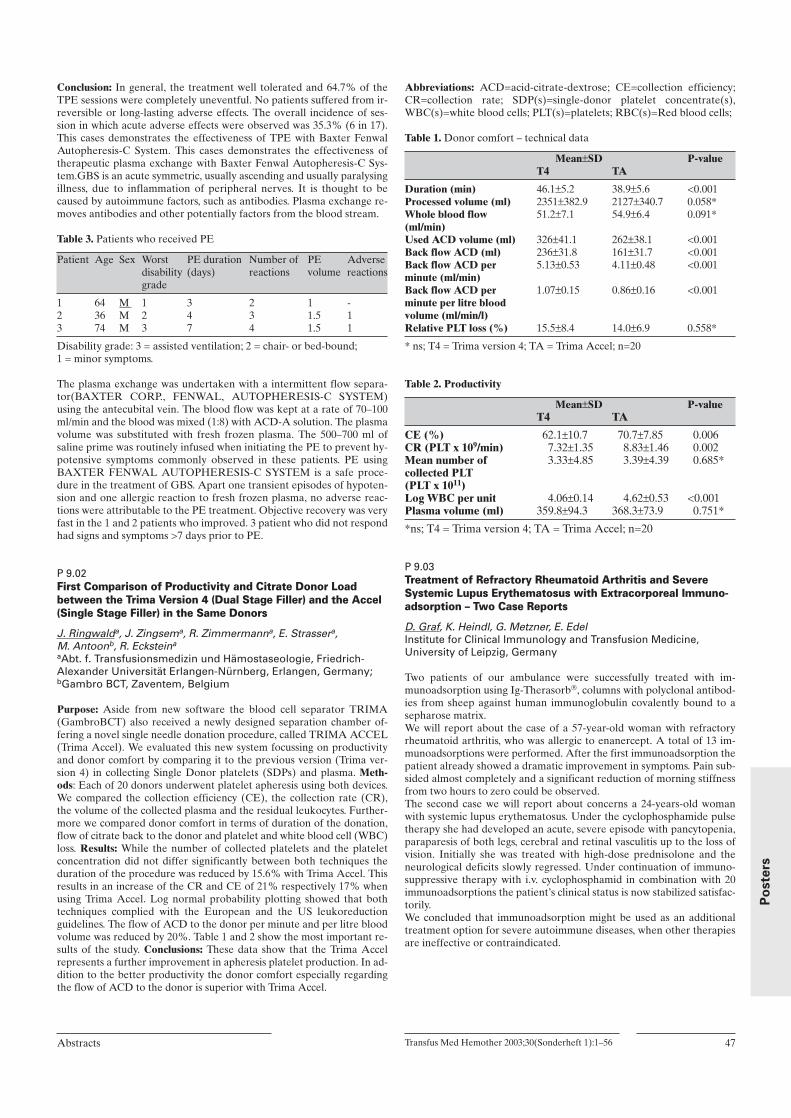
Transfus Med Hemother 2003;30(Sonderheft 1):1–56Abstracts 47
Po
ste
rs
Conclusion: In general, the treatment well tolerated and 64.7% of theTPE sessions were completely uneventful. No patients suffered from ir-reversible or long-lasting adverse effects. The overall incidence of ses-sion in which acute adverse effects were observed was 35.3% (6 in 17).This cases demonstrates the effectiveness of TPE with Baxter FenwalAutopheresis-C System. This cases demonstrates the effectiveness oftherapeutic plasma exchange with Baxter Fenwal Autopheresis-C Sys-tem.GBS is an acute symmetric, usually ascending and usually paralysingillness, due to inflammation of peripheral nerves. It is thought to becaused by autoimmune factors, such as antibodies. Plasma exchange re-moves antibodies and other potentially factors from the blood stream.
Table 3. Patients who received PE
Patient Age Sex Worst PE duration Number of PE Adverse disability (days) reactions volume reactions grade
1 64 M 1 3 2 1 - 2 36 M 2 4 3 1.5 13 74 M 3 7 4 1.5 1
Disability grade: 3 = assisted ventilation; 2 = chair- or bed-bound; 1 = minor symptoms.
The plasma exchange was undertaken with a intermittent flow separa-tor(BAXTER CORP., FENWAL, AUTOPHERESIS-C SYSTEM)using the antecubital vein. The blood flow was kept at a rate of 70–100ml/min and the blood was mixed (1:8) with ACD-A solution. The plasmavolume was substituted with fresh frozen plasma. The 500–700 ml ofsaline prime was routinely infused when initiating the PE to prevent hy-potensive symptoms commonly observed in these patients. PE usingBAXTER FENWAL AUTOPHERESIS-C SYSTEM is a safe proce-dure in the treatment of GBS. Apart one transient episodes of hypoten-sion and one allergic reaction to fresh frozen plasma, no adverse reac-tions were attributable to the PE treatment. Objective recovery was veryfast in the 1 and 2 patients who improved. 3 patient who did not respondhad signs and symptoms >7 days prior to PE.
P 9.02First Comparison of Productivity and Citrate Donor Loadbetween the Trima Version 4 (Dual Stage Filler) and the Accel(Single Stage Filler) in the Same Donors
J. Ringwalda, J. Zingsema, R. Zimmermanna, E. Strassera, M. Antoonb, R. Ecksteina
aAbt. f. Transfusionsmedizin und Hämostaseologie, Friedrich-Alexander Universität Erlangen-Nürnberg, Erlangen, Germany;bGambro BCT, Zaventem, Belgium
Purpose: Aside from new software the blood cell separator TRIMA(GambroBCT) also received a newly designed separation chamber of-fering a novel single needle donation procedure, called TRIMA ACCEL(Trima Accel). We evaluated this new system focussing on productivityand donor comfort by comparing it to the previous version (Trima ver-sion 4) in collecting Single Donor platelets (SDPs) and plasma. Meth-ods: Each of 20 donors underwent platelet apheresis using both devices.We compared the collection efficiency (CE), the collection rate (CR),the volume of the collected plasma and the residual leukocytes. Further-more we compared donor comfort in terms of duration of the donation,flow of citrate back to the donor and platelet and white blood cell (WBC)loss. Results: While the number of collected platelets and the plateletconcentration did not differ significantly between both techniques theduration of the procedure was reduced by 15.6% with Trima Accel. Thisresults in an increase of the CR and CE of 21% respectively 17% whenusing Trima Accel. Log normal probability plotting showed that bothtechniques complied with the European and the US leukoreductionguidelines. The flow of ACD to the donor per minute and per litre bloodvolume was reduced by 20%. Table 1 and 2 show the most important re-sults of the study. Conclusions: These data show that the Trima Accelrepresents a further improvement in apheresis platelet production. In ad-dition to the better productivity the donor comfort especially regardingthe flow of ACD to the donor is superior with Trima Accel.
Abbreviations: ACD=acid-citrate-dextrose; CE=collection efficiency;CR=collection rate; SDP(s)=single-donor platelet concentrate(s),WBC(s)=white blood cells; PLT(s)=platelets; RBC(s)=Red blood cells;
Table 1. Donor comfort – technical data
Mean±SD P-valueT4 TA
Duration (min) 46.1±5.2 38.9±5.6 <0.001Processed volume (ml) 2351±382.9 2127±340.7 0.058*Whole blood flow 51.2±7.1 54.9±6.4 0.091* (ml/min) Used ACD volume (ml) 326±41.1 262±38.1 <0.001Back flow ACD (ml) 236±31.8 161±31.7 <0.001Back flow ACD per 5.13±0.53 4.11±0.48 <0.001 minute (ml/min) Back flow ACD per 1.07±0.15 0.86±0.16 <0.001 minute per litre blood volume (ml/min/l) Relative PLT loss (%) 15.5±8.4 14.0±6.9 0.558*
* ns; T4 = Trima version 4; TA = Trima Accel; n=20
Table 2. Productivity
Mean±SD P-valueT4 TA
CE (%) 62.1±10.7 70.7±7.85 0.006CR (PLT x 109/min) 7.32±1.35 8.83±1.46 0.002Mean number of 3.33±4.85 3.39±4.39 0.685*collected PLT(PLT x 1011) Log WBC per unit 4.06±0.14 4.62±0.53 <0.001Plasma volume (ml) 359.8±94.3 368.3±73.9 0.751*
*ns; T4 = Trima version 4; TA = Trima Accel; n=20
P 9.03Treatment of Refractory Rheumatoid Arthritis and SevereSystemic Lupus Erythematosus with Extracorporeal Immuno-adsorption – Two Case Reports
D. Graf, K. Heindl, G. Metzner, E. EdelInstitute for Clinical Immunology and Transfusion Medicine,University of Leipzig, Germany
Two patients of our ambulance were successfully treated with im-munoadsorption using Ig-Therasorb, columns with polyclonal antibod-ies from sheep against human immunoglobulin covalently bound to asepharose matrix. We will report about the case of a 57-year-old woman with refractoryrheumatoid arthritis, who was allergic to enanercept. A total of 13 im-munoadsorptions were performed. After the first immunoadsorption thepatient already showed a dramatic improvement in symptoms. Pain sub-sided almost completely and a significant reduction of morning stiffnessfrom two hours to zero could be observed.The second case we will report about concerns a 24-years-old womanwith systemic lupus erythematosus. Under the cyclophosphamide pulsetherapy she had developed an acute, severe episode with pancytopenia,paraparesis of both legs, cerebral and retinal vasculitis up to the loss ofvision. Initially she was treated with high-dose prednisolone and theneurological deficits slowly regressed. Under continuation of immuno-suppressive therapy with i.v. cyclophosphamid in combination with 20immunoadsorptions the patient’s clinical status is now stabilized satisfac-torily. We concluded that immunoadsorption might be used as an additionaltreatment option for severe autoimmune diseases, when other therapiesare ineffective or contraindicated.

P 9.04The New Trima Accel: As Fast as Double Needle? A Comparisonof Plateletpheresesis Procedure Time of Three Different BloodSeparators
P. Krakowitzky, W. Sibrowski Institute for Transfusion Medicine, University Hospital Münster,Germany
Background: Procedure time is an important criterion for plateletphere-sis. A short processing time enhances productivity and donor comfort.Normally double needle procedures (DNP) are faster than single needleprocedures (SNP). Trima Accel(Gambro, BCT) is the recently intro-duced version of the automatic blood collection system Trima, both SNPseparators, that is said to be much faster than the presently used DNPseparators. To verify this statement we compared the Trima V4, theTrima Accel and the DNP Amicus Crescendo (Baxter) concerning pro-cedure time. Material and methods: Each of 40 donors underwentplateletpheresis on each of the three blood cell separators. Each donordonate the same type of product at each apheresis system (single or dou-ble apheresis platelets). Target endpoints were set at 3 × 10(11) platelets(PLT) for single apheresis platelets (SAP) and at 5.5 × 10(11) PLT fordouble apheresis platelets (DAP) in up to 90 minutes processing time.Results: Procedure times were shortest with the Accel in 37 donors. Inone donor the Amicus procedure was shortest. Results were for SAPmean value processing times (including re-infusion and re-suspension)of Amicus, V 4, and Accel: 55 min (42–82 min), 56 min (41–100 min), 42min (33–56 min), respectively. Results were for DAP mean value pro-cessing times (including reinfusion and resuspension) of Amicus, V 4,and Accel: 76 min (52–88 min), 74 min (59–88 min), 54 min (43–79 min),respectively. All platelet products achieved an acceptable PLT yieldwithin 2–4 × 10(11) PLT for SAP and ≥5.0 × 10(11) PLT for DAP. TwoAccel procedures did not result in a product because of separator trou-bles and collapsed venous access respectively. Adverse reactions to cit-rate occurred in approximately same frequency: Amicus 5 procedures, V4 4 procedures, Accel 3 procedures. Pressure alarms (draw and return)most frequently occurred in using the Accel: Amicus 3, V 4 15, Accel 24procedures. Conclusion: Trima V 4 and Amicus Crescendo are very sim-ilar concerning processing time. The new Trima Accel seems to be clear-ly superior. The high number of pressure alarms with the Trima Accelmay be due to the lack of practical experience but should deserve fur-ther observance.
P 9.05Therapeutic Approaches in the Management of Oral CyclosporinA Intoxication
G.C. Leitner, G. Stiegler, M. Hiesmayr#, P. Hoecker, B. Jilma*
Department of Blood Group Serology and Transfusion Medicine,#Department of Cardiothoracic Surgery, * Department of ClinicalPharmacology, University Hospital, Vienna, Austria
Background: A 68 years old male patient who was renal transplanted be-cause of bilateral end stage kidney disease of unknown origin received a100 fold oral overdose of CsA. Renal parameters were within the nor-mal range. Although conventional detoxification therapy was startedimmediately plasma levels of Cyclosporin A (CsA) exeeded 1500 ng/L.Because this patient was at high risk to develop renal failure wholeblood exchange (WBE) as additional detoxification management wasapplied. Material and Methods: For WBE a portable cell separator(MCS 3p/Haemonetics) was used. The exchange medium consisted of 4irradiated packed red blood cells (PRBCs) and 4 solvent detergent inac-tivated pooled plasmas (Octaplas®). Because of acute renal failure acontinuous hemofiltration with a filtration rate of 2000 mL/h was ap-plied two hours after the whole blood exchange. Results: WBE had noimmediate effect on serum CsA levels. 1110 ng/mL were detected afterthe exchange procedure. 1022 ng/mL were found in the depleted RBCfraction and 1237 ng/mL in the depleted plasma fraction of the patient.A rapid decrease of CsA in plasma, however, was seen after starting thehemofiltration. A CsA level of 159 ng/mL was found after 72 hours. Thepatient recovered and was dismissed from the hospital one month later.Conclusion: WBE has no immediate therapeutic effect in CsA intoxica-
tion. The decrease of CsA plasma levels after onset of hemofiltrationwould at first sight suggest therapeutic benefit but when compared tothe mean elimination half-life of CsA its effect is doubtful.
P 9.06Erythrocyte Loss in Case of Hemapheresis Donors: Are theLegal Guidelines Concerning Donor Protection Kept?
U. Werner, S. Rummler, F. Hofman, D. BarzInstitut für Transfusionsmedizin, Universitätsklinicum Jena,Germany
Introduction: The guidelines of the BÄK (BundesÄrtzeKammer) con-cerning the collection of blood and blood components specify the maxi-mum allowed annual RBC donation volume for women up to 1000 mland for men up to 1500 ml. The total donation volume cannot exceed 25L plasma per year. An interval of at least 8 weeks must be kept between2 whole blood donations. However apheresis donors are allowed to do-nate within much shorter time intervals. The actual apheresis techniquepermits the traceability of blood component loss. This follow up is so faronly possible with VISTATM. This software allows the management ofthe complete donation process, including donor selection and prepara-tion, donation monitoring and report generation. Methods and Materi-als: Procedure registration for 40 donors was handled with VISTATM.Donation volumes combined with the RBC loss can typically be calcu-lated as follows:1. Draw of blood sample for the legally required control testing: ca.
18ml RBC/27ml plasma.2. Residual volume of the tubing set with blood-return procedure: ca.
30ml RBC/35ml plasma and without blood-return procedure: ca. 95ml RBC/112 ml plasma.
Volume of blood products.Calculation of RBC loss was carried out with a fictitious hematocrit of40%.Results:Volume loss female donors (n=10)
No donations per Plasma loss RBC loss No incomplete proced./ year (L) (ml) RBC/loss (ml)
Mean: 19.5 8.595 339.6 1/95Min: 17 5.696 765 0/0Max: 22 9.590 1178 4/380
Volume loss male donors (n=30)
No donations per Plasma loss RBC loss No incomplete proced./year (L) (ml) RBC/loss
Mean: 15 10.435 1151.8 0.65/61.75Min: 27 6.664 1010 0/0Max: 23.2 13.089 1592 3/285
Conclusion: With VISTATM it is possible for the first time, to obtain wa-tertight computer supported documentation of the donation procedures,including the blood loss volumes that occurred. The results obtained in-dicate that, in case of regular donations, 30% of the female and 12% ofthe male donors lose more RBC volume than permitted by law. Supple-mentary investigations are necessary because the safety of the donors inrelation to the RBC loss is questioned.
48 Transfus Med Hemother 2003;30(Sonderheft 1):1–56 Abstracts
Po
ste
rs

P 9.07Aggregometry and Flow Cytometry Based Testing of PlateletFunction Induced by ADP and Adrenalin in ApheresisConcentrates
M. Notter1, F.H. Perschel2, A. Nierenheim-Ulrich1, R. Fitzner2, E. Thiel11Department of Hematology, Oncology, and Transfusion Medicine,2Department of Clinical Chemistry, Benjamin Franklin Free Univer-sity Medical Center, Berlin, FRG
Purpose: Test systems are needed to reliably assess the functional re-serve of platelet concentrates and the degree to which it invariably de-clines over time. Platelet activators in use for that purpose are eitherpoorly defined (collagen (C)), non-physiological (phorbol esters, risto-cetin, TRAP-6), or have weak activity (ADP, adrenalin (A)) associatedwith high variability of results. The aim of this study was to define condi-tions improving test characteristics in aggregometry and flow cytometricassessment of CD62P induction. Methods: Platelet function of singledonor apheresis concentrates (n=24) (Trima, Gambro BCT) in parallelto platelet-rich donor plasma for control was tested directly followingapheresis and after 5 days of storage. Various concentrations of collagen(0.095–0.38 mg/ml) and ADP (6.25 × 10⁻⁵–10-2 M) were used alone or incombination with a fixed concentration of adrenalin (5 × 10⁻⁵ M). Re-sults: C induced aggregation in 17/24 units with a mean of 65 ± 42% fad-ing to 5 ± 4% by day 5. Addition of adrenalin to collagen resulted in19/24 and 5/24 units responding on day 1 and 5, respectively, but datavariability remained high (80 ± 20 versus 30 ± 32%). Aggregation re-sponses in donor plasma uniformly were >70% arguing against donor-derived or pre-analytical confounding factors. Low-speed (134 g) pre-centrifugation had the potential to convert platelet concentrates not-ag-gregating with C+A to normal responders (13 versus 72%). ADP pro-duced 81 ± 17% aggregation which fell to 28 ± 15% by day 5. Asubstantial reduction of the variation coefficient to <10% on day 1 and<16% on day 5 was found when ADP was combined with A. Further-more, ADP+A profoundly increased the aggregation plateau (day 1: 99± 7%; day 5: 81 ± 12%). Lowering the ADP concentration allowed con-sistent evaluation of the deteriorating platelet function over time withhigh precision (81 ± 12 versus 68 ± 11%). ADP induced CD62P in fresh-ly produced platelet concentrates (40.3 ± 16.8 versus 9.1 ± 4.9%, p <.01)but not after storage (31 ± 11.3 versus 18.8 ± 12.6%, n.s.). In contrast,ADP+A demonstrated their functional reserve throughout (day 1: 64.9± 13.4 versus 9.1 ± 4.9%, p <.01; day 5: 50.6 ± 10.4 versus 18.8 ± 12.6%, p<.01). Conclusion: Pre-centrifugation and the synergy between A andADP are useful means to raise the precision and accuracy of in vitroplatelet function testing by aggregometry and flow cytometry facilitatingstandardization and comparability of the quality of platelet concentrates.
P 9.08Plasma Exchange in the Treatment of Severe Optic Neuritis: A Potential Beneficial Procedure
E. Klinker, K. Ruprecht*, A. Opitz, S. Kuhn, T. Dintelmann**, P. Rieckmann*, R. Gold*, M. Böck Depts. of Transfusion Medicine,* Neurology and ** Ophthalmology,University of Wuerzburg, Germany
Purpose: Beside standard high-dose corticosteroid therapy no successfultreatment could be established for patients with acute demylinatingoptic neuritis (ON). We reviewed the plasma exchange (PE) treatmentprocedures and the clinical outcome of the patients with ON, who un-derwent PE as an experimental therapy since 1/2000 up to now. Meth-ods: 10 patients with ON (22 to 51y; 2 male, 8 female; 4 diagnosed withMS before, 6 with ON as first event), were treated with PE [average of4.2 PE/patient (range 2–5); mean volume 40.3 ml plasma/kg (range30–40 ml/kg); intended number of PE 3–5]. PEs were performed every2nd or 3rd day. The primary outcome measure was change in visual acu-ity (VA) of the affected eye before and immediately after PE term aswell as on follow up. Baseline VA before PE ranged between no lightperception and 0.4. Results: PE was overall well tolerated. In 2 patientscentral venous access was necessary, 1 patient with peripheral accessbroke off therapy after the 2nd exchange because of flow problems. 7/10
patients improved immediately, 1 during follow up. In these patients PEswere started 14 to 41 days after ON event. 5/7 early-responders under-went 5, the other two 3 and 4 PEs. The 3 non-responders were treated 2,3 and 5 times with a time delay of 73 and 50 days after ON diagnosis.Mean length of follow up was 41 weeks (range 11–83). For 2/7 respon-ders follow up data are not yet available. 2 of the remaining 5 continuedto improve (VA 0.8 and 1.0), whereas 3 deteriorated again after 11–17weeks. Conclusion: This retrospective analysis suggests that PE may beof beneficial value in a subset of patients with severe ON which do notrespond to intravenous steroid therapy. It shows that in responders afavourable effect could be observed early, even after 2 PEs. However,neither a protracted effect of previous steroid therapy nor spontaneousimprovement can be ruled out. Our results encourage to further clinicalstudies for treatment of severe ON with PE and investigations of pre-dicting factors of a successful outcome.
P 9.09Collection of Red Packed Cells with Two Different Multi Com-ponent Apheresis Protocols: Cobe Trima and HaemoneticsMCS+ LDPRBC
B. Stephan, M. Dobonici, K. Erdlenbruch, V.M. Petrescu-Jipa, J. Stolte, J.F. Schenk, U. SeyfertDepartment for Clinical Hemostaseology and Transfusion Medicine,University Hospital of Saarland, Homburg, Germany
Purpose: Two different multi component apheresis protocols were ex-amined in our blood donation service: Haemonetics MCS+: LDPRBC,CPD, PAGGS-M with automatic addition of the additive solution andCobe Trima: ACD-A, SAG-M with manual addition of the additive so-lution. The results of the evaluation of the platelet concentrates are notpublished here. Methods: We collected from each of fifteen (Trima) re-spectively nineteen (MCS+) blood donors one platelet concentrate (PC)and one unit of packed red blood cells in SAG-M/ACD-A (Trima) re-spectively PAGGS-M/CPD (MCS+). Measurements of the products ac-cording to the guidelines of the German Medical Association were done.Descriptive statistics were performed. Results: Red blood cells (RBC):average: Trima: n: 15; volume (incl. SAG-M) [ml]: 256; hct/unit [L/L]:day 0: 0.55, day 42: 0.59; hemoglobin/unit (g): day 0: 45.9, day 42: 46.6;rate of hemolysis (%): 0.03; WBC (Nageotte): 2,115 per unit; donationtime [min]: 59. MCS+: n: 19; volume (incl. PAGGS-M) [ml]: 288; hct/unit[L/L]: day 0: 0.58, day 49: 0.60; hemoglobin/unit (g): day 0: 55.0, day 49:55.2; rate of hemolysis (%): 0.02; WBC (Nageotte): 77,642 per unit; do-nation time [min]: 108. Guidelines: hct/unit [L/L]: day 0/expiry date:0.5–0.7; hemoglobin/unit (g): day 0/expiry date: >40; rate of hemolysis(%): <0.8; WBC: <1,000,000 per unit. Conclusion: It is possible to collectone unit of red packed cells plus one platelet concentrate from oneblood donor in a quality according to the guidelines of the GermanMedical Association. The donation time of Trima is excellent but thereshould be an additional protocol with PAGGS-M (thus, red packed cellscan be kept for a maximum of 49 days) and the addition of the additivesolution should be automated. The donation time of MCS+ is too long(due to the collection time for the platelet concentrate) and there shouldbe an update of the LDPRBC protocol.
P 9.10Evaluation of the RBC Exchange Program Using the FreseniusCOM.TEC Cell Separator
F. Driss, G.TcherniaHaematology Unit – Bicêtre Hospital, Paris, France
Purpose: Fresenius Hemocare suspended the use of the RBC exchangeprogram due to a software bug in the first version on the COM.TEC.The new and corrected version (02.03.05) has been reintroduced andevaluated in the Bicêtre Hospital’s haematology unit. The aim of the ex-changes was the reduction of abnormal HbS levels in sickle cell patientsbelow 40%. The RBC exchange program allows to set an intended pa-tient’s hematocrit (hct) post procedure and predicts the elimination ofHbS. The aim of the study was to compare, for each procedure, the com-puter intended values and the achieved values at the end of the RBC ex-
Transfus Med Hemother 2003;30(Sonderheft 1):1–56Abstracts 49
Po
ste
rs

change. Methods: 16 procedures were carried out on 10 patients withsickle cell disease: mean age 25 years (range 6–44) and mean weight 54kg (range 20–69). Measurements of haematological parameters and ab-normal haemoglobin levels were done before, at the end and 1 hourafter the RBC exchange. To achieve the highest possible precision, thehct of all transfused Red Cell Concentrates (n=88) were precisely mea-sured to program the correct mean hct of the RCC required by the soft-ware. Results: The chart shows the mean values of the procedure fea-tures.
Mean Median Min Max
RCC volume (ml) 1446 1505 830 1775 RCC volume 28 26 22 42(ml/kg body weight) Time (min) 64 64 46 104
Hct Before 28.4 29.2 21.1 33.0[%] At the end 28.3 28.4 22.6 41.0
After 1h 29.2 28.3 24.0 37.0Intended 29.3 29.5 25.0 35.0Difference intended – achieved 1h after1 –0.16 0 0 2.9
HbS Before 60.9 59.0 46 97[%] After 1h 30.1 28.6 15 60
Intended 29.2 28.0 20 53Difference intended – achieved 1h after1 0.9 1.1 0 10
1difference obtained in every individual treatment
The procedures were carried out without any technical failures or med-ical problems. The RBC exchanges were well tolerated without oxygensupply, the pulse and the arterial pressures were very stable throughoutthe procedures. Conclusions: The benefit of this technology is the soft-ware reliability: the deviation between mean intended and achieved hct1 h after the procedure was only 0.16% (not significant with Studenttest, p >0.05). The patients appreciated the cell separator because of itscomfort and well tolerated procedures, the users appreciated the auto-matic feature, the safety system and the user-friendly software. The tech-nological simplicity of Fresenius COM.TEC allows a comfortable RBCexchange procedure which releases the user from all complicated andmore or less reliable calculations.
P 9.11Enhanced Granulocyte Yield Using 6% HES of High MolecularWeight for G-CSF Mobilized Granulocytapheresis
P. Reinhardt, H. Lux, E. Krug, H. Schrezenmeier, M. WiesnethInstitute for Clinical Transfusion Medicine and ImmunogeneticsUlm and Department of Transfusion Medicine, University of Ulm,Germany
Purpose: To improve the collection efficiency of granulocytaphereses byusing 6% hydroxy ethyl starch (HES) of high molecular weight (Plas-masteril 450/0.7, Fresenius) compared to HES of low molecular weight(HAES-sterile 200/0.5, Fresenius). Methods: Patients suffering of afebrile neutropenia and refractory to antimicrobial agents or patientswith progressive systemic mycosis may qualify for therapeutic granulo-cyte transfusions. Following informed consent, granulocytes fromhealthy HLA- and ABO-matched donors were mobilized with G-CSF 5γg/kg BW 8 to 12 hours prior to granulocytapheresis. Donors were sub-mitted to a maximum of four aphereses using a COBE Spectra cell sepa-rator (Gambro BCT, Martinsried, Germany). To accelerate erythrocytesedimentation and facilitate granulocyte collection HES of a molecularweight of 450,000 D (high molecular weight) or 200,000 D (low molecu-lar weight) was employed. Donors received some 450 ml of HES duringa 3-hours apheresis. Granulocyte collection efficiency was evaluatedwith respect to initial granulocyte count, processed blood volume andpurity of the apheresis products. Results: Fifteen collections with highmolecular weight (hmw) HES were compared to 15 collections withHES of low molecular weight (lmw). Initial mean leukocyte counts (26.5G/l -hmw- vrs. 26.0 G/l -lmw-) and mean processed blood volume (8914
ml -hmw- vrs. 8231 ml -lmw-) were comparable in both groups. Highmolecular weight HES allowed significantly higher mean leukocyte (7.8× 10E10 -hmw- vrs. 4.0 × 10E10 -lmw-) and granulocyte (77.7% -hmw-vrs. 60.6% -lmw-) yields, while mean contamination of erythrocytes (Hkt6%) and platelets (3 × 10E11) was similar. No adverse events caused byG-CSF mobilisation, ACD-A, HES or the apheresis procedure were ob-served in either group. Conclusions: Large quantities of granulocytes canreliably be collected with the use of high molecular weight HES. De-pending on the patients weight split daily granulocyte transfusions of1.5–3.5 × 10E8 granulocytes/kgBW are possible with collections everyother day, thus economizing both donor and material resources.
P 9.12Observations on Protein A Immunoadsorption as an AdjunctiveTherapy for Acquired Thrombotic Thrombocytopenic Purpura
S. Fontana, J.-D. Studt, B. Lämmle, L. Alberio, J.A. Kremer Hovinga,C. Marbacher, B. Mansouri TaleghaniCentral Hematology Laboratory, Inselspital, University Hospital,Bern, Switzerland
Purpose: According to preliminary results, Protein A immunoadsorp-tion (PAIA) should be useful in the treatment strategy of several severeautoantibody mediated haematological disorders. We summarize thedata on our first patient (P) with acquired thrombotic thrombocytopenicpurpura (TTP). In TTP plasma exchange (PE) with fresh frozen plasma(FFP) replacement is standard management, removing circulating in-hibitors and providing exogenous ADAMTS13 activity. We investigated,whether PAIA can induce a faster clinical response by removing circu-lating inhibitors more efficiently than PE and thereby restoring endoge-nous enzyme activity more rapidly. Methods: For PAIA and PE we uti-lize a cell separator (Spectra, Gambro BCT, USA). In PAIA the sec-ondary system consists of staphylococcal Protein A-columns and a plas-ma flow monitor (Immunosorba and Citem 10, Fresenius, Germany). Inevery single PE (PAIA) 1.0–1.5 (2.0–2.5) plasma volumes of the P werereplaced with FFP (processed). We started with 3 daily PE and switchedto PAIA on day 4–6. Then PAIA was discontinued and PE resumed foranother 10 days. Further treatment consisted of Methylprednisolone 125mg/day i.v.. Results:
Day PE/PAIA Platelets LDH ADAMTS 13 ADAMTS 13 (G/L) (U/L) Activity (%) Inhibitor (BU)
1 PE 7 3224 <3% ca. 34 PAIA 70 606 5% <17 PE 17 2571 <3% <1
14 PE 108 495 30% <1
Initial 3 PEs transiently improved platelet count and LDH. This paral-leled with normalized ADAMTS13 activity (prior/after 3rd PE 5%/>50%) and lowered the inhibitor titer. After switching to PAIA diseaseactivity deteriorated (see table). ADAMTS13 activity returned to <5%within <20 h although the inhibitor titer remained virtually undetectableduring PAIA. Resumption of PE again led to an improvement of clinicaland laboratory parameters. Conclusions: PAIA neither improves diseaseactivity nor restores ADAMTS13 activity, although circulatingADAMTS13 inhibitors are virtually completely removed. Residualtraces of the inhibitor or its re-entry from extravascular sites seem to besufficient for completely inhibiting newly synthesized endogenousADAMTS13 and/or the enzyme is produced insufficiently. For futureapplications to acquired TTP, PAIA should be combined with infusion ofexogenous ADAMTS13.
50 Transfus Med Hemother 2003;30(Sonderheft 1):1–56 Abstracts
Po
ste
rs

P 9.13Double Unit Red Cell Concentrate Collections – High Quality Components and a Safe Procedure for Donors
G. Matthes1, E. Edel1, N. Thriemer1, P. Seidel2, W. Gründer2
1Institute of Transfusion Medicine, University Hospital Leipzig,2Institute of Biophysics, University Leipzig, Germany
Purpose: Red cell automated collection gives new options, which allowscollection of two red cell concentrate (RCC) units from the same donor.The study evaluated the in-vitro quality parameter of apheresed RCCand donor regeneration after RCC double unit collection. Methods: Weperformed 20 double red cell unit automated collection procedures byAlyx (Baxter). Within 27 min two units of RCC are collected, resus-pended in SAG-M, in-line filtered during procedure, and stored for 42days. Each donor did meet requirements for a whole blood donation;additional eligibility criteria were applied for donor safety: male donor(Hct >40%, >60 kg, >1.70 m); female donor (Hct >40%, >70 kg, >1.68m). All donors were volume supplemented with 400 ml saline and havebeen analyzed for blood cell and iron status regeneration in frequent in-tervals after donation (1,3,5,14,21,42,56,70,120 days). In-vitro testing wasdone on RCC at days 0,7,14,21,28,35,42, and included Hb, pH, free he-moglobin/potassium, hemolysis, ATP/2,3-DPG (31P-NMR), glucose, lac-tose, and deformability of RBC. Results: No severe or serious adverseevents were reported during and after apheresis. Immediately after do-nation there is a Hb loss of 20,2 ± 5,2%, reduced to 14,1 ± 6,1% on day1, and to 4,3 ± 10,4% on day 42.
Table shows a summary of in-vitro quality data of RCC on day0,7,14,21,28,and 42:
Storage day 0 7 14 21 28 35 42Hb mmol/L 11.7 12.3 11.8 11.8 11.9 11.8 11.9pH 22 °C 7.14 6.92 6.76 6.70 6.64 6.58 6.54Glucose mmol/L 25.3 20.7 17.3 14.5 11.9 10.1 8.7Lactose mmol/L 2.5 12.6 20.1 24.5 27.3 30.9 33.9ATP µmol/g Hb 3.14 3.37 3.71 3.28 2.63 2.42 1.87 2,3-DPG µmol/g Hb 13.51 8.11 2.47 1.94 1.98 1.67 1.72Free Hb mg/dL 31.8 45.1 51.3 63.2 83.6 100.5 162.1Free potassium mmol/L 1.8 19.9 28.9 33.9 40.1 44.1 48.9Hemolysis % 0.07 0.09 0.11 0.13 0.17 0.21 0.32Deformability % 100 94 91 85 79 77 80
Conclusions: All apheresis collected RCC did fit the German and Euro-pean quality requirements. The collection of two units of leukocyte de-pleted RCC from one donor with the new separator Alyx is a safe, andcost-effective procedure for selected donor population (based on eligi-bility criteria approximately 20–30% of all donors). Integration of multi-component collection into transfusion service will have an impact onblood supply, blood inventory, and on clinical practice.
P 10: Stem- / Dendritic- / NK-Cells
P 10.01Collection of Peripheral Progenitor Cells in Children with Useof the Amicus® Blood Cell Separator
R. Moog1, O. Basu2, B. Kremens2
1Institute for Transfusion Medicine, 2Department of PaediatricOncology, University Clinics Essen, Germany
Purpose: When harvesting peripheral progenitor cells (PPC) in childrenthe special situation of their circulatory system has to be taken into ac-count. Extracorporeal blood volume and product volume should be lowto avoid side effects. Material and methods: 8 children (age 2–14 years,weight 12.8–58.5 kg) with neuroblastoma, Ewing sarcoma, retinoblas-toma, medulloblastoma and Non-Hodgkin lymphoma underwent 9 PPCcollections with the MNC programme of the Amicus blood cell separa-tor (Baxter). The disposable was primed with red blood cells (RBCs) orhuman albumin to avoid circulatory side effects. PPC were mobilised bychemotherapy and G-CSF application. Blood counts and CD 34 antigenexpressing cells were determined before apheresis and in the PPC prod-
uct. The children were monitored for blood pressure and heart rate dur-ing the whole apheresis procedure. Results: A median blood volume of4,584 ml (range 3,536–8,596 ml) was processed in a separation time of256 min (range 176–331 min). The median product weight was 81 g(range 53–107 g) and the yield of CD 34 antigen expressing cells was13.6 × 106/kg body weight (range 1.8–26 × 106/kg body weight). Only onechild had to undergo a second apheresis to collect the desired transplan-tation dose. The median haematocrit of the products was 12.5 g/dl(8.8–13.9 g/dl) and the platelet contamination was 0.3 × 10¹¹ (0.05–1.43 ×10¹¹). No circulatory side effects were observed. Blood flow alarms oc-curred in seven of eight aphereses and one collection had to be termi-nated due to insufficient flow. Conclusion: PPC can be efficiently col-lected in children with the MNC programme without circulatory side ef-fects provided that the disposable was primed with human albumin orpacked RBCs. The platelet contamination of the product was low due toelutriation principle of the collection process thereby avoiding thrombo-cytopenic bleeding episodes in the patients.
P 10.02Use and Efficiency of Lenograstim for Mobilising AllogeneicPeripheral Stem Cells: A Prospective Multicenter Evaluationwith One Year Follow Up
R. Moog1, W. Pönisch2, I. Krohn3, M. Wiesneth4
1Institute for Transfusion Medicine, University Clinics Essen; 2Clinicfor Internal Medicine II, Leipzig; 3Chugai Pharma, Frankfurt; 4Dept.Transfusion Medicine and Red Cross Blood Center, University ofUlm, Germany
Purpose: To determine the dose of lenograstim for mobilising allogeneicstem cells under routine conditions and to evaluate its influence on labo-ratory parameters. Materials and Methods: 89 allogeneic donors (51male, 38 female) underwent examination before and at the time point ofperipheral stem cell collection. Furthermore, a one year follow up exam-ination was performed in 40 donors. The stem cell donors were mobilisedwith a median daily lenograstim dose of 8.77 µg/kg body weight (BW).Lenograstim was administered in a split dose (98.2%) and the duration ofapplication varied between 3 and 6 days. Results: A sufficient transplan-tation dose was collected from all 89 donors. The yield of CD 34+ cellscollected was 8.78 ± 5.99 × 10⁶ per recipient BW (mean ± SD) on thefirst day of aphereses. If a second collection was necessary, the yield de-creased to 5.94 ± 1.47 × 10⁶. Gender significantly influenced the CD 34+yield: In male donors a higher yield (8.32 ± 4.29 × 10⁶) was collected thanin female donors (7.54 ± 6.41 × 10⁶, p=0.037). Non-significant parametersfor the CD 34+ yield were daily lenogastrim dose (<8 µg/kg BW, 8–12µg/kg BW, >12 µg/kg BW), time interval of lenogastrim before start ofapheresis (less or more than 3 hours) and duration of apheresis (less ormore than 5 hours). Besides the well known changes of the white bloodcells and their subpopulation the following parameters significantly in-creased due to the mobilisation regimen at the day of stem cell collection:uric acid (327 ± 95 mmol/L), glutamate oxalo-acetate transferase (26 ± 13U/L), gamma glutamyl transferase (27 ± 7 U/L), alkaline phosphatase(242 ± 67 U/L) and lactate dehydrogenase (444 ± 170 U/L). These para-meters were within normal ranges at the one year follow up, but lympho-cytes and monocytes were still reduced when compared with baseline val-ues. Conclusion: Stem cells can be efficiently collected with differentmobilisation regimens in allogeneic donors. A significant higher yield ofCD 34+ cells was harvested in male donors. Leukocyte subpopulationsshould be monitored at follow up visits.
P 10.03Method for Isolation of Mesenchymal Stem Cells from Umbili-cal Cord Blood
S. Kern, K. Bieback,, H. Klüter, H. Eichler Red Cross Blood Service of Baden-Württemberg-Hessen, Universityof Heidelberg, Faculty of Clinical Medicine Mannheim, Germany
Purpose: Mesenchymal stem cells (MSC) are multi-potent cells, whichcan be isolated from bone marrow. They can either replicate as undiffer-entiated cells or have the potential to differentiate to various non-
Transfus Med Hemother 2003;30(Sonderheft 1):1–56Abstracts 51
Po
ste
rs
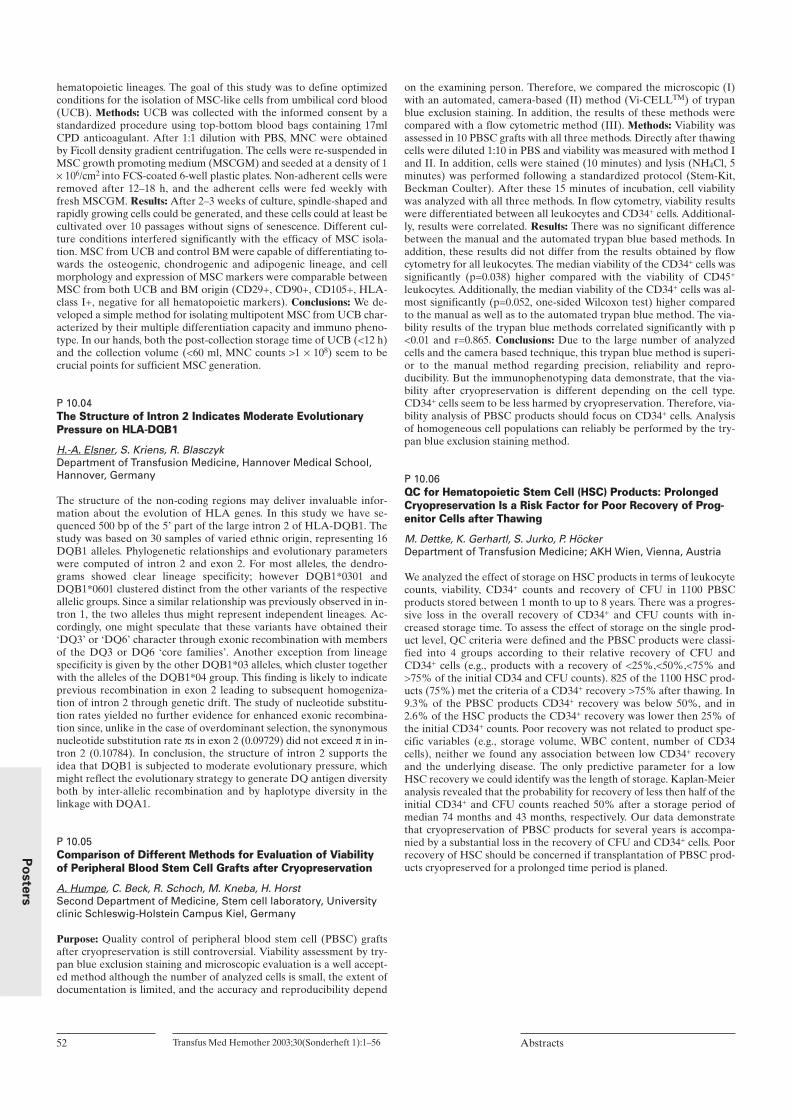
hematopoietic lineages. The goal of this study was to define optimizedconditions for the isolation of MSC-like cells from umbilical cord blood(UCB). Methods: UCB was collected with the informed consent by astandardized procedure using top-bottom blood bags containing 17mlCPD anticoagulant. After 1:1 dilution with PBS, MNC were obtainedby Ficoll density gradient centrifugation. The cells were re-suspended inMSC growth promoting medium (MSCGM) and seeded at a density of 1× 10⁶/cm2 into FCS-coated 6-well plastic plates. Non-adherent cells wereremoved after 12–18 h, and the adherent cells were fed weekly withfresh MSCGM. Results: After 2–3 weeks of culture, spindle-shaped andrapidly growing cells could be generated, and these cells could at least becultivated over 10 passages without signs of senescence. Different cul-ture conditions interfered significantly with the efficacy of MSC isola-tion. MSC from UCB and control BM were capable of differentiating to-wards the osteogenic, chondrogenic and adipogenic lineage, and cellmorphology and expression of MSC markers were comparable betweenMSC from both UCB and BM origin (CD29+, CD90+, CD105+, HLA-class I+, negative for all hematopoietic markers). Conclusions: We de-veloped a simple method for isolating multipotent MSC from UCB char-acterized by their multiple differentiation capacity and immuno pheno-type. In our hands, both the post-collection storage time of UCB (<12 h)and the collection volume (<60 ml, MNC counts >1 × 10⁸) seem to becrucial points for sufficient MSC generation.
P 10.04The Structure of Intron 2 Indicates Moderate EvolutionaryPressure on HLA-DQB1
H.-A. Elsner, S. Kriens, R. BlasczykDepartment of Transfusion Medicine, Hannover Medical School,Hannover, Germany
The structure of the non-coding regions may deliver invaluable infor-mation about the evolution of HLA genes. In this study we have se-quenced 500 bp of the 5’ part of the large intron 2 of HLA-DQB1. Thestudy was based on 30 samples of varied ethnic origin, representing 16DQB1 alleles. Phylogenetic relationships and evolutionary parameterswere computed of intron 2 and exon 2. For most alleles, the dendro-grams showed clear lineage specificity; however DQB1*0301 andDQB1*0601 clustered distinct from the other variants of the respectiveallelic groups. Since a similar relationship was previously observed in in-tron 1, the two alleles thus might represent independent lineages. Ac-cordingly, one might speculate that these variants have obtained their‘DQ3’ or ‘DQ6’ character through exonic recombination with membersof the DQ3 or DQ6 ‘core families’. Another exception from lineagespecificity is given by the other DQB1*03 alleles, which cluster togetherwith the alleles of the DQB1*04 group. This finding is likely to indicateprevious recombination in exon 2 leading to subsequent homogeniza-tion of intron 2 through genetic drift. The study of nucleotide substitu-tion rates yielded no further evidence for enhanced exonic recombina-tion since, unlike in the case of overdominant selection, the synonymousnucleotide substitution rate πs in exon 2 (0.09729) did not exceed π in in-tron 2 (0.10784). In conclusion, the structure of intron 2 supports theidea that DQB1 is subjected to moderate evolutionary pressure, whichmight reflect the evolutionary strategy to generate DQ antigen diversityboth by inter-allelic recombination and by haplotype diversity in thelinkage with DQA1.
P 10.05Comparison of Different Methods for Evaluation of Viabilityof Peripheral Blood Stem Cell Grafts after Cryopreservation
A. Humpe, C. Beck, R. Schoch, M. Kneba, H. HorstSecond Department of Medicine, Stem cell laboratory, Universityclinic Schleswig-Holstein Campus Kiel, Germany
Purpose: Quality control of peripheral blood stem cell (PBSC) graftsafter cryopreservation is still controversial. Viability assessment by try-pan blue exclusion staining and microscopic evaluation is a well accept-ed method although the number of analyzed cells is small, the extent ofdocumentation is limited, and the accuracy and reproducibility depend
on the examining person. Therefore, we compared the microscopic (I)with an automated, camera-based (II) method (Vi-CELLTM) of trypanblue exclusion staining. In addition, the results of these methods werecompared with a flow cytometric method (III). Methods: Viability wasassessed in 10 PBSC grafts with all three methods. Directly after thawingcells were diluted 1:10 in PBS and viability was measured with method Iand II. In addition, cells were stained (10 minutes) and lysis (NH4Cl, 5minutes) was performed following a standardized protocol (Stem-Kit,Beckman Coulter). After these 15 minutes of incubation, cell viabilitywas analyzed with all three methods. In flow cytometry, viability resultswere differentiated between all leukocytes and CD34+ cells. Additional-ly, results were correlated. Results: There was no significant differencebetween the manual and the automated trypan blue based methods. Inaddition, these results did not differ from the results obtained by flowcytometry for all leukocytes. The median viability of the CD34+ cells wassignificantly (p=0.038) higher compared with the viability of CD45+
leukocytes. Additionally, the median viability of the CD34+ cells was al-most significantly (p=0.052, one-sided Wilcoxon test) higher comparedto the manual as well as to the automated trypan blue method. The via-bility results of the trypan blue methods correlated significantly with p<0.01 and r=0.865. Conclusions: Due to the large number of analyzedcells and the camera based technique, this trypan blue method is superi-or to the manual method regarding precision, reliability and repro-ducibility. But the immunophenotyping data demonstrate, that the via-bility after cryopreservation is different depending on the cell type.CD34+ cells seem to be less harmed by cryopreservation. Therefore, via-bility analysis of PBSC products should focus on CD34+ cells. Analysisof homogeneous cell populations can reliably be performed by the try-pan blue exclusion staining method.
P 10.06QC for Hematopoietic Stem Cell (HSC) Products: ProlongedCryopreservation Is a Risk Factor for Poor Recovery of Prog-enitor Cells after Thawing
M. Dettke, K. Gerhartl, S. Jurko, P. Höcker Department of Transfusion Medicine; AKH Wien, Vienna, Austria
We analyzed the effect of storage on HSC products in terms of leukocytecounts, viability, CD34+ counts and recovery of CFU in 1100 PBSCproducts stored between 1 month to up to 8 years. There was a progres-sive loss in the overall recovery of CD34+ and CFU counts with in-creased storage time. To assess the effect of storage on the single prod-uct level, QC criteria were defined and the PBSC products were classi-fied into 4 groups according to their relative recovery of CFU andCD34+ cells (e.g., products with a recovery of <25%,<50%,<75% and>75% of the initial CD34 and CFU counts). 825 of the 1100 HSC prod-ucts (75%) met the criteria of a CD34+ recovery >75% after thawing. In9.3% of the PBSC products CD34+ recovery was below 50%, and in2.6% of the HSC products the CD34+ recovery was lower then 25% ofthe initial CD34+ counts. Poor recovery was not related to product spe-cific variables (e.g., storage volume, WBC content, number of CD34cells), neither we found any association between low CD34+ recoveryand the underlying disease. The only predictive parameter for a lowHSC recovery we could identify was the length of storage. Kaplan-Meieranalysis revealed that the probability for recovery of less then half of theinitial CD34+ and CFU counts reached 50% after a storage period ofmedian 74 months and 43 months, respectively. Our data demonstratethat cryopreservation of PBSC products for several years is accompa-nied by a substantial loss in the recovery of CFU and CD34+ cells. Poorrecovery of HSC should be concerned if transplantation of PBSC prod-ucts cryopreserved for a prolonged time period is planed.
52 Transfus Med Hemother 2003;30(Sonderheft 1):1–56 Abstracts
Po
ste
rs

Transfus Med Hemother 2003;30(Sonderheft 1):1–56Abstracts 53
Po
ste
rs
P 10.07Evaluation of a Predictive Algorithm for Stem Cell Yield
A. Humpe1, P. Schlenke2, J. Riggert3, M. Köhler3, M. Kneba1
1Stem cell laboratory of the second Department of Medicine,University of Schleswig-Holstein, Campus Kiel; 2Institute ofImmunology and Transfusion Medicine, University of Schleswig-Holstein, Campus Lübeck; 3Department of Transfusion Medicine,University of Göttingen, FRG
Purpose: To investigate the transferability and the accuracy of a previ-ously developed and published predictive algorithm for stem cell yieldon a new cell separator, data from aphereses performed with the BaxterAmicus were retrospectively evaluated and compared with results fromthe COBE Spectra and the Baxter CS3000+ cell separator. Methods:The yield of CD34+ cells in 86 aphereses of 38 different patients anddonors performed on a Baxter Amicus cell separator was compared withthe yield predicted by a recently published algorithm. The following pa-rameters were included in the algorithm: the donor’s sex, weight, andheight, the inlet flow, the duration of the apheresis, and the number ofCD34+ cells/µl in PB before apheresis. The results were compared withresults of 105 aphereses performed with a COBE Spectra and with re-sults of 108 aphereses performed with a Baxter CS3000+ cell separator.Results: In 24 of the 86 leukaphereses (27.9%), the predicted value washigher than the measured and in 18 (20.9%) of these the deviation wasabove 10%. The respective values for the latter parameter were 17.6%for the Baxter CS3000+ and 16.2% for the COBE Spectra cell separator.The median overestimation defined by the ratio between predicted andmeasured yield in% was 31.1% for the Baxter Amicus, 44.1% for theCOBE Spectra, and 31.0% for the Baxter CS3000+ cell separator. Thesedifferences were statistically not different. Regarding the median under-estimation of the yield there was no significant difference between thethree cell separators either. Correlating the logarithm of the predictedversus the logarithm of the measured value for the aphereses of the Bax-ter Amicus cell separator, a linear correlation with the equationlog(measured value) = 0.48 + 0.938×log(predicted value) and r=0.88 andp <0.0001 was calculated. The slope and the correlation coefficient arealmost identical to the data formerly deduced from the COBE Spectracell separator. Conclusions: These data confirm that our algorithm en-ables prediction of the minimal yield in autologous peripheral bloodstem cell harvests independent from the cell separator used. Althoughstatistically not significant, the accuracy defined by the extent of overes-timation might be better with the COBE Spectra and the BaxterCS3000+ cell separator compared with the Baxter Amicus machine. Fur-ther investigations will answer this question.
P 10.08Significance of Nucleated Cell Concentration in Blood Progen-itor Cell Products
W. Gebauer, E. Fallgren Gebauer, N. Grommes, E. Fitje, F. Schunter DRK Oldenburg, Oldenburg, Germany
Depending on patient characteristics and therapeutic intention cellularconstituents of blood progenitor cell products (bpcp) may vary consider-ably. Influence of concentration of nucleated cells on bpcp quality has tobe characterized. In short term culture assays clonogenicity of bpc was determined beforeand after cryopreservation and at the time of transplantation. Analysis was performed after 50 autologous transplantations into pa-tients suffering from lymphoma or germ cell cancer. Density of nucleat-ed cells in bpcp varied from 0.4 to 1.9 times 108/ml. Modification of clonogenicity of bpc resulting from variable cellulardensity during storage under liquid nitrogen was insignificant. Further-more at the time of transplantation there was no significantly modifiedclonogenicity of bpc related to concentration of nucleated cells. We demonstrated with these preliminary results variability in cellularconcentration in bpcp without obvious harmful effects on in vitro clono-genicity. These together with further investigations concerning engraft-ment characteristics may lead to improvement of bpcp by reducing theamount of critical substances involved in the cryoprocedure.
P 10.09Cryopreservation of Peripheral Blood Stem Cell ProductsAffects the Sensitivity to Determine Bacterial Contaminations
U. Cassens1, F. Kipp2, E. Linnemann1, C. Ahlke1, G. Peters2, W. Sibrowski11Institute of Transfusion Medicine, 2Institute of Medical Microbio-logy, University Hospital Münster, Germany
Purpose: Microbial contamination of peripheral blood stem cell prod-ucts (PBSCPs) may cause severe clinical complications in immunosup-pressed recipients. Therefore, we investigated the influence of cryopro-tectant and cryopreservation on the sensitivity to detect bacterial conta-minations in PBSCPs. Methods: Two different bacterial strains (Staphy-lococcus epidermidis DSM 20044 and Escherichia coli ATCC 25922)were adjusted to defined concentrations and inoculated into a total of 29bags of expired PBSCPs, respectively. For this purpose, the expired ster-ile PBSCPs were thawed according to the routine procedure and analiquot of 1 ml bacteria suspension was added to each PBSCP. A calcu-lated concentration of 3.1 × 10⁵ colony-forming units (CFUs) of S. epi-dermidis or 9.1 × 10⁵ CFUs E. coli were inoculated per bag (n=15 andn=14), respectively. After 20 min intermixing on an agitator, a represen-tative aliquot was drawn from each product and cultured on Columbia(S. epidermidis) or Mueller-Hinton (E. coli) agar. Then, the PBSCPswere cryopreserved for another 24 hours and thawed again. Anotherrepresentative aliquot was drawn and cultured as mentioned above. Re-sults: After 24 hour of incubation at 37 °C and quantitative analysis, amean concentration of 2529 CFUs/ml S. epidermidis were determinedbefore cryopreservation versus 2182 CFUs/ml after cryopreservationdemonstrating a decrease of detectable colonies (p <0.05 Wilcoxon test).For the PBSCPs contaminated with Escherichia coli, the mean numberswere 396 CFUs/ml before cryopreservation and 327 CFUs/ml after cry-opreservation also showing a decrease (p <0.05). A microbiological in-hibitor test – 24 hour incubation of sterile cryoprotectant (10% di-methylsulfoxide) with Bacillus subtilis – was negative. Conclusions: Ob-viously, the cryopreservation of PBSCPs affects the sensitivity to detectbacterial contaminations, since a drop of CFUs was verified for both,gram-positive and gram-negative strains after cryopreservation. Howev-er, it remains questionable whether this decrease of CFUs after cryop-reservation is caused by the cellular effects - as postulated for the pro-cessing of other blood components.
P 10.10Correlation between CD34+ Stem Cells and CFU-GM in Peri-pheral Blood Stem Cell Collections from Patients Treated withIntensive Chemotherapy and Autologous Transplantation
A. Dada, M. Klouche, G. Rothe, A. Reichle, E. Holler, G. SchmitzInstitute for Clinical Chemistry and Laboratory Medicine, Regens-burg, Germany
Purpose: There are discrepancies between the published studies aboutthe correlation between CFU-GM and CD34+ cells count in marrow au-totransplants. Therefore we evaluated the levels of CD34+ cells in theapheresis products in patients with different hematological malignan-cies, who have been treated at our center and compared them withCFU-GM. Methods: Stem cells were mobilized by G-CSF, which was ad-ministered subcutaneously (10 µg/kg) daily after completion ofchemotherapy. Apheresis was performed using the COBE-Spectra cellseparator and stem cell products were frozen using 10% DMSO and acontrolled-rate freezer. Absolute CD34+ cell enumeration was obtainedwith flow cytometry technique. Samples for the clonogenic assay werethawed after 24 hours of storage in liquid nitrogen. CFUs were per-formed using methylcellulose culture medium (Vancouver, Canada). Re-sults: A total number of 86 preparations was investigated. 53% of the in-vestigated samples showed a high correlation between the CD34+ cellcount in the grafts and the CFU-GM number. A poor correlation wasobserved between the number of CD34+ cells in patients with myeloma.Conclusions: We confirm that the count of CD34+ cells in stem cells con-centrates is correlated with CFU-GM in about half of the cases. Howev-er, a poor correlation was observed in a district subgroups of patients,particularly in poor-mobilized AML and multiple myeloma. We suggest
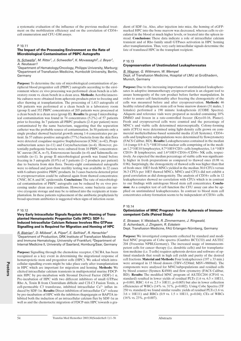
a systematic evaluation of the influence of the previous medical treat-ment on the mobilization efficiency and on the correlation of CD34+cell enumeration and CFU-GM assays.
P 10.11The Impact of the Processing Environment on the Rate ofMicrobiological Contamination of PBPC Autografts
N. Schwella1, M. Ritter1, J. Schwedler2, K. Movassaghi2, J. Beyer1, A. Neubauer11Department of Hematology/Oncology, Philipps University, Marburg;2Department of Transfusion Medicine, Humboldt University, Berlin,Germany
Purpose: To determine the rate of microbiological contamination of pe-ripheral blood progenitor cell (PBPC) autografts according to the envi-ronment where ex vivo processing was performed: clean bench in a lab-oratory room vs. clean bench in a clean area. Methods: Aerobic/anaero-bic cultures were obtained from apheresis products prior to freezing andafter thawing at transplantation. The processing of 1,413 autografts of626 patients was performed at a clean bench in a laboratory room(group I) and 352 PBPC concentrates of 205 patients were processed ata clean bench in a clean area (group II). Results: In group I microbiolog-ical contamination was found in 74 concentrates (5.2%) of 57 patientsprior to freezing. In 7 patients all PBPC products (2–6 per patient) werecontaminated by the same bacterium, suggesting that the separationcatheter was the probable source of contamination. In 50 patients only asingle product showed bacterial growth among 1-6 concentrates per pa-tient. In 57 culture-positive autografts (77%) bacteria from the skin florawere detected: coagulase-negative Staphylococcus (CNSC; n=42), Propi-onibacterium acnes (n=11) and Corynebacterium (n=4). However, po-tentially pathogenic bacteria were cultured from 10 PBPC concentrates:SC aureus (SCA, n=5), Enterococcus faecalis (n=4) and Salmonella en-teritidis (n=1). In group II microbiological growth was found beforefreezing in 3 autografts (0.9%) of 3 patients (1–2 products per patient)due to bacteria from skin flora. Twenty-eight contaminated autograftswere re-infused and post-thaw sterility testing was available in 22 caseswith 6 culture-positive PBPC products. In 3 cases bacteria detected priorto cryopreservation could be cultured again from thawed concentrates:CNSC, SCA and SC epidermidis. Conclusions: The rate of microbiologi-cal contamination of PBPC autografts can be reduced by ex vivo pro-cessing under clean area conditions. However, some bacteria can sur-vive cryogenic storage and may be re-infused into the recipients at trans-plantation. In these patients replacement of the antibiotic prophylaxis bybroad-spectrum antibiotics is suggested when signs of infection occur.
P 10.12Very Early Intracellular Signals Regulate the Homing of Trans-planted Hematopoietic Progenitor Cells (HPC): SDF-1αInduced Intracellular Calcium Release Involves Rho GTPaseSignalling and is Required for Migration and Homing of HPC
R. Bistrian1, D. Möbest1, A. Piiper2, E. Seifried1, R. Henschler1
1Department of Production, DRK Institute of Transfusion Medicineand Immune Hematology, University of Frankfurt; 2Department ofInternal Medicine II, University of Saarland, Homburg/Saar, Germany
Purpose: Signalling through the chemokine receptor, CXCR4, has beenrecognized as a key event in determining the migrational response ofhematopoietic stem and progenitor cells (HPC). We asked which intra-cellular signalling events might be take place early after transplantationin HPC which are important for migration and homing. Methods: Weelicited intracellular calcium transients in multipotential murine FDCP-mix HPC by pre-incubation with Stromal Derived Factor (SDF)-1 α.Pre-incubation of HPC with two different inhibitors of small GTPaseRho A, Toxin B from Clostridium difficile and C2I-C3 Fusion Toxin, acell-permeable C3 transferase, inhibited intracellular Ca2+ influx in-duced by SDF-1α. Results: Direct inhibition of intracellular Ca2+ releaseby pre-incubation of HPC with the inhibitors thapsigargin or BAPTA in-hibited both the induction of an intracellular calcium flux by SDF-1α aswell as and the chemotactic migration of FDCP-mix HPC towards a gra-
dient of SDF-1α. Also, after injection into mice, the homing of eGFP-marked HPC into the bone marrow was decreased, whereas cells re-cir-culated in the blood at much higher levels, or located into the spleen in-stead. Conclusions: These data indicate a role of intracellular calciumtransient and of Rho family small GTPases activation in HPC homingafter transplantation. Thus, very early intracellular signals determine thefate of transfused HPC in the transplant recipient.
P 10.13Cryopreservation of Unstimulated Leukapheresates
B. Wagner, G. Wittmann, W. MempelDept. of Transfusion Medicine, Hospital of LMU at Großhadern,Munich, Germany
Purpose: Due to the increasing importance of unstimulated leukaphere-sates in adoptive immunotherapy cryopreservation is an elegant tool toassure homogenity of the raw product before further manipulation. Inorder to assure cell functionality after freezing the clonogenicity of thecells was measured before and after cryopreservation. Methods: 46healthy related allogeneic stem cell or bone marrow donors (31 males, 1females) performed a 190 minute leukapheresis (COBE Spectra).Aliquots and reference vials were prepared as needed containing 10%DMSO and frozen in a rate-controlled freezer (Kryo10-16, Planer).Fresh and cryopreserved cells were counted and the percentage ofMNC´s and viable cells determined microscopically. Colony-formingunits (CFUs) were determined using light-density cells grown on com-mercial methylcellulose-based semisolid media (Cell Systems). CD34+cells and lymphocyte subpopulations were determined by flowcytometry(FACS Calibur, BD). Results: Leukapheresates contained in the median1.6 (range 0.9–4.7) *10E10 total nuclear cells comprising of in the medi-an 1.2*10E10 lymphocytes, 8.7*10E9 CD3+ cells lymphocytes, 1.6 *10E9CD19+ B lymphocytes, and 1.6*10E9 CD16+/CD56+ NK cells, respec-tively. As expected the median percentage of viable cells was significant-ly higher in fresh preparations as compared to thawed ones (0.98 vs.0.96). Surprisingly, the clonogenicity of thawed cells was higher than thatof fresh ones: 10E5 fresh MNCs yielded in the median 30.0 CFUs versus36.3 CFUs per 10E5 thawed MNCs. MNCs and CFUs did not exhibit agood correlation as did clonogenicity. The analysis of CD34+ cells in 12leukapheresates showed no correlation with CFUs which is in contrastto our findings with autologous blood stem cell concentrates. Conclu-sion: As a complex test of cell function the CFU assay can also be ap-plied on unstimulated leukapheresates. In contrast to blood stem cellconcentrates colony-formation seems to be independent of CD34+ cells.
P 10.14Optimiziation of MNC Programs for the Apheresis of Immuno-competent Cells (Paired Study)
E. Strasser, V. Weisbach, R. Zimmermann, J. Ringwald, S. Achenbach, J. Zingsem, R. Eckstein Dept. Transfusion Medicine, FAU Erlangen-Nürnberg, Germany
Purpose: We investigated components collected by standard and modi-fied MNC programs of Cobe spectra (Gambro BCT,CO) and AS.TEC204 (Fresenius NPBI,Germany). The increased usage of immunocom-petent cells for cancer therapy (i.e. dendritic cells) and for transplanta-tion medicine (i.e. T-cells) requires apheresis devices and software of op-timal standards that result in high cell yields and purity of the desiredcell fractions. Material and Methods: Four leukaphereses (197 ± 33 min.)were arranged in 15 blood donors (TBV=5230ml; MSV=9800ml). Thecomponents were analysed for MNC/subpopulations and residual cellsby blood counter (Sysmex K4500) and flow cytometry (FACS Calibur,BD). Results: The modified MNC program of AS.TEC204 (CP141 vs.standard) resulted in lower yields of residual PLTs (1.6 vs. 6.5 × 10E11,p=0.001; RBC: 4.4 vs. 2.5 × 10E11, p=0.005) but also in lower collectionefficiencies of WBCs (14% vs. 31%, p=0.002). Using Cobe Spectra (TF250 vs. standard) we found similar results: yields of residual PLTs (1.6 vs.3.6 × 10E11) and RBCs (0.9 vs. 1.5 × 10E11, p=0.04); CEs of WBCs(16% vs. 23%, p=0.007).
54 Transfus Med Hemother 2003;30(Sonderheft 1):1–56 Abstracts
Po
ste
rs

Transfus Med Hemother 2003;30(Sonderheft 1):1–56Abstracts 55
Po
ste
rs
Conclusion: The modifications of the MNC programs in either apheresisdevice result in lower residual PLTs while the collection efficiency ofMNCs and nearly all subpopulations were significantly higher using thestandard program. However, it should be considered that higher PLT-yields of the primary product require more purification steps that couldlower the cell yield prior to cell culture.
P 10.15Construction and Expression of FVIII/EGFP Fusion ProteinsSuitable for Analysis of FVIII Trafficking in Living Cells
S. Heinz, T. Tonn, C. Herder, S. Becker, E. Seifried Institute for Transfusion Medicine and Immunohematology, RedCross Blood Donor Service Baden-Württemberg-Hessen, Frankfurt am Main, Germany
Purpose: Compared to other proteins the recombinant expression ofFVIII is limited, which is – at least in part – attributed to its impaired se-cretion. To be able to further analyse the intracellular trafficking ofFVIII protein in living cells enhanced green fluorescent protein (EGFP),chimeras would be highly desirable. Here we have constructed FVIII, B-domain deleted FVIII (FVIIIdelB) and FVIII exhibiting a mutationin the C2-region (FVIIImut2315) as fusion proteins along with EGFP.The level of expression of these FVIII/EGFP fusion proteins was com-pared to the respective FVIII proteins. Methods: Fusion proteins ofFVIII, B-domain deleted factor VIII (FVIIIdelB) and a FVIII mutation(Ser2315Cys) along with EGFP have been generated using the pEGFP-N2 vector (Clonetech, Heidelberg). The resulting cDNA’s were furthercloned into the pcDNA3 expression plasmid under the control of theCMV-promotor. FVIII expression and activity was assessed in transfect-ed 293T and COS cells respectively, using a chromogenic assay (Baxter,Vienna). Expression levels were compared to levels, obtained by cellstransfected with pcDNA3 vector encoding for the respective FVIIIcDNA without EGFP. Results: Despite the fusion of EGFP to FVIIIprotein, comparable levels of FVIII coagulation activity are expressed.FVIII/EGFP fusion proteins showed 8.9% activity (FVIII/EGFP),13.6% (FVIIIdelB/EGFP) and 8.5% (FVIIImut2315/EGFP) comparedto 7.7% (FVIII), 17.7% (FVIIIdelB) and 8.5% (FVIIImut2315), respec-tively. Conclusion: Expression of FVIII fusion proteins along with EGFPseems not to alter the expression level and procoagulant activity. Hence,FVIII/EGFP chimeras appear to be suitable to further analyse the se-cretion pathway of FVIII in living cells. This work should ultimatelyallow the identification of factors that currently limit the effective secre-tion of FVIII in gene therapy and manufacture.
P 11: Transfusion Management in Desasters
P 11.01Effect of Donor Mini-Pool Size on Closure of the Hepatitis BVirus (Hbv) Detection Window: A Comparison of the ProcleixUltrio™ Assay to Surface Antigen Detection
J.M. Linnen1, A. Umali1, A. Broulik1, D. Kolk1, J. Dockter1, S. McDonough1, V. Shyamala2, P. Arcangel2, J. Cottrell2, L. Mimms1,C. Giachetti11Gen- Probe Incorporated, San Diego, CA; 2Chiron Corporation,Emeryville, CA, USA
Objective: To assess the impact of donor pool size on the sensitivity ofthe Procleix Ultrio Assay* (a Transcription-Mediated Amplification[TMA] triplex assay) for detection of HBV DNA during the HBV de-tection window, we tested 11 commercially available seroconversionpanels with panel members undiluted and at 1:8 and 1:16 dilutions. De-sign: We are developing the Procleix Ultrio Assay for simultaneouslyscreening blood donations for HIV-1, HCV, and HBV. The assay has95% detection of HBV DNA at or below about 7 IU/mL (about 20 to 30copies/mL) and uses the same semi-automated instrumentation andassay procedures as the Procleix HIV-1/ HCV Assay, which recentlygained US FDA approval for testing donor pools of 16 or less. Materialsand Methods: Seroconversion panels were purchased from Impath/Bio-Clinical Partners, Inc. Human serum, shown to be negative for HIV-1,HCV, and HBV nucleic acids, was used to make the 1:8 and 1:16 dilu-tions. The assay was carried out in single tubes using a 0.5 mL plasmaspecimen. Ultrio results were compared to those obtained with the Ab-bott PRISM HBsAg Assay. To avoid detection of possible co-infectionwith HIV-1 or HCV in the seroconversion samples, we used an HBVspecific probe for detection. Results: In neat samples, HBV was detectedan average of 20 days (range: 10–43 days) earlier than the HBsAg assay.DNA in samples diluted 1:8 was detected an average of 13 days (range:4–29 days) earlier and DNA in samples diluted 1:16 was detected 11.5days (range: 0–29 days) earlier than HBsAg detection. Conclusion: Al-though the neat panel members were detected earlier than the 1:8 and1:16 dilutions, we showed significantly earlier detection of HBV DNAby the Procleix Ultrio Assay, regardless of the dilution tested, comparedto detection of HBsAg by the PRISM assay. (Partially funded by NHLBI grant HB- 07148). *currently under devel-opment
Measure AS.TEC204 AS.TEC204 P value Cobe Spectra Cobe Spectra P value modif. stand. (SF 250) (SF 500) stand.
CD14+ Yield (10E9) 1.3±0.8 3.1±1.1 <0.001 † 1.4±0.6 2.2±0.9 0.005 †CD14+ CE (%) 37±25 90±37 0.002 † 37±14 61±13 0.001 †CD3+ Yield (10E9) 4.6±2.6 7.4±1.8 0.009 † 5.0±1.1 5.9±2.0 0.06CD3+ CE (%) 29±20 61±25 0.002 † 32±7 43±8 <0.001†CD4+ Yield (10E9) 3.0±1.7 5.0±1.5 0.006 † 3.3±1.0 3.9±1.5 0.09CD4+ CE (%) 45±26 93±51 0.008 † 47±13 66±15 0.002 †CD8+ Yield (10E9) 1.5±1.1 2.4±0.7 0.04 * 1.6±0.5 1.8±0.7 0.09CD8+ CE (%) 45±26 93±49 0.05 47±13 65±15 0.002 †CD16/56+ Yield (10E9) 11.9±11.2 14.3±7.4 0.08 11.4±5.6 14.1±8.2 0.09CD16/56+ CE (%) 4.8±3.1 9.5±4.6 0.01 * 4.4±1.5 6.9±2.2 0.001 †
p < 0.05 *; p < 0.01 †; SF: separation factor
Table: CD14+ and MNC subpopulations (components)

P 11.02Transfusion Medicine and Flooding – Demands and Conse-quences for the Collection, Processing and Distribution ofBlood Products
R. Knels1, K. Hölig2, A. Löffler3, S. Vogel4, D. Klemm5, M. Sieber6
U.-M. Liebscher1
1DRK-Blutspendedienst Sachsen gGmbH, 2Universitätsklinikum derTU Dresden 3Städtisches Klinikum Dresden-Friedrichstadt,4Städtisches Krankenhaus Dresden-Neustadt, 5KrankenhausSt.Josephstift Dresden, 6Diakonissenkrankenhaus Dresden, Germany
As an initial result of the extremely heavy downpours in the course ofAugust 12th in 2002, the flood waters from the little river Weißeritz burstinto the central Altstadt area and surrounding areas of the city of Dres-den. A primary casualty was a comprehensive-care hospital around 900beds which had to be completely evacuated. Thanks to the efforts ofstaff and helpers no patients came to serious harm, but little time couldbe spent in securing valuable equipment. It was only through personalintervention that the pharmacy and blood depot could be saved fromdestruction. The valuable experiences thereby won will be presented inthe first part of the lecture.Following on heavy rainfall further upstream, the level of the Elbe rosecontinuously to reach a new record in Dresden of 9.36 m by Saturdaymorning. Large parts of the Dresden Institute of the German Red CrossBlood Donation Service as well as an additional 4 blood depots (includ-ing in the University Hospital) had to be evacuated. This placed great lo-gistical demands on the correct production, storage and transport ofblood products. The previous close collaboration between individual in-stitutes over many years proved to be of advantage in such an extremesituation. A further important aspect in this kind of situation is the co-operation between the regional media and the catastrophe committee inorder to avoid unnecessary waves of donors and panic reactions in thepublic.The second part of the presentation will deal mainly with experiencesgained while tackling the effects of a large-scale natural catastrophe inrespect to blood donation, the production, storage and transport ofblood products as well as the question of maintaining clinic supplies. Aseries of more minor problems will also be outlined which arose, in part,only after the events. Also to be treated are particular features of large-scale natural catastrophes in contrast to the large numbers of injuredfollowing accidents or terrorist attacks of a localized area.
P 11.03Age Structure of the Blood Donors at the University Clinic ofMagdeburg and the Residential District of the Local Instituteof Transfusion Medicine
S. Schulze, S. Ludwig*, M.U. HeimInstitute of Transfusion Medicine and *Clinic of Anesthesiologie,University of Magdeburg, Germany
Introduction: The numerical development of the inhabitants of Saxonia-Anhalt, has taken a negative tendency for years, especially in its capitalMagdeburg. There has been a decrease in population in the course of thelast ten years (about 4500 persons per year in average). The mean age inMagdeburg has increased to 44 years and the part of persons youngerthan 18 has reduced to 14% of the total number of inhabitants. On theother hand, there has been an increase of population in the adjoiningrural districts. The present paper describes the potential of blood donorsat our institute under various statistical criterions. Methods: The follow-ing data referring the years 1998 to 2002 were registered and compared:– Age structure of the blood donors– How far each of the several age groups is concerned in the blood do-
nationsFurthermore in 2002 the addresses of the blood donors of the Instituteof Transfusion Medicine were adjoined and evaluated with their postcodes. Results: The average age of the blood donors at our institute hasrisen continuously since 1998. The age structure of the donors (Median)has changed from 1998 to 2002, too. It was pointed out that the rate ofcomparison of the age of the blood donors was 3 years, that of thedonors of thrombocyte even 5 years. The part of the blood donating age
56 Transfus Med Hemother 2003;30(Sonderheft 1):1–56 Abstracts
Po
ste
rs
group <25 years decreased from 30.2% to 24.6%. It was similar to fromthe age group <35 years as well as to those <45 years. In contrast, thepart of the blood donors >55 years increased by 6%. Only about 60% ofour donors live in Magdeburg city. 36% come from distances up to 100km maximum. 4% come from farther regions (may be students). Con-clusions: At present the number of inhabitants of Magdeburg is about230,000. About 165,000 (71.7%) between 18 an 68 years could donateblood. Out of this group 3.3% who are living in the town belong to theblood donors. Since the number of births has decreased in the past of1991 in the new federal territories the part of people being able to do-nate blood will decrease dramatically, too. Ten years later only 65.2% ofthe inhabitants of Magdeburg will belong to that group of people beingable to donate blood. To keep the numbers of donors constant in the fu-ture, it is necessary to increase the readiness to blood donation amongthe people. To reach that aim it is absolutely essential to activate thepublicity campaign also in the rural districts around Magdeburg.
P 11.04A Concept for Blood Supply in Patients with Highly ContagiousDiseases
E. Strobel, H.-U. SchmidtInstitut für Medizinische Mikrobiologie, Immunologie und Kranken-haushygiene, Städtisches Krankenhaus München-Schwabing,Munich, Germany
Background: Highly contagious, life-threatening diseases like viralhaemorrhagic fever (caused by Ebola, Marburg, Lassa or Krim-Kongo-virus), orthopoxvirus infection and pneumonic plaque require specialisolation of the patient for protection of the medical staff and of the pub-lic. As blood samples and other body fluids from these patients may bedangerous for the laboratory personal, also laboratory diagnostic proce-dures must be done under special safety conditions. Situation: In Ger-many clinical centers for intensive care of patients with highly conta-gious diseases have been established in Berlin, Frankfurt, Hamburg,Leipzig and Munich (Städtisches Krankenhaus München-Schwabing).Microbiological diagnosis of the above mentioned viral infections canbe done in two biosafety level 4 laboratories (and for plague in anothernational reference laboratory of BSL 3). For practical reasons routinelaboratory examinations (clinical chemistry, haematology and bloodgroup serology) can not be sent over long distances to these highly spe-cialized microbiological laboratories, but have to be done at the clinicalunit obeying the biosafety regulations. Conception: As it is not allowedto examine blood samples from patients with these highly infectious dis-eases in our routine laboratory, we plan to perform blood group serolog-ical tests (for ABO, Rh and K antigens, antibody screen and identifica-tion, direct antiglobulin test and cross-matches) within the isolation unitin a separate room near the patient’s room. An experienced member ofthe laboratory staff wearing the special protection clothing will do thetests using routine laboratory methods only with minimal modifications.To avoid production of aerosols no washing procedures with fluids willbe done. To minimize the risk of injuries only plastic materials will beused. Therefore we plan to perform all the tests in the gel-centrifugationmethod. All waste will be separately collected and disinfected beforeleaving the unit. Reagents and hardware will stay in the unit. The ABObedside test of the patient will be omitted because we will use onlygroup O red blood cell concentrates in such a case. Limitations: Ourconcept is thought for the blood supply of 1 or 2 patients with highlycontagious life-threatening diseases hospitalized in the unit at the sametime. If a greater number of such patients would have to be suppliedwith blood products the routine standards probably can no longer bemaintained.