SGP Congress 2018 - Karger Publishers
39
Basel • Freiburg • Paris • London • New York • Chennai • New Delhi • Bangkok • Beijing • Shanghai • Tokyo • Kuala Lumpur • Singapore • Sydney Abstracts are available online only, free of charge, at www.karger.com/doi/10.1159/000488898 SGP Congress 2018 St. Gallen, Switzerland, 24–25 May, 2018 Respiration 2018;95:478–516 DOI: 10.1159/000488898 Published online: May 9, 2018 Abstracts
-
Upload
khangminh22 -
Category
Documents
-
view
0 -
download
0
Transcript of SGP Congress 2018 - Karger Publishers

Basel • Freiburg • Paris • London • New York • Chennai • New Delhi •
Bangkok • Beijing • Shanghai • Tokyo • Kuala Lumpur • Singapore • Sydney
Abstracts are available online only, free of charge,at www.karger.com/doi/10.1159/000488898
SGP Congress 2018
St. Gallen, Switzerland, 24–25 May, 2018
Respiration 2018;95:478–516DOI: 10.1159/000488898
Published online: May 9, 2018
Abstracts

Poster Walk 1: Epidemiology / Health Policy
P01
Variability of Tidal Breathing Parameters in Preterm
Infants and Associations with Respiratory Morbidity
during Infancy: A Cohort-Study
J. Usemann1,2, A. Suter1, E. Zannin1,3, E. Proietti2,4, S. Fouzas5, S. Schulzke1, P. Latzin2, U. Frey1, BILD Study Group1Pediatric Respiratory Medicine, University of Basel, Basel, 2Pediatric Respiratory Medicine, Bern University Hospital, Bern, Switzerland, 3Dipartimento di Elettronica, Milano University, Milan, Italy, 4Pediatric Respiratory Medicine, University Hospital Zurich, Zurich, Switzerland, 5University Hospital Patras, Patras, Greece
Objectives: As previously shown, low variability of tidal breathing and capnography parameters in preterm infants are an expression of impaired lung function and gas exchange. We tested, whether these lung parameters assessed near term are associated with respiratory morbidity during the first year of life.
Methods: Using logistic regression analysis, we investigated in a birth-cohort of 133 preterm infants, whether unsedated lung function measurements at 44 weeks post-menstrual age (variabil-ity of tidal breathing and capnographic parameters) are associated with subsequent rehospitalization, wheeze, inhalation, and home oxygen therapy in infancy. Using receiver operating characteristic (ROC) analysis, we determined whether the predictive power of BPD classification is enhanced by adding these lung function or capnographic parameters.
Results: The coefficient of variation (CV) of VT (CVVT) (range 4%–35%) and the CV of expired CO2 volume (CVVE,CO2) (range 5%–40%) were significantly lower in preterm infants with rehos-pitalization during infancy, compared to those without. Per inter-quartile range decrease in CVVT and CVVE,CO2, the adjusted odds ratio (95% CI) for rehospitalization increased by 2.25 (1.21–4.20) and 2.31 (1.20–4.45), respectively. Low variability of both param-eters predicted rehospitalization significantly better than BPD classification alone. The area under the ROC curve increased from 0.56 to 0.66 for both, CVVT and CVVE,CO2. There was no associa-tion with the other outcomes.
Conclusions: In comparison to BPD classification alone, in-cluding variability of postnatal tidal breathing parameters increas-es the prediction for rehospitalization in infancy. These findings may have a potential impact on discharge procedures and moni-toring strategies in preterm infants.
Abstracts
Respiration 2018;95:478–516 DOI: 10.1159/000488898
Published online: May 9, 2018
SGP Congress 2018St. Gallen, Switzerland, 24–25 May 2018
E-Mail [email protected]/res
© 2018 Swiss Respiratory Society SGP. Published by S. Karger AG, Basel. All rights reserved.
Abstracts are available online only, free of charge, at www.karger.com/doi/10.1159/000488898

AbstractsRespiration 2018;95:478–516 DOI: 10.1159/000488898
480
P02
Evolution of Primary Ciliary Dyskinesia (PCD)
Diagnostic Testing in Europe
F. Halbeisen1, M. Goutaki1, E. Maurer1, M. Boon2, C. Casaulta3, S. Crowley4, C. Hogg5, B. Karadag6, C. Koerner-Rettberg7, H. Mazurek8, U. Özçelik9, F. Santamaria10, N. Schwerk11, P. Yiallouros12, J. Lucas13, C. Kuehni1
1Institute of Social and Preventive Medicine, University of Bern, Bern, Switzerland, 2Department of Paediatrics, University Hospital Gasthuisberg, Leuven, Belgium, 3Paediatric Respiratory Medicine, Children’s University Hospital of Bern, University of Bern, Bern, Switzerland, 4Unit for Paediatric Heart, Lung, Allergic Diseases, Rikshospitalet, Oslo, Norway, 5Department of Paediatrics, Primary Ciliary Dyskinesia Centre, Royal Brompton and Harefield Foundation Trust, London, United Kingdom, 6Department of Pediatric Pulmonology, Marmara University, School of Medicine, Istanbul, Turkey, 7Department of Paediatric Pneumology, University Children’s Hospital of Ruhr University Bochum, Bochum, Germany, 8Department of Pneumonology and Cystic Fibrosis, Institute of Tuberculosis and Lung Disorders, Rabka, Poland, 9Department of Pediatric Pulmonology, Hacettepe University Faculty of Medicine, Ankara, Turkey, 10Department of Translational Medical Sciences, Federico II University, Naploi, Italy, 11Clinic for Paediatric Pulmonology, Allergology, Lung Transplantation, Hannover Medical School, Hannover, Germany, 12Medical School, University of Cyprus, Nicosia, Cyprus, 13Primary Ciliary Dyskinesia Centre, NIHR Biomedical Research Centre, University of Southampton, Southampton, United Kingdom
Background: Diagnosis of PCD remains complex and varies across Europe. In 2009 a consensus statement from the European Respiratory Society Taskforce on PCD recommended the use of nasal nitric oxide (nNO), electron microscopy (EM) and video mi-croscopy (VM) as cornerstones of PCD diagnosis. This study uses individual-patient-data to describe PCD diagnostics in Europe be-fore and after the publication of the consensus statement.
Methods: We included 12 European datasets from the inter-national PCD (iPCD) cohort, with almost complete diagnostic data. We assessed the proportion of patients, who had been tested with nNO, EM and VM, comparing the period before and after 2009.
Results: From the 835 included patients, 565 (67%, 95% CI: 60–70%) had nNO, 616 (73%, 71–77%) had EM and 582 (69%, 66–73%) had VM testing; 209 (25%, 22–28%) had genetic analysis. Combination of nNO, EM and VM was used in 323 patients (39%, 35–42%). The use of EM decreased (from 88% to 64%; p < 0.001) after the 2009 consensus, while use of VM increased (from 66% to 74%; p < 0.001). We found no time trend in nNO testing (p = 0.2). The use of the test combination nNO, EM and VM decreased (from 44% to 35%; p = 0.002). There was significant heterogeneity between countries.
Conclusions: Overall, we found poor adherence to the 2009 consensus recommendations, due to the decrease in use of EM and the low use of test combinations. This may result in both under- and over-diagnosis of PCD. To improve PCD diagnosis, we must
implement the evidence-based guidelines published by the 2016 ERS PCD Taskforce.
Funding: FP7 grant 305404, SNF 32003B-162820, Milena Car-vajal -Pro Kartagener, Lung Leagues Bern, Ticino and Vaud, COST BM1407.
P03
Cost-Effectiveness of School-Based Tuberculosis
Screening for Migrant Children Arriving in Low-
Incidence Countries – Evidence from Switzerland
J. Usemann1, M. Ledergerber2, G. Fink3, N. Ritz4,5
1Pediatric Respiratory Medicine, University of Basel, 2Medizinische Dienste, Kinder- und Jugendgesundheitsdienst, 3Swiss Tropical and Public Health Institute, 4Pediatric Infectious Disease and Vaccinology, University of Basel, Basel, Switzerland, 5Department of Pediatrics, Royal Children’s Hospital Melbourne, Melbourne, VIC, Australia
Background: Tuberculosis (TB) screening programs in mi-grant children have become important, as the WHO recommends diagnosis and treatment of latent tuberculosis infection (LTBI), particularly in young children. This study evaluated the cost-effec-tiveness of TB screening for migrant children arriving in a low-incidence country.
Methods: Retrospective analysis of a school-based TB screen-ing program in Switzerland. Migrant children aged 5–18 years were screened using a tuberculin skin test (TST). Cost for TB screening and treatment were calculated from records of the Uni-versity Children’s Hospital Basel. Cost impact was analyzed as the difference between cost of treatment for active TB and screening plus treatment of LTBI. Cost per disability-adjusted life years (DALY) was assessed based on 2013 Global Burden of Disease es-timates.
Results: 1120 children were screened with a mean age of 10.9 years; 46% were female. A TST induration ≥10 mm was document-ed in 78 (6.9%) cases, of which 21 (1.9%) were further evaluated. Of those, 17 children were diagnosed as LTBI and none had active TB. The highest rates of positive TST results were found in migrant children from Africa (16.6%) and Turkey (15.4%). Screening for LTBI was cost-saving for prevalence rates of 11% and higher; screening was cost-effective (cost per DALY < gross domestic product (GDP) per capita) for any prevalence of 2% or higher.
Conclusion: School-based TB screening programs targeting migrant children from high-risk regions not only have the poten-tial to prevent progression to active TB but can also reduce health system cost if LTBI prevalence is sufficiently high.

Abstracts Respiration 2018;95:478–516 DOI: 10.1159/000488898
481
P04
Identification of Non-Tuberculosis Mycobacteria in
COPD Patients Undergoing Lung Volume Reduction:
More Frequent Than Expected?
G. Berra1, J. Plojoux1, P.M. Soccal1,2, J.-P. Janssens1,2
1Geneva University Hospitals, 2Université de Genève, Geneva, Switzerland
Introduction: Non-tuberculosis mycobacteria (NTM) are more and more frequently isolated in subjects with chronic respi-ratory diseases. Although this association is epidemiologically well known, specific data on NTM in COPD patients and relationship with prognosis in this specific group are lacking.
Methods: We retrospectively collected bacteriological findings from all bronchial aspirates obtained during bronchoscopy per-formed for endoscopic lung volume reduction (ELVR) in severe COPD patients. All patients included met eligibility criteria for ELVR, namely: age <75 years, ex smoker (>6 months), emphysema, persisting dyspnea despite maximal medical therapy including a re-cent rehabilitation program, FEV1 < 45% of predicted value, marked hyperinflation, a 6-minute walk distance >140 meters and no or few acute exacerbations during the year prior to the procedure.
Results: NTMs were isolated in 8/44 patients (18.2% of cases). Mycobacterium avium complex (MAC) was most the most preva-lent NTM identified (5/8, all M. avium subtype). In addition, one Mycobacterium xenopi, one Mycobacterium gordonae and one Runyon Group IV NTM (rapidly growing, non-pigmented) were isolated.
All specimens were isolated from culture of bronchial aspirates. No sample was microscopy positive. NTM were isolated once in most patients (6/8), twice in one patient and three times in anoth-er. With a mean follow up of 993 days, none of the patients re-quired specific antimycobacterial treatment for NTM-associated lung disease, defined as a progression of symptoms or radiological lung lesions or an accelerated lung function decline attributable to NTM infection.
Other pathogens frequently isolated from bronchial aspirates were Haemophilius influenzae (4/44 patients), various Gram neg-ative bacilli (11/44 patients) and Aspergillus species (6/44 pa-tients).
Conclusion: In a population of 44 patients with severe COPD undergoing endoscopic lung volume reduction, we identified a significantly higher rate of NTMs than previously reported. The clinical impact of this finding has to been further explored in a prospective study.
P05
A Prediction Model for COPD Exacerbation –
Data from the Swiss COPD Cohort Study
E. Thesenvitz, A. Naduvilekoot, S. Züsli, S. Maier, T. Jendricke, J. Leuppi, T. Dieterle
Clinical Research, University Clinic of Medicine, Cantonal Hospital Baselland, Liestal, Switzerland
Introduction: It is estimated that about 400,000 patients are af-fected by COPD in Switzerland. Despite the large prevalence long-term data on symptoms and lung function in this population are lacking. Therefore the goal of the Swiss COPD Cohor study (SCCS) was to assess the situation of COPD care in Switzerland. A further goal was to develop a prediction model for the risk of exacerbation.
Method: Patients with a verified COPD diagnosis (FEV1/FVC <70% without reversibility with bronchodilator [improvement of FEV1 < 200 ml or <12%]) were included into the study by general practitioners and pneumologists in Switzerland. Clinical and spi-rometry exams were performed at baseline and every six months thereafter including information about medical and non-medical therapy, COPD Assessment Test (CAT-Score), co-morbidities and exacerbation. Univariate analysis was performed to identify fac-tors statistically associated with the risk of exacerbation. Factors significant in univariate analysis were included into a binary logis-tic regression model to determine statistically independent predic-tors and a risk score.
Results: 166 Patients from SCCS recruited between May 2011 and June 2016 were included into this interim analysis. The follow-ing Table 1 summarizes statistically independent predictors for ex-acerbation. A score of ≥2 points was associated with an increased risk (HR = 2.321, 95% confidence interval (1.186–4.541)).
Conclusion: A score consisting from 5 variables is able to reli-ably predict the risk of COPD exacerbation within one year.
Table 1. (for Abstract P04)
Variables Patients colonized with NTMs (N = 8)
Patients withoutNTMs (N = 36)
Patients on OCS, n/N, (%) 1/8 (12.5%) 2/36 (5.5%)Patients on ICS, n/N, (%) 7/8 (87.5%) 31/36 (86%)FEV1, mean (SD) 31.5 (39.3–23.8) 31.3 (41.7–20–9)DLCO, mean (SD) 47.7 (34.8–60.6) 46.2 (30.7–61.7)6MWT, mean (SD), m 463 (388–537) 348 (236–459)Diabetes, n/N, (%) 1/8 (12.5%) 1/36 (2.8%)Immunosuppression, n/N, (%) 1/8 (12.5%) 0/36 (0%)Chronic renal disease, n/N, (%) 0/8 (0%) 1/36 (2.8%)
Table 1. Predictor for a COPD exacerbation within one year (for Abstract P05)
Predictor no yes
On-going cancer 0 1Previous exacerbation 0 1CAT-Score >12 0 1mMRC >0 0 1BMI <21 0 1

AbstractsRespiration 2018;95:478–516 DOI: 10.1159/000488898
482
P06
The Use of Combination Therapy in the Swiss COPD
Cohort Study – A Cross-Sectional Analysis
E. Thesenvitz, S. Züsli, A. Naduvilekoot, S. Maier, T. Jendricke, J. Leuppi, T. Dieterle
Clinical Research, University Clinic of Medicine, Cantonal Hospital Baselland, Liestal, Switzerland
Introduction: It is estimated that about 400,000 patients are affected by COPD in Switzerland. Despite the large prevalence long-term data on symptoms and lung function in this population are lacking. Therefore the goal of the Swiss COPD Cohort Study SCCS was to assess the situation of COPD care in Switzerland with a particular focus on medical therapy related to GOLD classifica-tion.
Method: Patients with a verified COPD diagnosis (FEV1/FVC <70% without reversibility with bronchodilator [improvement of FEV1 < 200 ml or <12%]) were included into the study by gen-eral practitioners and pneumologists in Switzerland. Clinical and spirometry exams were performed at baseline and every six months thereafter including information about medical and non-medical therapy, COPD Assessment Test (CAT-Score) and co-morbidities.
Results: 164 Patients from SCCS recruited between May 2011 and June 2016 were included into this interim analysis. Data on current medication is summarized in the following Table 1.
Conclusion: This interim analysis of COPD cohort demon-strates that combination therapy is widely used in patients GOLD class III and IV. However, the use of combination therapy in COPD GOLD IV is less frequent than expected.
P07
Towards Standardized Follow-Up Care for Patients
with Primary Ciliary Dyskinesia (PCD)
M. Goutaki1,2, F. Halbeisen1, M. Martin1, C. Hogg3, C. Casaulta2, I. Amirav4, J. Barben5, E. Moya6, K. Nielsen7, G. Thouvenin8, J. Lucas9, C. Kuehni1
1Institute of Social and Preventive Medicine, 2Paediatric Respiratory Medicine, Children’s University Hospital of Bern, University of Bern, Bern, Switzerland, 3Department of Paediatrics, Primary Ciliary Dyskinesia Centre, Royal Brompton and Harefield Foundation Trust, London, United Kingdom, 4Department of Pediatrics, Faculty of Medicine, Bar IIan University, Safed, Israel, 5Division of Paediatric Pulmonology, Children’s Hospital St. Gallen, St. Gallen, Switzerland, 6Division of Services for Women and Children, Women’s and Newborn Unit Bradford Royal Infirmary, University of Bradford, West Yorkshire, United Kingdom, 7Danish PCD Centre Copenhagen, Paediatric Pulmonary Service, Copenhagen University Hospital, Copenhagen, Denmark, 8Paediatric Pulmonary Department, Trousseau Hospital APHP, Sorbonne Universities and Pierre et Marie Curie University, Paris, France, 9Primary Ciliary Dyskinesia Centre, NIHR Biomedical Research Centre, University of Southampton, Southampton, United Kingdom
Introduction: Reporting of clinical information in medical charts is not standardized; thus data collected retrospectively from records is of little value for research. A recent systematic review (Goutaki et al, ERJ 2016), found that assessment of clinical symp-toms of patients with PCD varied considerably between studies making comparisons impossible. Management and follow-up dif-fer between countries, and is usually extrapolated from other dis-eases. We aimed to develop proformas for longitudinal data col-lection during routine care, allowing standardized reporting of fre-quency and severity of clinical signs and symptoms.
Methods: We collected available proformas for management of PCD and related diseases (e.g. cystic fibrosis) in different coun-tries from the BEAT-PCD network. We compared them with the available and designed a new harmonized proforma. We discussed several versions within a large multidisciplinary group of >30 col-laborators (including paediatric and adult pulmonologists, ENT specialists and diagnostic experts) and patient representatives to improve the content and the structure via an adapted Delphi pro-cess.
Table 1. The use of combination therapy (for Abstract P06)
GOLD Classification I II III IVN = 164 24 73 55 12
Short-acting Bronchodilators 4 (16.7) 24 (32.9) 33 (60.0) 6 (50.0)Long-acting Bronchodilators 17 (70.8) 63 (86.3) 49 (89.1) 10 (83.3)ICS 2 (8.3) 11 (15.1) 22 (40.0) 5 (41.7)Single compounds or combination of LABA and ICS 7 (28.2) 33 (45.2) 44 (80.0) 6 (50.0)LAMA and LABA as single compounds or as combination 3 (12.5) 17 (23.3) 25 (45.5) 5 (41.7)Triple-Therapy 4 (16.7) 23 (31.5) 38 (69.1) 6 (50.0)

Abstracts Respiration 2018;95:478–516 DOI: 10.1159/000488898
483
Results: The new standardized proforma consists of 7 themat-ic modules (Table 1) and includes key questions about signs, symp-toms, clinical tests, treatments and environmental exposures. As a next step, we will pilot the proforma in paediatric and adult PCD centres, before finalizing it. Consenting centres will be able to enter the data collected in a standardised way to an online database to be used for prospective research. Collected data will be linked to ex-isting databases allowing for data exchange and efficient data use.
Conclusion: Disease-specific and standardized follow-up can improve the quality of information collected for clinical care and research by adding standardized prospective longitudinal infor-mation from routine visits. This will be a step towards standard-ized care for PCD and enable using valuable data from routine care for research studies.
Funding: Lung League Bern, SNF 32003B_162820, COST BEAT-PCD BM1407.
P08
Tobacco Taxes in Switzerland: Losses for Public
Health – Gains for the Industry
R.M. Kaelin, P. Diethelm
Oxyromandie/OxySuisse, Geneva, Switzerland
Introduction: Decisions on Tobacco (T) Taxes (TX) are Public Health (PH) measures, if applied correctly: A. TX, following WHO-convention, should amount to 75% of retail prices (RP),resulting (B) in high RP to prevent T-uptake by youth; C. RP- increases should occur by steps of >10%, to curb consumption. We searched, if cigarette (cigs) TX increases 2003–2013 were positive for PH.
Methods: Analysis of RP, TTX,comparison with EU-countries.Results: 1. Different T-products are not taxed coherently: Cigs are taxed
61% of RP(VATincl.), pipe and heat not burn T only 19%. Cheap pipe T is thus used for handmade cigs, e.g.: “Marboro” pipe T, RP 6.50/30 g (1.25.CHF TX) yields 40–60 cigs at < half the RP of in-dustrial “Marlboro” cigs at 8.-CHF (TX 5 CHF) for a pack of 20.
Also, one day pack of “Marlboro”heat not burn T at 8.-CHF(TX 0.95 CHF) leaves 7.-CHF to to industry.
2. Gains of the indusry on a pack of industrial cigs in Switzer-land are 3.14 CHF, about the double that of EU-countries.
3. If the rate of 75% RP had been applied, the TX income in 2012–2016 would have been increased by 352–428 Million CHF per year.
4. The RP of cigs rose from 4.-to 8.- CHF in 2003–2013, but TX increases were so small, that industry could increase RP by steps of <10%, in order not to curb consumption significantly keeping gains high.
Conclusions: Tobacco Taxes in Switzerland serve more the in-dustry than Public Health. They were implemented as to result in too low retail prices compared to other countries, thus preventing to curb consumption. This allowed industry to keep gains high. The huge Tax difference between pipe/heat not burn Tobacco compared to industrial cigarettes fosters youth smoking and pro-vides big gains on the newer products.
P09
Development of Hospitalizations for Pulmonary
Hypertension in Switzerland from 2002 to 2015
F. Aigner, F. Baty, M. Brutsche
Department of Pulmonary and Sleep Medicine, Kantonsspital St. Gallen, St. Gallen, Switzerland
Introduction: The objective of the present study was to analyze the development of hospitalizations for pulmonary hypertension in Switzerland.
Methods: The data set was provided by the Swiss Federal Office for Statistics, which offers a nation-wide coverage of all hospital-ized patients using the ICD-10 coding system. The WHO-Classi-fication of pulmonary hypertension is not used in this coding sys-tem. We analyzed the number of hospitalizations as main diagno-sis for overall pulmonary hypertension (I27), pulmonary arterial hypertension (PAH; I27.0), chronic thromboembolic pulmonary hypertension (CTEPH; I27.20), other specified pulmonary-associ-ated heart diseases and Cor pulmonale (I27.8 and I27.9) and not otherwise specified secondary pulmonary hypertension (I27.28) from 2002–2015. The development of age and comorbidities in patients with PAH was analyzed.
Results: Overall, the incidence of hospitalizations for all causes of pulmonary hypertension did not change (average of 450 cases per year) since 2002. Since 2009, we observed a sudden significant de-crease in the incidence of hospitalizations for PAH, independent of the canton (from 255 per year in 2008 to 82 per year in 2015). At the same time coding for CTEPH and not otherwise specified second-ary pulmonary hypertension was introduced. Since then the inci-dence in these newly introduced categories significantly increased (from 0 cases in 2009 to 256 in 2015). There was no difference in age and cardiovascular comorbidities seen in patient with PAH.
Conclusions: Since the incidence of the overall hospitalizations for pulmonary hypertension remain stable, we assume that the profound decline of hospitalizations for PAH is due to a change in coding practices in 2009 and not due to an increase in availability of vasodilatory therapy PAH.
Table 1. Standardised PCD proforma modules (for Abstract P07)
Module 1 Basic patient information, diagnostic evaluation and baseline medical history
Module 2 Medical history (reported symptoms)
Module 3 Physical examination of lungs and heart
Module 4 Physical examination of Ear Nose Throat (ENT)
Module 5 Growth measurements and clinical tests (lung function, imaging, microbiology)
Module 6 Treatment (hospitalization, surgeries, medication and physiotherapy)
Module 7 Enviromental exposures and lifestyle

AbstractsRespiration 2018;95:478–516 DOI: 10.1159/000488898
484
P10
Echinacea for the Prevention of Respiratory Tract
Infections in Children 4–12 Years: A Randomized,
Blind and Controlled Study
M. Ogal1, P. Klein2, R. Schoop3
1Pediatric Clinic, Brunnen, Switzerland, 2d.s.h. statistical services GmbH, Rohrbach, Germany, 3Medical Department, Bioforce AG, Roggwil, Switzerland
Withdrawn.
Poster Walk 2: Clinical Lung / Patient Management 1
P11
Pyogenic Lung Abscesses: The Place of Conservative
Treatment
M. Udry1, A. Ogna1, M. Gonzalez2, M. Oriol3, L. Nicod1
1Service of Pulmonology, 2Service of Thoracic Surgery, 3Service of Infectious Diseases, Centre Hospitalier Universitaire Vaudois, Lausanne, Switzerland
Introduction: Lung abscesses (LA) have a low incidence but still carry a 5% mortality. As bronchoaspiration is a major cause of LA, they are mostly due to anaerobic bacteria. Medical treatment (MT) by antibiotics successfully cures the LA in 80–90% of cases. More invasive treatments such as surgical resection (SR) may be attempt-ed. There are currently no clear guidelines for surgical indication, but SR is generally recommended if MT fails or for complicated LA.
In this study, we tried to identify objective arguments to choose be-tween MT and SR as the first line treatment in a given patient.
Methods: we did a retrospective analysis of medical records of 68 patients treated for LA at our institution between 2013–2015. The patients were divided in two groups: 42 patients treated by MT and 22 patients treated by SR. We compared baseline characteris-tics and outcomes between the two groups.
Results: The median duration of antibiotic treatment was 52 days [37.25;61] in SR and 50 [36.75;86.75] in MT (p > 0.05). Mean length of hospital stay was equivalent between the two groups: 23.64 ± 16.11 days in SR and 23.85 ± 19.03 in MT (p > 0.05). Cases of complicated LA with empyema were significantly higher in SR (77.3%) than in MT (17.4%, p < 0.0001). There was no difference nei-ther in the LA size (4.49 ± 2.66 cm in SR vs 5.22 ± 2.43 cm in MT, p > 0.05) nor in the microbiologic repartition. The mortality rate was similar between the two groups: 4.5% in SR and 8.7% in MT(p > 0.05).
Conclusion: MT and SR have similar rate of mortality and mor-bidity. MT must always be considered as first line treatment. SR is indicated in MT failure. However, SR indication in complicated LA seems less evident, as some patients heal despite antibiotic treatment alone. Conservative treatment most often leads to good lung tissue healing, even in large cavities.
P12
Patients Rejected for Lung Transplantation:
Who Are They?
A. Lenoir1, C.R. Perez Valdes1,2, J.-D. Aubert1, On behalf of the Lausanne-Geneva lung transplantation programme1Service de Pneumologie, Centre Hospitalier Universitaire Vaudois, Lausanne, 2Hôpital de Rolle, Rolle, Switzerland
Listing a patient for lung transplantation is a critical decision given this treatment’s limited availability. The International Society for Heart and Lung Transplantation published in 2015 a consensus
P P Pb ca
Fig. 1. Right superior lobe lung abscess complicated with empyema (a) and the evolution at 1 month (b) and 4 months (c) with a con-servative treatment (for Abstract P11).

Abstracts Respiration 2018;95:478–516 DOI: 10.1159/000488898
485
document as decision guidance for selecting lung transplant candi-dates. However, a detailed analysis comparing patients placed on the waiting list to those deemed unsuitable has rarely been made. We retrospectively evaluated lung recipient candidates referred to our centre regarding diagnosis, final decision and outcomes.
Demographics, respiratory diagnosis, and the board’s decision were recorded for all patients referred to the Lausanne-Geneva lung transplant board from 04/2004 to 07/2017. If rejected, the primary contraindication or the combination of relevant contra-indications was determined. Furthermore, survival on/off the waiting list was calculated.
Among 363 referred candidates for lung transplant, 97 (26.7%) were rejected. Median age (range) was higher in non-listed patients: 61.5 (12–68) vs 53.2 (14–65) years. Acceptance rate was 90.2% among cystic fibrosis patients, 74.0% among idiopathic pulmonary fibrosis patients, 69.0% among chronic lung allograft dysfunction (CLAD) patients, 67.6% among COPD patients and 74.1% among other diagnostic categories. Main contraindications for listing were cardiovascular (17.5%), insufficient severity of the respiratory con-dition (15.5%), age (13.4%) and multiple organ dysfunction (12.4%). Pooled other causes accounted for 23.8%, including cancer, multiple comorbidities, surgical inoperability, psychosocial difficulties and inappropriate indications. A combination of causes constituted 17.5%. One-year-survival differed significantly between listed and non-listed patients: 88% vs 79% (p = 0.0001). This difference in-creased at three years: 80% vs 57%, with over 80% of listed patients meanwhile transplanted. Mortality on the waiting list was 12%.
Patients rejected for lung transplant tend to be older and rath-er suffer from COPD or CLAD than accepted candidates. Main contraindications leading to rejection in our centre were cardio-vascular, premature referral and age; a combination of various contraindications was common. Listed patients had a clear sur-vival benefit.
P13
Sensitivity and Specificity of EGFR Liquid Biopsy
versus Serum Carcinoembryonic Antigen in Patients
with Adenocarcinoma of the Lung
S. Saenghirunvattana1, P. Saenghirunvattana2, C. Gonzales1, K. Suthisri1, C. Siangproh1
1Pulmonary and Chest Center, Bangkok Hospital Medical Center, Bangkok Dusit Medical Services, 2Chulalongkorn University, Bangkok, Thailand
Introduction: Tissue biopsy is necessary to diagnose lung can-cer but it becomes impractical due to its invasive nature and com-plications. A new test, epidermal growth factor receptor (EGFR) liquid biopsy provides wider range of information obtained from blood samples that may help clinicians into better personalized treatment particularly in cases of adenocarcinoma of the lung.
The aim of this retrospective study is to compare which among EGFR liquid biopsy and carcinoembryonic antigen (CEA) has higher predictive values in patients with adenocarcinoma of the lung.
Methods: Thirty patients with lung nodules on their chest CT scan underwent diagnostic tests: EGFR liquid biopsy, serum CEA, tissue biopsy and/ or surgery between September 2016 and No-vember 2017. Data gathered were patient age, gender, smoking history, serum CEA levels, EGFR liquid biopsy results and final diagnosis. EGFR liquid biopsy result was positive when EGFR mu-tation is detected. The sensitivity, specificity, positive predictive and negative predictive values of CEA and EGFR liquid biopsy were compared based on data collected.
Results: There were thirty patients (27% male and 73% female) with mean age of 60 years old. Smokers were 20%, and non-smok-ers: 80%. Pathological diagnoses were: adenocarcinoma: 37% and others were: tuberculosis: 47%, benign cases: 7%, squamous meta-plasia: 3%, anaplastic giant cell carcinoma: 3% and carcinoid tu-mor at 3%.
EGFR liquid biopsy rates in diagnosing Adenocarcinoma are: sensitivity: 85.7%, specificity: 100%, positive predictive value: 100% and negative predictive value is 94.7%. Meanwhile, CEA rates in detecting Adenocarcinoma are: sensitivity: 69.2%, specific-ity: 88%, positive predictive value: 81.8% and negative predictive value is 78.9%.
Conclusion: Among cases of adenocarcinoma of the lung, liq-uid biopsy provides more specific, accurate data and has a higher predictive rate and yield compared to CEA.
P14
Adherence to COPD GOLD 2017 Guidelines in Swiss
Pulmologists and General Practitioners
J.L. Marmy1, J. Diedrich2, T. Dieterle1, J.D. Leuppi1
1University Clinic of Medicine, Cantonal Hospital Baselland, Liestal, 2Boehringer Ingelheim (Schweiz) GmbH, Basel, Switzerland
Background: A major revision of the Global Initiative for Chronic Obstructive Lung Disease (GOLD) guidelines on chronic obstructive pulmonary disease (COPD) was published 2017.
Objective: To explore loyalty to GOLD 2017 amongst pulmol-ogists and general practitioners.
Methods: This questionnaire-based survey was approved by ethics committee. 92 physicians from all over Switzerland reported demographics, exacerbation history, dyspnoea status and current therapy of their COPD patients. Patients were sorted into the GOLD 2017 treatment gird by exacerbation status and symptoms. A correct therapy for GOLD A was defined as a therapy with a short-acting beta-agonist (SABA), or short-acting muscarinic-an-tagonist (SAMA). For GOLD B and C, either a long-acting beta-agonist (LABA), long-acting muscarinic-antagonist (LAMA), or LAMA/LABA combination was judged as acceptable treatment. The use of inhaled corticosteroid (ICS) was valued incorrect for GOLD groups A to C. In GOLD D a correct therapy was at least the use of a LAMA/LABA combination.
Results: In total 511 Patients, 288 (56.4%) men and 223 (43.6%) women, were enrolled. 140 (27.4%) patients were grouped GOLD A, 184 (36.0%) GOLD B, 61 (11.9%) GOLD C and 118 (23.1%) GOLD D. Dyspnoea was reported in 319 (62.5%) patients (mMRC

AbstractsRespiration 2018;95:478–516 DOI: 10.1159/000488898
486
≥2). In GOLD A, 122 (87.1%) patients were over-treated with a LABA, LAMA, ICS or combination therapy. 96 (52.2%) GOLD B patients were treated correct, 68 (37.0%) were over-treated. In GOLD C, 30 (49.2%) patients were treated correct, and 29 (47.5%) patients were over-treated. GOLD D grouped patients were treated best, as 93 (78.8%) patients receiving a therapy in accordance with the guidelines. Nevertheless, 18 (15.2%) GOLD D patients were undertreated.
Conclusion: Although 57.6% of the participating physicians were pulmologists, half of the COPD patients are over-treated by ICS. Further educational efforts by the society are needed to get patients on adequate treatment.
P15
Effect of Inpatient Treatment at the
Hochgebirgsklinik Davos Located at 1600 m
Elevation on Patients with Chronic Obstructive
Pulmonary Disease
L. Kraus1, G. Menz1, T. Bieber2, H.-W. Duchna1
1Hochgebirgsklinik Davos, Davos-Wolfgang, Switzerland, 2Dermatologie, Universität Bonn, Bonn, Germany
Introduction: Rehabilitation therapy plays an important role in the treatment of chronic obstructive pulmonary disease (COPD). The benefit of climate therapy in high altitude in these patients is a matter of controversy in literature. Therefore, the impact of the combination of multimodal rehabilitation and Davos’ climatic conditions on patients with COPD were investigated in the present study.
Methods: In a prospective observational study 49 subjects were examined at baseline and at the end of their inpatient stay at the Ho-chgebirgsklinik Davos. Means of the analysis were a carefully struc-tured anamnesis, pulmonary function test, performance test and a validated disease-specific subjective COPD assessment test (CAT).
Results: During the inpatient rehabilitative treatment with an average duration of 16.45 days the results of the CAT improved significantly from 20.15 to 15.54 points (p = 0.006). In pulmonary function, the following parameters changed significantly: FEV1 in-creased from 1.35 L to 1.39 L. (p = 0.011), VC from 2.61 L to 2.70 L (p = 0.004) and PEF from 3.45 L/second to 3.93 L/second (p = 0.002). RV reduced significantly from 211.53% to 194.32% (p = 0.046). Physical performance measured in ergometry improved significantly from 35.83 W to 48.75 W (p = 0.007) as well as the distance in the 6-min walk test, which rose from 303.64 m to 331.90 m (p = 0.009).
Conclusion: The results of the study demonstrate that patients with COPD clearly benefit from an inpatient rehabilitation at the Hochgebirgsklinik Davos. Essential parameters of lung function and physical performance improved as well as the subjective state of health. The strongest effect, respectively the greatest clinical sig-nificance appeared in physical performance and CAT. It is remark-able that COPD patients benefit from the combination of moun-tain climate and multimodal rehabilitation despite the limited re-versibility of their bronchial obstruction and the altered oxygen conditions of altitude.
P16
Objective Adherence to Inhaled Medication and
Health-Related Outcomes in Asthma and Chronic
Obstructive Pulmonary Disease after an Electronic
Monitoring Intervention
C. Gregoriano1,2, T. Dieterle1,3, A.-L. Flamm1, S. Dürr1, S. Maier1, I. Arnet2, K.E. Hersberger2, J.D. Leuppi1,3
1University Department of Medicine, Cantonal Hospital Baselland, Liestal, 2Department of Pharmaceutical Sciences, 3Faculty of Medicine, University of Basel, Basel, Switzerland
Introduction: Poor medication adherence is common in asth-ma and COPD patients, resulting in reduced health outcomes and increased healthcare-costs. Sparse data are available on the impact of targeted interventions to improve adherence to inhaled medica-tion. The aim of this study was to investigate the impact of an acoustic reminder and a close supervision on adherence to inhaled medication and on course of disease and quality of life (Qol) in asthma and COPD patients.
Methods: In this single-blinded trial, asthma and/or COPD pa-tients were randomly assigned either to the intervention or the control group. Adherence to inhaled medication was monitored using electronic data capture devices, recording date and time of each inhalation device actuation. Follow-up was six months. Pri-mary outcome was defined as “time to next exacerbation”. Second-ary outcomes included number of exacerbations, number of exac-erbations with hospitalization, taking/timing adherence, and Qol during follow-up.
Results: Time to first exacerbation was longer (172 days [95% CI, 161 to 182] vs. 161 days [95% CI, 149 to 174], p = 0.27) and the risk for experiencing an exacerbation lower (HR, 0.67 [95% CI, 0.36 to 1.33], p = 0.14) in the intervention compared to the control group. Significantly more days with a taking adherence of 80–100% were observed in the intervention compared to the control group with puff inhalers (81.6 ± 14.2% vs. 60.1 ± 30.3%, p < 0.001) and dry powder capsules (89.6 ± 9.8% vs. 80.2 ± 21.3%, p = 0.01). In addition, timing adherence in patients using puff inhalers was significantly higher in the intervention group (68.9 ± 25.0% vs. 50.6 ± 32.5%, p < 0.001).
Conclusion: Significantly improved taking and timing adher-ence were observed in the intervention compared to the control group. This was associated with a trend towards a longer time to next exacerbation and lower incidence of exacerbations, indicating that regular automatic personalized reminders may have beneficial effects on adherence and outcomes in asthma and COPD patients.

Abstracts Respiration 2018;95:478–516 DOI: 10.1159/000488898
487
P17
Viral Detection in COPD – Implication in
Exacerbations
D. Stolz1, H. Hirsch2, D. Schilter3, R. Louis4, J. Rakic1, L. Boeck1, E. Papakonstantinou1, C. Schindler2,5, L. Grize2,5, M. Tamm1
1Pneumology, 2Department of Biomedicine, University of Basel, Basel, 3Lindenhof Hospital, Bern, Switzerland, 4Pneumology, University of Liege, CHU Liege, Belgium, 5Swiss Tropical and Public Health Institute, Basel, Switzerland
Background: Viral respiratory tract infections have been im-plicated as the predominant risk factor associated with acute exac-erbations of COPD (AECOPD). We aimed to evaluate the inci-dence of viral infection and its association to AECOPD.
Methods: 450 patients with stable, moderate to very severe COPD, were followed for a mean of 27 months. Detection of 18 viruses (Adenovirus, Influenza A-B-H1-H3, Parainfluenza 1-4, Respiratory Syncytial virus (RSV) A-B, Rhinovirus/enterovirus, Coronavirus NL63, –OC43, –229E, -HKU1, Bocavirus, Metapneu-movirus) was performed in naso- and orοpharyngeal swabs that were collected at URTI onset (n = 391), 10 days thereafter (n = 356), at exacerbations (n = 177) and stable periods (n = 1909) us-ing a commercial multiplex nucleic acid amplification testing, add-ing up to over than 50,000 individual viral PCRs.
Results: Evidence of at least one respiratory virus was signifi-cantly more common at exacerbation (39.0%, P < 0.001), at URTI onset (52.7%, P < 0.001), and at 10 days following an URTI (15.2%, P < 0.001), as compared to the stable period of the disease (5.4%). Rhinovirus was the most frequently detected virus (35.7% of all viruses at exacerbation). However, only 11% of all rhinoviruses’ detections were associated with exacerbation. Other viruses such as metapneumovirus, influenza viruses and respiratory syncytial virus were more frequently exacerbation-related. Patients present-ing a positive viral PCR at URTI onset did not have a higher inci-dence of exacerbation, as compared to those showing negative viral PCR results (14.6% versus 14.6%, P = 0.993). Detection of para-influenza 3 at URTI onset was associated with a significantly high-er risk of exacerbation as compared to the other viruses (HR 6.24, 95% CI 1.85–21.0, P = 0.003) and this risk remained significant after adjustment for the treatment effect.
Conclusion: The prevalence of viral infection at the stable pe-riod of COPD is low. The risk of exacerbation following the onset of URTI symptoms depends on the particular virus associated with the URTI.
P18
Oral Triple Combination Therapy in Pulmonary-
Arterial Hypertension and Chronic Thromboembolic
Pulmonary Hypertension: Data from the Zurich
Cohort
C. Berlier, E. Schwarz, S. Saxer, M. Lichtblau, S. Ulrich
Pulmonary Division, University Hospital of Zurich, Zurich, Switzerland
Introduction: Patients with pulmonary arterial hypertension (PAH) and chronic thromboembolic pulmonary hypertension (CTEPH) may benefit from early triple disease-targeted medical combination therapy since the availability of the oral selective prostacyclin-receptor agonist Selexipag. We reviewed Zürich co-hort patients who received Selexipag in a triple therapy.
Methods: We reviewed Zürich cohort patients diagnosed with PAH or CTEPH who received Selexipag. New York Heart Asso-ciation (NYHA) functional class, 6-min walk distance (6MWD) and NT-pro-BNP were retrieved before and at regular follow-up under triple therapy.
Results: To date, 14 patients with PAH (12)/CTEPH (2), 11 females, mean± SD age 57.2 ± 12.5 years were followed for a mean of 195 ± 120 days. 4 patients discontinued Selexipag (2 change to intravenous prostanoids, 2 side effects). The mean dose achieved was 2160 ± 847 mcg. There were no deteriorations in 6MWD (be-fore 441 ± 108 m, after 447 ± 68 m, p = 0.19), desaturation at peak 6MWD (SpO2 before 84 ± 11%, after 77 ± 16%, p = 0.71), NYHA class (before 2.7 ± 0.2, after 2.5 ± 0.2, p = 0.65) or NT-pro-BNP (before 646 ± 263 ng/l, after 701 ± 227 ng/l, p = 0.612).
Conclusions: Triple oral combination therapy including Selex-ipag was generally well supported and contributed to stabilize pa-tients in the NYHA functional class, 6MWD and NT-pro-BNP.
P19
Accidental Decannulation Following Tracheostomy
as Life-Threatening Event on a PICU
K. Hoyler1, C. Schlegel-Wagner2, K. Ganassi3, K. Schwendener3, P. Szavay4, M. Stocker3, N. Regamey1
1Pediatric Pneumology, Children’s Hospital Lucerne, 2Otorhinolaryngology, Lucerne Cantonal Hospital, 3Intensiv Care Unit, 4Pediatric Surgery, Children’s Hospital Lucerne, Lucerne, Switzerland
Introduction: In patients with recent tracheostomy accidental decannulation may be life-threatening if the cannula cannot be rapidly reinserted.
Case Report: We report on a patient who presented with severe hoarseness, blue spells and desaturation episodes. Bronchoscopy revealed a glottic to subglottic stenosis grade III (Myer-Cotton) due to a laryngeal web type IV. Tracheostomy was performed at the age of 3 weeks in order to bridge time until a laryngoplasty could be performed.

AbstractsRespiration 2018;95:478–516 DOI: 10.1159/000488898
488
After surgery the spontaneously breathing infant was allowed on his mother’s lap to comfort his postoperative distress. Within several hours he then developed increasing swelling and skin-em-physema before he rapidly deteriorated. Resuscitation and con-ventional intubation was performed until the cannula was correct-ly reinserted. During resuscitation the infant developed bilateral pneumothorax and severe hypoxemia. Brain MRI performed 4 days later showed signs of diffuse hypoxic brain damage. Neuro-logical assessment 6 months later showed normal psychomotor development but a cerebral movement disorder of the right arm.
Conclusion: Accidental decannulation within the first week fol-lowing tracheostomy is a severe complication that may occur, espe-cially after inadequate mobilisation. It should be anticipated by the staff in charge and necessitates 24/7 availability of a surgeon-on-call, who is familiar with tracheostomy and (re-)insertion of the cannula. A detailed handover after surgery with a written plan for accidental cannulation can further improve the readiness of the intensive care team for this potential life-threatening event. Our case together with two similar cases from other institutions in Switzerland subsequent-ly led to a change of recommendations regarding postoperative care of children undergoing tracheostomy in our institution.
P20
Allergic Bronchopulmonary Aspergillosis (ABPA)
Description of Typical Exacerbation. Classical
Remission Induction and Implementation of
Mepolizumab
P. Kubena, H.-W. Duchna, G. Capova, G. Menz
Pneumologie/Allergologie, Hochgebirgsklinik Davos, Davos-Wolfgang, Switzerland
Introduction: Allergic bronchopulmonary aspergillosis (ABPA) is the most common allergic bronchopulmonary mycosis in humans. ABPA is caused by complex immune-inflammatory reactions to Aspergillus fumigatus allergens.
Five different stages of ABPA are distinguished.Acute stage therapy is based on systemic corticosteroids.Case Report: History: Female patient (58 years), ABPA diagnosis in 2001.
3–4 exacerbations/year. Prednisolone 5 to 7,5 mg/day. Attempts to reduce prednisolone below 5 mg/day resulted in worsening of dis-ease.
Other medication: ICS, LABA, temporary itraconazole. 9/2015 omalizumab, 450 mg every 2 weeks, later every 3 weeks. No posi-tive effect and stop.
Findings: 31.7.2017: Routine examination, stable pat. Prednis-olone 5 mg/day, IgE 630 kU/l, Eosinophils 0.59*103/mm3.
The patient then travelled for holiday to Iceland and Green-land. It was cold and wet. Mould exposure was certainly given.
12.9.2017: Presentation: Worsening of clinical picture: copious secretions, cough, tightness in chest, laboured breathing. Crackles ventral apical. X-ray: infiltration left apical ventral.
Lab: IgE 2242 kU/l, Eosinophils 15.5%, absolute 1.31*103/mm3, ECP >200 μg/L.
Diagnosis: Exacerbation of ABPA (stage III).Classical therapy in exacerbation: Remission induction with
systemic steroids – Prednisolone 50 mg/day, cefuroxime, drainage of secretions.
15.9.2017: Clinical improvement. Eosinophils 1.7%, 0.17*103/mm3. Reduction of prednisolone.
19.9.2017: Start with mepolizumab to maintain remission and to allow faster tapering of corticosteroids.
Mepolizumab given every four weeks (100 mg subcutan). After the third injection of Mepo we could reduce prednisolone to 2,5 mg/day. This dose has been proved to be too low in the past and always resulted in exacerbation.
With Mepolizumab the patient remained clinical stable with normal lung function, low eosinophil count (0,1*103/mm3) and relatively low IgE 720 kU/l (15.01.2017).
There will be a regular follow up including clinical examina-tion, lung function, IgE, Eosinophils and ECP.
Conclusion: The implementation of mepolizumab for main-taining remission stage of ABPA seems to be successful.
P21
Unusual Severe Case of a Fire Eater’s Lung
J. Maxén, R. Bumm, M. Furrer, T.D. Latshang, P. Ludwig
Kantonsspital Graubuenden, Chur, Switzerland
Introduction: Fire eater’s lung (FEL) is a form of acute chemi-cal toxic pneumonitis, which is caused by accidental aspiration of flammable petrochemical derivates. The literature suggests a con-servative therapeutic approach. Courses with severe complications with need of a surgical intervention are very rare.
Patient History: A 29-yr-old male was referred to our hospital because of severe pleuritic left-sided chest pain after accidental as-piration of Pyrofluid Safex© while performing in a show as a fire eater. Morphine 6 mg i.v. didn’t relieve his pain sufficiently.
Initial Assessment and Therapeutic Interventions: After completing patient history and performing a computed tomogra-phy showing an extensive pulmonary consolidation in the left low-er lobe we diagnosed an acute chemical toxic pneumonitis, also called fire eaters lung (FEL).
Clinical Course: Because of a lack of clinical improvement with conservative therapy, rising inflammatory parameters, suspected occlusion of the bronchus of the left lower lobe and increasing pleural effusion with multiple septae we performed a bronchos-copy and a drainage of the effusion. Bronchoscopy showed a high grade stenosis due to inflammation and edema of the mucosa. Bronchial lavage revealed staphylococcus aureus, whereas pleural fluid showed no bacterial growth. After a course of 7 days of anti-biotics and still persistent therapy-resistant pain we performed a thoracoscopic debridement and decortication. Only after this this intervention a continuous clinical and radiological improvement was observed. The patient recovered completely after several weeks.
Conclusion: In contrast to other reports of FEL our case showed a very unusual severe course with complicated effusion as a conse-quence of the chemical toxic pneumonitis. Whereas most cases can

Abstracts Respiration 2018;95:478–516 DOI: 10.1159/000488898
489
be managed with a conservative approach, in our patient a thora-coscopic debridement was critical and provided a complete recov-ery.
We suspect that the outcome would have been worse without surgical intervention with persistent defect after healing.
Poster Walk 3: Clinical Lung / Patient Management 2
P22
When the Hummingbird Coughs
S. Guerin1, G. Duvoisin2, M. Koob3, A. Nydegger2, I. Rochat1
1Unité de Pneumologie Pédiatrique – Département Femme Mère Enfant, 2Unité de Gastroenterologie Pédiatrique – Département Femme Mère Enfant, 3Service de Radiodiagnostic et Radiologie Interventionnelle, CHUV, Lausanne, Switzerland
Introduction: Chronic cough is a common symptom in the paediatric population. A careful history and clinical examination are necessary to determine its origin, especially to differentiate re-spiratory from extra-respiratory causes, including digestive causes.
Description: We report the case of a previously healthy 9-year-old girl who presented with a 3-months history of nocturnal dry cough inducing nightly vomiting. There was no fever but signifi-cant weight loss was reported as well as progressive dysphagia to solids, less pronounced with liquids. The cough did not respond to a trial of inhaled corticosteroids and bronchodilators, nor to anti-biotics prescribed for a supposed atypical pneumonia based on CXR findings. Clinical examination was non-contributive. Be-cause of persisting symptoms, chest CT scan was performed and showed ground-glass multifocal opacities with centrolobular mi-cronodules, associated with a dilated oesophagus, highly suspi-cious for achalasia and secondary microaspirations. Bronchoal-veolar lavage showed no infectious agent, and 7% lipid-laden mac-rophages. Contrast imaging of the oesophagus showed esophageal dilatation associated with a “bird’s beak” appearance of the lower oesophagus, suggesting achalasia. The oesophageal manometry confirmed the absence of peristalsis with increased tone of the low-er oesophageal sphincter, a finding also consistent with achalasia. Oral feeds were replaced by nasogastric feeding with rapid resolu-tion of the cough, until the realization of a peroral endoscopic my-otomy (POEM). Post-intervention regular oral feeding was started successfully later on without the recurrence of coughing.
Conclusion: Achalasia is a rare motility disorder of the oesoph-agus mainly presenting with dysphagia but chronic respiratory symptoms such as cough are also reported. Cough can be due to chronic aspiration or tracheal compression by the dilated oesoph-agus. This case shows that a thorough history and clinical exam are essential for the investigation of chronic cough to rule out extra respiratory causes, which do not respond to usual respiratory treatment.
P23
Treatment of a Persisting Bronchopulmonary
Fistula with Endobronchial Valves in a Patient with
Necrotising Pneumonia
J. Wani, S. Braun, M. Hofer, T. Hess
Pneumologie, Kantonsspital Winterthur, Winterthur, Switzerland
Introduction: Occlusion of the affected segment by endobron-chial valves (EBV) is an accepted treatment option of a localized bronchopleural fistula with persistent air leak after pulmonary re-section, in spontaneous pneumothorax and other perforating lung diseases.
Method: A 50-year-old man with chronic alcoholism was hos-pitalised because of a severe necrotising pneumonia of the right upper lobe (RUL) and middle lobe (ML) with empyema and spon-taneous pneumothorax. Microbiology showed Streptococcus mil-leri in the pleural fluid and antibiotic treatment was initiated. A chest tube was inserted with good drainage of the empyema fluid but persistent air leak of up to 4 l/min over 3 weeks without ten-dency to decrease and incomplete expansion of the right lung. Sur-gery was denied because of the bad performance status and a bron-choscopic interventional therapy was performed instead.
Bronchoscopy with blockage of the RUL with a balloon cathe-ter (Bronchusblocker®) resulted in no decrease of the flow rate measured by Thopaz® drainage system. Simultaneous insertion of a second balloon into the ML resulted in immediate cessation of the air leak. During a second bronchoscopy with conscious seda-tion we placed 3 Zephyr® EBV-TS-4.0 and 1 Zephyr® EBV-TS-4.0-LP into the 4 segmental bronchi of the RUL and 1 Zephyr® EBV-TS-5.5 into the ML.
Result: Immediately after EBV placement the initial air leak of 2–4 l/min decreased to 20 ml/min (supposed slight collateral ven-tilation) and ceased after a couple of days. Consequently the chest tube was removed. With prolonged antibiotic therapy over 8 weeks the patient showed good recovery. A problem-free extraction of the EBV was performed 3 month later.
Conclusion: For selected patients with persisting air leak be-cause of necrotising pneumonia bronchial occlusion by multiple EBVs could be a therapeutic alternative to surgical resection after proof of sufficient blockade by (multiple) balloon catheter(s).

AbstractsRespiration 2018;95:478–516 DOI: 10.1159/000488898
490
P24
Right Heart Catheterization in Takayasu’s Arteritis:
Do Not Occlude the Last Remaining Vessel!
C. Teres1,2, A.-L. Hachulla2,3,4, S. Noble1,2,5, F. Lador2,5,6
1Division of Cardiology, University Hospitals of Geneva, 2Pulmonary Hypertension Program, University Hospitals of Geneva, 3Division of Radiology, University Hospitals of Geneva, 4Faculty of Medicine, Geneva University, 5Faculty of Medicine, Université de Genève, 6Division of Pneumology, University Hospitals of Geneva, Geneva, Switzerland
Introduction: Takayasu arteritis is a large-vessel vasculitis that affects the aorta and its primary branches. The pathogenesis is not well understood and it can lead to narrowing and occlusion of the affected arteries causing heterogeneous symptoms. Observational studies have shown up to 50% of pulmonary circulation involve-ment.
Case Report: A 49-year-old woman known for a mild to moder-ate aortic regurgitation had a routine transthoracic echocardiogra-phy showing high probability of pulmonary hypertension (PHT).
Contrast enhanced CT depicted wall thickening of the aortic arch (Fig. 1c) and the main pulmonary artery with complete occlusion of the left and some segmental arteries of the right arterial tree (Fig. 1a). Dual-energy computed tomography (DECT) showed complete left and partial right pulmonary hypoperfusion (Fig. 1b) all in favor with large vessel vasculitis and Takayasu’s arteritis (TA) was diagnosed.
Right and left heart catheterization revealed mild pre-capillary PHT (mPAP 26 mm Hg; PVR 3.4 WU). While wedging towards the right capillary circulation (7F Swan Ganz catheter, Fig. 1d), the patient presented dizziness and dyspnea and arterial blood oxygen saturation dropped to 69%. Desaturation completely resolved after dewedging and deflating the balloon.
Discussion: This observation confirms the important involve-ment of the pulmonary circulation in this case of arteritis with the pulmonary perfusion relying on a last remaining permeable vessel despite a near normal resting pulmonary arterial pressure. A trial of immunosuppressive treatment (steroids and methotrexate) was started and the patient had a control catheterization at 8 months follow-up that showed haemodynamic normalization (mPAP 19 mm Hg, PVR 2.4 WU) but without reperfusion of occluded zones. A cardiopulmonary exercise test confirmed a preserved exercise capacity (112W, 22.4 ml/kg/min) with physiological cardiovascular and ventilator response despite increased physiological dead space.
Fig. 1. (for Abstract P24).
b
c d
a

Abstracts Respiration 2018;95:478–516 DOI: 10.1159/000488898
491
P25
Microwave Ablation in Managing Hemoptysis from
Lung Cancer
S. Saenghirunvattana1, C. Napairee2, S. Geraplangsub2, P. Wetchanien2
1Bangkok Hospital Medical Center, Pulmonary and Chest Center, 2Bangkok Hospital Medical Center, Bangkok Dusit Medical Services, Bangkok, Thailand
Introduction: Patients diagnosed of advanced stage of lung cancer and inflammatory myofibroblastic tumor (IMT) who developed massive hemoptysis has limited options. The use of microwave ablation (MWA) is proven to be a helpful tech-nique.
Methods: These are two case reports of advanced lung cancer and IMT utilizing MWA to manage hemoptysis and control tumor growth of patients that are unable to undergo complete lung resec-tion.
Results: Case Report No. 1: A 69-year-old female was diagnosed of lung
adenocarcinoma, stage IV. Tyrokinase inhibitor (TKI) was pro-vided for a year until her follow up chest CT scan reported pri-mary lesion has increased from 2.7x1.8 cm to 3.2x2.0 cm. Repeat biopsy was positive of adenocarcinoma. She refused to undergo chemo and radiation therapy and prefers only surgical manage-ment which was not advised due to the extent of disease. Later, she developed moderate hemoptysis. Microwave ablation as palliative therapy was offered as an option. Immediately post MWA, no more hemoptysis was noted.
Case Report No. 2: A 29-year-old male diagnosed of inflamma-tory myofibroblastic tumor (IMT) who is unresponsive to low dose corticosteroids and azathioprine and complains of progressive he-moptysis.
Chest CT scan revealed consolidative mass like lesion at the right upper lung, measuring 4.9x8.6 cm in size, with internal cal-cifications and mass extended into the right main bronchus, mul-tiple spiculate nodules at both lungs are detected. MWA was per-formed on right upper lung mass. Bleeding was completely con-trolled. This is the first case of IMT of the lung reported to be destroyed by MWA and first reported case of using MWA with stent placement.
Conclusion: Microwave ablation therapy is a safe and effective tool for treatment of massive hemoptysis caused by lung cancer. This does not only help control tumor but this can also be used in complete termination of bleeding.
P26
Use of the Asthma-Tracker App: Cross-Sectional
Data Analysis
F. Bierreth1,2, A. Naduvilekoot1,2, T. Dieterle1,2, J.D. Leuppi1,2
1Cantonal Hospital Baselland, Liestal, 2Faculty of Medicine, University of Basel, Basel, Switzerland
Introduction: Asthma bronchiale represents a major global health burden. However, asthma control is still not satisfactory. Therefore we developed the Asthma-tracker-App, which can eas-ily be integrated into everyday routine. The aim of this cross-sec-tional data analysis is to provide an overview of the use of the Asth-ma-app. Detailed information can be found under: “www.asthma-app.ch”.
Methods: The Asthma-app was designed and developed for patients with asthma bronchiale and can be used as a diary for the recording of different parameters like peak-flow values, asthma symptoms, use of inhaled medication and completion of the asthma control test (ACT) once weekly. Moreover, the app is able to give reminders for measuring peak expiratory flow (PEF).
Results: The presented data show the use of the Asthma-app for December 2017. In total, the app was used by 469 users with 51814 records. It was used on a mean of 5.78 days per month, mostly for entering information about PEF (42.18%) and infor-mation regarding their inhaled medication (43.42%). The func-tions for recording symptoms (7.74%) as well as for the comple-tion of the ACT questionnaire (6.66%) were used rarely. Figure 1 shows the mean records during the weekdays. The most re-cords were observed on Saturday and Sunday while the lowest use was on Monday and increased till Saturday with a decrease on Friday.
Conclusion: The app was mostly used on Saturdays and Sun-days, showing a preference to enter the data on weekends. It also appears that the users prefer to enter information about their PEF and their inhaled medication rather than their symptoms or ACT.
P27
Bronchoscopy Under Treadmill Test Detects
Instability of the Right Main Bronchus after
Extended Extrapleural Lobectomy of the Right
Upper Lobe
R. Bumm1, G. Somaini1, P. Ludwig2, T. Rothe3, M. Furrer4, T.D. Latshang2
1Thoracic Surgery, 2Pneumology, Cantonal Hospital Chur, 3Departement Innere Medizin – Pneumologie, Kantonsspital Graubuenden, 4Cantonal Hospital Chur, Chur, Switzerland
Introduction: Dyspnoea due to instability of the main bron-chus after extended upper lobe resection is a very uncommon find-ing. Here, one particular case is presented focusing on the postop-

AbstractsRespiration 2018;95:478–516 DOI: 10.1159/000488898
492
erative diagnostic work-up by cine-MRI and flexible video-bron-choscopy while performing a treadmill test.
Method: A 44 year old male patient was submitted with a bulky pleural tumour being related to the right upper lobe and the chest wall. A soft tissue sarcoma was suspected and the patient under-went an extended open and extrapleural lobectomy of the right upper lobe with chest wall resection. Histology revealed a giant intrapleural leiomyoma. The postoperative course was uneventful. However, 6 weeks later the patient presented with progressive dys-pnoea and chest pain. A conventional work-up did not clarify the situation. The patient underwent lung function testing that re-vealed a combined severe restrictive and obstructive pattern. In order to further evaluate the functional deficit, the patient was scheduled for a cine-MRI scan as well as a flexible bronchoscopy while performing a treadmill test.
Results: Dynamic MRI scan did not detect instability of the chest wall or herniation of the lung. However, bronchoscopy, when performing 100 Watt in the treadmill test, showed a progres-sive functional stenosis of the right main bronchus during inspira-tion with related collapse of the lateral bronchial wall at the tra-cheobronchial angle. In addition, an expiratory protrusion of the pars membranacea of the trachea into the right main bronchus became obvious. The functional disorder had an inspiratory and expiratory component.
Conclusion: Flexible bronchoscopy under physical effort was able to demonstrate a clinically significant airway obstruction that occurred after uneventful extended lobectomy of the right upper lobe. The patient is under consideration for rethoracotomy and sleeve resection or airway-stenting of the respective bronchial seg-ment.
P28
Severe Asthma in Switzerland: Overview of the
Population and Treatments by Specialists
P. Jandus1, M. Jacobshagen2, A. Longatti2, P. Bonvin2
1Service d’Allergologie et d’Immunologie, Hôpitaux Universitaires de Genève, Genève, 2Novartis Pharma Schweiz A.G., Rotkreuz, Switzerland
Introduction: Although the majority of asthma patients can be effectively treated with currently available medications, 5–10% of them develop a severe asthma. No recent national data are cur-rently available, we therefore developed a survey to investigate
0
1,000
2,000
3,000
Mea
n
Monday Tuesday Wednesday Thursday Friday Saturday Sunday
Day of the week
Number of total recordsNumber of records for PEFNumber of records for PuffNumber of records for SymptomsNumber of records for ACT
Fig. 1. Mean numbers of records for the investigated month (for Abstract P26).

Abstracts Respiration 2018;95:478–516 DOI: 10.1159/000488898
493
how severe asthma is currently diagnosed and treated in Switzer-land.
Methods: The survey assessed the habits and knowledge of Swiss specialists regarding the diagnosis, evaluation and treatment of their severe asthmatic patients. The survey consisted of 19 ques-tions and was distributed to Swiss pulmonologists and allergists from 01/2017 to 01/2018.
Results: 23 specialists answered to the survey. 52% of the phy-sicians felt very confident in diagnosing severe asthma. Overall, they estimated that 51% of their asthmatic patients have a respira-tory allergy and that 28% of them are polymorbid. The most fre-quently mentioned comorbidities were cardiovascular diseases (59%) and psychological disorders (35%). Inhaled corticosteroid/long-acting beta-agonist combinations, leukotriene receptor an-tagonists and monoclonal antibodies (mAbs) were considered by more than 80% of specialists as potential background therapy whereas oral corticosteroids (OCS) were mentioned by 74% of them. Although no long-acting muscarinic antagonist is currently approved for the treatment of asthma in Switzerland, it was con-sidered as a potential treatment by more than half of the physi-cians. 57% of physicians reported using OCS before mAbs in their severe asthmatic patients, which may be linked to the fact that the majority (65%) reported limited experience with the clinical use of biologics.
Conclusion: These results support the idea that diagnosis and treatment of severe asthma are not obvious, even in a resourceful healthcare system. In contradiction with the GINA guidelines, the majority of Swiss specialists still mentioned OCS before mAbs in their own asthma treatment algorithm. Further, this survey high-lights the need of more studies regarding management of polymor-bid severe asthma patients which consist a quarter of the popula-tion.
P29
Chylothorax in Lymphangioleiomyomatosis:
Efficacy of Sirolimus
E. Diamanti1, C. Daccord1, I. Skaventzos2, R. Lazor1
1Respiratory Medicine Departement, CHUV, Lausanne, 2General Medicine Practice and Pneumology, Chateau-d’Oex, Switzerland
Introduction: Lymphangioleiomyomatosis (LAM) is a rare disease affecting young women, and characterized by multiple lung cysts, recurrent pneumothorax, chylous effusions, and renal angiomyolipomas. LAM is caused by the proliferation and dissem-ination of low-grade tumoral cells (LAM cells) bearing inactivating mutations in the TSC2 gene, with consequent loss of function of tuberin, an inhibitor of the mammalian/mechanistic target of ra-pamycin (mTOR), which plays a central role in cell growth regula-tion.
Case Presentation: A previously healthy 46-year old woman presented with dyspnea NYHA class II-III, non-productive cough, minor hemoptysis, left chest pain and diminished breath sounds on the left lung base. Chest computed tomography revealed mul-tiple lung cysts and a large left pleural effusion. Thoracentesis
showed a “milky” exudate with high triglycerides concentration, diagnosed as chylothorax. Abdominal CT identified a left renal tumor of 13 mm diagnosed as angiomyolipoma. Vascular endo-thelial growth factor D was 655 pg/ml (diagnostic threshold for LAM: >800 pg/ml). There were no features of tuberous sclerosis, and sporadic LAM was diagnosed. The chylothorax requested tho-racentesis every 10–15 days for several months to reduce dyspnea. Based on recent guidelines recommending mTOR inhibitors as first-line therapy for chylothorax in LAM, sirolimus was initiated at 2 mg/day. Over the subsequent 3 months, the volume of chyle removed at each thoracentesis, and the size of pleural effusion on consecutive chest X-rays progressively decreased. Simultaneously, forced vital capacity markedly improved from 2.74 L (70%pred) to 3.73 L (95%pred). 6 months after sirolimus initiation, chest X-ray showed no effusion and remained normal for another 6 months with continued therapy.
Conclusion: In agreement with previous observations, siroli-mus led to complete and sustained resolution of chylothorax in our patient. mTOR inhibitors are now the first-line therapy in LAM-associated chylous effusions. Pleurodesis and thoracic duct liga-tion are only second-line treatments.
P30
Lung Laceration as a Rare Complication after
Endobronchial Valve Implantation
S. Filippi
Pneumologie, Departement Medizin, Kantonsspital Winterthur, Winterthur, Switzerland
Introduction: Endobronchial valve (EBV) placement is a well-established procedure in severe emphysema with marked hyperin-flation without collateral ventilation.
We present a rare complication of lung laceration after EBV placement.
Case Report: A 69-year-old male patient with severe COPD GOLD IV and severe hyperinflation was referred because of per-sistent dyspnea.
The medical history is remarkable for a right-sided spontane-ous pneumothorax treated by surgical lung volume reduction and talc pleurodesis without functional benefit. After exclusion of col-lateral ventilation by Chartis measurement we performed a bron-choscopic insertion of 6 EBV (Zephyr) in the left upper lobe.
After the procedure the patient reported a significant improve-ment. Chest x-ray showed atelectasis of the left upper lobe with marked elevation of the left diaphragm. On the 5th day he developed a left sided pneumothorax. Drainage was inserted with incomplete expansion and persistent bronchopleural fistula. A CT-scan dem-onstrated an encapsulated fluid-air collection adjacent to the oblique fissure (Figure 1). After extraction of the valve in LB5 the air leak disappeared, and the drainage could be removed. Three weeks later he returned with fever, elevated CRP and persistent left sided fluid-air collection. A pigtail catheter was inserted and hema-toma infected by Eikenella corrodens drained. After removal of all EBVs thoracoscopy showed a laceration of the lung near the oblique fissure. A resection of the left apical lower lobe was performed.

AbstractsRespiration 2018;95:478–516 DOI: 10.1159/000488898
494
Result: 5 weeks after resection of the left apical lower lobe the patient reported a relevant amelioration of his performance. FEV1 improved by 340 ml (960 to 1130 ml), 6-MW-distance by 90 m (360 to 450 m).
Conclusion: This case demonstrates a lung laceration after EBV insertion presumably provoked by a large shift of the left low-er lobe. After combined surgical resection and lung volume reduc-tion the patient showed a marked improvement of lung function and 6-MW-distance.
P31
Conversion of Quantiferon-TB Gold Plus Following
Isoniazid Prophylaxis among Latent Tuberculosis
Patients
S. Saenghirunvattana, T. Kaenpon
Pulmonary and Chest Center, Bangkok Hospital Medical Center, Bangkok Dusit Medical Services, Bangkok, Thailand
Introduction: Tuberculosis (TB) remains to be one of the most common infectious diseases worldwide. World Health Organiza-tion global TB report in Thailand noted 62,135 new and relapse cases in 2015. Treatment of latent TB infection is an important factor in TB elimination plan. Diagnosis and medication evalua-tion through biomarkers are crucial factors for better control of spread of infection.
In 2011, Komiya et al reported a fifty-eight percent conversion rate of active tuberculosis patients from positive QuantiFERON-TB Gold to negative results five to seven months post-treatment. In this study, we assessed the use of QuantiFERON-TB Gold Plus in diagnosis of latent TB and response to a nine-month Isoniazid prophylaxis.
Methods: We conducted a case series on December 2016 and November 2017. Twenty asymptomatic cases were identified to have had exposure to active TB infection, undergone chest x-ray and QuantiFERON-TB Gold Plus test. Those with positive Quan-tiFERON-TB Gold Plus result with negative chest x ray report were prescribed with Isoniazid 300 mg tablet once daily for nine months. Post prophylaxis, chest-x ray and QuantiFERON-TB Gold Plus test were repeated to determine conversion results.
Results: Among the twenty participants, all were positive of QuantiFERON-TB Gold Plus test and negative of chest x-ray. The average age was 47 ± 5.7 years and there were eighteen females and two males. Two had underlying conditions: valvular heart disease and peripheral neuropathy. All took the nine-month Isoniazid prophylaxis and repeated QuantiFERON-TB Gold Plus test and chest x-ray. Chest x-ray results again were all negative. Eighteen cases revealed persistence of QuantiFERON-TB Gold Plus but two cases were negative. The conversion rate of QuantiFERON-TB Gold Plus was ten percent.
Conclusion: Our series revealed the first report of ten percent conversion rate of QuantiFERON-TB Gold Plus among latent TB participants who were prescribed with a nine-month Isoniazid prophylaxis.
P32
Bilateral Phrenic Nerve Paresis – But Still Enough Air
G. Hässig1, K. Huss-Mischler2, T. Latshang3, M. Furrer4
1Thoracic and Vascular surgical Unit, Kantonsspital Graubuenden, 2Oncology, 3Pneumology, 4Thoracic and Vascular surgical Unit, Chur, Switzerland
In case of progressive speech dyspnea and shortness of breath while lying down, we primarily consider a pulmonary or cardiac problem. In this case-report we demonstrate an impressive differ-ential diagnosis of a patient with a huge mediastinal tumour and bilateral phrenic nerve palsy. This clinical constellation outside of neurological diseases is rarely documented in the literature.
A 56-year-old smoker and radio-presenter, introduces himself to his general practitioner to clarify an increasing speech dyspnea. Conventional radiology reveals an impressive tumour in the tho-rax with moderate restriction of lungfunction.
In the Computer Tomography with needle puncture the diag-nosis of a neuroendocrine tumour is made. After the usual inter-disciplinary preoperative investigations, tumour resection was performed using a clamshell approach extended by an additional median sternotomy. The pericardium and the N. phrenicus had to be resected on both sides to achieve a radical tumour resection. Postoperatively, the patient was able to compensate surprisingly well the lack of diaphragm innervation by using the accessory mus-cles of respiration, so that pre-existing phrenic nerve paresis was suspected due to tumour invasion. Six months after surgery, an improved lung function with only borderline restriction and a nor-mal ergospirometry was found and no tumour recurrence was ob-vious. Nighttime hypoventilation had already disappeared com-pletely three months after surgery.
In the definitive histological result an atypical carcinoid of the thymus was found. A relationship with a carcinoid associated MEN type 1, Hippel Lindau syndrome, neurofibromatosis or tu-berous sclerosis could not be found.
Whether the preoperative shortness of breath was caused by the infiltrated and therefore non-functional Nn. phrenici or by the size of the tumourmass alone cannot be conclusively determined. The fact that the patient is practically free of symptoms in the mid-term course underlines the possibility of bilateral nerve resection when dictated by radicality in such difficult decision finding situ-ations.

Abstracts Respiration 2018;95:478–516 DOI: 10.1159/000488898
495
Poster Walk 4: Sleep / Ventilation / Respiratory Technology & Experimental/Bench & Occupational Respiratory Disease
P33
The Effects of CPAP-Withdrawal on Cerebral
Vascular Reactivity and Brain Oxygenation
in Patients with Obstructive Sleep Apnoea:
A Randomised-Controlled Trial
S. Thiel1, F. Lettau1, S.H. Haile2, P. Rejmer1, N. Sievi1, E.I. Schwarz1, A. Stöberl1, C. Rossi3, A. Boss3, A. Becker3, J.R. Stradling4, M. Kohler1,5
1Sleep Disorders Centre and Pulmonary Division, University Hospital Zurich, 2Epidemiology, Biostatistics and Prevention Institute, University of Zurich, 3Department for Radiology, University Hospital Zurich, Zurich, Switzerland, 4Oxford Biomedical Research Centre based at Oxford University Hospitals NHS Foundation Trust and University of Oxford, Oxford United Kingdom, National Institute for Health Reserch (NIHR), Oxford, United Kingdom, 5Centrer for Integrative Human Physiology, University of Zurich, Zurich, Switzerland
Introduction: Disturbed cerebral perfusion due to impaired cerebral vascular reactivity (CVR) has been shown to elevate long-term stroke risk. Obstructive sleep apnoea (OSA) is associated with vascular dysfunction and cardiovascular events. The aim of this study was to evaluate the effect of continuous positive airway pres-sure (CPAP) withdrawal on CVR and brain oxygenation.
Methods: Patients with moderate-to-severe OSA effectively treated with CPAP were randomised to continue or withdraw CPAP for two weeks. At baseline and after two weeks patients underwent a sleep study and magnetic resonance imaging (MRI). CVR was quantified using blood oxygen level-depen-dent (BOLD)-MRI during hyperoxic and hypercapnic stimuli. The treatment effect on CVR was determined by regression analysis.
Results: 43 subjects (81% male, mean age 57 ± 9.9 yrs) partici-pated. OSA did recur in the sham-CPAP (mean [SD] change in AHI of 35.1 [29.6]) but remained controlled in the CPAP group. CPAP withdrawal was associated with a reduction on grey matter BOLD Signal (%) during hyperoxic stimulus (mean withdrawal ef-fect (95% CI) –0.53 (–1.09/0.30)%, p = 0.06) while hypercapnia showed no significant signal change. Whole brain BOLD Signal (%) did not change under both, hyperoxic and hypercapnic, condi-tions (Table 1).
Conclusion: Preliminary data analysis suggests an impaired CVR in grey matter in patients with OSA during CPAP-withdraw-al. Implications on cardiovascular risk remains to be clarified.
P34
Risk Factors for Unintentional Leaks during Home
Non-Invasive Ventilation
D. Adler1, C. Cantero1, P. Pasquina1, A.B. Younossian2, M. Prella3, P.M. Soccal1, J.-P. Janssens1
1Service de Pneumologie, HUG, 2Service de Pneumologie, Hôpital de la Tour, Genève, 3Service de Pneumologie, CHUV, Lausanne, Switzerland
Background: Intentional air leaks through a vented mask are inherent to home NIV (HMV) and necessary for CO2 removal. Conversely, unintentional air leaks can interfere with treatment efficacy by reducing upper airway patency, decreasing alveolar ventilation and affecting trigger function. Therefore, reducing leaks is an important treatment goal.
Aim: To investigate modifiable and non-modifiable risk factors associated with unintentional leaks during HMV.
Methods: This study is part of a cross-sectional multicentric study of patients undergoing long term (>3 months) HMV in the Cantons of Geneva and Vaud between 1.06.2016 and 31.06.2017. Patients treated with adaptive servo-ventilation or with ventila-tors not reporting unintentional leaks were excluded from this analysis. 95th percentile of unintentional leaks (L/min) over a 30-day period prior to medical consultation was recorded at sta-ble state during follow-up. Linear regression models were used to assess the association between unintentional leaks and risk factors.
Table 1. (for Abstract P33)
Sham group N = 20 Baseline
Sham group N = 20 follow-up
Change CPAP group N = 20 Baseline
CPAP group N = 20 Follow-up
Change Treatment effect* p value adj.
BOLD signal change grey matter O2 2.45 (0.88) 2.54 (0.89) 0.09 (1.00) 2.61 (0.84) 3.14 (0.98) 0.53 (0.98) –0.53 (–1.09/0.30) 0.064BOLD signal change grey matter CO2 3.65 (1.22) 3.51 (1.23) –0.14 (1.10) 3.88 (2.01) 3.52 (1.12) –0.36 (1.95) –0.05 (–0.64/0.75) 0.873BOLD signal change overall O2 2.34 (0.83) 2.51 (0.92) 0.18 (0.67) 2.75 (1.53) 2.72 (0.50) –0.03 (1.56) –0.12 (–0.58/0.33) 0.586BOLD signal change overall CO2 3.00 (1.00) 3.23 (1.01) 0.23 (0.96) 3.18 (1.14) 3.15 (0.70) –0.03 (1.23) 0.13 (–0.39/0.66) 0.609
Values are presented as mean (SD) or 95% CI. * adj. for Baseline of sham.

AbstractsRespiration 2018;95:478–516 DOI: 10.1159/000488898
496
Results: 327 patients (56% men, median age 71 yrs, IQR: 59–77) treated with HMV were included: 96 (30%) COPD; 35 (11%) overlap syndromes; 81 (25%) OHS; 28 (9%) OSA; 78 (25%) NMD; diagnosis was missing for 9 patients. For the whole group, unin-tentional leaks were 14 L/min (IQR: 2.4–30). Univariate regression analysis demonstrated an association between unintentional leaks and age (coef 0.32, SE 0.12, p = 0.007), full face mask (coef –6.09, SE 3.93, p = 0.123), IPAP (coef 0.97, SE 0.43, p = 0.023), EPAP (coef 1.24, SE 0.54, p = 0.024) but not with gender or time since initia-tion of HMV. Using backward stepwise selection in a multivariate model incorporating all variables described above, only age (coef 0.38, SE 0.12, p = 0.002), full face mask (coef –9.95, SE 3.96, p = 0.012) and IPAP (coef 1.14, SE 0.43, p = 0.008) remained associ-ated with unintentional leaks.
Conclusions: Older age, use of nasal prongs or nasal mask, and higher inspiratory pressure are independently associated with un-intentional leaks during HMV.
P35
Thoracoscopic Thymectomy during Myasthenic
Crisis in a Patient with Late Onset Myasthenia Gravis
Associated with Thymoma
G. Somaini1, C. Michelitsch1, R. Sturzenegger2, P. Vanek3, M. Furrer1
1Department of Surgery, 2Department of Neurology, 3Department of Intensive Care Medicine, Kantonsspital Graubuenden, Chur, Switzerland
Objective: Thymectomy is indicated in all patients with thy-moma-associated myasthenia gravis (MG). But thymectomy has not been recommended during myasthenic crisis and is known to be a risk factor for postoperative worsening. Steroids are the most effective treatment for MG, but may initially cause disease exacer-bation.
Methods: A 51-year-old man was referred with rapidly pro-gressive weakness of shoulder and face muscles. A thymoma was radiologically seen on CT. The diagnosis of a late onset MG was made and oral corticosteroids and cholinesterase inhibitors were initiated. After initiation of therapy deterioration occurred due to myasthenic crisis. The patient presented with massive dyspnea and respiratory insufficiency, resulting in intubation, mechanical ven-tilation, and early tracheotomy has been performed in the inten-sive care unit.
There was no clinical improvement despite administration of intravenous immunoglobulin. After unsuccessful extubation, we decided to perform robot-assisted thoracoscopic (RATS) extended thymectomy by a right thoracic approach.
Results: The surgery was performed without complications. The patient recovered well from the surgery and was rapidly extu-bated and transferred to a rehabilitation facility. His MG symp-toms improved allowing to rapidly tapering the oral medication.
Conclusion: If medical treatment is unable to stabilize the pa-tient’s condition during a myasthenic crisis minimal invasive sur-gery can be a straightforward and successful approach in these pa-tients.
P36
How to Manage Tracheal Stenosis in a Patient
with Tetraplegia. Case Report and Review of the
Literature
A. Merkel1, F. Michel2, M. Tamm3
1Klinik für Neurorehabilitation und Paraplegiologie, 2Rehab Basel, 3Pneumologie, Universitätsspital Basel, Basel, Switzerland
A female patient 54 years old with complete tetraplegia C5, ASIA A, since a motorbike accident 1983 suffers from bronchitis and broncho-pneumonia due to retention of mucus with the need for repetitive hospitalisations. She uses CPAP during the night since the detection of an obstructive sleep-apnea syndrome 7/2015. Lung function evaluation revealed a severe neurogenic restrictive pattern in combination with typical signs of an extrathoracal ste-nosis. A high grade proximal circular stenosis due to long standing scars after tracheotomy and corrections of the tracheostoma dur-ing her first rehabilitation 1983 was bronchoscopically confirmed. CT-scan showed a goitre additionally compressing the trachea. During the short follow-up period after the evaluation the patient suffered from increasing dyspnea and had to be hospitalized due to acute ventilatory insufficiency. After a multidisciplinary team approach the decision for strumectomy was made and carried out followed by a short recovery period without the need for trache-otomy and a favorable course as well clinically as in lung function. Therapeutic options for para- and tetraplegic patients with tra-cheal narrowing will be discussed.
P37
Supportive Interaction of PI3K-Akt-mTOR and
STAT3-miR21-PTEN Controls IgE Induced Airway
Remodeling
L. Fang, M. Tamm, D. Stolz, M. Roth
University Hospital Basel, Basel, Switzerland
Introduction: Irreversible airway remodeling in allergic asth-ma is often linked to increased serum IgE and features with airway smooth muscle cells (ASMC) hypertrophy and/or hyperplasia. ASMC express IgE receptors and respond to IgE by increased ex-tracellular matrix (ECM) production and proliferation. This study investigates the mechanism of IgE induced ASMC remodeling, fo-cusing on intracellular signaling transduction and epigenetic regu-lation.
Methods: Human ASMC were isolated and expanded from bronchial endoscopic biopsies of asthma patients (n = 15). ASMC were stimulated with human non-antigenic IgE (1 μg/ml) for 48 hours. Protein phosphorylation was detected by western blot, and selective signaling inhibitors were used to test signaling pathways. The production ECM was evaluated by cell based ELISA. Mito-chondria mass and activity were determined by confocal micros-copy for cytochrome c, and by real-time PCR for PGC1α and PPARγ, as well as for microRNA21-5p expression. The effect of
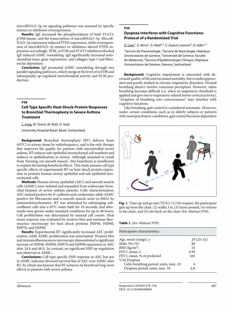
Abstracts Respiration 2018;95:478–516 DOI: 10.1159/000488898
497
microRNA21-5p on signaling pathways was assessed by specific mimics or inhibitors overexpression.
Results: IgE increased the phosphorylation of both STAT3, p70S6 kinase, and the transcription of microRNA21-5p. MicroR-NA21-5p expression reduced PTEN expression, while overexpres-sion of microRNA21-5p mimics or inhibitors altered PTEN ex-pression accordingly. PI3K, mTOR and STAT3 inhibitors blocked IgE induced ASMC remodeling. IgE significantly increased mito-chondrial mass, gene expressions, and collagen-type-I and fibro-nectin deposition.
Conclusion: IgE promoted ASMC remodeling through two parallel signaling pathways, which merge at the level of mTOR and subsequently up-regulated mitochondrial activity and ECM pro-duction.
P38
Cell Type Specific Heat-Shock-Protein Responses
to Bronchial Thermoplasty in Severe Asthma
Treatment
L. Fang, M. Tamm, M. Roth, D. Stolz
University Hospital Basel, Basel, Switzerland
Background: Bronchial thermoplasty (BT) delivers heats (65oC) to airway tissue by radiofrequency, and is the only therapy that improves life quality for patients with uncontrolled severe asthma. BT reduces sub-epithelial mesenchymal cell numbers and induces re-epithelization in airway. Although assumed to result from ‘burning out smooth muscle’, this hypothesis is insufficient to explain the lasting beneficial effects. This study assesses cell type specific effects of experimental BT on heat shock protein expres-sion in primary human airway epithelial and sub-epithelial mes-enchymal cells.
Methods: Human airway epithelial (AEC) and smooth muscle cells (ASMC) were isolated and expanded from endoscopic bron-chial biopsies of severe asthma patients. Cells characterization: AEC stained positive for E-cadherin and cytokeratin, while ASMC positive for fibronectin and α-smooth muscle actin (α-SMA) by immunohistochemistry. BT was mimicked by submerging sub-confluent cells into a 65°C water bath for 10 seconds, and after-wards were grown under standard conditions for up to 48 hours. Cell proliferation was determined by manual cell counts. Heat stress response was evaluated by western-blot and immune-fluo-rescence microscopy for heat shock proteins HSP40, HSP60, HSP70, and HSP90.
Results: Experimental BT significantly increased AEC prolif-eration, while ASMC proliferation was interrupted. Western blot and immunofluorescences microscopy demonstrated a significant increase of HSP40, HSP60, HSP70 and HSP90 expression in AEC after 24 h and 48 h. In contrast, no significant HSP up-regulation was observed in ASMC.
Conclusions: Cell type specific HSP response in AEC but not in ASMC indicates diversed survival fate of AEC over ASMC after BT, by which mechanism that BT achieves its beneficial long-term effects in patients with severe asthma.
P39
Dyspnea Interferes with Cognitive Functions:
Protocol of a Randomized Trial
D. Lawi1, G. Berra1, G. Allali2,3, E. Dupuis-Lozeron4, D. Adler1,3
1Service de Pneumologie, 2Service de Neurologie, Hôpitaux Universitaires de Genève, 3Université de Genève, Faculté de Médecine, 4Service d’Épidémiologie Clinique, Hôpitaux Universitaires de Genève, Geneva, Switzerland
Background: Cognitive impairment is associated with de-creased quality of life and increased mortality, but is underappreci-ated and poorly studied in chronic respiratory disorders. Normal breathing doesn’t involve conscious perception. However, when breathing becomes difficult (i.e. when an inspiratory threshold is applied and give rise to respiratory-related motor cortical activity), “irruption of breathing into consciousness” may interfere with cognitive functions.
Like breathing, gait control is considered automatic. However, under certain conditions, such as in elderly subjects or patients with neuropsychiatric conditions, gait control becomes dependent
Table 1. (for Abstract P39)
Participants characteristics
Age, mean (range), y 29 (25–32)Male, No (%) 80BMI (kg/m2) 24FEV1, mean, L 4.58FEV1, mean, % of predicted 103VAS Dyspnea
Calm breathing period, units, max. 10 0Dyspnea period, units, max. 10 6.8
Fig. 1. Time up and go test (TUG). (1) On request, the participant gets up from the chair, (2) walks 3 m, (3) turns around, (4) returns to his chair, and (5) sits back on the chair (for Abstract P39).
3
2
4
5
1
3 m

AbstractsRespiration 2018;95:478–516 DOI: 10.1159/000488898
498
on cognitive functions and involves the activation of specific corti-cal areas.
Our study hypothesizes that dyspnea leads to cognitive dys-function by “cortical competition”. We predict that experimen-tally induced dyspnea will interfere with gait control (primary
outcome) and affects cognitive function tests (secondary out-come).
Methods: 40 healthy subjects (Table 1) will be randomized to the sequence “calm spontaneous breathing”/”experimental dys-pnea” or “experimental dyspnea”/ “calm spontaneous breathing”. Dyspnea will be elicited by progressive increase of threshold load-ing aiming at an intensity of 6/10 on a visual analog scale. During each period of this cross-over trial, the timed up and go test (TUG), as a measure of gait control (Figure 1), and a battery of cognitive function tests will be performed (Figure 2).
Preliminary Results: To date, 10 subjects have been enrolled. Significant differences have been observed for the TUG test and the indirect span memory test (Table 2).
Expected Results and Conclusion: The purpose of this study is to unravel a causal link between dyspnea and cognitive impair-ment and its impact on gait control. Such demonstration should stimulate caregivers to consider dyspnea as a syndrome responsi-ble for cognitive impairment. Understanding the impact of dys-pnea, irrespective of physiological variables, is a necessary step to understand the many facets of chronic respiratory diseases and improve our patients’ daily lives.
P40
Effects of Trumpet Playing on Lung Function –
A Case-Control Study
L. Studer, D. Schumann, A. Siebeneichler, M. Tamm, D. Stolz
University Hospital Basel, Basel, Switzerland
Introduction: Lung function in wind instrument players is a much-discussed topic with indefinite conclusions. It has been a consideration to use the wind instrument as a treatment modality in respiratory illnesses.
Methods: Participants were recruited from wind orchestra or brass bands throughout Switzerland. Participants were excluded if they played a wind instrument other than a trumpet/cornet/flugel-horn. Controls were members of an orchestra, life-long no wind-instrument players. Both players and controls were excluded if they suffered from a chronic lung disease other than COPD or
Dyspnea
Dyspnea
Calm breathing
Calm breathing
TUGVerbal fluencyTMT A/BCODE testMemory span
TUGVerbal fluencyTMT A/BCODE testMemory span
TUGVerbal fluencyTMT A/BCODE testMemory span
TUGVerbal fluencyTMT A/BCODE testMemory span
P1
P1
P2
P2
20 subjects
40 subjects
20 subjects
Randomization
- Anthropometric data collection- Spirometry, saturation, SNIP/MIP- Determination of max IP% for dyspnea VAS 5–7, MDP score
Fig. 2. Flowchart (for Abstract P39).
Table 2. (for Abstract P39)
Outcome (Groups Combined (n = 10)) Calm breathing
Dyspnea Difference (95% CI)
P value
Time at Time Up ang Go Test (TUG), mean (sec.) 8.6 9.3 0.7 (0.29–1.11) 0.0038Verbal fluency: Categorical, mean (words) 33.5 30.3 3.2 (4–10.5) 0.34Verbal fluency: Lexical, mean (words) 24.2 24.9 0.7 (–5–6.7) 0.72Trail Making Test A, mean (sec.) 15.8 15.6 0.14 (–2.2–2.5) 0.89Trail Making Test B, mean (sec.) 31.5 35.9 44.3 (–12.6–3.9) 0.26Test Code, mean (symbols) 89.4 90.3 0.9 (–12–11) 0.86Memory Span: Direct mean (sequences) 10.3 10.2 0.1 (–1–1.2) 0.84Memory Span: Indirect mean (sequences) 6.1 7.6 1.5 (–2.9–1) 0.038

Abstracts Respiration 2018;95:478–516 DOI: 10.1159/000488898
499
asthma or had a current respiratory infection. The primary out-come of the study was the difference in FEV1 %predicted between players and controls. With a significance of 0.005, a power of 90% and an unequal sample size ratio 2:1 (players:controls), we calcu-lated a minimum sample size of 92 players and 46 controls.
Results: 99 trumpet/cornet/flughorn players and 48 controls were included in the study. The players played on average 5.2 ± 4.1 hours per week and had played their instrument for an average of 27 ± 14.4 years. Players were less commonly current smokers (13% vs. 21%) and male (67% vs 85%) compared to the controls. Players were significantly older than the controls (40.5 vs 33.3 years; p = 0.008) and had a significantly higher BMI (25.3 vs 24.0 kg/m2; p = 0.037). There was no significant difference in FEV1 %predicted (91.9 vs 94.2%, p = 0.316) and FVC %predicted (89.4 vs 92.6; p = 0.125) between controls and players. More controls had an FeNO value >25 ppb compared to trumpet players (41% vs. 19%), even after adjusting for age and smoking status (OR = 3.402, p = 0.038). There was a significant inverted association between number of years of playing and both a) FVC %predicted and b) FVC (L) after adjusting for a) smoking status and b) age, gender, height and smoking status.
Conclusion: In our cohort, playing the trumpet/cornet/flugel-horn does not adversely affect FEV1.
P41
Efficacy of a Multiplex Bacterial PCR in Identifying
Pathogens in Pleural Empyema
L. Franchetti, D. Schumann, M. Tamm, K. Jahn, D. Stolz
University Hospital Basel, Basel, Switzerland
Introduction: The identification of the pathogens involved in pleural empyema has mainly relied on conventional bacterial cul-ture or single species polymerase chain reaction (PCR), both with relatively low sensitivity. We investigated the efficacy of a com-mercial multiplex pneumonia PCR assay to identify the pathogens involved in pleural empyema.
Methods: Prospective, monocentric, observational study. Pleural fluid samples were collected from 194 consecutive patients. All samples underwent conventional microbiology at the time of hospitalisation. Commercial multiplex pneumonia bacterial PCR (Curetis Unyvero p55) was performed retrospectively.
Results: The final clinical diagnosis of empyema was made in 31% of all cases. There was a suspicion of empyema based on the ultrasound findings in 35/189 (18.5%) cases and of these, 30 cases (85.7%) were diagnosed with empyema (16 hosptial-acquired and 14 community-acquired pneumonia). When only taking into ac-count the 20 pathogens included in the multiplex PCR Assay, more pathogens were identified using the multiplex PCR assay than with conventional methods (23.3% (7/30) vs. 6.7% (2/30), p = 0.008). In those cases without suspicion of empyema, 15/154 (9.7%) had a final diagnosis of empyema. Using the same criteria, conventional methods identified pathogens in 0/15 (0%) cases and the multiplex PCR-based assay identified pathogens in 1/15 (6.7%) cases. When taking into account all available pathogens, 35 pathogens in 20 samples were identified when using conventional methods com-
pared to 11 pathogens in 11 samples when using the multiplex PCR-based assay. The most common microorganisms identified in the cases diagnosed with empyema, were anaerobes (31) fol-lowed by gram-positive cocci (10) and gram-negative rods (4).
Conclusion: The multiplex PCR-based assay had a higher sen-sitivity and specificity than conventional microbiology when only the pathogens on the panel were taken into account. A dedicated pleural empyema multiplex PCR panel including anaerobes would be needed to cover most common pathogens involved in pleural infection.
P42
Improved Assessment of Bronchodilator Response
Evaluating Spirometric and Plethysmographic
Target Parameters as Combined Primary End-Points
in Patients with COPD
R. Kraemer1, M. Leuenberger2, L. Kern2, M.H. Brutsche2, Asthma-COPD-Overlap Syndrome diversification study1Department of Biomedical Research, University of Berne, Bern-Muri, 2Clinic of Pneumology, Cantonal Hospital St. Gallen, St. Gallen, Switzerland
Background: Response to bronchodilators is currently evalu-ated by changes in forced expiratory volume at 1 s (FEV1) and/or forced vital capacity FVC) in patients with COPD. Bronchodila-tors additionally lower peripheral smooth muscle tone leading to a decrease of pulmonary hyperinflation and air trapping. Changes in small airway dysfunction assessed either by the forced expira-tory flow between 25% and 75% FVC (FEF25–75) or by the effective, specific airway conductance (sGeff) are rarely appreciated. The aim of the present study was to search for parameters characterizing bronchodilator response including changes in peripheral airway function.
Methods: Response to 200 mcg Salbutamol was evaluated by spirometric (FEV1, FVC, FEV1/FVC, FEF25–75) and plethysmo-graphic (sGeff) parameters in 268 COPD patients, including 55 clinically detected patients with asthma-COPD-overlap (ACO), and compared with 84 asthma patients. Reversibility of airway ob-struction was assessed using the ATS/ERS criteria, i.e. an increase of FEV1 and/or FVC ≥12%, and ≥200 mL, or an improvement of FEF25–75 and/or sGeff ≥20% from baseline.
Results: Bronchodilator response could be demonstrated by ATS/ERS criteria in COPD patients in 19.4%, compared to asthma in 28.6% and ACO in 36.4%. An additional response was observed by sGeff in 25.7%, by FEF25–75 in 17.2% resp. Similar additional re-sponse were found for asthma and ACO patients. Hence, ATS/ERS criteria failed to demonstrate bronchodilator response compared to sGeff in 27.4% to FEF25–75 in 18.5% resp.
Conclusions: A significant additional proportion of COPD pa-tients reached reversibility of airway obstruction if improvement of airway dysfunction (sGeff, FEF25–75) is taken into account. With the aid of mid-expiratory flow characteristics and specific airway conductance a subset of patients could be identified with a small airway reversibility phenotype – potentially indicating ACO.

AbstractsRespiration 2018;95:478–516 DOI: 10.1159/000488898
500
P43
How to Set Up High Frequency Non-Invasive
Ventilation (HF-NIV): A Bench Test
A. Lovis1, P. Vremaroiu1, O. Long2, J. Simon2, L. Noirez1, L. Piquilloud3, L. Nicod1, A. Ogna1,4
1Médecine Interne, 2Physiothérapie, 3Soin Intensif, CHUV-UNIL, Lausanne, 4Médecine Interne et Pneumologie, Ospedale di Locarno, Locarno, Switzerland
Introduction: High-frequency noninvasive ventilation (HF-NIV) is a new ventilation modality allowing to obtain prolonged apneas in non-sedated subjects. In preliminary studies, we showed good tolerance and promising clinical applications for thoracic MRI and radiation therapy. In this bench study, we aimed to in-
vestigate the impact of ventilation parameters, circuits and inter-faces to the delivered minute ventilation, in order to optimize ven-tilation efficacy.
Methods: The minute ventilation delivered by a Monsoon III ventilator (Acutronic Medical Systems) was measured in a bench test with a Michigan Lung Simulator, using 9 respiratory frequen-cies (100 to 500 bpm), 5 working pressures (0.8 to 1.6 bar), 4 inter-faces (Phasitron, Percussionnaire) and 4 different circuits.
Results: Minute ventilation decreased almost linearly by in-creasing ventilator frequency (Fig. 1) and increased by increasing working pressure (Fig. 2), with a flattening curve in the higher pressure range. The choice of the Phasitron interface had little im-pact on the ventilation efficacy, however, the single-use Homecare Universal (HC) Phasitron (A50605-D) showed slightly higher de-livered volumes for the same working pressure (Fig.1). Of note, the removal of the spring from the Phasitron (to make it MRI-com-
Fig. 1. Minute volume of the 3 phasitrons, dependent on the ventilator frequencies, SU = Sin-gle Use (A50605-D), with and without spring, Yellow/Red (A50007-12) and Green/Green phasitron working pressure 1 bar (for Abstract P43).
Fig. 2. Minute volume of the 3 phasitrons, dependent on the working pressure, SU = Single Use, with and without spring, ventilator frequency 250/min. (for Abstract P43).
0
5
10
15
20
25
30
35
40
45
50
Min
ute
volu
me,
L/m
in
100 150 200 250 300 350 400 450 500FR/min
SU + springSU no spring
Yellow/redGreen/green
0
5
10
15
20
25
30
35
40
45
50
Min
ute
volu
me,
L/m
in
0.8 1.0 1.0 1.4 1.6P, bar
SU + springSU no spring
Yellow/redGreen/green

Abstracts Respiration 2018;95:478–516 DOI: 10.1159/000488898
501
patible) did not modify its performance (Fig.1). A more rigid cir-cuit was associated with a more efficient ventilation, whilst a longer circuit impacted negatively on the delivered volume (Fig. 3 and 4 not upload, only 2 allowed).
Conclusion: Ventilation parameters, circuits and interfaces all seem to have an impact on the ventilation efficacy. The combina-tion of the single-use HC Phasitron, a short and rigid circuit and a ventilation frequency of 250–300 bpm seems to be the most effi-cient one to obtain tidal volumes just above the anatomical dead space, in order to maintain effective ventilation with the minimal thoracic movements.
P44
Automated Particle Analysis for Inorganic Lung Dust
Characterisation
B.K. Kuhn1, J. Rausch2,3, D. Jaramillo Vogel2,3, B. Vrugt1,4
1SILAG – Swiss Laboratory for Particle Analysis in Tissues, Zürich, 2Particle Vision GmbH, 3Département des Géosciences, Université de Fribourg, Fribourg, 4Institut für Pathologie USZ, Zürich, Switzerland
The cause or agent inducing occupational lung diseases is often difficult to identify. In such cases dust analysis of lung tissue may help. Up to now, inorganic dust particles were analysed manually by transmission electron microscope (TEM). In the last few years automated particle analysis using scanning electron microscope (SEM) was developed. The inorganic dust obtained from lung tis-sue from two patients was independently analysed by TEM and SEM respectively.
Inorganic dust particles were isolated from freeze-dried lung tissue by low temperature ashing and filtered through polycarbon-ate filters (Nuclepore) with 0.2 μm pore size, which were then car-bon coated for analysis.
For manual particle analysis a TEM (Tecnai Spirit, FEI) equipped with an Energy-dispersive X-ray spectrometer (EDX; Oxford Instruments) at the University of Zurich was used. The automated particle analysis was performed with a SEM (Zeiss GeminiSEM 300) at the Particle Vision laboratory equipped with an Oxford X-MAX 80 mm2 EDX detector and the Oxford AZtec Feature software.
Case 1: A 61 year old male with interstitial lung fibrosis. Two thirds of the 121 particles analysed by TEM were classified as sili-cate minerals, 30% as oxides and 2% as phosphor compounds. With automated SEM-EDX analysis, 2407 particles were analysed delivering a comparable result (Table 1).
Case 2: A 23 year old male with pneumonia. Histologic exami-nation showed abundant birefringent material in lung tissue. Of 211 analysed particles in TEM 35% were recognized as silicate minerals, 21% as titanium oxide and 38% as phosphor compounds. The predominance of phosphor compounds (43%) was confirmed by automated SEM analysis.
The evaluation of the automated particle analysis by SEM-EDX showed a good reproducibility of the TEM analysis with the advan-tage of considerably higher numbers of analysed particles guaran-tying a better statistical representativeness of the results without the need of an operator during the analysis.
Poster Discussion Posters
P45
Spirometry in Central Asian Lowlanders and
Highlanders, a Population Based Study
S. Ulrich1, M. Furian1, B. Estebesova2, N. Toktogulova2, G. Beishekeeva3, S. Ulrich1, P.G.J. Burney4, T.M. Sooronbaev2, K.E. Bloch1
1Department of Respiratory Medicine, University Hospital Zurich, Zurich, Switzerland, 2Department of Respiratory Medicine, National Center for Cardiology and Internal Medicine, 3Kyrgyz-Russian Slavic Univeristy, Bishkek, Kyrgyzstan, 4National Heart and Lung Institute & MRC-PHE Centre for Environment and Health, Imperimental College London, London, United Kingdom
Introduction: The purpose of the study was to establish spiro-metric reference values for a Central Asian population of high-landers and lowlanders.
Methods: Spirometries from a population-based cross-sec-tional study performed in 2013 in Kyrgyzstan (Burden Of Lung Disease survey, BOLD: Buist et al. COPD 2005) were analyzed. Us-ing multiple linear regression, Global Lung Function Initiative (GLI) equations were fitted separately for men and women, and altitude of residence (lowlands 700–800 m, highlands 1900–2800 m) to data from healthy, never-smoking Kyrgyz adults. The general GLI equation was applied: predicted value = exp(a0 + a1*ln(height) + a2*ln(age) + b1*(age/100) + b2*(age/100)^2 + b3*(age/100)^3 + b4*(age/100)^4 + b5*(age/100)^5).
Results: Of 2784 Kyrgyz in the BOLD survey, 448 highlanders (379 females) and 505 lowlanders (368 females) met inclusion cri-teria. Prediction equations for FVC in the Kyrgyz sample fit best with the GLI “North-East Asian” whereas equations for FEV1 fit best with “Other/Mixed” category. Predicted FEV1/FVC was low-
Table 1. Overview on particle occurrence (for Abstract P44)
Particle composition TEM 1 SEM 1 TEM 2 SEM 2
Quartz 23% 13% 21% 8%Other silicates 43% 58% 14% 17%Aluminium oxide 6% 2% 0% 0%Titanium oxide 17% 14% 21% 23%Iron oxide (± Ti, Cr, Ni, Si) 8% 7% 5% 8%P-compounds (± Fe, Na, Si) 2% 5% 21% 26%P-compounds with Ti 0% 0% 17% 17%Copper sulfide 2% 1% <1% <1%Number of analysed particles 121 2407 211 3462

AbstractsRespiration 2018;95:478–516 DOI: 10.1159/000488898
502
er than that of all GLI categories. Compared to GLI “Caucasians”, Kyrgyz females and males (lowlanders and highlanders combined) have lower age- and height-adjusted FVC: mean difference –0.104 l, –0.112 l, FEV1: –0.174 l, –0.201 l and FEV1/FVC: –0.032, –0.029 (P < 0.001, all comparisons), respectively. Among Kyrgyz, age- and sex-adjusted mean FEV1, FVC and PEF are higher in highlanders (+0.132 l, +0.138 l and +0.460 l/s) than in lowlanders (P < 0.001, all comparisons), but FEV1/FVC is similar.
Conclusion: We established prediction equations for an adult Central Asian population indicating that FVC is similar to the GLI category “North-East Asian” and FEV1/FVC is lower than in all other GLI categories. Kyrgyz highlanders have significantly higher predicted FVC, FEV1 and PEF than lowlanders.
P46
Effect of Endoscopic Lung Volume Reductions in
Patients with Emphysema
F. Baty, T. Schneider, K. Kozu, L. Kern, M. Brutsche
Pulmonary and Sleep Medicine, Cantonal Hospital St. Gallen, St. Gallen, Switzerland
Introduction: Endoscopic lung volume reduction (ELVR) is a commonly accepted treatment for patients with severe emphyse-ma. We performed a retrospective study in order to assess the ef-fect of ELVR on patients with emphysema.
Methods: 65 patients with emphysema were referred from Jan-uary 2010 to December 2017 to the Department of Pulmonary Medicine of the Cantonal Hospital St. Gallen for an ELVR. Lung function including forced expiratory volume in 1 second (FEV1), maximal vital capacity (VCmax), forced vital capacity (FVC), re-sidual volume (RV), total lung capacity (TLC) and diffusing capac-ity (DLCO) together with parameters of exercise capacity (6-min walking distance) were collected and analyzed using linear mixed models.
Results: Patients had a median age of 67 (IQR: 62.5–72) and a pre-operative FEV1 of 28% (sd: 8.80) and residual volume of 213% (sd: 41.57). Intrabronchial valves were used in 43 patients, where-as coil implantation was used in 21 patients. One patient received consecutively valves and coils. The effect of ELVR 3 months with-in insertion included a significant decrease of 0.36 L of TLC (p = 0.001) and 0.40 L of RV (p = 0.004), as well as a significant increase of 5.3% of VCmax (p = 0.029) and an improved exercise capacity (59 m improvement of the 6-min walking distance, p = 0.043). A significant improvement of 0.37 L of FVC was also observed 6 months after intervention.
Conclusion: There was a significant short-term improvement in lung function following ELVR in patients with emphysema. The effect was observed within 6 months after therapy.
P47
Cardiovascular and Metabolic Correlates of
Abnormal Spirometry (Pneumolaus Study)
A. Lenoir1, P.M. Marques-Vidal2, P. Vollenweider2, J.-W. Fitting1, L.P. Nicod1
1Service de Pneumologie, 2Service de Medecine Interne, Centre Hospitalier Universitaire Vaudois, Lausanne, Switzerland
Patients with impaired lung function frequently present with comorbidities further affecting their disease burden. We evaluated the association of cardiovascular risk factors with abnormal lung function in a sample of the general population of Lausanne, Swit-zerland.
Between 2014 and 2017, we examined 3351 participants of the CoLaus study, a prospective study investigating the occurrence of cardiovascular diseases and risk factors in the general population of Lausanne. Spirometry was performed according to ATS/ERS 2005 guidelines and interpreted using GLI 2012 references. If FEV1/FVC or FVC was below the Lower Limit of Normal (LLN), spirometry was repeated after bronchodilation (BD). Chronic air-way obstruction was defined as FEV1/FVC < LLN after BD; revers-ible airway obstruction as FEV1/FVC or FVC < LLN before BD and >LLN after BD; possible lung restriction as FVC < LLN after BD.
Among 3351 participants, 91.9% had normal spirometry, 2.4% reversible and 3.5% chronic obstruction; 1.8% had possible lung restriction without obstruction, 0.4% with chronic, <0.1% with re-versible obstruction. In participants with abnormal spirometry, prevalence of hypertension (52.9% vs. 43.3%, p = 0.002) and dia-betes (16.2% vs. 8.7%, p < 0.0001) was significantly higher. Par-ticularly, subjects with restrictive pattern often suffered from hy-pertension (68.0%) and diabetes (33.3%). Similarly, glucose (5.9 vs. 5.4 mmol/l, p = 0.0001) and insulin levels (12.7 vs. 9.7 μIU/ml, p = 0.0304) were significantly higher in the restrictive group. These findings were not confirmed for obstruction and independent of smoking status except for insulin levels. Although mean BMI was higher in the restrictive group (28.7 ± 7.0 vs. 26.3 ± 4.6 kg/m2, p < 0.0001), prevalence of hypertension but not diabetes remained sig-nificantly increased when analysing normal weight subjects only.
We conclude that abnormal lung function, particularly restric-tive spirometric pattern, is associated with increased prevalence of hypertension and diabetes. Identifying subjects with restrictive ventilatory pattern seems of high clinical and public health impor-tance.

Abstracts Respiration 2018;95:478–516 DOI: 10.1159/000488898
503
P48
Strong Increase in the Prevalence of COPD in
Switzerland between 2003 and 2012
A. Strassmann1, E. West1, C. Wang2, A. Turk3, K. De Hogh4, M. Röösli4, M. Bopp1, H. Dressel1, M. Puhan1, Swiss National Cohort Study Group1Epidemiology, Biostatistics und Prevention Institute, University of Zurich, 2Department of Mathematics, University of Zurich, Zurich, 3See-Spital Horgen, Horgen, 4Swiss Tropical and Public Health Institute, Basel, Switzerland
Introduction: Most studies to determine the prevalence of COPD are conducted only once. Our aim was to determine the prevalence of COPD yearly in general population samples between 2003 and 2012.
Methods: We based our analysis on data from 38,390 partici-pants aged ≥35 years who received a lung function measurement as part of the LuftiBus health promotion campaign of Lunge Zu-rich. The LuftiBus represents the adult Swiss population well and its data have been used to derive population-based lung function reference equations. Spirometry was performed using computer-ized pneumotachographs by trained staff, according to American Thoracic Society guidelines but without bronchodilation (not fea-sible in LuftiBus). We used both lower limit of normal (LLN) and FEV1/FVC of <0.7 to determine the presence of COPD. We used the linkage to the Swiss National Cohort to adjust for potential dif-ferences between yearly population samples.
Results: While the population samples were very similar from year to year, the prevalence of COPD increased between 2003–2012 from 9.3% to 17.9% (based on LLN, higher estimates if based on FEV1/FVC). FVC was stable (median across years 3.9 liter) over time. The increase in prevalence affected both ever and never smokers and was observed for mild but not moderate to severe airflow obstruction (Figure). The increase in COPD prevalence remained after adjusting for age, sex, body mass in-dex, smoking status, distance of residence to main road, educa-tional status, air pollution as well as temperature at the time of measurement.
Discussion: Although pre-bronchodilator measurements likely overestimate the prevalence of COPD in absolute terms, we found a strong increase in prevalence. Since the trend ex-isted in both ever and never smokers and is only observable for mild COPD, the explanation may be found in prevalent expo-sures 30 or 40 years ago like air pollution, which peaked in the 1970s.
Fig. 1. Prevalence of COPD and of severity of airflow ob-struction between 2003 and 2012 (for Abstract P48).
0
0.05
0.10
0.15
0.20
2004 2006 2008 2010 2012Years
Prev
alen
ce o
f CO
PD
COPD gold by stages
0
0.05
0.10
0.15
0.20
2004 2006 2008 2010 2012Years
Prev
alen
ce o
f CO
PD
COPD gold by stages
0
0.05
0.10
0.15
0.20
2004 2006 2008 2010 2012Years
Prev
alen
ce o
f CO
PD
COPD LLN by stages
All
0
0.05
0.10
0.15
0.20
2004 2006 2008 2010 2012Years
Prev
alen
ce o
f CO
PD
COPD LLN by stages
Never smokers
Mild (FEV1 %pred)Moderate (FEV1 %pred)Severe (FEV1 %pred)

AbstractsRespiration 2018;95:478–516 DOI: 10.1159/000488898
504
P49
Stability of the Eosinophilic Phenotype in Stable
and Exacerbated Chronic Obstructive Pulmonary
Disease
D. Schumann, M. Tamm, K. Kostikas, D. Stolz
University Hospital Basel, Basel, Switzerland
Introduction: There is controversy around the use of blood eosinophil levels as a biomarker in determining the responsiveness of patients to inhaled corticosteroids (ICS). Studies have shown that a blood eosinophil level ≥2% may be a predictor for response to ICS treatment and that patients with blood eosinophils >300 cells/μl are at a higher risk of severe exacerbations. We investigat-ed the stability of blood eosinophils in COPD patients in stable state and during exacerbation.
Method: This is a nested study of the PROMISE-COPD study where patients with stable COPD stage GOLD II to IV were en-rolled. Only data from patients with more than one visit were used for the current analysis. Concordance or stability was defined as blood eosinophil levels persistently lower than or persistently higher than the absolute cut-off points 150 cells/μl, 300 cells/μl or the percentage cut-off points 2%, 3%, and 4%. Discordance was obtained when the blood eosinophil levels varied between any two visits dichotomized by stable state or exacerbation status.
Results: We analysed data from 230 patients with 1557 visits during the stable phase of COPD and 522 visits with exacerbation. The median follow up period was 4.6 years. The median blood eo-sinophil level during stable state was 183 cells/μl, during mild/moderate exacerbation 169 cells/μl and during severe exacerbation 131 cells/μl. There was an overall high degree of discordance of up to 60% during mild/moderate exacerbation, 63% during severe ex-acerbation and up to 76% during stable state. 84.8% of the patients received ICS at the beginning of the study. The stability of the blood eosinophil was not associated with ICS treatment.
Conclusion: Our data suggest that blood eosinophil levels vary significantly throughout the course of COPD and therefore a sin-gle measurement may not be a reliable predictor of ICS response.
P50
Single-Port Thymectomy Using a Subxiphoid
Approach
B. Bédat, S.M. Sadowski, F. Triponez, W. Karenovics
Thoracic and Endocrine Surgery Unit, Geneva University Hospitals, Geneva, Switzerland
Background: Thymectomy is currently performed by a lateral thoracic approach. However, this approach can cause difficulties to identify the contralateral phrenic nerve. A subxiphoid approach could permit to visualize the bilateral phrenic nerves and to de-crease the postoperative pain. The aim of this study is to assess the feasibility of the single-port thymectomy using a subxiphoid ap-proach and to evaluate the early surgical outcomes.
Methods: We retrospectively reviewed all consecutive patients who underwent single-port subxiphoid thymectomy from Decem-ber 2015 to December 2017 at University Hospitals of Geneva. Pa-tient characteristics, indications, pathology findings and early postoperative outcomes were analyzed.
Results: Ten patients (4 male, 6 women) with a mean age of 43 ± 15 years old underwent a single-port thymectomy using a subxiphoid approach. Indications for thymectomy was : myasthe-nia gravis (n = 8), thymoma-associated myasthenia gravis (n = 1) and parathyroid adenoma (n = 1). The mean operation time was 126 ± 40 minutes. There was no complication during the operation and bilateral phrenic nerves were identified in all patients. No pa-tient died during the postoperative period. One complication was identified and consisted to a postoperative myasthenic crisis. In-traoperative drain removal was performed in three patients. The median duration of drainage was 1 day (range 0–1 day) and the median length of hospitalization was 2 days (range 1–13 days). The mean of maximal postoperative pain during the early postopera-tive period was shown in Figure 1. The intraoperative drain re-moval was not correlated to a decreased postoperative pain.
Conclusion: Single-port thymectomy using a subxiphoid ap-proach is safe, causes postoperative low pain, and permits the iden-tification of the bilateral phrenic nerves.
P51
Analysis of Hospitalizations for Tuberculosis in
Switzerland from 2002 to 2015
S. Tonko1, F. Baty1, M. Brutsche1, O.D. Schoch1,2
1Klinik für Pneumologie und Schlafmedizin, Kantonsspital St.Gallen, St.Gallen, 2Lungenliga Schweiz, Kompentenzzentrum Tuberkulose, Bern, Switzerland
Introduction: Historically, patients with tuberculosis have been hospitalized for several months. Today, Swiss guidelines rec-ommend outpatient management or discharge of patients from
Fig. 1. Postoperative pain (for Abstract P50).
0123456789
10
Pain
ratin
g sc
ale
D0n = 10
D1n = 10
D2n = 7
D3n = 5
D4n = 3
1.8
3.5 3.0 3.01.7
Maximal pain score, main (0–10)
Postoperative days

Abstracts Respiration 2018;95:478–516 DOI: 10.1159/000488898
505
hospital as quickly as possible, whenever permissible. The aim of this retrospective analysis was to describe the rate of hospitaliza-tions and length of stay (LOS) of patients with tuberculosis for the years 2002–2015 in Switzerland.
Methods: Data were extracted from a nation-wide database provided by the Swiss Federal Office for Statistics enclosing 19,832,602 hospital entries between 2002 and 2015. Hospitaliza-tions were retrieved by ICD-10 codes (A15-A19). A total of 8047 primary and 14041 co-diagnoses were identified. The number of hospitalizations were compared to the annual tuberculosis cases reported to the Swiss Federal Office of Public Health. Subgroup-analyses were performed including HIV and diabetes. Differences between miliary, non-miliary and extrapulmonary tuberculosis were investigated.
Results: Reported cases and hospitalisations were similar with a significant decline from 673 hospitalizations/year in 2002 to 471 in 2006 (p < 0.001) was followed by a stabilization (p = 0.309, Fig-ure 1). The median LOS was 14 days (IQR: 6–22) and did not change over time (p = 0.073). In-hospital mortality was 2.3% and did not change over time (p = 0.430). The age distribution was bi-modal with a first peak between 20–30 and another peak at 70–90 years-old. The older group (>50 years-old) stayed 1 day longer (14 [IQR: 5–20] vs. 15 days [IQR: 8–26]), however in-hospital mortal-ity increased from 0.5% to 5.7% in the older. Patients with miliary tuberculosis (vs. pulmonary) had a longer LOS (median 16 days vs. 20 days, p < 0.001) and a higher in-hospital mortality (median 2.3% vs. 6.9%, p < 0.001).
Conclusion: Few cases have been treated in outpatient settings. Tuberculosis hospitalizations decreased from 2002–2006, fol-lowed by a stabilization between 2006 and 2015. Mortality and comorbidities did not change. LOS and mortality in miliary tuber-culosis was significantly higher.
P52
Effect of Endoscopic Lung Volume Reductions in
Patients with Emphysema
F. Baty, T. Schneider, K. Kozu, L. Kern, M. Brutsche
Pulmonary and Sleep Medicine, Cantonal Hospital St. Gallen, St. Gallen, Switzerland
Introduction: Endoscopic lung volume reduction (ELVR) is a commonly accepted treatment for patients with severe emphyse-ma. We performed a retrospective study in order to assess the ef-fect of ELVR on patients with emphysema.
Methods: 65 patients with emphysema were referred from Jan-uary 2010 to December 2017 to the Department of Pulmonary Medicine of the Cantonal Hospital St. Gallen for an ELVR. Lung function including forced expiratory volume in 1 second (FEV1), maximal vital capacity (VCmax), forced vital capacity (FVC), re-sidual volume (RV), total lung capacity (TLC) and diffusing capac-ity (DLCO) together with parameters of exercise capacity (6-min walking distance) were collected and analyzed using linear mixed models.
Results: Patients had a median age of 67 (IQR: 62.5–72) and a pre-operative FEV1 of 28% (sd: 8.80) and residual volume of 213% (sd: 41.57). Intrabronchial valves were used in 43 patients, where-as coil implantation was used in 21 patients. One patient received consecutively valves and coils. The effect of ELVR 3 months with-in insertion included a significant decrease of 0.36 L of TLC (p = 0.001) and 0.40 L of RV (p = 0.004), as well as a significant increase of 5.3% of VCmax (p = 0.029) and an improved exercise capacity (59 m improvement of the 6-min walking distance, p = 0.043). A significant improvement of 0.37 L of FVC was also observed 6 months after intervention.
Conclusion: There was a significant short-term improvement in lung function following ELVR in patients with emphysema. The effect was observed within 6 months after therapy.
P53
Lung Function in Birt-Hogg-Dubé Syndrome
C. Daccord1, R. Borie2, M. Faouzi3, C. Khouatra4, A. Gondouin5, A. Briault6, J.-F. Cordier4, V. Cottin4, R. Lazor1,4
1Respiratory Medicine Department, Lausanne University Hospital, Lausanne, Switzerland, 2Respiratory Medicine Department, Bichat Claude-Bernard University Hospital, Paris, France, 3Institute of Social and Preventive Medicine, Lausanne University Hospital, Lausanne, Switzerland, 4Reference Centre for Rares Pulmonary Diseases, Claude Bernard University Hospital, Lyon, 5Respiratory Medicine Department, Besançon University Hospital, Besançon, 6Respiratory Medicine Department, Grenoble University Hospital, Grenoble, France
Introduction: Birt-Hogg-Dubé syndrome (BHDS) is a rare in-herited autosomal dominant disorder characterized by cutaneous fibrofolliculomas, renal tumors, multiple pulmonary cysts and
Fig. 1. Trend and Monthly Fluctuations of Tuberculosis Hospital-izations in Switzerland from 2002–2015 (for Abstract P51).
20
40
60
2002 2006 2010 2014Years
Num
ber o
f TB
hosp
ital
TB hospitalizations (1st diagnosis)

AbstractsRespiration 2018;95:478–516 DOI: 10.1159/000488898
506
spontaneous pneumothoraces. BHDS is caused by mutations in the tumor suppressor gene FLCN coding for folliculin. Only lim-ited data on lung function in BHDS are available, and whether lung function declines over time as in other cystic lung diseases is un-known.
Aims: To assess lung function at baseline and over time in BHDS.
Methods: We retrospectively studied lung function parameters from 30 patients with BHDS at baseline and during disease course, and looked for correlations with age, gender and smoking history.
Results: Mean±SD age at baseline was 42 ± 14 yr, 60% were males, 43% were smokers and 93% had pulmonary cysts at HRCT. Baseline FEV1 and FVC (n = 30) were 95 ± 13 and 100 ± 14%pred respectively, and FEV1/FVC ratio was 80 ± 6%. TLC (n = 21) was 98 ± 15%pred, RV (n = 21) was 111 ± 30%pred, DLCO (n = 18) was 86 ± 16%pred, KCO (n = 19) was 96 ± 20%pred, and PaO2 (n = 8) was 88 ± 11 mm Hg. FEV1, FVC and FEV1/FVC at 1, 2 and 3 years follow-up (n = 12, 9 and 6 respectively) were not significant-ly different from baseline. FEV1/FVC tended to decrease with age at baseline (p = 0.05) and over time (p = 0.02). In smokers, FEV1 was 9% lower at baseline (p = 0.08) and over time (p = 0.013) as compared to non-smokers. FVC significantly increased with age at baseline (p = 0.007) and over time (p = 0.002). Other parameters at baseline and over time were not correlated with age, gender and smoking history.
Conclusions: This preliminary analysis on 30 patients suggest that cystic lung disease in BHDS does not affect respiratory func-
tion at baseline, and that no deterioration occurs over a follow-up period of 3 years. The data of approximately 50 additional patients are currently being analysed.
P54
Respiratory Viral Infection in Immunocompromised
Patients
K. Jahn1, D. Schumann1, M. Tamm1, H. Hirsch2, J. Halter3, L. Junker1, W. Strobel1, S. Savic4, D. Stolz1
1Clinic of Pneumology and Pulmonary Cell Research, 2Transplantation and Clinical Virology, 3Department of Haematology, 4Department of Pathology, University Hospital Basel, Basel, Switzerland
Introduction: Knowledge on viral infections in immunocom-promised patients is sparse. We investigated the incidence of re-spiratory viruses in a well-characterized population of immuno-compromised patients.
Methods: All immunocompromised patients with suspicion of infection and/or respiratory symptoms and/or radiologically con-firmed lung infiltrates undergoing bronchoscopy were included in the study. In addition to the routine assessment of bronchoalveolar
Table 1. Incidence of respiratory viruses (for Abstract P54)
Respiratory viruses Haematological Group [virus pos. n = 374] n (%)
Solid organ Transplantation Group [virus pos. n = 261] n (%)
‘Other’ Group [virus pos. n = 301] n (%)
Adenovirus 18 (4.8) 4 (1.5) 6 (2.0)Bocavirus 3 (0.80) 4 (1.5) 3 (1.0)Coronavirus 32 (8.6) 23 (8.8) 28 (9.3)Human metapneumovirus 34 (9.1) 17 (6.5) 11 (3.7)H1N1 4 (1.1) 16 (6.1) 0 (0)Influenza 26 (7.0) 16 (6.1) 15 (5.0)Parainfluenza 54 (14.4) 18 (6.9) 24 (8.0)Rhinovirus-Enterovirus 124 (33.2) 107 (41.0) 100 (33.2)Respiratory syncytial virus 34 (9.1) 20 (7.7) 23 (7.6)
Table 2. Incidence of other viruses (for Abstract P54)
Other viruses Haematological Group [Virus pos. n = 374] n (%)
Solid organ Transplantation Group [Virus pos. n = 261] n (%)
‘Other’ Group [Virus pos. n = 301]n (%)
Cytomegalovirus 84 (22.5) 70 (26.8) 81 (26.9)Epstein-Barr virus 7 (1.9) 0 (0) 1 (0.33)Herpes simplex virus 44 (11.8) 22 (8.4) 52 (17.3)Human Herpes virus 4 (1.1) 0 (0) 0 (0)Varicella-zoster virus 1 (0.27) 0 (0) 0 (0)

Abstracts Respiration 2018;95:478–516 DOI: 10.1159/000488898
507
lavage fluid, a multiplex PCR for 20 viruses has been performed. Study period was from October 2009 to September 2017.
Results: 1303 patients with 2666 samples were included in the study. 35% (n = 936) of the samples were positive for viral infec-tion, 23% (n = 610) were positive for bacterial infection, 1.6% (n = 43) were positive for atypical mycobacteria, and 10% (n = 269) were positive for fungal infection. Bacterial coinfection was evi-dent in 26% (n = 242/936) and fungal coinfection in 13% (n = 117/936) of the virus positive samples. The incidence of respira-tory viruses in the virus positive samples was as follows: adenovi-rus 3.0% (n = 28), bocavirus 1.1% (n = 10), coronavirus 8.9% (n = 83), human metapneumovirus 6.6% (n = 62), H1N1 0.53% (n = 5), influenza 6.1% (n = 57), parainfluenza 10.3% (n = 96), rhinovirus-enterovirus 35.4% (n = 331), RSV 8.2% (n = 77). Other viruses in-cluded cytomegalovirus 25.1% (n = 235), Epstein-Barr virus 0.85% (n = 8), herpes simplex virus 12.6% (n = 118), human herpes vi-rus-6 0.32% (n = 3), human herpes virus-7 0.1% (n = 1) and vari-cella-zoster virus 0.1% (n = 1). In one case (0.1%), four viruses were identified, in 15 cases (1.6%) 3 viruses, in 146 cases (15.6%) 2 vi-ruses and in 774 cases (82.7%) one virus was identified. Patients were divided into groups according to the cause of immunosup-pression. In the haematological group 36.1% (n = 374/1035), in the solid organ transplantation group 39.4% (n = 261/663) and in the ‘Other’ group 31.1% (n = 301/968) of the samples were positive for viral infection (Table 1, 2).
Conclusion: There is a high prevalence of respiratory virus in-fections in immunocompromised patients undergoing diagnostic bronchoscopy for respiratory symptoms. This finding could influ-ence patient management.
P55
Decline of Physical Activity is Not Explained by
Progression of Exercise Intolerance in COPD
C. Clarenbach1, N. Sievi1, T. Brack2, M. Brutsche3, M. Frey4, S. Irani5, J. Leuppi6,7, R. Thurnheer8, M. Kohler9,10
1Pulmonology, University Hospital Zurich, Zurich, 2Cantonal Hospital of Glarus, Glarus, 3Cantonal Hospital St. Gallen, St. Gallen, 4Clinic Barmelweid, Barmelweid, 5Cantonal Hospital of Aarau, Aarau, 6Cantonal Hospital Baselland, Liestal, 7University of Basel, Basel, 8Cantonal Hospital of Münsterlingen, Münsterlingen, 9University Hospital Zurich, 10Zurich Centre for Integrative Human Physiology, University of Zurich, Zurich, Switzerland
Introduction: Daily physical activity (PA) and exercise capac-ity is reduced in patients with COPD. Whether the natural longi-tudinal course of both goes in parallel or one precedes the other is currently unclear.
Methods: In a heterogeneous cohort of COPD patients we an-nually investigated two exercise capacity tests (1-minute sit to stand test (STS) and 6 minutes walking test (6MWT)) and daily physical activity assessed by number of steps per day. Univariable and multivariable mixed effect models were used to investigate the annual change in STS, 6MWD and number of steps per day.
Results: 202 COPD patients (44% spirometric GOLD stage 1/2, 37% stage 3, 19% stage 4) with a mean (min/max) follow-up time of 2.4 (0.9/6.8) years were annually assessed. The number of steps per day decreased significantly over time (annual mean (95% CI) of –451.0 (–605.3/–296.6) steps, p < 0.001) while STS and 6MWD remained stable (Figure 1).
Conclusion: Our findings suggest that COPD patients are in-creasingly impaired in their daily PA while exercise capacity re-maines stable over time. Thus the longitudinal decline in PA seems not to be explained by a concomitant reduction in exercise toler-ance.
43210Annual visit
0
2,000
4,000
6,000
Num
ber o
f ste
ps p
er d
ay, n
43210Annual visit
0
100
300
500
200
400
6MW
D, m
43210Annual visit
0
5
15
20
25
10
1-m
in S
TS, n
a b c
Fig. 1. (for Abstract P55).

AbstractsRespiration 2018;95:478–516 DOI: 10.1159/000488898
508
P56
Cardiopulmonary Adaptation in Pediatric Patients
with Pectus Excavatum or Pectus Carinatum
S. Ramadan1, S. Toso2, J. Wilde3, R. Corbelli1, M. Beghetti4, C. Barazzone Argiroffo1, P. Lascombes5, I. Ruchonnet-Métrailler1
1Pediatric Pulmonology Unit, 2Radiology Unit, 3CURCP, 4Pediatric Cardiology Unit, 5Pediatric Orthopedic Division, Geneva University Hospitals, Geneva, Switzerland
Introduction: Pectus excavatum (PE) and Pectus carinatum (PC) are the most frequent morphological chest wall abnormalities with a prevalence between 0.1–2.06% live births for PE and 0.06% live births for PC. Even if both conditions differ in their physiopa-thology, each can affect cardiac and lung function. The objective of this study is to report cardiopulmonary parameters in pediatric patients suffering from these malformations.
Methods: This retrospective study was conducted in the de-partment of pediatrics of the University Hospitals of Geneva, be-tween January 2015 and December 2017. All patients presenting a congenital pectus malformation were eligible. Patients presenting a pectus malformation following a thoracic surgery were excluded. Clinical exam, radiological, pulmonary and cardiac evaluations were performed for each included patients.
Results: 110 patients were eligible. We included 32 patients with PE and 36 patients with PC who had a complete evaluation.
Among these patients 41% of PE and 16.2% of PC have a positive familial history of chest wall abnormalities. Spiromet-ric evaluations were normal. Cardiac measurements show a normal ventricular ejection fraction in both PE and PC. 42% of patients with PC and 12.5% of patients with PE complain of thoracic pain or exercise difficulties independently of their Haller index.
Conclusion: Our preliminary results show that PC and PE have no clear impact on pulmonary or cardiac function. How-
ever a complete lung function with exercise test might help to define more accurately the cardiopulmonary impact in these pa-tients.
P57
Telehealth Care vs. Standard Care in COPD –
An International Randomised Controlled Trial
(Interim Analysis)
F. Rassouli1, F. Baty1, D. Stolz2, M. Kohler3, R. Thurnheer4, T. Brack5, C. Kähler6, M. Tamm2, S. Widmer1, U. Tschirren1, M.H. Brutsche1
1Clinic for Pulmonology and Sleep Medicine, Kantonsspital St.Gallen, St. Gallen, 2Clinic for Pulmonology, University Hospital Basel, Basel, 3Clinic for Pulmonology, University Hospital Zurich, Zurich, 4Clinic for Internal Medicine, Cantonal Hospital Münsterlingen, Münsterlingen, 5Clinic for Internal Medicine, Cantonal Hospital Glarus, Glarus, Switzerland, 6Clinic for Pulmonology, Waldburg-Zeil-Kliniken, Wangen, Germany
Introduction: Different studies investigating telehealth care (THC) for COPD have shown conflicting results. We published a feasibility trial, which showed excellent patient adherence and sat-isfaction with our THC approach1. Here, we present an interim analysis of the consecutive international randomised-controlled trial.
Methods: Patients are randomly assigned to one group with a crossover after 6 months. Patients in the intervention daily answer 6 questions, focused on the early recognition of exacerbations (AE-COPD), using Evita by Swisscom (extended for COPD by the study team)1. We daily analyse all entries and react according to a pre-specified action plan. Patients in the control receive standard care without THC. In all, the COPD assessment test (CAT) is mea-sured once weekly. Primary endpoint is HRQOL according to CAT.
Results: Currently, 56 of 175 patients have completed the trial. The overall mean CAT score was 15.3 points. During the interven-tion, the intraindividual CAT was significantly lower than during the control (mean –0.6 points, p = 0.001). 23% of patients im-proved their CAT more than 2 points, 62% were stable and 15% worsened while being in the intervention phase. Satisfaction with COPD care (visual analogue scale) tended to be better after the in-tervention as compared to the control (mean 8.8 vs. 8.4 points, p = 0.46). Hospitalisation rate due to AECOPD was 0.18 per patient-year (py) during the intervention vs. 0.25 per py during the control. COPD-related costs were 4619 CHF per py during the intervention vs. 8230 CHF per py during the control.
Conclusion: After completing 32% of 175 patients, we found a discrete but significant improvement of HRQOL as measured by CAT after 6 months of our THC. Satisfaction with care was already high before the intervention and tended to increase further with THC. We found a lower hospitalisation rate during the interven-tion and consecutively relevantly lower total COPD-related costs.
Table 1. Data of patients with PE and PC (for Abstract P56)
PE PC
Age (year) 13 14Sex ratio (M/F) 30/2 34/4Height (cm) 163.3 170.9Weight (kg) 48.7 53.9BMI (kg/m2) 18.30 18.5Haller index 4.7 2.4FVC (z-score) –1.0.8 ± 1.33 –0.015 ± 1.01FEV1 (z-score) –0.94 ± 1.51 0.25 ± 1.3FEF 25–75 (z-score) –0.33 ± 1.24 0.19 ± 0.99LVEF (%) 60% 61%RVEF (%) 54% 58%
Results are mean ± SD; Pectus excavatum (PC); Pectus carlna-tum (PC); Forced vital capacity (FVC ), Forced expiratory volume in 1 s (FEV1); Forced expiratory flow (FEF 25–75); Left ventricular election fraction (LVEF); Right ventricular ejection fraction (RVEF).

Abstracts Respiration 2018;95:478–516 DOI: 10.1159/000488898
509
P58
Right Atrial Pressure at Rest and during Exercise in
Patients with and without Pulmonary Hypertension:
A Retrospective Analysis
P. Bader, L. Herren, S. Saxer, M. Lichtblau, C. Berlier, E. Schwarz, M. Furian, K. Bloch, S. Ulrich
Pulmonary Division, University Hospital of Zurich, Zürich, Switzerland
Introduction: Right atrial pressure (RAP) as measured during right heart catheterisation is of prognostic relevance in patients with pulmonary hypertension (PH). Pulmonary hemodynamics during exercise are of increasing interest in the diagnosis and as-sessment of prognosis of PH, but little is known about the course of the RAP during exercise.
Methods: Data of 275 patients who underwent supine bicycle exercise right heart catheterization between 10/2008 to 12/2015 were analysed. The course of RAP at rest and during exercise was compared between patients grouped into PH (mean pulmonary arterial pressure (mPAP) ≥25 mm Hg), exercise-induced PH (exPH, mPAP >30 mm Hg and mPAP/cardiac output >3 WU) and no PH.
Results: RAP at rest was significantly higher in PH (8.2 mm Hg (95% CI 7.6 to 8.8)) compared to exPH (5.7 mm Hg (4.9 to 6.5, p < 0.001) and no-PH 6.5 mm Hg (5.7 to 7.3, p = 0.002)). RAP signifi-cantly increased at maximal exercise in PH (by 5.3 mm Hg (4.6 to 6.0, p < 0.001 vs. no-PH) and exPH by 3.7 mm Hg (2.5 to 4.9, p = 0.027 vs. no PH). RAP at rest was predicted by the mPAP whereas RAP-increase during exercise by age, BMI, group and mPAP in-crease.
Conclusion: RAP at rest is higher in patients with PH com-pared to exPH and no PH and mPAP is its independent predictor. Since PH patients increase RAP during exercise, exercise induced changes in RAP may be used to detect a failing right heart function at an early stage.
P59
Temporal Stability of the Discriminative Power
of Exhaled Nitric Oxide (FeNO) before and after
Experimental Rhinovirus Challenge
A. Sinha1, E. Delgado Eckert1, X. Binbin2, R. Lutter3, P.J. Sterk4, U. Frey1
1Medicine, University Children’s Hospital, Basel, Basel, Switzerland, 2Bordeaux Population Health Research Center, Univ. Bordeaux, Inserm, Bordeaux, France, 3Department of Lung Immunology, 4Medicine Respiratory, Academic Medical Center, University of Amsterdam, Amsterdam, The Netherlands
Introduction: Respiratory biomarkers such as fractional ex-haled Nitric Oxide (FeNO) are dynamic and adaptively fluctuate over time, especially in asthma (Stern et al., 2011.). Previous re-ports have also shown that viral infections may elevate levels of
FeNO in breath (Gouw et al., 1998). FeNO being such a fluctuating biomarker, prone to perturbation by viral infections, the question arises as to how good FeNO levels can be for discriminating be-tween healthy and asthmatic subjects.
Hypothesis: FeNO levels discriminate between healthy and asthmatic subjects, despite its temporal fluctuations and suscepti-bility to perturbation by viral infections.
Aim: To assess the temporal stability of the discriminative pow-er of FeNO before and after an experimental rhinovirus challenge.
Methods: 12 mild asthmatic and 12 healthy adult volunteers have been prospectively followed up for 3 months, thrice weekly. It was a bi-phasic study, first two months (21-visits) constitute the stable phase where the participants are clinically stable. This pro-ceeded a rhinovirus challenge (unstable phase) and follow up for the next 1 month (12-visits). Repeated FeNO assessment (3x week-ly) were performed.
Analysis: Mean FeNO levels of healthy subjects at each visit was compared to their corresponding values of asthmatic subjects using Mann-Whitney test.
Results: FeNO levels varied temporally in healthy [mean-14.9925 ppb, CV-0.4524] and asthmatic subjects [mean-61.069 ppb, CV-0.8491]. At baseline their group means were significantly different [16 (0.4272) and 59.0833 (0.8635), respectively, p < 0.002]. FeNO levels in healthy and asthmatic groups remained sig-nificantly different (p < 0.05) during the stable phase (21 visits) and also after the viral challenge unstable phase-12 visits.
Conclusion: FeNO, despite its fluctuations, shows stable dis-criminative power to distinguish between healthy and asthmatic subjects even after experimental viral challenge. Determining the robustness of discriminative power of various biomarkers to tem-poral variability and viral infections under controlled conditions are crucial to determine their clinical usefulness.
P60
Sleep Apnea in Tibetans and Han Long-Term High
Altitude Residents
L. Tan1,2, T. Li1, M. Furian2, X. Xue3, R. Ren1, J. Zhou1, Y. Zhang1, J. He3, S. Ulrich2, K.E. Bloch2, X. Tang1
1Sleep Medicine Center, West China Hospital, Sichuan University, Chengdu, China, 2Sleep Disorders Center, Dept. of Respiratory Medicine, University Hospital Zurich, Zurich, Switzerland, 3Department of Cardiology, Diqing Tibetan Autonomous Prefectural People’s Hospital, Shangri-La, China
Introduction: Obstructive sleep apnea (OSA) is associated with adverse health outcomes in lowlanders. The propensity to OSA may be modified by ethnic and environmental influences. We compared the prevalence of sleep apnea between Tibetans and Han long-term residents at high altitude.
Method: 86 consecutive long-term residents (>5 y) of Shangri-La, 3200 m, China, referred for evaluation of snoring or witnessed apnea underwent clinical and questionnaire evaluation and poly-somnography (PSG) at the local hospital at 3200 m.
Results: In 42 Tibetans, 38 men, median age 50 y, body mass index (BMI) 25.1 kg/m2, forced expiratory volume in 1s (FEV1)

AbstractsRespiration 2018;95:478–516 DOI: 10.1159/000488898
510
93%pred., the apnea/hypopnea index (AHI) was 55.3/h. In 44 Hans, 32 men, age 47 y, BMI 24.4 kg/m2, FEV1 98%pred., the AHI was 25.8/h (P < 0.001 vs. Tibetans). In multiple regression analysis controlling for age, sex, BMI, Epworth sleepiness score (ESS) and several other known predictors of OSA, the AHI was positively correlated with Tibetan vs. Han ethnicity (coefficient +19.8/h, 95% CI +8.4 to +31.3, P = 0.001). Moreover, in 40 men, 20 Tibetans and 20 Hans, matched for age, BMI and ESS, the AHI in Tibetans was higher than in Hans (47.0 vs. 29.4/h, P = 0.028). In 36 men, 18 Ti-betans and 18 Hans, matched for age, BMI and AHI, ESS was sim-ilar (6.5 vs. 5.5, P = 0.628). The mean nocturnal oxygen saturation (SpO2) was lower [median 85% (quartiles 83; 88) vs. 89% (87; 90)] and the apnea duration longer [22 sec (20; 24) vs. 18 sec (17; 20) sec] in Tibetan vs. Han (P < 0.0001, both instances).
Conclusions: Our data suggest that long-term residents at high altitude of Tibetan ethnicity have a greater prevalence of sleep ap-nea than Han Chinese, possibly due to differences in control of ventilation.
P61
Generic Longitudinal and Real-Time Cloud-Based
Monitoring for Hospital- and Home-Based Non-
Invasive Ventilation – A Proof-of-Concept-Study
G.J.V. Benz1, B. Schillig1, D. Wattenhofer2, F. Michel3, M. Brutsche1
1Pneumologie, Kantonsspital St.Gallen, St. Gallen, 2Cyberfish, Biberbrugg, 3SGP, Basel, Switzerland
Background: Non-invasive ventilation (NIV) is an evidence-based treatment for respiratory failure. Different NIV-modes or ventilators are often applied sequentially. Real-time NIV-monitor-ing can facilitate adaptation through faster reduction of CO2 and NIV-tolerance. Not all manufacturers supply real-time-interfaces and graphical interfaces vary greatly.
Aim: To create a generic cloud-based platform to unify the graphical interface provided by the industry and allow real-time monitoring for hospital- and home-based NIV.
Methods: The required software and communication module was developed in collaboration with Cyberfish AG (Biberbrugg, Switzerland). Two NIV ventilator manufacturers cooperated. Löwenstein provided full access to real-time data (Prismavent 40). Limited access to offline data was given by Resmed (Stellar 150).
Results: Separate GSM-modules were created for the two ven-tilators allowing detailed capture of ventilation data; ventilator set-tings, real-time pressure curves and web-based post-hoc down-loads of NIV-sessions were retrieved on a manufacturer indepen-dent user interface.
Conclusion: Real-time data acquisition of NIV sessions is fea-sible, independent of manufacturer and location. It may facilitate adaptation and management of NIV. Resources might be better aligned with individual needs, e.g. check-up intervals. The plat-form is likely to improve care for patients in remote areas or short-ly after NIV-initiation. In the EU manufacturers are obliged by law to provide unencrypted ventilator interfaces. It is therefore sug-gested that steps be taken to facilitate further development of a manufacturer-independent platform in Switzerland.
P62
A Descriptive and Cross-Sectional Multicentric
Study of Long-Term Home Non-Invasive Ventilation:
Adaptive Servo-Ventilation Population
C. Cantero1, P. Pasquina2, D. Adler2, A.B. Younossian3, M. Prella4, P. Gasche2, J.-P. Janssens2
1Department of General Internal Medicine, 2Department of Pulmonology, Geneva University Hospitals, Geneva, 3Department of Pulmonology, La Tour Hospital, Meyrin, 4Department of Pulmonology, Lausanne University Hospital, Lausanne, Switzerland
Background: Prescription of adaptive-servo-ventilation (ASV) has increased considerably in our area over the last 20 years as a result of technical improvements and ease of use. However, indica-tions for ASV as well as the population treated by ASV are poorly defined.
Aim of study: Describe the indications for which ASV is pre-scribed, ventilator settings, and compliance.
Methods: Cross-sectional multicentric study of all patients un-dergoing long term (>3 months) home non-invasive ventilation in Geneva or Vaud (01.06.2016–31.06.2017).
Results: Patients included: 171 patients (86% men, median age 71 yrs; IQR 60–77).
Indications for ASV: sleep-related breathing disorders (SRBD, n = 171): emerging complex sleep apnea syndrome (ECSA) (100 (58.6%)), central sleep apnea syndrome (CSA) (49 (28.6%)), ob-structive sleep apnea syndrome (OSA) (13 (7.6%)). 9 (5.2%) SRBD patients with unknown subgroup.
Origin of CSA: neurologic: 13 (26.6%); cardiac: 11 (22.4%); drug-related: 8 (16.3%); idiopathic: 6 (12.2%); cardiac and neuro-logic: 4 (8.2%); missing data: 7 (14.3%).
Among CSA patients, 25 (51%) had an echocardiography, me-dian LVEF was 60% (IQR 55–65); 7 (4.1%) had a LVEF <45%.
Initiation of ASV: electively: 148 (86.5%) among whom 107 (62.6%) after failure of nCPAP; after an acute episode of respira-tory failure: 6 (3.5%). Started in a hospital ward for 129 (75.4%), as outpatient for 16 (9.3%). Medical follow-up: pulmonologist in pri-vate practice: 34 (19.9%); hospital center: 110 (64.3%); both: 27 (15.8%). Data from ventilator software: mean compliance 6.2/h (IQR 4.4–7.4), mean AHI 1/h (IQR 0–4).
Conclusion: Patients under ASV are a heterogeneous mostly male comorbid and aged population, with mainly ECSA and CSA. Most ASV are initiated electively, in a hospital ward by a hospital center although pulmonologists in private practice play a substan-tial role. Among CSA patients, 4% had a LVEF <45% and 40% had no echocardiography, in spite of the publication of SERVE-HF in 2014.

Abstracts Respiration 2018;95:478–516 DOI: 10.1159/000488898
511
P63
Gait Speed, Hospital Readmission and Death
Following Acute Hypercapnic Respiratory Failure
D. Adler1, E. Dupuis-Lozeron2, P.M. Soccal1, J.-P. Janssens1
1Service de Pneumologie, 2Service d’Épidémiologie Clinique, HUG, Genève, Switzerland
Background: Long term outcome of patients admitted to the hospital for acute hypercapnic respiratory failure (AHRF) is poor. Simple tools to stratify patients for risk of readmission and death may help clinicians to individualize care in AHRF survivors.
Aim: To prospectively evaluate the ability of gait speed, a mark-er of frailty, to predict future readmission and death in patients admitted to the ICU for AHRF.
Methods: 78 patients who survived AHRF treated by NIV in the ICU were recruited prospectively. Gait speed and all predic-tive variables were measured 15 days after resolution of acute re-spiratory failure and before discharge to home or to longer term facility. Patients confined to the bed were given an arbitrary gait speed value of 0 m/sec. COPD was diagnosed with spirometry. Heart failure was diagnosed with clinical rules and echocardiog-raphy.
Results: Baseline characteristics of the cohort: median age 69 IQR (63–75); 53% men; median FEV1 50% of predicted IQR (39–58). 10/78 (13%) patients had an imputed gait speed of 0 m/sec. Mean gait speed was 0.68 m±0.45 m/sec. Higher gait speed at hospital discharge was associated with decreased risk of read-
mission or death at 6 months (OR: 0.27, 95% CI: 0.08–0.81, p = 0.025) and decreased one year mortality (OR: 0.22, 95% CI: 0.04–0.97, p = 0.059). In a multivariate model also incorporating age, BMI, FEV1 and heart failure, only gait speed remained as-sociated with all-cause readmission at 6 months (OR: 0.20, 95% CI: 0.05–0.75, p = 0.022). Gait speed (OR: 0.08, 95% CI: 0.01–0.73, p = 0.042) in addition to FEV1 (OR: 0.93, 95% CI: 0.85–0.99, p = 0.04) and heart failure (OR: 11.4, 95 CI: 1.56–151, p = 0.032) were associated with 1 year mortality when adjusted for age and BMI. Slow gait speed was associated with higher risk of
Table 1. Baseline characteristics (for Abstract P62)
Variables Patients (n = 171) n (%), median (IQR) ECSA 100 (58.6) CSA 49 (28 6) OSA 13 (7.6) All 171 (100)
Male, n (%) 87 (87) 43 (87.7) 9 (69.2) 147 (85.9)Female, n (%) 13 (13) 6 (16.3) 4 (30.8) 24 (14.1)Age (years), median 71 (63.5–78) 70 (56–76) 72 (67–74) 71 (60–77)BMI (kg/m2), median 30 (27.7–33.6) 28.8 (25.3–32.4) 31.3 (27–34.5) 29.7 (27.2–33.6)Chronic heart failure, n (%) 20 (20) 14 (28.6) 4 (30.8) 40 (23.4)Left ventricular ejection fraction (%), median 60 (55–65) 60 (50–65) 65 (60–67.5) 60 (55–65)Cerebrovascular disease, n (%) 19 (19) 13 (26.5) 4 (30.8) 37 (21.6)Substitutive treatment for opiate addiction, n (%) 1 (1) 8 (16.2) 0 (0) 9 (5.3)Failure of nCPAP, n (%) 80 (80) 15 (30.6) 7 (53.8) 107 (62.6)Duration of use (months), median 52 (38–75.2) 54 (27–74) 39 (29–49) 50 (36–74)Adaptive servo-ventilation setting: 100 (100) 49 (100) 13 (100) 171 (100)
ASV, n (%) 47 (47) 34 (69.4) 4 (30.8) 92 (53.8)EPAP (cmH20), median 8 (6–10) 5 (5–7) 8 (6–10.5) 7 (5–9)Al min. (cmH20), median 3 (3–3) 3 (3–3.2) 3 (2.2–3) 3 (0–3)Al max. (cmH20), median 10 (10–12.2) 10 (10–12) 10 (10–11) 10 (10–13)
ASV-auto, n (%) 53 (53) 15 (30.6) 7 (53.8) 77 (45)EPAP min. (cmH20), median 7 (5–8.2) 4 (4–5) 5 (4–7) 6 (4–8)EPAP max. (cmH20), median 14 (11–15) 10 (9.5–11.5) 10 (10–12) 12 (10–14.2)Al min, (cmH20), median 0 (0–3) 0 (0–3) 3 (0–4.5) 3 (0–3)Al max. (cmH20), median 10 (9.7–15) 12 (10–14) 10 (10–14.5) 10 (10–14)
ECSA (emerging complex sleep apnea syndrome), CSA (central sleep apnea syndrome), OSA (obstructive sleep apnea syndrome); 9 (5.2%) SRBD patients with unknown subgroup.
5004003002001000Analysis time
0
0.25
0.50
0.75
1.00
Ove
rall
surv
ival
Number at riskSlow = 0Slow = 1
3338
3230
3029
2925
310
00
Gait speed >0.7 m/sGait speed <0.7 m/s
Fig. 1. (for Abstract P63).

AbstractsRespiration 2018;95:478–516 DOI: 10.1159/000488898
512
mortality within the 1 year observation period, p = 0.021 (Figure 1).
Conclusion: Gait speed independently predicts the risk of re-admission and death in patients surviving AHRF.
P64
Association of Pulmonary Hypertension with Sleep
Apnea in Highlanders
T.M. Sooronbaev1, M. Furian2, M. Mademilov1, B. Estebesova1, B. Osmonov1, N. Marajapov1, U. Sheraliev1, P.M. Scheiwiller2, M. Lichtblau2, S. Saxer2, P. Bader2, T. Rochat3, S. Ulrich2, K.E. Bloch2
1National Center for Cardiology and Internal Medicine, Bishkek, Kyrgyzstan, 2Pulmonary Division and Sleep Disorders Center, University Hospital of Zurich, Zurich, 3Pulmonary Division, University Hospital Geneva, Geneva, Switzerland
Background: In a recent cross-sectional study in Kyrgyz high-landers we found an association of obstructive sleep apnea (OSA) with high altitude pulmonary hypertension (HAPH), a chronic al-titude related illness causing exercise limitation and impaired quality of life (Latshang Eur Respir J 2017). A similar association in lowlanders is uncommon. The current study further evaluates a potential role of OSA in promoting HAPH.
Methods: Data from an ongoing, prospective, open cohort study in residents of the Aksay highlands (Kyrgyzstan, altitude 3–4000 m) were analyzed. Study participants in 2017 and in at least one previous examination were included. Clinical assessments, sleep studies and echocardiography deriving mean pulmonary ar-tery pressure (mPAP) from pulmonary artery flow acceleration were performed yearly.
Results: In 71 highlanders (36 females) 268 yearly examina-tions were performed over a mean (SD) observation period of 4.7 ± 0.6 y. Baseline age was 44 ± 12 y, mPAP 28 ± 8 mm Hg, apnea/hypopnea index (AHI) 18.2 ± 17.0/h, mean nocturnal oxygen sat-uration 88 ± 4% and body mass index (BMI) 26.9 ± 4.7 kg/m2. In univariate linear regression analysis mPAP increased with age (0.35 mm Hg/year, p < 0.001). In multivariate analysis, AHI (0.05 mm Hg/event/h, p = 0.013) was independently associated with mPAP controlling for age (0.31 mm Hg/year, p < 0.001), and BMI (0.23 mm Hg/kg/m2, p = 0.042) R2 = 0.41.
Conclusions: Our prospective observation over several years confirms an association of sleep apnea with mPAP in highlanders further supporting a role of sleep apnea in development of HAPH.
Grant Support: OPO foundation, Lunge Zürich.
P65
Bridging Host-Bacteria-Virus Interactions to Clinical
Outcome in Lung Transplantation
E. Bernasconi1, C. Pattaroni1,2, D.-A. Wurlod1, J.-D. Aubert1, A. Koutsokera1, B.J. Marsland1,2, L.P. Nicod1
1Service de Pneumologie, CHUV and STCS, Lausanne, Switzerland, 2Immunology Alfred Hospital, Monash University, Melbourne, VIC, Australia
Introduction: Long-term lung transplant stability is limited by the onset of obstructive and/or restrictive pathophysiological pro-cesses, which irreversibly progress towards distinct types of chron-ic rejection that lack predictive factors. The healthy lung is believed to be tied to carriage of a balanced microbiota that includes bacte-ria and viruses. We hypothesized that bioinformatics integration of lung microbiota and host gene expression features during rou-tine follow-up of lung transplant recipients may help improve pa-tient risk stratification.
Methods: DNA and RNA were isolated from 135 bronchoal-veolar lavages (BAL) obtained from 36 patients up to 24 months post-transplantation. Bacterial community composition was de-termined using 16S sequencing. Anellovirus-specific load and host gene expression for a set of 31 targets involved in host-mi-crobe interactions were quantified using qPCR. We derived a bioinformatics tool based on Similarity Network Fusion (SNF) for i) creating a comprehensive network that integrates shared and complementary information from lung microbiota composi-tion and host gene expression datasets, and ii) BAL sample clus-tering.
Results: SNF analysis identified four sample clusters, between which transplant recipients were commonly observed to switch over time. In addition to stand out in the combined analysis of bacterial community composition and host gene expression, two clusters linked to highly active host humoral and cellular immu-nity, respectively, displayed low bacterial and anellovirus carriage, BAL neutrophils and infection rate. In sharp contrast, a high-in-flammatory cluster that markedly differed based on this full set of criteria was linked to a significantly lower ratio of current FEV1 to best post-transplant FEV1, and a corresponding higher rate of cur-rent obstructive syndrome.
Conclusion: Integrating the dynamic and interrelated features of lung bacteria, viruses and host cells informs on local conditions, which align with host immunity and organ function, and has po-tential for predicting those lung transplant recipients with a trajec-tory towards obstructive pathophysiology.

Abstracts Respiration 2018;95:478–516 DOI: 10.1159/000488898
513
P66
Systemic Levels of Hyaluronidase-1 Are Increased in
COPD – Implication in COPD Exacerbations
E. Papakonstantinou1, M. Roth2, M. Tamm1, L. Boeck1, A. Scherr1, J. Rakic1, R. Louis3, B. Milenkovic4, W. Boersma5, K. Kostikas6, F. Blasi7, J. Aerts8, G. Rohde9, A. Lacoma10, A. Torres11, T. Welte12, D. Stolz1
1Pneumology, 2Department of Biomedicine, University of Basel, Basel, Switzerland, 3Pneumology, University of Liege, CHU Liege, Belgium, 4Clinic for Pulmonary Diseases, University of Belgrade, Belgrade, Serbia, 5Department of Pneumology, Medisch Centrum Alkmaar, Alkmaar, The Netherlands, 6Department of Pneumology, University of Thessaly, Larissa, Greece, 7Department of Pathophysiology and Transplantation, Università degli Studi di Milano, Milan, Italy, 8Department of Pneumology, Amphia Hospital/Erasmus MC, Breda, 9Department of Respiratory Medicine, Maastricht University Medical Center, Maastricht, The Netherlands, 10Department of Microbiology, Hospital Universitari Germans Trias i Pujol, CIBER Enfermedades Respiratorias, Badalona, 11Department of Pneumology, Hospital Clinic, Barcelona, Barcelona, Spain, 12Department of Pneumology, Medizinische Hochschule Hannover, Hannover, Germany
Introduction: Hyaluronic acid (HA) constitutes the major gly-cosaminoglycan in lung tissue, with HA degradation products, produced by hyaluronidases (HYAL), being implicated in several lung diseases including COPD. We have previously shown that acute exacerbations of COPD (AECOPD) are associated with in-creased levels and activity of HYAL in bronchoalveolar lavage (BAL).
Methods: In the present study, we measured circulating levels of HYAL-1 in 638 patients with COPD, GOLD II-IV, >10PY, in-cluded in the PROMISE cohort, at stable state, at exacerbation and four weeks after exacerbations, using ELISA.
Results: Systemic levels of HYAL-1 were positively correlated with age (p < 0.001) and comorbidities (p < 0.001) and negatively correlated with 6-minute walking distance (p = 0.001). HYAL-1 was significantly increased in COPD as compared to sex- and age-matched controls (p < 0.001). Furthermore, HYAL-1 was increase at moderate (p = 0.004) and severe exacerbations (p = 0.003) and remained significantly higher 4 weeks after exacerbations (p < 0.001).
Conclusion: Our results indicate that COPD is associated with increased systemic levels of HYAL-1 and this may lead to pro-in-flammatory degradation of HA in the lung. Furthermore, the in-creased levels of HYAL-1 at exacerbations may contribute to a per-sistent airway inflammation and subsequent lung function decline.
P67
Hepatocyte Growth Factor Increases and Activates
Lung Stem Cells in the Bleomycin Injured Lung
K. Fytianos1,2, A. Gazdhar1,2, S. Mueller1, K. Hostettler Haack3,4, T. Geiser1,2
1Department of Bio-Medical Research, University of Bern, Bern, 2Respiratory Medicine, Bern University Hospital, Bern, 3Pneumology, University Hospital Basel, Basel, 4Department of Biomedicine, University of Basel, Bael, Switzerland
Introduction: Idiopathic Pulmonary Fibrosis (IPF) is a fatal, age-related disease, with complicated pathophysiology. Mesen-chymal Stem Cells (MSC) in IPF lungs have anti-fibrotic proper-ties. Hepatocyte Growth Factor (HGF) plays key role in lung repair and regeneration, however its effect on lung MSCs is unknown. We hypothesize that HGF overexpression activates quiescent lung MSCs and support lung repair.
Methods: Adult male rats were instilled with bleomycin intra-tracheally. After 7 days, electroporation mediated gene transfer was performed with either HGF or empty vectors (n = 5 per group). 7 days after gene transfer, the rats were sacrificed and lung, Bron-cho Alveolar Lavage (BAL) and blood were collected. Singe cell homogenate from fresh lungs, BAL, Bone Marrow (BM) and blood were analyzed by flow cytometry for MSC characterization using defined rat MSC markers (CD29, CD44, CD90 and CD105) and markers for cell cycle analysis. Additionally, collagen content in the lung was measured and histological analysis was performed.
Results: Previously, our group has shown that HGF gene trans-fer reduced fibrosis. Hydroxyproline content was reduced after HGF gene transfer (230 ± 36.94 μg/mg vs 393.2 ± 139.2 μg/mg compared to control). Sirius Red and Hematoxylin-Eosin (H&E) staining further confirmed reduced collagen in histology sections. Lung MSCs were considerably increased after HGF gene transfer (2507 ± 175 MSCs vs 270 ± 132 MSCs, per 30,000 recorded single cells from lung homogenates) (p < 0.05). However, MSCs in BAL and blood did not show any significant differences. Moreover, lung MSC cell cycle analysis showed an almost 3-fold increase of MSCs in the lung (27.5 ± 3.2% of activated MSCs compared to 10.7 ± 7.5% in the control group).
Conclusions: Our results indicate that HGF gene transfer in-creases MSC cell numbers in the fibrotic lung and in particular enhances the amount of activated MSCs, therefore possibly sup-porting amelioration of lung fibrosis.

AbstractsRespiration 2018;95:478–516 DOI: 10.1159/000488898
514
P68
Lung Resident Stem/Progenitor Cells Express
Markers of Pluripotency
P. Khan1, A. Gazdhar2, L. Fang1, M. Roth1, M. Tamm1, T. Geiser2, K. Hostettler1
1Clinics of Respiratory Medicine and Department of Biomedicine, University Hospital Basel, Basel, 2Division of Pulmonary Medicine, University Hospital Bern, Bern, Switzerland
Introduction: Stem/progenitor cells exist in the lungs of pa-tients with fibrotic lung diseases and have been successfully cul-tured ex vivo. We have previously shown that cultured lung resi-dent stem/progenitor cells exhibited mesenchymal differentiation potential (adipocytes, osteoblasts, myoblasts, chondroblasts) and, interestingly, also differentiate into epithelial cells. In this study we further investigated the characteristics and differentiation poten-tial of lung-derived stem/progenitor cells.
Methods: Stem/progenitor cells were isolated from lung tissue obtained from patients with fibrotic lung diseases. To study dif-ferentiation capacity, the cells were cultured in embryoid body me-
dium for 3 weeks and then stained for α-fetoprotein, α-smooth muscle actin, and β-tubulin. Expression of pluripotency markers (Oct4, Nanog, Sox2, Tra-1-60, SSEA4) was detected by immuno-fluorescence (IF) and/or TaqMan PCR. Alkaline phosphatase ex-pression was determined in a co-culture of stem/progenitor cells with human lung fibroblasts.
Results: Lung resident stem/progenitor cells, cultured in em-bryoid body medium for three weeks, stained positive for α-fetoprotein (early endoderm marker), α-smooth muscle actin (mesoderm marker), and β-tubulin (ectoderm marker). Further-more, stem/progenitor cells stained positive for the pluripotency markers Oct 4, Nanog, SOX2, Tra-1-60 and SSEA4 and expressed Oct 4, Nanog and SOX2 RNA. In addition, lung stem/progenitor cells express much higher levels of alkaline phosphatase, when compared to human lung fibroblasts.
Conclusion: Or data provide evidence that lung-derived stem/progenitor cells may be pluripotent: (1) lung stem/progenitor cells seem to differentiate into cells that form tissue of the three primor-dial germ layers of the embryo, (2) the cells express a number of pluripotency markers, (3) cells express high levels of alkaline phos-phatase. In future studies it will be important to investigate the functional role and possible therapeutic use of these lung resident stem/progenitor cells in fibrotic lung diseases.

Author Index
Numbers refer to Abstract numbers
Adler, D. P34, P39, P62, P63Aerts, J. P66Aigner, F. P09Allali, G. P39Amirav, I. P07Arnet, I. P16Asthma-COPD-Overlap
Syndrome diversification study P42
Aubert, J.-D. P12, P65
Bader, P. P58, P64Barazzone Argiroffo, C. P56Barben, J. P07Baty, F. P09, P46, P51, P52,
P57Becker, A. P33Bédat, B. P50Beghetti, M. P56Beishekeeva, G. P45Benz, G.J.V. P61Berlier, C. P18, P58Bernasconi, E. P65Berra, G. P04, P39Bieber, T. P15Bierreth, F. P26BILD Study Group P01Binbin, X. P59Blasi, F. P66Bloch, K. P58Bloch, K.E. P45, P60, P64Boeck, L. P17, P66Boersma, W. P66Bonvin, P. P28Boon, M. P02Bopp, M. P48Borie, R. P53Boss, A. P33Brack, T. P55, P57Braun, S. P23Briault, A. P53Brutsche, M. P09, P46, P51,
P52, P55, P61Brutsche, M.H. P42, P57Bumm, R. P21, P27Burney, P.G.J. P45
Cantero, C. P34, P62Capova, G. P20Casaulta, C. P02, P07Clarenbach, C. P55Corbelli, R. P56Cordier, J.-F. P53
Cottin, V. P53Crowley, S. P02
Daccord, C. P29, P53De Hogh, K. P48Delgado Eckert, E. P59Diamanti, E. P29Diedrich, J. P14Dieterle, T. P05, P06, P14,
P16, P26Diethelm, P. P08Dressel, H. P48Duchna, H.-W. P15, P20Dupuis-Lozeron, E. P39, P63Dürr, S. P16Duvoisin, G. P22
Estebesova, B. P45, P64
Fang, L. P37, P38, P68Faouzi, M. P53Filippi, S. P30Fink, G. P03Fitting, J.-W. P47Flamm, A.-L. P16Fouzas, S. P01Franchetti, L. P41Frey, M. P55Frey, U. P01, P59Furian, M. P45, P58, P60, P64Furrer, M. P21, P27, P32, P35Fytianos, K. P67
Ganassi, K. P19Gasche, P. P62Gazdhar, A. P67, P68Geiser, T. P67, P68Geraplangsub, S. P25Gondouin, A. P53Gonzales, C. P13Gonzalez, M. P11Goutaki, M. P02, P07Gregoriano, C. P16Grize, L. P17Guerin, S. P22
Hachulla, A.-L. P24Haile, S.H. P33Halbeisen, F. P02, P07Halter, J. P54Hässig, G. P32He, J. P60Herren, L. P58
Hersberger, K.E. P16Hess, T. P23Hirsch, H. P17, P54Hofer, M. P23Hogg, C. P02, P07Hostettler, K. P68Hostettler Haack, K. P67Hoyler, K. P19Huss-Mischler, K. P32
Irani, S. P55
Jacobshagen, M. P28Jahn, K. P41, P54Jandus, P. P28Janssens, J.-P. P04, P34, P62,
P63Jaramillo Vogel, D. P44Jendricke, T. P05, P06Junker, L. P54
Kaelin, R.M. P08Kaenpon, T. P31Kähler, C. P57Karadag, B. P02Karenovics, W. P50Kern, L. P42, P46, P52Khan, P. P68Khouatra, C. P53Klein, P. P10Koerner-Rettberg, C. P02Kohler, M. P33, P55, P57Koob, M. P22Kostikas, K. P49, P66Koutsokera, A. P65Kozu, K. P46, P52Kraemer, R. P42Kraus, L. P15Kubena, P. P20Kuehni, C. P02, P07Kuhn, B.K. P44
Lacoma, A. P66Lador, F. P24Lascombes, P. P56Latshang, T. P32Latshang, T.D. P21, P27Latzin, P. P01Lawi, D. P39Lazor, R. P29, P53Ledergerber, M. P03Lenoir, A. P12, P47Lettau, F. P33
Leuenberger, M. P42Leuppi, J. P05, P06, P55Leuppi, J.D. P14, P16, P26Li, T. P60Lichtblau, M. P18, P58, P64Long, O. P43Longatti, A. P28Louis, R. P17, P66Lovis, A. P43Lucas, J. P02, P07Ludwig, P. P21, P27Lutter, R. P59
Mademilov, M. P64Maier, S. P05, P06, P16Marajapov, N. P64Marmy, J.L. P14Marques-Vidal, P.M. P47Marsland, B.J. P65Martin, M. P07Maurer, E. P02Maxén, J. P21Mazurek, H. P02Menz, G. P15, P20Merkel, A. P36Michel, F. P36, P61Michelitsch, C. P35Milenkovic, B. P66Moya, E. P07Mueller, S. P67
Naduvilekoot, A. P05, P06, P26
Napairee, C. P25Nicod, L. P11, P43Nicod, L.P. P47, P65Nielsen, K. P07Noble, S. P24Noirez, L. P43Nydegger, A. P22
Ogal, M. P10Ogna, A. P11, P43Oriol, M. P11Osmonov, B. P64Özçelik, U. P02
Papakonstantinou, E. P17, P66
Pasquina, P. P34, P62Pattaroni, C. P65Perez Valdes, C.R. P12Piquilloud, L. P43
E-Mail [email protected]/res
© 2018 S. Karger AG, Basel

Respiration 2018;95:478–516 DOI: 10.1159/000488898
516 Author Index
Plojoux, J. P04Prella, M. P34, P62Proietti, E. P01Puhan, M. P48
Rakic, J. P17, P66Ramadan, S. P56Rassouli, F. P57Rausch, J. P44Regamey, N. P19Rejmer, P. P33Ren, R. P60Ritz, N. P03Rochat, I. P22Rochat, T. P64Rohde, G. P66Röösli, M. P48Rossi, C. P33Roth, M. P37, P38, P66, P68Rothe, T. P27Ruchonnet-Métrailler, I. P56
Sadowski, S.M. P50Saenghirunvattana, P. P13Saenghirunvattana, S. P13,
P25, P31Santamaria, F. P02Savic, S. P54Saxer, S. P18, P58, P64Scheiwiller, P.M. P64
Scherr, A. P66Schillig, B. P61Schilter, D. P17Schindler, C. P17Schlegel-Wagner, C. P19Schneider, T. P46, P52Schoch, O.D. P51Schoop, R. P10Schulzke, S. P01Schumann, D. P40, P41, P49,
P54Schwarz, E. P18, P58Schwarz, E.I. P33Schwendener, K. P19Schwerk, N. P02Sheraliev, U. P64Siangproh, C. P13Siebeneichler, A. P40Sievi, N. P33, P55Simon, J. P43Sinha, A. P59Skaventzos, I. P29Soccal, P.M. P04, P34, P63Somaini, G. P27, P35Sooronbaev, T.M. P45, P64Sterk, P.J. P59Stöberl, A. P33Stocker, M. P19Stolz, D. P17, P37, P38, P40,
P41, P49, P54, P57, P66
Stradling, J.R. P33Strassmann, A. P48Strobel, W. P54Studer, L. P40Sturzenegger, R. P35Suter, A. P01Suthisri, K. P13Swiss National Cohort Study
Group P48Szavay, P. P19
Tamm, M. P17, P36, P37, P38, P40, P41, P49, P54, P57, P66, P68
Tan, L. P60Tang, X. P60Teres, C. P24Thesenvitz, E. P05, P06Thiel, S. P33Thouvenin, G. P07Thurnheer, R. P55, P57Toktogulova, N. P45Tonko, S. P51Torres, A. P66Toso, S. P56Triponez, F. P50Tschirren, U. P57Turk, A. P48
Udry, M. P11
Ulrich, S. P18, P45, P58, P60, P64
Usemann, J. P01, P03
Vanek, P. P35Vollenweider, P. P47Vremaroiu, P. P43Vrugt, B. P44
Wang, C. P48Wani, J. P23Wattenhofer, D. P61Welte, T. P66West, E. P48Wetchanien, P. P25Widmer, S. P57Wilde, J. P56Wurlod, D.-A. P65
Xue, X. P60
Yiallouros, P. P02Younossian, A.B. P34, P62
Zannin, E. P01Zhang, Y. P60Zhou, J. P60Züsli, S. P05, P06