In utero Repair of Myelomeningocele - Karger Publishers
10
Fax +41 61 306 12 34 E-Mail [email protected] www.karger.com Review Neuroembryol Aging 2006–07;4:165–174 DOI: 10.1159/000118926 In utero Repair of Myelomeningocele: Rationale, Initial Clinical Experience and a Randomized Controlled Prospective Clinical Trial Enrico Danzer Alan W. Flake Center for Fetal Diagnosis and Treatment and Center for Fetal Research, The Children’s Hospital of Philadelphia and University of Pennsylvania School of Medicine, Philadelphia, Pa., USA Introduction Myelomeningocele (MMC) is a devastating congenital defect with sequelae that effect both the central and pe- ripheral nervous system. The lesion is characterized by the protrusion of the spinal cord and meninges from the spinal canal with an abnormality in the overlying verte- bral arches, muscle and skin. In most cases, MMC leads to lifelong severe physical and developmental disabilities including impaired mental development, bladder and bowel incontinence, sexual dysfunction, various skeletal deformities, and paraplegia [1]. In addition to motor and sensory deficits caused by the spinal cord lesion, signifi- cant problems in MMC arise from hydrocephalus, the Arnold-Chiari II malformation, and spinal cord tether- ing at the site of surgical repair. Approximately 85% of patients with thoracolumbar, lumbar, and lumbosacral MMC eventually develop hydrocephalus, requiring ven- triculoperitoneal shunt placement to prevent the neuro- logical and intellectual compromise that accompanies significant ventriculomegaly [2, 3] . In addition, virtually all children with MMC also have the Arnold-Chiari II malformation, characterized by descent of the medulla, cerebellar tonsils and vermis through the foramen mag- num, a small posterior fossa, ‘beaking’ of the midbrain tectum, enlarged massa intermedia of the thalamus, par- Key Words Myelomeningocele Fetus Prenatal diagnosis In utero repair Management of Myelomeningocele Study (MOMS) Abstract Myelomeningocele (MMC), one of the most common con- genital malformations, can result in severe lifelong disabili- ties, including paraplegia, hydrocephalus, Arnold-Chiari II malformation, incontinence, sexual dysfunction, skeletal de- formations, and mental impairment. MMC was the first non- lethal anomaly to be treated by fetal surgery. Studies in ani- mals provide compelling evidence that the primary cause of the neurological deficit associated with MMC is not simply incomplete neurulation but rather chronic mechanical inju- ry and amniotic-fluid-induced chemical trauma that pro- gressively damage the exposed neural tissue during gesta- tion. Initial results suggest that the surgical repair of MMC before 25 weeks of gestation may preserve neurological function, reverse the hindbrain herniation of the Arnold- Chiari II malformation, and obviate the need for postnatal placement of a ventriculoperitoneal shunt. As it is currently unknown whether fetal surgery for MMC is truly beneficial compared to standard postnatal care, a randomized, con- trolled clinical trial has been initiated within the United States. Copyright © 2008 S. Karger AG, Basel Received: June 12, 2007 Accepted: September 18, 2007 Published online: February 26, 2008 Enrico Danzer, MD Abramson Research Center, Center for Fetal Diagnosis and Treatment The Children’s Hospital of Philadelphia, Rm. 1112F 3615 Civic Center Blvd., Philadelphia, PA 19104-4318 (USA) Tel. +1 215 590 1210, Fax +1 215 590 3324, E-Mail [email protected] © 2008 S. Karger AG, Basel 1661–3406/07/0044–0165$23.50/0 Accessible online at: www.karger.com/nba
-
Upload
khangminh22 -
Category
Documents
-
view
1 -
download
0
Transcript of In utero Repair of Myelomeningocele - Karger Publishers

Fax +41 61 306 12 34E-Mail [email protected]
Review
Neuroembryol Aging 2006–07;4:165–174 DOI: 10.1159/000118926
In utero Repair of Myelomeningocele: Rationale, Initial Clinical Experience and a Randomized Controlled Prospective Clinical Trial
Enrico Danzer Alan W. Flake
Center for Fetal Diagnosis and Treatment and Center for Fetal Research, The Children’s Hospital of Philadelphia and University of Pennsylvania School of Medicine, Philadelphia, Pa. , USA
Introduction
Myelomeningocele (MMC) is a devastating congenital defect with sequelae that effect both the central and pe-ripheral nervous system. The lesion is characterized by the protrusion of the spinal cord and meninges from the spinal canal with an abnormality in the overlying verte-bral arches, muscle and skin. In most cases, MMC leads to lifelong severe physical and developmental disabilities including impaired mental development, bladder and bowel incontinence, sexual dysfunction, various skeletal deformities, and paraplegia [1] . In addition to motor and sensory deficits caused by the spinal cord lesion, signifi-cant problems in MMC arise from hydrocephalus, the Arnold-Chiari II malformation, and spinal cord tether-ing at the site of surgical repair. Approximately 85% of patients with thoracolumbar, lumbar, and lumbosacral MMC eventually develop hydrocephalus, requiring ven-triculoperitoneal shunt placement to prevent the neuro-logical and intellectual compromise that accompanies significant ventriculomegaly [2, 3] . In addition, virtually all children with MMC also have the Arnold-Chiari II malformation, characterized by descent of the medulla, cerebellar tonsils and vermis through the foramen mag-num, a small posterior fossa, ‘beaking’ of the midbrain tectum, enlarged massa intermedia of the thalamus, par-
Key Words
Myelomeningocele � Fetus � Prenatal diagnosis � In utero repair � Management of Myelomeningocele Study (MOMS)
Abstract
Myelomeningocele (MMC), one of the most common con-genital malformations, can result in severe lifelong disabili-ties, including paraplegia, hydrocephalus, Arnold-Chiari II malformation, incontinence, sexual dysfunction, skeletal de-formations, and mental impairment. MMC was the first non-lethal anomaly to be treated by fetal surgery. Studies in ani-mals provide compelling evidence that the primary cause of the neurological deficit associated with MMC is not simply incomplete neurulation but rather chronic mechanical inju-ry and amniotic-fluid-induced chemical trauma that pro-gressively damage the exposed neural tissue during gesta-tion. Initial results suggest that the surgical repair of MMC before 25 weeks of gestation may preserve neurological function, reverse the hindbrain herniation of the Arnold- Chiari II malformation, and obviate the need for postnatal placement of a ventriculoperitoneal shunt. As it is currently unknown whether fetal surgery for MMC is truly beneficial compared to standard postnatal care, a randomized, con-trolled clinical trial has been initiated within the United States. Copyright © 2008 S. Karger AG, Basel
Received: June 12, 2007 Accepted: September 18, 2007 Published online: February 26, 2008
Enrico Danzer, MD Abramson Research Center, Center for Fetal Diagnosis and TreatmentThe Children’s Hospital of Philadelphia, Rm. 1112F 3615 Civic Center Blvd., Philadelphia, PA 19104-4318 (USA) Tel. +1 215 590 1210, Fax +1 215 590 3324, E-Mail [email protected]
© 2008 S. Karger AG, Basel1661–3406/07/0044–0165$23.50/0
Accessible online at:www.karger.com/nba

Danzer/Flake
Neuroembryol Aging 2006–07;4:165–174166
tial or complete dysgenesis of the corpus callosum, and structural changes of the skull [4, 5] . Clinical presenta-tions of this malformation depend upon the age of the child, but typically it includes dysfunction of the cerebel-lum, medullary respiratory center and cranial nerve dis-turbances, as well as hydrocephalus [6, 7] . While the de-gree of hindbrain herniation and hydrocephalus are felt to be correlated, the exact pathophysiological mecha-nisms relating the two remain poorly understood. The leading ‘unified’ theory focuses on abnormal cerebrospi-nal fluid hydrodynamics with leakage of cerebrospinal fluid out of the spinal lesion causing downward move-ment of the hindbrain and obstruction of the normal egress of cerebrospinal fluid from the fourth ventricle [8] .
Compelling experimental and clinical evidence sug-gests that the neurological deficits associated with MMC occur in stages (two-hit hypothesis) [1] . First, defective neurulation results in a neural tube defect with associ-ated myelodysplasia. Second, subsequent exposure to the amniotic fluid, direct trauma, hydrodynamic pressure or a combination of these factors causes secondary injury to the exposed spinal cord. Theoretically, in utero coverage for protection of the cord might prevent the secondary component of the acquired damage but not the primary injury. Thus the controversy in the context of prenatal therapy arises over how much each of the two hits con-tributes to the observed neurological deficits and from a practical standpoint, when during fetal development the secondary damage occurs.
The objective of this paper is to discuss the rationale for fetal MMC repair in the context of pathological ob-servations, animal models of spina bifida, short-term outcomes from initial cases of in utero closure of human fetuses as well as the recently initiated NIH-sponsored multicenter prospective randomized clinical trial, com-paring outcome after in utero and postnatal surgery for spina bifida.
MMC as Multifactorial Disorder
The etiology of MMC remains poorly understood and is believed to be multifactorial. The list of variables that have been implicated as risk factors for MMC is long and varied from exposure to viruses, chemicals and drugs to vitamin and mineral deficiencies, as well as maternal dis-eases (i.e. diabetes mellitus) [1, 9–11] . The most widely recognized trigger for MMC is a maternal and/or fetal deficiency in folic acid, an essential cofactor for enzymes
involved in DNA and RNA synthesis and as a methyl do-nor in the methylation cycle [12] . The etiologic role of folic acid was further demonstrated by two randomized clinical trials showing that periconceptional folic acid supplementation (4 mg of folic acid daily) reduces the oc-currence and recurrence of MMC and other neural tube defects by approximately 70% [13, 14] . In addition to en-vironmental factors, genetic and/or inherited features are implicated in the etiology of MMC [1, 9, 10, 15–18] . Sev-eral lines of evidence support the existence of a genetic predisposition for MMC including the variable incidence among ethnic groups and geographical locations, famil-ial recurrence, genetic syndromes associated with MMC, and numerous murine models with at least 80 mutant gene loci that are associated with MMC and other neural tube defects. However, clinical genetic studies have failed to identify any single gene responsible for human MMC [19] .
Epidemiology
MMC affects nearly 1 in 2,000 live births in the Unit-ed States [20, 21] . Not included in this figure are the esti-mated 23% of MMC pregnancies in which the fetus is aborted [22, 23] . Mothers who choose to continue the pregnancy must prepare for a child with significant care needs and tremendous medical expense. Despite aggres-sive intervention, nearly 14% of all spina bifida neonates do not survive past 5 years of age, with the mortality rising to 35% in those with symptomatic brainstem dysfunction secondary to the Arnold-Chiari II malfor-mation [24] . The Center for Disease Control (CDC)estimates that MMC and its associated abnormalities consume approximately USD 200 million in health care cost annually (in 1985 dollars) [25] . Hence MMC remains, at present, a major public health problem.
Prenatal Diagnosis
Maternal serum � -fetoprotein and ultrasound are routinely used to identify fetuses that have or are likely to have neural tube defects [26] . Positive findings from ei-ther of these two screens are generally followed by de-tailed sonography, to confirm the presence of an open defect and amniocentesis, for evaluation of amniotic flu-id � -fetoprotein and acetylcholinesterase, as well as ex-amination of fetal karyotype to rule out chromosomal anomalies [27, 28] . In affected fetuses, ultrasound is used

Fetal Myelomeningocele Repair Neuroembryol Aging 2006–07;4:165–174 167
to assess lower extremity configuration, position and motion, identify spine deformities and the level of the de-fect, the presence of the Arnold-Chiari II malformation and hydrocephalus, as well as other structural defects. In addition to ultrasound, prenatal ultrafast magnetic reso-nance imaging (MRI) has become a vital component for structural assessment of fetuses with MMC [29] .
Clinical Support for Fetal MMC Repair
There are many observations that support the premise that fetal coverage of the MMC defect might prevent the secondary, acquired injury to the exposed spinal cord. Pathological studies of human embryos and fetuses with MMC in earlier stages of gestation show an open but un-damaged neural tube with almost normal cytoarchitec-ture. In 1953, Patten [30] described two human embryos and one human fetus with lumbar MMC. The exposed neural elements were not neurulated but were otherwise well developed anatomically, especially in the fetus, and grossly undamaged. The lack of apparent tissue altera-tions led to the conclusion the spinal cord degeneration present postnatally must be a secondary and relatively late occurring process. In embryos with classical caudal myelodysplasia, Osaka et al. [31] noted that the everted neural plate retained its basic cellular orientation and that most of the membrane covering was preserved. In-terestingly, Arnold-Chiari II malformation and ventricu-lomegaly were not present in the embryo specimens, but were found in several later-gestation MMC fetuses. In pathological examination of spinal cords of stillborn hu-man fetuses with MMC (19–25 weeks of gestation) vary-ing degrees of neural tissue loss at the site of the lesion were observed, but the dorsal and ventral horns were nor-mal proximal to the defect [32] . This study was among the first to suggest the ‘two-hit pathophysiology’ and attrib-uted these alterations to injuries occurring subsequent to the failure of primary neural tube formation. A study of 10 additional fetuses revealed similar findings [33] . Taken together, the above-mentioned pathological studies indi-cate that early stages of MMC development are character-ized by nonneurulated but otherwise (near) normal neu-ral elements. There is a consensus that the prolonged ex-posure of spinal cord tissue leads to secondary and rather late occurring deterioration. Finally, the typically associated malformations of the central nervous system emerge rather late in gestation.
Additional support for the ‘two-hit hypothesis’ comes from sonographic observation of fetuses with MMC.
Multiple studies have assessed the quality, frequency and presence of fetal leg movements during fetal develop-ment, only to report inconsistency between pre- and postnatal function [34–36] . Korenromp et al. [34] ob-served that fetuses with MMC exhibited leg movement at 16–17 weeks of gestation. They suggested that these af-fected fetuses had good function at that point in gesta-tion. No follow-up could be reported in this series be-cause pregnancies were interrupted. Furthermore, Sival et al. [35] compared the leg movements of 13 fetuses with MMC pre- and postnatally. Only 1 of the 13 had abnor-mal leg movement before birth, but 11 demonstrated ab-normal leg movements postnatally. Two possible expla-nations for this observation exist. First, the prenatal leg movements could be secondary to spinal cord reflex rath-er than of cerebral origin, thus permitting motion with-out electrical impulses conducted through the damaged spinal cord tissue. Second, leg movement early in preg-nancy could be due to cerebral function conducted through an exposed spinal cord that is not yet damaged. Most newborns with MMC show neurological impair-ment of the lower extremities at birth, suggesting that the neurological injury might occur later in gestation or even during delivery. Indeed, vaginal delivery has been associ-ated with trauma to the placode and poorer function as compared with infants delivered by cesarean section. Al-though other studies have indicated that cesarean section for MMC may not affect neurological outcome, no group has compared vaginal delivery with elective cesarean sec-tion of vertex fetuses before onset of labor or rupture of membranes in a randomized, controlled fashion [37–40] . However, it should be noted that patients with lipo-meningomyelocele, in which the neural tissue is covered and protected by skin, have almost normal lower leg and continence function, despite a neurulation abnormality that is nearly identical to that present in newborns with MMC.
Experimental Support for Fetal MMC Repair
In addition to the multiple genetic [15, 16, 41–44] and teratogenic [45–47] models of MMC and other neural tube defects, several models of mechanical disruption of spina bifida have been described. The first was a primate model [48] in which a fetal lumbar laminectomy (L3–L5) was performed late in gestation. One group of animals was repaired immediately with allogeneic bone paste while a control group had no repair. The unrepaired fe-tuses showed cystic MMC-like lesions at birth and showed

Danzer/Flake
Neuroembryol Aging 2006–07;4:165–174168
severe neurological impairment including paraplegia, in-continence, and somatosensory loss, whereas the repaired group was neurologically normal at birth. Unfortunately, the experiment did not include a period of exposure to the intrauterine environment prior to closure [48] . Heffez et al. [49] reported similar findings in fetal pigs and fetal rats. In both studies, late gestation fetuses underwent sur-gical creation of an MMC-like defect with immediate or delayed (24 h later) surgical closure resulting in preserva-tion of neurological function compared to the untreated group. Notably, in some of the animals dehiscence of their skin closure occurred resulting in neurological def-icit [49, 50] . Histological examination of the exposed neu-ral tissue revealed erosion and necrosis, findings similar to those described in children with MMC.
These studies show that exposure of the fetal spinal cord to the intrauterine environment results in signifi-cant acquired neural tissue damage with corresponding neurological deficit at birth. In addition, these studies demonstrate that closure of a surgically created MMC-like lesion is feasible and improves neurological function at birth. The surgical model that is most similar to simu-lating the human disease is the fetal lamb model of MMC introduced by Meuli et al. [51] in 1994. The MMC-like defect was surgically created at 75 days of gestation (term 145–150 days) by a lumbosacral laminectomy. At 100 days of gestation a reversed latissimus dorsi flap was used to cover the exposed spinal cord and the animals were delivered by cesarean section just prior term [52, 53] . As in the primate, rat, and porcine models, the untreated fe-tuses showed MMC-like lesions at birth with similar neu-rological deficit: complete sensorimotor paraplegia, in-continence of stool and urine, and lack of sensory func-tion of the hind limbs. In contrast, animals that underwent closure of the defect had healed skin wounds and near-normal neurological function. Despite mild paraparesis, they were able to stand, walk, perform demanding motor test and demonstrated no signs of incontinence. Further-more, sensory function of the hind limbs was present clinically and confirmed electrophysiologically [52, 53] . Subsequent findings in sheep studies have shown that this model when combined with a lumber myelotomy leads to the hindbrain herniation characteristic of the Ar-nold-Chiari II malformation and that in utero repair re-stores normal hindbrain anatomy [54, 55] .
One of the criticisms of surgical models of MMC is that the lesion is artificially created late in gestation and is therefore unable to replicate the primary defect in neu-rulation, limiting their experimental relevance to the ‘secondary’ injuries of mechanical or chemical trauma.
Therefore we recently developed and characterized a novel short-gestational animal model of isolated MMC in fetal rats by maternal administration of all- trans retinoic acid [56] . Exposure to retinoic acid at the time of poste-rior neuropore closure (E10 in rats) leads to a pathology that has striking morphological and clinical similarity to human MMC in approximately 60% of the fetal rats [56] . With respect to fetal surgical intervention, this animal model may facilitate investigations in the very early de-velopmental abnormalities of spina bifida, as prenatal ad-ministration of retinoic acid induces a primary defect during the neural tube formation and allows subsequent evaluation of role of the intrauterine environment in sec-ondary neural tissue destruction [56–58] . Furthermore, the fetal rat is large enough for surgical manipulation and has minimal propensity for preterm labor after fetal sur-gery, allowing the potential use of this model to study various approaches for prenatal coverage of MMC and the subsequent effect on postnatal neurological out-come.
Fetal Surgical Intervention for MMC
The overriding concern in any fetal operation is ma-ternal safety [60] . Secondary goals are avoiding preterm labor and accomplishing the goals of the fetal interven-tion. Technical difficulties associated with the small size of the fetus and fragility of the fetal tissue generally limit prenatal intervention before 18 weeks of gestation, and after 32 weeks the risks of premature labor increases dra-matically, so that after 32 weeks it is more reasonable to deliver the fetus first and operate on the abnormality dur-ing the neonatal period. Fetal intervention for MMC re-quires the coordinated effort of many specialties, includ-ing perinatologists, radiologists, pediatric surgeons and neurosurgeons, neonatologists, geneticists, an experi-enced operating-room team, social workers and financial counselors. The issues associated with a serious congeni-tal malformation such as MMC are complex and emo-tionally challenging. Ideally at least two preoperative counseling meetings are held with the family to review the findings of diagnostic studies (i.e. ultrasound, MRI) and to provide nondirective informed consent including discussion of maternal risk and the implications of pre-mature delivery.
Anesthetic considerations for fetal surgical interven-tion include maternal, fetal and uteroplacetental factors [60] . Because of physiological alterations associated with pregnancy, the mother is at risk for aspiration pneumo-
Fetal Myelomeningocele Repair Neuroembryol Aging 2006–07;4:165–174 169
nia, hypoxemia, and hypotension. The fetus has an im-mature cardiovascular system and thus is at risk for fetal asphyxia and cardiovascular collapse. Uterine relaxation is essential to expose the appropriate fetal part, prevent placental separation, and avoid preterm labor. Continu-ous monitoring of both fetus and mother are required. For the mother, routine monitors include electrocardiog-raphy, pulse oximetry, blood pressure cuff before induc-tion, urinary catheter, and peripheral nerve stimulator. Fetal status is monitored by sterile intraoperative echo-cardiography [64] .
To reduce maternal uterine activity an H2 antagonist the evening before and the morning of the surgery is giv-en. Before induction, an oral antacid is given to reduce the risk of aspiration and an epidural catheter is placed for uterine relaxation and postoperative analgesia. A rap-id sequence induction and intubation are accomplished. General anesthesia is maintained with 0.5% expired iso-flurane and 50% nitrous oxide. Before uterine incision isoflurane is increased to 2% and titrated to uterine relax-ation. In addition to the anesthesia the fetus receives via the placental circulation, the fetus receives an intramus-cular injection of a narcotic just prior to the MMC repair. During uterine closure, magnesium sulfate is adminis-tered intravenously as a bolus and maintained intrave-nously for 24 h. The isoflurane is decreased to 0.5% ex-pired. After skin closure, the anesthetic agents are dis-continued, and the mother is extubated. Appropriate pain medications are given through the epidural catheter and postoperative tocolytic therapy is maintained using calcium channel blockers.
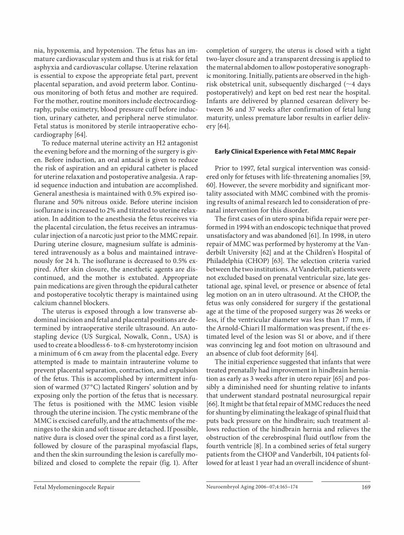
The uterus is exposed through a low transverse ab-dominal incision and fetal and placental positions are de-termined by intraoperative sterile ultrasound. An auto-stapling device (US Surgical, Nowalk, Conn., USA) is used to create a bloodless 6- to 8-cm hysterotomy incision a minimum of 6 cm away from the placental edge. Every attempted is made to maintain intrauterine volume to prevent placental separation, contraction, and expulsion of the fetus. This is accomplished by intermittent infu-sion of warmed (37 ° C) lactated Ringers’ solution and by exposing only the portion of the fetus that is necessary. The fetus is positioned with the MMC lesion visible through the uterine incision. The cystic membrane of the MMC is excised carefully, and the attachments of the me-ninges to the skin and soft tissue are detached. If possible, native dura is closed over the spinal cord as a first layer, followed by closure of the paraspinal myofascial flaps, and then the skin surrounding the lesion is carefully mo-bilized and closed to complete the repair ( fig. 1 ). After
completion of surgery, the uterus is closed with a tight two-layer closure and a transparent dressing is applied to the maternal abdomen to allow postoperative sonograph-ic monitoring. Initially, patients are observed in the high-risk obstetrical unit, subsequently discharged ( � 4 days postoperatively) and kept on bed rest near the hospital. Infants are delivered by planned cesarean delivery be-tween 36 and 37 weeks after confirmation of fetal lung maturity, unless premature labor results in earlier deliv-ery [64] .
Early Clinical Experience with Fetal MMC Repair
Prior to 1997, fetal surgical intervention was consid-ered only for fetuses with life-threatening anomalies [59, 60] . However, the severe morbidity and significant mor-tality associated with MMC combined with the promis-ing results of animal research led to consideration of pre-natal intervention for this disorder.
The first cases of in utero spina bifida repair were per-formed in 1994 with an endoscopic technique that proved unsatisfactory and was abandoned [61] . In 1998, in utero repair of MMC was performed by hysteromy at the Van-derbilt University [62] and at the Children’s Hospital of Philadelphia (CHOP) [63] . The selection criteria varied between the two institutions. At Vanderbilt, patients were not excluded based on prenatal ventricular size, late ges-tational age, spinal level, or presence or absence of fetal leg motion on an in utero ultrasound. At the CHOP, the fetus was only considered for surgery if the gestational age at the time of the proposed surgery was 26 weeks or less, if the ventricular diameter was less than 17 mm, if the Arnold-Chiari II malformation was present, if the es-timated level of the lesion was S1 or above, and if there was convincing leg and foot motion on ultrasound and an absence of club foot deformity [64] .
The initial experience suggested that infants that were treated prenatally had improvement in hindbrain hernia-tion as early as 3 weeks after in utero repair [65] and pos-sibly a diminished need for shunting relative to infants that underwent standard postnatal neurosurgical repair [66] . It might be that fetal repair of MMC reduces the need for shunting by eliminating the leakage of spinal fluid that puts back pressure on the hindbrain; such treatment al-lows reduction of the hindbrain hernia and relieves the obstruction of the cerebrospinal fluid outflow from the fourth ventricle [8] . In a combined series of fetal surgery patients from the CHOP and Vanderbilt, 104 patients fol-lowed for at least 1 year had an overall incidence of shunt-

Danzer/Flake
Neuroembryol Aging 2006–07;4:165–174170
ing of 54% [67] , compared with a predicted overall shunt rate of 84% based on 297 historical controls followed up at the Spina Bifida Clinic at the CHOP between 1983 and 2000 [68] . Although long-term follow-up is necessary, the ramifications of these preliminary observations and out-comes are significant. After fetal MMC repair, ascent of the hindbrain and improved cerebrospinal fluid dynam-ics may reduce hydrocephalus and avert the need and morbidity of ventriculoperitoneal shunts [64–66, 69, 70] . With a more normal location of the hindbrain, the symp-tomatic sequelae and need for subsequent surgery should be reduced. In the case of lower lumbar and sacral lesions in which less impairment in lower extremity function may be predicted, normalizing hindbrain position and minimizing the need for postnatal ventriculoperitoneal shunt placement may be the primary indication for fetal intervention. Indeed, neurodevelopmental outcome stud-ies of toddlers that underwent fetal MMC repair are prom-ising, suggesting that fetal spina bifida repair might im-prove neurocognitive and developmental skills by de-creasing the need for ventriculoperitoneal shunting [71] .
Any benefit in the lower extremity function or sphinc-ter continence has been very difficult to demonstrate. It has been recently reported that children with postnatally repaired MMC have a level of neurological function that correlates very well with the bony level of the defect [72, 73] . In contrast to the Vanderbilt experience in late ges-tational fetuses (25–30 weeks) [74] , lower extremity neu-romotor outcome at birth was significantly improved in newborns that underwent fetal MMC before 26 weeks of gestation at the CHOP [64] . The reason for this difference may reflect patient selection, careful evaluation for evi-dence of intact neurological function before surgery, and younger gestational age at which the fetal repair occurred. In addition, recent reevaluation of children that under-went fetal MMC repair at the CHOP with a mean age of 40 months showed that the initial improvement in lower extremity function at birth not only persisted into pre-school age, but also that fetal MMC repair results in a significant improvement of ambulatory potential (i.e. in-dependent walking) and a reduction in the incidence of club foot deformity [75] . The impact of fetal MMC repair
a b
c d
e
Fig. 1. Fetal MMC repair procedure. A hysterotomy is made using the uterine-sta-pling device ( a ) and the defect is positioned into the operative field ( b ). c The MMC sac is carefully resected and the placode is placed back into the spinal canal. The na-tive dura is closed over the spinal cord ( d ) and the paraspinal muscles and skin are closed in a multilayered fashion ( e ).

Fetal Myelomeningocele Repair Neuroembryol Aging 2006–07;4:165–174 171
on bowel and bladder continence remains unknown, but is currently under critical evaluation [Danzer, unpubl. data].
Despite these promising findings, there is already con-cern that some of the early benefit of fetal MMC repair may be at risk. Virtually all of the postnatal lumbosacral MRI studies of these patients suggest tethering, and re-cently some of the patients have developed symptomatic epidermoid inclusion cysts which have required repeat surgery [76] . It is unclear at this point whether this is a problem unique to fetal closure or simply that it is being detected during routine surveillance after fetal surgery.
Although long-term follow-up of these patients into adulthood is necessary, the implications of the short-term outcomes after fetal MMC repair are significant, if not without controversy [77] . The potential morbidity and mortality (perinatal mortality at the CHOP due to ex-treme prematurity is 7%, 4 out of 58) resulting from fetal surgery are real, and it is unclear whether the benefits in terms of shunt avoidance, reversal of hindbrain hernia-tion, and improvement of ambulatory potential will be sustained. The optimal timing of surgery is unclear, as is the optimal type of closure. Most uncertain of all are the appropriate criteria by which patients should be selected for fetal MMC repair. In addition to the above-mentioned risk for the fetus, maternal-fetal surgery has postsurgical and future risks for the maternal patients. Potential ma-ternal risks are surgical bleeding, infection (wound, am-niotic cavity), preterm rupture of membranes, preterm labor and delivery, medication side effects (tocolysis), an-esthesia complications (general, regional), prolonged hospitalization, impaired fertility, repeat cesarean deliv-eries, and death [60, 78] . With respect to fetal neurosurgi-cal intervention for MMC no maternal deaths have oc-curred and no patient experienced hysterotomy dehis-cence or rupture. No data have been presented to suggest diminished fertility in any of the maternal patients that underwent fetal MMC.
Management of MMC Study
Due to the lack of a control group of children with MMC who did not undergo prenatal repair, the initial clinical results of in utero repair of spina bifida lesions have been compared to historical controls. Comparison between infants with spina bifida who were treated in utero and historical controls is however subject to sub-stantial bias. Infants treated prenatally represent a highly selected subset of affected individuals. Additionally, the
medical management of such infants might differ from that of historical controls for reasons unrelated to the in utero repair. Taken together, it is currently unknown whether fetal surgery for MMC is truly beneficial com-pared to standard postnatal care. For these reasons, a Na-tional Institute of Health-sponsored multicenter, pro-spective, randomized, clinical trial, comparing outcome after in utero and postnatal surgery for MMC is current-ly underway. The study centers are the CHOP, the Uni-versity of California – San Francisco, and Vanderbilt University Medical Center, along with an independent Data and Study Coordinating Center at George Washing-ton University Biostatistics Center. The name of the trial is the Management of Myelomeningocele Study (MOMS).
Briefly, pregnant women who receive a prenatal diag-nosis of MMC between 16 and 25 weeks’ gestation will be referred to a central screening center. The eligibility and exclusion criteria for the MOMS trial are summarized in table 1 . Eligible patients who consent to participate in the
Table 1. Inclusion and exclusion selection criteria for MOMS tri-al patients
Inclusion criteriaMaternal age ≥18 yearsGestational age at randomization 19 weeks, 0 days to 25 weeks, 6 days Normal karyotypeS1 level lesion or higherConfirmed Arnold-Chiari II malformation on prenatal US and MRI
Exclusion criteriaNonresident of the United StatesMultifactorial pregnancyInsulin-dependent pregestational diabetesAdditional fetal anomalies unrelated to MMCFetal kyphosis ≥30°History of incompetent cervix and/or short cervix <20 mm by ultrasound scanPlacenta previaOther serious maternal medical conditionObesity defined by body mass index of 35 or greaterPrevious spontaneous singleton delivery at <37 weeks’gestationMaternal-fetal Rh isoimmunizationPositive maternal human immunodeficiency virus orhepatitis B or known hepatitis C positivityNo support person to stay with the pregnant women at the centerUterine anomalyPsychosocial limitationsInability to comply with travel and follow-up protocols

Danzer/Flake
Neuroembryol Aging 2006–07;4:165–174172
References
1 Danzer E, Rintoul NE, Crombleholme TM, Adzick NS: Pathophysiology of neural tube defects; in Pollin R, Fox WW, Abman SH (eds): Fetal and Neonatal Physiology, ed 3. Philadelphia, Saunders, 2003, pp 1772–1785.
2 Dias MS, McLone DG: Hydrocephalus in the child with dysraphism. Neurosurg Clin N Am 1993; 4: 715–726.
3 Caldarelli M, DiRocco C, LaMarca F: Shunt complications in the first postoperative year in children with myelomeningocele. Childs Nerv Syst 1996; 12: 748–754.
4 Bell JE, Gordon A, Maloney AF: The associa-tion of hydrocephalus and Arnold-Chiari malformation with spina bifida in the fetus. Neuropathol Appl Neurobiol 1980; 6: 29–39.
5 Gilbert JN, Jones KL, Rorke LB, Chernoff GF, James HE: Central nervous system anomalies associated with myelomeningo-cele, hydrocephalus, and the Arnold-Chiari malformation: reappraisal of theories re-garding the pathogenesis of posterior neural tube closure defects. Neurosurgery 1986; 18: 559–564.
6 Cochrane DD, Adderley R, White CP, Nor-man M, Steinbok P: Apnea in patients with myelomeningocele. Pediatr Neurosurg 1990; 16: 232–239.
7 Oaks W, Gaskill S: Symptomatic Chiari mal-formations in childhood; in Park T (ed): Spi-nal Dysraphism. Boston, Blackwell Scientif-ic Publications, 1992, pp 104–125.
8 McLone DG, Knepper PA: The cause of Chi-ari II malformation: a unified theory. Pediatr Neurosci 1989; 15: 1–12.
9 Mitchell LE, Adzick NS, Melchionne J, Pasquariello PS, Sutton LN, Whitehead AS: Spina bifida. Lancet 2004; 364: 1885–1895.
10 Botto LD, Moore CA, Khoury MJ, Erickson JD: Neural-tube defects. N Engl J Med 1999; 341: 1509–1519.
11 Manning SM, Jennings R, Madsen JR: Patho-physiology, prevention, and potential treat-ment of neural tube defects. Ment Retard Dev Disabil Res Rev 2000; 6: 6–14.
12 Scott JM, Weir DG, Molloy A, McPartlin J, Daly L, Kirke P: Folic acid metabolism and mechanisms of neural tube defects. Ciba Found Symp 1994; 181: 180–187.
13 Czeizel AE, Dudas I: Prevention of the first occurrence of neural-tube defects by peri-conceptional vitamin supplementation. N Engl J Med 1992; 327: 1832–1835.
14 MRC Vitamin Study Research Group: Pre-vention of neural tube defects: results of the medical research council vitamin study. Lancet 1991; 338: 131–137.
15 Copp AJ, Bernfield M: Etiology and patho-genesis of human neural tube defects: in-sights from mouse models. Curr Opin Pedi-atr 1994; 6: 624–631.
16 Copp AJ, Greene ND, Murdoch JN: The ge-netic basis of mammalian neurulation. Nat Rev Genet 2003; 4: 784–793.
17 Copp AJ: Prevention of neural tube defects: vitamins, enzymes, and genes. Curr Opin Neurol 1998; 11: 97–102.
18 Harris MJ, Juriloff DM: Genetic landmarks for defects in mouse neural tube closure. Ter-atology 1997; 56: 177–187.
19 Melvin EC, George TM, Worley G, Franklin A, Mackey J, Viles K, Shah N, Drake CR, En-terline DS, Mclone D, Nye J, Oakes WJ, Mclaughlin C, Walker ML, Peterson P, Brei T, Buran C, Aben J, Ohm B, Bermans I, Qumsiyeh M, Vance, J, Pericak-Vance MA, Speer MC: Genetic studies in neural tube de-fects. NTD Collaborative Group. Pediatr Neurosurg 2000; 32: 1–9.
20 Edmonds LD, James LM: Temporal trends in the prevalence of congenital malformations at birth based on the birth defects monitor-ing program, United States, 1979–1987. MMWR Morb Mortal Wkly Rep 1990; 39: 19–23.
21 Lary JM, Edmonds LD: Prevalence of spina bifida at birth – United States, 1983–1990: a comparison to two surveillance systems. MMWR Morb Mortal Wkly Rep 1996; 45: 15–26.
22 Roberts HE, Moore CA, Cragan JD, Fernhoff PM, Khoury MJ: Impact of prenatal diagno-sis on the birth prevalence of neural tube de-fects, Atlanta 1990–1991. Pediatrics 1995; 96: 880–883.
trial will be assigned to one of the three centers, and ran-domized to either fetal repair or cesarean delivery after demonstration of lung maturity. Both, in utero and stan-dard postnatal closure of the defect will be performed at the study center by one of the three study neurosurgeons. Patients who randomize to the fetal surgery arm will stay at the study center and undergo the in utero closure. The details of closure are left largely to the discretion of the neurosurgeon based on the available tissue, but artificial graft material will not be used, except for skin closure. Relaxing incisions may be required in some instances for skin closure. Tocolytic therapy has been standardized and will be supervised by the study obstetricians. Patients will stay in the vicinity of the study center until they are medically stable.
The primary objective of the trial is to determine if intrauterine repair of fetal MMC at 19–25 weeks’ gesta-tion improves outcome, as measured by death or the need for shunting by 1 year of life compared with postnatal MMC repair. Other objectives of the study are to deter-
mine if fetal MMC repair improves the degree of the Ar-nold-Chiari II malformation and neurological outcome as tested by neuroimaging, neuromotor function analy-sis, cognitive testing, and neurodevelopmental status at 12 and 30 months of life. Neonatal morbidity and the need for postnatal surgical interventions will be record-ed. Finally, the long-term psychological and reproductive consequences for mothers who undergo intrauterine re-pair of MMC will be compared with those in the postna-tal repair group. In summary, the trial should determine whether fetal intervention offers improved outcome with a reasonable quality of life for MMC children. Addition-al information about this trial can be obtained from the study website (www. spinabifidamoms.com).
Acknowledgment
This work was supported in part by Michael and Katherine Mulligan through the Sean Michael Mulligan Research Fund.
Fetal Myelomeningocele Repair Neuroembryol Aging 2006–07;4:165–174 173
23 Velie EM, Shaw GM: Impact of prenatal di-agnosis and elective termination on preva-lence and risk estimates of neural tube de-fects in California, 1989–1991. Am J Epidemiol 1996; 144: 473–479.
24 Worly G, Schuster JM, Oakes WJ: Survival at 5 years of a cohort of newborn infants with myelomeningocele. Dev Med Child Neurol 1996; 38: 816–822.
25 Goodman RA: Economic burden of spina bi-fida – United States, 1980–1990. MMWR Morb Mortal Wkly Rep 1989; 38: 264–267.
26 Drugan A, Weissman A, Evans MI: Screen-ing for neural tube defects. Clin Perinatol 2001; 28: 279–281.
27 Brock DJ, Sutcliffe RG: Alpha-fetoprotein in antenatal diagnosis of anencephaly and spi-na bifida. Lancet 1972;ii:197–199.
28 Loft AG: Determination of amniotic f luid acetylcholinesterase activity in the antenatal diagnosis of foetal malformations: the first ten years. J Clin Chem Clin Biochem 1990; 28: 893–911.
29 Simon EM, Pollock AN: Prenatal and post-natal imaging of spinal dysraphism. Semin Roentgenol 2004; 39: 182–196.
30 Patten B: Embryological stages in the estab-lishing of myeloschisis with spina bifida. Am J Anat 1953; 93: 365–395.
31 Osaka K, Tanimura T, Hirayama A, Matsu-moto S: Myelomeningocele before birth. J Neurosurg 1978; 49: 711–724.
32 Hutchins GM, Meuli M, Meuli-Simmen C, Jordan MA, Heffez DS, Blakemore KJ: Ac-quired spinal cord injury in human fetuses with myelomeningocele. Pediatr Pathol Lab Med 1996; 16: 701–712.
33 Meuli M, Meuli-Simmen C, Hutchins GM, Seller MJ, Harrison MR, Adzick NS: The spi-nal cord lesion in human fetuses with myelo-meningocele: implications for fetal surgery. J Pediatr Surg 1997; 31: 448–452.
34 Korenromp MJ, Van Good JD, Bruinese HW, Kriek R: Early fetal movements in myelome-ningocele. Lancet 1986;i:917–918.
35 Sival DA, Begeer JH, Staal-Schreinemachers AL, Vos-Niel JM, Beekhuis JR, Prechtl HF: Perinatal motor behavior and neurological outcome in spina bifida aperta. Early Hum Dev 1997; 50: 27–37.
36 Sival DA, Brouwer OF, Bruggink JL, Vies JS, Staal-Schreinemachers AL, Sollie KM, Sauer PJ, Bos AF: Movement analysis in neonates with spina bifida aperta. Early Hum Dev 2006; 82: 227–234.
37 Shurtleff DB, Luthy DA, Nyberg DA, Bene-detti TJ, Mack LA: Meningomyelocele: man-agement in utero and post partum. Ciba Found Symp 1994; 181: 270–280.
38 Luthy DA, Wardinsky T, Shurtleff DB, Hol-lenbach KA, Hickok DE, Nyberg DA, Bene-detti TJ: Cesarean section before the onset of labor and subsequent motor function in in-fants with myelomeningocele diagnosed an-tenatally. N Engl J Med 1991; 324: 662–666.
39 Merrill DC, Goodwin P, Burson JM, Sato Y, Williamson R, Weiner CP: The optimal route of delivery for fetal myelomeningocele. Am J Obstet Gynecol 1998; 179: 235–240.
40 Cochrane D, Aronyk K, Sawatzky B, Wilson D, Steinbok P: The effects of labor and deliv-ery on spinal cord function and ambulation in patients with myelomeningocele. Child Nerv Syst 1991; 7: 312–315.
41 Fleming A, Copp AJ: Embryonic folate me-tabolism and mouse neural tube defects. Sci-ence 1998; 280: 2107–2109.
42 Copp AJ, Brook FA, Roberts HJ: A cell-type-specific abnormality of cell proliferation in mutant (curly tail) mouse embryos develop-ing spinal neural tube defects. Development 1988; 104: 285–295.
43 Van Straaten HW, Copp AJ: Curly tail: a 50-year history of the mouse spina bifida model. Anat Embryol 2001; 203: 225–237.
44 Selcuki M, Manning S, Bernfield M: The curly tail mouse model of human neural tube defects demonstrates normal spinal cord dif-ferentiation at the level of the meningomy-elocele: implications for fetal surgery. Childs Nerv Syst 2001; 17: 19–23.
45 Duru S, Ceylan S, Ceylan S: Comparative ef-fects of valproic acid sodium for Chiari-like malformation at 9 and 10 days of gestation in the rat. Childs Nerv Syst 2001; 17: 399–404.
46 Ehlers K, Elmazar MA, Nau H: Methionine reduces the valproic acid-induced spina bi-fida rate in mice without altering valproic acid kinetics. J Nutr 1996; 126: 67–75.
47 Ehlers K, Struje H, Merker HJ, Nau H: Spina bifida aperta induced by valproic acid and by all- trans -retinoic acid in the mouse: distinct differences in morphology and periods of sensitivity. Teratology 1992; 46: 117–130.
48 Michejda M: Intrauterine treatment of spina bifida. Primate model. Z Kinderchir 1984; 39: 259–261.
49 Heffez DS, Aryanpur J, Rotellini NA, Hutchins GM, Freeman JM: Intrauterine re-pair of experimental surgically created dys-raphism. Neurosurgery 1993; 32: 1005–1010.
50 Heffez DS, Aryanpur J, Hutchins GM, Free-man JM: The paralysis associated with my-elomeningocele: clinical and experimental data implicating a preventable spinal cord injury. Neurosurgery 1990; 26: 987–992.
51 Meuli M, Meuli-Simmen C, Yingling CD, Hoffman KB, Harrison MR, Adzick NS: A new model of myelomeningocele: studies in the fetal lamb. Surg Forum 1994; 45: 587–589.
52 Meuli M, Meuli-Simmen C, Hutchins GM, Yingling CD, Hoffman KM, Harrison MR, Adzick NS: In utero surgery rescues neuro-logic function at birth in sheep with spina bifida. Nat Med 1995; 1: 342–347.
53 Meuli M, Meuli-Simmen C, Hutchins GM, Yingling CD, Hutchins GM, Timmel GB, Harrison MR, Adzick NS: In utero repair of experimental myelomeningocele spares neu-rologic function at birth. J Pediatr Surg 1996; 31: 397–402.
54 Paek BW, Farmer DL, Wilkinson CC, Alba-nese CT, Peacook W, Harrison MR, Jennings RW: Hindbrain herniation develops in surgi-cally created myelomeningocele but is absent after repair in fetal lambs. Am J Obstet Gy-necol 2000; 183: 1119–1123.
55 Bouchard S, Davey MG, Rintoul NE, Walsh DS, Rorke LB, Adzick NS: Correction of hindbrain herniation and anatomy of the vermis following in utero repair of myelome-ningocele in sheep. J Pediatr Surg 2003; 38: 451–458.
56 Danzer E, Schwarz U, Wehrli S, Radu A, Adzick NS, Flake AW: Retinoic acid induced myelomeningocele in fetal rats: character-ization by histopathological analysis and magnetic resonance imaging. Exp Neurol 2005; 194: 467–475.
57 Danzer E, Kiddoo DA, Redden RA, Robin-son L, Radu A, Zderic SA, Doolin EJ, Adzick NS, Flake AW: Structural and functional characterization of bladder smooth muscle in fetal rats with retinoic acid induced my-elomeningocele. Am J Physiol Renal Physiol 2007; 292: 197–206.
58 Danzer E, Radu A, Volpe MA, Adzick NS, Flake AW: Normal neuromuscular develop-ment of the lower gastrointestinal tract and anorectum in fetal rats with retinoic acid in-duced myelomeningocele – implications for fetal surgery. J Am Coll Surg 2006; 203:S36.
59 Adzick NS, Harrison MR: Fetal surgical therapy. Lancet 1994; 343: 897–902.
60 Danzer E, Sydorak RM, Harrison MR, Alba-nese CT: Minimal access fetal surgery. Eur J Obstet Gynecol Reprod Biol 2003; 108: 3–13.
61 Bruner J, Tulipan N, Richards W: Endoscop-ic coverage of fetal open myelomeningocele in utero. Am J Obstet Gynecol 1997; 176: 256–257.
62 Tulipan N, Bruner JP: Myelomeningocele re-pair in utero: a report of three cases. Pediatr Neurosurg 1998; 28: 177–180.
63 Adzick NS, Sutton LN, Crombleholme TM, Flake AW: Successful fetal surgery for spina bifida. Lancet 1998; 352: 1675–1676.
64 Johnson MP, Sutton LN Rintoul N, Cromble-holme TM, Flake AW, Howell LJ, Hedrick HL, Wilson RD, Adzick NS: Fetal myelome-ningocele repair: short-term clinical out-comes. Am J Obstet Gynecol 2003; 189: 482–487.
65 Sutton LN, Adzick NS, Bilaniuk LT, Johnson MP, Crombleholme TM, Flake AW: Im-provement in hindbrain herniation demon-strated by serial fetal magnetic resonance imaging following fetal surgery for myelo-meningocele. JAMA 1999; 282: 1826–1831.
66 Bruner JP, Tulipan N, Paschall RL, Boehm FH, Walsh WF, Silvia SR, Hernanz-Schul-man M, Lowe LH, Reed GW: Fetal surgery for myelomeningocele and the incidenceof shunt-dependent hydrocephalus. JAMA 1999; 282: 1819–1825.

Danzer/Flake
Neuroembryol Aging 2006–07;4:165–174174
67 Tulipan N, Sutton LN, Bruner JP, Cohen BM, Johnson M, Adzick NS: The effect of intra-uterine myelomeningocele repair on the in-cidence of shunt-dependent hydrocephalus. Pediatr Neurosurg 2003; 38: 27–33.
68 Rintoul NE, Sutton LN, Hubbard AM, Co-hen B, Melchionni J, Pasquariello PS, Adzick NS: A new look at myelomeningoceles: func-tional level, vertebral level, shunting, and the implications for fetal intervention. Pediat-rics 2002; 109: 409–413.
69 Danzer E, Johnson MP, Wilson RD, Flake AW, Hedrick HL, Sutton LN, Adzick NS: fe-tal head biometry following in-utero repair of myelomeningocele. Ultrasound Obstet Gynecol 2004; 24: 606–611.
70 Danzer E, Johnson MP, Bebbington M, Si-mon EM, Wilson RD, Bilaniuk LT, Sutton LN, Adzick NS: Fetal head biometry assessed by fetal magnetic resonance imaging follow-ing in utero myelomeningocele repair. Fetal Diagn Ther 2007; 22: 1–6.
71 Johnson MP, Gerdes M, Rintoul N, Pasquar-iello P, Melchionni J, Sutton LN, Adzick NS: Maternal-fetal surgery for myelomeningo-cele: neurodevelopmental outcomes at 2 years of age. Am J Obstet Gynecol 2006; 194: 1145–1150.
72 Kollias SS, Goldstein RB, Cogen PH, Filly RA: Prenatally detected myelomeningoceles: sonographic accuracy in estimation of the spinal levels. Radiology 1992; 185: 109–112.
73 Biggio JR, Owen J, Wenstrom KD, Oakes WJ: Can prenatal ultrasound findings predict ambulatory status in fetuses with open spina bifida? Am J Obstet Gynecol 2001; 185: 1016–1020.
74 Tubbs S, Chambers MR, Smyth MD, Barto-lucci AA, Bruner JP, Tulipan N, Oakes WJ: Late gestational intrauterine myelomenin-gocele repair does not improve lower ex-tremity function. Pediatr Neurosurg 2003; 38: 128–132.
75 Danzer E, Adzick NS, Gerdes M, Bebbington MW, Sutton LN, Melchionni J, Johnson MP: Lower extremity neuromotor function fol-lowing in utero myelomeningocele repair. Am J Obstet Gynecol 2006; 195:S22.
76 Mazzola C, Albright AL, Sutton LN, Tuite GF, Hamilton RL, Pollack IF: Dermoid in-clusion cysts and early spinal cord tethering after fetal surgery for myelomeningocele. N Engl J Med 2002; 347: 256–259.
77 Simpson JL: Fetal surgery for myelomenin-gocele: promise, progress, and problems. JAMA 1999; 282: 1873–1874.
78 Wilson RD, Johnson MP, Flake AW, Crom-bleholme TM, Hedrick HL, Wilson J, Adzick NS: Reproductive outcomes after pregnancy complicated by maternal-fetal surgery. Am J Obstet Gynecol 2004; 191: 1430–1436.