3D-Printed, Dual Crosslinked and Sterile Aerogel Scaffolds for ...
Upload
khangminh22Category
view
3download
0
REVIEW Open Access
A critical assessment of the “sterile womb”and “in utero colonization” hypotheses:implications for research on the pioneerinfant microbiomeMaria Elisa Perez-Muñoz1, Marie-Claire Arrieta2,3, Amanda E. Ramer-Tait4 and Jens Walter1,5*
Abstract
After more than a century of active research, the notion that the human fetal environment is sterile and that theneonate’s microbiome is acquired during and after birth was an accepted dogma. However, recent studies usingmolecular techniques suggest bacterial communities in the placenta, amniotic fluid, and meconium from healthypregnancies. These findings have led many scientists to challenge the “sterile womb paradigm” and propose thatmicrobiome acquisition instead begins in utero, an idea that would fundamentally change our understanding ofgut microbiota acquisition and its role in human development. In this review, we provide a critical assessment ofthe evidence supporting these two opposing hypotheses, specifically as it relates to (i) anatomical, immunological,and physiological characteristics of the placenta and fetus; (ii) the research methods currently used to study microbialpopulations in the intrauterine environment; (iii) the fecal microbiome during the first days of life; and (iv) the generationof axenic animals and humans. Based on this analysis, we argue that the evidence in support of the “in utero colonizationhypothesis” is extremely weak as it is founded almost entirely on studies that (i) used molecular approaches with aninsufficient detection limit to study “low-biomass” microbial populations, (ii) lacked appropriate controls for contamination,and (iii) failed to provide evidence of bacterial viability. Most importantly, the ability to reliably derive axenic animals viacesarean sections strongly supports sterility of the fetal environment in mammals. We conclude that current scientificevidence does not support the existence of microbiomes within the healthy fetal milieu, which has implications for thedevelopment of clinical practices that prevent microbiome perturbations after birth and the establishment of futureresearch priorities.
Keywords: Sterile womb, In utero colonization, Microbiome, Placenta, Contamination, Axenic animals
BackgroundThe gastrointestinal tract of humans is colonized by adense microbial community that has co-evolved with itshost to become a vital component of our biology. Thehost-microbiome interrelationship is therefore consid-ered a mutualistic symbiosis, with the human body pro-viding sustenance and an adequate physical environmentfor the microbial populations, while the microbes
execute essential functions, such as aiding in immunesystem development and providing defense againstenteric infections [1].Research in both animal models and humans suggests
that the process of microbial colonization is especiallysignificant during early life, as this period constitutes acritical window for immunological and physiological de-velopment [2, 3]. Given the importance of microbialsymbionts to their host’s development and survival,mechanisms must be in place to facilitate their reliabletransmission [4]. Symbiont transmission has been well-established in many host-microbial symbioses, especiallyin invertebrates (i.e., insects, nematodes, and the Hawaiiansquid Euprymna scolopes) where it ranges from being
* Correspondence: [email protected] of Agriculture, Food and Nutritional Sciences, 4-126 Li Ka ShingCentre for Health Research Innovation, University of Alberta, Edmonton,Alberta T6G 2E1, Canada5Department of Biological Sciences, 7-142 Katz Group Centre, University ofAlberta, Edmonton, Alberta T6G 2E1, CanadaFull list of author information is available at the end of the article
© The Author(s). 2017 Open Access This article is distributed under the terms of the Creative Commons Attribution 4.0International License (http://creativecommons.org/licenses/by/4.0/), which permits unrestricted use, distribution, andreproduction in any medium, provided you give appropriate credit to the original author(s) and the source, provide a link tothe Creative Commons license, and indicate if changes were made. The Creative Commons Public Domain Dedication waiver(http://creativecommons.org/publicdomain/zero/1.0/) applies to the data made available in this article, unless otherwise stated.
Perez-Muñoz et al. Microbiome (2017) 5:48 DOI 10.1186/s40168-017-0268-4

strictly vertical (maternal) to horizontal (transmission be-tween members of the same species or the environment)[5]. In contrast to these models of symbiosis, the modes oftransmission for the more complex microbiomes ofhumans and other vertebrates are more intricate and in-completely understood. Considering the importance ofthe pioneer infant microbiota for human developmentand biology, it is essential that we elucidate the exactmechanisms by which this community is acquired,the time-points when colonization events occur, andthe endogenous and exogenous factors that influencethese events.The degree of sterility of the fetal environment and
the possibility of in utero microbiome transfer have beendebated for almost 150 years [6]. In the second half ofthe last century, the field reached a consensus that thefetus was maintained in a sterile state [7]. According tothis concept, which has been referred to as the sterilewomb paradigm [4], microbes are acquired both verti-cally (from the mother) and horizontally (from otherhumans or the environment) during and after birth.However, there is now a multitude of recent studiesemploying modern sequencing technologies that havechallenged the traditional view of human microbiomeacquisition. These studies propose that neither the fetus,the placenta, nor the amniotic fluid are sterile, and thatacquisition and colonization of the human gastrointes-tinal tract begins in utero [8–10]. If this “in uterocolonization hypothesis” proves correct, there would bemajor repercussions on our understanding of the estab-lishment of the pioneer human microbiome, its role inhuman health and the role of environmental, lifestyle,and clinical factors that affect its assembly and function.This concept would also have significant implications onhow we view the fundamental aspects of host-microbialsymbiosis in humans as well as clinical practices such ascesarean sections (C-sections), which are currentlythought to disrupt transmission of microbes [11].In this review, we first describe the scientific evidence
in support of both the “sterile womb” and in uterocolonization hypotheses. We then compare and criticallyassess the two opposing ideas and discuss the limitationsof the research supporting each of them. We especiallyput effort into the historic perspective on this topic, withequal focus on both the older literature and more recentstudies. Based on this assessment, we conclude thatmost of the evidence is in support of the “sterile wombhypothesis,” and we discuss the implications for clinicalpractice and future research.
The traditional view: the sterile womb paradigmMost studies that established the sterile womb paradigmdate back to research that employed traditional culture-based methods and microscopy, which despite their
limitations are still considered valid today. As early as1885, Theodor Escherich described the meconium (theearliest stool from an infant) to be free of viable bacteria[7], suggesting that the human fetus develops within asterile environment (Fig. 1a). Later, two additional, inde-pendent studies conducted in 1927 and 1934 (n = 100and n = 50, respectively) using sterile diapers for collec-tion both found 62% of meconium samples from healthypregnancies to be negative for bacteria by aerobic andanaerobic culture [12, 13]. The observed time frame formeconium expulsion ranged from a few minutes to 26 hafter birth, with 50% of the meconiums expelled 5 to10 h post birth [13]. Interestingly, the 1934 studyreported a positive correlation between the detection ofbacteria and time elapsed between birth and meconiumpassage [13]. Some recent studies utilizing molecular ap-proaches also suggest that the majority of meconiumsamples from healthy pregnancies are negative for bac-teria (Table 1). In one study [14], meconium samplesfrom 66% (n = 15) of newborns evaluated showedevidence of bacteria based on fluorescence in situhybridization (FISH), while only 7% were positive byPCR. Interestingly, four out of the five samples that weredelivered within 500 min of birth showed no detectablebacteria by FISH, supporting the association betweentime of meconium passage and bacterial detection.Overall, these studies suggest that early meconium har-bors no detectable bacteria, while later samples do, indi-cating the need to account for time elapsed post-birthwhen investigating bacterial presence in these samples.Seminal work by Harris and Brown [15] significantly
shaped the concept of a sterile amniotic cavity by inves-tigating the presence of cultivable bacteria in the amni-otic fluid of women undergoing C-sections. All womenin labor for less than 6 h (n = 28) tested negative for bac-teria, while positive cultures were obtained from thosein labor for more than 6 h (n = 22). Subsequent culture-based studies of amniotic fluid confirmed sterility duringa healthy pregnancy [16, 17]. Specifically, bacterial cul-ture of amniotic fluid samples obtained by abdominalpuncture (n = 44) or by transcervical aspiration (n = 8)showed no growth in 96 and 50% of the cases, respect-ively [16]. Because fetal infection was absent, andbecause the positive samples were monocultures ofStaphylococcus albus, Streptococcus, or yeast, the authorconcluded that any colonies detected resulted from con-tamination during collection and that his results upheldthe notion of sterile amniotic fluid. Complementary tothis finding, an independent investigation found an asso-ciation between the presence of Mycoplasma hominis inthe amniotic fluid and the incidence of spontaneousabortions, thereby reinforcing the notion that the presenceof bacteria in the amniotic fluid should be considered aninfection [17]. More recent culture-based studies reported
Perez-Muñoz et al. Microbiome (2017) 5:48 Page 2 of 19

over 90% of amniotic fluid samples tested to be sterile[18–20]. The occasional presence of a bacteria was inter-preted to be due to subclinical (without maternal or fetalmorbidity) [18, 19] or clinical infections [20], the lattersupported by the fact that all positive cases presentedsymptoms of post-partum infection and pre-labor ruptureof membranes [20]. Subsequent research has found thatthe amniotic fluid, meconium, and placental tissue containno detectable bacteria under healthy progression of preg-nancy [21–25]. When bacteria have been detected in thefetal environment, those results were obtained under cir-cumstances where a predisposition to infection or preg-nancy complications was suspected [21–25].Because the overwhelming majority of research consist-
ently supported the sterile womb paradigm in healthy preg-nancies, later investigations into the microbiology ofamniotic fluid were mostly limited to cases of pregnancycomplications. These studies included cases of pretermlabor (where 15% of samples were positive, n = 166) [26],preeclampsia (9.6% positive samples, n = 62) [27], small-for-gestational-age pregnancies (6% positive samples, n = 52)
[28], preterm pre-labor membrane rupture (50% positivesamples, n = 204) [29], and neonatal sepsis (57% positivesamples, n = 36) [30]. Despite complications, 68% of thesamples still tested negative for bacteria (as measured bycultures, polymerase chain reaction (PCR), sequencingtechnologies, or a combination of these methods) ([22–26],as reviewed by DiGiulio [31]).Because the placenta is generally considered a barrier
to protect the fetus from microbial pathogens that in-vade the blood stream of the mother [32, 33], studiesdirectly aimed at the determination of a “placentalmicrobiome” in healthy pregnancies are scarce. Instead,microbial studies were, for the most part, focused oncomplications of pregnancy or the birth process, such asspontaneous abortions (21 and 24% positive aerobic andanaerobic cultures, respectively; n = 47) [34], or sus-pected or confirmed cases of infant infection (33% posi-tive cases, n = 33 and 32% positive cases, n = 72, asreported by [35, 36], respectively). However, even duringthese complications, the placentas were often found tonot contain viable bacteria. In particular, Aquino and
Fig. 1 Schematic representation of the opposing concepts by which human microbiota is acquired early in life. a In the sterile womb paradigm,the placenta, amniotic fluid, and fetal gut remain sterile during a healthy pregnancy, and the early microbiome is acquired during and after birth.Accordingly, the gut microbiota of infants born vaginally resemble the microbiota of the mother’s vagina, while the microbiota of infants born bycesarean section are similar to the mother’s skin microbiota. b The “in utero colonization hypothesis” proposes that some microbial members ofthe infants’ gut microbiome are acquired before birth, probably via contact with a placental microbiome, which has been suggested to originatefrom the mother’s gut or oral microbiome
Perez-Muñoz et al. Microbiome (2017) 5:48 Page 3 of 19

Table
1Summaryof
recent
stud
ieson
themicrobiolog
yof
amnioticfluid,p
lacenta,andmecon
ium
Sampletype
Popu
latio
nMetho
dsSign
ificant
finding
sand/or
authors’conclusion
sReference
Placen
talm
embranes,umbilical
veno
usbloo
dTerm
andpreterm
vaginaland
elective
cesarean
deliveries,(preterm
deliveries
includ
epreg
nanciescomplicated
with
preeclam
psia,fetalgrow
threstrictio
n,or
prolon
gedlabo
r),de
liveriesthat
presen
tedPPRO
M(n=52)
FISH
usingge
neric
prob
esfor
16SrRNAge
nes
Bacteriawerede
tected
in70%
ofplacen
tas.
Autho
rsconclude
dpresen
ceof
bacteriais
common
inplacen
talm
embranes,
butinsufficien
tto
causepreterm
labo
ror
PPRO
M
[37]
Mecon
ium
passed
with
inthe
first2hof
life
Term
healthyne
wbo
rns(n=21)
Culture
metho
ds,G
ram
staining
,16SrDNAsequ
encing
Bacterialspe
cies
isolated
from
onesing
lemecon
ium
samples
variedfro
m1to
5.Enterococcus
faecaliswas
themost
abun
dant
speciesfoun
din
80%
ofthe
samples.Sam
ples
clusteredby
processin
gtim
e
[8]
Placen
talm
embranes
Full-term
andpreterm
vaginaland
cesarean
deliveries;preterm
deliveries
with
andwith
outPROM
(n=74)
Standard
PCRof
16SrRNAgene
andqu
antitativePC
Rforselected
bacteria
BacterialD
NAwas
detected
in30%
ofplacen
taltissueby
standard
PCR,while
43%
werepo
sitiveby
qPCR;14%
were
positiveby
both
metho
ds.N
obacterial
DNAwas
detected
inC-sectio
nde
liveries
atterm
,while50%of
term
vaginaldeliveries
werepo
sitive
[93]
Placen
taFull-term
vaginaland
cesarean
deliveriesfro
mpreg
nant
wom
enparticipatingin
diet
stud
y(n=34)
Aerob
icandanaerobiccultu
res.
PCRof
16srRNAge
neusing
genu
sandspeciesspecific
prim
ersforLactobacillus
and
bifid
obacteria
DNAwas
detected
in94%
ofsamples
byPC
R.Bacteriaof
interestwereno
tdetected
byculturemetho
ds
[84]
Mecon
ium
andfeces
Preterm
neon
ates
(n=23)
454pyrosequ
encing
BacterialD
NAwas
detected
in91%
ofsamples.Low
erge
stationalage
was
associated
with
lower
bacteriald
iversity,
butthereareno
differences
indiversity
betw
eenC-sectio
nandvaginally
delivered
infants
[98]
Amnioticfluid,p
lacentaand
mecon
ium
Electivecesarean
deliveriesof
healthy
mothersenrolledinprob
iotic
stud
y(n=43)
QuantitativePC
Rforselected
bacterialg
roup
s(Lactobacillus,
Bifidobacterium,Bacteroides,
Clostridium
leptum
grou
p)
Lactob
acillus
DNAwas
foun
din
100%
ofplacentas,Bifidobacterium
in41%,Bacteroides
in34%andClostridium
leptum
in31%.D
NA
forthe
selected
bacterialgroup
swas
foun
din43%of
amnioticfluidsamples
[51]
Mecon
ium
passed
betw
een
2–48
hafterbirth
Infantsbo
rnby
vaginalo
rcesarean
deliveriesfro
mdiabeticand
non-diabeticmothe
rs(n=23)
16SrRNAsequ
encing
using
Pacbio
RSsystem
Bacteriawerefoun
din
100%
ofsamples.
Diversity
islower
inmecon
ium
whe
ncomparedto
adults;highe
rin
infants
from
diabeticwhe
ncomparedto
non-diabeticmothe
rs
[99]
Mecon
ium
Health
yfull-term
deliveries(n=20)
Pyrosequ
encing
of16SrRNA
gene
Mecon
ium
microbiotadifferedfro
mthe
microbiotaof
feces,vagina,and
skin
from
adultsbu
twas
similarto
that
ofyoun
ginfant
feces.Mecon
ium
microbiotahas
anintrauterin
eorigin
andisinfluen
ced
bymaternalfactors.
[149]
Perez-Muñoz et al. Microbiome (2017) 5:48 Page 4 of 19

Table
1Summaryof
recent
stud
ieson
themicrobiolog
yof
amnioticfluid,p
lacenta,andmecon
ium
(Con
tinued)
Maternalfeces,m
econ
ium,b
aby’s
fecesat
different
timep
oints
Health
ymothers,full-term
pregnancies,
allinfantsexclusivelybreastfedforat
least2mon
ths(n=17)
Culture
metho
ds,PCRof
16s
rRNAge
nes,qP
CRusing
Bifidobacterium
species-specific
prim
ers
Bifidobacterium
specieswerefoun
din
all
newbo
rn/in
fant
samples.Vaginallydelivered
mother-infant
pairs
show
edmon
ophyletic
bifidob
acterialstrains,w
hileno
neof
the
strainsidentifiedfro
mC-sectionpairs
were
identifiedas
mon
ophyletic
[135]
Placen
talb
asalplates
Term
andpreterm
deliverieswith
and
withouth
istoryof
PROM,chorioam
nionitis,
groupBStreptococcusinfection,sexually
transmitted
infection,and/or
UTIs(n=195)
Histology
usingH&E
,Gram
staining
,hem
a3(m
odificatio
nof
Giemsa
stain)
andBrow
n-Hop
psmod
ificationof
Gram
stain
27%
ofplacen
tascontaine
dintracellular
bacteriainbasalplate.N
odifferencefound
intheincidenceofbacteriainchorioam
nion
itis,
PTB,or
groupBStreptococcusinfection.There
was
atwofoldriskincrease
forintracellular
bacteriainverypreterm
birth
[38]
Mecon
ium
passed
betw
een
birthand48
hafterbirth
Vaginalorcesareandeliveriesof
preterm
neon
ates
(n=52)
Iontorren
tsequ
encing
of16S
rRNAge
nes
67.3%
ofsamples
show
edam
plificatio
nof
the16SrRNAge
ne.G
estatio
nalage
hada
greaterinfluen
cethan
mod
eof
delivery
onmicrobialcommunity
structure.Mecon
ium
isindicativeof
amnioticfluidbacterial
commun
ities
[107]
Placen
taHealth
ypreg
nanciescomparedto
preterm
birth
andhistoryof
antepartu
minfection(n=320)
Illum
inasequ
encing
with
of16SrRNAge
nesandWGS
metagen
omics
Placentasareno
tsterile.Placentalm
icrobiom
eisassociated
with
remotehistoryof
antenatal
infection.Microbialprofilesresembleoral
microbiom
e.
[9]
Placen
talm
embranes
(cho
rion
andam
nion
)Term
vaginald
eliveries,preterm
spon
taneou
svaginaldeliveriespo
sitive
forcho
rioam
nion
itisandcesarean
deliverieswith
intactmem
branes
(n=24)
Roche454FLXpyrosequ
encing
of16SrDNA
Therewas
increasedfre
quen
cyof
bacterial
detectionandwider
spectrum
ofbacteria
inpreterm
placen
talm
embranes
than
interm
deliveries
[50]
Placentaltissue,venou
sbloo
d,urine,am
nioticfluid
Normoten
sive
andpreeclam
ptic
prim
iparou
s(n=110)
Standard
PCRandIllum
ina
sequ
encing
of16SrRNAgenes
12.7%ofplacentaltissue
samples
from
wom
enwith
preeclam
psiawerepo
sitiveby
PCR,while
allnormotensivewom
enwerenegative.Blood,
urine,andam
nioticfluidsamples
werenegative
except
foro
neam
nioticfluidsamplecolonized
byBacilluscereus
[150]
Posteriorand
sidewallofvagina,
innersurface
ofplacenta,and
mecon
ium
One
vaginaldeliveryandon
ecesarean
delivery(n=2)
Pyrosequ
encing
of16SrRNA
gene
sPlacen
tasareno
tsterile.Placentalandfecal
samples
have
morediversity
than
vaginal
samples
[151]
Mecon
ium
passed
betw
een3
and23
hafterbirth
Full-term
,health
yvaginally
delivered
infantsexclusivelybreastfed(n=15)
FISH
,stand
ardPC
RBacteriawerede
tected
in66%
(10of
15)
ofmecon
ium
samples
usingFISH
and7%
(1of
10)by
PCR.Ahigh
erpe
rcen
tage
ofsterile
samples
isob
served
insamples
with
lower
MIC
[14]
Mecon
ium,m
aternaland
infant
feces,colostrum,p
lacenta,
amnioticfluid
Full-term
mother-infantp
airssubm
itted
toelectiveC-section(n=15)
Cultures,16SrRNApyrosequencin
g,qPCR,DGGE
Therewere41
bacterialp
hylotype
sshared
betw
eenmecon
ium,amnioticfluid,and
placenta.Bacterialcom
munities
ofmeconium
andcolostrum
shareacommon
maternal
source;colostru
mdo
esnotd
irectlycontribute
tothemeconium
microbiota
[10]
Perez-Muñoz et al. Microbiome (2017) 5:48 Page 5 of 19
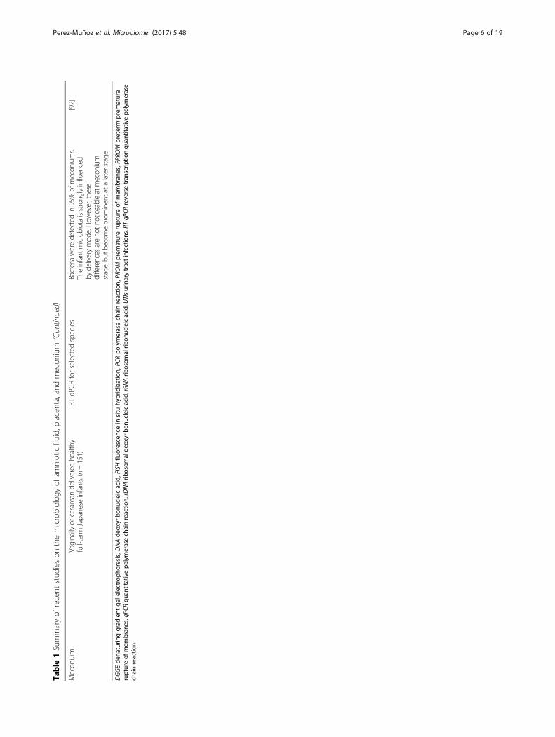
Table
1Summaryof
recent
stud
ieson
themicrobiolog
yof
amnioticfluid,p
lacenta,andmecon
ium
(Con
tinued)
Mecon
ium
Vaginally
orcesarean-delivered
healthy
full-term
Japanese
infants(n=151)
RT-qPC
Rforselected
species
Bacteriaweredetected
in95%of
mecon
iums.
Theinfant
microbiotaisstrong
lyinfluenced
bydeliverymod
e.How
ever,these
differences
areno
tnoticeableatmecon
ium
stage,bu
tbecom
eprom
inentata
laterstage
[92]
DGGEde
naturin
ggrad
ient
gele
lectroph
oresis,D
NAde
oxyribon
ucleicacid,FISHflu
orescencein
situ
hybridization,
PCRpo
lymerasechainreactio
n,PR
OM
prem
atureruptureof
mem
bran
es,P
PROM
preterm
prem
ature
ruptureof
mem
bran
es,qPC
Rqu
antitativepo
lymerasechainreactio
n,rDNArib
osom
alde
oxyribon
ucleicacid,rRN
Arib
osom
alrib
onucleicacid,U
TIsurinarytractinfections,RT-qP
CRreverse-tran
scrip
tionqu
antitativepo
lymerase
chainreactio
n
Perez-Muñoz et al. Microbiome (2017) 5:48 Page 6 of 19

colleagues reported positive bacterial cultures (in thesubchorionic fibrin of placentas) for only 11 out of 33placentas (33%) from pregnancies where there was a sus-pected underlying infection and for only one of 46 (2%)healthy controls [35]. The authors concluded that mostplacentas are sterile, and if bacteria are present, theymight originate through contamination during expul-sion. Although these studies were not aimed at thedetermination of a “placental microbiome” in healthypregnancies per se, their findings do reinforce the ideathat bacteria are not present in the healthy fetalenvironment.
In utero colonization hypothesis: the womb is notsterile and microbial colonization of the infantbegins prior to birthMost of the evidence supporting the sterile womb para-digm was generated with traditional microscopy orculture-based techniques, which are today considereddeficient for assessing a microbiome. Researchers havetherefore turned to molecular approaches, such as next-generation sequencing, and recent studies have pro-duced evidence (summarized in Table 1) that challengesthe sterile womb paradigm. Reports employing thesetechniques propose that bacteria are not only presentwithin the fetal environment in healthy full-term preg-nancies [8, 9, 37, 38], but that they also constitute a pla-cental microbiome that jump-starts the colonization ofthe fetus [8–10] as part of its normal developmentalprocess [39]. Additionally, it has been suggested that theuterus contains its own microbiome that can contributeto fetal colonization, as the placenta develops from bothfetal trophoblasts and maternal decidua (the inner liningof the uterus) [40, 41].After the early studies on the meconium discussed
above, microbiological research on the meconium ceasedfor a period of over 30 years (reviewed by [3]) until Jime-nez and colleagues reported 100% (n = 21) of meconiumsto be positive for bacteria by culture techniques [8]. Thedevelopment of molecular techniques and high-throughputsequencing spurred additional research on the microbiol-ogy of the meconium. Virtually all of these studies reportedthat over 90% of samples tested were positive for the pres-ence of bacteria (Table 1), thereby seeding the idea that theplacenta and the environment in which the fetus developsare not sterile.As stated before, there are no historical studies known
to us that were performed with the sole purpose of dir-ectly assessing the microbiology of the amniotic fluidfrom healthy pregnancies delivered at term. However, re-search that studied associations between microbial infec-tion/invasion and pregnancy outcomes have occasionallyincluded samples from uncomplicated pregnancies.Several independent studies showed that Mycoplasma
hominis and Ureaplasma urealyticum, which are amongseveral organisms highly correlated with preterm deliver-ies [42–44], have also been detected by culture methodsor standard PCR in asymptomatic pregnancies thatended in healthy deliveries [45–48].Recently, Collado and colleagues [10] aimed to specif-
ically investigate microbial prenatal and neonatal trans-fer in an array of maternal and fetal/newborn samplesfrom 15 full-term, healthy mother-infant pairs that sub-mitted to elective C-section. Using 16S rRNA pyrose-quencing, culture techniques, quantitative PCR (qPCR)and DGGE, the authors detected microbial populationsin the amniotic fluid that were low in abundance, rich-ness, and diversity that shared similarities with microbialpopulations found in the placenta. Enterobacter andEscherichia/Shigella were the most prevalent generapresent in both placenta and amniotic fluid, followed byPropionibacterium. Similarities between the microbialpopulations found in the colostrum and meconium werealso noted. The authors subsequently hypothesized thatmaternal intestinal microbes may be selectively trans-ported to the mammary gland, the placenta, and the amni-otic fluid, thereby contributing to an initial colonization ofthe fetal intestine in utero.Aagaard and colleagues [9] were the first to use Illu-
mina sequencing to comprehensively characterize theplacental microbiome in over 300 subjects, includingthose with healthy pregnancies, preterm births, andcases with history of antepartum infection. The authorsestimated that they isolated up to 0.002 mg of bacterialDNA per 1 g of placental tissue and detected a lowlyabundant but “metabolically rich” microbiome that in-cluded Fusobacterium spp., Neisseria lactamica, Neisseriapolysaccharea, Rhodococcus erythropolis, Propionibacter-ium acne, Streptomyces overmitilis, Bacteroides spp.,Prevotella tannerae, Eschericia sp. 4_1_408, and Escheri-chia coli (with the latter being the most abundant). Thismicrobiome clustered with the mothers’ oral microbiotavia Bray-Curtis analysis, was associated with a “remotehistory of antenatal infection”, and was enriched in spon-taneous preterm births. These findings prompted the au-thors to propose that bacteria translocate byhematogenous spread from the mother’s oral cavity intothe placenta and colonize the fetus in utero (Fig. 1b).In a subsequent analysis of the same samples,Aagaard and colleagues additionally concluded thatthe preterm placental microbiome and its metabolicprofile vary with gestational weight gain [49].In addition to sequencing and PCR, two types of
microscopy-based methods have also been applied to de-tect bacteria in placental tissues: FISH and classic hist-ology. Using these methods, two independent researchgroups [37, 38] showed that placentas from shortergestational age deliveries were more likely to harbor
Perez-Muñoz et al. Microbiome (2017) 5:48 Page 7 of 19
intracellular bacteria compared to full-term placentas.Doyle and colleagues [50] complemented this finding byusing Roche 454 FLX sequencing when they reported anincreased frequency and a wider spectrum of bacteriapresent in preterm relative to full-term placentas.Together, the studies above challenge the sterile womb
paradigm in that they provide evidence for bacteria inthe healthy in utero environment. Proponents of a fetalmicrobiome suggest several routes of bacterial access tothe placenta, including ascension from the lower genitaltract, entry through the mother’s bloodstream, or activetransport of microbes by immune cells from the gut ororal cavity (Fig. 1b). It has also been proposed that thefetal-placental environment has evolved to facilitatethe establishment of a diverse microbiome that fur-ther plays a role in the acquisition and assembly ofthe infant’s gut microbiome through in utero trans-mission of microbes [4, 39, 51].
Pondering the two hypothesesAlthough the sterile womb paradigm was generallyagreed upon until around 10 years ago, the alternativehypothesis is experiencing a renaissance. As there is cur-rently no clear consensus, we next evaluated the avail-able evidence in support of each model. Several aspectsmust be taken into consideration; these include the ana-tomical, immunological, and physiological features ofthe placenta, the immunological status of the fetus, thelimitations of the research methods used to study micro-bial populations, the microbiome during the very firstdays of life, and the evidence provided by the generationof germ-free animals (including humans).
A. Anatomical, physical, and immunologicalconsiderations
The two main functions of the placenta are nourish-ment of the fetus and its protection from microbialpathogens. Accordingly, the placenta has several ana-tomical, physiological, and immunological features thatprevent bacterial colonization.
(i) Anatomical and physiological barriers
The materno-fetal barrier contains two anatomicallydistinct elements, the chorioallantoic placenta and thechorioamnion. This barrier is formed at the placentallevel by the villous syncytiotrophoblast, a layer of spe-cialized epithelial cells differentiated from underlyingmononuclear cytotrophoblasts (Fig. 2). The syncytiotro-phoblast actively invades the uterine wall and eventuallyforms the outermost fetal constituent of the placentaand the placental villi. This important epithelial layeralso forms an interface between maternal blood and
embryonic extracellular fluid to mediate oxygen and nu-trient transfer between maternal capillaries and the fetus’environment. Additionally, the syncytiotrophoblast actsas a continuous cell without intercellular barriers, disal-lowing maternal or bacterial cells to squeeze throughintercellular junctions and into the fetal bloodstream.This provides a critical first level of structural protectionagainst invasion of maternal cells carrying foreign anti-gens and bacterial pathogens [52–55].Additionally, a basement membrane separates the syn-
cytiotrophoblast from connective tissue containing fetalcapillaries (Fig. 2). This placental membrane constitutesa second physical obstacle that potential microbial in-vaders must overcome to infect the developing fetus[33]. A third level of protection is provided by extravil-lous trophoblasts (EVTs). EVTs invade the decidua,functioning to anchor the placenta into the uterine wall[56] (Fig. 2). Besides being co-localized with naturalkiller cells, macrophages, and leukocytes, EVTs also pro-vide innate defense mechanisms [57] and possess bac-tericidal properties [53, 55]. Importantly, EVTs also sendtolerogenic signals to maternal leukocytes to preventimmune-mediated damage to the placenta [57].Together, the syncytiotrophoblast, the EVT, and the
basement membrane constitute the physical barrier thataverts the passage of bacteria into the amniotic sac and,consequently, the fetus. Only bona fide bacterial pathogens(for example, Listeria monocytogenes, Brucella abortus, andToxiplasma gondii) possess the factors necessary for suc-cessful invasion of these barriers, subversion of the immuneresponse, and establishment in the placenta as viableorganisms. For example, L. monocytogenes uses specificvirulence structures such as internalins (InlA and InlB), thehemolysin listeriolysin O, and the actin assembly-inducingprotein ActA to cross the intestinal, placental, and blood-brain barriers [58]. The requirement for these structures tosuccessfully invade mammalian cells has been demon-strated by introducing them into commensal bacteria usingplasmid vectors [59]. Together, these findings indicate thatonly pathogens and not commensals are capable of bypass-ing the materno-fetal anatomical barriers and establishingin the fetal environment.
(ii) Immunological barriers
Numerous immune cells and effector molecules arepresent in the placenta to ensure protection against bac-terial invaders. For example, toll-like receptors (TLR) 1through 10, which are important in recognizing molecularpatterns and facilitating immune responses, are present inthe human placenta [60, 61], and their expression is regu-lated both spatially and temporally depending on gestationperiod [62]. Additionally, the female reproductive tractconstitutively expresses antimicrobial peptides (AMP).
Perez-Muñoz et al. Microbiome (2017) 5:48 Page 8 of 19

These AMPs serve as crucial immune effectors for theplacenta and fetal membranes during pregnancy by pro-viding a chemical barrier to ascending infections [63]. Theconcentrations of some AMPs are increased during lategestation, while others are released directly into amnioticfluid and the fetal compartment during parturition to helpdefend the neonate against infection [64–66].Other important immune effectors present in the
placenta include immunoglobulins (e.g., IgG, IgA, andIgM), which play multiple important roles in regulatingthe course of a normal pregnancy [67, 68]. In the mater-nal part of the placenta, immunoglobulins protect themother against paternal antigens present in the fetus,
while in the fetal part, immunoglobulins protect thefetus against macromolecules and infectious agentsoriginating from the mother [67]. Interestingly, mostplacental IgG are bound to both the trophoblastic base-ment membrane and the surfaces of the syncytiotropho-blast [69]. In contrast, IgM is located in the placentalvillous structures [70]. In particular, all of these immu-noglobulins can be found as components of the outerlayers of the placenta, and this location is likely a key inprotecting against bacteria trying to gain access. Indeed,the presence of AMPs in the chorionic and amnioticmembranes and immunoglobulins in the placenta couldexplain why researches have not been able to find viable
Fig. 2 Schematic representation of the anatomical, physiological, and immunological placental barriers designed to limit microbial invasion. Threemain types of cells on the fetal side of the placenta prevent access of bacterial invaders to the fetal circulation: the syncytiotrophoblast, the cytotrophoblasts,and the extravillous trophoblasts (EVT). The basement membrane also serves as a physical barrier that averts bacterial invasion. Additionally, maternalimmune cells and immunoglobulins (not depicted) are near the EVTs to aid in the defense against microbial insults
Perez-Muñoz et al. Microbiome (2017) 5:48 Page 9 of 19

bacteria in placentas from healthy, full-term deliveries.Rather than live bacteria, what may be present in placen-tal tissues is simply bacterial products created by theantimicrobial actions of AMPs and immunoglobulins.Altogether, the placental epithelium possesses a series
of anatomical, physiological, and immunological featuresto prevent and combat microbial threat. Many other epi-thelial sites that harbor resident microbiomes also havethese features. However, when entertaining the idea of amicrobiome associated with the human fetus, oneshould consider that several immune system compo-nents needed to facilitate an “incident-free” prenatalinterrelationship with the microbiome are not yet inplace or mature in the fetus. Significant differences be-tween the neonatal and adult immune system include areduction in serum complement activity, decreased abil-ity to produce antibodies against bacterial polysaccharideantigens, and increased numbers of naïve T cells andantigen-presenting cells with a correspondingly naïvefunctional program [71, 72]. Apart from a limited subsetof AMPs that are expressed in distinct fetal compart-ments [66], fetuses do not have the immunity needed tosuccessfully overcome bacterial invasion. Additionally,studies documenting the limited immune functions ofvery premature newborns indicate that the complex im-mune system necessary for the development of immuno-logical tolerance to a microbiome would not be presentin a fetus [72, 73, 74]. Finally, intestinal permeability ishigher during the first 2 days of life for preterm infantsas compared to healthy term infants [75], suggesting thatthe fetal gut would permit bacterial translocation andpromote encounters with an underdeveloped immunesystem. Although there may be some evidence, albeit in-consistent, for the presence of bacterial DNA in placen-tal and amniotic fluid samples of healthy pregnancies, itis not at all clear how an immunologically immaturefetus would successfully control viable bacteria to pre-vent infections (and mortality) and develop normally.
B. Methodological considerations
Most of the studies that established the sterile wombparadigm are based on microbial culture, which fails todetect viable but non-cultivable microbes. DNA-basedPCR and sequencing methods overcome this limitation,and it is possible that bacteria detectable by thesemethods in the fetal-placental environment and meco-nium do represent viable, metabolically active organismsthat are non-cultivable. However, one must also considerthat these molecular methods have inherent limitations.First, even if bacterial DNA is detectable, the organismscould be dead. This consideration is especially relevantfor the placenta, as an important role for this tissue isthe removal of microbes and their components that
might be present in the blood [76]. Additionally, forresearch to successfully challenge the sterile womb para-digm, a demonstration of microbial viability is essential,as sites can be sterile even while containing bacterialDNA. Very few groups have demonstrated viable micro-organisms in the fetal environment despite these studiesemploying culture methods that readily grow bacteriafrom other parts of the body [77–83]. In the case ofSatokari and colleagues [84], the authors could not cul-ture bacteria detected by molecular methods (Bifidobac-terium and Lactobacillus) even though these are readilycultivable organisms. Although the authors attribute thisresult to freezing the samples prior to processing, theyalso ponder the possibility that only bacterial productsincluding DNA, rather than living bacteria, are presentin the placenta. In fact, freezing samples before process-ing has been performed in many culture-based studiesof microbiomes, and although it reduces bacterialcounts, it normally does not prevent cultivation. In thecase of Collado and colleagues [10], identification of bac-teria cultured from the placenta and amniotic fluid ofnewborns delivered by C-section was limited to multipleisolates of Propionibacterium and Staphylococcus spe-cies, and one isolate each of Streptomyces and Lachnos-piraceae. Propionibacterium and Staphylococcus speciesare ubiquitous normal skin commensals and couldtherefore originate from contamination (see below).Importantly, the authors reported that Enterobacter andEscherichia/Shigella were the most abundant genera de-tected in placenta and amniotic fluid samples. However,they were not able to recover these organisms by culturemethods despite the relative ease in cultivating thesebacterial groups. Taken together, these findings andother current data do not support the existence of livebacteria in the placenta.An even more important methodological consideration
is that DNA-based assessments of low microbial biomasssamples (such as the placenta, amniotic fluid, and meco-nium) are extremely prone to confounding findings fromcontaminant DNA. In fact, studies have demonstratedthat sequence-based analyses of samples with low DNAlevels are not reliable because reagents, consumables,and components of DNA extraction kits contain bacter-ial DNA [85–91]. Work by Salter and colleagues [90]has systematically demonstrated that the lower theamount of bacterial DNA in a sample, the higher theproportion of sequences that can be attributed to con-tamination. The authors provided a list of bacterial taxacommonly present in reagents and consumables that aredetected in negative controls (Fig. 3). Interestingly,around 36% of the total species reported by Aagaard andcolleagues [9] as “the placenta microbiome” overlap withtaxa on this list. Although some researchers do reportthe use of controls, such as sequencing of non-template
Perez-Muñoz et al. Microbiome (2017) 5:48 Page 10 of 19

extractions [9], even these have been criticized as notsufficient [76], and most studies on the microbiota ofthe fetal-placental environment do not report the use ofany controls [10, 38, 49, 50, 84, 92]. Clearly, lack of ap-propriate controls leaves the findings on fetal micro-biomes extremely doubtful. This notion was recentlyreinforced by Lauder and colleagues [91] who systemat-ically compared sequencing data obtained from placentalsamples with those from different contamination con-trols including sterile swabs, air swabs (swabs exposed tothe air of the clinical laboratory), and extraction blanksfrom two different DNA isolation kits (blank tubes as asource of possible extraction/reagent contaminants).This study revealed that placenta samples containedexactly the same marginal amounts of bacterial DNA asthe extraction blanks, and that the bacterial communities
detected clustered closely with the contaminant commu-nity of the respective DNA isolation kit. Most importantly,no bacterial lineages were identified as unique to orpresent at greater abundances in placental samples whencompared to contamination controls.Apart from preventing the contamination of DNA,
avoiding sample contamination per se is also a signifi-cant challenge when studying low-abundance and low-diversity bacterial communities. Samples for the study ofthe in utero environment are collected within a clinicalsetting (hospital, emergency room, delivery or operatingroom), making it difficult, if not impossible, to avoidsample contamination during collection and processing.In addition, the cleanliness of the tissue processing en-vironment is particularly important in laboratories wherebacterial cultures are also routinely handled. Accordingly,
Fig. 3 Venn diagram of bacterial genera hypothesized to contribute to the infant gut microbiome. Aagaard and colleagues [9] hypothesized thatbacteria translocate from the mother’s oral cavity into the placenta, contributing to in utero colonization of the fetal gut. They further suggest that placentascontain low abundance communities of commensal bacteria. However, 36% of the bacterial genera found by Aagaard and colleagues [9] also appear on thelist of contaminants found in reagents by several independent research groups as reported by Salter and colleagues [90]. Not all genera were included foreach individual microbiome due to space constraints. Genera found in the infant gut [2, 101, 102, 105, 148] include taxa described in both vaginally andcesarean section-delivered babies [101, 105] and show a substantial overlap with genera found in the adult gut microbiome [145–147], but little overlap withtaxa found in the placenta [9, 91] or as contaminants [85–91]
Perez-Muñoz et al. Microbiome (2017) 5:48 Page 11 of 19

processing and storage time also seem to influence results.For example, Jimenez and colleagues [8] reported thattheir samples clustered by time of processing, as half ofthem were processed at the time of collection while therest were processed 4 days after collection.Furthermore, the method of infant delivery can also
influence the degree of sample contamination andshould be considered during study design. Vaginally de-livered placentas are exposed to vaginal microbes duringexpulsion, and findings, in our opinion, can thereforenot be used to argue for in utero colonization, norshould they be compared to tissues extracted via C-sec-tion. As an example, Jones and colleagues evaluated fetaland placental tissues from 74 preterm and full-term C-section and vaginal deliveries [93]. They found 30 and43% of these tissues to be positive for bacterial DNAusing qPCR. However, once the authors stratified the tis-sues by delivery mode, none of the full-term C-sectionplacentas were found to be positive.Overall, the molecular techniques used to study the
fetal microbiome have inherent limitations due to theirsusceptibility to false positives because of contamination.In this respect, it is important to consider the limit ofDNA that can be reliably detected by these methods.Even in studies that supported the presence of a placen-tal microbiome, DNA concentrations were acknowl-edged to be very low [9, 10]. Therefore, only techniquescapable of detecting less than 100 bacterial cells pergram of sample are likely to provide reliable results.However, even PCR methods, despite the fact that theycan (in theory) detect one single DNA template, fre-quently have detection limits of 104 to 106 cells per gramwhen applied to samples with complex matrices [94, 95].Although detection limits of high-throughput sequen-cing technologies in low-biomass samples have not beenestablished, it is likely that they are not sufficient to reli-ably detect the low amounts of DNA in these samples(e.g., in the presence of contaminating DNA). Culturemethods do possess the required detection limit, but asdescribed above, most studies did not result in positiveresults. We therefore conclude that the in uterocolonization hypothesis rests on studies that used mo-lecular approaches with an insufficient detection limit tostudy “low-biomass” microbial populations and furtherlacked appropriate controls for contamination whilefailing to provide evidence of bacterial viability.
C. Interpreting results from the study of the very earlyneonate’s microbiome
The repeated detection of microbes in meconium isfrequently offered as evidence in support of the in uterocolonization hypothesis. However, it should be recog-nized that only a small subset of meconiums contains
detectable microbes [3, 12–14, 96]. Even if microbes aredetected, bacteria in the first stool of the newborn couldbe the result of postnatal colonization, especially if themeconium is expelled long after delivery. Experimentswith germ-free mice have shown that bacterialcolonization is rapid, with bacterial species detectable8 h after the initial exposure of the mice to conventionalhousing, and bacterial counts becoming equivalent tothose of conventional mice after 24 h [97]. If the “germ-free human” supports microbial growth similarly to thatof a germ-free mouse, then a rapid colonization processwould also be expected to occur in the neonatal gut.Hansen and colleagues [14] argued that there is a“meconium colonisation interval” that provides sufficientopportunity for the microbes to multiply between rup-ture of the membranes during birth and the time whenthe meconium is delivered and processed. In support ofthis idea, microbial colonization of the meconium hasrepeatedly been shown to increase with time of passage[12, 13, 14], indicating that colonization occurs ex utero.Accordingly, studies that do not categorize and differen-tiate this period of time in their analyses report a highernumber of positive cases [92, 98, 99].In addition, the composition of the pioneer infant
microbiome supports the sterile womb paradigm. If ster-ile in utero, initial inoculation of microbes is contingentupon the process of childbirth and subsequent environ-mental exposure (Fig. 1A) with the first major microbialexposure for a vaginally born infant occurring in thebirth canal. This step is bypassed during C-sections, anddelivery mode (vaginal delivery versus C-section) wouldtherefore heavily influence the microbial composition[100–106]. In contrast, if a subset of the early micro-biome were acquired in utero, then bacterial populationsshould be present in the infant gut that overlap withthose found in the placenta/meconium, and their pres-ence should be independent of delivery method. Somestudies report that the meconium contains bacteria simi-lar to those found in amniotic fluid [10], and authorshave proposed that fetal gut colonization could occurthrough ingestion of amniotic fluid that contains bac-teria [8, 98, 107]. However, the vast majority of the lit-erature demonstrates that the pioneer gut microbiome isheavily influenced by birth method and later dominatedby species that are characteristic gut microbes, while mi-crobes detected in the fetal environment are absent(Fig. 3). Several studies have reported significant differ-ences in the diversity and composition between vaginallyversus C-section-delivered infants [101, 105, 106, 108],with vaginally born infants harboring an early micro-biome that resemble that of the vagina, while the micro-biomes of C-section infants reflect those of the humanskin [101, 109]. Dominguez-Bello and colleagues showedthat the bacterial communities of vaginally delivered
Perez-Muñoz et al. Microbiome (2017) 5:48 Page 12 of 19

newborns were dominated by Lactobacillus, Prevotella,or Sneathia species—all of which were also found in themother’s vagina [101]. In contrast, the gut microbiota ofinfants delivered by C-section was dominated by theskin commensals Staphylococcus, Corynebacterium, andPropionibacterium [101]. Bäckhed and colleagues alsoreported that the gut microbiomes of infants born viaC-section were dominated by skin and oral microbes aswell as bacteria from the surrounding environment,while gut microbiomes of vaginally delivered infantswere enriched in classical gut microbes (Bacteroides, Bifi-dobacterium, Parabacteroides, and Escherichia/Shigella)[105]. The authors further established that 72% of theearly colonizers of vaginally delivered neonates could betraced back to the gut microbiota of their own mother,while this number was only 41% for C-section infants.Together, these studies convincingly show that deliverymethod strongly affects microbiome composition in neo-nates, while delivery method-independent microbialtaxa originating from the placenta/amniotic fluid havenot been reported. These findings support the con-cept of a sterile infant gut that is colonized by mi-crobes acquired during and after birth, dependent onthe environmental exposure.
D. Considerations from the derivation of germ-freemammals
The strongest evidence against the existence of micro-biomes in the fetal environment stems from the scienceof gnotobiology. Gnotobiology is the study of animalsraised and maintained in an environment in which allmicroorganisms are either defined or excluded [110].The science of gnotobiology is founded on our ability toderive germ-free animals via C-sections and subse-quently raise the offspring in a sterile environment.The first axenic animals were reported as early as the
end of the nineteenth century [111], but it took until the1940s to consistently derive axenic rodents successfullyand maintain them for successive generations [112–114].The first germ-free progenitors were generated by thelabor-intensive process of C-section and hand-feedingwith a sterilized artificial diet inside an aseptic isolatoruntil maturity, after which a breeding colony was estab-lished [114]. Since then, a wide variety of animals havebeen successfully derived germ-free over the past70 years, including mice, rats, guinea pigs, rabbits, dogs,cats, pigs, lambs, calves, goats, baboons, chimpanzees,and marmosets [115–122], demonstrating that the abilityto derive germ-free animals is not unique to only rodentspecies. Currently, both commercial companies and uni-versity animal facilities routinely offer derivation as aservice to the research community. Germ-free offspringcan be generated from non-germ-free stock by embryo
transfer and aseptic hysterectomy (Fig. 4), and aseptichysterotomy. To perform the hysterectomy in mice,donor females are euthanized when parturition isimminent, and the intact pregnant uterus is asepticallyharvested, clamped, introduced into a germicidal bath,and finally transferred into a sterile isolator. After re-moval from the uterus, the pups are warmed, dried tostimulate their breathing, and then placed under thecare of an axenic foster mother [123–125]. Hysterotomyis usually performed to generate large axenic animalswith the intent of maintaining the breeding potential ofthe female. In this scenario, a sterile canopy with glovesis attached to the mother’s abdomen prior to the sur-gery. Using sterile gloves, the surgeon makes an incisionin the uterus and removes the placenta and amnioticsac, which are then transferred into a second sterile iso-lator so the fetus(es) can be revived in an aseptic envir-onment [116, 119].During hysterotomies and hysterectomies, either the
intact pregnant uterus or its entire contents (includingthe placenta, the amniotic sac, and the fetus), respect-ively, are removed and transferred to generate germ-freeanimals. The success of these procedures provides clearevidence against the existence of a microbiome in theplacenta and fetus. If microbes were present, even at lowabundance, they would colonize the offspring and rap-idly grow to detectable levels. This phenomenon can beobserved during accidental contaminations that occur(very much to the dismay of the researchers) in germ-free animal facilities. The derivation process may involvethe treatment of the non-germ-free donor female withantibiotics to reduce her microbial load prior to the hys-terectomy [125]. However, oral administration of anti-microbial agents directly to the offspring is not applied,nor would this practice succeed in generating axenic off-spring from animals that are already colonized. The factthat axenic animals can be derived and maintained de-void of microbes under sterile conditions provides verycompelling evidence that, in most mammalian species,in utero transfer of the microbiome does not occur.Supporters of the in utero transmission hypothesis
often argue that the bacteria present in the fetal environ-ment would potentially not colonize a germ-free host orremain undetectable after birth. However, this phenomenonis unlikely. Almost any bacterium quickly and irreversiblycolonizes germ-free mice because there is no competition.Others also argue that animals are not humans, and thatfetal microbiomes might be unique to humans due tophysiological and anatomical differences. However, germ-free humans (although rare) have been established usingprotocols similar to those employed during the generationof large axenic mammals via aseptic hysterotomy [125–127]. This procedure has been applied in suspected cases ofsevere immune deficiency of the fetus [128]. The first
Perez-Muñoz et al. Microbiome (2017) 5:48 Page 13 of 19

germ-free human was delivered by performing a C-sectioninside a sterile canopy attached to the mother’s abdomen,which prevented exposure to environmental contaminants[126]. Gram stains of feces and aerobic and anaerobic cul-tures of swabs from both the infant and the isolator sur-faces confirmed the germ-free status of the infant [126, 129,130]. The published cases report axenic status from 6 daysto 3 months after which the subjects were either removedfrom the axenic isolators or microbial contamination wasdetected [126, 127, 129, 131]. Although axenic humans arevery rare for obvious reasons, the fact that they have beengenerated makes it extremely unlikely that humans are col-onized with bacteria in utero.
ConclusionsIn 1918, Arthur Kendall summarized the contemporaryknowledge on intestinal bacteriology in The AmericanJournal of the Medical Sciences [132]. He concluded that“at birth the intestinal tract and intestinal contents arenormally sterile. The first indications of bacterial con-tamination are recognizable several hours postpartum.The early invaders are adventitious microbes, similar inevery respect to those commonly present in the infant’senvironment. They gain entrance to the alimentary canal
through the mouth, although the possibility of rectal in-fection must be borne in mind.” After having reviewedthe available literature, we conclude that Kendall’s as-sumptions are still valid, and that there is little evidenceto successfully challenge the sterile womb paradigm.The recent findings that question this premise relymostly on (i) methodological approaches (PCR and next-generation sequencing) that do not have the detectionlimit necessary to study “low-density” bacterial popula-tions, (ii) the use of methodological approaches that areextremely susceptible to contamination without the in-clusion of appropriate controls, (iii) the study of samplescollected in clinical settings where it is difficult to pre-vent contaminations, and (iv) a flawed interpretation offindings from early stool samples, which can contain mi-crobial populations even if the fetus was sterile. Eventhough the bacterial species identified by moleculartechniques in fetal environments are known to be readilycultivable, bacterial culture (which does provide a suffi-cient detection limit) is almost always negative. In ouropinion, only one study published to date has used ro-bust controls and considered low DNA levels, and thefindings do not support the presence of a microbiome inthe placenta [91]. Moreover, the strongest evidence
Fig. 4 Schematic representation of the generation of axenic rodents by aseptic hysterectomy. In rodents, germ-free offspring are derived by aseptichysterectomy. Germ-free foster mothers housed in a sterile isolator are time-mated to become pregnant in synchrony with holoxenic (conventional)females. Breeding pairs are mated on such a schedule that the aseptic hysterectomy of the donor mother can be performed a few hours before herscheduled pupping and a few hours after the foster mother gives birth. To perform the hysterectomy, donor females are euthanized, and the uterusis harvested and clamped, aseptically introduced into a germicidal bath, and then transferred into the sterile isolator where the foster mothers reside.The pups are then revived and placed under the care of the foster mother [123–125]. If there are no germ-free foster mothers available, then pupsare hand-raised using sterile formula. Figure adapted from Hedrich and Hardy [125]
Perez-Muñoz et al. Microbiome (2017) 5:48 Page 14 of 19

against the hypothesis of a commensal placental micro-biome comes from the successful generation of germ-free animals via aseptic transfer of the entire uterus(containing the placenta).By writing this review, we aimed to contribute to the
discussion of this contested topic, as we are concernedwith the far-reaching implications that impact both ourbasic understanding of host-microbe symbiosis inhumans, as well as important applied aspects such asclinical decisions and funding priorities. Transmissionmode influences the mechanisms by which symbiosesand mutualistic interactions evolve, as well as the extentto which environmental and lifestyle factors alter suchinteractions. This understanding directly informs clinicalpractices and recommendations, including the deliveryof infants via C-sections, which have been argued bysupporters of the in utero colonization hypothesis to beless detrimental than previously thought. Such discus-sions are not scientifically valid. Although medicallynecessary C-sections should not be discouraged, thisprocedure clearly influences establishment of the earlygut microbiome [106, 133–137] and is epidemiologicallylinked to an increased risk for developing chronic dis-eases later in life [134, 137–140]. Therefore, strategies toprevent C-sections or their impact on the pioneermicrobiome remain important and should be researchedwith the goal of preventing chronic diseases [102].Further, it has been argued that the role of bacterial
communities in the in utero environment warrants add-itional study [102, 141]. However, given the insufficientevidence that such communities exist, we argue thatthese efforts are likely futile. In our opinion, future stud-ies (and resources) should instead focus on (i) the post-natal acquisition of the gut microbiome and itsimportance to health and (ii) the possible role of pre-natal exposure of the fetus to microbial metabolites andcompounds that originate from the maternal gut micro-biota. Indeed, a recent study elegantly showed that mi-crobial metabolites in the fetal environment can have amajor impact on the development of the offspring [142].Although the evidence does not support in uterocolonization, it does however suggest an association be-tween the presence of bacterial DNA in the placenta andpreterm birth [38, 50]. Research regarding the role ofthis DNA would be worthwhile, but such studies muststrictly control for DNA contamination during samplecollection and the DNA extraction process.Self-correction is one of science’s most fundamental
principles—all findings must be subject to scrutiny andverification to determine validity [143, 144]. If a findingis incorrect, then replication will prove it as such.Unfortunately, the scientific self-correction process isslower than the transfer of information. Today, scientificfindings can move freely from professional journals into
the public realm (e.g., through social media), often be-fore the scientific community has thoroughly discussedand vetted the evidence. Indeed, some of the researcharticles discussed in this manuscript were heavily cov-ered in the public press. Because most members of thenon-scientific community are not equipped to critiquescientific findings, it is our responsibility to debate thesecontroversial topics and facilitate the self-correctionprocess. Failure to do so may ultimately compromise hu-man health, damage scientific creditability, and poten-tially contribute to the erosion of the public’s trust inscience. We hope that this review has contributed tosome degree to prevent the latter.
AbbreviationsAMP: Antimicrobial peptide; C-section: Cesarean section; DGGE: Denaturinggradient gel electrophoresis; DNA: Deoxyribonucleic acid; EVT: Extravilloustrophoblast; FISH: Fluorescence in situ hybridization; PCR: Polymerase chainreaction; PPROM: Preterm premature rupture of membranes; PROM: Prematurerupture of membranes; qPCR: Quantitative polymerase chain reaction;rDNA: Ribosomal deoxyribonucleic acid; rRNA: Ribosomal ribonucleic acid;TGGE: Temperature gradient gel electrophoresis; TLR: Toll-like receptor;UTIs: Urinary tract infections
AcknowledgementsWe are grateful to Dr. Carlos Muñoz (Department of Biological Sciences,University of Puerto Rico, Mayaguez, Puerto Rico) for the editorial assistancewith the figures.
FundingJW acknowledges the Campus Alberta Innovates Program (CAIP) and theUniversity of Alberta, the Natural Science and Engineering Research Councilof Canada, and the National Institute of Health (5R01GM099525-02) for thefunds received. MCA receives funding from the Alberta Children HospitalResearch Institute and the University of Calgary. AERT receives funding fromthe National Institute of General Medical Sciences of the National Institutesof Health (P20GM104320), the Crohn’s and Colitis Foundation of America, theNebraska Corn Board, the Nebraska Research Initiative, and the University ofNebraska–Lincoln.
Availability of data and materialsNot applicable.
Authors’ contributionsMEPM, MCA, AERT, and JW wrote the review. All authors read and approvedthe final manuscript.
Competing interestsThe authors declare that they have no competing interests.
Consent for publicationNot applicable.
Ethics approval and consent to participateNot applicable.
Publisher’s NoteSpringer Nature remains neutral with regard to jurisdictional claims inpublished maps and institutional affiliations.
Author details1Department of Agriculture, Food and Nutritional Sciences, 4-126 Li Ka ShingCentre for Health Research Innovation, University of Alberta, Edmonton,Alberta T6G 2E1, Canada. 2Department of Physiology and Pharmacology,University of Calgary, Cumming School of Medicine, 3330 Hospital Drive NW,Calgary, Alberta T2N 4N1, Canada. 3Department of Pediatrics, University ofCalgary, Cumming School of Medicine, 3330 Hospital Drive NW, Calgary,
Perez-Muñoz et al. Microbiome (2017) 5:48 Page 15 of 19

Alberta T2N 4N1, Canada. 4Department of Food Science and Technology,260 Food Innovation Center, University of Nebraska-Lincoln, 1901 N 21stStreet, Lincoln, Nebraska 68588-6205, USA. 5Department of BiologicalSciences, 7-142 Katz Group Centre, University of Alberta, Edmonton, AlbertaT6G 2E1, Canada.
Received: 14 January 2017 Accepted: 21 April 2017
References1. Chow J, Lee SM, Shen Y, Khosravi A, Mazmanian SK. Host-bacterial symbiosis
in health and disease. Adv Immunol. 2010;107:243–74.2. Arrieta M-C, Stiemsma LT, Dimitriu PA, Thorson L, Russell S, Yurist-Doutsch S,
Kuzeljevic B, Gold MJ, Britton HM, Lefebvre DL, Subbarao P, Mandhane P,Becker A, McNagny KM, Sears MR, Kollmann T, Mohn WW, Turvey SE, BrettFinlay B. Early infancy microbial and metabolic alterations affect risk ofchildhood asthma. Sci Transl Med. 2015;7:307ra152.
3. Koleva PT, Kim J-S, Scott JA, Kozyrskyj AL. Microbial programming of healthand disease starts during fetal life. Birth Defects Res C Embryo Today. 2015;105:265–77.
4. Funkhouser LJ, Bordenstein SR. Mom knows best: the universality of maternalmicrobial transmission. PLoS Biol. 2013;11:e1001631.
5. Bright M, Bulgheresi S. A complex journey: transmission of microbial symbionts.Nat Rev Microbiol. 2010;8:218–30.
6. Kustner O. Beitrag zur Lehre von der puerperalen Infection der Neugeborenen.Arch Gynakol. 1877;11:256–63.
7. Escherich T. The intestinal bacteria of the neonate and breast-fed infant. RevInfect Dis. 1885;11:352–6.
8. Jiménez E, Marín ML, Martín R, Odriozola JM, Olivares M, Xaus J, FernándezL, Rodríguez JM. Is meconium from healthy newborns actually sterile? ResMicrobiol. 2008;159:187–93.
9. Aagaard K, Ma J, Antony KM, Ganu R, Petrosino J, Versalovic J. The placentaharbors a unique microbiome. Sci Transl Med. 2014;6:237ra65.
10. Collado MC, Rautava S, Aakko J, Isolauri E, Salminen S. Human gut colonisationmay be initiated in utero by distinct microbial communities in the placentaand amniotic fluid. Sci Rep. 2016;6:23129.
11. Blaser MJ, Dominguez-Bello MG. The human microbiome before birth. CellHost Microbe. 2016;20:558–60.
12. Burrage S. Bacteria in the supposedly sterile meconium. J Bacteriol. 1927;13:47.13. Hau IC, O’Toole E, Hall IP. Bacterial flora of first specimens of meconium
passed by fifty newborn infants. Am J Dis Child. 1934;47:1279–85.14. Hansen R, Scott KP, Khan S, Martin JC, Berry SH, Stevenson M, Okpapi A,
Munro MJ, Hold GL. First-pass meconium samples from healthy termvaginally-delivered neonates: an analysis of the microbiota. PLoS One. 2015;10:e0133320.
15. Harris JW, Brown JH. The bacterial content of the uterus at cesaren section.Am J Obstet Gynecol. 1927;13:133–43.
16. Stroup PE. Amniotic fluid infection and the intact fetal membrane. ObstetGynecol. 1962;19:736–9.
17. Harwick HJ, Iuppa JB, Fekety FR. Microorganisms and amniotic fluid. ObstetGynecol. 1969;33:256–9.
18. Prevedourakis C, Papadimitriou G, Ioannidou A. Isolation of pathogenicbacteria in the amniotic fluid during pregnancy and labor. Am J ObstetGynecol. 1970;106:400–2.
19. Prevedourakis CN, Strigou-Charalabis E, Kaskarelis DB. Bacterial invasion ofamniotic cavity during pregnancy and labor. Obstet Gynecol. 1971;37:459–61.
20. Lewis JF, Johnson P, Miller P. Evaluation of amniotic fluid for aerobic andanaerobic bacteria. Am J Clin Pathol. 1976;65:58–63.
21. Evaldson GR, Malmborg AS, Nord CE. Premature rupture of the membranesand ascending infection. Br J Obstet Gynaecol. 1982;89:793–801.
22. Ovalle A, Martínez MA, Kakarieka E, Gómez R, Torres J, Fuentes A, Ruiz M,Angel R. Placental histopathology in premature rupture of membranes. Itsrelationship with microbiological findings, maternal, and neonatal outcome.Rev Médica Chile. 1998;126:930–42.
23. Greig PC. The diagnosis of intrauterine infection in women with pretermpremature rupture of the membranes (PPROM). Clin Obstet Gynecol. 1998;41:849–63.
24. Gravett MG, Hummel D, Eschenbach DA, Holmes KK. Preterm labor associatedwith subclinical amniotic fluid infection and with bacterial vaginosis. ObstetGynecol. 1986;67:229–37.
25. Martius J, Eschenbach DA. The role of bacterial vaginosis as a cause ofamniotic fluid infection, chorioamnionitis and prematurity—a review. ArchGynecol Obstet. 1990;247:1–13.
26. DiGiulio DB, Romero R, Amogan HP, Kusanovic JP, Bik EM, Gotsch F, Kim CJ,Erez O, Edwin S, Relman DA. Microbial prevalence, diversity and abundancein amniotic fluid during preterm labor: a molecular and culture-basedinvestigation. PLoS One. 2008;3:e3056.
27. DiGiulio DB, Gervasi M, Romero R, Mazaki-Tovi S, Vaisbuch E, Kusanovic JP, SeokKS, Gómez R, Mittal P, Gotsch F, Chaiworapongsa T, Oyarzún E, Kim CJ, RelmanDA. Microbial invasion of the amniotic cavity in preeclampsia as assessed bycultivation and sequence-based methods. J Perinat Med. 2010;38:503–13.
28. DiGiulio DB, Gervasi MT, Romero R, Vaisbuch E, Mazaki-Tovi S, Kusanovic JP,Seok KS, Gómez R, Mittal P, Gotsch F, Chaiworapongsa T, Oyarzún E, Kim CJ,Relman DA. Microbial invasion of the amniotic cavity in pregnancies withsmall-for-gestational-age fetuses. J Perinat Med. 2010;38:495–502.
29. DiGiulio DB, Romero R, Kusanovic JP, Gómez R, Kim CJ, Seok KS, Gotsch F,Mazaki-Tovi S, Vaisbuch E, Sanders K, Bik EM, Chaiworapongsa T, Oyarzún E,Relman DA. Prevalence and diversity of microbes in the amniotic fluid, thefetal inflammatory response, and pregnancy outcome in women with pretermpre-labor rupture of membranes. Am J Reprod Immunol. 2010;64:38–57.
30. Wang X, Buhimschi CS, Temoin S, Bhandari V, Han YW, Buhimschi IA.Comparative microbial analysis of paired amniotic fluid and cord bloodfrom pregnancies complicated by preterm birth and early-onset neonatalsepsis. PLoS One. 2013;8:e56131.
31. DiGiulio DB. Diversity of microbes in amniotic fluid. Semin Fetal NeonatalMed. 2012;17:2–11.
32. Burton G, Watson A. The structure of the human placenta: implications forinitiating and defending against virus infections. Rev Med Virol. 1997;7:219–28.
33. Robbins JR, Bakardjiev AI. Pathogens and the placental fortress. Curr OpinMicrobiol. 2012;15:36–43.
34. Bartizal FJ, Pacheco JC, Malkasian GD, Washington JA. Microbial flora foundin the products of conception in spontaneous abortions. Obstet Gynecol.1974;43:109–12.
35. Aquino TI, Zhang J, Kraus FT, Knefel R, Taff T. Subchorionic fibrin cultures forbacteriologic study of the placenta. Am J Clin Pathol. 1984;81:482–6.
36. Maszkiewicz W, Rzeszutko-Adamiczka D, Kaliński R. Results of histologicalexaminations of afterbirth and other parameters of threatening infection innewborns at high risk of infection. Mater Med Pol. 1991;23:111–6.
37. Steel JH, Malatos S, Kennea N, Edwards AD, Miles L, Duggan P, Reynolds PR,Feldman RG, Sullivan MHF. Bacteria and inflammatory cells in fetalmembranes do not always cause preterm labor. Pediatr Res. 2005;57:404–11.
38. Stout MJ, Conlon B, Landeau M, Lee I, Bower C, Zhao Q, Roehl KA, NelsonDM, Macones GA, Mysorekar IU. Identification of intracellular bacteria in thebasal plate of the human placenta in term and preterm gestations. Am JObstet Gynecol. 2013;208:226.e1-7.
39. Wassenaar TM, Panigrahi P. Is a foetus developing in a sterile environment?Lett Appl Microbiol. 2014;59:572–9.
40. Mitchell CM, Haick A, Nkwopara E, Garcia R, Rendi M, Agnew K, Fredricks DN,Eschenbach D. Colonization of the upper genital tract by vaginal bacterialspecies in nonpregnant women. Am J Obstet Gynecol. 2015;212:611.e1-9.
41. Verstraelen H, Vilchez-Vargas R, Desimpel F, Jauregui R, Vankeirsbilck N, WeyersS, Verhelst R, De Sutter P, Pieper DH, Van De Wiele T. Characterisation of thehuman uterine microbiome in non-pregnant women through deepsequencing of the V1-2 region of the 16S rRNA gene. PeerJ. 2016;4:e1602.
42. Larsen B, Hwang J. Mycoplasma, ureaplasma, and adverse pregnancy outcomes:a fresh look. Infect Dis Obstet Gynecol. 2010;2010:1–7.
43. Capoccia R, Greub G, Baud D. Ureaplasma urealyticum, Mycoplasma hominisand adverse pregnancy outcomes. Curr Opin Infect Dis. 2013;26:231–40.
44. Pararas MV, Skevaki CL, Kafetzis DA. Preterm birth due to maternal infection:causative pathogens and modes of prevention. Eur J Clin Microbiol InfectDis. 2006;25:562–9.
45. Cassell GH, Davis RO, Waites KB, Brown MB, Marriott PA, Stagno S, Davis JK.Isolation of Mycoplasma hominis and Ureaplasma urealyticum fromamniotic fluid at 16-20 weeks of gestation: potential effect on outcome ofpregnancy. Sex Transm Dis. 1983;10(4 Suppl):294–302.
46. Horowitz S, Mazor M, Romero R, Horowitz J, Glezerman M. Infection of theamniotic cavity with Ureaplasma urealyticum in the midtrimester ofpregnancy. J Reprod Med. 1995;40:375–9.
47. Berg TG, Philpot KL, Welsh MS, Sanger WG, Smith CV. Ureaplasma/Mycoplasma-infected amniotic fluid: pregnancy outcome in treated and nontreated patients.J Perinatol. 1999;19:275–7.
Perez-Muñoz et al. Microbiome (2017) 5:48 Page 16 of 19

48. Nguyen DP, Gerber S, Hohlfeld P, Sandrine G, Witkin SS. Mycoplasmahominis in mid-trimester amniotic fluid: relation to pregnancy outcome.J Perinat Med. 2004;32:323.
49. Antony KM, Ma J, Mitchell KB, Racusin DA, Versalovic J, Aagaard K. Thepreterm placental microbiome varies in association with excess maternalgestational weight gain. Am J Obstet Gynecol. 2015;212:653.e1-16.
50. Doyle RM, Alber DG, Jones HE, Harris K, Fitzgerald F, Peebles D, Klein N.Term and preterm labour are associated with distinct microbial communitystructures in placental membranes which are independent of mode of delivery.Placenta. 2014;35:1099–101.
51. Rautava S, Collado MC, Salminen S, Isolauri E. Probiotics modulate host-microbeinteraction in the placenta and fetal gut: a randomized, double-blind, placebo-controlled trial. Neonatology. 2012;102:178–84.
52. Maltepe E, Bakardjiev AI, Fisher SJ. The placenta: transcriptional, epigenetic, andphysiological integration during development. J Clin Invest. 2010;120:1016–25.
53. Robbins JR, Skrzypczynska KM, Zeldovich VB, Kapidzic M, Bakardjiev AI. Placentalsyncytiotrophoblast constitutes a major barrier to vertical transmission of Listeriamonocytogenes. PLoS Pathog. 2010;6:e1000732.
54. Doran KS, Banerjee A, Disson O, Lecuit M. Concepts and mechanisms: crossinghost barriers. Cold Spring Harb Perspect Med. 2013;3:7.
55. Zeldovich VB, Clausen CH, Bradford E, Fletcher DA, Maltepe E, Robbins JR,Bakardjiev AI. Placental syncytium forms a biophysical barrier against pathogeninvasion. PLoS Pathog. 2013;9:e1003821.
56. Tarrade A, Lai Kuen R, Malassiné A, Tricottet V, Blain P, Vidaud M, Evain-BrionD. Characterization of human villous and extravillous trophoblasts isolatedfrom first trimester placenta. Lab Invest. 2001;81:1199–211.
57. Mor G, Cardenas I. The immune system in pregnancy: a unique complexity.Am J Reprod Immunol. 2010;63:425–33.
58. Lecuit M. Understanding how Listeria monocytogenes targets and crosseshost barriers. Clin Microbiol Infect. 2005;11:430–6.
59. Yagnik B, Padh H, Desai P. Construction of a new shuttle vector for DNAdelivery into mammalian cells using non-invasive Lactococcus lactis.Microbes Infect. 2016;18:237–44.
60. Klaffenbach D, Rascher W, Röllinghoff M, Dötsch J, Meissner U, Schnare M.Regulation and signal transduction of toll-like receptors in human chorioncarcinomacell lines. Am J Reprod Immunol. 2005;53:77–84.
61. Patni S, Bryant AH, Wynen LP, Seager AL, Morgan G, Thornton CA. Functionalactivity but not gene expression of toll-like receptors is decreased in the pretermversus term human placenta. Placenta. 2015;36:1031–8.
62. Abrahams VM, Bole-Aldo P, Kim YM, Straszewski-Chavez SL, ChaiworapongsaT, Romero R, Mor G. Divergent trophoblast responses to bacterial productsmediated by TLRs. J Immunol. 2004;173:4286–96.
63. Yarbrough VL, Winkle S, Herbst-Kralovetz MM. Antimicrobial peptides in thefemale reproductive tract: a critical component of the mucosal immunebarrier with physiological and clinical implications. Hum Reprod Update.2015;21:353–77.
64. Pacora P, Maymon E, Gervasi MT, Gomez R, Edwin SS, Yoon BH, Romero R.Lactoferrin in intrauterine infection, human parturition, and rupture of fetalmembranes. Am J Obstet Gynecol. 2000;183:904–10.
65. Espinoza J, Chaiworapongsa T, Romero R, Edwin S, Rathnasabapathy C, GomezR, Bujold E, Camacho N, Kim YM, Hassan S, Blackwell S, Whitty J, Berman S,Redman M, Yoon BH, Sorokin Y. Antimicrobial peptides in amniotic fluid:defensins, calprotectin and bacterial/permeability-increasing protein in patientswith microbial invasion of the amniotic cavity, intra-amniotic inflammation,preterm labor and premature rupture of membranes. J Matern Fetal NeonatalMed. 2003;13:2–21.
66. Kai-Larsen Y, Gudmundsson GH, Agerberth B. A review of the innate immunedefence of the human foetus and newborn, with the emphasis on antimicrobialpeptides. Acta Paediatr. 2014;103:1000–8.
67. Ben-Hur H, Gurevich P, Berman V, Tchanyshev R, Gurevich E, Zusman I. Thesecretory immune system as part of the placental barrier in the secondtrimester of pregnancy in humans. In Vivo. 2001;15:429–35.
68. Gurevich P, Elhayany A, Ben-Hur H, Moldavsky M, Szvalb S, Zandbank J, ShperlingI, Zusman I. An immunohistochemical study of the secretory immune system inhuman fetal membranes and decidua of the first trimester of pregnancy. Am JReprod Immunol. 2003;50:13–9.
69. Lin CT. Immunoelectron microscopy localization of immunoglobulin G inhuman placenta. J Histochem Cytochem. 1980;28:339–46.
70. Johnson PM, Natvig JB, Ystehede UA, Faulk WP. Immunological studies ofhuman placentae: the distribution and character of immunoglobulins inchorionic villi. Clin Exp Immunol. 1977;30:145–53.
71. Schelonka RL, Infante AJ. Neonatal immunology. Semin Perinatol. 1998;22:2–14.72. Velilla PA, Rugeles MT, Chougnet CA. Defective antigen-presenting cell function
in human neonates. Clin Immunol. 2006;121:251–9.73. Patni S, Wynen LP, Seager AL, Morgan G, White JO, Thornton CA. Expression
and activity of Toll-like receptors 1–9 in the human term placenta and changesassociated with labor at term. Biol Reprod. 2009;80:243–8.
74. Pérez A, Bellón JM, Gurbindo MD, Muñoz-Fernández MÁ. Impairment ofstimulation ability of very-preterm neonatal monocytes in response tolipopolysaccharide. Hum Immunol. 2010;71:151–7.
75. van Elburg RM, Fetter WPF, Bunkers CM, Heymans HSA. Intestinal permeabilityin relation to birth weight and gestational and postnatal age. Arch Dis ChildFetal Neonatal Ed. 2003;88:F52–5.
76. Kliman HJ. Comment on “The placenta harbors a unique microbiome”. SciTransl Med. 2014;6:254le4.
77. İspirli H, Demirbaş F, Dertli E. Characterization of functional properties ofEnterococcus faecium strains isolated from human gut. Can J Microbiol.2015;61:861–70.
78. Derrien M, Vaughan EE, Plugge CM, de Vos WM. Akkermansia muciniphilagen. nov., sp. nov., a human intestinal mucin-degrading bacterium. Int JSyst Evol Microbiol. 2004;54(Pt 5):1469–76.
79. Tsuzukibashi O, Uchibori S, Shinozaki-Kuwahara N, Kobayashi T, Takada K,Hirasawa M. A selective medium for the isolation of Corynebacterium speciesin oral cavities. J Microbiol Methods. 2014;104:67–71.
80. Fernandez-Feo M, Wei G, Blumenkranz G, Dewhirst FE, Schuppan D, OppenheimFG, Helmerhorst EJ. The cultivable human oral gluten-degrading microbiomeand its potential implications in coeliac disease and gluten sensitivity. ClinMicrobiol Infect. 2013;19:E386–94.
81. Haraszthy VI, Zambon JJ, Sreenivasan PK, Zambon MM, Gerber D, Rego R,Parker C. Identification of oral bacterial species associated with halitosis.J Am Dent Assoc. 2007;138:1113–20.
82. Prohic A, Simic D, Sadikovic TJ, Krupalija-Fazlic M. Distribution of Malasseziaspecies on healthy human skin in Bosnia and Herzegovina: correlation withbody part, age and gender. Iran J Microbiol. 2014;6:253–62.
83. Parolin C, Marangoni A, Laghi L, Foschi C, Ñahui Palomino RA, Calonghi N,Cevenini R, Vitali B. Isolation of vaginal Lactobacilli and characterization ofanti-candida activity. PLoS One. 2015;10:e0131220.
84. Satokari R, Grönroos T, Laitinen K, Salminen S, Isolauri E. Bifidobacterium andLactobacillus DNA in the human placenta. Lett Appl Microbiol. 2009;48:8–12.
85. Mühl H, Kochem A-J, Disqué C, Sakka SG. Activity and DNA contaminationof commercial polymerase chain reaction reagents for the universal 16SrDNA real-time polymerase chain reaction detection of bacterial pathogensin blood. Diagn Microbiol Infect Dis. 2010;66:41–9.
86. Grahn N, Olofsson M, Ellnebo-Svedlund K, Monstein HJ, Jonasson J. Identificationof mixed bacterial DNA contamination in broad-range PCR amplification of 16SrDNA V1 and V3 variable regions by pyrosequencing of cloned amplicons. FEMSMicrobiol Lett. 2003;219:87–91.
87. Tanner MA, Goebel BM, Dojka MA, Pace NR. Specific ribosomal DNA sequencesfrom diverse environmental settings correlate with experimental contaminants.Appl Environ Microbiol. 1998;64:3110–3.
88. Evans GE, Murdoch DR, Anderson TP, Potter HC, George PM, Chambers ST.Contamination of Qiagen DNA extraction kits with Legionella DNA. J ClinMicrobiol. 2003;41:3452–3.
89. van der Zee A, Peeters M, de Jong C, Verbakel H, Crielaard JW, Claas ECJ,Templeton KE. Qiagen DNA extraction kits for sample preparation forlegionella PCR are not suitable for diagnostic purposes. J Clin Microbiol.2002;40:1126.
90. Salter SJ, Cox MJ, Turek EM, Calus ST, Cookson WO, Moffatt MF, Turner P,Parkhill J, Loman NJ, Walker AW. Reagent and laboratory contamination cancritically impact sequence-based microbiome analyses. BMC Biol. 2014;12:87.
91. Lauder AP, Roche AM, Sherrill-Mix S, Bailey A, Laughlin AL, Bittinger K, LeiteR, Elovitz MA, Parry S, Bushman FD. Comparison of placenta samples withcontamination controls does not provide evidence for a distinct placentamicrobiota. Microbiome. 2016;4:29.
92. Nagpal R, Tsuji H, Takahashi T, Kawashima K, Nagata S, Nomoto K, Yamashiro Y.Sensitive quantitative analysis of the meconium bacterial microbiota in healthyterm infants born vaginally or by cesarean section. Front Microbiol. 2016;7:1997.
93. Jones HE, Harris KA, Azizia M, Bank L, Carpenter B, Hartley JC, Klein N,Peebles D. Differing prevalence and diversity of bacterial species in fetalmembranes from very preterm and term labor. PLoS One. 2009;4:e8205.
94. Zoetendal EG, Ben-Amor K, Akkermans ADL, Abee T, de Vos WM. DNAisolation protocols affect the detection limit of PCR approaches of bacteria
Perez-Muñoz et al. Microbiome (2017) 5:48 Page 17 of 19

in samples from the humangastrointestinal tract. Syst Appl Microbiol. 2001;24:405–10.
95. Milani C, Lugli GA, Turroni F, Mancabelli L, Duranti S, Viappiani A, Mangifesta M,Segata N, van Sinderen D, Ventura M. Evaluation of bifidobacterial communitycomposition in the human gut by means of a targeted amplicon sequencing(ITS) protocol. FEMS Microbiol Ecol. 2014;90(2):493-503.
96. Sunderarajan K, Kelkar SS. Bacterial flora of meconium and faeces during thefirst year of life. Indian J Pediatr. 1979;46:92–5.
97. Khoury KA, Floch MH, Hersh T. Small intestinal mucosal cell proliferation andbacterial flora in the conventionalization of the germfree mouse. J Exp Med.1969;130:659–70.
98. Mshvildadze M, Neu J, Shuster J, Theriaque D, Li N, Mai V. Intestinal microbialecology in premature infants assessed with non-culture-based techniques.J Pediatr. 2010;156:20–5.
99. Hu J, Nomura Y, Bashir A, Fernandez-Hernandez H, Itzkowitz S, Pei Z, StoneJ, Loudon H, Peter I. Diversified microbiota of meconium is affected bymaternal diabetes status. PLoS One. 2013;8:e78257.
100. Rotimi VO, Duerden BI. The development of the bacterial flora in normalneonates. J Med Microbiol. 1981;14:51–62.
101. Dominguez-Bello MG, Costello EK, Contreras M, Magris M, Hidalgo G, FiererN, Knight R. Delivery mode shapes the acquisition and structure of theinitial microbiota across multiple body habitats in newborns. Proc Natl AcadSci U S A. 2010;107:11971–5.
102. Mueller NT, Bakacs E, Combellick J, Grigoryan Z, Dominguez-Bello MG. The infantmicrobiome development: mom matters. Trends Mol Med. 2014;21:109–17.
103. Voreades N, Kozil A, Weir TL. Diet and the development of the human intestinalmicrobiome. Front Microbiol. 2014;5:494.
104. Shin H, Pei Z, Martinez KA, Rivera-Vinas JI, Mendez K, Cavallin H, Dominguez-Bello MG. The first microbial environment of infants born by C-section: theoperating room microbes. Microbiome. 2015;3:59.
105. Bäckhed F, Roswall J, Peng Y, Feng Q, Jia H, Kovatcheva-Datchary P, Li Y, XiaY, Xie H, Zhong H, Khan MT, Zhang J, Li J, Xiao L, Al-Aama J, Zhang D, LeeYS, Kotowska D, Colding C, Tremaroli V, Yin Y, Bergman S, Xu X, Madsen L,Kristiansen K, Dahlgren J, Jun W. Dynamics and stabilization of the humangut microbiome during the first year of life. Cell Host Microbe. 2015;17:690–703.
106. Bokulich NA, Chung J, Battaglia T, Henderson N, Jay M, Li H, D Lieber A, WuF, Perez-Perez GI, Chen Y, Schweizer W, Zheng X, Contreras M, Dominguez-Bello MG, Blaser MJ. Antibiotics, birth mode, and diet shape microbiomematuration during early life. Sci Transl Med. 2016;8:343ra82.
107. Ardissone AN, de la Cruz DM, Davis-Richardson AG, Rechcigl KT, Li N, DrewJC, Murgas-Torrazza R, Sharma R, Hudak ML, Triplett EW, Neu J. Meconiummicrobiome analysis identifies bacteria correlated with premature birth.PLoS One. 2014;9:e90784.
108. Biasucci G, Benenati B, Morelli L, Bessi E, Boehm G. Cesarean delivery mayaffect the early biodiversity of intestinal bacteria. J Nutr. 2008;138:1796S–800S.
109. Dominguez-Bello MG, De Jesus-Laboy KM, Shen N, Cox LM, Amir A,Gonzalez A, Bokulich NA, Song SJ, Hoashi M, Rivera-Vinas JI, Mendez K,Knight R, Clemente JC. Partial restoration of the microbiota of cesarean-borninfants via vaginal microbial transfer. Nat Med. 2016;22:250–3.
110. Trexler PC. A rationale for the development of gnotobiotics. Lab Anim.1978;12:257–62.
111. Heinecke H. The origins of the gnotobiotic technic—Küster/Nuttall/Schottelius/Thierfelder. Zeitschrift für Versuchstierkd. 1990;33:19–22.
112. Gustafsson B. Germ-free rearing of rats. Acta Anat (Basel). 1946-1947;2:376–91.113. Reyniers JA, Sacksteder MR. The use of germfree animals and techniques in
the search for unknown etiological agents. Ann N Y Acad Sci. 1958;73:344–56.114. Reyniers JA, Trexler PC, Ervin RF. Rearing germ-free albino rats. Lobund
reports. 1946;1:1–84.115. Landy JJ, Yerasimides TG, Growdon JH, Bausor SC. Germfree guinea pig
delivery by hysterectomy. Surg Forum. 1960;11:425–6.116. Waxler GL, Schmidt DA, Whitehair CK. Technique for rearing gnotobiotic
pigs. Am J Vet Res. 1966;27:300–7.117. Pilgrim HI, Parks RC. Foster nursing of germfree mice. Lab Anim Care. 1968;18:346–51.118. Stĕpánková R, Klepalová J, Kruml J. Rearing of germfree rabbits. Folia Microbiol
(Praha). 1972;17:505–12.119. Riou Y, Gouet P, Dubourguier HC, Contrepois M, Dardillat C, Lefaivre J.
Techniques for obtaining, fistulization and rearing of axenic or gnotoxeniclambs, kids and calves. Ann Rech vétérinaires Ann Vet Res. 1977;8:13–24.
120. Eichberg JW, Moore GT, Kalter SS, Rodriguez AR, Berchelmann ML. 5Rearingof conventional and gnotobiotic nonhuman primates (Pan troglodytes,Papio cynocephalus, Saguinus nigricollis). J Med Primatol. 1979;8:69–78.
121. Krakowka S, Austin NJ, Long DJ, Helphrey M. Revised surgical procedure forthe derivation of gnotobiotic dogs. Am J Vet Res. 1981;42:149–50.
122. Fletcher AM, Hoskins JD, Elkins AD. Germfree technique for the rearing ofkittens—a research tool. Cornell Vet. 1991;81:365–78.
123. Heine W. Operating procedures, equipment and housing facilities to maintainsmall laboratory animals under SPF conditions in large breeding colonies andin experiments. J S Afr Vet Assoc. 1978;49:171–4.
124. Bibiloni R. Rodent models to study the relationships between mammals andtheir bacterial inhabitants. Gut Microbes. 2012;3:536–43.
125. Hedrich HJ, Hardy P. The laboratory mouse. London: Elsevier; 2012.126. Barnes RD, Fairweather DV, Holliday J, Keane C, Piesowicz A, Soothill JF,
Tuffrey M. A germfree infant. Lancet (London, England). 1969;1:168–71.127. Wilson R, Mastromarino A. Gnotobiotic human infants. Am J Clin Nutr. 1977;
30:1896–903.128. Malinak LR, Wilson R, South MA, Montgomery JR, Mumford DM, Flowers CE.
Germ-free delivery. The initiation of management of infants with a highprobability of congenital immune deficiency states. Am J Obstet Gynecol.1973;116:201–4.
129. Barnes RD, Fairweather DV, Reynolds EO, Tuffrey M, Holliday J. A techniquefor the delivery of a germfree child. J Obstet Gynaecol Br Commonw. 1968;75:689–97.
130. Barnes RD, Bentovim A, Hensman S, Piesowicz AT. Care and observation ofa germ-free neonate. Arch Dis Child. 1969;44:211–7.
131. Trávnícek J, Mandel L. Germ-free newborn baby. Nahrung. 1987;31:631–3.132. Kendall AI. Recent developments in intestinal bacteriology. Am J Med Sci.
1918;156:157–73.133. Gregory KE, LaPlante RD, Shan G, Kumar DV, Gregas M. Mode of birth influences
preterm infant intestinal colonization with bacteroides over the early neonatalperiod. Adv Neonatal Care. 2015;15:386–93.
134. Dogra S, Sakwinska O, Soh S-E, Ngom-Bru C, Brück WM, Berger B, BrüssowH, Karnani N, Lee YS, Yap F, Chong Y-S, Godfrey KM, Holbrook JD. Rate ofestablishing the gut microbiota in infancy has consequences for futurehealth. Gut Microbes. 2015;6:321–5.
135. Makino H, Kushiro A, Ishikawa E, Kubota H, Gawad A, Sakai T, Oishi K, MartinR, Ben-Amor K, Knol J, Tanaka R. Mother-to-infant transmission of intestinalbifidobacterial strains has an impact on the early development of vaginallydelivered infant’s microbiota. PLoS One. 2013;8:e78331.
136. Huurre A, Kalliomäki M, Rautava S, Rinne M, Salminen S, Isolauri E. Mode ofdelivery—effects on gut microbiota and humoral immunity. Neonatology.2008;93:236–40.
137. Penders J, Gerhold K, Stobberingh EE, Thijs C, Zimmermann K, Lau S,Hamelmann E. Establishment of the intestinal microbiota and its role foratopic dermatitis in early childhood. J Allergy Clin Immunol. 2013;132:601–607.e8.
138. Thavagnanam S, Fleming J, Bromley A, Shields MD, Cardwell CR. A meta-analysisof the association between Caesarean section and childhood asthma. Clin ExpAllergy. 2008;38:629–33.
139. Cardwell CR, Stene LC, Joner G, Cinek O, Svensson J, Goldacre MJ, ParslowRC, Pozzilli P, Brigis G, Stoyanov D, Urbonaitė B, Šipetić S, Schober E, Ionescu-Tirgoviste C, Devoti G, de Beaufort CE, Buschard K, Patterson CC. Caesareansection is associated with an increased risk of childhood-onset type 1 diabetesmellitus: a meta-analysis of observational studies. Diabetologia. 2008;51:726–35.
140. Barros FC, Matijasevich A, Hallal PC, Horta BL, Barros AJ, Menezes AB, SantosIS, Gigante DP, Victora CG. Cesarean section and risk of obesity in childhood,adolescence, and early adulthood: evidence from 3 Brazilian birth cohorts. AmJ Clin Nutr. 2012;95:465–70.
141. Charbonneau MR, Blanton LV, DiGiulio DB, Relman DA, Lebrilla CB, Mills DA,Gordon JI. A microbial perspective of human developmental biology. Nature.2016;535:48–55.
142. Gomez De Aguero M, Ganal-Vonarburg SC, Fuhrer T, Rupp S, Uchimura Y, LiH, Steinert A, Heikenwalder M, Hapfelmeier S, Sauer U, McCoy KD, MacphersonAJ. The maternal microbiota drives early postnatal innate immune development.Science (80- ). 2016;351:1296–302.
143. Merton RK. The Sociology of Science: Theoretical and empiricalinvestigations. Chicago: University of Chicago Press; 1973.
144. Merton RK, Storer NW, Popper KR. Conjectures and refutations: the growthof scientific knowledge. London: Routledge Class; 2002, 2nd:417.
145. Martínez I, Muller CE, Walter J. Long-term temporal analysis of the humanfecal microbiota revealed a stable core of dominant bacterial species. PLoSOne. 2013;8:e69621.
146. Segata N, Haake SK, Mannon P, Lemon KP, Waldron L, Gevers D, HuttenhowerC, Izard J. Composition of the adult digestive tract bacterial microbiome based
Perez-Muñoz et al. Microbiome (2017) 5:48 Page 18 of 19

on seven mouth surfaces, tonsils, throat and stool samples. Genome Biol. 2012;13:R42.
147. Human Microbiome Project Consortium. Structure, function and diversity ofthe healthy human microbiome. Nature. 2012;486:207–14.
148. Bergström A, Skov TH, Bahl MI, Roager HM, Christensen LB, Ejlerskov KT,Mølgaard C, Michaelsen KF, Licht TR. Establishment of intestinal microbiotaduring early life: a longitudinal, explorative study of a large cohort of Danishinfants. Appl Environ Microbiol. 2014;80:2889–900.
149. Gosalbes MJ, Llop S, Vallès Y, Moya A, Ballester F, Francino MP. Meconiummicrobiota types dominated by lactic acid or enteric bacteria aredifferentially associated with maternal eczema and respiratory problems ininfants. Clin Exp Allergy. 2013;43:198–211.
150. Amarasekara R, Jayasekara RW, Senanayake H, Dissanayake VHW. Microbiomeof the placenta in pre-eclampsia supports the role of bacteria in themultifactorial cause of pre-eclampsia. J Obstet Gynaecol Res. 2015;41:662–9.
151. Dong X-D, Li X-R, Luan J-J, Liu X-F, Peng J, Luo Y-Y, Liu C-J. Bacterialcommunities in neonatal feces are similar to mothers’ placentae. Can JInfect Dis Med Microbiol = J Can des Mal Infect la Microbiol médicale/AMMI Canada. 2015;26:90–4.
• We accept pre-submission inquiries
• Our selector tool helps you to find the most relevant journal
• We provide round the clock customer support
• Convenient online submission
• Thorough peer review
• Inclusion in PubMed and all major indexing services
• Maximum visibility for your research
Submit your manuscript atwww.biomedcentral.com/submit
Submit your next manuscript to BioMed Central and we will help you at every step:
Perez-Muñoz et al. Microbiome (2017) 5:48 Page 19 of 19
Copyright © 2022 FDOKUMEN