Dothideomycetes and Leotiomycetes sterile mycelia isolated from the Italian seagrass Posidonia...
11
RESEARCH Open Access Dothideomycetes and Leotiomycetes sterile mycelia isolated from the Italian seagrass Posidonia oceanica based on rDNA data Giorgio Gnavi 1 , Enrico Ercole 2 , Luigi Panno 1 , Alfredo Vizzini 2 and Giovanna C Varese 1* Abstract Marine fungi represent a group of organisms extremely important from an ecological and biotechnological point of view, but often still neglected. In this work, an in-depth analysis on the systematic and the phylogenetic position of 21 sterile mycelia, isolated from Posidonia oceanica, was performed. The molecular (ITS and LSU sequences) analysis showed that several of them are putative new species belonging to three orders in the Ascomycota phylum: Pleosporales, Capnodiales and Helotiales. Phylogenetic analyses were performed using Bayesian Inference and Maximum Likelihood approaches. Seven sterile mycelia belong to the genera firstly reported from marine environments. The bioinformatic analysis allowed to identify five sterile mycelia at species level and nine at genus level. Some of the analyzed sterile mycelia could belong to new lineages of marine fungi. Keywords: Dothideomycetes; Fungal molecular phylogeny; Leotiomycetes; Marine fungi; Posidonia oceanica; Sterile mycelia Background The oceans host a vast biodiversity. Most of the marine microbial biodiversity has not yet been discovered and characterized, both taxonomically and biochemically. Marine fungal strains have been obtained from virtually every possible marine habitat, including inorganic matter, marine microbial communities, seagrasses, algae, drift- wood, invertebrates and vertebrates (Imhoff et al. 2011; Rateb and Ebel 2011). However, it is worth mentioning that the fraction of cultivable isolates is very low, around 1% or less, with regard to the overall estimated biodiver- sity (Rateb and Ebel 2011), similar to the situation with bacteria (Alain and Querellou 2009). Only recently, the immense diversity of microbes in the marine environ- ments has attracted the attention of the scientific commu- nity for their almost untouched capacity to produce bioactive natural products (Imhoff et al. 2011). Marine bacteria and fungi produce structurally unique secondary metabolites that often display promising biological and pharmacological properties (Rateb and Ebel 2011) and the remarkably high hit rates of marine compounds in screen- ing for drug leads makes the search in marine organisms quite attractive. In our previous work (Panno et al. 2013) the diversity, the ecological role and the potential biotechnological appli- cations of marine fungi associated with different parts (leaves, rhizomes, roots and matte) of the seagrass Posidonia oceanica (L.) Delile, were investigated. The results showed that the mycobiota associated to young P. oceanica plants is very rich, both in term of load and number of species, and is higher than those found on algae, corals, sponges and other seagrasses such as Thalassia testudinum Banks ex König and Zostera marina L. (Meyers et al. 1965; Newell 1981; Toledo-Hernández et al. 2007; Suryanarayanan 2012; Zuccaro et al. 2008). The mycobiota composition and structure changes significantly in the four parts of P. oceanica, displaying a “district specificity” that may be due to multiple factors: specific environmental parameters (nutrients, light, different hydrodynamic motions, etc.), * Correspondence: [email protected] 1 Mycotheca Universitatis Taurinensis (M.U.T.), Department of Life Sciences and Systems Biology (DBIOS), University of Turin, Viale Mattioli, 25, 10125 Turin, Italy Full list of author information is available at the end of the article a SpringerOpen Journal © 2014 Gnavi et al.; licensee Springer. This is an Open Access article distributed under the terms of the Creative Commons Attribution License (http://creativecommons.org/licenses/by/4.0), which permits unrestricted use, distribution, and reproduction in any medium, provided the original work is properly credited. Gnavi et al. SpringerPlus 2014, 3:508 http://www.springerplus.com/content/3/1/508
-
Upload
independent -
Category
Documents
-
view
2 -
download
0
Transcript of Dothideomycetes and Leotiomycetes sterile mycelia isolated from the Italian seagrass Posidonia...

a SpringerOpen Journal
Gnavi et al. SpringerPlus 2014, 3:508http://www.springerplus.com/content/3/1/508
RESEARCH Open Access
Dothideomycetes and Leotiomycetes sterilemycelia isolated from the Italian seagrassPosidonia oceanica based on rDNA dataGiorgio Gnavi1, Enrico Ercole2, Luigi Panno1, Alfredo Vizzini2 and Giovanna C Varese1*
Abstract
Marine fungi represent a group of organisms extremely important from an ecological and biotechnological point ofview, but often still neglected. In this work, an in-depth analysis on the systematic and the phylogenetic position of21 sterile mycelia, isolated from Posidonia oceanica, was performed.The molecular (ITS and LSU sequences) analysis showed that several of them are putative new species belonging tothree orders in the Ascomycota phylum: Pleosporales, Capnodiales and Helotiales. Phylogenetic analyses were performedusing Bayesian Inference and Maximum Likelihood approaches.Seven sterile mycelia belong to the genera firstly reported from marine environments.The bioinformatic analysis allowed to identify five sterile mycelia at species level and nine at genus level. Some of theanalyzed sterile mycelia could belong to new lineages of marine fungi.
Keywords: Dothideomycetes; Fungal molecular phylogeny; Leotiomycetes; Marine fungi; Posidonia oceanica; Sterilemycelia
BackgroundThe oceans host a vast biodiversity. Most of the marinemicrobial biodiversity has not yet been discovered andcharacterized, both taxonomically and biochemically.Marine fungal strains have been obtained from virtuallyevery possible marine habitat, including inorganic matter,marine microbial communities, seagrasses, algae, drift-wood, invertebrates and vertebrates (Imhoff et al. 2011;Rateb and Ebel 2011). However, it is worth mentioningthat the fraction of cultivable isolates is very low, around1% or less, with regard to the overall estimated biodiver-sity (Rateb and Ebel 2011), similar to the situation withbacteria (Alain and Querellou 2009). Only recently, theimmense diversity of microbes in the marine environ-ments has attracted the attention of the scientific commu-nity for their almost untouched capacity to producebioactive natural products (Imhoff et al. 2011). Marinebacteria and fungi produce structurally unique secondary
* Correspondence: [email protected] Universitatis Taurinensis (M.U.T.), Department of Life Sciencesand Systems Biology (DBIOS), University of Turin, Viale Mattioli, 25, 10125Turin, ItalyFull list of author information is available at the end of the article
© 2014 Gnavi et al.; licensee Springer. This is anAttribution License (http://creativecommons.orin any medium, provided the original work is p
metabolites that often display promising biological andpharmacological properties (Rateb and Ebel 2011) and theremarkably high hit rates of marine compounds in screen-ing for drug leads makes the search in marine organismsquite attractive.In our previous work (Panno et al. 2013) the diversity,
the ecological role and the potential biotechnological appli-cations of marine fungi associated with different parts(leaves, rhizomes, roots and matte) of the seagrass Posidoniaoceanica (L.) Delile, were investigated. The results showedthat the mycobiota associated to young P. oceanicaplants is very rich, both in term of load and number ofspecies, and is higher than those found on algae, corals,sponges and other seagrasses such as Thalassia testudinumBanks ex König and Zostera marina L. (Meyers et al. 1965;Newell 1981; Toledo-Hernández et al. 2007; Suryanarayanan2012; Zuccaro et al. 2008). The mycobiota composition andstructure changes significantly in the four parts of P.oceanica, displaying a “district specificity” that may be dueto multiple factors: specific environmental parameters(nutrients, light, different hydrodynamic motions, etc.),
Open Access article distributed under the terms of the Creative Commonsg/licenses/by/4.0), which permits unrestricted use, distribution, and reproductionroperly credited.

Gnavi et al. SpringerPlus 2014, 3:508 Page 2 of 11http://www.springerplus.com/content/3/1/508
presence of different antagonistic macro- and micro-organisms, presence of high concentration of tannic acid inthe leaves (Mazzella and Alberte 1986; Pergent et al. 2008).Surprisingly, most of the few fungi that have been alreadyreported associated with P. oceanica by other authors (Jones1963; Cuomo et al. 1985; Garzoli 2013) were not found inour survey. This result could be explained considering thedifferent samplings periods (young plants in spring vs old orsenescent plants in autumn and winter) in relation to thelife cycle of this seagrass, the different parts of the plantanalysed, and the focus on marine obligate species or on thetotal mycobiota.Moreover the results showed that about 30% of the
isolated fungal taxa grow only as sterile mycelia (SM)exclusively associated to matte and rhizomes. Most of theseSM remained unidentified, since their ITS sequences dis-played low homologies with those present in public data-bases. On the other hand, this preliminary molecularanalysis showed that they could belong to DothideomycetesO.E. Erikss. and Winka (mainly Pleosporales Luttr. ex M.E.Barr) and Leotiomycetes O.E. Erikss. and Winka (HelotialesNannf. ex Korf and Lizon).The Dothideomycetes represents the largest class of
Ascomycota Caval.-Sm. and displays a high level of eco-logical diversity (Hyde et al. 2013; Egidi et al. 2014).They are often found as pathogens, infecting a broadrange of hosts (Crous et al. 2013; Hyde et al. 2013), butalso as endophytes or epiphytes on living plants and assaprobes degrading cellulose and other complex carbo-hydrates in dead or partially digested plant matter, in leaflitter or dung. However, their nutritional modes are notlimited to associations with plants; several species aremycobionts in lichens, while others occur as parasiteson other fungi or animals (Shearer et al. 2009; Hydeet al. 2013; Perez-Ortega et al. 2014; Schoch et al. 2009).Adaptation to fresh- and salt-water habitats has occurredmultiple times within the Dothideomycetes (Shearer et al.2009; Perez-Ortega et al. 2014). Marine Dothideomycetesare considered mainly intertidal, occurring on a widerange of substrata like mangrove habitat, and sea andmarsh grasses, and they usually do not produce an ana-morphic state. Species that occur completely submergedare mostly parasites or symbionts of seagrasses or marinealgae (Suetrong et al. 2009; Hyde et al. 2013).As regard to Leotiomycetes, this class includes both non-
lichen- and lichen-forming fungi. These species colonize alarge variety of habitats, and act as saprobes or form para-sitic associations with a wide range of other organisms.Besides parasites and saprobes, the group includes endo-phytes and symbionts of a wide range of plants (Wanget al. 2006a) and it has recently been signalled also frommarine environments (Burgaud et al. 2009; Jones and Pang2012a, b). Moreover, similarly to Dothideomycetes, manyhelotialean fungi are known only from a teleomorphic
stage. Their anamorphs are either undiscovered or it is as-sumed that they have been lost in the process of evolution(Wang et al. 2006b).The aim of this work was to investigate the systematic
and the phylogenetic position of 21 SM isolated in ourprevious work (Panno et al. 2013), from rhizomes andmatte of P. oceanica, using the ITS and LSU (28S) rDNAregions.
Results and discussionAccording to the literature many marine-derived fungi(10–40% of the isolates according to the original sub-strate) are able to grow only as SM in axenic conditionsboth on normal and specific media containing seawater(Morrison-Gardiner 2002; Raghukumar 2004; Damareet al. 2006; Panno et al. 2013). The high presence of SMin marine environment supports the hypothesis thatmany fungi may have evolved, as a system of preferentialdispersion, the fragmentation of the hyphae in respect tothe production of conidia or spores (Damare et al. 2006).Another explanation could be the composition of the cul-ture media that do not mimic in situ conditions limitingfungi to grow and/or sporulate ex situ.The predominance of Ascomycota and, in particular, of
fungi belonging to Dothideomycetes and Leotiomycetesclasses in marine habitats has been discussed in the litera-ture, and the most important hypothesis is that these fungihave evolved efficient adaptations to the aquatic ecosys-tem (Prasannarai and Sridhar 2001; Vijaykrishna et al.2006); it is also possible that those groups of fungi aremore readily cultivable compared to other and it could beeasily recovered when culture-dependent techniques areapplied (Baker et al. 2008).The relevance of SM in marine ecosystems prompted us
to analyse the systematic and the phylogenetic position of21 sterile mycelia, isolated from Posidonia oceanica. TheITS and LSU analysis, molecular markers fundamental forsystematic and phylogenetic studies, allowed us to attri-bute the precise taxonomic position: 14 strains belongto Dothideomycetes-Pleosporales, 1 to Dothideomycetes-Capnodiales and six to Leotiomycetes-Helotiales s.l.Both the applied phylogenetic analysis yielded the same
topology; therefore, only the Bayesian trees with bothBayesian Posterior Probabilities (BPP) and MaximumLikelihood Bootstrap (MLB) values are shown (Figures 1,2 and 3). The LSU data matrix of the Dothideomycetes-Pleosporales tree included a total of 159 sequences (14newly generated, 145 from GenBank). The LSU datamatrix of the Dothideomycetes-Capnodiales tree includeda total of 31 sequences (1 newly generated, 30 fromGenbank). The LSU-5.8S data matrix of the Leotiomycetes-Helotiales tree included a total of 76 sequences (6 newlygenerated, 70 from GenBank).

Pyrenochaetopsis leptospora GQ387627
MUT 4378 KF636773 #
Pyrenochaeta quercina GQ387620
Pyrenochaeta lycopersici EU754205
MUT 4273 KJ395496 #
Phaeosphaeria eustoma
Pyrenochaeta corni GQ387609
Pyrenochaetopsis decipiens GQ387625
Pyrenochaeta unguishominis GQ387623
Pyrenochaeta nobilis GQ387615
Pyrenochaeta acicola GQ387603
Pyrenochaeta lycopersici GQ387612
Pyrenochaeta protearum JQ044453
Phaeosphaeria avenaria DAOM 226215
Pyrenochaeta cava EU754198
Pyrenochaetopsis microspora GQ387630
Pyrenochaetopsis indica GQ387626
MUT 4403 KF636780 #
MUT 4382 KJ395497 #
Cucurbitaria berberidis GQ387605
0.79/-0.73/100
0.99/57
0.99/58
1/96
0.99/-
0.94/-1/91
0.84/-
0.92/-
1/100
1/98
0.98/-
1/100
Cucurbitariaceae
Loratospora aestuarii JK 5535D
CBS 576.86
Pyrenophora tritici-repentis OSC 100066
Phaeosphaeria albopunctata CBS 254.64
Pyrenophora phaeocomes DAOM 222769
Leptosphaeria biglobosa CBS 303.51
Leptosphaeria maculans DAOM 229267
Alternaria alternata CBS 916.96
Pleospora sedicola CBS 109843
Alternaria maritima CBS 126.60
Phaeosphaeria olivacea JK 5540Q
Phoma foliaceiphila JQ318010
Pleospora typhicola JF740325
MUT 4379 KF636774 #
Phoma cladoniicola JQ238625
Leptosphaeria doliolum
Pleospora herbarum CBS 541.72
Cochliobolus heterostrophus AY544645
Cochliobolus sativus DQ678045
Phaeosphaeria spartinicola JK 5177A
MUT 4404 KF636781 §
Phaeosphaeria nodorum EF590318
0.95/73
0.82/-
1/98
1/93
1/100
1/100
0.98/66
0.98/66
1/100
/-
1/96
1/100 1/88
0.84/- Phaeosphaeriaceae
Pleosporaceae
Leptosphaeriaceae
Dothidotthia aspera EU673275
Acidomyces acidophilus HQ221578
Stagonosporopsis loticola GU238192
Dothidotthia symphoricarpi EU673273
Didymella cucurbitacearum IMI 373225
Scytalidium thermophilum HQ221581
Paraconiothyrium minitans EU754174
Stagonosporopsis dorenboschii GU238184
Leptosphaerulina australis CBS 939.69
Ascochyta pisi CBS 126.54
Phaeodothis winteri CBS182.58
Stagonosporopsis heliopsidis GU238187
Didymella vitalbina FJ515635
Kalmusia scabrispora AB524594
Coniothyrium telephii GQ387601
CBS 505.75
Paraconiothyrium tiliae EU754139
Phoma herbarum CBS 615.75
Bimuria novae-zelandiae CBS 107.79
Didymella fucicola JK 2932
MUT 4313 KF636771 #
0.91/53
1/98
0.72/-
1/80
0.86/-
1/92
1/100
1/1000.83/96
0.79/51
1/97
1/88
1/91
0.96/67
-/67
0.9/68
0.92/57
1/99
1/83
0.96/87
Didymellaceae
Coniothyriaceae
Dothidotthiaceae
Montagnulaceae
Keissleriella cladophila CBS 104.55
MUT 4420 KF636787 #
Lentithecium phragmiticola CBS 110446
Bambusicola irregulispora JX442036
Massarina eburnea FJ795449
EU754174
Splanchnonema platani DQ678065
Halomassarina thalassiae GQ925849
Lentithecium arundinaceum CBS 619.86
Byssothecium circinans CBS 675.92
Massarina igniaria CBS 845.96
Bambusicola bambusae JX442035
Pleurophoma pleurospora JF740326
Massarina cisti FJ795447
Bambusicola splendida JX442038
Katumotoa bambusicola AB524595
Bambusicola massarinia JX442037
Leptosphaeria coniothyrium AY849945
Corynespora olivacea GU301809
Montagnula opulenta CBS 168.34
Tingoldiago graminicola AB521743
Paraconiothyrium fuscomaculans EU754197
1/70
0.99/-
1/82
0.95/-
0.71/-
1
0.84/56
0.75/-
1/86
0.99/58
0.9/-
1/99
1/83
0.72/-
Bambusicolaceae
Lentitheciaceae
Massarinaceae
Nigrograna mackinnonii GQ387614
Julella avicenniae GU371822
Nigrograna mackinnonii GQ387613
Biatriospora marina GQ925848
MUT 4417 KF636785 #
Trematosphaeria pertusa CBS 122371
Biatriospora marina CY 1228
Morosphaeria rammunculicola GQ925853
Julella avicenniae BCC 20173
Trematosphaeria pertusa CBS 122368
Julella avicenniae EF177846
Corynespora smithii GU323201
Morosphaeria velataspora GQ925851
Thyridaria rubronotata GU301875
Julella avicenniae JK 5326A
Helicascus nypae BCC 36752
Halomassarina thalassiae GQ925850
Halomassarina thalassiae JK 5262D
Corynespora cassiicola GU301808
Falciformispora lignatilis BCC 21117
Biatriospora sp.
0.98/57
1/100
1/97
1/100
1/96
1/100
1/100
1/74
1/97
0.96/87
0.9/-
0.95/58
TrematosphaeriaceaeMorosphaeriaceae
HalojulellaceaeCorynesporaceae
Thyridariaceae
Biatriosporaceae
incertae sedis
Roussoellopsis tosaensis AB524625
Misturatosphaeria tennesseensis GU385207
Roussoella hysterioides AB524621
Preussia lignicola
Westerdykella dispersa CBS 508.75
Westerdykella angulata CBS 610.74
MUT 4407 KF636783 #
Arthopyrenia salicis AY607730
Massarina rubi FJ795453
MUT 4397 KF636775 §
Westerdykella capitulum GU238054
Misturatosphaeria claviformis GU385212
MUT 4323 KF636772 §
Arthopyrenia sp. GU385149
Misturatosphaeria uniseptata GU385167
Preussia funiculata GU301864
Preussia terricola DAOM 230091
Westerdykella cylindrica CBS 454.72
Roussoella pustulans AB524623
MUT 4419 KF636786 §
Preussia minima DQ678056
JN903539
1/100
0.87/-
1/98
1/90
1/97
-/60
1/98
1/100
1/96
1/100
0.98/82
0.91/62
0.95/57
1/95
1/59
Roussoellaceae
Sporormiaceae
Aigialus parvus BCC 18403
Lophiostoma sagittiforme HHUF 29754
Lophiostoma heterosporum CBS 644.86
Massarina corticola FJ795448
Lophiostoma arundinis CBS 621.86
Prosthemium canba AB553765
Aigialus grandis JK 5244A
Preussia lignicola GU301872
Lophiostoma crenatum CBS 629.86
MUT 4405 KF636782 §
Herpotrichia juniperi CBS 200.31
Byssosphaeria schiedermayeriana GU385168
Prosthemium orientale AB553749
Herpotrichia diffusa CBS 250.62
Lophiostoma scabridisporum BCC 22835
Byssosphaeria jamaicana GU385152
Aigialus mangrovei BCC 33563
Byssosphaeria villosa GU385151
Melanomma pulvis-pyrius CBS 109.77
Massarina corticola GQ254665
Trematosphaeria pertusa DQ678072
0.97/72
1/96
0.71/65
1/100
1/91
1/100
0.98/50
0.86/85
-/89
1/98
0.83/92
1/100
1/98
0.95/57
1/97
1/94
Lophiostomataceae
incertae sedis
Melanommataceae/ Pleomassariaceae
AigialaceaeAnteagloniaceae
0.06 expected changes per site
Anteaglonium globosum GQ221911
Hysterium angustatum FJ161180
Massaria gigantispora HQ599397
Hysterobrevium smilacis FJ161174
Ulospora bilgramii DQ384108
Lophiotrema vagabundum AB619025
Verruculina enalia GU479802
Massarina ricifera GU479793
Amniculicola immersa FJ795498
Massaria vomitoria HQ599438
Amniculicola parva GU301797
Massariosphaeria grandispora GU301842
Psiloglonium simulans FJ161178
Massarina arundinariae AB524597
Anteaglonium parvulum GQ221880
1/1001/100
1/94
1/93
1/100
1/100
1/100
1/100
0.82/-
Lophiotremataceae
incertae sedis
AmniculicolaceaeTestudinaceae
incertae sedis
Massariaceae
(Hysteriaceae) outgroup
Pleosporales
Hysteriales
Figure 1 Bayesian phylogram of Pleosporales (Dothideomycetes) taxa including the 14 fungal isolates (labelled as MUT, in bold), onthe dataset of rDNA large subunit (LSU). Clades designations were based on Hyde et al. (2013). All sequences were from Suetrong et al. (2009),and/or from GenBank. Numbers above branches indicated BPP over 0.70 and MLB over 50. The alignment comprised 1,432 characters and contained474 variable sites. # = strains isolated from P. oceanica matte; § = strains isolated from P. oceanica rhizomes.
Gnavi et al. SpringerPlus 2014, 3:508 Page 3 of 11http://www.springerplus.com/content/3/1/508
SM of P. oceanica in Pleosporales (Dothideomycetes)The 14 sequences belonging to the Pleosporales-Dothideomycetes were distributed over nine cladescorresponding to seven families and two incertae sedisclades (Figure 1, Tables 1 and 2).According to Jones et al. (2009), marine Dothideomycetes
comprise hundreds of species, the majority belonging to
the Pleosporales order, which is the largest one in theDothideomycetes and comprising a quarter of all dothideo-mycetous species (Kirk et al. 2008). Species in this orderare able to colonize various habitats, and can be epiphytes,endophytes or parasites of living leaves or stems, hyperpar-asites on fungi or arthropods, lichenized, or saprobes ofdead plant stems, leaves or bark (Schoch et al. 2009; Zhang

Figure 2 Bayesian phylogram of Capnodiales (Dothideomycetes) taxa including one fungal isolates (labelled as MUT, in bold), on thedataset of rDNA large subunit (LSU). Clades designations were based on Hyde et al. (2013). All sequences were from Suetrong et al. (2009),and/or from GenBank. Numbers above branches indicated BPP over 0.70 and MLB over 50. The alignment comprised 1,332 characters and contained299 variable sites. # = strain isolated from P. oceanica matte.
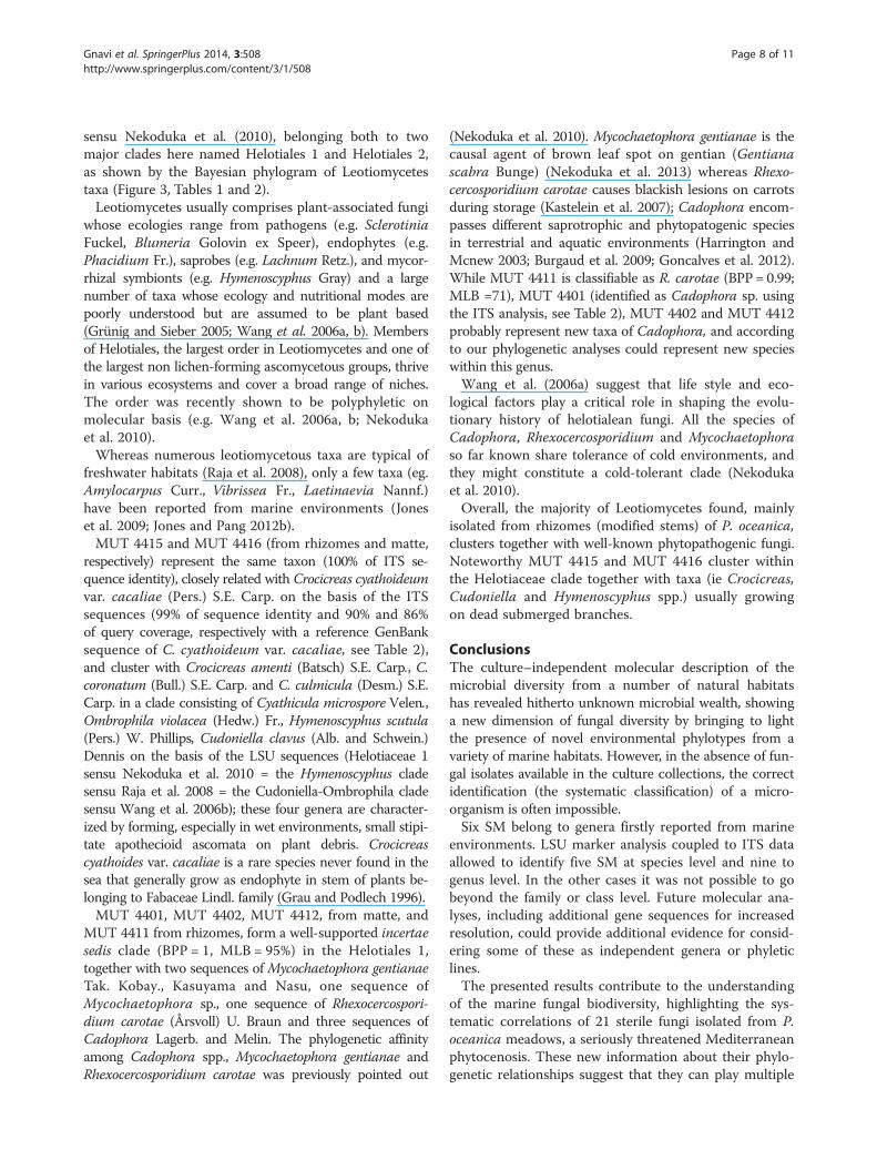
Figure 3 Bayesian phylogram of Leotiomycetes taxa including the six fungal isolates (labelled as MUT, in bold), on the dataset ofrDNA large subunit (LSU). Clades designations and sequences were from Wang et al. (2006b) and Nekoduka et al. (2010). Numbers abovebranches indicated BPP over 0.70 and MLB over 50. The alignment comprised 1,065 characters and contains 398 variable sites. # = strains isolatedfrom P. oceanica matte; § = strains isolated from P. oceanica rhizomes.
Gnavi et al. SpringerPlus 2014, 3:508 Page 4 of 11http://www.springerplus.com/content/3/1/508

Table 1 Taxonomic assessment of sterile mycelia isolated from P. oceanica
MUT code Part ofP. oceanica
Isolated taxa GenBank accession number
ITS 28S
Dothideomycetes
4273 Matte Pyrenochaetopsis sp. KJ395500 KJ395496
4313 Matte Didymellaceae sp. KF636765 KF636771
4323 Rhizomes Massarina rubi KF636766 KF636772
4378 Matte Pyrenochaeta sp. KF636767 KF636773
4379 Matte Pleospora typhicola KF636768 KF636774
4382 Matte Pyrenochaeta acicola KJ395501 KJ395497
4389 Matte Ramularia endophylla KJ395494 KJ395498
4397 Rhizomes Roussoellaceae sp. KC339235 KF636775
4403 Matte Cucurbitariaceae sp. KC339238 KF636780
4404 Rhizomes Phaeosphaeriaceaesp.
KC339239 KF636781
4405 Rhizomes Massarina sp. KC339240 KF636782
4407 Matte Biatriospora sp. KC339242 KF636783
4417 Matte Pleosporales sp. KF636769 KF636785
4419 Rhizomes Roussoellaceae sp. KC339245 KF636786
4420 Matte Lentitheciaceae sp. KF636770 KF636787
Leotiomycetes
4401 Rhizomes Cadophora sp. KC339236 KF636778
4402 Matte Cadophora sp. KC339237 KF636779
4411 Matte Rhexocercosporidiumcarotae
KF636763 KF636776
4412 Rhizomes Cadophora sp. KF636764 KF636777
4415 Rhizomes Crocicreas sp. KJ395495 KJ395499
4416 Rhizomes Crocicreas sp. KF636762 KF636784
Gnavi et al. SpringerPlus 2014, 3:508 Page 5 of 11http://www.springerplus.com/content/3/1/508
et al. 2009). This order has been extensively investigated inrecent years and many new families and marine lineageshave been identified. However, although many genera havebeen sequenced, their phylogenetic relationships remainunresolved and further taxon sampling with a wider rangeof genes being sequenced is required (Jones and Pang2012a; Zhang et al. 2012).According to LSU analysis, four SM isolated from P.
oceanica matte, MUT 4403, MUT 4378, MUT 4382 andMUT 4273, clearly belong to the family CucurbitariaceaeWinter. MUT 4403, MUT 4378 and MUT 4382 clusterwith Pyrenochaeta De Not. species; in particular MUT4382 is strictly related to Pyrenochaeta acicola (Moug.and Lév.) Sacc. Members of the genus Pyrenochaeta areCoelomycetes Grove widely distributed in the environ-ment in soil or in association with wood and plant deb-ris, and several species have been implicated in humaninfections (de Hoog et al. 2000; Badali et al. 2010); noPyrenochaeta species was so far reported from marineenvironments (Jones et al. 2009; Abdel-Wahab andBahkali 2012).
MUT 4273 falls among species of PyrenochaetopsisGruyter, Aveskamp and Verkley, a Coelomycetes genus closelyrelated to Pyrenochaeta. The described Pyrenochaetopsisspecies are all soilborne and mainly associated with gra-mineous plants (de Gruyter et al. 2010); no species wasso far reported as associated to marine environments(Jones et al. 2009; de Gruyter et al. 2010; Abdel-Wahaband Bahkali 2012).MUT 4404, isolated from rhizomes, clearly belongs
to Phaeosphaeriaceae M.E. Barr, (=Clade VII - Phaeo-sphaeriaceae according to Suetrong et al. 2009), but in anisolated lineage. Future molecular investigations includingadditional genetic markers will be necessary to better defineits taxonomic status. This taxonomic placement is also sup-ported by the ITS sequence analysis; BLAST search indi-cates a 100% of identity value (88% of query coverage) witha sequence of Septoria arundinacea Sacc., a taxon recentlyincluded in Phaeosphaeriaceae (Quaedvlieg et al. 2013).Jones et al. (2009) listed 3 genera with marine species inthis family: Carinispora K.D. Hyde, Lautitia S. Schatzand Phaeosphaeria I. Miyake, but till now only for 4

Table 2 Sterile mycelia isolated from P. oceanica, MUT Code and percentage of identity with ITS sequence of publicdatabase GenBank (NCBI)
MUT code Part of P. oceanica Its sequence homology % of sequence identity % of query coverage GenBank accession number
Dothideomycetes
4273 Matte Phoma sp. 98 100 KF646102
4313 Matte Didymella sp. 99 100 HQ607826
4323 Rhizomes Massarina sp. 96 99 AF383963
4378 Matte Pyrenochaeta inflorescentia 97 91 GU586851
4379 Matte Pleosporales sp. 100 93 FJ571480
4382 Matte Phaeophaeria sp. 99 97 EU715675
4389 Matte Mycosphaerella punticformis syn.Ramularia endophylla
100 100 EU343240
4397 Rhizomes Pleosporales sp. 93 99 JN572046
4403 Matte Pyrenochaeta sp. 97 99 KF561892
4404 Rhizomes Septoria arundinacea 100 88 GU361970
4405 Rhizomes Lophiostoma sp. 99 95 HQ914825
4407 Matte Pleosporales sp. 98 100 HM116750
4417 Matte Ascomycota sp. 99 96 FJ375144
4419 Rhizomes Pleosporales sp. 94 100 HM992495
4420 Matte Phoma herbarum 95 85 AB333774
Leotiomycetes
4401 Rhizomes Cadophora sp. 100 95 JF327417
4402 Matte Leptodontidium orchidicola 95 100 GQ302678
4411 Matte Rhexocercosporidium sp. 97 91 DQ249995
4412 Rhizomes Cadophora sp. 94 100 JN859261
4415 Rhizomes Crocicreas cf. cacaliae 99 90 FJ005108
4416 Rhizomes Crocicreas cf. cacaliae 99 86 FJ005108
Gnavi et al. SpringerPlus 2014, 3:508 Page 6 of 11http://www.springerplus.com/content/3/1/508
species of the latter genus, the LSU sequences are avail-able (P. albopunctata (Westend.) Shoemaker and C.E.Babc., P. olivacea Kohlm., Volkm.-Kohlm. and O.E. Erikss.,P. sparticola Leuchtm. and P. typharum (Desm.) L. Holm).However, the phylogram does not allow to include MUT4404 within this genus nor to the close genus LoratosporaKohlm. and Volkm.-Kohlm. recently included in this family(Schoch et al. 2009; Suetrong et al. 2009). The latter genusencompasses only one species, L. aestuarii Kohlm. andVolkm.-Kohlm. that occurs on Juncus roemerianus Scheeleculms (Kohlmeyer et al. 1995), a widespread monocotin saline aquatic environments.MUT 4379, frommatte, belongs to the family Pleosporaceae
Nitschke. It seems closely related to Pleospora typhicola(Cooke) Sacc. (BPP = 1; MLB = 100%), a taxon reportedfrom dead leaves of Typha spp. (Typha L., TyphaceaeJuss.) in moist or wet habitats (Shoemaker and Babcock1992).MUT 4313, from matte, belongs to Didymellaceae
Gruyter, Aveskamp and Verkley (=Clade IX - Didymellaceaeaccording to Suetrong et al. 2009). More in detail, it seemsmuch closely related to the genus Stagonosporopsis Died.,
which encompasses saprotrophic species from stem andleaves of terrestrial plants (Aveskamp et al. 2010). Ac-cording to Jones and Pang (2012a) few marine taxa ofDidymellaceae have been found in this family but onlyone species, Didymella fucicola (G.K. Sutherl.) Kohlm.,had been sequenced for LSU marker till now.MUT 4420, isolated frommatte, nests in the Lentitheciaceae
Yin. Zhang, C.L. Schoch, J. Fourn., Crous and K.D. Hyde,(=Clade I – according to Suetrong et al. 2009), but oc-cupies an isolated position. Till now only two marinespecies of Lentitheciaceae, Lentithecium phragmiticola(syn. Massarina phragmiticola Poon and K.D. Hyde)and Keissleriella rara Kohlm., Volkm.-Kohlm. and O.E.Erikss. have been isolated (Jones and Pang 2012a).MUT 4417, from matte, falls in an isolated position
close to Halojulella avicenniae (syn. Julella avicenniae(Borse) K.D. Hyde) (Halojulellaceae = the Clade X – JulellaFabre according to Suetrong et al. 2009) and related totwo Corynespora species (Corynespora Güssow, Coryne-sporascaceae Sivan.). Halojullela avicenniae is a fungusisolated from intertidal mangrove wood of Queensland(Hyde 1992; Ariyawansa et al. 2013). Little information is

Gnavi et al. SpringerPlus 2014, 3:508 Page 7 of 11http://www.springerplus.com/content/3/1/508
available about the Halojulellaceae family, to which manysaprobic and lichen-forming fungi belong (Ariyawansaet al. 2013). Corynespora genus has a widespread distribu-tion and includes 89 species of saprobes, pathogens, andendophytes fungi, some from woody and herbaceousplants, others on nematodes, and human skin (Dixon et al.2009). Corynespora cassiicola (Berk. and M.A. Curtis) C.T.Wei, the type species, is an important fungus causingtarget-spot on a wide host range in tropical and subtropicalcountries, especially Hevea brasiliensis (Willd. ex A. Juss.)Müll. Arg. in Sri Lanka and other countries (de Liyanageet al. 1986).Both MUT 4407 and MUT 4323 (isolated from matte
and rhizomes, respectively) cluster in Biatriosporaceae(Hyde et al. 2013). In particular, MUT 4407 clusters sisterto a clade consisting of Biatriospora marina K.D. Hydeand Borse, Biatriospora sp. and Nigrograna mackinnonii(Borelli) Gruyter, Verkley and Crous; according to ouranalysis, it could be regarded a species of genus BiatriosporaK.D. Hyde and Borse (Biatriospora sp.). MUT 4323 clusterswith high support (BPP = 1; MLB= 100%) with Massarinarubi (Fuckel) Sacc. The genus Biatriospora was intro-duced by Hyde and Borse (1986) for B. marina, a man-grove inhabiting species. After that, it has been collectedmany times from a wide range of hosts showing a widedistribution (Hyde et al. 2013). Nigrograna mackinnonii isa species recently described from mycetomas of humans(de Gruyter et al. 2012). Massarina Sacc. is a polyphyleticgenus (see below) of saprobic fungi that live both in ter-restrial and aquatic environments (Fallah and Shearer2001; Hyde et al. 2013). Moreover, Massarina rubi is aspecies sometimes recorded from freshwater (Fallah andShearer 2001).MUT 4397 and MUT 4419, both isolated from rhizomes,
are clearly assigned to the Roussoellaceae family, under def-inition by JK Liu et al. (Hyde et al. 2013). Most of the taxaincluding in Roussoellaceae have been isolated from bam-boo and palms (Hyde et al. 2013). The position of thesetwo strains within this family is unclear and the molecularanalyses did not allow including them in any of the knowngenera.MUT 4405, isolated from rhizomes, groups together
with Massarina corticola (Fuckel) L. Holm in an incertaesedis position close to Lophiostomataceae Sacc. as alreadysignalled for M. corticola by Suetrong et al. (2009) (basalpart of Clade XII – Lophiostomatceae); according to ourLSU phylogenetic analyses, it could be considerate asMassarina sp. Massarina corticola was reported fromsubmerged freshwater wood from China (Tsui et al. 2000,2001). As said before, Massarina is a polyphyletic genus(Zhang et al. 2009); most of the marine Massarina speciesthat have been sequenced have been referred to otherfamilies, while other species as M. beaurivagea Poonyth,K.D. Hyde, Aptroot and Peerally, M. cystophorae (Cribb
and J.W. Herb.) Kohlm. and E. Kohlm., M. lacertensisKohlm. and Volkm.-Kohlm., M. mauritiana Poonyth, K.D.Hyde and Aptroot, M. rhizophorae Poonyth, K.D. Hyde,Aptroot and Peerally and M. ricifera Kohlm., Volkm.-Kohlm. and O.E. Erikss. require study to confirm their pos-ition within this family (Jones et al. 2009).
SM of P. oceanica in Capnodiales (Dothideomycetes)The sequence belonging to the Capnodiales-Dothideomycetesclusters within the Mycosphaerellaceae Lindau. In Figure 2 isrepresented the Bayesian phylogram of Capnodialestaxa, including a sterile mycelium isolated from P. oceanica(Tables 1 and 2).The Capnodiales are mainly defined by their shared eco-
logical niche as leaf epiphytes associated with the honeydew produced by insects and saprobes or leaf pathogensand are an ascostromatal order without pseudoparaphyses(Hughes 1976; Crous et al. 2009a; Chomnunti et al. 2011).MUT 4389, isolated from matte, has been identified as
Ramularia endophylla Verkley and U. Braun accordingto the ITS and LSU sequence analysis (teleomorphMycosphaerella punctiformis (Pers.) Starbäck (Crous2009)). Mycosphaerella punctiformis (Mycosphaerellaceae),type species of the genus, is a plant pathogen belonging toa common genus in seawater (Verkley et al. 2004). Recentmolecular studies have shown that MycosphaerellaJohanson is a polyphyletic genus (Crous et al. 2007,2009b), with members belonging to many different genera,and even families, such as the Davidiellaceae C.L. Schoch,Spatafora, Crous and Shoemaker, Teratosphaeriaceae Crousand U. Braun, Dissoconiaceae Crous and de Hoog,Mycosphaerellaceae and Schizothyriaceae Höhn. exTrotter, Sacc., D. Sacc. and Traverso. Members of theMycosphaerella-complex are ecologically highly adaptable,and vary from being saprobic to fungicolous (Crous et al.2009a, b). Mycosphaerella species are also among themost common and destructive known plant pathogens,causing serious diseases on many economically importantcrops. Species are mainly foliicolous, although some ofthem are associated with stem cankers, fruit lesions orblemishes, spots and specks (Crous et al. 2011).On the whole, the majority of the Dothideomycetes
analysed in this study has been isolated from P. oceanicamatte, mainly constituted by decaying plant debris.This could explain why most of the SM can be as-cribed to families (i.e. Cucurbitariaceae, Didymellaceae,Mycosphaerellaceae and Pleosporaceae,) that include nu-merous taxa that can play multiple ecological roles assaprotrophs or opportunistic pathogens (Didymella,Phoma, Pyrenochaeta, and Stagonosporopsis spp.).
SM of P. oceanica in LetiomycetesThe six sequences belonging to the Leotiomycetes clus-tered in two subclades, incertae sedis and Helotiaceae 1
Gnavi et al. SpringerPlus 2014, 3:508 Page 8 of 11http://www.springerplus.com/content/3/1/508
sensu Nekoduka et al. (2010), belonging both to twomajor clades here named Helotiales 1 and Helotiales 2,as shown by the Bayesian phylogram of Leotiomycetestaxa (Figure 3, Tables 1 and 2).Leotiomycetes usually comprises plant-associated fungi
whose ecologies range from pathogens (e.g. SclerotiniaFuckel, Blumeria Golovin ex Speer), endophytes (e.g.Phacidium Fr.), saprobes (e.g. Lachnum Retz.), and mycor-rhizal symbionts (e.g. Hymenoscyphus Gray) and a largenumber of taxa whose ecology and nutritional modes arepoorly understood but are assumed to be plant based(Grünig and Sieber 2005; Wang et al. 2006a, b). Membersof Helotiales, the largest order in Leotiomycetes and one ofthe largest non lichen-forming ascomycetous groups, thrivein various ecosystems and cover a broad range of niches.The order was recently shown to be polyphyletic onmolecular basis (e.g. Wang et al. 2006a, b; Nekodukaet al. 2010).Whereas numerous leotiomycetous taxa are typical of
freshwater habitats (Raja et al. 2008), only a few taxa (eg.Amylocarpus Curr., Vibrissea Fr., Laetinaevia Nannf.)have been reported from marine environments (Joneset al. 2009; Jones and Pang 2012b).MUT 4415 and MUT 4416 (from rhizomes and matte,
respectively) represent the same taxon (100% of ITS se-quence identity), closely related with Crocicreas cyathoideumvar. cacaliae (Pers.) S.E. Carp. on the basis of the ITSsequences (99% of sequence identity and 90% and 86%of query coverage, respectively with a reference GenBanksequence of C. cyathoideum var. cacaliae, see Table 2),and cluster with Crocicreas amenti (Batsch) S.E. Carp., C.coronatum (Bull.) S.E. Carp. and C. culmicula (Desm.) S.E.Carp. in a clade consisting of Cyathicula microspore Velen.,Ombrophila violacea (Hedw.) Fr., Hymenoscyphus scutula(Pers.) W. Phillips, Cudoniella clavus (Alb. and Schwein.)Dennis on the basis of the LSU sequences (Helotiaceae 1sensu Nekoduka et al. 2010 = the Hymenoscyphus cladesensu Raja et al. 2008 = the Cudoniella-Ombrophila cladesensu Wang et al. 2006b); these four genera are character-ized by forming, especially in wet environments, small stipi-tate apothecioid ascomata on plant debris. Crocicreascyathoides var. cacaliae is a rare species never found in thesea that generally grow as endophyte in stem of plants be-longing to Fabaceae Lindl. family (Grau and Podlech 1996).MUT 4401, MUT 4402, MUT 4412, from matte, and
MUT 4411 from rhizomes, form a well-supported incertaesedis clade (BPP = 1, MLB = 95%) in the Helotiales 1,together with two sequences of Mycochaetophora gentianaeTak. Kobay., Kasuyama and Nasu, one sequence ofMycochaetophora sp., one sequence of Rhexocercospori-dium carotae (Årsvoll) U. Braun and three sequences ofCadophora Lagerb. and Melin. The phylogenetic affinityamong Cadophora spp., Mycochaetophora gentianae andRhexocercosporidium carotae was previously pointed out
(Nekoduka et al. 2010). Mycochaetophora gentianae is thecausal agent of brown leaf spot on gentian (Gentianascabra Bunge) (Nekoduka et al. 2013) whereas Rhexo-cercosporidium carotae causes blackish lesions on carrotsduring storage (Kastelein et al. 2007); Cadophora encom-passes different saprotrophic and phytopatogenic speciesin terrestrial and aquatic environments (Harrington andMcnew 2003; Burgaud et al. 2009; Goncalves et al. 2012).While MUT 4411 is classifiable as R. carotae (BPP = 0.99;MLB =71), MUT 4401 (identified as Cadophora sp. usingthe ITS analysis, see Table 2), MUT 4402 and MUT 4412probably represent new taxa of Cadophora, and accordingto our phylogenetic analyses could represent new specieswithin this genus.Wang et al. (2006a) suggest that life style and eco-
logical factors play a critical role in shaping the evolu-tionary history of helotialean fungi. All the species ofCadophora, Rhexocercosporidium and Mycochaetophoraso far known share tolerance of cold environments, andthey might constitute a cold-tolerant clade (Nekodukaet al. 2010).Overall, the majority of Leotiomycetes found, mainly
isolated from rhizomes (modified stems) of P. oceanica,clusters together with well-known phytopathogenic fungi.Noteworthy MUT 4415 and MUT 4416 cluster withinthe Helotiaceae clade together with taxa (ie Crocicreas,Cudoniella and Hymenoscyphus spp.) usually growingon dead submerged branches.
ConclusionsThe culture–independent molecular description of themicrobial diversity from a number of natural habitatshas revealed hitherto unknown microbial wealth, showinga new dimension of fungal diversity by bringing to lightthe presence of novel environmental phylotypes from avariety of marine habitats. However, in the absence of fun-gal isolates available in the culture collections, the correctidentification (the systematic classification) of a micro-organism is often impossible.Six SM belong to genera firstly reported from marine
environments. LSU marker analysis coupled to ITS dataallowed to identify five SM at species level and nine togenus level. In the other cases it was not possible to gobeyond the family or class level. Future molecular ana-lyses, including additional gene sequences for increasedresolution, could provide additional evidence for consid-ering some of these as independent genera or phyleticlines.The presented results contribute to the understanding
of the marine fungal biodiversity, highlighting the sys-tematic correlations of 21 sterile fungi isolated from P.oceanica meadows, a seriously threatened Mediterraneanphytocenosis. These new information about their phylo-genetic relationships suggest that they can play multiple

Gnavi et al. SpringerPlus 2014, 3:508 Page 9 of 11http://www.springerplus.com/content/3/1/508
ecological roles as saprotrophs or opportunistic patho-gens in marine environments. The results clearly showedthat all the sterile fungi belong to Dothideomycetes(Pleosporales and Capnodiales) and Leotiomycetes(Helotiales), despite the phylogenetic relationship ofsome of them remain to be determined since a majority ofthem may belong to new lineages of Ascomycota.
MethodsFungal collectionsThe analysed SM have been isolated in a previous work(Panno et al. 2013), from matte and rhizomes of P.oceanica growing in meadow localized in Punta Manaraclose to Riva Trigoso Bay - Liguria, Italy (44° 15′ 00″North - 9° 24′ 00″ West). A total of nine plants withthe surrounding matte were collected in March 2008 ata depth between −5 and −21 m. Plants were placed insealed sterile bags and maintained at about 4°C duringtransport. Within few hours the samples were seriallywashed with sterile water and divided into leaves, rhi-zomes, roots and matte. Five grams (fresh weight) ofeach composite sample were homogenized in 100 mlseawater sterilized by filtration (0.2 mm pores). The ho-mogenates were diluted 1:10 using sterilized seawaterand the final dilutions of each composite sample wereplated (1 ml per plate) on different media prepared withseawater. More information about fungal isolation pro-cedures is available in Panno et al. (2013).In details, 21 out of the previously 29isolated SM were
studied because the others were not able to survive inaxenic conditions despite the use of different oligo-trophic media prepared using seawater in order tomimic as much as possible the natural conditions.All fungal strains were preserved at the Mycotheca
Universitatis Taurinensis-MUT (DBIOS - University ofTurin, Table 1).
DNA extraction, PCR amplification and DNA sequencingGenomic DNA was extracted using CetylTrimethyl Am-monium Bromide (CTAB, Sigma-Aldrich St. Louis, USA)following the protocol of Graham et al. (1994), and thenrDNA ITS1–5.8S–ITS2 and LSU partial regions wereamplified using the universal primers ITS1F/ITS4 (Sigma-Aldrich St. Louis, USA) (White et al. 1990) and LR0R/LR7(Vilgalys and Hester 1990 Vilgalys lab, unpubl.,http://biology.duke.edu/fungi/mycolab/primers.htm), re-spectively. PCR amplifications were performed followingparameters by White et al. (1990) for the ITS region, andVilgalys and Hester (1990) for the LSU region. The ana-lyses were carried out by sequencing the purified ampli-cons. Newly generated sequences were deposited inGenBank database (www.ncbi.nlm.nih.gov) and the acces-sion numbers are shown in the Table 1.
Bioinformatics and Phylogenetic analysesThe obtained sequences were checked and assembledusing Geneious software (Drummond et al. 2010) andcompared to those available in the GenBank database(www.ncbi.nlm.nih.gov) using the blastn algorithm andCBS Mycobank Pairwise Sequence Alignment (http://www.mycobank.org/). A full phylogenetic analysis wasperformed only on LSU/LSU-5.8S sequences, since com-parable ITS sequences of fungi involved in the presentpaper were scant in public databases. Taxonomic assign-ment to SM based on ITS sequences was carried out byquerying with the blastn algorithm the SM representa-tive ITS sequence against the GenBank database (http://www.ncbi.nlm.nih.gov/Genbank/). For each isolatedtaxon the percentage ITS sequence identity value wasprovided (see Table 2). Blastn results were inspectedmanually to remove inconsistencies.LSU sequences were selected for phylogenetic analysis
on the basis of blastn and CBS (http://www.cbs.knaw.nl/)results. Accordingly, three sequences datasets were com-posed following Suetrong et al. (2009) and Hyde et al.(2013) for Pleosporales and Capnodiales Woron. (LSUdatasets), and Wang et al. (2006a, b) and Nekoduka et al.(2010) for Leotiomycetes (LSU + 5.8S dataset). Hysteriumangustatum Pers., Hysterobrevium smilacis (Schwein.) E.W.A. Boehm and C.L. Schoch and Psiloglonium simulans(W.R. Gerard) E.W.A. Boehm, C.L. Schoch and Spatafora(Hysteriales Lindau, Hysteriaceae Chevall.) were selectedas outgroup taxa for the Pleosporales (Dothideomycetes)dataset, Dothidea insculpta Wallr., Stylodothis pucci-nioides (DC.) Arx and E. Müll. and Dothiora cannabinaeFroid. (Dothideales, Dothideaceae), for the Capnodiales(Dothideomycetes) dataset, while Peziza varia (Hedw.)Alb. and Schwein. (Pezizomycetes O.E. Erikss. and Winka,Pezizales J. Schröt.) for the Leotiomycetes dataset.Alignments were generated using MAFFT (Katoh
et al. 2002) with default conditions for gap openings andgap extension penalties. The sequence alignments wererefined manually with MEGA 5.10 (Tamura et al. 2011).Phylogenetic analyses were performed using Bayesian In-ference (BI) and Maximum Likelihood (ML) approaches.The BI was performed with MrBayes 3.1.2 (Huelsenbeckand Ronquist 2001) with four incrementally heated sim-ultaneous Monte Carlo Markov Chains (MCMC) runover 10 million generations, under GTR + Γ evolutionarymodel. Trees were sampled every 1,000 generationsresulting in an overall sampling of 10,001 trees; the first2,500 trees were discarded as “burn-in” (25%). For theremaining trees, a majority rule consensus tree showingall compatible partitions was computed to obtain estimatesfor Bayesian Posterior Probabilities (BPP). ML estimationwas performed through RAxML v.7.3.2 (Stamatakis 2006)with 1,000 bootstrap replicates (Felsenstein 1985) using theGTRGAMMA algorithm to perform a tree inference and

Gnavi et al. SpringerPlus 2014, 3:508 Page 10 of 11http://www.springerplus.com/content/3/1/508
search for a good topology. Support values from bootstrap-ping runs (MLB) were mapped on the globally best treeusing the “-f a” option of RAxML and “-x 12345” as a ran-dom seed to invoke the novel rapid bootstrapping algo-rithm. BI and ML analyses were run on the CIPRESScience Gateway web server (Miller et al. 2010). Only BPPvalues over 0.70 and MLB over 50 were reported in theresulting trees (Figures 1, 2 and 3).Alignments and phylogenetic trees are available at
TreeBASE (http://www.treebase.org, submission numberS16179).
Competing interestsThe authors report no conflict of interest concerning the materials ormethods used in this study or the findings specified in this paper.
Authors’ contributionsGCV conceived of the study, and participated in its design, coordination anddata analysis. LP and GCV carried out the collection of materials and draftedthe manuscript. GG carried out all the molecular techniques and participatedto the sequence alignment and phylogenetic analysis and drafted andrevised the manuscript. EE carried out the sequence alignment and revisedthe manuscript. AV and EE carried out the phylogenetic and statisticalanalysis. GCV and AV wrote the final version of the manuscript. All authorsread, revised and approved the final version of the manuscript.
AcknowledgmentsWe are grateful to Pelagosphera - Soc. Cooperativa a r.l, for collecting theplant samples and for the useful comments and suggestions.
Author details1Mycotheca Universitatis Taurinensis (M.U.T.), Department of Life Sciencesand Systems Biology (DBIOS), University of Turin, Viale Mattioli, 25, 10125Turin, Italy. 2Systematic Mycology Lab, Department of Life Sciences andSystems Biology (DBIOS), University of Turin, Viale P.A. Mattioli, 25, 10125Turin, Italy.
Received: 28 May 2014 Accepted: 18 August 2014Published: 9 September 2014
ReferencesAbdel-Wahab MA, Bahkali AHA (2012) Taxonomy of filamentous anamorphic
marine fungi: morphology and molecular evidence. In: Jones EBG, Pang KL(eds) Marine fungi and fungal-like organisms, 1st edn. Walter de GruyterGmbH and Co. KG, Berlin/Boston
Alain K, Querellou J (2009) Cultivating the uncultured: limits, advances and futurechallenges. Extremophiles 13:583–594
Ariyawansa HA, Jones EBG, Suetrong S, Alias SA, Kang JC, Hyde KD (2013)Halojulellaceae a new family of the order Pleosporales. Phytotaxa 130:14–24
Aveskamp MM, de Gruyter J, Woudenberg JHC, Verkley GJM, Crous PW (2010)Highlights of the Didymellaceae: a polyphasic approach to characterisePhoma and related pleosporalean genera. Stud Mycol 65:1–60
Badali H, Chander J, Gulati N, Attri A, Chopra R, Najafzadeh MJ, Chhabra S, MeisJFGM, de Hoog GS (2010) Subcutaneous phaeohyphomycotic cyst caused byPyrenochaeta romeroi. Med Biol 48:763–768
Baker PW, Kennedy J, Dobson DW, Marchesi JR (2008) Phylogenetic diversity andantimicrobial activities of fungi associated with Haliclona simulans isolatedfrom Irish coastal waters. Mar Biotechnol 11:540–547
Burgaud G, Le Calvez T, Arzur D, Vandenkoornhuyse P, Barbier G (2009) Diversityof culturable marine filamentous fungi from deep-sea hydrothermal vents.Environ Microbiol 11:1588–1600
Chomnunti P, Schoch CL, Aguirre-Hudson B, Koko TW, Hongsanan S, Jones EBG,Kodsub R, Chukeatirote E, Bahkali AH, Hyde KD (2011) Capnodiaceae. FungalDivers 51:103–134
Crous PW (2009) Taxonomy and phylogeny of the genus Mycosphaerella and itsanamorphs. Fungal Divers 38:1–24
Crous PW, Braun U, Groenewald JZ (2007) Mycosphaerella is polyphyletic. StudMycol 58:1–32
Crous PW, Schoch CL, Hyde KD, Wood AR, Gueidan C, de Hoog GS, GroenewaldJZ (2009a) Phylogenetic lineages in the Capnodiales. Stud Mycol 64:17–47
Crous PW, Summerell BA, Carnegie AJ, Wingfield MJ, Hunter GC, Burgess TI,Andjic V, Barber PA, Groenewald JZ (2009b) Unravelling Mycosphaerella: doyou believe in genera? Persoonia 23:99–118
Crous PW, Tanaka K, Summerell BA, Groenewald JZ (2011) Additions to theMycosphaerella complex. IMA Fungus 2:49–64
Crous PW, Braun U, Hunter GC, Wingfield MJ, Verkley GJM, Shin HD, Nakashima C,Groenewald JZ (2013) Phylogenetic lineages in Pseudocercospora. Stud Mycol75:37–114
Cuomo V, Vanzanella F, Fresi E, Cinelli F, Mazzella L (1985) Fungal flora ofPosidonia oceanica and its ecological significance. T Brit Mycol Soc 84:35–40
Damare S, Raghukumar C, Raghukumar S (2006) Fungi in deep-sea sediments ofthe Central Indian Basin. Deep-Sea Res Pt I 53:14–27
de Gruyter JD, Woudenberg JH, Aveskamp MM, Verkley GJ, Groenewald JK, CrousPW (2010) Systematic reappraisal of species in Phoma section Paraphoma,Pyrenochaeta and Pleurophoma. Mycologia 102:1066–1081
de Gruyter J, Woudenberg JHC, Aveskamp MM, Verkley GJM, Groenewald JZ,Crous PW (2012) Redisposition of Phoma-like anamorphs in Pleosporales.Stud Mycol 75:1–36
de Hoog GS, Guarro J, Gené J, Figueras MJ (2000) Atlas of Clinical Fungi, 2ndedn. American Society of Microbiology, Centraalbureau voorSchimmelcultures / Utrecht, The Netherlands and Reus, Spain
de Liyanage AS, Jayasinghe CK, Liyanage NS, Jayaratne R (1986) Corynespora leafspot disease of rubber (Hevea brasiliensis) a new record. J Rubber ResInstitute of Sri Lanka 65:47–50
Dixon LJ, Schlub RL, Pernezny K, Datnoff LE (2009) Host specialization andphylogenetic diversity of Corynespora cassiicola. Phytopathology99:1015–1027
Drummond AJ, Ashton B, Buxton S, Cheung M, Cooper A, Duran C, Field M,Heled J, Kearse M, Markowitz S, Moir R, Stones-Havas S, Sturrock S, Thierer T,Wilson A (2010) Geneious v5.3, available from http://www.geneious.com
Egidi E, de Hoog GS, Isola D, Onofri S, Quaedvlieg W, de Vries M, Verkley GJM,Stielow JB, Zucconi L, Selbmann L (2014) Phylogeny and taxonomy ofmeristematic rock-inhabiting black fungi in the Dothideomycetes based onmulti-locus phylogenies. Fungal Divers 65:127–165
Fallah PM, Shearer CA (2001) Freshwater Ascomycetes: new or noteworthyspecies from north temperate lakes in Wisconsin. Mycologia 93:566–602
Felsenstein J (1985) Confidence limits on phylogenies: an approach using thebootstrap. Evolution 39:783–791
Garzoli L (2013) Marine Fungi in the Mediterranean Sea: Discovering theirDiversity and their Potential Application in Biotechnology, PhD Thesis.Università degli Studi di Pavia, Dipartimento di Scienze della Terra edell’Ambiente, Pavia, Italy
Goncalves VN, Vaz A, Rosa CA, Rosa LH (2012) Diversity and distribution of fungalcommunities in lakes of Antarctica. FEMS Microbiol Ecol 82:459–471
Graham GC, Mayers P, Henry RJ (1994) A simplified method for the preparationof fungal genomic DNA for PCR and RAPD analysis. Biotechniques16:48–50
Grau J, Podlech D (1996) Sendtnera. Herausgeber, Munchen, GermanyGrünig CR, Sieber TN (2005) Molecular and phenotypic description of the
widespread root symbiont Acephala applanata gen. et sp. nov., formerlyknown as dark-septate endophyte Type 1. Mycologia 97:628–640
Harrington TC, McNew DL (2003) Phylogenetic analysis places the Phialophora-like anamorph genus Cadophora in the Helotiales. Mycotaxon 10:459–472
Huelsenbeck JP, Ronquist F (2001) MrBayes: Bayesian inference of phylogeny.Bioinformatics 17:754–755
Hughes SJ (1976) Sooty moulds. Mycologia 68:693–820Hyde KD (1992) Julella avicenniae (Borse) comb. nov. (Thelenellaceae) from
intertidal mangrove wood and miscellaneous fungi from the NE coast ofQueensland. Mycol Res 96:939–942
Hyde KD, Borse BD (1986) Marine fungi from Seychelles V. Biatriospora marinagen. et sp. nov. from mangrove wood. Mycotaxon 26:263–270
Hyde KD, Jones EBG, Liu JK, Ariyawansa H, Boehm E, Boonmee S, Braun U,Chomnunti P, Crous PW, Dai DQ, Diederich P, Dissanayake A, Doilom M,Doveri F, Hongsanan S, Jayawardena R, Lawrey JD, Li YM, Liu YX, Lücking R,Monkai J, Muggia L, Nelsen MP, Pang KL, Phookamsak R, Senanayake IC,Shearer CA, Suetrong S, Tanaka K, Thambugala KM et al (2013) Families ofDothideomycetes. Fungal Divers 63:1–313
Imhoff JF, Labes A, Wiese J (2011) Bio-mining the microbial treasures of theocean: new natural products. Biotechnol Adv 29:468–482

Gnavi et al. SpringerPlus 2014, 3:508 Page 11 of 11http://www.springerplus.com/content/3/1/508
Jones EBG (1963) Marine fungi: II. Ascomycetes and deuteromycetes fromsubmerged wood and drift Spartina. T Brit Mycol Soc 46:135–144
Jones EBG, Pang KL (2012a) Marine fungi and fungal-like organisms. de Gruyter,Berlin/Boston, Germany/USA
Jones EBG, Pang KL (2012b) Tropical aquatic fungi. Biodivers Conserv 21:2403–2423Jones EBG, Sakayaroj J, Suetrong S, Somrithipol S, Pang KL (2009) Classification of
marine Ascomycota, anamorphic taxa and Basidiomycota. Fungal Divers35:1–187
Kastelein P, Stilma ESC, Elderson J, Koehl J (2007) Occurrence ofRhexocercosporidium carotae on cold stored carrot roots in the Netherlands.Eur J Plant Pathol 117:293–305
Katoh K, Misawa K, Kuma K, Miyata T (2002) MAFFT: a novel method for rapidmultiple sequence alignment based on fast Fourier transform. Nucleic AcidsRes 30:3059–3066
Kirk PM, Cannon PF, Minter DW, Stalpers JA (2008) Ainsworth and Bisby’sDictionary of the Fungi, 10th edn. CABI, Wallingford, UK
Kohlmeyer J, Volkmann-Kohlmeyer B, Eriksson OE (1995) Fungi on Juncus roemerianus2. New dictyosporous ascomycetes. Bot Mar 38:165–174
Mazzella L, Alberte RS (1986) Light adaptation and the role of autotrophicepiphytes in primary production of the temperate seagrass, Zostera marina L.J Exp Mar Biol Ecol 100:165–180
Meyers SP, Orpurt PA, Simms J, Boral LL (1965) Thalassiomycetes VII. Observationson fungal infestation of turtle grass, Thalassia testudinum König. B Mar Sci15:548–564
Miller MA, Pfeiffer W, Schwartz T (2010) Creating the CIPRES Science Gateway forinference of large phylogenetic trees. In: IEEE. Proceedings of the GatewayComputing Environments Workshop (GCE), New Orleans, LA, 14 November2010
Morrison-Gardiner S (2002) Dominant fungi from Australian coral reefs. FungalDivers 9:105–121
Nekoduka S, Tanaka K, Harada Y, Sano T (2010) Phylogenetic affinity ofMycochaetophora gentianae, the causal fungus of brown leaf spot on gentian(Gentiana triflora) to Pseudocercosporella-like hyphomycetes in Helotiales.Mycoscience 51:123–133
Nekoduka S, Tanaka K, Sano T (2013) Overwintering of brown leaf spot fungus,Mycochaetophora gentianae, in infected gentian leaves as the primaryinoculum source. J Gen Plant Pathol 79:175–177
Newell SY (1981) Fungi and bacteria in or on leaves of eelgrass (Zostera marinaL.) from Chesapeake Bay. Appl Environ Microb 41:1219–1224
Panno L, Bruno M, Voyron S, Anastasi A, Gnavi G, Miserere L, Varese GC (2013)Diversity, ecological role and potential biotechnological applications ofmarine fungi associated to the seagrass Posidonia oceanica. New Biotechnol30:685–694
Perez-Ortega S, Suija A, Crespo A, de los Rios A (2014) Lichenicolous fungi of thegenus Abrothallus (Dothideomycetes: Abrothallales ordo nov.) are sister tothe predominantly aquatic Janhulales. Fungal Divers 64:295–304
Pergent G, Boudouresque CF, Dumay O, Pergent-Martini C, Wyllie-Echeverria S(2008) Competition between the invasive macrophyte Caulerpa taxifolia andthe seagrass Posidonia oceanica: contrasting strategies. BMC Ecol 8:20
Prasannarai K, Sridhar KR (2001) Diversity and abundance of higher marine fungion woody substrates along the west coast of India. Curr Sci India 81:304–311
Quaedvlieg W, Verkley GJM, Shin HD, Barreto RW, Alfenas AC, Swart WJ,Groenewald JZ, Crous PW (2013) Sizing up Septoria. Stud Mycol 75:307–390
Raghukumar S (2004) The role of fungi in marine detrital processes. In: RamaiahN (ed) Marine microbiology: facets and opportunities. National Institute ofOceanography, Goa, pp 91–101
Raja HA, Miller AN, Shearer CA (2008) Freshwater ascomycetes: Aquapoteriumpinicola, a new genus and species of Helotiales (Leotiomycetes) from Florida.Mycologia 100:141–148
Rateb ME, Ebel R (2011) Secondary metabolites of fungi from marine habitats.Nat Prod Rep 28:290–344
Schoch CL, Crous PW, Groenewald JZ, Boehm EWA, Burgess TI, de Gruyter J, deHoog GS, Dixon LJ, Grube M, Gueidan C, Harada Y, Hatakeyama S, HirayamaK, Hosoya T, Huhndorf SM, Hyde KD, Jones EBG, Kohlmeyer J, Kruys A, Li YM,Lücking R, Lumbsch HT, Marvanová L, Mbatchou JS, McVay AH, Miller AN,Mugambi GK, Muggia L, Nelsen MP, Nelson P et al (2009) A class-widephylogenetic assessment of Dothideomycetes. Stud Mycol 64:1–15
Shearer CA, Raja HA, Miller AN, Nelson P, Tanaka K, Hirayama K, Marvanová L,Hyde KD, Zhang Y (2009) The molecular phylogeny of freshwaterDothideomycetes. Stud Mycol 64:145–153.
Shoemaker RA, Babcock CE (1992) Applanodictyosporous Pleosporales:Clathrospora, Comoclathris, Graphyllium, Macrospora, and Platysporoides. Can JBotany 70:1617–1658
Stamatakis A (2006) RAxML-VI-HPC: maximum likelihood-based phylogenetic analyseswith thousands of taxa and mixed models. Bioinformatics 22:2688–2690
Suetrong S, Schoch CL, Spatafora JW, Kohlmeyer J, Volkmann-Kohlmeyer B,Sakayaroj J, Phongpaichit S, Tanaka K, Hirayama K, Jones EBG (2009) Molecularsystematics of the marine Dothideomycetes. Stud Mycol 64:155–173
Suryanarayanan TS (2012) The diversity and importance of fungi associated withmarine sponges. Bot Mar 55:553–564
Tamura K, Peterson D, Peterson N, Stecher G, Nei M, Kumar S (2011) MEGA5:molecular evolutionary genetics analysis using maximum likelihood,evolutionary distance, and maximum parsimony methods. Mol Biol Evol28:2731–2739
Toledo-Hernández C, Bones-González A, Ortiz-Vázquez OE, Sabat AM, Bayman P(2007) Fungi in the sea fan Gorgonia ventalina: diversity and samplingstrategies. Coral Reefs 26:725–730
Tsui CKM, Hyde KD, Hodgkiss IJ (2000) Biodiversity of fungi on submerged woodin Hong Kong streams. Aquat Microb Ecol 21:289–298
Tsui CKM, Hyde KD, Hodgkiss IJ (2001) Longitudinal and temporal distribution offreshwater ascomycetes and dematiaceous hyphomycetes on submergedwood in the Lam Tsuen River, Hong Kong. J N Am Benthol Soc 20:533–549
Verkley JM, Crous PW, Groenewald JZ, Braun U, Aptroot A (2004) Mycosphaerellapunctiformis revisited: morphology, phylogeny, and epitypification of thetype species of the genus Mycosphaerella. Mycol Res 108:1271–1282
Vijaykrishna D, Jeewon R, Hyde KD (2006) Molecular taxonomy, origins andevolution of freshwater Ascomycetes. Fungal Divers 23:351–390
Vilgalys R, Hester M (1990) Rapid genetic identification and mapping ofenzymatically amplified ribosomal DNA from several Cryptococcus species.J Bacteriol 172:4238–4246
Wang Z, Binder M, Schoch CL, Johnston PR, Spatafora JW, Hibbett DS (2006a)Evolution of helotialean fungi (Leotiomycetes, Pezizomycotina): a nuclearrDNA phylogeny. Mol Phylogenet Evol 41:295–312
Wang Z, Johnston PR, Takamatsu S, Spatafora JW, Hibbett DS (2006b) Toward aphylogenetic classification of the Leotiomycetes based on rDNA data.Mycologia 98:1065–1075
White TJ, Bruns T, Lee S, Taylor JW (1990) Amplification and direct sequencing offungal ribosomal RNA genes for phylogenetics. In: Innis MA, Gelfand DH,Sninsky JJ, White TJ (eds). PCR protocols: a guide to methods andapplications, 1st edn, Academic Press Inc., New York
Zhang Y, Crous PW, Schoch CL, Hyde KD (2012) Pleosporales. Fungal Divers 53:1–221Zhang Y, Schoch CL, Fournier J, Crous PW, De Gruyter J, Woudenberg JHC,
Hirayama K, Tanaka K, Pointing SB, Spatafora JW, Hyde KD (2009) Multi-locusphylogeny of the Pleosporales: a taxonomic, ecological and evolutionaryre-evaluation. Stud Mycol 64:85–102
Zuccaro A, Conrad L, Spatafora LW, Kohlmeyer J, Draeger S, Mitchell JI (2008)Detection and identification of fungi intimately associated with the brownseaweed Fucus serratus. Appl Environ Microb 74:931–941
doi:10.1186/2193-1801-3-508Cite this article as: Gnavi et al.: Dothideomycetes and Leotiomycetessterile mycelia isolated from the Italian seagrass Posidonia oceanicabased on rDNA data. SpringerPlus 2014 3:508.
Submit your manuscript to a journal and benefi t from:
7 Convenient online submission
7 Rigorous peer review
7 Immediate publication on acceptance
7 Open access: articles freely available online
7 High visibility within the fi eld
7 Retaining the copyright to your article
Submit your next manuscript at 7 springeropen.com