Biological Matrices for the Evaluation of In Utero Exposure to ...
24
REVIEW ARTICLE Biological Matrices for the Evaluation of In Utero Exposure to Drugs of Abuse Jaime Lozano, MD,* Oscar Garcı ´a-Algar, MD, PhD,* Oriol Vall, MD, PhD,* Rafael de la Torre, PhD,† Giulia Scaravelli, MD, PhD,‡ and Simona Pichini, PhD§ Abstract: In recent years, the evaluation of in utero exposure to drugs of abuse has been achieved by testing biological matrices coming from the fetus or newborn (eg, meconium, fetal hair, cord blood, neonatal urine), the pregnant or nursing mother (eg, hair, blood, oral fluid, sweat, urine, breast milk), or from both the fetus and the mother (placenta, amniotic fluid). Overall, these matrices have the advantage of noninvasive collection (with the exception of amniotic fluid) and early detection of exposure from different gestational periods. Matrices such as amniotic fluid, meconium, fetal hair, and maternal hair provide a long historical record of prenatal exposure to certain drugs and can account for different periods of gestation: amniotic fluid from the early pregnancy, meconium for the second and third trimester of gestation, fetal hair for the third, and finally maternal hair (when long enough) for the whole pregnancy. Placenta may reveal the passage of a substance from the mother to the fetus. Cord blood and neonatal urine are useful for determining acute exposure to drugs of abuse in the period immediately previous to delivery. Drug detection in maternal blood, oral fluid, and sweat accounts only for acute consumption that occurred in the hours previous to collection and gives poor information concerning fetal exposure. Different immunoassays were used as screening methods for drug testing in the above-reported matrices or as unique analytical investigation tools when chromatographic techniques coupled to mass spectrometry were not commonly available. However, in the last decade, both liquid and gas chromatography-mass spectrometric methodologies have been routinely applied after appropriate extrac- tion of drugs and their metabolites from these biological matrices. Key Words: drugs of abuse, exposure, fetus, biological matrices (Ther Drug Monit 2007;29:711–734) BACKGROUND In utero exposure to drugs of abuse may have con- sequences not only for the development of the fetus but also for the physiology of some biological systems and neuro- cognitive aspects of children during later stages in life. 1,2 More than 75% of infants exposed to drugs have major medical problems as compared with 27% of unexposed infants. 4 The cost of treating drug-affected infants is twice the cost of nonaffected infants. Also, obstetric complications are higher among drug abusing mothers, 3 so that assessment of in utero drug exposure is relevant for the care of the mother and the offspring. In this sense, several approaches are possible, such as monitoring maternal drug consumption by urinalysis (approx- imately 3 times per wk) and weekly sweat analysis through patches or hair testing (approximately once per trimester of pregnancy). Except for hair analysis, the window of detection of urine and sweat is short, reflecting drug use during the previous few days. 3,4 Moreover, the 3 times a week urine testing and weekly sweat testing are both unrealistic and expensive. Alternatively, the assessment of maternal drug consumption may be investigated by the use of questionnaires regarding the use of drugs of abuse. This method, however, has already shown limitations in a clinical series of approximately 3000 newborns, 43% of whom tested positive for drugs of abuse in different biological matrices, whereas only 11% of mothers recognized drug consumption. 32 Another approach is monitoring exposure to drugs of abuse by testing alternative (also defined as nonconventional) biological matrices coming from the fetus or the newborn (eg, meconium, fetal hair, cord blood, neonatal urine), from the pregnant or nursing mother (eg, hair, blood, oral fluid, sweat, urine, breast milk), or from both fetus and mother (placenta, amniotic fluid). Overall, these matrices have the advantage of noninvasive collection (with the exception of amniotic fluid) and early detection of exposure from different gestational periods. The interrelation- ship between maternal and fetal exposure to drugs is pre- sented schematically in Figure 1. Biological matrices (other than urine) used for monitoring in utero drug exposure are outlined in the figure, and the respective time windows of drug detection as well as their usefulness in retrospective assessment of in utero exposure to illicit drugs are shown in Figure 2. The present review focuses on the feasibility and clinical significance of the use of alternate biological matrices for the assessment of in utero exposure to drugs of abuse. Each bio- logical matrix and the respective analytical methodologies Received for publication June 1, 2007; accepted August 20, 2007. From the *Unitat de Recerca Infa `ncia i Entorn (URIE), Paediatric Service, Hospital del Mar, and Departament de Pediatria, Ginecologia i Obstetricia, i Medicina Preventiva, Universitat Auto `noma, Barcelona, Spain; †Unitat de Recerca en Farmacologia, Institut Municipal d’Investigacio ´ Me `dica- Hospital del Mar and Departament de Cie `ncies Experimentals i de la Salut, Universitat Pompeu Fabra, Barcelona, Spain; ‡National Centre for Epidemiology, Surveillance and Health Promotion, Istituto Superiore di Sanita `, Rome, Italy; and §Department of Therapeutic Research and Medicines Evaluation, Istituto Superiore di Sanita `, Rome, Italy. Reprints: Simona Pichini, PhD, Department of Therapeutic Research and Medicines Evaluation, Istituto Superiore di Sanita ´, V.le Regina Elena 299, 00161, Rome, Italy (e-mail: [email protected]). Copyright Ó 2007 by Lippincott Williams & Wilkins Ther Drug Monit Volume 29, Number 6, December 2007 711
-
Upload
khangminh22 -
Category
Documents
-
view
6 -
download
0
Transcript of Biological Matrices for the Evaluation of In Utero Exposure to ...
REVIEW ARTICLE
Biological Matrices for the Evaluation of In UteroExposure to Drugs of Abuse
Jaime Lozano, MD,* Oscar Garcıa-Algar, MD, PhD,* Oriol Vall, MD, PhD,* Rafael de la Torre, PhD,†
Giulia Scaravelli, MD, PhD,‡ and Simona Pichini, PhD§
Abstract: In recent years, the evaluation of in utero exposure to
drugs of abuse has been achieved by testing biological matrices
coming from the fetus or newborn (eg, meconium, fetal hair, cord
blood, neonatal urine), the pregnant or nursing mother (eg, hair,
blood, oral fluid, sweat, urine, breast milk), or from both the fetus and
the mother (placenta, amniotic fluid). Overall, these matrices have the
advantage of noninvasive collection (with the exception of amniotic
fluid) and early detection of exposure from different gestational
periods. Matrices such as amniotic fluid, meconium, fetal hair, and
maternal hair provide a long historical record of prenatal exposure to
certain drugs and can account for different periods of gestation:
amniotic fluid from the early pregnancy, meconium for the second
and third trimester of gestation, fetal hair for the third, and finally
maternal hair (when long enough) for the whole pregnancy. Placenta
may reveal the passage of a substance from the mother to the fetus.
Cord blood and neonatal urine are useful for determining acute
exposure to drugs of abuse in the period immediately previous to
delivery. Drug detection in maternal blood, oral fluid, and sweat
accounts only for acute consumption that occurred in the hours
previous to collection and gives poor information concerning fetal
exposure. Different immunoassays were used as screening methods
for drug testing in the above-reported matrices or as unique analytical
investigation tools when chromatographic techniques coupled to
mass spectrometry were not commonly available. However, in the last
decade, both liquid and gas chromatography-mass spectrometric
methodologies have been routinely applied after appropriate extrac-
tion of drugs and their metabolites from these biological matrices.
Key Words: drugs of abuse, exposure, fetus, biological matrices
(Ther Drug Monit 2007;29:711–734)
BACKGROUNDIn utero exposure to drugs of abuse may have con-
sequences not only for the development of the fetus but alsofor the physiology of some biological systems and neuro-cognitive aspects of children during later stages in life.1,2 Morethan 75% of infants exposed to drugs have major medicalproblems as compared with 27% of unexposed infants.4
The cost of treating drug-affected infants is twice the cost ofnonaffected infants. Also, obstetric complications are higheramong drug abusing mothers,3 so that assessment of in uterodrug exposure is relevant for the care of the mother and theoffspring.
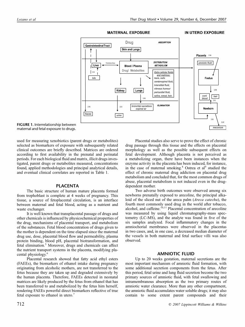
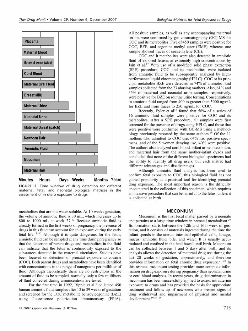
In this sense, several approaches are possible, such asmonitoring maternal drug consumption by urinalysis (approx-imately 3 times per wk) and weekly sweat analysis throughpatches or hair testing (approximately once per trimesterof pregnancy). Except for hair analysis, the window ofdetection of urine and sweat is short, reflecting drug use duringthe previous few days.3,4 Moreover, the 3 times a week urinetesting and weekly sweat testing are both unrealistic andexpensive. Alternatively, the assessment of maternal drugconsumption may be investigated by the use of questionnairesregarding the use of drugs of abuse. This method, however, hasalready shown limitations in a clinical series of approximately3000 newborns, 43% of whom tested positive for drugsof abuse in different biological matrices, whereas only 11%of mothers recognized drug consumption.32 Another approachis monitoring exposure to drugs of abuse by testing alternative(also defined as nonconventional) biological matrices comingfrom the fetus or the newborn (eg, meconium, fetal hair,cord blood, neonatal urine), from the pregnant or nursingmother (eg, hair, blood, oral fluid, sweat, urine, breast milk), orfrom both fetus and mother (placenta, amniotic fluid). Overall,these matrices have the advantage of noninvasive collection(with the exception of amniotic fluid) and early detection ofexposure from different gestational periods. The interrelation-ship between maternal and fetal exposure to drugs is pre-sented schematically in Figure 1. Biological matrices (otherthan urine) used for monitoring in utero drug exposure areoutlined in the figure, and the respective time windows ofdrug detection as well as their usefulness in retrospectiveassessment of in utero exposure to illicit drugs are shownin Figure 2.
The present review focuses on the feasibility and clinical
significance of the use of alternate biological matrices for the
assessment of in utero exposure to drugs of abuse. Each bio-
logical matrix and the respective analytical methodologies
Received for publication June 1, 2007; accepted August 20, 2007.From the *Unitat de Recerca Infancia i Entorn (URIE), Paediatric Service,
Hospital del Mar, and Departament de Pediatria, Ginecologia i Obstetricia,i Medicina Preventiva, Universitat Autonoma, Barcelona, Spain; †Unitatde Recerca en Farmacologia, Institut Municipal d’Investigacio Medica-Hospital del Mar and Departament de Ciencies Experimentals i de la Salut,Universitat Pompeu Fabra, Barcelona, Spain; ‡National Centre forEpidemiology, Surveillance and Health Promotion, Istituto Superiore diSanita, Rome, Italy; and §Department of Therapeutic Research andMedicines Evaluation, Istituto Superiore di Sanita, Rome, Italy.
Reprints: Simona Pichini, PhD, Department of Therapeutic Research andMedicines Evaluation, Istituto Superiore di Sanita, V.le Regina Elena 299,00161, Rome, Italy (e-mail: [email protected]).
Copyright � 2007 by Lippincott Williams & Wilkins
Ther Drug Monit � Volume 29, Number 6, December 2007 711
used for measuring xenobiotics (parent drugs or metabolites)selected as biomarkers of exposure with subsequently relatedclinical outcomes are briefly described. Matrices are orderedaccording to first availability in the prenatal and perinatalperiods. For each biological fluid andmatrix, illicit drugs inves-tigated, parent drugs or metabolites measured, concentrationsfound, applied methodologies and principal analytical details,and eventual clinical correlates are reported in Table 1.
PLACENTAThe basic structure of human mature placenta formed
from trophoblast is complete at 4 weeks of pregnancy. Thistissue, a source of fetoplacental circulation, is an interfacebetween maternal and fetal blood, acting as a nutrient andwaste exchanger.
It is well known that transplacental passage of drugs andother chemicals is influenced by physicochemical properties ofthe drug, mechanisms of placental transport, and metabolismof the substances. Fetal blood concentration of drugs given tothe mother is dependent on the time elapsed since the maternaldrug use, dose, placental blood flow and permeability, plasmaprotein binding, blood pH, placental biotransformation, andfetal elimination.5 Moreover, drugs and chemicals can affectthe nutrient transport systems in the placenta, modifying pla-cental physiology.6
Placental research showed that fatty acid ethyl esters(FAEEs), the biomarkers of ethanol intake during pregnancyoriginating from alcoholic mothers, are not transferred to thefetus because they are taken up and degraded extensively bythe human placenta. Therefore, FAEEs detected in neonatalmatrices are likely produced by the fetus from ethanol that hasbeen transferred to and metabolized by the fetus him herself,rendering FAEEs powerful direct biomarkers reflective of truefetal exposure to ethanol in utero.7
Placental studies also serve to prove the effect of chronicdrug passage through this tissue and the effects on placentalmorphology as well as the possible subsequent effects onfetal development. Although placenta is not perceived asa metabolizing organ, there have been instances when theenzyme activity in the placenta has been induced, for instance,in the case of maternal smoking.8 Ostrea et al9 studied theeffect of chronic maternal drug addiction on placental drugmetabolism and concluded that, for the most common drugs ofabuse, placental metabolism is not induced even in the drug-dependent mother.
Two adverse birth outcomes were observed among sixnewborns prenatally exposed to arecoline, the principal alka-loid of the sliced nut of the areca palm (Areca catechu), thefourth most commonly used drug in the world after tobacco,alcohol, and caffeine.10,11 Placental concentration of arecolinewas measured by using liquid chromatography-mass spec-tometry (LC-MS), and the analyte was found in five of thesix samples analyzed. Focal inflammatory changes in theamniochorial membranes were observed in the placentasin two cases, and, in one case, a decreased median diameter ofthe vessels in both maternal and fetal surface villi was alsoobserved.
AMNIOTIC FLUIDUp to 20 weeks gestation, maternal secretions are the
most important mechanism of amniotic fluid formation, withsome additional secretion components from the fetus. Afterthis period, fetal urine and lung fluid secretion become the twoprimary sources of amniotic fluid, with fetal swallowing andintramembranous absorption as the two primary routes ofamniotic water clearance. More than any other compartment,the amniotic fluid accumulates water soluble drugs; it may alsocontain to some extent parent compounds and their
FIGURE 1. Interrelationship betweenmaternal and fetal exposure to drugs.
712 q 2007 Lippincott Williams & Wilkins
Lozano et al Ther Drug Monit � Volume 29, Number 6, December 2007
metabolites that are not water soluble. At 10 weeks gestation,the volume of amniotic fluid is 30 mL, which increases up to800 to 1000 mL at week 37.12 Because amniotic fluid isalready formed in the first weeks of pregnancy, the presence ofdrugs in this fluid can account for an exposure during the earlyfetal life.13–15 Although it is quite dangerous for the fetus,amniotic fluid can be sampled at any time during pregnancy sothat the detection of parent drugs and metabolites in the fluidcan indicate that the fetus is continuously exposed to thesubstances detected in the maternal circulation. Studies havebeen focused on detection of prenatal exposure to cocaine(COC). Both parent drugs and metabolites have been identifiedwith concentrations in the range of nanograms per milliliter offluid. Although theoretically there are no restrictions in theamount of fluid to be sampled, normally only a few millilitersof fluid collected during amniocentesis are tested.
For the first time in 1992, Ripple et al16 collected 450human amniotic fluid samples after 13 to 39 weeks of gestationand screened for the COC metabolite benzoylecgonine (BZE)using fluorescence polarization immunoassay (FPIA).
All positive samples, as well as any accompanying maternalserum, were confirmed by gas chromatography (GC)-MS forCOC and its metabolites. Five of 450 samples were positive forCOC, BZE, and ecgonine methyl ester (EME), whereas onesample showed traces of cocaethylene (CE).
COC and it metabolites were also detected in amnioticfluid of exposed fetuses at extremely high concentrations byJain et al.17 With use of a modified solid phase extraction(SPE) procedure, COC and its metabolites were isolatedfrom amniotic fluid to be subsequently analyzed by high-performance liquid chromatography (HPLC). COC or its prin-cipal metabolite BZE were detected in 74% of amniotic fluidsamples collected from the 23 abusing mothers. Also, 61% and35% of maternal and neonatal urine samples, respectively,were positive for BZE on routine urine testing. Concentrationsin amniotic fluid ranged from 400 to greater than 5000 ng/mLfor BZE and from traces to 250 ng/mL for COC.
Recently, Eyler et al18 found that 56% of a series of16 amniotic fluid samples were positive for COC and itsmetabolites. After a SPE procedure, all samples were firstscreened for the presence of drugs using HPLC, and those thatwere positive were confirmed with GC-MS using a method-ology previously reported by the same authors.19 Of the 11mothers who admitted to COC use, 64% had positive speci-mens, and of the 5 women denying use, 40% were positive.The authors also analyzed cord blood, infant urine, meconium,and maternal hair from the same mother-infant dyads andconcluded that none of the different biological specimens hadthe ability to identify all drug users, but each matrix haddifferent advantages and disadvantages.
Although amniotic fluid analysis has been used toconfirm fetal exposure to COC, this biological fluid has notgained popularity as a practical tool for identifying prenataldrug exposure. The most important reason is the difficultyencountered in the collection of this specimen, which requiresan invasive procedure that can be harmful to the fetus, unless itis collected at birth.
MECONIUMMeconium is the first fecal matter passed by a neonate
and pertains to a large time window in prenatal metabolism.20
Its formation starts between the 12th and 16th week of ges-tation, and it consists of materials ingested during the time theinfant spends in the uterus: intestinal epithelial cells, lanugo,mucus, amniotic fluid, bile, and water. It is usually accu-mulated and confined in the fetal bowel until birth. Meconiumcan be collected between 1 and 5 days after birth, and itsanalysis allows the detection of maternal drug use during thelast 20 weeks of gestation, approximately, and thereforeprovides information on fetal chronic drug exposure.21–23 Inthis respect, meconium testing provides more complete infor-mation on drug exposure during pregnancy than neonatal urineor cord blood analyses. In recent years, drug determination inmeconium has been successfully applied to assess intrauterineexposure to drugs and has provided the basis for appropriatetreatment and follow-up of newborns who present signs ofdrug withdrawal and impairment of physical and mentaldevelopment.10,24–26
FIGURE 2. Time window of drug detection for differentmaternal, fetal, and neonatal biological matrices in theassessment of in utero exposure to drugs.
q 2007 Lippincott Williams & Wilkins 713
Ther Drug Monit � Volume 29, Number 6, December 2007 Biological Matrices for Fetal Exposure to Drugs

TABLE 1. Biological Matrices for the Evaluation of In Utero Exposure to Drugs of Abuse, Illicit Drugs Investigated, Parent Drugsand/our Metabolites Measured, Concentrations Found, Applied Methodologies and Principal Analytical Details, Possible Clinicaland Socio-demographic Correlates
DrugRange of
ConcentrationExtractionMethod Assay Method
Limit of Detection -LOD, and/or Limit ofQuantification - LOQ(Cut off in Case of
Immunological Methods)
Correlation WithClinicalOutcomes
and/or Socio-demographic
Status Reference
Placenta
Arecoline Arecoline: 0.009–0.015 mg/g Liq-Liq LC-MS LOQ: 0.004 mg/g Neonatalwithdrawal, lowbirth weight
10
Amniotic fluid
Cocaine BZE: 0–836 ng/mL SPE FPIA/GC-MS LOD: 5 ng/mL NA 16
COC: 0–24 ng/mL LOQ: 10 ng/mL
EME: 0–34 ng/mL
CE: traces
Cocaine COC: trace to 250 ng/mL SPE HPLC NA NA 17
BZE: 400–5000 ng/mL
Cocaine NA SPE HPLC/GC-MS LOD/LOQ (ng/mL) NA 18, 19
BZE: 5/10
EME: 5/10
EEE: 50/50
NCOC: 5/10
CE: 5/10
m-OH-BZE: 10/25
Meconium
Cocaine NA NP RIA Cut off: 15 ng/mL† Marital status
Obstetric/perinetalantecedents
34
Opiates NA NP RIA Cut off: 25 ng/mL† Marital status
Obstetric/perinetalantecedents
34
Cannabis NA NP RIA Cut off: 50 ng/mL† Marital status
Obstetric/perinetalantecedents
34
Cocaine NA Liq-Liq RIA/EMIT/FPIA Cut off: 50 ng/mL† NA 35
Opiates NA Liq-Liq RIA/EMIT/FPIA Cut off: 60 ng/mL† NA 35
Cannabis NA Liq-Liq RIA/EMIT/FPIA Cut off: 50 ng/mL† NA 35
Cocaine NA SPE HPLC/GC-MS LOD: 200 ng/mL$ NA 36
Opiates NA SPE HPLC/GC-MS LOD: 200 ng/mL$ NA 36
Cocaine NA NA RIA/GC-MS Cut off: 25 ng/mL† NA 38
Opiates NA NA RIA/GC-MS Cut off: 25 ng/mL† NA 38
Cannabis NA NA RIA/GC-MS Cut off: 15 ng/mL† NA 38
Cocaine NA — RIA Cut off: 300 ng/mL§§ Pregnancycomplications
39
Opiates NA — RIA Cut off: 300 ng/mL§§ Pregnancycomplications
39
Cannabis NA — RIA Cut off: 100 ng/mL§§ Pregnancycomplications
39
Cocaine COC: 0.1–0.78 mg/g SPE HPLC/GC-MS NA Prematurity 40
Cocaine COC: 0.24–0.78 mg/g SPE HPLC/GC-MS NA Prematurity 41
NCOC: 0.10–0.56 mg/g Low birth weight
CE: 0.12 mg/g
Cocaine BZE: 0.04–1.9 mg/mL* SPE FPIA/GC-MS Cut off: 0.10 mg/mL* Prenatal care 42
Low birth weight
714 q 2007 Lippincott Williams & Wilkins
Lozano et al Ther Drug Monit � Volume 29, Number 6, December 2007

TABLE 1. (continued ) Biological Matrices for the Evaluation of In Utero Exposure to Drugs of Abuse, Illicit Drugs Investigated, ParentDrugs and/our Metabolites Measured, Concentrations Found, Applied Methodologies and Principal Analytical Details, PossibleClinical and Socio-demographic Correlates
DrugRange of
ConcentrationExtractionMethod Assay Method
Limit of Detection -LOD, and/or Limit ofQuantification - LOQ(Cut off in Case of
Immunological Methods)
Correlation WithClinicalOutcomes
and/or Socio-demographic
Status Reference
Obstetricsantecedents
Demographicvariables
Cocaine NA Liq-Liq EMIT/GC-MS/TLC Cut off: 150 ng/g Low birth weight 44
Low headcircumference
Prematurity
Cannabis NA Liq-Liq EMIT/GC-MS/TLC Cut off: 50 ng/g ND 44
Cocaine NA SPE FPIA/GC-MS Cut off: 0.06 mg/g Race, social level 45
Motherantecedents
Obstetricantecedents
Cocaine NA Liq-Liq FPIA/GC-MS Cut off: 5 ng/g NA 45
Opiates NA Liq-Liq FPIA/GC-MS Cut off: 5 ng/g NA 46
Amphetamine NA Liq-Liq FPIA/GC-MS Cut off: 5 ng/g NA 46
Cannabis NA Liq-Liq FPIA/GC-MS Cut off: 2 ng/g NA 46
Benzodiazepines NA Liq-Liq EMIT/GC-MS Cut off: 200 mg/mL† NA 47
Cocaine NA Liq-Liq EMIT/GC-MS Cut off: 300 mg/mL† NA 47
Opiates NA Liq-Liq EMIT/GC-MS Cut off: 300 mg/mL† NA 47
Cannabis NA Liq-Liq EMIT/GC-MS Cut off: 50 mg/mL† NA 47
Methadone MET: 127–10.222 ng/g SPE FPIA/HPLC LOD (ng/g) NA 48
EDDP: 153–74.336 ng/g MET: 99
EDDP: 113
Cocaine AEME: 12.4–177.9 ng/gEME: 76.5–5672.1 ng/g
SPE GC-MS LOD: 7.5 ng/g for all theanalytes under investigation
NA 49
EEE: 0–36.7 ng/g
COC: 44.2–1294.3 ng/g
CE: 0–31.4 ng/g
BZE: 52.7–6370.0 ng/g
NCOC: 0–122.9 ng/g
NCE: 0–28.6 ng/g
BNE: 0–519.3 ng/g
m-HOCOC: 0–167.8 ng/g
p-HOCOC: 0–30.7 ng/g
m-HOBZE: 47.8–1061.1 ng/g
p-HOBZE: 0–709.3 ng/g
Cocaine NA SPE HPLC/GC-MS NA NA 18
Cocaine AECG: 0–12.905 mg/g SPE LC-MS NA NA 50
AEME: 0–0.4194 mg/g
ECG: 0–129.580 mg/g
EME: 0–6.885 mg/g
EEE: 0–0.145 mg/g
COC: 0–0.570 mg/g
CE: 0–0.582 mg/g
BZE: 0–4.091 mg/g
BN: 0–59.995 mg/g
(continued on next page)
q 2007 Lippincott Williams & Wilkins 715
Ther Drug Monit � Volume 29, Number 6, December 2007 Biological Matrices for Fetal Exposure to Drugs
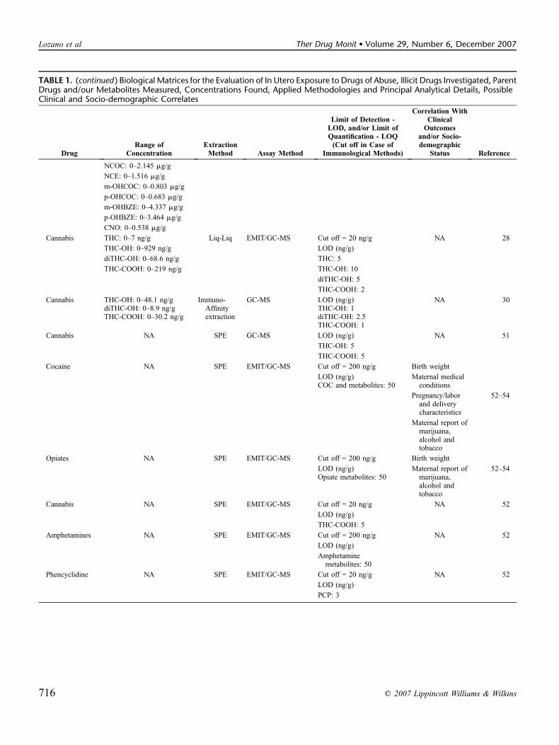
TABLE 1. (continued ) Biological Matrices for the Evaluation of In Utero Exposure to Drugs of Abuse, Illicit Drugs Investigated, ParentDrugs and/our Metabolites Measured, Concentrations Found, Applied Methodologies and Principal Analytical Details, PossibleClinical and Socio-demographic Correlates
DrugRange of
ConcentrationExtractionMethod Assay Method
Limit of Detection -LOD, and/or Limit ofQuantification - LOQ(Cut off in Case of
Immunological Methods)
Correlation WithClinicalOutcomes
and/or Socio-demographic
Status Reference
NCOC: 0–2.145 mg/g
NCE: 0–1.516 mg/g
m-OHCOC: 0–0.803 mg/g
p-OHCOC: 0–0.683 mg/g
m-OHBZE: 0–4.337 mg/g
p-OHBZE: 0–3.464 mg/g
CNO: 0–0.538 mg/g
Cannabis THC: 0–7 ng/g Liq-Liq EMIT/GC-MS Cut off = 20 ng/g NA 28
THC-OH: 0–929 ng/g LOD (ng/g)
diTHC-OH: 0–68.6 ng/g THC: 5
THC-COOH: 0–219 ng/g THC-OH: 10
diTHC-OH: 5
THC-COOH: 2
Cannabis THC-OH: 0–48.1 ng/gdiTHC-OH: 0–8.9 ng/gTHC-COOH: 0–30.2 ng/g
Immuno-Affinityextraction
GC-MS LOD (ng/g)THC-OH: 1diTHC-OH: 2.5THC-COOH: 1
NA 30
Cannabis NA SPE GC-MS LOD (ng/g) NA 51
THC-OH: 5
THC-COOH: 5
Cocaine NA SPE EMIT/GC-MS Cut off = 200 ng/g Birth weight
LOD (ng/g) Maternal medicalconditionsCOC and metabolites: 50
Pregnancy/laborand deliverycharacteristics
52–54
Maternal report ofmarijuana,alcohol andtobacco
Opiates NA SPE EMIT/GC-MS Cut off = 200 ng/g Birth weight
LOD (ng/g)Opiate metabolites: 50
Maternal report ofmarijuana,alcohol andtobacco
52–54
Cannabis NA SPE EMIT/GC-MS Cut off = 20 ng/g NA 52
LOD (ng/g)
THC-COOH: 5
Amphetamines NA SPE EMIT/GC-MS Cut off = 200 ng/g NA 52
LOD (ng/g)
Amphetaminemetabolites: 50
Phencyclidine NA SPE EMIT/GC-MS Cut off = 20 ng/g NA 52
LOD (ng/g)
PCP: 3
716 q 2007 Lippincott Williams & Wilkins
Lozano et al Ther Drug Monit � Volume 29, Number 6, December 2007
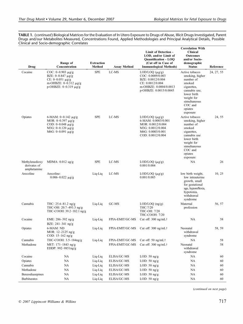
TABLE 1. (continued ) Biological Matrices for the Evaluation of In Utero Exposure to Drugs of Abuse, Illicit Drugs Investigated, ParentDrugs and/our Metabolites Measured, Concentrations Found, Applied Methodologies and Principal Analytical Details, PossibleClinical and Socio-demographic Correlates
DrugRange of
ConcentrationExtractionMethod Assay Method
Limit of Detection -LOD, and/or Limit ofQuantification - LOQ(Cut off in Case of
Immunological Methods)
Correlation WithClinicalOutcomes
and/or Socio-demographic
Status Reference
Cocaine COC: 0–0.903 mg/gBZE: 0–0.847 mg/gCE: 0–0.051 mg/gm-OHBZE: 0–0.312 mg/gp-OHBZE: 0–0.319 mg/g
SPE LC-MS LOD/LOQ (mg/g)COC: 0.0009/0.003BZE: 0.0012/0.004CE: 0.0012/0.004m-OHBZE: 0.0004/0.0013p-OHBZE: 0.0015/0.0045
Active tobaccosmoking, highernumber ofsmokedcigarettes,cannabis use,lower birthweight forsimultaneousCOC andopiatesexposure
24, 27, 55
Opiates 6-MAM: 0–0.142 mg/gMOR: 0–0.397 mg/gCOD: 0–0.048 mg/gM3G: 0–0.120 mg/gM6G: 0–0.091 mg/g
SPE LC-MS LOD/LOQ (mg/g)6-MAM: 0.0003/0.001MOR: 0.0012/0.004M3G: 0.0012/0.004M6G: 0.0003/0.001COD: 0.0012/0.004
Active tobaccosmoking, highernumber ofsmokedcigarettes,cannabis uselower birthweight forsimultaneousCOC andopiatesexposure
24, 55
Methylenedioxyderivates ofamphetamine
MDMA: 0.012 ug/g SPE LC-MS LOD/LOQ (mg/g)0.001/0.004
NA 26
Arecoline Arecoline:0.006–0.022 mg/g
Liq-Liq LC-MS LOD/LOQ (mg/g)0.001/0.005
low birth weight,low intrauterinegrowth, smallfor gestationalage, hyporeflexia,hypotonia,withdrawalsyndrome
10, 25
Cannabis THC: 25.6–81.2 ng/gTHC-OH: 20.7–493.3 ng/gTHC-COOH: 39.2–182.1 ng/g
Liq-Liq GC-MS LOD/LOQ (ng/g)THC:7/20THC-OH: 7/20THC-COOH: 7/20
Maternalprofession
56, 57
Cocaine EME: 286–392 ng/g Liq-Liq FPIA-EMIT/GC-MS Cut off: 300 ng/mL† NA 58
BZE: 241–341 ng/g
Opiates 6-MAM: NDMOR: 12–2125 ng/gCOD: 15–162 ng/g
Liq-Liq FPIA-EMIT/GC-MS Cut off: 300 ng/mL† Neonatalwithdrawalsyndrome
58, 59
Cannabis THC-COOH: 3.5–184ng/g Liq-Liq FPIA-EMIT/GC-MS Cut off: 50 ng/mL† NA 58
Methadone MET: 171–1843 ng/gEDDP: 992–9851ng/g
FPIA-EMIT/GC-MS Cut off: 300 ng/mL† Neonatalwithdrawalsyndrome
58
Cocaine NA Liq-Liq ELISA/GC-MS LOD: 50 ng/g NA 60
Opiates NA Liq-Liq ELISA/GC-MS LOD: 50 ng/g NA 60
Cannabis NA Liq-Liq ELISA/GC-MS LOD: 50 ng/g NA 60
Methadone NA Liq-Liq ELISA/GC-MS LOD: 50 ng/g NA 60
Benzodiazepines NA Liq-Liq ELISA/GC-MS LOD: 50 ng/g NA 60
Barbiturates NA Liq-Liq ELISA/GC-MS LOD: 50 ng/g NA 60
(continued on next page)
q 2007 Lippincott Williams & Wilkins 717
Ther Drug Monit � Volume 29, Number 6, December 2007 Biological Matrices for Fetal Exposure to Drugs
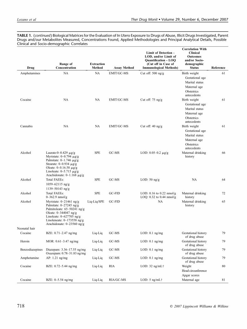
TABLE 1. (continued ) Biological Matrices for the Evaluation of In Utero Exposure to Drugs of Abuse, Illicit Drugs Investigated, ParentDrugs and/our Metabolites Measured, Concentrations Found, Applied Methodologies and Principal Analytical Details, PossibleClinical and Socio-demographic Correlates
DrugRange of
ConcentrationExtractionMethod Assay Method
Limit of Detection -LOD, and/or Limit ofQuantification - LOQ(Cut off in Case of
Immunological Methods)
Correlation WithClinicalOutcomes
and/or Socio-demographic
Status Reference
Amphetamines NA NA EMIT/GC-MS Cut off: 500 ng/g Birth weight 61
Gestational age
Marital status
Maternal age
Obstetricsantecedents
Cocaine NA NA EMIT/GC-MS Cut off: 75 ng/g Birth weight 61
Gestational age
Marital status
Maternal age
Obstetricsantecedents
Cannabis NA NA EMIT/GC-MS Cut off: 40 ng/g Birth weight 61
Gestational age
Marital status
Maternal age
Obstetricsantecedents
Alcohol Laurate:0–0.429 mg/gMyristate: 0–0.794 mg/gPalmitate: 0–1.746 mg/gStearate: 0–0.934 mg/gOleate: 0–0.16.58 mg/gLinoleate: 0–5.715 mg/gArachidonate: 0–1.168 mg/g
SPE GC-MS LOD: 0.05–0.2 mg/g Maternal drinkinghistory
66
Alcohol Total FAEEs: SPE GC-MS LOD: 50 ng/g NA 64
1059–62115 ng/g
1139–50143 ng/g
Alcohol Total FAEEs:0–362.9 nmol/g
SPE GC-FID LOD: 0.16 to 0.22 nmol/gLOQ: 0.32 to 0.44 nmol/g
Maternal drinkinghistory
72
Alcohol Myristate: 0–21461 ng/gPalmitate: 0–27245 ng/gPalmitoleate: 65–50241 ng/gOleate: 0–344047 ng/gLinoleate: 0–627705 ng/gLinoleneate: 0–173558 ng/gArachidonate: 0–23560 ng/g
Liq-Liq/SPE GC-FID NA Maternal drinkinghistory
65
Neonatal hair
Cocaine BZE: 0.71–2.47 ng/mg Liq-Liq GC-MS LOD: 0.1 ng/mg Gestational historyof drug abuse
79
Heroin MOR: 0.61–3.47 ng/mg Liq-Liq GC-MS LOD: 0.1 ng/mg Gestational historyof drug abuse
79
Benzodiazepines Diazepam: 3.36–17.55 ng/mgOxazepam: 0.78–31.83 ng/mg
Liq-Liq GC-MS LOD: 0.1 ng/mg Gestational historyof drug abuse
79
Amphetamine AP: 1.21 ng/mg Liq-Liq GC-MS LOD: 0.1 ng/mg Gestational historyof drug abuse
79
Cocaine BZE: 0.72–5.44 ng/mg Liq-Liq RIA LOD: 32 ng/mL† Weight 80
Head circunference
Apgar scores
Cocaine BZE: 0–5.54 ng/mg Liq-Liq RIA/GC-MS LOD: 5 ng/mL† Maternal age 81
718 q 2007 Lippincott Williams & Wilkins
Lozano et al Ther Drug Monit � Volume 29, Number 6, December 2007
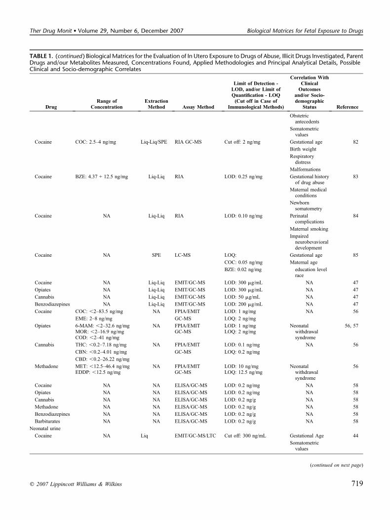
TABLE 1. (continued ) Biological Matrices for the Evaluation of In Utero Exposure to Drugs of Abuse, Illicit Drugs Investigated, ParentDrugs and/our Metabolites Measured, Concentrations Found, Applied Methodologies and Principal Analytical Details, PossibleClinical and Socio-demographic Correlates
DrugRange of
ConcentrationExtractionMethod Assay Method
Limit of Detection -LOD, and/or Limit ofQuantification - LOQ(Cut off in Case of
Immunological Methods)
Correlation WithClinicalOutcomes
and/or Socio-demographic
Status Reference
Obstetricantecedents
Somatometricvalues
Cocaine COC: 2.5–4 ng/mg Liq-Liq/SPE RIA GC-MS Cut off: 2 ng/mg Gestational age 82
Birth weight
Respiratorydistress
Malformations
Cocaine BZE: 4.37 + 12.5 ng/mg Liq-Liq RIA LOD: 0.25 ng/mg Gestational historyof drug abuse
83
Maternal medicalconditions
Newbornsomatometry
Cocaine NA Liq-Liq RIA LOD: 0.10 ng/mg Perinatalcomplications
84
Maternal smoking
Impairedneurobevavioraldevelopment
Cocaine NA SPE LC-MS LOQ: Gestational age 85
COC: 0.05 ng/mg Maternal age
BZE: 0.02 ng/mg education levelrace
Cocaine NA Liq-Liq EMIT/GC-MS LOD: 300 mg/mL NA 47
Opiates NA Liq-Liq EMIT/GC-MS LOD: 300 mg/mL NA 47
Cannabis NA Liq-Liq EMIT/GC-MS LOD: 50 mg/mL NA 47
Benzodiazepines NA Liq-Liq EMIT/GC-MS LOD: 200 mg/mL NA 47
Cocaine COC: ,2–83.5 ng/mg NA FPIA/EMIT LOD: 1 ng/mg NA 56
EME: 2–8 ng/mg GC-MS LOQ: 2 ng/mg
Opiates 6-MAM: ,2–32.6 ng/mgMOR: ,2–16.9 ng/mgCOD: ,2–41 ng/mg
NA FPIA/EMITGC-MS
LOD: 1 ng/mgLOQ: 2 ng/mg
Neonatalwithdrawalsyndrome
56, 57
Cannabis THC: ,0.2–7.18 ng/mg NA FPIA/EMIT LOD: 0.1 ng/mg NA 56
CBN: ,0.2–4.01 ng/mg GC-MS LOQ: 0.2 ng/mg
CBD: ,0.2–26.22 ng/mg
Methadone MET: ,12.5–46.4 ng/mgEDDP: ,12.5 ng/mg
NA FPIA/EMITGC-MS
LOD: 10 ng/mgLOQ: 12.5 ng/mg
Neonatalwithdrawalsyndrome
56
Cocaine NA NA ELISA/GC-MS LOD: 0.2 ng/mg NA 58
Opiates NA NA ELISA/GC-MS LOD: 0.2 ng/mg NA 58
Cannabis NA NA ELISA/GC-MS LOD: 0.2 ng/g NA 58
Methadone NA NA ELISA/GC-MS LOD: 0.2 ng/g NA 58
Benzodiazepines NA NA ELISA/GC-MS LOD: 0.2 ng/g NA 58
Barbiturates NA NA ELISA/GC-MS LOD: 0.2 ng/g NA 58
Neonatal urine
Cocaine NA Liq EMIT/GC-MS/LTC Cut off: 300 ng/mL Gestational Age 44
Somatometricvalues
(continued on next page)
q 2007 Lippincott Williams & Wilkins 719
Ther Drug Monit � Volume 29, Number 6, December 2007 Biological Matrices for Fetal Exposure to Drugs
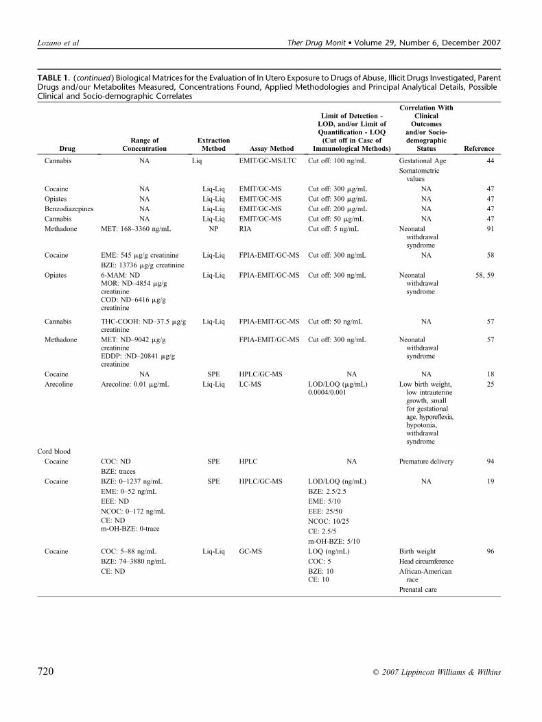
TABLE 1. (continued ) Biological Matrices for the Evaluation of In Utero Exposure to Drugs of Abuse, Illicit Drugs Investigated, ParentDrugs and/our Metabolites Measured, Concentrations Found, Applied Methodologies and Principal Analytical Details, PossibleClinical and Socio-demographic Correlates
DrugRange of
ConcentrationExtractionMethod Assay Method
Limit of Detection -LOD, and/or Limit ofQuantification - LOQ(Cut off in Case of
Immunological Methods)
Correlation WithClinicalOutcomes
and/or Socio-demographic
Status Reference
Cannabis NA Liq EMIT/GC-MS/LTC Cut off: 100 ng/mL Gestational Age 44
Somatometricvalues
Cocaine NA Liq-Liq EMIT/GC-MS Cut off: 300 mg/mL NA 47
Opiates NA Liq-Liq EMIT/GC-MS Cut off: 300 mg/mL NA 47
Benzodiazepines NA Liq-Liq EMIT/GC-MS Cut off: 200 mg/mL NA 47
Cannabis NA Liq-Liq EMIT/GC-MS Cut off: 50 mg/mL NA 47
Methadone MET: 168–3360 ng/mL NP RIA Cut off: 5 ng/mL Neonatalwithdrawalsyndrome
91
Cocaine EME: 545 mg/g creatinine Liq-Liq FPIA-EMIT/GC-MS Cut off: 300 ng/mL NA 58
BZE: 13736 mg/g creatinine
Opiates 6-MAM: ND Liq-Liq FPIA-EMIT/GC-MS Cut off: 300 ng/mL Neonatalwithdrawalsyndrome
58, 59MOR: ND–4854 mg/gcreatinineCOD: ND–6416 mg/gcreatinine
Cannabis THC-COOH: ND–37.5 mg/gcreatinine
Liq-Liq FPIA-EMIT/GC-MS Cut off: 50 ng/mL NA 57
Methadone MET: ND–9042 mg/gcreatinineEDDP: :ND–20841 mg/gcreatinine
FPIA-EMIT/GC-MS Cut off: 300 ng/mL Neonatalwithdrawalsyndrome
57
Cocaine NA SPE HPLC/GC-MS NA NA 18
Arecoline Arecoline: 0.01 mg/mL Liq-Liq LC-MS LOD/LOQ (mg/mL)0.0004/0.001
Low birth weight,low intrauterinegrowth, smallfor gestationalage, hyporeflexia,hypotonia,withdrawalsyndrome
25
Cord blood
Cocaine COC: ND SPE HPLC NA Premature delivery 94
BZE: traces
Cocaine BZE: 0–1237 ng/mL SPE HPLC/GC-MS LOD/LOQ (ng/mL) NA 19
EME: 0–52 ng/mL BZE: 2.5/2.5
EEE: ND EME: 5/10
NCOC: 0–172 ng/mL EEE: 25/50
NCOC: 10/25CE: ND
CE: 2.5/5m-OH-BZE: 0-trace
m-OH-BZE: 5/10
Cocaine COC: 5–88 ng/mL Liq-Liq GC-MS LOQ (ng/mL) Birth weight 96
BZE: 74–3880 ng/mL COC: 5 Head circumference
CE: ND BZE: 10CE: 10
African-Americanrace
Prenatal care
720 q 2007 Lippincott Williams & Wilkins
Lozano et al Ther Drug Monit � Volume 29, Number 6, December 2007
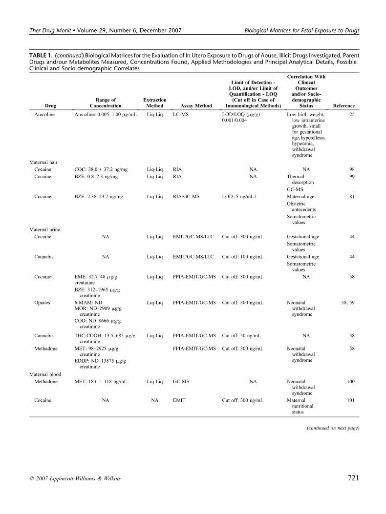
TABLE 1. (continued ) Biological Matrices for the Evaluation of In Utero Exposure to Drugs of Abuse, Illicit Drugs Investigated, ParentDrugs and/our Metabolites Measured, Concentrations Found, Applied Methodologies and Principal Analytical Details, PossibleClinical and Socio-demographic Correlates
DrugRange of
ConcentrationExtractionMethod Assay Method
Limit of Detection -LOD, and/or Limit ofQuantification - LOQ(Cut off in Case of
Immunological Methods)
Correlation WithClinicalOutcomes
and/or Socio-demographic
Status Reference
Arecoline Arecoline: 0.005–1.00 mg/mL Liq-Liq LC-MS LOD/LOQ (mg/g)0.001/0.004
Low birth weight,low intrauterinegrowth, smallfor gestationalage, hyporeflexia,hypotonia,withdrawalsyndrome
25
Maternal hair
Cocaine COC: 38.0 + 37.2 ng/mg Liq-Liq RIA NA NA 98
Cocaine BZE: 0.8–2.3 ng/mg Liq-Liq RIA NA Thermaldesorption
99
GC-MS
Cocaine BZE: 2.38–23.7 ng/mg Liq-Liq RIA/GC-MS LOD: 5 ng/mL† Maternal age 81
Obstetricantecedents
Somatometricvalues
Maternal urine
Cocaine NA Liq-Liq EMIT/GC-MS/LTC Cut off: 300 ng/mL Gestational age 44
Somatometricvalues
Cannabis NA Liq-Liq EMIT/GC-MS/LTC Cut off: 100 ng/mL Gestational age 44
Somatometricvalues
Cocaine EME: 32.7–48 mg/gcreatinine
Liq-Liq FPIA-EMIT/GC-MS Cut off: 300 ng/mL NA 58
BZE: 312–1965 mg/gcreatinine
Opiates 6-MAM: ND Liq-Liq FPIA-EMIT/GC-MS Cut off: 300 ng/mL Neonatalwithdrawalsyndrome
58, 59MOR: ND–2909 mg/gcreatinine
COD: ND–8666 mg/gcreatinine
Cannabis THC-COOH: 13.5–685 mg/gcreatinine
Liq-Liq FPIA-EMIT/GC-MS Cut off: 50 ng/mL NA 58
Methadone MET: 98–2925 mg/gcreatinine
FPIA-EMIT/GC-MS Cut off: 300 ng/mL Neonatalwithdrawalsyndrome
58
EDDP: ND–13575 mg/gcreatinine
Maternal blood
Methadone MET: 183 6 118 ng/mL Liq-Liq GC-MS NA Neonatalwithdrawalsyndrome
100
Cocaine NA NA EMIT Cut off: 300 ng/mL Maternalnutritionalstatus
101
(continued on next page)
q 2007 Lippincott Williams & Wilkins 721
Ther Drug Monit � Volume 29, Number 6, December 2007 Biological Matrices for Fetal Exposure to Drugs
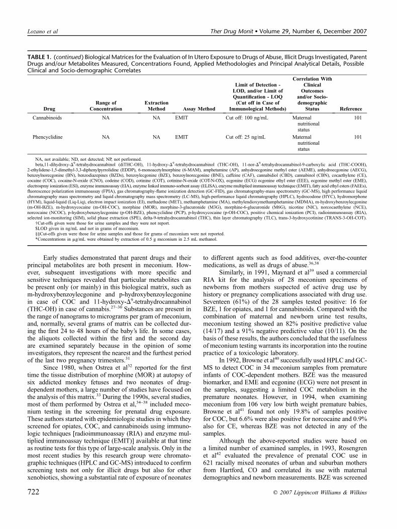
Early studies demonstrated that parent drugs and theirprincipal metabolites are both present in meconium. How-ever, subsequent investigations with more specific andsensitive techniques revealed that particular metabolites canbe present only (or mainly) in this biological matrix, such asm-hydroxybenzoylecgonine and p-hydroxybenzoylecgoninein case of COC and 11-hydroxy-D9-tetrahydrocannabinol(THC-OH) in case of cannabis.27–30 Substances are present inthe range of nanograms to micrograms per gram of meconium,and, normally, several grams of matrix can be collected dur-ing the first 24 to 48 hours of the baby’s life. In some cases,the aliquots collected within the first and the second dayare examined separately because in the opinion of someinvestigators, they represent the nearest and the furthest periodof the last two pregnancy trimesters.31
Since 1980, when Ostrea et al32 reported for the firsttime the tissue distribution of morphine (MOR) at autopsy ofsix addicted monkey fetuses and two neonates of drug-dependent mothers, a large number of studies have focused onthe analysis of this matrix.33 During the 1990s, several studies,most of them performed by Ostrea et al,34–38 included meco-nium testing in the screening for prenatal drug exposure.These authors started with epidemiologic studies in which theyscreened for opiates, COC, and cannabinoids using immuno-logic techniques [radioimmunoassay (RIA) and enzyme mul-tiplied immunoassay technique (EMIT)] available at that timeas routine tests for this type of large-scale analysis. Only in themost recent studies by this research group were chromato-graphic techniques (HPLC and GC-MS) introduced to confirmscreening tests not only for illicit drugs but also for otherxenobiotics, showing a substantial rate of exposure of neonates
to different agents such as food additives, over-the-countermedications, as well as drugs of abuse.36,38
Similarly, in 1991, Maynard et al39 used a commercialRIA kit for the analysis of 28 meconium specimens ofnewborns from mothers suspected of active drug use byhistory or pregnancy complications associated with drug use.Seventeen (61%) of the 28 samples tested positive: 16 forBZE, 1 for opiates, and 1 for cannabinoids. Compared with thecombination of maternal and newborn urine test results,meconium testing showed an 82% positive predictive value(14/17) and a 91% negative predictive value (10/11). On thebasis of these results, the authors concluded that the usefulnessof meconium testing warrants its incorporation into the routinepractice of a toxicologic laboratory.
In 1992, Browne et al40 successfully used HPLC and GC-MS to detect COC in 34 meconium samples from prematureinfants of COC-dependent mothers. BZE was the measuredbiomarker, and EME and ecgonine (ECG) were not present inthe samples, suggesting a limited COC metabolism in thepremature neonates. However, in 1994, when examiningmeconium from 106 very low birth weight premature babies,Browne et al41 found not only 19.8% of samples positivefor COC, but 6.6% were also positive for norcocaine and 0.9%also for CE, whereas BZE was not detected in any of thesamples.
Although the above-reported studies were based ona limited number of examined samples, in 1993, Rosengrenet al42 evaluated the prevalence of prenatal COC use in621 racially mixed neonates of urban and suburban mothersfrom Hartford, CO and correlated its use with maternaldemographics and newborn measurements. BZE was screened
TABLE 1. (continued ) Biological Matrices for the Evaluation of In Utero Exposure to Drugs of Abuse, Illicit Drugs Investigated, ParentDrugs and/our Metabolites Measured, Concentrations Found, Applied Methodologies and Principal Analytical Details, PossibleClinical and Socio-demographic Correlates
DrugRange of
ConcentrationExtractionMethod Assay Method
Limit of Detection -LOD, and/or Limit ofQuantification - LOQ(Cut off in Case of
Immunological Methods)
Correlation WithClinicalOutcomes
and/or Socio-demographic
Status Reference
Cannabinoids NA NA EMIT Cut off: 100 ng/mL Maternalnutritionalstatus
101
Phencyclidine NA NA EMIT Cut off: 25 ng/mL Maternalnutritionalstatus
101
NA, not available; ND, not detected; NP, not performed.beta,11-dihydroxy-D9-tetrahydrocannabinol (diTHC-OH), 11-hydroxy-D9-tetrahydrocannabinol (THC-OH), 11-nor-D9-tetrahydrocannabinol-9-carboxylic acid (THC-COOH),
2-ethylidene-1,5-dimethyl-3,3-diphenylpyrrolidine (EDDP), 6-monoacetylmorphine (6-MAM), amphetamine (AP), anhydroecgonine methyl ester (AEME), anhydroecgonine (AECG),benzoylnorecgonine (BN), benzodiazepines (BZDs), benzoylecgonine (BZE), benzoylnorecgonine (BNE), caffeine (CAF), cannabidiol (CBD), cannabinol (CBN), cocaethylene (CE),cocaine (COC), cocaine-N-oxide (CNO), codeine (COD), cotinine (COT), cotinine-N-oxide (COT-N-OX), ecgonine (ECG) ecgonine ethyl ester (EEE), ecgonine methyl ester (EME),electrospray ionization (ESI), enzyme immunoassay (EIA), enzyme linked immuno-sorbent assay (ELISA), enzymemultiplied immunoassay technique (EMIT), fatty acid ethyl esters (FAEEs),fluorescence polarization immunoassay (FPIA), gas chromatography-flame ionization detection (GC-FID), gas chromatography-mass spectrometry (GC-MS), high performance liquidchromatography mass spectrometry and liquid chromatography mass spectrometry (LC-MS), high-performance liquid chromatography (HPLC), hydrocodone (HYC), hydromorphone(HYM), liquid-liquid (Liq-Liq), electron impact ionization (EI), methadone (MET), methamphetamine (MA), methylendioxymethamphetamine (MDMA), m-hydroxybenzoylecgonine(m-OH-BZE), m-hydroxycocaine (m-OH-COC), morphine (MOR), morphine-3-glucuronide (M3G), morphine-6-glucuronide (M6G), nicotine (NIC), norcocaethylene (NCE),norcocaine (NCOC), p-hydroxybenzoylecgonine (p-OH-BZE), phencyclidine (PCP), p-hydroxycocaine (p-OH-COC), positive chemical ionization (PCI), radioimmunoassay (RIA),selected ion-monitoring (SIM), solid phase extraction (SPE), delta-9-tetrahydrocannabinol (THC), thin layer chromatography (TLC), trans-3-hydroxycotinine (TRANS-3-OH-COT).
†Cut-offs given were those for urine samples and they were not report.$LOD given in ng/mL and not in grams of meconium.§§Cut-offs given were those for urine samples and those for grams of meconium were not reported.*Concentrations in mg/mL were obtained by extraction of 0.5 g meconium in 2.5 mL methanol.
722 q 2007 Lippincott Williams & Wilkins
Lozano et al Ther Drug Monit � Volume 29, Number 6, December 2007
in meconium using FPIA and was confirmed after SPE withGC-MS in selected ion-monitoring (SIM) mode. A 3.4% pos-itivity to BZE was statistically correlated with multiparity,multigravidity, late-onset and evidence-based prenatal care,public assistance, race other than Caucasian, and low academicachievement. COC-exposed infants were significantly smaller,and this correlated best with nonCaucasian ethnicity.
These early studies supporting the use of meconium asa valuable investigation tool for prenatal exposure to drugswere in contrast with the findings reported by Wingert et al43
in 1994. After examining 345 meconium specimens in a largemetropolitan obstetrical population from Bronx, New YorkCity as well as maternal and neonatal urine samples for prin-cipal drugs of abuse, they concluded that meconium did notappear to offer any advantage over maternal or neonatal urinetesting for the assessment of cannabinoids, codeine (COD),MOR, or methadone (MET) exposure. The positive rate forBZE (12%) was virtually identical for meconium and formaternal as well as neonatal urine.
Likewise, in 1995, Bibb et al44 screened 386 meconiumsamples from 580 pairs mother/infants in Louisville, KY byEMIT and confirmed positive samples by GC-MS and thinlayer chromatography (TLC). They found that meconiumanalysis showed equal sensitivity with the interview andmaternal urine analysis, whereas with the newborn urine, thecorrelation was poor. Their percentage of positivity was 3.4%to COC, 1% to delta-9-tetrahydrocannabinol (THC). In con-trast with these two studies, Ryan et al45 demonstrated thatmeconium testing was more specific and sensitive than urinetesting to identify in utero drug exposure. By examining alarge cohort of mother-infants pairs (1030 meconium samples)from Rochester, NY, a 5.5% intrauterine exposure to COC wasreported by this study; BZE was measured by FPIA withconfirmation by GC-MS (SIM mode).
In 1995, another large epidemiologic study performedby Lewis et al46 using FPIA and confirmation with GC-MS(operated in SIM mode) detected COC, opiates, cannabinoids,and amphetamine (AP) in 1175 meconium samples fromRockford, IL. Confirmation cutoff values were lower than theprevious ones: 5 ng/g for COC metabolites, AP, opiates, andphencyclidine (PCP) and 2 ng/g for cannabinoids. Totalprevalence of newborns exposed to illicit drugs in this NorthAmerican midsize Midwestern city was 12.9%. COC-exposedneonates had the highest positive rate (5.4%), followed bycannabis (4.4%), CE (1%), and AP (0.1%). Nine (0.8%)patients had multiple drugs present in the meconium. Theinteresting point of this study was that the authors includedmeconium assays from 23 sets of multiple births (21 twins,2 triplets) as a quality control for the assay. There were 20 setsof multiple births (42 patients) all testing negative. Three setsof twins had concordance in testing positive, with one twintesting positive for COC, whereas the other twin testedpositive for COC and marijuana. No absolute discordance oftwin assays were noted.
In addition to comparison with neonatal and maternalurine samples, Montgomery et al33 investigated the rate ofagreement of testing for fetal exposure to illicit drugs (APs,opiates, COC, cannabinoids, and PCP) using specimens ofmeconium versus umbilical cord tissue. Paired samples of
meconium and umbilical cord tissue from 118 pregnancieswith high suspicion of illicit drug use by the mothers wereobtained. The agreement of drug screening between cord andmeconium was above 90% for all drugs tested. Umbilical cordtissue performs as well as meconium in assessing fetal drugexposure to AP, opiates, COC, and cannabinoids. With theseresults, the authors recommended that the analysis of umbilicalcord tissue may lead to more rapid test results than meconiumanalysis because the passage of meconium by the infant maybe delayed for several days.
During the 1990s, in addition to the principal drugs ofabuse, clinical investigations focused on identifying otherspecifically abused drugs and all the possible metabolitesuseful as biomarkers of in utero exposure. Prenatal exposure tobenzodiazepines (BZDs) was, for the first time, studied bymeconium analysis by Samperiz et al47 in 1996 in 31 infantswhose mothers were confirmed (n = 18) or suspected (n = 13)to be addicted to the drug. In this cohort COC, opiates andcannabis exposure were also investigated. The authors reported1 positive sample for oxazepam, 1 for BZE, 15 for MOR, and2 for THC using EMIT as the screening method and confir-mation with GC.
In 1997, Stolk et al,48 using FPIA and HPLC, analyzed16 meconium samples from neonates of opiate-dependentmothers in treatment with MET. The parent drug and itsprincipal metabolite 2-ethylidene-1,5-dimethyl-3,3-diphenyl-pyrrolidine were found in 14 and 15 positive samples,respectively. The amount of 2-ethylidene-1,5-dimethyl-3,3-diphenylpyrrolidine in meconium was much higher thanthe amount of MET (ratio: 9.6), showing that this metabolitecould be the potential biomarker of fetal exposure to this drug.
All the possible COC metabolites present in meconiumof newborns prenatally exposed to the drug were for the firsttime identified by Oyler et al49 Six meconium samples ofexposed infants were analyzed using GC-MS after SPE.Principal COC metabolites such as BZE and EME were foundin high concentrations together with m-hydroxybenzoylecgo-nine, which was shown to be a specific biomarker of COCexposure in this neonatal matrix.
This interesting study, which achieved the identificationof several COC metabolites, was supported by subsequentstudies of Eyler et al18 and Xia et al50 This group of investigators(Xia et al) developed an accurate LC-MS method that had theability to analyze COC and multiple metabolites from suspecteddrug-positive meconium samples using SPE.With respect to thestudy of Oyler et al, further COC metabolites were identifiedsuch as AEGC, ECG, benzoylnorecgonine, and cocaine-N-oxide (CNO). In addition, some variability in metaboliteconcentration between the two studies can be observed,although the order of magnitude of the amount was similar.49
Metabolites identified varied qualitatively and quantitativelyamong the samples. Interestingly, ECG and benzoylnorecgoninedetected for the first time were the highest measured. ECGappeared to hold the most promise as a diagnostic marker forneonatal COC exposure because this metabolite was present in100% of the 21 positive samples tested at a relatively highmedian concentration. However, a core group of eightmetabolites (present in at least 20 of 21 positive samples)was identified that appeared to possess the greatest utility for
q 2007 Lippincott Williams & Wilkins 723
Ther Drug Monit � Volume 29, Number 6, December 2007 Biological Matrices for Fetal Exposure to Drugs

determining COC exposure. Ultimately, pyrolytic products ofCOC, anhydroecgonine methyl ester, and anhydroecgoninewere suggested as biomarkers of crack COC consumption.
As previously mentioned, the identification of all thepossible cannabis metabolites present in meconium wassuccessfully carried out by the research group of ElSohlyand Feng28 and Feng et al30 When examining specimenscollected from neonates born to mothers with a history ofcannabis use, these authors looked for the presence of not only11-nor-D9-tetrahydrocannabinol-9-carboxylic acid (THC-COOH)but also THC and its 8- and 11-hydroxy metabolites.28 Delta9-THC and its 8-hydroxy metabolites were basically absent in19 analyzed meconium specimens, whereas 11-OH-delta9-THC and 8 beta 11-dioh-delta9THC contributed significantlyto the immunoassay response of meconium extracts. Analysisof meconium specimens that screened positive for cannabi-noids, but failed to confirm for THC-COOH, showed signifi-cant amounts of 8 beta,11-dihydroxy-D9-tetrahydrocannabinoland THC-OH.28 This latter metabolite was indeed confirmedas a major meconium metabolite (present in 75% of 24 ana-lyzed meconium samples) in a subsequent study.30
In 2005, Coles et al,51 with the analysis of 246specimens with positive cannabinoid screen, confirmed theimportance of including THC-OH in a meconium cannabisconfirmation procedure. Sixteen specimens were confirmedpositive for THC-OH only, resulting in a 6.5% increase in thepositivity rate compared with THC-COOH alone.
Concerning large epidemiologic studies, in 2002, animportant North American study (The Maternal LifestyleStudy) investigated the prevalence of fetal drug exposure inan initial cohort of 11,811 mother-infants dyads from differ-ent parts of the country using meconium as the biologicalmatrix of choice to assess maternal self reporting of drugconsumption.52–54 In fact, exposure was defined as the rec-ognition of consumption of COC, opiates, cannabinoids, APs,and PCP by the mother or the presence of drug or metabolitesin meconium. The EMIT method was used for initial screeningfollowed by GC-MS as a confirmation assay for positivesamples after SPE. With use of the meconium drug screen, ofthe 8527 analyzed samples, 9.5% were positive for COC or itsmetabolites, 2.3% for opiates, and 7.2% for cannabinoids. APsand PCP analysis were discontinued during the study becauseof low prevalence. This observational study confirmed manyof the reported adverse social and serious medical perinatalcomplications of newborns exposed to COC or opiates duringpregnancy. COC exposure was associated with significantdeviation in all growth measurements, including birth weight,length, and head circumference, whereas opiates had a sig-nificant effect on birth weight. Statistically significant differ-ences between COC-opiate exposed and nonexposed (n =7442) mothers included race (African-American 74.6% and47.0%, respectively), mean age (29.6 and 26.1 years, respec-tively), and polydrug use, including any combination ofalcohol, tobacco, or marijuana (93% and 42%, respectively).Exposed mothers had a significantly higher risk of medicalcomplications, psychiatric, nervous, and emotional disorders,and placental abruption. In the same period, for the first time inEurope, the Meconium Project sought to estimate the prev-alence of drug use by pregnant women and the effects of
prenatal exposure on the fetus and infant.55 A cohort of 1151mother-infant dyads from the Spanish city of Barcelona wererecruited and were examined for drug exposure using amaternal self-reported questionnaire and meconium analysis.Opiates (6-monoacetylmorphine,MOR,morphine-3-glucuronide,morphine-6-glucuronide, COD), COC, and its metabolites(BZE, CE), APs, and methylenedioxy derivatives (3,4-methylendioxymethamphetamine, 3,4-methylendioxyamphet-amine), and cannabis metabolites were investigated byLC-MS.24–27,55–57 Meconium analysis showed that the prev-alence for COC, opiates, both drugs, and cannabis were 8.7%,4.4%, 2.2%, and 5.3%, respectively, and disclosed a singlecase of 3,4-methylendioxymethamphetamine use. In addition,arecoline, the main areca nut alkaloid, was found in meconiumspecimens from four Asiatic newborns whose mothersdeclared beetle nut consumption during pregnancy. In thiscohort, parental ethnicity was not associated with drug use norwas the social class, although the professional and partlyskilled mothers showed a higher tendency toward COC andopiate consumption than the nonprofessional ones, and asignificantly higher percentage of cannabis consumer mothershad a managerial professional job compared with non-consuming mothers.
Women who used drugs of abuse during pregnancyshowed a higher number of previous pregnancies and abortionswhen compared with nonconsumer mothers (meconiumnegative test) probably because of a lack of family planning.Consumption of opiates and COC during pregnancy wasassociated with active tobacco smoking, a higher number ofsmoked cigarettes, and cannabis use. Exposure status andsmoking behavior correlated with significantly lower birthweight in newborns from mothers exposed only to COC and toopiates and COC simultaneously. Of the newborns exposed toarecoline, one showed a low birth weight, low intrauterinegrowth, hyporeflexia, and hypotonia, and another presenteda severe withdrawal syndrome.10 COC, 6-monoacetylmor-phine, and THC-COOH were the substances most frequentlyfound in samples positive for COC, opiates, and cannabis (86.5,88.5, and 80.8%, respectively). None of the combinations ofdifferent metabolites showed a higher percentage of positivity.
Although some authors comparedmeconiumwithmater-nal or neonatal urine analysis for detection of prenatal expo-sure to illicit drugs, others compared the sensitivity and thepredictive power of two biological matrices accounting forchronic exposure: meconium and neonatal hair. Vinneret al58,59 used both meconium and neonatal hair analysis toestablish a drug exposure profile and predict withdrawalsyndrome in exposed newborns. Cannabinoids, opiates, COC,andMETwere determined by immunologic screening methodsand positive results confirmed by GC-MS after extraction ofanalytes from the respective matrix. Examining 17 mother/neonate pairs with a history of drug abuse, the authors con-cluded that, in spite of some biases that might hinder anaccurate interpretation, neonatal hair analysis made it possibleto confirm fetal drug exposure and to reinforce the diagnosis ofthe withdrawal syndrome, particularly when results obtained inmeconium were negative.
Different evidence was obtained by Bar-Oz et al,60 whocompared meconium and neonatal hair as biological markers
724 q 2007 Lippincott Williams & Wilkins
Lozano et al Ther Drug Monit � Volume 29, Number 6, December 2007
to screen in utero exposure for COC opiates, MET, and can-nabinoids in 185 babies with clinical suspicion of maternaldrug abuse. On the basis of the results of the study, Bar-Ozet al60 concluded that meconium is marginally more sensitivethan neonatal hair for detection of COC and cannabis. Theauthors attributed this observation to the fact that meconiumdetects second trimester exposure, whereas hair grows onlyduring the third trimester of pregnancy. Because a significantcorrelation was observed between hair and meconium con-centrations of COC, cannabis, and opiates, both matrices wereconsidered effective biological markers of in utero illicit drugexposure: meconium was more sensitive, but neonatal hair isavailable for up to 4 to 5 months after birth. It is important tomention, however, that the use of meconium, being a discardedmaterial, is more acceptable to some parents than hair testing,which entails cutting scalp hair from the newborn.
As illustrated in the above reports, COC, opiates, andcannabinoids were the substances mostly consumed duringpregnancy and therefore investigated in meconium. Withrespect to APs, the widespread abuse of methamphetamine(MA) in the adult female population and consequently duringpregnancy was an occurrence prevalently confined to the West,Midwest, and Southwest of the United States. Using meco-nium testing, Buchi et al61 estimated in 2003 the prevalence ofprenatal exposure to MA and other drugs of abuse among1519 infants born in Utah and compared the results with thoseobserved in a study performed in 1991 in the same geographicarea. Meconium samples were collected and analyzed from1202 infants from well-baby nurseries and 317 from neonatalintensive care unit infants using EMIT and confirmation byGC-MS. Rates of positivity were 0.6% for MA, 2.9% forcannabis, and 0.3% for COC. There were no significant dif-ferences in the rates of positivity for MA or cannabis betweenthe 1991 and 2000/2001 studies. Conversely, COC prevalencedeclined from 1.1% in 1991 to 0.3% in 2000/2001. The overallprevalence of positivity for any of these three drugs declinedover the 10-year period from 4.4% to 2.4%; in comparing thetwo cohorts, the most recent study showed that the prevalenceof positivity for any of these three drugs was higher in thenewborns from the neonatal intensive care unit (4.7%) than inthe well-baby nurseries.
Whereas in 2001 the prevalence of prenatal exposure toMA was approximately 0.1% in the above-reported popula-tion, in the 2006 Infant Development, Environment, and Life-style study, Arria et al62 reported a 5.2% MA exposure in acohort of 1632 women and their newborns from some specificareas of United States known for a high rate of MA misuse:Los Angeles, CA, De Moines, IA, Tulsa, OK, and Honolulu,HI. A 6.0% exposure to cannabis, 1.3% to barbiturates, and22.8% self-reported exposure to alcohol was also high-lighted.62 Conversely, less than 1% of this cohort used heroin,BZDs, and hallucinogens.
Apart from the above-reported illicit drugs that havebeen tested in meconium, investigators have also focused onthe assessment of prenatal exposure to the most consumed licitdrug of abuse: alcohol. Alcohol is indeed the most popularlegal drug of abuse used in our society, and consumption bywomen at child-bearing age remains an important publichealth concern. FAEEs are nonoxidative metabolites of
alcohol that are formed by the conjugation of ethanol toendogenous fatty acids, and they have been proposed asbiological markers of acute and chronic alcohol consumptionin adults.63 Moreover, in recent years, FAEEs in fetal andneonatal biological matrices have been reported to be potentialbiomarkers to assess the extent of intrauterine exposure toalcohol.63–66
Because it was evident from preliminary studies thatcertain FAEEs could be found in the meconium of neonateswithout prenatal alcohol exposure, early studies tried to findout which FAEEs could better differentiate between exposedand nonexposed newborns.67–70 Furthermore, as extensivelyreviewed by Chan et al,63 different proposals appeared in theinternational literature to standardize endogenous FAEEslevels and to validate a positive screening cutoff to confi-dently establish prenatal alcohol exposure. A positive test wasdefined as the presence of one particular FAEE, ethyl linoleate(E18:2), above the limit of detection (1 nmol/g) in one study67
or the detection of ethyl oleate (E18:1) above the cutoff valueof 32 ng/g68 in another study, whereas cumulative presence ofsix individual FAEEs, palmitate (E16), palmitoleate (E16:0),stearate (E18), oleate (E18:1), linoleate (E18:2), and arach-idonate (E20:4), excluding ethyl laurate (E12) and myristate(E14), above 50 ng/g constituted a positive test in anotherlaboratory.69,70 The first population-based study to determinebasal levels of meconium FAEEs was conducted in twodistinct populations of nondrinking mothers and their neonatesfrom Toronto (n = 99) and Jerusalem (n = 101).71 Mothers(n = 17) from both populations who admitted to drinkingsocially were eliminated from the baseline cohort and analyzedas a separate group. In addition, meconium samples from sixinfants born to mothers who were confirmed heavy drinkerswere also tested. Six individual FAEEs including ethyl laurate(E12), myristate (E14), palmitate (E16), stearate (E18), oleate(E18:1), and linoleate (E18:2) were extracted from meconiumsamples by SPE and analyzed by GC with flame ioniza-tion detection (FID). A positive cutoff of 2 nmol cumulativeFAEEs/g meconium, when ethyl laurate and myristate wereexcluded, was established with 100% sensitivity and 98.4%specificity.
With use of the above-reported cutoffs, epidemiologicstudies have been conducted to investigate the prevalenceof prenatal exposure to ethanol in different mother-newborncohorts. Moore et al,64 in 2003, analyzed the prevalence of sixFAEEs, palmitate (E16), palmitoleate (E16:0), stearate (E18),oleate (E18:1), linoleate (E18:2), and arachidonate (E20:4), inthe meconium of two separate groups of neonates: 436 babiesborn in a large, regional perinatal center in Hawaii and 289infants admitted to six different newborn intensive care units.By the use of SPE, analysis by GC-MS in the chemical ioni-zation mode, and a cutoff of 50 ng/g total FAEEs, the authorsfound a prevalence of exposure of 16.7% and 12.1% of babiesfrom the two groups. Considering the quantitative resultsobtained, the authors concluded that in an adequate meconiumspecimen, a total FAEE concentration greater than 10,000 ng/gmay indicate that the newborn has been exposed to signif-icant amounts of alcohol during pregnancy.
In 2004, Chan et al investigated the potential use ofmeconium FAEE testing in a Canadian high-risk neonatal
q 2007 Lippincott Williams & Wilkins 725
Ther Drug Monit � Volume 29, Number 6, December 2007 Biological Matrices for Fetal Exposure to Drugs
population in the absence of maternal drinking history. Onehundred forty-two neonates were analyzed by enzyme-linkedimmunosorbent assay for illicit drugs and FAEEs (includinglaurate, myristate, palmitate, stearate, oleate, and linoleate)by GC-FID.72 A total of 71% of the samples tested positivefor at least one illicit drug, with cannabis being the mostprevalent (52.3%), and 14% of all samples were positivefor prenatal alcohol exposure, as evidenced by cumulativemeconium FAEEs of 2 nmol/g. Ethyl oleate, linoleate,palmitate, and arachidonate were detected most often and atthe highest levels.
Finally, in 2005, Bearer et al65 compared concentrationsof ethyl myristate, palmitate, palmitoleate, oleate, linoleate,linoleneate (E18:3), and arachidonate in 248 infants froma large urban hospital in Cleveland, OH with varying exposurestatus and 30 Muslim infants from an urban hospital inAmman, Jordan (abstaining mothers). FAEEs were quantifiedwith GC-FID and compared between abstainers and non-abstainers to identify FAEEs of interest. Six of seven FAEEswere significantly different between the nonabstainers and atleast one of two of the abstaining groups. FAEEs best pre-dicted drinks per drinking day, and ethyl linoleate showed thehighest sensitivity and specificity for identifying infants notexposed in utero to high levels of alcohol in a high-risk,substance-abusing, clinic-based sample.
As demonstrated by a large number of published studies,meconium has been the fetal matrix most used to assessprenatal exposure to drugs of abuse. The main advantages ofmeconium analysis are the easy and noninvasive manner ofcollection, which is generally well accepted by the parentsof the newborn, the considerable amount of matrix that can becollected, and finally the fact that although available only forfew days after birth, it is the fetal matrix that accounts for thelargest exposure period.
NEONATAL HAIRNeonatal hair is a sensitive biological marker that can
define cumulative exposure to drugs during the last trimesterof intrauterine life.13,73 In the fetus, hair starts growing duringthe last 3 to 4 months of pregnancy and therefore accounts forexposure occurring in the last trimester. Although the detec-tion window is smaller than for meconium, hair has the advan-tage of being available for as long as 4 to 5 months of postnatallife.60 However, hair samples that can be obtained in newbornsare often sparse, and collection in those particular cases can beconsidered ‘‘almost’’ invasive. Newborn hair samples are exter-nally contaminated by the amniotic fluid, which not onlyreaches hair but also the fetus by way of the transdermalroute.74 Nevertheless, this contamination should not be consid-ered as external in the context of the diagnosis of intrauterinedrug exposure.
Hair is composed of approximately 65% to 95% protein,1% to 9% lipid, small quantities of trace elements, polysaccha-rides, and water.75 It is well known that drugs, including drugsof abuse, can be incorporated into the hair where they remainindefinitely.76 Possible pathways for drug incorporation intohair can be summarized as follows: 1) passive diffusion fromarterial blood capillaries into the hair follicle; 2) excretion
onto the surface of hair from sweat and sebum; and 3) externalcontamination.
The main analytes generally found in hair are the parentcompounds rather than their more polar metabolites, whichusually predominate in cord blood, neonatal urine, and in somecases in meconium.77 Drugs are present in the range of nano-grams per milligram of hair, which means a mg/g matrix andtherefore theoretically a concentration similar to that found inurine (mg/mL) and one or two orders of magnitude higher thanthat found in meconium. Although in adults the sample supplyappears unlimited, practically, the best extraction and sub-sequent analysis is achieved when 20 to 50 mg of hair are used.This is not always possible in newborns because the hairsupply is limited, and sometimes not more than 5 to 10 mg ofhair is available.
Because hair is a solid biological matrix, a chemicalor enzymatic digestion is required to extract xenobiotics fromthe hair shaft. Both alkaline and acid digestions have beenperformed at temperatures ranging from 37 to 100�C and fortimes varying from 1 hour to overnight.76,77
In case of enzymatic digestions, glucuronidase andarylsulphatase are commonly used enzymes at slightly acidicpH and temperatures at approximately 37�C for 1 to 18 hours.77
Once the hair is digested, the analytes of interest are extractedeither by solvents or SPE, with the exclusion of methanol hairtreatment, which includes digestion and extraction as a uniqueprocedure.58,77
Since 1989, it was established by the research group ofGraham et al78 that, in studies reporting reproductive risks ofCOC, hair analysis might identify intrauterine exposure toCOC in babies when a maternal drug history was not availableor of doubtful truthfulness. COC has been the illicit drugmostly investigated in neonatal hair for confirmation of ges-tational exposure and subsequent association with birth out-comes. Indeed, in the early studies, hair testing focusedmore on confirming prenatal exposure in relatively smallgroups of newborns from suspected consumers rather than onexamining the prevalence of the exposure in large mother-infant cohorts.
In 1993, the work team of Kintz and Mangin,79 one ofthe pioneers of hair testing, analyzed hair samples collected atthe time of delivery from 57 French neonates whose motherswere known users of COC (2 cases) and also of heroin(9 cases), BZDs (11 cases), and AP (1 case). In all cases, thecorresponding drug or its metabolite was found in neonatalhair from the infants in a concentration range similar to thosefound in addicted adults.
During the 1990s, immunologic methods were oftenapplied for hair testing of COC and other illicit drugs. UsingRIA, Salle et al,80 in 1995, investigated the presence of BZE inhydrolyzed hair samples from 34 infants born to mothers withurine positive for COC at delivery compared with 33 infantsborn to urine-negative mothers from South Carolina atCharleston University Hospital. Twenty-eight neonates’ hairtested positive for BZE and differed significantly from thecontrol infants in head circumference and head growth per-centiles. A negative correlation approaching significance wasfound between mean BZE and head circumference in the groupof newborns with positive hair for BZE (n = 28). The study
726 q 2007 Lippincott Williams & Wilkins
Lozano et al Ther Drug Monit � Volume 29, Number 6, December 2007

associated for the first time head growth abnormalities withlevels of prenatal COC exposure. Seven years later, the sameresearch group confirmed those results when examining 251neonatal hair samples from babies born at the same hospital.81
In this study, the authors not only screened the hair using RIAbut also confirmed positive results by GC-MS. Aftercontrolling for gestational age, higher BZE levels correlatedsignificantly with smaller head circumference and birth weight.
In agreement with the above-mentioned reports, whenexamining hair from 123 Italian newborns with malforma-tions, low gestational age, low birth weight, and respiratorydistress admitted to an intensive care division, 3 (2.4%) infantswere positive for COC compared with none in the controlgroup of 39 healthy newborns.82 Although the low number ofexamined cases precluded any general conclusion, this was thefirst evidence of a correlation between prenatal exposure toCOC and adverse outcomes using an objective biomarker.
Ursitti et al,83 also using RIA, demonstrated that the useof the hair test in cases of clinical suspicion but negative urinetest yielded a substantially higher rate of positivity thanexpected in the general population. In a period of 5 years, 192neonatal hair samples from a Canadian urban population wereanalyzed to confirm clinical suspicions of intrauterine expo-sure to COC. Thirty percent of the samples were positive forBZE, a rate 5.5-fold higher than the 5.5% found previously bythe same investigators in a population-based research study inthree nurseries in Toronto (P , 0.001), thus documenting theefficiency of this test in confirming clinical suspicions of fetalexposure to COC. These results were confirmed by thesubsequent study of the same research group: 32% positivehair samples from 392 babies in a 6-year span of clinical use ofhair testing for COC.84
The fact that hair COC and BZE were the most sensitivebiomarkers of COC exposure with respect to maternal selfreport and urine screen was corroborated by the study of Savitzet al.85 On the basis of hair analysis, these authors reported13% to 15% COC exposure (vs. 2% based on self report and6% based on urine screen) in 604 neonates from prenatal careclinics affiliated with University of North Carolina hospitals.Hair COC and BZE were associated with black ethnicity, lowereducation, and poverty but not with preterm birth, with thepossible exception of higher levels of COC and BZE and birthbefore 34 weeks of completed gestation.
Neonatal hair analysis was not limited to assess intra-uterine exposure to COC. Similar to Kintz and Mangin,79 otherinvestigators used it to assess intrauterine exposure to otherillicit drugs. For example, in 1996, Samperiz et al47 tested notonly meconium, as previously discussed, but also neonatal hairin addition to urine to improve diagnosis of fetal drug exposure.Both alternative neonatal matrices performed better than urine indetecting prenatal exposure. Among 31 French newborns fromthe Hopital d’Enfants de la Timone, Marseille, hair analysisdetected one positive sample for COC, eight positives foropiates (MOR), one for cannabis (THC), and two for oxazepam.
In addition to the comparison of Samperiz et al of thediagnostic power of neonatal hair versus meconium, Vinneret al58,59 and Bar-Oz et al,60 both in 2003, also made the samecomparison. Although the latter authors had a confirmationmethod available with a higher sensitivity for all the analytes in
hair compared with the first authors, they obtained a betterestimate of prenatal exposure to drug by meconium analysis.Conversely, the results presented by Vinner et al demon-strated that only the neonatal hair concentration of COC (andits metabolites), opiates, and cannabinoids was associated withneonatal withdrawal syndrome.
NEONATAL URINEDetection of intrauterine drug exposure has traditionally
been accomplished through neonatal urine testing. This wasespecially useful during the last 2 decades when the samplingof neonatal biological matrices other than urine could notbe easily accomplished or analytical methodologies to extractsubstances from these matrices and subsequently measure themwith correct sensitivity and specificity were not routinelyavailable.
The major disadvantage of assessing in utero exposureto illicit drugs by neonatal urinalysis is that the time window ofdetection is short, reflecting drug use only a few days beforedelivery. In addition, analysis of the neonate urine may pro-duce false-negative results depending not only on the time ofthe last ingestion of the drug by the mother but the length oftime after birth when the specimen was collected.44 Urineformation starts in the fetus at the 8th week of gestational age,but it is not until the 16th week that urine production issufficient to account for most of the amniotic fluid.86 Excretionof drugs of abuse into urine occurs in different parts of the fetalrenal nephron. Some drugs are filtered by glomerular filtrationand others by transport and tubular secretion or sometimes bya combination of both. Therefore, although the xenobiotics towhich the fetus is exposed are excreted through urine, thislatter fluid is continuously eliminated into amniotic fluid,which is the ultimate repository of those substances. Thus, theinformation obtained by examining neonatal urine after deliv-ery is the assessment of the possible acute exposures (whichmay or may not reflect the chronic exposure) in the dayspreceding delivery.
Originally, systematic investigations of illicit drugmonitoring in neonatal urine were performed using mainlyimmunologic methods and included rather large mother-infantcohorts. A first study by Halstead et al88 established thatneonatal urine drug screening without strict protocols forspecimen collection was of limited usefulness for managementof drug abuse in pregnancy and neonatal drug withdrawal.These authors compared the results of maternal and neonatalurine drug analysis for opiates, COC, BZDs, cannabinoids,and PCP with clinical data and history to test the usefulness ofperipartum drug screening and to establish guidelines foroptimal testing. Urine from 28 mothers and 52 babies fromChildren and Vancouver Hospital, Vancouver, Canada wereanalyzed by EMIT, TLC, and GC-MS. Drugs not suspected byhistory were found in 10 mothers and 6 babies. Resultsassisted in the management of neonatal withdrawal in threebabies. Drugs suspected by history were not found in 23 of35 babies. Approximately half of these results were associatedwith delayed urine collection. The authors concluded in favorof a protocol that imposed neonatal urine collection on the firstday of life.
q 2007 Lippincott Williams & Wilkins 727
Ther Drug Monit � Volume 29, Number 6, December 2007 Biological Matrices for Fetal Exposure to Drugs
In 1989, Osterloh and Lee89 carried out general urine drugscreening in mothers (n = 601) and their newborns (n = 339)from the Neonatal Nursery and Obstetrics/Gynecology wardsat San Francisco General Hospital. Apart from principaldrugs of abuse (COC, opiates, APs, BDZs), approximately 80different drugs and pharmaceuticals were screened for bycombined analytical procedures (EMIT, spot test, TLC, GC-FID, GC–nitrogen-phosphorus detection). In newborns, urinedrug screens were ordered when there was suspicion or drugeffect, drug withdrawal, or history of drug abuse or a posi-tive drug screen in the mother. The results showed COC asthe most common detectable drug in the perinatal period.A percentage of 63.1% of urine samples of at-risk newbornswere positive for any drug (41.6% were positive for COC and21.1% for more than 1 drug). In addition, 11 samples ofneonatal urine were positive for MOR, 5 for COD, 8 for MET,and 1 for BZDs.
With the use of EMIT as a detection method, Schulmanet al90 screened the urine of newborns from a large publichospital in the Bronx, New York City, where 95% of womenwere members of minority groups. Of the 1196 live-borninfants delivered during a 5-month period, urine toxicologyscreening was performed in 304 infants with manifestationssuggestive of maternal drug use, poor prenatal care, or mater-nal history of drug abuse. COC, opiates, MET, barbiturates,APs, and BZDs were investigated. An apparent prevalence ofCOC exposure of 4.9% was found, with only two samplespositive for opiates, three for MET, one for barbiturates, andnone for AP and BZDs. However, in the opinion of the authors,selective infant testing failed to identify 42.1% of newborns ofCOC-positive women. For this reason, the authors advocateduniversal neonatal screening for drugs of abuse for immediateclinical treatment, subsequent follow-up, and possible socialservice intervention.
As previously mentioned above in the section onmeconium, although Wingert et al43 were in favor of neonatalurinalysis (12% positivity to BZE, 1.4% for opiates, and 0.6%for MET) for assessment of prenatal exposure to drugs com-pared with meconium (11.9% positivity to BZE, 0.6%for opiates, and 0.6% for MET), Bibb et al44 stronglysupported the use of meconium (4.5% and 1.4% positivity toCOC and cannabis) over neonatal urine (2.7% and 0%positivity to COC and cannabis) for neonatal drug screening.In both cases, an immunologic screen with confirmation byGC-MS was used.
In agreement with Bibb et al, in 1996, Samperiz et al47
demonstrated in a cohort of 31 French infants whose motherswere confirmed or suspected drug users that urinary detectionof prenatal drug exposure in neonates could give false-negative results. Meconium and neonatal hair were tested, inaddition to urine, to improve diagnosis of fetal drug exposure.Drugs and their metabolites were detected by EMIT, andpositive results were confirmed by GC-MS. Ten of 31 infantshad a positive urine test (8 opiates, 1 COC, 1 cannabis); 10of 19 had a positive hair test (8 opiates, 1 COC, 1 cannabis);and 11 had a positive meconium test (9 opiates, 1 COC,1 cannabis). The authors concluded that urine, meconium, andhair testing versus urine testing alone increased the sensitivityof detection of prenatal drug exposure.
In 1991, Mack et al91 assessed for the first time METconcentration in neonatal urine and cord blood to deter-mine whether the MET concentration was predictive of theoccurrence of neonatal withdrawal syndrome in newborns fromaddicted mothers in MET maintenance therapy. All of the 10analyzed urine samples were positive for MET, with con-centrations that were 6- to 87-fold higher than in cord blood.However, no relationship was found betweenmaternal dose,METurine concentration, and occurrence of abstinence syndrome.
In 2003, Vinner et al58,59 analyzed the feasibility ofneonatal urine, among other matrices, for establishing expo-sure profiles and for predicting withdrawal syndrome in 11neonates from addicted mothers in MET maintenance. At thistime, other neonatal matrices such as meconium or hair werealso available for drug testing. Urine analysis had the lowestpredictive ability and the highest sampling difficulty.
In recent years, there had been less reliance on neonatalurine analysis alone for the detection of prenatal exposure todrugs of abuse. Urine testing has been conducted in con-junction with other fluids or matrices to determine the abilityof the different biological specimens to detect in uteroexposure to drugs.
Eyler et al18 examined neonatal urine together withamniotic fluid, cord blood, meconium, and maternal hair toassess prenatal exposure to COC in 115 mother-infant pairswith a maternal history of probable COC use at the ShandsHospital at University of Florida. Among 51 COC users, urinespecimens identified the largest percentage of users: 69%overall and 62% of users who denied COC use. Meconiumshowed nearly equal sensitivity in detecting exposure. Inter-estingly, in this population of women whowere selected if theyadmitted to COC use during pregnancy, maternal self reportwas the single best method to identify COC use during preg-nancy. Therefore, the authors of the study supported the use ofinterviews rather than biologic specimen analysis to identifyCOC users. Although, traditionally, the maternal interview isnot considered very accurate, the cohort that was used in thisstudy was unique in that women with a known history of druguse before pregnancy were studied. It was therefore easy forthese mothers to admit to drug use during pregnancy.
Neonatal urinalysis had also been used to assess prenatalexposure to arecoline, the principal alkaloid of the areca nut,the seed of the betel palm (Areca catechu). The nut mixed withpiper beetle and tobacco are smoked or chewed by many adultsand pregnant women in Asia and is the third most consumeddrug (after caffeine and nicotine) worldwide.88,89 Although notan illicit drug, areca nut presents a considerable abuse poten-tial, and toxic effects to the fetus have been demonstrated.10,11
In 2003, Pichini et al,25 using the LC-MS method, determinedfor the first time arecoline in newborn urine, meconium, andcord serum from six infants born to areca nut consumingmothers. Urine from one neonate who suffered from severewithdrawal syndrome tested positive for the drug, indicatingthat the mother may have used the substance up to a few hourspreceding delivery.10,25
CORD BLOODDrugs administered to pregnant women have the
potential to cross the placenta and reach the fetus.15,16
728 q 2007 Lippincott Williams & Wilkins
Lozano et al Ther Drug Monit � Volume 29, Number 6, December 2007

Measuring concentrations in fetal blood or amniotic fluid canbe indicative of transplacental passage of drugs andmetabolites during pregnancy, and, comparison of the drugconcentrations in fetal and maternal blood allows calculationof the extent of such passage and an estimation of the quantityof fetal exposure to drugs.13 Because both amniotic fluid andfetal blood collection are invasive procedures that can beharmful to the fetus, the development of new techniques forthe sampling of blood from the cord vein and artery haspermitted safer sampling of cord blood from mid-gestation toterm or at delivery.
As with maternal blood, measuring levels of drugs andtheir metabolites in cord blood accounts only for fetal drugexposure during the previous hours or days before collec-tion and not for chronic exposure during the entire gestation.With respect to illicit drugs, both parent drug and metaboliteshave been found in cord blood, with concentrations rangingbetween nanograms per milliliter to mg/mL.
Although several studies have been published in the last2 decades, mostly with COC, limitations of cord blood asa matrix predictive of prenatal exposure has been recognized.Moore et al94 analyzed cord blood, amniotic fluid, and neonatalurine for COC and its metabolites by HPLC-ultraviolet lightafter a premature delivery Because only trace amounts of BZEwere found in cord blood compared with neonatal urine andamniotic fluid (0.29 and 1.95 mg/mL in urine and amnioticfluid, respectively) plus COC (0.07 and 0.04 mg/mL in urineand amniotic fluid, respectively), the authors did not rec-ommend cord blood testing. Winecker et al19 also preferredamniotic fluid compared with cord blood for drug analysis.They analyzed 32 and 70 amniotic fluid and cord tissuespecimens, respectively, from 90 women admitted to deliveryat Shands Hospital in Gainesville, FL. Half of them admittedCOC use during pregnancy. COC metabolites (predominantlyBZE) were detected by HPLC/GC-MS in 28.1% and 18.5%of the amniotic fluid and umbilical cord tissue specimens,respectively. BZE was the most common analyte detected,followed by EME. Cord blood showed analyte concentrationstwo or three orders of magnitude lower than those detected inthe amniotic fluid. Later, the authors concluded that infanturine and meconium were more useful and easier to collectmatrices compared with cord blood.18
Konkol et al95 reported that compared with cord blood,meconium showed a greater number of COC metabolites andat higher concentration, including the previously undetectablenorcocaine and BZE derivatives. BZE was the most commonmetabolite found in both matrices and was usually lower inconcentration in cord blood. This biological matrix (cordblood) suitable for acute fetal exposure gave less informationthan meconium as used to assess chronic fetal exposure.Nonetheless, Dempsey et al,96 in 1998, measured cord bloodlevels of COC in 36 neonates at risk for prenatal COC expo-sure at San Francisco Hospital, CA to determine clinicaleffects during the early neonatal period. Cord blood from18 neonates, analyzed by GC-MS, was positive for BZE, and50% of it was also positive for COC. CE was not found. Theauthors considered that the cord blood COC concentrationswere consistent with pharmacologic effects and were similar tothose from acute and chronic COC intoxications in adults.
They concluded that concentrations of COC and BZE in theirneonates were high enough to raise the possibility of in uteropharmacologic effects.
Finally, as previously reported, cord blood was one ofthe neonatal matrices investigated to assess in utero expo-sure to arecoline in association with possible adverse neona-tal outcomes.24 A recent study by Montgomery et al33
proposed the use of umbilical cord tissue, instead of blood,as an alternative to meconium drug testing. Paired samplesof meconium and umbilical cord tissue were obtained from118 pregnancies with high suspicion of maternal illicit druguse by McKay Dee Hospital from Ogden, UT. Each specimenwas tested for APs, opiates, COC, and cannabinoids usingdrug–class-specific immunoassays and GC-MS confirmation.The agreement of drug screening results between cord andmeconium was above 90% for all drugs tested. The authorsconcluded that umbilical cord tissue performed as well asmeconium in assessing fetal exposure to drugs and may havea faster availability of results because passage of meconium bythe infant may take several days, particularly in prematureinfants or when meconium is passed in utero.
MATERNAL HAIRMaternal hair testing has been considered the ‘‘gold stan-
dard’’ to assess chronic maternal drug use during pregnancybecause collection of hair is relatively noninvasive, a largequantity of hair can be collected, and obtaining information onearly drug use during pregnancy is possible. On the other hand,as for all the other maternal biological matrices, maternal hairgives a direct estimate of maternal exposure to drugs (active orpassive) but only an indirect estimate of those reaching thefetus. Similar to neonatal hair, parent drugs prevail in this matrixand are present in the range of nanograms per milligram of hair.
Maternal hair was first used by the Canadian groupof Graham et al78 to determine gestational COC exposurein seven pregnant women seeking prenatal counseling at theMotherisk Clinic of Toronto, Canada. Subsequently, Callahanet al,97 in 1992, evaluated the relationship between the pres-ence of COC in 59 paired samples of newborn and maternalhair. The quantity of COC in the newborn infants haircorrelated best with the proximal segment of maternal hair(r = 0.77, when excluding 9 prematures), representing the last12 weeks of antepartum hair growth. Approximately half of thevariation in the amount of COC in infants� hair was explainedby variation in the proximal maternal hair segment. The nearlyequal amount of unexplained variability implies the existenceof other determinant factors in COC deposition in fetal hair.
One of these factors was suggested by Marques et al,98
in 1993, when comparing COC in the hair of mother-infantpairs from Landover, MA. A group of mothers who had usedcrack COC while pregnant was evaluated during the post-partum period. Self-report questionnaires and paired hair sam-ples were acquired from 63 mothers and 63 infants togetherwith maternal urine, which were screened for COC and BZE.The initial correlation between concentrations of COC inmother and infant hair (r = 0.41, P = 0.001) was strengthened(r = 0.62, P , 0.0005) by the removal from the dataset of30 samples of maternal hair independently judged to be
q 2007 Lippincott Williams & Wilkins 729
Ther Drug Monit � Volume 29, Number 6, December 2007 Biological Matrices for Fetal Exposure to Drugs
damaged. Damage to hair was associated with certain types ofhair care products. The authors concluded that hair analysismight provide an index of exposure when hair is not damagedand therefore highlighted a the key problem in hair analysis,that of the influence of cosmetic hair treatments on thevariability in hair concentration of a consumed drug.
However, a limiting factor to be considered when cor-relating drug concentrations in infant and maternal hair is thevariability in transplacental passage of illicit drugs in generaland COC in particular. The variability of fetal drug metabolismand drug deposition in fetal hair should be taken into con-sideration as well. In support of this statement, Potter et al,99
in 1994, reported for the first time a case of lack of fetalexposure to COC, after extensive maternal use of COC, bothevidenced by hair analysis. BZE was found in the differentsegments of maternal hair samples corresponding to differ-ent pregnancy trimesters, whereas it was not found in babyhair. These data suggest that the mode of maternal use of COCand individual differences in placental handling of the drugmay protect some fetuses and highlight the need to addressinterpatient variability.98
In agreement with this, Katikaneni et al81 reported that5 of 18 studied newborns had no evidence of hair BZE,although they were prenatally exposed to high concentrationsof COC, as indicated by maternal hair BZE ranging from 2.3 to9.9 ng/mg of hair. Because of this occurrence, no significantlinear correlation between maternal and neonatal hairconcentration of BZE was found in the 18 COC consumermother-newborn pairs from the Medical University Hospital inCharleston, SC.
Finally, Eyler et al18 included measurement of COC inmaternal hair in a study concerning the relative ability ofbiological specimens and interviews to detect prenatal expo-sure to COC. Of the 115 mother-infant pairs with a probablehistory of COC use, 43 maternal hair specimens could beobtained and analyzed for COC and metabolites by GC-MS.A 65% positivity was obtained, with 6 positive cases amongwomen who denied drug use (50% of all women in this group)and 22 in the group of mothers who admitted to drug use (71%of mothers in this group).
MATERNAL URINEAs previously stated, maternal urine was used mainly in
conjunction with neonatal urine to assess maternal andpossibly fetal exposure in the days before delivery. As incases of neonatal urine, the window of detection is extremelyshort, although this matrix is easy to collect and alwaysavailable, with no limitations in the collected volume and withparent drugs and metabolites present in concentrations in therange of nanograms per milliliter to mg/mL.87 Published stud-ies were performed mostly at the end of the 1980s and inthe early 1990s when maternal urinalysis and self report werestill the two most commonly used methods for identifyinginfants prenatally exposed to drugs and when there were stillmethodologic and technical limitations in the collection andanalysis of nonconventional fetal and neonatal matrices. Cur-rently, maternal urinalysis for illicit drugs is used to monitordrug use during pregnancy.112
As reported in the section above on neonatal urine, theearly study of Halstead et al88 on maternal and neonatal urineprovided evidence that, for good, cost-effective drug screening,maternal urine should be collected at the time of admission forlabor. Indeed, in case of delayed collection, drugs suspected byhistory were not found in 11 of 22 mothers of the study, whereas,in 12 of 28 mothers, drugs administered in the hospital at deliverycreated a confusion in the interpretation of screen results.
In the study of Osterloh and Lee89 on general urine drugscreening in 601 mothers and 339 newborns, 68.2% ofmaternal urine had a positive drug screen, and of that, 38.8%was positive to more than one drug. In mother-newborn urinepairs (n = 191), 84% and 67% concordance was shown forCOC and MET, respectively, but for the other drugs, con-cordance was lower than 21%. COC was found to be the mostcommon detectable drug in the perinatal period, with motherand newborn urinalysis recommended to confirm the suspicionof drug effect or withdrawal symptoms in the newborn.
In 1993, Schulman et al90 investigated the prevalence ofdrug use in a population of parturient women at a municipalinstitution in the Bronx and its relationship with infant toxi-cology. Of the 204 screened women, 9.3% were positive forillicit drugs, 74% were positive for COC, and 21% showedpolydrug use. Only 28.6% of COC-positive mothers gave ahistory of use. Nevertheless, because of the anonymous natureof maternal screening during this study, there were motherswith positive toxicology whose infants did not meet clinicalcriteria for testing. Indeed, as reported in the Section onNeonatal Urine, infant testing criteria missed 42.1% of infantswhose mothers were exposed to drugs at the time of delivery,and, therefore, the authors required universal infant urinescreening.
Marques et al98 compared COC in the hair of mother-infant pairs and maternal urine and found that maternal urineBZE correlated with that in maternal hair (n = 60, r = 0.41, P =0.001) and with maternal urine COC (n = 60, r = 0.63, P ,0.0005). None of the three self-report measures (use in past 30days, duration since first use, average regular use) significantlycorrelated with any of the hair or urine measures. The amountof self-reported drug use could not be corroborated with any ofthe analytical drug measures in hair or urine. As previouslymentioned, Wingert et al43 in 1994 and Bibb et al44 in 1995included maternal urine together with neonatal urine andmeconium in their studies on maternal drug use during preg-nancy. Because the rate of positivity in maternal urine wassimilar to that of meconium in the first study (12% for BZE,6% for THC, 1.3% for opiates, and 1% for MET) and higherthan that of meconium in the second study (3.4% for COC and2.5% for THC), at that time, meconium analysis was notconsidered more suitable than the other two.
In 2003, Vinner et al58,59 used maternal urine analysis toestablish in utero exposure to drugs and to predict withdrawalsyndrome in exposed infants. Maternal urine was collected andanalyzed by EMIT and GC-MS together with neonatal urineand hair. From the initial 17 addicted mother/neonate couples,10 maternal urine samples were available for analysis of COC,opiates, cannabis, and MET. Maternal urine was shown to haveless predictive power to detect drug use during pregnancycompared with neonatal hair.
730 q 2007 Lippincott Williams & Wilkins
Lozano et al Ther Drug Monit � Volume 29, Number 6, December 2007
MATERNAL BLOOD AND ORAL FLUIDMaternal blood was one of the first matrices analyzed to
detect drug use during pregnancy and fetal exposure to
drugs.5,6 However, toxicologic findings were of limited interest
because drug concentrations in this matrix are related to drugintake occurring a few days or hours before blood sampling,thus disclosing acute consumption, and sample procurement isinvasive and not well accepted by subjects. For this reason, thepractical applications of using maternal blood for monitoringthe presence of drugs of abuse at any time during pregnancyare essentially lacking.13
To our knowledge, there are only two studies that
determined drugs of abuse using maternal blood. Doberczak
et al100 determined MET concentration in blood of MET-
dependent women and their newborns in association withneonatal opiate withdrawal. Twenty-one mothers and theirneonates born at the Israel Medical Center in New YorkCity, NY formed the study population. MET was determinedby GC-MS after liquid-liquid extraction from serum obtainedby blood centrifugation. The maternal MET dose at deliverycorrelated significantly with the maternal plasma level drawnat 16 hours postpartum (r = 0.512, P, 0.05), and the maternalplasma MET level in turn correlated significantly with theneonatal plasma MET level at day 1 of life (r = 0.545, P ,0.05). A positive correlation was found between the severity ofcentral nervous system signs of withdrawal and the rate ofdecline of the neonatal plasma MET level from day 1 to day 4of life (r = 0.550, P , 0.05). From the results obtained, theauthors concluded that careful reduction of the maternal METdose during pregnancy under intensive medical and psycho-social surveillance may benefit the drug-exposed newborninfant. However, after this observation, no further studiessupported the authors’ suggestion.
In 1994, Knight et al101 correlated illicit drug con-
centration in maternal blood during pregnancy and maternalnutritional status in a subset of African-American subjects inWashington, D.C. who participated in a project designed tostudy nutrition as a factor in the outcome of pregnancy. Fastingblood samples, drawn during each trimester of pregnancy andat delivery, were screened by EMIT for COC, PCP, andcannabinoids. These samples were also analyzed for serumfolate, vitamin B12, ferritin, and ascorbic acid. The motherswhose blood levels were above the cutoffs for COC (300ng/mL), cannabinoids (100 ng/mL), and PCP (25 ng/mL) hadconcentrations of folate and ferritin significantly less thanthose of subjects with lower drug levels (P # 0.05). Highmaternal blood concentrations of illicit drugs were also asso-ciated with a significant increase in leukocyte count (P ,0.05). Taken together, these results indicate that addictedmothers present with poor nutritional status during pregnancy,which contributes together with drug use to poor pregnancyoutcome.
Oral fluid (saliva) collection is less invasive and more
cost effective than blood, and, for this reason, this biological
matrix has gained popularity as an alternative matrix. Drugs
are incorporated in saliva by passive diffusion produced by
gradient concentration. Studies on drugs of abuse in oral
fluid102 have shown that weak bases, such as COC, opiates,
BZDs, or nicotine, tend to concentrate in oral fluid becausetheir pH is slightly acidic as compared with plasma.102–104
Although some metabolites have been detected, the parentdrug is usually the main analyte found in oral fluid.105 Sim-ilarly to maternal blood, maternal saliva accounts only foracute consumption that occurred in the hours previous to col-lection, and repetitive sampling is needed to verify a suspectedrepetitive maternal abuse of drugs. For this reason, althoughtheoretically interesting, maternal oral fluid testing has not yetbeen applied extensively to assess maternal abuse of illicitdrugs during pregnancy and fetal exposure.
MATERNAL SWEATSweat secretion is an important mechanism for main-
taining a constant core body temperature3 through sympatheticnerve stimulation. Multiple mechanisms have been suggestedfor the incorporation of drugs into sweat, including passivediffusion and transdermal migration.13,106 Passive diffusion ofdrugs from blood to sweat is favored among lipid-soluble com-pounds. Basic compounds may accumulate in sweat fromblood because of the pH differential between the two matrices.Sweat testing is relatively noninvasive, and identification ofdrugs in sweat may serve as a means of monitoring recent druguse with a window of detection that can be somewhat widerthan that provided by urine testing.106
Two methodologic approaches are currently applied fordrug testing in sweat.107 The first method aims to demonstraterecent drug use (less than 24 hours) and consists of a punctualsweat collection coupled with an immunochromatographictest, which provides a qualitative result,108,109 or by a cottonwipe, subsequently extracted for drugs and subjected to druganalysis.110 The second method is based on patch technologyand allows for monitoring of illicit drug use for time win-dows wider than those provided by urine testing. Because thepatches can be worn for up to 1 week, drugs tend to accu-mulate in the collection device, and no drug degradationappears to occur during this time interval.111 Both approachesbenefit from a low invasiveness and use a matrix that is easierto collect than blood or urine. A potential application of patchtechnology for assessing prenatal exposure to drugs would bein the weekly monitoring of maternal consumption in cases ofproven or suspected addiction. Despite this potential applica-tion, so far, maternal sweat has never been used to determinefetal exposure to drugs.
CONCLUSIONSThe fetal, neonatal, and occasionally maternal matrices
are, to varying degrees, repositories of drugs to which the fetusis exposed in utero. An accurate assessment of fetal exposure todrugs of abuse through the analysis of these matrices is impor-tant in identifying infants at risk and for providing treatmentand follow-up. Although currently available analytic methodsare specific and sensitive to allow the detection up to minuteamounts of parent drugs and their metabolites in these variousbiological matrices, further studies are needed to translate theinformation into clinical practice. Finally, the mechanism offetoplacental passage of drugs, drug metabolism, and the
q 2007 Lippincott Williams & Wilkins 731
Ther Drug Monit � Volume 29, Number 6, December 2007 Biological Matrices for Fetal Exposure to Drugs
potential adverse effects of drugs on developing organismshave to be further ascertained in animal and human studies.
REFERENCES1. Harvey JA. Cocaine effects on the developing brain: current status.
Neurosci Biobehav Rev. 2004;27:751–764.2. Feng Q. Postnatal consequences of prenatal cocaine exposure and
myocardial apoptosis: does cocaine in utero imperil the adult heart? Br JPharmacol. 2005;144:887–888.
3. Huestis MA, Cone EJ. Alternative testing matrices. In: S. Karch, ed.Drug Abuse Handbook. Boca Raton, FL: CRC Press; 1998:799–857.
4. Huestis MA, Choo RE. Drug abuse’s smallest victims: in utero drugexposure. Forensic Sci Int. 2002;128:20–30.
5. Olsen GD. Placental permeability for drugs of abuse and theirmetabolites. NIDA Res Monogr. 1995;154:152–162.
6. Sastry BV. Placental toxicology: tobacco smoke, abused drugs, multiplechemical interactions, and placental function. Reprod Fertil Dev. 1991;3:355–372.
7. Chan D, Knie B, Boskovic R, et al. Placental handling of fatty acid ethylesters: perfusion and subcellular studies. J Pharmacol Exp Ther. 2004;310:75–82.
8. Myllynen P. Human placenta: a human organ for developmentaltoxicology research and biomonitoring. Placenta. 2005;26:361–371.
9. Ostrea E, Porter T, Balun J, et al. Effect of chronic maternal drug addictionon placental drug metabolism. Dev Pharmacol Ther. 1989;12:42–48.
10. Garcia-Algar O, Vall O, Alameda F, et al. Prenatal exposure to arecoline(areca nut alkaloid) and birth outcomes. Arch Dis Child Fetal NeonatalEd. 2005;90:F276–F277.
11. Lopez-Vilchez MA, Seidel V, Farre M, et al. Areca-nut abuse andneonatal withdrawal syndrome. Pediatrics. 2006;117:129–131.
12. Langman J, Sadler TW. Langman’s Medical Embriology. Philadelphia:Williams & Wilkins; 1990;7:106–123.
13. Pichini S, Altieri I, Zuccaro P, et al. Drugs monitoring in non-conventionalbiological fluids and matrices. Clin Pharmacokinet. 1996;130:211–228.
14. Szeto HH. Kinetics of drug transfer to the fetus. Clin Obstet Gynecol.1993;36:246–254.
15. Pacifici GM, Nottoli R. Placental transfer of drugs administered to themother. Clin Pharmacokinet. 1995;28:235–269.
16. Ripple GM, Goldberger BA, Caplan YH, et al. Detection of cocaine andits metabolites in human amniotic fluid. J Anal Toxicol. 1992;16:328–331.
17. Jain L, Meyer W, Moore C, et al. Detection of fetal cocaine exposure byanalysis of amniotic fluid. Obstet Gynecol. 1993;81:787–790.
18. Eyler FD, Behnke M, Wobie K, et al. Relative ability of biologicspecimens and interviews to detect prenatal cocaine use. NeurotoxicolTeratol. 2005;27:677–687.
19. Winecker RE, Goldberger BA, Tebbett I, et al. Detection of cocaine andits metabolites in amniotic fluid and umbilical cord tissue. J Anal Toxicol.1997;21:97–104.
20. Gareri J, Klein J, Koren G. Drugs of abuse testing in meconium. ClinChim Acta. 2006;366:101–111.
21. Pichini S, Altieri I, Zuccaro P, et al. Meconium as a matrix. ClinPharmacokinet. 1996;131:81.
22. Ostrea EM Jr, Parks PM, Brady MJ. Rapid isolation and detection ofdrugs in meconium of infants of drug-dependent mothers. Clin Chem.1988;34:2372–2373.
23. Koren G, Chan D, Klein J, et al. Estimation of fetal exposure to drugs ofabuse, environmental tobacco smoke, and ethanol. Ther Drug Monit.2002;24:23–25.
24. Pichini S, Pacifici R, Pellegrini M, et al. Development and validation ofa liquid chromatography-mass spectrometry assay for the determinationof opiates and cocaine in meconium. J Chromatogr B Analyt TechnolBiomed Life Sci. 2003;794:281–292.
25. Pichini S, Pellegrini M, Pacifici R, et al. Quantification of arecoline(areca nut alkaloid) in neonatal biological matrices by high-performanceliquid chromatography/electrospray quadrupole mass spectrometry.Rapid Commun Mass Spectrom. 2003;17:1958–1964.
26. Pichini S, Pacifici R, Pellegrini M, et al. Development and validation ofa high-performance liquid chromatography-mass spectrometry assay fordetermination of amphetamine, methamphetamine, and methylenedioxyderivatives in meconium. Anal Chem. 2004;76:2124–2132.
27. Pichini S, Marchei E, Pacifici R, et al. Application of a validated high-performance liquid chromatography-mass spectrometry assay to theanalysis of m- and p-hydroxybenzoylecgonine in meconium.J Chromatogr B Analyt Technol Biomed Life Sci. 2005;820:151–156.
28. ElSohly MA, Feng S. delta 9-THC metabolites in meconium:identification of 11-OH-delta 9-THC, 8 beta,11-diOH-delta 9-THC,and 11-nor-delta 9-THC-9-COOH as major metabolites of delta 9-THC.J Anal Toxicol. 1998;22:329–335.
29. ElSohly MA, Stanford DF, Murphy TP, et al. Immunoassay and GC-MSprocedures for the analysis of drugs of abuse in meconium. J AnalToxicol. 1999;23:436–445.
30. Feng S, ElSohly MA, Salamone S, et al. Simultaneous analysis of delta9-THC and its major metabolites in urine, plasma, and meconium byGC-MS using an immunoaffinity extraction procedure. J Anal Toxicol.2000;24:395–402.
31. Moore C, Negrusz A, Lewis D. Determination of drugs of abuse inmeconium. J Chromatogr B Biomed Sci Appl. 1998;713:137–146.
32. Ostrea EM Jr, Lynn SM, Wayne RN, et al. Tissue distribution ofmorphine in the newborns of addicted monkeys and humans. Clinicalimplications. Dev Pharmacol Ther. 1980;1:163–170.
33. Montgomery D, Plate C, Alder SC, et al. Testing for fetal exposure toillicit drugs using umbilical cord tissue vs meconium. J Perinatol. 2006;26:11–14.
34. Ostrea EM Jr, Brady M, Gause S, et al. Drug screening of newborns bymeconium analysis: a large-scale, prospective, epidemiologic study.Pediatrics. 1992;89:107–113.
35. Ostrea EM Jr, Romero A, Yee H. Adaptation of the meconium drug testfor mass screening. J Pediatr. 1993;122:152–154.
36 Ostrea EM Jr, Matias O, Keane C, et al. Spectrum of gestational exposureto illicit drugs and other xenobiotic agents in newborn infants bymeconium analysis. J Pediatr. 1998;133:513–515.
37. Ostrea EM Jr. Testing for exposure to illicit drugs and other agents in theneonate: a review of laboratory methods and the role of meconiumanalysis. Curr Probl Pediatr. 1999;29:37–56.
38. Ostrea EM Jr, Knapp DK, Tannenbaum L, et al. Estimates of illicit druguse during pregnancy by maternal interview, hair analysis, andmeconium analysis. J Pediatr. 2001;138:344–348.
39. Maynard EC, Amoruso LP, OHW. Meconium for drug testing. Am J DisChild. 1991;145:650–652.
40. Browne S, Tebbett IR, Moore C, et al. Analysis of meconium for cocainein neonates. J Chromatogr. 1992;575:158–161.
41. Browne S, Moore C, Negrusz A, et al. Detection of cocaine, norcocaine,and cocaethylene in the meconium of premature neonates. J Forensic Sci.1994,39:1515–1519.
42. Rosengren SS, Longobucco DB, Bernstein BA, et al. Meconium testingfor cocaine metabolite: prevalence, perceptions, and pitfalls. Am J ObstetGynecol. 1993;168:1449–1456.
43. Wingert WE, Feldman MS, Kim MH, et al. A comparison of meconium,maternal urine and neonatal urine for detection of maternal drug useduring pregnancy. J Forensic Sci. 1994;39:150–158.
44. Bibb KW, Stewart DL, Walker JR, et al. Drug screening in newborns andmothers using meconium samples, paired urine samples, and interviews.J Perinatol. 1995;15:199–202.
45. Ryan RM, Wagner CL, Schultz JM, et al. Meconium analysis forimproved identification of infants exposed to cocaine in utero.Pediatrics. 1994;125:435–440.
46. Lewis D, Moore C, Leikin JB, et al. Multiple birth concordance ofstreet drug assays of meconium analysis. Vet Hum Toxicol. 1995;37:318–319.
47. Samperiz S, Millet V, Arditti J, et al. Value of toxicological research innewborn infants of addicted mothers by the study of several samples(urine, meconium, hair). Arch Pediatr. 1996;3:440–444.
48. Stolk LM, Coenradie SM, Smit BJ, et al. Analysis of methadone andits primary metabolite in meconium. J Anal Toxicol. 1997;21:154–159.
49. Oyler J, Darwin WD, Preston KL, et al. Cocaine disposition inmeconium from newborns of cocaine-abusing mothers and urine of adultdrug users. J Anal Toxicol. 1996;20:53–62.
50. Xia Y, Wang P, Bartlett MG, et al. An LC-MS-MS method for thecomprehensive analysis of cocaine and cocaine metabolites inmeconium. Anal Chem. 2000;72:764–771.
51. Coles R, Clements TT, Nelson GJ, et al. Simultaneous analysisof the Delta9-THC metabolites 11-nor-9-carboxy-Delta9-THC and
732 q 2007 Lippincott Williams & Wilkins
Lozano et al Ther Drug Monit � Volume 29, Number 6, December 2007
11-hydroxy-Delta9-THC in meconium by GC-MS. J Anal Toxicol. 2005;29:522–527.
52. Lester BM, ElSohly M, Wright LL, et al. The Maternal Lifestyle Study:drug use by meconium toxicology and maternal self-report. Pediatrics.2001;107:309–317.
53. Bada HS, Das A, Bauer CR, et al. Gestational cocaine exposure andintrauterine growth: maternal lifestyle study. Obstet Gynecol. 2002;100:916–924.
54. Bauer CR, Shankaran S, Bada HS, et al. The Maternal Lifestyle Study:drug exposure during pregnancy and short-term maternal outcomes.Am J Obstet Gynecol. 2002;186:487–495.
55. Pichini S, Puig C, Zuccaro P, et al. Assessment of exposure to opiates andcocaine during pregnancy in a Mediterranean city: preliminary results ofthe ‘‘Meconium Project.’’ Forensic Sci Int. 2005;153:59–65.
56. Marchei E, Pellegrini M, Pacifici R, et al. Quantification of Delta-9-tetrahydrocannabinol and its major metabolites in meconium by gaschromatographic-mass spectrometric assay: assay validation and prelimi-nary results of the ‘‘meconium project’’. Ther Drug Monit. 2006;28:700–706.
57. Lozano J, Garcia-Algar O, Marchei E, et al. Prevalence of gestationalexposure to cannabis in a Mediterranean city by meconium analysis.Acta Paediatr. 2007; (Epub ahead to print).
58. Vinner E, Vignau J, Thibault D, et al. Neonatal hair analysis contributionto establishing a gestational drug exposure profile and predictinga withdrawal syndrome. Ther Drug Monit. 2003;25:421–432.
59. Vinner E, Vignau J, Thibault D, et al. Hair analysis of opiates in mothersand newborns for evaluating opiate exposure during pregnancy. ForensicSci Int. 2003;133:57–62.
60. Bar-Oz B, Klein J, Karaskov T, et al. Comparison of meconium andneonatal hair analysis for detection of gestational exposure to drugs ofabuse. Arch Dis Child Fetal Neonatal Ed. 2003;88:F98–F100.
61. Buchi KF, Zone S, Langheinrich K, et al. Changing prevalenceof prenatal substance abuse in Utah. Obstet Gynecol. 2003;102:27–30.
62. Arria AM, Derauf C, Lagasse LL, et al. Methamphetamine and othersubstance use during pregnancy: preliminary estimates from the InfantDevelopment, Environment, and Lifestyle (IDEAL) study.Matern ChildHealth J. 2006;10:293–302.
63. Chan D, Caprara D, Blanchette P, et al. Recent developments inmeconium and hair testing methods for the confirmation of gestationalexposures to alcohol and tobacco smoke. Clin Biochem. 2004;37:429–438.
64. Moore C, Jones J, Lewis D, et al. Prevalence of fatty acid ethyl esters inmeconium specimens. Clin Chem. 2003;49:133–136.
65. Bearer CF, Santiago LM, O’Riordan MA, et al. Fatty Acid ethyl esters:quantitative biomarkers for maternal alcohol consumption. J Pediatr.2005;146:824–830.
66. Ostrea E. Hernandez J, Bielawski D, et al. Fatty acid ethyl esters inmeconium: are they biomarkers of fetal alcohol exposure and effect?Alcohol Clin Exp Res. 2006;30:1152–1159.
67. Bearer CF, Lee S, Salvator AE, et al. Ethyl linoleate in meconium:a biomarker for prenatal ethanol exposure. Alcohol Clin Exp Res. 1999;23:487–493.
68. Bearer CF, Jacobson JL, Jacobson SW, et al. Validation of a newbiomarker of fetal exposure to alcohol. J Pediatr. 2003;143:463–469.
69. Moore C, Jones J, Lewis D, et al. Prevalence of fatty acid ethyl esters inmeconium specimens. Clin Chem. 2003;49:133–136.
70. Moore CM, Lewis D. Fatty acid ethyl esters in meconium: biomarkers forthe detection of alcohol exposure in neonates. Clin Chim Acta. 2001;312:235–237.
.71. Chan D, Bar-Oz B, Pellerin B, et al. Population baseline of meconiumfatty acid ethyl esters among infants of nondrinking women in Jerusalemand Toronto. Ther Drug Monit. 2003;25:271–278.
72. Chan D, Klein J, Karaskov T, et al. Fetal exposure to alcohol as evi-denced by fatty acid ethyl esters in meconium in the absence ofmaternal drinking history in pregnancy. Ther Drug Monit. 2004;26:474–481.
73. Klein J, Karaskov T, Koren G. Clinical applications of hair testing for drugsof abuse: the Canadian experience. Forensic Sci Int. 2003;10:281–288.
74. Bailey B, Klein J, Koren G. Noninvasive methods for drug measurementin pediatrics. Pediatr Clin North Am. 1997;44:15–26.
75. Harkey MR. Anatomy and physiology of hair. Forensic Sci Int. 1993;63:9–18.
76. Kintz P, Mangin P. Evidence of gestational heroin or nicotine exposureby analysis of fetal hair. Forensic Sci Int. 1993;63:99–104.
77. Kintz P, Villain M, Cirimele V. Hair analysis for drug detection. TherDrug Monit. 2006;28:442–446.
78. Graham K, Koren G, Klein J, et al. Determination of gestational cocaineexposure by hair analysis. JAMA. 1989;262:3328–3330.
79. Kintz P, Mangin P. Determination of gestational opiate, nicotine,benzodiazepine, cocaine and amphetamine exposure by hair analysis.J Forensic Sci Soc. 1993;33:139–142.
80. Sallee FR, Katikaneni LP, McArthur PD, et al. Head growth in cocaine-exposed infants: relationship to neonate hair level. J Dev Behav Pediatr.1995;16:77–81.
81. Katikaneni LD, Salle FR, Hulsey TC. Neonatal hair analysis for BZE:a sensitive and semiquantitative biological marker for chronic gestationalcocaine exposure. Biol Neonate. 2002;81:29–37.
82. Strano-Rossi S, Chiarotti M, Fiori A, et al. Cocaine abuse in pregnancy:its evaluation through hair analysis of pathological newborns. Life Sci.1996;59:1909–1915.
83. Ursitti F, Klein J, Koren G. Clinical utilization of the neonatal hair testfor cocaine: a four-year experience in Toronto. Biol Neonate. 1997;72:345–351
84. Koren G, Klein J, McMartin K. Diagnosing intrauterine exposure tococaine by hair testing: six years of clinical use. Ther Drug Monit. 1998;20:478–480.
85. Savitz DA, Henderson L, Dole N, et al. Indicators of cocaine exposureand preterm birth. Obstet Gynecol. 2002;99:458–465.
86. Areses R, Arruabarrena D, Alzueta MT, et al. Protocolos de Nefrologıa.2002. Available at: http://www.aeped.es/protocolos/nefro/index.htm.Accessed October 2007.
87. Sanaullah F, Gillian M, Lavin T. Screening of substance misuse duringearly pregnancy in Blyth: an anonymous unlinked study J ObstetGynaecol. 2006;26:187–190.
88. Halstead AC, Godolphin W, Lockitch G, et al. Timing of specimencollection is crucial in urine screening of drug dependent mothers andnewborns. Clin Biochem. 1988;21:59–61.
89. Osterloh JD, Lee BL. Urine drug screening in mothers and newborns.Am J Dis Child. 1989;143:791–793.
90. Schulman M, Morel M, Karmen A, et al. Perinatal screening for drugsof abuse: reassessment of current practice in a high-risk area. Am JPerinatol. 1993;10:374–377.
91. Mack G, Thomas D, Giles W, et al. Methadone levels and neonatalwithdrawal. Paediatr Child Health. 1991;27:96–100.
92. Trivedy CR, Craig G, Warnakulasuriya S. The oral health consequencesof chewing areca nut. Addict Biol. 2002;7:115–125.
93. Winstock A. Areca nut-abuse liability, dependence and public health.Addict Biol. 2002;7:133–138.
94. Moore CM, Brown S, Negrusz A, et al. Determination of cocaine and itsmajor metabolite, benzoylecgonine, in amniotic fluid, umbilical cordblood, umbilical cord tissue, and neonatal urine: a case study. J AnalToxicol. 1993;17:62.
95. Konkol RJ, Murphey LJ, Ferriero DM, et al. Cocaine metabolitesin the neonate: potential for toxicity. J Child Neurol. 1994;9:242–248.
96. Dempsey DA, Partridge JC, Jones RT, et al. Cocaine, nicotine, caffeine,and metabolite plasma concentrations in neonates. J Anal Toxicol. 1998;22:220–224.
97. Callahan CM, Grant TM, Phipps P, et al, Measurement of gestationalcocaine exposure. Sensitivity of infant’s hair, meconium, and urine.J Pediatr. 1992;120:763–768.
98. Marques PR, Tippetts AS, Branch DG. Cocaine in the hair of mother-infant pairs: quantitative analysis and correlations with urine measuresand self-report. Am J Drug Alcohol Abuse. 1993;19:159–175.
99. Potter S, Klein J, Valiante G, et al. Maternal cocaine use without evidenceof fetal exposure. J Pediatr. 1994;125:652–654.
100. Doberczak TM, Kandall SR, Friedmann P. Relationship betweenmaternal methadone dosage, maternal-neonatal methadone levels, andneonatal withdrawal. Obstet Gynecol. 1993;81:936–940.
101. Knight EM, James H, Edwards CH, et al. Relationships of serum illicitdrug concentrations during pregnancy to maternal nutritional status.J Nutr. 1994;124:973S–980S.
102. Cone EJ. Saliva testing for drugs of abuse. Ann N Y Acad Sci. 1993;694:91–127.
q 2007 Lippincott Williams & Wilkins 733
Ther Drug Monit � Volume 29, Number 6, December 2007 Biological Matrices for Fetal Exposure to Drugs
103. Kidwell DA, Holland JC, Athanaselis S. Testing for drugs of abusein saliva and sweat. J Chromatogr B Biomed Sci Appl. 1998;713:111–135.
104. Jenkins AJ, Oyler JM, Cone EJ. Comparison of heroin and cocaineconcentrations in saliva with concentrations in blood and plasma. J AnalToxicol. 1995;19:359–374.
105. Moolchan ET, Cone EJ, Wstadik A, et al. Cocaine and metaboliteelimination patterns in chronic cocaine users during cessation: plasmaand saliva analysis. J Anal Toxicol. 2000;24:458–466.
106. Wolff K, Farrell M, Marsden J, et al. A review of biological indicatorsof illicit drug use, practical considerations and clinical usefulness.Addiction. 1999;94:1279–1298.
107. de la Torre R, Pichini S. Usefulness of sweat testing for the detection ofcannabis smoke. Clin Chem. 2004;50:1961–1962.
108. Kintz P, Cirimele V, Ludes B. Codeine testing in sweat and saliva with theDrugwipe. Int J Legal Med. 1998;111:82–84.
109. Samyn N, van Haeren C. On-site testing of saliva and sweat withDrugwipe and determination of concentrations of drugs of abuse insaliva, plasma and urine of suspected users. Int J Legal Med. 2000;113:150–154.
110. Samyn N, de Boeck G, Verstraete AG. The use of oral fluid and sweatwipes for the detection of drugs of abuse in drivers. J Forensic Sci. 2002;47:1380–1387.
111. Caplan YH, Goldberger BA. Alternative specimens for workplace drugtesting. J Anal Toxicol. 2001;25:396–399.
112. Land DB, Kushner R. Drug abuse during pregnancy in an inner-cityhospital: prevalence and patterns. JAmOsteopath Assoc. 1990;90:421–426.
734 q 2007 Lippincott Williams & Wilkins
Lozano et al Ther Drug Monit � Volume 29, Number 6, December 2007