Vasculature Regulates the Neural Niche in Cranial Sensory ...

In utero phenotyping of mouse embryonic vasculature with MRI
Cesar A. Berrios-Otero1,2, Brian J. Nieman1,†, Prodromos Parasoglou1, and Daniel H.Turnbull1,2,3,4,*
1The Kimmel Center for Biology and Medicine at the Skirball Institute of Biomolecular Medicine,New York University School of Medicine, New York, NY, USA2Developmental Genetics Graduate Program, New York University School of Medicine, NewYork, NY, USA3Department of Radiology, New York University School of Medicine, New York, NY, USA4Department of Pathology, New York University School of Medicine, New York, NY, USA
AbstractThe vasculature is the earliest developing organ in mammals and its proper formation is critical forembryonic survival. Magnetic resonance imaging (MRI) approaches have been used previously toanalyze complex three-dimensional (3D) vascular patterns and defects in fixed mouse embryos.Extending vascular imaging to an in utero setting with potential for longitudinal studies wouldenable in vivo, dynamic analysis of the vasculature in normal and genetically engineered mouseembryos. In this study we utilized an in utero MRI approach that corrects for motion, using acombination of interleaved gated acquisition and serial co-registration of rapidly acquired 3Dimages. We tested the potential of this method by acquiring and analyzing images from wildtypeand Gli2 mutant embryos, demonstrating a number of Gli2 phenotypes in the brain and cerebralvasculature. These results show that in utero MRI can be used for in vivo phenotype analysis of avariety of mutant mouse embryos.
KeywordsAngiogenesis; basilar artery; carotid artery; Gli2 mutant mice
INTRODUCTIONThe mouse is the model organism of choice for studies of mammalian development andhuman developmental disorders. The ability to create targeted mutations and transgeneinsertions into the mouse genome has enabled genetic experiments that have providedimportant insights into the complex processes that take place during mammaliandevelopment and disease. Although MRI analyses of postnatal and adult mouse phenotypesis now commonplace, many genetic mutations cause embryonic and early postnatal lethality,limiting phenotype analysis to ex vivo studies in the majority of cases. Of particular interestdue to its critical function during embryogenesis is the elucidation of genes involved invascular development and disease (1). This process results in a complex, 3D network ofvessels that is essential for the transport of oxygen, metabolites and hormones necessary forgrowth. Volumetric imaging methods such as MRI can provide powerful and quantitativetools for the analysis of vascular patterning in both normal and mutant mouse embryos (2).
*Correspondence to: Daniel H. Turnbull, Ph.D., Skirball Institute of Biomolecular Medicine, New York University School ofMedicine, 5th Floor, Labs 6-7, 540 First Avenue, New York, NY 10016, Tel: (212) 263-7262, [email protected].†Current Address: Mouse Imaging Centre, Hospital for Sick Children, Toronto ON, Canada
NIH Public AccessAuthor ManuscriptMagn Reson Med. Author manuscript; available in PMC 2013 January 01.
Published in final edited form as:Magn Reson Med. 2012 January ; 67(1): 251–257. doi:10.1002/mrm.22991.
NIH
-PA Author Manuscript
NIH
-PA Author Manuscript
NIH
-PA Author Manuscript

Previous MRI studies of mouse embryonic vasculature have mostly been performed ex vivoon fixed samples, providing high-resolution, 3D images (3–8). These methods, however,only provide a static view of the vascular system at a particular embryonic stage, limitingour ability to describe the dynamic processes that take place during embryogenesis. For invivo MRI of vascular development, ferritin-expressing transgenic mice were generated forin utero detection of T2-weighted signal changes in embryonic vascular endothelial cells (9).While this is potentially a powerful method, high-resolution 3D in utero images of vascularpatterns have not been demonstrated, and the approach requires breeding the ferritin-expressing transgenic mice with each mutant mouse line under investigation. A sensitive,high-resolution MRI method for 3D imaging of the embryonic mouse vasculature, basedsolely on endogenous contrast mechanisms, would provide a more accessible approach forlongitudinal in utero studies of vascular development and phenotype analysis in the widestvariety of mutant mouse models.
The Sonic Hedgehog (Shh) signaling pathway is critical for normal mammaliandevelopment (10), and recently there has been an increased interest in the role of Shh invascular development (11). However, no detailed analyses have been reported on the rolesof the mouse Gli genes, the major effectors of Shh signaling, particularly during vasculardevelopment. We have previously reported an MRI phenotype analysis of Gli2−/− embryosusing ex vivo imaging techniques and described a severe vascular defect in the posteriorbrain regions (3).
In this study we employed an in utero MRI method that combines self-gated acquisition toselect data with minimal motion artifacts, with serial co-registration and averaging of shortacquisition time, low signal-to-noise ratio (SNR) 3D images (12). Embryonic vasculaturewas detected and visualized using the inherent contrast between blood and surrounding fetaltissues. We tested the potential of this method for developmental and phenotypic analysis byimaging embryonic day (E)17.5 wildtype (WT) and Gli2−/− mutant embryos (13), in utero,and demonstrated the ability to detect both brain and cerebral vascular phenotypes.
MATERIALS AND METHODSAnimals
All procedures involving mice were approved by the Institutional Animal Care and UseCommittee at New York University School of Medicine. WT analysis was initiallyperformed on pregnant Swiss Webster female mice (Taconic). For mutant analysis, Gli2+/−
heterozygous mutant mice (kindly provided by the Joyner lab) were bred to generate Gli2−/−
(homozygous mutant), Gli2+/− and Gli2+/+ embryos (grouped together as “wildtype”, WT,since no mutant phenotypes have been reported or observed by us in Gli2+/− embryos orpostnatal mice). All in utero imaging was performed in pregnant mice (6–10 weeks of age),acquiring data from embryos on embryonic day E17.5, where E0.5 was defined as noon ofthe day after overnight mating. To ensure proper identification of the imaged embryo, eitherthe left or the right uterine horn was imaged in each mouse. The pregnant female wassacrificed immediately post imaging and the embryo from the imaged side was extractedbased on anatomical landmarks from the MRI pilot scans. Polymerase chain reaction (PCR)of embryonic tissue DNA was used to identify genotypes, using primers for Neo and Gli2,as previously described (13).
Imaging methodsPregnant mice were prepared for imaging in an induction chamber with 4% to 5% isofluraneand then transferred to the imaging holder and coil assembly where they were maintainedunder anesthesia using 1.0% to 1.5% isoflurane in air supplemented with 30% oxygen. The
Berrios-Otero et al. Page 2
Magn Reson Med. Author manuscript; available in PMC 2013 January 01.
NIH
-PA Author Manuscript
NIH
-PA Author Manuscript
NIH
-PA Author Manuscript

murine uterine environment has been reported to be very hypoxic (1–5% oxygen) (14–16),resulting in relatively high levels of deoxygenated fetal blood. Previous reports haveestimated that T2 values can be as low as 4ms in deoxygenated blood (17–18), with aconcomitant much lower expected value for T2*. We therefore used a 3D gradient echosequence (echo time, TE = 5.5 ms; repetition time, TR = 17 ms; flip angle = 12°), whichresulted in sensitive detection of fetal blood even with a relatively short TE (5.5ms). Weinitiated the study by acquiring 125-μm isotropic resolution images, and subsequentlyimplemented an acquisition protocol with variable field of view (min = 24.0 × 13.8 × 9.0mm; max = 24.0 × 18.0 × 9.0 mm) and matrix size (min = 208 × 120 × 78; max = 208 × 156× 78), to accommodate for variable embryo position. This resulted in 115-μm isotropicresolution images acquired in an imaging time of 3.3 mins as described previously (12). Thisscan was repeated serially 50 times for a total acquisition time of 2h 45mins. The serialscans were combined in reconstruction to obtain a single 3D image. The gradient-echosequence also incorporated a modified gradient timing—a delay in the phase encodegradients relative to the readout diphase gradient—so that a gating signal could be acquiredevery TR-period for retrospective gating during the image reconstruction (9). All imagingexperiments were performed on a 7T MRI system, consisting of a Biospec Avance IIconsole (Bruker Biospin MRI, Ettigen, Germany) interfaced to a 200-mm horizontal boresuperconducting magnet (Magnex Scientific, Yarnton, UK) with an actively shieldedgradient coil (BGA9-S; Bruker BioSpin MRI; 90-mm inner diameter, 750-mT/m gradientstrength, 100-μs rise time). Image data was acquired with a custom surface coil (length =40mm; width = 16mm) for receive and a volume resonator (72-mm inner diameterquadrature resonator; Bruker BioSpin MRI) for transmit.
For ex vivo embryo micro-MRI we used a previously described protocol (3). Briefly,embryos were surgically removed from the uterus maintaining vascular connections to theplacenta and warmed at 37°C. Following the perfusion of a phosphate buffered saline/heparin solution and fixation with a 2% [volume/volume] glutaraldehyde/1% formalinsolution in phosphate buffer saline, individual embryos were subsequently perfused with acontrast agent (Gd-DTPA-BSA in gelatin). The umbilical vessels were sutured and theembryos were immersed in 4°C fixative to completely fix the embryonic tissues and solidifythe gelatin based contrast agent. Embryos were then imaged using the 7T MRI systemdescribed above. A quadrature Litz coil (inner diameter = 25mm, length = 22mm; DotyScientific, Columbia SC) was used to image multiple embryos mounted inside a 30mlsyringe and imaging was performed using a 3D T1- weighted gradient echo sequence (echotime, TE = 5ms; repetition time, TR = 50ms; flip angle = 35°; field of view = 25.6-mm;matrix size = 5123; isotropic resolution = 50μm; total imaging time = 14 h 35 min).
In vivo Embryonic Image Registration and ReconstructionFor reconstruction of in utero image acquisitions, the series of 50 low SNR embryo imageswere either averaged directly in k-space, or registered before averaging to correct embryopositional shifts over the course of the scan as previously described (12). Briefly, six-parameter, rigid body registration was performed using software produced by the MontrealNeurological Institute (http://www.bic.mni.mcgill.ca/software/mni_autoreg) (19). A coarse,manually drawn mask covering the embryonic brain in the initial image was used forregistration of each subsequent image acquired during the scan (Fig 1b). Translation androtation parameters from the six-parameter transforms were used to compute equivalent k-space transformations. Subsequently the transformed k-space lines were averaged togetherafter discarding lines affected by maternal respiration (Fig 1c). Finally, a histogram-basedintensity non-uniformity correction was performed on individual 3D datasets to account forthe linear drop of the signal due to the use of a surface receiver coil (20). For vesselvisualization (as in Fig 1d), images were contrast inverted and filtered using a second-order
Berrios-Otero et al. Page 3
Magn Reson Med. Author manuscript; available in PMC 2013 January 01.
NIH
-PA Author Manuscript
NIH
-PA Author Manuscript
NIH
-PA Author Manuscript
derivative of a Gaussian kernel (Fig 1d). During the registration process, we also obtaineddata on the (x, y, z)-displacements of each image voxel from the initial (reference) imagevoxel. As a simple measure of the relative motion in each scan, we computed from thesedata a net magnitude displacement representing the translational displacement of the embryohead from the start to the end of the 2.75h scan.
Image AnalysisFor in vivo data, images were converted into 16-bit and interpolated using a windowed sincinterpolation function. After processing individual embryos were manually segmented usinga combination of Display (Montreal Neurological Institute) and Analyze (V7.0;AnalyzeDirect, Overland Park, KS). For ex vivo data, multi-embryo datasets were convertedinto 16-bit and interpolated using a windowed sinc interpolation function. Subsequentlyimages were imported and segmented using Analyze. Individual WT and Gli2−/− slices wereselected from in vivo image sets and visualized using tricubic interpolation. 3D visualizationof the developing vasculature was achieved by semi-automatic, threshold-basedsegmentation of the vasculature using Amira (V4.1.1; Visage Imaging, San Diego, CA). Wealso performed region-of-interest (ROI) analysis in Analyze to compare signal intensities inselected ROIs of WT and Gli2−/− mouse embryos, using the two-tailed Student’s t-test totest for statistical significance (set at p < 0.005).
RESULTSIn utero MRI reveals the developing vasculature
In utero MRI was used to locate mouse embryos in the maternal abdomen using pilot scans,and then high-resolution (125- or 115-μm isotropic) images were acquired. Using a 3Dgradient echo pulse sequence, the rapidly-acquired, low SNR images provided sufficientdetail to identify specific regions, including the embryonic mouse brain (Fig. 1a, b).Previously, we showed that embryonic displacement over the course of a long (2–3h) scancan be a significant factor limiting image quality (12). To further demonstrate this point, andthe advantage of the image registration methods, we reconstructed images of E17.5 WTembryos (N=6), comparing direct k-space averaging (Fig. 1c, f) or k-space averaging afterimage registration (Fig. 1d, e, g, h). Direct averaging resulted in obvious blurring of finedetails in the final images, an effect that increased with increasing embryonic displacement.Registration averaging resulted in significantly improved images with resolution of finevascular features, even when the net embryonic displacement was close to 1-mm (Fig. 1g,h). A blooming of the T2* blood contrast beyond the vascular walls due to susceptibilityeffects was observed, nonetheless, this method proved to be excellent for revealing cerebralvascular structures, both arterial and venous (Fig. 2; N=6). Analysis of the acquired imagesrevealed the Circle of Willis and other blood vessels, including the vertebral arteries, middlecerebral arteries, internal carotid arteries, superior cerebellar artery, basilar artery, anteriorinferior cerebellar arteries as well as the transverse sinuses, the network of veins thatsurround the brain.
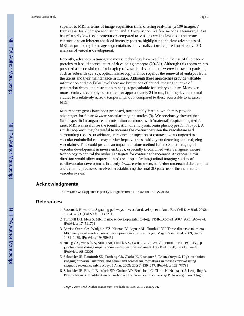
Mutant phenotypes can be visualized using in utero micro-MRITo investigate the potential of this technique for the identification and analysis ofdevelopmental phenotypes, we compared 3D, in vivo images acquired from WT (N=6) andGli2−/− (N=4) mutant embryos (Fig. 3). As reported previously from ex vivo data, Gli2−/−
mutants showed hydrocephaly (enlarged cerebral ventricles), as well as a reduction in size ofthe midbrain and cerebellum (21–23). Moreover, Gli2−/− mutant embryos also showed analteration of spinal cord shape, in the form of a “kink” in the cervical region that facilitatedtheir identification even on pilot scans (not shown).
Berrios-Otero et al. Page 4
Magn Reson Med. Author manuscript; available in PMC 2013 January 01.
NIH
-PA Author Manuscript
NIH
-PA Author Manuscript
NIH
-PA Author Manuscript
Vascular phenotypes can be identified and analyzed using in utero micro-MRIAnalysis of the cerebral vasculature of WT and Gli2−/− mutant embryos also revealed anumber of vascular phenotypes in the mutant brains (Figs. 4, 5; N=6, WT; N=3, Gli2−/−).Most notably, in all Gli2−/− mutant embryos there was an obvious absence of the basilarartery and associated arterial branches, as well as an alteration in the size and geometry ofthe Circle of Willis evidenced by a reduction in the distance between its component arteriessimilar to our previous report using ex vivo micro-MRI (3). Signal intensity on the ventralside of the brain, measured in a region-of-interest (ROI) covering the normal location of thebasilar artery, was significantly lower in Gli2−/− mutant embryos, confirming the visuallydescribed phenotype (mean ± standard deviation: Gli2−/− = 1.02 ± 0.03, N=3 vs. WT = 1.35± 0.15 N=6; p < 0.003). In Gli2−/− mutant embryos, we also observed a truncation of thevertebral arteries, which failed to enter the more posterior brain regions, as well as a variableregion in the medial-posterior brain, where abnormal vessels occupied part of the spacenormally occupied by the vertebral and basilar arteries, possibly as a result of abnormalangiogenesis to compensate for the loss of those vessels. Segmentations were alsogenerated, comparing ex vivo contrast-enhanced micro-MRI (Fig. 5a–d) and in utero MRI(Fig. 5e–h) in E17.5 WT (Fig. 5a, b, e, f) and Gli2−/− embryos (Fig. 5c, d, g, h). Thesecomparisons demonstrated similar genotype-specific 3D patterns, except in the variablemedial-posterior regions of the Gli2−/− mutants, after accounting for the inherent differencein resolution (50-μm for ex vivo compared to 115-μm for in vivo) and the blooming of theT2* blood contrast beyond the vascular walls.
DISCUSSIONSevere disruptions in numerous organs and vascular networks often result in embryonic andearly postnatal lethality. Previous reports have demonstrated the utility of ex vivo micro-MRI as a tool for analyzing 3D anatomical and vascular defects in fixed mouse embryos (3–8). With the use of self-gating to minimize motion artifacts, combined with imageregistration methods, we have shown that high resolution, highly detailed data can beacquired in utero from living mouse embryos. Previous reports have shown that some formsof periodic motion, including cardiac and respiratory motion in postnatal animals, can besuppressed with simple k-space averaging (24–25). We showed that this strategy is noteffective in the face of a permanent displacement, as in fetal imaging, which requiresregistration before k-space averaging (Fig. 1). Using our approach, we demonstrated the invivo detection and analysis of brain and cerebral vascular phenotypes in Gli2 mutantembryos. Our results show great potential for in utero 3D MRI to analyze the developmentof normal vasculature and vascular phenotypes associated in a variety of mutant mouseembryos. This will provide future opportunities for investigating in utero phenotypes lethalin late embryonic or early postnatal stages and should permit longitudinal observation ofvascular development in vivo. With the use of higher resolution imaging parameters,combined with closer fitting surface coils and vascular-targeted contrast agents we anticipatethat mouse embryos can be imaged from much earlier stages, enabling longitudinal studiesof organ and vascular development.
From a practical point of view, future longitudinal studies will require identification andimaging of an individual mouse embryo from one time point to the next. This challenge issimilar for MRI and other in utero imaging modalities such as ultrasound biomicroscopy(UBM) (26–27). Similar to UBM, careful positioning of the pregnant mouse within the coiland use of the pilot scans to map the fetal positions with respect to adjacent anatomicalstructures should enable accurate identification of individual embryos over intervals of atleast 12–24 hours (28). In embryos with clearly defined mutant phenotypes, such as thespinal cord and basilar artery phenotypes in the Gli2−/−mice, the mutant defects themselvesmay be useful for identifying individual embryos on subsequent imaging sessions. UBM is
Berrios-Otero et al. Page 5
Magn Reson Med. Author manuscript; available in PMC 2013 January 01.
NIH
-PA Author Manuscript
NIH
-PA Author Manuscript
NIH
-PA Author Manuscript
superior to MRI in terms of image acquisition time, offering real-time (≥ 100 images/s)frame rates for 2D image acquisition, and 3D acquisition in a few seconds. However, UBMhas relatively low tissue penetration compared to MRI, as well as low SNR and tissuecontrast, and an inherent speckled intensity pattern, highlighting the clear advantages ofMRI for producing the image segmentations and visualizations required for effective 3Danalysis of vascular development.
Recently, advances in transgenic mouse technology have resulted in the use of fluorescentproteins to label the vasculature of developing embryos (29–31). Although this approach hasprovided a successful tool for imaging of vascular development in vivo in lower organisms,such as zebrafish (29,32), optical microscopy in mice requires the removal of embryos fromthe uterus and their maintenance in culture. Although these approaches provide valuableinformation at the cellular level there are limitations of optical imaging in terms ofpenetration depth, and restriction to early stages suitable for embryo culture. Moreovermouse embryos can only be cultured for approximately 24 hours, limiting developmentalstudies to a relatively narrow temporal window compared to those accessible to in uteroMRI.
MRI reporter genes have been proposed, most notably ferritin, which may provideadvantages for future in utero vascular imaging studies (9). We previously showed that(brain specific) manganese administration combined with (maternal) respiration gated inutero MRI was useful for the identification of embryonic brain phenotypes in vivo (33). Asimilar approach may be useful to increase the contrast between the vasculature andsurrounding tissues. In addition, intravascular injection of contrast agents targeted tovascular endothelial cells may further improve the sensitivity for detecting and analyzingvasculature. This could provide an important future method for molecular imaging ofvascular development in mouse embryos, especially if combined with transgenic mousetechnology to control the molecular targets for contrast enhancement. Advances in thisdirection would allow unprecedented tissue specific longitudinal imaging studies ofcardiovascular development in a truly in situ environment, to further understand the complexand dynamic processes involved in establishing the final 3D patterns of the mammalianvascular system.
AcknowledgmentsThis research was supported in part by NIH grants R01HL078665 and R01NS038461.
References1. Rossant J, Howard L. Signaling pathways in vascular development. Annu Rev Cell Dev Biol. 2002;
18:541–573. [PubMed: 12142271]
2. Turnbull DH, Mori S. MRI in mouse developmental biology. NMR Biomed. 2007; 20(3):265–274.[PubMed: 17451170]
3. Berrios-Otero CA, Wadghiri YZ, Nieman BJ, Joyner AL, Turnbull DH. Three-dimensional micro-MRI analysis of cerebral artery development in mouse embryos. Magn Reson Med. 2009; 62(6):1431–1439. [PubMed: 19859945]
4. Huang GY, Wessels A, Smith BR, Linask KK, Ewart JL, Lo CW. Alteration in connexin 43 gapjunction gene dosage impairs conotruncal heart development. Dev Biol. 1998; 198(1):32–44.[PubMed: 9640330]
5. Schneider JE, Bamforth SD, Farthing CR, Clarke K, Neubauer S, Bhattacharya S. High-resolutionimaging of normal anatomy, and neural and adrenal malformations in mouse embryos usingmagnetic resonance microscopy. J Anat. 2003; 202(2):239–247. [PubMed: 12647873]
6. Schneider JE, Bose J, Bamforth SD, Gruber AD, Broadbent C, Clarke K, Neubauer S, Lengeling A,Bhattacharya S. Identification of cardiac malformations in mice lacking Ptdsr using a novel high-
Berrios-Otero et al. Page 6
Magn Reson Med. Author manuscript; available in PMC 2013 January 01.
NIH
-PA Author Manuscript
NIH
-PA Author Manuscript
NIH
-PA Author Manuscript

throughput magnetic resonance imaging technique. BMC Dev Biol. 2004; 4:16. [PubMed:15615595]
7. Smith BR, Johnson GA, Groman EV, Linney E. Magnetic resonance microscopy of mouse embryos.Proc Natl Acad Sci U S A. 1994; 91(9):3530–3533. [PubMed: 8170941]
8. Wadghiri YZ, Schneider AE, Gray EN, Aristizabal O, Berrios C, Turnbull DH, Gutstein DE.Contrast-enhanced MRI of right ventricular abnormalities in Cx43 mutant mouse embryos. NMRBiomed. 2007; 20(3):366–374. [PubMed: 17451172]
9. Cohen B, Ziv K, Plaks V, Israely T, Kalchenko V, Harmelin A, Benjamin LE, Neeman M. MRIdetection of transcriptional regulation of gene expression in transgenic mice. Nat Med. 2007; 13(4):498–503. [PubMed: 17351627]
10. Fisher, CE.; Howie, SEM., editors. Shh and Gli signalling and development. Georgetown, Tex.,New York, N.Y: Landes Bioscience/Eurekah.com; Springer Science+Business Media; 2006. p.158
11. Nagase T, Nagase M, Machida M, Fujita T. Hedgehog signalling in vascular development.Angiogenesis. 2008; 11(1):71–77. [PubMed: 18301996]
12. Nieman BJ, Szulc KU, Turnbull DH. Three-dimensional, in vivo MRI with self-gating and imagecoregistration in the mouse. Magn Reson Med. 2009; 61(5):1148–1157. [PubMed: 19253389]
13. Mo R, Freer AM, Zinyk DL, Crackower MA, Michaud J, Heng HH, Chik KW, Shi XM, Tsui LC,Cheng SH, Joyner AL, Hui C. Specific and redundant functions of Gli2 and Gli3 zinc finger genesin skeletal patterning and development. Development. 1997; 124(1):113–123. [PubMed: 9006072]
14. Pringle KG, Kind KL, Thompson JG, Roberts CT. Complex interactions between hypoxiainducible factors, insulin-like growth factor-II and oxygen in early murine trophoblasts. Placenta.2007; 28(11–12):1147–1157. [PubMed: 17658597]
15. Okazaki K, Maltepe E. Oxygen, epigenetics and stem cell fate. Regen Med. 2006; 1(1):71–83.[PubMed: 17465821]
16. Lee YM, Jeong CH, Koo SY, Son MJ, Song HS, Bae SK, Raleigh JA, Chung HY, Yoo MA, KimKW. Determination of hypoxic region by hypoxia marker in developing mouse embryos in vivo: apossible signal for vessel development. Dev Dyn. 2001; 220(2):175–186. [PubMed: 11169851]
17. Ogawa S, Lee TM, Nayak AS, Glynn P. Oxygenation-sensitive contrast in magnetic resonanceimage of rodent brain at high magnetic fields. Magn Reson Med. 1990; 14(1):68–78. [PubMed:2161986]
18. Ogawa S, Lee TM. Magnetic resonance imaging of blood vessels at high fields: in vivo and in vitromeasurements and image simulation. Magn Reson Med. 1990; 16(1):9–18. [PubMed: 2255240]
19. Collins DL, Neelin P, Peters TM, Evans AC. Automatic 3D intersubject registration of MRvolumetric data in standardized Talairach space. J Comput Assist Tomogr. 1994; 18(2):192–205.[PubMed: 8126267]
20. Sled JG, Zijdenbos AP, Evans AC. A nonparametric method for automatic correction of intensitynonuniformity in MRI data. IEEE Trans Med Imaging. 1998; 17(1):87–97. [PubMed: 9617910]
21. Palma V, Ruiz i Altaba A. Hedgehog-GLI signaling regulates the behavior of cells with stem cellproperties in the developing neocortex. Development. 2004; 131(2):337–345. [PubMed:14681189]
22. Corrales JD, Rocco GL, Blaess S, Guo Q, Joyner AL. Spatial pattern of sonic hedgehog signalingthrough Gli genes during cerebellum development. Development. 2004; 131(22):5581–5590.[PubMed: 15496441]
23. Blaess S, Corrales JD, Joyner AL. Sonic hedgehog regulates Gli activator and repressor functionswith spatial and temporal precision in the mid/hindbrain region. Development. 2006; 133(9):1799–1809. [PubMed: 16571630]
24. Beckmann N, Tigani B, Ekatodramis D, Borer R, Mazzoni L, Fozard JR. Pulmonary edemainduced by allergen challenge in the rat: noninvasive assessment by magnetic resonance imaging.Magn Reson Med. 2001; 45(1):88–95. [PubMed: 11146490]
25. Ble FX, Cannet C, Zurbruegg S, Karmouty-Quintana H, Bergmann R, Frossard N, Trifilieff A,Beckmann N. Allergen-induced lung inflammation in actively sensitized mice assessed with MRimaging. Radiology. 2008; 248(3):834–843. [PubMed: 18647843]
Berrios-Otero et al. Page 7
Magn Reson Med. Author manuscript; available in PMC 2013 January 01.
NIH
-PA Author Manuscript
NIH
-PA Author Manuscript
NIH
-PA Author Manuscript

26. Ji RP, Phoon CK, Aristizabal O, McGrath KE, Palis J, Turnbull DH. Onset of cardiac functionduring early mouse embryogenesis coincides with entry of primitive erythroblasts into the embryoproper. Circ Res. 2003; 92(2):133–135. [PubMed: 12574139]
27. Phoon CK, Ji RP, Aristizabal O, Worrad DM, Zhou B, Baldwin HS, Turnbull DH. Embryonicheart failure in NFATc1−/− mice: novel mechanistic insights from in utero ultrasoundbiomicroscopy. Circ Res. 2004; 95(1):92–99. [PubMed: 15166096]
28. Ji RP, Phoon CK. Noninvasive localization of nuclear factor of activated T cells c1−/− mouseembryos by ultrasound biomicroscopy-Doppler allows genotype-phenotype correlation. J Am SocEchocardiogr. 2005; 18(12):1415–1421. [PubMed: 16376776]
29. Motoike T, Loughna S, Perens E, Roman BL, Liao W, Chau TC, Richardson CD, Kawate T, KunoJ, Weinstein BM, Stainier DY, Sato TN. Universal GFP reporter for the study of vasculardevelopment. Genesis. 2000; 28(2):75–81. [PubMed: 11064424]
30. Fraser ST, Hadjantonakis AK, Sahr KE, Willey S, Kelly OG, Jones EA, Dickinson ME, BaronMH. Using a histone yellow fluorescent protein fusion for tagging and tracking endothelial cells inES cells and mice. Genesis. 2005; 42(3):162–171. [PubMed: 15986455]
31. Larina IV, Shen W, Kelly OG, Hadjantonakis AK, Baron MH, Dickinson ME. A membraneassociated mCherry fluorescent reporter line for studying vascular remodeling and cardiacfunction during murine embryonic development. Anat Rec (Hoboken). 2009; 292(3):333–341.[PubMed: 19248165]
32. Lawson ND, Weinstein BM. In vivo imaging of embryonic vascular development using transgeniczebrafish. Dev Biol. 2002; 248(2):307–318. [PubMed: 12167406]
33. Deans AE, Wadghiri YZ, Berrios-Otero CA, Turnbull DH. Mn enhancement and respiratory gatingfor in utero MRI of the embryonic mouse central nervous system. Magn Reson Med. 2008; 59(6):1320–1328. [PubMed: 18506798]
Berrios-Otero et al. Page 8
Magn Reson Med. Author manuscript; available in PMC 2013 January 01.
NIH
-PA Author Manuscript
NIH
-PA Author Manuscript
NIH
-PA Author Manuscript

Fig. 1. Image registration and processing methods for visualizing the embryonic vasculatureA series of individual low SNR images were acquired using in vivo imaging methods (a)and a manually drawn mask (dashed outline, panel b) was drawn around the embryonicbrain and blood vessels and used to register the 50 serially-acquired low SNR images.Examples are provided of two embryos with different net displacements over the 2.75h scan,comparing simple k-space averaging with registration-averaging: Embryo 1 (netdisplacement = 0.5-mm) (c, d, e); Embryo 2 (net displacement = 1.0-mm) (f, g, h).Averaging without registration (c, f) produced obviously blurred images with limited(hypointense) vascular detail compared to averaging after registration (d, g), especially inEmbryo 2 (f, g). Processing of the registered-averaged images with a contrast-inverting filter(e, h) produced improved visualization of the vasculature. [Note: for visualization purposes,cropped mid-sagittal sections of the 3D images have been presented here.]
Berrios-Otero et al. Page 9
Magn Reson Med. Author manuscript; available in PMC 2013 January 01.
NIH
-PA Author Manuscript
NIH
-PA Author Manuscript
NIH
-PA Author Manuscript

Fig. 2. 2D sections from in vivo 3D MRI data show the developing vasculatureHorizontal and coronal sections at different levels of the embryonic brain show multiplevessels of the Circle of Willis (CW). Abbreviations: anterior inferior cerebellar artery,AICA; basilar artery, BA; internal carotid arteries, ICA; middle cerebral artery, MCA;superior cerebral artery, SCA; transverse sinus, TS; vertebral arteries, VA.
Berrios-Otero et al. Page 10
Magn Reson Med. Author manuscript; available in PMC 2013 January 01.
NIH
-PA Author Manuscript
NIH
-PA Author Manuscript
NIH
-PA Author Manuscript
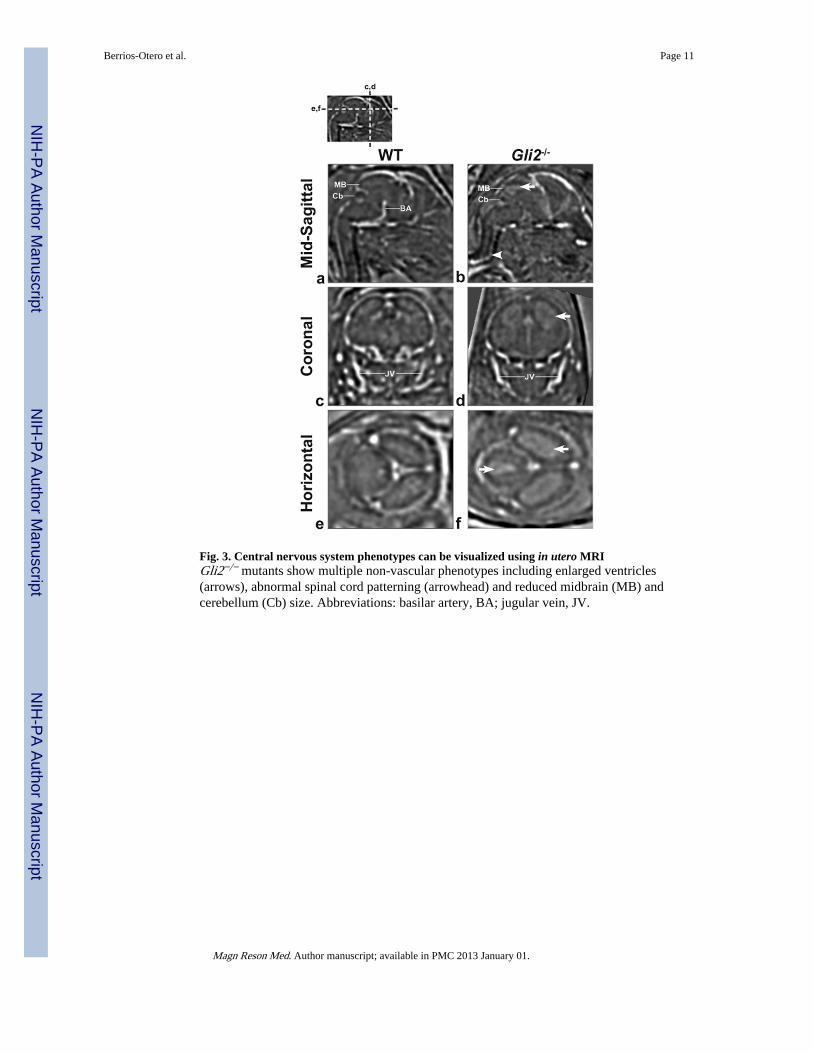
Fig. 3. Central nervous system phenotypes can be visualized using in utero MRIGli2−/− mutants show multiple non-vascular phenotypes including enlarged ventricles(arrows), abnormal spinal cord patterning (arrowhead) and reduced midbrain (MB) andcerebellum (Cb) size. Abbreviations: basilar artery, BA; jugular vein, JV.
Berrios-Otero et al. Page 11
Magn Reson Med. Author manuscript; available in PMC 2013 January 01.
NIH
-PA Author Manuscript
NIH
-PA Author Manuscript
NIH
-PA Author Manuscript
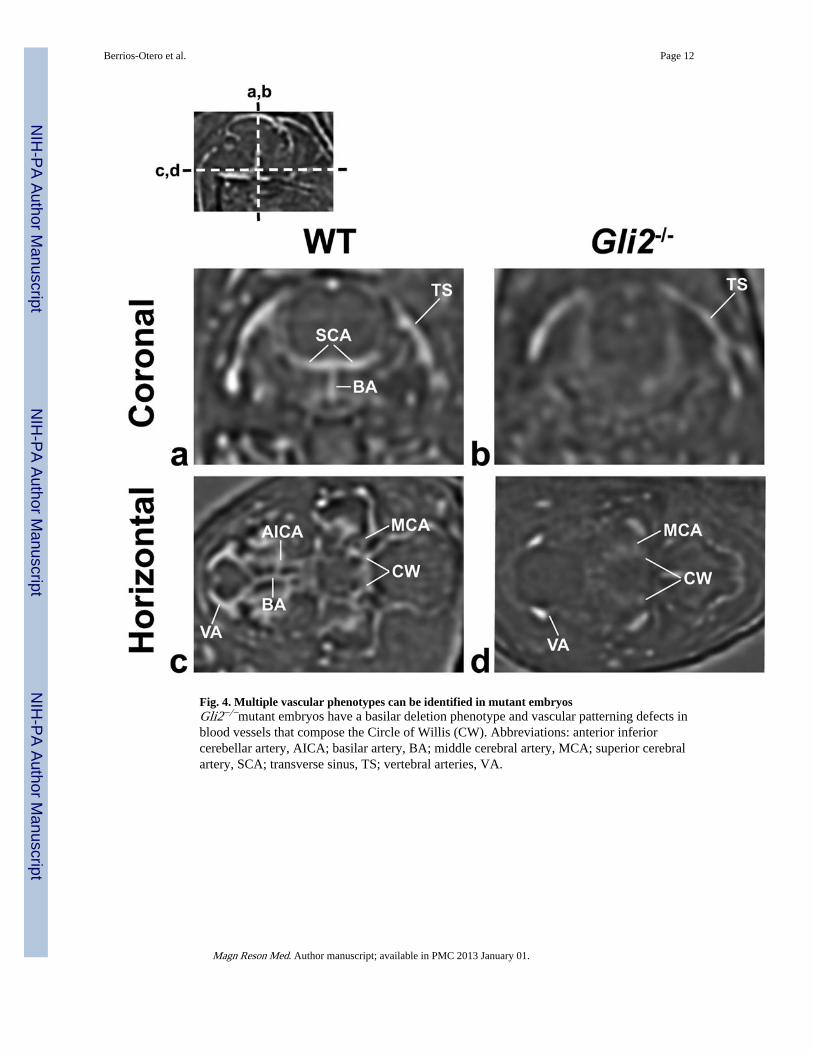
Fig. 4. Multiple vascular phenotypes can be identified in mutant embryosGli2−/−mutant embryos have a basilar deletion phenotype and vascular patterning defects inblood vessels that compose the Circle of Willis (CW). Abbreviations: anterior inferiorcerebellar artery, AICA; basilar artery, BA; middle cerebral artery, MCA; superior cerebralartery, SCA; transverse sinus, TS; vertebral arteries, VA.
Berrios-Otero et al. Page 12
Magn Reson Med. Author manuscript; available in PMC 2013 January 01.
NIH
-PA Author Manuscript
NIH
-PA Author Manuscript
NIH
-PA Author Manuscript
Fig. 5. 3D volumetric rendering of wild type and Gli2 mutant cerebral vasculature in vivo MRIcorrelates with similar ex vivo renderingsEx vivo (a–d) and in vivo (e–h) 3D rendering views of the Circle of Wills and cerebralvasculature show deletion of the basilar artery and variable alteration of posterior cerebralartery branches (blue), constriction between the left and right carotid arteries (red) andoverall geometric differences in the Circle of Willis (yellow) between WT (top row) andGli2 mutants (bottom row). Abbreviations: anterior cerebral artery, ACA; anterior inferiorcerebellar artery, AICA; basilar artery, BA; middle cerebral artery, MCA; vertebral arteries,VA.
Berrios-Otero et al. Page 13
Magn Reson Med. Author manuscript; available in PMC 2013 January 01.
NIH
-PA Author Manuscript
NIH
-PA Author Manuscript
NIH
-PA Author Manuscript
Copyright © 2022 FDOKUMEN