Exposure to acrolein by inhalation causes platelet activation


DISCUSSION
The current inhalation reference concentration (RfC) for manganese, as set by the US Environmental Protection
Agency, is 0.05 µg Mn/m3 (24). Thus, the concentrations of manganese used in this study were 1000, 10,000,
and 20,000 times this standard for the lowest, median, and highest doses, respectively. Manganese
concentrations in the olfactory bulb, striatum, and cerebellum of rats that were exposed similarly to manganese
sulfate have been reported (25). Dorman and co-workers (26) observed increased brain manganese
concentrations in PND 14 and PND 19 pups that were exposed to manganese sulfate at > 0.05 mg Mn/m3.
Increased striatal, olfactory bulb, and cerebellum concentrations were approx twofold to threefold higher than
those observed in controls. Adult (F1) rats that were exposed as neonates to MnSO4 had returned to control
brain manganese concentrations when assessed at PND 45 ± 1 (26). When a similar paradigm of manganese
exposure followed by a recovery period was administered to older rats, manganese concentrations in some brain
regions, in particular the olfactory bulb, remained elevated when compared to air-exposed rats (19). However,
in the present study, younger rats that were exposed to manganese sulfate in utero and during lactation fully
normalized their brain regional manganese concentrations by PND 45 (i.e., 3 wk after exposure sessions ended).
It is likely that this ability to normalize brain manganese concentrations reflects the increased requirements of
the developing rat brain (27).
This study presents novel data that show alterations in oxidative stress markers in specific brain regions of
developing male and female rats after 3 wk of recovery from in utero and neonatal exposures to airborne
manganese. Furthermore, these changes in brain regional oxidative stress markers occurred in the absence of
increased brain manganese concentrations and were in contrast to our earlier studies conducted in adult rats (1–
3).
One can detect the presence of oxidants by measuring species that are known to increase or decrease in response
to oxidative stress. This includes ubiquitous antioxidants, such as GSH, as well as biomarkers that are more
specific to particular tissue types (e.g., glutamine synthetase). In the central nervous system, GS is localized
exclusively in astrocytes (28), where it has a critical role in amino acid metabolism. Glutamine synthetase
metabolizes glutamate that is removed by astrocytes from the extracellular space to glutamine, and the latter is
recycled to neurons as part of the glutamate–glutamine cycle (29). Glutamine synthetase is highly susceptible to
oxidation and subsequent rapid degradation and, therefore, it serves as an excellent marker for the presence of
reactive oxygen species in the brain (30).
Although GS protein levels decreased in most brain regions of manganese exposed male and female rats, only
the cerebellum in the male rats exposed to the median dose had a statistically relevant decrease in GS protein
levels compared to nonexposed rats (Table 1) and this decrease occurred despite normal manganese
concentrations in this brain region. This is in contrast to our previous studies in which the effects of manganese
exposure in older animals of both sexes were more selectively affected [e.g., hippocampus only in male rats and
hypothalamus in female rats (3)]. Corroborating earlier findings (1–3), the GS mRNA levels (Table 2) measured
in this study was not reflected in altered amounts of GS protein (i.e., brain regions that had increased mRNA
levels did not have increased protein levels). In fact, there were more statistically significant changes in GS
mRNA than there were in the protein levels, which is in contrast to our prior studies (1–3). There were
significant increases in GS mRNA levels in the hypothalamus and olfactory bulb of male rats recovering from
the highest manganese exposure, whereas GS mRNA levels were significantly decreased in the striatum of
female rats recovering from the median and high exposures (Table 2). Sex differences in the response to
manganese exposure were exemplified in the striatum, where the females had significant decreases in GS
mRNA but the males were unaffected in this brain region.
The MTs are a class of highly conserved proteins known to bind metals, but in recent years, evidence has shown
that they might also have some important antioxidant properties (31). In vitro experiments demonstrated that
MTs had a greater ability at scavenging oxygen radicals when compared with other sulfhydryl-containing
molecules (32) and they increased more than glutathione (GSH), Mn-SOD (superoxide dismutase) catalase, and
other well-known antioxidants (33). We found that MT mRNA levels were significantly decreased in the
hippocampus, hypothalamus, and striatum in males recovering from the lowest manganese dose and in the
hippocampus and hypothalamus in those recovering from the median dose manganese exposure (Table 3). The
only statistically relevant change in the females was in the olfactory bulb of female rats recovering from the
median dose (Table 3).
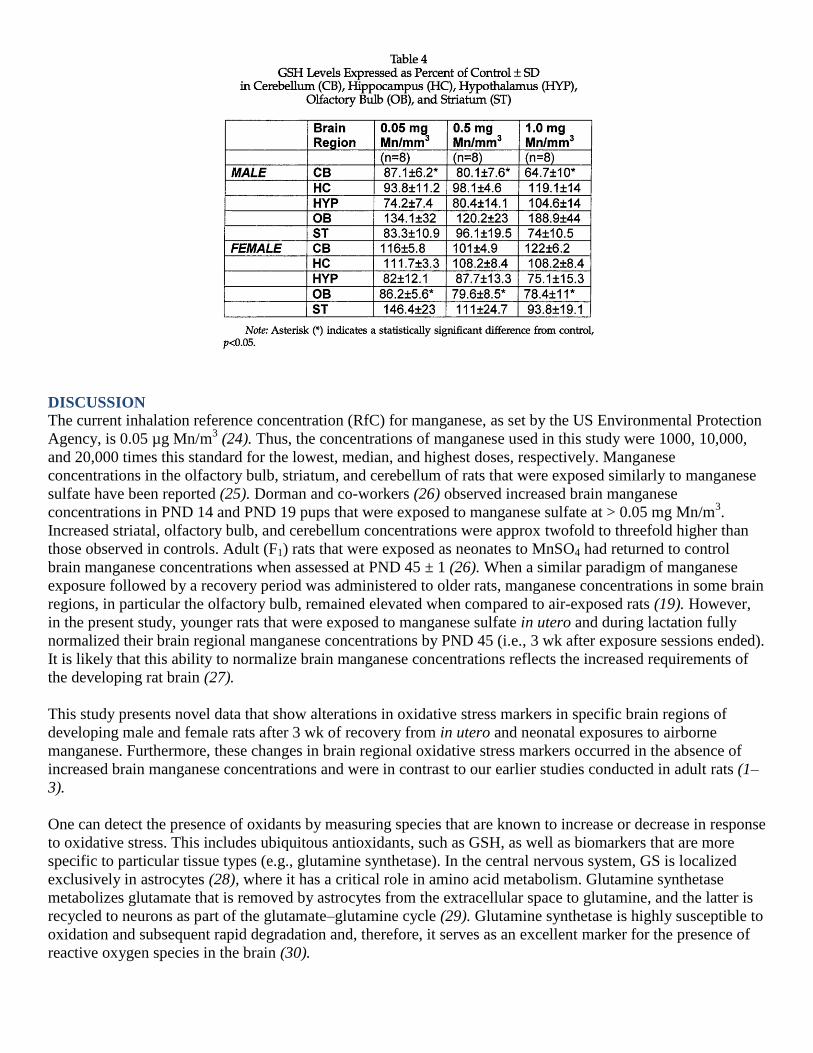
Glutathione is a ubiquitous antioxidant formed from three amino acids, glutamate, cysteine, and glycine (γ-
glutamylcysteinylglycine). It constitutes approx 90% of the intracellular nonprotein thiols (34) and functions in
conjugation and elimination of toxic molecules, thereby maintaining cellular redox homeostasis (34). We
previously reported that GSH levels were significantly lowered in the striatum of female and old male rats upon
manganese exposure (3) and that this effect was absent in the young male rats, which was consistent with our
other study (1). These data are consistent with the findings of a previous study that showed decreased GSH
levels in the striatum of aged rats exposed to manganese chloride (35). In this current study, the only
statistically significant effect of manganese exposure appeared in the cerebellum of male rats recovering from
the highest manganese exposure level (Table 4). This relative lack of an effect of manganese exposure on GSH
in these animals likely reflects the reversibility of manganese exposure and/or the plasticity of the developing
nervous system in terms of GSH metabolism. Although this is speculative, it is consistent with the critical role
GSH plays in the maintenance of redox homeostasis.
In conclusion, we report for the first time the consequences of in utero and early developmental airborne
manganese exposure on end points of oxidative stress in developing female and male rats. These alterations are
not associated with increased manganese as in our prior studies, but these changes persist even after brain
regional manganese concentrations return to control levels. Furthermore, the reversibility of these alterations in
markers of oxidative stress caused by manganese exposure is influenced by the sex of the rat. Unlike our
previous studies (1–3), manganese exposure in young male rats had more of an impact on oxidative stress end
points than it did in female rats. We cannot determine whether this difference is the result of a lack of
reversibility in the male rats or whether markers of oxidative stress in the female rat are less affected by
manganese exposure. It is noteworthy that these effects occurred at doses of manganese 10–20 thousand times
higher than the current RfC. We are presently undertaking a study in which we will specifically examine some
of these reversibility issues.
REFERENCES
1. A. W. Dobson, S. Weber, D. C. Dorman, L. K. Lash, K. M. Erikson, and M. Aschner, Inhaled manganese
sulfate and measures of oxidative stress in rat brain. Biol. Trace Element Res. 93,113–126 (2003).
2. S. Weber, D. C. Dorman, L. H. Lash, K. Erikson, K. E. Vrana, and M. Aschner, Effects of manganese (Mn)
on the developing rat brain: oxidative-stress related endpoints. Neurotoxicology 23,169–175 (2002).
3. K. M. Erikson, D. C. Dorman, L. H. Lash, A. W. Dobson, and M. Aschner. Airborne manganese exposure
differentially affects endpoints of oxidative stress in an age and sex-dependent manner. Biol Trace Element
Res., 100, 49–62 (2004).
4. L. S. Hurley and C. L. Keen, Manganese, in Trace Elements in Human Health and Animal Nutrition, E.
Underwood and W. Mertz, eds., Academic, New York, pp. 185–223 (1987).
5. M. Aschner, K. M. Erikson, and D. C. Dorman, Manganese dosimetry: species differences and implications
for neurotoxicity. Crit. Rev. Toxicol. 35,1–32 ( 2005).
6. ATSDR (Agency for Toxic Substances and Disease Registry). Toxicological Profile for Manganese, US
Department of Health and Human Services Public Health Service. Available at
http://www.atsdr.cdc.gov/toxprofiles/tp151.html (accessed September 2000).
7. D. Mergler, G. Huel, R. Bowler, et al., Nervous system dysfunction among workers with long-term
exposure to manganese. Environ. Res. 64,151–180 (1994).
8. P. K. Pal, A. Samii, and D. B. Calne, Manganese neurotoxicity: a review of clinical features, imaging and
pathology. Neurotoxicology 20, 227–238 (1999).
9. E. D. Pellizzari, C. A. Clayton, C. E. Rodes, et al., Particulate matter and manganese exposures in
Indianapolis, Indiana. J. Exp. Anal. Environ. Epidemiol. 11, 423–440 (2001).
10. M. Aschner, Manganese neurotoxicity and oxidative damage, in Metals and Oxidative Damage in
Neurological Disorders, J. R. Connor, ed., Plenum, New York, pp. 77–93 (1997).
11. W. N. Sloot, J. Korf, J. F. Koster, L. E. A. DeWit, and J. B. P. Gramsbergen, Manganese-induced hydroxyl
radical formation in rat striatum is not attenuated by dopamine depletion or iron chelation in vivo. Exp. Neurol.
138, 236–245 (1996).
12. P. Galvani, P. Fumagalli, and A. Santagostino, Vulnerability of mitochondrial complex I in PC12 cells
exposed to manganese. Eur. J. Pharmacol. 293,377–383 (1995).
13. C. E. Gavin, K. K. Gunter, and T. E. Gunter, Manganese and calcium transport in mitochondria:
implications for manganese toxicity. Neurotoxicology 20, 445–453 (1999).
14. F. S. Archibald and C. Tyree, Manganese poisoning and the attack of trivalent manganese upon
catecholamines. Arch. Biochem. Biophys. 256, 638–650 (1987).
15. S. F. Ali, H. M. Duhart, G. D. Newport, G. W. Lipe, and W. Slikke, Manganese-induced reactive oxygen
species: comparison between Mn+2
and Mn+3
. Neurodegeneration 4, 329–334 (1995).
16. J. Y. Chen, G. C. Tsao, Q. Zhao and W. Zheng, Differential cytotoxicity of Mn(II) and Mn(III): special
reference to mitochondrial [Fe-S] containing enzymes. Toxicol. Appl. Pharmacol. 175,160–168 (2001).
17. K. K. Gunter, L. M. Miller, M. Aschner, et al., XANES spectroscopy: a promising tool for toxicology: a
tutorial. Neurotoxicology 23,127–146 (2002).
18. D. HaMai A. Campbell, and S. C. Bondy, Modulation of oxidative events by multivalent manganese
complexes in brain tissue. Free Radical Biol. Med. 31, 763–768 (2001).
19. D. C. Dorman, B. E. McManus, M. W. Marshall, R. A. James, and M. F. Struve, Old age and gender
influence the pharmacokinetics of inhaled manganese sulfate and manganese phosphate in rats. Toxicol. Appl.
Pharmacol. 197,113–124 (2004).
20. V. Barbu and F. Dautry, Northern blot normalization with a 28S rRNA oligonucleotide probe, Nucleic Acids
Res. 17, 7115 (1989).
21. M. W. Fariss and D. J. Reed, High-performance liquid chromatography of thiols and disulfides:
dinitrophenol derivatives, Methods Enzymol. 143,101–109 (1987).
22. L. H. Lash and J. J. Tokarz, Oxidative stress in isolated rat renal proximal and distal tubular cells, Am. J.
Physiol. 259, F338–F347 (1990).
23. L. H. Lash and E. B. Woods, Cytotoxicity of alkylating agents in isolated rat kidney proximal and distal
tubular cells, Arch. Biochem. Biophys. 286,46–56 (1991).
24. J. M. Davis, Inhalation health risks of manganese: an EPA perspective, Neurotoxicology 20,511–518
(1999).
25. D. C. Dorman, A. M. McElveen, M. W. Marshall, et al., Maternal–fetal distribution of manganese in the rat
following inhalation exposure to manganese sulfate. Neurotoxicology, in press.
26. D. C. Dorman, A. M. McElveen, M. W. Marshall, et al., Tissue manganese concentrations in lactating rats
and their offspring following combined in utero and lactation exposure to inhaled manganese sulfate. Toxicol.
Sci. 84,12–21 (2005).
27. C. L. Keen, J. G. Bell, and B. Lonnerdal, The effect of age on manganese uptake and retention from milk
and infant formulas in rats, J. Nutr. 116(3), 395–402 (1986).
28. A. Martinez-Hernandez, K. P. Bell, and M. D. Norenberg, Glutamine synthetase: glial localization in the
brain, Science 195,1356–1358 (1977).
29. U. Sonnewald, N. Westergaard, and A. Schousboe, Glutamate transport and metabolism in astrocytes, Glia
21, 56–63 (1997).
30. E. R. Stadtman, Protein oxidation and aging, Science 257,1220–1224 (1992).
31. M. Aschner, The functional significance of brain metallothioneins, FASEB J. 10, 1129–1136 (1996).
32. S. Hussain, W. Slikker, Jr., and S. F. Ali, Role of metallothionein and other antioxidants in scavenging
superoxide radicals and their possible role in neuroprotection, Neurochem. Int. 29,145–152 (1996).
33. M. Kondoh, Y. Inoue, S. Atagi, et al., Specific induction of metallothionein synthesis by mitochondrial
oxidative stress, Life Sci. 69, 2137–2146 (2001).
34. A. Meister and M. E. Anderson, Glutathione, Annu. Rev. Biochem. 52, 711–760 (1983).
35. M. S. Desole, G. Esposito, R. Mighelli, et al., Cellular defence mechanisms in the striatum of young and
aged rats subchronically exposed to manganese. Neuropharmacology 34,289–295 (1995).
Copyright © 2022 FDOKUMEN