From Water to Aquaretics: a Legendary Route - Karger ...
20
Cell Physiol Biochem 2014;33:1369-1388 DOI: 10.1159/000358704 Published online: May 05, 2014 © 2014 S. Karger AG, Basel www.karger.com/cpb 1369 Cellular Physiology and Biochemistry Cellular Physiology and Biochemistry 1421-9778/14/0335-1369$39.50/0 Review Copyright © 2014 S. Karger AG, Basel Accepted: January 24, 2014 This is an Open Access article licensed under the terms of the Creative Commons Attribution- NonCommercial 3.0 Unported license (CC BY-NC) (www.karger.com/OA-license), applicable to the online version of the article only. Distribution permitted for non-commercial purposes only. From Water to Aquaretics: a Legendary Route Valentina Donato Antonio Lacquaniti Valeria Cernaro Giuseppina Lorenzano Domenico Trimboli Antoine Buemi Rosaria Lupica Michele Buemi Chair of Nephrology, Department of Internal Medicine, University of Messina, Messina, Italy Key Words Aquaretics • Vasopressin • Osmolarity Abstract Man is water. When life appeared on earth, the primordial cell had a simple structure and could immediately ascertain from the surrounding aquatic environment the substances for nutrition and oxygen, without any need for structural complexity. As part of evolution, during the transition from aquatic to terrestrial life, vertebrates had to fight against dehydration as well as fish in the sea. In this complex mechanism of osmoregulation, the structure and function of some osmoregulatory hormones have been maintained during the evolution of species, from fish to man. Within the homeostatic mechanism, the renin-angiotensin-aldosterone system (RAAS) is crucial in the regulation of renal reasorption of water and sodium. It is also involved in the regulation of renal plasma flux, blood volume and blood pressure. Vasopressin plays a hormonal function in the mechanisms of water homeostasis acting through Aquaporins (AQP), channel-proteins that allow bi-directional water transport across cell membranes. Introduction Man is water. If we drop water in any form into this sea, we play an active part in the life of this being. When life appeared on earth, the primordial cell had a simple structure and could immediately ascertain from the surrounding aquatic environment the substances for nutrition and oxygen, without any need for structural complexity. Via Salita Villa Contino, 30 98100 Messina (Italy) Tel. +39 090 2212396, Fax +39 090 2935162, E-Mail [email protected] Prof. Michele Buemi
-
Upload
khangminh22 -
Category
Documents
-
view
0 -
download
0
Transcript of From Water to Aquaretics: a Legendary Route - Karger ...

Cell Physiol Biochem 2014;33:1369-1388DOI: 10.1159/000358704Published online: May 05, 2014
© 2014 S. Karger AG, Baselwww.karger.com/cpb 1369
Donato et al: From Water to Aquaretics
Cellular Physiology and Biochemistry
Cellular Physiology and Biochemistry
1421-9778/14/0335-1369$39.50/0
Review
Copyright © 2014 S. Karger AG, Basel
Accepted: January 24, 2014
This is an Open Access article licensed under the terms of the Creative Commons Attribution-NonCommercial 3.0 Unported license (CC BY-NC) (www.karger.com/OA-license), applicable to the online version of the article only. Distribution permitted for non-commercial purposes only.
From Water to Aquaretics: a Legendary Route
Valentina Donato Antonio Lacquaniti Valeria Cernaro Giuseppina Lorenzano Domenico Trimboli Antoine Buemi Rosaria Lupica Michele Buemi
Chair of Nephrology, Department of Internal Medicine, University of Messina, Messina, Italy
Key WordsAquaretics • Vasopressin • Osmolarity
AbstractMan is water. When life appeared on earth, the primordial cell had a simple structure and could immediately ascertain from the surrounding aquatic environment the substances for nutrition and oxygen, without any need for structural complexity. As part of evolution, during the transition from aquatic to terrestrial life, vertebrates had to fight against dehydration as well as fish in the sea. In this complex mechanism of osmoregulation, the structure and function of some osmoregulatory hormones have been maintained during the evolution of species, from fish to man. Within the homeostatic mechanism, the renin-angiotensin-aldosterone system (RAAS) is crucial in the regulation of renal reasorption of water and sodium. It is also involved in the regulation of renal plasma flux, blood volume and blood pressure. Vasopressin plays a hormonal function in the mechanisms of water homeostasis acting through Aquaporins (AQP), channel-proteins that allow bi-directional water transport across cell membranes.
Introduction
Man is water. If we drop water in any form into this sea, we play an active part in the life of this being. When life appeared on earth, the primordial cell had a simple structure and could immediately ascertain from the surrounding aquatic environment the substances for nutrition and oxygen, without any need for structural complexity.
Via Salita Villa Contino, 3098100 Messina (Italy)Tel. +39 090 2212396, Fax +39 090 2935162, E-Mail [email protected]
Prof. Michele Buemi

Cell Physiol Biochem 2014;33:1369-1388DOI: 10.1159/000358704Published online: May 05, 2014
© 2014 S. Karger AG, Baselwww.karger.com/cpb 1370
Donato et al: From Water to Aquaretics
Cellular Physiology and Biochemistry
Cellular Physiology and Biochemistry
The primordial cells needed only a cellular wall to protect their "Milieu Intérieur" from the external environment and an internal mechanism capable of producing energy.
Things became complicated later, when multicellular organisms appeared on earth. In this new situation condition, a new environment among cells was formed, as well as the intra-and extra-cellular environment, able to connect cells together and create a multi-cellular protective system. The conditions of the world around us are constantly changing but the delicate internal balance of our bodies seems to remain unaffected. This is due to homeostasis (derived from the Greek ὅμοιος, hómoios, meaning “similar” and στάσις, stasis meaning “stable” or “standing still” ), which is the natural tendency of the chemical and physical properties of a living organism to reach a relative internal stability. For this reason, a state of equilibrium is maintained over time by means of specific self-regulatory mechanisms, despite variations in external conditions [1].
Claude Bernard’s concept of Milieu Interieur has been revolutionized: it no longer corresponds to the individual interiority of each cell, but to the entire organism. It is required to make the multi-cellular system independent of the external environment by homeostasis and conveying energetic substances, such as glucose [2].
From Bernard’s concept springs René Quinton’s notion of La mer intérieure which claimed that "Animal life appeared as simple cells in the sea, tends to maintain cellular functions in organisms evolved through collections of cells surrounded in a marine medium that depicts the origins of life." Hence, it can be said that “men are like a walking aquarium with an internal sea in which cells can live” and as Bernard claims, the stability of the Milieu Interieur is the basic condition for life and a free and independent life [3].
Ultimately, "when a factor is able to change the homeostatic state in one direction, it is reasonable to research an automatic control of this factor through other mechanisms that may have a strictly opposite effect" [4].
Osmolarity
Osmolarity espresses the concentration of a solution, highlighting the number of particles dissolved in it, regardless of the electrical charge and size [5].
In any organisms the intracellular and extracellular medium has the same ions concentrations. The extra sodium in the extracellular medium tends to enter the cell and the potassium does the same thing but in the opposite side. The exchange between sodium and potassium determines a difference in the electric charges on the two sides of the cellular membrane and this is the base of the nervous transmission. The osmotic gradient between the internal and external medium defined three categories of animals:
Iso-osmotic animals: internal and external osmolarity are equal (invertebrates and cyclostomes).
Ipo-osmotic animals: internal osmolarity is inferior than in the external medium (water mammals and teleosts).
Iper-osmotic animals: their osmolarity is superior than in the external medium (fresh-water animals and terrestrials).
The kidney, along with some other glands, plays a major role in this situation by regulating the stability of tissue homeostasis through the balance of sodium chloride, remindful of the original sea. In particular, the renal medulla and the counterflow system build the key elements which affect the production of free water according to the needs of the body to maintain iso-osmolarity of the extracellular and intracellular fluid (although the composition of ionic liquids is very different).
Fish, amphibians, reptiles, birds and mammals have had to develop systems that are able to maintain their milieu interieur during the long evolutionary march to establish a free and independent life from seawater.
As part of evolution, it has been possible to trace the migration of vertebrates from salt water to fresh water to the land, with a reverse path. During the transition from aquatic

Cell Physiol Biochem 2014;33:1369-1388DOI: 10.1159/000358704Published online: May 05, 2014
© 2014 S. Karger AG, Baselwww.karger.com/cpb 1371
Donato et al: From Water to Aquaretics
Cellular Physiology and Biochemistry
Cellular Physiology and Biochemistry
to terrestrial life, vertebrates had to fight against dehydration as well as fish in the sea. However, in terrestrial vertebrates, in contrast to saltwater fish, the movement of water and ions has been carried out in the same direction: vertebrates, in fact, must retain water and sodium ions. In this complex mechanism of osmoregulation, the structure and function of some osmoregulatory hormones have been maintained during the evolution of species, from fish to man. Among these hormones are: angiotensin II which can increase thirst in humans; ADH, the anti-diuretic hormone, a homologue of vasotocin in fish, which allows water reabsorption in the kidney; aldosterone, which acquires a determining role as a mineralocorticoid and induces retention of sodium ions; apelin which, being sensitive to changes in osmolarity, participates in the regulation of water-salt balance in synergy with AVP, acting as a central antagonist of arginine-vasopressin antidiuretic effect [6]. These and others osmoreceptors are functionally as neurons and they can detect the osmolarity changes. They are widely expressed in the body: they are located in cerebral areas and others, peripheral, located in the gastrointestinal tract, in the hepatic portal vein and liver. Osmotic stimuli involve a non-selective cation channel in magnocellular neurosecretory cells (MNC) and cells of the Organum Vasculosum of the Lamina Terminalis perceiving thirst through projections to the Anterior Cingulate Cortex and Insula [7].
In a recent work [8] it has been shown the presence of a specific population of TRPV 4 hepatic sensory that can detect physiological changes in blood osmolarity. Differences in the nature of TRPV4 suggest that other proteins may lead to high speeds and more sensivity to hepatic sensory afferents. At the moment we know that Trpv1 and Trpv4 are osmoreceptors which probably contribute to modulation of hypertonicity in Organum Vasculosum Lamina Terminalis (OVLT) neurons. We know that Trpv4 expression is not required for the activation of channels exposure to hypertonicity, but it is required for hypotonicity. These findings suggest that other Trpv4-dependent factors are involved in the central control of osmoregulation [9].
The renin-angiotensin-aldosterone system
Within the homeostatic mechanism, the renin-angiotensin-aldosterone system (RAAS) has been recognized as the key element in the regulation of renal reasorption of water and sodium. It is also involved in the regulation of renal plasma flux, blood volume and blood pressure (Fig. 1) [10].
It is interesting to study the development of this system in the evolution of living beings. The phylogenetic primary function of the RAAS seems to be linked to the migration of living beings from sea to fresh water to the land and vice versa [11]. The various components of the RAAS derived from ancestral genes, which have inevitably undergone a series of mutations and gene duplications. Nevertheless, the components of the RAAS have mantained some characteristics of the superfamilies from which they are descended. The study of RAAS phylogenesis has allowed the evaluation of some ancestral functions: renin, an aspartyl protease, found in retroviruses like HIV; angiotensinogen, a serpin, also found in invertebrates; angiotensin-converting enzyme (ACE), a zinc-dependent metalloproteinases, also discovered in bacteria, whereas angiotensin II (ATIIr), belonging to the family of G protein-coupled receptors or GPCRs, has been found in yeast and plants (Table 1).
This multifaceted expression is proof of the importance of the role played by the RAAS in the evolution of the species, in the adaptation to the sea-land path and in the tolerance of heat and salt reduction in the environment [12]. The establishment of an integrated RAAS coincided with the age of vertebrates. In mammals, epithelioid renin-secreting cells are in the context of the final stretch of the afferent arteriolar wall in direct contact with the glomerulus; renin directly affects the RAAS, whereas in other species, these cells are located at different distances from the glomerulus. In some kinds of fish without a glomerulus, these epithelioid renin-secreting cells are found. Whatever their position is, these cells are stimulated by dehydration. They can activate sodium and water reasorption.

Cell Physiol Biochem 2014;33:1369-1388DOI: 10.1159/000358704Published online: May 05, 2014
© 2014 S. Karger AG, Baselwww.karger.com/cpb 1372
Donato et al: From Water to Aquaretics
Cellular Physiology and Biochemistry
Cellular Physiology and Biochemistry
This demonstrates the essential role of the RAAS in the homeostasis of fluid and electrolyte metabolism and cardiovascular hemodynamics in vertebrates. This enzyme/hormone cascade, in fact, is involved in the regulation of sodium balance and in the maintenance of osmolarity of body fluids through the regulation of thirst, the activation of smooth muscle cells (vasoconstrictor effect) rather than the direct effect on the kidney.
At the origin of humanity, the hyperactivity of RAAS may have bestowed a selective advantage. The gene mutation that occurred afterwards, during evolution, reduced this activity and it might have conferred a new kind of advantage.
Thus, Angiotensin II amplifies the mechanosensitivity osmosensory transduction by enhancing the relationship between osmolality, receptor potential, and action potential in rat supraoptic nucleus neurons. This effect could be mediated by a phospholipase C- and protein kinase C-dependent protein by changes in F- actin density. And this may lead to a regulation of fluids in the body [13].
ADH and Apelin
Vasopressin (or ADH, Anti-Diuretic Hormone) is a 9-amino acid neuropeptide that plays a hormonal function in the mechanisms of water homeostasis; it also acts as a neurotransmitter and modulator of nerve transmission. At the level of the central nervous system, it appears to play an active role in the regulation of body temperature and the control of thirst and, in particular, in neurons of the nucleus of the stria terminalis it may play a role in the phenomena of learning and memory. As regards the role in the metabolism of water, human vasopressin plays a role in hydro-saline homeostasis, as it does in fish, amphibians, reptiles, birds and mammals (Table 2) [14].
Most of the vasopressin present in the circulation is synthesized in the central nervous system, precisely in the supraoptic nucleus (SON) and paraventricular nucleus (PVN) of the hypothalamus. From these nuclei, AVP is then transported to the neurohypophysis via axonal (or posterior pituitary) and released into the bloodstream. AVP and its corresponding carrier protein, neurophysin, are synthesized from a precursor of high molecular weight, in cell bodies of magno-and parvo-cellular neurons from the hypothalamic nuclei previously indicated. The proteolytic processing begins in the cell bodies of neurons and continues along axons during axonal transport to the neurohypophysis, where AVP is then stored in vesicles and released by exocytosis. Traditionally, the regulation of AVP release from neurohypophysis depends on two mechanisms which involve a more sensitive osmotic route, and a non-osmotic one. In the osmotic route, hypothalamic neurons which constitute supraoptic and paraventricular nuclei, synthesize vasopressin and act as osmoreceptors. When there is an osmotic stimulus, with increased plasma osmolarity (however small), these neurons are activated, with a subsequent increase in synthesis and secretion of vasopressin in the rear pituitary [15].
Fig. 1. The renin-angiotensin system. Tab. 1. Phylogenesis and role of ACE

Cell Physiol Biochem 2014;33:1369-1388DOI: 10.1159/000358704Published online: May 05, 2014
© 2014 S. Karger AG, Baselwww.karger.com/cpb 1373
Donato et al: From Water to Aquaretics
Cellular Physiology and Biochemistry
Cellular Physiology and Biochemistry
In non-osmotic regulation, on the contrary, the release of AVP is dependent on blood volume stimuli. A pronounced decrease in extracellular fluid (hypovolemia), detected by baroreceptors present in veins, carotid arteries and other arteries, triggers a nerve signal that induces an increased synthesis of AVP and its release from the neurohypophysis. The antidiuretic hormone released into the circulation, due to hyperosmolarity and/or marked hypovolemia, operates in the distal convoluted tubule epithelial and collecting ducts cells, where it promotes the synthesis and insertion of channel proteins called aquaporins (AQP) in the apical membrane [16].
In particular, AVP controls the activity of aquaporin-2 (AQP-2), which plays a key role in the process of urine concentration and therefore, water reabsorption in the kidney: it depends on the water permeability of the tubular walls and on the resulting osmotic water flow from the tubule into the interstitium. AVP binds a minimum of 4 subtypes of receptor: receptors for vasopressin V1a, V1b and V2, and oxytocin receptor. These receptors have been cloned and sequenced (Table 3).
At the level of tubular cells, vasopressin binds specific receptors called V2R, located on the basolateral membrane of distal convoluted tubule cells. These receptors belong to the receptors stimulatory G protein-coupled (GPCR) family [17]. The stimulatory G subunits, activated by binding the receptor of AVP V2R, stimulate the enzyme adenylate cyclase A (PKA) with the formation of cAMP and pyrophosphate from ATP. The cAMP activates a signaling cascade that ends with the insertion of aquaporins in the apical plasma membrane that are normally included in membrane storage vesicles. The insertion of proteins in the plasma membrane occurs through a process of vesicle exocytosis (fusion of membrane vesicles in which aquaporins are inserted in the tubular epithelial cells membrane). Where there is a reduction of vasopressin, aquaporins will be re-internalized into the cell via endocytosis. Therefore, AVP influences the process by which portions of membrane are inserted or removed from the plasma membrane: this is known as membrane recycling. The repressor protein that regulates the expression of the gene encoding the PKA has a binding site for cAMP. After the link, the protein is separated from the gene promoter leading to a greater synthesis of PKA. The protein kinase cascade phosphorylates other enzymes that release glucose from glycogen; this is a primary reaction of energy-producing processes that
Tab. 2. Vasopressin family
Tab. 3. Vasopressin receptors

Cell Physiol Biochem 2014;33:1369-1388DOI: 10.1159/000358704Published online: May 05, 2014
© 2014 S. Karger AG, Baselwww.karger.com/cpb 1374
Donato et al: From Water to Aquaretics
Cellular Physiology and Biochemistry
Cellular Physiology and Biochemistry
occur in a cell, also used in the process of membrane recycling [18].
Another function of vasopressin in the kidney is to stimulate the reabsorption of sodium in the ascending portion of Henle's loop. Vasopressin performs this function together with the action of other hormones, aldosterone and natriuretic peptides (such as, atrial natriuretic peptide, ANP), which play an important role in the regulation of absorption and excretion of water and sodium in the kidney. By regulating the recovery of fluids through the formation of more or less concentrated urine and partly through the regulation of reasorbption/excretion of Na and water, vasopressin participates in the
Fig. 2. APJ receptor and its activation.
front line in the modulation of blood pressure. AVP performs this function not only in an indirect way (as explained above) but also directly, because it is also a vasoactive peptide able to induce vasoconstriction,to increase peripheral resistance and consequent blood pressure elevation.
This type of adjustment is mild in the healthy, and acquires greater importance in cases of hypovolemic shock due to, for example, haemorrhage, where secreted vasopressin reveals efficiently as a compensation mechanism. Vasopressin also has the ability to increase the permeability of the urea in the portion of papillary collecting ducts, leading to an increased reabsorption of urea in the interstitium of the renal medulla, due to the concentration gradient created by the removal of water in the cortical collecting ducts. Previously it was believed that absorbed urea, at the level of hypothalamic osmoreceptors, spreading freely across membranes, constituted a stimulus for the inhibition of the release of vasopressin. Today it is known that the regulation of AVP secretion depends not only on changes in blood volume and osmolarity, but is influenced by other hormones, such as angiotensin II which can stimulate the secretion of vasopressin; or apelin, a neuropeptide recently discovered as an endogenous ligand of the orphan receptor APJ, which seems to be capable of inhibiting the synthesis and secretion of AVP.
In 1993, the cloning of a human gene, named APJ (locus Cr.11; q12), has been announced to the scientific world. This gene, coding for a putative receptor protein, showed close similarities with the gene coding for the angiotensin II receptor type 1 (ATIII-1r), a member of the family of G protein-coupled receptors (GPCRs from "G-protein coupled receptors "). In particular, in regions coding for hydrophobic transmembrane portions, APJ has a sequence homology with the gene coding for ATIII-1 receptor variable from 40 to 50% [19].
The receptor protein encoded by the human gene APJ contains 380 amino acid residues, and has a structure with 7 trans-membrane domains typical of a class A GPCR, as well as the consensus sites for phosphorylation by a cAMP-dependent protein kinase, for palmitoylation and glycosylation (Fig. 2). The APJ receptor activation, expressed in cell lines placed in culture, inhibits the production of cAMP stimulated by forskolin, suggesting that this receptor is coupled to a protein G inhibitory (Gi). Humans have so far not been identified APJ receptor subtypes [20].
Although analogous with Angiotensin II receptor type 1, the APJ receptor is unable to bind Angiotensin II, while recent studies have reported interactions between the endogenous ligand of APJ (apelin) and angiotensin receptors.In particular, it has been emphasized the formation of heterodimers between apelin and AT1 receptor has been particularly emphasized.
The APJ receptor has been detected in organs and tissues of various animal species. The mouse- APJ is composed of 377 amino acids and has 91% sequence homology with the human receptor, whereas that of the rat (consisting of 377 amino acids) has 89% sequence

Cell Physiol Biochem 2014;33:1369-1388DOI: 10.1159/000358704Published online: May 05, 2014
© 2014 S. Karger AG, Baselwww.karger.com/cpb 1375
Donato et al: From Water to Aquaretics
Cellular Physiology and Biochemistry
Cellular Physiology and Biochemistry
homology. The APJ receptor had been classified as an orphan until 1998, when K. Tatemoto et al. isolated a protein identified as the endogenous ligand of APJ from a bovine stomach extract; this protein was named apelin from "APJ - Endogenous - ligand" [21].
In light of the identification of the ligand apelin, the IUPHAR (International Union of Pharmacology) recommended using the word "APeLiN-Receptor" as a nomenclature for the receptor APJ and APLNR for the gene encoding APJ, approved by the HUGO (Human Genome Organization). The gene for the receptor APJ has other aliases, such as AGTRL1 and FLJ90771 [22].
The apelin gene (locus Cr.X; q25-26.3) called APLN according to the nomenclature, encodes a peptide of high molecular weight, pre-pro-apelin, both in humans and bovines, consisting of 77 amino acid residues. The apelin-APJ system is expressed in man and in various animal species (mouse, rat, bovine, Macacus rhesus, Xenopus laevis, Danio rerio, etc.). In the animal species that have been studied, apelin presents important similarities in the amino acid sequence, particularly in the last 23 amino acid C-terminal extremity, well preserved in all mammals.
In the human organism, apelin is present in several isoforms all derived from the common precursor, pre-pro-apelin, by means of post-transcriptional modifications by the endopeptidase. In fact, members of the apelin family differ in terms of the length of the N-terminal amino acid chain, while the C-terminal end is identical in all neuropeptides [23].The isoforms which have so far been identified are:
- Apelin-36, derived from a single precursor of 37 aa, pro-apelin; - Apelin-17 or K17F, contained in the extreme C-terminus of apelin-36, fully conserved among species; - Apelin-13 (E13F) and the pyroglutamyl form of apelin-13,r (Pyr1) apelin or pE13F, included in the extreme C-terminus of apelin-17.The degradation pathways of biologically active peptides of apelin are not fully known
[24]. Some studies have shown that apelin-13, one of the most active metabolites of the apelin family, and apelin-36 are degraded by Angiotensin-converting enzyme (ACE2), which, with the same power with which it degrades Angiotensin II, catalyzes the cleavage of phenylalanine residues at the C-terminal, rendering the aforementioned neuropeptides inactive.
This could suggest a close interaction between the SRAA and APLN/APJ systems. Under conditions of hyperactivation of the SRAA and therefore, ACE2 hyperactivity, there would be a greater degradation of apelin-13, which could also be related to increased blood pressure and decreased vasodilator response. The mRNAs coding for the receptor APJ and its ligand apelin, are widely expressed in various organs and tissues both in mice and humans; the greatest expression of the apelin/APJ system has been detected in the brain (Table 4) [25].
The localization of the apelin-APJ system in the human brain has not been studied well yet, whereas, much is known about the distribution of brain apelinergic structures in rats and mice. In these species, there is a marked expression of mRNA coding for the neuropeptide
Tab. 4. Sites of expression of the apln apj system

Cell Physiol Biochem 2014;33:1369-1388DOI: 10.1159/000358704Published online: May 05, 2014
© 2014 S. Karger AG, Baselwww.karger.com/cpb 1376
Donato et al: From Water to Aquaretics
Cellular Physiology and Biochemistry
Cellular Physiology and Biochemistry
apelin and its receptor (APJ) in magnocellular neuronal centers of the hypothalamus and the circumventricular organs, in particular the paraventricular nucleus (PVN) and nucleus supraoptic (SON), the median preoptic nucleus, subfornical organs and the organum vasculosum of the lamina terminalis. The most important of these is the nucleus circularis which, like the PVN and SON, receives contributions from the subfornical organ. Localization of apelin in these regions of the central nervous system, considered to be key areas in the thirst mechanism, strongly suggests that the apelinergic system is involved in homeostasis of body fluids (a correlation supported by studies of animal and human physiology and pathology).
The presence of apelin has also been observed along the axons of the hypothalamic-pituitary stalk, in the inner part of the median eminence and posterior pituitary gland or neurohypophysis, where apelin is released at a systemic level, as happens with other hypophyseal neurohormones (AVP or ADH, oxytocin). In the SON and PVN nuclei of rats, most of the neurons which are immunoreactive to apelin also contain AVP mRNA coding for the receptor APJ. It is co-expressed in the bodies of these vasopressin neurons in both these nuclei. Although co-expressed within the same cells, apelin and AVP are, however, largely segregated within distinct subcellular compartments; in the same way that AVP (or ADH) is synthesized in the cell body and then stored in cytoplasmic granules with its respective receptors (V1-V1-a and b) along the axon and at the extremity of nerve endings in the neurohypophysis, apelin is also generated in the body and then stored in vesicles, some of which also contain arginine vasopressin in the cytoplasm, while others, identified along the axons and at the extremities of the hypothalamic-pituitary stalk , contain only apelin [26].
Co-expression and co-localization observed in hypothalamic magnocellular neurons suggest the association (at a central level) between the apelin-APJ system and the synthesis and release of arginine vasopressin. We know that there is a wide number of other neuropeptides expressed in hypothalamic nuclei, as Agouti related protein (Agrp). These are sensory neurons that are activated by circulating signals of energy deficit, such as ghrelin and the fine interaction among them it is not known well yet [27].
Furthermore, hypothalamic magnocellular nuclei, in which the expression pattern of apelin and its receptor was observed, are also known to secrete hypophysiotropic hypothalamic hormones, in particular CRH (Corticotropin Releasing Hormone), which promotes the release of ACTH (adenocorticotropic hormone) or pituitary corticotropin, in turn responsible for the secretion of adrenocortical hormones. This observation and studies conducted on laboratory animals show an involvement of the hypothalamic-pituitary and the apelinergic axis as well, in particular, in the regulation of mineralocorticoid and glucocorticoid secretion of hormones (as that would strengthen further the involvement of apelin in the regulation of salt and water balance and glucose metabolism). According to the evidence gathered so far, apelin intervenes in the hydro-electrolytic homeostasis through a neuroendocrine mechanism and in close correlation with AVP, acting at two levels:
- paracrinally and autocrinally at the central level;- at the peripheral level, endocrinely (as is well known for AVP) via a hypothetical hypothalamus-pituitary-kidney axis.Osmotic stimuli, such as those produced by dehydration and hypovolemia, are known to
be capable of stimulating the release of AVP by vasopressinergic neurons.AVP secretion occurs not only at the level of dendrites at the axon into neurohypophysis,
but also at the level of somato-dendrites present inside magnocellular hypothalamic nuclei. AVP released from the somato-dendrites acts autocrinally/paracrinally on hypothalamic AVP-secreting neurons, thus providing an adjustment of neuronal AVP activity through direct feedback. This autocrine/paracrine AVP effect is assumed to be exerted through two AVP receptors, V1a and / or V1b, which are selectively expressed in AVP-secreting neurons of SON and PVN and are up-regulated in response to dehydration ( as a consequence of the osmotic/volume stimulus, vesicles containing AVP and their respective receptors are released by exocytosis) (Fig. 3) [28].

Cell Physiol Biochem 2014;33:1369-1388DOI: 10.1159/000358704Published online: May 05, 2014
© 2014 S. Karger AG, Baselwww.karger.com/cpb 1377
Donato et al: From Water to Aquaretics
Cellular Physiology and Biochemistry
Cellular Physiology and Biochemistry
Recent studies conducted to better understand the mechanism of direct feedback of AVP have shown that AVP, through its autocrine/paracrine action on V1 receptors, can inhibit the secretion of circulating apelin at the level of vasopressinergic magnocellular neurons and it facilitates the subsequent accumulation of the neurohormone inside them. Conversely, the lack of AVP secretion lessens the inhibition of apelin secretion, with apelinergic vesicle exocytosis, reduction of respective intracellular deposits, and the introduction of apelin into the bloodstream. Apelin, secreted at the level of somato-dendrites within the magnocellular hypothalamic nuclei and acting on its receptor APJ, has in turn the effect of inhibiting phasic electrical activity of vasopressinergic neurons with the consequent reduction in secretion of AVP and its accumulation inside neurons [29].
In conclusion, the release of one peptide inhibits the excretion of the other, synthesized in the same cells and in adjacent areas (so this would generate an autocrine paracrine inhibition). The evidence of the existence of a sophisticated mechanism for central counter-regulation, explains the inverse relationship between plasma levels of one of the two peptides compared to the other, as clearly observed in humans [30, 31].
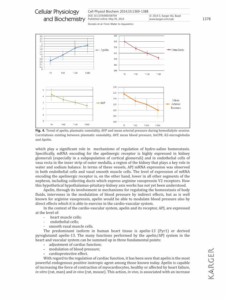
Every increase of plasma osmolarity increases vasopressin and decreases apelin and vice versa, as has been pointed out in our own study. The synthesis and secretion of these two peptides appear to be regulated in opposite ways in response to osmotic stimuli caused by changes in water-salt balance; they play complementary roles in controlling the homeostasis of body fluids.
Apelin is therefore involved in the mechanisms of the regulation of fluid and electrolyte balance in close cooperation with AVP. Apelin, by the reduction in plasma levels of vasopressin with an increase of free water diuresis, may act as a potent diuretic neuropeptide, which counters the action of vasopressin at a central level by inhibiting the activity of vasopressinergic neurons and AVP release [32] (Fig. 4).
Apelin, as well as playing a role of "central blocker" of AVP, can somehow exercise a synergistic action (at a central level): the inhibition of apelin secretion from AVP through the V1 receptor, in addition to the accumulation of apelin in hypothalamic magnocellular neurons, results in a reduction of its concentration in the extracellular environment. The decrease of inhibitory apelin action on vasopressinergic neurons would depend on this, resulting in an increase in their electrical activity and facilitation of the phasic activities of AVP expression and release into the circulation. Apelin could thus modulate the antidiuretic effect of AVP in a positive way at the peripheral level.
The hypothesis of the existence of an endocrine hypothalamus-pituitary-kidney axis stems from the evidence of the expression of the apelin-APJ system in kidney structures
Fig. 3. AVP-secreting neu-rons of SON and PVN up-reg-ulated in response to dehy-dration :as a consequence of the osmotic/volume stimu-lus, vesicles containing AVP and their respective recep-tors are released by exocy-tosis.
Cell Physiol Biochem 2014;33:1369-1388DOI: 10.1159/000358704Published online: May 05, 2014
© 2014 S. Karger AG, Baselwww.karger.com/cpb 1378
Donato et al: From Water to Aquaretics
Cellular Physiology and Biochemistry
Cellular Physiology and Biochemistry
which play a significant role in mechanisms of regulation of hydro-saline homeostasis. Specifically, mRNA encoding for the apelinergic receptor is highly expressed in kidney glomeruli (especially in a subpopulation of cortical glomeruli) and in endothelial cells of vasa recta in the inner strip of outer medulla, a region of the kidney that plays a key role in water and sodium balance. In terms of these vessels, APJ mRNA expression was observed in both endothelial cells and vasal smooth muscle cells. The level of expression of mRNA encoding the apelinergic receptor is, on the other hand, lower in all other segments of the nephron, including collecting ducts which express arginine vasopressin V2 receptors. How this hypothetical hypothalamus-pituitary-kidney axis works has not yet been understood.
Apelin, through its involvement in mechanisms for regulating the homeostasis of body fluids, intervenes in the modulation of blood pressure by indirect effects, but as is well known for arginine vasopressin, apelin would be able to modulate blood pressure also by direct effects which it is able to exercise in the cardio-vascular system.
In the context of the cardio-vascular system, apelin and its receptor, APJ, are expressed at the level of:
- heart muscle cells;- endothelial cells;- smooth vasal muscle cells.The predominant isoform in human heart tissue is apelin-13 (Pyr1) or derived
pyroglutamil apelin-13. The many functions performed by the apelin/APJ system in the heart and vascular system can be summed up in three fundamental points:
- adjustment of cardiac function;- modulation of blood pressure;- cardioprotective effect.With regard to the regulation of cardiac function, it has been seen that apelin is the most
powerful endogenous positive inotropic agent among those known today. Apelin is capable of increasing the force of contraction of myocardiocytes, healthy or affected by heart failure, in vitro (rat, man) and in vivo (rat, mouse). This action, in vivo, is associated with an increase
Fig. 4. Trend of apelin, plasmatic osmolality, AVP and mean arterial pressure during hemodialytic session. Correlations existing between plasmatic osmolality, AVP, mean blood pressure, hsCPR, ß2-microglobulin and Apelin.

Cell Physiol Biochem 2014;33:1369-1388DOI: 10.1159/000358704Published online: May 05, 2014
© 2014 S. Karger AG, Baselwww.karger.com/cpb 1379
Donato et al: From Water to Aquaretics
Cellular Physiology and Biochemistry
Cellular Physiology and Biochemistry
in cardiac output and a decrease in pre-load. Apelin would also exercise a chronotropic, dromotropic and positive bathmotropic effect [33].
These actions of apelin appear to depend on the ability of this neuropeptide to increase the current level of Na in the sarcolemma and therefore, to vary intracellular concentration of Na in the myocytes, lowering the threshold potential for activation and thus, facilitating the opening of a voltage-gated channel. Apelin seems to be able to slow the sodium current during the resting phase of repolarization, increasing the duration of the "window" of the sodium current up to 600% [34].
Apelin, because of its cardiac functions, is considered part of the compensatory mechanisms that come into play in the context of heart failure for the maintenance of good myocardial performance. This is demonstrated by the detection of increased levels of plasma apelin and the expression of APJ receptor in heart tissue in patients with heart failure at an early stage. In later stages, there is an inverse correlation between apelin plasma levels and the expression of apelinergic system in the myocardium, probably due to a downregulation of APJ-apelin system, a condition which could in turn contribute to the inexorable progression of the clinical condition (Fig. 5).
The positive inotropic effect of apelin also seems to have significant importance in maintaining good cardiac performance in aging and chronic pressure overload to delay the progression of myocardial hypertrophy and subsequent dysfunction of the cardiac pump. Low levels of this peptide do not correlate only to heart failure, but also to stable angina and atrial fibrillation.
The cardioprotective effect of apelin on the cardiovascular system, according to research, is due to its direct hypotensive effect, its ability to counteract the hypertensive effect of angiotensin II and to block a number of pathological processes, dependent on the heart and arteries, induced by ATII [25]. Apelin is, in fact, a vasoactive peptide capable of regulating vascular tone, both in vivo and in vitro. Apelin causes arterial vasodilation by means of an endothelial mechanism using a NO-dependent way. The vasodilatory effect of apelin reduces peripheral resistance and, therefore, the afterload; such a positive inotropic effect on the heart (which is reflected in a decrease in pre-load) leads, finally, to the reduction in cardiac work and lowering of blood pressure . The antagonistic action performed by apelin on the effects of angiotensin II has been highlighted by a recent study in an experimental model of atherosclerosis in mice. Study results have provided evidence that the administration of apelin is able to block those pathological processes that are the basis of atherosclerosis and abdominal aortic aneurysm, caused by ATII, and that this vasoprotective action is abolished by the inhibition of NOS (Nitric oxide-Synthase) [35, 36]
Fig. 5. Vasoactive effects of apelin by the stimulation of smooth muscle cells.

Cell Physiol Biochem 2014;33:1369-1388DOI: 10.1159/000358704Published online: May 05, 2014
© 2014 S. Karger AG, Baselwww.karger.com/cpb 1380
Donato et al: From Water to Aquaretics
Cellular Physiology and Biochemistry
Cellular Physiology and Biochemistry
Apelin is also an important angiogenic and mitogen factor for endothelial cells and smooth muscle cells of blood vessels. In the neoangiogenesis process, apelin seems to play a synergistic role with VEGF; as we have seen, together they more effectively stimulate the development of functional and large-caliber vessels, compared to VEGF alone [37].
The Apelin/APJ system have multiple roles in vascular formation in physiological and pathological situations, including tissue regeneration and tumourigenesis. Apelin has a unique function as a regulator of vascular maturation and stabilization by increasing the caliber of formed blood vessels and their strength [38].
Due to the function of an angiogenic and mitogenic factor, the APLN/APJ system, plays an important role in normal development of the cardiovascular system, during embryogenesis. Mice deficient in the apelin peptide are vital, fertile and show an almost normal development, but with smaller blood vessels, APJ-receptor deficient mice either are not vital or they show developmental defects in the cardiovascular system. The mice that reach adulthood, both those with apelin deficiency and those with the missing receptor APJ, have modest reductions in contractile function in basal conditions, while under physical stress, both mutant lines show a consistent and striking reduction in physical endurance. The differences in development between the mice lacking apelin phenotype and those with missing receptor APJ phenotype suggest the possibility of the existence of other APJ receptor ligands or other APJ receptor ligand-independent effects still undiscovered.
In conclusion we can assume that apelin is an important factor involved in hydro-saline homeostasis and human cardiovascular physiology and pathophysiology [39].
Aquaporins
In order to understand the function of AVP in the physiology and pathophysiology of salt and water homeostasis, a discussion of aquaporins must not be omitted. Aquaporins (AQP) are channel-proteins with molecular weights of 23-31 kDa that allow bi-directional water transport across cell membranes [40]. Aquaporins are homo-or hetero-tetramers containing four independent channels for water. The channel for the passage of water is not, in fact, among the 4 proteins, but each protein subunit has a pore for the passage of water. The subunit consists of a single peptide chain of 250-300 amino acids, which has 6 transmembrane domains of alpha helix, and two extremities, C-and N-terminal, which are intracytoplasmatic [41].
At present the family of aquaporins has 450 isoforms, identified in all living organisms (Bacteria, Archaea and Eukarya). The presence of these proteins in Bacteria and Archaea date back to over 3 billion years ago. In humans, 13 isoforms have been identified. The AQP-6 and AQP-8 are yet to be defined in their regulation, while AQP-11 and AQP-12 are defined inortodosse. The first two allow the passage of water while others pass water and glycerol.In humans, aquaporins are ubiquitous though their maximum expression is in the kidney [42]. They have been performing a variety of vital functions since the dawn of life. AQP are involved in the exchange of water and glycerol in the amnios and placenta. At birth, transient overexpression of AQP-1 and AQP-4, respectively, in capillary endothelium of the pulmonary alveoli, would play an important role in the reasorption of the residual neonatal pulmonary secretions produced during fetal life (100 ml/kg/24h). Around the age of 18 months, the kidney reaches its maximum power of concentration with a gradual increase in the expression of AQP-2 ADH-sensitive. This is explained in part by the increased risk of dehydration in children in this age group. During pregnancy, in fact, the fetus produces diluted urine which participates in progressive hypotonicity of the amniotic fluid. At the same time, the lining of the fetal integument, highly permeable in the first few weeks, keratinized at 24 weeks and later covered with a stratum corneum at 34 weeks, thus avoiding free exchange with the diluted amniotic fluid. The acquisition of this characteristic is implemented within one year of life along with the expression of AQP2 [43]. In the kidneys, the presence and abundance of water channels of the family of aquaporins affect

Cell Physiol Biochem 2014;33:1369-1388DOI: 10.1159/000358704Published online: May 05, 2014
© 2014 S. Karger AG, Baselwww.karger.com/cpb 1381
Donato et al: From Water to Aquaretics
Cellular Physiology and Biochemistry
Cellular Physiology and Biochemistry
the permeability or impermeability to water of the entire system of tubular collectors and vascular structure, enabling the kidney to take advantage of the osmotic gradient that is created between the concentrated medullary interstitium and diluted tubule fluid collectors to implement the mechanism of urine concentration. In fact, urine is not concentrated by active transport of water from tubular fluid to the blood, because such a system would require an enormous amount of metabolic energy. Urine is concentrated with a relatively low expenditure of energy by means of a complex interaction between the loop of Henle, the medullary interstitium, blood vessels or spinal cord vasa recta, and tubular collectors, strongly influenced by the expression of aquaporins. The kidney in its difficult path of urine concentration (from 180 l of filtered blood to 1.5 liter of urine / 24h) uses as many as 8 different isoforms of aquaporins (AQP 1, 2, 3, 4, 6, 7, 8, 11). The role of AQP-8 in the proximal tubule is speculative. AQP-7, expressed in the proximal straight tubule, would allow the reabsorption of glycerol. AQP-11 is expressed in the cytoplasm of proximal tubular cells and plays a role in the development of the kidney and cyst formation; its invalidation, in fact, induces a very severe polycystic phenotype in mice. The role of AQP-6 expressed in intercalated cells remains to be defined, and the essential role of AQP-1 and AQP-2 in renal reabsorption of water has been well demonstrated. The phenotype of knockout mice for AQP-1 or AQP-2 clearly demonstrates the predominant role of this protein channel in renal reabsorption of water and thus, in the mechanism of urine concentration. AQP-1 is expressed in the proximal tubule, in the final stretch of the descending arm of Henle's loop and vasa recta. AQP-1-knockout mice have severe polyuria and a defect of concentrating urine which is explained both by the failure of water reabsorption in the proximal part of kidney and in reducing hypertonicity of the medulla.
AQP-2 is localized at the level of principal cells of the collector channel and it is known as the main protein channel involved in regulating water reabsorption at this level. The invalidation of the gene coding for AQP-2 in mice leads to a lethal phenotype. AQP-2 is regulated by ADH, after an increase in plasma osmolarity, from the endings of magnocellular neurons in the posterior pituitary. The connection between circulating ADH and its receptor (V2R), located on the basolateral membrane of principal cells of the collector channel, causes an increase of intracellular cAMP, activation of protein kinase-A and phosphorylation of AQP-2 in four serine residues near the C-terminal ending. Phosphorylation of Ser-256 causes the fusion of intracytosolic AQP-2 to the cell membrane. The localization of AQP-2 on the apical membrane leads collector channels to the permeability to the water. This leads to the reabsorption of fluid, or else a passage of water from the tubular lumen to the cell and the formation of hypertonic urine. The basolateral reabsorption is due to the presence of AQP-3 AQP-4, costitutively localized on the basolateral membrane of the principal collecting tubule cells. The expression of AQP-2 on the apical membrane of tubular cells is subject to adjustment in the short term, through processes of exo-endo-citosis, and long-term in a quantitative way. The abundance of the protein is regulated at a transcriptional and post-transcriptional level mainly by vasopressin, which increases not only the amount of AQP-2 but also AQP-3, via the transcription factor NF-kB ("nuclear factor k-light-chain-enhancer of activated B cells") (decrease in the abundance of AQP2).
The rare AQP1-null patients, identified because of incompatibility of blood transfusion (AQP1 in red blood cells membrane is the Colton blood antigen subgroup) do not have significant renal disease, unlike the AQP1 knock-out mouse model. These patients are not in fact polyuric and their water consumption has to be restricted in order to detect their defect of urine-concentration. In contrast to the minimal impact of AQP1 deficiency, the altered function of AQP2 or its absence in humans lead to diabetes insipidus (DI), a hereditary or acquired disease, characterized by impaired renal mechanisms assigned to urine concentration despite normal or elevated serum concentration of the antidiuretic hormone, arginine vasopressin (AVP). This is different from neurogenetic diabetes insipidus, due to a lack of antidiuretic hormone release at the central level.When the disease is genetic, urinary symptoms (polyuria with polydipsia and hypostenuria) are characterized by a significant deficit in the mechanism of urine concentration and may affect quod vitam prognosis, while
Cell Physiol Biochem 2014;33:1369-1388DOI: 10.1159/000358704Published online: May 05, 2014
© 2014 S. Karger AG, Baselwww.karger.com/cpb 1382
Donato et al: From Water to Aquaretics
Cellular Physiology and Biochemistry
Cellular Physiology and Biochemistry
signs are less pronounced and the quod vitam prognosis better if the disease is acquired. In general, DI is a chronic disease with a good prognosis if hypernatremic dehydration can be avoided; polyuria with increased risk of dehydration can be lethal in children suffering from the genetic form. Forms of diabetes insipidus of genetic origin are very rare and most of these are due to mutations in the gene coding for the vasopressin receptor type 2 V2R placed in the Xq28 locus. In this case, the clinical picture is presented as X-linked recessive trait (transmission android). Only 5% of diabetes insipidus are due to a post-receptor-defect dependent on AQP2 gene mutations in locus 12q13, coding for the vasopressin-sensitive water channel. In this case, the disease presents as a recessive transmission form of DI in 90% of cases and is not X-linked. In the few cases of dominant transmission that have been reported in literature, the dominant and severe effect of the mutation is due to the fact that in these variants, when ADH stimulates the protein kinase A, abnormal tetramers are formed which are addressed to all endosomes or basolateral membrane rather than the apical membrane, seriously compromising the ability of the kidney to concentrate urine. In cases of recessive transmission, on the other hand, the mutant proteins are retained in the endoplasmic reticulum and then rapidly degraded by proteasomes, without ever reaching the apical membrane; in conclusion, the phenotype in autosomal recessive AQP2 mutations is as severe as the phenotype observed in AVPR2 mutations, except for « mild » AQP2 mutants [44].
As regards the forms of acquired nephrogenic diabetes insipidus, these are more frequent and their origin can be iatrogenic or metabolic, depending on the establishment of hypokalemia or hypercalcemia, mechanisms established following the displacement of the obstacle to the flow of urine. Among the iatrogenic forms of acquired diabetes insipidus, the best known is one that can often occur after bipolar treatment with lithium salts. This effect occurs in approximately 30% of patients because of decreased expression of AQP2; reversibility of the clinical manifestation after stopping the therapy is uncertain. In addition to the contribution of water, the symptomatic treatment of diabetes insipidus consists of a low-sodium diet, to decrease the filtered load, in terms of water and sodium, which arrives at the channel collector. Associating a thiazide-type diuretic (hydrochlorothiazide) with a low-salt regime or eventually amiloride is recommended. An inhibition of cyclooxygenase with non-steroidal anti-inflammatories may also be taken into consideration, because the inhibition of PGE2 production has the effect of increased expression and decreased endocytosis of AQP2 and V2R, resulting in an increased ability to concentrate urine.
With regard to the inhibition of cyclooxygenase rather than indomethacin, which can have pronounced effects on the digestive system, the use of selective inhibitors of COX2, under cardiological supervision, may be suggested. It should also be noted that the range of action of the treatment of diabetes insipidus is rich and complex. Thiazide diuretics, as well as COX2 inhibitors, increase the expression of co-transport of NKCC28 and co-transport of Na-K-2Cl in the thick tract of the ascending arm of Henle's loop, sensitive to furosemide. Therefore, the decrease in polyuria is achieved through several mechanisms.
In a mouse model that reproduces a mutation of AQP2 observed in forms of dominant transmission, it has been observed that the use of the Rolipram-8, an inhibitor of phosphodiesterase-4 (PDE4), has led to an improvement in the power of urinary concentration; on the other hand, Milrinone and Sildenafil, respective inhibitors of PDE-3 and PDE-5, as well as PDE-4 (expressed in the canal collector) have had no impact on polyuria in mice, demonstrating the complexity of the effects of these molecules on models in vivo.
Aquaretics
To conclude the discussion about AVP and AQP, "aquaretics", without doubt, deserve a mention. In 2004, a class of drugs including conivaptan, tolvaptan, relcovaptan and lixivaptan, was studied; these act by inhibiting the action of vasopressin receptor V1 and V2.

Cell Physiol Biochem 2014;33:1369-1388DOI: 10.1159/000358704Published online: May 05, 2014
© 2014 S. Karger AG, Baselwww.karger.com/cpb 1383
Donato et al: From Water to Aquaretics
Cellular Physiology and Biochemistry
Cellular Physiology and Biochemistry
Conivaptan has a higher selectivity on V1a and V2 type receptors, while the other molecules are selective above all of V2 receptors. The appearance of therapeutic aquaretic drugs is destined to revolutionize the treatment of many pathological conditions, particularly those associated with hyponatremia and hypo-osmolality of fluid overload (Table 5). Regardless of the cause that determines these states, traditional "symptomatic" therapy has to date been based on very precise cornerstones: restriction of water intake, diuretics (especially of the loop) and, in more serious cases, supplements of sodium chloride, or alternative therapies also toxic [45] (see Table 6).
A recent study highlighted a clear therapeutic advantage of the use of the aquaretic tolvaptan compared to a traditional diuretic (furosemide) in the treatment of congestive heart failure (CHF) in humans, in particular with regard to hemodynamic renal impairment. In 14 patients with CHF NYHA II-III, with the same diuretic effect, no variation in renal blood flow and urinary excretion of sodium and potassium was observed after treatment with tolvaptan; the condition of the patients worsened, on the other hand, with furosemide [46]. In contrast to furosemide, tolvaptan did not activate the renin-angiotensin-aldosterone (RAA). This activation in fact, depending on the natriuretic effects of loop diuretics which lead to a reduction in serum sodium, cannot occur since aquaretics, on the contrary, induce an hypernatremic effect [47].
Schrier et al. [48] examined the use of tolvaptan for 30 days to correct and maintain serum sodium concentrations in a population with hyponatremia. These two month-long
Tab. 5. Main molecules with aquaretic activities
Tab. 6. Possible treatments for SIADH

Cell Physiol Biochem 2014;33:1369-1388DOI: 10.1159/000358704Published online: May 05, 2014
© 2014 S. Karger AG, Baselwww.karger.com/cpb 1384
Donato et al: From Water to Aquaretics
Cellular Physiology and Biochemistry
Cellular Physiology and Biochemistry
trials suggested that tolvaptan, added to standard therapy, was superior to placebo in raising and maintaining serum sodium concentrations in patients with hyponatremia of diverse origin.
Ultimately aquaretics, because of their completely different mechanism of action, would guarantee the almost total absence of all those side effects that are associated with classic diuretics therapy. Among the most notable are electrolyte disturbances, alkalosis, while renal and extra-renal toxicity have always been a limitation in the use of these drugs, for example, in the treatment of hypertension [49]. Moreover, but no less importantly, it has been shown how aquaretics, in particular OPC-31260, apparently do not affect renal function in normal conditions. In subjects where there is already a state of chronic renal failure, however, there is a state of retention not only of water but also of sodium, as demonstrated by a recent Italian study which has applied bioelectrical impedance techniques [50].
An increase in oral intake of salt also seems to lead, through a mechanism independent of the RAA-mediated rise in blood pressure, to an increase of proteinuria with tubular damage and progression of chronic renal failure [51]. It is still unclear whether hypernatremia induced by aquaretics can somehow worsen an already pre-existing chronic renal failure by a similar mechanism.
However, it is true that there is also evidence in literature suggesting, on the contrary, that this class of drugs may play a protective role in countering the development of glomerular disease. A recent experimental observation shows that the injection of high doses of vasopressin analogues in healthy subjects results in a marked increase in urinary albumin excretion (UAE) and that the achievement of this requires the integrity of V2 receptors, antagonized, as it is known, by aquaretics. This finding suggests the involvement of AVP in the pathogenesis of proteinuria and albuminuria present, for example, in pathological conditions such as diabetes mellitus and hypertension and for this reason the use of V2-R antagonists, opposing this action, may slow the progression of renal injury through an anti-proteinuric effect similar to that already observed for other "famous" receptor blockers such as ACEIs and ARBs. A subsequent study highlighted this exact effect in rats with albuminuria due to diabetes after administration of an aquaretic agent, SR121463 [52].
The problem of the relationship between aquaretics and renal disease is still more than ever open and it is expected, therefore, that future studies may shed more light on this.
Meanwhile, the main field of application of therapy with these drugs today is undoubtedly cardiovascular disease and particularly, heart failure. A large multicenter randomized double-blind study is still in progress to define what terms and to what degree the use of Tolvaptan may improve the current treatment of fluid overload associated with heart failure and lead, therefore, to an effective improvement of prognosis in this category of patients. This study, named EVEREST (Efficacy of Vasopressin Antagonism in The Heart Failure Outcome Study with Tolvaptan), aims to evaluate the mortality, morbidity and global clinical status in comparison with traditional treatment, thus establishing if the positive effects of tolvaptan (reduction in body weight, increased urine output without the risk of hypokalemia and renal toxicity) may result in an effective improvement in overall clinical condition.
It is also hoped that this study, performed in the long term, in contrast to those described previously, may provide additional guidance about the safety and efficacy of a "chronic" and prolonged use of Tolvaptan. Let us remember that this molecule, acting as a selective V2 antagonist, could lead to both a down-regulation of V2 receptors themselves and to an increase in circulating levels of AVP, as a result of a long-term administration. This could in turn be due to increased stimulation of peripheral V1a receptors with potential effects on cardiovascular function but this needs to be evaluated.
The extent of benefits with prolonged administration of aquaretic drugs for hypertension is also to be demonstrated. The rationale of this effect could be based on the recent demonstration that chronic stimulation of V2 receptors leads to the onset of salt-sensitive high blood pressure based on prolonged sodium reabsorption induced by AVP with consequent sodium retention [53]. In regard to this, Naitoh and others have shown that in mouse models of hypertension and heart failure, the addition in therapy of the antagonist

Cell Physiol Biochem 2014;33:1369-1388DOI: 10.1159/000358704Published online: May 05, 2014
© 2014 S. Karger AG, Baselwww.karger.com/cpb 1385
Donato et al: From Water to Aquaretics
Cellular Physiology and Biochemistry
Cellular Physiology and Biochemistry
V1a/V2 conivaptan (1 mg / kg / day) improved some of the beneficial effects exerted by the ACE inhibitor captopril (50 mg / kg / day), such as reduction of right and left ventricular mass, pulmonary mass and arterial pressure, while the selective antagonism of V2, always in association with the ACE-inhibition, prevented the onset of hypertension in young spontaneously hypertensive rats (SHR) [54].
It is more evident today that the benefit of aquaretics treatment for hyponatremia is related, for example, to nephrotic syndrome, ascites from cirrhosis or syndrome of inappropriate secretion of antidiuretic hormone (SIADH). A very recent randomized placebo-controlled study showed the efficacy and safety of oral administration of conivaptan in patients with euvolemic or hypervolemic hyponatremia. In spite of the common side effects observed such as, headache, postural hypotension and nausea, 71% of patients treated with a dose of 40 mg/day conivaptan, and 82% of those treated with a dose of 80 mg/day obtained an improvement in serum sodium levels, confirming the effectiveness and manageability of the molecule.
Based on what we have examined up to now, we can say that the main purpose of the therapeutic use of aquaretics is closely related to the most well-known mechanisms of action, that is the inhibition of vasopressin-mediated water reabsorption. But interest in these molecules may be destined over time to focus on other effects observed in parallel with the possibility of new, unexpected and future therapeutic implications. If the contrasting and in some cases even regressive action exerted by aquaretics on polycystic kidney disease is now a great hope that has yet to be confronted with appropriate in vivo studies in humans, experimentation in other circumstances is, on the contrary, more rational and immediate. Among these we cannot but mention Meniere's disease. It has been seen as having a key role in regulating water homeostasis of the inner ear, performed by the vasopressin-AQP2 system. Excessive activation of this system contributes to the pathogenesis of endolymphatic hydrops of Meniere's disease and positive results have already been observed after administration of OPC-31260 in an animal model of this disease [55].
Good prospects also exist for treating ocular hypertension which is the basis of the pathogenesis of glaucoma. In a rabbit-model with this condition, the effect of the reduction of intraocular pressure after instillation of local timolol (0.5%) and clonidine (0.25%), two reference drugs, was compared with various doses of SR121463, a potent aquaretic agent. In the absence of controlateral and systemic effects and tachyphylaxis after 10 days, the effectiveness of the vasopressin antagonist in countering ocular hypertension was entirely comparable to that of the control drug; and a similar result was also obtained with intravenous administration.
Moreover, it is known that aquaretics drug, in particular Tolvaptan, may play a role in the inhibition of cysts growth in patient with Polycystic Kidney Disease (APKD) by vasopressin V2-receptor blockade [56]. In a study conducted by Torres et al., 2122 patients with APKD were enrolled; 1445 were randomly assigned to either tolvaptan or placebo, for a tree-year trial. They estimated the cists growth and the decline of kidney function during this period. The reduction in total kidney volume was more pronounced in the first year of treatment, compared to placebo. Tolvaptan showed to reduce the decline in kidney function in patients with ADPKD and a better control of high blood pressure. But these potential benefits are not without risks.
In conclusion, the usefulness of aquaretics in well-defined clinical-pathological contexts and their advantage over traditional therapies have been established.
References
1 Corvol P, Elghozi JL: Sortir de l'eau. Paris,Editions Odile Jacob, 2011, pp 1-240.2 Noble D: Claude Bernard, the first systems biologist, and the future of physiology. Exp Physiol 2008;93:16-
26.

Cell Physiol Biochem 2014;33:1369-1388DOI: 10.1159/000358704Published online: May 05, 2014
© 2014 S. Karger AG, Baselwww.karger.com/cpb 1386
Donato et al: From Water to Aquaretics
Cellular Physiology and Biochemistry
Cellular Physiology and Biochemistry
3 Mariotti M: Marine serum, concept of its inventor, René Quinton; possibility of its application today. Minerva Med 1952;43:777-779.
4 Cannon WB: pharmacological injections and physiological inferences. Science 1929;70:500-501.5 Thornton SN: Thirst and hydration: physiology and consequences of dysfunction. Physiol Behav
2010;26:100:515-21.6 Lorens-Cortes C, Moos F: Opposite potentiality of hypothalamic coexpressed neuropeptides,apelin and
vasopressin in maintaining body-fluid homeostasis. Prog Brain Res 2008;170:559-570.7 Bourque CW: Central mechanisms of osmosensation and systemic osmoregulation. Nat Rev Neurosci
2008;9:519–531.8 Lechner SG, Markworth S, Poole K, Smith ES, Lapatsina L, Frahm S: The molecular and cellular identity of
peripheral osmoreceptors. Neuron 2011;69:332–344.9 Ciura S, Liedtke W, Bourque CW: Hypertonicity sensing in organum vasculosum lamina terminalis neurons:
a mechanical process involving TRPV1 but not TRPV4. J Neurosci 2011;31:14669–14676.10 Laragh JH: Renin angiotensin aldosterone system for blood pressure and electrolyte homeostasis and
its involvement in hypertension, in congestive heart failure and in associated cardiovascular damage (myocardial infarction and stroke). J Hum Hypertens 1995;9:385-390.
11 Tracy RE: Renal vasculature in essential hypertension: a review of some contrarian evidence Contrib Nephrology 2011;169:327-336.
12 Shestakova MV: [Renin-angiotensin-aldosteron system: evolution of views from renin discovery to nowadays. Perspectives of therapeutic block]. Ter Arkh 2011;71-77.
13 Zhang Z, Bourque CW: Bourque Amplification of Transducer Gain by Angiotensin II Mediated Enhancement of Cortical Actin Density in Osmosensory Neurons. J Neurosci 2008;9536–9544.
14 Treschan TA, Peters J: The vasopressin system: physiology and clinical strategies. Anesthesiology 2006;105:599-612.
15 Pequeux C, Keegan BP, Hagelstein MT, Geenen V, Legros JJ, North WG: Oxytocin- and vasopressin-induced growth of human small-cell lung cancer is mediated by the mitogen-activated protein kinase pathway. Endocr Relat Cancer 2004;11:871-885.
16 Martin PY, Abraham WT, Lieming X, Olson BR, Oren RM, Ohara M, Schrier RW: Selective V2-receptor vasopressin antagonism decreases urinary aquaporin-2 excretion in patients with chronic heart failure. J Am Soc Nephrol 1999;10:20165-20170.
17 Buemi M, Corica F, Di Pasquale G, Aloisi C, Sofi M, Casuscelli T, Floccari F, Senatore M, Corsonello A, Frisina N: Water immersion increases urinary excretion of aquaporin-2 in healthy humans. Nephron 2000;85:1:20-26.
18 Buemi M, Di Pasquale G, Ruello A, Floccari F, Aloisi C, Latassa G, Corsonello A, Sturiale A, Corica F, Frisina N: Effect of a prostacyclin analogue, iloprost, on urinary aquaporin-2 excretion in humans. Nephron 2002;91:197-202.
19 O'Carroll AM, Selby TL, Palkovits M, Lolait SJ: Distribution of mRNA encoding B78/apj, therat homologue of the human APJ receptor, and its endogenous ligand apelin in brain and peripheraltissues. Biochim Biophys Acta 2000;21;72-80.
20 Medhurst AD, Jennings CA, Robbins MJ, Davis RP, Ellis C, Winborn KY, Lawrie KW, Hervieu G, Riley G, Bolaky JE, Herrity NC, Murdock P, Darker JG: Pharmacological and immunohistochemical characterization of the APJ receptor and its endogenous ligand apelin. J Neurochem 2003;84:1162-1172.
21 Tatemoto K, Hosoya M, Habata Y, Fujii R, Kakegawa T, Zou MX, Kawamata Y, Fukusumi S, Hinuma S, Kitada C, Kurokawa T, Onda H, Fujino M: Isolation and characterization of a novel endogenous peptide ligand for the human APJ receptor. Biochem Biophys Res Commun 1998;251:471-476.
22 Pitkin SL, Maguire JJ, Bonner TI, Davenport AP: Apelin receptor nomenclature, distribution, pharmacology, and function. Pharmacol Rev 2010;62:331-342.
23 O'Dowd BF, Heiber M, Chan A, Heng HH, Tsui LC, Kennedy JL, Shi X, Petronis A, George SR, Nguyen T: A human gene that shows identity with the gene encoding the angiotensin receptor is located on chromosome 11. Gene 1993;136:355-360.
24 Habata Y, Fujii R, Hosoya M, Fukusumi S, Kawamata Y, Hinuma S, Kitada C, Nishizawa N, Murosaki S, Kurokawa T, Onda H, Tatemoto K, Fujino M: Apelin, the natural ligand of the orphan receptor APJ, is abundantly secreted in the colostrum. Biochim Biophys Acta 1999;1452:25-35.

Cell Physiol Biochem 2014;33:1369-1388DOI: 10.1159/000358704Published online: May 05, 2014
© 2014 S. Karger AG, Baselwww.karger.com/cpb 1387
Donato et al: From Water to Aquaretics
Cellular Physiology and Biochemistry
Cellular Physiology and Biochemistry
25 Maguire JJ, Kleinz MJ, Pitkin SL, Davenport AP: [Pyr1]apelin-13 identified as the prominent apelin isoform in the human heart: vasoactive mechanism and inotropic action in disease. Hypertension 2009;54: 598-604.
26 Reaux A, Gallatz K, Palkovits M, Llorens-Cortes C: Distribution of apelin-synthesizing neurons in the adult rat brain. Neuroscience 2002;113:653-662.
27 Bardoux P, Bruneval P, Heudes D, Bouby N, Bankir L: Diabetesinduced albuminuria: role of antidiuretic hormone as revealed by chronic V2 receptor antagonism in rats. Nephrololy Dialysis Transplant 2003;18:1755-1763.
28 Herrera VL, Ruiz-Opazo N: Identification of a novel V1-type AVP receptor based on the molecular recognition theory. Mol Med 2001;7:499-506.
29 Thompson AM, Oliver JA: Endogenous and exogenous vasopressin during hemodialysis. Semin Dial 2009;22:472-475.
30 Rho M, Perazella MA, Parikh CR, Peixoto AJ, Brewster UC: Serum vasopressin response in patient with intradialytic hypotension: a pilot study. Clin J Am Soc Nephrol 2008;3:729-735.
31 Shimizu K, Kurosawa T, Sanjo T: Effect of hyperosmolality on vasopressin secretion in intradialytic hypotension: a mechanistic study. Am J Kidney Dis 2008;52:294-304.
32 Cernaro V, Lacquaniti A, Lorenzano G, Loddo S, Romeo A, Donato V, Lupica R, Buemi A, Buemi M: Apelin, plasmatic osmolality and hypotension in dialyzed patients. Blood Purif 2012;334:317-323.
33 El-Shehaby AM, El-Khatib MM, Battah AA, Roshdy AR: Apelin: a potential link between inflammation and cardiovascular disease in end stage renal disease patients. Scand J Clin Lab Invest 2010;70:421-427.
34 Kalea AZ, Batlle D: Apelin and ACE2 in cardiovascular disease. Curr Opin Investig Drugs 2010;11:273-282.35 Zhong JChang, Yu XY, Huang Y, Yung LM, Lau CW, Lin SG: Apelin modulates aortic vascular tone via
endothelial nitric oxide synthase phosphorylation pathway in diabetic mice. Cardiovasc Res 2007;74:388-395.
36 Tatemoto K, Takayama K, Zou MX, Kumaki I, Zhang W, Kumano K, Fujimiya M: The novel peptide apelin lowers blood pressure via a nitric oxide-dipendent mechanism. Regul Pept 2001;99:87-92.
37 Eyries M, Siegfried G, Ciumas M, Montagne K, Agrapart M, Lebrin F, Soubrier F: Hypoxia-Induced Apelin Expression Regulates Endothelial Cell Proliferation and Regenerative Angiogenesis. Circ Res 2008:103:432-440.
38 Kidoya H, Takakura N: Biology of the apelin-APJ axis in vascular formation. J Biochem 2012;152:125–131.39 Japp AG, Cruden NL, Amer DA, L VK, Goudie EB, Johnston NR, Sharma S, Neilson I, Webb DJ, Megson IL,
Flapan AD, Newby DE: Vascular effects of apelin in vivo in man. J Am Coll Cardiol 2008;52:908-913.40 Agre P, Nielsen S: The aquaporin family of water channels in kidney. Nephrologie 1996;17:409-415.41 Nielsen S, Kwon TH, Christensen BM, Promeneur D, Frøkiaer J, Marples D: Physiology and pathophysiology
of renal aquaporins. J Am Soc Nephrol 1999;10:647-663.42 Kwon TH, Nielsen J, Møller HB, Fenton RA, Nielsen S, Frøkiaer J: Aquaporins in the kidney.Handb Exp
Pharmacol 2009;95-132.43 Nedvetsky PI, Tamma G, Beulshausen S, Valenti G, Rosenthal W, Klussmann E: Regulation of aquaporin-2
trafficking.Handb Exp Pharmacol 2009;133-157.44 Cheng CY, Chu JY, Chow BK: Vasopressin-independent mechanisms in controlling water homeostasis. J Mol
Endocrinol 2009;43:81-92.45 Torres VE, Wang X, Qian Q, Somlo S, Harris PC, Gattone VH 2nd: Effective treatment of an orthologous model
of autosomal dominantpolycystic kidney disease. Nat Med 2004;10:363-364. 46 Danovitch GM, Le Roith D, Glick S: Renal function during treatmentof inappropriate secretion of
antidiuretic hormone with demeclocycline. Isr J Med Science 1978;14:852-857.47 Miller PD, Linas SL, Schrier RW: Plasma demeclocycline levels and nephrotoxicity. Correlation in
hyponatremic cirrhotic patients. JAMA 1980;243:2513-2515.48 Schrier RW, Gross P, Gheorghiade M, Berl T, Verbalis JG, Czerwiec FS, Orlandi C: Tolvaptan, a selective oral
vasopressin V2-receptor antagonist, for hyponatremia. New Engl J Med 2006;355:2099–2112.49 Costello-Boerrigter LC, Smith WB, Boerrigter G, Ouyang J, Zimmer CA, Orlandi C, Burnett JC Jr: Vasopressin-
2-receptor antagonism augments water excretion without changes in renal hemodynamics or sodium and potassium excretion in humanheart failure. Am J Physiol Renal Physiol 2006;290:273-238.

Cell Physiol Biochem 2014;33:1369-1388DOI: 10.1159/000358704Published online: May 05, 2014
© 2014 S. Karger AG, Baselwww.karger.com/cpb 1388
Donato et al: From Water to Aquaretics
Cellular Physiology and Biochemistry
Cellular Physiology and Biochemistry
50 Hirano T, Yamamura Y, Nakamura S, Onogawa T, Mori T: Effects of the V(2)-receptor antagonist OPC-41061 and the loop diuretic furosemide alone and in combination in rats. J Pharmacol Exp Ther 2000;292:288-294.
51 Weinberger MH: Diuretics and their side effects. Dilemma in the treatment of hypertension. Hypertension 1988;11:16-20.
52 Bellizzi V, Scalfi L, Terracciano V, De Nicola L, Minutolo R, Marra M, Guida B, Cianciaruso B, Conte G, Di Iorio BR: Early changes in bioelectrical estimates of body composition in chronic kidney disease. J Am Soc Nephrol 2006;17:1481-1487.
53 Bardoux P, Bruneval P, Heudes D, Bouby N, Bankir L: Diabetes induced albuminuria: role of antidiuretic hormone as revealed by chronic V2 receptor antagonism in rats. Nephrol Dial Transplant 2003;18:1755-1763.
54 Lacheretz F, Barbier A, Serradeil-Le Gal C, Elena PP, Maffrand JP,Le Fur G: Effect of SR121463, a selective non-peptide vasopressin V2 receptor antagonist, in a rabbit model of ocular. J Ocul Pharmacol Ther 2000;16:203-216.
55 Gheorghiade M, Orlandi C, Burnett JC, Demets D, Grinfeld L, Maggioni A, Swedberg K, Udelson JE, Zannad F, Zimmer C, Konstam MA: Rationale and design of the multicenter, randomized, double-blind, placebo-controlled study to evaluate the Efficacy of Vasopressin antagonism inHeart Failure: Outcome Study with Tolvaptan (EVEREST). J CardFail 2005;11:260-269.
56 Torres VE, Chapman AB, Devuyst O, Gansevoort RT, Grantham JJ, Higashihara E,Perrone RD, Krasa HB, Ouyang J, Czerwiec FS: Tolvaptan in patients with autosomal dominant polycystic kidney disease. N Engl J Med 2012;367:2407–2418.