Long-term bone remodelling around 'legendary' cementless ...
13
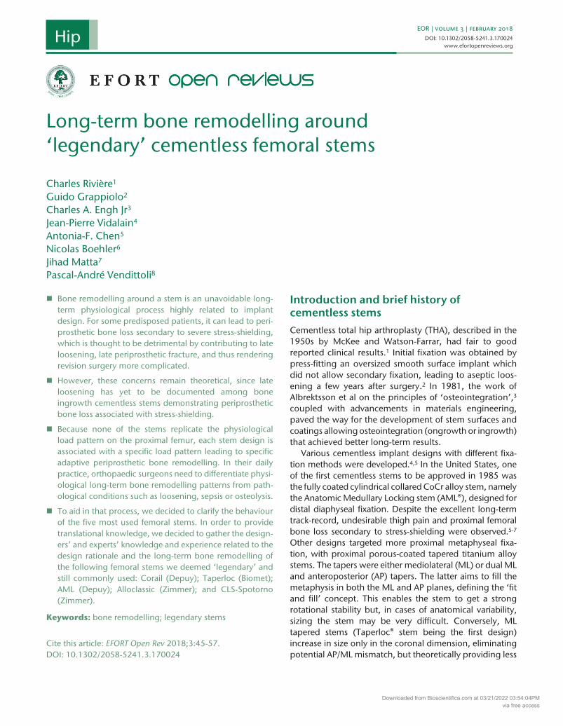
EOR | volume 3 | February 2018 DOI: 10.1302/2058-5241.3.170024 www.efortopenreviews.org Bone remodelling around a stem is an unavoidable long- term physiological process highly related to implant design. For some predisposed patients, it can lead to peri- prosthetic bone loss secondary to severe stress-shielding, which is thought to be detrimental by contributing to late loosening, late periprosthetic fracture, and thus rendering revision surgery more complicated. However, these concerns remain theoretical, since late loosening has yet to be documented among bone ingrowth cementless stems demonstrating periprosthetic bone loss associated with stress-shielding. Because none of the stems replicate the physiological load pattern on the proximal femur, each stem design is associated with a specific load pattern leading to specific adaptive periprosthetic bone remodelling. In their daily practice, orthopaedic surgeons need to differentiate physi- ological long-term bone remodelling patterns from path- ological conditions such as loosening, sepsis or osteolysis. To aid in that process, we decided to clarify the behaviour of the five most used femoral stems. In order to provide translational knowledge, we decided to gather the design- ers’ and experts’ knowledge and experience related to the design rationale and the long-term bone remodelling of the following femoral stems we deemed ‘legendary’ and still commonly used: Corail (Depuy); Taperloc (Biomet); AML (Depuy); Alloclassic (Zimmer); and CLS-Spotorno (Zimmer). Keywords: bone remodelling; legendary stems Cite this article: EFORT Open Rev 2018;3:45-57. DOI: 10.1302/2058-5241.3.170024 Introduction and brief history of cementless stems Cementless total hip arthroplasty (THA), described in the 1950s by McKee and Watson-Farrar, had fair to good reported clinical results. 1 Initial fixation was obtained by press-fitting an oversized smooth surface implant which did not allow secondary fixation, leading to aseptic loos- ening a few years after surgery. 2 In 1981, the work of Albrektsson et al on the principles of ‘osteointegration’, 3 coupled with advancements in materials engineering, paved the way for the development of stem surfaces and coatings allowing osteointegration (ongrowth or ingrowth) that achieved better long-term results. Various cementless implant designs with different fixa- tion methods were developed. 4,5 In the United States, one of the first cementless stems to be approved in 1985 was the fully coated cylindrical collared CoCr alloy stem, namely the Anatomic Medullary Locking stem (AML®), designed for distal diaphyseal fixation. Despite the excellent long-term track-record, undesirable thigh pain and proximal femoral bone loss secondary to stress-shielding were observed. 5-7 Other designs targeted more proximal metaphyseal fixa- tion, with proximal porous-coated tapered titanium alloy stems. The tapers were either mediolateral (ML) or dual ML and anteroposterior (AP) tapers. The latter aims to fill the metaphysis in both the ML and AP planes, defining the ‘fit and fill’ concept. This enables the stem to get a strong rotational stability but, in cases of anatomical variability, sizing the stem may be very difficult. Conversely, ML tapered stems (Taperloc® stem being the first design) increase in size only in the coronal dimension, eliminating potential AP/ML mismatch, but theoretically providing less Long-term bone remodelling around ‘legendary’ cementless femoral stems Charles Rivière 1 Guido Grappiolo 2 Charles A. Engh Jr 3 Jean-Pierre Vidalain 4 Antonia-F. Chen 5 Nicolas Boehler 6 Jihad Matta 7 Pascal-André Vendittoli 8 3.1700EOR 0 0 10.1302/2058-5241.3.170024 research-article 2018 Hip Downloaded from Bioscientifica.com at 03/21/2022 03:54:04PM via free access
-
Upload
khangminh22 -
Category
Documents
-
view
5 -
download
0
Transcript of Long-term bone remodelling around 'legendary' cementless ...
EOR | volume 3 | February 2018DOI: 10.1302/2058-5241.3.170024
www.efortopenreviews.org
� Bone remodelling around a stem is an unavoidable long-term physiological process highly related to implant design. For some predisposed patients, it can lead to peri-prosthetic bone loss secondary to severe stress-shielding, which is thought to be detrimental by contributing to late loosening, late periprosthetic fracture, and thus rendering revision surgery more complicated.
� However, these concerns remain theoretical, since late loosening has yet to be documented among bone ingrowth cementless stems demonstrating periprosthetic bone loss associated with stress-shielding.
� Because none of the stems replicate the physiological load pattern on the proximal femur, each stem design is associated with a specific load pattern leading to specific adaptive periprosthetic bone remodelling. In their daily practice, orthopaedic surgeons need to differentiate physi-ological long-term bone remodelling patterns from path-ological conditions such as loosening, sepsis or osteolysis.
� To aid in that process, we decided to clarify the behaviour of the five most used femoral stems. In order to provide translational knowledge, we decided to gather the design-ers’ and experts’ knowledge and experience related to the design rationale and the long-term bone remodelling of the following femoral stems we deemed ‘legendary’ and still commonly used: Corail (Depuy); Taperloc (Biomet); AML (Depuy); Alloclassic (Zimmer); and CLS-Spotorno (Zimmer).
Keywords: bone remodelling; legendary stems
Cite this article: EFORT Open Rev 2018;3:45-57. DOI: 10.1302/2058-5241.3.170024
Introduction and brief history of cementless stemsCementless total hip arthroplasty (THA), described in the 1950s by McKee and Watson-Farrar, had fair to good reported clinical results.1 Initial fixation was obtained by press-fitting an oversized smooth surface implant which did not allow secondary fixation, leading to aseptic loos-ening a few years after surgery.2 In 1981, the work of Albrektsson et al on the principles of ‘osteointegration’,3 coupled with advancements in materials engineering, paved the way for the development of stem surfaces and coatings allowing osteointegration (ongrowth or ingrowth) that achieved better long-term results.
Various cementless implant designs with different fixa-tion methods were developed.4,5 In the United States, one of the first cementless stems to be approved in 1985 was the fully coated cylindrical collared CoCr alloy stem, namely the Anatomic Medullary Locking stem (AML®), designed for distal diaphyseal fixation. Despite the excellent long-term track-record, undesirable thigh pain and proximal femoral bone loss secondary to stress-shielding were observed.5-7 Other designs targeted more proximal metaphyseal fixa-tion, with proximal porous-coated tapered titanium alloy stems. The tapers were either mediolateral (ML) or dual ML and anteroposterior (AP) tapers. The latter aims to fill the metaphysis in both the ML and AP planes, defining the ‘fit and fill’ concept. This enables the stem to get a strong rotational stability but, in cases of anatomical variability, sizing the stem may be very difficult. Conversely, ML tapered stems (Taperloc® stem being the first design) increase in size only in the coronal dimension, eliminating potential AP/ML mismatch, but theoretically providing less
Long-term bone remodelling around ‘legendary’ cementless femoral stems
Charles Rivière1
Guido Grappiolo2
Charles A. Engh Jr3
Jean-Pierre Vidalain4
Antonia-F. Chen5
Nicolas Boehler6
Jihad Matta7
Pascal-André Vendittoli8
3.1700EOR0010.1302/2058-5241.3.170024research-article2018
Hip
Downloaded from Bioscientifica.com at 03/21/2022 03:54:04PMvia free access
46
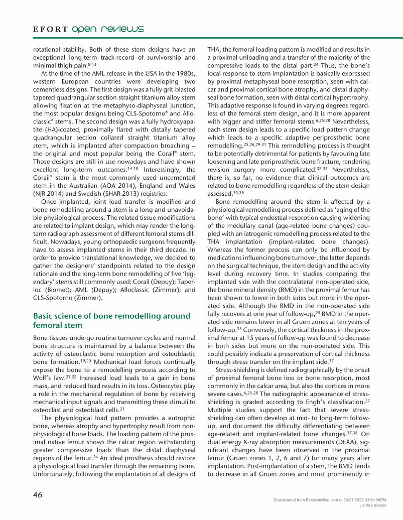
rotational stability. Both of these stem designs have an exceptional long-term track-record of survivorship and minimal thigh pain.8-13
At the time of the AML release in the USA in the 1980s, western European countries were developing two cementless designs. The first design was a fully grit-blasted tapered quadrangular section straight titanium alloy stem allowing fixation at the metaphyso-diaphyseal junction, the most popular designs being CLS-Spotorno® and Allo-classic® stems. The second design was a fully hydroxyapa-tite (HA)-coated, proximally flared with distally tapered quadrangular section collared straight titanium alloy stem, which is implanted after compaction broaching – the original and most popular being the Corail® stem. Those designs are still in use nowadays and have shown excellent long-term outcomes.14-18 Interestingly, the Corail® stem is the most commonly used uncemented stem in the Australian (AOA 2014), England and Wales (NJR 2014) and Swedish (SHAR 2013) registries.
Once implanted, joint load transfer is modified and bone remodelling around a stem is a long and unavoida-ble physiological process. The related tissue modifications are related to implant design, which may render the long-term radiograph assessment of different femoral stems dif-ficult. Nowadays, young orthopaedic surgeons frequently have to assess implanted stems in their third decade. In order to provide translational knowledge, we decided to gather the designers’ standpoints related to the design rationale and the long-term bone remodelling of five ‘leg-endary’ stems still commonly used: Corail (Depuy); Taper-loc (Biomet); AML (Depuy); Alloclassic (Zimmer); and CLS-Spotorno (Zimmer).
Basic science of bone remodelling around femoral stemBone tissues undergo routine turnover cycles and normal bone structure is maintained by a balance between the activity of osteoclastic bone resorption and osteoblastic bone formation.19,20 Mechanical load forces continually expose the bone to a remodelling process according to Wolf’s law.21,22 Increased load leads to a gain in bone mass, and reduced load results in its loss. Osteocytes play a role in the mechanical regulation of bone by receiving mechanical input signals and transmitting these stimuli to osteoclast and osteoblast cells.23
The physiological load pattern provides a eutrophic bone, whereas atrophy and hypertrophy result from non-physiological bone loads. The loading pattern of the prox-imal native femur shows the calcar region withstanding greater compressive loads than the distal diaphyseal regions of the femur.24 An ideal prosthesis should restore a physiological load transfer through the remaining bone. Unfortunately, following the implantation of all designs of
THA, the femoral loading pattern is modified and results in a proximal unloading and a transfer of the majority of the compressive loads to the distal part.24 Thus, the bone’s local response to stem implantation is basically expressed by proximal metaphyseal bone resorption, seen with cal-car and proximal cortical bone atrophy, and distal diaphy-seal bone formation, seen with distal cortical hypertrophy. This adaptive response is found in varying degrees regard-less of the femoral stem design, and it is more apparent with bigger and stiffer femoral stems.6,25-28 Nevertheless, each stem design leads to a specific load pattern change which leads to a specific adaptive periprosthetic bone remodelling.25,26,29-31 This remodelling process is thought to be potentially detrimental for patients by favouring late loosening and late periprosthetic bone fracture, rendering revision surgery more complicated.32-34 Nevertheless, there is, so far, no evidence that clinical outcomes are related to bone remodelling regardless of the stem design assessed.35,36
Bone remodelling around the stem is affected by a physiological remodelling process defined as ‘aging of the bone’ with typical endosteal resorption causing widening of the medullary canal (age-related bone changes) cou-pled with an iatrogenic remodelling process related to the THA implantation (implant-related bone changes). Whereas the former process can only be influenced by medications influencing bone turnover, the latter depends on the surgical technique, the stem design and the activity level during recovery time. In studies comparing the implanted side with the contralateral non-operated side, the bone mineral density (BMD) in the proximal femur has been shown to lower in both sides but more in the oper-ated side. Although the BMD in the non-operated side fully recovers at one year of follow-up,25 BMD in the oper-ated side remains lower in all Gruen zones at ten years of follow-up.35 Conversely, the cortical thickness in the prox-imal femur at 15 years of follow-up was found to decrease in both sides but more on the non-operated side. This could possibly indicate a preservation of cortical thickness through stress transfer on the implant side.37
Stress-shielding is defined radiographically by the onset of proximal femoral bone loss or bone resorption, most commonly in the calcar area, but also the cortices in more severe cases.6,25-28 The radiographic appearance of stress-shielding is graded according to Engh’s classification.27 Multiple studies support the fact that severe stress- shielding can often develop at mid- to long-term follow-up, and document the difficulty differentiating between age-related and implant-related bone changes.37,38 On dual energy X-ray absorption measurements (DEXA), sig-nificant changes have been observed in the proximal femur (Gruen zones 1, 2, 6 and 7) for many years after implantation. Post-implantation of a stem, the BMD tends to decrease in all Gruen zones and most prominently in
Downloaded from Bioscientifica.com at 03/21/2022 03:54:04PMvia free access

47
LONG-TERM BONE REMODELLING AROUND ‘LEGENDARy’ CEMENTLESS FEMORAL STEMS
the proximal region, reaching the lowest values at two years. The reduction of BMD in the first two years after implantation is calculated to be around 10% to 40%.25,35,39 In the period following that, a steady increase in BMD was noted, reaching at ten years’ post-implantation a value similar to the post-operative baseline value.25
Implant-related bone remodelling is also characterized by excessive bone turnover during the post-operative period that can last for more than three years.40 It depends on the new stress distribution imposed by the femoral stem on the proximal femur. The location and magnitude of stress transfer from the femoral component to the bone varies depending on many mechanical and biologi-cal factors. The former is related to the implant and the latter is related to the stiffness of the patient’s bone. Fac-tors promoting proximal femur bone resorption second-ary to stress-shielding include: when the osteointegration area is distal (stem length, extended coating); stiff femo-ral stems (which is determined by their size and composi-tion);4,27-30,32,38 and low density proximal femoral bone (elderly patient, large and straight femoral canals, female sex).32,37,38,41 Postmenopausal alteration of bone turnover predisposing to osteoporosis may explain why females are affected more by stress-shielding than males.41
Material and methodsWhen the femoral stems were initially designed, long-term bone remodelling around them was difficult to pre-dict. The aim of this paper is to relay the long-term experience of expert clinicians and designers who are renowned for having a long-term world class experience with femoral stems we deemed legendary. Selected femo-ral stems had to be first implanted before the year 1990 and still be widely used in clinical practice. In addition, the stem’s designer or expert user (since its early days) had to be available to participate in the current review. Selected stems were: Corail (Depuy); Taperloc (Biomet); AML (Depuy); Alloclassic (Zimmer); and CLS-Spotorno (Zim-mer). We asked the experts to present their femoral stem design rationale. Second, we asked them to describe bone remodelling seen on standard radiographs over the years of implantation, especially the ‘normal’ and ‘abnormal’ radiographic signs. This knowledge prevents misinterpre-tation of normal radiographic appearances specific to each stem and supplies some insight into what to expect after a femoral stem has been implanted.
Designers’/ Experts’ points of viewCorail® stem
Design rationale: The Corail® stem, made of forged titanium alloy (TiAl6V4), is a straight implant with a quadrangular cross-section. The proximal part has horizontal grooves and
is flared in both sagittal and coronal planes. The distal por-tion has longitudinal grooves and a tapered design. In the absence of the formal theoretical argument, the Corail stem was available in two versions, with and without collar sup-port. An extensive HA coating, with a 150-μm thickness, is applied to the stem using an atmospheric plasma spray process. In addition to the standard stem, three series exist, namely ‘coxa vara’, ‘high offset’ and ‘short neck’. The ideal implantation technique is done by bone compaction broaching. The design rationale for this stem is to obtain initial mechanical stability by press-fitting the compacted cancellous bone all around the stem, with the collar acting to augment the stability by preventing its subsidence. The surgeon should avoid close cortical contact between the stem and the diaphysis, and thus it is normal for the stem to look slightly under-sized on an immediate post-operative radiograph. This low volume rectangular stem design with a compaction broaching technique aims to preserve both the bone stock and the endosteal blood supply, enabling the stem to fit a variety of bone shapes. The different neck options address different extramedullary morphotypes.
Long-term bone remodelling: The biological fixation starts during the first week and early migration over 3 mm is rare. Radiological signs of osteointegration (spot welds) appear as early as the third month; they are present all along the stem, but especially visible distally in the dia-physeal area. They persist throughout the life of the pros-thesis. Radiolucent lines are almost never observed, as well as cortical diaphyseal reactions (atrophy or hypertro-phy). Bone remodelling is very limited; nonetheless, cal-car atrophy and some non-progressive radiolucent lines in Gruen zone 1 can be observed. In the vast majority of cases, radiological ‘silence’ is the rule with no significant changes in the periprosthetic bone trophicity observed even after more than 20 years of follow-up. The position-ing of the stem in the canal (varus or valgus) has no signifi-cant influence on the periprosthetic bone pattern. Wear-related osteolysis progresses slowly and is only visi-ble in the proximal femur (Gruen zones 1 and 7). In con-trast, failure of bony ingrowth is characterized by the development of progressive radiolucent lines adjacent to the porous coating, calcar hypertrophy and distal bone pedestal formation (both are signs of load transfer to bone) and finally stem migration (Fig. 1).
Taperloc® stem
Design rationale: The Taperloc® femoral stem is a forged titanium alloy stem with a circumferential porous coating of the same titanium alloy that is applied using a plasma-spray technique. This flat, single-tapered stem (frontal plane) includes a reduced distal smooth wedge geometry to address proximal and distal femoral mismatch. Addi-tionally, there is a 3° taper in sagittal plane to improve proximal progressive offloading and provide stability
Downloaded from Bioscientifica.com at 03/21/2022 03:54:04PMvia free access

48
between the stem and bone interface. Size increments occur only in the ML plane. The design rationale for this stem is to obtain fixation by a ML press-fit within the proxi-mal aspect of the femur, whereas the smooth distal por-tion of the stem is designed to intentionally prevent distal fixation. This design promotes proximal loading and thus optimizes bone remodelling by reducing stress-shielding. The nearly single taper shape overcomes difficulties related to sizing the stem when facing anatomy variability like mismatch in the sagittal and coronal dimensions of the metaphysis, but theoretically at the cost of less rota-tional stability.
Long-term bone remodelling: It is common to see corti-cal thickening around the femoral stem at the distal
tapered sections, especially distal to the plasma-sprayed portion (Fig. 2a). At later follow-up, this stem can also demonstrate some radiolucent lines around the tip of the stem (Fig. 2b). The key feature here is that there is evi-dence of increased radio-opacity around the stem com-pared with the distal femoral canal, which demonstrates evidence of ingrowth. These zones may be osteopaenic, but the lucent zones around the stem do not indicate stem loosening. Pedestal formation is not common with this implant and evidence of this feature would indicate that the stem may be loose. Long-term results with over 22 years of follow-up utilizing this uncemented stem have demonstrated low revision rates for aseptic loosening and high rates of patient satisfaction.8-13
AML® stem
Design rationale: Extensively porous-coated femoral com-ponents were first approved for cementless use in the USA in 1985. These cast CoCr alloy femoral components are straight and approximately 15 cm long. The distal portion is cylindrical to match the femoral diaphysis and the proxi-mal portion is triangular to match the metaphysis. There are typically two metaphyseal sizes. Currently the porous coating using sintered beads covers all but the tip of the stem, but prior versions had porous coating over 30% to 80% of the stem’s length. The design rationale for this stem is to obtain primary diaphyseal bone ingrowth with the comparatively long cylindrically shaped stem. Distal fixation was chosen because the diaphyseal bone is strong and the diaphysis is easily reamed to match the cylindrical shape of the stem. This is called a ‘fit and fill’ surgical tech-nique, in which the intramedullary canal is prepared by clearing its contents. Stem stability relies on fitting the dia-physeal canal by achieving a scratch fit. The proximal tri-angular portion provides additional porous coating for
Fig. 1 a) Pre-operative radiographs of a 54-year-old male patient operated in 1988 for osteoarthritis. b) Immediate post-operative control; Corail® stem KA11, ceramic-on-poly bearing was implanted with the stem in a slight varus position but with good reconstruction of the hip anatomy. c, d, e) Successive AP radiographic controls done at five years, 15 years and 20 years, respectively, showing limited resorption in the calcar region. Note the radiological ‘silence’ with no significant modification of the periprosthetic bone pattern. No osteolysis on both sides, despite significant PE wear.
Fig. 2 a) 17-year follow-up on a Taperloc® stem with evidence of distal cortical thickening. b) 12-year follow-up on a Taperloc® stem with evidence of lucent zones.
Downloaded from Bioscientifica.com at 03/21/2022 03:54:04PMvia free access

49
LONG-TERM BONE REMODELLING AROUND ‘LEGENDARy’ CEMENTLESS FEMORAL STEMS
ingrowth and supplements initial rotational and axial fixa-tion of the stem.
Long-term bone remodelling: Findings consistent with ingrowth include spot welding of the bone to the porous coating of the diaphysis, bone resorption secondary to stress-shielding proximal to the spot weld, and calcar atrophy. In contrast, failure of bony ingrowth appears as an absence of these findings combined with radiolucent lines adjacent to the porous coating, calcar hypertrophy, distal bone pedestal formation and stem migration. Radi-ographic signs of bone ingrowth can take more than two years to appear. Spot welds and bone resorption second-ary to stress-shielding are typically first seen on the lat-eral femoral radiograph. Proximal bone resorption or stress-shielding is always easier to identify among patients with osteopaenia or osteoporosis, who typically require a larger diameter stem. The radiographic findings associated with a bone ingrown stem tend to vary based on the patient’s bone quality. For a patient with good
bone quality, a spot weld and stress shielding do not appear immediately (Fig. 3). Among patients with osteo-porosis, who often require larger diameter stems, a spot weld and stress-shielding are easily seen by three years, especially on the lateral radiograph (Fig. 4). When bone ingrowth does not occur, a stem may become fibrous stable or loose. Instead of a spot weld, a fibrous stable stem will develop parallel radiolucent lines (Fig. 5). Over many years this stem does not migrate, there is no calcar hypertrophy, nor is there a distal pedestal and, most importantly, the patient is asymptomatic. Retrieval anal-ysis in cases like this has demonstrated a dense fibrous attachment between the radiolucent line and the porous coating that stabilizes this stem without bone ingrowth.27 The hallmark of a loose stem is distal stem migration (Fig. 6). Additional signs of loosening include radiolu-cent lines and a distal pedestal. By far, the most impor-tant clinical sign of a loose stem is thigh pain. Although thigh pain in the first two years could represent a loose
Fig. 3 Patients with AML stem and good bone quality might develop a spot weld and stress-shielding after a long time. a) Successive AP radiographs at one year, seven years and 15 years post-operatively showing a spot weld developing after seven years on the distal medial aspect of the stem. b) Later views confirm the spot weld formation.
Downloaded from Bioscientifica.com at 03/21/2022 03:54:04PMvia free access
50
stem, alone it is not indicative of stem loosening since 4% to 10% of patients with a bone ingrown stem have thigh pain. Because lucent lines and a pedestal can take more than two years to develop, waiting for these radio-graphic signs to diagnose a loose stem can be frustrating for both the patient and the surgeon. Luckily, extensively porous coated stems have only a 2% to 3% rate of failed ingrowth and a 95% survivorship after 20 years of
follow-up as reported by The Anderson Orthopaedic Research Institute (AORI).42,43
Alloclassic SL® stem
Design rationale: The Alloclassic SL® stem was introduced on the market in 1986. The stem is manufactured from TI-6A1-7N alloy (Protasul®-100) and has a rectangular cross-section with a taper shape in both frontal and
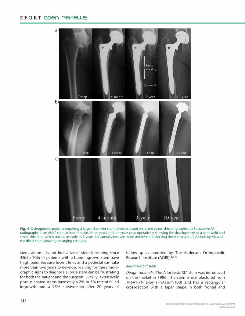
Fig. 4 Osteoporotic patients requiring a larger diameter stem develop a spot weld and stress-shielding earlier. a) Successive AP radiographs of an AML® stem at four months, three years and ten years post-operatively showing the development of a spot weld and stress-shielding which started as early as 3 years. b) Lateral views are more sensitive in detecting these changes. c) A close-up view of the distal stem showing enlarging changes.
Downloaded from Bioscientifica.com at 03/21/2022 03:54:04PMvia free access

51
LONG-TERM BONE REMODELLING AROUND ‘LEGENDARy’ CEMENTLESS FEMORAL STEMS
sagittal planes (dual taper). Increments from size to size are in all directions. The stem is fully grit-blasted to pro-duce a 3- to 5-µm thickness micro-texture. The design
rationale for this stem is to achieve stability due to a meta-diaphyseal junction cortical press fit. The Alloclassic stem uses a ‘fit without fill’ surgical technique and provides a
Fig. 5 A fibrous stable stem will be seen when bone ingrowth does not occur. Instead of a spot weld, a fibrous stable stem will develop parallel radiolucent lines. The stem is stable and does not migrate distally. No calcar hypertrophy and no distal pedestal can be seen. a) Successive AP radiographs of an AML® stem at one year, two years and ten years post-operatively showing the development of parallel radiolucent lines along the stem. b) Lateral views showing similar changes. c) A close-up view of the distal stem revealing more of these changes.
Downloaded from Bioscientifica.com at 03/21/2022 03:54:04PMvia free access

52
Fig. 6 A loose stem characteristic hallmark is distal stem migration. a) Successive AP radiographs of an AML® stem at four weeks, one year and six years post-operatively showing an increase in the distance between the tip of the femoral stem and the tip of the greater trochanter, indicating distal stem migration. Pedestal formation can be seen as early as one year post-operatively. b) Lateral views showing the pedestal formation. c) A close-up view of the distal stem showing more of these changes.
Downloaded from Bioscientifica.com at 03/21/2022 03:54:04PMvia free access

53
LONG-TERM BONE REMODELLING AROUND ‘LEGENDARy’ CEMENTLESS FEMORAL STEMS
posterior-anterior-posterior (PAP) cortical fixation when seen in the axial view. This technique preserves endosteal blood supply, improves initial stability and enables to fit a variety of bone shapes.
Long-term bone remodelling: Proximal cortical atrophy, a major sign of stress-shielding, is seen mainly in Gruen zones 1, 2, 6 and 7 (Fig. 7a). The average BMD loss usually ranges between 27% and 50%, and appears two years
Fig. 7 a) Successive AP radiographs of an Alloclassic SL® stem immediately following surgery, and ten years and eleven years post-operatively, showing proximal cortical atrophy mainly in Gruen zones 1, 2, 6 and 7, indicating stress-shielding. b) Sequential AP radiographs of an Alloclassic SL® stem at three months, one year and nine years post-operatively showing cortical diaphyseal hypertrophy developing combined with proximal stress-shielding. c) Sequential AP radiographs of an Alloclassic SL® stem showing the adaptive behaviour of the stem in patients with osteoporosis.
Downloaded from Bioscientifica.com at 03/21/2022 03:54:04PMvia free access

54
after implantation, but usually reaches a steady state at five years post-implantation. The amount of stress-shield-ing depends on the pre-operative anatomical situation, especially the cortical index with a proximal ‘stove pipe’ femoral shape tending to show more stress-shielding. Severe stress-shielding is uncommon and was only seen in two of 4000 patients (0.5%), suggesting that the low vol-ume rectangular design is forgiving and reduces stress-shielding. Cortical diaphyseal hypertrophy developing in early years can be seen in about one-third of patients. It is a sign of distal stress transfer and is normally non- progressive and usually accompanied by proximal bone re sorption secondary to stress-shielding (Fig. 7b). Non-progressive benign radiolucent lines have been described in 40% of patients and mainly occur in the proximal femur (Gruen zones 1 and 7). A reason for those radiolucent lines might be a proximal micro-motion with a distally well-fixed stem, but might also result from an intra-opera-tive change of the primary rasping direction. Wear-related osteolysis is normally found proximally in Gruen zones 1 and 7, but in rare cases can occur more distally, thus allowing the distalization of the osteointegration area. In these rare cases, diaphyseal hypertrophy can develop sec-ondarily. The positioning of the stem in the canal (varus or valgus) does not significantly influence the periprosthetic bone remodelling pattern or long-term survival of the stem. The Alloclassic stem has also proven to be suitable for patients with osteoporosis (Fig. 7c).16,17,44,45
CLS-Spotorno® stem
Design rationale: The CLS-Spotorno® stem is a collarless, tapered straight, grit-blasted stem. The design consists of a wider proximal and lateral portion with a thinner distal and medial portion, creating a triple wedge-shape (triple taper). The primary rotational stability is increased by ribs on the anterior and posterior surface providing interdigi-tation with the cancellous bone.46 The stem is made of a titanium alloy that is roughened by ‘grit-blasting’ with 3- to 4-μm sized particles of aluminium oxide or corundum. The roughened surface allows bone ongrowth, leading to long-term stability. The design rationale for this stem is to obtain a press-fit in both the metaphysis and the meta-diaphyseal junction. The tapered shape prevents a com-plete fill in the distal diaphyseal portion, encouraging a more physiological load transfer in the proximal part of the femur. The triple taper creates compressive loading forces throughout the proximal femur to optimize further bone remodelling by reducing stress-shielding.
Long-term bone remodelling: The tapered geometry of the CLS provides long-term stability through osteointe-gration in the metaphyseal region (taper-lock effect). Indeed, rare severe stress-shielding and distal cortical hypertrophy have been detected after 30 years of follow-up (Fig. 8a). In patients with high congruence between
the femur and the stem shape (‘champagne-flute’ proximal femur), stress-shielding is unlikely to occur. Conversely, in patients with low congruence (‘stove-pipe’ proximal femur), the stress-shielding onset is mild to moderate, sug-gesting osteointegration in the inter-sub- trochanteric region. At long-term follow-up, non-progressive radiolu-cent lines can be seen in one and up to three Gruen zones around the stem without affecting the stability of the stem.47 These findings suggest that the CLS design pre-vents a more distal load transfer even in the long term. Wear-related osteolytic lesions are usually restricted to the proximal zones of the femur (Fig. 8b). The eutrophic bone in the metaphyseal region and circumferential stem osteo-integration tend to prevent the distal progression of osteo-lysis. However, bone loss from wear-related osteolysis along with bone aging can result in the distalization of the load transfer, resulting in increased proximal bone loss second ary to stress-shielding.
DiscussionBone remodelling around a stem is an unavoidable long-term physiological process highly related to implant design. For some predisposed patients, it can lead to periprosthetic bone loss secondary to severe stress-shield-ing, which is thought to be detrimental by contributing to late loosening, late periprosthetic fracture and rendering revision surgery more complicated. However, these con-cerns remain theoretical since late loosening has yet to be documented among bone ingrown cementless stems demonstrating periprosthetic bone loss associated with stress-shielding. In their daily practice, orthopaedic sur-geons need to differentiate physiological long-term bone remodelling patterns from pathological conditions such as loosening, sepsis or osteolysis. To aid in this, we decided to clarify the behaviour of five of the most successful femoral stems still widely used. Our experts’ review of the remod-elling of these stems after long-term implantation showed that none of these replicate the physiological load pattern on the proximal femur, each stem design being associated with a specific load pattern leading to specific adaptive periprosthetic bone remodelling.
In a randomized study, Karachalios et al have shown the impact of stem designs on bone remodelling. When compared with the Alloclassic stem, the Corail stem has been shown to generate less bone resorption in the proxi-mal femur with a return to baseline BMD values at ten years of follow-up.25 Interestingly, no correlation was found between a set timeline and the severity of stress-shielding,32 which may imply that only a subgroup of ‘predisposed patients’ might develop severe stress-shield-ing. Capello et al found in a cohort of patients with CLS stems followed for 15 years that one-third had no or little bone remodelling, and women with poorer bone quality
Downloaded from Bioscientifica.com at 03/21/2022 03:54:04PMvia free access

55
LONG-TERM BONE REMODELLING AROUND ‘LEGENDARy’ CEMENTLESS FEMORAL STEMS
at the time of implantation were more likely to develop periprosthetic cortical porosity after ten years or more.37 HA coating was found to improve osteointegration or ‘internal bone remodelling’ as defined by Braun.48 Many studies supported this by finding significantly fewer radio-lucent lines, superior proximal femoral osteointegration and less proximal stress-shielding, but without affecting the clinical outcomes.17,25,49
Despite all the differences between the stems reviewed, there is currently no evidence that one design is superior to another in terms of clinical outcome or extended long-term survival (Table 1). Thus, it is currently not possible to
define what exact design features are more advantageous for long-term bone-stem compatibility. Nonetheless, by knowing the strengths and weaknesses of each design, it is possible to select the stem design that best serves each patient. For example, patients with low bone density who are at higher risk of developing stress-shielding-related bone loss will benefit more from a Corail-type proximally loaded, titanium alloy, HA-coated stem.
When looking at the long-term evidence for those stems more precisely, everyone should realize that excel-lent survival rates for aseptic loosening with more than 93% in the third decade does not reflect the true behav-iour of these stems. In fact, those figures were gathered in retrospective studies describing the loosening rate by tak-ing into account the whole initial cohort. It included patients lost to follow-up and those who died at a shorter follow-up. Using the Kaplan-Meier survival curve, includ-ing all implanted hips, the failure rate would become much higher. For instance, previously cited major studies relating excellent survival rates of the reviewed
Fig. 8 a) Successive AP radiographs of a CLS-Spotorno® stem at one-year, seven-year, 15-year and 30-year follow-up revealing no stress-shielding and distal cortical hypertrophy. b) Successive AP radiographs of a CLS-Spotorno® stem at one year, three years, seven years and after a revision surgery performed to change the worn polyethylene of the acetabular cup shows that wear-related osteolytic lesions are restricted to the proximal zones of the femur.
Table 1. Femoral stems: long-term survival7-12, 13-16, 44, 45
Stem type Survival (percentage) Follow-up (years)
CLS stem 93 22AML stem 98 20Corail stem 99 23Taperloc stem 99 26Alloclassic stem 99 24
Downloaded from Bioscientifica.com at 03/21/2022 03:54:04PMvia free access

56
stems – 93% in Streit’s study (20 CLS loose stems from 354 initial THAs), 98% in Belmont’s study (six AML from 223 THAs), 99% in Vidalain’s study (four Corail from 347 THAs), 99% in McLaughlin’s study (two Taperloc from 145 THAs) and 99% in Kolb’s study (two Alloclassic from 208 THAs) – have, respectively, only 40% (143 hips), 53% (119), 36.5% (127 hips), 25% and 34% of patients fol-lowed in the third decade.10,14,15,42 In other words, the long-term survival rates given in the literature reflect the percentage of loose stems to be revised rather than the real survival rate of the stem. Because nowadays THAs are becoming much more common for younger patients with longer life expectancy, and are inserted in randomized studies, further reports based on the current trend of THAs could potentially show more realistic and perhaps inferior outcomes. On the other hand, tying to improve current results of these legendary stems is a very difficult task and the risk of failure probably outweighs the benefits. Arnand et al found analysing the Australian registry to be of no benefit in the introduction of new prostheses over the years 2003 to 2007, and moreover 27% had a significantly higher revision rate.50 As shown in our review, replicating femoral bone remodelling using a femoral stem made of a material and a shape that differ from the natural bone structure was not possible. Each expert surgeon described the normal and abnormal bone remodelling patterns associated with their stem. Such knowledge is of primary importance for the follow-up of these well-performing stems over time.
ICMJE ConflICt of IntErEst statEMEntC. Rivière declares payment for lectures for Medacta, activity outside the submitted work. G. Grappiolo declares personal fees from Zimmer Biomet, outside the sub-mitted work. C. Engh Jr. declares grants and other funds from DePuy Synthes Joint Reconstruction, a Division of DePuy Orthopaedics, Inc.; grants from Smith & Nephew, outside the submitted work. J.-P. Vidalain declares personal fees and other funds
from Depuy, during the conduct of the study; in addition, patent FR 02 06122 with royalties paid to Depuy. A. F. Chen declares other funds from SLACK publishing, non-financial support from Joint Purification Systems, personal fees from ACI, personal fees from DJO, personal fees from Stryker, outside the submitted work. N. Boehler declares consultancy fees from Zimmer-Biomet, outside the submitted work. A.-P. Vendittoli declares consultancy fees from Medacta, Microport and Stryker; grants/grants pending from Medacta, Microport, Stryker and Zimmer; payment for lectures from Medacta, Stryker and Ethicon; royalties from Microport, outside the submitted work.
fundIng statEMEntNo benefits in any form have been received or will be received from a commercial party related directly or indirectly to the subject of this article.
lICEnCE© 2018 The author(s)This article is distributed under the terms of the Creative Commons Attribution-Non Commercial 4.0 International (CC BY-NC 4.0) licence (https://creativecommons.org/licenses/by-nc/4.0/) which permits non-commercial use, reproduction and distribu-tion of the work without further permission provided the original work is attributed.
rEfErEnCEs
1. McKee gK, Watson-farrar J. Replacement of arthritic hips by the McKee-Farrar prosthesis. J Bone Joint Surg Br 1966;48:245-9.
2. Kawamoto K, Hasegawa Y, Iwase t, et al. Failed cementless total hip arthroplasty for osteoarthrosis due to hip dysplasia. A minimum five-year follow-up study. Bull Hosp Jt Dis 1998;57:130-5.
3. albrektsson t, Brånemark PI, Hansson Ha, lindström J. Osseointegrated titanium implants. Requirements for ensuring a long-lasting, direct bone-to-implant anchorage in man. Acta Orthop Scand 1981;52:155-70.
4. Engh Ca, Mcgovern tf, Bobyn Jd, Harris WH. A quantitative evaluation of periprosthetic bone-remodeling after cementless total hip arthroplasty. J Bone Joint Surg Am 1992;74:1009-20.
5. Khanuja Hs, Vakil JJ, goddard Ms, Mont Ma. Cementless femoral fixation in total hip arthroplasty. J Bone Joint Surg Am 2011;93:500-9.
6. Engh Ca, o’Connor d, Jasty M, et al. Quantification of implant micromotion, strain shielding, and bone resorption with porous-coated anatomic medullary locking femoral prostheses. Clin Orthop Relat Res 1992;(285):13-29.
7. Brown tE, larson B, shen f, Moskal Jt. Thigh pain after cementless total hip arthroplasty: evaluation and management. J Am Acad Orthop Surg. 2002;10:385-92.
8. Mclaughlin Jr, lee Kr. Total hip arthroplasty in young patients. 8- to 13-year results using an uncemented stem. Clin Orthop Relat Res 2000;373:153-63.
9. Mclaughlin Jr, lee Kr. Cementless total hip replacement using second-generation components: a 12- to 16-year follow-up. J Bone Joint Surg Br 2010;92:1636-41.
10. Mclaughlin Jr, lee Kr. Uncemented total hip arthroplasty with a tapered femoral component: a 22- to 26-year follow-up study. Orthopedics 2010;33:639.
11. Mclaughlin Jr, lee Kr. Uncemented total hip arthroplasty using a tapered femoral component in obese patients: an 18-27 year follow-up study. J Arthroplasty 2014;29:1365-8.
12. Parvizi J, Keisu Ks, Hozack WJ, sharkey Pf, rothman rH. Primary total hip arthroplasty with an uncemented femoral component: a long-term study of the Taperloc stem. J Arthroplasty 2004;19:151-6.
autHor InforMatIon1MSK Lab, Imperial College London, UK; South West London Elective Orthopaedic Centre, UK.2Unit of Hip Diseases and Joint Replacement Surgery, Humanitas Clinical and Research Center, Italy.3Anderson Orthopaedic Research Institute, USA.
4Artro Group, France.
5Rothman Institute of Orthopaedics, USA.
6Orthopaedic Department, Kepleruniklinikum Linz, Austria.
7Hôpital Maisonneuve-Rosemont, Canada.
8Hôpital Maisonneuve-Rosemont, Université de Montréal, Canada.
Correspondence should be sent to: P.-A. Vendittoli, Hôpital Maisonneuve-Rosemont, 5345 Boulevard de L'Assomption, H1T 2M4, Montréal, Québec, Canada. Email: [email protected]
Downloaded from Bioscientifica.com at 03/21/2022 03:54:04PMvia free access

57
LONG-TERM BONE REMODELLING AROUND ‘LEGENDARy’ CEMENTLESS FEMORAL STEMS
13. rao rr, sharkey Pf, Hozack WJ, Eng K, rothman rH. Immediate weightbearing after uncemented total hip arthroplasty. Clin Orthop Relat Res 1998;349:156-62.
14. Vidalain JP. Twenty-year results of the cementless Corail stem. Int Orthop 2011;35:189-94.
15. streit Mr, Innmann MM, Merle C, et al. Long-term (20- to 25-year) results of an uncemented tapered titanium femoral component and factors affecting survivorship. Clin Orthop Relat Res 2013;471:3262-9.
16. Kolb a, grübl a, schneckener Cd, et al. Cementless total hip arthroplasty with the rectangular titanium Zweymüller stem: a concise follow-up, at a minimum of twenty years, of previous reports. J Bone Joint Surg Am 2012;94:1681-4.
17. delaunay C. Effect of hydroxyapatite coating on the radio-clinical results of a grit-blasted titanium alloy femoral taper. A case-control study of 198 cementless primary total hip arthroplasty with the Alloclassic™ system. Orthop Traumatol Surg Res 2014;100:739-44.
18. Mallory tH, lombardi aV Jr, leith Jr, et al. Why a taper? J Bone Joint Surg [Am] 2002;84-A(suppl 2):81-9.
19. Hughes JM, Petit Ma. Biological underpinnings of Frost’s mechanostat thresholds: the important role of osteocytes. J Musculoskelet Neuronal Interact 2010;10:128-35.
20. Parfitt aM. Osteonal and hemi-osteonal remodeling: the spatial and temporal framework for signal traffic in adult human bone. J Cell Biochem 1994;55:273-86.
21. Basso n, Jia Y, Bellows Cg, Heersche Jn. The effect of reloading on bone volume, osteoblast number, and osteoprogenitor characteristics: studies in hind limb unloaded rats. Bone 2005;37:370-8.
22. Vico l, Collet P, guignandon a, et al. Effects of long-term microgravity exposure on cancellous and cortical weight-bearing bones of cosmonauts. Lancet 2000;355:1607-11.
23. Ehrlich PJ, lanyon lE. Mechanical strain and bone cell function: a review. Osteoporos Int 2002;13:688-700.
24. oh I, Harris WH. Proximal strain distribution in the loaded femur. An in vitro comparison of the distributions in the intact femur and after insertion of different hip-replacement femoral components. J Bone Joint Surg Am 1978;60:75-85.
25. Karachalios t, tsatsaronis C, Efraimis g, et al. The long-term clinical relevance of calcar atrophy caused by stress shielding in total hip arthroplasty: a 10-year, prospective, randomized study. J Arthroplasty 2004;19:469-75.
26. Yamauchi Y, Jinno t, Koga d, et al. Comparison of different distal designs of femoral components and their effects on bone remodeling in 1-stage bilateral total hip arthroplasty. J Arthroplasty 2012;27:1538-43.
27. Engh Ca, Bobyn Jd, glassman aH. Porous-coated hip replacement. The factors governing bone ingrowth, stress shielding, and clinical results. J Bone Joint Surg [Br] 1987;69:45-55.
28. Herrera a, rebollo s, Ibarz E, et al. Mid-term study of bone remodeling after femoral cemented stem implantation: comparison between DXA and finite element simulation. J Arthroplasty 2014;29:90-100.
29. Macdonald sJ, rosenzweig s, guerin Js, et al. Proximally versus fully porous-coated femoral stems: a multicenter randomized trial. Clin Orthop Relat Res 2010;468:424-32.
30. Hirata Y, Inaba Y, Kobayashi n, et al. Comparison of mechanical stress and change in bone mineral density between two types of femoral implant using finite element analysis. J Arthroplasty 2013;28:1731-5.
31. Chandran P, azzabi M, andrews M, Bradley Jg. Periprosthetic bone remodeling after 12 years differs in cemented and uncemented hip arthroplasties. Clin Orthop Relat Res 2012;470:1431-5.
32. Yamada H, Yoshihara Y, Henmi o, et al. Cementless total hip replacement: past, present, and future. J Orthop Sci 2009;14:228-41.
33. Engh Ca Jr, Young aM, Engh Ca sr, Hopper rH Jr. Clinical consequences of stress shielding after porous-coated total hip arthroplasty. Clin Orthop Relat Res 2003;(417):157-63.
34. lindahl H. Epidemiology of periprosthetic femur fracture around a total hip arthroplasty. Injury 2007;38:651-4.
35. Kwon dg, lee tJ, Kang Js, Moon KH. Correlation between stress shielding and clinical outcomes after total hip arthroplasty with extensively porous coated stems. J Arthroplasty 2013;28:1728-30.
36. Mclaughlin Jr, lee Kr. Total hip arthroplasty with an uncemented tapered femoral component. J Bone Joint Surg Am 2008;90:1290-6.
37. Capello Wn, d’antonio Ja, geesink rg, feinberg Jr, naughton M. Late remodeling around a proximally HA-coated tapered titanium femoral component. Clin Orthop Relat Res 2009;467:155-65.
38. nishino t, Mishima H, Kawamura H, et al. Follow-up results of 10-12 years after total hip arthroplasty using cementless tapered stem - frequency of severe stress shielding with synergy stem in Japanese patients. J Arthroplasty 2013;28:1736-40.
39. Pitto rP, Hayward a, Walker C, shim VB. Femoral bone density changes after total hip arthroplasty with uncemented taper-design stem: a five year follow-up study. Int Orthop 2010;34:783-7.
40. Venesmaa P, Vanninen E, Miettinen H, Kröger H. Periprosthetic bone turnover after primary total hip arthroplasty measured by single-photon emission computed tomography. Scand J Surg 2012;101:241-8.
41. Merle C, streit Mr, Volz C, et al. Bone remodeling around stable uncemented titanium stems during the second decade after total hip arthroplasty: a DXA study at 12 and 17 years. Osteoporos Int 2011;22:2879-86.
42. Belmont PJ Jr, Powers CC, Beykirch sE, et al. Results of the anatomic medullary locking total hip arthroplasty at a minimum of twenty years. A concise follow-up of previous reports. J Bone Joint Surg Am 2008;90:1524-30.
43. Mcauley JP, Culpepper WJ, Engh Ca. Total hip arthroplasty. Concerns with extensively porous coated femoral components. Clin Orthop Relat Res 1998;355:182-8.
44. suckel a, geiger f, Kinzl l, Wulker n, garbrecht M. Long-term results for the uncemented Zweymuller/Alloclassic hip endoprosthesis. A 15-year minimum follow-up of 320 hip operations. J Arthroplasty 2009;24:846-53.
45. Pieringer H, auersperg V, Böhler n. Long-term results of the cementless ALLOCLASSIC hip arthroplasty system using a 28-mm ceramic head: with a retrospective comparison to a 32-mm head. J Arthroplasty 2006;21:967-74.
46. spotorno l, romagnoli s, Ivaldo n, et al. The CLS system. Theoretical concept and results. Acta Orthop Belg 1993;59(suppl 1):144-8.
47. grappiolo g, Blaha Jd, gruen ta, Burastero g, spotorno l. Primary total hip arthroplasty using a grit-blasted, press-fit femoral prosthesis. Long-term results with survivorship analysis. Hip Int 2002;12:55-72.
48. Braun a, Papp J, reiter a. The periprosthetic bone remodelling process-signs of vital bone reaction. Int Orthop 2003;27(suppl 1):S7-S10.
49. Chambers B, st Clair sf, froimson MI. Hydroxyapatite-coated tapered cementless femoral components in total hip arthroplasty. J Arthroplasty 2007;22(suppl 1):71-4.
50. anand r, graves sE, de steiger rn, et al. What is the benefit of introducing new hip and knee prostheses? J Bone Joint Surg Am 2011;93(suppl 3):51-4.
Downloaded from Bioscientifica.com at 03/21/2022 03:54:04PMvia free access