24th Surgical Research Days - Karger Publishers
60
24th Surgical Research Days Section of Surgical Research of the German Society of Surgery Leipzig, Germany, September 2–3, 2021 Abstracts Eur Surg Res 2021:62:161–220 Published online: August 31, 2021 DOI: 10.1159/000518793 Basel • Freiburg • Hartford • Oxford • Bangkok • Dubai • Kuala Lumpur • Melbourne • Mexico City • Moscow • New Delhi • Paris • Shanghai • Tokyo Guest Editors Ines Gockel, Leipzig Bruno Christ, Leipzig Topics Biomechanics and Implant Research Computer Support Systems Virtual Surgery Planning Cell Transplantation and Regenerative Therapies Barrier Disorders and Pneumonia Oncology, Malignancies Trauma Research COVID-19: Impact on Surgical Research and Education Obesity and Metabolic Surgery Molecular Genetics of Gastrointestinal Tumors Liver Perfusion and Function - A Systems Medicine Approach Virtual Liver Career Planning and Mentoring Free Topics Biomechanics Abstracts containing errors in spelling, grammar, or scientific content have been published as typed by the author and have not been corrected by the Publisher.
-
Upload
khangminh22 -
Category
Documents
-
view
1 -
download
0
Transcript of 24th Surgical Research Days - Karger Publishers

24th Surgical Research DaysSection of Surgical Research of the German Society of Surgery
Leipzig, Germany, September 2–3, 2021
Abstracts
Eur Surg Res 2021:62:161–220 Published online: August 31, 2021
DOI: 10.1159/000518793
Basel • Freiburg • Hartford • Oxford • Bangkok • Dubai • Kuala Lumpur • Melbourne • Mexico City • Moscow • New Delhi • Paris • Shanghai • Tokyo
Guest EditorsInes Gockel, LeipzigBruno Christ, Leipzig
Topics
Biomechanics and Implant ResearchComputer Support SystemsVirtual Surgery PlanningCell Transplantation and Regenerative TherapiesBarrier Disorders and PneumoniaOncology, MalignanciesTrauma ResearchCOVID-19: Impact on Surgical Research and EducationObesity and Metabolic SurgeryMolecular Genetics of Gastrointestinal TumorsLiver Perfusion and Function - A Systems Medicine ApproachVirtual LiverCareer Planning and MentoringFree TopicsBiomechanics
Abstracts containing errors in spelling, grammar, or scientific content have been published as typed by the author and have not been corrected by the Publisher.

Abstracts
Biomechanics and Implant Research
ID 133Double Splinting Techique for Rib Fractures - A Traditional Technique Applied ExperimentallyD.H. Borchert1, D. Wulsten2, J. Nayantai3
1Bundeswehrkrankenhaus, Klinik für Allg., Viszeral u. Thoraxchirurgie, Berlin, Germany, 2Charité, Julius Wolff Institut, Berlin, Germany, 3Zentralkrankenhaus des Militärs, Chirurgie, Ulaan Bator, Mongolia
Background/Aims: Among several techniques intramedullary splints are used for repair of rib fractures. So far these splints have not been used in the traditional technique of crossed K-wires.
Methods: A sheep hemithorax was harvested with the sternum and spine from an animal used for experimental mandibular sur-gery. It was cut in half resulting in two specimens of approximately 25 cm in width and 30 cm in length consisting of the upper and lower half of the hemithorax. Specimes were fixed in PMMA blocks and tested for stiffness and burst pressure of ribs using a Zwick Roell Z010 material testing machine. Pressue from the first step of the experiment was used to adjust for burst pressure of the upper half of the hemithorax. Rib fractures were then repaired with a single splint followed by repair in a double splint technique (crossed K-wire technique).
Results: Preparing half of a sheep hemithorax for fixation in PMMA blocks is feasible and allows for testing of stiffness and burst pressure. Repair with single intramedullary splints and double-splints (3 mm splints) in a crossed K-wire techinque was feasible in the upper half of the hemithorax. The crossed K-wire technique resulted in a more stable repair.
Conclusions: Using a sheep hemithorax for testing of intra-medullary splints for rib fracture repair is feasible using a quasi physiological setting with preservation of the anterior and poste-rior rib joints. The model has several limitations including the lon-gitudinal oval shape compared to the human thorax. Secondly the intramedullary canal of sheep ribs is of small size only. Therefore repair with 3 mm size splints can be tested only. Machine testing of large specimen size is a challenge due to three dimensional con-figuration of the hemithorax, but nonetheless feasible. Fracture testing in this model allowed for a good estimate of rib fracture repair and handling of splints. The modell is recommend for fur-ther testing in experimental and training settings.
ID 139
The Pectineal Ligament Acts as a Secondary Stabilizer for Anterior Pelvic Ring FracturesG. Osterhoff 1, R. Reise2, E. Riemer2, A. Höch1, J. K. Fakler1, C.-E. Heyde1, S. Schleifenbaum2
1University of Leipzig Medical Center, Department of Orthopaedics, Trauma and Plastic Surgery, Leipzig, Germany, 2Leipzig University, ZESBO - Center for Research on Muscoskeletal Systems, Leipzig, Germany
Background/Aims: There is ongoing discussion whether oper-ative fixation of partially stable lateral compression fractures of the pelvis is beneficial for the patient. Recent studies suggest that the pectineal ligament may act as a secondary stabilizer of the anterior pelvis ring. The purpose of this study was to investigate the influ-ence of the pectineal ligament’s integrity on the biomechanical sta-bility and displacement in anterior pelvic ring fractures.
Methods: In a biomechanical setup, a cyclic loading protocol was applied with sinusoidal axial force from 100 to 500 N on cadaver hemipelves with soft tissues (n = 5). After testing the native specimens (“No fracture”), increasing degrees of injury were cre-ated on the samples: 1. an osseous defect to the pubic ramus (“Bone #”), 2. cutting of all soft tissues including obturator membrane except for the pectineal ligament intact (“ObtM #”) , 3. cutting of the pectineal ligament (“PectL #”) - with the loading protocol being applied to each sample at each state of injury. Fracture motion and vertical displacement were measured using a digital image correla-tion system and opto-metric analysis.
Results: No failure of the constructs was observed. Creating a pubic ramus fracture (p = 0.042) and cutting the pectineal ligament (p = 0.042) each significantly increased relative fracture move-ment. The mean change in absolute movement was 0.067 mm (range, 0.02 mm to 0.19 mm) for ObtM # and 0.648 mm (range, 0.07 mm to 2.93 mm), for PectL # in relation to Bone # (p = 0.043). Also for absolute vertical movement, there was a significant change when the pectineal ligament was cut (p = 0.043), while there was no such effect with cutting all other soft tissues including the obtu-rator membrane.
Conclusions: Based on the findings of this in vitro study, the pectineal ligament significantly contributes to the stability of the anterior pelvic ring. An intact pectineal ligament reduces fracture movement in presence of a pubic ramus fracture.
© 2021 S. Karger AG, [email protected]/esr

Abstracts 163Eur Surg Res 2021:62:161–220DOI: 10.1159/000518793
ID 141
Development and Validation of Artificial Bone ModelsC. Neupetsch1, F. Metzner2, A. Carabello1, M. Werner3, W.-G. Drossel1
1Technische Universität Chemnitz, Adaptronics and Lightweight Design in Production, Chemnitz, Germany, 2Universität Leipzig, Center for Research on Musculoskeletal System, Leipzig, Germany, 3Fraunhofer Institute for Maschine Tools and Forming Technology IWU, Medical Technology, Dresden, Germany
Background/Aims: Simulations of surgery on human speci-mens offer the opportunity to practice complex procedures in the advanced training of physicians. However, human specimens are only available for this purpose to a limited extent and different comorbidities of the donors have to be considered. Artificial train-ing models offer a more cost-effective and reproducible alternative and can also be used for functional biomechanical testing, such as developing new implants. However, the realism of representation, especially of Osseous structures, is very limited and measurements do not provide the same results compared to human specimens. The objective for the future is to develop realistic bone models as a base for improved surgical training models to enhance medical education. Further, bone models with correct biomechanical properties can improve and accelerate the development processes for new medical devices and technologies.
Methods: The first step was the determination of the biome-chanical parameters of human bones to generate reproducible arti-ficial bone models. Both conventional methods from materials testing and structural dynamic measurement were used to charac-terize and validate the bone models. With suitable manufacturing processes and materials, these properties were replicated. Here, 3D printing served as an efficient manufacturing tool and enabled at the same time to produce complex internal structures. Further-more, a variety of usable options for the creation of artificial bone structures are available through different printing processes and materials.
Results: In the future, this development will enable surgical training models to gain a much greater degree of realism and con-tribute to improving medical training. In the field of implant test-ing, the results can be used to improve and accelerate development processes. Thus, for the first time, artificial bone models are avail-able which rebuild more realistic ossous material properties and can build up diseases such as osteoporosis in a more specific way.
Conclusions: The results of this project could also reduce the number of human specimens and test animals required in research. Initially, the bone model will be used in the field of research and training of surgical orthopedics of hip arthroplasty. Results and findings of this platform technology can subsequently be trans-ferred to other surgical disciplines.
Computer Support Systems
ID 43
First Experiences with an Augmented Reality Assistance Tool for Laparoscopic SurgeryF. Huettl1, F. Heinrich2, M. Paschold1, H. Lang1, C. Hansen2, W. Kneist1, T. Huber1
1University Medical Center of the Johannes Gutenberg-University Mainz, Department of General, Visceral and Transplant Surgery, Mainz, Germany, 2Otto-von-Guericke University Magdeburg, Institute of Simulation and Graphics, Faculty of Computer Science, Magdeburg, Germany
Background/Aims: In surgery novice physicians depend on the guidance of experts (trainers) to gain surgical proficiency. Espe-cially in laparoscopic surgery teaching depends mainly on verbal instructions. To expand the communication through a visual com-ponent, we propose an optical see-through augmented reality (AR) tool to visualize an interactive virtual pointer on the laparoscopic monitor. This application is operated solely via verbal commands to allow a sterile workflow. This is a first feasibility study to evalu-ate the integration of this new technology into the operating room.
Methods: To use the especially developed AR application, the main surgeon and the first assistant wore augmented reality devices (Microsoft HoloLens®) during a laparoscopic cholecystec-tomy. The surgical team performed the set up and calibration after a short standardized tutorial with technical support present throughout the entire surgery. With the application a holographic pointer can simultaneously be displayed and manipulated on the laparoscopic monitor, enabling the camera assistant (trainer) to highlight important or critical structure in the surgical field and show the direction of preparation or traction of an instrument.
Results: The application was tested during five laparoscopic cholecystectomies with different surgical teams (n = 9 surgeons). 78% stated that surgical training could be improved with this application. In three cases technical malfunctions regarding regis-tration, recording or command recognition occurred. 67% of par-ticipants stated a poor wearing comfort due to the heavy weight of the AR device.
Conclusions: In this first feasibility study we could demon-strate the high potential for surgical training of the holopointer. The technical malfunctions have been resolved due to a close inter-professional cooperation. To improve the wearing comfort we switched to a more comfortable AR device (Microsoft Hololens 2®). A prospective, randomized clinical study to evaluate the application is currently carried out.

24th Surgical Research DaysEur Surg Res 2021:62:161–220164DOI: 10.1159/000518793
ID 73
Hyperspectral Imaging (HSI) as a New Diagnostic Tool in Free Flap Monitoring for Soft Tissue Reconstruction: A Proof of Concept StudyR. Nuwayhid1, L.H. Kohler1, H. Köhler3, S. Langer1, I. Gockel2, N. Spindler1, G. Osterhoff 1
1Universitätsklinikum Leipzig, Abteilung für Orthopädie, Unfallchirurgie und Plastische Chirurgie, Leipzig, Germany, 2Universitätsklinikum Leipzig, Abteilung für Viszeral-, Thorax-, Transplantations- und Gefäßchirurgie, Leipzig, Germany, 3Universität Leipzig, Innovation Center Computer Assisted Surgery (ICCAS), Leipzig, Germany
Background/Aims: Free flap surgery is an essential procedure in soft tissue reconstruction. Complications due to vascular com-promise often require revision surgery or flap removal. We present hyperspectral imaging (HSI) as a new tool in flap monitoring to improve sensitivity compared to established monitoring tools.
Methods: We performed a prospective observational cohort study including 22 patients. Flap perfusion was assessed by stan-dard clinical parameters, Doppler ultrasound, and HSI on t0 (0 h), t1 (16–28 h postoperatively), and t2 (39–77 h postoperatively). HSI records light spectra from 500 to 1000 nm and provides informa-tion on tissue morphology, composition, and physiology. These parameters contain tissue oxygenation (StO2), near-infrared per-fusion- (NIR PI), tissue hemoglobin- (THI), and tissue water index (TWI).
Results: Total flap loss was seen in n = 4 and partial loss in n = 2 cases. Every patient with StO2 or NIR PI below 40 at t1 had to be revised. No single patient with StO2 or NIR PI above 40 at t1 had to be revised. Significant differences between feasable (StO2 = 49; NIR PI = 45; THI = 16; TWI = 56) and flaps with revision surgery [StO2 = 28 (p < 0.001); NIR PI = 26 (p = 0.002); THI = 56 (p = 0.002); TWI = 47 (p = 0.045)] were present in all HSI parameters at t1 and even more significant at t2 (p < 0.0001).
Conclusions: HSI provides valuable data in free flap monitor-ing. The technique seems to be superior to the gold standard of flap monitoring. StO2 and NIR PI deliver the most valuable data and 40 could be used as a future threshold in surgical decision making.
Virtual Surgery Planning
ID 105
Through the Pandemic in Virtual Reality: Digital Teaching with State-of-the-Art Technology in Liver SurgeryL. Vradelis1, C. Boedecker1, F. Hüttl1, T. Borchardt1, N. Müller1, D. Schott2, P. Saalfeld2, C. Hansen2, H. Lang1, T. Huber1
1Universitätsmedizin Mainz, Klinik für Allgemein-, Viszeral- und Transplantationschirurgie, Mainz, Germany, 2Universität Magdeburg, Institut für Simulation und Grafik, Fakultät für Informatik, Magdeburg, Germany
Background/Aims: Comprehension of liver anatomy is essen-tial for preoperative planning in liver surgery. Teaching this to stu-dents can be challenging for educators. With state-of-the-art technologies, such as virtual reality (VR), visualization of 3D organ models can be optimized. The aim of this first user study was to evaluate the feasibility of a VR multi-user prototype for liver sur-gery education as well as the acceptance for novel digital learning techniques of medical students.
Methods: The program includes a virtual seminar room in which 3D liver models and CT imaging of original patients can be viewed from different perspectives. Furthermore the associated anonymized patient history can be accessed. Medical students from the 7th to 9th semester (n=20, 50% female) were able to par-ticipate via home access (regular PC) in addition to fully immersive on-site participation. Rotation between face-to-face and remote access on a daily basis allowed the course to be offered even under COVID-19 hygiene requirements. Standardized questionnaires were used to evaluate the courses and applications.
Results: Only 6 participants (30%) had used VR glasses before. Both the VR program on site and the application in home viewing mode were able to achieve a good acceptance among the students in the “System of Usability Score” (74%; 80% respectively). To measure the feeling of experienced presence in a virtual environ-ment, the “Igroup Presence Questionnaire (IPQ)” was used. Four different categories are rated on a 7-point scale (0-6). “Spatial pres-ence” was rated best (mean: 4.99), “experienced realism” was rated worst (mean: 3.19). Mean scores of 3.45 and 4.35 were obtained in the “environment” and “general presence” categories. One person reported relevant cyber sickness, which required removing the VR goggles during use.
Conclusions: The novel virtual liver shelf has been well accepted by students and faculty for knowledge transfer in liver surgery. Most of the participants could imagine using such a program fre-quently in the future. Furthermore, the extension via home access has enabled teaching even under pandemic conditions. VR solu-tions including demonstration and - in the future - even online volume calculation may be able to enhance surgical education and resection planning.

Abstracts 165Eur Surg Res 2021:62:161–220DOI: 10.1159/000518793
Cell Transplantation and Regenerative Therapies
ID 23
Osteogenic Differentiation of Human Adipose Tissue-Derived MSCs Using a Novel 3D Silicone-Based Cell Culture SystemP. Stock1, J. Hildebrand2, G. Böttcher2, B. Christ1
1Universität Leipzig, Chirugie II, AG Angewandte Molekulare Hepatologie, Leipzig, Germany, 2KET GmbH, Biomedizintechnik, Freiberg, Germany
Background/Aims: Conventional two-dimensional as well as established three-dimensional cell culture systems exhibit a high modulus of elasticity, yet, without representing the stiffness of human organs. Aim of the study was to characterize cell differen-tiation of hAT-MSC on fibronectin-coated 3D silicone scaffolds with low stiffness (E-modulus approx. 250 kPA) mimicking the stiffness of human organs including surface modifications of the silicone to increase hydrophilic properties.
Methods: hAT-MSCs were cultured on 3D silicone scaffolds. Osteogenic differentiation was initiated by the 2-step protocol. The capability of the osteoblast-like cells to form three-dimen-sional cell clusters, the metabolic activity measured by MTT assay, the activity of alkaline phosphatase and the appearance of collagen fibers were analysed and compared to the conventional 2D cell culture on polystyrene (E-modulus 103 MPa).
Results: hAT-MSC attached to the 3D silicone scaffolds and formed 3D cell clusters. 10 days after starting the osteogenic dif-ferentiation until the end of the culture on day 24, the cells on 3D silicone scaffolds displayed more intensive cytochemical staining for alkaline phosphatase as compared to the cells on 2D cultures indicating superior activity. The metabolic activity of these cells was equal as compared to cells on 2D culture throughout the cul-ture period. Masson’s trichrome staining revealed the presence of collagen fibers in cells cultivated in 3D conditions after 24 d.
Conclusions: With these results, medically approved silicone treated with plasma was identified to foster osteogenic differentia-tion of hAT-MSC, likely by providing the appropriate three- dimensional growth conditions. Thus, the silicone scaffolds represent an important tool for the development of cell cultures featuring organ-like stiffness and improving physiological func-tions of osteogenic differentiated MSC. The clinical applicability in terms of tissue replacement and regeneration needs to be established.
ID 24
A Complex Proteomic Approach Towards Understanding Human Fingertip RegenerationJ. Schultz1, P. Glatte1, R. Aires2, T. Sandoval-Guzman2, G. Fitze1, Center for Regenerative Therapies Dresden (CRTD) at TU Dresden (Zentrum für Regenerative Therapien Dresden an der TU-Dresden)Dr. Marc Gentzel (Molecular Analysis / Mass Spectrometry Facility)1Technische Universität Dresden, Universitätsklinikum, Klinik und Poliklinik für Kinderchirurgie, Dresden, Germany, 2Technische Universität Dresden, Universitätsklinikum, Zentrum für Regenerative Therapien Dresden (CRTD), Dresden, Germany
Background/Aims: When the axolotl regenerates whole limbs, human limb regeneration is confined to the distal fingertip. Despite advances in understanding axolotl regeneration, little is known about human regeneration. A novel silicone finger cap provides optimal conditions for fingertip regeneration after distal amputa-tion injuries. It has a wound fluid reservoir from which we rou-tinely aspirated excess wound fluid. By analyzing the proteome of these samples we wanted to understand the phases of regenerative healing. We compared our findings to animal models while iden-tifying players that orchestrate human fingertip regeneration.
Methods: We aspirated 76 wound fluids from 22 patients weekly after amputation injuries of all Allen types. We primarily used Q-Exactive HF Orbitrap mass spectrometry (ThermoScien-tific). An antibody array (Quantibody L507 by RayBiotech) mea-sured 507 additional proteins. Candidate proteins known from model organisms were tested by Western blotting. All findings were correlated to different clinical phases of regenerative healing based on the severity of the injury, the evaluation of the injuries’ appearances at different time points, and the total time until full epithelialization.
Results: Through MS we identified up to several hundred low abundance proteins in each sample. Their assignment to different aspects of cell functioning outlined different periods in fingertip regeneration, each phase orchestrated by a myriad of proteins with clear differences to control wound fluids from non-regenerating wounds. High abundance proteins as albumin and immunoglobu-lins, making up 95% of proteins in some samples, masked many low abundance proteins. The antibody array, therefore, completed our MS results and gave additional starting points for possible sig-naling cascades in human regeneration. Western blotting for known candidate proteins helped to characterized phases of human regeneration in comparison to animal models.
Conclusions: This study is the first to longitudinally analyze the proteome of regenerating human fingertips. Analyzing wound flu-ids from the same patients at different time points can help to understand human regeneration and its underlying molecular mechanisms. The proteomic approach identified distinct phases of fingertip regeneration. Furthermore, we can compare these find-ings with our regenerative model organisms. Knowing proteins that orchestrate human fingertip regeneration will identify signal-ing cascades in human regeneration and possibly translate into therapeutic options in the future.

24th Surgical Research DaysEur Surg Res 2021:62:161–220166DOI: 10.1159/000518793
ID 25
Mesenchymal Stromal Cells Inhibit Thrombocyte Secretion to Protect Liver Damage after Partial Hepatectomy in the PigS. Nickel1, M. Christ1, R. Henschler2, A. Roth3, H.-M. Tautenhahn4,5, B. Christ1
1Department of Visceral, Transplant, Thoracic and Vascular Surgery, University of Leipzig Medical Center, Leipzig, Germany, 2Institute of Transfusion Medicine, University of Leipzig Medical Center, Leipzig, Germany, 3Department of Orthopedics, Trauma and Plastic Surgery, University of Leipzig Medical Center, Leipzig, Germany, 4Division of General, Visceral and Vascular Surgery, Jena University Hospital, Jena, Germany, 5Research Programme „Else Kröner-Forschungskolleg AntiAge“, Jena University Hospital, Jena, Germany
Background/Aims: Post-surgery liver failure is a critical com-plication after extended partial hepatectomies (ePHx). In this con-text, thrombospondin-1 (THBS1) was reported a negative predictor of surgical outcome. Since transplantation of mesenchy-mal stromal cells (MSC) improved liver function after 70% liver resection in the pig model, we asked, whether thrombocytes, a major source of THBS1, might be a target of MSC action.
Methods: Human bone marrow for isolation of MSC (hBM-MSC) was obtained from waste material during elective knee or hip joint surgery, human thrombocytes (THC) were isolated from voluntary plasma donations. Cytokines were determined by ELISA or cytokine array analyses, pSMAD using an ELISA. Primary hepa-tocytes were isolated by the 2-step collagenase perfusion method.
Results: Conditioned medium from hBM-MSC (CM-MSC) decreased THBS1 secretion from THC to about one half, both without and with stimulation of platelets with 10 U/mL thrombin. Platelet secretion inhibition was not restricted to THBS1, but affected also other secretory proteins known to be contained in THC α-granules. To substantiate this assumption in vivo in a por-cine model of 70% liver resection, we determined serum levels of THC-derived THBS1, PDGF, and PF4. Liver surgery increased serum and liver tissue levels of these factors significantly, which was attenuated by BM-MSC treatment. Attenuation of secretion did not only target platelets, but also human umbilical vein endo-thelial cells (HUVEC) known to secrete THBS1 autonomously. Secretion was inhibited by MSC-derived conditioned medium, indicating the involvement of as yet unknown soluble factor(s).
THBS1 can activate TGF-β by release from its extracellular matrix-deposited latent form (LTGF-b). To demonstrate a causal relationship between the levels of THBS1 secretion by THC and TGF-β signaling in hepatocytes, we treated mouse hepatocyte (mHC) cultures with conditioned medium from human THC (CM-THC), pre-treated or not with CM-MSC. Hepatocytic pSmad was augmented by CM-THC, which was attenuated by pre- treatment of THC with CM-MSC. Thus, CM-MSC-mediated inhi-bition of THBS1 secretion from THC was mirrored by inhibition of TGF-β signaling in hepatocytes .
Conclusions: MSC-derived factors attenuate secretion of THBS1 from thrombocytes and down-stream activation of TGF-β in hepatocytes. Thus, MSC treatment may improve liver function after ePHx by interference with the THBS1/TGF-β axis.
ID 57
Characteristics of Human Bone Marrow-Derived Mesenchymal Stem Cells are Influenced by Donor Age from Female but not from Male DonorsS. Noack, M. Selle, J. Ehlers, W. Ye, Z. Jiang, C. Krettek, C. Neunaber
Hannover Medical School, Trauma Department, Hannover, Germany
Background/Aims: Treatment of larger bone defects and cartilage damage represents a great challenge in orthopedics and trauma surgery. The use of mesenchymal stem cells (MSCs) is a promising opportunity due to their regenerative properties. How-ever, it has been shown that MSCs of different donors show sig-nificant functional differences. Previous study results are contradicting due to small group sizes and heterogeneous study design, so that these studies are difficult to compare. Therefore, the aim of this study is to examine a large patient population in order to find the causes of the donor-dependent differences between MSCs.
Methods: MSCs were obtained by bone marrow aspiration of the iliac crest from patients undergoing elective surgery. After-wards cells were purified using a Biocoll gradient, expanded and cryopreserved in passage 1. Cryopreserved MSCs were used in pas-sage 4 for the following investigations. Adipogenic, chondrogenic and osteogenic differentiation was recorded over 28 days. The expression profile of 34 surface antigens was determined by flow cytometry. A total of 175 patients were included and compared. The statistical evaluation was carried out using Mann-Whitney U test for pairwise comparisons and using Kruskal-Wallis test for multi-group comparisons.
Results: The proliferation rate of MSCs from female donors was significantly lower compared to MSCs from male donors (p = 0.017). Furthermore, a negative correlation between the prolifera-tion rate and the donor age could be observed in MSCs from female donors (p <0.001). No differences in gender or age of the bone marrow donors were found for either chondrogenic or osteogenic differentiation. The gender of the bone marrow donor also had no influence on adipogenic differentiation. However, it was found that the strength of adipogenic differentiation in MSCs from female donors decreases significantly with increasing donor age (p = 0.007). With regard to the surface antigens investigated, there was a reduced expression of SSEA-4 (p <0.0001), CD146 (p <0.0001) and CD274 (p = 0.021) in female donors with increas-ing age.
Conclusions: Since the results show a loss of function and an immunophenotypic change in MSCs only in older women, this could indicate that postmenopausal effects have an impact on the properties of MSCs.

Abstracts 167Eur Surg Res 2021:62:161–220DOI: 10.1159/000518793
ID 74
The Inhibition of the NLRP3 Inflammasome Improves Islet Transplantation by Accelerating Graft Revascularization in an Insulin-Dependent MannerS. Wrublewsky1, T. Speer2, L. Nalbach1, M. Pack3, B.M. Schmitt1, L.P. Roma4, M.W. Laschke1, M.D. Menger1, E. Ampofo1
1Saarland University, Institute for Clinical and Experimental Surgery, Homburg, Germany, 2Saarland University, Department of Internal Medicine IV, Nephrology and Hypertension, Homburg, Germany, 3Saarland University, Medical Biochemistry and Molecular Biology, Homburg, Germany, 4Saarland University, Biophysics Department, Center for Human and Molecular Biology, Homburg, Germany
Background/Aims: Hypoxia-induced cell death, which is caused by an insufficient revascularization of the grafts, is a major problem of pancreatic islet transplantation. It has been reported that the loss of the nucleotide-binding oligomerization domain (NOD)-like receptor protein (NLRP)3 inflammasome protects islets against hypoxia-induced cell death and enhances insulin secretion. Therefore, we hypothesized that the inhibition of NLRP3 improves pancreatic islet transplantation.
Methods: Islets were isolated from wild type (WT), Nlrp3-/-, caspase (Casp)1-/- and interleukin (IL)-1β-/- mice and cultured for 24 hours with or without the NLRP3 inhibitor CY-09. The viability and cellular composition of the islets were analyzed by flow cytom-etry and immunohistochemistry. The effect of NLRP3 on angio-genesis was studied by tube formation, aortic ring and spheroid sprouting assays. Insulin secretion was determined by an enzyme-linked immunosorbent assay (ELISA) and insulin expression by quantitative real-time (qRT)-PCR, Western blot and co- immunoprecipitation. In vivo, islets were transplanted into mouse dorsal skinfold chambers and their revascularization was assessed by intravital fluorescence microscopy. Moreover, we used the streptozotocin-induced diabetic model for evaluating the endo-crine function of the grafts.
Results: We found that Nlrp3 deficiency and loss of NLRP3 activity do not affect the viability and cellular composition of the islets. WT islets exposed to CY-09 as well as transplanted Nlrp3-/- islets exhibited a higher functional microvessel density (442 ± 18 cm/cm2 and 400 ± 9 cm/cm2) when compared to controls (413 ± 21 cm/cm2 and 365 ± 8 cm/cm2). Additional in vitro analyses revealed that NLRP3-dependent insulin release stimulates angio-genesis. Moreover, the inhibition of NLRP3 in hypoxic β-cells trig-gered insulin gene expression by inducing the shuttling of MafA and pancreatic and duodenal homeobox (PDX)-1 into the nucleus. This was mediated by a reduced interaction of NLRP3 with the thioredoxin-interacting protein (TXNIP). Transplantation of Nlrp3-/- islets or WT islets exposed to CY-09 under the kidney cap-sule of diabetic mice accelerated the restoration of normoglycemia. In contrast, transplantation of Casp1-/- and IL-1β-/- islets did not restore physiological blood glucose levels when compared to con-trols.
Conclusions: This study demonstrates that suppression of NLRP3 in isolated islets markedly improves their revascularization after transplantation in an insulin-dependent manner.
Barrier Disorders and Pneumonia
ID 130
Glial Cell Line-Derived Neurotrophic Factor Improves Intestinal Wound Healing under Inflammatory ConditionsS. Flemming, N. Burkard, M. Kelm, M. Hörner, C.-T. Germer, S. Nicolas
Universitätsklinikum Würzburg, Klinik und Poliklinik für Allgemein-, Viszeral-, Transplantations-, Gefäß- und Kinderchirurgie, Würzburg, Germany
Background/Aims: Inflammatory Bowel Diseases (IBD) encompassing Crohn’s disease and Ulcerative colitis are chronic relapsing disorders. The etiology is multifactorial and remains incompletely understood. However, it is well known from clinical studies that mucosal healing is an important predictor for long-term remission and clinical improvement of patients suffering from IBD. In recent years, the enteric nervous system (ENS) has been elucidated to play an important role in the regulation of the intestinal epithelial barrier (IEB) and to be involved in IBD patho-genesis. The soluble factor Glial cell line-derived neurotrophic fac-tor (GDNF) secreted by enteric glial cells as well as intestinal epithelial cells (IECs) has been shown to be critically involved in IEB maturation. The aim of this study was to decipher the role of GDNF in mucosal healing during intestinal mucosal inflammation and to characterize potential molecular pathways.
Methods: Intestinal wound healing was analyzed in colonos-copy biopsy-based wound assay and dextran sodium sulphate (DSS)-induced colitis. C57BL/6 mice were treated either with GDNF or sodium chloride intraperitoneally. Tissue samples were used for RNA and protein analyses. To decipher potential signal-ing pathways wound scratch assay and proliferation assay were performed in CaCo2 cells known as intestinal epithelial cell line.
Results: Intrapertioneal application of GDNF resulted in an improved intestinal would healing with an increased wound clo-sure rate as revealed in biopsy-based wound assay. In the DSS- induced colitis model, GDNF treated mice showed a faster recovery after termination of DSS application indicating an enhanced intes-tinal healing. These observations could be confirmed in vitro by performing wound scratch assays in Caco2 cells. Further molecu-lar analyses reveal that the improved wound healing after GDNF application was caused by an upregulation of LGR5-positive cells resulting in enhanced cell proliferation.
Conclusions: GDNF promotes intestinal wound healing under inflammatory conditions by upregulation of LGR5 positive cells that leads to an improved cell proliferation. Thus, GDNF applica-tion could be a new therapeutic strategy that aim to limit mucosal inflammation and improve intestinal barrier function.

24th Surgical Research DaysEur Surg Res 2021:62:161–220168DOI: 10.1159/000518793
Oncology, Malignancies
ID 1
The Role of x-linked Inhibitor of Apoptosis Protein (XIAP) in Colorectal Cancer ProgressionL. Schiffmann1,2, F. Schorn2, P. Werthenbach2, F. Gebauer1, H. Alakus1, T. Zander4, H. Fuchs1, A. Quaas3, C. Wunderlich5, T. Wunderlich5, C. Bruns1, H. Kashkar2
1Universität zu Köln, Klinik und Poliklinik für Allgemein-, Viszeral-, Tumor- und Transplantationschirurgie, Köln, Germany, 2Universität zu Köln, IMMIH, CECAD, Köln, Germany, 3Universität zu Köln, Institut für Pathologie, Köln, Germany, 4Universität zu Köln, Klinik I für Innere Medizin, Köln, Germany, 5Max-Planck-Institut für Metabolismusforschung, Köln, Germany
Background/Aims: The x-linked inhibitor of apoptosis protein (XIAP) is a member of the IAP protein family which has frequently been shown to be upregulated in different cancer entities. Based on its ability to bind and to inhibit caspases, XIAP has been viewed as a promising target in cancer in order to enhance the cytotoxic activity of anti-cancer therapy. The initial therapeutic targeting strategies using small molecules designed to target XIAP (such as birinapant), however failed to antagonize XIAP or consistently induce cytotoxic action. Independently, the notion is explored that the elevated XIAP expression frequently observed in malig-nant tissues is, at least, not exclusively responsible for the resis-tance of tumor cells to conventional therapeutic treatment; rather, the function of XIAP seems to be conducive to the process of malignant transformation and/or progression.
Methods: We analysed Tisse-micro arrays stained against XIAP and correlated the intensity of tumor cell XIAP staining to patient survival.
We furthermore crossed genetically engeneered mice that express Cre under the control of a intestinal epithelial cell specific promoter (Villin-Cre) with mice that carry two XIAP floxed alleles resulting in intestine specific XIAP knock-out (KO) mice and their control littermates. These mice underwent a colitis-associated ade-noma protocol (by AOM/DSS treatment) to induce tumor forma-tion in the colon.
Results: The absence of tumor cell XIAP expression is associ-ated with long term survival in CRC patients. Epithelium-specific XIAP KO decreases tumor progression in AOM/DSS treated mice which is accompanied by reduced immune-cell infiltration to colonic adenoma/carcinoma.
Conclusions: Our data suggest XIAP as a strong prognostic fac-tor in CRC. Targeting XIAP could offer a promising new treatment strategy in CRC .
ID 6
Using Small-Molecule RNA Polymerase I inhibitor CX-5461 for Induction of Senescent-Like Phenotype in Colorectal Carcinoma Cells with Enhanced Response to Pro-Apoptotic NavitoclaxK.L. Uttinger1, M. Hofmann1, B. Pörsch1, S. Gahn1, M. Koospal1, C.-T. Germer1, A. Wiegering1,2, C. Otto1
1Universitätsklinikum Julius-Maximilians Universität Würzburg, Chirurgische Klinik und Poliklinik I, Würzburg, Germany, 2Julius-Maximilians Universität Würzburg, Theodor-Boveri-Institut, Biozentrum, Würzburg, Germany
Background/Aims: Tumor cell proliferation is facilitated by up- and dysregulated ribosome biogenesis (RiBi). RiBi is located in the nucleolus and its inhibition offers a promising antiprolif-erative target. Ribosomes consist of RNA-Polymerase-I (Pol I)-synthesized ribosomal RNA (rRNA) and ribosomal proteins (RPs). It is known that an imbalance in synthesis of ribosome com-ponents caused by Pol I inhibition can result in cellular senescence triggered by an accumulation of RPs in the nucleolus. Therapeuti-cally, the senescent phenotype is susceptible to pro-apoptotic senolytics that selectively clear senescent cells. Here, we analyzed the antiproliferative effects of the Pol I small molecule inhibitor CX-5461 in seven colorectal carcinoma (CRC) cell lines and com-bined it with Navitoclax.
Methods: The effects of CX-5461 on cell viability, cell prolif-eration and molecular targets were determined using crystal vio-let-assay, western blotting, RT-qPCR and flow cytometry.
Results: We found an overexpression of rRNA in CRC with a significant downregulation of rRNA 6h after incubation with CX-5461. The antiproliferative effect of CX-5461 was determined as half-maximal inhibitory concentration (IC50) and ranged between 0.2-2.5 µmol/L CX-5461. CX-5461 treated CRC cells were cell cycle arrested and positive for senescence-associated β-galactosidase and molecular markers of nucleolus-stress induced senescence, e.g. accumulation of ribosomal protein L29 (RPL29). To overcome natural resistance of senescent cells to pro-apoptotic stimuli, changes in apoptotic signaling are effective, e.g., by inhibi-tion of anti-apoptotic molecules using a senotytic drug Navitoclax targeting Bcl-proteins. The potency of Navitoclax to eliminate senescent CRC cells after CX-5461 treatment cells was obvious in a dramatic increase in Annexin V-positive cells up to 89% (2% in CX-5461 treated cells and 16% in Navitoclax treated cells) indicat-ing a massive pro-apoptotic effect for the combination.
Conclusions: For selective elimination of CX-5461-treated tumor cells, the combination of CX-5461 and Navitoclax was effec-tive in inducing apoptosis. The innovative aspect represented by this drug combination is based on induction of cell cycle arrested tumor cells with senescent-like phenotype to limit excessive tumor cell proliferation and clearance of senescent tumor cells by the induction of apoptosis.

Abstracts 169Eur Surg Res 2021:62:161–220DOI: 10.1159/000518793
Fig. 1. Overview Figure - XIAP in CRC progression.

24th Surgical Research DaysEur Surg Res 2021:62:161–220170DOI: 10.1159/000518793
ID 7
NOTCH Activation via gp130/STAT3 Signaling Confers Resistance to ChemoradiotherapyM. Spitzner1, K. Koerdel2, T. Meyer3, N. Engels2, F. Krause1, J. Gaedcke1, L.-C. Conradi1, M. Haubrock4, T. Beißbarth4, A. Leha5, S.A. Johnsen6, B.M. Ghadimi1, S. Rose-John7, J. Wienands2, M. Grade1
1University Medical Center Goettingen, Department of General, Visceral and Pediatric Surgery, Göttingen, Germany, 2University Medical Center Goettingen, Institute of Cellular and Molecular Immunology, Göttingen, Germany, 3University Medical Center Goettingen, Department of Psychosomatic Medicine and Psychotherapy, Göttingen, Germany, 4University Medical Center Goettingen, Institute of Medical Bioinformatics, Göttingen, Germany, 5University Medical Center Goettingen, Department of Medical Statistics, Göttingen, Germany, 6Mayo Clinic, Division of Gastroenterology and Hepatology, Rochester, USA, 7Christian-Albrechts-University, Institute of Biochemistry, Kiel, Germany
Background/Aims: For locally advanced rectal cancer, the treatment concept includes a preoperative chemoradiotherapy (CRT), followed by radical surgical resection of the tumor. Unfor-tunately, the response to CRT varies tremendously among patients, and about one third will have only little or no or response to the preoperative treatment. Therefore, resistance of tumor cells to
CRT represents a fundamental problem in clinical oncology. The underlying molecular mechanisms remain complex and have not yet been sufficiently clarified.
Methods: The effect of blocking inflammatory cytokine recep-tor signaling on response to CRT was assessed in colorectal and esophageal cancer cell lines, complemented by in vivo experiments using a rectal cancer xenograft mouse model with fractionated chemoradiotherapy. Mutational analyses of STAT3 and subse-quent reconstitution studies in the absence or presence of cytokine receptor activation were performed to assess the requirement of active STAT3 signaling. To delineate how inflammatory signals control CRT resistance, global transcriptional activity was assessed using RNA sequencing.
Results: Here we show that blocking inflammatory cytokine receptor signaling via STAT3 re-sensitized treatment-refractory cancer cells and abolished tumor growth in a xenograft mouse model when applied together with chemoradiotherapy. STAT3 executed treatment resistance by triggering the expression of RBPJ, the key transcriptional regulator of the NOTCH pathway. The mandatory RBPJ interaction partner, NOTCH intracellular domain, was provided by tumor cell-intrinsic expression of NOTCH ligands that caused tonic NOTCH proteolysis. In fact, NOTCH inhibition phenocopied the effect of blocking STAT3 sig-naling. Moreover, genetic profiling of rectal cancer patients revealed the importance of the STAT3/NOTCH axis as NOTCH expression correlated with clinical outcome.
Fig. 1. RNA-Polymerase I inhibition using CX-5461 induces a senescence susceptible to Bcl-inhibition. CX-5461 as a small-molecule RNA Polymerase I (Pol I) inhibitor leads to a reduction in ribosomal RNA (rRNA) disrupting the balanced ratio of rRNA and ribosomal proteins (RPs) in the nucleolus - the location of ribosomal biogenesis (RiBi). A subsequent accumulation of RPs leads to nucleolar RiBi stress and, following a cell cycle arrest, results in a senescent-like phenotype in colorectal cancer cells. These cells are susceptible to func-tional senolytics like Navitoclax.

Abstracts 171Eur Surg Res 2021:62:161–220DOI: 10.1159/000518793
Conclusions: Our data uncovered an unprecedented signal alliance between inflammation and cellular development that orchestrated resistance to chemoradiotherapy. Clinically, our findings allow for biomarker-driven patient stratification and offer novel treatment options.
ID 8
Mortality and Complication Management after Surgery for Esophageal, Gastric, Pancreatic and Liver Cancer Patients Based on the DKG Minimum AmountsK.L. Uttinger1, J. Diers3, C.-T. Germer1, A. Wiegering1,2
1 Universitätsklinikum Julius-Maximilians-Universität Würzburg, Chirurgische Klinik und Poliklinik, Chirurgie I, Würzburg, Germany, 2Julius-Maximilians-Universität Würzburg, Theodor-Boveri-Institut, Biozentrum, Würzburg, Germany, 3Klinikum Havelhöhe, Berlin, Germany
Background/Aims: The German Cancer Society (DKG) board certifies cancer centers in treating esophageal, gastric, liver and pancreatic cancer among others. One major criterion used is annual surgical caseload. To this date, there has been no systematic verification of the number of yearly surgical resections set by DKG with regards to in-house mortality and failure to rescue (FtR).
Methods: This is a retrospective analysis of anonymized nationwide hospital billing data (DRG data, 2009–2017). In cor-respondance with the Official Office of Statistics in Germany (Statistisches Bundesamt) and their data regularities regarding data protection, patient identification was done using ICD and/or OPS codes as certified by the DKG.
Results: 160.787 patients were identified based on DKG certi-fication definition, including 30.810 Esophageal, 54.155 Gastric, 47.031 Pancreatic and 28.791 Liver resections. Overall in-house mortality ranged between 6.2% (Gastric resections) and 8.9% (Pancreatic resections). Differences in in-house mortality between hospitals which fulfilled the surgical (annual) minimum amount (SMA) on average and those which did not fulfill SMA on average was 2.9% (5.3% vs 8.2%) in Esophageal, 2.0% (4.8% vs 6.8%) in Gastric and 4.1% (6.4% vs 10.5%) in Pancreatic cancer patients, while it was -0.6% (7.6% vs 7.0%) in Liver cancer patients. Com-plication occurrence rates for Esophageal, Gastric and Pancreatic resections were similar in hospitals which did or did not fulfill SMA while FtR in hospitals fulfilling SMA was significantly lower for all analyses in operative as well as non-operative complications. These observed correlations were not clear in Liver cancer patients.
Conclusions: This study demonstrates a clear association between fulfillment of SMA set by DKG and lower FtR in Esopha-geal, Gastric and Pancreatic cancer patients and subsequent improved in-house mortality rate. In Liver cancer patients, out-come has no clear correlation with annual surgical volume sug-gesting that different quality indicators may be of significance in this patient group.
ID 10
Targeted Therapy of Papillary Thyroid Cancer - A Comprehensive Genomic AnalysisD. Hescheler2, B. Riemann2, M. Hartmann1, M. Faust3, C. Bruns1, H. Alakus1, C. Chiapponi1
1Uniklinik Köln, Klinik und Poliklinik für Allgemein-, Viszeral-, Tumor- und Transplantationschirurgie, Köln, Germany, 2Universitätsklinikum Münster, Klinik für Nuklearmedizin, Münster, Germany, 3Uniklinik Köln, Poliklinik für Endokrinologie, Diabetologie und Präventivmedizin, Köln, Germany
Background/Aims: A limited number of targeted therapy options exist so far for papillary thyroid cancer (PTC). We explored, if any targeted drug approved by the FDA for other solid cancers may play a role for PTC, based on genetic alterations reported by the “The Cancer Genome Atlas (TCGA)”.
Methods: Databases of the National Cancer Institute and MyCancerGenome were screened to identify FDA-approved drugs for targeted therapy. Drugs were linked to target genes by querying Drugbank. MyCancerGenome, CiViC, TARGET and OncoKB were mined for genetic alterations resulting in drug sen-sitivity or resistance. Genomic data were extracted from TCGA and mined for alterations predicting drug response.
Results: 129 FDA approved drugs with 128 potentially targe-table genes were identified. The total number of genomic altera-tions were 529 mutations in 153 genes and CNVs affecting 466 genes in 395 patients. For classic PTC 259 alterations were identi-fied in 29 (22.6%), for follicular variant 39 in 19 (14.8%) and for tall cell variant 31 in 2 (1.6%) targetable genes. BRAF V600 muta-tion was seen in 68% of classic, 16% of follicular variant and 93% of tall cell variant. RET gene fusion was seen in 8%, NTRK1 & 3
Fig. 1. Crude Odds Ratios for in-house mortality: lower if mini-mum amount fulfilled. Anadjusted crude Odds Ratios (OR) for in-house mortality and their 95% confidence interval demonstrate a clear association of fulfillment of the surgical (annual) minimum amounts (SMA) by the DKG on average over the years 2009–2017 with a lower in-house mortality in this time period in Esophageal, Stomach and Pancreatic cancer patients. This association was not clear in Liver cancer patients, demonstrating a higher in-house mortality, albeit not statistically significant.
24th Surgical Research DaysEur Surg Res 2021:62:161–220172DOI: 10.1159/000518793
gene fusion in 3% and other alterations <2% in classic variant. 99 (77%) targetable genes did not show any genetic alteration. Beside selective and non-selective BRAF-inhibitors no other FDA- approved drug showed any frequent predicted drug sensitivity (<10%).
Conclusions: Treatment strategies need to focus on resistance mechanisms to BRAF inhibition and on genetic alteration inde-pendent alternatives rather than on current targeted drugs.
ID 28
Patient-Derived Organoids of CholangiocarcinomaC.F. Maier1,2, L. Zhu1,2, S. Schölch1,2
1Universitätsmedizin und Medizinische Fakultät Mannheim, Ruprecht-Karls-Universität Heidelberg, Chirurgische Klinik, Mannheim, Germany, 2Deutsches Krebsforschungszentrum (DKFZ), NWG-KKE Translationale Chirurgische Onkologie (A430), Heidelberg, Germany
Background/Aims: Cholangiocarcinoma is an aggressive malignancy with a poor prognosis and rising incidence. These tumors are characterized by a pronounced heterogeneity in both phenotypes and genotypes, contributing to limited clinical treat-ment options. Patient-derived cancer organoids (PDOs) present useful preclinical cancer models which retain characteristics of their parental tumors and therefore potentially provide a deeper insight into future cholangiocarcinoma precision therapy.
Methods: Human cholangiocarcinoma organoid cultures were established using a refined protocol, their molecular as well as immunohistochemical profiles were examined and their response to conventional anti-cancer agents tested. PDOs were used to gen-erate classical cholangiocarcinoma cell lines and murine xeno-grafts. TFK-1, a widely used extrahepatic classical cancer cell line, was used for comparative purposes.
Results: We stablished a robust protocol of cholangiocarci-noma patient-derived organoid culture. Two human cholangio-carcinoma organoid lines (one intrahepatic, one extrahepatic) and two classical cholangiocarcinoma cell lines were generated. PDOs and cell lines showed distinct morphologic characteristics in accordance with different origins in the biliary system. Both sub-cutaneous and orthotopic murine organoid-based xenograft mod-els were generated with high success rates. Parental tumors, organoids and xenografts exhibited highly consistent immunohis-
tochemical profiles. Transcriptomic and genomic sequencing data confirmed that PDOs closely recapitulate the original features of their parental tumors. Treatment experiments demonstrated the consistent responses across different models as well as the useful-ness of these novel models for precision therapy purposes.
Conclusions: This study establishes cholangiocarcinoma organoids, organoid-derived cell lines and organoid-based xeno-grafts, therefore expanding translational research resources of cholangiocarcinoma. The results indicate that patient-derived organoids may provide a deeper insight into future cholangiocar-cinoma precision therapy.
ID 35
Enhanced Intraperitoneal Delivery of Charged, Aerosolized Curcumin Nanoparticles by Electrostatic PrecipitationA. Castagna1, A. Zander2, I. Sautkin1, M. Schneider2, R. Shegokar3, A. Königsrainer1, M.A. Reymond1
1University of Tübingen, Department of General, Visceral and Transplant Surgery, Tübingen, Germany, 2Saarland University, Department of Pharmacy, Biopharmaceutics and Pharmaceutical Technology, Saarbrücken, Germany, 3Capnomed GmbH, Zimmern o.R., Germany
Background/Aims: To investigate the potential of curcumin-loaded polylactic-co-glycolic acid nanoparticles (CUR-PLGA-NPs), alone and with electrostatic precipitation, for improving tissue uptake during pressurized intraperitoneal aerosol chemo-therapy (PIPAC).
Methods: Positively and negatively charged CUR-PLGA-NPs were delivered as PIPAC into inverted bovine urinary bladders ex vivo. The experiment was repeated with the additional use of electrostatic precipitation pressurized intraperitoneal aerosol che-motherapy (electrostatic PIPAC).
Results: Positively charged CUR-PLGA-NPs increased depth of tissue penetration by 81.5% and tissue concentration by 80%. Electrostatic precipitation further improved the uptake of posi-tively charged CUR-PLGA-NPs by 41.8%.
Conclusions: The combination of positive charge and electro-static precipitation have significant potential to improve tissue uptake of nanoparticles during intraperitoneal chemotherapy.

Abstracts 173Eur Surg Res 2021:62:161–220DOI: 10.1159/000518793
ID 36
What is the Minimal Application Time for Electrostatic Pressurised IntraPeritoneal Aerosol Chemotherapy (ePIPAC)?I. Sautkin1,2, A. Castagna1,2, A. Königsrainer1,2, G. Nadiradze1,2, M. Reymond1,2
1University of Tübingen, University Hospital Tübingen, Department of General, Visceral and Transplantation Surgery, Tübingen, Germany, 2University of Tübingen, University Hospital Tübingen, National Center for Pleura and Peritoneum, Tübingen, Germany
Background/Aims: Electrostatic precipitation (EP) improves PIPAC’s spatial distribution and pharmacological properties.
However, the minimal time needed to reach an optimal tissue drug concentration remains uncertain.
Methods: Ex-vivo study in inverted bovine urinary bladders (eIBUB model). Aerosolization of doxorubicin (DOX) 2.7mg and cisplatin (CIS) 13.5mg diluted in 50 and 150ml saline solution, respectively. Five groups (Gr.) were compared: I: EP for 6min, II: EP for 10min, III: EP for 30min, IV: EP for 36min, V: control= PIPAC for 36min. Activation of EP at T0min (defined as the start of aerosolization) in groups I & IV, at T6min in groups II & III. Outcome criteria: a) aerosol tissue uptake, b) DOX tissue concen-tration, measured by HPLC in a GLP-certified laboratory.
Results: Aerosol absorption into the peritoneal tissue was superior by 177±51% in Gr. I (ePIPAC, 6min application time vs. PIPAC 36min application time), p>0.05. DOX concentration after 6min ePIPAC was comparable with 36min PIPAC, and decreased
Fig. 1. Graphical abstract.

24th Surgical Research DaysEur Surg Res 2021:62:161–220174DOI: 10.1159/000518793
with EP time (Gr. I: 2.0±2.5ng/mg, II: 1.3±1.1ng/mg, III: 0.6±0.8ng/mg, IV: 1.00±1.2ng/mg, V = PIPAC (control): 2.3±1.7ng/mg; (V vs II, III, IV p<0.05)). Droplets were observed at the outer eIBUB surface at longer eIBUB exposition times.
Conclusions: Aerosol absorption was superior after ePIPAC vs PIPAC. DOX tissue concentration after 6min ePIPAC (starting at T0) approaches PIPAC after 36min. DOX tissue concentration decreased over time, suggesting the electrostatic field transporting the drug to the outer eIBUB surface after initial tissue uptake. These results should be confirmed in a large animal model before ePIPAC time can be safely decreased to 6min in human patients.
ID 47
Ex-vivo and in-vitro HIPEC Simulations with Oxaliplatin Fail to Induce Robust Cytotoxic EffectsC. Yurttas1, N. Seyfried1,2, M. Burkard5,12, B. Oswald2, A. Tolios11, F. Herster2, J. Kauer2,3, T. Jäger6, K. Thiel1, S.P. Haen10,7, H.-G. Rammensee2,3, S. Venturelli5,12, M. Schwab8,4, A. Königsrainer1,3, S. Beckert1,9, M.W. Löffler1,2
1University Hospital Tübingen, Department of General, Visceral and Transplant Surgery, Tübingen, Germany, 2University of Tübingen, Interfaculty Institute for Cell Biology, Department of Immunology, Tübingen, Germany, 3University of Tübingen, German Cancer Consortium (DKTK) and German Cancer Research Center (DKFZ) partner site Tübingen, Tübingen, Germany, 4University Hospital Tübingen, Department of Clinical Pharmacology, Tübingen, Germany, 5University Hospital Tübingen, Institute of Physiology, Department of Vegetative and Clinical Physiology, Tübingen, Germany, 6Paracelsus Medical University Salzburg, Department of Surgery, Salzburg, Austria, 7University Hospital Tübingen, Internal Medicine, Department for Oncology, Hematology, Immunology and Rheumatology, Tübingen, Germany, 8Dr. Margarete Fischer-Bosch-Institute, Institute of Clinical Pharmacology, Stuttgart, Germany, 9Schwarzwald-Baar Hospital, Department of General and Visceral Surgery, Villingen-Schwenningen, Germany, 10University Medical Center Hamburg-Eppendorf, Department of Oncology, Hematology and Bone Marrow Transplantation with Section of Pneumology, Hamburg, Germany, 11Department for Blood Group Serology and Transfusion Medicine, Medical University of Vienna, Wien, Austria, 12Department of Nutritional Biochemistry, University of Hohenheim, Stuttgart, Germany
Background/Aims: Hyperthermic intraperitoneal chemother-apy (HIPEC) with oxaliplatin (OX) following cytoreductive sur-gery (CRS) has improved overall survival for many patients with peritoneal metastasis (PM) from colorectal cancer (CRC). The PRODIGE7 trial however has taken apart the contributions of those two treatment components and demonstrated futility for short-term HIPEC with OX in PM from CRC.
Methods: We simulated respective HIPEC treatments ex-vivo by using oxaliplatin-containing solutions (OCS) sampled after CRS as well as in-vitro by spiking OX into peritoneal dialysis solu-tion (PDS) or dextrose 5 % in water (D5W), treating 100µm thick cell layers for 30 or 60 minutes at 42 °C and subsequently analyzing
cytotoxic effects with continuous impedance-based real-time cell analysis (RTCA) using OAW-42 cells
Results: OCS obtained during HIPEC in vivo failed to induce robust cell death ex-vivo and exerted effects that proved insuffi-cient to obliterate a 100µm cancer cell layer following 30 minutes of exposure with heated OCS. Likewise, simulation of HIPEC con-ditions prevailing during surgery with spike-ins of OX into PDS or D5W in vitro also failed to exert respective cytotoxic effects in RTCA analyses.
Conclusions: Our ex-vivo and in-vitro results provide a phar-macological explanation for HIPEC failure in PM from CRC. Reduced OX effects might derive from drug solvents causing fluid shifts with both PDS and D5W as well as an insufficient penetra-tion depth to eliminate even minimal tumor cell implants and lim-ited exposure times. Our data call for intensified basic pharmacological research prior to unfocussed clinical research in HIPEC.
ID 50
Relevance of Clinical Long Term Observation – Dramatic Decrease of UICC-Stage pI in Rectal Cancer in the Era of Neoadjuvant TherapyS. Stelzner1, E. Puffer2, J. Zimmer3, D. Bleyl4, T. Kittner5, M. Kuhn6, C. Jacob2, H. Witzigmann1, S.T. Mees1
1Städtisches Klinikum Dresden, Klinik für Allgemein- und Viszeralchirurgie, Dresden, Germany, 2Städtisches Klinikum Dresden, Institut für Pathologie, Dresden, Germany, 3Städtisches Klinikum Dresden, Abteilung für Strahlentherapie, Dresden, Germany, 4Städtisches Klinikum Dresden, Klinik für Onkologie, Dresden, Germany, 5Städtisches Klinikum Dresden, Klinik für Radiologie, Dresden, Germany, 6Technische Universität Dresden, Institut für Medizinische Informatik und Biometrie, Dresden, Germany
Background/Aims: Neoadjuvant Therapy (nTx) in rectal can-cer is indicated for UICC-stages II and III according to various guidelines including the German guidelines. Patients with UICC-stage I should undergo surgery straight away. The appropriatness of the indication for nTx depends on the accuracy of clinical stag-ing. Clinical staging, however, is prone to under- and especially overstaging. This study investigates trends in the proportion of patients with UICC-stage pI over a 26-year period in relation to introduction of nTx.
Methods: All consecutive patients with histologically proven rectal carcinoma were identified from the prospectively held data-base of our specialized colorectal unit. Patients with carcinoma in situ were excluded. We defined three time intervals according to the use of nTx: basline phase 1994–1997 (only exceptional use of nTx), implementation phase 1998–2005 (nTx within clinical stud-ies), guideline phase 2006–2019 (after implementation of national guidelines).
Results: According to the in- and exclusion criteria we identi-fied 1468 patients. There were no significant differences between the three groups with respect to age, distance from the anal verge, and proportion of patients with stage IV disease (synchronous

Abstracts 175Eur Surg Res 2021:62:161–220DOI: 10.1159/000518793
metastases). The vast majority of patients received radiochemotherapy for nTx. It was administered in 1.2%, 29.6%, and 59.6% respectively in the three time periods (p<0.001). The corresponding proportions of patients with UICC-stage pI were 31.0%, 26.3%, and 14.2% (p<0.001).
Conclusions: Our long-time observational study reveals a clear opposite trend of the proportion of UICC-stage pI and the use of nTx. The constant proportion of UICC-stage IV patients signals no major changes in the patient population over time. At the con-trary, after introduction of screening colonoscopy in Germany in 2002, an increase of stage pI tumors was to be expected. Our study discloses the tendency to overtreatment if indication for nTx is based on clinical T- and N-staging. Other criteria to trigger nTx are therefore urgently needed.
ID 64
Brassinin Inhibits Tumor Angiogenesis by Promoting the Degradation of Tie2 and Fibroblast Growth Factor Receptor 1 in Endothelial CellsY. Gu1, V. Becker1, M. Qiu1,2, E. Ampofo1, M.D. Menger1, M.W. Laschke1
1Saarland University, Institute for Clinical and Experimental Surgery, Homburg, Germany, 2Guangxi Medical University Cancer Hospital, Department of Respiratory Oncology, Nanning, China
Background/Aims: Brassinin is a phytoalexin derived from cruciferous vegetables, such as cabbage and broccoli, which exhib-its potential anti-cancer activity. However, the cellular and molec-ular mechanisms of brassinin action still remain elusive. In the present study, we aimed to investigate the effects of brassinin on triple-negative breast cancer (TNBC) development with a special focus on tumor angiogenesis.
Methods: TNBC growth and vascularization were analyzed by transplantation of murine 4T1 cell spheroids into dorsal skinfold chambers of brassinin- (n=7) and vehicle-treated (n=7) Balb/c mice. Furthermore, cell viability, death and proliferation were assessed by means of water-soluble tetrazolium salt 1 assay, lactate dehydrogenase assay and bromodeoxyuridine assay. Moreover, the effects of brassinin on the migration, tube formation and sprouting activity of endothelial cells (ECs) were analyzed in a panel of in vitro angiogenesis assays and confirmed in an ex vivo aortic ring assay and an in vivo Matrigel plug assay. Western blot-ting and quantitative real-time polymerase chain reaction were additionally performed to elucidate the underlying mechanisms of the anti-angiogenic activity of brassinin. All values were expressed as means ± SEM. Statistical significance was accepted for a value of P < 0.05.
Results: Daily intraperitoneal injection of brassinin (180 mg/kg) significantly reduced tumor size (day 14: 9.5 ± 1.2 mm2 vs. 5.2 ± 0.9 mm2) and functional microvessel density (day 14: 185.0 ± 15.0 cm/cm2 vs. 101.6± 14.2 cm/cm2) when compared to con-trols. Further in vitro assays revealed that brassinin preferentially reduces the viability of ECs when compared to other types of cells
in the tumor microenvironment, such as breast cancer cells, peri-cytes and fibroblasts. Moreover, brassinin at non-cytotoxic doses significantly suppressed EC proliferation, migration, tube forma-tion and spheroid sprouting. It also efficiently inhibited the angio-genic process ex vivo and in vivo. Additional mechanistic analyses showed that brassinin selectively stimulates the degradation of Tie2 and fibroblast growth factor receptor 1 in ECs, leading to the down-regulation of AKT and ERK signaling pathways.
Conclusions: Our novel findings demonstrate a potent and preferential anti-angiogenic activity of brassinin. Accordingly, this phytochemical represents a promising candidate for the future anti-angiogenic treatment of cancer.
ID 75
Surgical Treatment of Liver Metastases from Non-Colorectal Non-Neuroendocrine CarcinomasA. Bauschke1, A. Altendorf-Hofmann1, H. Göbel2, H. Helfritzsch3, M. Homman4, T. Manger5, J. Pertschy6, U. Settmacher1
1University Hospital, Department of General, Visceral and Vascular Surgery, Jena, Germany, 2HELIOS Klinikum Erfurt GmbH, Clinical Cancer Registry Thuringia with Tumor Center e.V., Erfurt, Germany, 3Thüringen-Kliniken „Georgius Agricola“, Department of General, Visceral and Vascular Surgery, Saalfeld, Germany, 4Zentralklinik Bad Berka, Department of General, Visceral Surgery;, Bad Berka, Germany, 5SRH Wald-Klinikum Gera GmbH, 3SRH Wald-KlinikuDepartment of General, Visceral Surgery, Gera, Germany, 6Katholisches Krankenhaus Erfurt, Department of General, Visceral and Vascular Surgery, Erfurt, Germany
Background/Aims: In the literature, results after surgical treat-ment of non-colorectal non-neuroendocrine liver metastases (NCNNLM) are reported that are often inferior to those from colo-rectal liver metastases. The selection of patients with favorable tumor biology is currently still a matter of discussion.
Methods: The retrospective data analysis was based on data that were collected for the multi-center study „Role of surgical treatment for non-colorectal liver metastases” in county Thuringia.
Results: For the study, 637 patients were included from 1995 to 2018. Five- and 10-year survival of R0 resected patients were 33 and 19%, respectively. In the multi-variate analysis of the entire group, sex, timing, disease-free interval, number of metastases, R classification as well as lymph node status of the primary lesion showed an independent statistical influence on the 5- yearssurvival. In the group of R0 resected patients, disease-free interval, number of metastases and lymph node status of the primary lesion influ-enced the 5-year survival in the multi-variate analysis. In kidney malignancies, R classification, timing and number of liver metas-tases were statistically significant in the multi-variate analysis of the 5-year survival, in mamma carcinomas only the R classification.
Conclusions: The Adam score identifies some risk factors which influence prognosis in most but not in all tumor entities. For kidney cancer and breast cancer it can be simplified.

24th Surgical Research DaysEur Surg Res 2021:62:161–220176DOI: 10.1159/000518793
ID 81
Linking Cancer Stem Cell Plasticity to Resistance in Esophageal CancerN. Horstmann1, N. Fan1, N. Fang2, C.J. Bruns1, Y. Zhao1
1Universitätsklinikum Köln, Surgical Research Lab, Köln, Germany, 2Singleron Biotechnologies, Köln, Germany
Background/Aims: Esophageal cancer (EC) is the 6th leading cause of cancer-related death worldwide with poor prognosis. It is still challenging to cure the disease due to the aggressiveness of the cancer progression and chemo/radioresistance upon relapse. Can-cer stem cells (CSCs) are a small group of cancer cells that have stem-cell like properties such as self-renewal, differentiation potential, proliferation, heterogeneity and therapeutic resistance.
Regarding the relatively poor chemo- and radio- therapeutic sen-sitivity of EC, it is of highest importance to comprehend the role of CSCs in EC and to explore therapeutic strategies targeting CSCs. We aim to understand the mechanism of CSC plasticity to the pri-mary and secondary resistance during the treatment.
Methods: Single cell RNA sequencing analysis will be applied in esophageal cancer biopsy and cell lines. One Esophageal squa-mous cell carcinoma (ESCC) patient and one Esophageal Adeno-carcinoma (EAC) patient tumor tissue sample will be collected and processed to perform a single cell dissociation and library con-struction. Additionally, cisplatin/paclitaxel/oxaliplatin-resistant commercial EAC cell lines will be established and sorted into dif-ferent CSC subpopulations. The microfluidic 10× Chromium sys-tem will be used to comprehensively profile gene expression at cellular resolution in thousands of cells isolated from both sensi-tive and resistant variants, as well as CSC and non-CSC cells.
Fig. 1. Overall survival dependent on location of the primary.

Abstracts 177Eur Surg Res 2021:62:161–220DOI: 10.1159/000518793
Results: Single cell analysis explored the heterogenous expres-sion of stem cell like genes in different components of esophageal cancer tissues. The CSC dynamics on drug resistance of esophageal cancer will be observed via single cell-based analysis. Particular clusters of the stem cell like genes, drug resistant markers and EMT associated genes will be in focus. Using IPA (Ingenuity Pathway Analysis) we may reach the novel genes involved in CSC induced resistance, including diseases and functions, regulator effects and networks. Our study may provide novel hints to targeting CSCs to overcome the therapy resistance of EC patients.
ID 84
Aldo-Keto Reductase 1C3 Enhances Radioresistance in Esophageal Adenocarcinoma Cells via SLCA11 Mediating Metabolic ReprogrammingF. Ju1, N. Fan1, C. Zhou1, X. Wu1, N. Horstmann1, S. Hoppe2, A.M. Hillmer2, C.J. Bruns1, Y. Zhao1
1University Hospital of Cologne, General, Visceral and Cancer Surgery, Cologne, Germany, 2University Hospital of Cologne, Institute of Pathology, Cologne, Germany
Background: Esophageal adenocarcinoma (EAC) is the 7th most commonly diagnosed malignancy worldwide. The overall prognosis of EAC remains poor mainly due to recurrence and therapy resistance. Aldo-keto reductase 1C3 (AKR1C3), an enzyme involved in prostaglandins metabolism, has been reported that may induce radioresistance in many cancers. However, the role of AKR1C3 mediating the radiation resistance of EAC has not yet been clarified.
Methods: In our study, we first established a specifically radio-resistant OE33 cell line by consistently exposing ionizing radiation with a total dose of 50Gy. Clonogenic cell survival assay confirmed enhanced survival of the radioresistant OE33 variant (OE33 R). Cell apoptosis was validated by flow cytometry. The oxidative phosphorylation and glycolysis were detected by Seahorse real time metabolic analysis. Solute Carrier Family 7 Member 11 (SLC7A11) and the lipid metabolic associated genes were detected in OE33, OE33R and AKR1C3 overexpressed cells for downstream analysis.
Results: OE33R has a higher expression level of AKR1C3 than OE33. High expression of AKR1C3 was significantly correlated to the increase of radioresistance in EAC resistant models. Silencing ARK1C3 could restore the radiosensitivity of EAC cells. AKR1C3 dysregulation modified the radiation induced apoptosis of EAC cells. AKR1C3 overexpression rewired the EAC metabolism by modifying oxidative consumption rate (OCR) and extracellular acidification rate (ECAR). SLC7A11 was upregulated in AKR1C3 overexpressed cell lines and decreased in AKR1C3-knockdown cells. The lipid metabolism related proteins dysregulated in OE33R and AKR1C3-overexpressed cells.
Conclusions: Our preliminary data indicated that AKR1C3 might regulate the radioresistance of EAC and may function as a promising target to restore the radiosensitivity of EAC.
ID 88
Assessment of Concentration and Penetration Depth of Cisplatin in Human Lung Tissue after Decortication and Hyperthermic ExposureC. Larisch1, T. Markowiak1, E. Loch1, C. Großer2, P. Bednarski3, K. Müller4, H.-S. Hofmann1,2, M. Ried1
1Universitätsklinik Regensburg, Abteilung für Thoraxchirurgie, Regensburg, Germany, 2Krankenhaus Barmherzige Brüder, Klinik für Thoraxchirurgie, Regensburg, Germany, 3Universität Greifswald, Institut für Pharmazie, Greifswald, Germany, 4Universitätsklinik Regensburg, Zentrum für klinische Studien, Regensburg, Germany
Background/Aims: Hyperthermic perfusion of the pleural cav-ity with cisplatin after pleurectomy/decortication is an additional therapeutic option to reduce local relapse of malignant pleural tumours. Although there are data on the clinical effect, only little is known about the local impact on human lung tissue by cisplatin. The objective of this experimental study is to evaluate both the concentration and the penetration depth of cisplatin in human lung tissue after after decortication and hyperthermic exposure under ex-vivo-in-vitro conditions.
Methods: This study was approved by the local ethics commit-tee. In total, 46 patients underwent elective lobectomy and wedge resections were taken from the resected lobes. A decortication of the visceral pleura was performed under ex-vivo conditions, and the tissue samples were incubated with cisplatin (c = 0.05 mg/mL) at 37, 42 or 45 °C for 60 minutes. Then the mass concentration of platinum was measured with atomic absorption spectroscopy and then converted into cisplatin concentration. In addition, the cur-rent data were compared with previous data of our working group (42 °C, without decortication).
Results: The overall maximum penetration depth of cisplatin was 7.5 mm (limited by our method), with a median of 4.5 mm (n = 46, IQR = 3.5 - 5.8; range = 2.5 - 7.5). The functional maximum penetration depth (defined as a depth with a cisplatin concentra-tion higher than 1 µg/mL) did not significantly differ in decorti-cated tissue between various temperatures (p = 0.243) but between decorticated and non-decorticated tissue at 42 °C (p < 0.001).
The cisplatin concentration decreased significantly with increasing penetration depth (p < 0.001). The cisplatin concentra-tion decreased approximately 0.5 µg/mL per 1 mm of penetration depth. The mean cisplatin concentration did not differ between various temperatures in decorticated tissue (p = 0.985). However, the mean cisplatin concentration was significantly higher in decor-ticated compared to not decorticated tissue at 42 °C (mean differ-ence = 1.34 µg/mL; 95 % CI = - 2.25/- 0.44; p = 0.005).
Conclusions: Decortication of the visceral pleura increases the cisplatin concentration in the lung tissue. Therefore, it possibly reduces the likelihood of a local relapse. An increase of tempera-ture did not show any effect.

24th Surgical Research DaysEur Surg Res 2021:62:161–220178DOI: 10.1159/000518793
ID 94
Aldo-Keto Reductase 1C3 in Regulating the Chemotherapy Resistance of Esophageal Adenocarcinoma via ROS-Induced Activation of PI3K/AKT PathwayC. Zhou, Z. Wang, J. Li, X. Wu, N. Fan, C.J. Bruns, Y. Zhao
University Hospital Cologne, Department of General, Visceral, Cancer and Transplantation Surgery, Köln, Germany
Background/Aims: Esophageal adenocarcinoma (EAC) is one of the most lethal malignancies with increasing incidence during the last four decades. Despite the remarkable improvement by multimodal treatment algorithms, the overall prognosis of EAC remains poor. AKR1C3 represents a promising therapeutic target to combat the emergence of chemo-resistance in many cancers. However, the exact role and molecular mechanism of AKR1C3 in chemotherapy resistance in EAC is still unclear.
Methods: Public Datasets were applied and analyzed for the characterization of AKR1C3 in EAC. AKR1C3 overexpression and the corresponding knockdown cells were established for in vitro analysis. Cell proliferation assay, colony forming assay, wound healing assay, chemotherapy-induced apoptosis, ROS production, chromatin-immunoprecipitation and western blotting were per-formed to further explore the function of AKR1C3 in chemother-apy resistance of EAC.
Results: The public databases showed that AKR1C3 expression level was higher in EACs than those in normal esophagus samples. While patients with higher AKR1C3 expression were correlated with poorer overall of EAC. Overexpression of AKR1C3 remark-ably promoted the capacity of colony formation, proliferation, migration of EAC cells in vitro. Opposite results were observed when AKR1C3 was knockdown in EAC cells. Overexpression of AKR1C3 decreased cell death of EAC cells induced by cisplatin, oxaliplatin, 5-fluorouracil, and paclitaxel treatments. While knockdown of AKR1C3 attenuated the chemotherapy resistance of EAC cells. Importantly, AKR1C3 expression was highly related
to ROS production in EAC cells, with higher intracellular ROS level in AKR1C3 knockdown cells but lower level in AKR1C3 over-expression cells. Further experiments showed that AKT phosphor-ylation was regulated by AKR1C3 and might be responsible for the regulation of ROS in EAC cells. Supportively, AKT inhibitor VIII mimics the effect of AKR1C3 knockdown on ROS and chemotherapy- induced apoptosis in EAC cells. Subsequent exper-iments determined that both AKR1C3 knockdown and AKT inhi-bition reduced GSH level in EAC cells, which is the main reductant of cell ROS.
Conclusions: Our current data have shown that AKR1C3 ren-ders chemotherapy resistance through regulation of ROS levels via phosphorylation of AKT. Targeting AKR1C3 may represent a novel strategy to sensitize esophageal adenocarcinoma to conven-tional chemotherapy treatment and benefit the overall survival of EAC patients.
ID 97
The Crosstalk of Cancer-Associated Fibroblast and Cancer Cells via GDF-15 in Regulating Therapy Resistance of Esophageal AdenocarcinomaN. Fan1, Z. Wang1, C. Zhou1, X. Wu1, F. Ju1, N. Horstmann1, O.V. Camacho2, P.S. Plum1,2, S. Gromnitza2, A. Hillmer2, T. Zander3, S.-H. Chon1, A. Quaas2, C.J. Bruns1, Y. Zhao1
1University Hospital Cologne, Department of General, Visceral, Cancer and Transplantation Surgery, Köln, Germany, 2University of Cologne/Faculty of Medicine and University Hospital Cologne, Institute of Pathology, Köln, Germany, 3University of Cologne/Center for Integrated Oncology (CIO), Department I of Internal Medicine, Köln, Germany
Background/Aims: Cancer-associated fibroblasts (CAFs) are key components within the tumor microenvironment. GDF-15 is a divergent member of the TGF-β superfamily, which is involved in obesity, cardiovascular disease, diabetes, tissue damage and
Fig. 1. Cisplatin concentration related to penetration depth dependent on temperature and decortication. Decortication of the visceral pleura increases the cisplatin concentration in the lung tissue whereas temperature does not show any effect.

Abstracts 179Eur Surg Res 2021:62:161–220DOI: 10.1159/000518793
cancer. We aim to identify the molecular function of GDF-15 in the crosstalk between CAFs and esophageal adenocarcinoma (EAC) in the regulation of therapy resistance.
Methods: We established two primary CAF cell lines from the endoscopic biopsy of EAC patients in the University Hospital of Cologne. We performed in vitro co-culture of patient-derived CAFs with EAC cell line OE33 for 7 days and both CAF cells and OE33 were collected for transcriptomic analysis. We detected GDF-15 mRNA expression in 25 paired EAC tumor and adjacent non-tumor tissues by using real-time PCR. Survival information of 87 EAC patients was obtained from the TAGC database. Lenti-viral transduction was conducted to establish stable GDF-15 knockdown EAC cells and CAF cells. Cell viabilities and apoptosis were carried out after cisplatin and oxaliplatin treatment for 48 hours.
Results: Two CAF cell lines TBE60 and TBE63 were success-fully established and validated. Transcriptomic analysis enriched several dysregulated genes where GDF-15 expression was dramat-ically decreased in OE33 after co-culturing with either TBE60 or TBE63 whereas was increased in TBE63. The mRNA level of GDF-15 was significantly higher in the EAC patients’ tissue when compared with Barrett’s esophagus, esophageal squamous cell car-cinoma and squamous epithelium from the GEO database and was higher in tumor tissue as compared to adjacent normal tissue (p=0.0056) in our EAC cohort. Lower GDF-15 mRNA expression had a favorable overall survival (p=0.025) in EAC patients. More-over, the knockdown of GDF-15 in EAC cancer cells induced more apoptosis against cisplatin and restored the chemosensitivity of OE19 and OE33 against cisplatin and oxaliplatin.
Conclusions: GDF-15 may act as a communicator linking CAF and EAC in the tumor microenvironment, indicating a function of GDF-15 in the regulation of EAC therapy resistance. Further in vitro analysis and 3D EAC organoids culture system will be per-formed for better understanding of GDF-15 in EAC and help to develop novel therapeutic strategies to overcome the resistance.
ID 102
Repeated Hepatic Resection for Colorectal Liver Metastases: Is This Concept Safe and Feasible?M. von Heesen2, J. Schuld3, S. Holländer1, A. Spiliotis1, P.-R. Scherber1, D. Igna1, G. Gäbelein1, M. Glanemann1
1Saarland University Medical Center, Department of General Surgery, Vascular-, Visceral- and Pediatric Surgery, Homburg/Saar, Germany, 2Helios Hospital, Department of General and Visceral Surgery, Wiesbaden, Germany, 3Knappschaftsklinikum, Department of General and Visceral Surgery, Sulzbach, Germany
Background/Aims: The beneficial outcomes of hepatectomy in patients with colorectal metastases have encouraged the attempts of repeat hepatectomy in patients with recurrent disease. Although studies have provided encouraging results regarding the periop-erative outcomes and survival rates following repeat hepatectomy, it has remained unclear if the reported outcomes reflect the thera-peutic results of repeat hepatectomy or rather reflect the effect of selection bias. Aim of this study was to investigate the differences among patients who underwent single and repeat hepatectomy
and hereby to identify prognostic factors that contribute to the premises of repeat resection.
Methods: Patients who underwent hepatectomy due to colorec-tal metastases were listed in a retrospective database. Study par-ticipants were divided into the single partial hepatectomy group, multiple partial hepatectomy group and into the subgroups of two or more than two hepatectomies.
Results: A total of 338 patients with 439 partial liver resections were included in the analysis. The overall survival rate after 1-, 3- and 5-years was 89%, 56%, and 36%, respectively. The survival benefit of patients who underwent multiple partial liver resections versus those with a single partial resection was 10%, 16% and 4% after 1-, 3- and 5-years, respectively. Postoperative complications occurred in 26.3% and 24.1% of patients in the single and repeat hepatectomy group, respectively (p = 0.770).
Uni- and multivariate analysis showed that age less than 70 years, low ASA score, and UICC stage IV are prognostic factors for repeat hepatectomy. Furthermore, there is a trend towards multi-ple liver resections in patients with primary rectal cancer.
Conclusions: Beneficial outcomes have been found in median overall survival and perioperative morbidity in patients with recur-rence of colorectal hepatic metastases after repeat partial and tissue sparing liver resections.
ID 104
Evaluation of Electrochemotherapy with Bleomycin in the Treatment of Colorectal Hepatic Metastases; A Rat Model StudyA. Spiliotis1, G. Gäbelein1, S. Holländer1, M. Laschke2, M. Menger2, M. Glanemann1
1Saarland University Medical Center, Department of General Surgery, Vascular-, Visceral- and Pediatric Surgery, Homburg/Saar, Germany, 2Saarland University Medical Center, Institute for Clinical & Experimental Surgery, Homburg/Saar, Germany
Background/Aims: Current ablative methods, such as radio-frequency ablation and microwave ablation, are prone to heat-sink effect and cannot be utilized in tumor lesions adjacent to large ves-sels or bile ducts. Electrochemotherapy (ECT) is an ablation method that achieves tumor treatment without thermonecrosis. The principle of ECT is based on the properties of reversible elec-troporation (EP) combined with administration of chemothera-peutic agents. Enhanced cellular permeability is achieved with EP, which allows increased concentration of otherwise poorly pene-trating chemotherapeutic agents to diffuse into cells, increasing their cytotoxicity and inducing cell death mitosis.
The aim of the study was to evaluate the effectiveness of ECT in the treatment of colorectal hepatic metastases in a rat model and to compare different application methods of the chemotherautic agent.
Methods: Colorectal carcinoma cells were injected into the left hepatic lobe followed by ECT with bleomycin eight days after the tumor implantation. Three treatment groups with intratumoral, intravenous, and combined intratumoral and intravenous applica-tion of bleomycin as well as two sham groups with EP only and intravenous chemotherapy only were included in the study.

24th Surgical Research DaysEur Surg Res 2021:62:161–220180DOI: 10.1159/000518793
Response of tumor to ECT was evaluated radiologically with ultra-sound and by histological analyses.
Results: Tumor necrosis following intravenous, intratumoral, and combined injection of bleomycin was 82%, 80%, and 75%, respectively. Statistically significant differences were not found between the three treatment groups. On the other hand, the ECT groups showed statistically better tumor response compared to the two sham groups with chemotherapy or EP as monotherapy, where tumor response ranged between 20% and 50% (Figure). The rate of apoptosis was 20-40 cells per high power field in the three treatment groups (p > 0.05). Proliferation rate was less than 50% in all groups (p > 0.05). Oxygen saturation and hemoglobin levels as evaluated in the photoacoustic imaging were similar following intravenous, intratumoral, or combined treatment.
Conclusions: ECT with bleomycin is an effective method in the treatment of hepatic colorectal metastases. The combined treat-ment with simultaneous intravenous and intratumoral injection of the chemotherapeutic agent showed no superior outcomes com-pared to the intravenous or intratumoral application.
ID 107
Short Chain Fatty Acids - A Promising Approach Overcoming Chemotherapy Resistance in Hepatocellular Carcinoma (HCC)M.C. Dörr1, K. Richter1, L.F.K. Kohnen1, P.M.K. Wolf1, V. Singh2, W. Padberg1, J. Liese1
1Justus-Liebig-University, Laboratory of Experimental Surgery, Department of General and Thoracic Surgery, Gießen, Germany, 2Justus-Liebig-University, Department of Pediatric Hematology and Oncology, Gießen, Germany
Background/Aims: Hepatocellular carcinoma (HCC) is the fourth leading cause for cancer-associated deaths worldwide. The lack of effective chemotherapies for HCC is still an unsolved prob-lem. For decades, short chain fatty acids (SCFAs) have been known for their antiproliferative effects on various cancer entities in vitro. The aim of our study is to evaluate natural and synthetic SCFAs as a new potential therapy strategy in HCC and to investigate the mechanism of action.
Methods: The experiments were performed in vitro in two human HCC cell lines Huh7 and HepG2. In addition to cell death measurements using MTT (ELISA reader) and propidium iodide staining (FACS analysis), inhibitor experiments, ROS stainings and Western blot analyses were carried out. For evaluation of a possible long-term effect of the SCFAs we performed a colony for-mation assay.
Results: We were able to show that the SCFAs acetate, propio-nate and butyrate as well as the free fatty acid receptor (FFA) ago-nists AR420626 and 4-CMTB had an antiproliferative effect on cells of the HepG2 and Huh7 series and were able to initiate cell death. Kinetic analysis showed cell death induction starting after 24 hours. Furthermore, we could demonstrate that butyrate and AR420626 had an antiproliferative long-term effect beyond the duration of the stimulation. In contrast to previous publications from other laboratories, neither butyrate nor AR420626 showed an induction of the ROS system. In addition, cell death could not be prevented by using ROS scavengers. Our first findings showed a caspase-independent cell death induction. Further experiments are still pending. Histone deacetylases (HDACs) inhibition and an induction of cell cycle arrest remain of interest as possible mecha-nisms of action.
Conclusions: In conclusion, SCFAs and synthetic FFA agonists are a novel approach in overcoming chemotherapy resistance in HCC. Further experiments regarding the mechanism of action are needed.
Fig. 1. Tumor necrosis three and five days following ECT. The three treatment groups showed statistically better response com-pared to the sham groups. (Ch, chemotherapy; EP, electropora-tion; iv, intravenous; it, intratumoral).

Abstracts 181Eur Surg Res 2021:62:161–220DOI: 10.1159/000518793
ID 109
The Importance of Ileus-Prophylactic Surgery for Metastasized Small Intestinal Neuroendocrine TumoursL. Kürmann1,5, H. Lahner2,5, N. Unger2,5, S. Theurer3,5, W. Fendler4,5, K.W. Schmid3,5, K. Hermann4,5, D. Führer2,5, H. Dralle1,5, F. Weber1,5
1University Duisburg-Essen, Division of Endocrine Surgery, Department of General-, Visceral- and Transplantation Surgery, Essen, Germany, 2University Duisburg-Essen, Department of Endocrinology, Diabetes and Metabolism, Essen, Germany, 3University Duisburg-Essen, Institute of Pathology and Neuropathology, Essen, Germany, 4University Duisburg-Essen, Department of Nuclear Medicine, Essen, Germany, 5University Duisburg-Essen, ENETS Center of Excellence, Member of EUROCAN, Endocrine Tumor Center at West German Cancer Center, Essen, Germany
Background/Aims: Small intestinal neuroendocrine tumour (SI-NET) is a rare tumour entity and many patients present with distant metastasis at time of diagnosis. The objective of this study is to investigate the benefit of ileus-prophylactic surgery for SI-NET.
Methods: 54 patients (m=41%; f=59%) surgically treated at our division between 2015 -2020 (5 years) were enrolled in this retro-spective study.
Results: The median age was 57 years (39 to 81 years). 63 % of patients already harboured liver metastases at time of diagnosis. The indication for surgery was under curative intent for 19 patients (35%, group 1) and ileus-prophylactic for 32 patients (59%, group 2). Three patients required emergency surgery for intestinal obstruction. The median tumour size was 1.75 cm and 94% of tumours were in the distal ileum. Tumour grade was G1 for 72% and G2 for 24% of patients. The 5-year cumulative survival rate was 100% for group 1 and 94% for group 2 (p=0.292). 35 % of patients developed tumour progression during follow-up. The mean progression-free survival was 12.6 months (95% CI 8.3 - 16.9). Importantly, while 14 of 32 patients (43%) of group 2 showed tumour progression only two of 19 patients (11%) in group 1 developed metastases during follow up (p=0.013). Progression in group 2 occurred predominantly in the liver (79%), while isolated lymphatic progression was rare (7%). Thorough re-evaluation of patients´ charts identified signs for potential subileus in 14 of 51 patients with a significant correlation between the presence of those slight clinical signs and the intraoperative and pathological findings for intestinal obstruction (p<0.001).
Conclusions: Our study provides further evidence in favour for surgery for SI-NET even in the setting of liver metastases. Ileus-prophylactic surgery is associated with an excellent overall survival and the radical resection of the primary tumour in com-bination with vessel-preserving lymphadenectomy helps to con-trol disease outside of the liver. Furthermore, about 30% of patients
already showed evidence for intestinal obstruction. Therefore, we consider ileus prophylactic surgery in SI-NET as part of the thera-peutic concept. As tumour progression occurs frequently, close follow-up and a multi-disciplinary management is vital for these patients.
ID 119
Radiation Induces Complex Changes in PARylationM. Haase, F. Mayer, I. Kemnitz, G. Fitze
Universitätsklinikum Dresden, Klinik für Kinderchirurgie, Dresden, Germany
Background/Aims: Ionizing radiation (IR) during cancer treatment induces damage also in the tissue surrounding the tumor. This damage is characterized by inhibition of proliferation and differentiation, premature tissue aging and carinogenesis. These changes are mediated by ionization of cellular components leading to the production of reactive oxygen intermediates and to ineffective repair of macromolecules such as DNA. PARylation (poly-ADP ribosylation) is a post-translational modification of different proteins and has multiple functions in processes like DNA repair and cell death. PARylation is catalyzed by poly-ADP ribose polymerases (PARPs). Aim of the study was to determine changes in PARylation during radiation-induced tissue damage in two rat models.
Methods: Rats were either treated with 20 Gy of external IR selectively on the right lung or with internal radiation with radio-actively labelled microbeads applied intravenously (up to 56 Gy). Lungs were analyzed after 2 and 3 months. Protein expression was analyzed by western blotting and histochemical methods includ-ing immunohistochemistry and immunofluorescence.
Results: Full-length PARP1 is nearly lost in radiation-damaged tissue of both models. Only fragments of the protein can be detected. PARP2 protein expression is also decreased after external IR. In contrast, PARylation remains constant in radiation- damaged tissue. This can be explained by a decreased expression of the depa-rylation enzymes PARG (poly(ADP-ribose) glycohydrolase) and ARH3 (ADP-Ribosylhydrolase 3). PARP1 and -2 are expressed in type-II pneumocytes but most alveolar macrophages and all mast cells are negative for both proteins. Type-I pneumocytes contain PARP2 and are strongly PARylated.
Conclusions: Since both PARylating and dePARylating enzymes are reduced in radiation-damaged tissue, the net PARyla-tion remains constant. This inevitabely leads to a loss of PARyla-tion flexibility. Since dynamic processes like cell division and DNA repair are dependent on a quick reacting machinery of PARylation, such processes cannot function properly. These complex changes of PARylation are very likely a key to the pathogenesis of radiation induced tissue damage.

24th Surgical Research DaysEur Surg Res 2021:62:161–220182DOI: 10.1159/000518793
ID 127
Genetic Profiling Circulating Tumor DNA as Prognostic and Predictive Biomarker in Hepatocellular CarcinomaX. Wu1, J. Li1, A. Gassa2, H. Alakus1, M. Odenthal3, U. Drebber3, D. Stippel1, C. Bruns1, Y. Zhao1, R. Wahba1
1Uniklinik Köln, Department of General, Visceral, Cancer and Transplantation Surgery, Köln, Germany, 2Uniklinik Köln, Department of Cardiothoracic Surgery, Heart Center, Köln, Germany, 3Uniklinik Köln, Institute of Pathology, Köln, Germany
Background/Aims: Hepatocellular carcinoma (HCC) is one of the most common malignant tumors worldwide with a poor prog-nosis due to late diagnosis. Early diagnosis is a critical challenge for hepatocellular carcinoma (HCC). The advancements in the ‘omics information’ integration of liquid biopsy catch huge poten-tial in the clinical diagnosis and therapy of cancer patients cur-rently. As one of the vital components of liquid biopsy, circulating tumor DNA (ctDNA) is originates from tumor cells and has been proposed as an alternative source for tumor DNA for molecular profiling of cancer patients. The application of ctDNA may pro-vide a new tool for cancer detection and disease monitoring. Our study will assess genetic profiling of ctDNA in primary HCC patients.
Methods: The study has recruited patients with HCC (n=30) and liver benign disease (n=11). Plasma, matched paraffin biopsy and blood leukocytes were collected from patients with HCC or liver benign disease for cfDNA, tumor DNA (tDNA) and germline DNA isolation. Next-generation sequencing for the DNA was per-formed by a panel targeting 100 common mutation genes in HCC.
Results: CfDNA was detected in all plasma samples and revealed similar size distributions, peaking at 150 to 200 bp in length and showed good qualities. 5 mutation genes (TP53 /AFF3 /FGFR3 /KDM6A /NOTCH1) were detected both in ctDNA and tDNA, while 5 mutation targets (CTNNB1 /ALK /BRCA2 /FLT3 /KMT2D) were simply detected from plasma ctDNA and NFE2L2 was only detected in tDNA. The mutation genes in ctDNA could be associated with typical signaling pathways in HCC, including the p53 signaling pathway and Wnt-β catenin pathway.
Conclusions: More mutation genes are found in ctDNA and almost contain the mutated genes targets detected in tDNA, which may be due to the heterogeneity of the HCC nodule. The ctDNA from plasma shows a better ability to reflect the whole genetic pro-filing of HCC tumor than the tDNA from a tissue biopsy.
ID 135
CD26/DPP4 as a Novel Prognostic Marker for Lung AdenocarcinomaW. Jungraithmayr1, M. Haberecker4, A. Curioni3, F. Janker2, A. Soltermann4, I. Hwang2, K. Kwon2, I. Opitz2, W. Weder2, J.-H. Jang2
1Universitätsklinik Freiburg, Klinik für Thoraxchirurgie, Freiburg, Germany, 2Universitätsspital Zürich, Klinik für Thoraxchirurgie, Zürich, Switzerland, 3Universitätsspital Zürich, Klinik für Onkologie, Zürich, Switzerland, 4Universitätsspital Zürich, Institut für Pathologie, Zürich, Switzerland
Background/Aims: CD26/dipeptidyl peptidase 4 (DPP4) is a transmembrane exopeptidase expressed on various malignancies and is associated with the activity of epithelial-to-mesenchymal transition (EMT). We found previously that the activity of CD26/DPP4 in human lung adenocarcinoma to be four times higher than in normal lung tissue and that the inhibition of CD26/DPP4 decreased the growth of lung tumors in experimental models. We here analyze the expression of CD26/DPP4 and its correlation with EMT markers in a large multicentirc cohort of non-small cell lung cancer (NSCLC) patients.
Methods: NSCLC from 1128 patients from two institutions, University Hospital Zurich and Dongsan Medical Center were analyzed for immunhistochemistry (IHC) of CD26/DPP4 and EMT markers (epithelial cell marker; E-cadherin: mesenchymal cell marker; Vimentin, β-catenin, Elastin, Periostin, and Versican). Three pathologists scored the immunoreactivity of CD26/DPP4 and EMT markers using a semiquantitative sum scoring system. The expression level of CD26/DPP4 and cisplatin resistance was measured in in vitro - cultured primary lung cancer cell lines. Pro-gramed death ligand-1 (PD-L1) expression in cell lined was mea-sured by western blotting with or without IFN-γ stimulation.
Results: The expression of CD26/DPP4 IHC was significantly higher in adenocarcinomas compared to normal lung, squamous carcinoma or large cell carcinoma (p=0.035, p<0.0001, p<0.0001, respectively). Patients with adenocarcinoma and high CD26/DPP4 score (4-6) showed the worst overall survival compared to patients with low scoring (1-3) or negative scores. The correlation analysis of CD26/DPP4 with EMT markers were significant: the epithelial cell marker E-cadherin was negatively correlated (p=0.001), while the mesenchymal cell markers Vimentin, β-catenin, Elastin were positively correlated with CD26/DPP4 (p=0.03, 0.01, and 0.001, respectively). CD26/DPP4 high cell line showed a higher resistance to cisplatin and PD-L1 expression induced by IFN-γ via phosphorylation of Stat1.
Conclusions: The expression of CD26/DPP4 is significantly higher in adenocarcinoma among NSCLC and is associated with a worse survival of patients. Moreover, the expression of CD26/DPP4 is significantly correlated with the EMT status. We thus deem CD26/DPP4 to be a novel prognostic marker for lung adeno-carcinoma and to be a target of therapy to reduce lung cancer growth and improve the survival of patients.

Abstracts 183Eur Surg Res 2021:62:161–220DOI: 10.1159/000518793
Trauma Research
ID 2
Effects of Age on Fracture Healing after Severe Blood Loss in a Murine ModelK. Bundkirchen1, W. Ye1, B. Xu2, C. Krettek1, B. Relja2, C. Neunaber1
1Medizinische Hochschule Hannover, Unfallchirurgie, Hannover, Germany, 2Otto-von-Guericke Universität Magdeburg, Radiologie, Magdeburg, Germany
Background/Aims: In multiple trauma patients, as well as in the healing of isolated fractures (Fx) with heavy bleeding (trauma hemorrhage, TH), complications occur very often. This is particu-larly evident in elderly patients over 65 years of age. Since these accompanying circumstances strongly influence the clinical course of treatment, the influence of age on bone regeneration after fem-oral fracture and severe blood loss was investigated in this study.
Methods: 12 young and 12 old male C57BL/6J mice were examined per group. The fracture group Fx underwent an oste-otomy after applying an external fixator. The THFx group addi-tionally received blood pressure-controlled trauma hemorrhage (TH). In the Sham group only a catheter and an external fixator were implanted. µCT scans of the femora were performed in vivo after 2 weeks and ex vivo after 3 weeks. Histological and biome-chanical examinations were also carried out. The statistical sig-nificance was set at p≤0.05. The data were analyzed using the Mann-Whitney-U or Kruskal-Wallis test.
Results: A lower volume of bone and callus was found via µCT in old THFx animals (3.14mm³[0.64mm³]); 2.07mm³[0.57mm³]) compared with the Fx animals (4.29mm³[0.74mm³],p=0.008; 3.02mm³[0.77mm³],p=0.008) 2 weeks after operation. After 3 weeks a reduced callus percentage (61.18%[13.9 9%]), as well as a lower number of trabeculae (1.81mm-1[0.23mm-1]) occured in old THFx animals compared to animals without blood loss (68.72%[15.71%],p=0.030; 2.06mm-1[0.37mm-1],p=0.041). In the biomechanical test, a reduced elasticity limit of the old THFx mice (7.75N[3.33N]) in contrast to the old Fx (10.24N[3.32N]) animals was discovered (p=0.022). The histology showed less mineralized bone in old animals of both Fx (25.41%[1.68%]) and THFx groups (25.50%[4.07%]) compared with the young ones (34.20%[6.36%],p= 0.003; 34.31%[5.12%],p=0.009). A severe blood loss lead to more cartilage in both young (6.91%[5.08%]) and old animals (4.17%[1.42%]) compared to animals with only a fracture (2.45%[1.04%],p=0.004; 2.95%[1.12%],p=0.032). In old animals (11.37/nm²[17.17/nm²]) in contrast young mice with an isolated fracture (33.6/nm²[8.83/nm²]) fewer osteoclasts were present (p=0.009). Therefore, the severe blood loss further reduced the number of osteoclasts only in young animals (16.83/nm² [6.07/nm²], p=0.004).
Conclusions: A severe blood loss has a higher negative effect on the healing, morphometry, and biomechanical properties of previ-ously fractured femora in old compared to young individuals.
ID 19
Isolated Neutrophil Extracellular Traps are Highly Toxic to Mesenchymal Stem CellsC. Linnemann, A.K. Nussler, T. Histing, S. Ehnert
Eberhard Karls Universität Tübingen, Siegfried Weller Institut für unfallmedizinische Forschung an der BG Unfallklinik Tübingen, Tübingen, Germany
Background/Aims: Neutrophils are the first immune cells to arrive at inflammatory sites like fracture gaps. Apart from releas-ing cytokines and acting in phagocytosis, neutrophils can release their own DNA as net-like structures covered with histones and antimicrobial proteins: neutrophil extracellular traps (NETs). NETs are released following trauma and during sterile inflamma-tion. When accumulating, NETs were reported to interfere with wound healing. Thus, aim of this study was to analyze effects of NETs on mesenchymal stem cells, which are the main osteopro-genitor cells during fracture healing
Methods: Freshly isolated neutrophils were stimulated with PMA to induce NET release. NETs were collected by centrifuga-tion and added to the mesenchymal stem cell line SCP-1 in differ-ent concentrations. Viability was determined by Resazurin conversion, total protein content and LDH release. The EC50 was determined and 0.5 ng/µL selected for further experiments. Migra-tion of SCP-1 cells was analyzed by cell migration into a cell-free area. To analyze the active component of NETs SCP-1 cells were treated with (heat-inactivated) NETs and additionally DNase, pro-teinase K or the protease inhibitor Leupeptin.
Results: Incubation of SCP-1 cells with isolated NETs but not genomic DNA was strongly toxic. At higher concentrations (>4 ng/µL) cells detached. At lower concentration (0.25-1 ng/µL) cells showed reduced mitochondrial activity, reduced total protein con-tent and increased LDH release. The EC50 for NETs was deter-mined at 0.5 ng/µL. Addition of the protease inhibitor Leupeptin, but not DNase or proteinase K, slightly reduced toxicity of NETs. Heat-inactivation at 75°C or 99°C was effective in reducing NET toxicity. Migration of SCP-1 cells was analyzed in a dilution series of added NETs with 0.5 ng/µL as highest concentration. Starting at a concentration of 0.0625 ng/µL NETs the migration of SCP-1 cells was significantly decreased by 25%. Addition of 0.5 ng/µL decreased migration by more than 50%.
Conclusions: NETs are highly toxic to SCP-1 cells. Even at lower concentrations (~10% of the EC50) NETs affect cellular func-tions such as migration. Thus, our data suggest that extensive release of NETs after trauma could inhibit proper fracture healing and controlling NET release could be a target for new treatment strategies.

24th Surgical Research DaysEur Surg Res 2021:62:161–220184DOI: 10.1159/000518793
ID 20
Cigarette Smoke Extract Affects Osteogenic Potential of in vitro Fracture HematomasH. Rinderknecht, A. Nuessler, T. Histing, S. Ehnert
Eberhard Karls Universität Tübingen, Siegfried-Weller-Institut für Unfallmedizinische Forschung, BG Klinik Tübingen, Tübingen, Germany
Background/Aims: Cigarette smoking is one of the main risk factors for aberrant fracture healing. During early fracture healing, the fracture hematoma is of particular importance. The hematoma is formed directly after injury and orchestrates the following immunological reactions and recruitment of osteoprogenitor cells. Even though proper hematoma formation seems crucial for heal-ing outcome, information about aberrant reactions in early frac-ture healing is limited. Thus, aim was to determine effects of cigarette smoke extract (CSE) on an in vitro fracture hematoma model and to identify its mode of damage.
Methods: In vitro fracture hematomas were produced follow-ing Pfeiffenberger et al. 2019, by incorporating human immortal-ized mesenchymal stem cell line (SCP-1) in whole blood clots. Smoking conditions were induced by stimulation with 10% or 20% CSE, which reflects smoking 1 to 2 packages of cigarettes a day. Viability was determined by resazurin conversion. Oxidative stress was detected by DCFH-DA assay and Western blotting. Alkaline phosphatase (ALP) activity represented osteogenic function.
Results: Exposure to 10% or 20% CSE did not significantly affect viability of the in vitro hematomas. Interestingly, intracel-lular ROS could not be detected directly by DCFH-DA assay, but indirectly by increased levels of nitrotyrosine. The increase in oxi-dative stress resulted in increased levels of phospho-Nrf-2, which in turn induced expression of anti-oxidative enzymes catalase and hemeoxygenase-1 (HO-1). In control in vitro hematomas ALP activity significantly increased within 4 days of culture, showing their osteogenic differentiation potential. ALP activity was signifi-cantly suppressed in in vitro hematomas exposed to CSE.
Conclusions: With the here presented model we showed that exposure to CSE increased intracellular NO levels in the model system. Although, antioxidative defense mechanisms were stimu-lated, CSE exposure decreased the osteogenic capacity of in vitro hematomas. CSE was reported to inhibit activity of antioxidative enzymes, thus antioxidants could possibly be applied to trap radi-cals in the system exposed to CSE and thus support bone regen-eration in smokers.
ID 22
PAF Induced Activity of Neutrophils is Linked to the Formation of Platelet-Neutrophil ComplexesA.O.K. Mohamed1, L. Wohlgemuth1, C.L. Knapp1, A.E.P. Stratmann1, L.M. Höpfer1, M. Mannes1, L. Stukan1,2, M. Weiss2, M. Huber-Lang1, D.A.C. Messerer1,2
1University Hospital of Ulm, Institute of Clinical and Experimental Trauma Immunology, Ulm, Germany, 2University Hospital of Ulm, Department of Anesthesiology and Intensive Care Medicine, Ulm, Germany
Background/Aims: Platelet-activating factor (PAF) is an important proinflammatory mediator generated after trauma and during sepsis. PAF can activate both, neutrophils and thrombo-cytes. In this work, we report about the thromboinflammatory response of neutrophils triggered by PAF, elucidate the relation of the PAF-induced response of neutrophils with the generation of platelet-neutrophil complexes (PNCs), and describe potential inhibitory strategies targeting PNC formation.
Methods: After ethical approval (#459/18) and informed writ-ten consent, blood was drawn from healthy human volunteers (23.9 ± 3.1 years). Whole blood was stimulated with either PBS++ as ctrl or PAF (1µM) and pharmacological agents as indicated for 15 min. The formation of PNCs and neutrophil activity was ana-lyzed by flow cytometry. Data are reported as mean ± SD from at least six independent specimens.
Results: PAF caused neutrophil activation as indicated by an upregulation of CD11b (+763 ± 323% vs. ctrl, p<0.05), CD66b (+258 ± 93%, p<0.05), and CD10 (+640 ± 286 %, p<0.05) as well as a shedding of CD62L (−48.5 ± 8.8%, p<0.05) in a time- and dose- dependent manner. Furthermore, PAF increased the phagocytotic activity (+157 ± 136%, p<0.05) and the generation of radical oxy-gen species (+169 ± 114%, p<0.05) of neutrophils. Moreover, PAF caused a significant increase in PNC formation (ctrl: 19.2 ± 7.2% vs. PAF: 62.7 ± 23.4%, p<0.05). Screening various potential agents, the generation of PNCs was inhibited by anti-CD62p (p-Selectin), iloprost (analogue of prostacyclin), and ketanserin (5-HT2 antago-nist) but not N-acetylcysteine (scavenger of radical oxygen spe-cies) and zileuton (lipoxygenase inhibitor). In a more detailed analysis, PNCs were compared with neutrophils without platelet satellitism (NT−), revealing that PNCs had a significant higher phagocytotic activity and generated more reactive oxygen species in comparison to NT− in unstimulated blood as well as after addi-tion of PAF. However, the analyzed activation markers (CD11b, CD10, CD66b, CD62L) did not exhibit a significant difference comparing PNCs and NT−.
Conclusions: PAF triggers neutrophil activity, partially in a platelet-dependent manner. Further studies need to elucidate the discrepancy regarding cellular effector functions and activation markers. Moreover, pharmacological interventions preventing the occurrence of PNCs might dampen an excessive inflammatory response evoked by trauma or sepsis.

Abstracts 185Eur Surg Res 2021:62:161–220DOI: 10.1159/000518793
ID 31
Establishment of an in vitro Scab Model to Analyze Early Phases of Wound HealingC. Liu1, H. Rinderknecht1, T. Histing3, A. Nüssler2, S. Ehnert4
1Tübingen Universität, Siegfried Weller Institut für Unfallmedizinische Forschung, Tuebingen, Germany, 2Tübingen Universität, Siegfried Weller Institut für Unfallmedizinische Forschung, Tuebingen, Germany, 3BG Unfallklinik Tübingen, Siegfried Weller Institut für Unfallmedizinische Forschung, Tuebingen, Germany, 4Tübingen Universität, Siegfried Weller Institut für Unfallmedizinische Forschung, Tuebingen, Germany
Background/Aims: Almost 1 million patients in Germany suf-fer from chronic wounds, which represent a significant burden to patients, health care professionals, and the health care system. Investigating the mechanisms behind chronic wounds is crucial for developing new treatment strategies. However, to do so there is a lack of suitable in vitro models. Therefore, this study aimed to develop an in vitro scab model that allows systematic analyses of the mechanisms involved in normal and aberrant fracture healing.
Methods: In vitro scabs were produced by incorporating HaCaT cells (keratinocytes) in whole blood clots, as proposed by the work of Pfeiffenberger et al. 2019 describing the generation of in vitro fracture hematomas. Over time, cell viability was deter-mined by resazurin conversion and life-dead staining. Further-more, the size (microscopic images) and weight of the in vitro scabs were measured. The expression of TGF-β and TGF-β-target genes was determined by RT-PCR. Release of active TGF-β was deter-mined reporter assay.
Results: Our results show that the in vitro scabs were stable and viable in culture for at least 18 days. During this time the size and weight of the in vitro scabs continuously decreased. Live-dead staining rearrangement of the HaCaT cells to form cellular struc-tures over time. Within the first days (peak on day 5) of culture, the most active TGF-β was released into the culture supernatant. This was associated with an increased expression of TGF-β1 and TGF-β3. Expression of target genes, e.g. CTGF, Fibronectin, Col1A1, MMP2, or MMP9 followed the same trend. In contrast, the expression of TGF-β2 and its associated target genes VEGFA, TIMP1, or TIMP2 continuously increased with culture time.
Conclusions: In summary, our data show that the here devel-oped in vitro scab model can be cultured and analyzed for at least 2 weeks, which includes at least three phases of normal wound healing. The first characterization revealed responses in TGF-β signaling and expression of associated target genes. Thus, this model can be used to systematically investigate mechanisms of early wound healing in vitro.
ID 32
Exposure to 16 Hz Pulsed Electromagnetic Fields Protects Structural Integrity of Primary Cilia and Associated TGF-β Signaling in Osteoprogenitor Cells Harmed by Cigarette SmokeY. Chen1, A.K. Nussler1, T. Histing1, K. Falldorf 2, S. Ehnert1
1Eberhard Karls Universität Tübingen, Siegfried Weller Institut für Unfallmedizinische Forschung, Tübingen, Germany, 2Sachtleben GmbH, Sachtleben GmbH, Haus Spectrum am UKE, Martinistraße 64, D-Hamburg, Germany, Hamburg, Germany
Background/Aims: Cigarette smoking (CS) is one of the main factors related to many avoidable diseases and death across the world. Cigarette smoke extract (CSE) consists of numerous toxic compounds that lead to fracture nonunion. Extremely low fre-quency pulsed electromagnetic field (ELF- PEMF) exposure was proven to be a safe and effective therapy to support bone healing, and thus may bring benefits to CS-related bone disease. Aim of this study was to investigate if 16 Hz ELF-PEMFs may be beneficial to treat CS-related bone disease, and which effect the duration of exposure has.
Methods: Immortalized mesenchymal stem cells (SCP-1 cells) were treated with 5% CSE. 16 Hz ELF-PEMF were generated with the Somagen (Sachtleben GmbH, Hamburg). Duration of expo-sure ranged from 7 to 90 min. Cell viability was determined by total protein content and life-dead staining. Cell migration was deter-mined microscopically by invading a cell free area. Cell attachment and primary cilia structure were determined by ( immuno-)fluo-rescent staining. TGF-β signaling was evaluated by reporter assay, RT-PCR and Western blot.
Results: CSE significantly impaired cell viability, attachment and migration. Exposure to 16 Hz ELF-PEMFs rescued these cel-lular activities from the damage caused by CSE. Strongest effects were observed in the group which received 30 min daily exposure with the ELF-PEMF. TGF-β signaling, which is dependent on functional primary cilia, is a crucial regulator of cell attachment and migration. CSE significantly reduced expression and activa-tion of the transcription factors Smad2 and Smad3, resulting in a significantly reduced TGF-β signaling. 30 min daily exposure to 16 Hz ELF-PEMF did not significantly affect the basal expression lev-els of Smad2 and Smad3, but restored their expression in presence of CSE. This was accompanied by a significant increase in the number of ciliated cells and the length of the primary cilia.
Conclusions: In summary, our data suggest that 30 min daily exposure to 16 Hz ELF-PEMF can be used as an adjunct therapy to support early fracture healing and to improve bone mineral den-sity, also in smokers.

24th Surgical Research DaysEur Surg Res 2021:62:161–220186DOI: 10.1159/000518793
ID 41
Comparison of Two Compromised Fracture Healing Models in Mice: Segmental Defect versus Periosteal CauterizationM.M. Menger1,2, D. Bauer2, M. Bleimehl2, C. Scheuer2, B.J. Braun1, S.C. Herath1, M.F. Rollmann1, M.D. Menger2, T. Histing1, M.W. Laschke2
1Eberhard Karls University Tuebingen, BG Trauma Center Tuebingen, Department of Trauma and Reconstructive Surgery, Tübingen, Germany, 2Saarland University, Institute for Clinical and Experimental Surgery, Homburg, Germany
Background/Aims: Non-union formation still represents a major burden in orthopedic surgery. Non-union models in mice gain increasing interest, because they allow to investigate the molecular pathophysiology of failed fracture healing. In most cases, these models use segmental defects for non-union forma-tion. However, failed fracture healing can also be induced by peri-osteal injury. In the present study, we systematically compared the reliability of these two approaches.
Methods: CD-1 mice with an age of 3-4 months were used. In the periosteal cauterization group, a 0.6 mm K-wire was inserted into the femoral canal in a retrograde fashion. A trans-verse mid-shaft fracture was created. Subsequently, the fracture site was exposed by a lateral approach and the periosteum was cau-terized circumferentially. In the segmental defect group, a distally flattened pin was implanted through the intramedullary canal. Afterwards, a clip was implanted ventro-dorsally by a lateral approach. Then, an osteotomy with a size of 1.8 mm was created. The femora were analyzed by means of radiology, biomechanics, µCT, histology and Western blotting.
Results: The periosteal cauterization group showed an enhanced bending stiffness at 10 weeks (0.46 ± 0.04 vs. 3.80 ± 1.50 N/mm) after surgery when compared to the segmental defect group. Radiological and histological analyses further revealed a reliable non-union formation in 10 out of 12 femora in the perios-teal cauterization group. In the segmental defect group, however, radiological analysis revealed an atrophic non-union formation in 12 out of 12 femora. Moreover, Western blot analysis demon-strated an increased expression of bone morphogenetic protein 4 (1.14 ± 0.59 vs. 34.64 ± 11.11 pixel intensity 104), receptor activator of NF-ĸB ligand (0.57 ± 0.30 vs. 21.62 ± 9.37 pixel intensity 104) as well as osteoprotegerin (0.62 ± 0.09 vs. 19.57 ± 6.05 pixel intensity 104) in the periosteal cauterization group.
Conclusions: The present study demonstrates that the genera-tion of a large segmental defect and rigid pin-clip stabilization are superior for the reliable induction of non-union formation in mice when compared to the model using periosteal cauterization. The model with periosteal injury, on the other hand, may be used in experimental studies investigating the effects of damaged perios-teum and impaired angiogenic capacity on trauma-induced frac-tures.
ID 56
Influence of Magnesium Supplementation on 3D Bone Co-Culture System Exposed to Cigarette SmokeW. Weng, S. Ehnert, B. Braun, R. Breinbauer, T. Histing, A. Nussler, R. Aspera Werz
Eberhard Karls University Tübingen, Siegfried Weller Institute for Trauma Research, Tübingen, Germany
Background/Aims: Cigarette smoke extract (CSE) has been shown to inhibit osteoblast activity and enhance osteoclast activity in vitro and smoking is a risk factor for osteoporosis and delayed fracture healing. A clinical study found that serum levels of mag-nesium (Mg) are significantly lower in heavy smokers than non-smokers. Mg positively affects bone homeostasis. Our study aimed to explore the role of Mg on bone cells under CSE exposure.
Methods: The human immortalized mesenchymal stem cell line (SCP-1, osteogenic precursors) and human monocytic cell line (THP-1, osteoclast precursors) were seeded onto 3D gelatin scaf-folds for bone cell cultivation and differentiation. 3D co-culture systems were exposed to 5% CSE with or without magnesium chlo-ride (≤4 mM MgCl2). After 3, 7, and 14 days of culture, bone cell viability (total DNA content) and functionality (alkaline phospha-tase (AP), tartrate-resistant acid phosphatase 5b (TRAP 5b) activ-ity, carbonic anhydrase II (CA II) activity) were assessed using standard assays. Procollagen type I N-terminal propeptide (PINP) level and matrix metalloproteinase 9 (MMP-9) activity in the supernatants were measured by Dot blot and zymography, respectively.
Results: Our results showed that MgCl2 at concentrations up to 4 mM was non-toxic to the 3D bone co-cultures. 4 mM MgCl2 sig-nificantly increased the total DNA content by 31% in the co- culture system exposed to CSE over 14 days. In addition, we observed that co-incubation with 4 mM MgCl2 and CSE signifi-cantly increased AP activity compared to cells exposed to CSE alone. Furthermore, 4 mM MgCl2 was able to significantly reduce TRAP 5b activity and CA II activity by 41% and 78%, respectively, in the CSE-exposed cells. For matrix mineralization, addition of 4 mM MgCl2 improved matrix remodeling in CSE-induced cells by up-regulating collagen formation (PINP) and down-regulating MMP-9 activity. Taken together, 4 mM MgCl2 reversed osteopo-rotic-like changes induced by CSE in the 3D co-culture system.
Conclusions: Our data suggest that Mg supplementation (4 mM) improves bone metabolism under smoking conditions, therefore, Mg supplementation may be an effective supplementa-tion for smokers with secondary osteoporotic bone damage.

Abstracts 187Eur Surg Res 2021:62:161–220DOI: 10.1159/000518793
ID 58
Cytological Effects of Serum Isolated from Polytraumatized Patients on Human Bone Marrow-Derived Mesenchymal Stem CellsY. Long1, K. Bundkirchen1, S. Noack1, C. Krettek2, C. Neunaber1
1Hannover Medical School, Experimental Trauma Surgery – Laboratory for Musculoskeletal Trauma and Regenerative Therapies, Hannover, Germany, 2Hannover Medical School, Trauma Department, Hannover, Germany
Background/Aims: Due to their immunomodulatory and regenerative capacity, human bone marrow-derived mesenchymal stem cells (hBMSCs) are promising in the treatment of patients suffering from polytrauma. However, few studies look at the effects of sera from polytraumatized patients on hBMSCs. The aim of this study was to explore changes in hBMSCs properties in response to serum from polytrauma patients taken at different time points after the trauma incident.
Methods: Sera from 84 patients with polytrauma (collected between 2010-2020 in our department) were used. In order to test the differential influence on hBMSC, sera from the 1st (D1), the 5th (D5) and the 10th day (D10) after polytrauma were pooled, respectively. As a control, sera from three healthy donors (HS), matched with respect to age and gender to the polytrauma group, were collected. Furthermore, hBMSCs from four healthy donors were used in the experiments. The pooled sera of HS, D1, D5, and D10 were analyzed by Multicytokine Array for pro-/anti- inflammatory cytokines. Furthermore, the influence of the differ-ent sera on hBMSC regarding cell proliferation, colony forming unit- fibroblast (CFU-F) assay, cell viability and toxicity, cell migration, and osteogenic and chondrogenic differentiation was analyzed. One-Way-ANOVA and LSD test were used for the para-metric test, Kruskal-Wallis test was used for the non-parametric test, p≤0.05 was considered as statistically significant deviation.
Results: The results showed that D5 serum significantly reduced hBMSCs cell proliferation capacity by 41.26% (p=0.000) compared with HS and significantly increased the proportion of dead cells by 3.19% (p=0.008) and 2.25% (p=0.020) compared with D1 and D10. The frequency of CFU-F was significantly reduced by 49.08% (p=0.041) in D5 and 53.99% (p=0.027) in D10 compared with HS, whereas the other parameters were not influenced.
Conclusions: The serological effect of polytrauma on hBMSCs was related to the time after trauma. It is disadvantageous to use BMSCs in polytraumatized patients five days after the incidence as obvious cytological changes could be found at that time point. However, it is promising to use hBMSCs to treat polytrauma after 10 days, combined with the concept of “Damage Control Ortho-paedics” (DCO).
ID 72
Sulforaphane-Dependent Up-Regulation of Nrf2 Activity after Hemorrhagic Shock / Resuscitation in MiceK. Qin1, W. Liang1, J. Greven1, A. Fragoulis2, K. Horst1, F. Bläsius1, C. Wruck2, T. Pufe2, P. Kobbe1, F. Hildebrand1, P. Lichte1
1Uniklinik RWTH Aachen, Department of Trauma and Reconstructive Surgery, Aachen, Germany, 2RWTH Aachen University, Department of Anatomy and Cell Biology, Aachen, Germany
Background/Aims: Hemorrhagic shock/resuscitation (HS/R) is related to oxidative stress response and systemic inflammation. Both can cause remote tissue damage, and the lung is one of the most commonly involved organs. As an activator of the Nrf2 path-way, sulforaphane (SFN) plays an antioxidant and anti- inflammatory role.Therefore, we investigated the effects of SFN in a murine HS/R model.
Methods: Male C57 / BL6 mice and C57 / BL6 transgenic ARE-luciferase (luc) mice were either exposed to pressure-controlled HS/R (MAP 35–45 mmHg for 90 mins), a sham procedure (sur-gery without HS/R) or were sacrificed before any intervention (Control group). post-HS/R surgery was performed using drawn blood and 0.9% saline for resuscitation. After fluid resuscitation, SFN (50 mg/kg B.W.) or 0.9% saline was injected i.p. .Mice were killed at 6 h, 24 h or 72 h after resuscitation. Bioluminescence imaging (BLI) was performed on C57/BL6 transgenic ARE-lucif-erase (luc) mice to measure pulmonary Nrf2 activity. Plasma was collected and systemic cytokine levels were measured. Alveolar macrophages (AMs) were isolated and incubated for 24 h. Cyto-kines were detected in the supernatant, and intracellular Nrf2 was stained by immunofluorescence and Western blot. Histological lung injury was measured by acute lung injury score, wet/dry (W/D) ratio, and neutrophil infiltration.
Results: HS/R was involved in pulmonary Nrf2 activation. In addition, the use of SFN increased pulmonary Nrf2 activity, reduced lung injury, and led to Nrf2 activation of AMs after HS/R. SFN exerted a down-regulatory effect on plasma and AMs produc-tion of some pro-inflammatory cytokines, while having no effect on IL-10.
Conclusions: In conclusion, in vivo administration of SFN can have an impact on Nrf2-ARE interactions in lung tissue. This may not only have a protective effect on the lung but also exert a sys-temic effect by decreasing pro-inflammatory cytokines. Therefore, the administration of SFN in the acute phase after HS/R may become a new therapeutic strategy.
24th Surgical Research DaysEur Surg Res 2021:62:161–220188DOI: 10.1159/000518793
COVID-19: Impact on Surgical Research and Education
ID 65
SARS-CoV-2 in Solid Organ Transplant Recipients: A Systematic Review of the Literature and a Patient Survey during the First Wave of Infections in a Highly Affected Region in GermanyL. Brake1, M. Quante1, A. Tolios2,3, A. Della Penna1, C. Steidle1, M. Gruendl4, A. Grishina5, H. Haeberle6, M. Guthoff7,8, S.G. Tullius9, C. Berg10, A. Königsrainer1, S. Nadalin1, M.W. Löffler1,11
1University Hospital Tübingen, Department of General, Visceral and Transplant Surgery, Tübingen, Germany, 2Medical University of Vienna, Department of Blood Group Serology and Transfusion Medicine, Vienna, Austria, 3Medical University of Vienna, Center for Medical Statistics, Informatics, and Intelligent Systems, Institute for Artificial Intelligence and Decision Support, Vienna, Austria, 4Technical University Munich, Department of Sport and Health Sciences, Chair of Epidemiology, Munich, Germany, 5University of Duisburg-Essen, Department of Pediatrics I, University Medicine Essen, Essen, Germany, 6University Hospital Tübingen, Department of Anesthesiology and Intensive Care Medicine, Tübingen, Germany, 7University Hospital Tübingen, Department of Diabetology, Endocrinology, Nephrology, Section of Nephrology and Hypertension, Tübingen, Germany, 8University Hospital Tübingen, Institute for Diabetes Research and Metabolic Diseases of the Helmholtz Center Munich at the University of Tübingen, Tübingen, Germany, 9Brigham and Women’s Hospital, Harvard Medical School, Division of Transplant Surgery and Transplant Surgery Research Laboratory, Boston, USA, 10University Hospital Tübingen, Department of Internal Medicine I, Tübingen, Germany, 11University of Tübingen, Department of Immunology, Interfaculty Institute for Cell Biology, Tübingen, Germany
Background/Aims: The SARS-CoV-2 pandemic has chal-lenged health systems all over the world. Infected patients show exceptionally high mortality rates, particularly among various high-risk groups. Several case reports, case series and ultimately also systematic reviews in solid organ transplant (SOT) recipients suggest a high risk for morbidity and mortality. Meanwhile, a vast array of publications and information leading to an increasing complexity of the field, resulting demanding even for the expert reader.
Methods: We therefore performed a structured literature review comprising electronic databases, transplant journals and included literature from previous systematic reviews, covering the entire year 2020. We further conducted a survey on SARS-CoV-2 infection status among 387 SOT recipients treated at our center, following the first wave of infections and located in a severely affected German region at this time.
Results: From 164 included articles in our literature search, we identified 3451 cases of SARS-CoV-2 infected SOT recipients. SARS-CoV-2 infections resulted in a hospitalization rate of 84 %
with 24 % of patients requiring intensive care according to these reports. Notably, 21.1% of infected SOT recipients deceased.
In our own patient cohort, an incidence rate of 0.4% SARS-CoV-2-positive SOT recipients was determined, which is in line with reported local infection rates in the general population. How-ever, the only SARS-CoV-2 infection known to us among our cohort led to severe morbidity resulting in prolonged mechanical ventilation, hospitalization > 60 days and graft failure.
Conclusions: SARS-CoV-2 infections in SOT are associated with high morbidity and mortality worldwide. Therefore, contin-ued risk mitigation strategies seem justified.
ID 108
Surgical Education of Medical Students during the COVID-19 Pandemic – Necessary Adjustments are our Chance to Change in the FutureL.I. Hanke1, N. Wachter1, C. Boedecker1, L. Penzkofer1, M. Koch1, S. O. Dietz3, T.T. König4, M.K. Heinmann5, A. Neulen6, S. Kurz2, H. Lang1, T. Huber1
1Universitätsmedizin der Johannes Gutenberg-Universität Mainz, Klinik für Allgemein-, Viszeral- und Transplantationschirurgie, Mainz, Germany, 2Universitätsmedizin der Johannes Gutenberg-Universität Mainz, Rudolf Frey Lernklinik, Mainz, Germany, 3Universitätsmedizin der Johannes Gutenberg-Universität Mainz, Zentrum für Orthopädie und Unfallchirurgie, Mainz, Germany, 4Universitätsmedizin der Johannes Gutenberg-Universität Mainz, Klinik und Poliklinik für Kinderchirurgie, Mainz, Germany, 5Universitätsmedizin der Johannes Gutenberg-Universität Mainz, Klinik und Poliklinik für Herz- und Gefäßchirurgie, Mainz, Germany, 6Universitätsmedizin der Johannes Gutenberg-Universität Mainz, Neurochirurgische Klinik und Poliklinik, Mainz, Germany
Background/Aims: Education of medical students in surgical specialties includes the transfer of knowledge about diseases and their treatment but also practical skills like i.e. surgical sutures. In the clinical training of medical students, professional interaction and communication with patients is a key component.
Due to social distancing and restricted admission to the clinic premises during the COVID-19 pandemic, clinical training of medical students was challenged severely. To combat these restric-tions digital modern teaching concepts had to be implemented.
Methods: Surgical education of medical students was reorga-nized during the summer semester 2020 and winter semester 2020/2021 and the measures taken, as well as their evaluation by students, were analyzed. Furthermore a survey of all surgical clin-ics of German Medical Education (n = 39) was conducted to com-pare the different ways of handling this very new and challenging situation.
Results: All participating centers were performing surgical education with medical students during the COVID-10 pandemic. Overall, digital teaching methods were well accepted by students and teachers, even though short term changes were necessary dur-ing the second wave of the pandemic. Both students and teachers missed the direct interaction with each other as well as with patients (summer semester 2020 36%, winter semester 2020/2021 40%).

Abstracts 189Eur Surg Res 2021:62:161–220DOI: 10.1159/000518793
Modern and digital teaching concepts were assessed positively (summer semester 2020 45%, winter semester 2020/2021 40%) and long term implementation was desired by students and teachers (winter semester 2020/2021 60%).
Conclusions: Training of practical surgical skills, as well as communication skills can only be taught in presence. Digital learn-ing concepts can support, but not replace surgical courses held in presence, including contact to patients and manual training. Blended learning concepts facilitate a leap towards modern teach-ing concepts and increase the quality of classes spent in presence.
Obesity and Metabolic Surgery
ID 110
“Transition” in Case of Obesity and in Metabolic Surgery – A Representative Clinical Case with Morbid Manifestation of the Disease, Indicated Surgical Intervention and Medical History Significant for Gastroschisis Surgery as NewbornC. Stroh1, D. Luderer1, F. Meyer2
1Municipal Hospital (“SRH Wald-Klinikum“) at Gera, Dept. of Obesity and Metabolic Surgery, Gera, Germany, 2Otto-von-Guericke University with University Hospital at Magdeburg, Dept. of General, Abdominal, Vascular and Transplant Surgery, Magdeburg, Germany
Background/Aims: Aim: To illustrate the instructive case of a young patient with morbid obesity based on1. Selective references from the scientific medical literature (and)2. Insights from the daily clinical practice in the case-specific
medical and perioperative management after successful sur-gery for malformation in his childhood and the, thus, limited therapeutic options of metabolic surgery.Methods: Scientific case reportResults: Case description (case-/diagnostic-/treatment-
specific aspects):– 35-years old patient with morbid obesity.– Medical history: Status after surgical intervention for gastros-
chisis as a newborn (surgery report not available).– Clinical findings: Super obesity characterized by 234 kg/174 cm
(BMI: 77.3 kg/m²), hypogonadotrophic hypogonadism.– Approach/course:
* Initial treatment with gastric balloon followed by a weight reduction of 46 kg within the first 6 months – however, despite weight reduction development of an insulin- dependent diabetes with insulin resistance out of a diet-based diabetes;
* Repeat gastric balloon therapy for “bridging” but with no further weight reduction despite additional administration of GLP-1 analogues.
– Surgical intervention:1.) Removal of the balloon – termination because of excessive
adhesions to the liver and spleen as well as filiforme hepatic lesions (histopathology, liver hamartoma).
2.) Open surgery: Extensive adhesiolysis because of previous pediatric surgery for gastroschisis including associated non-rotation of the intestine with complete right-sided position of the intestine (left side: colon; right flexure: at infralienal position) prompting to SADI-procedure (“sin-gle-anastomosis duodeno-ileostomy”) leaving the stomach in situ with simultaneous cholecystectomy and herniotomy in sublay technique.
– Outcome (early postoperative/mid-/long-term):The patient tolerated the intervention well. Postoperative course was uneventful with regard to mobilization, beginning of oral nutrition and wound healing – there was a subsequent weight reduction due to a “common channel” of 250 cm.Conclusions: The increasing prevalance of obesity in child-
hood leads to the possible consequence that in rare cases, adults who underwent pediatric surgery because of embryonal malfor-mations do need an appointment with the bariatric surgeon as shown in this representative patient of the transition phenome-non. Then, risk of metabolic surgical intervention is increased – such operation requires appropriate knowledge/expertise of the bariatric surgeon on embryonal malformations and their approach by pediatric surgery.
Molecular Genetics of Gastrointestinal Tumors
ID 4
The HIF-PHD Enzymes have Distinct and Non-Redundant Roles in Colitis-Associated CancerK.B. Kennel1, J. Burmeister1, P. Radhakrishnan1, M. Salfenmoser1, J.M. Gebhardt1, M.J. Strowitzki1, C.T. Taylor2, B. Wielockx3, M. Schneider1, J.M. Harnoss1
1Universitätsklinikum Heidelberg, Klinik für Allgemein-, Viszeral- und Transplantationschirurgie, Heidelberg, Germany, 2University College Dublin, Conway Institute of Biomolecular and Biomedical Research, Dublin, Ireland, 3Technische Universität Dresden, Institut für Klinische Chemie und Laboratoriumsmedizin, Dresden, Germany
Background/Aims: Patients with inflammatory bowel disease (IBD) are at increased risk of developing Colitis-associated colorec-tal cancer (CAC). IBD is hallmarked by mucosal hypoxia. The pro-lyl hydroxylase containing enzymes (PHD1, PHD2, and PHD3) control the hypoxia-inducible transcription factors (HIFs), thus mediating the cellular response to hypoxia. Therefore, they are increasingly considered as promising therapeutic targets in IBD

24th Surgical Research DaysEur Surg Res 2021:62:161–220190DOI: 10.1159/000518793
for small-molecule pan-inhibition. However, the biological rele-vance of PHD1-3 in the pathogenesis of CAC remains elusive.
Methods: CAC or sporadic colorectal cancer (CRC) were induced by subjecting PHD1, 2, or 3 deficient (PHD1-/-, PHD2+/-, and PHD3-/-) and wild type (WT) mice to either Azoxymethane (AOM) in combination with Dextran sulfate sodium (DSS) or AOM alone. Colitis disease activity and tumor incidence were assessed. The effect of PHD2 expression in tumor-associated mac-rophages (TAMs) on CAC tumor growth was evaluated by analyz-ing the mRNA expression of PHD2+/-and WT bone marrow-derived macrophages (BMDMs) in response to pro-inflammatory stimuli. Moreover, CAC was induced in conditional knockout mice har-boring PHD2-deficiency in their hematopoietic (Vav:Cre- PHD2flox/flox) or intestinal epithelial cells (Villin:Cre-PHD2flox/flox) to validate PHD2 as a tumor-suppressor in TAMs.
Results: PHD1-deficiency conferred protection against chronic colitis. In keeping with this, PHD1 disruption diminished CAC tumor growth. Intriguingly, while in PHD2+/- mice colitis disease activity was unaffected, CAC tumor growth was significantly amplified and associated with increased tumor cell proliferation, macrophage invasion, and vessel density. In vitro, PHD2+/- BMDMs produced elevated levels of pro-angiogenic and STAT3-inducing growth factors upon proinflammatory stimulation. In keeping with this, the pro-tumorigenic role of PHD2in TAMs was further validated in Vav:Cre-PHD2flox/flox. In PHD3-/- mice colitis activity and tumor incidence were comparable to WT controls.
Conclusions: The three different PHD isoforms have distinct and non-redundant either tumor promoting, diminishing, or no effects on CAC tumor growth. Our data indicate that in CAC PHD2 functions as a tumor suppressor in TAMs by perturbation of tumor vascularization and STAT3-induced tumoral proliferation. Thus, caution may be warranted when targeting PHDs with pan-inhibitors for colitis therapy.
ID 12
Cell Type-Specific Transcriptome Analysis in Esophageal Adenocarcinoma - Development of a Feasible WorkflowP.S. Plum1,2, K. Max2, O. Velazquez Camacho2, K. Folz-Donahue3, M. Thelen4, C. Wölwer2, S. Büsker2, J. Wittig2, I. Garcia-Marquez4, M. Franitza5, J. Altmüller5, H. Löser2, H. Schlößer1, R. Büttner2, W. Schröder1, C. Bruns1, H. Alakus1, A. Quaas2, S.-H. Chon1, A. Hillmer2,4
1Uniklinik Köln, Klinik und Poliklinik für Allgemein-, Viszeral-, Tumor- und Transplantationschirurgie, Köln, Germany, 2Uniklinik Köln, Insitut für Pathologie, Köln, Germany, 3Max Planck Institute for Biology of Ageing, FACS & Imaging Core Facility, Köln, Germany, 4Universität Köln, Center for Molecular Medicine Cologne, Köln, Germany, 5Universität Köln, Cologne Center for Genomics, Köln, Germany
Background/Aims: Despite the new opportunities based on single-cell transcriptomics in terms of fundamental understanding of the cellular composition of tumors, it is still a resource-intensive methodology. Therefore, there is an urgent need for innovative and feasible approaches for profiling large numbers of clinical
samples. In esophageal adenocarcinoma (EAC), previous genomic analyses have failed to improve prognosis. Therefore, the interac-tion of the different cellular tumor components in large sample cohorts, instead of focusing on the epithelial tumor cells alone, seems to offer an opportunity for new insights.
Methods: We developed a protocol for the preparation of sin-gle-cell suspensions derived from EAC samples. We were able to work up the fractions of leukocytes (CD45+), epithelial cells (EpCAM+), and fibroblasts (PDGFRα, CD90, anti-fibroblast) by fluorescence-activated cell sorting and RNA sequencing (FACS-seq). For this purpose, a total of 31 cell populations were success-fully separated from nine patients.
Results: Our data confirmed the successful separation of the three targeted cell populations in endoscopic biopsies and surgical specimens. Thus, we compared the transcriptome data with expression analyses in accessible reference datasets. The analysis supported the isolation of individual cell populations with high expression of subpopulation-specific genes in leukocytes (CD3, CD4, CD8, CD19, and CD20), epithelial cells (CDH1 and MUC1), and fibroblasts (FAP, SMA, COL1A1, and COL3A1). In addition, expression differences were identified in comparison between nor-mal and tumor tissues.
Conclusions: Our dissection protocol is a useful complement to single-cell sequencing and in this way allows to uncover possible links between clinical parameters and transcriptome data of the different cellular populations in the EAC.
This original work has been published in Mol Oncol. 2020 Jun;14(6):1170-1184.doi: 10.1002/1878-0261.12680.
ID 45
Identification of Targeted Therapy Options for Gastric Adenocarcinoma from Current Clinical TrialsM.J.M. Hartmann1, D.A. Hescheler2,3, C.J. Bruns1, H. Alakus1
1Universitätsklinikum Köln, Department of General, Visceral, Tumor and Transplant Surgery, Köln, Germany, 2Universitätsklinikum Münster, Department of Nuclear Medicine, Münster, Germany, 3Universität Münster, European Institute for Molecular Imaging (EIMI), Münster, Germany
Background/Aims: There is a wide variety of clinical trials for numerous target genes all over the world. Therefore, and as a con-tinuation of a similar in silico research for all FDA-approved tar-geted therapies, we screened different databases for all clinical trials that haven’t been approved by the FDA to obtain an overview of the targeted therapies in clinical trials.
Methods: The databases of AdisInsight, Drugbank, Clinical trials.gov, National Cancer Institute, and MyCancerGenome were investigated for clinical trials of targeted therapies that haven’t been approved by the FDA yet. Trials that have started after the first of January 2015 and which currently are in an active clinical trial in phase I, II, or III have been included in the database. These therapies were linked to their target genes and a new database was created containing different information like clinical phases, investigated cancer types, target genes, mechanisms, exclusion criteria, and discontinued trials.

Abstracts 191Eur Surg Res 2021:62:161–220DOI: 10.1159/000518793
Results: Out of 5809 studies, 619 different targeted therapies were identified with 358 different gene target combinations. 310 studies were in clinical phase I, 235 in phase II, and 99 in clinical phase III. 76 trails have been discontinued. PDCD1 (4,8%) and CD19 (3,7%) were the largest groups of single-gene targets.
Conclusions: The extent of targeted therapies has become con-tinuously bigger and this new database is a trial to identify drugs for certain gene targets early which are not only promising for their intended use but also for other tumor entities. The second part of this research will cross-reference this database with gene altera-tions recorded in “The Cancer Genome Atlas (TCGA)” of gastric adenocarcinoma at first so that new treatment strategies can be identified and be included in trials early.
ID 96
Characterization of Cytoskeleton-Associated Protein 4 (CKAP4) in an in vitro Barrett’s Cell Culture ModelB. Limberger, A. Ludwig, I. Gockel, O. Lyros, R. Thieme
Universitätsklinikum Leipzig, Klinik und Poliklinik für Viszeral-, Transplantations-, Thorax- und Gefäßchirurgie, Leipzig, Germany
Background/Aims: Barrett’s esophagus (BE), the result of chronic exposure of the esophageal mucosa to gastric acid, predis-poses to the development of esophageal adenocarcinoma (EAC). While the incidence of EAC is rapidly increasing over the last decades, its prognosis remains poor. For advanced cancer stages, chemotherapy together with surgery is considered to be the stan-dard in therapy. Consequently, investigating new molecular tar-gets for an efficient characterization of EAC patients is necessary. The Cytoskeleton-Associated Protein 4 (CKAP4), also known as CLIMP-63 or p63, has been attributed to be increased in various cancer types and has a role in tumorigenesis. However, its role in EAC is widely unknown.
Methods: In an in vitro cell culture model of BE and EAC, CKAP4-knockdown was induced by siRNA and mRNA and pro-tein expression levels were verified with qPCR and western blot. FACS (apoptosis) and western blot (CKAP4 and phospho-Akt) analysis, colony forming assays and spheroid formation were per-formed in siCKAP4 transfected cells. Using western blot, we inves-tigate the impact of CKAP4-knockdown on Akt-phosphorylation.
Results: CKAP4 mRNA- and protein expression was found in cells from squamous epithelium, metaplasia, high-grade dysplasia and EAC in vitro. Both, qPCR and western blot analysis, con-firmed an efficient siRNA-related knockdown of CKAP4 in EAC cells 48h after transfection. The phosphorylation of Akt was decreased, suggesting a role of CKAP4 in tumor-associated prolif-eration, which was confirmed by a decreased number of colonies in a colony forming assay and smaller spheroids in siCKAP4 cells.
Conclusions: CKAP4 is a Dickkopf-1 (DKK1) receptor and the DKK1-CKAP4-axis might promote cell proliferation indepen-dently to the Wnt-pathway. Addressing the DKK1-CKAP4-axis potentially will inhibit tumor growth. Therefore, DKK1 and CKAP4 could be new molecular targets for EAC therapy.
ID 114
Gene Expression Profiling of Pancreatic Fibroblasts during CarcinogenesisJ.A. Häußler, V. Böker, B. Trojanowicz, Y. Sunami, M. Sommerer, J. Kleeff
Universitätsklinikum Halle (Saale), Klinik für Viszerale, Gefäß- und Endokrine Chirurgie, Halle, Germany
Background/Aims: Pancreatic ductal adenocarcinoma (PDAC) is surrounded by a stroma that consists of multiple cell types: among them are pancreatic stellate cells (PSC) that produce parts of the extracellular matrix and influence cancer cells and vice versa. In this study, we were interested in how these fibroblasts change their expression profile during carcinogenesis as we have previously observed that there is a prolonged response of PSCs in a pancreatitis triggered mouse model of pancreatic carcinogenesis.
Methods: To observe the early stages of carcinogenesis, we used a genetic mouse model of pancreatic cancer (KPC). These KPC-mice develop PDACs with a high penetrance in contrast to the Cre-mice that were used as a control group. The animals were sacrificed at the age of 4, 6, 8, 10, 12 and 16 weeks and PSCs were isolated. The cells were cultured to create primary cell-lines. We isolated RNA and performed RNA-Seq to create a gene expression profile of PSCs that were isolated at different time points during carcinogenesis. Additionally, we established a magnetic 3D- culture of these cell-lines to analyse the gene expression of quiescent PSCs.
Results: The RNA-Seq-Data showed numerous genes with a regulated expression in the isolated fibroblasts over the timeline of carcinogenesis. We showed that the regulation was significant in various biological clusters by performing GO-enrichment- analysis. Specifically, the top clusters were extracellular processes, immune response and proliferation processes. We decided to take a closer look on genes that were interesting from different biolog-ical perspectives and saw genes that seem to regulate the immune response such as Il1R2, Cxcl1, and IL6 as well as stromal proteins or surface receptors like Fbn2, Htr2b, Dpt and Igfbp5. Further-more, we found a peak in the overall regulation at the age of 8 weeks, which is why we assume alterations in the expression profile might lead to the first pro-carcinogenic effects while there is no pathological evidence for cancer yet.
Conclusions: In conclusion, we observed changes in gene expression in key signaling pathways in PSCs during early pancre-atic carcinogenesis. The alterations are possible targets for follow-ing functional studies and might offer the possibility to target pancreatic cancer in a new way.

24th Surgical Research DaysEur Surg Res 2021:62:161–220192DOI: 10.1159/000518793
ID 120
Characterization of Pontin, Reptin and CD82 in Esophageal AdencocarcinomaA. Ludwig, B. Limberger, O. Lyros, I. Gockel, R. Thieme
Universitätsklinikum Leipzig, Klinik und Poliklinik für Viszeral-, Transplantations-, Thorax- und Gefäßchirurgie, Leipzig, Germany
Background/Aims: The incidence of esophageal adencocarci-noma (EAC) has been increased in the western world. Regardless to improved perioperative chemotherapies and surgical approaches, the prognosis for EAC in advanced stages is poor. The leading risk factors, which are associated with EAC development, are gastroesophageal reflux disease, smoking, obesity and Barrett’s esophagus (BE). Pontin (RUVBL1) and reptin (RUVBL2), two members of the AAA+ (ATPases associated with diverse cellular activities) family of proteins, are associated with the chromatin remodeling complexes, transcription regulation, DNA damage response, telomerase activity and modulation of the Wnt/ß-catenin pathway. Their overexpression has been reported in several cancer entities. CD82 (KAI1) is a tumor suppressor gene, and its down-regulation is related to metastases formation and cancer progres-sion. CD82 might be regulated by pontin and reptin.
Methods: We used an in vitro cell culture model, representing the Barrett’s sequence from esophageal squamous cell epithelium (EPC1 and EPC2), metaplasia (CP-A), dysplasia (CP-B) and esophageal adenocarcinoma (OE33, OE19, SK-GT-4, FLO-1). mRNA was analyzed by quantitative real-time PCR and western blot analyses. Pontin, reptin and CD82 mRNA expression were analyzed in 5-fluoruracil, cisplatin, oxaliplatin treated OE33 and OE19 cells for 24h, 48h or 72h.
Results: Pontin and reptin are expressed in all investigated cell lines, with the highest reptin expression (mRNA and protein) in the EAC cell line OE19. Expression of CD82 was shown to be pres-ent in all cell lines, except in the metaplastic cell line CP-A. The pontin mRNA expression was increased after 5-FU exposure in OE33 and FLO-1, whereas SK-GT-4 showed a decrease.
Conclusions: Increased expression levels of reptin and CD82 and their contribution to the Wnt-pathway activation could be a novel biomarker in the progression of EAC. Further molecular characterizations are needed to elucidate their impact on cell pro-liferation, cellular motility, Wnt-pathway activation and their role in the carcinogenesis of EAC.
121
Increased HLA-ABC in Esophageal Adenocarcinoma by Histone Deacetylase InhibitionR. Nowotny1, C. Martí1, O. Lyros1, F.K. Hansen2, I. Gockel1, R. Thieme1
1Universitätsklinikum Leipzig, Klinik und Poliklinik für Viszeral-, Transplantations-, Thorax- und Gefäßchirurgie, Leipzig, Germany, 2Universität Bonn, Pharmazeutisches Institut Pharmazeutische und Zellbiologische Chemie, Bonn, Germany
Background/Aims: Whether epigenetic modifications due to histone deacetylase (HDAC) inhibition affect immunologically relevant targets is widely unknown in esophageal adenocarcinoma
cells (EAC). Especially, HLA class I expression in highly impor-tant, as they are needed for T cells to recognize tumor-associated neo-antigens.
Methods: Six HDACi (vorinostat, DKK137, entinostat, TMP269, PCI34051 and tubostatin A) were investigated in EAC cells (OE33 and OE19) and PBMCs with 300nM and 600nM for 48h. The IC50 and HLA-ABC expression by FACS analyses were determined after HDACi treatment for 48h.
Results: The experimental HDACi DKK137 had a sub- micromolar IC50 in OE33 (0.44µM) and OE19 (0.60µM) cells. Treatment with vorinostat and entinostat revealed IC50 values between 1-4.5µM. TMP269, PCI34051 and tubostatin A showed IC50 values > 20µM. OE33 showed a 7fold higher HLA-ABC expression than OE19 cells. Only the treatment with DKK137 and entinostat was able to increase the HLA-ABC expression in OE33 and OE19 cells. In PBMCs, none of the used HDACi was able to change the HLA-ABC surface expression.
Conclusions: We were able to investigate six HDACi for their anti-proliferative capacity in EAC cells and their impact to the HLA-ABC surface expression. The impact of an HDACi treatment to the tumor immunology due to a HLA-mediated increased neo-antigen presentation might be a novel treatment concept for HDACi.
ID 122
Nrf2-Keap1-Pathway Analyses to Characterize Chemotherapy Resistance Mechanisms in Esophageal Adenocarcinoma CellsP. Walther, O. Lyros, I. Gockel, R. Thieme
Universitätsklinikum Leipzig, Klinik und Poliklinik für Viszeral-, Transplantations-, Thorax- und Gefäßchirurgie, Leipzig, Germany
Background/Aims: The 5-year overall survival rate of esopha-geal adenocarcinoma (EAC) patients is quiet poor. Only 40% of the patients show a good response to the current chemotherapy regimens (FLOT). The transcription factor Nrf2, which is mainly responsible for the regulation of antioxidant defense, might play a role in overcoming chemotherapy resistance in cancer.
Methods: To investigate the Nrf2-Keap1-pathway, the EAC cells OE33 and OE19 were used. Cell viability assays were performed for 5-FU, cisplatin and oxaliplatin treatment, the Nrf2 activity was determined by luciferase assay using an ARE ( Antioxidative Responsive Element) plasmid and the nuclear accumulation of NRF2 was analyzed in fractionized protein samples (nuclear and plasma) by western blot and Nrf2 target genes were measured by qPCR (Nqo1, Idh1, Me1, Gclc, Glo1, Glo2, HO1).
Results: OE33 cells are highly sensitive against 5-FU and had an IC50 of 0.58µM, whereas in OE19 cells no IC50 could be reached. While 5-FU treated OE33 showed an increased HO1 expression, in OE19 cells Nqo1 was increased. OE19 cells treated with 5-FU and cisplatin displayed an increase in Nrf2, Idh1 and Glo2 mRNA expression. In OE33 and OE19, a nuclear accumulation of NRF2 was determined after cisplatin and 5-FU treatment. While OE33 cells showed no change in the ARE activity, ARE activity in OE19 cells was increased after 5-FU, cisplatin and oxaliplatin treatment.

Abstracts 193Eur Surg Res 2021:62:161–220DOI: 10.1159/000518793
Conclusions: The investigated EAC cells showed a heteroge-neous response to chemotherapeutics and a divergent activation of the Nrf2-Keap1-signaling pathway, which is in line with the observed pathological response rates in EAC patients after neoad-juvant chemotherapy. To overcome chemotherapy response mechanisms, a disturbance of the Nrf2-mediated cellular integrity in tumor cells might be a novel target in EAC cells.
ID 123
Bioinformatics Analysis of the Expression and Prognosis of BGN, COL1A1, CXCL8, SPARC, TIMP1 in Gastric CancerpatientsC. Yang, I. Gockel, R. Thieme
Universitätsklinikum Leipzig, Klinik und Poliklinik für Viszeral-, Transplantations-, Thorax- und Gefäßchirurgie, Leipzig, Germany
Background/Aims: This study was designed to discover effi-cient and sensitive gastric cancer biomarkers and to assess their prognostic values through identifying key differentially expressed genes (DEGs).
Methods: The GSE54129, GSE79973, and GSE118916 raw data were downloaded from the GEO dataset. The overlapped DEGs and constructed protein-protein interaction (PPI) networks were identified. Then the module and hub gens were conducted by Cytoscape software. Afterward, a correlation of the hub genes with clinical information was performed by Oncomine, Gene Expres-sion Profiling Interactive Analysis (GEPIA), c-BioPortal and Kaplan-Meier Plotter databases.
Results: A total of 361 DEGs were selected, of which 181 were up-regulated and 180 were down-regulated. The identified top 10 hub genes were FN1, IL6, COL1A1, COL1A2, BGN, CXCL8(IL8), TIMP1, THBS2, SPARC, THBS1. The expression levels of BGN, COL1A1, COL1A2, CXCL8, FN1, SPARC, THBS2, TIMP1 were higher in gastric cancer tissues than in normal gastric tissues. BGN, COL1A1, COL1A2, SPARC, THBS1, THBS2, TIMP1 expression levels were correlated with advanced tumor stage. The survival analysis revealed, that the high BGN, COL1A1, COL1A2, FN1, SPARC, THBS2, TIMP1 expressions were related to lower overall survival. Conversely, a high CXCL8 expression level predicted lon-ger overall survival in these patients.
Conclusions: This study had shown BGN, COL1A1, CXCL8, SPARC, TIMP1 to be altered in gastric cancer tissue and their cor-relation to the overall survival in gastric cancer patients. Whether these biomarkers might serve as a novel biomarker for gastric cancer patients must be confirmed in independent GC patient cohorts.
ID 137
Patterns of Adaption to Radio-Chemotherapy in Esophageal AdenocarcinomaS. Hoppe, O.V. Camacho, M. Odenthal, A. Quaas, R. Büttner, A.M. Hillmer
University of Cologne, Institute of Pathology, Faculty of Medicine and University Hospital Cologne, Cologne, Germany
Background/Aims: Esophageal carcinoma is the sixth most common cause of cancer-related death world-wide. The most prevalent subtype in western countries is esophageal adenocarci-noma (EAC) with highly increasing rates in the past decades. Sur-vival time of less than a year and a mortality rate of >85 % outline the very poor prognosis for EAC patients. EAC patients usually are diagnosed at an advanced stage of the disease and either show poor response to current treatment strategies or suffer from lethal relapse. In this project we aim to predict common mechanisms that evolve after treatment and lead to resistance against neoadju-vant chemo-radiotherapy (“CROSS”).
Methods: Different EAC cell lines were genomically modified with CRISPR/Cas9 to obtain a stable BRCA2 deficiency, which increases the genomic instability and mimics the BRCAness that previously was discovered in ~20 % of EAC patients (Secrier et al., 2016). Parental and BRCA2-deficient cell lines were used to gener-ate mouse xenografts, which were treated with a combination of radiation and chemotherapy (RCT) comprising carboplatin and paclitaxel. Dissected control and treated tumors were analyzed via 3’-mRNA sequencing, whole-proteomics and phospho-pro-teomics.
Results: We observed that complete BRCA2 deletion is lethal to EAC cells. A partial deletion, which we call BRCA2 knockdown (“BRCA2kd”), leads to enhanced migration of cells in vitro. In vivo BRCA2kd slows down tumor growth. RNA and proteomic analysis revealed that BRCA2kd leaves a stronger expression phenotype than RCT with multiple differentially expressed tumor-related genes and enrichment of pathways involved in migration, angio-genesis, metabolic processes and more. Nevertheless, RCT treated tumors are enriched for pathways involved in resistance mecha-nisms that partly were not reported before, i.e. cells seem to undergo epithelial differentiation towards a basal cell type, a pro-cess similar to keratinization. Furthermore, phospho-proteomics revealed an increased phosphorylation of Erk1/2 targets and hippo pathway related proteins after treatment. Interestingly, RCT treat-ment also led to alterations in murine cells that inhabit the tumor microenvironment within the xenografts, emphasizing the impor-tance of cancer associated fibroblasts and immune cells on tumor progression and resistance.
Conclusions: EAC cells treated with radio-chemotherapy develop a resistance phenotype of a basal cell type with novel promising targets for tailored therapies.

24th Surgical Research DaysEur Surg Res 2021:62:161–220194DOI: 10.1159/000518793
Liver Perfusion and Function - A Systems Medicine Approach
ID 17
Physiological Based Pharmacokinetic Models of Dynamical Liver Function Tests as a Promising Tool in HepatectomyA. Köller, J. Grzegorzewski, M. König
Humboldt-Universität zu Berlin, Institut für Biologie, Institut für Theoretische Biologie, Berlin, Germany
Background/Aims: Determining liver function is a crucial task in hepatology, e.g., for liver disease diagnosis or evaluation of
pre- and postoperative functional capacity of the liver. An accurate assessment is especially relevant in the context of liver surgery as postoperative complications are often associated with reduced functional capacity of the liver. An important method for quantita-tive evaluation of liver function are pharmacokinetic measurements of test compounds specifically metabolized by the liver, often called dynamical liver function tests. Test substances such as meth-acetin (LiMAx), indocyanine green (ICG) or galactose are routinely applied in the clinics. Key challenges are hereby the large interin-dividual variability of such tests and that test values such as ICG-R15 or LiMAx often poorly correlate with clinical outcome.
Methods: The physiological based pharmacokinetic (PBPK) models were parameterized and validated based on extensive lit-erature curation. All data was made open and FAIR using our pharmacokinetics database (https://pk-db.com). All models were encoded in the Systems Biology Markup Language (SBML). Based on the model predictions, classification models were developed to predict outcome after hepatectomy.
Fig. 1. Classification based on PBPK models predicts survival after hepatectomy. A) Physiological-based pharmacokinetics (PBPK) model of indocyanine green (ICG). B) The classification model PBPK1 using the PBPK model to predict individual post-hepatectomy ICG-R15 results in a better discremination of survivors and nonsurvivors compared to Data1A using preoperative ICG-R15. C) ROC curves for the evaluation of the PBPK1 classification model and data based classification models Data1A and Data1B. Depicted are ROC curves on the complete data set (dashed) and cross-validation results.

Abstracts 195Eur Surg Res 2021:62:161–220DOI: 10.1159/000518793
Results: We developed for the evaluation of dynamical liver function tests and applied them in the context of liver surgery. We applied our approach among others to the ICG based tests such as ICG-R15 and ICG-PDR and methacetin based tests such as LiMAx and MBT. By combining extensive data curation, physiological based pharmacokinetics models and classification models we could demonstrate that our approach allows us to predict survival in hepatectomy based on model predicted postoperative ICG-R15 values (see figure). An important advantage of the approach is that model parameters correspond to physiological parameters (e.g. cardiac output, hematocrit, bilirubin, body weight, liver volume, liver perfusion) which can readily be determined in the clinics and allow directly for model individualization.
Conclusions: Evaluation of liver function based on PBPK com-putational models can provide important insights in physiological factors affecting liver function. By combining the approach with classification methods survival after hepatectomy could be pre-dicted.
ID 18
A New ex vivo Precision-Cut Liver Slice (PCLS) for Long-Term Hepatotoxicity StudiesA. Othman1, S. Ehnert1, M. Rouß1, A. Keric2, J. Pavel2, A. Friese2, T. Histing1, A. Nüssler1
1Uniklinikum Tübingen-BG Klinik Tübingen, Siegfried- Weller -Institute, Tübingen, Germany, 2Omni Life Science GmbH &CO.KG, Bremen, Germany
Background/Aims: precision-cut liver slices (PCLS) represent an ex vivo model of viable 3D tissue. Additionally, PCLS contain all liver cells in their natural environment with intact/physiological intercellular and cell-matrix interactions. It has been previously shown that murine or human PCLS is closely reflecting the in vivo situation. Therefore, PCLS is frequently applied for studying mul-ticellular processes and drug toxicity. However, one pitfall of PCLS is a short ex vivo viability; therefore, this model should be opti-mized for long-term hepatotoxicity studies. We aimed to maintain the PCLS viability and functionality for 10 days after isolation
Methods: We aimed to maintain the PCLS viability and func-tionality for 10 days after isolation. In this current study, freshly isolated rat liver tissue was cut into PCLS of 150 µm thickness and 1 mm length using medical Biopsy. Approximately 80 liver pieces were cultivated in the CERO 3D Incubator & Bioreactor(OLS, OMNI Life Science, Bremen Germany). This is an innovative sys-tem for long-term incubation based on a rotation system. Tem-perature, CO2 and O2 were manually adjusted. Growth factors i.e. HGF, insulin and supplementary i.e. hydrocortisone were used. 10 PCLS were used for each assay in a time-resolved manner. Mito-chondrial activity, Adenosine triphosphate (ATP) and lactate dehydrogenase were analyzed. The stability of housekeeping genes and cytochrome P450 expression were measured by RT-PCR for up to 10 days.
Results: To determine the viability of the liver slices cultured ex vivo ver an extended period, Mitochondrial activity was
measured, we observe a decrease in the activity in the first 2 days and stabilized after day 4. until day 7. Housekeeping genes and cytochrome P450 enzymes show a stabilization till day 10 after culturing.
Conclusions: we conclude that the present technology might be useful to study drug metabolism(s) and/or signalling pathways among various cells or decorticate their role in certain liver dis-eases.
ID 53
In vitro Fibrosis Modeling in 3D Liver MicrotissuesE. Zahmatkesh1,2, A. Othman1, B. Braun1, M. Vosough2, A. Nüssler1
1University of Tübingen, Siegfried Weller Institute, Tübingen, Germany, 2Royan institute, regenerative medicine department, Tehran, Iran
Background/Aims: Liver fibrosis, the main cause of morbidity and mortality worldwide, is a common outcome chronic liver injury. Hepatic stellate cells are the main cellular source of matrix-producing myofibroblast and are the major driver of liver fibro-genesis. There is no clear evidence of whether activated hepatic stellate cells participate in the progression of epithelial to mesen-chyme transition (EMT) in hepatocytes during liver fibrosis pro-cess. Our Study, aim to investigate the impact of activated LX-2 cells as a hepatic stellate cell in possible acceleration of EMT in hepatocytes in a three-dimensional (3D) liver fibrosis model.
Methods: First, to find out the best concentration of TGF-β1 to activate the LX-2 cells, different concentration of TGF-β1 were examined. Then, to investigate the duration of keeping LX -2 cells active after detachment, the activated cells were detached and cul-tured for one week. After that, activated LX-2 cells are character-ized in terms of gene expression. To optimize the media ratio for co-culture, the cells (differentiated Hepa RG, HUVEC, and active/ inactive LX-2) cultivated with different media ratios of Hepa RG: EGM media in monoculture and 3D co-culture for one week after that the cell viability was evaluated by biochemistry assays.
Results: Our results indicated that 10 ng/ml of TGF-β1 could significantly upregulate the gene expression of Vimentin, collagen 1A2, and α-SMA in LX-2 cells. Moreover, our results showed that activated LX-2 cells remained active by the day five in monocul-ture, however, in 3D co-culture they remained active by the day 7. In addition, based on preliminary data of media ratio optimiza-tion, two ratios: 50:50 and 25:75 of HepaRG: EGM were selected for 3D co-cultures. Furthermore, we could demonstrate a signifi-cant decrease of ATP content and UGT activity from day 3 to day 7 in co-cultures when the co-culture consisted of activated LX-2 cells (fibrotic spheroid).
Conclusions: Our data suggest that 10 ng/ml of TGF- can maintain LX-2 cell activity up to 7 days. Moreover, preliminary data in 3D co-cultures suggest that activated LX-2 induce fibro-genic changes in 3D liver fibrotic spheroids. This, however, will be further explored within the next months.
24th Surgical Research DaysEur Surg Res 2021:62:161–220196DOI: 10.1159/000518793
ID 67
Impact of Impaired Perfusion on Drug Metabolism in Different SpeciesM. Albadry1,2, A. Lupp3, O. Dirsch4, H.-M. Tautenhahn1, R. Klopfleisch5, V. Liška6,7, U. Dahmen1, Supported by FOR 5151, Da 251/17-1 to 19-11Jena University Hospital, Experimental Transplantation Surgery, Department of General, Visceral, and Vascular Surgery, Jena, Germany, 2Menoufia University, Department of Pathology, Faculty of Veterinary Medicine, Menoufia, Egypt, 3Jena University Hospital, Institute of Pharmacology and Toxicology, Jena, Germany, 4Klinikum Chemnitz, Institute of Pathology, Chemnitz, Germany, 5Freie Universität Berlin, Institute of Veterinary Pathology, Berlin, Germany, 6Charles University in Prague, Biomedical Center, Faculty of Medicine in Pilsen, Pilsen, Czech Republic, 7Charles University in Prague, Department of Surgery, Faculty of Medicine in Pilsen, Pilsen, Czech Republic
Background/Aims: Hepatobiliary research in clinical and experimental-surgery requires the assessment of liver function. One important parameter of liver function is drug metabolism (DM). DM is affected by the underlying liver disease, liver-surgery, and the subsequent disturbance of hepatic perfusion (HP). How-ever, DM is also affected by the species in question. For transla-tional purposes, it is therefore of importance to compare key aspects of DM in different species.
Methods: First, we assess the impact of species on DM in nor-mal liver-tissue by comparing CYP-expression and activity in three widely used species (mouse, rat, pig) in comparison to human samples.
Second, we investigate the impact of HP on DM. Therefore, we use rats and mice with a particular impairment of HP: (1) moder-ate hepatic steatosis inducing heterogeneous perfusion due to zonal and lobular inhomogeneous fat accumulation in hepatocytes and (2) 70% partial hepatectomy (PH) inducing portal hyperper-fusion due to the loss of liver mass.
CYP-expression is visualized using immunohistochemistry and quantified by image analysis (Histokat, Fraunhofer Mevis) in whole slide scans (Nanozoomer, Hamamatsu). CYP-activity is assessed in cryo-preserved liver-tissue utilizing model-reactions.
Results: Pericentral expression of all CYP-isozymes in mice and humans extends to zone2. CYP-expression in rats is limited to 2-3 lines of hepatocytes, except CYP1A2 extending to zone2. In pigs, CYP1A2 is expressed in the whole liver lobule.
In mice, steatosis induced by feeding a methionine-choline deficient but fat-enriched diet for 2-4 weeks did neither affect CYP-activity nor CYP-distribution pattern, with one exception (significantly higher CYP2E1-activity as revealed by the model reaction p-Nitrophenol-Hydroxylation).
In contrast, CYP-expression in the hyperperfused remnant liver was reduced in terms of signal intensity and distribution 24h after 70%PH in both species.
Conclusions: CYP-expression pattern in normal mouse liver is similar to normal human liver, whereas normal rat and pig liver present with different distribution patterns. However, surgery- induced substantial hyperperfusion caused similar relative changes in rats and mice.
In conclusion, mice seem to be the most suitable species for DM-studies. Rats can be used as well, if relative changes are of importance. However, translation of results into the human setting should be done with caution.
ID 79
The Post-Hepatectomy Relationship between Liver Perfusion and Metabolism on the Cellular LevelJ. Kosacka, M. Christ, S. Nickel, P. Stock, B. Christ
Univeristy of Leipzig, Department of Visceral, Transplant, Thoracic and Vascular Surgery, Applied Molecular Hepatology Lab, Leipzig, Germany
Background/Aims: Liver resection is the most common liver surgery and consists of removal of liver tissue due to focal lesions, most often hepato- (80%) or cholangiocellular carcinoma. The need for extended liver resection is increasing due to the growing incidence of liver tumors in aging societies. Extended rescetions, however, bear a high risk of post-operative liver failure. Previous data show that metabolic impairment of the hepatocytes contrib-uted to post-operative liver failure after extended hepatectomy. Since liver metabolism is zonated along the hepatic sinusoid, the understanding of liver function on the organ level requires the understanding on the sinusoidal and cellular level. Liver surgery- induced perfusion perturbation may impact on hepatic metabolic functions. Quantitative relationships are missing, but are manda-tory to establish a multi-scale computational model for the predic-tion of surgery-induced perfusion and function changes.
Methods: We will investigate the impact of surgery- and steatosis-induced perturbations of liver perfusion on metabolic functions on the sinusoidal level. Moreover, we hypothesize that metabolic functions of the zonated hepatocytes depend on the mitochondrial oxidative capacity providing energy for post-sur-gery organ regeneration.
Results: The specification of hepatocyte functions is dependent on their positioning along the hepatic sinusoids, and is driven by blood-born morphogen, hormone and nutrient gradients. Upon extended liver resection, likely to induce changes in sinusoidal per-fusion, these gradients are abrogated and hepatocyte epithelial organization and polarity is likely to be concerned. This in turn causes metabolic impairment as evidenced by heavy lipid accumu-lation in the rat liver after 90% hepatectomy. Metabolomics analyses revealed that lipid overload might be due to mitochon-drial impairment, which are unable to metabolize the excess fatty acid supply from the adipose tissue provided by surgery stress-induced activation of adipocyte lipolysis. In order to facilitate pre-diction of outcome after liver resection, it is necessary to correlate changes in liver perfusion with changes of hepatocyte metabolic control coupled to mitochondrial function on the sinusoidal level.
Conclusions: Thus, the project contributes data for computa-tional modelling on the tissue and cellular level, and supports elucidation of biological mechanisms of perfusion control of met-abolic functions in the context of liver surgery.

Abstracts 197Eur Surg Res 2021:62:161–220DOI: 10.1159/000518793
ID 89
Metabolic Differentiation States of Hepatic Tumor CellsG. Schicht1,2, S. Prinz1,2, L. Seidemann1,2, D. Seehofer1, G. Damm1,2
1University Hospital Leipzig, Department of Hepatobiliary Surgery and Visceral Transplantation, Leipzig, Germany, 2University of Leipzig, Saxon Incubator for Clinical Translation (SIKT), Leipzig, Germany
Background/Aims: Hepatocellular carcinoma (HCC) is the most frequent type of primary cancer in the liver. The liver repre-sents an organ responsible for a variety of synthesis capacity sup-ported by a highly efficient aerobe energy metabolism. In HCC, the cellular metabolism is altered to anaerobic processes (Warburg-effect). Therapeutic options for patients with HCC are limited, so metabolic enzymes and related signalling offer potential targets for therapy and diagnostics.
Aim of the present study was to investigate the dedifferentia-tion state of primary hepatoma cells and an analysis of their meta-bolic profiles.
Methods: Primary human hepatoma cells (PHCs) and corre-sponding primary human hepatocytes (PHHs) were isolated from HCC-diagnosed livers. Hepatocytes from patients without HCC diagnosis and hepatoma cell lines HepG2 and Huh7 were used as controls. All cells were cultured cell-type-specifically or snap fro-zen after isolation. Cell type specific markers and key players in energy metabolism and signaling-related targets were analyzed on transcript (RT-qPCR), protein (Western Blot) and functional lev-els (biochemical assays).
Results: In our study we successfully established a method to isolate PHCs. Initial characterization reveals a purity of PHCs of 50-73% (CK18+). In majority, results on RT-qPCR were con-firmed on protein and functional level. Notably, expression of HIF1A on transcript level, suggesting anaerobic metabolism, was higher in hepatoma cell lines and PHHs from HCC-diagnosed patients. Translation of the embryonic or tumorigenic marker AKT3 on protein level did not occur in all cell types. In PHCs FOXO1, indicating fasting states, was overexpressed compared to cell lines and PHHs from non-HCC tissue. Considering that PHHs represent fully differentiated hepatocytes and hepatoma cell lines represent self-sufficient, dedifferentiated cancer cells, our PHCs lie in between. Although they already show processes as observed in hepatoma cell lines, they are closer related to the metabolically altered tissue they origin from.
Conclusions: Taken together, our findings suggest that PHCs are closer related to PHHs than to hepatoma cell lines or related to the tumor environment. For translational research it has to be taken into account that hepatoma cell lines cultured under stan-dard conditions are not closely related to resectable HCC with respect to their metabolism.
ID 101
Bayesian Statistics for Uncertainty Quantification of a Physiologically Based Model of Indocyanine Green Liver Function TestsS. Höpfl1, M. König2, N. Radde1
1University of Stuttgart, Institute for Systems Theory and Automatic Control, Stuttgart, Germany, 2Humboldt-University of Berlin, Institute for Biology, Institute for Theoretical Biology, Berlin, Germany
Background/Aims: Quantitative modeling in a medical con-text is often accompanied by sparse and noisy data, resulting in ill-posed optimization problems for model calibration. In addi-tion, data varies considerably among different samples and subjects, reflecting interindividual heterogeneity. For a personal-ized medicine approach, the individual differences between patients must be considered to provide reliable predictions which support decisions about treatment options. This can be done via computational modelling, integrating heterogeneous data types and information from various sources into a holistic framework. Our project contributes towards an advanced understanding of liver function in the research unit QuaLiPerF, by providing the statistical methodology for a consistent estimation of uncertainty in kinetic models. This estimation is a prerequisite for the risk assessment of (extended) liver resections and the prediction of accurate recovery courses for individual patients.
Methods: We have developed a workflow which uses statistical approaches for uncertainty tracking from heterogeneous and sparse data, via model parameters to model predictions. If compu-tationally feasible, we use Markov Chain Monte Carlo (MCMC) sampling to assess information about the uncertainties in terms of probability distributions. From these, credibility intervals for unobserved parameters and predictions are derived, which can aid clinical decisions.
Results: In a first approach, we apply methodology to a physi-ologically based model of the Indocyanine Green (ICG) liver func-tion test. The model allows to predict postoperative liver function after hepatectomy as well as survival and includes data from mul-tiple curated clinical studies. The data reflects a large heterogeneity due to individual differences in the patients and different reporting protocols. To ensure compliance with the FAIR principles, we make use of standard exchange formats such as the Systems Biol-ogy Markup Language (SBML) and PEtab. Parameter estimation and further uncertainty analysis is performed with advanced tools such as the Parameter EStimation TOolbox for python (pyPESTO), which ultimately allows to integrate these analysis steps into mod-eling workflows.
Conclusions: Our analysis shows how statistical methodology for uncertainty quantification can be implemented in systems medicine modeling approaches. This contributes to personalized medicine by linking predictions for individual patients with their confidence, an important prerequisite for the application of pre-dictive computational models in clinical decision workflows.

24th Surgical Research DaysEur Surg Res 2021:62:161–220198DOI: 10.1159/000518793
Fig. 1. Workflow uncertainty quantification via MCMC sampling. Our Markow-Chain-Monte-Carlo (MCMC) sam-pling workflow leads to a posterior distribution of the model parameters and credibility intervals for the model output. MCMC is used to sample parameter estimates for a given dataset with the physiological Indocyanine Green (ICG) liver function test model. After a burn-in phase the parameters converge to the target distribution. A posterior distribution of the model parameters can be drawn from this converged parameter samples. This posterior distribution is used to calculate credibility intervals for the model output, which can aid clinical decisions.
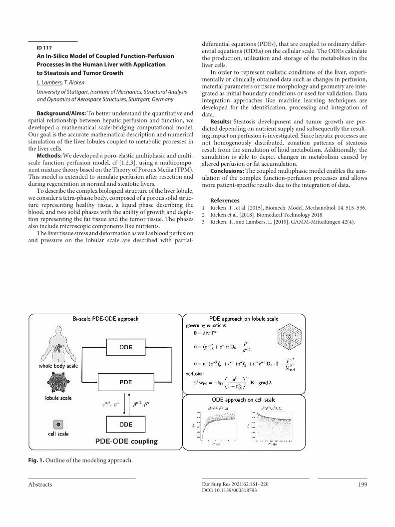
Abstracts 199Eur Surg Res 2021:62:161–220DOI: 10.1159/000518793
ID 117
An In-Silico Model of Coupled Function-Perfusion Processes in the Human Liver with Application to Steatosis and Tumor GrowthL. Lambers, T. Ricken
University of Stuttgart, Institute of Mechanics, Structural Analysis and Dynamics of Aerospace Structures, Stuttgart, Germany
Background/Aims: To better understand the quantitative and spatial relationship between hepatic perfusion and function, we developed a mathematical scale-bridging computational model. Our goal is the accurate mathematical description and numerical simulation of the liver lobules coupled to metabolic processes in the liver cells.
Methods: We developed a poro-elastic multiphasic and multi-scale function-perfusion model, cf [1,2,3], using a multicompo-nent mixture theory based on the Theory of Porous Media (TPM). This model is extended to simulate perfusion after resection and during regeneration in normal and steatotic livers.
To describe the complex biological structure of the liver lobule, we consider a tetra-phasic body, composed of a porous solid struc-ture representing healthy tissue, a liquid phase describing the blood, and two solid phases with the ability of growth and deple-tion representing the fat tissue and the tumor tissue. The phases also include microscopic components like nutrients.
The liver tissue stress and deformation as well as blood perfusion and pressure on the lobular scale are described with partial-
differential equations (PDEs), that are coupled to ordinary differ-ential equations (ODEs) on the cellular scale. The ODEs calculate the production, utilization and storage of the metabolites in the liver cells.
In order to represent realistic conditions of the liver, experi-mentally or clinically obtained data such as changes in perfusion, material parameters or tissue morphology and geometry are inte-grated as initial boundary conditions or used for validation. Data integration approaches like machine learning techniques are developed for the identification, processing and integration of data.
Results: Steatosis development and tumor growth are pre-dicted depending on nutrient supply and subsequently the result-ing impact on perfusion is investigated. Since hepatic processes are not homogenously distributed, zonation patterns of steatosis result from the simulation of lipid metabolism. Additionally, the simulation is able to depict changes in metabolism caused by altered perfusion or fat accumulation.
Conclusions: The coupled multiphasic model enables the sim-ulation of the complex function-perfusion processes and allows more patient-specific results due to the integration of data.
References1 Ricken, T., et al. [2015], Biomech. Model. Mechanobiol. 14, 515–536.2 Ricken et al. [2018], Biomedical Technology 2018.3 Ricken, T., and Lambers, L. [2019], GAMM-Mitteilungen 42(4).
Fig. 1. Outline of the modeling approach.

24th Surgical Research DaysEur Surg Res 2021:62:161–220200DOI: 10.1159/000518793
ID 118
Differential Gene Expression Analysis to Determine Liver Perfusion and Liver FunctionD. Meyer, M. Marz
Friedrich-Schiller-Universität Jena, RNA Bioinformatics and High Throughput Sequencing, Jena, Germany
Background/Aims: For the understanding of the quantitative and spatial relationship between hepatic perfusion and function in healthy, steatotic and regenerating livers using a systems surgery approach, we focus on analyzing the transcriptome differences of healthy liver, steatotic liver, venous ligated liver lobe and during regeneration.
Methods: The transcriptome gives insights into the state of the cells from that the genetic material was sequenced. For each liver condition, we specifically distinguish spatial information between perivenous and periportal liver and compare it to whole liver. For both periportal and perivenous liver cells the differences between healthy liver to steatotic liver and healthy liver to venous ligated liver lobe and non-ligated liver lobe can be analyzed. The observed genes (with a focus on non-coding RNAs and isoforms) serve as potential marker genes for regeneration capacity and potential therapeutic targets. Additionally, by analyzing differentially expressed genes pathways can be detected, that are activated dur-ing liver perfusion restriction, liver function restriction and liver regeneration.
Results: The results of this analysis can be input for the meta-bolic and multiscale computational models to limit the parameter space of models used to describe the function and perfusion restricted liver more accurately.
Conclusions: Thus RNA-Sequencing analysis contributes essentially to the understanding of the liver perfusion and func-tion.
ID 126
Cell Atlas of the Regenerating Human Liver after Portal Vein EmbolizationA. Brazovskaja1, T. Gomes2, C. Körner3, Z. He2, T. Schaffer1, J.C. Eckel3, R. Hänsel3, M. Santel2, T. Denecke3, M. Dannemann4, M. Brosch5, J. Hampe5, D. Seehofer3, G. Damm3, G. Camp6, B. Treutlein2
1Max Planck Institute for Evolutionary Anthropology, Max Planck Institute for Evolutionary Anthropology, Leipzig, Germany, 2ETH Zürich, ETH Zürich, Basel, Switzerland, 3Leipzig University, Leipzig, Germany, 4University of Tartu, Tartu, Estonia, 5Technical University Dresden, Dresden, Germany, 6Institute of Molecular and Clinical Ophthalmology Basel, Basel, Switzerland
Background/Aims: The human liver has extraordinary com-plexity on an architectural, cellular and functional level. It is responsible for diverse metabolic, detoxification and immunologic tasks, and it also has the remarkable capacity to regenerate. Feature of liver regeneration after partial liver removal is used in medicine treating different liver diseases and even though this process has
been frequently studied in animals it remains a major challenge to explore and understand how hepatic cells orchestrate liver replen-ishment in humans. Our study focuses on understanding cellular basis of human liver regeneration.
Methods: In our study we took advantage of a medical proce-dure called portal vein embolization (PVE) that triggers the growth of the liver as a model to explore the cellular basis of liver regen-eration in humans. PVE occludes portal blood flow to the diseased part of the liver that needs to be removed resulting in liver hyper-trophy in locations with increased blood supply, and atrophy of embolized segments. We applied high throughput transcriptomics on hypertrophic and atrophic liver tissue cells freshly isolated from patient-derived biopsies, and compared the processes to those in healthy liver.
Results: We detected distinct cell types within the healthy and post-PVE liver. In addition, we resolved different subpopulations within specific cell types that are linked to architecture of the liver. We found that this procedure alters heterogeneity, that is referred to as spatial zonation phenomena, within hepatocytes and liver endothelial cells. This allowed us to compare the expression differ-ences in these subpopulations of healthy and post-PVE tissues. In addition, we replicated our findings in frozen tissue suggesting that this usually more accessible resource can be used to study regeneration and for example other liver related processes.
Conclusions: Our work delivers a human liver regeneration atlas which can be used as a reference for studying the mechanisms of liver regeneration, in its gene expression and microanatomical facets.
ID 128
Prediction of Liver Regeneration Following Major ResectionK. Dehlke1, L. Krause2, S. Tyufekchieva1, A. Lemekhova1, P. Mayer4, A. Vlasov5, M. Buechler1, U. Klingmüller5, N. Mueller3, K. Hoffmann1
1Universitätsklinikum Heidelberg, Abteilung der Allgemein-, Viszeral- und Transplantationschirurgie, Heidelberg, Germany, 2University Medical Center Hamburg-Eppendorf, Institute of Medical Biometry and Epidemiology, Hamburg-Eppendorf, Germany, 3Helmholtz Center Munich, Institute of Computational Biology, München, Germany, 4Universitätsklinikum Heidelberg, Department of Diagnostic and Interventional Radiology, Heidelberg, Germany, 5German Cancer Research Center, Division of Systems Biology of Signal Transduction, Heidelberg, Germany
Background/Aims: Post-hepatectomy liver failure is charac-terized by an impaired regenerative potential and declining liver function. It is also associated with high mortality. Emerging evi-dence suggests that individual cytokine and growth factor profiles represent potent markers for prediction of postoperative recovery of liver function.
Methods: This study investigated a time-dependent cytokine and growth factor profiling dataset of 44 patients undergoing major liver resection with integration of mathematical models identifying individual pathway signatures
Results: Cytokines and growth factors show individual periop-erative dynamics, yet, common expression trajectories were

Abstracts 201Eur Surg Res 2021:62:161–220DOI: 10.1159/000518793
identified with strong correlation with PHLF, morbidity and mor-tality. A global association network was developed and validated according to the type of underlying risk-factors. By regularized regression modelling, preoperative cytokine and growth factor sig-nature were identified allowing prediction of mortality following major liver resection. Prediction of PHLF was herewith possible as early as postoperative day 1. Proliferation analysis of correspond-ing primary human hepatocytes showed association of individual regenerative potential and clinical outcome.
Conclusions: Individual liquid-biopsy-based risk profiling allows prediction of liver regeneration capacity and can be used for tailoring interventional strategies.
Virtual Liver
ID 103
Lobular Rearrangements in Liver RegenerationJ.C. Eckel1,2, R. Hänsel3,1, T. Denecke4, D. Seehofer1,2, G. Damm1,2
1University Hospital Leipzig, Department of Hepatobiliary Surgery and Visceral Transplantation, Leipzig, Germany, 2University of Leipzig, Saxon Incubator for Clinical Translation (SIKT), Leipzig, Germany, 3University of Leipzig, Institute for Medical Informatics, Statistics and Epidemiology (IMISE), Leipzig, Germany, 4University of Leipzig, Department of Diagnostic and Interventional Radiology, Leipzig, Germany
Background/Aims: The liver has the remarkable capacity to regenerate. This allows liver resections up to 70 % of the liver vol-ume. For extended liver surgeries beyond this point the portal vein embolization (PVE) is used. This technique redirects blood flow to specific liver segments, resulting in a volume increase by hyperpla-sia which functionally compensates the atrophied embolized vol-ume which is later surgically removed. Aim of our study was to investigate PVE tissue samples for mechanisms of liver regenera-tion on the lobular level.
Methods: PVE outcome was investigated by volume analysis of MRI/CT scans and derived virtual 3D reconstructed livers. Liver tissue samples of regenerated (reg) and embolized (emb) tissue were collected from PVE treated patients after partial hepatec-tomy. Tissue samples were fixed in paraformaldehyde (PFA) and embedded in paraffin. PFA fixed tissue were stained with H/E and additionally stained for liver lobule specific zonation markers (HAL, IGFPB2, CYP3A4, SAA1/2, GLUL). Detection was per-formed using DAB and IF. Hepatocyte density was investigated using IF staining for GLUL, HAL and Hoechst by bioinformatics analysis using a Kernel density estimation (KDE) based algorithm.
Results: Virtual 3D reconstructed livers of PVE treated patients revealed that all 3 investigated livers have undergone a volume increase. The degree of volume increase was not related to the application time ranging from 2.5 to 8 weeks. H/E stainings of embolized and regenerated tissue samples showed a decrease in cell density around the central vein and an increase in the midzone within the portal-central axis of the lobules of regenerated tissue.
Quantification of cell density confirmed these results and showed a decline of the intensity of these effects in dependence of the PVE time span. In short PVE time spans regeneration was dominated by hepatocyte proliferation (IGFBP2) while vascular rearrange-ment (HAL and GLUL) was predominant in long PVE time spans.
Conclusions: Taken together our data show that tissue samples derived from PVE treated patients are suitable for investigation of lobular liver regeneration. Depending on the application time of the PVE the tissue displays different windows of liver regeneration ranging from hepatocyte proliferation to vascular rearrangement.
ID 111
Multiscale Modeling of Liver Regeneration and Steatosis in 3DA. Friebel1, L.A. D‘Alessandro3, G. Damm5, J.G. Hengstler2, U. Klingmueller3, D. Drasdo4, S. Hoehme1
1Universität Leipzig, IZBI / SIKT, Leipzig, Germany, 2Leibniz-Institut für Arbeitsforschung (IfaDo), Toxikologie / Systemtoxikologie, Dortmund, Germany, 3Deutsches Krebsforschungszentrum (DKFZ), Heidelberg, Germany, 4Institut national de recherche en informatique et en automatique (INRIA), Paris, France, 5Universitätsklinikum Leipzig, SIKT, Leipzig, Germany
Background/Aims: During the last years, modeling of different physiological and pathological aspects of the liver advanced sig-nificantly with the development of increasingly realistic computa-tional models on molecular, cellular, tissue and whole organ scale. Nevertheless, model driven liver research is still hampered by a lack of techniques that allow robust integration of these different scales into unifying frameworks. We here present a novel multi-scale spatio-temporal 3D model of liver tissue that is based on in-vivo 3D imaging and that may serve as such unifying framework.
Methods: We use this model to study liver regeneration after tissue loss by intoxication and surgical intervention, and the onset of steatosis. All three processes involve complex intracellular and tissue scale processes, which interplay with tissue mechanics.
Fig. 1. Spatio-temporal 3D model of a liver lobule. Visualisation of spatio-temporal 3D model of a liver lobule depicting an early state in the liver regeneration process 20h after intoxication with CCl4.

24th Surgical Research DaysEur Surg Res 2021:62:161–220202DOI: 10.1159/000518793
Results: In order to capture these processes on their respective scales, the presented multiscale modelling framework integrates sub-models at all relevant scales from intracellular signalling to body level. It thereby allows predictions on a wide range of possible scenarios and helps to identify on one hand particularly informa-tive experiments permitting to distinguish between alternative mechanisms, on the other hand impossible scenarios that should not be pursued experimentally. Thereby, model predictions can guide the experimental strategy. The presented multiscale tissue model is able to simultaneously reproduce experimental observa-tions including for example liver regeneration kinetics on the tis-sue scale.
Conclusions: The presented work is an example for how the tight systems-biological integration of experimentation and mod-elling, both covering multiple scales, can facilitate understanding of complex multi-scale processes as liver regeneration and the onset of liver steatosis.
Career Planning and Mentoring
ID 124
Soft Liver Modell with a Hollow Biliary SystemX. Tan, B. Mothes, U. Schweizer, A. Königsrainer, D. Wichmann
Universitätsklinik Tübingen, Klinik für Allgemeine, Viszeral und Transplantations-Chirurgie, Tübingen, Germany
Background/Aims: Hepatobiliary interventional procedures are regarded as difficult minimally-invasive procedures that require both technical experience and anatomic skills of physi-cians. Traditionally, these procedures were mainly taught by an
apprenticeship approach that trainees acquire competency from the practices on real patients undergoing the supervision of experts. The insufficient clinical volume and risking examination impose the development of simulation-based training. The fabrication of a soft, high-fidelity, and durable liver organ phantom with detailed morphology remains a challenge.
Methods: A realistic liver phantom that possesses a complex hollow biliary system was established based on advanced 3D print-ing and soft materials molding technologies. The fabricated liver phantom was validated via multiply modality medical imaging examinations, including computer tomography (CT), ultrasound, and endoscopy. The transhepatic puncture procedure was simu-lated in the liver phantom. A real-time and precise assessment sys-tem was established to improve the efficacy of training and competency evaluations.
Results: A soft liver phantom with a hollow biliary system was manufactured. The phantom is realistic and anatomically accu-rate, which allows for multi-modality medical imaging, including CT, ultrasound, and endoscopy. The CT results indicate that the liver model replicates the detailed anatomy of real human livers and the corresponding biliary system, with a spatial root mean square error (RMSE) of 0.9 ± 0.2 mm and 1.7 ± 0.7 mm for the liver outer shape and the biliary duct, respectively. The endoscopic appearance highly resembles the mucosal surface and dimension of biliary tracts. The sonographic signals also greatly mimic that of the real live organ. A transhepatic puncture procedure was suc-cessfully performed in the liver phantom. Furthermore, an electric sensing system was carried out for the quantitative localization of the transhepatic puncturing needle in real-time.
Conclusions: The fabrication approach herein is high resolu-tion and reproducible. It provides a possibility of massive produc-tion in the industry. The liver organ phantoms are applicable in the training of transhepatic interventional procedures, including ERCP, PTC/PTCD, POC, EUS-BD. The needle tracking system offers a robust and general method to assess the performance of physicians during puncture procedures.
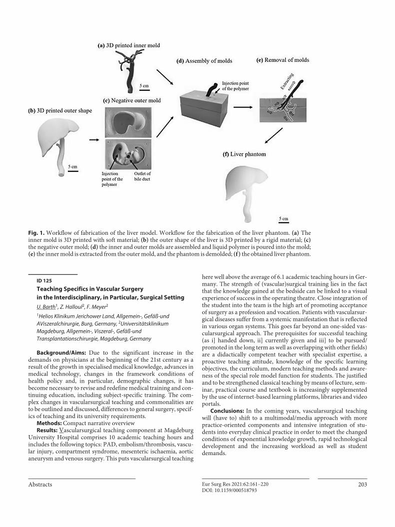
Abstracts 203Eur Surg Res 2021:62:161–220DOI: 10.1159/000518793
ID 125
Teaching Specifics in Vascular Surgery in the Interdisciplinary, in Particular, Surgical SettingU. Barth1, Z. Halloul2, F. Meyer2
1Helios Klinikum Jerichower Land, Allgemein-, Gefäß-und AViszeralchirurgie, Burg, Germany, 2Universitätsklinikum Magdeburg, Allgemein-, Viszeral-, Gefäß-und Transplantationschirurgie, Magdeburg, Germany
Background/Aims: Due to the significant increase in the demands on physicians at the beginning of the 21st century as a result of the growth in specialised medical knowledge, advances in medical technology, changes in the framework conditions of health policy and, in particular, demographic changes, it has become necessary to revise and redefine medical training and con-tinuing education, including subject-specific training. The com-plex changes in vascularsurgical teaching and commonalities are to be outlined and discussed, differences to general surgery, specif-ics of teaching and its university requirements.
Methods: Compact narrative overviewResults: Vascularsurgical teaching component at Magdeburg
University Hospital comprises 10 academic teaching hours and includes the following topics: PAD, embolism/thrombosis, vascu-lar injury, compartment syndrome, mesenteric ischaemia, aortic aneurysm and venous surgery. This puts vascularsurgical teaching
here well above the average of 6.1 academic teaching hours in Ger-many. The strength of (vascular)surgical training lies in the fact that the knowledge gained at the bedside can be linked to a visual experience of success in the operating theatre. Close integration of the student into the team is the high art of promoting acceptance of surgery as a profession and vocation. Patients with vascularsur-gical diseases suffer from a systemic manifestation that is reflected in various organ systems. This goes far beyond an one-sided vas-cularsurgical approach. The prerequisites for successful teaching (as i] handed down, ii] currently given and iii] to be pursued/ promoted in the long term as well as overlapping with other fields) are a didactically competent teacher with specialist expertise, a proactive teaching attitude, knowledge of the specific learning objectives, the curriculum, modern teaching methods and aware-ness of the special role model function for students. The justified and to be strengthened classical teaching by means of lecture, sem-inar, practical course and textbook is increasingly supplemented by the use of internet-based learning platforms, libraries and video portals.
Conclusions: In the coming years, vascularsurgical teaching will (have to) shift to a multimodal/media approach with more practice-oriented components and intensive integration of stu-dents into everyday clinical practice in order to meet the changed conditions of exponential knowledge growth, rapid technological development and the increasing workload as well as student demands.
Fig. 1. Workflow of fabrication of the liver model. Workflow for the fabrication of the liver phantom. (a) The inner mold is 3D printed with soft material; (b) the outer shape of the liver is 3D printed by a rigid material; (c) the negative outer mold; (d) the inner and outer molds are assembled and liquid polymer is poured into the mold; (e) the inner mold is extracted from the outer mold, and the phantom is demolded; (f ) the obtained liver phantom.

24th Surgical Research DaysEur Surg Res 2021:62:161–220204DOI: 10.1159/000518793
Free Topics
ID 3
Bcl-2 Inhibition Mitigates Pouchitis in a Preclinical Rat ModelS. Yuan1, J.M. Gebhardt1, B. Köhler2, M. Schneider1, J.M. Harnoss1
1Universitätsklinikum Heidelberg, Klinik für Allgemein-, Viszeral- und Transplantationschirurgie, Heidelberg, Germany, 2Nationales Centrum für Tumorerkrankungen, Abteilung Medizinische Onkologie, Heidelberg, Germany
Background/Aims: Pouchitis is a frequent and severe compli-cation after proctocolectomy with ileal pouch-anal anastomosis (IPAA). The anti-apoptotic protein B-cell lymphoma 2 (Bcl-2) is highly expressed in inflammatory cells and its inhibition has been demonstrated to have therapeutic potential in various inflamma-tory conditions. However, the impact of Bcl-2 inhibition on pou-chitis has not been investigated so far. Here, we aimed to evaluate Bcl-2 as a putative therapeutic target in pouchitis.
Methods: In a preclinical rat model of IPAA, pouchitis was induced by oral administration of dextran sulfate sodium (DSS) and animals were treated with ABT-199, a specific Bcl-2 antagonist or vehicle control. A modified disease activity index (mDAI, com-prising body weight change, stool consistency, and hematochezia) was recorded. Pouch and ileum samples were collected, the expres-sion of Bcl-2 expression assessed by qPCR and immunoblotting, the number of CD45-positive leucocytes and CD3-positive lym-phocytes evaluated by immunohistochemistry, and the levels of inflammatory cytokines investigated by qPCR. Cell apoptosis was assessed by immunofluorescence staining of Cleaved Caspase-3.
Results: In inflamed pouch tissue, Bcl-2 expression was sig-nificantly increased as compared to healthy mucosa. ABT-199 significantly mitigated body weight loss and hematochezia and improved the stool consistency, thus ameliorating the mDAI. ABT-199 attenuated mucosal damage, leucocyte infiltration and pro-inflammatory cytokine levels and decreased the number of inflammatory cells in the peripheral blood. Mechanistically, ABT-199 treatment impaired apoptosis of T lymphocytes.
Conclusions: Collectively, these results suggest that Bcl-2 is a putative novel therapeutic target in the treatment of pouchitis.
ID 9
Tumor Indicative Markers from Serum Exosomes of Esophageal Adenocarcinoma and Oral Squamous Cell Carcinoma PatientsU. Warnecke-Eberz1, M. Cronauer1, M.-P. Lentzen2, T. Loers1, J. Buller2, S.-H. Chon1, P. Plum1, C.J. Bruns1, E. Bollschweiler1, M. Zirk2
1Uniklinik Köln, Allgemein-, Viszeral-, Tumor- und Transplantationschirurgie, Köln, Germany, 2Uniklinik Köln, Mund-, Kiefer- und plastische Chirurgie, Köln, Germany
Background/Aims: The incidence of adenocarcinoma of the esophagous (EAC) is dramatically rising and diagnosed at advanced stages. The incidence of oral squamous cell carcinoma (OSCC) increases, especially in younger patients.
Exosomes are released by living cells into body fluids allowing intercellular communication. Formation and cargo of exosomes excreted by tumor cells have a major impact on carcinogenesis. Exosomes transfer their “oncogenic cargo” to non-transformed cells, influencing oncogenic field effects. MicroRNAs (miRs), sta-ble non-coding RNAs, as part of exosomal cargo regulate protein expression by targeting mRNAs.
We aim to identify tumor indicating miRs within the exosomal cargo of OSCC and EAC patients, as well as exosomal surface markers for earlier non-invasive diagnosis.
Methods: We profiled miR cargos of serum exosomes of eigh-teen EAC and sixteen OSCC patients compared to sixteen healthy controls by TaqMan miR-arrays. Serum exosomes have been visu-alized for quality, quantity and size by Nanoparticle Tracking Analysis. For quantification of exosomal surface proteins we have established a Trific Sandwich ELISA.
Results: Profiling of 754 miRs identified miR-99b and snoU6 as tumor markers discriminating between EAC patients and healthy individuals. miR-409-3p andmiR-28-3p have been verified as exosomal markers for OSCC, by separate verification cohorts.
A Time-resolved immune fluorescence assay (Trific) has been established to quantify proteins on the surface of exosomes, like EMMPRIN (extracellular matrix metalloproteinase inducer).
Conclusions: The “exosomal miRs” have to be applied to a pro-spective cohort to finally validate their diagnostic impact for liquid biopsy based detection of the tumors in clinical practice. By sensi-tive Trific assay further tumor indicative protein candidates have to be quantified on exosomal surfaces comparing tumor patients to healthy volunteers, intending to separate exosomes of different origin, and to apply exosomal surface markers for diagnosis, as well.

Abstracts 205Eur Surg Res 2021:62:161–220DOI: 10.1159/000518793
ID 11
Regulation of Peritoneal B cell Trafficking by Sphingosine-1-Phosphate Receptor Type 4 (S1P4)-Mediated SignalingJ. Riese, A. Kleinwort, S. Kersting, T. Schulze
Universitätsmedizin Greifswald, Chirurgisches Forschungslabor, Greifswald, Germany
Background/Aims: Peritoneal B cells (pBC) mediate both pro-tective and detrimental effects during microbial challenge of the abdomen. Reduced peritoneal B1 B cell numbers are associated with higher mortality and morbidity in abdominal sepsis models. The mechanisms regulating migration and cytokine reproduction of pBC as potential therapeutic targets are poorly understood. Recently, we described a substantial reduction of peritoneal B1 B cell populations in S1P4-deficient (s1pr4
–/–) mice. The impact of S1P4-mediated signaling on pBC biology has not been described yet. In this study, S1P4 interaction with chemokine-mediated sig-naling was analyzed in vitro, and the impact of S1P4 expression on pBC migration was characterized in vivo.
Methods: Transwell migration assays were used to assess the interaction of S1P4- and CXCR4/CXCR5 mediated signaling in pBC in vitro. Adoptive cell transfer by the intraperitoneal and intravenous route of wt and s1pr4
–/– peritoneal cells was performed to analyze the impact of S1P4-mediated signaling on pBC traffick-ing, cytokine, and antibody production in vivo.
Results: S1P4 synergistically enhances migration of pBC to combined gradients of S1P with CXCL12 and CXCL13 in vitro. S1pr4
–/– B1a B cells showed both reduced immigration into and decreased emigration from the peritoneal cavity. Similarly, S1P4 deficiency affected pBC tropism to various secondary lymphoid organs, IL-10 levels, and IgM synthesis. In vitro proliferation or viability was similar in wt and s1pr4
–/– animals.Conclusions: These findings suggest that S1P4 is a vital receptor
modulating chemokine-mediated trafficking of peritoneal B cells. Regulation of S1P4-mediated signaling can modify pBC trafficking and subsequent immunoglobulin production. Thus, S1P4 constitutes a potential target to modulate B cell function in inflam-matory pathologies.
ID 16
Ischemic Musculocutaneous Flap Tissue is Protected from Necrosis by Perioperative Intermittent FastingA. Weinzierl1, Y. Harder2,3, M.D. Menger1, M.W. Laschke1
1Saarland University, Institute for Clinical & Experimental Surgery, Homburg, Germany, 2Ospedale Regionale di Lugano, Ente Ospedaliero Cantonale (EOC), Department of Plastic, Reconstructive and Aesthetic Surgery, Lugano, Switzerland, 3Università della Svizzera Italiana, Faculty of Biomedical Sciences, Lugano, Switzerland
Background/Aims: Dietary restriction, such as intermittent fasting (IF), has previously been shown to protect various tissues from ischemia-induced necrosis. Based on this finding, we evalu-ated in the present study the tissue-protective effects of IF in a murine musculocutaneous flap model.
Methods: C57BL/6N mice were randomly assigned to an IF group (n = 8) and a control group with unrestricted access to stan-dard diet (n = 8). IF animals were put on a perioperative feeding schedule with 8 hours unrestricted access to standard diet per day starting 7 days before flap elevation up to 3 days after surgery. Ran-dom pattern musculocutaneous flaps were elevated and mounted into a dorsal skinfold chamber, which provided direct microscopic access to the flap tissue through an observation window. Intravital fluorescence microscopy was performed on days 1, 3, 5, 7 and 10 after surgery for the quantitative assessment of flap necrosis, nutri-tive perfusion and angiogenesis. After the in vivo observation period, the flaps were harvested for additional histological and immunohistochemical analyses.
Results: We found that the IF group exhibited a significantly lower rate of flap tissue necrosis (24 ± 7%) on day 10 when com-pared to untreated controls (47 ± 7%; p<0.05). This was associated with a higher functional capillary density and more newly formed microvessels within the flap tissue. Immunohistochemical detec-tion of different inflammatory cell subtypes revealed a reduced number of invading myeloperoxidase (MPO)+ neutrophilic gran-ulocytes in the musculocutaneous tissue of IF-treated animals. The suppression of neutrophilic granulocyte invasion was particularly pronounced in the transition zone from vital to necrotic flap tissue, where we detected a significantly lower number of 125 ± 37 MPO+ cells/high power field (HPF) in IF-treated mice when compared to controls (254 ± 35 MPO+ cells/HPF).
Conclusions: The present study demonstrates that short-term perioperative IF protects ischemic flap tissue from necrosis by maintaining nutritive tissue perfusion and suppressing ischemia-induced inflammation. In contrast to other conditioning strate-gies, IF may bear the major advantage that it can be easily implemented into standard clinical procedures without causing additional costs or inducing severe side effects.

24th Surgical Research DaysEur Surg Res 2021:62:161–220206DOI: 10.1159/000518793
ID 21
Mapping of Perigastric Lymphatic Network using Indocyanine Green Fluorescence Imaging and Tissue Marking Dye in Clinically Advanced Gastric CancerF. Berlth1,2, J.-H. Park2, J.-H. Choi2, S. Park2, S.-H. Kong2, D.J. Park2, H.-J. Lee2, Y.J. Kwak3, W.H. Kim3, H.-K. Yang2
1Universitätsmedizin Mainz, Allgemein-, Viszeral- und Tranplantationschirurgie, Mainz, Germany, 2Seoul National University Hospital, Department of Surgery, Seoul, South Korea, 3Seoul National University Hospital, Department of Pathology, Seoul, South Korea
Background/Aims: This study aimed to explore whether indo-cyanine green (ICG) fluorescence imaging guided lymph node
(LN) dissection can be performed in advanced gastric cancer (AGC). The investigation included single lymph node evalua-tion and retracking to its original localization.
Methods: Patients undergoing open gastrectomy for AGC in 2018 and 2019 in Seoul National University Hospital, Seoul, Korea, were enrolled. ICG was serially injected into the subserosal layer along the greater and lesser curvature. Resected specimens were examined under near-infrared camera on back-table. Lym-phatic image was recorded and LNs were named separately, excised, and tattoed with different colors of tissue marking dyes for exact backtracing of location after pathological examination. All ICG stained and non-stained LNs were reconstructed topographically including metastatic status.
Results: Eleven patients underwent distal (n=7) or total (n=4) gastrectomy were included and LN metastasis presented in 8 patients. Overall 687 LN were examined. ICG fluorescence did not cover all of the LN metastasis. The average number of ICG-stained and ICG-unstained LNs was 23.6±12.3 (37.8%), and 38.8±17.1
Fig. 1. Perigastric lymphatic networks visualized by ICG. Included are information about number and metastatic status of the patho-logically examined lymph nodes per station.

Abstracts 207Eur Surg Res 2021:62:161–220DOI: 10.1159/000518793
(62.2%), respectively. The average number of ICG-stained meta-static (+) LNs was 3.8±1.8 among 9.4±3.8 metastatic (+) LNs. In 8 cases with LN metastasis, metastatic LNs were detected by ICG staining overall in 40% (11.1-75% per case). Overall 28 separated LN station with metastatic LN were found in all 8 pN+ cases. Of those 23 (82.1%) LN stations showed any LN specific ICG staining and in 7 LN stations (25%) actual metastatic LN were stained (Table 2). The 5 missed metastatic LN stations not being ICG pos-itive by any means were combined in two cases, so in 6 out of 8 cases (75%) all metastatic LN stations showed LN-specific ICG sig-nal, if also ICG-stained non-metastatic LNs are considered.
Conclusions: This study demonstrates that ICG fluorescent imaging and tissue marking dye are useful tools for visualiz-ing acomplex perigastric lymphatic network. Selective LN dissec-tion solely relying on ICG imaging in AGC would still not be accurate enough as of its limited staining of (metastatic) LNs. Some metastatic lymphatic direction might however be suggested by ICG fluorescence.
ID 29
Previous Intraperitoneal Manipulation Results in Higher Mortality and Altered Local Immune Response in Subsequent Secondary Peritonitis in MiceJ. Menz1, L. Hundt1, T. Schulze1, K. Schmoeckel2, G. Domanska2, P. Menges1
1Universität Greifswald, Klinik und Poliklinik für Allgemeine Chirurgie, Viszeral-, Thorax- und Gefäßchirurgie, Greifswald, Germany, 2Universität Greifswald, Institut für Immunologie und Transfusionsmedizin, Greifswald, Germany
Background/Aims: Postoperative peritonitis is characterized by a more severe clinical course than other forms of secondary peritonitis. The pathophysiological mechanisms behind this phe-nomenon are incompletely understood. This study used an innovative model to investigate these mechanisms, combining the models of murine Colon Ascendens Stent Peritonitis (CASP) and Surgically induced Immune Dysfunction (SID). Moreover, the impact of the N. vagus mediated anti-inflammatory reflex on sep-sis mortality was characterized.
Methods: SID and consecutive CASP were performed in C57BL/6N mice with an interval of 3 days. Subdiaphragmatic vagotomy was performed six days before SID. The immune status was assessed by FACS analysis and measurement of cytokines. Local intestinal inflammatory changes were characterized by immunohistochemistry.
Results: Mortality was increased in CASP animals previously subjected to SID. Subclinical bacteremia occurred after SID, and an immunosuppressive milieu occurred secondary to SID just before the induction of CASP. Previous SID modified the histo-logical pattern of intestinal inflammation induced by CASP.
Subdiaphragmatic vagotomy had no influence on sepsis mortality in our model of postoperative peritonitis.
Conclusions: Surgery-induced inflammation of the small intestine and the peritoneal cavity with bacterial translocation may lead to a more severe course of secondary peritonitis.
ID 38
The Effect of Electrical Fields on HaCaT Cells: A Promising Prospect for Wound HealingC. Lu1,3, C. Prahm1, U. Cebron1, S. Ehnert2, J. Kolbenschlag1
1University of Tuebingen, Department of Hand-, Plastic, Reconstructive and Burn Surgery, BG Trauma Center Tübingen, Tuebingen, Germany, 2University of Tuebingen, Siegfried Weller Research Institute, Department of Trauma and Reconstructive Surgery, BG Trauma Center Tübingen, Tuebingen, Germany, 3The Affiliated Hospital of Qingdao University, Hand and Foot Surgery, Qingdao, China
Background/Aims: There is growing evidence that cell behav-ior can be significantly influenced by external electric fields (EFs). Some of the behavior is thought to be closely related to wound healing. This study aimed at investigating the effect of EFs (200 mV/mm) on viability, migration, and growth factor secretion of HaCaT cells (human keratinocyte cell line), as these are vital steps of wound healing.
Methods: We built a set-up that can transmit specific electrical signals and maintain a stable cell culture environment. EFs of 200 mV/mm was introduced to the HaCaT cells. Scratch-assays were performed to observe cell migration. Cell viability was evaluated by mitochondrial activity, total protein content, and total DNA content on days 1, 4, and 7. Healing–associated cytokines were evaluated via the RayBio® human growth factor array, and Western blot was applied to investigate signaling pathway alterations.
Results: When compared to the control group, the migration speed of HaCaT cells that received EFs significantly increased; after 48 hours, the EFs group had a 2.2-fold increase in migration (P < 0.001) compared to the control. After 7 days, the changes in cell viability (mitochondrial activity, total protein content, and total DNA content) was significantly increase (P < 0.05). Human growth factor array revealed that GM-CSF was the most abundant (+197.74 % compared to ctrl) growth factor secreted by HaCaT cells after EFs exposures. The signals for phospho-ERK1/2 showed a significant (approx. +360 %) increase following EFs exposure with a signal peak at 90 min.
Conclusions: The results demonstrate that EFs (200 mV/mm) have a positive effect on HaCaT cells viability, migration, and cyto-kine secretion, three processes crucial for wound healing. These effects may be mediated by activation of the ERK1/2 signaling pathway. Our findings suggest EFs have a potentially beneficial therapeutic effect on wound healing in vitro and could play an essential role in treating complex wounds in clinic.

24th Surgical Research DaysEur Surg Res 2021:62:161–220208DOI: 10.1159/000518793
ID 40
Disruption of Prolyl Hydroxylase Containing Enzyme 1 Mitigates Colitis via Increased Goblet Cell ActivityM. Salfenmoser1, P. Radhakrishnan1, K.B. Kennel1, J. Burmeister1, A. Ritter1, N.M. Wirsik1, C.T. Taylor2, M. Schneider1, J.M. Harnoss1
1Universitätsklinikum Heidelberg, Klinik für Allgemein-, Viszeral- und Transplantationschirurgie, Heidelberg, Germany, 2University College Dublin, Conway Institute of Biomolecular and Biomedical Research, Dublin, Ireland
Background/Aims: Inflammatory bowel disease (IBD) is a common chronic intestinal disorder characterized by loss of the intestinal epithelial barrier function (IBF) and subsequent mucosal hypoxia. This leads to inactivation of hypoxia-inducible transcrip-tion factor-prolyl hydroxylase containing enzymes (PHD1, PHD2, and PHD3), which induces the expression of barrier protective genes. Several studies have validated PHD1 as a promising thera-peutic target in IBD. However, the biological importance of PHD1 for intestinal goblet cells, the key mediators of intestinal homeo-stasis, and their activity in IBD remains unknown.
Methods: PHD1-deficient (PHD1-/-) and wild type (WT) mice were subjected to dextran sulfate sodium (DSS) and the impact on colitis disease activity was studied. Subsequently, the number of intestinal goblet cells was analyzed by PAS-staining and their activity assessed by RT-qPCR and ELISA. In vitro, goblet cell-like LS174T cells were treated with tumor necrosis factor-alpha and interferon-gamma (TNFa/IFNg) and the PHD-inhibitor dimethy-loxalylglycine (DMOG) and the impact on cell viability and the expression and secretion of the main intestinal gel-forming mucins, MUC2 and MUC3 was analyzed.
Results: Upon DSS treatment, PHD1-/- mice had a significantly decreased colitis disease activity compared to WT mice. Intrigu-ingly, in the early phase of colitis mRNA expression of MUC2 and MUC3 was significantly elevated in goblet cells of PHD1-/- mice compared to WT mice, indicating increased goblet cell activity. Moreover, while in untreated control animals the number of gob-let cells was comparable in PHD1-/- and WT mice, their abundance was profoundly increased in PHD1-/- mice during the late phase of DSS-induced colitis. In vitro, PHD inhibition significantly increased viability of LS174T cells treated with TNFa/IFNg.
Conclusions: Collectively, these results suggest a novel mecha-nism by which PHD1 regulates the number and secretory function of intestinal goblet cells in colitis and exerts protective effects on the intestinal mucosal barrier.
ID 46
Models of Postoperatively Altered Anatomy for Training of Advanced Flexible EndoscopyK. Koch, B. Mothes, U. Schweizer, A. Königsrainer, D. Wichmann
Universitätsklinik Tübingen, Klinik für Allgemeine, Viszeral- und Transplantationschirurgie, Tübingen, Germany
Background/Aims: Flexible endoscopy offers the possibility of simultaneous diagnosis and therapy, especially in patients after complex previous visceral surgery. For the treating endoscopist,
anatomical knowledge, knowledge of resection procedures in the past and handling of advanced, endoscopic techniques play an important role. In the training of endoscopists, specific training on patients with postoperative altered anatomy is not established. Animal-free training models can close the gap in the training of interdisciplinary endoscopists.
Methods: Patient analogue data were used to create and work on digital 3D models. The models were printed with a plastic filament in a 3D printer and machined with epoxy resin. An immersion casting technique was then used. Three models with postoperatively altered anatomy were developed and realised. Evaluation of the models was questionnaire-based by experts and novices in the field of endoscopy.
Results: Three animal material-free models were created.A) A model corresponding to a “short limb” Roux-Y recon-
struction after subtotal gastric resection with the possibility of ERCP.
B) A model corresponding to a Billroth II situation jejunal anastomosis.
C) A model corresponding to a bariatric “long limb” Roux-Y gastric bypass.
The models were evaluated partly by experts in interventional endoscopy and partly by novices in endoscopy for realism and training effects. All models were rated as suitable for training.
Conclusions: A technical implementation of three models with frequently encountered postoperative altered anatomy was feasi-ble. For the difficult ERCP in bariatric Roux-Y gastric bypass with alternative access route, model C is the first available training model. The relevance of the models for the training of interven-tional endoscopists is present. Further evaluation is needed to con-firm the suitability of the models presented.
Fig. 1. Endoscopic view on the artificial gastrojejunostomy

Abstracts 209Eur Surg Res 2021:62:161–220DOI: 10.1159/000518793
ID 48
Influencing Factors and Outcome of Conventional Vascularsurgical and Image-Guided Treatment of Suture AneurysmsP. Romancik1, F. Meyer2, A. Hribaschek1, U. Redlich3, Z. Halloul2, J. Tautenhahn1
1Klinikum Magdeburg, Klinik für Gefäßchirurgie, Magdeburg, Germany, 2Universitätsklinik für Allgemein-, Viszeral-, Gefäß- und Transplantationschirurgie, Fachbereich Gefäßchirurgie, Magdeburg, Germany, 3Klinikum Magdeburg, Institut für diagnostische und interventionelle Radiologie, Magdeburg, Germany
Background/Aims: Suture aneurysms (SA) have increasingly become a relevant focus of secondary care due to the favorable long-term outcome and survival. The aim of the study was to detect the occurrence of SA in retrospective case series and based on periinterventional/-operative management experiences.
Methods: 86 consecutive patients with 106 SA were docu-mented as part of this retrospective (1.4.2010 - 1.9.2016) unicenter observational study who either had undergone image-guided or vascularsurgical treatment. The cases were characterized with patient- (age, sex ratio), finding-characteristics (SA site/size), image-guided interventional/vascularsurgical approach and out-come aspects (patency rate, SA recurrency, morbidity, mortality).
Results: - The majority of SA occurred after implantation of an aorto-bifemoral prosthesis (43.4 %) and at the groin (81.1 %).
– The mean time interval to SA manifestation in the groin was 55.4 months and 51.6 months to manifestation of recurrent SA (p = 0.683).
– Though Thromboendarterectomy (TEA) has been reported as risk factor for SA generation in the literature, there was no significant difference for the frequency of SA occurrence comparing former vascularsurgical interventions with versus without former TEA (p = 0.325).
– The mean diameter of the inguinal SA was 46.4 mm and of recurrent SA at the groin 54.5 mm (p = 0.34). This might be a hint of incompliant patients or of an insufficient postinterventional/-operative follow-up care.
– After treatment of SA, there was no complication in 56.6 % of cases (n = 60). All subjects who underwent image-guided intervention showed no complication.
– In the spectrum of postoperative complications, the most fre-quent problems were lymphocele with no need for surgical intervention (20.8 %), hematoma with need of surgical inter-vention (8.5 %) and postoperative explantation of alloplastic materials (3.8 %). The 30-days mortality in all SA was 2.8 %.- Emergency operations for SA showed a significantly higher probability of a postoperative complication (p = 0.038).
Conclusions: In SA treatment, image-guided techniques may become more important due to a lower complication rate and lim-ited invasiveness. However, open reconstruction remains the stan-dard procedure for SA at the groin (implantation of an interponate). Postoperatively, patients need to be notified on the importance of a life-long vascularsurgical follow-up.
ID 51
Manifestation of Acute Appendicitis as Known but Paradox Visceral Side Effect of Ulcerative Colitis Anti-Inflammatory Therapy with Januskinase-Inhibitor Tofacitnib (XeljanzTM)F. Weber1, M. Dölling1, T. Oksentyuk Polyakova1, K.I. Eger2, J. Rabczak3, R.S. Croner1, F. Meyer1
1University Hospital at Magdeburg, Dept. of General, Abdominal, Vascular and Transplant Surgery, Magdeburg, Germany, 2University Hospital at Magdeburg, Institute of Pathology, Magdeburg, Germany, 3University Hospital at Magdeburg, Dept. of Radiology and Nuclear Medicine, Magdeburg, Germany
Background/Aims: The etiopathogenesis of accompanying inflammatory phenomena and consequences of immunomodula-tion constitute a challenging and innovative field in the medical treatment of patients with autoimmune diseases. Because of i) clin-ical management experiences derived from this challenging clinical case and ii) selective references from the scientific medical litera-ture, an unusual counterfactual scientific case report is illustrated. A patient diagnosed with ulcerative colitis under Januskinase (JAK)-Inhibitor therapy developed acute appendicitis as unusual complication or visceral side effect of immunosuppressive/anti-inflammatory therapy.
Methods: Scientific case reportResults: Current anamnesis: A 52-year-old male had spas-
modic pain in the right lower belly for two days. No fever, no bowel movement changes, no vomiting.
Medical history: steroid-resistant ulcerative colitis under immunosuppressive therapy (10 month Adalimumab, 9 month Vendolizumab, for 6 month Tofacitinib), fructose intolerance, no abdominal surgery, medication: XeljanzTM (Tofacitinib, 5 mg 2x1; JAK–Inhibitor); MutaflorTM (1x1).
Clinical findings: Pressure pain in the right lower belly with local muscular defense (Mc-Burney/Lanz+), no peritonism, Psoas-sign+.
Diagnostic measures: - Labs: standard value of leucocytes, CrP: 25 mg/l.
– Transabdominal ultrasound: hypertrophic appendix ver-miformis with target-phenomenon and occlusive fluid.
Decision-making: Indication for laparoscopic exploration.Treatment: Urgent laparoscopic appendectomy under periop-
erative single-shot antibiotic with Unacid, with additional lavage and placement of local drainage. In surgery the diagnose of appen-dicitis was validated.
Clinical course: Uncomplicated postoperative phase. Sufficient analgesia, removal of local drainage on the 2nd and dismissal on the 4th postoperative day.
Histopathology: Ulcero-phlegmonoesis, acute-purulent appen-dicitis with fibrinous-purulent mesenteriolitis.
Further course of action: Immunosuppressive therapy was con-tinued.
Conclusions: Based on the paradoxon that an acute inflamma-tory disease (acute appendicitis) developed in case of an ongoing immunosuppressive/anti-inflammatory treatment using a JAK-Inhibitor for ulcerative colitis, such uncommon clinical case is reported even though this side effect is formally known. This might

24th Surgical Research DaysEur Surg Res 2021:62:161–220210DOI: 10.1159/000518793
be the manifestation of i) animmunmodulatorily reduced or at least altered mucosal defence including an elevated risk for oppor-tunistic infections as specific visceral “side effect“ of the JAK- Inhibitor and/or as a consequence of ii) an induced alternative inflammatory mechanism/proinflammatory signal transduction as well as an intestinal drainage defect in the segment of right colic artery with consecutive collection of necrotic cells and activation of inflammatory mediators.
ID 52
Spectrum of Diagnoses and Surgical Interventions as well as Outcome in a Representative Case Series on Rare Peritoneal Tumor Lesions (PTLs) in Abdominal Surgery at a Tertiary CenterF. Weber1, M. Dölling1, T. Oksentyuk Polyakova1, C. March2, K.I. Eger3, R.S. Croner1, F. Meyer1
1University Hospital at Magdeburg, Dept. of General, Abdominal, Vascular and Transplant Surgery, Magdeburg, Germany, 2University Hospital at Magdeburg, Dept. of Radiology and Nuclear Medicine, Magdeburg, Germany, 3University Hospital at Magdeburg, Institute of Pathology, Magdeburg, Germany
Background/Aims: Exemplary demonstration of 8 cases with rare PTLs and their histopathological results in abdominal surgery emphasizing biological diversity.
Methods: Representative scientific case reportsResults: 1) Diverticulum of the jejunum with pancreatic meta-
plasia: Intraoperative incidental finding of a jejunal PTL during gynecological surgery of endometrium carcinoma lead to segmen-tal resection and reconstruction with side-to-side jejunojejunos-tomy.
2) Twisted “appendix epiploicae” (with lipofibrosis and necro-sis of the appendix): Patient with progressive abdominal pain in the right lower abdomen was diagnosed with acute appendicitis followed by laparoscopic appendectomy with partial resection of the greater omentum.
3) Acute epiploic appendagitis: In laparoscopic hernioplasty of incarcerated trocar hernia, a suspicious inflamed fatty tumor-like tissue was resected from the descending colon.
4) “Appendagitis epiploicae” of the descending/sigmoid colon: A 53-year-old patient with pain in the left lower abdomen dis-played an oval paracolic mass in abdominal CT scan leading to the consequence of a conservative therapeutic approach.
5) Primary peritoneal cancer: During explorative laparotomy for ovarian tumor, multiple 8- to 15-mm PTLs were found and subsequently resected along with radical hysterectomy resulting in R0 resection status (5 years ago, mastectomy for breast cancer).
6) Primary serous papillary adenocarcinoma of the peritoneum (histological finding): Patient underwent subtotal peritonectomy, right hemicolectomy, partial resection of the urinary bladder and HIPEC.
7) Pseudomyxoma peritonei: Patient with PTL (PCI: 32) of unknown origin underwent palliative ileocoecal resection includ-ing 4 PTLs of mucinous adenocarcinoma.
8) Plant food ingredients (plus older fat necrosis mimicking PTL): In a patient with chronic subileus, small PTLs were found causing scarring adhesion of the jejunum after previous appendec-tomy and ileum resection.
Conclusions: PTL can comprise diverse origins and entities. Histopathological investigation and close cooperation of the pathologist and surgeon can be considered a substantial prerequi-site (necessary due to their rareness) for definite diagnosis-finding, following decision-making and further therapeutic steps using various modi.
ID 54
Is there an Association of Visceral Surgery with Back Pain?M. Brinkers1, G. Pfau1, F. Meyer2
1Universität Magdeburg, Schmerzambulanz der Klinik für Anästhesie und Intensivtherapie, Magdeburg, Germany, 2Universität Magdeburg, Klinik für Allgemein-, Gefäß- und Transplantationschirurgie, Magdeburg, Germany
Background/Aims: The connection between internal organs and the superficial segments via the Head and Mackenzie zones is well known. The question arises whether or not this connection is responsible for back pain caused by surgery of visceral organs.
Methods: One thousand consecutively admitted patients over a period of five years were retrospectively evaluated.
Results: The relative risk of developing back pain is increased by 47.1 % if patients were subjected to previous visceral surgery. The OR is 2.19. The lumbar spine is the area most frequently affected. This is in accordance with the majority of organs operated upon in the same segment height: gallbladder/liver, uterus, and colon.
Conclusions: Many cases of back pain are not caused by somatic damage. Therefore, this type of back pain is considered somato-form, which means there is a lack of sufficient somatic change. However, we believe that previous surgery is a sufficient cause to classify the pain as somatic. When taking the patients’ history, they should be specifically asked about previous surgeries carried cor-responding the same segment height. In contrast to somatoform disorders, it is possible to treat back pain connected to visceral organ surgery with acupuncture, neural therapy, and opioids.

Abstracts 211Eur Surg Res 2021:62:161–220DOI: 10.1159/000518793
ID 55
Rare Cause and Demanding Differential Diagnosis of Right-Sided Abdominal Discomfort:diverticulitis of the Ascending Colon – Scientific Case ReportC. Paasch1, J. Rabczak2, R. Croner1, M. Hünerbein3, F. Meyer1
1Otto von Guericke Universität Magdeburg, Klinik für Allgemein-, Viszeral-, Gefäß- und Transplantationschirurgie, Magdeburg, Germany, 2Otto von Guericke Universität Magdeburg, Klinik für Radiologie und Nuklearmedizin, Magdeburg, Germany, 3Oberhavel Klinik Oranienburg, Klinik für Allgemein- und Visceralchirurgie, Oranienburg, Germany
Background/Aims: Diverticulosis has a high and age- dependent prevalence. In certain industrial nations, only approxi-mately 1.5 % of diverticulitis cases originate in the ascending colon.
Methods: Case ReportResults: Here, a 68-years old male patient who suffered from
right-sided upper abdominal pain, deterioration of general condi-tion, showed slightly elevated body temperature, had the chills and elevated inflammatory parameters is presented. Abdominal ultra-sound and CT scan revealed diverticulitis (Ib staged according to CDD classification) of the ascending colon, which was adequately treated with antibiotics and temporary nutrition restriction.
Conclusions: Diverticulitis of the ascending colon is an impor-tant and rare differential diagnosis of the far more common appendicitis. The sufficient distinction and correct differential diagnosis is of great importance due to different therapeutic strat-egies. Under the circumstances of low evidence in the literature, patients suffering from a diverticulitis of the ascending colon seem to have less frequently nausea, vomiting and fever than individuals who suffer from an appendicitis and who are rather of younger age.
ID 62
Manifestation of a Diverticulitis of the Sigmoid Colon as an Uncommon Adverse Effect of the Antiinflammatory Treatment with Januskinase Inhibitor Baricitinib (OlumiantTM) for Rheumatoid ArthritisS. Al-Madhi, S. Acciuffi, R.S. Croner, F. Meyer
University Hospital Magdeburg, Department of General, Abdominal, Vascular and Transplant surgery, Magdeburg, Germany
Background/Aims: Aim: To illustrate an uncommon case with great learning effect.
Methods: Scientific case report on diverticulitis of the sigmoid colon as uncommon “visceromedical“ side effect of a rheumatoid arthritis therapy with the anti-inflammatory/immunosuppressive januskinase inhibitor Baricitinib (OlumiantTM) - based on i) selec-tive references from the medical literature, and ii) clinical manage-ment experiences obtained in daily practice.
Results: Case presentation: 70-years old female patient who presented with perianal pain for 24h (in sitting position, radiating to the abdomen).
Medical Hx: Rheumatoid arthritis with immunsuppressive medication: OlumiantTM 2 or 4 mg (Baricitinib=januskinase inhibitor for the subtypes 1 and 2); Calcilac 1x1; Pantozol 40 mg 1x1) - osteoporosis, previous extrauterine gravidity, chronic gas-tritis (previous surgical interventions: cholecystectomy, varicec-tomy, uterus resection, prolaps of the intervertebral disc).
Clinical finding: Reduced physical status, soft abdominal wall, pain in the left lower abdomen, tumor-like palpable resistence.
Diagnostic measures: Slight right shift of white blood cell count and elevation of CrP level, inflammatory conglomerate in abdom-inal CT scan – colonoscopy, lumenal obstruction as in chronic diverticulitis.
Fig. 1. CT of the abdomen. An inflamed right colon is depicted.

24th Surgical Research DaysEur Surg Res 2021:62:161–220212DOI: 10.1159/000518793
Therapy: Conservative Tx with n.p.o., bed rest, infusion, anti-biotics (Ciprofloxacin/Metronidazol), OlumiantTM-interruption; later, initiation of oral nutrition, interdisciplinary case board con-ference, consultation of a clinical pharmacologist (recommenda-tion, alert sheet for unexpected side effects of drugs) & extensive talk with the patient.
Course: After 5 d (successful initiation of oral nutrition with distinct improvement of clinical status), patient was discharged and transfered to the family practitioner for further care (direct information).
Conclusions: This appears a side effect described for the first time reflecting the “paradoxon“ that inflammatory manifestation (diverticulitis of the sigmoid colon) occurred under anti-inflamma-tory/immunosuppressive medication (januskinase inhibitor). Inflammatory reaction is possibly a side effect of “targeted ther-apy“ or it can be interpreted as only specific for januskinase inhib-itors. However, it remains unclear whether the colon/sigmoid colon is the only organ of manifestation (incl. the pathophysiolog-ical prediction of a diverticulosis as basic disease). In addition, the predominating immunsuppressive effect of the medication might have altered the antibacterially mucosal immune response within a susceptible colon diverticle as the basic pathophysiological mechanism. The accompanying endoluminal observation of chronic gastritis could help to clarify the mechanism in detail.
ID 71
Acute Epiploic Appendagitis: A Rare Differential Diagnosis of Acute Abdomen A Teaching Case ReportL.C. Harling1, S. Peglow1, K.I. Eger2, C. March3, R.S. Croner1, F. Meyer1
1Otto-von-Guericke University of Magdeburg with University Hospital, Department of General, Abdominal, Vascular and Transplant Surgery (Head, Prof. Dr. Dr. h.c. R. S. Croner, MA, FACS), Magdeburg, Germany, 2Otto-von-Guericke University of Magdeburg with University Hospital, Institute of Pathology, Magdeburg, Germany, 3Otto-von-Guericke University of Magdeburg with University Hospital, Department of Radiology and Nuclear Medicine, Magdeburg, Germany
Background/Aims: Acute epiploic appendagitis is a rare dif-ferential diagnosis of acute abdomen.
Aim: To describe the extraordinary diagnosis of acute epiploic appendagitis along with contained diverticulitis and incarcerated herniation of the greater omentum into the hernial sac of a former trocar site (medical history, significant for laparoscopic ovarian-cyst-removal) in a 29-year-old female based on experiences obtained in the successful clinical management and selective refer-ences from medical scientific literature.
Methods: Scientific case reportResults: Results (case description)A 29-year-old female was admitted with abdominal pain in the
lower left quadrant and reported a laparoscopic ovarian-cyst- removal 3 years prior.
Physical examination of the abdomen revealed tenderness in the lower left quadrant without a palpable mass.
Leading diagnoses were found using transabdominal ultra-sound and confirmed by abdominal CT-scan, namely, incarcer-ated trocar hernia and diverticulitis of sigmoid colon.
Therapeutic approach comprised explorative laparoscopy (because of incarcerated hernia), adhesiolysis, removal of a greater-omentum-tip out of the hernial sac, closure of the hernial orifice and removal of an unclear, inflamed and bloody fatty tissue from the wall of the descending colon (histopathology confirmed acute epiploic appendagitis). This was flanked by conservative treatment of diverticulitis of the sigmoid colon.
Discussion/Summary:Acute epiploic appendagitis is an inflammatory, usually self-
limiting condition. It typically manifests with abdominal pain in the lower left quadrant.
Imaging is an important diagnostic tool to determine the cor-rect diagnosis, so recognizing the characteristic oval lesions with surrounding inflammation and central fat attenuation on CT - hyperechoic oval lesions with a hypoechoic peripheral band on ultrasound images is crucial. Although the condition appears infrequently, it is essential to be proficient in the diagnostic evalu-ation, as it is often overlooked in favor of its differential diagnoses. Misdiagnosis may lead to unnecessary treatment or even surgical intervention.
Conclusions: The patient was initially diagnosed with incarcer-ated abdominal hernia and subsequently underwent surgery. The inflamed epiploic appendage was discovered in laparoscopic exploration, removed, and confirmed through the histopathology report. This is an approach to be performed with great caution, as to not misinterpret an inflamed diverticula or covered perforation of it as well as overlook a peritoneal tumor lesion.
ID 77
Cholinergic Regulation of Damage-Mediated Release of the Pro-Inflammatory Cytokine Interleukin-1β – Therapeutic Options for Crohn’s Disease?P. Wolf1, K. Richter1, V. Singh2, J. Liese1, M. Bruns-Toepler3, N. Krauss3, M. Lambertz3, A.-L. Amati1, M. Reichert1, M. Rohde2, J. McIntosh4,5, W. Padberg1, P. Hardt3, V. Grau1, A. Hecker1
1German Centre for Lung Research, Justus-Liebig-University Giessen, Laboratory of Experimental Surgery, Department of General and Thoracic Surgery, Giessen, Germany, 2Justus-Liebig-University Giessen, Department of Pediatric Hematology and Oncology, Giessen, Germany, 3University Hospital Giessen and Marburg, Department of Gastroenterology, Giessen, Germany, 4University of Utah, Department of Biology, Salt Lake City, USA, 5George E. Wahlen Veterans Affairs Medical Center, Salt Lake City, USA
Background/Aims: The pro-inflammatory cytokines interleukin-1β (IL-1β) and IL-18 are mainly produced by mono-cytes and macrophages. The production and secretion of these cytokines must be strictly controlled as increased cytokine levels contribute to the pathogenesis of inflammatory diseases like

Abstracts 213Eur Surg Res 2021:62:161–220DOI: 10.1159/000518793
Crohn’s disease (CD). Previously, we have identified a novel anti-inflammatory mechanism that efficiently inhibits the ATP- mediated release of IL-1β from human monocytic cells via activation of nicotinic acetylcholine receptors (nAChRs). Here, we tested if this cholinergic mechanism is active in CD patients.
Methods: Basic research was performed on monocytic THP-1 cells and THP-1-derived M1-like macrophages. Moreover, ex vivo experiments were performed on human monocytes, which were enriched via negative selection from blood samples of healthy donors and CD patients. In addition, similar experiments on mouse bone marrow-derived macrophages (BMDMs) were per-formed. After priming the cells with lipopolysaccharide (LPS) for 3-5 hours, the ATP-induced release of pro-inflammatory cyto-kines (IL-1β/IL-18) was studied in the presence and absence of classical (e.g. acetylcholine) and unconventional (e.g. phospho-choline) nAChR agonists. To test for the involvement of nAChRs, the antagonistic conopeptides RgIA4 (subunits α9 and α10) and ArIB[V11L;V16D] (subunit α7) were used.
Results: The ATP-mediated release of IL-1β by THP-1 mono-cytes, THP-1-derived M1-like macrophages and mouse BMDMs was inhibited using classical and unconventional nAChR agonists. This inhibitory effect was reversed by the specific conopeptides RgIA4 and ArIB indicating the involvement of nAChRs contain-ing subunits α7, α9 and/or α10. Moreover, the ATP-mediated release of IL-1β and IL-18 by freshly isolated primary monocytes from healthy donors and CD patients was efficiently inhibited by the nAChR agonists.
Conclusions: In conclusion, we found that the ATP-mediated release of IL-1β and IL-18 by monocytic cells, M1-like macro-phages and primary human monocytes is inhibited using classical and unconventional nAChR agonists. Furthermore, we provide evidence that the cholinergic mechanism is functional in primary monocytes from CD patients. The cholinergic control of pro- inflammatory cytokines such as IL-1β and IL-18 could be a prom-ising opportunity for the development of therapies against inflammatory diseases like CD.
ID 87
Histological and Immunohistochemical Analysis of Human Pulmonary Vessels: Physiological vs. Pathological VesselsN. Käser1, M. Ried1, S. Golovchenko1, J. Benstetter1, L. Englert1, K. Lehle2, K. Bielenberg2, R. Hatz3, C. Schneider3, T. Kauke3, M. Hönicka4, H.-S. Hofmann1
1Universitätsklinikum Regensburg, Abteilung für Thoraxchirurgie, Regensburg, Germany, 2Universitätsklinikum Regensburg, Klinik und Poliklinik für Herz-, Thorax- und herznahe Gefäßchirurgie, Regensburg, Germany, 3Klinikum der Universität München, Campus Großhadern, Transplantationszentrum München der LMU, Abteilung für Thoraxchirurgie, München, Germany, 4Universitätsklinikum Ulm, Klinik für Herz-, Thorax- und Gefäßchirurgie, Ulm, Germany
Background/Aims: Current knowledge about the histopathol-ogy of pulmonary arterial hypertension (PAH) is largely based on studies of human autopsy samples or of tissue sections from
explanted lungs. The purpose of this work, however, was to per-form a partially automated analysis of histological and immuno-histochemical features of isolated human pulmonary vessels.
Methods: Pulmonary arteries and veins of a normotensive con-trol group (“physiological”, n=25), as well as pathologically altered vessels of patients undergoing lung transplantation (n=28) were subject of investigation. A comparative analysis regarding histo-pathological abnormalities of the vascular wall with the focus on atheromatous changes was carried out. In addition, dimensions of intima and media were measured to detect possible thickening of these layers. Immunohistochemical stains were used to investigate the expression of the endothelin A and B receptors (ETR), as well as of the enzymes phosphodiestarse-5 (PDE5A) and endothelial NO synthase (eNOS).
Software for digital microscopy and image processing found application in the analysis.
As the pulmonary veins are of minor importance in the thera-peutic regime of the PAH and various structural aspects of the venous vascular wall complicated the image analysis, the decision was made to focus on the arterial vessels.
Results: 64% of the arterial preparations of the study group exhibited atheromatous changes whereas only 28% of the control vessels showed plaques (p=0,008). In the morphometric analysis of the vascular wall significantly higher medians of the intima (77,4 (59,5-120,2) μm vs. 52,8 (32,6-70,3) μm, p=0,001), as well as of the media (283,4 (233,7-333,8) μm vs. 236,6 (202,4-287,8) μm, p=0,034) were detected in the study group compared to the control group. No clear differences of the receptor and enzyme expression of ETR and PDE5A could be noted between the two groups. The expression of eNOS tended to be higher in the physiological arteries.
Conclusions: Arterial vessels of the study group were more affected by atheromatous changes and thickening of media and intima. In addition, a lower expression of eNOS could be detected in these arteries. These data give an insight into the pathohisto-logical and immunohistochemical features of pathologically altered arterial vessels and can be used as a basis for further research.
ID 90
Quantitative Assessment of Intraoperative Laser Fluorescence Angiography with Indocyanine Green Predicts Early Graft Function after Kidney TransplantationA. Gerken1, C. Reissfelder1, I. Karampinis1, W. Lang2, A. Meyer2, M. Keese1, K. Nowak3, U. Rother2
1Universitätsklinikum Mannheim, Chirurgische Klinik, Mannheim, Germany, 2Friedrich Alexander Universität Erlangen-Nürnberg, Gefäßchirurgische Abteilung, Erlangen, Germany, 3RoMed Hospital Rosenheim, Chirurgische Klinik, Rosenheim, Germany
Background/Aims: Delayed graft function (DGF) is a relevant problem after kidney transplantation; sufficient microperfusion of the allograft is crucial for postoperative organ function. Fluores-cence angiography with ICG can serve as an intraoperative quality control of microperfusion. This study was designed to demonstrate

24th Surgical Research DaysEur Surg Res 2021:62:161–220214DOI: 10.1159/000518793
the predictive ability of quantitative indocyanine green (ICG) flu-orescence angiography for the short-term postoperative outcome, the occurrence of DGF, and long-term graft survival.
Methods: This prospective diagnostic study, conducted in two German transplantation centers from November 2015 to October 2018, included 128 consecutive kidney transplantations. Intraop-erative assessment of the allograft microperfusion was performed by near-infrared fluorescence angiography with ICG; a software was used for quantitative analysis. The associations between perfu-sion parameters (e.g. ICG Ingress) and donor, recipient, peripro-cedural, and postoperative characteristics were evaluated.
Results: DGF occurred in 23 (24%) kidney recipients from deceased donors. ICG Ingress (p = 0.0027), donor age (p = 0.0452), recipient age (p = 0.0139) and recipient body mass index (p = 0.0017) were associated with DGF. ICG Ingress correlated significantly with recipient age (r = −0.27662, p = 0.0016), cold and warm ischemia time (r = −0.25204, p = 0.0082; r = −0.19778, p = 0.0283), operating time (r = −0.32208, p = 0.0002), eGFR on postoperative days 1 (r = +0.22674, p = 0.0104) and 7 (r = +0.33189, p = 0.0001). The cutoff value for ICG Ingress was 106.23 AU with sensitivity of 78.3% and specificity of 80.8% (p < 0.0001) for the prediction of DGF.
Conclusions: Fluorescence angiography with ICG allows intra-operative quantitative assessment of microperfusion during kid-ney transplantation. The parameter ICG Ingress reflects recipient and procedure characteristics and is able to predict the incidence of DGF.
ID 100
Effect of Intraoperative Nerve Monitoring on Postoperative Vocal Cord Palsy Rates after Thyroidectomy: European Multicentre Registry-Based StudyJ.I. Staubitz1, F. Watzka1, A. Poplawski2, P. Riss3, T. Clerici4, A. Bergenfelz5, T.J. Musholt1, on behalf of the EUROCRINE® Council1Universitätsmedizin Mainz, Sektion Endokrine Chirurgie der Allgemein-, Viszeral- und Transplantationschirurgie, Mainz, Germany, 2Universitätsmedizin Mainz, Institut für medizinische Biometrie, Epidemiologie und Informatik, Mainz, Germany, 3Medizinische Universität Wien, Klinik für Chirurgie, Wien, Austria, 4Kantonsspital St Gallen, Klinik für Chirurgie, St. Gallen, Switzerland, 5Skåne University Hospital, Klinik für Chirurgie, Lund, Sweden
Background/Aims: Intraoperative nerve monitoring (IONM) of the recurrent laryngeal nerve (RLN) predicts the risk of vocal cord palsy (VCP). IONM can be used to adapt the surgical strategy in order to prevent bilateral VCP and associated morbidity. Con-troversial results have been reported in the literature for the effect of IONM on rates of VCP, and large multicentre studies are required for elucidation.
Fig. 1. Use of Intraoperative Nerve Monitoring in European Hospitals of Endocrine Surgery. In overall 91 per cent of operations, which were included in the study, intraoperative nerve monitoring (IONM) was performed. The use of IONM was independent from hospital volume, as illustrated by the figure.

Abstracts 215Eur Surg Res 2021:62:161–220DOI: 10.1159/000518793
Methods: Patients undergoing first-time thyroidectomy for benign thyroid disease between May 2015 and January 2019, doc-umented prospectively in the European registry EUROCRINE®, were included in a cohort study. The influence of IONM and other factors on the development of postoperative VCP was analysed using multivariable regression analysis.
Results: Of 4598 operations from 82 hospitals, 3542 (77.0 per cent) were performed in female patients. IONM was used in 4182 (91.0 per cent) of 4598 operations, independent of hospital vol-ume. Postoperative VCP was diagnosed in 50 (1.1 per cent) of the 4598 patients. The use of IONM was associated with a lower risk of postoperative VCP in multivariable analysis (odds ratio (OR) 0.34, 95 per cent c.i. 0.16 to 0.73). Damage to the RLN noted dur-ing surgery (OR 24.77, 12.91 to 48.07) and thyroiditis (OR 2.03, 1.10 to 3.76) were associated with an increased risk of VCP. Higher hospital volume correlated with a lower rate of VCP (OR 0.05, 0.01 to 0.13).
Conclusions: Use of IONM was associated with a low rate of postoperative VCP.
ID 112
Aneurysm of the Internal Jugular Vein – Case Report on a Rare EntityJ. Deeb
Uni-Magedburg, Gefäßchirurgie, Magdeburg, Germany
Background/Aims: Background: An aneurysm is defined as local bulging of blood vessels, which can develop due to a weakness or damage of the vascular wall. Aneurysms can occur in the whole body.
Aim: By means of a case description, a patient with the rare entity of an aneurysm of the right internal jugular vein is to be characterized based on selected references of scientific medical lit-erature and own experiences obtained in the clinical management including aspects on medical history, symptomatology, clinical finding, diagnostic measures, therapy outcome and flow-up.
Methods: Method: Scientific case reportResults: Results (case characteristics): In a62-years old female
patient, primary hyperparathyreoidism was diagnosedwith selec-tive jugular vein catheterization (after previous szintigraphy of parathyroideal gland and F-18-Cholin-PET/CT) revealing lateral-ization of hormone secretion of the parathyroideal gland on the right side. In addition, an aneurysm of the right jugular vein and “pseudoxanthoma elasticum“ were found - therefore, simultanous surgical intervention was derived:The aneurysm was resected (tan-gential ablation of a 10-cm segment of the anterior venous wall with a spindle shaped wall specimen) and longitudinalgathering suture as well as adenoma resection of the parathyroideal gland (because of osteoporosis and nephrolithiasis). Postoperatively, local wound seroma developed, which was treated with local cool-ing (regressive swelling). Histopathological investigation revealed disturbed tissue texture and luminal parts of parietalthrombus (no hint for infection or malignant tumor growth) as well as intima-layer thickening fibrotization. Follow-up investigations over 12 months did not show any pathological finding using Duplex- ultrasonography and clinical examination (no hint for a residual swelling).
Conclusions: Conclusion:Venous aneurysms are extremely rare compared with the arterial system. Etiologically, weakness of the vascular wall can be considered themost frequent cause. Fur-ther causes are traumas,inflammations, degenerative processes, mechanicalstressand venous hypertension (the latter most likely determining the site). To diagnose a venous aneurysm and for postoperative follow-up control investigation, Duplex ultrasonog-raphy is a suitable diagnostic measure. Surgical treatment of venous aneurysms is determined by its site and symptomatology – eventually, surgical intervention can be combined with an operative procedure treating accompanying findingsif justifiable (typical histological findings: intima-layer thickening and fibroti-zation).
ID 113
How to Obtain a Mega-Intestine with Normal MorphologyM. Quaas1, F. Hofmann1, T. Thalheim2, K. Rother1,2, C. Kerner1, J. Przybilla3, J. Galle2, G. Aust1
1Leipzig University, Department of Surgery, Research Laboratories, Leipzig, Germany, 2Leipzig University, Interdisciplinary Institute for Bioinformatics (IZBI), Leipzig, Germany, 3Leipzig University, Institute for Medical Informatics, Statistics and Epidemiology (IMISE), Leipzig, Germany
Background/Aims: Intestinal cylindrical growth peaks in mice a few weeks after birth, simultaneously with crypt fission activity. It nearly stops after weaning and cannot be reactivated later. Transgenic mice expressing Cd97/Adgre5 in the intestinal epithe-lium develop a mega-intestine with normal microscopic morphol-ogy in adult mice.
Here, we look for mechanisms that control epithelial cell fates in these periods, with a particular emphasis on proliferation.
Methods: To understand postnatal intestinal growth mecha-nistically, we study epithelial fate specification during this period at both the cellular and molecular level, and by use of a 3D indi-vidual cell-based computer model.
Results: We demonstrate premature intestinal differentiation in Cd97/Adgre5 transgenic mice until postnatal day 14. Subse-quently, the growth of the intestinal epithelium becomes activated and its maturation suppressed. These changes are paralleled by postnatal regulation of growth factors and by an increased expres-sion of secretory cell markers, suggesting growth activation of non-epithelial tissue layers as the origin of enforced tissue growth. In our computer-based model, the expansion of the intestinal stem cell (SC) population, a prerequisite for crypt fission, is largely inde-pendent of the tissue growth rate and therefore not spontaneously adaptive. Accordingly, the model suggests that besides growth activation of non-epithelial layers, the formation of a mega-intes-tine requires a released growth control in the epithelium, enabling accelerated SC expansion. The similar intestinal morphology in Cd97/Adgre5 transgenic and wild type mice indicates a synchroni-zation of tissue growth and SC expansion, likely by a crypt density-controlled contact inhibition of intestinal SC proliferation.

24th Surgical Research DaysEur Surg Res 2021:62:161–220216DOI: 10.1159/000518793
Conclusions: The formation of a mega-intestine with normal microscopic morphology originates in changes of autonomous and conditional specification of the intestinal cell fate induced by activation of Cd97/Adgre5.
ID 115
CD97 Overexpression Attenuates TNFα Induction in a Murine in-vitro Model for NEC-like Intestinal InflammationN. Dressler, T. Wiemers, S. Mayer, X. Feng, N. Peukert, M. Lacher, J. Riedel
Universität Leipzig, Klinik und Poliklinik für Kinderchirurgie, Leipzig, Germany
Background/Aims: Exaggerated intestinal inflammation due to an impaired bacterial endotoxin tolerance is seen in human pre-terms suffering from necrotizing enterocolitis (NEC). The adhe-sion G-protein-coupled receptor CD97 is crucial for the epithelial susceptibility to inflammatory stimuli like endotoxins, e.g. Lipo-polysaccharide (LPS). We therefore aimed to study the role of CD97 in LPS-induced inflammation of intestinal epithelial cells (IECs) in early postnatal stages of gut development in a murine CD97 overexpression model.
Methods: Primary IECs from small intestines of wild type C57BL/6J (WT) and transgenic, tissue-specific CD97-overex-
pressing mice (CD97) were isolated and brought into culture at 1,2,4 and 8 weeks of age, mimicking the intestinal maturation of fetal, preterm, term born, and adult humans. Subsequently, IECs were stimulated with 1000 ng/mL LPS for 3 hours and expression of pro-inflammatory mediators (TNF-α, IL-1β, TLR4) was quanti-fied by qRT-PCR. Data are presented as mean±SD at p<0.05.
Results: IECs of both, WT and CD97 mice showed a similar decline of the LPS induced pro-inflammatory cytokine expression of TNF-α and IL-1β with increasing age. The overall expression of TNF-a was significantly reduced after LPS stimulation in the CD97 group compared to WT at early stages of intestinal maturation (Fig. 1). In contrast, IL-1β expression did not differ between WT and CD97. Analysis of the TLR4 gene expression revealed no difference between WT and CD97 at early stages of intestinal maturation.
Conclusions: CD97 overexpression does not alter the age-dependent resolution of endotoxin response of IECs in general but attenuates LPS induced TNF-α early in life. Thus, CD97 may play a crucial role in TNF-α mediated intestinal inflammatory path-ways. These findings may pave the way for new therapeutic approaches targeting CD97 regulation in the prevention and treat-ment of intestinal inflammation as seen in NEC
ID 116
Educational Team Time Out in Oncologic Visceral Surgery: A Concept for Optimizing Surgical EducationC. Boedecker1, T. Borchardt1, F. Huettl1, N. Wachter1, L. Vradelis1, P. Grimminger1, T. Musholt1, S. Heinrich1, S. Mädge3, E. Griemert2, H. Lang1, T. Huber1
1Universitätsmedizin Mainz, Klinik für Allgemein-, Viszeral-, und Transplantationschirurgie, Mainz, Germany, 2Universitätsmedizin Mainz, Klinik für Anästhesiologie, Mainz, Germany, 3Universitätsmedizin Mainz, Zentrales OP-Management, Mainz, Germany
Background/Aims: Surgical education is highly dependent on communication between trainers and trainees. The cognitive and practical training levels of the trainee should be known to the trainer. A systematic preoperative surgical dialogue (Educational Team Time Out, ETO) was established in order to discuss the most important sub-steps of each surgical procedure. The ETO is pre-sented immediately after the “WHO Team Time Out” by the trainee and supplemented or corrected by the trainer.
Methods: In advance, the surgical steps of 5 oncologic surgeries (thyroidectomy, right hemicolectomy, left colorectal surgery, par-tial pancreaticoduodenectomy, liver resections) were defined in an internal Delphi process. From September 2020 - February 2021, ETO was used and recorded in a structured manner. Surgeries with no trainee present were excluded from the analysis. Online surveys on the utility of the ETO and the impact on the own performance were conducted immediately postoperatively and at the end of the data collection period among the staff of the participating disci-plines. In addition, the number of assisted partial steps was sur-veyed and compared with the equivalent period one year earlier.
Results: ETO was performed in sixty-four operations. Liver resections (n=37) were recorded most frequently, followed by
Fig. 1. Attenuated LPS-induced TNF-α expression in IECs from CD97 overexpressing mice early in life. Primary IECs of the small intestine of wild type (WT) and CD97 overexpressing (CD97) mice at the age of 1, 2, 4 and 8 weeks were simulated with 1000 ng/ml LPS for 3 hours. Box-Plots respresent relative TNF-α expres-sion after LPS-stimulation. GAPDH was used as housekeeping gene. Data is presented as mean ± SD and interquartile range (p<0,05).

Abstracts 217Eur Surg Res 2021:62:161–220DOI: 10.1159/000518793
left-sided colorectal surgery (n=12), duodenopancreatectomies (n=6), right-sided hemicolectomies (n=5), and thyroidectomies (n=4). Anesthesiologists most frequently reported that ETO had a direct impact on their work during surgery (90.9%). Influence scores were 46.8% for trainees, 8.8% for trainer, 53.3% for assis-tants-at-surgery, and 66.6% for involved medical students. In the final survey after the data collection period, the overall benefit of ETO was seen by trainers for trainees and medical students (50%; 50%). Trainees, students, and anesthesiologists saw the benefit of the ETO particularly for themselves (90.9%; 90.9%; 72.7%). In the period since ETO was implemented, there was a trend towards more assisted sub-steps compared to the corresponding period one year earlier (53 vs. 39; p=0.15).
Conclusions: A positive perceived effect was recorded for trainees and other involved professional groups as a result of the implementation of the ETO. It also had a positive impact on assisted substeps in oncologic visceral surgery.
ID 129
Value of Research and Education in Today´s University Medicine: Results from a Study in all 38 Departments for General and Visceral SurgeryA.A. Röth1,2, D. Jauch3,4, H.J. Buhr6, C. Klinger6, N. Wachter5,4, C. Geiss7,4, N. Sommer8,4, D. Hartmann9,2, A.A. Schnitzbauer10,2, C. Boedecker5,4, T. Huber5,4
1Uniklinik RWTH Aachen, Klinik für Allgemein-, Viszeral- und Transplantationschirurgie, Aachen, Germany, 2Deutsche Gesellschaft für Allgemein- und Viszeralchirurgie, Arbeitsgruppe “Zukunftsdialog“, Berlin, Germany, 3Universitätsklinikum Freiburg, Klinik für Allgemein-, Viszeralchirurgie, Freiburg, Germany, 4Deutsche Gesellschaft für Allgemein- und Viszeralchirurgie, Chirurgische Arbeitsgemeinschaft Junge Chirurgie, Berlin, Germany, 5Universitätsmedizin Mainz, Klinik für Allgemein-, Viszeral- und Transplantationschirurgie, Mainz, Germany, 6Deutsche Gesellschaft für Allgemein- und Viszeralchirurgie, Berlin, Germany, 7Bundeswehrzentralkrankenhaus Koblenz, Klinik für Allgemein- und Viszeralchirurgie, Koblenz, Germany, 8Universitätsklinikum Bonn, Klinik und Poliklinik für Allgemein-, Viszeral-, Thorax- und Gefäßchirurgie, Bonn, Germany, 9Technische Universität München, Klinik und Poliklinik für Chirurgie des Klinikums rechts der Isar, München, Germany, 10Universitätsklinikum Frankfurt/ Main, Klinik für Allgemein-, Viszeral- und Transplantationschirurgie, Frankfurt, Germany
Background/Aims: University medicine is changing due to economical pressure with the focus shifting from research and education towards patient care. Especially in surgical disciplines, it becomesharder to value all three pillars sufficiently between time spent in the OR and the restrictions by the European working time directive, even in spite of initiatives such as Clinician Scientist Programs. To investigate the status quo in German University Hospitals, the German work group of young surgeons (CAJC) has conducted this study together with the work group “Future Dialogue” (both German Society of General and Visceral Surgery, DGAV).
Methods: To address everyone in the field of General and Vis-ceral Surgery, the demography and e-mail-addresses were taken from the web-sites of all 38 surgical University Hospitals for all 1569 surgeons and surgeons-to-be. The survey consisted of 29 questions regarding the structure of the clinic, motivation to work in research/ education as well as possibilities and appreciation of academic performances. In addition, we evaluated teaching duties and the level of preparation for these.
Results: 352 surgeons and surgical residents participated in our study. Of these, the highest academic title was “Professor” for 11.1 %, “Privatdozent” (German habilitation) for 11.6 % and 45.2 % had performed a doctoral thesis. 31.3 % of the attendings (Oberärzte) had undergone further scientific research to become “Privatdoz-ent”. 93.5 % of all participants were scientifically active, most of them within clinical data evaluation. Two major fields of research included translational and/ or experimental research, whereas educational research was only performed seldomly. 45 % were not able to work scientifically during their regular work hours. Appre-ciation of the work was by attendance of congresses and clinical appreciation. The majority had to perform 3- 4 teaching duties per week, but 24.4 % reported no preparation for these.
Conclusions: Serving all three pillars of academic surgery, i.e. patient care, research and educational duties, is still of high impor-tance for many academic surgeons. The participating surgeons demonstrated a high motivation for research and education in the face of growing economic pressure. Nonetheless, structures are needed to honor dedication in research and education and appre-ciate them in a structured manner.
ID 131
Laparoscopic Extraperitoneal Endoscopic Staple-Based Sublay Operation (LEESS) with Mesh – Interims Analysis of an Initial Consecutive Patient CohortD. Hashim, F. Meyer, N. Albayrak
St. Anna Hospita herne, Chirurgie, herne, Germany
Background/Aims: Patients with symptomatic umbilical her-nia with additional Rectus Abdominis Diastasis (RAD) represent an increasing clinical problem. This paper reports on the early results of an innovative surgical technique aimed at managing this hernia situation.
Aim To analyse feasibility and safety, in particular, early post-operative outcome characterized by morbidity (in particular, by intraoperative, specific and general complication rate) and mortal-ity using a novel surgical approach such as laparoscopic extraperi-toneal endoscopic staple-based sublay operation (LEESS).
Methods: Stapler abdominal wall reconstruction with mesh is a surgical technique currently known in the literature with favor-able outcome for midline ventral hernia repair. The early postop-erative outcome results for the first patients are presented here in this systematic clinical uni-center observational study.
Results: In total, 93 patients with an umbilical hernia and 1-year follow-up were selected from the Herniamed Hernia Regis-try between January 1, 2016 and August 30, 2020 (sex ratio, m:f=3:1; mean age, 54 [range, 30–90] years). Mean BMI of 33 (19–40) Median ASA: 2. Mean operative time: 100 min. Mean length

24th Surgical Research DaysEur Surg Res 2021:62:161–220218DOI: 10.1159/000518793
of stay: 5 days. Mean hernial size, 2.58 cm. Mean mesh size, 83 cm, Tow out of 93 (2.1 %) developed a symptomatic subfascial seroma (minor complication). Tow patients (2.1 %) developed postopera-tive bleeding. Two out of 93 developed mild wound infection with-out mesh infection and one patient (1.07 %) develop ileus with internal herniation. All other complications were successively managed with conservative treatment. There were no surgery- related deaths (in-hospital mortality, 0). The overall 30 day post-operative morbidity rate was 12,3%.
After a total follow-up of 12 months, 64 patients out of 93 recorded their information in the registry, 9 out of 64 (14.06%) reported pain, including pain at rest, three of them (4.6 %) needed medical therapy. In one patient (1.56%), hematoma was treated with conservative therapy and one patient (1.56 %) developed recurrence, which required redo-surgery with hernial repair.One Patient (1.56%) have syptomatic residual hernial sac which required surgery to remove the sac.
Conclusions: The mesh augmentation with LEESS technique is an advanced, minimally invasive, effective and feasible surgical treatment for treating patients with symptomatic umbilical hernia.
ID 132
Kurzfristiges Ergebnis Einer Neuartigen Doppeltrakt-Rekonstruktion für Eine Fehlgeschlagene Reparatur der Redo-Hiatus-HernieD. Hashim1, F. Meyer2, N. Albayrak1
1St. Anna Hospita herne, Chirurgie, herne, Germany, 2Unikliunik Magdeburg, Chirurgie, Magdeburg, Germany, 3St Anna Hospita Herne, Chirurgie, Herne, Germany
Background/Aims: In syptomatic patients after failed Re-do Fundoplication, reoperation is considered. Recurrence of hiatal hernia is seen in up to 70% of patients undergoing reoperative antireflux Procedure. Failure defined subjectively as recurrence of symptoms and objectively through esophagogastroduodenoscopy (EGD), pH studies, and radiographic imaging , which can diag-nosed recurrent hiatal hernias, wrap disruption, and/or recurrent reflux disease. About 25% from patients required a partial or sub-total esophagectomy—mainly secondary to scarring and fibrosis at the site of hiatus.
Methods: A retrospective review of prospectively maitained data(Herniamed) on patients undergoing salvage operation of failed Re-do Fundoplication. Data pertaining to demographiecs, operative details, and perioperative outcomes, syptomatic out-comes and patient satisfication were reviewed
Results: 3 patients underwent double tract reconstruction in the last 6 months. All patients in this study had previously under-gone a Re-do fundoplication with concurrent hiatal hernia repair. The first patient is 60 years old female BMI(18.6) have third recur-rence after 3 previous failed laparoscopic hiatus hernia repair. the second patient is 50 years old female BMI (22,3) with second recur-rence after 2 previous failed laparoscopic and open repair. The third patient is 50 years old female BMI (42) with third recurrence after 2 previously failed laparoscopic fundoplication with mesh. All patients undergo Laparoscopic transhiatal distal esophagec-tomy with proximal gastric resection with Laparoscopic double-
tract reconstruction (Roux-Y esophagojejunostomy with Gastrojejunostomy). Mean operative time is 308 min. Mean length of stay is 13 day. No 30 day reoperation and no 30-day readmis-sion. One patient have repeated abdominal pain and vomiting with loss of wieght. The investigation shows no organic causes for her complain. She had psychotherapy with additional jejunal feeding. The other two patients show clinical and endoscopic improvement
Conclusions: Double tract reconstruction is a good option in treating syptomatic recurrent failed Redo hiatus hernia repair especially after second or third recurrence with disruption of the anatomy and scarring of gastroesophageal junction by previous multiple surgery.
ID 134
Corrosion Cast and 3D-Reconstruction of the Murine Biliary Tree after Biliary Obstruction: Quantitative Assessment and Comparison with 2D-HistologyB. Richter1, S. Zafarnia2, F. Gremse2, F. Kiessling2,3, H. Scheuerlein5, U. Settmacher4, U. Dahmen1,4
1Viszeral Surgery, UK Jena, Exp. Tx-Surgery, Prof. Dahmen, Jena, Germany, 2RWTH Aachen, Institute of exp. Imaging, Aachen, Germany, 3Fraunhofer Institute, Digital Imaging MEVIS, Bremen, Germany, 4UK Jena, Viszeral Surgery, Jena, Germany, 5UK Göttingen, Vincenz Hospital Paderborn, Viszeral Surgery, Paderborn, Germany
Background/Aims: Obstructive cholestasis can lead to signifi-cant alterations of the biliary tree depending on extent and dura-tion of the biliary occlusion. Current experimental studies reported about advanced techniques for corrosion cast and 3D-reconstruc-tion (3D-reco) visualizing delicate microvascular structures in ani-mals. We compared two different techniques for visualization and quantitative assessment of the obstructed murine biliary tree with classical 2D-histology.
Methods: Male C567/Bl6 mice (n=36) were allocated to 3 dif-ferent experiments (with each n=12). In Experiments 1+2, we injected two different media (Microfil©MV for 3D-reco, MV; Batson´s No.17 for corrosion cast, CC) into the extrahepatic bile duct. In Experiment 3 we sampled liver tissue for 2D-histology (HE, BrdU). Time points of interest after biliary occlusion (BDT) in all experiments were postoperative days (POD) 1, 3, 5, 7, 14, and 28.
Results: Due to variations of intraductal pressure levels of the highly fragile biliary tree, we used the manual injection technique. In 75% of the animals (18/24) we achieved samples suitable for evaluation either using µCT (MV, 9/12 ~75%) or focused imaging (CC, 9/12 ~75%). Contrasting of terminal bile ducts was achieved with either technique. MV permitted a fast 3D-reco of the biliary tree. CC revealed focused evaluation of dense meshes of smallest bile ducts. MV and CC allowed a quantitative assessment with different limitations (visible hierarchy of biliary tree, segmental number of branching, diameter and length). Classical 2D- histology enabled morphological examination (HE) on the microscale with additional assessment of areas with increased proliferative activity (BrdU).

Abstracts 219Eur Surg Res 2021:62:161–220DOI: 10.1159/000518793
Conclusions: Corrosion casting and 3D-reco of the murine bil-iary tree is feasible and opens new opportunities for the visualiza-tion of architectural alterations of the biliary tree following biliary occlusion in comparison to classical 2D-histology. Further 3D- visualization projects will include molecular imaging to explore the dynamics of structural and functional alterations of the biliary tree in cholestatic diseases.
ID 136
Selective Biliary Occlusion in Rodents: Description of a New TechniqueB. Richter1, C. Sänger1, F. Mussbach1, H. Scheuerlein2, U. Settmacher3, U. Dahmen1,3
1UK Jena, Exp. Tx-Surgery, Prof. Dahmen, Jena, Germany, 2UK Göttingen, Vincenz Hospital, Paderborn, Paderborn, Germany, 3UK Jena, Viszeral Surgery, Jena, Germany
Background/Aims: Modern therapy concepts are of limited success in patients with lobar cholestasis (e.g., Klatskin IIb-III°). Therefore we established a new animal model of selective biliary occlusion without systemic signs of cholestasis enabling further investigation of liver regeneration.
Methods: Biliary occlusion was induced in two different extents in 50 male rats: Ligation and transection of the common bile duct (100% of liver, tBDT, n=25); or the left bile duct (70% of liver, sBDT, n=25). At five postoperative days (1, 3, 7, 14, 28) we assessed the hepatic histomorphological alterations, proliferative repair and progress of liver fibrosis (HE, BrdU, EvG). In addition we determined systemic markers of hepatocellular injury (ASAT, ALAT), cholestasis (Bilirubin) and synthetic liver function (INR). The animals were monitored daily (body weight gain, stress score, survival).
Results: All animals survived until the planned date of sacrifice. sBDT induced in the biliary occluded liver lobes similar hepatic histomorphological alterations, proliferative repair and progress of liver fibrosis like tBDT. In the biliary non-ligated liver lobes in sBDT-animals we noticed a temporary enhanced biliary prolifera-tion and liver fibrosis in the periportal area.
Conclusions: Our model of sBDT represents a safe and valid method to induce selective cholestasis. The model enables further comparative investigation of liver regeneration in different extents of occlusive cholestasis (e.g., mimicking Klatskin IIb-III°) and facilitates additional interventions at the same time or at later time points.
ID 138
Establishment of Primary Pleural Mesothelium with 3D Organotypic Co-CultureO. Kurow1, M. Steinert1, S. Langer2, S. Kraemer1, I. Metelmann1
1Universitätsklinikum Leipzig, Klinik und Poliklinik für Viszeral-, Transplantations-, Thorax- und Gefäßchirurgie, Leipzig, Germany, 2Universitätsklinikum Leipzig, Klinik und Poliklinik für Orthopädie, Unfallchirurgie und Plastische Chirurgie, Leipzig, Germany
Background/Aims: Pleural mesothelial cells (PMCs) lead the pleural immune response and serve as progenitor cells after pleural or lung damage. They therefore play a key role in pleural diseases such as bacterial infections, malignant pleural effusion, pleural carcinosis or pleural mesothelioma. All these diseases prove a sig-nificant healthcare burden, however, its scientific consideration is quite low. The aim of the present study was to develop a three-dimensional (3D) in vitro organotypic model for investigation of pathological conditions of the pleural mesothelium. 3D organo-typic models are a promising approach to gain an in vivo like understanding of molecular disease development. While these models are well established for a variety of organs, such as lung, colon, intestine, liver, pancreas, esophagus, prostate, or omentum majus, a 3D organoid model of the pleura is still missing.
Methods: To construct a 3Dorganotypic model, primary human PMCs and fibroblasts were isolated from human pleura biopsies. Purification of primary human PMCs and fibroblasts was verified by immunofluorescence staining. The 3D collagen gel cul-ture was assembled by plating of human pleural fibroblasts inside the gel, followed by seeding of PMCs on the gel to construct the normal pleura.
Results: Isolated human PMCs showed cobblestone appear-ance and expressed mesenchymal characteristics: α-SMA, vimen-tin but not prolylhydroxylase 1 (PHD1). The extracted fibroblasts maintained their spindle cell appearance and were positive for PHD1 (fibroblast marker). Grown on top of matrix-embedded fibroblasts, the primary human PMCs establish a monolayer and have direct contact with the underlying fibroblasts. Forty-eight hours after attachment, PMCs had cobblestone appearance and intercellular junctions were present between the mesothelial cells as shown by immunostaining for ZO-1. The structural and func-tional phenotype of the PMCs in our 3D organotypic culture was preserved over six days of culture, as evidenced by the expression of mesenchymal (vimentin, α-SMA, ZO-1) and proliferation marker (Ki67).
Conclusions: The presented 3Dorganotypic model of pleura functions as a robust assay for pleural research serving as a precise reproduction of the in vivo morphology and microenvironment and presents a novel tool for development of preventive and ther-apeutic enhancement of various pleural diseases.

24th Surgical Research DaysEur Surg Res 2021:62:161–220220DOI: 10.1159/000518793
Biomechanics
ID 91
Establishment of a Finite Element Model Describing Mechanical Characteristics of Mice Tibia in a 3-Point Bending SetupX. Huang1,2, M. Reumann1, S. Ehnert1, P. Augat3, T. Histing1
1University of Tübingen, Dept. of Trauma and Reconstructive Surgery, Tuebingen, Germany, 2the first affiliated hospital of Soochow University, Department of Orthopedic Surgery, suzhou, China, 3Paracelsus Medical University Salzburg, BG ClinicUnfallklinik Murnau, murnau, Germany
Background/Aims: Finite element (FE) analysis based on micro-CT scanning has become popular in characterizing mechanical property of mice bones. The aim of this study was to establish a finite element model simulating the setup of a 3-point bending test performed with a material testing machine.
Methods: 6 Mice tibia (C3H/HeJ) were scanned with a micro-CT (µCT 80, SCANCO MEDICAL, Switzerland). Obtained data sets were used to establish the FE-model. For model validation, mechanical properties obtained from the FE-model were com-pared to mechanical properties measured when mice tibia were applied to a 3-point bending test using the Zwicki Z2.5 TN mate-rial testing machine (Zwick Roell, Germany).
Results: The FE-model was obtained as follows: (i) µCT data (DICOM format) were imported into Mimics software (Materi-alise); (ii) assignment of bone material was carried out in Mimics according to the gray values obtained from µCT scans; (iii) the resulting FE mesh was imported into hypermesh 14.0 (ALTAIR Engineering) for setting the boundary conditions as well as defin-ing axial displacement loads; (iv) the processed data (inp format) were then imported into Abaqus 14.0 software (Dassault Systems) for calculation and visualization. Boundary conditions for simula-tion included designing a rigid cylinder (Ø=2 mm) above the tibia with all nodes fixed at one point so that displacement load can be applied through this point. The support contact surfaces below the tibia were fixed at two points separately: the 6 degrees of freedom of the proximal point were all constrained and the distal point can move along the axis of the tibia. A translation load was applied to obtain a load-displacement curve, from which the bending stiff-ness coefficient of the tibia was calculated. The stiffness coefficient calculated by the FE model was 109.3±12.00 N/mm which was similar to the stiffness coefficient (108.6±7.28 N/mm) measured in the elastic phase of the three-point bending test.
Conclusions: The difference in stiffness coefficient between the established FE-model and the 3-point test was below 10%, indicat-ing that this FE-model can well simulate the mechanical character-istics of the mouse tibia in the elastic phase.
ID 140
Is the Sacroiliac Joint a Preloaded Spring-Damper System? A Preload Analysis of the Sacrotuberous LigamentS. Kurz1, B. Fischer1, S. Schuberth1, C. Hobusch2, C.-E. Heyde1, H. Steinke2
1Universität Leipzig, Medizinesche Fakultät/ZESBO - KOUP, Leipzig, Germany, 2Universität Leipzig, Medizinische Fakultät/Institut für Anatomie, Leipzig, Germany
Background/Aims: The finite element (FE) method is increas-ingly used in pelvic surgery. However, before dealing with a suf-ficient pelvic FE model, mechanical parameters should be known to predict failure and to estimate the success of further surgical strategies.
While initially the focus of FE analysis of the pelvis was focused on material properties of bone (osteosynthesis) our group main hypothesis was ligamentous stability, which was successfully tested improving our FE models. This resulted at Leipzig surgery in clin-ical diagnosis of pelvic ligaments to predict pelvic stability, even in fractures of the bone. The ligaments are proven to contribute to pelvis stability. However, we just estimated the ligaments’ preload to be 1-10 N.
Methods: Based on our main thesis of ligamentous tension banding of the pelvis, interacting with the articular elements of the sacroiliac joint as a biomechanical spring-damper system, preload measurements were performed and evaluated on 20 specimens of the sacrotuberous ligament (STL) from 10 human body donors.
Results: The result was an unexpectedly high preload force in situ at 118 N (SD=74 N). In addition, a clear sex dependence as well as other influencing factors on the preload of the STL were found.
Conclusions: In the cadaver, the STL is thereby preloaded con-trary to the common assumptions in simulations, or significantly more preloaded than previously assumed. Thus, the stability of the pelvis is equally more dependent on the STL and antagonistic liga-ments than previously expected. These considerations should be taken more into account in numerical biomechanical simulations, as well as in the diagnosis of sacroiliac joint pain. This is a next proof of ligament contribution to pelvic stability.