Abnormal CpG island methylation occurs during in vitro differentiation of human embryonic stem cells
39
Abnormal CpG island methylation occurs during in vitro differentiation of human embryonic stem cells Yin Shen 1 , Janet Chow 1 , Zunde Wang 2 and Guoping Fan 1* 1 Department of Human Genetics, Institute of Stem Cell Biology and Medicine, David Geffen School of Medicine, UCLA, 695 Charles Young Drive South, Los Angeles, CA 90095; 2 EpiGenX Pharmaceuticals, Inc., 5385 Hollister Ave., Santa Barbara, CA 93111. * Correspondence should be addressed to: Guoping Fan, Ph.D. Department of Human Genetics UCLA David Geffen School of Medicine 695 Charles Young Drive South Los Angeles, CA 90095 Email: [email protected] Tel: 310-267-0439 Fax: 310-794-5446 © 2006 The Author(s) This is an Open Access article distributed under the terms of the Creative Commons Attribution Non-Commercial License (http://creativecommons.org/licenses/by-nc/2.0/uk/ ) which permits unrestricted non-commercial use, distribution, and reproduction in any medium, provided the original work is properly cited. HMG Advance Access published July 26, 2006 by guest on November 9, 2015 http://hmg.oxfordjournals.org/ Downloaded from
Transcript of Abnormal CpG island methylation occurs during in vitro differentiation of human embryonic stem cells
Abnormal CpG island methylation occurs during in vitro differentiation of human
embryonic stem cells
Yin Shen1, Janet Chow1, Zunde Wang2 and Guoping Fan1*
1Department of Human Genetics, Institute of Stem Cell Biology and Medicine, David Geffen
School of Medicine, UCLA, 695 Charles Young Drive South, Los Angeles, CA 90095;
2EpiGenX Pharmaceuticals, Inc., 5385 Hollister Ave., Santa Barbara, CA 93111.
*Correspondence should be addressed to:
Guoping Fan, Ph.D.
Department of Human Genetics
UCLA David Geffen School of Medicine
695 Charles Young Drive South
Los Angeles, CA 90095
Email: [email protected]
Tel: 310-267-0439 Fax: 310-794-5446
© 2006 The Author(s) This is an Open Access article distributed under the terms of the Creative Commons Attribution Non-Commercial License (http://creativecommons.org/licenses/by-nc/2.0/uk/) which permits unrestricted non-commercial use, distribution, and reproduction in any medium, provided the original work is properly cited.
HMG Advance Access published July 26, 2006 by guest on N
ovember 9, 2015
http://hmg.oxfordjournals.org/
Dow
nloaded from

2
ABSTRACT
Directed differentiation of human embryonic stem cells (hESCs) into specific somatic cells
holds great promise for cell replacement therapies. However, it is unclear if in vitro hESC
differentiation causes any epigenetic abnormality such as hypermethylation of CpG islands.
Using a differential methylation hybridization (DMH) method, we identified 65 CpG
islands (out of 4,608 CpG islands or 1.4%) that exhibited increased DNA methylation
during the conversion of hESCs into neural progenitor/stem cells (NPCs). These
methylated CpG islands belong to genes in cell metabolism, signal transduction, and cell
differentiation, which are distinctively different from oncogenic CpG island
hypermethylation observed in cancer-related genes during tumorigenesis. We further
determined that methylation in these CpG islands, which is probably triggered by de novo
DNA methyltransferase Dnmt3a, is abnormally higher in hESC-NPCs than in primary
NPCs and astrocytes. Correlating with hypermethylation in promoter CpG islands of
metabolic enzyme gene CPT1A and axoneme apparatus gene SPAG6, levels of CPT1A and
SPAG6 mRNAs are significantly reduced in hESC-NPCs when compared to hESCs or
primary neural cells. Because CPT1A is involved in lipid metabolism and CPT1A
deficiency in human is associated with the hypoketotic hypoglycemia disorder, the reduced
CPT1A expression in hESC-NPCs raises a potential concern for the suitability of these cells
in cell transplantation. Collectively, our data show that abnormal CpG island methylation
takes place in a subset of genes during the differentiation/expansion of hESC derivatives
under current culture conditions, which may need to be monitored and corrected in future
cell transplantation studies.
by guest on Novem
ber 9, 2015http://hm
g.oxfordjournals.org/D
ownloaded from

3
Introduction
DNA cytosine methylation is one of the major epigenetic factors in vertebrate animals
that is involved in gene regulation, genomic imprinting, X-chromosome inactivation, and
genome stability [1]. During embryonic development, DNA methylation is established by the
coordinated actions of a family of DNA (cytosine-5) methyltransferases (Dnmts) including the
maintenance enzyme Dnmt1 and de novo DNA methyltransferases Dnmt3a and Dnmt3b [2].
DNA methylation is required for animal development because mutant mice lacking either the
maintenance enzyme (Dnmt1) or both of the de novo DNA methyltransferases (Dnmt3a/3b)
exhibit significant demethylation in the genome and die at embryonic day (E) 8-10 just after
gastrulation [3, 4]. Alterations in DNA methylation have been associated with cancer and human
genetic disorders [5]. For example, DNMT3B deficiency in humans leads to significant
demethylation in centromeric minor satellite repeats, and patients suffer a rare genetic disorder,
namely ICF syndrome (Immunodeficiency, Centromere instability, and Facial anomalies) [4, 6].
DNA methylation deficiency in mouse embryos or in neural precursor/stem cells induces a precocious astrogliogenesis phenotype [7, 8], suggesting that proper control of DNA methylation
is essential for the cell differentiation program. In somatic cell nuclear transfer experiments,
reprogramming the DNA methylation pattern is one of the essential epigenetic changes and its
faulty reprogramming in cloned embryos is postulated to be one of the leading causes for the
very low efficiency of animal cloning [9].
Pluripotent human embryonic stem cells (hESCs) have the capacity to differentiate into
multi-lineage cell types and the potential to be a major source of donor cells for cell
transplantation and tissue repair [10, 11]. For example, hESC-derived oligodendrocyte
progenitor cells have been shown to remyelinate and restore locomotion after spinal cord injury
by guest on Novem
ber 9, 2015http://hm
g.oxfordjournals.org/D
ownloaded from

4
in rodents [12]. However, recent work suggests that genetic lesions and epigenetic instability
including abnormal DNA methylation in cancer-related genes occur during long-term passage of
hESCs [13]. This raises concerns about whether in vitro differentiation of hESCs may also lead
to abnormalities in DNA methylation patterns in comparison to normal somatic cells. In this
study, we used a DNA microarray-based method to first assess if changes in DNA methylation
occur in a library of 4,608 CpG islands during the conversion of undifferentiated hESCs into
neural progenitor/stem cells (NPCs). The majority of CpG islands are associated with the gene
promoter/first exon and are normally protected from DNA methylation during embryogenesis
[14-16]. Our results indicate that only a small fraction of CpG islands are subject to de novo
DNA methylation upon hESC differentiation. However, this subset of CpG islands is
distinctively different from CpG islands methylated in cancer cells [17]. Furthermore, levels of
DNA methylation in these selected CpG islands in hESC-derived NPCs (hESC-NPCs) become
abnormally high when compared to levels in primary human cells, leading to the silencing of
these genes in hESC-NPCs.
Results
In vitro neural differentiation of hESCs
We have adopted a culture protocol for directed neural differentiation of hESCs (Fig. 1A-
C), which has been successfully used to derive homogenous populations of NPCs from mouse
and human ES cells [18, 19] (Fig. 1D-I, See Methods). Through consecutive passages in serum-
free DMEM/F12 medium with B27 supplement and bFGF treatment, homogenous populations of
NPCs can be obtained from the third passage of NPCs (P3). Using antibodies against neuronal
marker βIII TUBULIN (TuJ1 antigen), astroglial marker glial fibrillary acidic protein (GFAP),
by guest on Novem
ber 9, 2015http://hm
g.oxfordjournals.org/D
ownloaded from

5
and neural stem cell marker NESTIN [7], we found that P3 NPC cultures contained over 95%
NESTIN- and SOX2-positive NPCs and a small number of spontaneously differentiated βIII
TUBULIN-positive neurons (Fig. 1E). In late passage hESC-derived NPCs (≥ P8), while
virtually all cells are NESTIN- and SOX2-positive NPCs (Fig. 1F and I), a small number of cells
(5-10%) are also GFAP-positive (data not shown), suggesting the presence of a small percentage
of glial progenitors in these late passage hESC-NPCs. Upon withdrawal of bFGF treatment and
treatment of neurotrophins, approximately 50% of P3 hESC-NPCs differentiate into postmitotic
neurons within a week. In contrast, most late passage hESC-NPCs are prone to differentiate into
mixed population of neurons and astrocytes (Supplementary Fig. S1), suggesting a switch of
neurogenic to gliogenic competence over the time [20]. The hESC-NPCs after long-term
passaging (>P20) maintain a normal karyotype as assayed by standard G-band staining of
metaphase chromosome spreads (data not shown).
Selective increase in CpG island methylation
We have used a modified method of differential DNA methylation hybridization (DMH)
[21] to identify potential methylation changes in genomic DNA harvested from hESCs and P3
and P14 hESC-NPCs. Genomic DNA was cut with methyl-sensitive SmaI restriction enzyme at
the site CCC↓GGG that is preferentially localized within CpG islands [22] and ligated to blunt
adaptors for ligation-mediated PCR amplification. The amplified DNA fragments from hESCs,
and P3 and P14 hESC-NPCs were then labeled with either Cy3 or Cy5 fluorescent dye and cross-
hybridized to a custom-made DNA microarray containing 4,608 CpG island fragments to
identify differentially hybridized spots that resulted from different levels of DNA methylation.
This CpG island library was constructed with presumably unmethylated CpG islands purified
by guest on Novem
ber 9, 2015http://hm
g.oxfordjournals.org/D
ownloaded from

6
from SmaI-cut DNA fragments of adult human leukocyte DNA and a number of known CpG
islands that become hypermethylated in cancer cells (Z.W. et al. in preparation. See Materials
and Methods). If all arrayed CpG islands are unmethylated in undifferentiated hESCs, this DMH
method can identify increases in DNA methylation in particular CpG islands, but would not be
useful for detecting any decreases in DNA methylation during in vitro hESC differentiation.
Our first analysis of the microarray data show that there are two distinct groups of hybridized
spots on each CpG island microarray among three pairs of comparisons (hESCs vs. P3 hESC-
NPCs, hESCs vs. P14 hESC-NPCs, and P3 hESC-NPCs vs. P14 hESC-NPCs). In the first group
of hybridized spots, the signal intensity is biased towards probes from DNA samples of hESCs
and P3 hESC-NPCs when compared to P14 hESC-NPCs, suggesting that these CpG island
clones could be genomic regions with increased DNA methylation in P14 hESC-NPCs. The
second group of hybridized spots, representing the majority of hybridized signals with no
significant difference or biased hybridization toward P14 hESC-NPCs. We experimentally
checked methylation status of several clones in this second group and confirmed that these CpG
clones did not exhibit changes in DNA methylation between hESCs and P14 hESC-NPCs (data
not shown).
Among the first group of hybridized clones that exhibited stronger hybridized signals in
hESCs samples (thus suggesting increased DNA methylation in differentiated hESC-NPCs), we
first set up a 1.5 fold cut-off (hESC-NPCs/hESCs ratio <0.667) and identified 45 candidate
clones. By sampling six microarray data sets between hESCs and P14 hESC-NPCs, or P3 and
P14 hESC-NPCs, we further identified additional 20 clones that consistently showed a ratio
<0.90 in all the arrays. By combining these two sets of clones together, we estimated that 1.4%
of clones (or 65 out of 4,608 probe sets on the CpG island microarray) that presumably have
by guest on Novem
ber 9, 2015http://hm
g.oxfordjournals.org/D
ownloaded from

7
higher levels of DNA methylation in P14 hESC-NPCs than in undifferentiated hESCs or early
passage P3 hESC-NPCs. It is worth noting that our current result is solely based on the SmaI
site-based DMH method, which cannot detect increased methylation in CpG islands lacking the
SmaI restriction enzyme site (CCCGGG). Thus, our result could be potentially an under-
estimate of CpG island methylation across the genome during hESC differentiation.
To identify the nature of these genomic clones that exhibited increased DNA methylation
in during neural differentiation of hESCs and passages of hESC-NPCs, we randomly sequenced
56 clones and blasted them against the human genome sequence database. As annotated in Table
1, we found that 91.1% of clones (51/56) are located in designated CpG island regions
(designated as GC%>50%, >200bp, and the ratio of observed/expected CpG>0.6) (23),
confirming the high quality of this CpG island library. Among these CpG island clones,
approximately 51% (26/51) are localized at promoter regions, 25% in exon and/or intron regions,
and the remaining 24% in intergenic regions or without any hit against the human sequence
database (UCSC hg17 freeze May 2004). Gene ontology analysis of these annotated CpG
islands (Table 1) suggests that these genes are involved in cell metabolism, signal transduction,
and cell differentiation and development. Among them are metabolic enzymes (e.g. Carnitine
Palmitoyltransferase 1A or CPT1A), transcription regulators (e.g. Zinc Finger Protein 451), and
signaling molecules (e.g. BMP4 and Brain Adenylate Cyclase 1), which could be important for
the survival and differentiation of hESC-NPCs. To confirm the quality of DMH microarray
results, we randomly picked 11 CpG islands and experimentally verified the increase in CpG
island methylation in all these CpG islands in hESC-NPCs (Fig. 2 and Supplementary Fig. S2).
Bisulfite sequencing analysis [24] and COBRA assay [25] were used to quantify levels of DNA
methylation of selected CpG sites in three gene promoters [CPT1A, ARMADILLO REPEAT
by guest on Novem
ber 9, 2015http://hm
g.oxfordjournals.org/D
ownloaded from

8
CONTAINING 7 (ARMC7), and SPERM-ASSOCIATED ANTIGEN 6 ISOFORM 1 (SPAG6)] and
in two coding exon regions [PROTOCADHERIN 17 (PCDH17) and STATHMIN 3 (STMN3)]
(Fig. 2A-D). The CPT1A gene actually contains two alternative gene promoters: the exon1a
promoter contains a 1.2 kb CpG island that was identified on our DMH array, and the exon1b
promoter contains a 2.5 kb CpG island that is approximately 1.5 kb downstream of exon1a. We
therefore analyzed if an increase in methylation in the CpG island occurs in both exon1a and 1b
promoters (Fig. 2A) with the aim to see if methylation is regulated in a coordinated fashion on
these two alternative CPT1A gene promoters. SPAG6 and ARMC7 gene promoters contain a
single CpG island in the proximal gene promoter/first exon region (Fig. 2B and D). Our results
confirmed that a significant increase in DNA methylation, albeit at various degrees, occurs in all
these CpG islands during the conversion of hESCs into early passages of hESC-NPCs (e.g. P3).
Moreover, all CpG islands reach the highest level of DNA methylation in late passage P14
hESC-NPCs, suggesting that levels of DNA methylation are further elevated during the
prolonged passaging of hESC-NPCs.
Selective DNA hypermethylation in hESC-NPCs in comparison to primary human NPCs,
astrocytes, and leukocytes
To determine if methylation patterns of these CpG islands in hESC-NPCs (Table 1) are
unique to hESC-NPCs or simply reflect neural cell-type specific methylation that could occur in
primary neural cells during embryogenesis, we performed extensive bisulfite methylation
analysis in four representative CpG islands within CPT1A and SPAG6 gene promoters and the
PCDH17 exon region in hESC-NPCs, and primary NPCs and astrocytes derived from human
fetal brain (Fig. 2A-C). We also analyzed adult leukocytes which represent a compeletely
by guest on Novem
ber 9, 2015http://hm
g.oxfordjournals.org/D
ownloaded from
9
different cell lineage. If CpG island methylation in hESC-NPCs belongs to cell-type specific
methylation, we would expect to see a similar degree of CpG island methylation in hESC-NPCs
when compared to primary NPCs and astrocytes. Overall, these select CpG islands remain
unmethylated in cultured primary NPCs and astrocytes, as well as in fetal cortical tissue and
adult leukocytes. Our results suggest that higher-than-normal levels of DNA methylation in
these select CpG islands in hESC-NPCs represent an accumulation of aberrant DNA
hypermethylation during the conversion of hESCs into NPCs and the expansion of hESC-NPCs
in culture.
Hypermethylated CpG islands in hESC-NPCs are distinctively different from those in the
promoter of cancer-related genes
To further determine whether hypermethylation in select gene loci is unique to hESC-
NPCs or shares the same characteristics as hypermethylation found in cancer cells, we compared
our list of hypermethylated loci to the list of known hypermethylated genes as reported for
cancer cells [17]. Abnormal methylation in tumor suppressor genes or other cell-cycle control
genes is one of the major causes underlying cancer formation [26, 27]. For example, a recent
comprehensive genome-wide search for hypermethylated CpG islands in colon cancer cell line
Caco-2 and prostate cancer cell line PC-3 as well as colon tumor samples has identified a total of
367 hypermethylated CpG islands [17]. Among the 26 methylated CpG island promoters
identified in our study (Table 1), there is no overlap with any of the 367 CpG island promoters
reported for cancer cells [17]. Our data suggests that the population of methylated CpG islands
in hESC-NPCs is distinctively different from that found in cancer cells. We further confirmed
the unmethylated status of five CpG islands in promoter regions of cancer-related genes (VHL,
by guest on Novem
ber 9, 2015http://hm
g.oxfordjournals.org/D
ownloaded from

10
THBS1, ERBIN, TMEFF2, and P16). As shown in Supplementary Fig. S3, CpG islands in these
cancer-related genes (VHL, THBS1, ERBIN, and TMEFF2) do not exhibit any increase in DNA
methylation in hESC-NPCs. Furthermore, we also found that undifferentiated hESCs maintain
unmethylated CpG islands in either CPT1A, SPAG6, PCDH17 genes, or these five cancer-related
genes (VHL, THBS1, ERBIN, TMEFF2, and P16) at moderate passage P54 and extremely late
passage P170 (data not shown), suggesting that this undifferentiated hESC line (HSF6) is
epigenetically stable at these gene loci in our culture conditions.
DNA hypermethylation and gene silencing
To understand the significance of hypermethylation of CpG islands on gene promoter
activity during hESC differentiation, we performed semi-quantitative RT-PCR analysis of
CPT1A and SPAG6 transcripts. CPT1A is a mitochondrial protein involved in fatty acid
metabolism, and mutation of this gene causes recurrent attacks of hypoketotic hypoglycemia [28,
29]. SPAG6 gene is a component of the axoneme central apparatus in sperm cells and may also
be involved in the motility of ependymal cilia in the nervous system [30, 31]. We found that
CPT1A and SPAG6 mRNA levels are high in hESCs, but significantly decreased or below
detectable levels in P3 and P14 hESC-NPCs (Fig. 3A). This result is consistent with the notion
that methylation of CpG islands in the promoter causes gene silencing. In contrast, when CpG
methylation occurs in the coding exon of PCDH17, there is no correlation between the levels of
CpG methylation and the level of PCDH17 mRNA (data not shown).
Neural cells in the brain express both CPT1A and CPT1C isoforms [32]. Indeed, we
found that both CPT1A and CPT1C mRNA are detected in human fetal NPCs and astrocytes by
RT-PCR (Fig.3B and Supplementary Fig. S4). In addition, the level of CPT1C isoform is
by guest on Novem
ber 9, 2015http://hm
g.oxfordjournals.org/D
ownloaded from

11
significantly increased in hESC-NPCs, consistent with its abundant expression in differentiated
neural lineage cells [32] (Supplementary Fig. S4). In contrast, the level of CPT1A in late
passage P14 hESC-NPCs is significantly lower than in normal fetal NPCs and astrocytes (Fig.
3B), consistent with the possibility that DNA hypermethylation in CPT1A promoters could result
in the imbalanced expression of CPT1 isoforms in hESC-NPCs. To test whether the inhibition of
CPT1A gene expression in late passage hESC-NPCs can be reversed in culture, we treated late
passage hESC-NPCs (P8-10) with the demethylating agent 5-azacytidine (AzaC) for six days in
culture. AzaC treatment significantly increased mRNA levels of CPT1A, suggesting that DNA
methylation-mediated gene silencing of CPT1A can be prevented in late passage hESC-NPCs by
treating cultures with a DNA methyltransferase inhibitor such as AzaC (Fig. 3C).
Potential involvement of DNMT3A with de novo methylation in CpG islands
We next examined which DNA methyltransferases carry out de novo DNA methylation
activity in CpG islands during the conversion of hESCs into NPCs and further passaging of
hESC-NPCs. We first determined the expression of DNMT3A, DNMT3B, and DNMT1 in
hESCs and P3 and P14 hESC-NPCs by immunocytochemistry and RT-PCR analysis. DNMT1
mRNA is expressed at comparable levels in hESCs, and P3 and P14 hESC-NPCs (data not
shown), consistent with its maintenance role in DNA methylation. Both DNMT3A and
DNMT3B are highly expressed in hESCs, raising the possibility that both enzymes may be
involved in de novo methylation of CpG islands during the conversion of hESCs into early
passage hESC-NPCs. However, immunocytochemistry shows that DNMT3B protein is
dramatically down-regulated in P3 and P14 hESC-NPCs (Fig. 4A), while DNMT3A enzyme is
expressed at a similar level during all stages of cell differentiation. Quantification of DNMT3A
by guest on Novem
ber 9, 2015http://hm
g.oxfordjournals.org/D
ownloaded from

12
and DNMT3B mRNAs levels by real-time RT-PCR shows a similar pattern for the stage-specific
expression of DNMT3A and DNMT3B in hESCs, and P3 and P14 hESC-NPCs (Fig. 4B). In fact,
DNMT3A mRNA level is transiently increased in P3 hESC-NPCs (Fig. 4B) and maintained at
levels in P7-10 or P14 hESC-NPCs comparable to those in hESCs or in fetal astrocytes,
respectively. The expression analysis of DNMTs suggests that DNMT3A could be the enzyme
mediating the increase in DNA methylation in CpG islands in late passage hESC-NPCs. To
determine if DNMT3A is directly associated with those CpG islands, we performed chromatin-
immunoprecipitation (ChIP) experiments with a rabbit antibody against DNMT3A in P3 and P14
hESC-NPCs. ChIP assays showed that gene promoters of CPT1A (both exon1a and exon1b
promoters) and SPAG6 are associated with DNMT3A (Fig. 5). In contrast, there is no DNMT3A
association with the ERBIN gene promoter, which is unmethylated throughout the entire period
of cell culture. Taken together, our data suggests that selective targeting of DNMT3A to specific
gene promoters could be a leading mechanism underlying the hypermethylation of these CpG
island promoters during the prolonged culture of NPCs.
Methylation status of repeat elements and non-CpG island promoters of OCT4 and glial
fibrillary acidic protein (GFAP) genes during hESC differentiation
The bulk of DNA methylation in the mammalian genome occurs in various types of
repeat elements including Alu repeats, retrotransposons, long- or short- interspersed repetitive
elements (LINE and SINE), and pericentromeric repeats, which constitute approximately 40% of
the human genome sequence [33, 34]. We surveyed different classes of repeat elements in the
genome which were not covered in our initial DMH screen. Except for a slight transient increase
in the MER52C class of repeat elements in P3 hESC-NPCs, we did not find any significant
by guest on Novem
ber 9, 2015http://hm
g.oxfordjournals.org/D
ownloaded from

13
changes in DNA methylation patterns among hESCs, and P3 and P14 hESC-NPCs. Importantly,
the methylation patterns in these repeat elements in hESCs and hESC-NPCs are similar to those
in normal leukocytes (Fig. 6), suggesting that DNA methylation patterns in the bulk of repeat
elements are stable during in vitro hESC differentiation.
It is also noted that de novo methylation can occur in non-CpG island gene promoters.
Upon neural induction, the pluripotent marker gene OCT4 is silenced in P3 hESC-NPCs. Indeed,
our DNA methylation analyses show dramatic increases in DNA methylation, consistent with a
previous report [35]. Moreover, our DNMT3A ChIP assay demonstrates that the OCT4 gene
promoter is targeted by DNMT3A for de novo DNA methylation during in vitro hESC
differentiation (Supplementary Fig. S5). We also examined methylation status in a non-CpG
island promoter region (-1640 to -1215 bp) within the human glial fibrillary acidic protein
(GFAP) gene. In both hESCs and hESC-NPCs, this region is heavily methylated (80-90%) and
only a minor decrease in DNA methylation occurs in late passages of P14 hESC-NPCs when
compared to hESCs and P3 hESC-NPCs (Supplementary Fig. S6). In contrast, cortical NPCs
exhibit a lower level of methylation (65.6%) in vivo and cultured primary NPCs only contain a
minimal level of methylation (7.8%) in this region, suggesting that demethylation occurs rapidly
in primary NPCs in vitro (Supplementary Fig. S6). Our data suggest the DNA methylation in
non-CpG islands could also be differentially regulated between primary NPCs and hESC-NPCs
during in vivo and in vitro differentiation.
Discussion
Our present study provides the first glimpse of methylation changes in 4,608 CpG
islands during neural differentiation of hESCs. By using a DNA microarray-based
by guest on Novem
ber 9, 2015http://hm
g.oxfordjournals.org/D
ownloaded from

14
methylation profiling technique, we demonstrate that approximately 1.4% of CpG islands
undergo distinct de novo methylation during the conversion of hESCs into differentiated
hESC-NPCs. One of the goals of this study is to determine if the current neural
differentiation protocol will cause any abnormal methylation in cancer-related genes in
hESC-derivatives. After comparing our list of hypermethylated CpG islands with the list of
genes reported to become hypermethylated in different types of cancer cells [17], we did not
find any overlap between these two groups of CpG island genes. Our results indicate that
the event of de novo methylation during hESC differentiation is distinctively different from
that in cancer cells. One caveat of the above comparison is that hESC-NPCs are lineagely
distinctively different from colon and prostate cancer cells. It may be more meaningful to
compare our results with the profile of hypermethylated CpG islands in brain tumors that are
derived from neural progenitors in future studies.
In addition, this study addresses whether certain cases de novo methylation of CpG
islands in hESC-NPCs are a result of a normal cell differentiation program that occurs in vivo
or represents abnormal hypermethylation that takes place under our current
differentiation/culture conditions. Bisulfite sequencing analysis in hESC-NPCs, primary
fetal NPCs and astrocytes, and leukocytes indicates that levels of DNA methylation in
CPT1A and SPAG6 promoters are higher in hESC-NPCs than in primary cells, indicating that
methylation levels in these two CpG islands are abnormally high. In fact, such an increase in
promoter methylation can lead to transcriptional silencing (Fig. 3), which may affect cell
physiology. Indeed, deficiency of CPT1A has been associated with hypoketotic
hypoglycemia in humans [28, 29], suggesting that reduced expression of CPT1A in hESC-
NPCs can potentially render these cells less efficient in lipid metabolism. Our results
by guest on Novem
ber 9, 2015http://hm
g.oxfordjournals.org/D
ownloaded from

15
indicate that monitoring methylation changes in hESC derivatives may be a necessary step
with the current differentiation/culture conditions of hESCs. Preventing de novo DNA
methylation at these specific gene loci may be required if expanded cultures of NPCs are to
be used in therapeutic applications in the future.
It has been recently reported that although the DNA methylation pattern is relatively
stable in imprinting loci during long-term culture of hESCs, an increase in methylation in the
promoters of certain tumor suppressor genes is observed in several lines of hESCs [13]. In
our studies, we did not observe any increased methylation in five cancer-related genes in
HSF6 hES cell line over a period of three years with 170 passages, suggesting that HSF6 cell
line is epigenetically quite stable under current hESC culture conditions.
De novo DNA methyltransferases must be involved in CpG island methylation during
hESC differentiation, as has been postulated previously for methylation of CpG islands in
mouse ES cells [37]. In our study, we found that expression of de novo methyltransferase
DNMT3A is much higher than DNMT3B expression in hESC-NPCs, suggesting that
DNMT3A could be the major enzyme for de novo CpG island methylation. This possibility
was confirmed by our ChIP assays which demonstrated the association of DNMT3A with
CpG island DNA sequences in hESC-NPCs. Therefore, DNTM3A could be a prime target
for preventing DNA methylation in CpG islands through biochemical and pharmacological
approaches [38, 39].
Our current study is limited to profiling DNA methylation in a CpG island library
containing 4,608 fragments during neural differentiation of one cell line of HSF6 hESCs. It
will be of interest to verify and extend our findings with additional hESC cell lines and
different differentiation paradigms such as lymphocyte and myocyte differentiation.
by guest on Novem
ber 9, 2015http://hm
g.oxfordjournals.org/D
ownloaded from

16
Furthermore, dynamic DNA methylation changes could also occur in non-CpG island regions
in cell lineage differentiation genes [7, 8]. Thus, with the advent of whole-genome tiling
arrays [40] and new methods for genome-wide DNA methylation profiling such as DNA
immunoprecipitation with 5’methyl-cytosine antibodies [17, 41], it will be feasible to analyze
the step-by-step changes in the DNA methylation pattern at all genomic regions during hESC
differentiation. Because DNA methylation can directly or indirectly influence gene
expression, it will be necessary to couple the genome-wide analysis of DNA methylation
patterns with gene expression patterns. Comparing genome-wide methylation patterns with
gene expression changes will allow us to determine if DNA methylation is causally linked to
lineage-specific gene expression during development. This information will be extremely
useful for developing novel strategies to control DNA methylation and gene expression
during hESC differentiation for the ultimate goal of obtaining the most suitable hESC-
derivatives for cell therapy in the future.
by guest on Novem
ber 9, 2015http://hm
g.oxfordjournals.org/D
ownloaded from

17
Materials and Methods
Cultures of hESCs and fetal astrocytes
Undifferentiated hESCs (HSF6 cell line with NIH code UC 06) were maintained on
gamma-irradiated mouse feeder layer in DMEM high glucose supplement with 20% Knockout
Serum Replacer (Invitrogen), 1mM glutamine, 1% non-essential amino acids, and 0.1mM β-
mercaptoethanol. hESCs were passaged every 3-4 days using Dispase and Collagenase IV
(Invitrogen) at a concentration of 1mg/ml in KSR medium. Medium was changed everyday for
optimal cell growth. Fetal astrocytes were purchased from AllCells LLC., and cultures were
expanded as suggested by the vendor. Fetal brain tissue was obtained from the Surgical
Pathology laboratory at UCLA. Human primary NPCs were derived from primary cultures of
fetal cortical tissues at the 11-week gestation stage using a protocol for culturing mouse NPCs
[7]. The use of hESCs and fetal brain tissues was approved by UCLA IRB.
Directed neural differentiation of hESCs in vitro
hESCs were passaged onto gamma-irradiated mouse feeder cell layer using the standard culture
conditions as described [10]. To induce neural differentiation, hESCs between passages 50-90
were first preplated for 1-2 hours to get rid of most feeder cells, and then plated onto matrigel-
coated plates with hESC medium and bFGF treatment for two consecutive passages. In the
middle of the second passage, hESC culture medium was switched to DMEM/F12 serum-free
medium/B27 supplement, which is suitable for the expansion of NPCs. Subsequently, partially
differentiated hESCs were dissociated and plated onto poly-ornothine/fibronectin substrates with
the serum-free medium and bFGF treatment (10ng/ml) and continuously passaged to be
converted into homogenous hESC-derived NPCs. In the first passage of hESC-NPCs (P1), we
by guest on Novem
ber 9, 2015http://hm
g.oxfordjournals.org/D
ownloaded from

18
observed significant cell death in our cultures, suggesting a selection pressure for many non-NPC
cell types. However, cell death was reduced to a minimum in the following two passages (P2
and P3) with enriched populations of hESC-NPCs. After NPCs became 80% confluent, they
were sequentially passaged by scraping them off the plate or with light trypsin-EDTA treatment
with serum-free DMEM/F12-B27 medium. Sequentially passaged cells were fixed with 4%
PFA/PBS for immunostaining or harvested for RNA and DNA purification. For AzaC treatment,
late passaged hESC-NPCs (P8-10) were treated with AzaC at the concentration of 1uM for 6
days.
Bisulfite genomic sequencing analysis and COBRA assay
Genomic DNA samples were obtained from hESCs and early (P3 hESC-NPCs) and late (P14
hESC-NPCs) passages of hESC-NPCs. DNA was digested with BglII overnight and treated with
sodium bisulfite for 15 hours as described previously [24]. Converted DNA samples were
cleaned using Wizard® DNA Clean Up Kit (Promega) and were amplified by PCR reactions.
PCR products were either directly sequenced or cloned into the Topo Vector 4.0 (Invitrogen) for
sequencing of allelic methylation patterns. The sequence and primer information are available
upon request. The COBRA assay followed the previously published protocol [25]. MethPrimer
program (http://www.urogene.org/methprimer/index1.html) was used to design primers for
bisulfite sequencing PCR or restriction PCR for STMN3 (6F5) and ARMC7 (33A11) gene
promoter CpG islands. The primers for STMN3 are 5’-
TTTTAGGGTATTTTTAATATAATGAATTTA and 5’-
ACAAAACTAACAACAAACAAAAAAC which amplify the bisulfite treated DNA into a 247
bp fragment with a HpyCH4IV site. Expected fragment sizes are 143 bp and 104 bp if the CpG
by guest on Novem
ber 9, 2015http://hm
g.oxfordjournals.org/D
ownloaded from

19
site is methylated. The primers for ARMC7 gene promoter CpG island are 5’-
TTTTTTGTAAAAGGGGTAAAGGATT and 5’- CAACAAAATCTAACAACACCCATATTA
which amplify the bisulfite treated DNA into a 169 bp fragment restricted by TaqI into 132 bp
and 37 bp fragments if the CpG site is methylated. The PCR reactions were performed with
AmpliTaq Gold DNA polymerase from Applied Biosciences for 35 cycles with an annealing
temperature of 55°C.
RT-PCR assays
RNA samples were extracted from hESCs, hESC-NPCs, and primary astrocytes using TRIzol®
(Invitrogen), and converted to cDNA with Omniscript® RT Kit (Qiagen) or iScript cDNA
Synthesis Kit (Bio-Rad) for real-time PCR. PCR primers were designed across the exons for the
genes of interest. PCR was carried out to assay gene expression, and β-ACTIN was used as a
control. Real-time PCR was carried out with Bio-Rad iCycler real-time PCR machine using iQ
SYBR Green Supermix (Bio-Rad). Primers were designed across the exons, and each primer pair
was tested for PCR efficiency by using a standard curve and melting curve of series diluted
cDNA. Relative gene expression levels were calculated after they were normalized with
expression levels of GAPDH.
Immunohistochemistry
For SSEA4 (Chemicon) staining, hESCs on coverslips were transferred to 24-well plates and
live-stained with primary antibody directly diluted in culture medium for 30 minutes at 37oC.
The cells were washed with PBS 3 times and incubated with Cy2 or Cy3 labeled anti-mouse IgG
(1:500) secondary antibody (Jackson Immunoresearch, West Grove, PA) for 1 hour at 37oC.
by guest on Novem
ber 9, 2015http://hm
g.oxfordjournals.org/D
ownloaded from

20
Stained cells were then fixed with 4% PFA/PBS and counter-stained with DAPI. For OCT4,
NESTIN, SOX2, TuJ1, DNMT3A, and DNMT3B immunostaining, cultured cells were first fixed
with 4% PFA/PBS for 20 minutes at room temperature and washed with PBS. Cells were then
permeablized with 0.4% triton-X in TBST for 20 minutes and blocked in 10% milk and 1%
normal goat serum for 1 hour. Coverslips were incubated overnight at 4°C in primary antibodies
diluted in 3% BSA in TBST [monoclonal mouse anti-OCT4 (1:20, Santa Cruz), polyclonal rabbit
anti-NESTIN (1:200), polyclonal goat anti-SOX2 (1:200, Santa Cruz), monoclonal mouse anti-
TuJ1 (1:2000, a gift from Dr. Frankfurter at University of Virginia), monoclonal mouse anti-
Dnmt3a (1:400, Imgenex) and polyclonal rabbit anti-Dnmt3b (1:1000, a gift from Dr. En Li at
Novartis Institute)]. After being washed with PBS three times, coverslips were incubated in
fluorochrome-conjugated secondary antibodies for 1 hour at room temperature with protection
from light. DAPI was used to label cell nuclei.
Chromatin Immunoprecipitation (ChIP)
ChIP assays were performed according to Upstate protocol. Cells were crosslinked with 1%
formaldehyde in PBS for 20 minutes at room temperature and scraped off the plate following 3
washes with cold PBS. After that, cells were lysed with SDS lysis buffer with proteinase
inhibitors and 1mM DTT. Cell lysates were sonicated to break down chromatin into 200-500
base pairs in length. Chromatin was 1:10 diluted, and precleared with protein A agarose beads
(Upstate) for 1 hour at 4oC. About 3% of the chromatin from each sample was saved as input.
The rest of the chromatin was used for immunoprecipitation with either polyclonal rabbit anti-
Dnmt3a (a gift from Dr. En Li at Novartis Institute) or no antibody as a control at 4oC overnight.
Immunoprecipitated antibody-protein-DNA complexes were collected by protein A agarose
by guest on Novem
ber 9, 2015http://hm
g.oxfordjournals.org/D
ownloaded from

21
beads at 4oC for 1 hour and extensively washed. The complex was eluted and reverse cross-
linked at 65oC for 6 hours and proteinase K was added for further incubation at 45oC for 1 hour.
DNA samples were extracted using phenol:chloroform method. ChIP primers were designed
within the corresponding methylation assay regions and PCR was performed for 35 (CPT1A
exon1b, SPAG6, ERBIN) or 40 cycles (for CPT1A exon1a) with the following cycling condition
( 94 oC 30 second, 56 oC 30 second, 72 oC 1 minute).
Microarray-based methylation profiling
A genomic library of human unmethylated CpG island fragments was constructed by size
fraction of SmaI cut DNA samples pooled from healthy human leukocytes (Z. Wang,
unpublished data). Individual clones were cultured in 96-well microplates and inserts from 4,226
random clones (44 plates) were PCR amplified with M13 vector primers. The quality of this
CpG island fragment library was first assessed by sequencing analysis of 58 randomly picked
clones. The majority of these 58 clones from this SmaI genomic fragment library were from CpG
islands: of the 63 SmaI fragments (5 clones have double inserts), 45 (71%) were from gene
promoter region CpG islands; 8 were from rRNA gene repeats; and the remaining were repetitive
sequences (3 inserts) and non-CGI sequences (7 inserts). Additional 384 CpG island fragments
from 320 cancer-related genes were directly amplified from human genomic DNA. The total
4,608 PCR amplified fragments were purified and spotted onto polylysine-coated microscope
slides with an Affymetrix pin spotting machine.
For microarray hybridization, genomic DNA samples were treated with the
methylation-sensitive restriction enzyme SmaI at 25oC overnight. After ligation of a partially
double-stranded linker with a blunt end for SmaI-digested fragments, the samples were PCR
by guest on Novem
ber 9, 2015http://hm
g.oxfordjournals.org/D
ownloaded from

22
amplified into either unmethylated or methylated amplicons. For linker preparation, oligos
(100µM each) are mixed and incubated at 75 oC for 2 minutes and cool to room temperature
for 30-60 minutes. The oligo sequences for linker are:
5’- AGCACTCTCCAGCCTCTCACCGAC-3’
3’-AGAGTGGCTG-5’
Linkers were ligated to digested genomic DNA samples with T4 DNA ligase at 16 oC
overnight. The liagted genomic DNA were subjected to PCR amplification. PCR conditions
are: 72 oC 5 minutes to fill the bottom stand of the linker, then 25 cycles of 95 oC for 40
seconds, 72 oC for 2 minutes using standard PCR reagents with the addition of betaine and
DMSO. PCR products were run on agarose gel to check for amplification efficiency before
labeling.
The PCR products were then purified by Qiagen PCR cleaning kit, labeled with either
Cy3 or Cy5-dCTP, mixed, and hybridized to the CpG island fragment microarray. The
hybridized arrays were scanned with an Axon 4000B Microarray Scanner and the images
were analyzed with GenPix Pro software. Three pairs of array results (hESCs vs. P3 hESC-
NPCs, hESCs vs. P14 hESC-NPCs, and P3 hESC-NPCs vs. P14 hESC-NPCs ) were
normalized and the ratios of hybridization signals were averaged. For identifying CpG island
clones exhibiting increased methylation, we initially set up a threshold of averaged ratio of
hESC-NPCs probe intensity over hESCs probe intensity at < 0.667 and hybridization signals
must be present on at least 3 out of 6 slides (hESCs vs. P3 NPCs and hESCs vs. P14 NPCs).
Additional candidate clones were included that are consistently below a ratio of 0.90 across
all the arrays examined. Candidate clones were subsequently amplified and sequenced and
their chromosomal locations were determined with UCSC BLAT genome browser
by guest on Novem
ber 9, 2015http://hm
g.oxfordjournals.org/D
ownloaded from

23
(genome.ucsc.edu). All the sequences of CpG islands described in this paper are available
upon request.
ACKNOWLEDGEMENTS
We thank Mike Teitell, Yi Sun, Karen Reue, and Peyman Golshani for critically reading the
paper. We also thank Yan Hu for technical help in the initial phase of this study.
Author contributions. YS conducted most of experiments and participated in paper writing.
JC assisted in experiments and in paper revision. ZW assisted in microarray experiments and
data analysis. GF conceived and designed experiments, and wrote the paper.
Funding. This work is supported by NIH grant 44405 and a research contract grant from
Healthbanks Biotech, CO. at Taiwan.
Conflicts of Interests: The authors have declared that no competing interests exist.
by guest on Novem
ber 9, 2015http://hm
g.oxfordjournals.org/D
ownloaded from

24
References
1. Jaenisch, R. and Bird, A. (2000) Epigenetic regulation of gene expression: how the genome integrates intrinsic and environmental signals. Nat. Genet., 33, Suppl:245-254.
2. Grace-Goll, M. and Bestor, T.H. (2005) Eukaryotic cytosine methyltransferases. Annu Rev Bioche., 74, 481-514.
3. Li, E., Bestor, T.H. and Jaenisch, R. (1992) Targeted mutation of the DNA methyltransferase gene results in embryonic lethality. Cell, 69, 915-926.
4. Okano, M., Bel,l D.W., Haber, D.A. and Li, E. (1999) DNA methyltransferases Dnmt3a and Dnmt3b are essential for de novo methylation and mammalian development. Cell, 99, 247-257.
5. Robertson, K.D. (2005) DNA methylation and human disease. Nat. Rev. Genet., 6, 597-610. 6. Xu, G.L., Bestor, T.H., Bourc'his, D., Hsieh, C.L., Tommerup, N., Bugge, M., Hulten, M.,
Qu, X., Russo, J.J. and Viegas-Pequignot, E. (1999) Chromosome instability and immunodeficiency syndrome caused by mutations in a DNA methyltransferase gene. Nature, 402,187-191.
7. Fan, G., Martinowich, K., Chin, M.H., He, F., Fouse, S.D., Hutnick, L., Hattori, D., Ge, W., Shen, Y., Wu, H., ten Hoeve, J., Shuai, K. and Sun, Y.E. (2005) DNA methylation controls the timing of astrogliogenesis through regulation of JAK-STAT signaling. Development, 132, 3345-3356.
8. Takizawa, T., Nakashima, K., Namihira, M., Ochiai, W., Uemura, A., Yanagisawa, M., Fujita, N., Nakao, M. and Taga, T. (2001) DNA methylation is a critical cell-intrinsic determinant of astrocyte differentiation in the fetal brain. Dev. Cell, 1,749-758.
9. Hochedlinger, K. and Jaenisch, R. (2003) Nuclear transplantation, embryonic stem cells, and the potential for cell therapy. N. Engl. J. Med., 349, 275-286.
10. Thomson, J.A., Itskovitz-Eldor, J., Shapiro, S.S., Waknitz, M.A., Swiergiel, J.J., Marshall, V.S. and Jones, J.M. (1998) Embryonic stem cell lines derived from human blastocysts. Science, 282,1145-1147.
11. Keller, G. (2005) Embryonic stem cell differentiation: emergence of a new era in biology and medicine. Genes Dev., 19, 1129-1155.
12. Nistor, G.I., Totoiu, M.O., Haque, N., Carpenter, M.K. and Keirstead, H.S. (2005) Human embryonic stem cells differentiate into oligodendrocytes in high purity and myelinate after spinal cord transplantation. Glia, 49, 385-396.
13. Maitra, A., Arking, D.E., Shivapurkar, N., Ikeda, M., Stastny, V., Kassauei, K., Sui, G., Cutler, D.J., Liu, Y., Brimble, S.N., Noaksson, K., Hyllner, J., Schulz, T.C., Zeng, X., Freed, W.J., Crook, J., Abraham, S., Colman, A., Sartipy, P., Matsui, S.I., Carpenter, M., Gazdar, A.F., Rao, M. and Chakravarti, A. (2005) Genomic alterations in cultured human embryonic stem cells. Nat. Genet., 37, 1099-1103.
14. Bestor, T.H., Gundersen, G., Kolsto, A.B. and Prydz, H. (1992) CpG islands in mammalian gene promoters are inherently resistant to de novo methylation. Genet. Anal. Tech. Appl., 9, 48-53.
15. Bestor, T.H. (2000) The DNA methyltransferases of mammals. Hum. Mol. Genet., 9, 2395-2402.
16. Li, E. (2002) Chromatin modification and epigenetic reprogramming in mammalian development. Nat. Rev. Genet., 3, 662-673.
by guest on Novem
ber 9, 2015http://hm
g.oxfordjournals.org/D
ownloaded from

25
17. Keshet, I., Schlesinger, Y., Farkash, S., Rand, E., Hecht, M., Segal, E., Pikarski, E., Young, R.A., Niveleau, A., Cedar, H. and Simon, I. (2006) Evidence for an instructive mechanism of de novo methylation in cancer cells. Nat. Genet., 38, 149-153.
18. Ying, Q.L., Stavridis, M., Griffiths, D., Li, M. and Smith, A. (2003) Conversion of embryonic stem cells into neuroectodermal precursors in adherent monoculture. Nat. Biotechnol., 21, 183-186.
19. Conti, L., Pollard, S.M., Gorba, T., Reitano, E., Toselli, M., Biella, G., Sun, Y., Sanzone, S., Ying, Q.L. and Cattaneo, E., Smith, A. (2005) Niche-independent symmetrical self-renewal of a mammalian tissue stem cell. PLoS Biol., 3, e283.
20. Qian, X., Shen, Q., Goderie, S.K., He, W., Capela, A., Davis, A.A. and Temple, S. (2000) Timing of CNS cell generation: a programmed sequence of neuron and glial cell production from isolated murine cortical stem cells. Neuron, 28, 69-80.
21. Huang, T.H., Perry, M.R. and Laux, D.E. (1999) Methylation profiling of CpG islands in human breast cancer cells. Hum. Mol. Genet., 8, 459-470.
22. Toyota, M., Ho, C., Ahuja, N., Jair, K.W., Li, Q., Ohe-Toyota, M., Baylin, S.B. and Issa, J.P. (1999) Identification of differentially methylated sequences in colorectal cancer by methylated CpG island amplification. Cancer Res., 59, 2307-2312.
23. Gardiner-Garden, M. and Frommer, M. (1987) CpG islands in vertebrate genomes. J. Mol. Biol., 196, 261-282.
24. Clark, S.J., Harrison, J., Paul, C.L. and Frommer, M. (1994) High sensitivity mapping of methylated cytosines. Nucleic Acids Res., 22, 2990-2997.
25. Xiong, Z. and Laird, P.W. (1997) COBRA: a sensitive and quantitative DNA methylation assay. Nucleic Acids Res, 25, 2532-2534.
26. Jones, P.A. and Baylin, S.B. (2002) The fundamental role of epigenetic events in cancer. Nat. Rev. Genet., 3, 415-428.
27. Reya, T., Morrison, S.J., Clarke, M.F. and Weissman, I.L. (2001) Stem cells, cancer, and cancer stem cells. Nature, 414, 105-111.
28. Gobin, S., Thuillier, L., Jogl, G., Faye, A., Tong, L., Chi, M., Bonnefont, J.P., Girard, J. and Prip-Buus, C. (2003) Functional and structural basis of carnitine palmitoyltransferase 1A deficiency. J. Biol. Chem., 278, 50428-50434.
29. Prip-Buus, C., Thuillier, L., Abadi, N., Prasad, C., Dilling, L., Klasing, J., Demaugre, F., Greenberg, C.R., Haworth, J.C., Droin, V., Kadhom, N., Gobin, S., Kamoun, P., Girard, J. and Bonnefont, J.P. (2001) Molecular and enzymatic characterization of a unique carnitine palmitoyltransferase 1A mutation in the Hutterite community. Mol. Genet. Metab., 73, 46-54.
30. Neilson, L.I., Schneider, P.A., Van Deerlin, P.G., Kiriakidou, M., Driscoll, D.A., Pellegrini, M.C., Millinder, S., Yamamoto, K.K., French, C.K. and Strauss, J.F., 3rd (1999) cDNA cloning and characterization of a human sperm antigen (SPAG6) with homology to the product of the Chlamydomonas PF16 locus. Genomics, 60, 272-280.
31. Zhang, Z., Sapiro, R., Kapfhamer, D., Bucan, M., Bray, J., Chennathukuzhi, V., McNamara, P., Curtis, A., Zhang, M., Blanchette-Mackie, E.J. and Strauss, J.F., 3rd (2002) A sperm-associated WD repeat protein orthologous to Chlamydomonas PF20 associates with Spag6, the mammalian orthologue of Chlamydomonas PF16. Mol. Cell. Biol., 22,7993-8004.
32. Price, N., van der Leij, F., Jackson, V., Corstorphine, C., Thomson, R., Sorensen, A. and Zammit, V. (2002) A novel brain-expressed protein related to carnitine palmitoyltransferase I. Genomics, 80, 433-442.
by guest on Novem
ber 9, 2015http://hm
g.oxfordjournals.org/D
ownloaded from

26
33. Lander, E.S., Linton, L.M., Birren, B., Nusbaum, C., Zody, M.C., Baldwin, J., Devon, K., Dewar, K., Doyle, M., FitzHugh, W., et al. (2001) Initial sequencing and analysis of the human genome. Nature, 409, 860-921.
34. Venter, J.C., Adams, M.D., Myers, E.W., Li, P.W., Mural, R.J., Sutton, G.G., Smith, H.O., Yandell, M., Evans, C.A., Holt, R.A., et al. (2001) The sequence of the human genome. Science, 291, 1304-1351.
35. Cowan, C.A., Atienza, J., Melton, D.A., Eggan, K. (2005) Nuclear reprogramming of somatic cells after fusion with human embryonic stem cells. Science, 309, 1369-1373.
36. Rugg-Gunn, P.J., Ferguson-Smith, A.C. and Pedersen, R.A. (2005) Epigenetic status of human embryonic stem cells. Nat. Genet., 37, 585-587.
37. Hattori, N., Abe, T., Suzuki, M., Matsuyama, T., Yoshida, S., Li, E. and Shiota, K. (2004) Preference of DNA methyltransferases for CpG islands in mouse embryonic stem cells. Genome Res. Genome Res., 14, 1733-1740.
38. Brueckner, B. and Lyko, F. (2004) DNA methyltransferase inhibitors: old and new drugs for an epigenetic cancer therapy. Trends Pharmacol. Sci., 25, 551-554.
39. Schneider-Stock, R., Diab-Assef, M., Rohrbeck, A., Foltzer-Jourdainne, C., Boltze, C., Hartig, R., Schonfeld, P., Roessner, A. and Gali-Muhtasib, H. (2005) 5-Aza-cytidine is a potent inhibitor of DNA methyltransferase 3a and induces apoptosis in HCT-116 colon cancer cells via Gadd45- and p53-dependent mechanisms. J. Pharmacol. Exp. Ther., 312, 525-536.
40. Kim, T.H., Barrera, L.O., Zheng, M., Qu, C., Singer, M.A., Richmond, T.A., Wu, Y., Green, R.D. and Ren, B. (2005) A high-resolution map of active promoters in the human genome. Nature, 436, 876-880.
41. Schumacher, A., Kapranov, P., Kaminsky, Z., Flanagan, J., Assadzadeh, A., Yau, P., Virtanen, C., Winegarden, N., Cheng, J., Gingeras, T. and Petronis, A. (2006) Microarray-based DNA methylation profiling: technology and applications. Nucleic Acids Res., 34, 528-542.
by guest on Novem
ber 9, 2015http://hm
g.oxfordjournals.org/D
ownloaded from

27
Legends to Figures
Figure 1. Characterization of hESCs and hESC-derived NPCs.
(A-C) Undifferentiated hESCs in phase-contrast microscopic graph (A), or stained with
antibodies against OCT4 (B) and SSEA-4 (C). Panels D-F show immunostaining with NESTIN
(NPC marker in red) and TuJ1 (neuronal marker in green) antibodies of hESCs (D) and P3 (E)
and P14 (F) hESC-NPCs . The dotted line in Panel D outlines a hESC colony that exhibits
moderate NESTIN staining signals when compared to the strong staining in hESC-NPCs (E and
F). Mouse MEF feeder cells also have a background staining of NESTIN, which is much weaker
than hESC-NPCs. (G-I) SOX2 staining of undifferentiated hESCs and hESC-NPCs. H and I
panels also show colocalization of SOX2 and NESTIN in hESC-NPCs. DAPI counterstaining
(in blue) shows nuclear morphology.
Figure 2. Bisulfite sequencing analysis and COBRA assay of selective CpG island
methylation in hESCs, hESC-NPCs, primary NPCs and astrocytes, and normal cortex and
leukocytes.
Methylation patterns in CPT1A exon1a, exon1b (A), SPAG6 (B) gene promoters, and PCDH17
(C) exon CpG island. Each filled black dot represents one methylated CpG site and an open dot
an unmethylated CpG. Schematic diagram of promoter and CpG sites in vertical bars are
presented for each gene. Arrows indicate the transcription initiation site of human promoters
examined. CpG sites in the regions analyzed by bisulfite genomic sequencing are underlined by
black bars. (D) COBRA methylation assay of DNA methylation in ARMC7 gene promoter and
STMN3 exon/intron region. The CpG site analyzed is indicated by arrow.
by guest on Novem
ber 9, 2015http://hm
g.oxfordjournals.org/D
ownloaded from

28
Figure 3. Semi-quantitative RT-PCR analysis of mRNA levels of CPT1A and SPAG6
genes during hESC differentiation.
(A) Down-regulation of mRNA transcripts from CPT1A exon1a and exon1b, and SPAG6
mRNAs upon hESC differentiation and prolonged passaging of hESC-NPCs. Exon specific
primers for CPT1A gene are labeled next to the gel picture. (B) Comparison of CPT1A and
SPAG6 mRNA levels among hESCs, hESC-NPCs, primary NPCs and astrocytes. (C)
Treatment of AzaC (1uM) to late passages of hESC-NPCs (P8-10) for 6 days led to an
increase in CPT1A gene expression. Ratio of densitometry reading of CPT1A transcripts over
β-ACTIN is shown in bar graph to the right with P value < 0.01 (unpaired t-test).
Figure 4. Analysis of mRNA and protein levels of DNMT3A and DNMT3B genes during
hESC differentiation.
(A) Immunostaining of hESCs, and P3 and P14 hESC-NPCs with antibodies against DNMT3A
(red) and DNMT3B (green). DAPI counter-staining shows nuclear morphology. (B) Real-time
PCR assay for expression levels of DNMT3A and DNMT3B. Relative DNMT3A and DNMT3B
levels were normalized with GAPDH.
Figure 5. Chromatin immunoprecipitation (ChIP) analysis of DNMT3A association with
selective gene promoters in NPCs.
ChIP primers were designed within the methylation assay region. ERBIN promoter, which
displayed no methylation change during differentiation, was used as the negative control.
DNMT3A was selectively associated with CPT1A exon1a and exon1b, and SPAG6
promoters in P3 and P14 hESC-NPCs. Note that PCR conditions were slightly different for
by guest on Novem
ber 9, 2015http://hm
g.oxfordjournals.org/D
ownloaded from

29
CPT1A exon1a prmoter region (40 cycles) from CPT1A exon1b, SPAG6 and ERBIN
promoters (35 cycles), potentially due to the different PCR amplication efficiencies at
different promoter regions.
Figure 6. Bisulfite sequencing analysis of DNA methylation in repeat elements during
hESC differentiation by (A) Alusg (SINE), (B) L1PA13 (LINE), and (C) MER52C (LTR).
Each dot represents one CpG site with filled black dots as methylated and open dots as
unmethylated. Chromosome locations of these repeat elements are shown in schematic
diagrams. Analyzed regions are underlined by black bars, and CpG sites represented by
vertical bars.
by guest on Novem
ber 9, 2015http://hm
g.oxfordjournals.org/D
ownloaded from
30
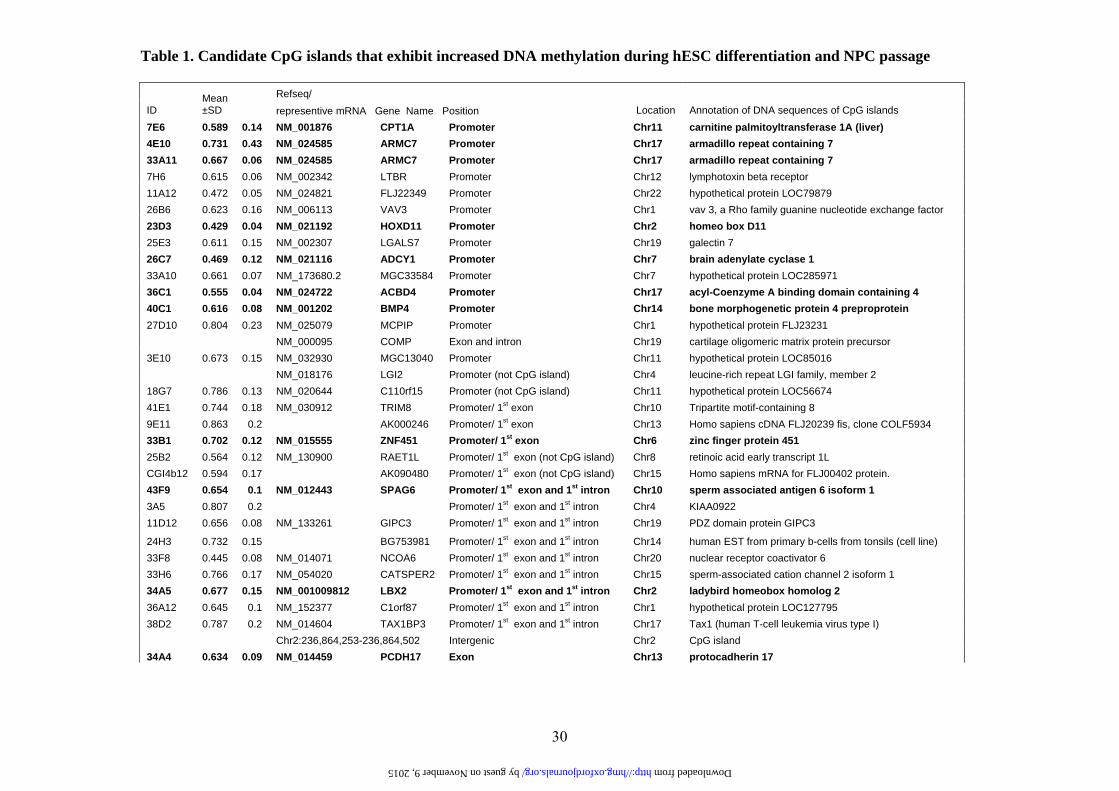
Table 1. Candidate CpG islands that exhibit increased DNA methylation during hESC differentiation and NPC passage
Refseq/ ID
Mean ±SD representive mRNA Gene Name Position Location Annotation of DNA sequences of CpG islands
7E6 0.589 0.14 NM_001876 CPT1A Promoter Chr11 carnitine palmitoyltransferase 1A (liver) 4E10 0.731 0.43 NM_024585 ARMC7 Promoter Chr17 armadillo repeat containing 7 33A11 0.667 0.06 NM_024585 ARMC7 Promoter Chr17 armadillo repeat containing 7 7H6 0.615 0.06 NM_002342 LTBR Promoter Chr12 lymphotoxin beta receptor 11A12 0.472 0.05 NM_024821 FLJ22349 Promoter Chr22 hypothetical protein LOC79879 26B6 0.623 0.16 NM_006113 VAV3 Promoter Chr1 vav 3, a Rho family guanine nucleotide exchange factor 23D3 0.429 0.04 NM_021192 HOXD11 Promoter Chr2 homeo box D11 25E3 0.611 0.15 NM_002307 LGALS7 Promoter Chr19 galectin 7 26C7 0.469 0.12 NM_021116 ADCY1 Promoter Chr7 brain adenylate cyclase 1 33A10 0.661 0.07 NM_173680.2 MGC33584 Promoter Chr7 hypothetical protein LOC285971 36C1 0.555 0.04 NM_024722 ACBD4 Promoter Chr17 acyl-Coenzyme A binding domain containing 4 40C1 0.616 0.08 NM_001202 BMP4 Promoter Chr14 bone morphogenetic protein 4 preproprotein 27D10 0.804 0.23 NM_025079 MCPIP Promoter Chr1 hypothetical protein FLJ23231 NM_000095 COMP Exon and intron Chr19 cartilage oligomeric matrix protein precursor 3E10 0.673 0.15 NM_032930 MGC13040 Promoter Chr11 hypothetical protein LOC85016 NM_018176 LGI2 Promoter (not CpG island) Chr4 leucine-rich repeat LGI family, member 2 18G7 0.786 0.13 NM_020644 C110rf15 Promoter (not CpG island) Chr11 hypothetical protein LOC56674 41E1 0.744 0.18 NM_030912 TRIM8 Promoter/ 1st exon Chr10 Tripartite motif-containing 8 9E11 0.863 0.2 AK000246 Promoter/ 1st exon Chr13 Homo sapiens cDNA FLJ20239 fis, clone COLF5934 33B1 0.702 0.12 NM_015555 ZNF451 Promoter/ 1st exon Chr6 zinc finger protein 451 25B2 0.564 0.12 NM_130900 RAET1L Promoter/ 1st exon (not CpG island) Chr8 retinoic acid early transcript 1L CGI4b12 0.594 0.17 AK090480 Promoter/ 1st exon (not CpG island) Chr15 Homo sapiens mRNA for FLJ00402 protein. 43F9 0.654 0.1 NM_012443 SPAG6 Promoter/ 1st exon and 1st intron Chr10 sperm associated antigen 6 isoform 1 3A5 0.807 0.2 Promoter/ 1st exon and 1st intron Chr4 KIAA0922 11D12 0.656 0.08 NM_133261 GIPC3 Promoter/ 1st exon and 1st intron Chr19 PDZ domain protein GIPC3
24H3 0.732 0.15 BG753981 Promoter/ 1st exon and 1st intron Chr14 human EST from primary b-cells from tonsils (cell line) 33F8 0.445 0.08 NM_014071 NCOA6 Promoter/ 1st exon and 1st intron Chr20 nuclear receptor coactivator 6 33H6 0.766 0.17 NM_054020 CATSPER2 Promoter/ 1st exon and 1st intron Chr15 sperm-associated cation channel 2 isoform 1 34A5 0.677 0.15 NM_001009812 LBX2 Promoter/ 1st exon and 1st intron Chr2 ladybird homeobox homolog 2 36A12 0.645 0.1 NM_152377 C1orf87 Promoter/ 1st exon and 1st intron Chr1 hypothetical protein LOC127795 38D2 0.787 0.2 NM_014604 TAX1BP3 Promoter/ 1st exon and 1st intron Chr17 Tax1 (human T-cell leukemia virus type I) Chr2:236,864,253-236,864,502 Intergenic Chr2 CpG island 34A4 0.634 0.09 NM_014459 PCDH17 Exon Chr13 protocadherin 17
by guest on November 9, 2015 http://hmg.oxfordjournals.org/ Downloaded from

31
34D6 0.527 0.07 AK090480 Exon Chr15 Homo sapiens mRNA for FLJ00402 protein. 44H4 0.659 0.14 AK055250 Exon Chr11 Homo sapiens cDNA FLJ30688 fis, clone FCBBF2000473 6F5 0.532 0.13 NM_015894 STMN3 Exon and intron Chr20 Stathmin 3 (SCG10-like-protein) 2A12 0.614 0.11 AY356349 CYP26C1 Exon and intron Chr10 cytochrome P450, family 26, subfamily C, polypeptide 1 14G3 0.665 0.12 NM_030572 MGC10946 Exon and intron Chr12 hypothetical protein LOC80763 27B5 0.456 0.1 NM_021948 BCAN Exon and intron Chr1 brevican isoform 1 29H7 0.579 0.08 BC007360 BC007360 Exon and intron ChrX MGC16121 protein 32G10 0.574 0.19 NM_001012334 MDK Exon and intron Chr11 midkine 16F6 0.591 0.07 NM_006480 RGS14 Intron Chr5 RGS14 gene for regulator of G protein signalling 14 19D9 0.673 0.23 AF410154 MHC2TA Intron Chr16 MHC class II transactivator 21C3 0.436 0.14 NM_016307 PRRX2 Intron Chr9 paired related homeobox 2 22H12 0.319 0.08 NM_002398 MEIS1 Intron Chr2 Meis1 homolog 13A11 0.856 0.22 Chr2:45,088,398-45,088,572 Intergenic Chr2 CpG island 17A5 0.687 0.13 Chr6:21,772,601-21,772,897 Intergenic Chr6 CpG island 23H1 0.643 0.07 Chr18:11,937,736-11,937,908 Intergenic Chr18 CpG island 25H5 0.796 0.21 Chr18:11,937,591-11,937,911 Intergenic Chr18 CpG island 30A7 0.621 0.15 Chr2:66,720,759-66,720,954 Intergenic Chr2 CpG island 31C9 0.657 0.11 Chr2:66,720,854-66,720,952 Intergenic Chr2 CpG island 32H3 0.446 0.12 Chr17:43,959,366-43,959,604 Intergenic Chr17 CpG island 36E6 0.551 0.07 Chr14:99,750,365-99,750,723 Intergenic Chr14 CpG island 37H5 0.677 0.07 Chr2:66,720,759-66,720,954 Intergenic Chr2 CpG island 40A4 0.546 0.15 Chr17:43,959,366-43,959,604 Intergenic Chr17 CpG island 44F2 0.688 0.1 Chr3:129,809,598-129,809,918 Intergenic Chr3 CpG island 11D11 0.546 0.11 Chr10:73,758,730-73,759,224 Intergenic (not CpG island) Chr10 14H10 0.584 0.29 Chr12:123,108,420-123,108,832 Intergenic (not CpG island) Chr12 8C1 0.526 0.16 no hit
Clone ID was adopted from customary microarray. The ratio of hybridization signals was averaged from 3 to 6 data sets and expressed as mean±SD (hESC-NPCs probe intensity over hESCs probe intensity). Ref sequence, sequence annotation, and CpG island position in the gene structure were labeled according to UCSC Genome Browser on Human May 2004 Assembly. Clones in bold are those CpG islands that have been verified to exhibit increased methylation in P14 hESC-NPCs when compared to hESCs as shown in Fig. 2 and online Supplementary Fig. 2.
by guest on November 9, 2015 http://hmg.oxfordjournals.org/ Downloaded from

32
by guest on Novem
ber 9, 2015http://hm
g.oxfordjournals.org/D
ownloaded from

33
by guest on Novem
ber 9, 2015http://hm
g.oxfordjournals.org/D
ownloaded from

34
by guest on Novem
ber 9, 2015http://hm
g.oxfordjournals.org/D
ownloaded from

35
by guest on Novem
ber 9, 2015http://hm
g.oxfordjournals.org/D
ownloaded from

36
by guest on Novem
ber 9, 2015http://hm
g.oxfordjournals.org/D
ownloaded from

37
by guest on Novem
ber 9, 2015http://hm
g.oxfordjournals.org/D
ownloaded from

38
by guest on Novem
ber 9, 2015http://hm
g.oxfordjournals.org/D
ownloaded from

39
by guest on Novem
ber 9, 2015http://hm
g.oxfordjournals.org/D
ownloaded from