Airway Management in Prolonged Field Care (CPG ID:80)
362
JOINT TRAUMA SYSTEM CLINICAL PRACTICE GUIDELINE (JTS CPG) Airway Management in Prolonged Field Care (CPG ID:80) This Role 1, prolonged field care (PFC) clinical practice guideline (CPG) is intended to be used after Tactical Combat Casualty Care (TCCC) Guidelines, when evacuation to higher level of care is not immediately possible Contributors SSG Collin Dye, SOCM, USA COL (Ret) Sean Keenan, MC, USA CPT Brandon Carius, PA-C, USA SFC Paul Loos, 18D, USA MSG Michael Remley, SOCM, USAS SFC Brandon Mendes, 18D, USA CPT Jacob L. Arnold, MC, USA MAJ Ian May, MC, USA LTC Douglas Powell, MC, USAR CAPT Joshua M. Tobin, MC, USNR LTC (P)Jamie Riesberg, MC, USA Col Stacy Shackelford, USAF, MC Publication Date: 01 May 2020 JTS CPGs are developed and peer reviewed by subject matter experts serving on the Defense Committees on Trauma: the Committee on Tactical Combat Casualty Care; the Committee of Surgical Combat Casualty Care; and the Committee of En Route Combat Casualty Care. Special thanks goes to these individuals who donate their time and share their experience to aid the JTS mission of publishing standardized clinical practice guidelines which improve patient care and save lives. TABLE OF CONTENTS Purpose .......................................................................................................................................................................... 3 Background .................................................................................................................................................................... 3 Indications for Airway Management ............................................................................................................................. 3 Disrupted Face or Neck Anatomy.............................................................................................................................. 3 Insufficient Oxygenation or Ventilation .................................................................................................................... 4 Machine, Suction, Monitor, Airway, Intravenous Access, Drugs ................................................................................... 5 Machine..................................................................................................................................................................... 5 Suction....................................................................................................................................................................... 6 Monitor ..................................................................................................................................................................... 6 Airway ....................................................................................................................................................................... 7 Proper Tube Placement ........................................................................................................................................ 9 IV/IO Access............................................................................................................................................................... 9 Drugs ....................................................................................................................................................................... 10 Airway Placement ............................................................................................................................................... 10 Prolonged Sedation............................................................................................................................................. 10 Performance Improvement (PI) Monitoring................................................................................................................ 11 Population of Interest ............................................................................................................................................. 11 Intent (Expected Outcomes) ................................................................................................................................... 11
-
Upload
khangminh22 -
Category
Documents
-
view
1 -
download
0
Transcript of Airway Management in Prolonged Field Care (CPG ID:80)

JOINT TRAUMA SYSTEM CLINICAL PRACTICE GUIDELINE (JTS CPG)
Airway Management in Prolonged Field Care (CPG ID:80) This Role 1, prolonged field care (PFC) clinical practice guideline (CPG) is intended to be used after Tactical Combat Casualty Care (TCCC) Guidelines, when evacuation to higher level of care is not immediately possible
Contributors
SSG Collin Dye, SOCM, USA COL (Ret) Sean Keenan, MC, USA CPT Brandon Carius, PA-C, USA SFC Paul Loos, 18D, USA MSG Michael Remley, SOCM, USAS SFC Brandon Mendes, 18D, USA
CPT Jacob L. Arnold, MC, USA MAJ Ian May, MC, USA LTC Douglas Powell, MC, USAR CAPT Joshua M. Tobin, MC, USNR LTC (P)Jamie Riesberg, MC, USA Col Stacy Shackelford, USAF, MC
Publication Date: 01 May 2020
JTS CPGs are developed and peer reviewed by subject matter experts serving on the Defense Committees on Trauma: the Committee on Tactical Combat Casualty Care; the Committee of Surgical Combat Casualty Care; and the Committee of En Route Combat Casualty Care. Special thanks goes to these individuals who donate their time and share their experience to aid the JTS mission of publishing standardized clinical practice guidelines which improve patient care and save lives.
TABLE OF CONTENTS Purpose .......................................................................................................................................................................... 3 Background .................................................................................................................................................................... 3 Indications for Airway Management ............................................................................................................................. 3
Disrupted Face or Neck Anatomy .............................................................................................................................. 3 Insufficient Oxygenation or Ventilation .................................................................................................................... 4
Machine, Suction, Monitor, Airway, Intravenous Access, Drugs ................................................................................... 5 Machine ..................................................................................................................................................................... 5 Suction ....................................................................................................................................................................... 6 Monitor ..................................................................................................................................................................... 6 Airway ....................................................................................................................................................................... 7
Proper Tube Placement ........................................................................................................................................ 9 IV/IO Access ............................................................................................................................................................... 9 Drugs ....................................................................................................................................................................... 10
Airway Placement ............................................................................................................................................... 10 Prolonged Sedation ............................................................................................................................................. 10
Performance Improvement (PI) Monitoring ................................................................................................................ 11 Population of Interest ............................................................................................................................................. 11 Intent (Expected Outcomes) ................................................................................................................................... 11

Airway Management in Prolonged Field Care CPG ID: 80
Guideline Only/Not a Substitute for Clinical Judgment 2
Performance/Adherence Metrics ........................................................................................................................... 11 Data Sources ............................................................................................................................................................ 11 System Reporting & Frequency ............................................................................................................................... 11 Responsibilities ........................................................................................................................................................ 12
References ................................................................................................................................................................... 12 Appendix A: Airway Equipment ................................................................................................................................... 13 Appendix B: Airway Management Nursing/Continued Care ....................................................................................... 15 Appendix C: Bag-Valve-Mask Technique ..................................................................................................................... 19 Appendix D: Supraglottic Airway Placement ............................................................................................................... 20 Appendix E: Cricothyroidotomy Procedure Checklist .................................................................................................. 21 Appendix F: Post Cricothyrotomy/Endotracheal Intubation Checklist ........................................................................ 22 Appendix G: Waveform Capnography & Pulse Oximetry Interpretation .................................................................... 23 Appendix H: Pediatric Considerations ......................................................................................................................... 25 Appendix I: Airway Management (using MSMAID) in Prolonged Field Care: Summary Table .................................... 27 Appendix J: Additional Information Regarding Off-Label Uses In CPGs ...................................................................... 29

Airway Management in Prolonged Field Care CPG ID: 80
Guideline Only/Not a Substitute for Clinical Judgment 3
PURPOSE
This Role 1, prolonged field care (PFC) clinical practice guideline (CPG) is intended to be used after Tactical Combat Casualty Care (TCCC) Guidelines, when evacuation to higher level of care is not immediately possible. A provider must first and foremost be an expert in TCCC, the Department of Defense standard of care for first responders. The intent of this PFC CPG is to provide evidence and experience-based solutions to those who manage airways in an austere environment. An emphasis is placed on utilizing the tools and adjuncts most familiar to a Role 1 provider. The PFC capability of airway is addressed to reflect the reality of managing an airway in a Role 1 resource-constrained environment. A separate Joint Trauma System CPG will address mechanical ventilation. This PFC CPG also introduces an acronym to assist providers and their teams in preparing for advanced procedures, to include airway management.
BACKGROUND
Airway compromise is the second leading cause of potentially survivable death on the battlefield after hemorrhage.1 Complete airway occlusion can cause death from suffocation within minutes. Austere environments present significant challenges with airway management. Limited provider experience and skill, equipment, resources, and medications shape the best management techniques. Considerations include: limited availability of supplemental oxygen; medications for induction/rapid sequence intubation, paralysis, and post-intubation management; and limitations in available equipment. Another reality currently is limitations in sustainment training options, especially for advanced airway techniques. Due to these challenges, some common recommendations that may be considered “rescue” techniques in standard hospital airway management may be recommended earlier or in a non-standard fashion to establish and control an airway in a PFC environment. Patients who require advanced airway placement tend to undergo more interventions, be more critically injured, and ultimately have a higher proportion of deaths. The ability to rapidly and consistently manage an airway when indicated, or spend time on other resuscitative needs when airway management is not indicated, may contribute to improved outcomes.2,3
INDICATIONS FOR AIRWAY MANAGEMENT
DISRUPTED FACE OR NECK ANATOMY
If managing an isolated injury that disrupts face or neck anatomy, healthy adults may only need a mechanical airway placed and may not require assisted ventilation if they are not heavily sedated. Examples of conditions requiring early airway management include:
Massive facial trauma
Burns to the face or inhalation injury with hoarseness or stridor
Massive neck trauma
Expanding neck mass/hematoma
Acute pharyngeal infection (Retropharyngeal abscess, peritonsillar abscess, epiglottitis)
Foreign body aspiration
Anaphylaxis (airway swelling)

Airway Management in Prolonged Field Care CPG ID: 80
Guideline Only/Not a Substitute for Clinical Judgment 4
INSUFFICIENT OXYGENATION OR VENTILATION
These indications may be apparent on initial evaluation or may emerge during the course of patient management. If the need to establish an airway develops more gradually, additional elective airway techniques can be employed. Interventions such as prolonged manual bag or mechanical ventilation require an airway intervention to ensure best overall management. Examples include:
Chest wall and pulmonary trauma (blunt or penetrating) such as flail segments, pulmonary contusions (from blast, blunt or crush mechanism)
Burns with greater than 40% body surface area
Traumatic brain injury with decreased level of consciousness (GCS ≤ 8)
Traumatic brain injury with suspected herniation requiring hyperventilation (target ETCO2 between 30 and 35)
Procedural sedation or surgical procedure
Respiratory failure from disease, infection, or injury:
Chemical or toxic inhalation injuries
Acute Respiratory Distress Syndrome (ARDS) from infection, massive resuscitation, drug-induced or other causes
Primary lung infection (pneumonia)
Massive pulmonary embolism
TRALI (transfusion related acute lung injury) or TACO (transfusion associated circulatory overload) due to massive transfusion
Table 1. TCCC Airway Management Adjuncts (Consider basic adjuncts first)
Device/ Techniques Pros Cons
Pharm Reqs to Maintain (0/+/++)
Skill Level (NM: Non-Medic; M: Medic)
Head-Tilt/ Chin-Lift
Easy Requires hands-on continuously
0 NM
Recovery Position
Easy; tactically feasible (TCCC/MASCAL)
May limit patient assessment or ongoing management
0 NM
Sit Up/ Lean Forward
Easy and practical (may be position of comfort)
May indicate impending airway loss
0 NM
Nasopharyngeal Airway (NPA)
Easy Risk of nose bleeds with placement
0 NM
Oropharyngeal Airway (OPA)
Easy Not tolerated in conscious patient
+ NM
Supraglottic Airway (SGA)
Easy Not tolerated in conscious patient
++ M

Airway Management in Prolonged Field Care CPG ID: 80
Guideline Only/Not a Substitute for Clinical Judgment 5
Device/ Techniques Pros Cons
Pharm Reqs to Maintain (0/+/++)
Skill Level (NM: Non-Medic; M: Medic)
Oral Endotracheal Tube (ETT) Airway
Familiar to trained providers; Definitive; No surgical incision needed
Skill sustainment challenging, requires neuromuscular blockade (rapid-sequence intubation) for best success, may require suction to visualize vocal cords, requires sedation (potential limitation in PFC)
++ M+
Cricothyrotomy (Cric)
Definitive; Better tolerated than Oropharyngeal (OP), SGA or ETT. May require less sedation to sustain once in place
Invasive procedure, high failure rate in some studies.4,5
+ M
For Pharm Reqs: Maintain: 0 = no additional medications; + = some or intermittent dosing medications required; ++ = continuous or multiple medications required
MACHINE, SUCTION, MONITOR, AIRWAY, INTRAVENOUS ACCESS, DRUGS
Checklists are commonly used in medical practice. In preparation for an advanced procedure (including securing an airway) using an acronym or other checklist approach will prove invaluable. One such acronym, originally developed as a simple pre-operative anesthesia checklist, is presented. The MSMAID acronym (Machine, Suction, Monitor, Airway, Intravenous access, Drugs) organizes an approach to preparation for airway management and may also prove useful in preparation for other procedures.
The patient’s condition dictates the available time for a provider to consider all items on this checklist. A rapidly deteriorating patient with airway disruption or compromise will need the airway procedure first and follow-on considerations later. If a patient can be more appropriately classified as semi-urgent (e.g., worsening respiratory status due to an underlying pulmonary cause) the provider will have more time to consider the algorithm and prepare. Logistic considerations, and sometimes considerable time constraints, will affect preparation for advanced procedures. Recommendations follow the “minimum, better, best” format.
MACHINE
(Equipment required post-intubation)
Minimum: Bag-Valve-Mask (BVM) with positive end-expiratory pressure (PEEP) valve
Better: Automated portable ventilator (preferably with PEEP); oxygen concentrator
Best: Full-feature portable ventilator (e.g. several ventilatory modes, PEEP); supplemental oxygen if available

Airway Management in Prolonged Field Care CPG ID: 80
Guideline Only/Not a Substitute for Clinical Judgment 6
Managing ventilators or advanced equipment unfamiliar to a provider presents challenges. Initiate telemedicine consultation for best guidance.
PEEP is important for prolonged ventilation. PEEP is the pressure in the airway at the end of the expiratory phase which prevents the alveoli of the lung from completely collapsing. In a spontaneously breathing person, this pressure is maintained by closing the glottis, clearing the throat, coughing, sighing, etc. With an invasive airway, the glottis is bypassed with the tube and “natural” PEEP is lost. PEEP should therefore be introduced into the ventilated patient using a PEEP valve on the BVM or using the PEEP setting on a ventilator. When using BVM or ventilator, provide PEEP (recommended initial setting is 5cm H2O).6
SUCTION
Minimum: Improvised suction (i.e. syringe + nasopharyngeal airway [NPA]) and patient positioning if not contraindicated
Better: Manual suction bulb with adapter
Best: Powered commercial suction with oral tip and in-line endotracheal tube suction adapter
Suction should be available when establishing and maintaining an airway to remove excessive secretions or blood. It is particularly important to utilize suction to facilitate view of the vocal cords during endotracheal intubation. In addition, suction should be available for routine patient care and maintenance requirement for any intubated patients. Suction should be utilized as needed to remove secretions, mucous or blood from the airway device or oropharynx. In the event of high airway pressures, suction may be used to remove mucus/mucus plugs or to clear obstructions. In the case of thick secretions, a saline flush of 1-2 mL followed by in-line suctioning of the endotracheal tube may be useful. Note: During in-line suctioning of tubes, the suction should only be applied when withdrawing the catheter and not upon initial insertion.
MONITOR
(Monitoring and telemedicine support)
Minimum: Pulse oximeter (SpO2), assistant to monitor respirations and record manual vital signs. Trending vital signs documentation. PFC flow sheet is recommended. Refer to PFC Documentation CPG.7 Voice or data connections to perform telemedicine communication
Better: Portable Capnometry (ETCO2)/capnography in addition to SpO2. Transmit photographs from smartphones or personal devices to augment telemedicine communications.
Best: Automatic vital signs monitor with SpO2, ETCO2/waveform capnography, +/- electrocardiogram (EKG); Synchronous (real-time continuous) telemedicine using video or remote patient monitoring systems
Monitoring is the active process of assessing the patient throughout a procedure. It involves the gathering, documenting and interpretation of vital signs and other data, and the continuous assessment of their clinical status. Telemedicine can be an important adjunct and critical capability to employ when monitoring a patient undergoing complex procedures.

Airway Management in Prolonged Field Care CPG ID: 80
Guideline Only/Not a Substitute for Clinical Judgment 7
AIRWAY
Minimum: Medic is prepared for a ketamine cricothyroidotomy.
Better: Add ability to provide continuous sedation.
Best: Add a responsible rapid-sequence intubation capability (to include chemical paralysis) with airway maintenance (to include suction) and continuous sedation.
Per the PFC Capabilities Position Paper, a definitive airway requires control of the patient’s airway with an inflated cuff in the trachea. In addition, sedation is needed to keep the patient comfortable and sustain the airway.8
A proposed algorithm was developed by Mabry RL et al for an awake surgical airway (Figure 1).9 This algorithm incorporates the skills recommended for Tactical Combat Casualty Care and presents the decision process deemed adequate (minimum standard) for definitive airway control in PFC. The airway algorithm is presented in its published form and surgical airway is synonymous with cricothyrotomy. As noted above, the indications for surgical airway include disrupted face or neck anatomy, as well as a need for prolonged positive pressure ventilation in a resource constrained PFC environment. Consider temporizing, if possible, with basic airway maneuvers or other airway adjuncts (e.g.; NPA, SGA). Please see Table 1.
Figure 1. An Awake Patient Surgical Airway Algorithm
Source: Mabry RL, Kharod CU, Bennett BL. Awake cricothyrotomy: a novel approach to the surgical airway in the tactical setting. Wilderness Environ Med. 2017;28(2S):S61-8. Adapted with permission from the Wilderness Medical Society. ©2017 Wilderness Medical Society.

Airway Management in Prolonged Field Care CPG ID: 80
Guideline Only/Not a Substitute for Clinical Judgment 8
Positioning of the patient to help clear airway obstruction should be considered first, when possible. The simple option of placing the patient in a sitting position, placing the patient in the lateral “recovery” position, or head tilt-chin lift/jaw thrust maneuver may be enough to ensure adequate respirations. Simple adjuncts such as a nasal or oropharyngeal airway (in an unconscious patient) may be utilized in addition to proper positioning to help ensure a clear airway.
For those patients requiring active airway assistance, the first step should be inserting an NPA to open the airway. BVM ventilation is the next step in the algorithm and though it appears to be relatively simple, the procedure requires preparation, training and skill to perform correctly. A spontaneously breathing patient may prove challenging, but properly delivered, synchronous breaths may be the only requirement to assist a patient’s respirations. Care should be taken to ensure proper volume and rate of bag-delivered breath. One hand should provide moderate pressure to the bag for no more than 50% of the volume of an adult bag or just enough to see the chest begin to rise at a rate of 12-16 breaths per minute (one breath every 4-5 seconds) initially. It is important to avoid hyperventilation through large or rapid breaths, particularly with traumatic brain injury (TBI) casualties, therefore ETCO2 monitoring is indicated for all patients requiring assisted ventilation and all patients with altered mental status (including both TBI casualties and sedated patients). BVM is an important airway management skill (see Appendix C: Bag-Valve-Mask Technique for a detailed description). It is highly recommended that all PFC training on airway skills cover BVM skills and techniques.
OP and SGA insertion can present a considerable noxious stimulus and may not be tolerated by conscious or even some semiconscious patients. Additionally, the dyssynchronous use of BVM ventilation may lead to poor patient cooperation, abnormal tidal volumes, gastric insufflation (and resultant regurgitation/aspiration), or other complications.
The decision to perform a cricothyroidotomy is one that is sometimes difficult. When possible, in urgent but not emergency situations, a telemedicine call should be considered to help with medical decision making.
An important adjunct to passing an endotracheal tube, either via the cricothyroid membrane or the oropharynx, is the use of a gum elastic bougie (sometimes also referred to as an Eschmann Stylet or, simply bougie). This device is simple, rugged and should be used to guide tube placement. The bougie is placed in the trachea before the endotracheal tube and may be used first to confirm proper positioning by either tactile discrimination (feeling the tube bump against the tracheal rings on introduction), or by encountering a hard stop when abutted against the carina. An endotracheal tube is then introduced over the bougie into the trachea. Lastly, the bougie is removed. A bougie may also be used to change tubes in the case of a tube malfunction. This may be accomplished by placing a bougie in a tube that is currently positioned, remove the tube over the bougie (ensuring the bougie remains in the proper position within the airway lumen), and replacing a new tube over that bougie. Remove the bougie, leaving the new tube in place. Confirmation procedures discussed below must be repeated once the new tube is in place.
Consistent with TCCC guidelines, the routine use of orotracheal intubation is not recommended as the minimum standard in PFC. This procedure requires considerable skill and sustainment;10 and requires appropriate sedation for both rapid sequence intubation and post-intubation management. If a provider is appropriately trained, current and practiced in the procedure, and has the required support equipment and medications, then orotracheal intubation may be considered. Although preferred when

Airway Management in Prolonged Field Care CPG ID: 80
Guideline Only/Not a Substitute for Clinical Judgment 9
possible, training in orotracheal intubation is not required to obtain a definitive airway in the PFC operational setting. Consider basic measures first before proceeding to either type of invasive airway. Additional details on orotracheal intubation are included in the JTS Airway CPG.
Airway management in the tactical setting requires a different conceptual approach than airway management in the hospital, or even the civilian prehospital environment. Differences in epidemiology, injury patterns, equipment and environment must be considered if airway management is to be optimized. First, most military casualties requiring a prehospital airway have trauma to the head, face or neck. Surgical airway is often the final common pathway due to bleeding or distorted anatomy. In comparison, most airways in the civilian prehospital environment are placed in elderly people for cardiac arrest. When reliable suction and oxygen delivery are not available, or personnel are not experienced in rapid sequence intubation using neuromuscular blockade, a definitive airway will often mean a surgical airway.
Proper Tube Placement
Minimum: Visualization of the tube passing through the vocal cords (in the case of endotracheal intubation); auscultation of epigastric region (should be silent) and bilateral lung sounds (should be present). Colorimetric capnography + endotracheal detection device (EDD). Easy bilateral rise and fall of the chest + misting of the tube + no signs of gastric insufflation. (Reassess frequently and have another medic double check if unsure.)
Better: Minimum plus portable capnometer. Ultrasound if trained/available to guide and/or verify placement
Best: Continuous ETCO2/waveform capnography
Verification of correct tube placement must be performed every time as incorrect tube placement may be fatal. The REACH study5 showed that right mainstem and hypopharynx placement are the most common locations of incorrect placement of ETT. Esophageal intubation is also common. Subcutaneous placement of cricothyroidotomy tube may occur. Use capnography to verify correct tube placement as tube misplacement can be fatal. Auscultate, if possible, to verify bilateral breath sounds. If ultrasound is available, this can be used to further verify placement in the correct position.11
IV/IO ACCESS
Minimum: If (IV) / intraosseous (IO) attempts fail or when unavailable: medication may be given intramuscularly or intranasally for immediate sedation to facilitate surgical cricothyroidotomy. Continue attempts at IV/IO access after airway has been controlled
Better: 1-2 patent IV/IO
Best: 2-3x patent intravenous IV/IO with additional IO device on standby
Though an important consideration to administer medications and fluids, do not delay an emergent airway to obtain IV/IO access in the instance you are the sole provider.
Ultrasound may be used to help identify small or deep veins. Ultrasound guided IV access may be attempted if trained. Other sites to consider for superficial IV attempts include the external jugular and saphenous veins. If appropriately trained, consider central venous access or venous cutdown.

Airway Management in Prolonged Field Care CPG ID: 80
Guideline Only/Not a Substitute for Clinical Judgment 10
DRUGS
Airway Placement
Minimum: Local anesthetic for cricothyroidotomy (superficial skin anesthesia plus 1-2 mL injected through the cricothyroid membrane); or placement without medications in unconscious patient. **Note: most sedating agents can be given IM if IV/IO has not been established
Better: Any IV/ IO sedating agent (opioid, benzodiazepine: reference the Analgesia and Sedation Management for PFC CPG for procedural doses of such agents).12
Best: Procedural dose ketamine (1-2 mg/kg IV push) for ETT or cricothyroidotomy placement + local anesthetic (lidocaine) for cricothyroidotomy placement
Prolonged Sedation
(post-airway placement)
Minimum (without IV access): Ketamine (sedation dose), 3-4mg/kg IM
Better: IV/IO pushes of ketamine, opioid, and/or midazolam (alone or in combination as per the individual’s scope of practice, experience and availability of medications)
Best: Ketamine IV/IO Drip. Hydromorphone or alternate opioid IV/IO push for breakthrough pain and midazolam IV/IO push as needed for sedation
(Reference Analgesia and Sedation Management in PFC CPG for details and drug doses).12
Be cautious with sedation, advanced airway placement and positive pressure ventilation in patients who are hypotensive or under-resuscitated. Blood pressure can fall rapidly during airway management due to a variety of mechanisms.
In all cases, monitor BP closely (every 1-2 minutes during the procedure, every 3-5 minutes for 15 minutes post-procedure). A BP drop may be brief (if due to vagal effects of epiglottis stimulation during ETT placement) or sustained (due to positive pressure ventilation, increased intrathoracic pressure and decreased venous return to the heart). Loss of sympathetic drive secondary to pain and sedation medications, and/or continued hypovolemia can also result in hypotension. Continue resuscitation with blood products (trauma patients) or crystalloid (non-trauma patients) if a hypotensive patient requires immediate airway interventions. Be prepared to support blood pressure with vasopressors (e.g. epinephrine bolus or drip) if trained or under direct telemedicine guidance.
Neuromuscular blockade (succinylcholine, rocuronium, vecuronium, etc.) is NOT recommended for use by the average practitioner of PFC. Though these are standard medications to use in rapid sequence intubation and ventilator management, their potential lethality in inexperienced hands does not justify routine recommended use. If trained and/or under direct supervision of telemedicine support, the use of neuromuscular blockade may be considered, subject to local medical direction and protocols.

Airway Management in Prolonged Field Care CPG ID: 80
Guideline Only/Not a Substitute for Clinical Judgment 11
PERFORMANCE IMPROVEMENT (PI) MONITORING
POPULATION OF INTEREST
1. All patients who received ETT/cricothyroidotomy/supraglottic airway/NPA
2. All patients with compromised airway (initial GCS < 8 or abbreviated injury scale (AIS) head and neck ≥ 3, or AIS face ≥ 3).
INTENT (EXPECTED OUTCOMES)
1. All injured patients who present with obtundation (GCS<8), apnea, respiratory distress or insufficiency, airway obstruction, or impending airway loss will have a secure and definitive airway established expeditiously upon arrival to a Role 2 or Role 3 if not done prehospital.
2. SpO2 is maintained ≥ 90%.
3. Patients with a definitive airway (endotracheal tube, cricothyroidotomy, tracheostomy) have ETCO2 monitoring to confirm airway placement.
PERFORMANCE/ADHERENCE METRICS
1. Number and percentage of patients in the population of interest who had a secure and definitive airway (endotracheal tube, cricothyroidotomy, tracheostomy) established or verified, or documentation of appropriate intervention.
2. Number and percentage of patients in the population of interest with SpO2 < 90%<80%, <70%, <60%.
3. Number and percentage of patients who had definitive airway (endotracheal tube, cricothyroidotomy, tracheostomy) with ETCO2 documented at the same role of care where the procedure is done.
DATA SOURCES
Patient Record
Department of Defense Trauma Registry (DoDTR)
SYSTEM REPORTING & FREQUENCY
The above constitutes the minimum criteria for PI monitoring of this CPG. System reporting will be performed annually; additional PI monitoring and system reporting may be performed as needed.
The system review and data analysis will be performed by the Joint Trauma System (JTS) Director, JTS Program Manager, and the JTS PI Branch.

Airway Management in Prolonged Field Care CPG ID: 80
Guideline Only/Not a Substitute for Clinical Judgment 12
RESPONSIBILITIES
It is the trauma team leader’s responsibility to ensure familiarity, appropriate compliance and PI monitoring at the local level with this CPG.
REFERENCES
1. Eastridge BJ, Mabry RL, Sequin P, et al. Death on the battlefield (2001-2011): implications for the future of combat casualty care. J Trauma Acute Care Surg. 2012;73(6 Suppl 5):S431-7.
2. Hudson I, Blackburn MB, Mannsalinas EA, et al. Analysis of casualties that underwent airway management before reaching role 2 facilities in the Afghanistan conflict 2008-2014. Mil Med. 2020;185(Suppl 1):10-18.
3. Blackburn MB, April MD, Brown DJ, et al. et al “Prehospital airway procedures performed in trauma patients by ground forces in Afghanistan” J Trauma Acute Care Surg 2018;85(1S Suppl 2):S154-S160.
4. Mabry RL. An analysis of battlefield cricothyrotomy in Iraq and Afghanistan. J Spec Oper Med. 2012;12:17-23.
5. Adams BD, Cuniowski PA, Muck A, De Lorenzo RA. Registry of emergency airways arriving at combat hospitals (REACH). J Trauma 2008;64(6):1548-54.
6. Acosta P, Santisbon E, Varon J. The Use of Positive End-Expiratory Pressure in Mechanical Ventilation. Critical Care Clin, 2007 Apr;23(2):251-61.
7. Loos PE, Glassman E, Doerr D, et al. Documentation in prolonged field care. J Spec Oper Med. 2018; 18(1): 126-32.
8. Ball J, Keenan S. Prolonged Field Care Working Group position paper: prolonged field care capabilities. J Spec Oper Med. 2015; 15(3): 76-7.
9. Mabry RL, Kharod CU, Bennett BL. Awake cricothyrotomy: a novel approach to the surgical airway in the tactical setting. Wilderness Environ Med. 2017;28(2S):S61-8.
10. Gillett B, Saloum D, Aghera A, Marshall JP. Skill proficiency is predicted by intubation frequency of emergency medicine attending physicians. West J Emerg Med. 2019;20(4):601-609.
11. Gottlieb M, Holladay D, Peksa GD. Ultrasonography for the Confirmation of Endotracheal Tube Intubation: A Systematic Review and Meta-Analysis. Ann Emerg Med. 2018;72(6):627-636.
12. Pamplin J, Fisher A, Penny A, Olufs R, Rapp J, Hampton K, Riesberg J, Powell D, Keenan S, Shackelford S. Analgesia and Sedation Management During Prolonged Field Care. J Spec Oper Med. 2017;17(1):106-20.

Airway Management in Prolonged Field Care CPG ID: 80
Guideline Only/Not a Substitute for Clinical Judgment 13
APPENDIX A: AIRWAY EQUIPMENT
AIRWAY EQUIPMENT :
Minimum (Ruck/Aid Bag): Nasopharyngeal airway, Cricothyrotomy kit; BVM with PEEP valve
Better: Minimum PLUS supraglottic airway, gum elastic bougie to facilitate intubation, nasogastric/orogastric tube
Best: Better PLUS endotracheal tubes, direct laryngoscopy or video laryngoscopy equipment
WAVEFORM CAPNOGRAPHY:
Minimum: Colorimetric ETCO2 detector
Better: Portable capnometer
Best: Waveform capnography on patient monitor
See Appendix G for Capnography Interpretation
HEAT-MOISTURE EXCHANGER:
Heat-Moisture Exchangers (HME) are small, relatively inexpensive, in-line ETT adjuncts that contain hygroscopic salts that utilize differences in vapor pressure, expired moisture content, and the patient’s temperature to increase the humidity of inspired air. Increased moisture on inspiration helps maintain alveolar moisture. Capnography should not be positioned in-line following HME as these moisture devices can increase breathing resistance and work of respiration, as well as increase breathing apparatus dead space (especially in pediatric patients). This may lead to potential worsening hypercapnia and respiratory acidosis. Recommended order: endotracheal tube-capnograph-HME device (proximal to distal)
NASOGASTRIC/OROGASTRIC TUBE:
Placement of a nasogastric (NG) or orogastric (OG) tube should be considered following intubation of a patient in order to decompress gastric contents, prevent aspiration and gastric distention.
If only supraglottic airway access is available, consider using a supraglottic device that incorporates an orogastric tube port.
Always measure the distance from nose to stomach and note the distance prior to insertion, then verify epigastric sounds. Verify placement with second practitioner if sounds are questionable or difficult to auscultate.
Do not feed, aggressively hydrate or give oral medications through an OGT or NGT without telemedicine guidance. Always reassess tube position prior to putting anything into stomach to ensure proper gastric placement. Feeding should not be considered until 72 hours after injury in the PFC environment. If volume instilled is too large or rate is too fast, there is increased risk for vomiting. If feeding or hydration is begun through an OGT or NGT, they must be accurate, measured, and monitored. Safe airway management takes precedence over nutrition.

Airway Management in Prolonged Field Care CPG ID: 80
Guideline Only/Not a Substitute for Clinical Judgment 14
SUGGESTED PACKING LIST
Equipment Notes Minimum Nasopharyngeal airway (NPA)
Oropharyngeal airway (OPA) Cricothyroidotomy kit: a standardized kit should include at a minimum a #10 scalpel, a tracheal hook (or small, curved hemostat), an airway tube, a 10mL syringe, and a securing strap. The airway tube may be a prefabricated cricothyroidotomy tube such as that included in the Cric-Key (Control Cric), a Shiley tracheostomy tube, or a 6.0-sized endotracheal tube.
Bag valve mask (BVM) PEEP valve: either separate device, or affixed to the BVM device
Capnography/capnometry device Bougie
Better Additional endotracheal tubes Portable suction device
Best Supraglottic airway: options include LMA (Size 4 if only one; size 5 for >90kg patient) or King-LT.
Waveform capnography Commercial power suction device Automatic (electronic) patient monitor Direct laryngoscope: Macintosh blade size 3 or 4 for adults; Miller 1 or 2 for infants and small children as needed.
For those trained in endotracheal tube placement and opt to include equipment in their aid bag
Endotracheal tubes: for cricothyroidotomies (6.0 mm tubes). Personnel trained in endotracheal intubation should also carry a 7.5 or 8.0mm tube for primary intubation. A 7.0 mm tube should be carried as a secondary ET tube in case of difficult intubation. (If pediatric trauma is common, consider adding smaller sizes, but these should be limited to the most common ages expected to encounter, and calculated on the classic rule of tube size = 4 + (age/4). Examples of what to carry include 4.0 mm uncuffed for infants, and 6.0 cuffed for older children).
For those trained in endotracheal tube placement and opt to include equipment in their aid bag
Video laryngoscope: hand-held devices should utilize a blade with an angle that allows for direct laryngoscopy function should the video screen/battery fail during intubation.
For those trained in endotracheal tube placement and opt to include equipment in their aid bag
Mechanical ventilator Pediatric-sized equipment Other equipment listed above but not carried due to cube/weight restrictions

Airway Management in Prolonged Field Care CPG ID: 80
Guideline Only/Not a Substitute for Clinical Judgment 15
APPENDIX B: AIRWAY MANAGEMENT NURSING/CONTINUED CARE
Long-Term Airway Management, Nursing and Prolonged Field Care Below is the recommended Prolonged Field Care Nursing Flowsheet to use as a guide.*
Patient ID: Time 1hr 2hr 3hr 4hr 5hr 6hr 7hr 8hr 9hr 10hr 11hr 12hr 13hr 14hr 15hr 16hr 17hr 18hr 19hr 20hr 21hr 22hr 23hr 24hr
Action (suggested interval) Interval
Vita
ls
Check BP/HR/RR/T/SPO2/ETCO2 (Q1H)
Check Peripheral Pulses (Q1H)
Check Skin Temp and Color (Q1H)
Check Lactate (Q4H)
Check Blood Glucose (Q8H)
Ins/
Out
s
Check Drip Rates/Fluids In (Q1H)
Check Urine Output (Q1H)
Check Urine Dipstick (Q1H)
Perform NG/OG Tube Care (Q2H)
Perform Foley Care (Q24H)
Flush PRN Locks (Q8H)
Pain
/Sed
atio
n Check GCS/RASS/PAIN (Q1H)
Give Pain Rx (per Rx)
Give Sedation Rx (per Rx)

Airway Management in Prolonged Field Care CPG ID: 80
Guideline Only/Not a Substitute for Clinical Judgment 16
Patient ID: Time 1hr 2hr 3hr 4hr 5hr 6hr 7hr 8hr 9hr 10hr 11hr 12hr 13hr 14hr 15hr 16hr 17hr 18hr 19hr 20hr 21hr 22hr 23hr 24hr
HEEN
T
Perform Tube Suctioning (PRN)
Perform Oral Suctioning (PRN)
Perform Nasal Care/Moisten (Q4H)
Perform Oral Care/Moisten (Q4H)
Apply Lip Balm (Q1H)
Apply Eye Ointment/Drops (per Rx)
Brush Teeth (Q12H)
Change All Tape (Q24H)
Resp
irato
ry
Check Ventilator Settings (Q1H)
Auscultate Lungs (Q1H)
Turn, Cough, Deep Breathe (Q1H)
Check Chest Drainage (Q1H)
Inte
gum
enta
ry
Check S/S Compartment Syndrome (Q2H)
Reposition (Q2H)
Check Padding (Q2H)
Perform LE Massage (Q2H)
Check Dressings (Q4H)

Airway Management in Prolonged Field Care CPG ID: 80
Guideline Only/Not a Substitute for Clinical Judgment 17
Patient ID: Time 1hr 2hr 3hr 4hr 5hr 6hr 7hr 8hr 9hr 10hr 11hr 12hr 13hr 14hr 15hr 16hr 17hr 18hr 19hr 20hr 21hr 22hr 23hr 24hr
Do A/P Limb ROM (Q8H)
Wash and Dry Skin (Q24H)
Perform Burn Skin Care (Q24H)
Irrigate Wounds (Q24H)
Debride Wounds (Q24H)
Change Dressings (Q24H)
Give Antibiotics Rx (Q24H)
Gas
troi
ntes
tinal
Give PPI Rx if Indicated (per Rx)
Give Antiemetic Rx (per Rx)
Auscultate Abdomen (Q2H)
Palpate Abdomen (Q2H)
Give Food/Nutrition (Q8H)
Extr
a St
uff
Check O2 Supply
Check/change batteries
Compression Socks/Stockings

Airway Management in Prolonged Field Care CPG ID: 80
Guideline Only/Not a Substitute for Clinical Judgment 18
AIRWAY-SPECIFIC PFC NURSING GUIDELINES/ASSESSMENT/TASKS
Oral Suction (or in-line/tube suctioning, if placed) as indicated during routine patient assessments
Nose and mouth moistened Q4H
Lip Balm applied Q1H
Teeth brushed Q12H - Prevents pneumonia and other infections.
Auscultate lungs Q1H. If available, get a second medic to double check and verify if sounds are questionable or cannot otherwise auscultate. Extra diligence should be given to check for diminished breath sounds for suspected pneumothorax, as well as for pulmonary edema, especially if giving aggressive fluids in the case of a massive resuscitation, or replacement fluids. Consider ultrasound evaluation if available.
Naso/Orogastric Tube (NGT/OGT) and Abdominal Care
(as applicable)
Consider placing an NG/OG tube for prolonged care of intubated/cricothyrotomy patient or those with an SGA in place (if a compatible port is available on the SGA).
Abdominal palpation AND auscultation Q2H.
Nutrition ONLY under telemedicine guidance (optional and not indicated in first 72 hours).
*Loos PE, Glassman E, Doerr D, et al. Documentation in prolonged field care. J Spec Oper Med. 2018; 18(1): 126-32.

Airway Management in Prolonged Field Care CPG ID: 80
Guideline Only/Not a Substitute for Clinical Judgment 19
APPENDIX C: BAG-VALVE-MASK TECHNIQUE
Proper technique is essential to perform successful bag-valve-mask (BVM) airway management. Patients should be in the supine position, neck in a neutral position and the occiput slightly elevated (on a folded blanket, sheet, small pillow, etc.) to achieve a “sniffing position,” with the opening of the ears at the same level as the sternal notch. Masks should be of the proper size and should be fitted to the face to obtain a seal. This is best achieved by first placing the tapered portion of the opening of the mask over the bridge of the nose, then covering the patient’s mouth. If using the one-person method, the non-dominant had should use the “C & E” method with the thumb and index finger forming a C to cover the mask, and the middle, ring and small fingers forming an E on the bone of the mandible effectively lifting the jaw into the mask (rather than pressing the mask onto the face).
The other hand should gently squeeze the bag delivering a breath at a rate of one squeeze every 5-6 seconds. There should not be a leak around the mask, and you should be able to observe the rise and fall of the patient’s chest. An ETCO2 monitor may be placed in-line with the bag and mask. PEEP should be used with the BVM and initially set to 5mmHg. Every attempt should be made to maintain a seal to ensure continued PEEP. If the patient is breathing spontaneously, careful observation and delivery of an assisted synchronous breath should be a priority.
Some additional considerations are below: 1. If unable to perform adequate single hand C & E clamp, use two handed technique with a
second person to bag.
2. Don’t press mask down onto face when performing C & E clamp, visualize you are lifting face into mask.
3. Be aware fingertips on “E-clamp” should be positioned on the bones of the mandible and not on soft tissue, which could possibly occlude the airway.
4. Use “BOOTS” to predict difficult face-mask seals: Bearded, Obese, Old, Toothless, Snoring. In addition, maxillofacial trauma and edema from burns may prevent effective ventilation by BVM.
5. NPA should be used to assist with face mask ventilations (unless obvious contraindications such as mid-face trauma). OPA are also effective in obtunded patients or those who have received chemical sedation or neuromuscular blockade.
6. For bearded patients, lubrication of mask may assist seal. Tegaderm occlusive dressings over beard may also help if available. (If Tegaderm is used in sedated patient, consider pulling off before medication wears off, to lessen pain).
7. Consider mask straps to make face-mask seal more "hands free", especially if a non-medic is assisting you with BVM.

Airway Management in Prolonged Field Care CPG ID: 80
Guideline Only/Not a Substitute for Clinical Judgment 20
APPENDIX D: SUPRAGLOTTIC AIRWAY PLACEMENT
SUPRAGLOTTIC AIRWAY (SGA) PLACEMENT CHECKLIST
� Open airway manually, measure and insert simple airway adjunct (NPA -or- OPA).
� Ventilate patient with bag-valve-mask (BVM) (attach supplemental oxygen, if available).
� If ventilations insufficient, or the patient is clearly unconscious and not breathing adequately, prepare for supraglottic airway insertion. Inspect SGA to ensure appropriate size. Lubricate airway to facilitate passage. Cricothyroidotomy kit should be prepared for use if SGA fails.
� Follow MSMAID and for induction, use ketamine (1-2 mg/kg IV/IO or 3-4mg/kg IM) if time permits and the recommended medications are available.
INSERTING THE AIRWAY:
� Properly position head in a neutral or “sniffing” position (neck extended, as on a pillow or small blanket while lying flat) and open airway.
� Remove oropharyngeal airway (OPA) if previously placed.
� Insert device to proper depth (may adjust later if need for improved ventilation).
� Inflate cuff, if applicable; inflate as per device-specific volume instructions and immediately remove syringe.
� Confirm placement with ventilation and auscultation over epigastrium, then bilaterally over chest, left lung then right lung. Get a second practitioner to double check and verify in sounds are questionable or cannot otherwise auscultate.
� Verify proper SGA placement by secondary confirmation such as capnography/capnometry or colorimetric device.
� Place orogastric tube and decompress stomach if available, and compatible with SGA device (has a port specifically for OGT placement).
SGA Size Chart
Estimated patient size LMA King LT* Neonates/Infants (up to 5kg) 1 0 Infants 5-10 kg 1.5 1 Infants/Children 10-20kg 2 2 Children 20-30kg 2.5 2.5 Children 30-50kg 3 3* Adults 50-70kg 4 4* Adults 70-100kg 5 5* Adults over 100kg 6

Airway Management in Prolonged Field Care CPG ID: 80
Guideline Only/Not a Substitute for Clinical Judgment 21
APPENDIX E: CRICOTHYROIDOTOMY PROCEDURE CHECKLIST
PREPARE PATIENT
1. Pre-oxygenate patient if possible.
2. Inspect/assemble/test equipment for cricothyroidotomy.
3. Prepare site with alcohol and betadine -or- Chlorhexadine (Chlora-prep).
4. Follow MSMAID and for induction, use ketamine (1mg/kg IV/IO or 3-4mg/kg IM) if time permits and the medication is available.
For awake cricothyrotomy: Explain procedure to patient; *Use local anesthesia: lidocaine (1% or 2%), bupivacaine (0.25%, 0.5% or 1%); local through planned incision area AND approx. 1-2mL through cricothyroid membrane
PERFORM PROCEDURE
1. Stabilize thyroid cartilage and keep overlying skin taught. Maintain control with hand until the membrane incision is secured (step 8 below).
2. Locate cricothyroid membrane (Palpate for hyoid and tracheal rings. If unsure or difficult landmarks, then measure three finger widths above sternal notch for adults).
3. Make vertical incision through the skin over cricothyroid membrane.
4. Make horizontal incision through cricothyroid membrane, then immediately:
5. Open and maintain membrane incision with tracheal hook (or curved hemostat, bougie or blunt end of scalpel).
6. Insert endotracheal/tracheostomy tube into opening and direct tube caudad into trachea until the balloon is just inside the airway.
7. Inflate cuff and detach syringe (palpate bulb to ensure it’s not under-inflated or over-inflated).
8. Maintain control of tube at all times to prevent dislodgement.
9. Attach waveform capnography, or capnometry, or colorimetric device to confirm proper placement of tube.
10. Being careful not to dislodge the tube, attach BVM with PEEP and further check placement (epigastric and bilateral chest) and adequacy of bilateral insufflation of lungs.
11. Remove BVM (if sufficient respiratory effort), assess respirations for adequacy (rate, rhythm, and quality), assist ventilations if needed.
12. Secure with sutures and tie with girth hitch passed around the neck if time permits. As a stopgap, may use chest seal or secure around the neck with tie, ensuring inflation bulb does not get caught.
13. Consider placing NG/OG tube if available.

Airway Management in Prolonged Field Care CPG ID: 80
Guideline Only/Not a Substitute for Clinical Judgment 22
APPENDIX F: POST CRICOTHYROTOMY/ENDOTRACHEAL INTUBATION CHECKLIST
POST CRICOTHYROTOMY/ENDOTRACHEAL INTUBATION CHECKLIST
� Double check placement with waveform capnography or capnometry, placed directly on ET tube adapter.
� Check proper tube depth (not main stem) by auscultating bilateral lung sounds
� Check that tube is secured (suture to skin + tie with girth hitch around neck, should be able to fit 2 fingers under the tube tie)
� Bag-valve-mask (BVM) with positive end-expiratory pressure (PEEP) valve @ 5 of PEEP at proper volume (one hand moderate squeeze) and proper rate (one squeeze every 5-6 seconds)
� Provide adequate analgesia and sedation (follow analgesia and sedation CPG)
� Calculate remaining medication and establish analgesia and sedation plan. A patient with a cricothyroidotomy may not require heavy continuous sedation.
� Raise the head and torso to 30 - 45°
� Filter and humidify the air with a heat moisture exchanger. Place HME in-line distal to EtCO2 device.
� As needed, place in-line suction for the tube, and suction the mouth for any excess secretions
� Check cuff pressure (palpate bulb – should be moderately firm but still compressible)
� Place orogastric tube, if available.
� Put a BVM +PEEP valve at the bedside if using a mechanical ventilator.
� Decontaminate the mouth with chlorhexidine swab or toothbrush without paste as per the nursing care plan.

Airway Management in Prolonged Field Care CPG ID: 80
Guideline Only/Not a Substitute for Clinical Judgment 23
APPENDIX G: WAVEFORM CAPNOGRAPHY & PULSE OXIMETRY INTERPRETATION
WAVEFORM CAPNOGRAPHY (END TIDAL CO2 (ETCO2)) AND PULSE OXIMETRY (SPO2) INTERPRETATION
Detection of ETCO2 is the most reliable way to continuously monitor ventilation and therefore confirm placement of an advanced airway (the only exception is during CPR when ETCO2 may be undetectable). Waveform capnography is the preferred method to detect ETCO2, and with the development of small, portable devices, is the recommended technique even in austere field environments. Inexpensive colorimetric CO2 detectors are available, however the color change method may be very difficult to visualize with poor lighting or night vision devices. Waveform capnography measures the end-tidal carbon dioxide that passes through the device as the patient exhales in real time since it is placed directly in-line with the endotracheal tube. ETCO2 may also be attached to a face mask to verify normal and spontaneous breaths, if an advanced airway has not been placed. With most portable, field capnographs, a number in mmHg will be appear on the display, which indicates the value of the CO2 in the exhaled breath and can be an immediate confirmation of correct tube placement. If the airway was placed correctly, and the patient is ventilating normally, the capnograph should read between 35-45 mmHg. Some other examples include:
ETCO2 = 0: the tube is not transmitting any CO2: disconnected, tube placed in wrong position or has become dislodged. This may also occur if the patient is dead and there is no gas exchange.
ETCO2 <35: Hyperventilation. The most common cause is over-bagging the patient, but may also indicate pain or anxiety. The only indication for “induced” hyperventilation is severe traumatic brain injury with signs of acute herniation, GOAL = 30-35 (no less than 30)
ETCO2 >45: Retaining CO2, ineffective ventilation may indicate oversedation, primary lung problem, brain injury, worsening obstructive disease (asthma). If the trend is rising, this is an indicator of need for active ventilation assistance (BVM or mechanical ventilator)
During CPR (as an indicator of effectiveness of chest compressions and return of circulation):
ETCO2 < 10: there is no return of CO2 to the lungs (no effective circulation). If CPR is initiated, it is ineffective
ETCO2 = 10-20: EFFECTIVE CPR
ETCO2 = 40 OR GREATER: You may see an abnormally high CO2 reading immediately after return of spontaneous circulation (ROSC), for instance after a successful defibrillation or return of effective cardiac activity.
Monitor that can provide waveform capnography can provide much more insight into a patient’s ventilation and oxygenation status. A quick reference to the most common waveforms is helpful to understanding the status of a patient.
Pulse oximetry is also of use in monitoring the oxygenation status of a patient. It can be an indirect measure of oxygen delivery to the tissues, and overall pulmonary function. Pulse oximetry monitors oxygenation by measuring absorbance differences between oxyhemoglobin and deoxyhemoglobin through the use of an infrared light. Pulse oximetry, however, has some important key limitations. A pulse oximetry reading indirectly reflects the patient’s central (pulmonary) oxygenation status by measuring the peripheral oxygenation. This means that any intervention that addresses oxygenation in the lungs may not be detected by the pulse oximeter until 30-90 seconds after the intervention.

Airway Management in Prolonged Field Care CPG ID: 80
Guideline Only/Not a Substitute for Clinical Judgment 24
Additionally, if the patient is suffering from carbon monoxide or some other forms of poisoning, the pulse oximetry may read inaccurately. Also, a strong peripheral pulse and warm extremity are required to perfuse the capillary beds of the extremities and allow the pulse oximeter to obtain a valid measurement, therefore it may be difficult to measure in cold or hypotensive patients.
CAPNOGRAPHY (ETCO2) WAVEFORM CHARACTERISTICS CONSIDERATIONS
Cuff Leak Waveform degrades sharply
• Check ET Tube for leak and replace as necessary (recommend exchange over a bougie)
Curare Cleft Cleft or notch in waveform
• Represents asynchronous ventilation
• Reassess patients level of sedation and augment as needed
Obstruction Classic shark fin pattern
• Represents bronchospasm or airway obstruction
• Assess for Hx of asthma, kinked ET tube, foreign body in airway or other anatomical airway obstruction.
Loss of Waveform Normal waveform and then no waveform
• Assess for total airway obstruction, apnea, or cardiac arrest.
• Assess ET tube for dislodgement and/or the need for suction
• Check equipment for failure
Increasing ETCO2 Waveform increases in amplitude and width
• Assess for insufficient respirations, if breathing spontaneously, and treat underlying cause
• Assess for respiratory failure • Assess for temperature increase • Consider increasing minute
ventilation • Note: TQ release and sodium
bicarbonate may cause a temporary increase in ETCO2
Decreasing ETCO2 Waveform decreases in amplitude and width
• Assess for tachypnea and treat underlying cause, if patient breathing spontaneously,
• Assess for pulmonary embolism (If possible)

Airway Management in Prolonged Field Care CPG ID: 80
Guideline Only/Not a Substitute for Clinical Judgment 25
APPENDIX H: PEDIATRIC CONSIDERATIONS
PEDIATRIC CONSIDERATIONS FOR MSMAID & AIRWAY MANAGEMENT
MSMAID:
Similar to adult patients, the same preparatory steps should be followed prior to pediatric airway intervention. There are, however, significant differences with respect to equipment requirements and provider skill level. The core principles of MSMAID remain the same, with the following considerations:
Machine – Continuous ventilation requires specific rate and volume based on the patient’s age/size. Ensure your equipment (ventilator) and/or BVM can provide the required rate and volume prior to intubating or performing advanced procedures.1,2
Suction – Similar to adults, ideally you can provide in-line suction, but you also will at a minimum need the correct size tubes to be able to provide suction
Monitor – having pediatric sized equipment is essential as well as being able to adapt to pediatric sizes using adult monitors
Airway – Multiple sizes of tubes are essential and proper pre-mission logistic planning includes pediatric supplies
Intravenous access – Ensure you have pediatric sized needles with 24, 22, 20 and 18g as the primary access based on age of the patient. Pediatric intraosseous needles may be required.
Drugs – Ensure you have a drug estimation guide, but always double check pediatric weight-based doses and measurements.
ADDITIONAL EQUIPMENT
1. Broselow Tape or similar height-based treatment aids.3 2. Video Laryngoscopy (VL) – While this is frequently an adjunctive (better/best) consideration for
adult airway management, in the case of pediatric airways, VL has been shown in studies to out-perform direct laryngoscopy.4-6
3. Pediatric Kit – Pediatric airway equipment should be pre-packaged and set apart to allow for a rapid inclusion or addition to baseline airway kits.
ESSENTIAL MEASUREMENTS AND FORMULAS
Unlike adult airways that are narrowest at the level of the vocal cords (and therefore visible during DL or VL) pediatric tracheas are narrowest at the infraglottic level (19). In consideration of this, the following formulas and treatment aids are presented to best estimate tube sizes and measurements:
1. ETT size: age/4 +3.5. 7 2. ETT insertion depth (for children over 1 year of age) in centimeters: age/2 + 13 3. ETT insertion depth (for children under 1 year of age) in centimeters: weight/2 + 8 4. Tidal volume: 5-8 cc/kg, rate concordant with pre-arrest breathing or rate prior to intervention.
PALS recommends initial rate of at least 10-12 breaths/minute.1,2 5. Use caution with BVM, especially if improvising with adult BVM. Inflate gently only until the
chest begins to rise. Surgical Airway Management

Airway Management in Prolonged Field Care CPG ID: 80
Guideline Only/Not a Substitute for Clinical Judgment 26
LMAs should be the first choice in all children prior to consideration of surgical intervention. Surgical airways should NOT be attempted on children younger than 12 years of age given the maturity of the thyroid cartilage and the cricothyroid membrane. It should never be attempted in children where the thyroid cartilage cannot be palpated. For children needing advanced airway intervention younger than 12 years of age, a combination of bag-valve-mask ventilation or placement of a supraglottic airway is recommended.8,1
References
1. Eastridge BJ, Mabry RL, Sequin P, et al. Death on the battlefield (2001-2011): implications for the future of combat casualty care. J Trauma Acute Care Surg. 2012;73(6 Suppl 5):S431-7.
2. Hudson I, Blackburn MB, Mannsalinas EA, et al. Analysis of casualties that underwent airway management before reaching role 2 facilities in the Afghanistan conflict 2008-2014. Mil Med. 2020;185(Suppl 1):10-18.
3. Blackburn MB, April MD, Brown DJ, et al. et al “Prehospital airway procedures performed in trauma patients by ground forces in Afghanistan” J Trauma Acute Care Surg 2018;85(1S Suppl 2):S154-S160.
4. Mabry RL. An analysis of battlefield cricothyrotomy in Iraq and Afghanistan. J Spec Oper Med. 2012;12:17-23.
5. Adams BD, Cuniowski PA, Muck A, De Lorenzo RA. Registry of emergency airways arriving at combat hospitals (REACH). J Trauma 2008;64(6):1548-54.
6. Tao B, Liu K, Zhao P, et al. Comparison of GlideScope Video Laryngoscopy and Direct Laryngoscopy for Tracheal Intubation in Neonates. Anesth Analg. 2019 Aug;129(2):482-486.
7. Acosta P, Santisbon E, Varon J. The Use of Positive End-Expiratory Pressure in Mechanical Ventilation. Critical Care Clin, 2007 Apr;23(2):251-61.
8. Loos PE, Glassman E, Doerr D, et al. Documentation in prolonged field care. J Spec Oper Med. 2018; 18(1): 126-32.

Airway Management in Prolonged Field Care CPG ID: 80
Guideline Only/Not a Substitute for Clinical Judgment 27
APPENDIX I: AIRWAY MANAGEMENT (USING MSMAID) IN PROLONGED FIELD CARE: SUMMARY TABLE
GOAL/CAPABILITY MINIMUM BETTER BEST MACHINE: Equipment needed post-intubation
Bag-Valve-Mask (BVM) with PEEP valve • Automated portable ventilator (preferably with PEEP)
• Oxygen concentrator
• Full-feature portable ventilator (e.g.; several ventilatory modes, PEEP)
• Supplemental oxygen if available SUCTION • Improvised suction, such as using syringe +
nasopharyngeal airway (NPA) • Patient positioning if not contraindicated
• Manual suction bulb with adapter
Powered commercial suction with oral tip In-line endotracheal tube suction adapter
MONITOR: Monitoring and telemedicine support
• Pulse oximeter (SpO2) • Assistant to monitor respirations and record
manual vital signs • Trending vital signs documentation • Voice or data connections to perform
telemedicine communication
• Portable Capnometry ETCO2) /capnography in addition to SpO2
• Transmit photographs from smartphones or personal devices to augment telemedicine communications
• Automatic vital signs monitor with SpO2, ETCO2/waveform capnography, +/- electrocardiogram (EKG)
• Synchronous (real-time continuous) telemedicine using video or remote patient monitoring systems
AIRWAY Ketamine cricothyroidotomy Add ability to provide continuous sedation
Add a responsible rapid-sequence intubation capability (to include chemical paralysis) with airway maintenance (to include suction) and continuous sedation
AIRWAY: Verify proper tube placement
• Visualization of the tube passing through the vocal cords (in the case of endotracheal intubation)
• Auscultation of epigastric region (should be silent) and bilateral lung sounds (should be present)
• Colorimetric capnography + endotracheal detection device (EDD)
• Easy bilateral rise and fall of the chest + misting of the tube + no signs of gastric insufflation
• Minimum plus portable capnometer
• Ultrasound if trained/available to guide and/or verify placement
Continuous ETCO2/waveform capnography
IV/IO ACCESS • If IV/IO attempts fail or when unavailable: medication may be given intramuscularly or intranasally for immediate sedation to facilitate surgical cricothyroidotomy
• Continue attempts at IV/IO access after airway has been controlled
1-2 patent IV/IO 2-3x patent intravenous (IV) / intraosseous (IO) with additional IO device on standby

Airway Management in Prolonged Field Care CPG ID: 80
Guideline Only/Not a Substitute for Clinical Judgment 28
GOAL/CAPABILITY MINIMUM BETTER BEST DRUGS: Airway placement
• Local anesthetic for cricothyroidotomy (superficial skin anesthesia plus 1-2 mL injected through the cricothyroid membrane)
• Placement without medications in unconscious patient
• Also: most sedating agents can be given IM if IV/IO has not been established
Any IV/ IO sedating agent (i.e. opioid, benzodiazepine)
• Procedural dose ketamine (1-2 mg/kg IV push) for ETT or cricothyroidotomy placement
• Local anesthetic (lidocaine) for cricothyroidotomy placement
DRUGS: Prolonged sedation (post-placement)
No I.V. access: ketamine (sedation dose), 3-4mg/kg IM
IV/ IO pushes of ketamine, opioid, and/or midazolam (alone or in combination as per the individual’s scope of practice, experience and availability of medications)
• Ketamine I.V./ IO Drip • Hydromorphone or alternate opioid
IV/IO push for breakthrough pain • Midazolam IV/IO push as needed for
sedation
Airway Equipment • Nasopharyngeal airway • Cricothyrotomy kit • BVM with PEEP valve
• Minimum PLUS supraglottic airway
• Gum elastic bougie to facilitate intubation
• Nasogastric/orogastric tube
• Better PLUS endotracheal tubes • Direct laryngoscopy or video
laryngoscopy equipment
Waveform Capnography
Colorimetric EtCO2 detector Portable capnometer Waveform capnography on patient monitor
References 9. Tao B, Liu K, Zhao P, et al. Comparison of GlideScope Video Laryngoscopy and Direct Laryngoscopy for Tracheal Intubation in Neonates. Anesth Analg. 2019
Aug;129(2):482-486.
10. Mizuguchi S, Motomura Y, Maki J, et al. Tracheal Size and Morphology on the Reconstructed CT Imaging. Pediatr Crit Care Med. 2019 Aug;20(8):e366-e371.
11. Okubo M, Komukai S, Izawa J, et al. Prehospital advanced airway management for paediatric patients with out-of-hospital cardiac arrest: A nationwide cohort study. Resuscitation. 2019;145:175-184.
12. Samerchia A, Suraseranivongse S, Komoltri C. A Comparison of Pediatric Weight Estimation Methods for Emergency Resuscitation. Pediatr Emerg Care. 2019;35(10):705-711.
13. Sutton RM, Reeder RW, Landis WP, et al. Ventilation Rates and Pediatric In-Hospital Cardiac Arrest Survival Outcomes. Crit Care Med. 2019 Nov;47(11):1627-1636.
14. 2018. ATLS - Advanced Trauma Life Support. Chicago, Ill.: American College of Surgeons, Committee on Trauma. Pg 194. 15. Duff JP, Topjian AA, Berg MD, et al. 2019 American Heart Association Focused Update on Pediatric Advanced Life Support: An Update to the American
Heart Association Guidelines for Cardiopulmonary Resuscitation and Emergency Cardiovascular Care. Circulation. 2019 Dec 10;140(24):e904-e914.

Airway Management in Prolonged Field Care CPG ID: 80
Guideline Only/Not a Substitute for Clinical Judgment 29
APPENDIX J: ADDITIONAL INFORMATION REGARDING OFF-LABEL USES IN CPGS
PURPOSE
The purpose of this Appendix is to ensure an understanding of DoD policy and practice regarding inclusion in CPGs of “off-label” uses of U.S. Food and Drug Administration (FDA)–approved products. This applies to off-label uses with patients who are armed forces members.
BACKGROUND
Unapproved (i.e. “off-label”) uses of FDA-approved products are extremely common in American medicine and are usually not subject to any special regulations. However, under Federal law, in some circumstances, unapproved uses of approved drugs are subject to FDA regulations governing “investigational new drugs.” These circumstances include such uses as part of clinical trials, and in the military context, command required, unapproved uses. Some command requested unapproved uses may also be subject to special regulations.
ADDITIONAL INFORMATION REGARDING OFF-LABEL USES IN CPGS
The inclusion in CPGs of off-label uses is not a clinical trial, nor is it a command request or requirement. Further, it does not imply that the Military Health System requires that use by DoD health care practitioners or considers it to be the “standard of care.” Rather, the inclusion in CPGs of off-label uses is to inform the clinical judgment of the responsible health care practitioner by providing information regarding potential risks and benefits of treatment alternatives. The decision is for the clinical judgment of the responsible health care practitioner within the practitioner-patient relationship.
ADDITIONAL PROCEDURES
Balanced Discussion
Consistent with this purpose, CPG discussions of off-label uses specifically state that they are uses not approved by the FDA. Further, such discussions are balanced in the presentation of appropriate clinical study data, including any such data that suggest caution in the use of the product and specifically including any FDA-issued warnings.
Quality Assurance Monitoring
With respect to such off-label uses, DoD procedure is to maintain a regular system of quality assurance monitoring of outcomes and known potential adverse events. For this reason, the importance of accurate clinical records is underscored.
Information to Patients
Good clinical practice includes the provision of appropriate information to patients. Each CPG discussing an unusual off-label use will address the issue of information to patients. When practicable, consideration will be given to including in an appendix an appropriate information sheet for distribution to patients, whether before or after use of the product. Information to patients should address in plain language: a) that the use is not approved by the FDA; b) the reasons why a DoD health care practitioner would decide to use the product for this purpose; and c) the potential risks associated with such use.

Guideline Only/Not a Substitute for Clinical Judgment 1
JOINT TRAUMA SYSTEM CL INICAL PRACTICE GUIDELINE ( JTS CPG)
Analgesia and Sedation Management during Prolonged Field Care (CPG ID: 61) The intent of this guideline is to identify potential issues one must consider when providing analgesia with or /without sedation for an extended time. This guideline begins where Tactical Combat Casualty Care (TCCC) guidelines end.
Contributors
LTC Jeremy Pamplin, MD MAJ Andrew D. Fisher, PA-C SFC Andrew Penny, 18D SFC Robert Olufs,18D SFC Justin Rapp, 18D
Dr. Katarzyna Hampton, MD LTC Jamie Riesberg, MD MAJ Doug Powell, MD COL Sean Keenan, MD Col Stacy Shackelford, MD
First Publication Date: 11 May 2017 Publication Date: 11 May 2017
TABLE OF CONTENTS
Purpose ........................................................................................................................................................................................ 2 Priorities of Care .......................................................................................................................................................................... 2 Principles ...................................................................................................................................................................................... 2
Principles of Medication Use in the PFC Setting ..................................................................................................................... 3 Monitoring ................................................................................................................................................................................... 3 Medications ................................................................................................................................................................................. 4 Regional Anesthesia ..................................................................................................................................................................... 5 Analgesia and Sedation for Expectant Care (i.e. End- of –Life Care) ........................................................................................... 5 References ................................................................................................................................................................................... 6 About the Authors ....................................................................................................................................................................... 7 Appendix A: PFC Analgesia and Sedation Guideline .................................................................................................................... 8 Appendix B: Ketamine Drip Dosing Tables ................................................................................................................................... 9 Appendix C: Common IV Meds Cheat Sheet .............................................................................................................................. 10 Appendix D: Recommended Pain and Sedation Medication ..................................................................................................... 11 Appendix E: Regional Anesthesia – Recommendations ............................................................................................................. 14 Appendix F: The Richmond Agitation-Sedation Scale (RASS)12 .................................................................................................. 17 Appendix G: Planning Considerations ........................................................................................................................................ 18 Appendix H: Additional Information Regarding Off-label Uses in CPGs .................................................................................... 19

Analgesia and Sedation Management During Prolonged Field Care CPG ID: 61
Guideline Only/Not a Substitute for Clinical Judgment 2
PURPOSE
This Role 1, prolonged field care (PFC) guideline is intended to be used after Tactical Combat Casualty Care (TCCC) Guidelines, when evacuation to higher level of care is not immediately possible. A provider of PFC first must be an expert in TCCC. The intent of this guideline is to identify potential issues one must consider when providing analgesia with or without sedation for an extended time (i.e. 4–72 hours). As a principle, the guideline attempts to decrease complexity by reducing options for monitoring, medications, and so forth. It prioritizes experience with a limited number of options rather than providing recommendations about many different options that can be used in a more customized way. It does not address induction of anesthesia before airway management (i.e. rapid sequence intubation). The CPG should be used by all clinicians providing analgesia and sedation in a Role 1 (R1) or PFC environment.
PRIORITIES OF CARE
Priorities of care as related to analgesia and sedation:
1. Keep the casualty alive. Do not give analgesia and/or sedation if there are other priorities of care (e.g., hemorrhage control).
2. Sustain adequate physiology to maintain perfusion. Do not give medications that lower blood pressure or suppress respiration if the patient is in hemorrhagic shock or respiratory distress (or is at significant risk of developing either condition).
3. Relieve pain. Give medications to treat pain first.
4. Maintain safety. Agitation and anxiety may cause patients to do unwanted things (e.g., remove devices, fight, fall). Sedation may be needed to maintain patient safety.
5. Stop awareness. During painful procedures, and during some mission requirements, amnesia may be desired.
PRINCIPLES
• In an R1 or PFC setting, intravenous (IV) or interosseous (IO) medication delivery is preferred over intramuscular (IM) therapies. The IV/IO route is more predictable in terms of dose-response relationship.
• Remember, you can always give more, but it is very difficult to take away. Therefore, it is easier to prevent cardiorespiratory depression by being patient and methodical. Titrate to effect.
• Smaller, more frequent doses of medications are preferred to single large doses to achieve a constant level of pain control and sedation over a longer time.
• The lower the blood volume, the less drug and time will be needed to achieve similar affects as compared with a normovolemic patient. Start low, go slow.
Engage telemedicine support early and often if you are inexperienced in delivering analgesia and sedation beyond TCCC or if you are having difficulty.

Analgesia and Sedation Management During Prolonged Field Care CPG ID: 61
Guideline Only/Not a Substitute for Clinical Judgment 3
PRINCIPLES OF MEDICATION USE IN THE PFC SETTING
Comparative effectiveness data for one analgesia/sedation strategy versus another are lacking. The principles of medication use in the PFC setting include:
1. Consider pain in three categories:
a. Background: the pain that is always present because of an injury or wound. This should be managed to keep a patient comfortable at rest but should not impair breathing, circulation, or mental status.
b. Breakthrough: the acute pain induced with movement or manipulation. This should be managed as needed. If breakthrough pain occurs often or while at rest, background pain medication should be increased.
c. Procedural: the acute pain associated with a procedure. This should be anticipated and managed periprocedurally.
2. Analgesia is the alleviation of pain and should be the primary focus of using these medications. In other words, treat pain before considering sedation. Remember, not every patient needs (or should receive) pain medication at first, and unstable patients may require other therapies or resuscitation before the administration of pain or sedation medications.
3. Sedation is used to relieve agitation or anxiety and, in some cases, induce amnesia. The most common causes of agitation are untreated pain or other serious physiologic problems like hypoxia, hypotension, or hypoglycemia. Sedation is used most commonly to ensure patient safety (e.g., when agitation is not controlled by analgesia and there is need for the patient to remain calm to avoid movement that might cause unintentional tube, line, dressing, splint, or other device removal or to allow a procedure to be performed) or to obtain patient amnesia to an event (e.g., forming no memory of a painful procedure or during paralysis for ventilator management).
4. Each patient responds differently to medications, particularly with respect to dose. Some individuals require substantially more opioid, benzodiazepine, or ketamine; some require significantly less. Once you have a “feel” for how much medication a patient requires, you can be more comfortable giving similar amounts during redosing. In general, a single medication will achieve its desired effect if enough is given; however, the higher the dose, the more likely the side effects. Additionally, ketamine, opioids, and benzodiazepines given together have a synergistic effect: the effect of medications given together is much greater than a single medication given alone (i.e. the effect is multiplied, not added. Go with less than what you might normally use if each were given alone).
5. PFC requires a different treatment approach than TCCC. Go slow, use lower doses of medication, titrate to effect, and redose more frequently. This will provide more consistent pain control and sedation. High doses may result in dramatic swings between oversedation with respiratory suppression and hypotension alternating with agitation and emergence phenomenon.
MONITORING
Patients receiving analgesia and sedation require close monitoring for life-threatening side effects of medications.
Best: Portable monitor providing continuous vital signs display and capnography; document vital signs trends frequently.
Better: Capnography (if controlled airway) in addition to minimum requirements.
Minimum: Blood pressure cuff, stethoscope, pulse oximeter; document vital signs trends.

Analgesia and Sedation Management During Prolonged Field Care CPG ID: 61
Guideline Only/Not a Substitute for Clinical Judgment 4
MEDICATIONS
NOTE: Use the PFC Analgesia and Sedation Guideline table (Appendix A) for recommended treatments. Ketamine drip recommendations are detailed in Appendix B. A “cheat sheet” of common IV medications is listed in Appendix C. Providers using these guidelines should be intimately familiar with the medications in Appendix D,
including their pharmacology, and side-effects. The PFC Analgesia and Sedation Guideline table in Appendix A is arranged according to anticipated clinical conditions, corresponding goals of care, and the capabilities needed to provide effective analgesia and sedation according to (1) the minimum standard, (2) a better option when mission and equipment support is available (all medics should be trained to this standard), and (3) the best option that may only be available in the event a medic has had additional training and experience, and/or equipment is available. The table is intended to be a quick reference guide but is not stand alone: you must also know the information in the rest of the guideline.
Medications in the table are presented as either Give or Consider.
Give: Strongly recommended.
Consider: Requires a complete assessment of patient condition, environment, risks, benefits, equipment, and provider training.
Step 1. Identify the clinical condition.
Standard analgesia is for most patients. The therapies used here are the foundation for pain management during PFC. Expertise in dosing oral transmucosal fentanyl citrate (OTFC) and augmenting it with low dose ketamine IV or IO is a must.
Difficult analgesia or sedation needed is for patients in whom standard analgesia does not achieve adequate pain control without suppressing respiratory drive or causing hypotension, OR when mission requirements necessitate sedating a patient to gain control over his/ her actions to achieve patient safety, quietness, or necessary positioning.
Protected airway with mechanical ventilation is for patients who have a protected airway and are receiving mechanical ventilatory support or are receiving full respiratory support via assisted ventilation (i.e., bag valve).
Shock present is for patients who have hypotension and shock.
Step 2. Read down the column to the row representing your available resources and training.
Step 3. Provide analgesia/sedation medication accordingly.
Step 4. Consider using the Richmond Agitation-Sedation Scale (RASS) score (Appendix F) as a method to trend the patient’s sedation level.
For IV/IO drip medications:
• Use normal saline to mix medication drips when possible, but other crystalloids (e.g., lactated Ringer’s, Plasmalyte, and so forth) may be used if normal saline is not available.
DO NOT mix more than one medication in the same bag of crystalloid because this practice has not been studied and may not be safe. Mixing medications together, even for a relatively short time, may

Analgesia and Sedation Management During Prolonged Field Care CPG ID: 61
Guideline Only/Not a Substitute for Clinical Judgment 5
cause changes to the chemical structure of one or both medications and could lead to toxic compounds. There is ongoing research to determine the safety of such practices.
If a continuous drip is selected, use only a ketamine drip in most situations, augmented by push doses of opioid and/or midazolam if needed. Multiple drips are difficult to manage and are generally not recommended. Multiple drips should only be undertaken with assistance from a telemedicine consultant with critical care experience. Multiple drips are most likely to be helpful in patients who remain difficult to sedate with ketamine drip alone and can “smooth out” the sedation (i.e., fewer peaks and troughs of sedation with corresponding deep sedation mixed with periods of acute agitation).
REGIONAL ANESTHESIA
(Appendix E)
Regional anesthesia (e.g., local anesthetic such as ropivacaine or lidocaine injected adjacent to a large, extremity nerve bundle or on either side of a finger or toe) is a useful technique that can markedly reduce or eliminate limb pain without risk of opioid or benzodiazepine side-effects of respiratory depression, sedation, and hypotension. There are, however, serious potential morbidities (and mortality from proximal injections or injection directly into blood vessels) that may occur.
For these reasons, this guideline has attempted to balance the overall risks and potential benefits of this intervention by recommending optimal procedure technique (e.g., use of ultrasound), a limited number of block sites, and the safest medication and dose combination. It should be noted that even with optimal technique, the risk of systemic toxicity (e.g., seizure or cardiac arrest) is not eliminated. Toxicity occurs either with direct injection of anesthetic into the systemic blood circulation or by absorption over the first 15–20 minutes after injection. Close monitoring MUST be available during this time.
Regional anesthesia should only be used by trained individuals. There should be documentation of competency. Three techniques exist:
1. Ultrasound-guidance: used to visualize targeted nerves, needle placement, and the spread of local anesthetic in real time.
2. Nerve stimulation: requires an assistant, a nerve stimulator, specialized needles, and cannot be reliably applied in cases of partial or complete amputations, given the inability to elicit motor response in severed muscles.
3. Blind or anatomical technique: should be reserved for distal nerve blocks only (i.e. fingers or toes).
ANALGESIA AND SEDATION FOR EXPECTANT CARE (I .E. END- OF –LIFE CARE)
An unfortunate reality of our profession, both military and medical, is that we encounter clinical scenarios that will inevitably end in a patient’s death. In these situations, it is a healthcare provider’s obligation to give palliative therapy to minimize the person’s suffering. In these circumstances, the use of opioid analgesics and sedative medications is therapeutic and indicated, even if these medications worsen a patient’s vital signs (i.e., cause respiratory depression and/or hypotension). If a patient is expectant:
Call a telemedicine consult.
Prepare to

Analgesia and Sedation Management During Prolonged Field Care CPG ID: 61
Guideline Only/Not a Substitute for Clinical Judgment 6
Give opioid (morphine is preferred, but hydromorphone, fentanyl, or other opioid can be given) until the patient’s pain is relieved. If the patient is unable to communicate their pain, give opioid medication until the respiratory rate is less than 20/min.
If the patient complains of feeling anxious (i.e. is worrying about the future but not complaining of pain) or he cannot express himself but is agitated despite having a respiratory rate less than 20/min, give a benzodiazepine until the anxiety is relieved or the patient is sedated (i.e. is not feeling anxious or is no longer agitated).
Position the patient as comfortably as possible. Pad pressure points.
Provide anything that gives the patient comfort (e.g., water, food, cigarette).
Relief of suffering, primarily through pain relief, is the goal during expectant care.
Call a telemedicine consult to discuss.
REFERENCES
1. Barr J, Fraser GL, Puntillo K, et al. Clinical practice guidelines for the management of pain, agitation, and delirium in adult patients in the intensive care unit. Crit Care Med. 2013;4:263–306.
2. Buckenmaier C III, Bleckner L. Military advanced regional anesthesia & analgesia handbook. http://www.dvcipm.org/clinical-resources/dvcipm-maraa-book-project. Accessed 14 February 2017.
3. Chang AK, Bijur PE, Meyer RH, et al. Safety and efficacy of hydromorphone as an analgesic alternative to morphine in acute pain: a randomized clinical trial. Ann Emerg Med. 2006;48:164–172.
4. Devabhakthuni S, Armahizer MJ, Dasta JF, et al. Analgosedation: a paradigm shift in intensive care unit sedation practice. Ann Pharmacother. 2012;46:530–540.
5. Ely EW, Truman B, Shintani A, et al. Monitoring sedation status over time in ICU patients: reliability and validity of the Richmond Agitation-Sedation Scale (RASS). JAMA. 2003;289:2983–2991.
6. Felden L, Walter C, Harder S, et al. Comparative clinical effects of hydromorphone and morphine: a meta-analysis. Brit J Anaesth. 2011;107:319–328.
7. Green SM, Roback MG, Kennedy RM, et al. Clinical practice guideline for emergency department ketamine dissociative sedation: 2011 update. Ann Emerg Med. 2011;57:449–461.
8. Guldner GT, Petinaux B, Clemens P, et al. Ketamine for procedural sedation and analgesia by non-anesthesiologists in the field: a review for military health care providers. Mil Med. 2006;171:484–490.
9. McGhee LL, Maani CV, Garza TH, et al. The intraoperative administration of ketamine to burned U.S. service members does not increase the incidence of post-traumatic stress disorder. Mil Med. 2014;179(8 suppl):41–46.
10. Mohr CJ, Keenan S. Prolonged Field Care Working Group Position Paper: Operational context for prolonged field care. J Spec Oper Med. 2015;15(3):78–80.
11. Reade MC, Finfer S. Sedation and delirium in the intensive care unit. N Engl J Med. 2014;370:444–454.
12. Special Operations Medical Association. Tactical Combat Casualty Care guidelines. 3 June 2015. http://specialoperationsmedicine.org/Pages/tccc.aspx. Accessed 14 Feb 2017.

Analgesia and Sedation Management During Prolonged Field Care CPG ID: 61
Guideline Only/Not a Substitute for Clinical Judgment 7
13. Joint Trauma System, Management of Pain, Anxiety, and Delirium CPG, 13 Mar 2017. https://jts.amedd.army.mil/index.cfm/PI_CPGs/cpgs Accessed Mar 2018
ABOUT THE AUTHORS
LTC Pamplin, USA, is a board-certified intensivist and is currently the director of Virtual Critical Care at Madigan Army Medical Center, Joint Base Lewis-McChord, Washington. Previously, he was the director of the US Army Burn Intensive Care Unit and chief of Clinical Trials in Burns and Trauma at the US Army Institute of Surgical Research, San Antonio, Texas. He has served as the Simulation and Training Director for the Extracorporeal Membrane Oxygenation Program, San Antonio Military Medical Center, and the director of the Surgical Intensive Care Unit, Brooke Army Medical Center.
MAJ Fisher, USAR, is a first-year medical student at Texas A&M College of Medicine and assigned to the 7249th MSU, Houston, Texas. He was previously assigned to the 75th Ranger Regiment, where he served as a battalion and regimental physician assistant.
SFC Penny, USA, is a senior medic and instructor at the Special Operations Combat Medic Course, US Army John F. Kennedy Special Warfare Center and School.
SFC Olufs, USA, is a Special Forces Medical Sergeant, currently serving as noncommissioned officer in charge of Emergency Medicine at the Joint Special Operations Medical Training Center. He has previously served as a Special Forces Operational Detachment-A Senior Medical Sergeant on four deployments to the Central Command Area of Operations with 5th Special Forces Group.
SFC Rapp, USA, is assigned to the Office of Special Warfare as the NCOIC for Unconventional Warfare Medicine Research.
Dr Hampton is a board-certified emergency physician with additional fellowship training in emergency ultrasonography. She is an avid medical educator and a volunteer subject matter expert and ultrasound instructor for the military medical community. Dr Hampton is currently practicing at the Landstuhl Regional Medical Center, Germany (US Army Medical Department).
MAJ Powell, USA, is a board-certified intensivist. He is currently the 4th Battalion, 3rd Special Forces Group (Airborne) surgeon and staff intensivist at Womack Army Medical Center, Fort Bragg, North Carolina. He serves on the Prolonged Field Care Working Group as chair of the research subcommittee and a member of the telemedicine subcommittee.
COL Keenan, USA, is a board-certified emergency medicine physician, and is currently serving as Command Surgeon, Special Operations Command, Europe. He has previously served as Battalion Surgeon in both 1st and 3rd SFG(A), and as Group Surgeon, 10th SFG(A). He is the coordinator for the SOMA Prolonged Field Care Working Group.
Col Shackelford, USAF, is a trauma surgeon, currently serving as the chief of Performance Improvement, Joint Trauma System, San Antonio, Texas. She is a member of the Committee on TCCC and has previously deployed as the director of the Joint Theater Trauma System.

Analgesia and Sedation Management During Prolonged Field Care CPG ID: 61
Guideline Only/Not a Substitute for Clinical Judgment 8
APPENDIX A: PFC ANALGESIA AND SEDATION GUIDELINE
Step 1. Identify the clinical situation on the top row. Step 2. Read down the column to the row representing your available resources and training. Step 3. Provide analgesia/sedation medication accordingly.
Standard Analgesia (Most Patients)
Difficult Analgesia or Sedation Needed (e.g., Polytrauma/Litter Bound/Mission Demand)
Protected Airway (e.g., Intubated/Cricothyrotomy + Assisted Ventilation
Shock Present
Goa
l
Minimize pain and anxiety and maintain normal physiology: • Airway – mental status adequate to protect airway (i.e., coughs, not snoring or obstructing airway) • Breathing – adequate ventilation (RR > 12/min, EtCO2 < 50mmHg) and oxygenation (SpO2 > 94% • Perfusion – systolic blood pressure > 90mmHg
In addition to standard analgesia goals: • Control pain unresponsive to standard analgesia • Achieve quiet, calm casualty who can still be aroused
• Maintain airway device (deep sedation) • Achieve patient-ventilator synchrony • Maintain blood pressure
• Initiate treatment for shock before giving analgesia or sedation • Do not worsen shock
Min
imum
• Give: acetaminophen 1,000mg PO every 6 hours • Give: meloxicam 15mg PO daily • Give: OTFC 800μg per TCCC guidelines • Give: ketamine push1 • Give: ondansetron 4mg ODT/IV/IO/IM every 4 hours PRN
• Give: Standard analgesia plus • Give: hydromorphone or alternate opioid2 • Give: ondansetron 4mg ODT/IV/IO/IM every 4 hours PRN
• Give: ketamine push1 • Give: hydromorphone or alternate opioid2 • Give: ondansetron 4mg ODT/IV/IO/IM every 4 hours PRN
• Give: ketamine push1 • Give: ondansetron 4mg IV/IO every 4 hours PRN
Bet
ter
• After initial pain control with OTFC and/or ketamine • Give: acetaminophen/oxycodone (e.g., Percocet; if able to take PO)2
• Give: Standard analgesia plus • Give: hydromorphone or alternate opioid2 • Give: midazolam2
• Give: ketamine push1 • Give: hydromorphone or alternate opioid2 • Give: midazolam2
• Give: ondansetron 4mg ODT/IV/IO/IM every 4 hours PRN
• Same as minimum
Bes
t
• Give: Regional nerve block for limb trauma (See Appendix E)
• Standard analgesia plus • Give: hydromorphone or alternate Opioid 2,5 • Give: midazolam2 • Consider: ketamine load, then drip (for sedation)3,4
• Give: ketamine load, then drip (for sedation) 3,4 • Give: hydromorphone or alternate opioid 2,5 • Give: midazolam2
• Give: ketamine push1 OR • Consider: ketamine load, then drip (for sedation)3,4 If additional sedation needed AND blood pressure will tolerate: • Consider: hydromorphone or alternate opioid 2,5 • Consider: midazolam2
EtCO2, end-tidal carbon dioxide; IM, intramuscular; IO, intraosseous; IV, intravenous; OTFC, oral transmucosal fentanyl citrate; PO, per os (by mouth); PRN, as needed; RR, respiratory rate; SpO2, oxygen saturation; TCCC, Tactical Combat Casualty Care. Note: Lower doses of pain/sedation medications given more frequently are best for steady control. START LOW, GO SLOW. 1Ketamine push (for pain): 20mg (range, 0.1–0.2mg/kg) IV/IO every 10–30 minutes PRN. If an IV/IO is not available, quadruple the ketamine dose to give 80mg IM (range, 0.4–0.8mg/kg IM). 2To identify the effective dose of pain or sedation medication, give the lowest dose every 2–5 minutes until therapeutic target achieved or maximum dose is given. The effective dose is the amount required to achieve the desired effect (i.e., pain control or sedation). If unable to achieve target with maximum-dose opioid, add midazolam.
• Hydromorphone 0.5mg (range, 0.25–2mg) IV/IO every 1–6 hours PRN (long acting, effective, fewest side-effects) • ALTERNATIVE opioid: o Fentanyl 50μg (range, 25–100μg) IV/IO every 30 minutes to 2 hours PRN (short acting, extremely fast onset, good for severe pain/procedures, greatest risk of respiratory depression) o Morphine 5mg (range, 2.5–10mg) IV/IO every 1–6 hours PRN (Long acting, more side-effects)
• Midazolam 1mg (range, 0.5–2 mg) IV/IO every 1–6 hours PRN (sedation, amnesia) • Percocet 1–2 tab PO every 4 hours PRN (slower onset but provides steady pain control for up to 6 hours). Can supplement with effective dose of IV/IO opioids or ketamine for breakthrough pain. Contains 650mg acetaminophen. Do not exceed 4,000mg
acetaminophen per day. 3Ketamine loading dose (for sedation): 1mg/kg IV push over 60 seconds, then drip for continuous sedation. See mixture and dosing tables in Appendix B. Ketamine may be used for either pain control or sedation, depending on the dose. 4A continuous ketamine drip may take an hour to take full effect or wear off. Always start with a loading dose and augment with an effective dose of opioid and/or midazolam if additional sedation is needed. Increase or decrease drip gradually. 5If breakthrough pain occurs on ketamine drip, give an effective dose of opioid. If effective dose is needed twice in 1 hour, increase drip rate to next higher level. If no breakthrough pain in past 2 hours, decrease drip rate to next lower level. Note: Consider using the RASS score as a method to trend the patient’s sedation level. See Appendix F. Call telemedicine consult if you are having difficulty managing pain or sedation.

Analgesia and Sedation Management During Prolonged Field Care CPG ID: 61
Guideline Only/Not a Substitute for Clinical Judgment 9
APPENDIX B: KETAMINE DRIP DOSING TABLES
Ketamine drip (for sedation): Sedation loading dose first (1mg/kg IV/IO over 60 seconds). MIX: 750mg (1.5 vials of 500mg/5mL) in 250mL of normal saline (3mg/mL solution).
Initial drip dose: • Best: Using an IV pump, set to μg/kg/min dose desired. Increase or decrease dose by 5–10μg/kg/min increments. • Better: Using a dial flow adaptor, initial drip rate in mL/h equals the casualty’s weight in kg divided by 2 (see mL/h table). • Minimum: Count drip rate. Increase or decrease rate by 1–2 drips/min (very slowly) to achieve goal. Drip adjustments: Increase or decrease drip by 0.25mg/kg/h (1 row).
*dial flow adaptor not accurate for rate < 10mL/h; use drip count
Ketamine Drip Dosing Tables Ketamine drip rate for dial flow or IV pump (starting dose highlighted)
Dose Patient’s Weight, kg
40 60 80 100 mg/kg/h μg/kg/min Infusion Rate, mL/h
0.5 8 7* 10 13 17 0.75 13 10 15 20 25 1.0 17 13 20 27 33
1.25 21 17 25 34 42 1.5 25 20 30 40 50
1.75 29 24 35 47 59 2.0 33 27 40 53 67
Ketamine drip count for 15 drips/mL tubing (starting dose highlighted) Infusion Rate, 1 drip/X seconds
0.5 8 1/35 1/24 1/18 1/9 0.75 13 1/27 1/18 1/14 1/8 1.0 17 1/18 1/12 1/9 1/7
1.25 21 1/15 1/10 1/8 1/6 1.5 25 1/12 1/8 1/6 1/5 1.75 29 1/11 1/7 1/6 1/5 2.0 33 1/9 1/6 1/5 1/4
Ketamine drip count for 10 drips/mL tubing (starting dose highlighted) Infusion Rate, 1 drip/X seconds
0.5 8 1/53 1/36 1/27 1/14 0.75 13 1/41 1/27 1/21 1/12 1.0 17 1/27 1/18 1/14 1/11
1.25 21 1/23 1/15 1/12 1/9 1.5 25 1/18 1/12 1/9 1/8
1.75 29 1/17 1/11 1/9 1/8 2.0 33 1/14 1/9 1/8 1/6
Procedural Sedation
Step 1: Bolus (1.0–2.0mg/kg) 80–160mg ketamine IV/IO over 60 seconds (250–400mg IM if necessary). Step 2: Consider adding (start low, give more):
• 25–100μg fentanyl IV/IO • 1–4mg midazolam IV/IO
Step 3: May need to repeat doses as below if procedure lasts longer than 10–15 minutes. • Ketamine every 10–15 minutes • Fentanyl every 15–30 minutes • Midazolam every 30–60 minutes

Analgesia and Sedation Management During Prolonged Field Care CPG ID: 61
Guideline Only/Not a Substitute for Clinical Judgment 10
APPENDIX C: COMMON IV MEDS CHEAT SHEET
Common IV Meds Cheat Sheet Moderate Pain Pain Dose Range Sedation Ketamine 20mg IV 0.1–0.2mg/kg IV 1mg/kg IV Midazolam 1mg IV 0.5–2mg IV 1mg every 1–6 hours Morphine 5mg IV 2.5–10mg IV Fentanyl IV 25μg IV 25–100μg IV Hydromorphone 0.5–1mg IV 0.25–2mg IV OTFC 800μg pop: between cheek and gum Opioid overdose Naloxone: Dilute 0.4mg (1mL) with 9mL normal saline. Give 1ml slowly. Repeat dose PRN Benzodiazepine overdose Flumazenil:
Draw 1mL (0.5mg) Dilute with 4mL sterile water (0.1mg/mL) Give 3mL/0.3mg over 15 seconds Additional 1mL/0.1mg every 1 minute PRN
Maximum dose, 1mg/h (can cause seizures) The moderate-pain category follows this clinical practice guideline philosophy: Prolonged field care analgesia/sedation should begin with smaller amounts of a drug first: START LOW, GO SLOW. The doses are not all inclusive; therefore, the dose by weight or a safe range is also included.
IV, intravenous; OTFC, oral transmucosal fentanyl citrate; PRN, as needed.

Analgesia and Sedation Management During Prolonged Field Care CPG ID: 61
Guideline Only/Not a Substitute for Clinical Judgment 11
APPENDIX D: RECOMMENDED PAIN AND SEDATION MEDICATION
Recommended Pain and Sedation Medication Name Purpose/Dose Side Effects and Notes Pharmacology
Keta
min
e (K
etala
r)
Background pain: • Use low dose 10–20mg (0.1–0.2mg/kg) OV/IO PRN. • Avoid oversedation
Breakthrough pain in hemodynamically stable or unstable patient: IV/IO push: dose every 5 minutes until goal achieved or nystagmus occurs or RR < 10/min. • 10–20mg (or 0.1–0.2mg/kg) slow push
IM/IN: every 15 minutes until goal achieved or nystagmus occurs • 40–60mg (or 0.5–0.75mg/kg)
Sedation: IM sedation dose: • 250–400mg (or 4–5mg/kg)
IV/IO sedation loading dose: • 1mg/kg IV push over 60 seconds
Then IV/IO drip for ongoing sedation (load above dose, then drip): • Nonintubated: 1mg/kg/h • Intubated 1–2mg/kg/h
• Cataleptic-like state (dissociated from the surrounding environment) • Respiratory depression at higher doses (>1mg/kg), especially with fast
administration IV/IO • Sialorrhea (hypersalivation) (can be problematic in an austere setting). • Releases endogenous catecholamines (epinephrine, norepinephrine),
which maintain (or increase) blood pressure and heart rate. • Consider adding midazolam to avoid emergence phenomenon (e.g.,
delusions, agitation, irrational/violent behavior) in adults with higher doses (>0.3mg/kg IV/IO)
• Consider glycopyrrolate if significant sialorrhea • Consider antiemetic (e.g., odansatron) empirically (may vomit when
recovering from sedation) • To avoid rapid respiratory depression, IV/IO administration should be slow:
Push no faster than over 60 seconds • No additional sedation or analgesic effects with doses >1.5mg/kg—only
longer duration of effects. • There are no absolute contraindications for the use of ketamine; ketamine
is safe for use in TBI and/or eye injury.
• NMDA antagonist • Time to onset: 30 seconds IV or 1–5 minutes IM • Duration of action: 10–15 minutes IV or 20–30 • minutes IM • S(+) ketamine has four times the affinity of R(−)
ketamine for the NMDA receptor (S ketamine is common in non-US pharmacies)
• In practice, S(+) ketamine (e.g., Esketamin, Ketanest) is twice as potent; use half the recommended dose in mg as racemic (“regular”) ketamine
• Mid-range dose (0.3–0.8mg/kg IV/IO) has the highest incidence of emergence reactions and dysphoria. AVOID THIS DOSE WHENEVER POSSIBLE. Treat with midazolam or other benzodiazepine (or rebolus ketamine with sedation dose)
• Metabolized in the liver to an active metabolite, norketamine, which has a potency one-third that of ketamine
• Renal excretion
Hydr
omor
phon
e (D
ilaud
id)*
Breakthrough pain in hemodynamically stable patient: IV/IO/ IN: dose every 5 minutes until goal achieved or RR < 10/min. • Nonintubated: 0.25–2mg • Intubated: 1–4mg IM: not recommended
• Respiratory/cardiac/mental status depression • Nausea/vomiting • Pruritus (itching) • Constipation
• Onset <5 minutes • Duration of action 1–4 hours • Hepatic metabolism • Renal clearance (~10% as unchanged drug) • Caution in hepatic/renal impairment (reduce dose by
25%) • IM dose variable and delayed

Analgesia and Sedation Management During Prolonged Field Care CPG ID: 61
Guideline Only/Not a Substitute for Clinical Judgment 12
Recommended Pain and Sedation Medication Name Purpose/Dose Side Effects and Notes Pharmacology
Fent
anyl*
** (A
ctiq
)
Background pain: Oral: only in NONINTUBATED, awake patients, per TCCC guidelines • OTFC 800μg
• Place lozenge between the cheek and the gum • Do not chew the lozenge
Breakthrough pain in hemodynamically stable patient: IV/IO/ IN: dose every 5 minutes until goal achieved or RR < 10/min. • Nonintubated: 25–50μg • Intubated: 50–200μg **IV push should be over 30–60 seconds, monitor for difficulty breathing (e.g., rigid chest syndrome) IM: not recommended
• Respiratory/cardiac/mental status depression • Nausea/vomiting • Pruritus (itching) • Constipation Unique concerns: • Chest-wall muscle rigidity with rapid IV infusion (rare) • Bradycardia (rare) • QT-interval prolongation (rare) • Highly lipophilic
• Rapid IV onset (<2 minutes) • Duration of action: 30–60 minutes • Hepatic metabolism • Renal clearance (~10% as unchanged drug) • Caution in hepatic/renal impairment (reduce dose by
25%)
Morp
hine
**
Breakthrough pain in hemodynamically stable patient: IV/IO/ IN: dose every 5 minutes until goal achieved or RR < 10/min. • Nonintubated: 2.5–10mg • Intubated: 5–10mg IM: not preferred; can give 5–10mg IM if necessary
• Respiratory/cardiac/mental status depression • Nausea/vomiting • Pruritus (itching) • Constipation • Anticholinergic like effects, particularly urinary retention
• Onset <5 minutes. • Active metabolites. • Duration of action: 1–4 hours • 85% renal clearance; 7%–10% bile/stool clearance • Significantly reduced clearance in renal failure • IM dose variable and delayed
Perc
ocet
Background pain Contains oxycodone (5mg) AND acetaminophen (325mg) PO/enteral (may be crushed): 1–2 tabs every 4–6 hours. • DO NOT exceed 4,000mg total acetaminophen per
day.
• Respiratory/cardiac/mental status depression • Nausea/vomiting • Pruritus (itching) • Constipation • Hypersensitivity (rare) • Liver toxicity (acetaminophen) at high doses or if compromised liver
function at baseline
• Oxycodone • Hepatic metabolism • Active metabolites • Urinary excretion • Duration of effect: 4–6 hours Acetaminophen (see below)
Mida
zolam
(V
erse
d)
Sedation (includes anxiety or agitation): IV/IO: dose every 5 minutes until goal achieved or RR < 10/min. • Nonintubated: 0.5–2mg • Intubated: 1–4mg IM: not recommended
• Respiratory/cardiac/mental status depression • Amnestic • Nausea/vomiting • Hypotension • Constipation • Personnel and equipment needed for standard respiratory resuscitation
should be available during midazolam administration.
• Onset: 1–5 minutes • Duration of effect: 1–4 hours • Hepatic metabolism (active metabolites) • Renal excretion
Glyc
opyr
rolat
e (R
obin
ul)
Antisialogogue (i.e., to stop hypersalivation from ketamine) SC/IM/IV/IO: • 0.1–0.2mg every 4 hours
• Tachycardia/palpitations • Nausea/vomiting • Flushing • Urinary retention • Not to exceed 4 doses/d
• Anticholinergic • Rapid onset • Duration of effect: 2–6 hours • Renal excretion (85%, 80% unchanged)

Analgesia and Sedation Management During Prolonged Field Care CPG ID: 61
Guideline Only/Not a Substitute for Clinical Judgment 13
Recommended Pain and Sedation Medication Name Purpose/Dose Side Effects and Notes Pharmacology
Aceta
mino
phen
(T
yleno
l)
For mild to moderate pain: IV/PO: 500–1000mg every 6 hours
• Hypersensitivity (rare) • Increased serum transaminases • Nausea/vomiting (IV) • Acute liver failure: limit daily dose of acetaminophen and
acetaminophen-containing products (e.g., Percocet) to 4,000mg
• Onset: <1 hour PO, 5–10 minutes IV • Duration of effect: 4–6 hours • Hepatic metabolism • Renal excretion
Odan
satro
n (Z
ofra
n) For nausea and vomiting:
• 4mg ODT/IV/IO/IM, every 4–8 hours PRN • Can repeat once at 15 minutes if nausea and vomiting
are not improved
• Maximum 8mg in any 8-hour interval • QT-interval prolongation (rare) • Constipation • Dizziness/headache
• Selective serotonin 5-HT3 receptor antagonist • Hepatic metabolism • Excreted in the urine and feces
Flum
azen
il (R
omaz
icon)
For reversal of benzodiazepine overdose (e.g., midazolam) • 0.2mg IV over 15 seconds
• DO NOT USE IN CHRONIC BENZODIAZEPINE USERS! (May cause seizures)
• Use only to reverse benzodiazepines YOU have given the patient.
• Specific benzodiazepine receptor antagonist • Resedation may occur 20–60 minutes after initial
dose, may require redosing • Hepatic metabolism • Renal excretion
Nalo
xone
(N
arca
n)
For reversal of opioid overdose • 0.4–2mg IV/IM/SC/IN; repeat every 2–3 minutes PRN;
not to exceed 10mg (0.01mg/kg)
• Withdrawal reaction precipitated • Abrupt reversal of opioid depression may result in nausea, vomiting,
sweating, tachycardia, increased blood pressure, and tremulousness • Short duration of action relative to longer-acting opioids (e.g.,
morphine); may need to redose before opioid effect has worn off
• Competitive opioid antagonist • Onset: 2 minutes IV; 2–5 minutes IM/SC • Duration of effect: 30–60 minutes, may require
redosing • Hepatic metabolism • Renal excretion
Diph
enhy
dram
ine
(Ben
adry
l)
For itching or allergic reaction (may also cause drowsiness) • 25–50mg IV/IO/PO every 4–6 hours PRN (maximum:
400mg daily)
• May potentiate the effect of other sedative agents • May reduce seizure threshold • May cause paradoxical CNS stimulation (e.g., agitation or anxiety) and/or
psychosis • Mild anticholinergic and may cause dry secretions (dry mouth, constipation,
urinary retention), blurred vision, flushing, fever, tachycardia • May reduce nausea
• Histamine receptor (H1) antagonist • Hepatic metabolism • Renal excretion
CNS, central nervous system; IM, intramuscular; IN, intranasal; IO, interosseous; IV, intravenous; NMDA, N-methyl-d-aspartate; ODT, oral disintegrating tablet; OTFC, oral transmucosal fentanyl citrate; PO, per os (by mouth); PRN, as needed; RR, respiratory rate; SC, subcutaneous; TBI, traumatic brain injury; TCCC, Tactical Combat Casualty Care. *Hydromorphone is selected as the opioid medication of choice in the PFC setting for the following reasons:
1. Long acting 2. Lower likelihood of accumulating in the setting of organ dysfunction (particularly renal injury/insufficiency) and, therefore, less likely to cause respiratory depression or hypotension 3. Smaller doses produce greater effect; thus, less medication needs to be carried for longer duration of treatment 4. Less histamine activation, less pruritus, better tolerated
**Morphine
1. Long acting 2. Can give IM if necessary, but not preferred 3. More side-effects compared with hydromorphone (e.g., higher rate of respiratory depression,
hypotension, and pruritus)
***Fentanyl 1. Short acting 2. Faster onset 3. Greatest risk of respiratory depression; highly recommend monitoring SpO2 (oxygen saturation) 4. Be prepared to support breathing if necessary 5. Reserved for severe pain or procedures

Analgesia and Sedation Management During Prolonged Field Care CPG ID: 61
Guideline Only/Not a Substitute for Clinical Judgment 14
APPENDIX E: REGIONAL ANESTHESIA – RECOMMENDATIONS

Analgesia and Sedation Management During Prolonged Field Care CPG ID: 61
Guideline Only/Not a Substitute for Clinical Judgment 15
Regional Anesthesia – Recommendations (Continued)

Analgesia and Sedation Management During Prolonged Field Care CPG ID: 61
Guideline Only/Not a Substitute for Clinical Judgment 16
Regional Anesthesia – Recommendations (Continued)

Analgesia and Sedation Management During Prolonged Field Care CPG ID: 61
Guideline Only/Not a Substitute for Clinical Judgment 17
APPENDIX F: THE RICHMOND AGITATION-SEDATION SCALE (RASS)1 2
+4 Combative Combative, violent, immediate danger to staff +3 Very agitated Pulls to remove tubes or catheters; aggressive +2 Agitated Frequent non purposeful movement; fights ventilator +1 Restless Anxious, apprehensive, movements not aggressive 0 Alert and calm Spontaneously pays attention to caregiver -1 Drowsy Not fully alert but has sustained awakening to voice (eye opening and contact >10 seconds) -2 Light sedation Briefly awakens to voice (eyes open and contact <10 seconds) -3 Moderate sedation Movement of eye opening to voice (no eye contact) -4 Deep sedation No response to voice but movement or eye opening to physical stimulation -5 Unarousable No response to voice or physical stimulation
Procedure for RASS assessment Score 1. Observe patient.
• Patient is alert, restless, or agitated. 0 to +4
2. If not alert, state patient’s name and tell patient to open eyes and look at speaker. • Patient awakens with sustained eye opening and eye contact. • Patient awakens with eye opening and eye contact, but not sustained. • Patient has any movement in response to voice but no eye contact.
−1 −2 –3
3. When no response to verbal stimulation, physically stimulate patient by shaking shoulder and/or rubbing sternum. • Patient has any movement to physical stimulation. • Patient has no response to any stimulation.
−4 −5

Analgesia and Sedation Management During Prolonged Field Care CPG ID: 61
Guideline Only/Not a Substitute for Clinical Judgment 18
APPENDIX G: PLANNING CONSIDERATIONS
Best: Better: Minimum: • Fluids: fluid for medication administration/drip (normal saline
[NS]: 100mL/250mL bags and 5mL/10mL prefilled syringes) • Equipment: portable monitor with capnography, laboratory
capability for serum electrolytes, arterial blood gases, lactate, Foley catheter with graduated collection system, portable ventilator, portable suction, portable sonography, airway management kit to include endotracheal suction catheter
• Macro/microdrip intravenous (IV) administration tubing; • infusion pump • Analgesic medications: Ketamine, hydromorphone, fentanyl,
morphine (for IV use), oral transmucosal fentanyl citrate (OTFC), Percocet tabs for oral (PO) use
• Mild pain: meloxicam, acetaminophen • Sedation/Anxiety: midazolam for IV use (diazepam tabs for PO) • Mild pain: meloxicam, acetaminophen • Antisialogogue: glycopyrrolate • Antiemetic: ondansatron • Antihistamine and reversals: diyphenhydramine, flumazenil,
naloxone • Local/regional anesthesia: 2% lidocaine/ropivacaine • Monitoring: portable monitor providing continuous vital sign
display; capnography if intubated; document vital signs trends, intake and output, GCS, and pain level every 5 minutes until goal achieved, every 15 minutes thereafter; pain assessment sheet
• Communications: real-time video telemedicine consultation • Push-pack capability: prepackaged additional 24-hour supplies
of fluids, and medications for scenarios >24 hours.
• Fluid for medication administration/ drip (NS: 250mL bags and 10mL prefilled syringes)
• Equipment: blood pressure cuff, stethoscope, pulse oximeter, capnometer, portable ventilator, Stimuplex (peripheral nerve stimulation), airway management kit to include endotracheal suction catheter, Micro/macrodrip tubing with dial-a-flow adaptor
• Graduated container to monitor urine output • Analgesic medications: ketamine, hydromorphone for IV • Percocet tabs for PO • Mild pain: meloxicam, acetaminophen • Sedation/Anxiety: midazolam • Antiemetic: ondansatron • Antihistamine and reversals: diyphenhydramine, flumazenil,
naloxone • Nonsteroidal anti-inflammatory drug (NSAID): acetminophen • Local/regional anesthesia: 2% lidocaine/ropivacaine • Monitoring: frequent vital signs, examination, fluid input/urine
output • Communications: telephone; e-mail digital photos
• Fluids: NS: 250mL bags • Equipment: blood pressure cuff, stethoscope, pulse
oximeter, bag-valve mask with positive end-expiratory pressure valve, airway management kit
• Macrodrip tubing and counting drops per second to get infusion rate
• Analgesic medications: ketamine, hydromorphone for IV, Percocet tabs for PO
• Mild pain: meloxicam, acetaminophen • Sedation/Anxiety: midazolam • Antiemetic: ondansatron • Antihistamine and reversals: diyphenhydramine, flumazenil,
naloxone • Local/regional anesthesia: 2% lidocaine/ropivacaine • Monitoring: frequent vital signs, examination, fluid input
documented on preprinted or improvised flowsheet. • Communications: telephone
.

Analgesia and Sedation Management during Prolonged Field Care CPG ID: 61
Guideline Only/Not a Substitute for Clinical Judgment
APPENDIX H: ADDITIONAL INFORMATION REGARDING OFF-LABEL USES IN CPGS
PURPOSE
The purpose of this Appendix is to ensure an understanding of DoD policy and practice regarding inclusion in CPGs of “off-label” uses of U.S. Food and Drug Administration (FDA)–approved products. This applies to off-label uses with patients who are armed forces members.
BACKGROUND
Unapproved (i.e., “off-label”) uses of FDA-approved products are extremely common in American medicine and are usually not subject to any special regulations. However, under Federal law, in some circumstances, unapproved uses of approved drugs are subject to FDA regulations governing “investigational new drugs.” These circumstances include such uses as part of clinical trials, and in the military context, command required, unapproved uses. Some command requested unapproved uses may also be subject to special regulations.
ADDITIONAL INFORMATION REGARDING OFF-LABEL USES IN CPGS
The inclusion in CPGs of off-label uses is not a clinical trial, nor is it a command request or requirement. Further, it does not imply that the Military Health System requires that use by DoD health care practitioners or considers it to be the “standard of care.” Rather, the inclusion in CPGs of off-label uses is to inform the clinical judgment of the responsible health care practitioner by providing information regarding potential risks and benefits of treatment alternatives. The decision is for the clinical judgment of the responsible health care practitioner within the practitioner-patient relationship.
ADDITIONAL PROCEDURES
Balanced Discussion
Consistent with this purpose, CPG discussions of off-label uses specifically state that they are uses not approved by the FDA. Further, such discussions are balanced in the presentation of appropriate clinical study data, including any such data that suggest caution in the use of the product and specifically including any FDA-issued warnings.
Quality Assurance Monitoring
With respect to such off-label uses, DoD procedure is to maintain a regular system of quality assurance monitoring of outcomes and known potential adverse events. For this reason, the importance of accurate clinical records is underscored.
Information to Patients
Good clinical practice includes the provision of appropriate information to patients. Each CPG discussing an unusual off-label use will address the issue of information to patients. When practicable, consideration will be given to including in an appendix an appropriate information sheet for distribution to patients, whether before or after use of the product. Information to patients should address in plain language: a) that the use is not approved by the FDA; b) the reasons why a DoD health care practitioner would decide to use the product for this purpose; and c) the potential risks associated with such use.

JOINT TRAUMA SYSTEM CL INICAL PRACTICE GUIDELINE ( JTS CPG)
Burn Wound Management in Prolonged Field Care (CPG ID: 57) This Role 1 prolonged field care (PFC) guideline is intended to be used after Tactical Combat Casualty Care (TCCC) Guidelines, when evacuation to higher level of care is not immediately possible.
Contributors
Leopoldo C. Cancio, MD Doug Powell, MD Britton Adams, NREMT-P, ATP Kenneth Bull, MD Alexander Keller, MD
Jennifer Gurney, MD Jeremy Pamplin, MD Stacy Shackelford, MD Sean Keenan, MD
Publication Date 13 Jan 2017
TABLE OF CONTENTS
Introduction ................................................................................................................................................................................. 2 Urgent Burn Care ......................................................................................................................................................................... 2 Airway Management ................................................................................................................................................................... 2 Assess Burn Size .......................................................................................................................................................................... 4 Fluid Resuscitation ...................................................................................................................................................................... 6 Monitoring ................................................................................................................................................................................... 7
Vital Signs ................................................................................................................................................................................ 7 Urine Output ........................................................................................................................................................................... 8
Extremity Burns ........................................................................................................................................................................... 8 Pain Management ....................................................................................................................................................................... 9 Infection ...................................................................................................................................................................................... 9 Antibiotics .................................................................................................................................................................................. 10 About the Authors ..................................................................................................................................................................... 10 Appendix A: Escharotomy Incisions ........................................................................................................................................... 12 Appendix B: Lund & Browder Burn Wound Calculation Worksheet ......................................................................................... 13 Appendix C: Rule of Nines Burn Wound Calculation ................................................................................................................. 14 Appendix D: Joint Trauma System (JTS) Burn Resuscitation Flow Sheet ................................................................................... 15 Appendix E: Fluid & Equipment Planning Considerations ......................................................................................................... 17 Appendix F: Summary Table ...................................................................................................................................................... 18 Appendix G: Additional Information Regarding Off-Label Uses in CPGs ................................................................................... 20

Burn Wound Management Under Prolonged Field Care CPG ID: 57
Guideline Only/Not a Substitute for Clinical Judgment 2
INTRODUCTION
This Role 1, prolonged field care (PFC) guideline is intended to be used after Tactical Combat Casualty Care (TCCC) Guidelines, when evacuation to higher level of care is not immediately possible. A provider of PFC must first and foremost be an expert in TCCC. This Clinical Practice Guideline (CPG) is meant to provide medical professionals who encounter burns in austere environments with evidence-based guidance. Recommendations follow a “best,” “better,” “minimum” format that provides alternate or improvised methods when optimal hospital options are unavailable. A more comprehensive guideline for burn care is available in the Joint Trauma System Clinical Practice Guideline (JTS CPG) for Burn Care at https://jts.amedd.army.mil/index.cfm/PI_CPGs/cpgs
URGENT BURN CARE
Burns covering >20% of the total body surface area (TBSA), or those with smoke inhalation injury (and airway or breathing problems), are life threatening. Burns that affect vision, decrease hand function, or cause severe pain can take the warfighter out of action.
Hypothermia risk is high in burn patients. Anticipate that all burn casualties will become hypothermic and take immediate measures to prevent it by covering patient. Aggressively rewarm if temperature falls below 36°C (96.8°F).
Telemedicine: Management of burns is complex. Also, burns are highly visual and a lot can be communicated via pictures or video. Establish telemedicine consult as soon as possible.
US Army Institute of Surgical Research, (USAISR) Burn Center DSN 312-429-2876 (429-BURN) Commercial (210) 916-2876 or (210) 222-2876 E-mail to [email protected]
AIRWAY MANAGEMENT
Goal: Avoid airway obstruction due to inhalation injury or burn-induced swelling.
Patients with smoke inhalation injury may present with a range of symptoms in terms of severity.
Patients with severely symptomatic smoke inhalation injury (e.g., respiratory distress, stridor) require immediate definitive airway (cuffed tube in trachea) because they are at risk of immediate airway loss. Oxygenate and ventilate.
All patients with burns covering >40% TBSA should be intubated because total-body swelling will tend to obstruct the airway. Patients with facial burns around the mouth may require intubation (Figure 1).

Burn Wound Management Under Prolonged Field Care CPG ID: 57
Guideline Only/Not a Substitute for Clinical Judgment 3
Figure 1. Severe facial burns with airway secured.
Best: Rapid-sequence intubation by skilled provider, followed by continuous sedation and airway maintenance, supplemental oxygen, portable ventilator.
Better: Cricothyroidotomy followed by continuous sedation and airway maintenance, supplemental oxygen via an oxygen concentrator, portable ventilator.
Minimum: Cricothyroidotomy, ketamine, ambu bag with positive end-expiratory pressure (PEEP) valve.
NOTES:
Patients with mild symptoms of smoke inhalation injury (e.g., some cough, no respiratory distress) can be observed.
Burns or explosions in a closed space are associated with higher risk of inhalation injury than burns occurring in open areas.
Supraglottic airway (e.g., laryngeal mask airway [LMA], King LT [Ambu, http://www.ambuusa.com/], or Combitube [Medtronic Minimally Invasive Therapies, http://www.medtronic.com/covidien]) is not appropriate because edema will continue to increase over 48 hours and these tubes do not overcome vocal-cord edema.
Endotracheal tube must be secured circumferentially around the neck using cotton ties or similar. Tape does not stick to the face well enough in burn patients.
Place nasogastric (or orogastric) tube to decompress stomach in intubated patients.
Perform frequent endotracheal suction of intubated patients to ensure tube patency and remove mucus/debris (approximately once an hour or more frequently if oxygen saturation [SpO2] drops).
If there is evidence of inhalation injury, use 3–5mL of endotracheal saline to facilitate suctioning and prevent tube insipation and obstruction.
Monitoring end-tidal CO2 is an important capability for all intubated patients. A rising end-tidal CO2 could indicate clogging of endotracheal tube or poor ventilation from another cause (e.g., bronchospasm, tight eschar across chest).

Burn Wound Management Under Prolonged Field Care CPG ID: 57
Guideline Only/Not a Substitute for Clinical Judgment 4
Use PEEP on all intubated patients.
Perform a surgical escharotomy of the chest for tight, circumferential, full-thickness burns that impair breathing. Incision goes through the full thickness of the burn and into the fat (Appendix A). Expect some pain and bleeding.
Use bronchodilators (e.g., albuterol inhaler) for intubated patients with inhalation injury, if available.
Ventilator management of burn patients can be complicated and evolve as pulmonary conditions change due to volume overload/edema and acute respiratory distress syndrome (ARDS). Telemedicine consultation with skilled providers is recommended.
ASSESS BURN SIZE
Goal: Accurately identify burn wound size to identify appropriate fluid resuscitation needs.
Estimating burn wound size may be difficult. Engage remote specialty consultants early. If possible, send pictures of wounds that have been cleaned and debrided.
Best: When wounds are cleaned/debrided, recalculate TBSA using the Lund-Browder chart (Appendix B).
Better: Same as minimum.
Minimum: For small wounds, calculate the size of the wound by using the patient’s hand size (including fingers) to represent a 1% TBSA. For larger wounds, calculate the patient’s initial burn size using the Rule of Nines (Appendix C).
NOTE: Significant over- or underestimation of burn wound size (by more than 10%) may lead to significant morbidity. Underestimation may lead to under-resuscitation and organ failure (i.e., renal failure, shock); overestimation may lead to resuscitation morbidity (i.e., respiratory failure, compartment syndromes).
1st degree (superficial) burns look like a mild-moderate sunburn. They appear red, blanch readily, do not blister, and hurt when touched. Do NOT include these wounds in the estimation of TBSA used for fluid resuscitation (Figure 2).
2nd degree (partial thickness) burns are moist, blister, blanch, and hurt. Include these wounds in the TBSA estimation (Figure 3).
3rd degree (full thickness) burns appear leathery, dry, nonblanching, do not hurt, and often contain thrombosed vessels that are visible. Include these wounds in the TBSA estimation (Figure 4).

Burn Wound Management Under Prolonged Field Care CPG ID: 57
Guideline Only/Not a Substitute for Clinical Judgment 5
Figure 2. First degree burns. (Do not include these wounds in the TBSA estimate!)
A. Sunburn
B. Mostly first degree burns with small area of superficial second degree.
Figure 3. Second-degree burns A. Second-degree burn with intact blisters. B. Deep (D), intermediate (I) & superficial (S) second degree burns.
Figure 4. Third-degree burns
A. Third-degree burn before cleaning and debridement of loose, dead skin.
B. Third-degree burn with eschar.

Burn Wound Management Under Prolonged Field Care CPG ID: 57
Guideline Only/Not a Substitute for Clinical Judgment 6
FLUID RESUSCITATION
Goal: Over the first 24–48 hours post burn, plasma is lost into the burned and unburned tissues, causing hypovolemic shock (when burn size is >20%). The goal of burn-shock resuscitation is to replace these ongoing losses while avoiding over-resuscitation.
Best: Isotonic crystalloids (e.g., lactated Ringer’s, Plasma-Lyte IV [Baxter, http://www.baxter.com/]);
Start intravenous (IV) or intraosseous (IO) administration IMMEDIATELY.
IV/IO can be placed through burned skin if necessary.
NO bolus (unless hypotensive, in which case, bolus only until palpable pulses are restored).
Initial IV rate 500mL/h; start while completing initial assessment.
Adults: Measure burn size (TBSA) and multiply by 10. This is now your IV fluid rate. For example, if the burn size is 30%: 30 × 10 = 300. Starting rate is 300mL/h.
For patients with weight >80kg, add an extra 100mL/h for each 10kg. For example, for a 100kg patient with 30% burns, the starting rate is 300mL/h + 200mL/h = 500mL/h.
If resuscitation is delayed, DO NOT try to “catch up” by giving extra fluids.
For children, 3 × TBSA × body weight in kg gives the volume for the first 24 hours. One half is given during the first 8 hours.
Figure 4. Third-degree burns (Continued)
C. Third-degree burns after cleaning and debridement and escharotomy.
D. Extensive third-degree burns with eschar formation.

Burn Wound Management Under Prolonged Field Care CPG ID: 57
Guideline Only/Not a Substitute for Clinical Judgment 7
Better: Enteral (oral or gastric) intake of electrolyte solution
Sufficient volume replacement will require “coached” drinking on a schedule using approximately the same amount of fluids that would be given IV/IO (see above).
Oral resuscitation of patients with burns up to about 30% TBSA is possible (see Hydration side bar below).
If a nasogastric tube (NGT) is available, it is preferable to resuscitate with infusion of electrolyte solution via NGT (e.g., 300–500mL/h. But watch for nausea/vomiting.
Minimum: Rectal infusion of electrolyte solution. Rectal infusion of up to 500mL/h can be supplemented with oral hydration (see Hydration side bar.)
Hydration
Plain water is ineffective for shock resuscitation and can cause hyponatremia. If using oral or rectal fluids, they must be in the form of a premixed or improvised electrolyte solution to reduce this risk.
Examples:
World Health Organization (WHO) Oral Rehydration Solution (per package instructions or 1L of potable water with 6 level teaspoons sugar, 0.5 level teaspoon salt)
Mix 1L of D5W solution with 2L of Plasma-Lyte
Per 1L water: add 8tsp sugar, 0.5tsp salt, 0.5tsp baking soda
Per quart of Gatorade (Stokely-Van Camp Inc., http://www.gatorade.com/): add 0.25tsp salt, 0.25tsp baking soda (If no baking double the amount of salt in the recipe.)
The principles of hypotensive resuscitation according to TCCC DO NOT apply in the setting of burns (without severe bleeding).
HOWEVER
In the unusual setting of burns associated with noncompressible (e.g., thoracic, abdominal, pelvic) hemorrhage, aggressive fluid resuscitation may result in increased hemorrhage. Balancing the risk of uncontrolled hemorrhage against the risk of worsening burn shock from under-resuscitation should be guided by expert medical advice (in-person or telemedicine). Be prepared for blood transfusion.
MONITORING
Goal: Maintain adequate oxygenation and ventilation, avoid hypotension, trend response to resuscitation. Document blood pressure (BP), heart rate (HR), urine output (UO), mental status, pain, pulse oximetry, and temperature, and record data on a flow sheet (Appendix D).
VITAL SIGNS
Best: Portable monitor providing continuous vital-signs display; capnography if intubated; document vital-signs trends frequently (every 15 minutes initially, then every 30–60 minutes once stable for more than 2 hours).
Better: Capnometry in addition to minimum requirements (if intubated).
Minimum: blood-pressure cuff, stethoscope, pulse oximetry, document vital-signs trends frequently.

Burn Wound Management Under Prolonged Field Care CPG ID: 57
Guideline Only/Not a Substitute for Clinical Judgment 8
URINE OUTPUT
Urine output (UO) is the main indicator of resuscitation adequacy in burn shock.
Goal: Adjust IV (or oral/rectal intake) rate to UO goal of 30–50mL/h. For children, titrate infusion rate for a goal UO 0.5–1 mL/kg/hr.
Best: Place Foley catheter
If UO too low, increase IV rate by 25% every 1–2 hours (e.g., if UO = 20mL/h and IV rate =300mL/h, increase IV rate by 0.25 × 300 =75mL/h. New rate is 375mL/h.)
If UO too high, decrease IV rate by 25%.
Better: Capture urine in premade or improvised graduated cylinder
Collect all spontaneously voided urine and carefully measure; >180mL every 6 hours is adequate for adults.
A Nalgene® (Thermo Fisher Scientific Inc., http://www.nalgene.com/) water bottle is an example of an improvised graduated cylinder)
Minimum: Use other measures
If unable to measure UO, adjust IV rate to maintain HR less than 140, palpable peripheral pulses, good capillary refill, intact mental status.
Measure the BP and consider treating hypotension, but remember: BP does not decrease until relatively late in burn shock, because of catecholamine release. On the other hand, BP may be inaccurate (artificially low) in burned extremities.
NOTE: Electric injury
Patients with high-voltage electric injury causing muscle damage and gross pigment in the urine (and similar patients, such as rhabdomyolysis or crush injury) have a higher target UO of 70–100mL/h in adults. See PFC Crush CPG.
If this does not cause gradual clearing of the pigment (urine turns lighter on three or four hourly checks), the patient likely needs urgent surgery for decompression/debridement
EXTREMITY BURNS
Burned extremities are vulnerable to injury from post burn swelling.
Goal: Prevent and manage swelling (edema) of burned extremities to prevent long-term damage.
Best: Elevate burned extremities above heart level. Encourage patient to exercise burned extremities to decrease edema. Monitor peripheral pulses on all burned extremities hourly, using a Doppler flowmeter if available. If the peripheral pulses are diminished or absent and evacuation will be delayed, perform escharotomies of circumferential burns to restore blood flow (Appendix A). Be prepared to stop bleeding with combat gauze, electrocautery, or tourniquet as needed. Anticipate blood loss and prepare for blood transfusion.
Obtain teleconsultation.

Burn Wound Management Under Prolonged Field Care CPG ID: 57
Guideline Only/Not a Substitute for Clinical Judgment 9
Better: Consider doing escharotomies for circumferential full thickness (3rd degree) burns of an extremity if extremity is edematous, you are unable to palpate distal pulses, and evacuation will be delayed. Anticipate blood loss and prepare for blood transfusion.
Obtain teleconsultation.
Minimum: Triage patient to more rapid evacuation if extremity is edematous and you are unable to palpate distal pulses. Elevate burned extremities above heart level and have patient exercise or provide passive range of motion (PROM) to burned extremities to mobilize edema. Provide pain control to enable PROM.
PAIN MANAGEMENT
Refer to Analgesia and Sedation Management CPG for Prolonged Field Care.
Burns can be painful and can cause hypovolemia. Thus, frequent, smaller doses of an IV opioid or ketamine are preferred.
In hypovolemic burn patients, ketamine can be used for severe pain or for painful procedures, but less than the full anesthetic dose is safer (e.g., 0.1–0.2mg/kg IV push, assess response and redose ketamine as needed every 5–10 minutes).
For prolonged care of burn patients, a ketamine infusion may provide more consistent analgesia and help conserve supplies of analgesic medications.
Burn wound care is extremely painful. Ensure an adequate supply of analgesic agents is available before starting wound cleaning, debridement, escharotomy, or dressing change. Refer to Analgesia and Sedation CPG or obtain telemedicine advice for adequate dosing of procedural analgesia for burn care.
Consider administering an oral or IV benzodiazepine for anxiety (common with repeated painful wound care).
INFECTION
Burn wounds are easily infected.
Goal: Prevent burn wound infection through wound care. If evacuation to higher level of care is anticipated within 24 hours, simply cover burns with clean, dry gauze and leave intact blisters in place. Always avoid wet dressings, because of the risk of hypothermia. If evacuation is not anticipated for more than 24 hours, and time, medication, and human resources permit, provide wound care as soon as possible after the injury (within the first 24 hours). If resources are not available initially, provide wound care as soon as possible.
Best: Clean wounds and debride loose dead skin by scrubbing gently with gauze and chlorhexidine gluconate solution (e.g., Hibiclens, Mölnylcke Health Care, http://www.hibiclens.com/) in clean water; apply topical antimicrobial cream (silver sulfadiazine [Silvadene, Pfizer Inc., http://www.pfizer.com/] or mafenide acetate [Sulfamylon, Mylan, http://www.mylan.com/]), followed by gauze dressing. Repeat daily.
Alternative: instead of cream, use silver nylon dressing (Silverlon, Argentum Medical, http://www.silverlon.com/), covered by gauze dressing.
Silverlon can be left in place for 3–5 days as long as the wound is clean when the Silverlon is applied.

Burn Wound Management Under Prolonged Field Care CPG ID: 57
Guideline Only/Not a Substitute for Clinical Judgment 10
The outer gauze dressings (e.g., Kerlix [Covidien]) should be moistened (not soaked) at least daily. Use sterile (or at least clean, uncontaminated) water or normal saline.
The outer gauze dressings should be changed, leaving the Silverlon in place, sooner than 3 days if they become saturated with exudate or otherwise dirty.
If the patient develops any evidence of infection, the Silverlon must be removed and the wound inspected sooner than 3–5 days.
The Silverlon can be removed and cleaned in sterile, or at least clean uncontaminated, water and reused for up to 5 days.
Better: Clean wounds and debride loose dead skin by washing with any antibacterial soap in clean water, dress wounds with any available dressings; optimize wound and patient hygiene to the extent possible given environment.
Minimum: Cover with clean sheet or dry gauze. Leave blisters intact. Avoid wet dressings.
ANTIBIOTICS
IV or oral antibiotics are not normally used for prophylaxis in burn patients in the absence of other open wounds requiring them (e.g., open fractures.)
After several days, if patient develops cellulitis (spreading erythema around edges of burn), treat for gram-positive organisms, (e.g., cefazolin or clindamycin).
If patient develops invasive burn wound infection (signs: sepsis/septic shock, changes in color of wound, possible foul smell of wound), treat with broad-spectrum antibiotics to include gram-positive and gram-negative coverage that ideally includes coverage for Pseudomonas aeruginosa (e.g., ertapenem + ciprofloxacin).
Fluid and equipment planning considerations. See Appendix E.
Summary Table. See Appendix F.
ABOUT THE AUTHORS
COL (Ret) Cancio, MC, USA, is the senior burn surgeon at the US Army Burn Center. He directs the Multi-Organ Support Task area. He deployed as 504 Parachute Infantry Regiment Surgeon, 82d Airborne Division, Operation Just Cause and Desert Storm; with SOCCENT and 86th Combat Support Hospital during Operation Iraqi Freedom; and with a Forward Surgical Team during Operation Enduring Freedom.
MAJ Powell, MC, USA, is an intensive care physician currently serving as the 4th Battalion 3rd Special Forces Group (Airborne) Surgeon and a staff intensivist at Womack Army Medical Center.
MSgt Adams, USAF, is an IDMT-P, FP-C, ATP, and Combat Aviation Advisor with the Air Force Special Operations Air Warfare Center (AFSOAWC)/Irregular Warfare Directorate, where he directs/coordinates Aviation Foreign Internal Defense/Global Health Engagement missions in Special Operations Command Africa (SOCAF). He has served multiple deployments to Iraq/Afghanistan and Africa supporting Base Support Operations, casualty evacuations (CASEVAC) and the CAA TCCC/CASEVAC missions. He also works part-time as a civilian critical care flight paramedic.

Burn Wound Management Under Prolonged Field Care CPG ID: 57
Guideline Only/Not a Substitute for Clinical Judgment 11
LT Bull, MC, USN, formerly the Battalion Surgeon for 3d Marine Raider Battalion, Marine Special Operations Command, is a family medicine resident at Naval Hospital Camp Lejeune. He is also a Navy Undersea Medical Officer.
Maj Keller, MC, USAF, is an emergency medicine physician serving as the group surgeon for the 720th Special Tactics Group (AFSOC). He previously served as a CSAR flight surgeon with multiple deployments to Iraq and Afghanistan supporting rescue forces. Maj Keller is also an experienced tactical EMS provider having provided support to multiple law enforcement agencies to include the Dayton Police Department SWAT and Vice Squad, as well as the FBI.
LTC Gurney, MC USA, is a general, trauma, and burn surgeon and currently works as the Chief of Trauma Systems Development, Joint Trauma System, and the Deputy Director of the Burn Center in San Antonio, Texas. She has multiple deployments to Iraq and Afghanistan as part of Combat Support Hospitals and Forward Surgical Teams.
LTC Pamplin, MC, USA, is a board-certified intensivist and is currently the Director of Virtual Critical Care at Madigan Army Medical Center, Joint Base Lewis-McChord, Washington. Previously, he was the Director of the US Army Burn Intensive Care Unit and Chief of Clinical Trials in Burns and Trauma at the US Army Institute of Surgical Research, San Antonio, Texas, and has served as the Simulation and Training Director for the Extracorporeal Membrane Oxygenation Program, San Antonio Military Medical Center, and the Director of the Surgical Intensive Care Unit, Brooke Army Medical Center.
Col Shackelford, MC, USAF, is a trauma surgeon, currently serving as the Chief of Performance Improvement, Joint Trauma System, San Antonio, Texas. She is a member of the Committee on TCCC and has previously deployed as the director of the Joint Theater Trauma System.
COL Keenan, MC, USA, is a board-certified emergency medicine physician, and is currently serving as Command Surgeon, Special Operations Command, Europe. He has previously served as Battalion Surgeon in both 1st and 3rd SFG(A), and as Group Surgeon, 10th SFG(A). He is the coordinator for the SOMA Prolonged Field Care Working Group. E-mail: [email protected].

Burn Wound Management Under Prolonged Field Care CPG ID: 57
Guideline Only/Not a Substitute for Clinical Judgment 12
APPENDIX A: ESCHAROTOMY INCISIONS
The incisions on the extremities are placed along the mid-medial and/ or mid-lateral joint lines. The bold lines indicate the importance of always carrying the incisions across any involved joints. The incisions on the chest are intended to free up a mobile “plate” of tissue to restore adequate chest movement with breathing.
Source: Figure 26.1, p. 379, Chapter 26 (Burns). In: Anonymous, Emergency War Surgery, 4th United States Revision. Fort Sam Houston, TX: Office of the Surgeon General, Borden Institute, 2013.
Figure A1. Photograph of a patient undergoing escharotomy of the leg, using electrocautery. The goal is to go through the burned skin into viable tissue (i.e., subcutaneous fat).
Figure A2. Photograph of a patient undergoing escharotomy of the leg and foot, using a scalpel. When using a scalpel, there may be increased blood loss compared to electrocautery, as shown here.

Burn Wound Management Under Prolonged Field Care CPG ID: 57
Guideline Only/Not a Substitute for Clinical Judgment 13
APPENDIX B: LUND & BROWDER BURN WOUND CALCULATION WORKSHEET

Burn Wound Management Under Prolonged Field Care CPG ID: 57
Guideline Only/Not a Substitute for Clinical Judgment 14
APPENDIX C: RULE OF NINES BURN WOUND CALCULATION
Use this image to calculate the percent of total body surface area (%TBSA) involved by second and third degree burn wounds (do NOT include first degree wounds in this assessment). Example: Second and third degree wounds involving the entire anterior torso and right upper extremity, front and back, would be cover 27% TBSA. If this wound had scattered areas of unburned skin and/or first degree burns, adjust the %TBSA downward. %TBSA is an estimate. Both over- and underestimates have potential negative impacts on a patient’s resuscitation.

Burn Wound Management Under Prolonged Field Care CPG ID: 57
Guideline Only/Not a Substitute for Clinical Judgment 15
APPENDIX D: JOINT TRAUMA SYSTEM (JTS) BURN RESUSCITATION FLOW SHEET
Date Initial Treatment Facility
Name SSN Pre-burn estimated weight (kg)
%TBSA (Do not include superficial 1st degree burn)
Calculate Rule of Tens (if >40<80kg, %TBSA x 10 = starting rate for LR
Calculate max 24hr volume (250ml x kg) Avoid over-resuscitation, use adjuncts if necessary
Date & Time of Injury BAMC/ISR Burn Team DSN 312-429-2876: Yes No
Tx Site/ Team
HR from burn
Local Time
Crystalloid*
(LR)
Colloid
Total UOP
(Target 30-50ml/hr)
Base Deficit/ Lactate
Heart Rate
MAP (>55) / CVP (6-8 mmHg)
Pressors (Vasopressin 0.04 u/min)
Bladder Pressure (Q4)
1st 2nd 3rd 4th 5th 6th 7th 8th 9th 10th 11th 12th 13th 14th 15th 16th 17th 18th 19th 20th 21st 22nd 23rd 24th 25th 26th 27th 28th 29th 30th 31st 32nd

Burn Wound Management Under Prolonged Field Care CPG ID: 57
Guideline Only/Not a Substitute for Clinical Judgment 16
Tx Site/ Team
HR from burn
Local Time
Crystalloid*
(LR)
Colloid
Total UOP
(Target 30-50ml/hr)
Base Deficit/ Lactate
Heart Rate
MAP (>55) / CVP (6-8 mmHg)
Pressors (Vasopressin 0.04 u/min)
Bladder Pressure (Q4)
33nd 34th 35th 36th 37th 38th 39th 40th 41st 42nd 43rd 44th 45th 46th 47th 48th 49th 50th 51st 52nd 53rd 54th 55th 56th 57th 58th 59th 60th 61st 62nd 63rd 64th 65th 66th 67th 68th 69th 70th 71st 72nd
Total Fluids:

Burn Wound Management Under Prolonged Field Care CPG ID: 57
Guideline Only/Not a Substitute for Clinical Judgment 17
APPENDIX E: FLUID & EQUIPMENT PLANNING CONSIDERATIONS
Assumptions: One patient with a 50% total body surface area (TBSA) burn, weighing 80kg, and requiring 4mL/kg/%TBSA for resuscitation the first day (16L), half that the second day (8L), and half that the third day (4L). Note: For planning purposes only, the Parkland formula of 4mL/kg/%TBSA provides an estimate for the first 24-hour fluid requirements; however, hourly fluid resuscitation should start with the rule of 10s.
Best: Fluids: IV fluid (lactated Ringer’s solution or Plasma-Lyte) to provide resuscitation for 72 hours (28L) Equipment: Portable monitor with capnography; lab capability for serum electrolytes, arterial blood gases,
and lactate; Foley catheter with graduated collection system; portable ventilator; portable suction; electrocautery or scapel; oxygen or oxygen concentrator; airway management kit to include endotracheal suction catheter
Medications: pain medications (refer to Analgesia, Sedation CPG) Burn-specific dressings: Hibiclens to clean wounds, Silvadene and/or Sulfamylon cream (two 400g jars per
patient per day), or silver nylon (Silverlon) dressings Nonspecific dressings: roller gauze, torso dressings, tape or stapler Hypothermia prevention: sleeping bag or Hypothermia Prevention & Management Kit (HPMK) Monitoring: Portable monitor providing continuous vital-signs display; capnography, if intubated; document
vital-signs trends, intake and output, GCS, and pain level on a regular basis; burn-resuscitation flow sheet Push-pack capability: prepackaged additional 24-hour supplies of fluids, dressings for scenarios >24 hours or
>1 patient Communications: real-time video telemedicine consultation
Better: Fluids: IV fluid (lactated Ringer’s solution or Plasma-Lyte) to provide resuscitation for 24 hours (16L); oral
electrolyte replacement Equipment: Blood pressure cuff, stethoscope, pulse oximeter, capnometer, portable ventilator, oxygen or
oxygen concentrator, airway management kit to include endotracheal suction catheter Graduated container to monitor urine output Pain medications Nonspecific dressings: roller gauze, torso dressings, tape or stapler Hypothermia prevention: sleeping bag/HPMK/ Blizzard Blanket (Blizzard Protection Systems Ltd.,
http://www.blizzardsurvival.com/) Monitoring: Frequent vital signs, examination, fluid input, urine output, flowsheet to document
Communications: telephone; e-mail digital photos
Minimum: Fluids: Resuscitation with commercial or improvised electrolyte solution (oral, enteral, rectal) Equipment: Blood pressure cuff, stethoscope, pulse oximeter, bag-valve mask with positive end-expiratory
pressure (PEEP) valve, airway management kit Graduated or improvised graduated container to monitor urine output Pain medications Clean sheet, any available trauma dressings Hypothermia prevention: sleeping bag/emergency blanket/blankets Monitoring: Frequent vital signs, examination, fluid input, urine output documented on preprinted or
improvised flowsheet
Communications: Telephone.

Burn Wound Management Under Prolonged Field Care CPG ID: 57
Guideline Only/Not a Substitute for Clinical Judgment 18
APPENDIX F: SUMMARY TABLE
Airway Best • Rapid Sequence Intubation
• Continuous sedation + airway maintenance and suctioning • O2 and portable ventilator
Better • Cricothyroidotomy • Continuous sedation + airway suctioning • O2 concentrator and portable ventilator
Minimum • Cricothyroidotomy • Ketamine • Bag-valve-mask with PEEP valve
Assess Burn Size Best • For initial estimate: Rule of 9s
• After wounds are cleaned/debrided: re-calculate burn size using Lund-Browder chart Better • Same as minimum Minimum • For large burns: Rule of 9s
• For small burns: Use patient’s hand = 1% TBSA Fluid Resuscitation Best • Use isotonic crystalloid: Lactated Ringer’s (LR) or Plasma-Lyte
• Starting fluid rate is rule of 10s (TBSA x 10; +100 ml/hr for each 10 kg over 80 kg) Better • Oral resuscitation with electrolyte solution (avoid plain water)
• Possible for up to 30% TBSA burns • “Coached” drinking on a schedule to meet target fluid rate
Minimum • Rectal infusion of electrolyte solution • Can infuse up to 500 ml/hr • May use to supplement oral hydration
Teleconsultation • Establish contact early • Ventilator management • Measuring burn size • Hemorragic shock + burns
• Burn > 20% TBSA • Electrical burn • Escharotomy needed • Infection
Monitoring Vital Signs Best • Portable monitor
• Capnography • Document vital signs (VS) and I/O on flow sheet
Better • BP cuff, stethoscope • Pulse oximetry, Capnography • Document VS and I/O on flow sheet
Minimum • Blood Pressure (BP) cuff, stethoscope • Pulse oximetry • Document VS on flow sheet
Urine Output Best • Foley catheter, titrate fluids to keep urine output (UO) 30-50 ml/hr
• Increase or decrease fluid rate by 25% each hour if UO not at goal Better • Collect urine in graduated container
• >180 ml every 6 hours is adequate Minimum • If unable to measure UOP, adjust fluids to maintain HR <140, good capillary refill, intact
mental status • Treat hypotension if needed, but this is a late sign of hypovolemia

Burn Wound Management Under Prolonged Field Care CPG ID: 57
Guideline Only/Not a Substitute for Clinical Judgment 19
Extremity Burns Best • Elevate, Exercise
• Monitor pulses hourly, Doppler flow meter • Escharotomy if circumferential 3rd degree burn
Better • Elevate, Exercise • Monitor pulses hourly • Escharotomy only if unable to palpate distal pulses and evacuation delayed
Minimum • Elevate, Exercise • Monitor pulses hourly
Pain Management Best • Ketamine infusion
• Supplement with IV opioids and midazolam (e.g., Versed), frequent small doses Better • Ketamine IV
• Supplement with IV opioids and midazolam, frequent small doses Minimum • Fentanyl Lozenge
• Oral acetaminophen/oxycodone (e.g. Percocet, Endo Pharmaceuticals, http://www.endo.com/)
Infection Prevent Infection Best • Clean wound and debride loose dead skin using gauze and Hibiclens in clean water
• Apply antimicrobial cream (Silvadene or Sulfamylon), cover with gauze. • Alternative: Apply Silverlon dressings to clean wounds, cover with gauze
Better • Clean wound and debride loose dead skin using any antibacterial soap in clean water • Apply any available dressing • Optimize wound care and hygiene to extent possible
Minimum • Cover with clean sheet or dry gauze • Leave blisters intact
Treat Infection Best • If cellulitis (spreading erythema around edge of burn) treat with IV antibiotics (e.g., cefazolin
or clindamycin) • If invasive infection with sepsis, foul smell, or burn wound color change, cover gram-positive
and gram-negative and Pseudomonas bacteria (e.g. ertapenem + ciprofloxacin) Better • Same as minimum
Minimum • If cellulitis (spreading erythema around edge of burn) or invasive infection, treat with antibiotics.
• Any available antibiotic

Burn Wound Management Under Prolonged Field Care CPG ID: 57
Guideline Only/Not a Substitute for Clinical Judgment 20
APPENDIX G: ADDITIONAL INFORMATION REGARDING OFF-LABEL USES IN CPGS
PURPOSE
The purpose of this Appendix is to ensure an understanding of DoD policy and practice regarding inclusion in CPGs of “off-label” uses of U.S. Food and Drug Administration (FDA)–approved products. This applies to off-label uses with patients who are armed forces members.
BACKGROUND
Unapproved (i.e., “off-label”) uses of FDA-approved products are extremely common in American medicine and are usually not subject to any special regulations. However, under Federal law, in some circumstances, unapproved uses of approved drugs are subject to FDA regulations governing “investigational new drugs.” These circumstances include such uses as part of clinical trials, and in the military context, command required, unapproved uses. Some command requested unapproved uses may also be subject to special regulations.
ADDITIONAL INFORMATION REGARDING OFF-LABEL USES IN CPGS
The inclusion in CPGs of off-label uses is not a clinical trial, nor is it a command request or requirement. Further, it does not imply that the Military Health System requires that use by DoD health care practitioners or considers it to be the “standard of care.” Rather, the inclusion in CPGs of off-label uses is to inform the clinical judgment of the responsible health care practitioner by providing information regarding potential risks and benefits of treatment alternatives. The decision is for the clinical judgment of the responsible health care practitioner within the practitioner-patient relationship.
ADDITIONAL PROCEDURES
Balanced Discussion
Consistent with this purpose, CPG discussions of off-label uses specifically state that they are uses not approved by the FDA. Further, such discussions are balanced in the presentation of appropriate clinical study data, including any such data that suggest caution in the use of the product and specifically including any FDA-issued warnings.
Quality Assurance Monitoring
With respect to such off-label uses, DoD procedure is to maintain a regular system of quality assurance monitoring of outcomes and known potential adverse events. For this reason, the importance of accurate clinical records is underscored.
Information to Patients
Good clinical practice includes the provision of appropriate information to patients. Each CPG discussing an unusual off-label use will address the issue of information to patients. When practicable, consideration will be given to including in an appendix an appropriate information sheet for distribution to patients, whether before or after use of the product. Information to patients should address in plain language: a) that the use is not approved by the FDA; b) the reasons why a DoD health care practitioner would decide to use the product for this purpose; and c) the potential risks associated with such use.

JOINT TRAUMA SYSTEM CL INICAL PRACTICE GUIDELINE ( JTS CPG)
Crush Syndrome - Prolonged Field Care (CPG ID: 58) This Role 1 prolonged field care (PFC) guideline is meant to provide medical professionals who encounter crush syndrome in austere environments with evidence-based guidance for how to manage the various aspects of crush injury care and monitoring.
Contributors
Thomas Walters, PhD MAJ Douglas Powell, MC, USA SFC Andrew Penny, SO-ATP, USA Maj Ian Stewart, MC, USAF
LTC (P) Kevin Chung, MC, USA COL Sean Keenan, MC, USA Col Stacy Shackelford, MC, USAF
Publication Date: 28 December 2016
TABLE OF CONTENTS
Introduction ................................................................................................................................................................................. 2
Fluid Resuscitation .................................................................................................................................................................. 2 Fluids ....................................................................................................................................................................................... 2 Monitoring ............................................................................................................................................................................... 3 Urine Myoglobin ...................................................................................................................................................................... 4 Hyperkalemia and Cardiac Arrhythmias .................................................................................................................................. 4 Tourniquets for Management of Crush ................................................................................................................................... 5 Fasciotomy .............................................................................................................................................................................. 6 Infection .................................................................................................................................................................................. 6
References ................................................................................................................................................................................... 7 Appendix A: Fluid and Equipment Planning Considerations ........................................................................................................ 9 Appendix B: Crush Injury, Monitoring and Management Considerations ................................................................................. 10

Crush Syndrome - Prolonged Field Care CPG ID: 58
Guideline Only/Not a Substitute for Clinical Judgment 2
INTRODUCTION
This Role 1 prolonged field care (PFC) guideline is intended to be used after Tactical Combat Casualty Care (TCCC) Guidelines when evacuation to higher level of care is not immediately possible. A provider of PFC must first and foremost be an expert in TCCC. This CPG is meant to provide medical professionals who encounter crush syndrome in austere environments with evidence-based guidance for how to manage the various aspects of crush injury care and monitoring. Recommendations follow a “minimum,” “better,” “best” format that provides alternate or improvised methods when optimal hospital options are unavailable.
Crush syndrome is a life and limb-threatening condition that can occur as a result of entrapment of the extremities accompanied by extensive damage of a large muscle mass. It can develop following as little as 1 hour of entrapment. Effective medical care is required to reduce the risk of kidney damage, cardiac arrhythmia, and death.
Crush syndrome is a reperfusion injury that leads to traumatic rhabdomyolysis. Reperfusion results in the release of muscle cell components, including myoglobin and potassium that can be lethal. Myoglobin release results in rhabdomyolysis, with risk of kidney damage. Kidney damage leads to hyperkalemia and eventually cardiac arrhythmias. Calcium is taken up by injured muscle cells and this can cause hypocalcemia, contributing to cardiac arrhythmias. The risks are increased with large areas of tissue crushed (one or both lower extremities) and the length of time the casualty is pinned prior to extrication.
The primary treatment is aggressive fluid administration. Reperfusion after prolonged tourniquet application (>2 hours), extremity compartment syndrome, and severe limb trauma involving blunt trauma can also result in rhabdomyolysis by the same mechanisms as crush syndrome, and the treatment is the same.
For feedback or additional input on this CPG, please visit PFCare.org .
Telemedicine: Management of crush syndrome is complex. Establish telemedicine consult as soon as possible.
FLUID RESUSCITATION
The principles of hypotensive resuscitation according to TCCC DO NOT apply in the setting of extremity crush injury requiring extrication.
However:
In the setting of a crush injury associated with noncompressible hemorrhage, aggressive fluid resuscitation may result in increased hemorrhage. Balancing the risk of uncontrolled hemorrhage against the risk of cardiotoxic levels of potassium should ideally be guided by expert medical advice (in-person or telemedicine).
FLUIDS1 – 5
Goal: Correct hypovolemia to prevent myoglobin injury to the kidneys and dilute toxic concentrations of potassium to reduce risk of kidney damage and lethal arrhythmias.

Crush Syndrome - Prolonged Field Care CPG ID: 58
Guideline Only/Not a Substitute for Clinical Judgment 3
Best: IV crystalloids Start intravenous (IV) or intraosseous (IO) administration IMMEDIATELY (before extrication). Rate and volume: initial bolus, 2L; initial rate: 1L/h, adjust to urine output (UOP) goal of >100–200mL/h (see below)
Better: oral intake of electrolyte solution. Sufficient volume replacement may require “coached” drinking on a schedule.6
Minimum: rectal infusion of electrolyte solution. Rectal infusion of up to 500mL/h can be supplemented with oral hydration.6,7
Life-threatening hyponatremia can result from large-volume administration of plain water. If using oral or rectal fluids because of unavailability of IV fluids or access, they must be in the form of a premixed or improvised electrolyte solution to reduce this risk.6
Examples of mixed or improvised electrolyte solutions include the following:
World Health Organization (WHO) oral rehydration salts (ORS): preferred
Pedialyte® (Abbott Laboratories, https://pedialyte.com)
Per 1L water: 8 tsp sugar, 0.5 tsp salt, 0.5 tsp baking soda
Per quart Gatorade® (Stokely-Van Camp Inc, www.gatorade.com): 0.25 tsp salt, 0.25 tsp baking soda
MONITORING
Goal: Maintain high UOP, detect cardiotoxicity, ensure adequate oxygenation and ventilation, avoid hypotension, trend response to resuscitation. Document blood pressure (BP), heart rate (HR), fluid input, urine output (UOP), mental status, pain, pulse oximetry, and temperature on a flowsheet.
Urine Output8 , 9
Goal: UOP of 100–200mL/h. The fluid rate should be adjusted to maintain this level of UOP.
Best: place Foley catheter.
Minimum: capture urine in premade or improvised graduated cylinder (e.g., Nalgene® bottle [Thermo Fisher Scientific, nalgene.com]).
Maintain goal UOP until myoglobin can be monitored and normalized.
If UOP is below goal at IV fluid rate of 1L/h for >2 hours, kidneys may be damaged and unable to respond to fluid resuscitation. Consider:
Teleconsultation, if available:
Decreasing the fluid rate to reduce risks of volume overload (e.g., pulmonary edema)
Hemorrhage or third spacing may cause hypovolemia. Consider: Increasing the fluid rate

Crush Syndrome - Prolonged Field Care CPG ID: 58
Guideline Only/Not a Substitute for Clinical Judgment 4
URINE MYOGLOBIN1 0 – 1 3
Goal: Monitor for worsening condition
Best: laboratory monitoring of urine myoglobin
Better: urine dipstick monitoring of erythrocyte/hemoglobin (Ery/Hb)10 Urine dipstick Ery/Hb will be positive in patients with myoglobinuria.
Minimum: monitor urine color. Darker urine (red, brown, or black), either consistently or worsening over time, is associated with increasing myoglobinuria and increased risk of kidney damage.
HYPERKALEMIA AND CARDIAC ARRHYTHMIAS
Release of potassium from tissue damage and kidney damage can result in hyperkalemia (>5.5mEq/L), resulting in life-threating cardiac arrhythmias or heart failure14–17
Goal: Monitor for life-threatening hyperkalemia
Best: laboratory monitoring of potassium levels, 12- lead electrocardiogram (ECG), cardiac monitor (e.g., ZOLL® [ZOLL Medical Corp, www.zoll.com]; Tempus Pro™ [Remote Diagnostic Technologies, http://www.rdtltd.com])
Better: laboratory monitoring of potassium levels, cardiac monitor (e.g. ZOLL®, Tempus Pro™)
Minimum: close monitoring of vital signs and circulatory examination
Frequency: every 15 minutes for initial 1–2 hours
Decrease frequency to every 30 minutes, then hourly if stable or if urine is clearing
Monitor for premature ventricular contractions (PVCs; skipped beats), bradycardia, decreased peripheral pulse strength, hypotension
Specific ECG signs: sinus bradycardia (primary sign); peaked T waves, lengthening PR interval (early signs), prolonged QRS interval, PVCs or runs of ventricular tachycardia, conduction block (bundle branch, fascicular)
If PVCs become more frequent, the patient develops bradycardia, peripheral pulse strength decreases, or potassium levels are >5.5mEq/L or rising, treat urgently for hyperkalemia.
Insulin and 50% dextrose (D50); calcium gluconate; albuterol (see treatment instructions below).
Consider teleconsultation or more urgent evacuation to facility with laboratory and ECG monitoring, if possible.
Use tourniquets to isolate limb(s). (See Tourniquets below.)
Treatments for Cardiac Arrhythmias Due to Hyperkalemia
Treat if potassium level is >5.5mEq/L or there are cardiac arrhythmias (see above). Note that a normal ECG may occur in patients with hyperkalemia.
Goal: Restore normal ECG/prevent fatal cardiac complications.

Crush Syndrome - Prolonged Field Care CPG ID: 58
Guideline Only/Not a Substitute for Clinical Judgment 5
Treatment for Hyperkalemia
Best: calcium gluconate; insulin + D50; albuterol; sodium polystyrene sulfonate
Better: calcium gluconate; insulin + D50
Minimum: any individual or combination of treatments, as available
Calcium gluconate (calcium replacement): Increases serum calcium to overcome the effect of hyperkalemia on cardiac function.18 Alternate: may use calcium chloride, which is more irritating when administered via peripheral IV.
Treatment instructions: Administer 10 mL (10%) calcium gluconate or calcium chloride IV over 2–3 minutes. Onset of effect: immediate. Duration of action: 30–60 minutes.
Insulin and glucose: Insulin is given to lower the serum potassium level by driving it back into the cells; glucose is given to prevent hypoglycemia.18
Treatment instructions: Give 10 units of regular insulin followed immediately by 50mL of D50. Onset of effect: 20 minutes. Duration of action: 4–6 hours.
Albuterol: Lowers serum potassium level by driving it back into the cells; effect is additive with insulin.19
Treatment instructions: Administer 12mL of albuterol sulfate inhalation solution, 0.083% (2.5mg/3mL) in nebulizer. Onset of effect: 30 minutes. Duration of action: 2 hours.
Sodium polystyrene sulfonate (Kayexalate®; Concordia Pharmaceuticals, http://concordiarx.com): Lowers serum potassium level by removing potassium from the gut.18
Treatment instructions: 15–30g suspended in 50–100mL liquid. Oral or rectal. Onset of action: >2 hours. Duration of action: 4–6 hours.
Bicarbonate: Although routinely recommended as mainstay treatment to reduce kidney damage by raising the urine pH and diminishing intratubular pigment cast formation, and uric acid precipitation; to correct metabolic acidosis; and to reduce potassium levels, there is no clear evidence that bicarbonate reduces kidney damage,20 and the effect of reducing potassium is slow and unsustained.21
Sodium polystyrene sulfonate removes potassium from the body. All other treatments temporarily lower potassium by shifting it out of circulation and into the cells. Continue to monitor and repeat treatment when needed.
TOURNIQUETS FOR MANAGEMENT OF CRUSH
Tourniquets may delay the life-threating complications of a reperfusion injury if immediate fluid resuscitation or monitoring is not initially available. Consider tourniquet placement for crush injury before extrication if the length of entrapment exceeds 2 hours and crush injury protocol cannot be initiated immediately.22–24
Goal: Delay acute toxicity until after fluid resuscitation and monitoring are available.
Best: Apply two tourniquets side by side and proximal to the injury immediately before extrication

Crush Syndrome - Prolonged Field Care CPG ID: 58
Guideline Only/Not a Substitute for Clinical Judgment 6
Minimum: Apply two tourniquets side-by-side proximal to the injury immediately after extrication
Initiate crush injury protocol before loosening tourniquet, and then only if the patient meets criteria for tourniquet conversion or removal given in the TCCC guideline
A limb that is cool, insensate, tensely swollen, and pulseless is likely dead. Patient may develop shock and kidney damage, and may die. Consider fasciotomy. If no improvement, place two tourniquets side by side and proximal to the injury and do not remove. Amputation anticipated.
FASCIOTOMY
Extremity compartment syndrome must be anticipated with crush injury and reperfusion injury.25–27
Goal: Decompress muscle, restore blood flow.
Best: Perform fasciotomy (only if there are clinical signs of compartment syndrome). The earliest sign is limb swelling with severe pain with or without passive motion, persisting despite adequate analgesia, followed by paresthesia, pallor, paralysis, poikilothermia, and pulselessness.
Only if qualified medical personnel or teleconsultation (ideally with real-time video capability) available.
Then only if wound care available.
Regional anesthesia with nerve block or IV sedation required.
Minimum: Cool limb to reduce extremity edema (evaporative or environmental cooling only, do not pack limb in ice or snow because of risk of further tissue damage).
Pain management: Refer to TCCC Guidelines for analgesia on the battlefield.28
INFECTION
For infection due to associated wounds and not crush injury itself, follow the Joint Theater Trauma System Infection Control Guidelines: “Prevent Infection in Combat-Related Injuries for Extremity Wounds.”29
Goal: Prevent infection.
Best: Ertapenem, 1 gm IV/day (1g, 10 ml saline or sterile water)
Better: Cefazolin, 2g IV every 6 to 8 hours; clindamycin (300–450 mg by mouth three times daily or 600 mg IV every 8 hours); or oxifloxacin (400 mg/day; IV or by mouth)
Minimum: Ensure wounds are cleaned and dressed, and hygiene of wounds and patient optimized to the extent possible given environment.
Two appendices accompany this article: Appendix A presents a summary of fluid and equipment planning considerations; Appendix B summarizes monitoring and management considerations relative to time.

Crush Syndrome - Prolonged Field Care CPG ID: 58
Guideline Only/Not a Substitute for Clinical Judgment 7
REFERENCES
1. Brochard L, Abroug F, Brenner M, et al. An official ATS/ERS/ESICM/SCCM/SRLF Statement: prevention and management of acute renal failure in the ICU patient: an international consensus conference in intensive care medicine. Am J Respir Crit Care Med. 2010;181:1128–1155.
2. Greaves I, Porter K, Smith JE, et al. Consensus statement on the early management of crush injury and prevention of crush syndrome. J R Army Med Corps. 2003;149:255–259.
3. Greaves I, Porter KM. Consensus statement on crush injury and crush syndrome. Accid Emerg Nurs. 2004;12:47–52.
4. Gunal AI, Celiker H, Dogukan A, et al. Early and vigorous fluid resuscitation prevents acute renal failure in the crush victims of catastrophic earthquakes. J Am Soc Nephrol. 2004;15:1862–1867.
5. Sever MS, Vanholder R. Management of crush victims in mass disasters: highlights from recently published recommendations. Clin J Am Soc Nephrol. 2013;8:328–335.
6. Michell MW, Oliveira HM, Kinsky MP, et al. Enteral resuscitation of burn shock using World Health Organization oral rehydration solution: a potential solution for mass casualty care. J Burn Care Res. 2006;27:819–825.
7. Foex BA, Dark P, Rees Davies R. Fluid replacement via the rectum for treatment of hypovolaemic shock in an animal model. Emerg Med J. 2007;24:3–4.
8. Li W, Qian J, Liu X, et al. Management of severe crush injury in a front-line tent ICU after 2008 Wenchuan earthquake in China: an experience with 32 cases. Crit Care. 2009;13:R178.
9. Huang KC, Lee TS, Lin YM, et al. Clinical features and outcome of crush syndrome caused by the Chi-Chi earthquake. J Formo Med Assoc. 2002;101:249–256.
10. Alavi-Moghaddam M, Safari S, Najafi I, et al. Accuracy of urine dipstick in the detection of patients at risk for crush-induced rhabdomyolysis and acute kidney injury. Eur J Emerg Med. 2012;19:329–332.
11. Better OS. The crush syndrome revisited (1940-1990). Nephron. 1990;55:97–103.
12. Better OS, Abassi ZA. Early fluid resuscitation in patients with rhabdomyolysis. Nat Rev Nephrol. 2011;7:416–422.
13. Malinoski DJ, Slater MS, Mullins RJ. Crush injury and rhabdomyolysis. Crit Care Clin. 2004;20:171–192.
14. Huerta-Alardin AL, Varon J, Marik PE. Bench-to-bedside review: rhabdomyolysis—an overview for clinicians. Cri Care. 2005;9:158–169.
15. Nespoli A, Corso V, Mattarel D, et al. The management of shock and local injury in traumatic rhabdomyolysis. Minerva Anestesiol. 1999;65:256–262.
16. Polderman KH. Acute renal failure and rhabdomyolysis. Int J Artif Organs. 2004;27:1030–1033.
17. Zimmerman JL, Shen MC. Rhabdomyolysis. Chest. 2013;144: 1058–1065.
18. Weisberg LS. Management of severe hyperkalemia. Crit Care Med. 2008;36:3246–3251.
19. McCullough PA, Beaver TM, Bennett-Guerrero E, et al. Acute and chronic cardiovascular effects of hyperkalemia: new insights into prevention and clinical management. Rev Cardiovasc Med. 2014;15:11–23.
20. Scharman EJ, Troutman WG. Prevention of kidney injury following rhabdomyolysis: a systematic review. Ann Pharmacother. 2013;47:90–105.
21. Parham WA, Mehdirad AA, Biermann KM, et al. Hyperkalemia revisited. Tex Heart Inst J. 2006;33:40–47.

Crush Syndrome - Prolonged Field Care CPG ID: 58
Guideline Only/Not a Substitute for Clinical Judgment 8
22. Porter K, Greaves I. Crush injury and crush syndrome: a consensus statement. Emerg Nurse. 2003;11:26–30.
23. Schwartz DS, Weisner Z, Badar J. Immediate lower extremity tourniquet application to delay onset of reperfusion injury after prolonged crush injury. Prehosp Emerg Care. 2015;19: 544–547.
24. Centers for Disease Control and Prevention. Crush injury and crush syndrome. http://www.acep.org/MobileArticle.aspx?id=46079&parentid=740
25. Chen X, Zhong H, Fu P, et al. Infections in crush syndrome: a retrospective observational study after the Wenchuan earthquake. Emerg Med J. 2011;28:14–17.
26. Guner SI, Oncu MR. Evaluation of crush syndrome patients with extremity injuries in the 2011 Van Earthquake in Turkey. J Clin Nurs. 2014;23:243–249.
27. Michaelson M, Taitelman U, Bursztein S. Management of crush syndrome. Resuscitation. 1984;12:141–146.
28. Joint Trauma System, Tactical Combat Casualty Care Guidelines. 2014. https://www.deployedmedicine.com/market/11/content/40 Accessed Mar 2018.
29. Joint Trauma System, Infection Prevention in Combat-related Injuries, 08 Aug 2016. https://jts.amedd.army.mil/index.cfm/PI_CPGs/cpgs Accessed Mar 2018.

Crush Syndrome - Prolonged Field Care CPG ID: 58
Guideline Only/Not a Substitute for Clinical Judgment 9
APPENDIX A: FLUID AND EQUIPMENT PLANNING CONSIDERATIONS
Best Fluids: IV fluid to provide 1L/h for 24 to 48 hours (depending on evacuation availability) Equipment: ECG, laboratory tests for serum potassium and urine myoglobin, Foley catheter
with graduated collection system, tourniquets Medications: hyperkalemia*: calcium gluconate (5 x 10mL vial or Bristojet), insulin: 1 vial
Humulin R (500 units; Lilly USA, www.humulin.com), D50 (120mL), albuterol (24 vials), Kayexalate (360g; Concordia Pharmaceuticals, http://concordiarx.com)
Pain: refer to Analgesia, Sedation Clinical Practice Guidelines Antibiotics: Ertapenem Monitoring: Continuous monitoring with portable monitor; 15-minute to hourly vital signs,
examination, urine output documented on flowsheet
Communications: real-time video telemedicine consultation
Better Fluids: IV fluid to provide 1L/h for 24 to 48 hours Equipment: Dipstick urine tests to monitor urine, graduated container to monitor urine
output, tourniquets Medications: hyperkalemia: calcium gluconate (5 x 10mL vial or Bristojet), insulin: 1 vial
Humulin R (500 units), D50 (120mL) Pain medications Antibiotics Monitoring: 15-minute to hourly vital signs, examination, urine output documented on
flowsheet
Communications: telephone, possibly e-mail telemedicine consultation
Minimum Fluids: IV fluid for initial bolus resuscitation (2L), then oral or rectal fluid resuscitation with commercial or improvised electrolyte solution
Equipment: Graduated container to monitor urine output, tourniquets Medications: hyperkalemia: calcium gluconate (5 x 10mL vial or Bristojet) Pain medications Antibiotics Monitoring: 15-minute to hourly vital signs, examination, urine output documented on
flowsheet or other written format
Communications: telemedicine by telephone

Crush Syndrome - Prolonged Field Care CPG ID: 58
Guideline Only/Not a Substitute for Clinical Judgment 10
APPENDIX B: CRUSH INJURY, MONITORING AND MANAGEMENT CONSIDERATIONS
Phase 1 Entrapment
Phase 2 Extrication
Phase 3 Immediately Following Extrication
Phase 4 Prolonged Field Care
FLUIDS Best IV or IO crystalloids Initial bolus: 2L,
continue 1L/h Continue 1L/h Titrate to UOP 100–
200mL/h Titrate to UOP 100–200mL/h
Better Oral electrolyte solution Continue Continue Continue. Goal UOP 100–200mL/h
Continue. Goal UOP 100–200mL/h
Minimum Rectal electrolyte solution Continue Continue Continue. Goal UOP 100–200mL/h
Continue. Goal UOP 100–200mL/h
Telemedicine: Consult on management Monitoring: 15-minute to hourly vital signs, examination, urine output documented on flowsheet
VITAL SIGNS Best Portable monitor with ECG Record every 15
minutes Record every 15 minutes
Record every 15 minutes
Record every 1 hour
Better Check intermittent vital signs Every 15 minutes Every 15 minutes Every 15 minutes Record every 1 hour Minimum Monitor pulse and mental status Every 15 minutes Every 15 minutes Every 15 minutes Record every 1 hour
UOP Best Place Foley catheter N/A
N/A
Record UOP every 1 hour
Record UOP every 2 hours Minimum Capture urine in premade or improvised
graduated cylinder Urine myoglobinuria Best Laboratory monitoring N/A
N/A
N/A
Every 6 hours Better Assess urine color (red, brown, or even black) Minimum Dark urine (red, brown, or even black)
Potassium and cardiac arrhythmia
Best Laboratory monitoring of potassium levels N/A N/A Check Every 4-6 hours 12-lead ECG N/A N/A Check Every 4-6 hours
Better Laboratory monitoring of potassium levels N/A N/A Check Every 4-6 hours 3–5 lead ECG Initiate Continue Continue Continue
Minimum Close monitoring of vitals and circulatory examination
Initiate Continue Continue Continue
Treatments for Hyperkalemia (>5.5mEq/L) or Cardiac Arrhythmia
Calcium gluconate (10%) Best 10mL IV over 2–3 minutes N/A N/A Monitor; repeat as required Insulin (regular) and D50 10 units IV push + 50mL D50 N/A N/A Albuterol (2.5mg/3mL vial) 10mg (4 vials) in nebulizer N/A N/A Sodium polystyrene sulfonate (Kayexalate)
15–30g suspended in 50– 100mL liquid, oral or rectal
N/A N/A

Crush Syndrome - Prolonged Field Care CPG ID: 58
Guideline Only/Not a Substitute for Clinical Judgment 11
Phase 1 Entrapment
Phase 2 Extrication
Phase 3 Immediately Following Extrication
Phase 4 Prolonged Field Care
Calcium gluconate 10% Alternate: calcium chloride 10%
Better 10mL IV over 10 minutes
Insulin (regular) and D50 10 units IV push + 50mL D50 N/A N/A Monitor; repeat as required Any individual or combination of above, as available
Minimum See above N/A N/A
Management of Injured Extremity
Extremity compartment syndrome
Best Clinical assessment • 6 Ps* • Rigid compartment
--- --- Fasciotomy: only if qualified medical personnel or teleconsultation available
Minimum Cool limb (evaporative or environmental cooling, no ice/snow)
Tourniquet (for crush management)
Best If adequate fluids are unavailable, or arrhythmia cannot be managed during entrapment and extrication
If entrapment time >2 hours, consider tourniquet. Place two tourniquets side by side and proximal to the injury
If the patient meets criteria for tourniquet conversion or removal, and fluids are available, initiate crush injury protocol before loosening tourniquet.
Tourniquet (for irreversible injury)
A limb that is cool, insensate, tensely swollen, and pulseless is likely dead. Patient may develop shock and kidney damage, and may die.
Consider fasciotomy. If no improvement, place two tourniquets side by side and proximal to the injury. Amputation anticipated
Pain
Per TCCC Per TCCC Per TCCC Pain/sedation CPG
Infection Control
Antibiotics Best Portable monitor with ECG Ertapenem, 1g IV/day (1g, 10mL saline or sterile water) Better Check intermittent vital signs Cefazolin, 2g IV every 6 to 8 hours; clindamycin (300–450mg by mouth three times daily or
600mg IV every 8 hours); or moxifloxacin (400mg/day; IV or by mouth) Minimum Monitor pulse and mental status --- --- Ensure wounds cleaned and dressed, and
hygiene of wounds and patient optimized to the extent possible given environment.
N/A, not applicable; UOP, urine output. *6 Ps: Pain persisting despite adequate analgesia is most important symptom, followed by paresthesia, pallor, paralysis, poikilothermia, pulselessness

JOINT TRAUMA SYSTEM CLINICAL PRACTICE GUIDELINE (JTS CPG)
Damage Control Resuscitation (DCR) in Prolonged Field Care (PFC) (CPG ID:73) The purpose of this guideline is to improve implementation of DCR in the Role 1 PFC environment.
Contributors
Andrew D. Fisher, MPAS, PA-C, LP Geoffrey Washburn, MPAS, APA, PA-C Douglas Powell, MD David W. Callaway, MD Ethan A. Miles, MD Jacob Brown, 18Z Paul Dituro, 18D Jay Baker, MD Jon B. Christensen, APA-C Cord W. Cunningham, MD, MPH Jennifer Gurney, MD
James Lopata, MPAS, APA-C Paul Loos, 18D John Maitha, MPAS, APA, PA-C Jamie Riesberg, MD Zsolt Stockinger, MD Geir Strandenes, MD Philip Spinella, MD18 Andrew P. Cap, MD, PhD Sean Keenan, MD Stacy Shackelford, MD
Publication Date: 01 Oct 2018
TABLE OF CONTENTS Introduction .................................................................................................................................................................... 3 Recognizing Patients Who Need (R)DCR ........................................................................................................................... 3
Goal ......................................................................................................................................................................................... 3 Initial Survey: Severe Injury Pattern.................................................................................................................................... 3 Advanced Capabilities ......................................................................................................................................................... 4
Hemorrhage Control ........................................................................................................................................................ 5 Goal ......................................................................................................................................................................................... 5 Tourniquet Notes .................................................................................................................................................................... 5
Wound Packing Notes ......................................................................................................................................................... 5 Emerging Technologies ....................................................................................................................................................... 5
Resuscitation ................................................................................................................................................................... 6 Goal ......................................................................................................................................................................................... 6
Use Of Whole Blood During Resuscitation ........................................................................................................................ 7 WB Products ............................................................................................................................................................................ 7 Transfusion Notes .................................................................................................................................................................... 8
Hemostatic Adjuncts ........................................................................................................................................................ 8 Goal ......................................................................................................................................................................................... 8
Monitoring ...................................................................................................................................................................... 9 Goal ......................................................................................................................................................................................... 9 Vital Signs ................................................................................................................................................................................ 9 Urine Output ......................................................................................................................................................................... 10

Emergency General Surgery in Deployed Locations CPG ID: 71
Guideline Only/Not a Substitute for Clinical Judgment 2
Laboratory Tests .................................................................................................................................................................... 10 Assessing Response to Resuscitation .............................................................................................................................. 11
Overview ............................................................................................................................................................................... 11 End Points of Resuscitation ............................................................................................................................................ 12
Goal ....................................................................................................................................................................................... 12 Documentation.............................................................................................................................................................. 13 Pediatric Considerations ................................................................................................................................................ 13 End-Of-Life/Expectant Management .............................................................................................................................. 14 References .................................................................................................................................................................... 15 Appendix A: Blood Products Available - Armed Services Blood Program ......................................................................... 18 Appendix B: Unscreened Donor Procedures ................................................................................................................... 19 Appendix C: Whole Blood Draw and Storage Planning Guide .......................................................................................... 21 Appendix D: Damage Control Resuscitation in Prolonged Field Care: Summary Table ..................................................... 22 Appendix E: Additional Information Regarding Off-Label Uses In CPGs ........................................................................... 25

Damage Control Resuscitation in Prolonged Field Care CPG ID: 73
Guideline Only/Not a Substitute for Clinical Judgment 3
INTRODUCTION
Early recognition and intervention for life-threatening hemorrhage are essential for survival. The immediate priorities are to control life-threatening hemorrhage and maintain vital organ perfusion with rapid blood transfusion.1
Experience with fresh whole blood (FWB) resuscitation by military surgical teams deployed in US Central Command 2–4 led to a revolutionary change in resuscitation practices, termed damage control resuscitation (DCR).5 As DCR became the accepted standard in military and civilian trauma practice, the realization that the majority of potentially preventable battlefield deaths occurred prehospital and were attributed to hemorrhage6 launched a campaign to bring advanced resuscitation capabilities closer to the point of injury.7,8
Efforts to prevent death from hemorrhage begin with external hemorrhage control, followed by transfusion of whole blood (WB) or reconstituted WB with components in a 1:1:1 unit ratio when possible.9 DCR also limits the use of crystalloids to avoid dilutional coagulopathy and incorporates other adjunctive measures to mitigate hemorrhagic shock and acute traumatic coagulopathy, including:
Early use of tranexamic acid (TXA)10
Calcium repletion in patients at risk of hypocalcemia
Prevention of acidosis and hypothermia
Expeditious delivery to a damage control surgical capability
The purpose of this prolonged field care (PFC) guideline is to improve implementation of DCR in the Role 1 11 PFC environment by augmenting and consolidating the Tactical Combat Casualty Care (TCCC) and Joint Trauma System (JTS) guidelines for PFC situations. When patient evacuation is delayed or not available, evidence-based solutions may not be possible. In such cases, experience-based solutions may provide the best option in a compromised setting with limited resources. The CPG recommendations are presented in a "minimum, better, best" format that presents a hierarchy of approaches to address a spectrum of Role 1 situations and available resources. In all cases, this hierarchy builds on itself with “minimum” clinical standards still applicable in scenarios with the “best” available resources.
DCR principles practiced in the presurgical phase of resuscitation have been termed remote damage control resuscitation (RDCR).12 It is important to distinguish between RDCR and DCR, because capabilities are different in prehospital versus hospital settings.
RECOGNIZING PATIENTS WHO NEED (R)DCR
GOAL
Recognize patients with traumatic hemorrhage who will benefit from implementing DCR early to decrease mortality.
Initial Survey:
Recognize hemorrhagic shock based on rapid examination and recognition of severe injury pattern.
Injury pattern consistent with massive hemorrhage:

Damage Control Resuscitation in Prolonged Field Care CPG ID: 73
Guideline Only/Not a Substitute for Clinical Judgment 4
Above-the-knee traumatic amputation, especially if associated with pelvic injury
Proximal, bilateral, or multiple amputations (including mangled extremity)
Clinically obvious penetrating injury to chest or abdomen
Uncontrolled truncal or junctional bleeding
Uncontrolled major bleeding secondary to large soft-tissue injuries
Severe trauma with altered mental status (in the absence of brain injury) and/or weak or absent radial pulse.9
If initial survey does not indicate severe blood loss, continue assessment, check vital signs, and assess for signs of shock. Recognize hemorrhagic shock on the basis of presence of severe traumatic injury associated with the following:
Systolic blood pressure (SBP) less than 100mmHg
Pulse greater than 100 bpm
Clinical signs of shock, such as cool extremities, delayed capillary refill
Clinical signs of coagulopathy (e.g., thin, nonclotting bleeding from multiple sites, bleeding from minor wounds such as intravenous [IV] or intraosseous [IO] sites)
Advanced Capabilities
When additional laboratory capability and/or ultrasound are available, confirm evidence of hemorrhagic shock using laboratory and/or imaging studies. Do not delay initiating DCR if hemorrhagic shock is clinically suspected: Begin treating immediately once hemorrhagic shock is suspected.
Predictors associated with massive transfusion (i.e. more than 10 units of blood in the first 24 hours) may help identify patients who will require massive transfusion. The more predictors present, the higher the risk of massive transfusion.13–22
Penetrating mechanism
Positive focused assessment with sonography for trauma (FAST) examination (especially if two or more regions are positive)
Lactate concentration greater than 4mmol/L on presentation
Base deficit more than 6mEq/L (base excess less than −6mEq/L)
pH less than 7.25
International normalized ratio (INR) 1.5 or greater

Damage Control Resuscitation in Prolonged Field Care CPG ID: 73
Guideline Only/Not a Substitute for Clinical Judgment 5
HEMORRHAGE CONTROL
GOAL
Stop external hemorrhage and reduce internal hemorrhage to the greatest extent possible. The first step in DCR is limiting blood loss by early and effective hemorrhage control. Interventions to control external hemorrhage are well described in the TCCC guidelines and should be applied as indicated.
CoTCCC recommended limb tourniquets
Wound packing
Pressure dressings
Hemostatic dressings (Combat Gauze, Celox Gauze, Chito Gauze, and XStat
Junctional tourniquets
Pelvic binders
TOURNIQUET NOTES9
Tourniquets (limb and junctional) should be transitioned to pressure dressings within 2 hours when criteria for conversion are met (i.e. the casualty is not in shock, it is possible to monitor the wound closely for bleeding, and the tourniquets are not being used to control bleeding from an amputated extremity). Tourniquets that have been in place longer than 6 hours should not be removed unless close monitoring and laboratory capability are available.
If the tactical situation or injury pattern does not allow for transition of a tourniquet to a pressure dressing, recognize that the priority is life over limb. At times, the decision to leave a tourniquet in place and commit the patient to an amputation is difficult. Consider telemedicine consultation.
If a tourniquet has been applied for longer than 2 hours and the decision is made to reduce the tourniquet, resuscitation should address hyperkalemia and reperfusion syndrome, similar to crush injuries. See PFC Clinical Practice Guideline (CPG) Crush Syndrome under Prolonged Field Care.23
Wound Packing Notes
If bleeding continues through the hemostatic dressing, reassess the wound. If the wound is fully packed and additional hemostatic dressings are available, consider removing the first dressing and packing a second dressing. If additional space exists in the wound cavity, augment the first dressing with a second hemostatic dressing or gauze dressing.
The effectiveness of wound packing may be improved when skin closure over the packing can be achieved, either with suture, skin staples, iTClamp®, or application of a chest seal.
Emerging Technologies
Emerging technologies that may be considered for internal hemorrhage control
Abdominal Aortic and Junctional Tourniquet (AAJT):

Damage Control Resuscitation in Prolonged Field Care CPG ID: 73
Guideline Only/Not a Substitute for Clinical Judgment 6
This device, although not well studied, is the only noninvasive device available for aortic occlusion. Application of the AAJT (Compression Works; http:// compressionworks.com) may be considered for occlusion of the distal aorta when the injury pattern suggests bleeding in the pelvis and/or junctional lower extremities.
If there is bleeding above the level of the umbilicus, in the upper abdomen or chest, application of the AAJT may increase bleeding.
Ideally, ultrasound of the abdomen and chest should be performed to look for bleeding before AAJT application.
Per manufacturer's guidelines, the AAJT should not be applied in the abdominal position for more than about 30 minutes.
In the absence of evidence-based protocol, providers may consider: after 15–30 minutes of active resuscitation with blood products and attention to external hemorrhage control, the AAJT should be slowly released.
If the systolic blood pressure drops below 90mmHg, reinflate the balloon and transfuse an additional unit of blood before releasing the balloon again. Look for other causes of hemodynamic instability.
Continue to repeat as resources allow until blood pressure stabilizes or arrival at surgical capability.
Resuscitative Endovascular Balloon Occlusion of the Aorta (REBOA): Only an advanced resuscitation team with the capabilities for massive transfusion, ultrasound, and arterial access would be expected to obtain the capability for REBOA placement. This device may be considered for occlusion of the aorta at either the position of the diaphragm (zone I, for abdominal hemorrhage or traumatic arrest) or the distal aorta above the aortic bifurcation (zone III, for pelvic and/or junctional lower extremity hemorrhage). Balloon time should be limited to 30 minutes for zone I. Maximum zone III inflation time is not known, but is likely in the range of 2–3 hours. See Joint Trauma System REBOA CPG.24 Aggressive blood product resuscitation and balloon deflation protocol must be followed, even without surgical intervention.
RESUSCITATION
GOAL
Optimize fluid resuscitation to treat and reverse hemorrhagic shock effectively. Fluid resuscitation principles in PFC are the same as TCCC. Blood products are strongly preferred, and every effort should be made to ensure the capability to transfuse blood products is available near the point of injury. Survival is improved when blood products are transfused within about 30 minutes of injury.25
Resuscitation products of choice for casualties in hemorrhagic shock, listed from most to least preferred, are:
Whole blood
Plasma, red blood cells (RBCs), and platelets in a 1:1:1 ratio
Plasma and RBCs in a 1:1 ratio
Plasma or RBCs alone
Crystalloid (lactated Ringer’s or Plasma-Lyte A).

Damage Control Resuscitation in Prolonged Field Care CPG ID: 73
Guideline Only/Not a Substitute for Clinical Judgment 7
Caution: Crystalloids or Hextend are therapies of last resort and can worsen coagulopathy and bleeding. They should be used only in severely bleeding patients with no radial pulse when no blood products are available. Every effort should be made through training and preparation to ensure availability of stored blood products and ability to draw FWB.
USE OF WHOLE BLOOD DURING RESUSCITATION
WB PRODUCTS
WB products listed from most to least preferred:
Best: Low-titer group O whole blood (LTOWB) for all
Food and Drug Administration–compliant LTOWB supplied by the Armed Services Blood Program.
LTOWB drawn from prescreened donors at deployed location, either before mission or during combat casualty care.
- Identify LTOWB donors before deployment. Test all personnel with group O blood for anti-A and anti-B antibodies; low titer is defined as immunoglobulin M anti-A and anti-B ratio less than 1:256.
- Test for transfusion transmitted diseases (TTDs) before deployment and maintain a roster of donors while deployed. Repeat TTD testing every 90 to 120 days when possible.
- If adequate staff available, confirm ABO group of LTOWB donor prior to transfusing patient using Eldon card or other approved ABO testing kit. If the wrong blood group is transfused, there is a possibility of fatal transfusion reaction.
Better: Administer group-specific WB from prescreened donors
Group A to group A, group O to group O and LTOWB for group B and group AB.
Group specific for all ABO Group
The ABO group of the patient must be confirmed using Eldon card or other approved ABO testing kit. If the wrong blood group is transfused, there is a possibility of fatal transfusion reaction.
Minimum: when prescreened donors for LTOWB or group-specific WB are not available, identify unscreened donors using an Eldon card or other approved ABO testing kit. If the need arises to use blood from unscreened donors. See Appendix B.
Group specific (if adequate staff are available, perform testing and verify with second round of testing; i.e. two Eldon cards by two providers for both donor and recipient to be sure that groupings are correct; if the wrong blood group is transfused, there is a possibility of fatal transfusion reaction).
If adequate staff or supplies not available or in chaotic situations, use group O for any patient (possibility of transfusion reaction if not titer tested; however, less likely to result in acute hemolytic transfusion reaction than mistaken group-specific transfusion).

Damage Control Resuscitation in Prolonged Field Care CPG ID: 73
Guideline Only/Not a Substitute for Clinical Judgment 8
Note: If blood-group testing has not been performed on the casualty before receiving LTOWB, it may not be possible to establish the underlying blood group and they should only receive universal donor blood products. Every effort should be made to obtain a blood sample for later typing before transfusion of LTOWB. When WB cannot be obtained, resuscitation using blood products should proceed according to the order of priority for fluid administration, targeting an equal balance of all blood products that are available (RBCs, plasma, platelets).
TRANSFUSION NOTES
If time and staffing permit, utilize rapid TTD test kits when prescreened donors are not available. Priority for testing should be for HIV and/or any disease of high significance in local area for which test kits are available.
During resuscitation, blood products and fluids should be warmed using a fluid warmer and infused rapidly.
WB can be collected and transfused as warm, fresh whole blood (WFWB) or cold-stored whole blood (CS-WB). CS-WB will almost always be LTOWB. See WB CPG for more details.28
WFWB may have some advantages for resuscitation in the PFC environment when prescreened donors are available and the tactical situation allows, because WFWB may be associated with improved survival in trauma patients.3,4,29 However, CS-WB is more completely tested for infectious disease and does not require additional personnel to collect or donate the blood, and should be used preferentially when available.
Blood and blood products should only be administered by personnel who are trained in the proper procedure and the identification and management of transfusion reactions.
Usually only one unit of FWB should be collected per donor. However, in extremis, two units may be taken from a single donor. Depending on the size and physical fitness of the donor, a two-unit collection may degrade the tactical performance of a donor, whereas a single unit collection will not. If a second unit is collected from the same donor, consider evacuating the donor with the casualty.
Freeze-dried plasma (FDP) may be administered to initiate resuscitation while obtaining FWB and/or moving the casualty to a location where blood products are available. The indications to give FDP are the same as the indications for transfusion. FDP is a universal blood product that can be given to any blood group.
See Appendix A for a summary of blood products provided by the Armed Services Blood Program.
HEMOSTATIC ADJUNCTS
GOAL
Use medications to optimize the casualty’s ability to form blood clots.
TXA is an antifibrinolytic medication that helps to stabilize blood clots and may improve survival from hemorrhage. TXA should be administered for casualties with signs of hemorrhagic shock and all casualties who meet criteria for DCR within 3 hours of injury. TXA should not be given more than 3 hours after injury, because this has been associated with increased mortality.10,26,27

Damage Control Resuscitation in Prolonged Field Care CPG ID: 73
Guideline Only/Not a Substitute for Clinical Judgment 9
Minimum: Administer TXA 1g IV as soon as possible after injury (not more than 3 hours after injury).
Administration of undiluted TXA by slow IV push is acceptable if supplies or tactical situation prevent 100mL IV infusion. Ideally, slow IV push should be given over 10 minutes; however, it may be given faster when the tactical situation indicates, accepting the risk of transient hypotension. Use greater caution when the casualty is already hypotensive before TXA administration.
Best: administer TXA as soon as possible (within 1 hour) after injury and give a second dose of TXA 1g IV over 8 hours.
Administer initial 1g of TXA IV/IO in 100 mL of normal saline (NS) over 10 minutes.
To prepare the second dose as an 8-hour drip, inject 1g TXA into a 100mL bag of NS. Using a dial-flow drip set, place the drip rate on 13mL/h (OR by drip count: 1 drip every 5 seconds for 60 drip/mL tubing; 1 drip every 19 seconds for 15 drip/mL tubing; 1 drip every 29 seconds for 10 drip/mL tubing).
OR inject 1g TXA into a 250mL bag of NS. Using a dial-a-flow drip set, place the drip rate on 30mL/h and administer over 8 hours.
Calcium administration replaces serum calcium lost during hemorrhage and transfusion of citrated blood products. Calcium helps prevent cardiac dysfunction and hypotension.
Minimum: Administer 1g of calcium (30mL of 10% calcium gluconate or 10mL of 10% calcium chloride) IV/IO during or immediately after transfusion of the first unit of blood product.
Better: With ongoing resuscitation, give additional 30mL of calcium gluconate or 10mL calcium chloride after every four units of blood product.
Best: Monitor serum calcium during ongoing resuscitation and administer calcium gluconate 30mL or calcium chloride 10mL for ionized calcium less than 1.2mmol/L.
Caution: Calcium gluconate is safer for peripheral use. Calcium chloride may cause severe skin necrosis if extravasation occurs through a partially dislodged IV or IO catheter. The risk of bone necrosis with IO injection of calcium chloride is not known. When using peripheral IV or IO access, use extreme caution to ensure the device is in good intravascular position and no extravasation occurs.
Caution: Do not mix medications and blood products in the same IV line. Use a separate line or flush well between giving medications and blood products.
MONITORING
GOAL
Maintain adequate oxygenation and ventilation, avoid hypotension, trend response to resuscitation.
VITAL SIGNS
Document vital signs frequently (every 15 minutes initially, then every 30–60 minutes once stable for more than 2 hours) on a flow sheet.30

Damage Control Resuscitation in Prolonged Field Care CPG ID: 73
Guideline Only/Not a Substitute for Clinical Judgment 10
Minimum: Document signs and symptoms that could indicate hypovolemic shock and the response to resuscitation in order of early to late appearance in shock.
Mental status
Respiratory rate
Heart rate
Peripheral pulses
Blood pressure
Also document temperature and pulse oximetry
Better: In addition to minimum requirements, monitor capnometry (displaying end-tidal CO2 (EtCO2) value, ideally with waveform).
Best: Portable monitor providing continuous vital signs display; capnography
URINE OUTPUT
Urine output (UO) is a valuable indicator of adequate resuscitation from hemorrhagic shock.
Minimum: If patient can void, capture urine in premade or improvised graduated cylinder.
Collect all spontaneously voided urine and carefully measure; more than 180mL every 6 hours is adequate for adults.
A Nalgene® (Thermo Fisher Scientific, http://www.nalgene.com/) water bottle is an example of an improvised graduated cylinder.
Best: Place Foley catheter and record UO hourly.
LABORATORY TESTS
The utility of laboratory tests in the field setting should not be overlooked for simpler and readily available measurements, like UO. When a portable laboratory device is used, it may be useful to trend the following laboratory test values along with vital signs and UO to obtain a more exact clinical picture.
Minimum: None
Better: Check initial point-of-care lactate concentration.
Best: Monitor one or more of the following laboratory values every 60 minutes until the patient is stabilized, then every 6 hours:
Lactate
pH and base deficit
Hemoglobin/hematocrit
INR
Note: Neurologic examination and vital signs trends are essential to identifying a deteriorating patient with traumatic brain injury (TBI). Monitoring EtCO2 is critical for patients with severe TBI. Ensure this capability is available.

Damage Control Resuscitation in Prolonged Field Care CPG ID: 73
Guideline Only/Not a Substitute for Clinical Judgment 11
ASSESSING RESPONSE TO RESUSCITATION
OVERVIEW
Assessing response to resuscitation is a dynamic process that is most accurate when all available data are used to paint a composite picture of the casualty. This whole picture is more accurate than any single normal (or abnormal) component.
Casualties will follow one of three trajectories in response to resuscitation: responder, transient responder, or nonresponder.
1. Responder: Clinical and objective trends improve after resuscitation and remain stable.
2. Transient responder: Trends improve after resuscitation, then decline. This decline should prompt reassessment of hemostatic procedures, assessment for missed hemorrhage, assessment of acidosis or hypothermia, and a trial of repeated resuscitation. If bleeding and factors that affect coagulopathy are controlled, transient responders will likely respond to more resuscitation. A subset of transient responders includes casualties with slow, noncompressible hemorrhage for whom resuscitation can “keep up” with blood loss until surgical hemostasis can be obtained, provided casualty has access to enough blood products and surgery can be obtained.
3. Nonresponder: Trends do not improve or continue to worsen after initial trial of resuscitation. Before declaring a casualty to be a nonresponder, reassess hemostatic procedures, assess for missed sources of bleeding, and decompress both sides of the chest (needle decompression/finger thoracostomy/tube thoracostomy). Assess for pericardial tamponade by ultrasound or as a last resort, blind pericardiocentesis. If major bleeding cannot be controlled, it is not likely that the casualty will respond; this includes noncompressible torso hemorrhage where it is not possible for resuscitation to replace blood loss. If factors affecting coagulopathy (e.g., acidosis and hypothermia) cannot be corrected (with resuscitation and rewarming), it is also unlikely that the casualty will respond.
Note: If a casualty does not respond to resuscitation, a decision must be made whether to continue resuscitative efforts. If the casualty has worsened, further resuscitation is likely futile. If they have not responded but not worsened, a trial of additional resuscitation if resources are available can be considered to assess whether the initial resuscitation was insufficient.
Managing a nonresponding casualty is a complex decision that must balance availability of medical
resources, demand of other casualties (or contingencies) for those resources, ability of the team to continue the effort of resuscitation, and other factors. It is highly advised to manage in consultation with medical experts (e.g., by telemedicine discussion).

Damage Control Resuscitation in Prolonged Field Care CPG ID: 73
Guideline Only/Not a Substitute for Clinical Judgment 12
END POINTS OF RESUSCITATION
GOAL
Determine when to stop administration of blood products. It may be difficult to determine when to stop resuscitation and transition to maintenance monitoring and care. Patients may have abnormal vital signs for many reasons. Obtain teleconsultation if targets are not being met and/or trending in the wrong direction.
Minimum: Identify clinical stabilization through ongoing monitoring and examination.
Slowing heart rate, palpable peripheral pulses, brisk capillary refill, warming extremities, improving mental status (if no brain injury), slowing/cessation of coagulopathic bleeding (wounds and/or IV site bleeding).
Better: In addition to minimum, recognize improved vital signs and objective criteria.
SBP at goal
- Goal SBP is approximately 100mmHg if resuscitating with blood products (maintain mild hypotension until definitive bleeding control).
- In patients with traumatic brain injury, goal SBP is greater than 110mmHg.
- If unable to resuscitate with blood products, a lower blood pressure goal of SBP from 80– 90mmHg is acceptable.
Oxygen saturation (Spo2) greater than 92%, fraction of inspired oxygen (Fio2) required should be less than 50%
Temperature greater than 95ºF (35ºC)
UO greater than 30mL/h or greater than 0.5mL/kg/h o
Best: In addition to minimum and better, confirm that hemorrhagic shock is resolving, using the following laboratory values:
Hemoglobin concentration greater than 8.0g/dL
Hematocrit greater than 27%
Lactate concentration less than 2.5mmol/L
Base deficit less than 4 (base excess greater than −4)
Note: Improving trends are as important as meeting absolute goals when assessing response.
If laboratory values do not improve or trend in the wrong direction for two or more different values, either
additional resuscitation and/or hemorrhage control interventions are needed or the injury is not survivable given resources and capabilities available. Consider teleconsultation. Expectant management may be appropriate in some circumstances.

Damage Control Resuscitation in Prolonged Field Care CPG ID: 73
Guideline Only/Not a Substitute for Clinical Judgment 13
DOCUMENTATION
Documentation should consist of the following:30
Minimum: TCCC Card (DD1380)
Better: PFC flowsheet
During prolonged care, once all available time blocks on the TCCC card are filled and evacuation to higher level of care is not imminent, transition to PFC flowsheet.
Best: After action report in addition to above
Note: Blood products transfused, patient and donor identification must be reported to the COCOM Joint Blood Program Office (JBPO). Recipients of emergency collected whole blood must be enrolled into a follow-up infectious disease monitoring program (contact JBPO or ASBP for guidance).
PEDIATRIC CONSIDERATIONS
Critically wounded or ill pediatric patients are more difficult for the Role 1 provider or medic because of the lack of regular exposure to pediatric care. It is recommended that a pediatric reference card or Broselow tape be available to identify pediatric ranges for vital signs, drugs, and supplies.
Total circulating blood volume in children can be estimated at 70–80mL/kg for children younger than age 12 years.31 In very young or small children, this is a very small volume. For example, an average 1-year-old American child weighs 10–11kg and has a total blood volume approximately equivalent to two units of blood. Underestimating blood loss percentage and over resuscitating should both be avoided.
As in adults, do not hesitate to use an IO catheter as the first and primary line for initial resuscitation. A second IV or IO line should be started as soon as possible for additional drug administration.
TXA is indicated in pediatric casualties. The dose is 15mg/kg TXA loading dose (maximum, 1g) over 10 minutes followed by 2mg/kg/h for 8 hours (maximum, 1g).32
To prepare the second dose as a drip, inject 15mg/kg TXA into a 100mL bag of NS. Using a dial-a-flow drip set, place the drip rate at 13 mL/h (OR by drip count: 1 drip every 5 seconds for 60 drip/mL tubing; 1 drip every 18 seconds for 15 drip/mL tubing; 1 drip every 27 seconds for 10 drip/mL tubing).
Similar to adults, early transfusion therapy should be started sooner rather than later. LTOWB and FWB are both acceptable to give to children with life-threatening hemorrhage. There is no contraindication to the use of any WB product in children. The initial dose for blood is 10mL/kg, but in children with massive hemorrhage, blood products can be given in higher doses as fast as needed to gain hemodynamic stability. Massive transfusion in pediatrics has been defined as more than 40mL/kg of blood products in 24 hours. WB is easier to titrate effectively in children than component therapy.
Children are at high risk of developing hypocalcemia, hypomagnesaemia, metabolic acidosis, hypoglycemia, hypothermia, and hyperkalemia during transfusion. Therefore, frequent monitoring

Damage Control Resuscitation in Prolonged Field Care CPG ID: 73
Guideline Only/Not a Substitute for Clinical Judgment 14
(every hour if possible) and correction of acid/base status, electrolytes, and core temperature is indicated during the resuscitation of pediatric casualties, when available.
END OF LIFE/EXPECTANT MANAGEMENT
Determining futility of care: despite best efforts, certain injuries are not survivable in austere environments. The tactical situation, casualty clinical condition, and operational constraints (e.g., mass casualty incidents, logistics) may warrant the consideration of ceasing ongoing, aggressive resuscitative efforts.
The provider must use all their operational knowledge to determine the utility of ongoing resuscitation. As a guide only, examples of wounds with low chance of survival include:
Cranial injuries with exposed brain matter (exception may be isolated frontal lobe injuries) or severe TBI with signs of herniation (i.e. dilated pupils, hypertension plus bradycardia) or Glasgow Coma Scale score .3–5
Penetrating thoracic or abdominal injuries that are hypotensive or nonresponsive after:
two units blood transfusion
bilateral chest decompression (needle decompression/finger thoracostomy/tube thoracostomy)
and
assessment for pericardial tamponade
Junctional amputations with pelvic disruption
Out-of-hospital cardiac arrest (despite bilateral chest decompression), especially if surgical resuscitation is more than 10 minutes away.
Blunt trauma mechanism without organized activity on cardiac monitor
Cardiopulmonary resuscitation for longer than 5 minutes
No cardiac motion observed on FAST examination or palpated with left finger thoracostomy
Cardiac monitoring reveals:
- Asystole
- No organized rhythm
- Wide complex/idioventricular rhythm
Cervical spine trauma with cardiovascular collapse
Obvious massive trauma, such as total body disruption and decapitation
Nonresponders
Before cessation of resuscitation attempts by the provider, every attempt should be made to contact medical direction for input. If contact with higher medical direction is not feasible, then the on-scene provider must use their best judgment for ongoing resuscitation attempts. This may include an assessment of available resources, timing of evacuation, and the clinical condition of all the patients requiring the attention of the provider making the decision. It will not always be possible to save all team members.

Damage Control Resuscitation in Prolonged Field Care CPG ID: 73
Guideline Only/Not a Substitute for Clinical Judgment 15
A determination that further attempts to save a life are futile does not necessarily mean cessation of clinical care for the patient. All reasonable interventions to reduce pain and suffering, short of hastening death (e.g., not giving a lethal dose of opiates) should be provided. At times, agonal respirations and bodily movement can take place in the dying process; be prepared for this in the sight of comrades and be prepared to explain this. This is not a reason to continue resuscitative efforts.
If a casualty dies, all medical interventions should be left in place if feasible. If supplies may be needed for future casualties and resources are critically limited, it may be necessary to reuse medical devices (e.g., cricothyrotomy kits, chest tubes).
After death, a casualty should not be used as a blood donor for surviving casualties. Every attempt should be made to find alternate donors for any surviving casualties. Transfusing blood from a deceased casualty may result in transfusing acidotic and hypocoagulable blood, thus worsening the surviving casualty’s hemostatic physiology and possibly causing death.
Providers facing the dilemmas around patient death and cessation of resuscitative efforts should also be cognizant of the effects on team members, other patients, and on themselves. Various strategies for coping with these challenges may be appropriate in different circumstances. A full exploration of the topic is beyond the scope of this CPG; however, some consideration of these issues should be built into individual and unit training for PFC scenarios.
REFERENCES
1. Gurney JM, Spinella PC. Blood transfusion management in the severely bleeding military patient. Curr Opin Anesthesiol. 2018;31: 207–214.
2. Spinella PC, Perkins JG, Grathwohl KW, et al. Risks associated with fresh whole blood and red blood cell transfusions in a combat support hospital. Crit Care Med. 2007;35:2576–2581.
3. Spinella PC, Perkins JG, Grathwohl KW, et al. Warm fresh whole blood is independently associated with improved survival for patients with combat-related traumatic injuries. J Trauma. 2009;66: S69–S76.
4. Nessen SC, Eastridge BJ, Cronk D, et al. Fresh whole blood use by forward surgical teams in Afghanistan is associated with improved survival compared to component therapy without platelets. Transfusion. 2013;53:107S–113S.
5. Holcomb JB, Jenkins D, Rhee P, et al. Damage control resuscitation: directly addressing the early coagulopathy of trauma. J Trauma. 2007;62:307–310.
6. Eastridge BJ, Mabry RL, Seguin P, et al. Death on the battlefield (2001-2011): implications for the future of combat casualty care. J Trauma Acute Care Surg. 2012;73:S431–437.
7. Malsby RF, Quesada J, Powel-Dunford, et al. Prehospital blood product transfusion by U.S. army MEDEVAC during combat operations in Afghanistan: a process improvement initiative. Mil Med. 2013;178:785–791.
8. Fisher AD, Miles EA, Cap AP, et al. Tactical damage control resuscitation. Mil Med. 2015;180:869-875.
9. Department of Defense Center for Excellence for Trauma. Joint Trauma System Tactical Combat Casualty Care Guidelines. 28 Aug 2017. http://jts.amedd.army.mil/index.cfm/committees/cotccc/guidelines

Damage Control Resuscitation in Prolonged Field Care CPG ID: 73
Guideline Only/Not a Substitute for Clinical Judgment 16
10. CRASH-2 collaborators, Shakur H, Roberts I, et al. Effects of tranexamic acid on death, vascular occlusive events, and blood transfusion in trauma patients with significant haemorrhage (CRASH-2): a randomized, placebo-controlled trial. Lancet. 2010; 376:23–32.
11. Joint Health Services. Joint Publication 4-02. 11 December 2017. http://www.jcs.mil/Portals/36/Documents/Doctrine/pubs/jp4_02.pdf
12. Gerhardt RT, Strandenes G, Cap AP, et al. Remote damage control resuscitation and the Solstrand Conference: defining the need, the language, and a way forward. Transfusion. 2013;53: 9S–16S.
13. Joint Trauma System Clinical Practice Guideline: Damage Control Resuscitation. 3 Feb 2017. https://jts.amedd.army.mil/index.cfm/PI_CPGs/cpgs Accessed Sep 2018.
14. Schreiber MA, Perkins J, Kiraly L, et al. Early predictors of massive transfusion in combat casualties. J Am Coll Surg. 2007;205: 541–545.
15. Nunez TC, Voskresensky IV, Dossett LA, et al. Early prediction of massive transfusion in trauma: simple as ABC (Assessment of Blood Consumption)? J Trauma. 2009;66:346–352.
16. Ogura T, Nakamura Y, Nakano M, et al. Predicting the need for massive transfusion in trauma patients: the Traumatic Bleeding Severity Score. J Trauma Acute Care Surg. 2014;76(5):1243–1250.
17. Brown JB, Lerner BE, Sperry JL, et al. Prehospital lactate improves accuracy of prehospital criteria for designating trauma activation level. J Trauma Acute Care Surg. 2016;81(3):445–452.
18. Brohi K, Cohen MJ, Ganter MT, et al. Acute coagulopathy of trauma: hypoperfusion induces systemic anticoagulation and hyperfibrinolysis. J Trauma. 2008;64(5):1211–1217.
19. Lewis CT, Naumann DN, Crombie N, et al. Prehospital point- of-care lactate following trauma: a systematic review. J Trauma Acute Care Surg. 2016;81:748–755.
20. Guyette F, Suffoletto B, Castillo J-L, et al. Prehospital serum lactate as a predictor of outcomes in trauma patients: a retrospective observational study. J Trauma. 2011;70:782–786.
21. Callcut RA, Cotton BA, Muskat P, et al. Defining when to initiate massive transfusion (MT): a validation study of individual massive transfusion triggers in PROMMTT patients. J Trauma Acute Care Surg. 2013;74:59–67.
22. Pidcoke HF, Aden JK, Mora AG, et al. Ten-year analysis of transfusion in Operation Iraqi Freedom and Operation Enduring Freedom: increased plasma and platelet use correlates with improved survival. J Trauma Acute Care Surg. 2012;73:S445–S452.
23. Walters T, Powell D, Penny A, et al. Joint Trauma System Clinical Practice Guideline: Crush Syndrome under Prolonged Field Care (CPG ID: 58). 28 December 2016. http://jts.amedd.army.mil/assets/docs/cpgs/Prolonged_Field_Care_CPGs/Crush_Syndrome_Management_PFC_28_Dec_2016_ID39.pdf
24. Joint Trauma System Clinical Practice Guideline: Resuscitative Endovascular Balloon Occlusion of the Aorta (REBOA) for Hemorrhagic Shock. 6 Jul 2017. https://jts.amedd.army.mil/index.cfm/PI_CPGs/cpgs Accessed Sep 2018

Damage Control Resuscitation in Prolonged Field Care CPG ID: 73
Guideline Only/Not a Substitute for Clinical Judgment 17
25. Shackelford SA, del Junco D, Powell-Dunford N, et al. Association of prehospital blood product transfusion during medical evacuation of combat casualties in Afghanistan with acute and 30-day survival. JAMA. 2017;318:1581–1591.
26. CRASH-2 collaborators, Roberts I, Shakur H, et al. The importance of early treatment with tranexamic acid in bleeding trauma patients: an exploratory analysis of the CRASH-2 randomised controlled trial. Lancet. 2011;377:1096–1101.
27. Ausset S, Glassberg E, Nadler R, et al. Tranexamic acid as part of remote damage-control resuscitation in the prehospital setting: a critical appraisal of the medical literature and available alternatives. J Trauma Acute Care Surg. 2015;78:S70–S75.
28. Joint Trauma System Clinical Practice Guideline: Whole blood transfusion 15 May 2018. https://jts.amedd.army.mil/index.cfm/PI_CPGs/cpgs Accessed Sep 2018.
29. Perkins JG, Cap AP, Spinella PC, et al. Comparison of platelet transfusion as fresh whole blood versus apheresis platelets for massively transfused combat trauma patients. Transfusion. 2011; 51:242–252.
30. Loos P, Glassman E, Doerr D, et al. Documentation in prolonged field care. J Spec Oper Med. 2018;18(1):126–132.
31. Linderkamp O, Versmold HT, Riegel KP, et al. Estimation and prediction of blood volume in infants and children. Eur J Pediatr. 1977;125:227–234.
32. Royal College of Paediatrics and Child Health. Evidence Statement. Major Trauma and the Use of Tranexamic Acid in Children. Nov 2012. https://www.tarn.ac.uk/content/downloads/3100/121112_TXA%20evidence%20statement_final%20v2.pdf Accessed Sep 2018.
33. Douhty H, Thompson P, Cap AP, et al. A proposed field emergency donor panel questionnaire and triage tool. Transfusion. 2016;56:S119–S127

D a m a g e C o ntr ol R e s us cit ati o n i n Pr ol o n g e d Fi el d C ar e C P G I D: 7 3
G ui d eli n e O nl y / N ot a S u b stit ut e f or Cli ni c al J u d g m e nt 1 8
A P P E N DI X A: B L O O D P R O D U C T S A V AI L A B L E – A R M E D S E R VI C E S B L O O D P R O G R A M
T e m p er at ur e f or Tr a n s p ort
Pr o d u ct V ol u m e, m L S h elf Lif e S t or a g e Tr a n s p ort
B ul k: C olli n s B o x ( st or a g e
r e q uir e m e nt s)
T a cti c al * * (St or a g e ti m e w h e n
pr e c o n diti o n e d t o − 1 8° C)
W ar m fr e s h w h ol e bl o o d *
4 5 0 2 4 h o urs at R T
If r efri g er at e d i n 8 h o urs (s e e C S W B)
1 – 6° C N/ A N/ A N/ A
C S W B ( L T O W B)
4 5 0 – 5 0 0 C P D: 2 1 d a y s
C P D A -1: 3 5 d a y s 2 4 h o ur s at R T
1 – 6° C 1 – 1 0° C 2 1 u nits/ e a c h
~ 1 4 l b w et i c e e v er y 4 8 h o urs
2 u nits
( 2 4– 4 8 h o urs)
R e d bl o o d c ells 3 1 0 C P D/ A S 5: 4 2 d a ys
C P D A -1: 3 5 d a y s
1 – 6° C 1 – 1 0° C 3 0 u nits ( ~ 1 4 l b w et i c e e v er y 4 8 h o urs)
2- 4 u nits d e p e n di n g o n c o nt ai n er
( 2 4– 4 8 h o urs)
Fr o z e n pl a s m a 2 2 0 Fr o z e n: 1 y e ar Fr o z e n:
≤ – 1 8° C
Fr o z e n: m ai nt ai n fr o z e n
Fr o z e n: 1 5 u nits ( ~ 2 0– 3 0 l b dr y i c e e v er y 4 8 h o urs)
Fr o z e n: N/ A
Aft er t h a wi n g:
5 d a ys if st or e d at 1 – 6° C
T h a w e d: 1 – 6° C
T h a w e d: 1 –1 0° C
T h a w e d: 3 0 u nits ( ~ 1 4l b w et i c e e v er y 4 8 h o urs)
T h a w e d: 2 – 4 u nits d e p e n di n g o n c o nt ai n er ( 2 4– 4 8 h o urs)
Li q ui d pl a s m a 2 2 0 C P D: 2 6 d a y s
C P D A - 1: 4 0 d a y s
1 – 6° C 1 – 1 0° C 3 0 u nits
( ~ 1 4l b w et i c e e v er y 4 8 h o urs)
2 -4 u nits d e p e n di n g o n c o nt ai n er
( 2 4– 4 8 h o urs)
Pl at el et s, R T
1 5 0 – 4 0 0 7 d a ys 2 0 – 2 4° C 2 0 – 2 4° C N/ A N/ A
Pl at el ets, c ol d -st or e d
1 5 0 – 4 0 0 1 0 d a ys 1 – 6° C 1 – 1 0° C 4 u nits
( ~ 1 4l b w et i c e e v er y 4 8 h o urs)
2 u nits
( 2 4– 4 8 h o urs)
Cr y o pr e ci pit at e 1 5 (si n gl e)
1 5 0 ( p o ol e d)
Fr o z e n (si n gl e & p o ol e d): 1 y e ar
Fr o z e n:
≤ – 1 8° C
Fr o z e n: m ai nt ai n fr o z e n
Fr o z e n: 1 5 u nits ( ~ 2 0– 3 0 l b dr y i c e e v er y 4 8 h o urs)
N/ A
T h a w e d: wit hi n 6 h o urs t h a w e d or 4 h o urs p o ol e d if st or e d a t 2 0 – 2 4° C
T h a w e d: 2 0 – 2 4° C
Fr e e z e dri e d pl a s m a * * *
2 2 0 ml r e c o nstit ut e d
2 y e ars 1 2 – 2 5° C A m bi e nt t e m p er at ur e
N/ A N/ A
C P D, citr at e -p h os p h at e -d e xtr os e; C P D A -1, citr at e -p h os p h at e -d e xtr os e -a d e ni n e; C S W B, c ol d -st or e d w h ol e bl o o d; L T O W B, l o w -tit er gr o u p O w h ol e bl o o d; R T, r o o m t e m p er at ur e; N/ A, n ot a p pli c a bl e.
* A p pr o xi m at e di stri b uti o n of bl o o d t y p es i n U S p o p ul ati o n: t y p e O, 4 5 %; t y p e A, 4 0 %; t y p e B, 1 1 %; t y p e A B, 4 %.
* * T a cti c al bl o o d st or a g e: O pti o n s i n cl u d e G ol d e n H o ur C o nt ai n er, G ol d e n H o ur M e di c C o nt ai n er . Ot h er tra n s p ort c o nt ai n ers m a y al s o b e a v ail a bl e. E n s ur e all bl o o d tr a n s p ort c o nt ai n ers ar e v ali d at e d f or u s e i n c o or di n ati o n wit h Ar m y S er vi c es Bl o o d Pr o gr a m.
* * * T h e U S F o o d a n d Dr u g A d mi ni str ati o n gr a nt e d a n e m er g e n c y u s e a ut h ori z ati o n f or Fr e n c h fr e e z e- dri e d pl a s m a t o t h e U S milit ar y o n 9 J ul 2 0 1 8, h o w e v er s u p pl y i s li mit e d.

Damage Control Resuscitation in Prolonged Field Care CPG ID: 73
Guideline Only/Not a Substitute for Clinical Judgment 19
APPENDIX B: UNSCREENED DONOR PROCEDURES
When an emergency situation does not allow complete donor assessment according to the Joint Trauma System Whole Blood Transfusion Clinical Practice Guidelines,13 the following rapid donor screen may be used. Relaxing the donor acceptance criteria will increase the risk of transfusion-transmitted disease. The decision is a risk-benefit analysis.
Primary Triage (question as a group)
Serial Question Yes No Action
1 Do you want to give blood? Disqualify if NO
2 Have you given blood before? If YES, consider early selection
3 In the past 48 hours, have you taken aspirin, motrin, or other NSAID? If YES, donor priority is after those who
answer NO
Secondary Triage (question potential donors individually)
Serial Question Yes No Action
4 Are you unwell now? New fever/diarrhea/vomiting Chronic medical condition and not well
Disqualify if YES
5 Have you ever had cancer, heart problems, bleeding conditions, or lung disease?
Disqualify if YES
6 Have you had a blood transfusion or blood products in the last year?
Disqualify if YES Accept after 1 year
7 Are you living with hepatitis B or C, HIV/AIDS, OR living with anyone with these conditions?
Disqualify if YES
8 Have you ever been refused as a donor or told not to donate blood? (A past history of treated anemia may be acceptable)
Disqualify if YES
9 Male donors only: Have you ever had sex with another male? Disqualify if YES
10 Have you ever used needles to take drugs, steroids, or anything not prescribed by your doctor?
Disqualify if YES
11 Are you currently pregnant or breastfeeding? Disqualify if YES
12 Conduct a physical examination. Check: temperature/rash/ malnutrition/pallor/jaundice/cyanosis/shortness of breath/ intoxication from alcohol or drugs/veins
Disqualify any potentially unwell donor or donors with very difficult veins
13 Have you ever had Malaria, Chagas, or Babesiosis? Disqualify if YES
14 Have you ever received money, drugs, or other payment of sex? Disqualify if YES

Damage Control Resuscitation in Prolonged Field Care CPG ID: 73
Guideline Only/Not a Substitute for Clinical Judgment 20
Risk Triage (question potential donors individually)
Score Questions Subtotal Notes
Blood donation history 1 Regular donor Optimum
2 Previous donor
3 Nondonor
Veins and body weight 1 Good lateral (outer) vein Optimum
2 Poor or difficult vein
3 <60kg
Travel 1 No travel in the countries below in the last 6
months Optimum
2 South America
4 Asia and Africa
Lifestyle 1 Sex with one partner Optimum
3 Sex with multiple partners but protected
— Sex with a sex worker or in exchange for money/drugs
Avoid for 12 months
Serious medical conditions 1 None Optimum
3 Past or present serious medical conditions but managed and well
3 Untreated current medical conditions but well
TOTAL
Add up score and record total: Lowest score = Lowest risk. Use point-of-care test for transfusion transmitted infections. Eliminate and counsel any positives. Blood type donor and recipient and document results.

Damage Control Resuscitation in Prolonged Field Care CPG ID: 73
Guideline Only/Not a Substitute for Clinical Judgment 21
APPENDIX C: WHOLE BLOOD DRAW AND STORAGE PLANNING GUIDE
(Provides for 25 whole-blood collections from donors and 25 recipients)
Donor Testing Transfuse to Patient
• Eldon military kit – 2 x Eldon bags, 50 Eldon cards – 200 x Eldon sticks – 50 x Standard lancets – 50 x Skin cleansing swabs – 50 x Cotton balls – 2 x Plastic droppers – 2 x Set of instructions
• 25 x 10mL prefilled NS syringe • 25 x Point-of-care disease testing kit
– HIV, HCV, HBV, RPR, malaria, area specific (e.g. Ebola) • 50 x red top and 100 x purple top tubes for
confirmatory laboratory testing • 2 x Permanent marker • 5 x Nitrile glove, L • 2 x Surgical tape
• Fluid warming device • 25 x IV catheter, 18G x 1.25" • 25 x IO catheter, 18G • 25 x 100mL NS • 25 x Y-type administration set with filter • 2 x Pressure infusing device • 25 x OPSITE wound dressing
Donor Blood Draw Monitor Patient
• 25 x Single collection 450mL CPD/CPDA-1 blood pack • 25 x 1mL syringes with 25G needles • 5 x Kelly forceps for tube clamping • 1 x 10" 550 cord • 200 x Alcohol pads • 25 x 18G needle • 200 x Woven gauze sponges • 25 x OPSITE wound dressing • 25 x Blood bag labels
• 2 x Thermometer • 1 x BP cuff • 1 x stethoscope • 2 x Spo2 monitor • 2 x CO2 monitor • 2 x Foley catheter
Storage Drugs
• Cooling unit (maintain 6°C) • White board (to record blood types and draw dates)
• 10 x 1:1,000 epinephrine • 10 x 50mg diphenhydramine • 10 x 1g calcium chloride
(or calcium gluconate 3g) • 25 x 1g TXA
– 25 x 10mL syringe – 25 x 18G needle – 25 x 100mL NS – 25 x administration set

Damage Control Resuscitation in Prolonged Field Care CPG ID: 73
Guideline Only/Not a Substitute for Clinical Judgment 22
APPENDIX D: DAMAGE CONTROL RESUSCITATION IN PROLONGED FIELD CARE: SUMMARY TABLE
Goal Minimum Better Best
Recognizing casualties who require DCR
Recognize patients with traumatic hemorrhage who will benefit from early DCR.
Initial survey, recognize need for DCR based on: • Severe injury pattern: proximal,
bilateral, or multiple amputations; penetrating injury to chest/abdomen; pelvic or junctional hemorrhage
• Altered mental status (in absence of TBI)
• Weak/absent radial pulse
If initial survey does not indicate severe blood loss, continue to assess for signs of shock: • SBP <100mmHg • Pulse >100 bpm • Physiologic signs of shock (e.g., cool
extremities, delayed capillary refill) • Clinical signs of coagulopathy (e.g.,
bleeding from minor wounds such as IV or IO sites)
If uncertain, confirm using laboratory and/or imaging studies. Predictors of massive transfusion (more predictors = higher risk) • Penetrating mechanism • Positive FAST examination (especially if 2 or
more regions) • Initial lactate >4mmol/L • Base deficit >6mEq/L
• pH <7.25 • INR≥1.5
– Do not delay initiating DCR if hemorrhagic shock is clinically suspected
Hemorrhage control Stop external hemorrhage and reduce internal hemorrhage per TCCC guidelines.
• Limb tourniquets • Wound packing • Pressure dressings • Hemostatic dressings • Junctional tourniquets • Pelvic binders
Emerging technologies: • AAJT • REBOA • ≤30 minutes inflation time, see protocol for use.
Resuscitation—products of choice for casualties in hemorrhagic shock, listed from most to least preferred
• Whole blood • Plasma, RBC, and platelets in a 1:1:1 ratio • Plasma and RBCs in a 1:1 ratio
• Plasma or RBCs alone • Crystalloid (lactated Ringer’s or Plasma-Lyte A)

Damage Control Resuscitation in Prolonged Field Care CPG ID: 73
Guideline Only/Not a Substitute for Clinical Judgment 23
Goal Minimum Better Best
Resuscitation—use of whole blood
Maintain vital-organ perfusion and minimize acute coagulopathy of trauma through administration of WB within 30 minutes of injury.
When prescreened donors or LTOWB are not available, transfuse: • Type-specific WB (verify using Eldon card
x2; if wrong blood type transfused, possibility of fatal transfusion reaction).
• If adequate staff or supplies not available or in chaotic situations, use Group O WB for any patient (possibility of transfusion reaction if not titer tested).
Type-specific WB drawn from prescreened donors in the following order of preference: • Group A to Group A, Group O to
Group O and LTOWB for Group B and Group AB.
• Type specific for all ABO Groups.
LTOWB for all • FDA-compliant cold-stored LTOWB
drawn from prescreened donors • LTOWB drawn from prescreened
donor at deployed location, either before mission or during combat casualty care
Hemostatic adjuncts
TXA
Administer TXA early after injury to reduce fibrinolysis and stabilize clot.
TXA 1g IV as soon as possible after injury. May administer undiluted by slow IV push when necessary.
• TXA 1g IV as soon as possible after injury and give a second dose of TXA 1g IV over 8 hours.
• Dilute in 100mL NS, first dose over 10 minutes, second dose over 8 hours.
– TXA should be administered for casualties with signs of hemorrhagic shock and all casualties who meet criteria for DCR within 3 hours of injury. TXA should not be given more than 3 hours after injury. Rapid IV push may cause hypotension.
Calcium Maintain calcium levels lost during hemorrhage and transfusion of citrated blood products to prevent cardiac dysfunction and hypotension.
Administer 1g of calcium (30mL of 10% calcium gluconate or 10mL of 10% calcium chloride) IV/IO during or immediately after transfusion of the first unit of blood product.
With ongoing resuscitation, give additional 30mL of calcium gluconate or 10mL calcium chloride after every four units of blood product.
Give initial dose then monitor serum calcium level during ongoing resuscitation and administer calcium gluconate 30mL or calcium chloride 10mL for ionized calcium level <1.2mmol/L.
– Calcium gluconate is safer for peripheral use. Calcium chloride may cause severe skin necrosis if extravasation occurs through a partially dislodged IV or IO. The risk of bone necrosis with IO injection of calcium chloride is not known. When using a peripheral IV or IO catheter, use extreme caution to ensure that the device is in good intravascular position and no extravasation occurs. – Do not mix medications and blood products in the same IV line. Use a separate line or flush well between giving medications and blood products.
AAJT, Abdominal Aortic and Junctional Tourniquet; CPD, citrate-phosphate-dextrose; CPDA-1, citrate-phosphate-dextrose-adenine; CPG, clinical practice guideline; DCR, damage control resuscitation; EtCO2, end-tidal CO2; FAST, focused assessment with sonography for trauma; FDA, Food and Drug Administration; HBV, hepatitis B virus; Hct, hematocrit; HCV, hepatitis C virus; INR, international normalized ratio; IO, intraosseous; IV, intravenous; LTOWB, low-titer group O whole blood; NS, normal saline; PFC, prolonged field care; RBC, red blood cell; REBOA, resuscitative endovascular balloon occlusion of the aorta; SBP, systolic blood pressure; Spo2, oxygen saturation; TBI, traumatic brain injury; TCCC, Tactical Combat Casualty Care; TXA, tranexamic acid; UO, urine output; WB, whole blood.

Damage Control Resuscitation in Prolonged Field Care CPG ID: 73
Guideline Only/Not a Substitute for Clinical Judgment 24
Goal Minimum Better Best
Monitoring Vital signs • Mental status
• Blood pressure • Respiratory rate • Heart rate
• Peripheral pulses • Pulse oximetry • Temperature
Minimum + capnometry Portable monitor with continuous vital signs display; capnography
UO goal: UO >30mL/h (or 0.5mL/kg/h)
Collect all spontaneously voided urine and carefully measure; >180mL every 6 hours is adequate for adults.
Place Foley catheter and record UO hourly.
Laboratory tests None Check initial POC lactate level Lactate, pH, base deficit, hemoglobin/ Hct, INR measured every 60 minutes until stabilized, then every 6 hours
– Neurologic examination and vital signs trends are essential to identifying a deteriorating patient with TBI. – Monitoring EtCO2 is critical for patients with severe TBI; ensure this capability is available.
Assess response to resuscitation
• Responder: Clinical and objective trends improve after resuscitation and remain stable. • Transient responder: Trends improve after resuscitation, then decline. • Nonresponder: Trends do not improve or continue to worsen after initial trial of resuscitation (see End-of-life/expectant management in CPG text).
End points of resuscitation Determine when to stop administration of blood products and transition to maintenance monitoring and care.
Clinical stabilization indicated by: • Slowing heart rate • Improved peripheral pulses, brisk
capillary refill • Warming extremities • Improving mental status (if no TBI)
In addition to minimum, recognize improved vital signs and objective criteria. • SBP at goal • SpO2 >92%, FiO2 required <50% • Temperature >95ºF (35ºC) • UO >30 mL/h or >0.5mL/kg/h
In addition, confirm by laboratory values that hemorrhagic shock is resolving. • Hemoglobin >8.0 g/dL • Hematocrit >27% • Lactate <2.5mmol/L • Base deficit <4
– Goal SBP ~100mmHg if resuscitating with blood products; if unable to resuscitate with blood products, goal SBP is 80–90mmHg. In TBI, goal SBP >110mmHg. – Obtain teleconsultation if goals are not being met and/or trending in the wrong direction.
Documentation Adequately capture clinical information to ensure continuity of care
TCCC Card (DD1380) PFC flowsheet: once all time blocks on the TCCC card are filled and evacuation to higher level of care is not imminent, transition to PFC flowsheet.
TCCC care (DD1380) + PFC flowsheet + after-action report
– Blood products transfused, patient and donor identification must be reported to the COCOM Joint Blood Program Office (JBPO). Recipients of emergency collected whole blood must be enrolled into a follow-up infectious disease monitoring program (contact JBPO or ASBP for guidance).

Damage Control Resuscitation in Prolonged Field Care CPG ID: 73
Guideline Only/Not a Substitute for Clinical Judgment 25
APPENDIX E: ADDITIONAL INFORMATION REGARDING OFF-LABEL USES IN CPGS
PURPOSE
The purpose of this Appendix is to ensure an understanding of DoD policy and practice regarding inclusion in CPGs of “off-label” uses of U.S. Food and Drug Administration (FDA)–approved products. This applies to off-label uses with patients who are armed forces members.
BACKGROUND
Unapproved (i.e. “off-label”) uses of FDA-approved products are extremely common in American medicine and are usually not subject to any special regulations. However, under Federal law, in some circumstances, unapproved uses of approved drugs are subject to FDA regulations governing “investigational new drugs.” These circumstances include such uses as part of clinical trials, and in the military context, command required, unapproved uses. Some command requested unapproved uses may also be subject to special regulations.
ADDITIONAL INFORMATION REGARDING OFF-LABEL USES IN CPGS
The inclusion in CPGs of off-label uses is not a clinical trial, nor is it a command request or requirement. Further, it does not imply that the Military Health System requires that use by DoD health care practitioners or considers it to be the “standard of care.” Rather, the inclusion in CPGs of off-label uses is to inform the clinical judgment of the responsible health care practitioner by providing information regarding potential risks and benefits of treatment alternatives. The decision is for the clinical judgment of the responsible health care practitioner within the practitioner-patient relationship.
ADDITIONAL PROCEDURES
Balanced Discussion
Consistent with this purpose, CPG discussions of off-label uses specifically state that they are uses not approved by the FDA. Further, such discussions are balanced in the presentation of appropriate clinical study data, including any such data that suggest caution in the use of the product and specifically including any FDA-issued warnings.
Quality Assurance Monitoring
With respect to such off-label uses, DoD procedure is to maintain a regular system of quality assurance monitoring of outcomes and known potential adverse events. For this reason, the importance of accurate clinical records is underscored.
Information to Patients
Good clinical practice includes the provision of appropriate information to patients. Each CPG discussing an unusual off-label use will address the issue of information to patients. When practicable, consideration will be given to including in an appendix an appropriate information sheet for distribution to patients, whether before or after use of the product. Information to patients should address in plain language: a) that the use is not approved by the FDA; b) the reasons why a DoD health care practitioner would decide to use the product for this purpose; and c) the potential risks associated with such use.

JOINT TRAUMA SYSTEM CLINICAL PRACTICE GUIDELINE (JTS CPG)
Documentation in Prolonged Field Care (CPG ID:72) This CPG is meant to provide medical professionals who treat severely injured or sick patients in austere environments with recommendations for documentation that will allow them and subsequent providers along the evacuation chain to optimally manage complex, often unstable casualties.
Contributors
Paul Loos, 18D
Erik Glassman, MS, NRP
Dan Doerr, 18D (Ret)
Roger Dail, 18D
Jeremy Pamplin, MD
Douglas Powell, MD
Jamie Riesberg, MD
Sean Keenan, MD
Stacy Shackelford, MD
Publication Date: 13 Nov 2018
TABLE OF CONTENTS
Introduction ................................................................................................................................................................... 2
Background .................................................................................................................................................................... 2
Patient Demographics ................................................................................................................................................... 2
Documentation of Prehospital Care .............................................................................................................................. 3
Telemedicine Guide ....................................................................................................................................................... 4
Handoff Report .............................................................................................................................................................. 5
Electronic Documentation ............................................................................................................................................. 5
References ..................................................................................................................................................................... 6
Appendix A: Tactical Combat Casualty Care Card, DD 1380 .......................................................................................... 7
Appendix B: Prolonged Field Care Flowsheet ................................................................................................................ 8
Appendix C: Virtual Critical Care Consultation Guide .................................................................................................. 10

Documentation in Prolonged Field Care CPG ID: 72
Guideline Only/Not a Substitute for Clinical Judgment 2
INTRODUCTION
This Role 1, prolonged field care (PFC) Clinical Practice Guideline (CPG) is intended to be used after Tactical Combat Casualty Care (TCCC) guidelines when evacuation to higher level of care is not immediately possible. A provider of PFC must first and foremost be an expert in TCCC. This CPG is meant to provide medical professionals who treat severely injured or sick patients in austere environments with recommendations for documentation that will allow them and subsequent providers along the evacuation chain to optimally manage complex, often unstable casualties. Recommendations follow a “minimum,” “better,” “best” format that pro- vides alternate methods when optimal hospital options are unavailable.
BACKGROUND
PFC frequently involves the care of complicated, critically injured or sick casualties who are normally managed in medical treatment facilities. For patients that survive the initial trauma or sickness, the biggest risk of death is from circulatory shock and its complications. All severely injured and sick patients must be closely monitored for signs of shock and decompensation because the best treatment for shock is early recognition, treatment of the cause, and resuscitation. One method used by intensive care units to monitor critical patients is trending vital signs, physical exams, and fluid outputs recorded on a flowsheet that facilitates recognition of changes that could mark the early signs of decompensation.
In the PFC environment, one of the few techniques available to the medical provider that is identical to those used in hospitals is documentation of key clinical trends. It is critical that Medics are trained on the interpretation of clinical trends. It is also essential that Medics cross-train nonmedical teammates to take and record vital signs, outputs, key exam findings, and interventions to free the medic to do other tasks as well as to sleep if care of the casualty is especially prolonged.
Documentation that can help the medic and successive caregivers manage complicated patients includes:
TCCC Card, DD1380
PFC flowsheet
Telemedicine guide
Handoff report
Finally, completion of the PFC after-action report (AAR) will contribute greatly to performance improvement to develop training, tools, and techniques for improving the care of casualties in austere environments.
PATIENT DEMOGRAPHICS
While some casualties will be unable to provide name, identification number, date of birth (DOB), or other identifying information, every effort should be made to collect and document this information in order to facilitate the inclusion of prehospital documentation into the patient’s medical record. This information not only helps the longitudinal care of casualties as they progress through the evacuation chain, it also provides the vital link to connect prehospital treatments delivered to survival and long-term outcomes in order to guide recommendations for improving trauma care.

Documentation in Prolonged Field Care CPG ID: 72
Guideline Only/Not a Substitute for Clinical Judgment 3
NOTE: Medical treatment facilities use pseudo names assigned when a patient’s real name is unknown. In such cases, every effort should be made to continue the same pseudo name through transfers of care. Prehospital documentation submitted after patient transfer, to include AARs, should use the same name or pseudo name assigned at the first treating MTF.
DOCUMENTATION OF PREHOSPITAL CARE
GOALS
Transmit important medical information to the next level of care
Permanently record information vital to service members injured in combat
Contribute to performance improvement in prehospital care.
Minimum: TCCC Card DD1380
The DD1380 is organized as a MIST (Mechanism, Injuries, Signs and Symptoms, Treatments) report (Appendix A).
Note the time casualty is received and include time of injury (if known and different from when received) and time of all key interventions (e.g., tourniquet, blood transfusion, tranexamic acid [TXA] dosing).
List injuries and annotate on the diagram. Tourniquets and tourniquet times are also annotated on the diagram.
Vital signs, including mental status AVPU (alert or responsive to voice, pain, or unresponsive) and pain scale, should be recorded to the greatest extent possible—up to four sets of vital signs can be recorded on the TCCC card.
Document treatments to include external hemorrhage control, airway, breathing, fluids, medications, and other interventions on the reverse side of the TCCC card.
Better: PFC Flowsheet
As a follow-on to the TCCC card, the PFC flowsheet is used to document trends over time and is the most useful tool to recognize important clinical changes in complex casualties such as decompensation, response to resuscitation, development of complications, effectiveness of medications, etc. The PFC flowsheet is one of the most effective ways to improve the level of care provided in PFC situations.
When prehospital care transitions to PFC, documentation should transition from the TCCC card to the PFC flowsheet. There is no exact time for this transition to occur; however, once all of the available time blocks on the TCCC card are filled and evacuation to higher level of care is not imminent, then documentation can transition to the PFC flowsheet (Appendix B).
The PFC flowsheet not only serves to document care and identify trends but also contains a checklist of interventions that may be needed through the included patient care and nursing care checklists. Such checklists can greatly aid task-saturated, fatigued Medics by providing a quick point of reference for important tasks that should be performed regularly to improve care and reduce the risk of complications to their patients.

Documentation in Prolonged Field Care CPG ID: 72
Guideline Only/Not a Substitute for Clinical Judgment 4
The PFC flowsheet also includes:
Vital signs
Fluid input and output
Medication times, route, dose
Physical exam findings
Problem list
Treatment plan
Telemedicine call script
Best: AAR
An AAR should be completed after patient handoff. In addition to the TCCC card and PFC flowsheet, a structured AAR is used to collect lessons learned and improve care. In cases where documentation is not able to be completed before patient handoff or was lost after handoff, the AAR can also serve as a supplement to the medical record.
TCCC and PFC AARs are available at http://jts.amedd.army.mil/index.cfm/documents/forms_after_action
TCCC or PFC AARs, along with any medical documentation not completed before patient handoff, should be completed within 24 hours of patient handoff and summited to the Joint Trauma System (JTS) prehospital organizational email box: [email protected]
The unclassified medical AAR should be accomplished in addition to unit-required classified AARs.
TELEMEDICINE GUIDE
GOAL
Goal: Facilitate communication between prehospital provider and telemedicine consultant.
Rehearsal of telemedicine consultation between prehospital providers and remote physician consultants has shown that communication is optimized when the caller completes a telemedicine guide or script before calling the consultant and uses it during the consultation. In addition to transmit- ting medical information to the consultant, it is important for the caller to provide information about the care context and a summary of capabilities currently available. An image of the casualty and an image of the care environment are helpful for remote consultants to understand the operational constraints faced by the local caregiver. Capabilities that are important to convey to remote consultants may include the training level of the provider, available medications, medical supplies, monitoring, ultrasound, etc. Reading or sending a photograph of a written capabilities list will more quickly orient the consultant to the operational environment of the caller and reduce time spent asking the caller for items that are not available. If urgent teleconsultation is needed, do not delay calling to fill out a guide sheet or send e-mails. For additional details, see Teleconsultation in prolonged field care position paper.1
Minimum: Read from TCCC card.
Better: Use telemedicine report incorporated in the PFC flowsheet.

Documentation in Prolonged Field Care CPG ID: 72
Guideline Only/Not a Substitute for Clinical Judgment 5
Best: Use the Virtual Critical Care Consultation guide (Appendix C) and send a picture of casualty, capabilities, and vital sign trends to the consultant via email or text using appropriate operational security and protections of patient privacy.
HANDOFF REPORT
GOAL
Goal: Ensure safe transition to the next level of care.
Adverse events may occur due to poor handover of a patient from one level of care to another. The PFC provider’s job is not done until the receiving team understands the patient’s condition and can begin to manage the patient appropriately.
Summarize in organized format:
1. Overall condition of the patient: stable or unstable; better, same, or worse.
2. Mechanism of injury or illness
3. Injury(ies), current physical exam
4. Vital signs to include trends and urine output
5. Treatments (procedures, dressings, airway management, fluids, blood products, medications)
Minimum: Written handoff report that follows the MIST format (e.g., TCCC Card).
Better: Add the PFC flowsheet.
Best: Add a dedicated handoff sheet (e.g., SBAR handoff report2, PFC handoff report3).
ELECTRONIC DOCUMENTATION
Electronic documentation is the standard in hospitals and advanced field medical facilities. Devices such as the Tempus Pro (Remote Diagnostic Technologies LTD, United Kingdom) and BATDOK (USAF, 711 Human Performance Wing, OH) are devices designed for the operational environment that can compile detailed patient records that support many of the recommendations in this CPG. These and other similar devices and applications may improve the accuracy of patient records, reduce the burden of data entry for the prehospital provider, and provide other features to improve patient care such as critical value alarms and telemedicine communication. Where such devices are fielded and supported with network connectivity, their use for austere PFC environments is encouraged.
JTS hosts a variety of fillable forms at http://jts.amedd.army.mil/index.cfm/documents/forms_after_action

Documentation in Prolonged Field Care CPG ID: 72
Guideline Only/Not a Substitute for Clinical Judgment 6
REFERENCES
1. Vasios W, Pamplin JC, Powell D, et al. Teleconsultation in prolonged field care. J Spec Oper Med. 2017:17(3);141–144.
2. Air Force Instruction 48-307, Volume 1, En Route Care and Aeromedical evacuation Medical Operations, 9 Jan 2017. http://static.e-publishing.af.mil/production/1/af_sg/publication/afi48-307v1/afi48-307v1.pdf Accessed 28 Dec 2017.
3. Prolonged Care MTF Handover Sheet. https://prolongedfieldcare.org Accessed 28 Dec 2017.

Documentation in Prolonged Field Care CPG ID: 72
Guideline Only/Not a Substitute for Clinical Judgment 7
APPENDIX A: TACTICAL COMBAT CASUALTY CARE CARD, DD 1380

Documentation in Prolonged Field Care CPG ID: 72
Guideline Only/Not a Substitute for Clinical Judgment 8
APPENDIX B: PROLONGED FIELD CARE FLOWSHEET –PAGE 1 ( N e w e s t V e r s i o n A v a i l a b l e a t P r o l o n g e d f i e l d c a r e . o r g )
Day D Checklist Hour H Reassess Tx
Minute M Expose Detailed Exam 200 200 Send MIST Report 190 190 Monitors 180 180 2nd IV/IO 170 170 GCS/Neuro/MACE
160 160 Analgesia 150 150 Sedation
BP 140 140 NG / OG Systolic ˅ 130 130 Upgrade Airway Diastolic ˄ 120 120 Post Cric Checklist
110 110 Vent w/ PEEP Temp X 100 100 Hypothermia Tx
95 95 Recalc TBSA & Fluids SPO2 ◊ 90 90 Ultrasound eFast
85 85 Fluid Challenge 80 ̊̊ 80 1st TXA dose (<3hrs)
Pulse ● 75 75 Blood Type Card 70 70 FWB Transfusion
MAP ∆ 65 65 Convert TQ <4hrs 60 60 Foley/Bladder Tap 55 55 Adjust Vent Settings 50 50 UA Dipstick
ETCO2 ▪ 45 45 Clear C-Spine 40 40 Position Pad Patient 35 35 Peripheral Pulses
30 30 Compartment
Syndrome 25 25 Escharotomy
Respirations ○
20 20 Reduce / Splint Fx 15 15 DVT Prophylaxis
10 10 Antibiotic War
Wound Tx 5 5 Tetanus 0 0 Teleconsult No read No read Labs
Output X-Ray / Imaging Fluid Input PreOp Eval
Pain scale/RASS Debridement AVPU/Neuro/MACE
Eye response 4 Nursing Care Reminders
Oral Response 5 Vitals q1h Motor Response 6 Flush Saline Locks GCS Total 15 Suction ET Tube Drug/Intervention: Dose: Reposition q2hrs
(30˚ Each side) Change Blood Tube
q4hrs Drug/Intervention: Dose: Oral Care / Hygeine
q4hrs Foley Care q4hrs
Drug/Intervention: Dose: Sponge Bath q8hrs Change IV Bag
q24hrs Drug/Intervention: Dose: Change Foley Cath
q72hrs Change IV Lines
q72hrs Drug/Intervention: Dose: Change HME q72hrs
Drug/Intervention: Dose:
Drug/Intervention: Dose: Drug/Intervention: Dose

Documentation in Prolonged Field Care CPG ID: 72
Guideline Only/Not a Substitute for Clinical Judgment 9
PROLONGED FIELD CARE FLOWSHEET PAGE 2
Name: Date: Time: Blood type: EVAC Category:
S:
MOI
A: Injuries / illness / problems Treatment plan
1
M:
2
P: 3
4
L:
5
E:
6
TQ 1 time on: TQ 2 time on: TQ 3 time on: TQ 4 time on: TXA Dose 1 on:
TQ 1 Converted: TQ 2 Converted: TQ 3 Converted: TQ 4 Converted: TXA Dose 2 on:
Notes:
Telemedicine Call Script
This is _________________________, an______________________ (Job Position) I have a patient with ____________________________________________ who I think has__________________________________________________ and I need ___________________________________________________________________ Chief Complaint_____________________________________________________ Brief History________________________________________________________ Vitals HR___________ RR__________ BP__________ Temp___________ Pulse Ox___________ UOP ___________ AVPU ___________ Exam Findings ______________________________________________________________________________________________________________________________________________
Recommendations ____________________________________________________________________________________________________________________________________________________________________________________________________________________
Fluids/Meds________________________________________________________ Interventions_______________________________________________________ Red Flags__________________________________________________________

Documentation in Prolonged Field Care CPG ID: 72
Guideline Only/Not a Substitute for Clinical Judgment 10
APPENDIX C: VIRTUAL CRITICAL CARE CONSULTATION GUIDE
1. Before calling, E-mail image of the casualty (wounds, environment, etc.), "capabilities" (back of page), & vital signs trends to ___________________________ 2. If call not answered: a) call next number on PACE or call back in 5 – 10 min. 3. If unable to provide information due to operational security, state so.
P: A: C: E:
This is ___________________________________ I am a (job/ position) _______________________________
My best contact info is: ______________________________________________________________
YOUR best contact info is (Consultant’s number): ___________________Alternate e-mail: ___________________
*** PAUSE POINT to CONFIRM CONTACT INFO***
I have a _____ year–old ______(sex) ___________ (active duty/foreign national/OGA,etc.), who has the following:
Mechanism of Injury or known diagnosis(es) O that occurred in (location)
The injury/start of care occurred hours ago. Anticipated evacuation time is (range) O
Injuries/Problems/Symptoms: O
O
O
Treatments: O
O
O
He/she is currently (circle) stable/ unstable, getting better/ getting worse/ getting worse rapidly
Known Medication Allergies/Past medical/Surgical history is:
O
I need help with (be specific if possible, i.e. “I need help reading this ECG,” or “I need help stabilizing this patient,” etc.)
O
Other Consultants have recommended: O
O
*** PAUSE POINT for Remote Consultant to ask clarification questions ***
VITALS (current & trend as of ): HR BP RR SpO2 EtCO2 Temp……….
UOP(ml/hr) over (# hours) Mental Status (GCS/ AVPU) O
EXAM: Neuro Ext/ MSK O
Heart Pulses O
Lungs Skin/ Wounds O
Abd O
LABS: ABG: Lactate: Other: O
*** PAUSE POINT for Remote Consultant to ask clarification questions **

Documentation in Prolonged Field Care CPG ID: 72
Guideline Only/Not a Substitute for Clinical Judgment 11
VIRTUAL CRITICAL CARE CONSULTATION GUIDE PAGE 2
TO-DO/ FOLLOW-UP/TO-STOP NOTES
1.
2.
3.
4.
5.
6.
*** PAUSE POINT, for Medic/Local Caregiver to ask clarification questions/READBACK*** Available “kit” (supplies, equipment, medications) !! IF POSSIBLE PHOTOGRAPH AND SEND VIA EMAIL BEFORE CALLING !!
IV access: IV Central line IO (location) Other:_____________________________________
Monitor: Propaq Tempus Foley Graduated urinal PulseOx only Exam Only
Other:________________________________________________________
Commo: Tempus i2i ID: _______________THIAB: ___ SAT# ___ Local Cell#_____________
Web VTC Address_____________________________________________________________________
Other (e.g. “FaceTime, VSee, Skype, etc.):__________________________________________________
IV Fluids: Plasma-Lyte LR Normal Saline 3% saline Other:____________________
Colloids: Hetastarch Albumin Other:_________________________________________________
Blood products: Whole blood PRBC Plasma FDP Platelets Other:_____________
Medications: Antibiotics: name/route/dose_____________________________________________
Morphine IV/ PO Other opioid (name/ IV/ PO): ______________________________
Fentanyl IV/ PO (pop) Ketamine
Midazolam Diazepam (IV/ PO)
TXA Other(s): ______________________________________________
Airway supplies: ETT Cric kit LMA Ventilator BVM O2 Suction
Misc: O
Plans/Recommendations
PRIORITY SYSTEM/PROBLEM RECOMMENDATION
Neuro or problem #1
CV or problem #2
Pulm or problem #3
GI or problem #4
Renal or problem #5
Endocrine or problem #6
MSK/ Wound or problem #7
Tubes, lines, drains or problem #8
Prophylaxis/prevention or prob#9
Other

JOINT TRAUMA SYSTEM CLINICAL PRACTICE GUIDELINE (JTS CPG)
Mechanical Ventilation Basics (CPG ID: 92) This CPG provides an overview of prehospital and interfacility mechanical ventilation of a patient for point of injury to en route care up to Role 2 levels of care in a standardized approach.
Contributors
CAPT Michael Tripp, MC, USN HMC Wayne Papalski, FP-C, USN MSG Michael Remley, NRP, SO-ATP, USA SFC Sam Patrick, ATP, FP-C, USA SFC Paul Loos, ATP, USA MAJ Seth Assar, MC, USA CAPT Benjamin Walrath, MC, MPH, USN SFC James Johnson, FP-C, NRP, USA SFC Phillip Hogsed, ATP, FP-C, USA HMC Ryan Honnoll, NR-P, USN LTC Cord Cunningham, MC, USA
HM1 Steve Brooks, ATP, FP-C, USN HMCS Tyler Scarborough, ATP, USN 1LT Jamie Eastman, RN, FP-C, USA CDR Joshua Tobin, MC, USA Jonathan Friedman, RN, FP-C (Civ) HM2 John Siedler, FP-C Andrew Rowley, FP-C, TP-C (Civ) LCDR (Ret) Nikki Selby, RN, USN 1SG Branden Coughlin, NRP, FP-C, USA SGT (Ret) Ricky M Ditzel Jr, MC, ATP, FP-C, USA SMSgt Brit Adams, USAF, NRP, FP-C Col Stacy A Shackelford, USAF, MC
Publication Date: 27 Dec 2021
Opinions, interpretations, conclusions, and recommendations are those of the authors and are not necessarily endorsed by the Services or DoD.
TABLE OF CONTENTS
Background ................................................................................................................................................... 3 Definitions ..................................................................................................................................................... 3
Mechanics/Physiology of Breathing ......................................................................................................... 3 Ventilation ................................................................................................................................................. 4 ABG Definitions ......................................................................................................................................... 5 Ventilator Terms ....................................................................................................................................... 6
Ventilator Modes .......................................................................................................................................... 6 Volume-targeted Modes ........................................................................................................................... 6 Ventilator Adjustable Settings .................................................................................................................. 7
Assessment ................................................................................................................................................... 8 Application of Mechanical Ventilation .......................................................................................................... 9 Initial Ventilator Settings .............................................................................................................................. 9

Mechanical Ventilation Basics CPG ID: 92
Guideline Only/Not a Substitute for Clinical Judgment 2
Troubleshooting .......................................................................................................................................... 10 Changes in Respiratory Status .................................................................................................................... 12 Performance Improvement (PI) Monitoring ............................................................................................... 15
Population of Interest ............................................................................................................................. 15 Intent (Expected Outcomes) ................................................................................................................... 15 Performance/Adherence Measures........................................................................................................ 15 Data Sources ........................................................................................................................................... 16 System Reporting & Frequency .............................................................................................................. 16 Responsibilities ....................................................................................................................................... 16
References .................................................................................................................................................. 16 Appendix A: Predicted Body Weight and Tidal Volume .............................................................................. 18 Appendix B: IMPACT 754 ............................................................................................................................ 19 Appendix C: Zoll EMV+ (731 Series) ............................................................................................................ 23 Appendix D: Hamilton T1 ............................................................................................................................ 24 Appendix E: SAVe II ..................................................................................................................................... 27 Appendix F: Information Regarding Off-label Uses in CPGs ...................................................................... 28

Mechanical Ventilation Basics CPG ID: 92
Guideline Only/Not a Substitute for Clinical Judgment 3
B A C K G R O U N D
Appropriate consideration must be taken when placing a patient on a mechanical ventilator. Ventilator management is resource intensive and demanding which might not make it appropriate for all tactical situations. It is not without risk. The benefits should outweigh the attributable cost and risk of managing the mechanically ventilated patient, especially within the austere/ battlefield setting. Pathology associated with failure to oxygenate and failure to ventilate will most often require definitive airway interventions and appropriate mechanical ventilator support. At times, it may only serve as a temporizing measure while seeking definitive critical care. Institution of mechanical ventilation must be guided by clinical suspicion of underlying pathophysiology and clear criteria. Definitive airway management and sedation are often required for effective mechanical ventilation. See JTS CPGs Airway Management of Traumatic Injuries and Analgesia and Sedation Management during Prolonged Field Care. The intent of this CPG is to provide the non-critical care trained/proficient medical personnel with guidance on basic ventilator management until the appropriate level of care can be reached physically or via telemedicine in the operational environment while supporting ventilation, oxygenation, and reduce labored breathing while ensuring patient comfort. Additional JTS CPGs with ventilatory support considerations are Acute Respiratory Failure and Wartime Thoracic Injury.
D E F I N I T I O N S
M E C H A N I C S / P H Y S I O L O G Y O F B R E A T H I N G
1. Respiratory drive: The normal respiratory rate is 12-20 bpm. Respiratory drive is controlled by the hydrogen ion concentration in the cerebrospinal fluid and modified by receptors throughout the body. Increased intracranial pressure, administration of opioids and other medications may cause a decreased respiratory drive causing ventilation insufficient to maintain an adequate level of oxygenation and clearance of carbon dioxide (CO2).1-3
2. Work of breathing: Work of breathing is the mechanical work needed to maintain oxygenation and ventilation. Pain, acidosis, and hypermetabolic states will cause an increased work of breathing. This is not necessarily pathologic, but may indicate that the patient has increased CO2 production and that respiratory support may be needed. Tachypnea secondary to shock could lead to inspiratory muscle fatigue which could eventually require mechanical ventilator support. 1-3
3. Lung compliance: The tendency for the lungs to recoil from an inflated state is a good conceptual representation of compliance. Lung compliance impacts all functions of the respiratory system. Changes in lung compliance may be caused by both intrinsic and extrinsic causes. Atelectasis or fluid/blood in the alveoli can cause an intrinsic decrease in lung compliance. Examples of extrinsic causes of decreased compliance include obesity, pregnancy, burns, and chest wall injury. A decrease in lung compliance from any cause can lead to hypoxemia and hypercapnia. Fentanyl may also cause chest wall rigidity, leading to poor lung compliance. 1-3
4. Tidal volume (TV or VT): Is the volume of air that is exchanged in one breath. Decreases in tidal volume can result from external pressure (i.e. Pneumothorax, hemothorax, tension pneumothorax) by effectively reducing lung volume. Dynamic hyperinflation also known as “breath stacking” is caused by the inability to completely exhale and can lead to “auto-positive end expiratory pressure (auto-PEEP).” This may be due to inadequate exhalation time, airflow obstruction, or both. This condition leads to decreasing tidal volumes and can cause hemodynamic compromise. 1-3

Mechanical Ventilation Basics CPG ID: 92
Guideline Only/Not a Substitute for Clinical Judgment 4
5. Oxygenation: The successful binding of oxygen to hemoglobin at the cellular level in the alveoli, drives SaO2 (arterial oxygen saturation) and SpO2 (oxygen saturation) values. Successful alveolar gas exchange enables efficient aerobic respiration at the cellular level in all perfused body tissues. 1-3
6. Diffusion/exchange: The process where oxygen (O2) is exchanged with CO2 on red blood cells in the alveoli/pulmonary capillaries for transport to body tissues. Pathologic conditions such as pulmonary edema, pneumonia, and acute respiratory distress syndrome (ARDS) can impair diffusion of oxygen across the alveolar membrane leading to reduced oxygen saturation of hemoglobin. 1-3
7. Fraction of Inspired Oxygen (FIO2): Normal atmospheric air contains 21% oxygen or an FIO2 of 0.21. By increasing the percentage of oxygen delivered to the patient (supplemental oxygen), you can potentially increase the arterial oxygen saturation and oxygen content of the bloodstream. 1-3
8. Dead Space: Any part of the airway where gas exchange does not occur, pharynx, larynx, trachea, bronchi, and ventilator tubing are examples.
9. Hypoxia: A state of O2 deficiency in the tissue significant enough to cause impairment of function. There are four types of hypoxia that exist that must be accounted for during ventilator support: 1-3
Hypoxic Hypoxia: Occurs when there is not enough available O2 in the surrounding environment or when decreasing atmospheric pressure prevents diffusion of O2 from the lungs to the bloodstream. Most commonly occurs during unpressurized transport at high altitude (>10,000 ft). This is correctable with supplemental oxygen.
Hypemic Hypoxia: Reduction of the blood's oxygen carrying capacity due to inadequate red blood cells (RBCs) (e.g., hemorrhage, anemia) or impairment to RBCs, carbon monoxide ([CO] poisoning, etc.). Correction of the causal factor is required.
Stagnant Hypoxia: Occurs at the circulatory level. Blood’s O2-carrying capacity is adequate but circulation is inadequate (e.g., high-gravitational (G) forces, heart failure, blood vessel occlusion). Correction of underlying causal factor is required.
Histotoxic Hypoxia: Results from an interference with the use of O2 by body tissues. Alcohol, narcotics, or a poison such as cyanide (blue) is inhaled (or can be ingested) and is delivered to the tissues by the blood where it poisons the tissues, preventing use of available O2. Correction of underlying causal factor is required to positively change. 1-3
V E N T I L A T I O N
1. Minute ventilation (VE): Tidal volume multiplied by the respiratory rate (normal is 60cc/kg/min), usually expressed in liters. The body regulates carbon dioxide through changes in minute ventilation. Increases in carbon dioxide leads to increased respiratory rate and/or tidal volume and increased minute ventilation (amount of air exchanged during one minute of ventilation). 1-3
2. Peak Inspiratory Pressure (PIP): The greatest pressure within the lungs during inspiration. Pressures above 35mmHg have been shown to cause pressure-related lung injury (barotrauma). Ideally, pressures should remain at 30 mmHg and below. Increased peak pressures are usually due to increases in resistance within the respiratory system (e.g., tension pneumothorax, inability for adequate exhalation, edema). 1-3

Mechanical Ventilation Basics CPG ID: 92
Guideline Only/Not a Substitute for Clinical Judgment 5
3. Arterial Blood Gas (ABG): This is the gold standard for evaluating acid-base status, oxygenation, ventilation and adjusting ventilation settings. If a point of care blood gas analyzer is available, this will enable targeted ventilator settings. Knowing these values will greatly improve critical care guidance via telemedicine resources. Consider placement of an arterial cannula for continuous blood pressure and ABG sampling if it is appropriate for the operational setting and training level of the care provider.
Normal ABG values:
pH (7.35-7.45)
PaO2 (75-100 mmHg)
PaCO2 (35-45 mmHg)
HCO3 (22-26 meq/L)
Base excess/deficit (-4 to +2)
SaO2 (95-100%)
A B G D E F I N I T I O N S
1. pH: Measure of hydrogen ion concentration (i.e. acid-base status). Acidosis (low pH) leads to coagulopathic states in trauma patients as well as development of potentially fatal cardiac arrhythmias.
2. PaO2: Measurement of dissolved oxygen in blood, also a measurement of adequacy of gas exchange at the cellular level.
3. PaCO2: Measurement of dissolved carbon dioxide in blood, also a measurement of adequacy of gas exchange at the cellular level.
4. HCO3 : Measurement of bicarbonate in the blood, serves as a buffer against acid.
5. Base excess: Gives indication of metabolic component of blood gas results, most likely will not change field ventilator management but can provide information for telemedicine consultation regarding adequacy of resuscitation.
6. SaO2 : Percentage of oxygen bound to hemoglobin in arterial blood, correlates closely with SpO2 values.
7. End Tidal CO2 (ETCO2): Measurement of carbon dioxide on end tidal expiration. Normal values are 35mmHg-45mmHg. Exhaled gasses are analyzed by either vital signs monitor or portable ETCO2 devices (e.g., EMMA) a quantitative capnograph or capnometer is the clinical standard of care with invasively ventilated patients.
8. Plateau Pressure: (PPLAT): The pressure applied to small airways and alveoli during positive-pressure mechanical ventilation. It is measured during an inspiratory pause on the mechanical ventilator. A mechanically ventilated patient with a plateau pressure greater than 35cm is at an increased risk for barotrauma.

Mechanical Ventilation Basics CPG ID: 92
Guideline Only/Not a Substitute for Clinical Judgment 6
V E N T I L A T O R T E R M S
1. Volume-targeted modes: Volume constant, inspiration terminates when preset VT delivered. Peak airway pressure is variable and increases as needed to deliver prescribed VT. This is generally represented by a constant flow waveform.
2. Pressure-targeted modes: Volume variable, terminates when airflow falls below threshold level. Peak airway pressure is fixed, determined by set pressure level. This is generally represented by a decelerating flow waveform.
3. Tidal volume (VT): Is the volume of gas, exchanged during a breath and commonly expressed in milliliters. VT is generally set between 4-8 ml/kg ideal body weight (IBW), to prevent lung over distension and barotrauma.
4. Frequency (f): Is the rate, per minute, of breathing (patient or ventilator). Known as respiratory rate (RR).
5. Minute Ventilation (Ve): Is the average volume of gas entering, or leaving, the lungs per minute, commonly expressed in liters per minute. The product of VT and RR (respiratory rate). Normal Ve is 5 – 10 L/min.
6. Inspiratory (I) and Expiratory (E) time and I:E ratio: Is the time period over which the VT is delivered. Setting a shorter inspiratory time (I) results in a faster inspiratory flow rate in volume cycled ventilation. Average adult inspiratory time is 0.7 to 1 second. I:E ratio is usually 1:2.
7. Positive end-expiratory pressure (PEEP): Is the amount of positive pressure that is maintained at end-expiration. It is expressed in centimeters of water (cmH2O). The purpose of PEEP is to increase end-expiratory lung volume and reduce air-space closure at end-expiration. Normal physiologic PEEP is 5 cmH2O.
8. Pressure Support (PS): Delivers flow at a set pressure, generally to overcome resistance of the airway and ventilator circuit. PS can also be used to support a spontaneously breathing patient, such as with Bi-PAP.
9. Flow: Is the velocity at which gas is delivered to the patient, expressed in liters per minute. When the flow rate is set higher, the speed of gas delivery is faster and inspiratory time is shorter.
10. Peak Inspiratory Pressure (PIP): Represents the total pressure that is required to deliver the VT and depends upon various airway resistance, lung compliance, and chest wall factors. It is expressed in centimeters of water (cmH2O).
11. Sensitivity or trigger sensitivity: Effort, or negative pressure, required by the patient to trigger a machine breath, commonly set so that minimal effort (-1 to -2 cmH2O) is required to trigger a breath.1,3
V E N T I L A T O R M O D E S
V O L U M E - T A R G E T E D M O D E S
1. Volume-targeted modes:
a. Volume Assist/Control (V-AC), Assist Control (AC) or Volume Control Ventilation (VCV): Delivers a preset number of mandatory breaths per minute. The patient can take their own

Mechanical Ventilation Basics CPG ID: 92
Guideline Only/Not a Substitute for Clinical Judgment 7
breaths in addition to mandatory breaths, with each spontaneous breath receiving the full preset tidal volume. Airway pressures can vary during delivery. 1-3
b. Volume Synchronized Intermittent Mandatory Ventilation (V-SIMV) combines mandatory breaths and supported breaths. The ventilator delivers a preset number of mandatory breaths per minute. Any breaths over the set rate will be supported with a fixed amount of pressure support. 1-3
2. Pressure-targeted modes:
a. Pressure Support Ventilation (PSV) supports every patient breath with a preset amount of pressure support. No mandatory breaths are given, so the patient must be spontaneously breathing.
b. Pressure Assist control (P-AC), or Pressure Control Ventilation (PCV) delivers a set number of mandatory pressure controlled breaths, in which the patient receives a fixed pressure during a preset inspiratory time. Any breaths over the set rate will be pressure controlled with the same amount of pressure. The delivered tidal volume will be based on the amount of pressure applied, the lung compliance and airway resistance of the patient.
3. Adaptive Support Ventilation (ASV): (Is only available on the Hamilton T1) ASV provides intelligent ventilation mode that continuously adjusts respiratory rate, tidal volume, and inspiratory time depending on the patient’s lung mechanics and effort. This is similar to “Auto-Flow” or other like settings on different brands of ventilators.
4. Continuous Positive Airway Pressure (CPAP).
5. Pressure Regulated Volume Control (PRVC) (may be Impact 731 only).
V E N T I L A T O R A D J U S T A B L E S E T T I N G S
1. Tidal volume (VT ): the volume of gas, exchanged during a breath and commonly expressed in milliliters. VT is generally set between 4-8ml/kg IBW, to prevent lung over distension and barotrauma.4-8
2. Ideal IBW: the weight at which tidal volume is calculated against instead of using actual weight. This enables patients to be ventilated in a lung-protective strategy. A quick reference chart can be found in Appendix A. IBW can be calculated manually as follows:
a. Male: {(Height in inches-60) x 2.2} +50 (e.g. 72in-60= 12; 12 x 2.2 = 26.4; 26.4 + 50 = IBW of 76.4 kg)
b. Female: {(Height in inches-60) x 2.2} +45 (e.g. 65in-60= 5; 5 x 2.2 = 11; 11 + 45 = IBW of 56 kg)
Minute Ventilation (VE): The average volume of gas entering, or leaving, the lungs per minute, commonly expressed in liters per minute. Also called minute volume. Minute ventilation is the product of VT and RR (respiratory rate). Normal VE is 5 – 10 L/min. 4-8
I:E Ratio: See I:E definition. I:E might need to be adjusted for physiology that requires extended exhalation time. 4-8 For example: Asthma patient may require an I:E of 1:3, 1:4, or 1:5 to allow for more exhalation time.
Flow Rate: Is the velocity at which gas is delivered to the patient, expressed in liters per minute. When the flow rate is set higher, the speed of gas delivery is faster and inspiratory time is shorter. 4-8

Mechanical Ventilation Basics CPG ID: 92
Guideline Only/Not a Substitute for Clinical Judgment 8
Peak Inspiratory Pressure (PIP): Represents the total pressure that is required to deliver the VT and depends upon various airway resistance, lung compliance, and chest wall factors. It is expressed in centimeters of water (cm H2O).
Sensitivity or trigger sensitivity: Is the effort, or negative pressure, required by the patient to trigger a machine breath, commonly set so that minimal effort (-1 to -2 cm H2O) triggers a breath.1,3 This is usually seen with assist modes of ventilator operation.
Pressure alarms: Pressure alarms ensure that providers are alerted to pressures that fall outside of appropriate ranges and have potential to harm the patient via barotrauma (over-pressure) or under-ventilation (circuit disconnect or under-pressure). Pressures will be determined by placing the patient on the vent for ~1-2 minutes and determining intrinsic peak inspiratory pressure. (Labeled as PEAK on 754 Ventilator (top right); Labeled as peak on Hamilton T1 ventilator (top left); Labeled as PIP on ZOLL EMV+ (731) (right center). Standard alarm settings should be:
a. High pressure alarm: 10 cmH2O above peak airway pressure.
b. Low pressure alarm: 5 cmH2O below peak airway pressure.
A S S E S S M E N T
Determining the need for mechanical ventilation early is critical for the effective application of a mechanical ventilation device. Clinical suspicion is vital to adequately predict a patient who will progress to respiratory compromise. Respiratory compromise requiring ventilator support can be identified early in M-massive hemorrhage, A-airway, R-respiratory, C-circulation, and H-hypothermia (MARCH) assessment. Inability to adequately oxygenate, ventilate, or guard the airway are indications for mechanical ventilatory support. Though it is uncommon for a mechanical ventilator to be applied at point of injury, the treating provider can begin to take steps necessary to prepare for the application of a ventilator (i.e. supplemental O2, positioning, airway interventions).
NOTE: Apneic patients with adequate circulation and an open airway require immediate mechanical ventilation (i.e. bag valve mask [BVM]).
Though ABGs are the standard for managing patients with respiratory compromise, they are not often readily available in the operational environment. Utilization of pulse oximetry (SpO2) and capnography/capnometry (ETCO2) can provide a rapid assessment of a patient’s respiratory status (devices to achieve both are readily available in medical logistics systems).
SpO2 values < 90% are indicative of potential oxygenation issues; however, values can be unreliable due to poor perfusion and altitude.
ETCO2 values >45mmHg are indicative of hypoventilation, especially in the absence of tachypnea. Values <35mmHg are indicative of hyperventilation and should be corrected in the least invasive manner. Mental preparation for mechanical respiratory support should be considered.
A high index of suspicion for sepsis or other metabolic acidosis should be maintained in patients with low ETCO2 and hyperventilation.

Mechanical Ventilation Basics CPG ID: 92
Guideline Only/Not a Substitute for Clinical Judgment 9
The inability for a casualty to maintain appropriate SpO2 or ETCO2 values through less invasive measures (airway adjuncts, supplemental O2, etc.) indicates the need for escalating support. For locations with point of care blood analyzers, inadequate blood gas values also indicate the need for respiratory support.
WARNING: Prior to the application of mechanical ventilation and/or initiation of invasive airway devices (e.g., Endotracheal Tube (ETT), extraglottic airways), patients must receive adequate sedation (and paralysis as appropriate/required). Detailed guidelines for the initiation and maintenance of this sedation can be found in Analgesia and Sedation Management during Prolonged Field Care.
A P P L I C A T I O N O F M E C H A N I C A L V E N T I L A T I O N
If respiratory insufficiency amenable to respiratory support is identified during the assessment, the initiation of timely mechanical ventilation can improve casualty outcomes. This can be as simple as bag BVM support or as complex as a critical care transport ventilator (e.g. Hamilton T1 or Zoll EMV+).9,15
CAUTION: BVM respiratory support should be utilized as a temporary measure until mechanical ventilators can be applied. BVMs deliver irregular VT and respiratory rates leading to inconsistent blood gas values.
NOTE: Many patients may require increased sedation (and paralysis) for effective ventilation. Patients who are not properly sedated may cause issues with high pressure alarms as they breathe over the ventilator increasing their minute volume and intrathoracic pressures.
I N I T I A L V E N T I L A T O R S E T T I N G S
*Prior to first use, ensure ventilator (as applicable) has been pre-set to utilize parameters of the user’s choice. Some ventilators may be preset to deliver an inspiration time (I-Time) instead of an I:E ratio. Failure to appropriately set up and save these ventilator settings may delay use of the ventilator.
1. MODE: AC or ASV (Hamilton T1 only)
2. BPM/RRate: 14BPM (10-30 range)
3. TIDAL VOLUME: 6ml/kg IBW (4-8ml/kg IBW range)
Quick Reference (Male): (Detailed reference in Appendix A)
66” = ~380cc [min: 255 / max: 510]
69” = ~420cc [min: 283 / max: 566]
72” = ~465cc [min: 310 / max: 621]
75” = ~505cc [min: 338 / max: 676]
4. FiO2 : 21 - 100% (0.21-1.0) (low flow O2 @ 3 LPM (liters per minute) = ~ 40% FiO2 [flowrate on Saros oxygen concentrator])
5. I:E Ratio: 1:2
6. PEEP: 5 [Range 5-20 cmH20]
7. Pressure Support: 5 [Range 5-20 cmH20] Consider adding if the patient has airway edema or is being ventilated through a narrower diameter ETT.

Mechanical Ventilation Basics CPG ID: 92
Guideline Only/Not a Substitute for Clinical Judgment 10
INITIAL VENTILATOR SETTINGS
NOTE: Initial Ventilator settings are based on the patient's ideal body weight (IBW) and clinical condition. However, “basic” or standard starting points are necessary to begin treatment.
1. Set the ventilator to Volume Assist/Control.
2. Set driving mechanics by type of ventilation. Tidal Volume at 4-6 mL/kg IBW (Average adult male is 500 mL).
3. Set rate to maintain an appropriate minute ventilation (VE) of 4-8 L/min (VT x Rate = VE).
a. For a MV of 6L and a VT of 500, set the rate at 12.
b. Continuous capnometry or capnography is the standard for invasive airway management and should be used to monitor ventilations and as a tool to increase or decrease frequency. ETCO2 is especially important for monitoring TBI cases.
4. Set appropriate PEEP. Minimum of 5 cmH2O. Start there and titrate up as needed. In hypoxic patients, consider starting at 10 cmH2O.
5. Set Fi02. Depending on clinical condition the patient may need high Fi02. Start at 100% and titrate down using arterial blood gas and SpO2 data. Do not rely on Sp02 alone (if possible).7
6. Set Inspiratory:Expiratory Ratio (I:E) to 1:2 for most patients. Adjust to clinical conditions.
7. Oxygen requirements can be roughly calculated using Minute Ventilation x FiO2 fraction to determine LPM of pure O2 required (e.g., VE of 6 LPM @ 50% FiO2 (0.5) = 3 LPM bottled or generated O2 required.)
NOTE: Full D cylinder is ~425 liters O2 at 2200psi (i.e. ~141minutes O2 @ 3 LPM requirement above). Calculations should account for discrepancies in tank fill, leaks, dead space, etc. (consider a planning factor of 1.5X calculated requirement).
T R O U B L E S H O O T I N G
Airway compromise or lost airway: If at any time the patient begins to de-saturate or develop respiratory problems, immediately disconnect the ventilator and manually ventilate the patient with BVM (with PEEP valve if available) and 100% O2 while correcting issues utilizing the following D.O.P.E. algorithm.7
Displacement: Verify that ETT is in place, patient not extubated/tube did not move during transfer. If the ETT has advanced – pull back to original length and attempt to bag; if tube has pulled farther out of trachea, DO NOT ATTEMPT TO ADVANCE the ETT without laryngoscopy or placement of bougie to verify tracheal placement. When advancing the bougie, feel for tracheal rings or stop/ resistance at the carina. If in doubt, remove the endotracheal tube and attempt BVM. If air movement is adequate, continue to bag ventilate the patient. Upon stabilization, consider alternative advanced airways (supraglottic airway or cricothyroidotomy).
**If ETT moves freely, access for ETT bulb rupture via cuff manometer.9

Mechanical Ventilation Basics CPG ID: 92
Guideline Only/Not a Substitute for Clinical Judgment 11
Obstructions: Assess for secretions in ETT. Suction if indicated.
Pressure: Ensure that a tension pneumothorax / hemothorax has not developed (if the chest tube is in place, ensure it is properly suctioning, not kinked or clamped). If tension pneumo/hemothorax is suspected, perform immediate needle thoracentesis. Assess the need for escharotomy if circumferentially burned. Consider additional paralysis and sedation if patient does not tolerate ventilation.7,9
Equipment: Ensure that ventilator did not fail; O2 tank not empty. If ventilator is operational, trace all tubes to the patient connection (airway tube, transducer line, exhalation line) ensuring patency and connections.7
The utilization of concurrent waveform capnography may assist in determining the causative issue while working the D.O.P.E mnemonic.
High pressure alarms / Peak airway pressure alarms (Peak pressure >35 cm H2O): Correct problems causing increased airway resistance and decreased lung compliance, including pneumothorax or pulmonary edema. Check the ventilator to ensure the prescribed tidal volume is being delivered. Check for linked/crushed tubing.
Air leaks causing low pressure alarms / volume loss: Assess, correct air leaks in endotracheal tube, tracheostomy cuff, ventilator system; recheck ventilator to make sure prescribed tidal volume is delivered.9
Ventilator dyssynchrony: Is a clinical entity in which ventilator gas delivery and patient respiratory mechanics are not matched. Agitation and respiratory distress that develop in a patient on a mechanical ventilator who has previously appeared comfortable represents an important clinical circumstance that requires a thorough assessment and an organized approach. The patient should not always be automatically re-sedated, but must instead be evaluated for several potentially life-threatening developments that can present in this fashion.14
Lung hyperinflation (air trapping) and auto-PEEP: Dynamic hyperinflation is associated with positive end-expiratory alveolar pressure, or auto-PEEP. The physiologic effects of air trapping include decreased cardiac preload because of diminished venous return into the chest. This can lead to hypotension and, if severe, to pulseless electrical activity and cardiac arrest. Dynamic hyperinflation can also lead to local alveolar over-distention and rupture. Prevent and manage lung hyperinflation by decreasing tidal volume, changing inspiratory and expiratory phase parameters, switching to another mode, and correcting physiological abnormalities that increase airway resistance.11, 12 In an emergency, auto-PEEP from air trapping can be relieved by simply disconnecting the circuit from the endotracheal tube for 3-5 seconds, and then reconnecting.

Mechanical Ventilation Basics CPG ID: 92
Guideline Only/Not a Substitute for Clinical Judgment 12
C H A N G E S I N R E S P I R A T O R Y S T A T U S
1. Assess patient’s prior interventions.
2. Assess the following patient’s respiratory status:
Rate
Rhythm
Depth
Effort
3. Assess monitors:
Oxygenation (SpO2)
ETCO2 (with advanced airway)
Cardiac monitoring if available
4. Determine cause of ventilation issue and/or ventilator alarm using DOPE mnemonic.
Table 1. Trouble shooting – DOPE Algorithm
Alarm DOPE Possible Cause Troubleshooting
High Pressure
D Mainstem intubation
If the tube has advanced and unilateral ventilation is confirmed, retract the tube to proper depth using bougie technique to maintain placement.
High Pressure
D Esophageal intubation
If the tube is advanced and unilateral ventilation is not present, rule out esophageal intubation. If breath sounds are present over abdomen, or gastric distention noted, remove the ET tube and secure airway by other means and place gastric tube for evacuation of gastric contents.
High Pressure
O Obstruction of ET tube
Place patient on FiO2 1.0 (100%) and prepare suction equipment. Suction airway using standard technique. If inhalation injury is suspected (burn, agent), saline may be used to facilitate suctioning.
High Pressure
O/E Obstruction of ventilator circuit
Ensure circuit connections are attached and not kinked paying particular attention to connections and sharp bends.
High Pressure
P Pulmonary circuit Rule out/treat hemo/pneumothorax.
High Pressure
P Pulmonary circuit Consider Pulmonary Edema. Prolong Inspiratory time if appropriate (i.e. adjust from 1:3 to 1:2 to 1:1).
High Pressure
P Pulmonary circuit Consider airway swelling; may need to add or increase Pressure Support
High Pressure
P Pulmonary circuit Evaluate Tidal Volume. Consider lowering by 1cc/kg (min. 4cc/kg).
High Pressure
P Patient arousal Address analgesia/sedation needs.
High Pressure
P Stacked breath/air trapping
Disconnect patient from the circuit and allow full exhale. Address cause (patient triggering, high rate, incomplete exhalation).
High Pressure
P Chest tube malfunction
If hemo/pneumothorax are suspected, disconnect all attachments and troubleshoot chest tube and components.
High Pressure
P Patient position If laying supine, elevate head of bed (HOB) to reduce gravitational pressure on the chest.

Mechanical Ventilation Basics CPG ID: 92
Guideline Only/Not a Substitute for Clinical Judgment 13
Alarm DOPE Possible Cause Troubleshooting
High Pressure
E Alarm setting After ensuring patient optimization, adjust alarm settings.
Low Pressure
D Extubation If tube has been removed from the trachea, secure the airway using method within scope/skill of the provider.
Low Pressure
D Esophageal intubation
If tube is advanced and unilateral ventilation is not present, rule out esophageal intubation. If breath sounds present over abdomen, or gastric distention noted, remove ET tube and secure airway by other means and place gastric tube for evacuation of gastric contents.
Low Pressure
E ET tube balloon Ensure ET Tube cuff is inflated (25-35 cmH2O). If the cuff will not maintain inflation, exchange ET tube using bougie technique.
Low Pressure
E Ventilator disconnect/leak
Ensure all connections are attached securely to the appropriate point. Run bare hand along circuit to feel any air escaping during inhalation paying special attention to valves and connections.
Low SpO2 DOPE Assess patient For acute desaturation, place FiO2 at 1.0 (100%). Check chest rise and fall, ETCO2, SpO2 probe placement. Check all conditions from high/low pressure chart to rule out other alarm failures.
Low SpO2 x Increase in altitude Increase FiO2 to compensate for decrease in pressure. Low SpO2 x Patient
deterioration If desaturation is gradual and presumed to be caused by patient pathology, increase PEEP and FiO2 in a stepwise fashion according to ARDSNet table..
Low SpO2 x Patient deterioration
Attempt alveolar recruitment maneuvers. Inflation to 30 - 40 cm H2O for 30 - 40 seconds (difficult with PMI). Recruitment maneuver can be performed with bag-valve manual ventilation. 1. Set PEEP valve on bag-valve unit to 15 - 20 cm H2O. 2. Deliver five sequential breaths, each held for 5 - 8
seconds. 3. Watch blood pressure closely. Terminate if hypotension
develops. 4. Clamp endotracheal tube while switching between
ventilator and bag. 5. Immediately assess for tension pneumothorax, if
applicable. Low SpO2 E O2 supply Check O2 PSI and condition of hose/connections.) High ETCO2 E Incorrect vent
settings VE may be too low (Adjust VT f/I:E for patients IWB).
High ETCO2 x Hypermetabolic state
Address pain, shivering, hyperthermia / infection.
High ETCO2 x Respiratory insufficiency
Increase rate (current EtCO2 x current rate/40).VE may be too high (Ensure proper VT/f/I:E for patients IWB).
Low ETCO2 E Incorrect vent settings
VE may be too high (Ensure proper VT/f/I:E for patients IWB).
Low ETCO2 Ventilator dyssynchrony
If on AC and patient is not properly sedated, the patient may be breathing over the ventilator settings, increasing their VE. Consider sedation medications followed by paralytics, as needed.

Mechanical Ventilation Basics CPG ID: 92
Guideline Only/Not a Substitute for Clinical Judgment 14
Alarm DOPE Possible Cause Troubleshooting
Low ETCO2 x Low perfusion state (hypovolemia or sepsis)
CHECK PATIENT’S PULSE FOLLOWING RAPID DROP. Continue to resuscitate patient within scope and skill.
Low ETCO2 x Decrease in alveolar ventilation
Suction patient if suspected mucus/secretion plug. If associated with high pressure alarm, consider alveolar distention (air trapping/stacked breathing): remove patient from ventilator and allow full exhale.
Low ETCO2 x Respiratory compensation (metabolic acidosis)
DO NOT ATTEMPT TO NORMALIZE patient’s breathing without ABG and expert consultation.
Source: USASAM, Enroute Care Branch Ventilator Guide16
Figure 1. Recently published USAF EMS protocols
Normal Capnogram, Normal ETCO2: 35-45 mmHg
Courtesy Respironics Inc. (2005). Capnography Reference Handbook.
Endotracheal Tube in Esophagus
Possible causes: • Missed intubation
• A normal capnogram is the best evidence that the ET tube is correctly positioned.
• With ET tube in the esophagus, little or no CO2 is present.

Mechanical Ventilation Basics CPG ID: 92
Guideline Only/Not a Substitute for Clinical Judgment 15
Inadequate Seal around ET Tube
Possible causes:
• Leaky or deflated endotracheal or tracheostomy cuff
• Artificial airway is too small for the patient. Obstruction in Airway or Breathing Circuit
Possible causes:
• Partially kinked or occluded artificial airway • Presence of foreign body in the airway • Obstruction in expiratory limb of breathing circuit
• Bronchospasm Source: https://openairway.org/capnography
P E R F O R M A N C E I M P R O V E M E N T ( P I ) M O N I T O R I N G
P O P U L A T I O N O F I N T E R E S T
All patients with an advanced airway requiring mechanical ventilation during transport.
I N T E N T ( E X P E C T E D O U T C O M E S )
1. Initial tidal volume is based on ideal body weight, targeting 6cc/kg.
2. EtCO2 is monitored when available, targeting a range between 35-45 mmHg.
3. Adjustments to respiratory rate, tidal volume, FIO2, and PEEP are based on clinical indicators (i.e. oxygen saturation, peak airway pressure, EtCO2) and documented on DA 4700 (TACEVAC Patient Care Record).

Mechanical Ventilation Basics CPG ID: 92
Guideline Only/Not a Substitute for Clinical Judgment 16
4. All patients in the population of interest arrive with PaCO2 35-45mmHg.
P E R F O R M A N C E / A D H E R E N C E M E A S U R E S
Number and percentage of patients with an advanced airway that arrive with initial PaCO2 35-45mmHg.
D A T A S O U R C E S
Patient Record
Department of Defense Trauma Registry (DoDTR)
S Y S T E M R E P O R T I N G & F R E Q U E N C Y
The above constitutes the minimum criteria for PI monitoring of this CPG. System reporting will be performed annually; additional PI monitoring and system reporting may be performed as needed.
The system review and data analysis will be performed by the JTS Chief and the JTS PI Branch.
R E S P O N S I B I L I T I E S
It is the trauma team leader’s responsibility to ensure familiarity, appropriate compliance and PI monitoring at the local level with this CPG.
R E F E R E N C E S
Grossbach I, Chlan L., Tracy MF. Overview of mechanical ventilatory support and management of patient-and ventilator-related responses. Critical care nurse, 2011. 31(3), 30-44. https://doi.org/10.4037/ccn2011595 Accessed Dec 2021.
How the Lungs Work. (n.d.). Retrieved Sep 25, 2020, from https://www.nhlbi.nih.gov/health-topics/how-lungs-work
Wilcox SR, Richards JB, Fisher DF, et al. Initial mechanical ventilator settings and lung protective ventilation in the ED. The American journal of emergency medicine, 2016. 34(8), 1446-1451. https://linkinghub.elsevier.com/retrieve/pii/S0735-6757(16)30051-1 Accessed Dec 2021.
Maddry JK, Mora AG, Savell SC, et al. Impact of Critical Care Air Transport Team (CCATT) ventilator management on combat mortality. Journal of Trauma and Acute Care Surgery, 2018. 84(1), 157-164. https://journals.lww.com/jtrauma/Fulltext/2018/01000/Impact_of_Critical_Care_Air_Transport_Team__CCATT_.24.aspx Accessed Dec 2021.
Hardy GB, Maddry JK, Ng PC, et al. Impact of prehospital airway management on combat mortality. The American journal of emergency medicine, 2018. 36(6), 1032-1035. https://linkinghub.elsevier.com/retrieve/pii/S0735-6757(18)30127-X Accessed Dec 2021.
Shah AA, Kettle PB, Niven AS. Ventilator management: a practical approach to respiratory failure in combat casualties. In Front Line Surgery, 2017. 631-646. https://doi.org/10.1007/978-3-319-56780-8_36 Accessed Dec 2021.
Wilcox SR, Saia MS, Waden H, et al. Mechanical ventilation in critical care transport. Air Medical

Mechanical Ventilation Basics CPG ID: 92
Guideline Only/Not a Substitute for Clinical Judgment 17
Journal, 2016. 35(3), 161-165. https://doi.org/10.1016/j.amj.2016.01.004 Accessed Dec 2021.
Hollott J, Stokoe A, Vallance S, et al. Advanced ventilation and monitoring during helicopter hoist extraction of an intubated patient. Air Medical Journal, Nov 2020. 39(6), 512-515 https://doi.org/10.1016/j.amj.2020.07.003 Accessed Dec 2021.
Delorenzo AJ, Shepherd M, Jennings PA. Endotracheal cuff pressure changes during helicopter transport: a systematic review. Air Medical Journal, 2017. 36(2), 81-84. https://linkinghub.elsevier.com/retrieve/pii/S1067-991X(16)30242-5 Accessed Dec 2021.
Galvagno, SM. Understanding ventilation vs oxygenation is key in airway management. Journal of Emergency Medical Services. Nov 19, 2012. https://www.jems.com/patient-care/understanding-ventilation-vs-oxygenation/ Accessed Dec 2021.
Acosta P, Santisbon E, Varon J. The use of positive end-expiratory pressure in mechanical ventilation. Critical Care Clin, Apr 23, 2007 (2), 251-61. https://doi.org/10.1016/j.ccc.2006.12.012 Accessed Dec 2021
Pierson, D. J. (2008). A primer on mechanical ventilation. https://courses.washington.edu/med610/mechanicalventilation/mv_primer.html Accessed Dec 2021.
Marino, PL. The ICU Book, 4/e. Wolters Kluwer India Pvt Ltd. 2014
Hamilton Medical. (2015). HAMILTON-T1 Quick Guide [Pamphlet]. Reno, NV. 2015. https://www.hamilton-medical.com/en_US/dam/jcr:a67fa177-8302-4074-95e4-d2692248f22f/HAMILTON-T1-quick-guide-v2.2.x-en-624840.01.pdf Accessed Dec 2021.
Fundamental Critical Care Support. 6th ED. Society of Critical Care Medicine. 2017
U.S. Army MEDEVAC Critical Care Flight Paramedic Standard Medical Operating Guidelines (SMOG), 2021. https://jts.amedd.army.mil/assets/docs/cpgs/Standard_Medical_Operating_Guidelines_(SMOG)_for_Critical_Care_Flight_Paramedics_2021.pdf

Mechanical Ventilation Basics CPG ID: 92
Guideline Only/Not a Substitute for Clinical Judgment 18
APPENDIX A: PREDICTED BODY WEIGHT AND TIDAL VOLUME

Mechanical Ventilation Basics CPG ID: 92
Guideline Only/Not a Substitute for Clinical Judgment 19
APPENDIX B: IMPACT 754
NOTE: Some Geographic Combatant Commands (GCC) have placed restrictions on use of Impact 754 Ventilators in U.S. Central Command. Check with the GCC Surgeon cell prior to utilization in theater.
RULE OF 5S (KEEP IT SIMPLE VENT SETTINGS FOR ADULTS)
There are five numbered dials on the front of the Eagle/Impact Vent. Most settings can be set by remembering multiples of five. Do not hook up casualty to the breathing circuit until after the PEEP is set.
Dial 1 — Turn on to calibrate. You must calibrate the vent every time you turn it on. Turn the knob to the right. The LED display will show "Cal-Mode" and then "Cal-OK;" turn the knob to the left past Continuous Positive Airway Pressure (CPAP) and to Synchronized Intermittent Mandatory Ventilation (SIMV) or Assist Control.
Dial 2 — Ventilatory Rate — set at 10.
Dial 3 — I:E ratio of 1:2. Turn the knob all of the way to the left. This setting is generally satisfactory for all adults and pediatrics with trauma related requirements for mechanical ventilation. Medical problems like chronic obstructive pulmonary disease and asthma may require longer ratios, and this should be assessed after the initial settings have been set.
Dial 4 — Tidal Volume of 500 ml and adjust as needed. Tidal volume calculation is 4-8 ml/kg, therefore 6 ml/kg is recommended after the initial setting of 500.

Mechanical Ventilation Basics CPG ID: 92
Guideline Only/Not a Substitute for Clinical Judgment 20
Dial 5 — Air/oxygen mixer. For room air the knob will be all of the way to the left and for 100% 02 will be all of the way to the right.
IMPACT 754 VENTILATOR PRE-MISSION CHECKS AND TROUBLESHOOTING
Routine Care
Clean the unit and hose attachments with damp soapy cloth and wipe dry. Remove the inlet filter to check for dirt or debris. Check metal hose couplings for thread wear and debris.
Duty Inspection
Power Off Checks:
a. Verify inspection is within the calibration date (6-month maintenance cycle).
b. Check the air inlet is clear of obstructions and the filter is in place (Right side of vent).
c. Verify the Gas (“OXYGEN IN” and “AIR IN”) and Patient (“EXHALATION VALVE” and “TRANSDUCER”) connections are clear and tight (Top of ventilator).
d. Verify the “GAS OUT” clear leaf valve is installed and seated (reseat if loose, replace if missing).
e. Inspect green high pressure oxygen hose for cracks, dry rot, threads, black O-ring (replace if damaged).
Connect the ventilator to a high pressure oxygen source, turn on the oxygen tank and ensure no leaks are present. Turn off O2 when complete (conduct in environment conducive to hearing leaks).
Power On Checks
a. Turn “MODE” (knob 1) to desired setting (AC, SIMV, CPAP). The ventilator will run a SELF-TEST upon set up (ventilator circuit should be disconnected). At this point, (CAL) is not required. If SELF-TEST results in a Calibration Failure, place (1) to CAL until CAL OK is displayed. If the calibration fails, the ventilator must be taken out of service.
b. Check BATT OK
c. Preset ventilator knobs to:
Rate (knob 2) 14 Inspiration Time (knob 3) 1:2 TIDAL VOLUME: 6ml/kg IBW (4-8ml/kg IBW range)
*QUICK REFERENCE (Male): (Detailed quick reference in Appendix A) 66” = ~380cc [min: 255 / max: 510] 69” = ~420cc [min: 283 / max: 566] 72” = ~465cc [min: 310 / max: 621] 75” = ~505cc [min: 338 / max: 676]
FiO2 (knob 5) 100% Set HIGH pressure alarm to 35 cmH2O Set LOW pressure to 15 cmH2O
d. Turn OFF.
e. Make sure the air inlet and gas out ports of the ventilator are protected and covered.

Mechanical Ventilation Basics CPG ID: 92
Guideline Only/Not a Substitute for Clinical Judgment 21
EMERGENCY PROCEDURES
These procedures should be practiced before being performed on live patients.
NOTE: Any known malfunction of the ventilator should be addressed prior to utilization. The following are not for routine use, but for emergencies when alternate ventilatory measures are not available and long term BVM is not practical.
*ALL CAUTIONS, WARNINGS, AND NOTIFICATIONS THAT CORRESPOND WITH THE 754 SCREEN WILL BE IN ALL CAPS*
Inability to deliver high pressure O2 when needs exceed 21% FiO2 (i.e. missing/unserviceable green high pressure hose).
Vent will alarm and show O2 Low/Fail-Check Oxygen Source/Connections on screen.
NOTE: First place the patient on BVM with supplemental oxygen. Second, check oxygen tank volume. Third, check the oxygen lines and connections.
Alternative methods to increase delivered oxygen content:
1. Oxygen reservoir kit for low pressure supply (Part # 820-0097-15).
2. Oxygen reservoir fashioned from primary circuit and BVM.
a. Connect short portion of main circuit tube to the BVM and to the air-inlet port.
b. Connect BVM oxygen hose to the BVM and regulator.
c. Set regulator to desired setting (~10LPM, but no lower than total minute volume).
3. Oxygen reservoir fashioned from second ventilator patient circuit.
a. Cut/disconnect exhalation valve off of second ventilator circuit.
b. Feed green transducer hose at least ¾ of the way down vent tubing (the goal is to get as close as possible to the air inlet port) and secure in place with tape (do not cover the end of the circuit).
c. Connect the 90 degree/”L” shape fitting of the green transducer line to the oxygen regulator.
d. Connect the opposite end of the vent tubing to the air inlet port.
e. Set regulator on oxygen source to 10 LPM to deliver up to 99% FIO2.
Missing or damaged “Gas Out” leaf valve
Missing Gas Out leaf valve will trigger an alarm, give a DISCONNECT-CHECK CIRCUIT CONNECTIONS, no PEAK value will display, and little to no volume will be delivered to the patient.
1. Place the patient on BVM with supplemental oxygen.
2. Perform DOPE (Dislodgment, Obstruction, Pneumothorax, and Equipment) assessment.
3. Check Gas Out clear plastic leaf valve for installation and proper seating.
If the valve is folded, use a small object to gently unfold or push the valve back into place.
If missing, cover exterior Gas Out side ports with occlusive dressing (replacing Gas Out leaf valve is optimal, but is time consuming.)

Mechanical Ventilation Basics CPG ID: 92
Guideline Only/Not a Substitute for Clinical Judgment 22
WARNING: Occluding “Gas Out” side ports will enable the ventilator to provide full respirations, however, this will eliminate the anti-asphyxia function these ports provide. (Ventilator failure will result in increased resistance in spontaneous respiration) and strict surveillance must be kept on ventilator to ensure any further failure is caught immediately. Patients must immediately be transitioned to BVM in the event of any failure.
Compressor failure/alarms (may show CODE 2).
Place patient on BVM with supplemental O2.
Cycle ventilator to OFF.
Turn FiO2 (knob 5) to 100%.
Cycle back on and to desired settings. MUST leave FiO2 at 100%. PEEP will have to be reset when the vent is cycled on.
NOTE: This technique will transition the ventilator to using oxygen pressure instead of the compressor to gather drive ventilation and may hasten oxygen usage.
Battery Failure
Place patient on BVM with supplemental O2.
Turn ventilator OFF.
Replace ventilator battery with battery from 326M suction apparatus (per the manufacturer, they are the exact same). The 326M battery is in the same location as the 754.
Resume normal operations. PEEP will have to be reset.

Mechanical Ventilation Basics CPG ID: 92
Guideline Only/Not a Substitute for Clinical Judgment 23
APPENDIX C: ZOLL EMV+ (731 SERIES)
Turn on the ventilator and ensure that the machine is functional and the battery is charged. Attach the ventilator tubing and O2 tubing to the machine. If the patient is a transfer on a vent, maintain ventilator settings from the medical treatment facility. If the patient is “newly” on the ventilator, initial settings should include:
a. MODE: AC b. BPM/RRate: 14BPM (10-30 range) c. TIDAL VOLUME: 6ml/kg IBW (4-8ml/kg IBW range) QUICK REFERENCE (Male): (Detailed quick reference in Appendix A)
66” = ~380cc [min: 255 / max: 510] 69” = ~420cc [min: 283 / max: 566] 72” = ~465cc [min: 310 / max: 621] 75” = ~505cc [min: 338 / max: 676]
d. FiO2 : 21 - 100% (0.21-1.0) (low flow O2 @ 3 LPM = ~ 40% FiO2 [flowrate on Saros oxygen concentrator])
e. I:E Ratio: 1:2 f. PEEP: 5 [Range 5-20]
Monitor waveform on the machine and visually inspect the patient to ensure no “breath stacking.” If this occurs, a high-pressure alarm may sound. However, if breath stacking is suspected even in the absence of alarm – disconnect tubing and allow exhalation and decrease the I:E if possible from 1:2 to 1:4.

Mechanical Ventilation Basics CPG ID: 92
Guideline Only/Not a Substitute for Clinical Judgment 24
APPENDIX D: HAMILTON T1

Mechanical Ventilation Basics CPG ID: 92
Guideline Only/Not a Substitute for Clinical Judgment 25
1. Set up the ventilator.
a. Install expiratory valve.
b. Connect coaxial breathing circuit
c. Enable Hamilton T1, EtC02, or Spo2 add ons if capable.
2. Perform pre-operational checks.
a. Click PreOp Checks on main page.
b. Conduct Tightness Test.
c. Conduct Flow Sensor Test.
**Do not attach a patient to the ventilator without conducting both tests.**
3. Select modes
a. Input patient Gender and Height (this calculates all alarm values and “normal” ranges. Do not bypass this step)
b. Touch Modes to change ventilator mode.
c. Select ASV.
4. Select settings
Set Tidal Volume (4-8 ml/kg IBW) or Pressure Support (not to exceed 30 mmHg).

Mechanical Ventilation Basics CPG ID: 92
Guideline Only/Not a Substitute for Clinical Judgment 26
NOTE: This ventilator is “PEEP Compensated” which means when in Pressure Support mode if your Pressure Support is 20 and your PEEP is 10, your settings are actually 30 over 10. If you want 20 over 10, you need to see the Pressure Support at 10 and the PEEP at 10.
**This can get confusing, seek direction from medical control if necessary.**
Set appropriate rate for age group.
Set Fi02 (21 to 100%).
Set PEEP (5 to 20).
Adjust I:E Ratio as necessary.
5. Set Flow Trigger (0.5 to 5). Press the start ventilation button prior to connecting patient.
6. Once the patient is on the ventilator, you may have to adjust alarm parameters

Mechanical Ventilation Basics CPG ID: 92
Guideline Only/Not a Substitute for Clinical Judgment 27
APPENDIX E: SAVE I I
NOTE: The SAVe II is designed for use in place of a Bag Valve Mask (BVM) in the pre-hospital environment. The intended use is to provide ventilator support or Positive Pressure Ventilation (PPV) to adults weighing >45kg (99lbs.).
Power on. The device will run through a self-test (all lights will illuminate, and will alarm for a
moment).
Connect tubing: Tubing will only attach one way. Ensure any patient circuit is being attached on the light grey side of ports, as pictured above. Ensure all connections of the circuit are tight; loose connections may cause low pressure or disconnect alarm.
Select height and confirm (failure to select confirm will not change and or set appropriate settings). Any time a change is made, the CONFIRM button must be pressed. The device will default to a PEEP of 0; ensure adjustments are made IAW CPG or protocol.
4. Attach to airway device.
5. Confirm ventilations.
6. Adjust PEEP if needed by setting PEEP and hitting confirm.
NOTE: This device is intended for short term or transport use, and is not intended to replace other ventilators available. Consider transitioning the patient to another ventilator (731, 754, or Hamilton T1) as soon as feasible.

Mechanical Ventilation Basics CPG ID: 92
Guideline Only/Not a Substitute for Clinical Judgment 28
APPENDIX F: INFORMATION REGARDING OFF-LABEL USES IN CPGS
PURPOSE
The purpose of this Appendix is to ensure an understanding of Department of Defense (DoD) policy and practice regarding inclusion in CPGs of “off-label” uses of U.S. Food and Drug Administration (FDA)–approved products. This applies to off-label uses with patients who are armed forces members.
BACKGROUND
Unapproved (i.e. “off-label”) uses of FDA-approved products are extremely common in American medicine and are usually not subject to any special regulations. However, under Federal law, in some circumstances, unapproved uses of approved drugs are subject to FDA regulations governing “investigational new drugs.” These circumstances include such uses as part of clinical trials, and in the military context, command required, unapproved uses. Some command requested unapproved uses may also be subject to special regulations.
ADDITIONAL INFORMATION REGARDING OFF-LABEL USES IN CPGS
The inclusion of off-label uses in CPGs is not a clinical trial, nor is it a command request or requirement. Further, it does not imply that the Military Health System requires that use by DoD health care practitioners or considers it to be the “standard of care.” Rather, the inclusion of off-label uses in CPGs is to inform the clinical judgment of the responsible health care practitioner by providing information regarding potential risks and benefits of treatment alternatives. The decision is for the clinical judgment of the responsible health care practitioner within the practitioner-patient relationship.
ADDITIONAL PROCEDURES
Balanced Discussion
Consistent with this purpose, CPG discussions of off-label uses specifically state that they are uses not approved by the FDA. Further, such discussions are balanced in the presentation of appropriate clinical study data, including any such data that suggest caution in the use of the product and specifically including any FDA-issued warnings.
Quality Assurance Monitoring
With respect to such off-label uses, the DoD procedure is to maintain a regular system of quality assurance monitoring of outcomes and known potential adverse events. For this reason, the importance of accurate clinical records is underscored.
Information to Patients
Good clinical practice includes the provision of appropriate information to patients. Each CPG discussing an unusual off-label use will address the issue of information to patients. When practicable, consideration will be given to including in an appendix an appropriate information sheet for distribution to patients, whether before or after use of the product. Information to patients should address in plain language: a) that the use is not approved by the FDA; b) the reasons why a DoD health care practitioner would decide to use the product for this purpose; and c) the potential risks associated with such use.

Guideline Only/Not a Substitute for Clinical Judgment 1
JOINT TRAUMA SYSTEM CLINICAL PRACTICE GUIDELINE (JTS CPG)
Nursing Intervention in Prolonged Field Care (CPG ID: 70) The intent of this guideline is to provide medical professionals who encounter extended casualty evacuation times in austere environments the evidence-based guidance for nursing interventions necessary to improve patient outcome.
Contributors
CPT Dawn Ostberg, RN, NC, USA SFC Paul Loos, 18D, USA COL (Ret) Elizabeth Mann-Salinas, RN, NC, USA Cody Creson, 18D, USA
MAJ Douglas Powell, MD, USA LTC Jamie Riesberg, MD, MC, USA COL Sean Keenan, MD, MC, USA Col Stacy Shackelford, MD, USAF,MC
Publication Date: 22 Jul 2018
TABLE OF CONTENTS Introduction ................................................................................................................................................................................. 2 Assessment .................................................................................................................................................................................. 2
Vital Signs ................................................................................................................................................................................ 2 Monitor Input and Output ...................................................................................................................................................... 3 Inspect Skin and Splints ........................................................................................................................................................... 3
Plan and Document Nursing Interventions ................................................................................................................................. 3 Flush Saline Locks .................................................................................................................................................................... 3 Suction Advanced Airway ........................................................................................................................................................ 4 Reposition and Check Padding ................................................................................................................................................ 4 Oral Care ................................................................................................................................................................................. 5 Foley Catheter Care................................................................................................................................................................. 6 Wash and Dry Skin, Apply Lotion ............................................................................................................................................ 6 Change Intravenous Bag and Tubing ....................................................................................................................................... 7 Check Blood Glucose Level ...................................................................................................................................................... 7 Change Tape ............................................................................................................................................................................ 7 Lower Extremity Massage, DVT Prevention ............................................................................................................................ 7 Range of Motion Exercises ...................................................................................................................................................... 8 Turn, Cough, Deep Breathe ..................................................................................................................................................... 8
Data Sources ................................................................................................................................................................................ 9 Appendix A: Prolonged Field Care Flowsheet ........................................................................................................................... 10 Appendix B: PFC Nursing Care 12/24 HR - Blank ....................................................................................................................... 12 Appendix C: PFC Nursing Care Plan - Example .......................................................................................................................... 14 Appendix D: Assessment and Intervention Packing List ............................................................................................................ 15 Appendix E: Recommend Nursing Skills Checklist for Clinical Rotation .................................................................................... 16 Appendix F: Additional Information Regarding Off-label Uses in CPGS .................................................................................... 17

Nursing Intervention in Prolonged Field Care CPG ID: 70
Guideline Only/Not a Substitute for Clinical Judgment 2
INTRODUCTION
This Role 1, prolonged field care (PFC) guideline is intended to be used after Tactical Combat Casualty Care (TCCC) Guidelines when evacuation to a higher level of care is not immediately possible. A provider of PFC first must be an expert in TCCC. The intent of this guideline is to provide medical professionals who encounter extended casualty evacuation times in austere environments the evidence-based guidance for nursing interventions necessary to improve patient outcomes. Recommendations follow a “minimum, better, best” format that provides alternate or improvised methods when optimal hospital options are unavailable.
Basic activities of daily living become impaired or nonexistent depending on the severity of wounds. Simple tasks such as brushing teeth, breathing, drinking, coughing, moving extremities, and turning become impossible for an injured or unconscious patient. Thus, the patient requires regular assessments and nursing interventions to monitor their condition and prevent the development of complications.
Nursing interventions may not appear important to the medical professionals caring for a patient, but such interventions greatly reduce the possibility of complications such as deep vein thrombosis (DVT), pneumonia, pressure sores, wound infection, and urinary tract infection. Critically ill and injured casualties are at high risk for complications that can lead to adverse outcomes such as increased disability and death. Nursing care is a core principle of PFC to reduce the risk of preventable complications and can be provided without costly or burdensome equipment.
Using a nursing care checklist assists with developing a schedule for performing appropriate assessments and interventions.
Cross-training all team members on these interventions prior to deployment will lessen the demand on the medic, especially when caring for more than one patient.
ASSESSMENT
After initial stabilization, every patient requires regular assessments. Document results on a PFC flowsheet (Appendix A) and monitor trends to identify signs of decompensation. Initiate nursing interventions early to prevent further harm.
Minimum: Manual blood pressure (BP) cuff, stethoscope, thermometer, pulse oximeter, glucometer, urinary catheter, flashlight, watch
Better: Digital, wrist BP cuff
Best: Portable monitor providing continuous vital signs display and capnography capability, glucometer
VITAL SIGNS
Obtain BP, heart rate, respiratory rate, temperature, oxygen saturation, end-tidal CO2 (when available), Glasgow Coma Scale score, pain score, and peripheral pulses.
Inspect and Monitor Tubes
Examine all tubes (e.g., endotracheal tube [ETT] or cricothyroid tube, nasogastric [NG] tube, intravenous [IV] line, chest tube, urinary catheter) for correct placement and appropriate function, and ensure they are secured properly.

Nursing Interventions in Prolonged Field Care CPG ID: 70
Guideline Only/Not a Substitute for Clinical Judgment 3
High-volume burn resuscitation results in global edema and ETT/cricothyroid tube position must be closely monitored. Securing tubes with circumferential ties is required when burned skin weeps fluid.
Caution: NG tubes should only be placed when radiographic or intraoperative confirmation is available, or when the benefit outweighs the risk. Routine NG placement for unconscious or intubated patients is not recommended in austere environments.
MONITOR INPUT AND OUTPUT
Check IV drip rate or give oral fluids as needed.
Ensure adult patients void an average of 30–50mL/h, or 100–200mL/h if exhibiting signs of rhabdomyolysis.
Check drainage from wounds and tubes.
INSPECT SKIN AND SPLINTS
Examine skin, including nares and mouth, for changes and ensure splints are fitted properly and pulses are present below splint. Monitor for allergic reactions to tape, developing erythema, excessive dryness, pressure indenting the skin, cracking, or breakdown.
Nursing Interventions
Applicable nursing interventions are identified and adjusted after every assessment is completed. Interventions are individualized on the basis of each patient’s illness or injury. Different interventions may be required depending on a patient’s level of consciousness, and a previously conscious patient may become unconscious. Positioning a patient in a comfortable position with head and injured extremities elevated is a basic and important intervention; one positioning method is to use a trifold lawn chair, or similar improvised support, to maintain elevation of the patient’s head and legs as needed.
The PFC nursing care plan (Appendix B) is a chart of nursing interventions with recommended intervals that the primary medical professional can fill out for the team to continue caring for a patient while the primary medical professional rests. Before deployment, medical professionals can use this tool to train teammates on nursing interventions so they can assist with patient care. Appendix C is an example of a completed chart with instructions.
PLAN AND DOCUMENT NURSING INTERVENTIONS
Minimum: Nursing care checklist portion of the PFC flowsheet (Appendix A)
Best: PFC nursing care plan (Appendix B)
FLUSH SALINE LOCKS
Minimum: Empty 10mL syringe, needle, bag of normal saline (NS), alcohol pads
Best: Prefilled 10mL NS syringes, needle (if applicable), alcohol pads
At least every 8 hours, flush saline locks with 10mL of NS.
1. Gather equipment.

Nursing Interventions in Prolonged Field Care CPG ID: 70
Guideline Only/Not a Substitute for Clinical Judgment 4
2. Clean access port with alcohol pad.
3. Take prefilled 10mL syringe of NS and needle (if applicable) or attach syringe to port.
4. With constant pressure, inject NS into port to flush catheter to ensure line remains open.
5. If resistance is met, gently use pulsating pressure on end of syringe until NS flows freely.
6. Carefully observe the IV site for swelling or pain. Start a new IV if swelling or pain occurs.
7. Detach syringe and dispose; place needle in sharps container (if applicable).
If prefilled syringes are unavailable, draw up NS into unused, empty syringe from a bag of NS, then follow steps above.
SUCTION ADVANCED AIRWAY
Minimum: Manual suction device or improvised suction device, such as a 25cm length portion of IV tubing connected to a 60mL syringe
Better: Open suction tube, suction machine
Best: Closed inline suction tube, suction machine
Perform airway suction only when needed, using sterile technique for advanced airways or clean technique for the mouth and throat. Humidify the air using a humidifier, moist gauze, or by boiling a pot of water.
1. Gather necessary equipment.
2. Ensure patient’s head is elevated.
3. Perform hand hygiene.
4. Place a clean towel under patient’s chin.
5. Don eye protection.
6. Hyperventilate patient for 10 seconds.
7. Perform appropriate suctioning with available equipment (ensuring suction is performed while withdrawing the catheter for no longer than 10 seconds at a time).
8. Allow at least 30 seconds before repeating suctioning, if needed.
9. Perform oral care as needed but at least every 4 hours.
REPOSITION AND CHECK PADDING
Minimum: Extra clothing, soft items
Best: Pillows, blankets, towels
Identify patients who cannot reposition themselves. Reposition patient and check padding at least every 2 hours. To prevent ischemic tissue injury and the formation of pressure sores, frequent movement of the patient

Nursing Interventions in Prolonged Field Care CPG ID: 70
Guideline Only/Not a Substitute for Clinical Judgment 5
is necessary. Relieving pressure from superficial capillaries allows the skin to recover from the temporary ischemia.
1. Roll the patient onto one side (if concerned about spine injury, carefully log roll while maintaining spine stabilization).
2. Have an assistant remove pillows, blankets, or soft items being used for positioning and gently guide the patient down onto their back.
3. Using the same procedure, have assistant gently roll patient in the opposite direction.
4. Place pillows, blankets, or soft items under patient for positioning and have assistant guide patient back down.
5. Ensure the patient’s ankles, knees, and elbows are not resting on top of each other and arms are not resting on the abdomen, by placing padding between them.
6. Ensure the patient’s head and neck are in line with the spine.
7. Use additional padding items for bony prominences on hard surfaces.
8. Ensure creases and bumps in clothing, sheets, and blankets are smoothed out under the patient.
9. Be aware of the location of external equipment such as Foley catheter, IV tubing, and ventilator tubing to prevent dislodging during repositioning.
10. If any areas of nonblanchable erythema are noted, outline area with marker and prevent placing patient on the affected area until it recovers.
11. Burned and injured extremities should be slightly elevated and slightly flexed to optimize venous return and maintain adequate peripheral pulses.
ORAL CARE
Minimum: Gloves, gauze, lip moisturizer
Better: Mouth wash, mouth moisturizer, gloves, gauze, tongue depressor, tape, lip moisturizer
Best: Oral cleansing and suction system, lip moisturizer
Good oral hygiene reduces oropharyngeal colonization, which is associated with ventilator-acquired pneumonia. Patients who are conscious and able should brush their teeth a minimum of every 12 hours. For unconscious patients, perform oral care at least every 4 hours. Ensure some type of suction is available (e.g., manual suction device, syringe with IV tubing).
To keep the patient’s mouth open, make a padded tongue depressor by wrapping gauze around one end of it and securing with tape.
If available, use swab and chlorhexidine gluconate rinse from oral cleansing and suction system. Make sure not to oversaturate the swab to avoid aspiration of fluid. Clean teeth and oral cavity for approximately 1 minute. If the system is unavailable, use a 2×2 gauze wrapped around pointed gloved finger and hold firmly with the rest of hand. Moisten the gauze with mouthwash (ensuring not to oversaturate) and gently clean the teeth and mouth cavity. Multiple gauze swabs may be needed depending on the level of contamination in the mouth. Follow up with mouth moisturizer if available.

Nursing Interventions in Prolonged Field Care CPG ID: 70
Guideline Only/Not a Substitute for Clinical Judgment 6
Apply lip moisturizer.
FOLEY CATHETER CARE
Minimum: Basin, warm water, nonirritating soap, linen saver pad, towels
Perform Foley care once a day or as needed for excessive drainage.
1. Wash hands thoroughly with soap and water, apply gloves, and place linen-saver pad or dry towels under patient.
2. Using mild soap and water, clean genital area.
3. For male patient: retract the foreskin, if needed, and clean the area, including the penis.
4. For female patient: separate the labia, and clean the area from front to back.
5. Clean urethra (urinary opening), where the catheter enters the body.
6. Clean the catheter from where it enters the body and then down, away from urethra. Hold the catheter at the point it enters the patient so that tension is not placed on it.
7. Rinse the area well, dry gently, and replace linen-saver pad under patient.
WASH AND DRY SKIN, APPLY LOTION
Minimum: Water, gauze, or well rinsed “baby” wipes
Better: Bowl, baby or mild wash, unscented lotion, 2×2 and 4×4 gauze pads, gloves, tongue depressor, tape
Best: Basin, linen-saver pads, disposable wash cloths, compression stockings, trifold lawn chair, nasal mist, pillows, padding, urine test strips, toothbrush and toothpaste, oral cleansing and suctioning kit, tongue depressor, gauze, tape
At least once per day or as needed, wash, dry, and apply lotion to skin. Cleaning the skin is an opportunity to evaluate additional injuries and visualize any new areas of erythema.
1. Prepare basin or bowl with warm water and a small amount of baby wash.
2. Obtain multiple 4×4 gauze pads or clean washcloths and place in water
3. Expose body part to be washed, keeping the rest of the patient covered, and place linen-saver pad under the area to absorb water.
4. Take one gauze or washcloth out of the basin and wring out excess water. Wash skin a little bit at a time, throwing away used gauze or washcloths until clean. DO NOT place contaminated gauze or washcloths back into basin or bowl.
5. Wash face first and genitalia last. (Cleaning genitalia is detailed under Foley care.)
6. Ensure the skin is thoroughly dried, including all skin folds, and apply lotion.

Nursing Interventions in Prolonged Field Care CPG ID: 70
Guideline Only/Not a Substitute for Clinical Judgment 7
Caution: If baby wipes or skin wipes are used to wash the skin, the wipes should be thoroughly rinsed with water first, because most contain alcohol and residues that can irritate the skin.
CHANGE INTRAVENOUS BAG AND TUBING
Every 72 hours if possible, replace infusing bag of fluids and tubing with new equipment. If fluids infusing at a to-keep-open rate and a bag has been up for 72 hours, ensure a fresh bag and tubing are hung and marked with new time and date.
CHECK BLOOD GLUCOSE LEVEL
If available, check blood glucose level (BGL) every 8 hours or more frequently as dictated by patient status. A low BGL (less than 80 mg/dL) must be treated immediately with oral sugar or juice or IV glucose. A high BGL (greater than 200 mg/dL) is less dangerous than low glucose, but may be treated if the capability is available.
CHANGE TAPE
1. Once a day, change tape on patient’s skin (except for peripheral IV sites, which can be changed every 72 hours to avoid exposing puncture site to contaminants). Daily tape changes decrease the potential for skin breakdown. This intervention may be accomplished after patient’s daily wash.
2. For ETT or cricothyroid tube, gently remove tape. If tape is strongly adhered, use an alcohol swab to moisten the top of the tape. As the tape is lifted back, use the alcohol swab and gently rub across the skin at the junction with tape to loosen, ensuring not to dislodge tube placement.
3. For ETT, after tape is removed, gently move the tube to the opposite side of the mouth, again ensuring not to dislodge it or rest on lip.
4. Apply new tape to a section of skin next to where tape was previously removed. To give skin a break, do not place over the same area.
LOWER EXTREMITY MASSAGE, DVT PREVENTION
If available, compression stockings, or elastic bandages (wrapped starting from the toes upward) should be placed on immobile or unconscious patients, ensuring toes remain exposed for capillary refill assessment. Patients who are conscious and able may perform the following exercises, completing 10 repetitions of each exercise every hour while awake. This may be done in burned extremities or in the presence of open wounds, but should be avoided when fractures or severe extremity injuries are present.
Foot pumps. Have the patient stretch toes up and back, flexing feet, and hold for a few seconds. Then point toes and hold before repeating.
Ankle circles. Have patient raise both feet and trace a circle or each letter of the alphabet with their toes
Leg raises. With left leg straight, have the patient raise foot off the bed or floor, then lower. Repeat with right leg. Alternatively, slowly have patient lift left knee up to chest, then bring foot back to the bed or floor; repeat with right leg.
Thigh stretches. While patient is lying on their back with straight legs, have them raise one leg to 90°. Instruct patient to pull the leg gently toward the head and hold for up to 30 seconds. Slowly bring leg back down to a flat position, and repeat with other leg.

Nursing Interventions in Prolonged Field Care CPG ID: 70
Guideline Only/Not a Substitute for Clinical Judgment 8
Shoulder rolls. Although developing a clot in the upper body is not likely, blood still needs to keep flowing. Have the patient raise shoulders and circle them back and down five times. Then reverse direction for five more repetitions.
Perform DVT prevention for unconscious patients at least every 2 hours.
Ankle plantarflexion-dorsiflexion. Hold the ankle and heel of one foot and alternately bend the foot forward into plantarflexion and then push the foot upward into dorsiflexion. Hold each position for 5–10 seconds.
Lower extremity massage. Using both hands and starting at the ankle, apply consistent pressure, massaging the leg in an upward motion through the thigh. (Items such as a plastic bottle may be used to roll the skin toward the head.) Ensure deep pressure is avoided when massaging behind the knee or over bony prominences. Alternate legs (to simulate walking) for five times on each leg.
RANGE OF MOTION EXERCISES
At least every 8 hours, perform range of motion exercises on all movable joints such as ankles, knees, hips, wrists, fingers, elbows, and shoulders, except where joint mobility is restricted by injury.
TURN, COUGH, DEEP BREATHE
1. For a conscious patient, encourage them to turn, cough, and take deep breaths to prevent atelectasis.
2. Instruct the patient to breathe in deeply and slowly through their nose, expanding lower rib cage, and letting abdomen move forward.
3. Hold for a count of 3 to 5.
4. Instruct patient to breathe out slowly and completely through pursed lips.
5. Have patient rest and repeat 10 times every hour.
6. Code Brown: Unconscious Patient Bowel Movement Recommend leaving unconscious patients unclothed from the waist down and covered with a sheet or blanket, with linen-saver pad or towel under the buttocks for easy bowel movement clean up. During assessment, check if the unconscious patient has had a bowel movement and clean if needed.
7. Gather a basin with warm, soapy water; rolled linen-saver pad or towel; gloves; and washcloths.
8. Have assistant log roll the patient toward them.
9. Start cleaning from top of patient toward soiled linen-saver pad or towel, obtain a new washcloth when current one is completely used.
10. When patient is cleaned as far as can be reached on that side, roll soiled pad or towel on itself to contain fecal matter and start to unroll new linen-saver pad, keeping clean one under dirty one.
11. Gently log roll patient to the other side and have assistant finish cleaning soiled area; dry completely.
12. Discard soiled pad or towel and finish unrolling clean pad, ensuring there are no folds under patient.
13. Gently roll patient to previous position of comfort and cover.

Nursing Interventions in Prolonged Field Care CPG ID: 70
Guideline Only/Not a Substitute for Clinical Judgment 9
14. For nursing assessment and intervention packing list, see Appendix D.
15. Hospital clinical rotations are an excellent opportunity to learn and practice nursing assessments and interventions. The recommended nursing skills checklist for clinical rotations is included in Appendix E.
DATA SOURCES
Patient Record
Department of Defense Trauma Registry (DoDTR)
Morbidity and Mortality Conference Reports

Nursing Interventions in Prolonged Field Care CPG ID: 70
Guideline Only/Not a Substitute for Clinical Judgment 10
APPENDIX A: PROLONGED FIELD CARE FLOWSHEET

Nursing Interventions in Prolonged Field Care CPG ID: 70
Guideline Only/Not a Substitute for Clinical Judgment 11

Nursing Interventions in Prolonged Field Care CPG ID: 70
Guideline Only/Not a Substitute for Clinical Judgment 12
APPENDIX B: PFC NURSING CARE 12/24 HR - BLANK
.

Nursing Interventions in Prolonged Field Care CPG ID: 70
Guideline Only/Not a Substitute for Clinical Judgment 13

Nursing Interventions in Prolonged Field Care CPG ID: 70
Guideline Only/Not a Substitute for Clinical Judgment 14
APPENDIX C: PFC NURSING CARE PLAN - EXAMPLE

Nursing Interventions in Prolonged Field Care CPG ID: 70
Guideline Only/Not a Substitute for Clinical Judgment 15
APPENDIX D: ASSESSMENT AND INTERVENTION PACKING LIST
The following list incorporates equipment categorized as “Best” although item substitutions categorized as “Minimum” may be described throughout performance steps.
� Portable Monitor � Blood Pressure Cuff/Stethoscope/pulse oximeter � Gloves � Thermometer � Urinary catheter � Linen-saver pads � Watch � Oral Cleansing and Suctioning Kit/Toothbrush/Toothpaste/Mouthwash/Tongue depressor � Lip balm � Nasal mist � Baby/mild wash � Unscented lotion � Compression stockings � Pillows/Blankets/HPMK/Padding � Basin/Bowl � Suction device � Tri-fold lawn chair � Flashlight or headlamp � Syringes (various sizes) � Skin marker � Gauze pads (2x2, 4x4)/Washcloths � Glucometer � Medical tape � Urine test trips � Saline flushes
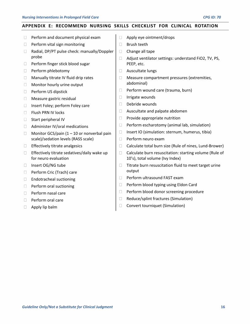
Nursing Interventions in Prolonged Field Care CPG ID: 70
Guideline Only/Not a Substitute for Clinical Judgment 16
APPENDIX E: RECOMMEND NURSING SKILLS CHECKLIST FOR CLINICAL ROTATION
� Perform and document physical exam � Perform vital sign monitoring � Radial, DP/PT pulse check: manually/Doppler
probe � Perform finger stick blood sugar � Perform phlebotomy � Manually titrate IV fluid drip rates � Monitor hourly urine output � Perform US dipstick � Measure gastric residual � Insert Foley; perform Foley care � Flush PRN IV locks � Start peripheral IV � Administer IV/oral medications � Monitor GCS/pain (1 – 10 or nonverbal pain
scale)/sedation levels (RASS scale) � Effectively titrate analgesics � Effectively titrate sedatives/daily wake up
for neuro evaluation � Insert OG/NG tube � Perform Cric (Trach) care � Endotracheal suctioning � Perform oral suctioning � Perform nasal care � Perform oral care � Apply lip balm
� Apply eye ointment/drops � Brush teeth � Change all tape � Adjust ventilator settings: understand FiO2, TV, PS,
PEEP, etc. � Auscultate lungs � Measure compartment pressures (extremities,
abdominal) � Perform wound care (trauma, burn) � Irrigate wounds � Debride wounds � Auscultate and palpate abdomen � Provide appropriate nutrition � Perform escharotomy (animal lab, simulation) � Insert IO (simulation: sternum, humerus, tibia) � Perform neuro exam � Calculate total burn size (Rule of nines, Lund-Brower) � Calculate burn resuscitation: starting volume (Rule of
10’s), total volume (Ivy Index) � Titrate burn resuscitation fluid to meet target urine
output � Perform ultrasound FAST exam � Perform blood typing using Eldon Card � Perform blood donor screening procedure � Reduce/splint fractures (Simulation) � Convert tourniquet (Simulation)

Nursing Interventions in Prolonged Field Care CPG ID: 70
Guideline Only/Not a Substitute for Clinical Judgment 17
APPENDIX F: ADDITIONAL INFORMATION REGARDING OFF-LABEL USES IN CPGS
PURPOSE
The purpose of this Appendix is to ensure an understanding of DoD policy and practice regarding inclusion in CPGs of “off-label” uses of U.S. Food and Drug Administration (FDA)–approved products. This applies to off-label uses with patients who are armed forces members.
BACKGROUND
Unapproved (i.e. “off-label”) uses of FDA-approved products are extremely common in American medicine and are usually not subject to any special regulations. However, under Federal law, in some circumstances, unapproved uses of approved drugs are subject to FDA regulations governing “investigational new drugs.” These circumstances include such uses as part of clinical trials, and in the military context, command required, unapproved uses. Some command requested unapproved uses may also be subject to special regulations.
ADDITIONAL INFORMATION REGARDING OFF-LABEL USES IN CPGS
The inclusion in CPGs of off-label uses is not a clinical trial, nor is it a command request or requirement. Further, it does not imply that the Military Health System requires that use by DoD health care practitioners or considers it to be the “standard of care.” Rather, the inclusion in CPGs of off-label uses is to inform the clinical judgment of the responsible health care practitioner by providing information regarding potential risks and benefits of treatment alternatives. The decision is for the clinical judgment of the responsible health care practitioner within the practitioner-patient relationship.
ADDITIONAL PROCEDURES
Balanced Discussion
Consistent with this purpose, CPG discussions of off-label uses specifically state that they are uses not approved by the FDA. Further, such discussions are balanced in the presentation of appropriate clinical study data, including any such data that suggest caution in the use of the product and specifically including any FDA-issued warnings.
Quality Assurance Monitoring
With respect to such off-label uses, DoD procedure is to maintain a regular system of quality assurance monitoring of outcomes and known potential adverse events. For this reason, the importance of accurate clinical records is underscored.
Information to Patients
Good clinical practice includes the provision of appropriate information to patients. Each CPG discussing an unusual off-label use will address the issue of information to patients. When practicable, consideration will be given to including in an appendix an appropriate information sheet for distribution to patients, whether before or after use of the product. Information to patients should address in plain language: a) that the use is not approved by the FDA; b) the reasons why a DoD health care practitioner would decide to use the product for this purpose; and c) the potential risks associated with such use.

Guideline Only/Not a Substitute for Clinical Judgment 1
JOINT TRAUMA SYSTEM CL INICAL PRACTICE GUIDELINE ( JTS CPG)
Ocular Injuries and Vision-Threatening Conditions in Prolonged Field Care (CPG ID: 66) This guide provides medical professionals with essential information on the recognition and treatment of ocular conditions when evacuation to an eye specialist is delayed.
Contributors
COL Mark Reynolds, MC, USA MSG Carl Hoover, 18D, USA LTC Jamie Riesberg, MC, USA COL (Ret) Robert Mazzoli, MD LTC Marcus Colyer, MC, USA COL (Ret) Scott Barnes, MD
MAJ Christopher Calvano, MC, USAR Dr. James Karesh, MD COL Clinton Murray, MC, USA CAPT (Ret) Frank K Butler, Jr, MD COL Sean Keenan, MC, USA Col Stacy Shackelford, USAF, MC
Publication Date: 01 Dec 2017
TABLE OF CONTENTS
Introduction ................................................................................................................................................................................. 4 Goals of Care ............................................................................................................................................................................... 4
1. Maintain High Suspicion for Ocular Injuries ........................................................................................................................ 4 2. Assess and Document Visual Function ................................................................................................................................ 4 3. Examine for Critical Physical Findings ................................................................................................................................. 5 4. Maintain Patient Comfort. Prevent Further Damage. ......................................................................................................... 5 5. Establish Contact with Eye Care Specialist. Prioritize Evacuation. ...................................................................................... 5
Specific Conditions ...................................................................................................................................................................... 6 1. Open Globe Injury ............................................................................................................................................................... 6 Figure 1. Open globe injury with corneal laceration, abnormal pupil shape, and blood in anterior chamber. ...................... 6 Figure 2. Central corneal laceration and lid laceration (due to large intraocular foreign body). ........................................... 6 Figure 3. Multiple, deep corneal lacerations, found to be closed globe injury on surgical exploration. ................................ 6
Goals ................................................................................................................................................................................... 6 Minimum ............................................................................................................................................................................ 6 Better .................................................................................................................................................................................. 7 Best ..................................................................................................................................................................................... 7
Figure 4. Open globe with posterior rupture and collapsed anterior chamber. ..................................................................... 7 Figure 5. Post-traumatic endophthalmitis with inflamed conjunctiva, inflammatory cells layered in anterior chamber. ..... 7 2. Retrobulbar Hemorrhage/Orbital Compartment Syndrome .............................................................................................. 8 Figure 6. Retrobulbar hemorrhage (pupil dilated for examination). ...................................................................................... 8 Figure 7. Retrobulbar hemorrhage, evaluation for proptosis. ................................................................................................ 8
Goal ..................................................................................................................................................................................... 8 Minimum ............................................................................................................................................................................ 8 Better .................................................................................................................................................................................. 9 Best ..................................................................................................................................................................................... 9
3. Blunt/Closed Globe Injury ................................................................................................................................................... 9 Figure 8. Layered hyphema in anterior chamber. ................................................................................................................... 9
Goal ................................................................................................................................................................................... 10 Minimum .......................................................................................................................................................................... 10

Ocular Injuries and Vision-Threatening Conditions in Prolonged Field Care CPG ID: 66
Guideline Only/Not a Substitute for Clinical Judgment 2
Best ................................................................................................................................................................................... 10 4. Eyelid Laceration ............................................................................................................................................................... 11 Figure 9. Eyelid margin laceration ......................................................................................................................................... 11 Figure 10. Complex lid laceration with medial canthus avulsion .......................................................................................... 11 Figure 11. Eyelid laceration with tissue loss due to camel bite. ........................................................................................... 11
Goals ................................................................................................................................................................................. 11 Minimum .......................................................................................................................................................................... 11 Better ................................................................................................................................................................................ 12 Best ................................................................................................................................................................................... 12
5. Orbital Fracture ................................................................................................................................................................. 12 Goals ................................................................................................................................................................................. 12 Minimum .......................................................................................................................................................................... 12 Better ................................................................................................................................................................................ 13 Best ................................................................................................................................................................................... 13
6. Chemical Injuries ............................................................................................................................................................... 13 Table 1. Modified Hughes Classification ............................................................................................................................... 13
Goals ................................................................................................................................................................................. 14 Minimum .......................................................................................................................................................................... 14
Figure 12. Grade II chemical injury with ischemia and epithelial defect. ............................................................................. 14 Figure 13. Grade III chemical injury. ..................................................................................................................................... 14 Figure 14. Grade IV chemical Injury. ..................................................................................................................................... 14
Better ................................................................................................................................................................................ 15 Best: .................................................................................................................................................................................. 15
7. Preseptal and Orbital Cellulitis .......................................................................................................................................... 15 Goal ................................................................................................................................................................................... 16 Preseptal cellulitis (Figure 15) .......................................................................................................................................... 16 Best ................................................................................................................................................................................... 16 Orbital cellulitis (Figure 16)............................................................................................................................................... 16 Minimum .......................................................................................................................................................................... 16
Figure 15. Preseptal cellulitis ................................................................................................................................................ 16 Figure 16. Orbital cellulitis .................................................................................................................................................... 16
Better ................................................................................................................................................................................ 17 Best ................................................................................................................................................................................... 17
8. Infectious Keratitis (Corneal Ulcer) ................................................................................................................................... 17 Figure 17. Early corneal ulcer ................................................................................................................................................ 17 Figure 18. Advanced corneal ulcer ........................................................................................................................................ 17
Goal ................................................................................................................................................................................... 17 Minimum .......................................................................................................................................................................... 17 Better ................................................................................................................................................................................ 17 Best ................................................................................................................................................................................... 18
9. Angle-closure Glaucoma ................................................................................................................................................... 18 Figure 19. Dendrite staining with fluorescein in herpes simplex keratitis. ........................................................................... 18 Figure 20. Angle-closure glaucoma ....................................................................................................................................... 18
Goals ................................................................................................................................................................................. 18 Better ................................................................................................................................................................................ 19 Best ................................................................................................................................................................................... 19
10. Eye Care in the Multitrauma/Thermal Burn Patient ......................................................................................................... 19 Figure 21. Extensive facial burns with lid contraction and orbital congestion. .................................................................... 19 Figure 22. (A) Exposure keratopathy; (B) with fluorescein staining. ..................................................................................... 19
Goal ................................................................................................................................................................................... 20 Minimum .......................................................................................................................................................................... 20 Better ................................................................................................................................................................................ 20 Best ................................................................................................................................................................................... 20
References ................................................................................................................................................................................. 20 Appendix A: Relative Afferent Pupillary Defect ......................................................................................................................... 22

Ocular Injuries and Vision-Threatening Conditions in Prolonged Field Care CPG ID: 66
Guideline Only/Not a Substitute for Clinical Judgment 3
Appendix B: Use of the Direct Opthalmoscope ......................................................................................................................... 23 Appendix C: Basic Ocular Examination ...................................................................................................................................... 24 Appendix D: Packing and Planning Considerations ................................................................................................................... 25 Appendix E: Ocular Injuries & Vision Threatening Conditions Summary Table ........................................................................ 26 Appendix F: Additional Information Regarding Off-label uses in CPGs ..................................................................................... 29

Ocular Injuries and Vision-Threatening Conditions in Prolonged Field Care CPG ID: 66
Guideline Only/Not a Substitute for Clinical Judgment 4
INTRODUCTION
Evaluation and treatment of ocular injuries and vision threatening conditions in a prolonged field care (PFC) situation can be extremely challenging. These events can lead to irreversible loss of vision with lasting effects on military service and overall quality of life. The goal of this clinical practice guideline (CPG) is to provide medical professionals with essential information on the recognition and treatment of ocular conditions when evacuation to an eye specialist is delayed. The guidelines are based on standard ophthalmic practice adapted to address the austere or remote environment, when the “Shield and Ship” guidelines are interrupted by delayed evacuation.
As with all medical concerns, recognition of the problem is the first step. This is a particular challenge for ocular conditions. Comprehensive ocular evaluation is not usually possible in austere locations and training in rapid recognition of ocular conditions may be limited. The ocular conditions covered in this guideline are the most common traumatic injuries and vision-threatening conditions that require rapid identification and treatment to prevent loss of vision. A more comprehensive review can be found in the Joint Trauma System CPG1 or Wilderness Medicine textbook.2
Telemedicine: Management of eye injuries is complex. Detailed physical examination information can only be communicated via pictures or video. Establish telemedicine consultation as soon as possible.
GOALS OF CARE
1. MAINTAIN HIGH SUSPICION FOR OCULAR INJURIES
The mechanism of injury will often suggest an ocular injury. Direct trauma with ocular laceration may be fairly obvious, but blunt trauma leading to an occult rupture on the posterior aspect of the globe or injury to the retina can be easily overlooked in a multitrauma patient. Small metallic fragments can penetrate the eye and leave it appearing deceptively intact. An awake and alert patient can report change in vision or eye pain; an unconscious patient cannot. Thermal burns to the face frequently cause eyelid burns and contraction, which increase the risk for exposure keratopathy. Critical details in the evaluation of ocular trauma include mechanism of injury and presence of properly worn ballistic eyewear at the time of injury.
2. ASSESS AND DOCUMENT VISUAL FUNCTION
Visual acuity is the vital sign of the eye. Whenever possible, visual acuity should be assessed and documented. Visual function immediately after trauma is an important prognostic indicator for visual outcome. Visual acuity can be effectively estimated with several field-expedient methods, starting with ability to read any printed letters such as labels on medical supplies. If the patient is unable to read letters, assess their ability to count fingers. If they cannot count fingers, evaluate for the ability to detect hand motions. If they cannot detect hand motion, evaluate light perception using a bright light. Document visual acuity along with other vital signs.
Assessing for a relative afferent pupillary defect (RAPD, also called the swinging flashlight test or Marcus Gunn Pupil; Appendix A) gives an indication of retina and optic nerve function. Shine a light in either eye; normally, both pupils should constrict equally. This reaction to light is equal unless there is damage to the optic nerve or the retina, in which case the pupil of the injured eye will dilate when the light is shone in the injured eye. Evaluating for an RAPD is essential in conditions such as retrobulbar hemorrhage (orbital compartment syndrome) or suspected retinal detachment. Notation of an RAPD is also an important prognostic factor for ophthalmic providers at later level of care.

Ocular Injuries and Vision-Threatening Conditions in Prolonged Field Care CPG ID: 66
Guideline Only/Not a Substitute for Clinical Judgment 5
3. EXAMINE FOR CRITICAL PHYSICAL FINDINGS
An obvious globe laceration or rupture with prolapsed intraocular contents can be a striking picture but may not be present in every severe eye injury. At times, the only findings of a significant eye injury may be penetrating periocular trauma or lid lacerations, a peaked or teardrop pupil, or abnormal anterior chamber depth.
Estimation of intraocular pressure (IOP) is essential in injuries such as retrobulbar hemorrhage, but is contraindicated in injuries with obvious or suspected open globe injury (OGI). If no OGI is suspected, IOP can be estimated using a two finger method. Using the index finger of each hand, gently apply alternating pressure on the globe through closed lids. There should be mild indentation of the eye with normal IOP (normal range, 10–21mmHg). With increased IOP, the globe will be much firmer when compared to the opposite eye, or the examiner’s own eye. The orbit around the eye may also feel tense in a retrobulbar hemorrhage.
A more detailed examination of the eye can be facilitated by the use of a direct ophthalmoscope for magnification. The examiner can use the plus lens dial (green or black numbers) to provide additional magnification. Details are found in Appendix B Use of the Direct Ophthalmoscope. Although deployed locations may have specific guidance against contact lens use, this may still be encountered. If a contact lens is clearly visible and accessible, it can be gently removed with forceps. Fluorescein will stain a contact lens, allowing for easy visualization.
4. MAINTAIN PATIENT COMFORT. PREVENT FURTHER DAMAGE.
Pain control is an important component of care in ocular injuries. Standard Tactical Combat Casualty Care (TCCC) pain control guidance applies to ocular injuries, including analgesic doses of ketamine if needed in systemic polytrauma. Additional guidance for pain control in PFC may be found in the CPG Analgesia and Sedation Management during Prolonged Field Care.3
Pain is not always present with serious eye injuries, and lack of pain should not be interpreted as lack of injury. Ocular injuries can cause a great deal of anxiety for patients, and this may affect care. A benzodiazepine may be added to the treatment plan for anxiety control and facilitation of care (diazepam 10 mg by mouth [PO] every 6 hours as needed).
A traumatized eye is highly susceptible to further damage; antiemetic medications are essential to prevent retching and increased pressure that can have significant effects on visual outcome.
Patching both eyes to decrease sympathetic eye movements has not been shown to improve visual outcome. Occluding both eyes will render the patient unable to move independently, may increase anxiety, and may put the patient and provider at increased risk in any PFC environment. The use of standard eye protection during patient transport can reduce the risk of further ocular injury or injury to the fellow eye. Eye protection can be used over a rigid shield to provide increased protection.
5. ESTABLISH CONTACT WITH EYE CARE SPECIALIST. PRIORITIZE EVACUATION.
Determining the full extent of ophthalmic injuries and the resultant threat of permanent loss of vision is challenging without full ophthalmic training and equipment. All potential vision-threatening injuries should be evacuated with a goal of care by an eye surgeon within 24 hours. In some cases, with prompt teleconsultation or video consultation, it may be safe to delay evacuation to reduce the effect on the operational situation while providing necessary ophthalmic care. Evacuation within 24 hours is not possible in all situations; therefore, the goal of teleconsultation and forward care is to reduce morbidity and achieve the best possible outcome. In some operational environments, optometrists may be available to provide additional care or consultation closer to the point of injury.

Ocular Injuries and Vision-Threatening Conditions in Prolonged Field Care CPG ID: 66
Guideline Only/Not a Substitute for Clinical Judgment 6
Ocular examinations have many specialized components that a specialist may request. An example template with explanation can be found in Appendix C Basic Ocular Examination.
SPECIFIC CONDITIONS
1. OPEN GLOBE INJURY
OGIs can result from penetrating/perforating trauma or from rupture of the globe due to massive compressive forces (Figures 1–5). Prompt surgical exploration and repair are crucial to restore or salvage vision and to prevent a devastating outcome. Safe and effective closure of an OGI is not yet feasible in a prehospital setting.
Figure 1. Open globe injury with corneal laceration, abnormal pupil shape, and blood in anterior chamber.
Figure 2. Central corneal laceration and lid laceration (due to large intraocular foreign body).
Figure 3. Multiple, deep corneal lacerations, found to be closed globe injury on surgical exploration.
Photographs by COL Mark Reynolds.
Goals
Prevent further damage to the eye, prevent infection in the eye (endophthalmitis), and evacuate to an eye surgeon as soon as possible.
Minimum
Maintain high suspicion for OGI; treat any suspected open globe as an open globe until surgical exploration is available.
Obtain and record visual acuity from the injured and noninjured eye.
Apply a rigid eye shield without any type of gauze or bandaging under the shield to prevent further damage, per TCCC guidelines.4
Initiate endophthalmitis prophylaxis with moxifloxacin 400mg PO daily or levofloxacin 750mg PO daily; if intravenous (IV) administration is necessary, ertapenem 1g IV or intraosseously (IO) daily.5 Initiate pain control as needed.
Initiate antiemetic (ondansetron 4mg oral dissolving tablet [ODT] IV/IO or intramuscularly [IM] every 8 hours as needed).
Raise head 30°–45°.
Activate evacuation with the goal of surgery within 24 hours.
Initiate teleconsultation with photographs.

Ocular Injuries and Vision-Threatening Conditions in Prolonged Field Care CPG ID: 66
Guideline Only/Not a Substitute for Clinical Judgment 7
Better
Minimize patient movements; maintain supine position with head at 30°–45°.
Maintain endophthalmitis prophylaxis with an additional dose of moxifloxacin 400mg PO daily or levofloxacin 750mg PO daily; if IV administration is necessary, ertapenem 1g IV/IO daily and the addition of clindamycin 300mg PO or IV every 8 hours if available; this is to cover Bacillus cereus, a particular concern in contaminated OGI.6
Maintain antiemetic and pain control.
Best
Perform a detailed ocular evaluation to include visual acuity and RAPD, and note any suspicious findings.
Evaluation should be repeated with any reported change in vision or pain level by the patient. If symptoms are stable, perform ocular evaluation every 4 hours and before transfer.
Initiate real-time video telemedicine consultation.
Coordinate surgical care within 8 hours of injury.
No altitude restrictions are required for OGIs.
NOTES: Ultrasound is contraindicated for suspected OGI because it places pressure on the eye.
Rigid eye shields are available in several different designs. Fit should be checked to ensure protection without any pressure on the eye. Standard eye protection may also be used to shield the injured eye.
Figure 4. Open globe with posterior rupture and collapsed anterior chamber.
Figure 5. Post-traumatic endophthalmitis with inflamed conjunctiva, and inflammatory cells layered in anterior chamber.
Photographs by COL Mark Reynolds.

Ocular Injuries and Vision-Threatening Conditions in Prolonged Field Care CPG ID: 66
Guideline Only/Not a Substitute for Clinical Judgment 8
2. RETROBULBAR HEMORRHAGE / ORBITAL COMPARTMENT SYNDROME
Retrobulbar hemorrhage (RBH) is the most common cause of orbital compartment syndrome (OCS). It is a result of bleeding into the confined orbital space behind the eye, usually associated with blunt trauma (Figures 6 and 7). It is a vision-threatening condition causing increased pressure in the eye, leading to irreversible vision loss. Vision loss typically will occur after approximately 90 minutes of increased pressure.
Figure 6. Retrobulbar hemorrhage (pupil dilated for examination).
Figure 7. Retrobulbar hemorrhage, evaluation for proptosis.
Photographs by COL Mark Reynolds.
Other causes of OCS include orbital congestion secondary to burn resuscitation and significant orbital emphysema after orbital fracture (pneumo-orbita). OCS from any cause may have a delayed onset. Patients with trauma to the orbit must be closely monitored for development of OCS.
Goal
Lower the orbital compartment pressure as soon as possible to prevent tissue damage.
Minimum
Prompt recognition of injury and identification of the need for intervention
History of trauma with any of the following findings:
Proptosis: bulging of the affected eye compared with the other eye; proptosis in RBH is often tense and painful
Increased orbital pressure around the eye or IOP by palpation (increased firmness and resistance compared with opposite eye)
Decrease in or loss of visual acuity
Presence of an RAPD (Appendix A)
Raise head 30°–45°.
Initiate pain control as needed.
Initiate antiemetic (ondansetron 4mg ODT/IV/IO/IM every 8 hours as needed).
Perform lateral canthotomy/cantholysis (LCC) as soon as possible, within 90 minutes of injury if evacuation to a surgical capability is anticipated to take more than 60 minutes.
Activate evacuation with goal of evaluation by an eye surgeon within 24 hours.
Initiate teleconsultation with photographs.

Ocular Injuries and Vision-Threatening Conditions in Prolonged Field Care CPG ID: 66
Guideline Only/Not a Substitute for Clinical Judgment 9
Better
Minimize patient movements; maintain supine position with head at 30°–45°.
Ice packs and avoidance of compressive dressings7
Monitor for return of elevated orbital pressure.
Best
Initiate a detailed ocular evaluation and continue monitoring IOP, vision, and RAPD.
Continue to check for recurrence of elevated IOP even after LCC. If the vision deteriorates and the eye again becomes firm after LCC, this may signify rebleeding in the orbit. Evacuation of orbital hemorrhage is not feasible in a PFC environment and rebleeding will require medical treatment.8
Acetazolamide: 500mg PO initial dose, followed by 250mg PO 4 times per day (Note: contraindicated in patients with sickle cell trait)
If acetazolamide is not available or if the patient cannot take PO, either 3% hypertonic saline 250mL IV or mannitol: 1g/kg IV over 30–60 minutes can be used to decrease IOP.9
Corticosteroid: 1g methylprednisolone IV once 10
Initiate real-time video telemedicine consultation.
No altitude restrictions are required for evacuation.
NOTES: LCC is a vision-saving procedure with minimal risk of causing additional ocular injury. When in doubt,
perform the LCC immediately.
In thermal burns, consider early LCC (before full OCS develops). Fluid resuscitation requirements will take precedence over the use of medical treatments to reduce IOP.
3. BLUNT / CLOSED GLOBE INJURY
This category includes anterior segment injuries such as hyphema (bleeding into the anterior chamber) and posterior segment injuries such as vitreous hemorrhage and retinal detachment. Blunt trauma can result in severe loss of vision.
Figure 8. Layered hyphema in anterior chamber.
©2017 American Academy of Ophthalmology, reprinted with permission.
Hyphema can lead to increased IOP and corneal blood staining. This is graded on the amount of blood in the anterior chamber. The risk of IOP elevation increases with the grade of the hyphema (Figure 8).11
Grade 0: no visible blood layering
Grade 1: blood fills less than one-third of anterior chamber
Grade 2: blood fills one-third to one-half of anterior chamber

Ocular Injuries and Vision-Threatening Conditions in Prolonged Field Care CPG ID: 66
Guideline Only/Not a Substitute for Clinical Judgment 10
Grade 3: blood fills one-half to less than total anterior chamber
Grade 4: blood fills entire anterior chamber
Goal
Identify significant ocular injuries; protect the eye from further injury.
Minimum
Obtain and record visual acuity and critical injury details (e.g., mechanism of injury, presence of eye protection).
Protect the injured globe and prevent further damage with a rigid shield.
Raise the head 30°–45°; this allows any free-floating blood in the anterior chamber to settle away from the pupil and prevent pupillary block (which can lead to angle closure and elevated IOP).11
Initiate pain control as needed; avoid nonsteroidal anti-inflammatory drugs, because of risk of worsening intraocular bleeding.
Prevent further injury with antiemetics (ondansetron 4mg ODT/IV/IO/IM every 8 hours as needed).
Activate evacuation with goal of evaluation by an eye surgeon within 24 hours.
Initiate teleconsultation with photographs.
Best
Initiate a detailed ocular evaluation to direct treatment.
Hyphema (anterior chamber injury)11
Topical corticosteroid drop (prednisolone acetate 1%) 4 times per day
Cycloplegic eye drop (cyclopentolate 1%), 1 drop every 8 hours
Monitor for rebleeding when the clot in anterior chamber retracts, usually at 3–5 days after injury.
This may result in vision change and increased size of hyphema.12
If there is evidence of further bleeding or increasing IOP, initiate medications to decrease IOP:
Timolol 0.5%, 1 drop twice a day in affected eye
Acetazolamide 500mg PO initial dose, followed by 250mg PO 4 times per day (Note: contraindicated in patients with sickle cell trait) or 3% hypertonic saline 250mL IV or mannitol: 1g/kg IV over 30–60 minutes.
NOTE: Tranexamic acid for prevention of rebleeding in hyphema has not shown any benefit 13 but may be used in multitrauma patients if otherwise indicated.
Posterior chamber injury: Injuries to the retina and optic nerve as a result of blunt injury will result in vision loss. Findings may include decreased visual acuity, vision loss, loss of red reflex through the pupil, positive RAPD, or evidence of vitreous hemorrhage or retinal detachment on ultrasound evaluation.
Initiate supplemental oxygen as available if suspicious for retinal detachment (e.g., cut in visual field, decreased vision, positive RAPD); this may improve visual outcome.14
If no evidence of OGI, perform careful ultrasound to evaluate vitreous and retina, if available/trained. Transmit ultrasound images with telemedicine consultation to an eye specialist.
Initiate real-time video telemedicine consultation.

Ocular Injuries and Vision-Threatening Conditions in Prolonged Field Care CPG ID: 66
Guideline Only/Not a Substitute for Clinical Judgment 11
No altitude restrictions are required for blunt/closed globe injury.
4. EYELID LACERATION
Lid lacerations can result from either sharp or blunt trauma (Figures 9–11). As with other injuries, the primary concern with lid injuries is the possibility of underlying globe injury. Lid lacerations have a low incidence of infection (unless the causative factor is an animal or human bite). Any avulsed tissue should be preserved in saline and chilled, whenever possible, and sent with the patient—not discarded or debrided. Meticulous closure of eyelid structures with proper magnification is usually required to maintain lid function. If fat is visible in an eyelid laceration, this indicates violation of the orbital septum, a key anatomic barrier to infection. If prolapsed orbital fat is identified, appropriate antibiotic coverage is needed as well as expedited evacuation for surgical exploration and repair. Do not attempt to excise or suture exposed orbital tissue; this can lead to uncontrolled bleeding in the orbit.
Figure 9. Eyelid margin laceration
Figure 10. Complex lid laceration with medial canthus avulsion
Figure 11. Eyelid laceration with tissue loss due to camel bite.
Photographs by LTC Marcus Colyer (9) and COL Mark Reynolds (10, 11).
Goals
Prevent infection; protect the eye from further injury.
Minimum
Maintain high suspicion for OGI; treat any suspected open globe as such until surgical capability is available.
Obtain and document visual acuity from the injured and noninjured eyes.
If there is any concern for OGI, protect the injured globe and prevent further damage with a rigid eye shield. Polyethylene film (food grade) may be used to cover the eyelid wound under the rigid shield to prevent drying of the injured tissue.
Initiate pain control as needed.
Activate evacuation with goal of evaluation by an eye surgeon within 24 hours.
Initiate teleconsultation with photographs.

Ocular Injuries and Vision-Threatening Conditions in Prolonged Field Care CPG ID: 66
Guideline Only/Not a Substitute for Clinical Judgment 12
Better
For foreign body penetration, animal bite, or laceration with visible orbital fat, start antibiotics: moxifloxacin 400mg PO daily or levofloxacin 750mg PO daily or amoxicillin/clavulanic acid 875mg/125mg PO every 12 hours or ertapenem 1g IV/IO daily.
Best
Initiate a detailed ocular evaluation to include visual acuity, RAPD, and note any suspicious findings.
Irrigate wound very gently with clean water (or sterile saline, if available).15
Do not debride any tissue.
Temporary closure with steristrips
Tetanus prophylaxis
Consider the need for rabies vaccination.15
Initiate real-time video telemedicine consultation.
No altitude restrictions are required for evacuation.
5. ORBITAL FRACTURE
Fracture of the orbital bones occurs when an object that is larger than the width of the orbit (e.g., fist or softball) strikes the orbit. The acute expansion of orbital contents and mechanical buckling forces can result in fractures of the medial wall or orbital floor. This can cause herniation of orbital contents into the surrounding sinuses and entrapment of the extraocular muscles in the fracture site. Physical examination findings consistent with orbital fracture include a palpable and painful step-off along the orbital rim, enophthalmos (globe is further back in the orbit compared with the other eye), restricted eye movement, and numbness below the eye (caused by damage to the infraorbital nerve).16 Trismus and malocclusion may indicate a larger zygomaticomaxillary complex fracture. Orbital fractures are not ophthalmic emergencies but may require surgical treatment to prevent the complication of double vision from ocular misalignment.
Goals
Evaluate for concurrent open or closed globe injury and prevent long-term complications.
Minimum
Maintain a high suspicion for associated OGI; treat as a suspected open globe until eye surgical evaluation is available.
Obtain and record visual acuity from the injured and noninjured eyes.
Instruct the patient not to blow nose. This may force air into the orbit through fracture site, leading to OCS from pneumo-obita, which would require LCC.
Initiate pain control as needed.
Raise head 30°–45°.
Initiate teleconsultation with photographs.
Activate evacuation with goal of evaluation by an eye surgeon within 24 hours.

Ocular Injuries and Vision-Threatening Conditions in Prolonged Field Care CPG ID: 66
Guideline Only/Not a Substitute for Clinical Judgment 13
Better
Initiate antibiotics if an orbital fracture suspected; this is to prevent sinus pathogens from spreading to the orbital tissues: moxifloxacin 400mg PO daily or levofloxacin 750mg PO daily or amoxicillin/clavulanic acid 875mg/125mg PO every 12 hours or ertapenem 1g IV/IO daily.
Nasal decongestants such as oxymetazoline (e.g., Afrin; Bayer, http://www.bayer.us/) nasal spray twice a day for 3 days (limit use to 3 days to prevent rebound effect). Oral decongestants, such as pseudoephedrine 30mg every 6 hours, can be used if nasal spray is not available.
Prevent further injury with antiemetics (ondansetron 4mg ODT/IV/IO/IM every 8 hours as needed).
Best
Initiate a detailed ocular evaluation to include visual acuity, RAPD, and note any suspicious findings.
Ice packs for 20 minutes every 1–2 hours for the first 48 hours to reduce swelling.
Monitor for delayed development of OCS and perform LCC as needed.
Initiate real-time video telemedicine consultation.
NOTES: No altitude restrictions are required with orbital fractures, but patient should be monitored for
increasing pain and/or decreasing vision from pneumo-orbita OCS requiring LCC.
An important consideration in orbital floor fractures is the inferior rectus muscle becoming entrapped in the fracture (so-called trapdoor fracture). The resultant traction on the rectus muscle can trigger the oculocardiac reflex and result in intractable nausea and vomiting, symptomatic bradycardia, and possibly heart block. Although this is more common in pediatric patients (termed “white-eye” blow-out fractures), it is not exclusive to the pediatric population and has been reported in young healthy adults. Urgent surgical repair (within 72 hours) is recommended for an entrapped fracture with these symptoms.
6. CHEMICAL INJURIES
Acid (e.g., sulfuric, hydrochloric) and alkali (e.g., bleach, lime, ammonia) burns can cause significant injuries leading to permanent loss of vision and are considered ophthalmic emergencies. Alkali burns are more common and have more potential for damage than acid burns.16 Ongoing ocular care beyond the initial thorough irrigation will be required if evacuation is delayed. Chemical injuries are graded on a scale of I to IV.17 The modified Hughes classification (Table 1) can be used to grade the degree of limbal ischemia that correlates with prognosis. Regardless of the chemical causing the injury, immediate irrigation is the essential first step. Additional treatment will be based on grade of injury. Injuries are graded on the basis of the following examination findings (Figures 12–14):
Table 1. Modified Hughes Classification
Grade Cornea Epithelium Corneal Clarity Limbal Ischemia I Less than one-third loss Iris details clearly visible No ischemia II More than one-third epithelial loss Iris details blurred but visible Less than 25% ischemia III Complete epithelial loss Pupil can be seen 25%–50% ischemia IV Complete epithelial loss Opaque cornea More than 50%

Ocular Injuries and Vision-Threatening Conditions in Prolonged Field Care CPG ID: 66
Guideline Only/Not a Substitute for Clinical Judgment 14
Corneal epithelial damage: How much epithelium has been lost?
Clarity of cornea: Can the normal structures (iris, pupil) be seen through the cornea?
Limbal ischemia: Does the conjunctiva at the edge of the cornea appear normal or are there areas that are blanched white?
Grade I injuries may have corneal epithelial damage but a clear cornea, no corneal opacity, and no limbal ischemia.
These injuries generally carry a good prognosis for recovery. Irrigation and topical care are frequently the only required interventions.
Grade II through IV injuries will have corneal haze or opacity and limbal ischemia.
These injuries will have a guarded prognosis and will require more intensive treatment. Grading is determined by the most severe finding. For example, an eye with a clear cornea but showing limbal ischemia would be classified as grade II or higher.
Goals
Initiate eye irrigation as quickly as possible to reduce damage to the eye, treat the injury to prevent or reduce scarring and visual loss.
Minimum
Immediate thorough irrigation to remove the chemical agent is the essential first step (IV fluid, sterile water, or clean water).
Continue irrigation, using at least 2L of fluid.
Use tetracaine eye drops as needed to facilitate irrigation (unpreserved lidocaine 2% can be substituted as eye drops if tetracaine is not available).
Irrigation may not flush all chemical agents from the eye; examine for particulate matter and remove using a cotton tip applicator (CTA).
Initiate pain control as needed. DO NOT use topical anesthetics for pain control; they significantly impair corneal healing.
Initiate teleconsultation with photographs (include full facial views).
Figure 12. Grade II chemical injury with ischemia and epithelial defect.
Figure 13. Grade III chemical injury.
Figure 14. Grade IV chemical Injury.
Photographs by The University of Iowa and EyeRounds.org (12) and ©2017 American Academy of Ophthalmology, reprinted with permission. (13, 14).

Ocular Injuries and Vision-Threatening Conditions in Prolonged Field Care CPG ID: 66
Guideline Only/Not a Substitute for Clinical Judgment 15
Better
Evaluate ocular pH using a urine test strip and CTA.
Do not place the test strip directly on the eye. Roll a
CTA across the conjunctival surface and then onto the test strip. If pH ≠ 7, continue irrigation and recheck until pH = 7.
Best:
Initiate real-time video telemedicine consultation; treatment duration for more significant chemical burns will vary depending on the injury and are best determined by an eye-care specialist
Further treatments and need for evacuation will be directed by the grade of injury. Evaluation will require fluorescein strips to evaluate the corneal epithelium and a light source, preferably with a red-free option (i.e. green lens), for evaluation of limbal ischemia.
Grade I:
Topical antibiotic ointment (e.g., erythromycin ophthalmic ointment) 3 times per day Cycloplegic eye drop (cyclopentolate 1%), 1 drop every 8 hours, if available, for photophobia
Preservative-free artificial tears 3 times per day, alternating between ointment treatments
Grades II–IV:
Moxifloxacin 0.5% eye drops, 1 drop every 8 hours
Topical corticosteroid (e.g., tobramycin/dexamethasone or prednisolone acetate 1%) 1 drop every hour while awake
Cycloplegic drop (cyclopentolate 1%), 1 drop every 8 hours, if available
Doxycycline 100mg PO every 12 hours; this has anti-inflammatory and anticollagenase benefits for the ocular surface.
The following have been shown to improve corneal healing with severe chemical burns; add to the treatment if available:
Vitamin C 2g 4 times per day.17
Supplemental oxygen (administer 100% for 1 hour twice daily).19 No data are available for the effectiveness of lower doses of oxygen.
Reassess frequently until evacuation.
No altitude restrictions for flight
7. PRESEPTAL AND ORBITAL CELLULITIS
Infection anterior to the orbital septum (usually involving the eyelid) is termed preseptal cellulitis. Preseptal cellulitis will present with tenderness, swelling, and erythema of the eyelids, with no orbital findings (e.g., no sign of proptosis, eye movement restriction, or change in vision). Preseptal cellulitis can generally be managed with oral antibiotics, but the possibility of methicillin resistant Staphylococcus aureus (MRSA) must be considered.
Infection in the orbit (posterior to the orbital septum) can occur as a result of adjacent sinusitis, skin infection, puncture wounds, or orbital foreign bodies. Multiple organisms, including staphylococcal, streptococcal, and gram-negative bacteria, are usually responsible. Orbital cellulitis has the potential to progress rapidly and may lead to irreversible loss of vision or intracranial extension. Orbital cellulitis presents with pain, proptosis, conjunctival injection, decreased vision, and loss of ocular mobility (which may cause double vision).

Ocular Injuries and Vision-Threatening Conditions in Prolonged Field Care CPG ID: 66
Guideline Only/Not a Substitute for Clinical Judgment 16
Goal
Recognize infection early and start oral antibiotics for preseptal cellulitis and IV antibiotics for orbital cellulitis. Evacuate to an eye surgeon as rapidly as possible if orbital cellulitis is suspected.
Preseptal cel lul it is (Figure 15)
Minimum
Moxifloxacin 400mg PO daily or levofloxacin 750mg PO daily.
Does not cover MRSA; follow closely for worsening condition.
Initiate pain control as needed.
Activate evacuation with goal of evaluation by an eye surgeon within 24 hours.
Initiate teleconsultation with photographs.
Best
Trimethoprim sulfamethoxazole DS 1 tablet PO every 8 hours combined with amoxicillin/clavulanic acid 875mg every 12 hours.
Orbital cellul it is (Figure 16)
Minimum
Prompt recognition of the condition and the need for rapid intervention
Initiate pain control as needed.
Initiate IV access with broad-spectrum IV antibiotics: ertapenem 1g IV/IO daily or levofloxacin 500mg IV once a day.
Initiate pain control as needed.
Activate evacuation with goal of evaluation by an eye surgeon within 24 hours.
Initiate teleconsultation with photographs (include full facial views).
Figure 15. Preseptal cellulitis Figure 16. Orbital cellulitis
Reprinted with permission of the University of Iowa and EyeRounds.org.
Reprinted with permission of the American Academy of Ophthalmology.

Ocular Injuries and Vision-Threatening Conditions in Prolonged Field Care CPG ID: 66
Guideline Only/Not a Substitute for Clinical Judgment 17
Better
Nasal decongestants such as oxymetazoline (e.g., Afrin) nasal spray twice a day for 3 days (limit use to 3 days to prevent rebound effect); this will aid in draining of contributing sinusitis. Oral decongestants, such as pseudoephedrine 30mg every 6 hours, can be used if nasal spray is not available.
Best
Initiate a detailed ocular examination.
Monitor vision every 4 hours until evacuation (may take 24–36 hours to show improvement).
Initiate real-time video telemedicine consultation.
No altitude restrictions for flight
8. INFECTIOUS KERATITIS (CORNEAL ULCER)
Infections in the cornea can lead to corneal scarring and permanent effects on vision (Figures 17 and 18). The most common risk factor for corneal ulcer is contact lens use.
Figure 17. Early corneal ulcer Figure 18. Advanced corneal ulcer
Photograph courtesy of John D. Sheppard, MD, and Clelia Sheppard.)
Reprinted with permission of the American Academy of Ophthalmology.
Goal
Early recognition and treatment to prevent long-term scarring of the cornea.
Minimum
Moxifloxacin eye drops 1 drop every 15 minutes for the first 2 hours after diagnosis, then 1 drop every hour while awake
Initiate pain control as needed. DO NOT use topical anesthetics for pain control; they significantly impair corneal healing.
Initiate teleconsultation with photographs.
Activate evacuation (goal is ophthalmic care within 24 hours if lesion is large, central, or affects vision).
Better
Obtain a culture before beginning treatment for sight-threatening or severe keratitis with suspected infection, such as large central corneal infiltrate that extends to the middle to deep stroma.20

Ocular Injuries and Vision-Threatening Conditions in Prolonged Field Care CPG ID: 66
Guideline Only/Not a Substitute for Clinical Judgment 18
Provide intense loading dose of moxifloxacin 0.5% eye drops 1 drop every 5–15 minutes for the first 30–60 minutes (patient can self-administer loading dose if reliable) after culture obtained.
Treatment dose: 1 drop every 30–60 minutes around the clock until epithelial defect is closed.20
Cycloplegic eye drop (cyclopentolate 1%), 1 drop every 8 hours for photophobia.
Best
Real-time video telemedicine consultation
Collagen corneal shield (national stock no. [NSN] 6515-01-482-9391) soaked in moxifloxacin drops for transport (generally 5–10 drops) and placed over the corneal infiltrate. This enables release of the medication to the ocular surface during transport, rather than administering repeated dosing.21
No altitude restrictions for flight
NOTE: Topical steroid drops may be useful to reduce inflammation after the infection is controlled with topical antibiotics. Initiation of topical steroid drops should only be done under the direction of an eye care specialist after teleconsultation.
Herpes simplex virus (HSV) keratitis is an additional form of keratitis that usually occurs in patients with a history of previous episodes. HSV keratitis may demonstrate a specific dendritic staining pattern with fluorescein (Figure 19). After recognition, treatment can be initiated with oral acyclovir (400mg PO 5 times per day).
9. ANGLE -CLOSURE GLAUCOMA
Blockage of the normal flow of aqueous fluid in the anterior chamber of the eye will lead to increased IOP. If left untreated, blood flow to the posterior segment of the eye will be affected, leading to irreversible vision loss. The aqueous drain system can become blocked owing to anatomic variations, changes in lens size, inflammation, and trauma (Figure 20).
Figure 19. Dendrite staining with fluorescein in herpes simplex keratitis.
Figure 20. Angle-closure glaucoma
Reprinted with permission of the University of Iowa and EyeRounds.org.
Reprinted with permission of the American Academy of Ophthalmology.
Goals
Prompt diagnosis and identification, lowering of IOP.
Minimum
Diagnosis
Pain (often described as a deep pain, similar to tooth pain)
Decrease in or loss of visual acuity

Ocular Injuries and Vision-Threatening Conditions in Prolonged Field Care CPG ID: 66
Guideline Only/Not a Substitute for Clinical Judgment 19
Photophobia
Dull or cloudy appearance of the cornea due to corneal edema
Fixed, mid-dilated pupil (usually occurs after IOP reaches 30–40mmHg)
Increased IOP by palpation
Acetazolamide 500mg PO initial dose, then 250mg PO every 4 hours to decrease IOP. (Note: contraindicated in patients with sickle cell trait.)
Initiate teleconsultation with photographs. Activate evacuation with goal of evaluation by an eye surgeon within 24 hours.
Better
Oral acetazolamide plus topical IOP-lowering eye drops (timolol 0.5%, 1 drop twice a day in the affected eye).
Administer antiemetics as required by patient symptoms (ondansetron 4mg ODT/IV/IO/IM every 8 hours as needed).
Best
Topical corticosteroids (prednisolone acetate 1%) 1 drop every hour after consultation with ophthalmology or optometry.
3% hypertonic saline 250mL IV or mannitol 1g/kg over 30–60 minutes can be used to decease IOP if the aforementioned interventions are not effective.9
10. EYE CARE IN THE MULTITRAUMA / THERMAL BURN PATIENT
Patients with multisystem trauma who are intubated and sedated are at risk of developing corneal complications due to metabolic derangements and impaired ocular protective mechanisms.22 The presence of thermal facial burns puts the patient at high risk for exposure keratopathy. Loss of the normal blink reflex, impaired tear production, abnormal tear film dynamics, and incomplete eyelid closure, combined with the inability to relay ocular complaints all contribute to the development of exposure keratopathy and increase the risk for infectious keratitis.23 If there is no concern for OGI, ultrasound examination may be performed if personnel are equipped and trained. Patients with head and facial burns with eyelid involvement are especially prone to entropion (with burned eyelash stubs abrading the cornea) as well as exposure keratopathy from scar-related lid retraction and proptosis from orbital congestion (Figures 21 and 22).
Figure 21. Extensive facial burns with lid contraction and orbital congestion.
Figure 22. (A) Exposure keratopathy; (B) with fluorescein staining.
Photograph by COL Mark Reynalds Reprinted with permission of the University of Iowa and EyeRounds.org

Ocular Injuries and Vision-Threatening Conditions in Prolonged Field Care CPG ID: 66
Guideline Only/Not a Substitute for Clinical Judgment 20
Goal
Prevent ocular exposure and corneal injury in high risk patients.
Minimum
Initiate ocular surface protection with sterile petrolatum or methylcellulose drops to keep the ocular surface from drying out. (Do not substitute a nonophthalmic lubricant).
For thermal facial burns, instill erythromycin ophthalmic ointment or sterile petrolatum every 2 to 4 hours.
Better
Gentle horizontal taping of lids with hypoallergenic tape in conjunction with ocular surface protection to protect the eyes.24
Evaluate the eyes and instill a surface protectant at least every 8 hours. Ensure the surface of the eye is not dry, there is no pressure on the eye, pupil reactivity has not changed, and the eyelids are completely closed to protect the eye.
Best
Detailed ocular evaluation
Cover eyes with polyethylene film to prevent drying.
Food-grade film is safe for use around the eyes.
Apply from the eyebrow to the maxilla to ensure proper coverage
No altitude restrictions for flight
NOTE: Surgilube should never be instilled into the eye as a lubricant, because if can cause corneal toxicity.18 When used for ultrasound examination, place a thin film (e.g., food-grade polyethylene or Tegaderm [3M, http://www.3m. com]) over the closed eyelid.
REFERENCES
1. Joint Trauma System, Initial care of ocular and adnexal injuries by non-ophthalmologists at Role 1, Role 2, and nonophthalmic Role 3 facilities CPG. 24 Nov 2014. https://jts.amedd.army.mil/index.cfm/PI_CPGs/cpgs Accessed Mar 2018.
2. Butler FK Jr. The eye in the wilderness. In: Auerbach P (ed.) Wilderness Medicine. 6th ed. Maryland Heights, MO: Mosby; 2012.
3. Joint Trauma System, Analgesia and sedation management during prolonged field care CPG, 11 May 2017. https://jts.amedd.army.mil/index.cfm/PI_CPGs/cpgs Accessed Mar 2018.
4. Committee on Tactical Combat Casualty Care. Tactical Combat Casualty Care guidelines for medical personnel. https://www.deployedmedicine.com/market/11/content/40 Accessed Mar 2018
5. Peterson K, Colyer MH, Hayes DK, et al. Prevention of infections associated with combat-related eye, maxillofacial, and neck injuries. J Trauma. 2011;71:S264–S269.
6. Bhagat N, Nagori S, Zarbin M. Post-traumatic infectious endophthalmitis. Surv Ophthalmol. 2011;56:214–251.

Ocular Injuries and Vision-Threatening Conditions in Prolonged Field Care CPG ID: 66
Guideline Only/Not a Substitute for Clinical Judgment 21
7. Ord RA. Postoperative retrobulbar haemorrhage and blindness complicating trauma surgery. Br J Oral Surg.1981;19:202–207.
8. Lima V, Burt B, Leibovitch I, et al. Orbital compartment syndrome: the ophthalmic surgical emergency. Surv Ophthalmol. 2009;54: 441–449.
9. Harju M, Kivela T, Lindbohm N, et al. Intravenous hypertonic saline to reduce intraocular pressure. Acta Ophthalmologica. 2013;91:625–629.
10. Wood CM. The medical management of retrobulbar haemorrhage complicating facial fractures: a case report. Br J Oral Maxillofac Surg. 1989;27:291–295.
11. Walton W, Von Hagen S, Grigorian R, et al. Management of traumatic hyphema. Surv Ophthalmol. 2002;47:297–334.
12. Fong LP. Secondary hemorrhage in traumatic hyphema. Predictive factors for selective prophylaxis. Ophthalmology. 1994;101: 1583–1588.
13. Albiani DA, Hodge WG, Pan YI, et al. Tranexamic acid in the treatment of pediatric traumatic hyphema. Can J Ophthalmol. 2008;43:428–431.
14. Mervin K, Valter K, Maslin J, et al. Limiting photoreceptor death and deconstruction during experimental retinal detachment: the value of oxygen supplementation. Am J Ophthalmol. 1999;128:155–164.
15. Rapp J, Plackett TP, Crane J, et al. Acute traumatic wound management in the prolonged field care setting. J Spec Oper Med. 2017;17(2):132–149.
16. Long J, Tann T. Orbital trauma. Ophthalmol Clin North Am. 2002;15;249–253.
17. Colby K. Chemical injuries of the cornea. Focal Points: Clinical Modules for Ophthalmologists. San Francisco, CA: American Academy of Ophthalmology; 2010, module 1.
18. Sawyer WI, Burwick K, Jaworski, et al. Corneal injury secondary to accidental Surgilube exposure. Arch Ophthalmol. 2011;129: 1229–1230.
19. Sharifipour F, Baradaran-Rafii A, Idani E, et al. Oxygen therapy for acute ocular chemical or thermal burns: a pilot study. Am J Ophthalmol. 2011;151:823–828.
20. American Academy of Ophthalmology Preferred Practice Pattern Cornea/External Disease Panel, Hoskins Center for Quality Eye Care. Cornea/external disease summary Benchmarks—2016. https:// www.aao.org/summary-benchmark-detail/cornea-external -disease-summary-benchmarks-2016.
21. Willoughby CE, Batterbury M, Kaye SB. Collagen corneal shields. Surv Ophthalmol. 2002;47:174–182.
22. Grixti A, Sadri M, Datta AV. Uncommon ophthalmologic disorders in intensive care unit patients. J Crit Care. 2012;27:746. e9–22.
23. Saritas TB, Bozkurt B, Simsek B, et al. Ocular surface disorders in intensive care unit patients. ScientificWorldJournal. 2013;2013: 182038.
24. Stevens S. Taping an eyelid closed. Community Eye Health. 2012; 25(78):36.
25. van Wyck D, Loos PE, Friedline N, et al. Traumatic brain injury management in prolonged J Spec Oper Med. 2017;17 (3):130–140.

Ocular Injuries and Vision-Threatening Conditions in Prolonged Field Care CPG ID: 66
Guideline Only/Not a Substitute for Clinical Judgment 22
APPENDIX A: RELATIVE AFFERENT PUPILLARY DEFECT

Ocular Injuries and Vision-Threatening Conditions in Prolonged Field Care CPG ID: 66
Guideline Only/Not a Substitute for Clinical Judgment 23
APPENDIX B: USE OF THE DIRECT OPTHALMOSCOPE
The direct ophthalmoscope is designed to use the patient’s own optical system to magnify the optic disc and retina for diagnostic purposes, but it has multiple additional uses, such as obtaining a magnified view of the anterior segment, evaluating for visually significant cataract, and evaluating pupil responses in uncooperative or pediatric patients.
Equipment: Direct Ophthalmoscope
Techniques:
To obtain a magnified view of the anterior segment of the eye. Viewing from 2-3 inches from the eye using the plus lenses (black or green numbers, ~5) will provide an enlarged view when evaluating for foreign bodies or corneal abrasions. Using the cobalt blue filter with fluorescein dye will enhance the view of corneal epithelial defects.
To evaluate for cataracts, stand approximately 1 meter from the patient and evaluate the red reflex from the retina with no lens dialed in (no red or green numbers). Blockage of the red reflex will highlight any visually significant cataracts.
To check for pupil reactivity in uncooperative patients or children, view the red reflex from 2-3 feet (again with no lenses dialed in); movie the light from one eye to the other and observe the change in the size of the red reflex.
Assessment of retinal spontaneous venous pulsations may be possible in the setting of traumatic brain injury.25

Ocular Injuries and Vision-Threatening Conditions in Prolonged Field Care CPG ID: 66
Guideline Only/Not a Substitute for Clinical Judgment 24
APPENDIX C: BASIC OCULAR EXAMINATION

Ocular Injuries and Vision-Threatening Conditions in Prolonged Field Care CPG ID: 66
Guideline Only/Not a Substitute for Clinical Judgment 25
APPENDIX D: PACKING AND PLANNING CONSIDERATIONS
Level Equipment Medications
Minimum
Rigid Eye shield Moxifloxacin 400 mg oral tablets or Levofloxacin 750 mg oral tablets
Lateral canthotomy/cantholysis instrument set
Clindamycin 300 mg oral tablets
Bright light source Ketamine IV Visual acuity estimate reference Odansetron oral dissolving tablets or IV Teleconsultation capability Tetracaine eye drops or 2% lidocaine without
epinephrine (unpreserved) Fluid for ocular irrigation
Fluorescein ophthalmic stain
Better
Ice packs IV ertapenem 1 gm Direct ophthalmoscope Oxymetazoline HCL nasal spray Hypoallergenic tape Erythromycin ophthalmic ointment Cyclopentolate hydrochloride 1% eye drops Moxifloxacin 0.5% ophthalmic drops
Best
Collgen eye shields (NSN 6515-01-482-9391) Mannitol IV Steristrips Acetazolamide 250mg oral tablets Polyethylene film (food grade) Corticosteroids IV Oxygen source Timolol 0.5% eye drops Urine test strips Prednisolone acetate 1% eye drops Portable ultrasound with linear probe Preservative-free artificial tears Tobramycin/dexamethasone combination eye
drops (tobradex) Acyclovir 400 mg oral tablets Vitamin C Tetanus vaccine Human rabies immune globulin and rabies vaccine

Ocular Injuries and Vision-Threatening Conditions in Prolonged Field Care CPG ID: 66
Guideline Only/Not a Substitute for Clinical Judgment 26
APPENDIX E: OCULAR INJURIES & VISION THREATENING CONDITIONS SUMMARY TABLE
Eye Care Goals • Maintain high suspicion for ocular injuries. • Assess and document visual function. • Examine for critical physical findings. • Maintain patient comfort and prevent further injury (e.g., pain medication, anti-emetic, eye shield,
elevate head 30º-45º). • Establish telemedicine contact with eye care specialist; provide photographs or real-time video. • For eyesight-threating conditions, prioritize evacuation with goal to arrive at an eye surgeon or eye specialist
within 24 hours. • Provide optimal Role 1 care when evacuation goal cannot be met
Open globe injury/suspected open globe
Goal Prevent further damage to the eye, prevent infection in the eye (endophthalmitis), and evacuate to an eye surgeon as soon as possible.
Minimum Rigid shield, pain control, antiemetic, raise head 30º-45 º. Antibiotic prophylaxis: moxifloxacin 400mg PO daily or levofloxacin 750mg PO daily or ertapenem 1g IV/IO daily.
Better Add clindamycin 300 mg PO or IV/IO every 8 hours to endophthalmitis prophylaxis.
Best Detailed ocular exam every 4 hours. Coordinate surgical care within 8 hours of injury.
Ultrasound is contraindicated for suspected OGI because it places pressure on the eye.
Retrobulbar hemorrhage/orbital compartment syndrome (OCS)
Goal Lower the orbital compartment pressure as soon as possible to prevent tissue damage.
Minimum Prompt recognition (i.e., bulging of eye, increased pressure by palpation, decreased vision, +RAPD). Lateral canthotomy/cantholysis (LCC) within 90 minutes of injury (if evacuation to surgical capability will take more than 60 minutes). Pain control, antiemetic, raise head 30º-45 º.
Better Minimize patient movement, ice packs, monitor for return of increased intraorbital pressure (IOP).
Best If rebleeding after initial response to lateral canthotomy/cantholysis, acetazolamide 500mg PO once then 250mg PO 4 times per day. If unable to take PO, 3% hypertonic saline 250ml IV or mannitol 1 g/kg IV over 30-60 minutes. Corticosteroid (e.g. 1g methylprednisolone IV once).
Relative afferent pupilary defect (RAPD), abnormal dilation of pupil when light is shined into injured eye. LCC is a vision-saving procedure with minimal risk of causing additional ocular injury. When in doubt, perform LCC.
Closed globe injury (caused by blunt trauma)
Goal Identify significant ocular injuries; protect the eye from further injury.
Minimum Document vision, pain control. Prevent further damage with rigid shield, anti-emetic, raise head 30º-45º.
Best
Hyphema (anterior chamber injury): Topical steroid eye drop (prednisolone acetate 1% 4 times per day) and cycloplegic drop (cyclopentolate 1% 1 drop every 8 hours). Monitor vision and IOP. Treat elevated IOP with timolol eye drops 0.5%, 1 drop twice a day or acetazolamide 500mg PO once then 250mg po 4 times per day. (Note: contraindicated in patients with sickle cell trait). If unable to take PO, 3% hypertonic saline 250ml IV or mannitol 1 g/kg IV over 30-60 minutes. Retina/optic nerve (posterior chamber injury): Supplemental oxygen. Perform careful ultrasound and transmit images with telemedicine consult.

Ocular Injuries and Vision-Threatening Conditions in Prolonged Field Care CPG ID: 66
Guideline Only/Not a Substitute for Clinical Judgment 27
Eyelid laceration
Goal Prevent infection, protect the eye from further injury.
Minimum Maintain high suspicion for open globe injury (treat as such if suspected). Keep injured eyelid tissue moist by covering with polyethylene film (food grade).
Better For foreign body penetration, animal bite, or laceration with visible orbital fat, start antibiotic: moxifloxacin 400mg PO daily or levofloxacin 750mg PO daily or amoxicillin/clavulanic acid 875mg/125mg PO every twelve hours or ertapenam 1 gm IV/IO daily.
Best Detailed ocular exam. Irrigate and perform temporary closure of wounds. Tetanus and rabies prophylaxis if indicated.
Orbital fracture
Goal Evaluate for concurrent open or closed globe injury and prevent long-term complications.
Minimum Maintain high suspicion for open globe injury. No nose blowing. Pain control, antiemetic, raise head 30º-45º.
Better Antibiotic moxifloxacin 400mg PO daily or levofloxacin 750mg PO daily or amoxicillin/clavulanic acid 875mg/125mg PO every twelve hours or ertapenam 1 gm IV/IO daily. Initiate nasal decongestant (e.g., Afrin nasal spray twice a day for 3 days) or oral decongestant (e.g., pseudoephedrine 30mg every 6 hours).
Best Detailed ocular exam. Ice pack for 20 min every 1-2 hours for first 48 hours. Monitor for delayed onset of OCS requiring LCC.
Chemical injuries
Goal Initiate eye irrigation as quickly as possible to reduce damage to the eye, treat the injury to minimize scarring and loss of vision.
Minimum Immediate irrigation with IV fluid, sterile water, or clean water with at least 2L of fluid. Remove any particulate matter using a cotton tip applicator.
Better Continue irrigation until pH=7, verified using urine test strip.
Best
Grade I - erythromycin ophthalmic ointment, cyloplegic drops (cyclopentolate 1%), lubrication with artificial tears. Grade II-IV - topical antibiotic drops (moxifloxacin 0.5% eye drops, 1 drop every 8 hours), topical corticosteroid (tobradex or prednisolone acetate 1%, 1 drop every hour while awake), doxycycline 100 mg po every 12 hours, Vitamin C 2g 4 times per day, 100% O2 for 1hr twice daily.
Preseptal and orbital cellulitis
Goal Recognize infection early and start appropriate antibiotics; evacuate suspected cases of orbital cellulitis to an eye surgeon as rapidly as possible
Minimum Preseptal: Moxifloxacin 400mg PO daily or levofloxacin 750mg PO daily. Does not cover methicillin-resistant Staphylococcus aureus (MRSA); follow closely for worsening condition. Orbital: IV antibiotics: ertapenem, 1 g IV/IO daily or levofloxacin 500mg IV once a day.
Better Orbital: Add nasal decongestant (e.g. Afrin nasal spray twice a day for 3 days) or oral decongestant (e.g., pseudoephedrine 30mg every 6 hours).
Best Preseptal: Trimethoprim sulfamethoxazole DS 1 tablet PO every 8 hours combined with amoxicillin/clavulanic acid 875mg every 12 hours. Orbital: Continue IV antibiotics. Monitor vision every 4 hours until evacuation.
Infectious keratitis
Goal Prompt recognition and treatment to minimize scarring and loss of vision.
Minimum Moxifloxacin eye drops 1 drop every 15 minutes for first 2 hours, then 1 drop every hour while awake.
Better
Obtain a culture prior to beginning treatment for sight-threatening keratitis; intense loading dose of moxifloxacin 0.5% eye drops 1 drop every 5-15 min for the first 30-60 minutes (patient can self-administer loading dose if reliable) after culture obtained; then 1 drop every 30-60 minutes around the clock until epithelial defect is closed; cycloplegic eye drop (cyclopentolate 1%), 1 drop every 8 hours for photophobia.
Best Collagen corneal shield soaked in moxifloxacin drops for transport (5-10 drops) placed over the corneal infiltrate.

Ocular Injuries and Vision-Threatening Conditions in Prolonged Field Care CPG ID: 66
Guideline Only/Not a Substitute for Clinical Judgment 28
Angle-closure glaucoma Goal Prompt recognition and treatment to decrease intraocular pressure.
Minimum Diagnose based on signs and symptoms: pain, decreased vision, photophobia, dull or cloudy cornea, fixed mid-dilated pupil, increased IOP by palpation. Acetazolamide 500 mg PO initial dose, then 250mg PO every 4 hours to decrease IOP.
Better Oral acetazolamide plus topical IOP-lowering eye drops (timolol 0.5%, 1 drop twice a day in the affected eye), antiemetic as needed.
Best Topical corticosteroid (prednisolone acetate 1%) 1 drop every hour after consultation with eye specialist. IV medication for refractory cases (3% hypertonic saline 250ml IV or mannitol 1 g/kg over 30-60 min).
Multi-trauma/thermal burn Goal Prevent ocular exposure and corneal injury in high-risk patients.
Minimum Keep the ocular surface from drying out using lubricants: sterile petrolatum or methylcellulose drops (do not substitute a non-ophthalmic lubricant). For burns, erythromycin ophthalmic ointment or sterile petrolatum every 2-4 hours.
Better Horizontal taping of eyelids to protect eyes. Evaluate the eyes and instill a lubricant every 8 hours. Best Conduct a detailed ocular exam and cover eyes with food grade polyethylene film to protect eyes. Surgilube should never be instilled into the eye as a lubricant due to corneal toxicity. When used for ultrasound examination, place a thin film over the closed eyelid.

Ocular Injuries and Vision-Threatening Conditions in Prolonged Field Care CPG ID: 66
Guideline Only/Not a Substitute for Clinical Judgment 29
APPENDIX F: ADDITIONAL INFORMATION REGARDING OFF-LABEL USES IN CPGS
PURPOSE
The purpose of this Appendix is to ensure an understanding of DoD policy and practice regarding inclusion in CPGs of “off-label” uses of U.S. Food and Drug Administration (FDA)–approved products. This applies to off-label uses with patients who are armed forces members.
BACKGROUND
Unapproved (i.e. “off-label”) uses of FDA-approved products are extremely common in American medicine and are usually not subject to any special regulations. However, under Federal law, in some circumstances, unapproved uses of approved drugs are subject to FDA regulations governing “investigational new drugs.” These circumstances include such uses as part of clinical trials, and in the military context, command required, unapproved uses. Some command requested unapproved uses may also be subject to special regulations.
ADDITIONAL INFORMATION REGARDING OFF-LABEL USES IN CPGS
The inclusion in CPGs of off-label uses is not a clinical trial, nor is it a command request or requirement. Further, it does not imply that the Military Health System requires that use by DoD health care practitioners or considers it to be the “standard of care.” Rather, the inclusion in CPGs of off-label uses is to inform the clinical judgment of the responsible health care practitioner by providing information regarding potential risks and benefits of treatment alternatives. The decision is for the clinical judgment of the responsible health care practitioner within the practitioner-patient relationship.
ADDITIONAL PROCEDURES
Balanced Discussion
Consistent with this purpose, CPG discussions of off-label uses specifically state that they are uses not approved by the FDA. Further, such discussions are balanced in the presentation of appropriate clinical study data, including any such data that suggest caution in the use of the product and specifically including any FDA-issued warnings.
Quality Assurance Monitoring
With respect to such off-label uses, DoD procedure is to maintain a regular system of quality assurance monitoring of outcomes and known potential adverse events. For this reason, the importance of accurate clinical records is underscored.
Information to Patients
Good clinical practice includes the provision of appropriate information to patients. Each CPG discussing an unusual off-label use will address the issue of information to patients. When practicable, consideration will be given to including in an appendix an appropriate information sheet for distribution to patients, whether before or after use of the product. Information to patients should address in plain language: a) that the use is not approved by the FDA; b) the reasons why a DoD health care practitioner would decide to use the product for this purpose; and c) the potential risks associated with such use.

JOINT TRAUMA SYSTEM CLINICAL PRACTICE GUIDELINE (JTS CPG)
Prolonged Casualty Care Guidelines (CPG ID:91) The Prolonged Casualty Care (PCC) guidelines are a consolidated list of casualty-centric knowledge, skills, and best practices intended to serve as the DoD baseline clinical practice guidance to guide casualty management over a prolonged amount of time in austere, remote, or expeditionary settings, and/or during long-distance movements.
Contributors
Lead Authors: MSG Michael Remley, NRP, SO-ATP, USA SFC Paul Loos, 18D, USA COL Jamie Riesberg, MC, USA
Editors: MSG Michael Remley, NRP, SO-ATP, USA Dan Mosely, MD
MSG (Ret) Harold Montgomery, SO-ATP, USA CAPT Brendon Drew, DO, USNSean Keenan, MDCPT John Maitha, PA-C, USA*HMC Wayne Papalski, FP-C, USNSMSgt Brit Adams, USAF, NRP, FP-C*SCPO Tyler Scarborough, ATP, USN*CAPT Michael S. Tripp, MC, USNShelia C Savell, PhD, RNCDR Brendan Byrne, MC, USNMAJ Andrew D. Fisher, SP, ANGJohn Fruendt, MDCol John Wightman, USAF, MCCPT Steven Benavides, USAWalter Engle, PA-CMAJ Laura Tilley, MC, USA*Edward Otten, MD, FACMTCDR Shane Jensen, MC, USN
COL Matthew Martin, MC, USA SFC Justin C. Rapp, 18D, USA*LT Dana Flieger, RN, USN*LTC Christopher VanFosson, PhD, RN, USA MAJ Sabas Salgado, MC, USACPT Jeffrey Maler, RN, USACMSgt (Ret) Tom Rich, NRP, USAFDon Adams, MDMSG Kaleb Twilligear, NRP, SO-ATP, USA* Don L. Parsons, PACOL Sandra Wanek, MC, USA*CDR Levi Kitchen, MC, USNCAPT Matthew D. Tadlock, MC, USN Maj Erica Simon, USAF, MCCOL Cord Cunningham, MC, USACOL Jennifer Gurney, MC, USA Col Stacy A. Shackelford, USAF, MC* Denotes a sub-working group chair
Publication Date: 21 Dec 2021

TABLE OF CONTENTS
PROLONGED CASUALTY CARE BACKGROUND ..................................................................................................... 5 PCC PRINCIPLES .................................................................................................................................................. 6
Guideline User Notes ................................................................................................................................................ 8 MASCAL/TRIAGE - PCC ....................................................................................................................................... 9
Background ............................................................................................................................................................... 9 MASCAL Decision Points ............................................................................................................................................ 9 Key Considerations in MASCAL ............................................................................................................................... 10
MASSIVE HEMORRHAGE - PCC ........................................................................................................................... 11 Background ............................................................................................................................................................. 11 Pre-deployment, Mission Planning, and Training Considerations .......................................................................... 11
AIRWAY MANAGEMENT - PCC ........................................................................................................................... 13 Background ............................................................................................................................................................. 13
RESPIRATION AND VENTILATION - PCC .............................................................................................................. 15 Background ............................................................................................................................................................. 15 Additional Considerations ....................................................................................................................................... 16
CIRCULATION AND RESUSCITATION - PCC .......................................................................................................... 17 Background ............................................................................................................................................................. 17
COMMUNICATION AND DOCUMENTATION - PCC .............................................................................................. 20 Background ............................................................................................................................................................. 20 Communication ....................................................................................................................................................... 20 Documentation of Care ........................................................................................................................................... 20
HYPOTHERMIA - PCC ......................................................................................................................................... 22 Background ............................................................................................................................................................. 22
HYPERTHERMIA - PCC........................................................................................................................................ 24 Background ............................................................................................................................................................. 24 Heat exhaustion ...................................................................................................................................................... 24 Heat stroke .............................................................................................................................................................. 24
HEAD INJURY/TBI - PCC ..................................................................................................................................... 26 Background ............................................................................................................................................................. 26 Pre-deployment, mission planning, and training considerations: ........................................................................... 26 Treatment Guidelines .............................................................................................................................................. 27
PAIN MANAGEMENT (ANALGESIA AND SEDATION) FOR PCC ............................................................................. 30 Background ............................................................................................................................................................. 30 Priorities of care related to analgesia and sedation ................................................................................................ 30 General Principles ................................................................................................................................................... 30 Drips and Infusions .................................................................................................................................................. 31 The PCC Pain Management Guideline Tables ......................................................................................................... 32 Special Considerations ............................................................................................................................................ 37
Patient Monitoring during Sedation ................................................................................................................... 37 Analgesia and Sedation for Expectant Care (i.e. End-of-Life Care) ..................................................................... 37
ANTIBIOTICS, SEPSIS, AND OTHER DRUGS - PCC ................................................................................................. 38 Background ............................................................................................................................................................. 38 Sepsis Management ................................................................................................................................................ 39
Sepsis Treatment ................................................................................................................................................ 40 Ancillary Medications .............................................................................................................................................. 41
WOUND CARE AND NURSING - PCC ................................................................................................................... 42

Prolonged Casualty Care Guidelines CPG ID: 91
Guideline Only/Not a Substitute for Clinical Judgment 3
Background ............................................................................................................................................................. 42 Pre-deployment, Mission Planning, and Training Considerations .......................................................................... 42
SPLINTING AND FRACTURE MANAGEMENT - PCC .............................................................................................. 46 BURN TREATMENT - PCC ................................................................................................................................... 47
Background ............................................................................................................................................................. 47 Burn Characteristics ................................................................................................................................................ 47 Special Considerations in Burn Injuries ................................................................................................................... 51
Chemical Burns ................................................................................................................................................... 51 Electrical Burns ................................................................................................................................................... 51 Pediatric Burn Injuries ......................................................................................................................................... 52 Rule of Nines ....................................................................................................................................................... 53
LOGISTICS - PCC ................................................................................................................................................ 54 Background ............................................................................................................................................................. 54
REFERENCES ...................................................................................................................................................... 56 APPENDIX A: TCCC GUIDELINES ......................................................................................................................... 58
TCCC Guidelines ...................................................................................................................................................... 58 APPENDIX B: AIRWAY RESOURCES .................................................................................................................... 58
Nursing Care Checklist ............................................................................................................................................. 58 APPENDIX C: MASCAL RESOURCES .................................................................................................................... 59
Triage Guiding Principles ......................................................................................................................................... 59 Triage Class 1 MASCAL ............................................................................................................................................ 60 Triage Class 2 (MASCAL) .......................................................................................................................................... 61 Triage Class 3 (Ultra-MASCAL) ................................................................................................................................. 62 MASCAL/Austere Team Resuscitation Record ........................................................................................................ 62 Tactical Triage Protocol (algorithm) ........................................................................................................................ 63
APPENDIX D: DOCUMENTATION RESOURCES .................................................................................................... 64 DD 1380 TCCC Card ................................................................................................................................................. 64 DD 1380 - POI TCCC After Action Report ................................................................................................................. 64 DD 3019 Resuscitation Record ................................................................................................................................ 64 DA 4700 TACEVAC Form .......................................................................................................................................... 64 Prolonged Field Care Casualty Card v22.1, 01 Dec 2020 ......................................................................................... 64 Virtual Critical Care Consultation Guide .................................................................................................................. 65
APPENDIX E: TBI RESOURCES ............................................................................................................................. 67 Neurological Examination ....................................................................................................................................... 67 Ultrasonic Assessment of Optic Nerve Sheath Diameter ........................................................................................ 68 Spontaneous Venous Pulsations ............................................................................................................................. 69 Glasgow Coma Scale ................................................................................................................................................ 70 Richmond Agitation Sedation Scale (RASS) ............................................................................................................. 70 Signs and Symptoms of Elevated Intracranial Pressure .......................................................................................... 71 Hypertonic Saline (HTS) Protocol ............................................................................................................................ 71 Military Acute Concussion Evaluation 2 (MACE 2) Form, 2021 ............................................................................... 71 MHS Progressive Return to Activity Following Acute Concussion/Mild TBI ............................................................ 71
APPENDIX F: LOGISTICS RESOURCES .................................................................................................................. 72 Prolonged Field Care – Patient Packaging, 11 Aug 2021 ......................................................................................... 72
Prepare Documentation ..................................................................................................................................... 72 Prepare Report .................................................................................................................................................... 72 Prepare Medications ........................................................................................................................................... 72 Hypothermia Management ................................................................................................................................ 73 Flight Stressor/ Altitude Management ............................................................................................................... 73

Prolonged Casualty Care Guidelines CPG ID: 91
Guideline Only/Not a Substitute for Clinical Judgment 4
Secure Interventions and Equipment ................................................................................................................. 73 Prepare Dressings ............................................................................................................................................... 73 Secure the Patient ............................................................................................................................................... 73 Moving a Critical Care Patient ............................................................................................................................ 74
Prolonged Casualty Care Patient Packaging Flowchart ........................................................................................... 74
LEGEND OF TABLES AND FIGURES
Figure 1. Steps of PCC principles ............................................................................................................................ 6 Table 1. Roles of care ............................................................................................................................................ 8 Table 2. PCC role-based guidelines for massive hemorrhage management ....................................................... 11 Table 3. PCC role-based guidelines for airway management .............................................................................. 13 Table 4. PCC role-based guidelines for respiration management ....................................................................... 15 Table 5. PCC level for circulation and resuscitation ............................................................................................ 17 Table 6. PCC role-based guidelines for communications and documentation .................................................... 21 Table 7. PCC role-based guidance for hypothermia management ...................................................................... 22 Table 8. PCC role-based guideline for hyperthermia management .................................................................... 24 Table 9. PCC role-based guideline for head injury/TBI management ................................................................. 27 Table 10. PCC role-based guideline for pain management (analgesia and sedation) ........................................... 33 Table 11. TCCC antibiotics ..................................................................................................................................... 38 Table 12. Alternative antibiotics ............................................................................................................................ 38 Table 13. Physiologic parameters and news score ................................................................................................ 39 Table 14. Sepsis treatments/interventions ........................................................................................................... 40 Table 15. Epinephrine 1:10,000 (adrenaline) or norepinephrine (levophed) drip ................................................ 40 Table 16. Ancillary medications ............................................................................................................................. 41 Table 17. PCC role-based guidelines for nursing care and wound management .................................................. 43 Table 18. Splinting and fracture treatment ........................................................................................................... 46 Table 19. PCC role-based guidelines for burn management ................................................................................. 47 Figure 2. Adult rule of nines ................................................................................................................................. 53 Figure 3. Pediatric rule of nines ............................................................................................................................ 53 Table 20. Logistics interventions ........................................................................................................................... 54 Figure 4. Triage cheat cards start ......................................................................................................................... 59 Table 21. Triage assessment .................................................................................................................................. 60 Table 22. Triage class 1 actions and goals ............................................................................................................. 61 Table 23. Triage class 2 actions and goals ............................................................................................................. 61 Table 24. Triage class 3 actions and goals ............................................................................................................. 62 Figure 5. Tactical triage protocol .......................................................................................................................... 63 Figure 6. Typical appearance of a healthy retina. ................................................................................................. 69

Prolonged Casualty Care Guidelines CPG ID: 91
Guideline Only/Not a Substitute for Clinical Judgment 5
PROLONGED CASUALTY CARE BACKGROUND
Prolonged Casualty Care (PCC): The need to provide patient care for extended periods of time when evacuation or mission requirements surpass available capabilities and/or capacity to provide that care.
The PCC guidelines are a consolidated list of casualty-centric knowledge, skills, abilities, and best practices intended to serve as the DoD baseline clinical practice guidance (CPG) to direct casualty management over a prolonged period of time in austere, remote, or expeditionary settings, and/or during long-distance movements. These PCC guidelines build upon the DoD standard of care for non-medical and medical first responders as established by the Committee on Tactical Combat Casualty Care (CoTCCC), outlined in the Tactical Combat Casualty Care (TCCC) guidelines,1 and in accordance with (IAW) DoDI 1322.24.
The guidelines were developed by the PCC Work Group (PCC WG). The PCC WG is chartered under the Defense Committee on Trauma (DCoT) to provide subject matter expertise supporting the Joint Trauma System (JTS) mission to improve trauma readiness and outcomes through evidence-driven performance improvement. The PCC WG is responsible for reviewing, assessing, and providing solutions for PCC-related shortfalls and requirements as outlined in DoD Instruction (DoDI) 1322.24, Medical Readiness Training, 16 Mar 2018, under the authority of the JTS as the DoD Center of Excellence pursuant to DoDI 6040.47, JTS, 05 Aug 2018.
Operational and medical planning should seek to avoid categorizing PCC as a primary medical support capability or control factor during deliberate risk assessment; however, an effective medical plan always includes PCC as a contingency. Ideally, forward surgical and critical care should be provided as close to casualties as possible to optimize survivability.2 DoD units must be prepared for medical capacity to be overwhelmed, or for medical evacuation to be delayed or compromised. When contingencies arise, commanders’ casualty response plans during PCC situations are likely to be complex and challenging. Therefore, PCC planning, training, equipping, and sustainment strategies must be completed prior to a PCC event. The following evidence-driven PCC guidelines are designed to establish a systematic framework to synchronize critical medical decisions points into an executable PCC strategy, regardless of the nature of injury or illness, to effectively manage a complex patient and to advise commanders of associated risks.
The guidelines build upon the accepted TCCC categories framed in the novel MARC2H3-PAWS-L treatment algorithm, (Massive Hemorrhage/MASCAL, Airway, Respirations, Circulation, Communications, Hypo/Hyperthermia and Head Injuries, Pain Control, Antibiotics, Wounds (including Nursing and Burns), Splinting, Logistics).
The PCC guidelines prepare the Service Member for “what to consider next” after all TCCC interventions have been effectively performed and should only be trained after having mastering the principles and techniques of TCCC.
The guidelines are a consolidated list of casualty-centric knowledge, skills, abilities, and best practices are the proposed standard of care for developing and sustaining DoD programs
MARC2H3-PAWS-L
Massive Hemorrhage/MASCAL
Airway
Respirations
Circulation
Communication
Hypothermia/Hyperthermia
Head Injury
Pain Control
Antibiotics
Wounds (+ Nursing/Burns)
Sprinting
Logistics

Prolonged Casualty Care Guidelines CPG ID: 91
Guideline Only/Not a Substitute for Clinical Judgment 6
required to enhance confidence, interoperability, and common trust among all PCC-adept personnel across the Joint force.
The JTS CPGs are foundational to the PCC guidelines and will be referenced throughout this document in an effort to keep these guidelines concise. General information on the Joint Trauma System is available on the JTS website (https://jts.amedd.army.mil) and links to all of the CPGs are also available by using the following link: https://jts.amedd.army.mil/index.cfm/PI_CPGs/cpgs.
The TCCC guidelines are included in these guidelines as an attachment because they are foundational AND prerequisite to effective PCC. Remember, the primary goal in PCC is to get out of PCC!!!
PCC PRINCIPLES
The principles and strategies of providing effective prolonged casualty care are meant to help organize the overwhelming amount of critical information into a clear clinical picture and proactive plan regardless of the nature of injury or illness. The following steps can be implemented in any austere environment from dispersed small team operations in permissive environments to large scale combat operations to make the care of a critically ill patient more efficient for the medic and their team. These mimic the systems and processes in typical intensive care units without relying on technology while leaving the ability to add technological adjuncts as they become available. The following checklist is meant to emphasize some of the most important principles in efficient care of the critically ill patient.
Figure 1. Steps of PCC Principles

Prolonged Casualty Care Guidelines CPG ID: 91
Guideline Only/Not a Substitute for Clinical Judgment 7
1. Perform initial lifesaving care using TCCC guidelines and continue resuscitation. The foundation of good PCC is mastery of TCCC and a strong foundation in clinical medicine.
2. Delineate roles and responsibilities, including naming a team leader. A leader should be appointed who will manage the larger clinical picture while assistants focus on attention intensive tasks.
3. Perform comprehensive physical exam and detailed history with problem list and care plan. After initial care and stabilization of a trauma or medical patient, a detailed physical exam and history should be performed for the purpose of completing a comprehensive problem list and corresponding care plan.
4. Record and trend vital signs. Vital signs trending should be done with the earliest set of vital signs taken and continued at regular intervals so that the baseline values can be compared to present reality on a dedicated trending chart.
5. Perform a teleconsultation. As soon as is feasible, the medic should prepare a teleconsultation by either filling out a preformatted script or by writing down their concerns along with the latest patient information.
6. Create a nursing care plan. Nursing care and environmental considerations should be addressed early to limit any provider-induced iatrogenic injury.
7. Implement team wake, rest, chow plan. The medic and each of their first responders should make all efforts to take care of each other by insisting on short breaks for rest, food, and mental decompression.
8. Anticipate resupply and electrical issues
9. Perform periodic mini rounds assessments. Stepping back from the immediate care of the patient periodically and re-engaging with a mini patient round and review of systems can allow the medic to recognize changes in the condition of the patient and reprioritize interventions.
Is the patient stable or unstable?
Is the patient sick or not sick?
Is the patient getting better or getting worse?
How is this assessment different from the last assessment?
10. Obtain and interpret lab studies. When available, labs may be used to augment these trends and physical exam findings to confirm or rule out probable diagnoses.
11. Perform necessary surgical procedures. The decision to perform invasive and surgical interventions should consider both risks and benefit to the patient’s overall outcome and not merely the immediate goal.
12. Prepare for transportation or evacuation care. If the medic is caring for the patient over a long tactical move or strategic evacuation, they should be prepared with ample drugs, fluids, supplies and be ready for all contingencies in flight.

Prolonged Casualty Care Guidelines CPG ID: 91
Guideline Only/Not a Substitute for Clinical Judgment 8
13. Prepare documentation for patient handover. The preparation for transportation and evacuation care should begin immediately upon assuming care for the patient and should include hasty and detailed evacuation requests up both the medical and operational channels with the goal of getting the patient to the proper role of care as soon as possible.
Guideline User Notes PCC operational context uses the following paradigm for phases of care for different periods of time one is in a PCC scenario:
Table 1. Roles of Care
Role Definition Time Period 1a Carried/Point of Need/Ruck <1 Hour 1b Mission-specific transportation platform/Truck 1-4 Hours 1c Mission support site/House >4 Hours 1d Evacuation platform/Plane (as planned or available) No Timeframe
Where appropriate, a minimum-better-best format is included for situations in which the operational reality precludes optimal care for a given scenario:
Minimum: This is the minimum level of care which should be delivered for a specified level of capability
Better: When available or practical, this includes treatment strategies or adjuncts that improve outcomes while still not considered the standard of care.
Best: This is the optimal medical for a given scenario based on the level of medical expertise of the provider
Expectations of prehospital care, based on TCCC's role-based standard of care, are included within each section:
Tier 1: This is the basic medical knowledge for all service-members.
Tier 2: Those who have been through approved CLS training are expected to be able to meet the standards at this level of care.
Tier 3 (Combat Medics/Corpsmen [CMC]): Those who are trained medics/corpsmen are expected to meet the medical standards for this tier.
Tier 4 (Combat Paramedic/Provider [CPP]): This is the highest level of prehospital capability and will have a significantly expanded scope of practice.

Prolonged Casualty Care Guidelines CPG ID: 91
Guideline Only/Not a Substitute for Clinical Judgment 9
MASCAL/TRIAGE - PCC
Background The foundation of effective PCC is accurate triage for both treatment in the PCC setting and for transportation to a higher level of care, as well as effective resource management across the entire trauma system. Resource management includes the appropriate utilization of medical and non-medical personnel, equipment and supplies, communications, and evacuation platforms. Like most Mass Casualty incidents (MASCAL), the purpose of triage in a PCC setting is to swiftly identify casualty needs for optimal resource allocation in order to improve patient outcomes. However, PCC presents unique and dynamic triage challenges while managing casualties over a prolonged period with a low likelihood of receiving additional medical supplies or personnel with enhanced medical capabilities apart from pre-established networks. MASCAL in a PCC environment will necessitate more conservative resource allocation than traditional MASCAL in mature theaters or fixed medical facilities where damage control surgery, intensive care, and medical logistical support are more readily available, and resupply is more likely. PCC dictates the need for implementing various triage and resource management techniques to ensure the greatest good for all. The objectives and basic strategies are the same for all MASCAL; however, tactics will vary depending on the available resources and situations.
MASCAL Decision Points 1. Determine if a PCC MASCAL is occurring – do the requirements for care exceed capabilities?
What is the threat? Has it been neutralized or contained? If not, security takes priority.
What is the total casualty estimate?
Are there resource limitations that will affect survival?
Can medical personnel arrive at the casualty location, or can the casualty move to them?
Is evacuation possible?
Communicate the situation to all available personnel conducting or enabling PCC.
Assess requirements for which class of triage you are facing (see Appendix C) and scale medical action to maximize lethality then survivability.
Remain agile and be ready to move based on the mission.
2. Determine if conditions require significant changes in the commonly understood and accepted standards of care (Crisis Standards of Care)3 or if personnel who are not ordinarily qualified for a particular medical skill will need to deliver care. MASCAL in PCC requires both medical and non-medical responders initially save lives and preserve survivable casualties. Both groups will need skills traditionally outside existing paradigms, such as non-medical personnel taking and record vital signs or Tier 3 TCCC medical personnel maintaining vent settings on a stable patient. The MASCAL standard of care will be driven by the volume of casualties, resources, and risk or mortality/morbidity due to degree of injury/illness; as such, remain agile throughout the MASCAL and trend in both directions based upon resources available.
3. MASCAL management is often intuitive and reactive (due to lack of full mission training opportunities) and should rely on familiar terminology and principles. Treatment and casualty movement should be rehearsed to create automatic responses.

Prolonged Casualty Care Guidelines CPG ID: 91
Guideline Only/Not a Substitute for Clinical Judgment 10
4. The tactical and strategic operational context will underpin every facet of MASCAL in a PCC environment, operational commanders MUST be involved in every stage of MASCAL response (The mere fact that a medical professional or team of medical professionals is forced to hold a casualty longer than doctrinal planning timelines means there is a failure in the operational/logistical evacuation chain. Battle lines, ground-to-air threat, etc. levels may have shifted.)
5. Logistical resupply may need to include non-standard means and involve personnel and departments not typically associated with Class VIII in other situations (i.e. aerial resupply, speedballs, caches, local national market procurement).
6. The most experienced person should establish MASCAL roles and responsibilities, as appropriate.
Key Considerations in MASCAL Usually, simpler is better.
Focus on those that will preserve scarce resources, such as blood.
Triage is a continuous process and should be repeated as often as is clinically and operationally practical.
Avoid high resource and low yield interventions.
Emergency airway interventions should prioritize REVERSIBLE pathology in salvageable patients.
Decisions will depend on available resources and skillsets (i.e. penetrating traumatic brain injury [TBI] triaged differently if no neurosurgery is available in a timely manner or at all in theater).
Conserve, ration, and redistribute additional scarce resources (i.e. blood, drug).

Prolonged Casualty Care Guidelines CPG ID: 91
Guideline Only/Not a Substitute for Clinical Judgment 11
MASSIVE HEMORRHAGE - PCC
Background Early recognition and intervention for life-threatening hemorrhage are essential for survival. The immediate priorities are to control life-threatening hemorrhage and maintain vital organ perfusion with rapid blood transfusion.4
Pre-deployment, Mission Planning, and Training Considerations 1. Conduct unit level blood donor testing (for blood typing, transfusion transmitted diseases and
Low Titer blood type O titers) and develop operational roster.
2. Define Cold Chain Stored Whole Blood (CSWB) distribution quantities in area of responsibility.
3. Manage and equip prehospital blood storage program if unit policies and procedures allow for prehospital blood storage.
Table 2. PCC Role-Based Guidelines for Massive Hemorrhage Management
PCC Role-based Guidelines for Massive Hemorrhage Management T C C C - A S M
T C C C - C L S
T C C C - C M C
T C C C - C P P
* All Personnel - Complete Basic TCCC Management Plan for Massive Hemorrhage then:
Identify life-threatening bleeding that may have started or was not adequately controlled with initial interventions in TCCC Basic Management Plan for Massive Hemorrhage.
Check tourniquets to ensure that they have not shifted or loosened.
Re-assess and re-apply MARCH interventions. Perform all recommended interventions from guidelines for above tier level.
Ensure all interventions noted above are completed by TCCC ASM and CLS personnel. Conduct inventory of all resources. Document all pertinent information on PCC Flowsheet (attached). Additional interventions include:
Role 1a Conduct Triage – Time Assessment. Assess extremities distal to pressure dressings to ensure that they are not
acting as a venous tourniquet which could result in compartment syndrome by checking pulses and the skin color distal to the dressing.
Communicate evacuation and re-supply requirements (i.e. Blood resupply/Speedball).
Administer Calcium and Tranexamic Acid (TXA) per TCCC guideline.
Role 1b Re-assess and re-apply MARCH interventions. Consider tourniquet conversion (> 2 hours but before 6 hours). Assess for refractory shock – see Circulation Section.
Role 1c Evaluate for compartment syndrome. Consider teleconsultation. Continue resuscitation until: min: palpable radial pulse or improved mental
status better: SBP > 90 mmHg best: SBP between 100-110 mmHg. Discontinue fluid administration when one of the above end points have been achieved.

Prolonged Casualty Care Guidelines CPG ID: 91
Guideline Only/Not a Substitute for Clinical Judgment 12
Ensure interventions noted above are completed by TCCC ASM, CLS and Combat Medic/Corpsmen (CMC) personnel.
Conduct inventory of all resources. Document all pertinent information on PCC Flowsheet (attached). Additional interventions include:
Role 1a Re-assess all prior MARCH interventions. Role 1b Assess using ultrasound (if available) including Extended Focused Assessment
with Sonography in Trauma, Central Venous Pressure. Determine hypovolemia vs. refractory shock to drive decision on further
resource utilization. Role 1c Convert to type-specific blood replacement, if testing available.
Establish Foley catheter with goal Urine Output (UOP) of > ½ ml/kg per hour.
Link to Damage Control Resuscitation (DCR) in Prolonged Field Care CPG, 01 Oct 2018 5
Legend: TCCC - ASM TCCC-CLS TCCC-CMC TCCC-CPP

Prolonged Casualty Care Guidelines CPG ID: 91
Guideline Only/Not a Substitute for Clinical Judgment 13
AIRWAY MANAGEMENT - PCC
Background Airway compromise is the second leading cause of potentially survivable death on the battlefield after hemorrhage.6 Complete airway occlusion can cause death from suffocation within minutes. Austere environments present significant challenges with airway management. Limited provider experience and skill, equipment, resources, and medications shape the best management techniques. Considerations include: limited availability of supplemental oxygen; medications for induction/rapid sequence intubation, paralysis, and post-intubation management; and limitations in available equipment. Another reality is limitations in sustainment training options, especially for advanced airway techniques. Due to these challenges, some common recommendations that may be considered “rescue” techniques in standard hospital airway management may be recommended earlier or in a non-standard fashion to establish and control an airway in a PCC environment. Patients who require advanced airway placement tend to undergo more interventions, be more critically injured, and ultimately have a higher proportion of deaths. The ability to rapidly and consistently manage an airway when indicated, or spend time on other resuscitative needs when airway management is not indicated, may contribute to improved outcomes.7,8
Table 3. PCC Role-based Guidelines for Airway Management
PCC Role-based Guidelines for Airway Management T C C C - A S M
T C C C - C L S
T C C C - C M C
T C C C - C P P
* All Personnel - Complete Basic TCCC Management Plan for Airway then: Assess for airway problem; use patient positioning per TCCC guidelines to maintain open airway.
Re-assess airway interventions performed in TCCC. Positive end-expiratory pressure (PEEP) valves should be used anytime you are using a bag
valve mask. Use nasal pharyngeal airway (NPA). Ensure all interventions noted above are completed by TCCC ASM and CLS personnel. Conduct inventory of all resources. Document all pertinent information on PCC Flowsheet (attached). Additional interventions include:
Role 1a Airway adjuncts should be assessed for efficacy by checking the patient’s work of breathing, end-tidal CO2 (ETCO2) and pulse oximetry levels.
Level of sedation should be continuously assessed every 5 minutes for patients sedated deep enough for endotracheal intubation.
Role 1b Re-asses airway before, after and during any patient movement. Airway adjuncts with an inflatable cuff such as ET or cricothyrotomy tube or
inflatable laryngeal mask airways (LMA) should be assessed for proper inflation levels to ensure that they are not under or over inflated.
Inflate the cuff with a 10cc syringe and then releasing your thumb from the plunger to let the plunger equalize.

Prolonged Casualty Care Guidelines CPG ID: 91
Guideline Only/Not a Substitute for Clinical Judgment 14
Role 1c Airway adjuncts with an inflatable cuff such as ET or cricothyrotomy tube or inflatable LMA should be assessed for proper inflation levels to ensure that they are not under or over inflated.
Mechanical suction device and yankauer suction for suctioning out the oropharynx.
Airway adjuncts should be assessed for efficacy by checking the patient’s work of breathing, ETCO2 and pulse oximetry levels.
Mouth care should be performed per the attached nursing care checklist in appendix.
Ensure above interventions are completed by TCCC ASM, CLS and CMC personnel. Conduct inventory of all resources. Document all pertinent information on PCC Flowsheet (attached). Additional interventions include:
Role 1a Re-assess all prior MARCH interventions. Role 1b Re-assess cuff pressures per above.
Continued assessment of patient’s work of breathing, ETCO2 and pulse oximetry levels.
Role 1c Inflate and periodically check cuff pressures with a cuff manometer to a goal of 20 mmHg.
Use heat moisture exchanger to keep contaminants out and endogenous heat and moisture in the lungs.
Inline suction catheter for suctioning airway adjunct as indicated.
Link to Airway Management in Prolonged Field Care, 01 May 2020 9
Legend: TCCC - ASM TCCC-CLS TCCC-CMC TCCC-CPP

Prolonged Casualty Care Guidelines CPG ID: 91
Guideline Only/Not a Substitute for Clinical Judgment 15
RESPIRATION AND VENTILATION - PCC
Background Respiration is the process of gas exchange at the cellular level. Oxygen is conducted into the lung and taken up by the blood via hemoglobin to be transported throughout the body. In the peripheral tissues, carbon dioxide is exchanged for oxygen, which is transported by the blood to the lungs, where it is exhaled. This process is essential to cellular and organism survival. Dysfunction of this process is a feature of multiple-injury patterns that can lead to increased morbidity and mortality.
Table 4. PCC Role-based Guidelines for Respiration Management
PCC Role-based Guidelines for Respiration Management T C C C - A S M
T C C C - C L S
T C C C - C M C
T C C C - C P P
* All Personnel - Complete Basic TCCC Management Plan for Respiration then:
Identify Respiratory distress. Some Level 1 providers may be trained in Basic Life Support (BLS), and if so, may offer the
following interventions: Open the airway using Head Tilt or Jaw Thrust maneuver. Provide rescue breaths per BLS.
Perform all recommended interventions from guidelines for above Tier level Additional interventions include: Use Bag Valve Mask with PEEP Valve. Use NPA.
Ensure all interventions noted above are completed by TCCC ASM and CLS personnel. Conduct inventory of all resources. Document all pertinent information on PCC Flowsheet (attached). Additional interventions include: Target ventilation to pulse oximetry level of 92%; use supplemental oxygen if available. Use end-tidal carbon dioxide monitor and maintain ETCO2 between 35-45 mmHg. If definitive airway is required, consider cricothyrotomy tube as less sedation and pain
management is required to facilitate a patent and secure method for respirations. Ensure interventions noted above are completed by TCCC ASM, CLS, and CMC personnel.
Conduct inventory of all resources. Document all pertinent information on PCC Flowsheet (attached). Additional interventions include: Mechanical Ventilation (For trained providers) Use of mechanical ventilators in the PCC environment requires experience and training,
best accomplished under board-certified medical personnel and sustained routinely. Ensure appropriate amount of induction, sedation, and pain management to sustain the
patient for up to 96 hours in a PCC environment. Role 1a BVM, NPA, Pulse oximetry to maintain >92%.
Intubate if no gag reflex and casualty is salvageable (TBI). Role 1b Add ETCO2 monitoring, goal 35-45 mmHg; initiate mechanical ventilation. Role 1c Establish sedation, pain management maintenance plan for >96 hours; use non-
invasive ventilation as able. Monitor ABGs.
Legend: TCCC - ASM TCCC-CLS TCCC-CMC TCCC-CPP

Prolonged Casualty Care Guidelines CPG ID: 91
Guideline Only/Not a Substitute for Clinical Judgment 16
Additional Considerations When in a PCC environment, simple monitoring technologies are able to be used by most
providers in each of the provider categories to ensure adequate gas exchange and oxygen delivery. Peripheral oxygen saturation can be measured using a pulse oximeter which provides a measurement of hemoglobin saturation and, by inference, the effectiveness of measures to oxygenate a patient. Ventilation can be monitored with end-tidal carbon dioxide. The use of these tools together in a PCC environment provides estimates of oxygen transport to the cells, tissue metabolism, and adequacy of ventilation.
Providers in the PCC environment can adopt, implement, monitor, and sustain respiration using concepts of manipulating minute ventilation (respiratory rate multiplied by tidal volume). Put simply, it is the number of times a patient is breathing each minute multiplied by the amount of air breathed in with each breath.
Support of adequate minute ventilation can be performed in an escalating algorithm with rescue breathing, bag valve mask assisted ventilation, and mechanical ventilation. Each of these methods may require escalation of airway management skills and respiratory skills. Manipulation of any of the variables of minute ventilation will alter gas exchange. Therefore, medical providers in the PCC environment at all levels will need to be competent with the monitoring devices appropriate to their level of training. At a minimum, all providers with specific medical training should be competent to use and interpret the previous paragraph's monitoring devices.
The causes of respiratory failure can overlap and become confusing. When in doubt and whenever possible, initiate a Telemedicine Consultation for further guidance and input.

Prolonged Casualty Care Guidelines CPG ID: 91
Guideline Only/Not a Substitute for Clinical Judgment 17
CIRCULATION AND RESUSCITATION - PCC
Background PCC presents a unique challenge for implementing damage control resuscitation (DCR) as defined by the JTS guideline. PCC goes beyond DCR and should bridge the gap between the prevention of death, the preservation of life, and definitive care. The goals are a return to a normal level of consciousness (LOC), increase and stabilization of systolic blood pressure at 100 - 110 mm Hg when appropriate, and stabilization of vital signs – Heart rate, respiratory rate, oxygen saturation, etc.
Table 5. PCC Level for Circulation and Resuscitation
PCC Level for Circulation and Resuscitation T C C C - A S M
T C C C - C L S
T C C C - C M C
T C C C - C P P
* All Personnel - Complete Basic TCCC Management Plan for Massive Hemorrhage then:
Role 1a Re-assess all tourniquets and wound dressings. Ensure that bleeding has stopped. If bleeding persists, consider additional tightening of the tourniquet, the use of
an additional tourniquet, or the use of hemostatic dressings with wound packing to stem the hemorrhage.
Conduct the principles of wound care to avoid infection and possible follow-on sepsis.
Initiate hypothermia prevention measures. Role
1b/1c Continue and/or initiate above circulation interventions. Initiate hypothermia prevention measures, if not already completed.
Perform all recommended interventions from guidelines for above Tier level. Additional interventions include:
Role 1a Re-assess all tourniquets and wound dressings. Ensure that bleeding has stopped. If bleeding persists, consider additional tightening of the tourniquet, the use of
an additional tourniquet or the use of hemostatic dressings with wound packing to stem the hemorrhage.
Replace any limb tourniquet placed proximal over the uniform with one applied directly to the skin 2-3 inches above the wound.
Assess extremities distal to pressure dressings. Check pulses and the skin color distal to the dressing. Decreased pulses or skin mottling may indicate the dressing is acting as a
venous tourniquet. If present, dressing may need to be replaced or readjusted. Ongoing venous tourniquet could result in limb damage or development of
compartment syndrome. Conduct the principles of wound care to avoid infection and possible follow-on
sepsis. Initiate hypothermia prevention measures.
Role 1b/1c
Continue and/or initiate above circulation interventions. Initiate hypothermia prevention measures, if not already completed.
Re-assess and re-apply MARCH interventions. Review transfusion transmitted disease (TTD)/titer of present unit members. Ensure all interventions noted above are completed by TCCC ASM and CLS personnel

Prolonged Casualty Care Guidelines CPG ID: 91
Guideline Only/Not a Substitute for Clinical Judgment 18
PCC Level for Circulation and Resuscitation Conduct inventory of all shock treatment supplies including whole blood, testing
equipment, IVs, and other resources. Document all pertinent information on PCC Flowsheet (attached). Additional interventions include:
Role 1a Re-assess tourniquets and wound dressings as noted in above tier recommendations.
Convert tourniquets per TCCC guidelines. In less than 2 hours if bleeding can be controlled with other means. DO NOT remove a tourniquet that has been in place more than 6 hours.
Initiate hypothermia prevention measures. If present, assess pelvic compression device and verify placement and tightness. IV or intraosseous (IO) access if not already initiated in MARCH interventions: If the casualty remains in hemorrhagic shock or at significant risk of shock. If the casualty needs medications, but cannot take them by mouth.
Initiate resuscitation with fluid replacement: For casualties in hemorrhagic shock. Give blood products per DCoT and TCCC guidelines. Give calcium per TCCC guidelines. If not already done, give TXA per TCCC guidelines. Re-assess the casualty after each unit of blood and note on PCC FC vitals
tracker. The goals of resuscitation: Return to a normal LOC. Return of palpable radial pulse Continue resuscitation until: Minimum: palpable radial pulse or improved mental status Better: SBP > 90 mmHg Best: SBP between 100-110 mmHg. Stabilization of vital signs – Heart rate, respiratory rate, oxygen saturation.
If the patient has signs of ongoing shock despite hemorrhage control: Re-assess look for bleeding! Consider alternate causes of shock – hypovolemic (burn, sepsis, diarrheal
illness and other causes of non-hemorrhagic shock), obstructive (tension pneumothorax or cardiac tamponade), distributive (spinal cord injury, sepsis, anaphylaxis, etc.).
If shock is not hemorrhagic, then treat for alternate cause of shock: judicious crystalloid for sepsis and burns, chest tube for tension pneumothorax; crystalloid and vasopressors* for evidence of spinal cord injury with neurogenic shock.
If resuscitation goals can all be met, maintain crystalloid IV or discontinue IV/IO resuscitation and have the casualty orally rehydrate (avoid free water due to risk of hyponatremia) until 0.3 – 0.5 mL/kg/hr. UOP is achieved.
Initiate hypothermia prevention measures. Differentiate between transient responder, non-responder, and refractory
shock. Communicate evacuation and re-supply requirements (i.e. blood
resupply/speedball).

Prolonged Casualty Care Guidelines CPG ID: 91
Guideline Only/Not a Substitute for Clinical Judgment 19
PCC Level for Circulation and Resuscitation Roles
1b/1c Continue and/or initiate above circulation and resuscitation interventions. Manage IV or IO access for ongoing resuscitation. Initiate hypothermia prevention measures. Differentiate between transient responder, non-responder, and refractory
shock. Communicate evacuation and re-supply requirements (i.e. blood
resupply/speedball). Initiate teleconsultation to medical control.
Re-assess and re-apply MARCH interventions. Review TTD/titer of present unit members. Ensure all interventions noted above are completed by TCCC ASM, CLS and CMC personnel Conduct inventory of all shock treatment supplies including whole blood, testing
equipment, IVs, and other resources etc. Document all pertinent information on PCC Flowsheet (attached). Additional interventions include:
Role 1a Interventions for both Tier 3 and Tier 4 level providers at this phase are the same.
Role 1b Ultrasound may be used to further refine the cause of ongoing hemorrhage or other causes of shock if available and medical provider is trained in its use.
If ultrasound is available, teleconsultation can also be used to guide the provider in its implementation.
Continually observe for changes in patient status, signs of clinical deterioration, alternate causes of shock, and need for change in resuscitation strategies. Continue resuscitation until: Minimum: palpable radial pulse or improved mental status Better: SBP > 90 mmHg Best: SBP between 100-110 mmHg.
Role 1c Convert to type-specific blood replacement. Ultrasound may be used to further refine the cause of ongoing hemorrhage or
other causes of shock if available and medical provider is trained in its use. If ultrasound is available, teleconsultation can also be used to guide the
provider in its implementation. Continually observe for changes in patient status, signs of clinical deterioration,
alternate causes of shock and need for change in resuscitation strategies. Continue resuscitation until: Minimum: palpable radial pulse or improved mental status Better: SBP > 90 mmHg Best: SBP between 100-110 mmHg.
If SBP remains less than 100-110 mmHg despite appropriate resuscitation and hemorrhage control, a vasopressor agent should be started if available*.
* All use of pressors should be administered by role-based approved protocols or teleconsultation approval: • norepinephrine continuous infusion 0.1–0.4 mcg/kg/min • vasopressin continuous infusion 0.01-0.04 units
Legend: TCCC - ASM TCCC-CLS TCCC-CMC TCCC-CPP

Prolonged Casualty Care Guidelines CPG ID: 91
Guideline Only/Not a Substitute for Clinical Judgment 20
COMMUNICATION AND DOCUMENTATION - PCC
Background Communication and documentation in PCC are linked priorities as they are activities that are synergistic. For instance, the standard documentation forms (see below) that are used to track the important medical interventions and trends are the recommended scripts that are used in a teleconsultation. Effective documentation leads to effective communication, both in the immediate PCC environment and as a long-term medical management tool for the casualty.
Communication Communicate with the casualty if possible. Encourage, reassure, and explain care.
Communicate with tactical leadership as soon as possible and throughout casualty treatment as needed. Provide leadership with casualty status and evacuation requirements to assist with coordination of evacuation assets.
Verify evacuation request has been transmitted and establish communication with the evacuation platform as soon as tactically feasible relaying: mechanism of injury, injuries sustained, signs/symptoms, treatments rendered, and other information as appropriate. Have a rehearsed script to relay vital information to the next echelon of care prioritize interventions that cannot be seen by the next provider, such as medications.
Ensure appropriate notification up the chain of command that PCC is being conducted; requesting support based on the MASCAL decision points.
Call for teleconsultation as early and as often as needed (e.g., higher medical capability in the Chain of Command, the Advanced VIrtual Support for OpeRational Forces system line, etc.).
Remember, communication of the situation and medical interventions that have been done and are ongoing includes both teleconsultation and the “handoff report.”
Documentation of Care There are 3 levels of documentation, categorized in a minimum, better, best format:
Minimum - Documentation of care on the TCCC card (DD1380).
Better - Utilization of a standard PCC flowsheet (if available), example attached.
Best - Completion of a formal After Action Report (AAR) after patient handoff.
Transfer documented clinical assessments and treatments rendered. If the availably to scan and/or transmit this information to all parties involved teleconsultation (using all approved and available means), do so for them to have as much of the information as possible.
Perform a detailed head-to-toe assessment and record all findings as a problem list so that a comprehensive care plan can then be constructed using the attached flow sheet.

Prolonged Casualty Care Guidelines CPG ID: 91
Guideline Only/Not a Substitute for Clinical Judgment 21
Table 6. PCC Role-based Guidelines for Communications and Documentation
PCC Role-based Guidelines for Communication and Documentation T C C C - A S M
T C C C - C L S
T C C C - C M C
T C C C - C P P
Complete Basic TCCC Communication and Documentation Principles then: Identify requirements for communicating care to the casualty, leadership, and medical
personnel in accordance with TCCC Guidelines. Document casualty information on the DD Form 1380 TCCC Card and ensure proper
placement of that card on the casualty, in accordance with DHA-PI 6040.01. Initiate scripted teleconsultation.
Monitor the documentation for each casualty and ensure that it is completed by those service members assisting with care.
Initiate scripted teleconsultation.
Ensure documentation and communication is completed for each casualty in accordance with PCC standards: Ensure that communication is established with evacuation assets and/or receiving
facilities. Prepare evacuation request and set up priorities for evacuation for each casualty. Ensure DD1380 TCCC Cards are completed for every casualty. Initiate scripted teleconsultation. Complete AAR.
Ensure documentation and communication is completed for each casualty in accordance with PCC standards: Ensure communication is established with evacuation assets and/or receiving facilities. Initiate scripted teleconsultation, if needed. Prepare evacuation request and set up priorities for evacuation for each casualty. Ensure DD1380 TCCC Cards are completed for every casualty. Complete After Action Report with an emphasis on the scenario’s impact on future unit-
level medical training and logistics requirements.
*Link to Documentation in Prolonged Field Care, 13 Nov 2018 CPG 10
* Link to Documentation Requirements for Combat Casualty Care, 18 Sep 2020 CPG 11
Legend: TCCC - ASM TCCC-CLS TCCC-CMC TCCC-CPP

Prolonged Casualty Care Guidelines CPG ID: 91
Guideline Only/Not a Substitute for Clinical Judgment 22
HYPOTHERMIA - PCC
Background Prevention of hypothermia must be emphasized in combat operations and casualty management at all levels of care. Hypothermia occurs regardless of the ambient temperature; hypothermia can, and does, occur in both hot and cold climates. Because of the difficulty, time, and energy required to actively re-warm casualties, significant attention must be paid to preventing hypothermia from occurring in the first place. Prevention of hypothermia is much easier than treatment of hypothermia; therefore prevention of heat loss should start as soon as possible after the injury. This is optimally accomplished in a layered fashion with rugged, lightweight, durable products that are located as close as possible to the point of injury, and then utilized at all subsequent levels of care, including ground and air evacuation, through all levels of care.12
Table 7. PCC Role-based Guidance for Hypothermia Management
PCC Role-based Guidance for Hypothermia Management T C C C - A S M
T C C C - C L S
T C C C - C M C
T C C C - C P P
Complete Basic TCCC Management Plan for Hypothermia then:
Role 1a Take early and aggressive steps to prevent further body heat loss and add external heat when possible for both trauma and severely burned casualties.
Minimize casualty’s exposure to cold ground, wind and air temperatures. Place insulation material between the casualty and any cold surface as soon as possible.
Keep protective gear on or with the casualty, if feasible. Replace wet clothing with dry clothing, if possible, and protect from further
heat loss. If unable to replace the dry clothing, wrap an impermeable layer around the casualty.
Place an active heating blanket on the casualty’s anterior torso and under the arms in the axillae. Caution: DO NOT place any active external heating directly on the skin or
in areas of skin which are under pressure or have poor blood flow as this increases risk of injury and/or skin burns.
Enclose the casualty with the exterior impermeable enclosure bag, if available.
Protect the casualty from exposure to wind and precipitation on any evacuation platform.
Role 1b Continue and/or initiate above hypothermia interventions. Pre-stage an insulated hypothermia enclosure system with external active
heating for transition from the non-insulated hypothermia enclosure systems; seek to improve upon existing enclosure system when possible.
Upgrade hypothermia enclosure system to a well-insulated enclosure system using a hooded sleeping bag or other readily available insulation inside the enclosure bag/external vapor barrier shell. Best: Improvised hypothermia wrap with high-quality insulation with cold-
rated sleeping bag combined with heat source, internal vapor barrier, outer impermeable enclosure.
When using the Hypothermia Prevention and Management Kit (HPMK) ready-heat-blanket, perform frequent skin checks to monitor for contact burns.
Protect the casualty from exposure to wind and precipitation on any evacuation platform.

Prolonged Casualty Care Guidelines CPG ID: 91
Guideline Only/Not a Substitute for Clinical Judgment 23
PCC Role-based Guidance for Hypothermia Management
Role 1c Continue and/or initiate the Role 1a/Role 1b phases as detailed above. Replace ready-heat-blanket when using >10 hours.
Perform all recommended interventions from guidelines for above Tier level Additional interventions include:
Role 1a Communicate re-supply requirements. Role 1b Protect the casualty from exposure to wind and precipitation on any evacuation
platform. Role 1c Continue and/or initiate the Role 1a/Role 1b phases as detailed above
Replace ready-heat-blanket when using >10 hours. Interventions for both CMC and CPP are the same.
Ensure all interventions noted above are completed by TCCC ASM and CLS personnel Conduct inventory of all resources. Document all pertinent information on PCC Flowsheet (attached). Additional interventions include:
Role 1a Use a battery-powered warming device to deliver IV resuscitation fluids, in accordance with current TCCC guidelines, at flow rate up to 150 ml/min with a 38°C output temperature.
Communicate re-supply requirements.
Role 1b Convert to continuous temperature monitoring. Minimum: Scheduled temperature measurement with vital sign
evaluations. Better: Continuous forehead dot monitoring. Best: Continuous core temperature monitoring.
Protect the casualty from exposure to wind and precipitation on any evacuation platform.
Role 1c Continue and/or initiate the Role 1a/Role 1b phases as detailed above. Replace ready-heat-blanket when using >10 hours.
Interventions for both CMC and CPP are the same.
*Link to Hypothermia Prevention, Monitoring and Management, 18 Sep 2012 CPG 12
Legend: TCCC - ASM TCCC-CLS TCCC-CMC TCCC-CPP

Prolonged Casualty Care Guidelines CPG ID: 91
Guideline Only/Not a Substitute for Clinical Judgment 24
HYPERTHERMIA - PCC
Background 1. Hyperpyrexia is elevated body temperature.
2. Fever is elevated body temperature in response to a change in hypothalamic set point (infections).
3. Hyperthermia is elevated body temperature without a change in hypothalamic set point (heat illness, hyperthyroid, drugs).
4. The Second Law of Thermodynamics states that heat flows from hot to cold.
5. Heat transfer can occur through several processes:
a. Radiation
b. Conduction
c. Convection
d. Evaporation
Heat exhaustion Symptoms: weak, dizzy, nauseated, headache, sweating, normal mental status. Heat exhaustion requires replacement of fluids and electrolytes.
Heat stroke Symptoms: Hyperthermia + mental status changes. Heat stroke requires immediate cooling.
Table 8. PCC Role-based Guideline for Hyperthermia Management
PCC Role-based Guidance for Hyperthermia Management T C C C - A S M
T C C C - C L S
T C C C - C M C
T C C C - C P P
Complete Basic TCCC Management Plan for Hyperthermia then:
Role 1a Move the casualty to the shade if possible. Insulate the casualty from the ground (conduction). Remove the casualty from a vehicle (radiation). If situation allows, remove the casualty’s helmet and vest (evaporation). Fan the casualty (convection). If the casualty is conscious and not vomiting, give liquids. Protect the casualty from exposure to sources of heat if possible. DO NOT give acetaminophen, aspirin or ibuprofen for hyperthermia, only for
fever. Prevent heat illness/injury in casualties by maintaining hydration, adding salt to
food, resting in shade, staying off hot surfaces (ground or vehicle), removing tactical gear when possible.
Role 1b Continue and/or initiate above hyperthermia interventions. Role 1c Continue and/or initiate the Role 1a/Role 1b phases as detailed above. Perform all recommended interventions from guidelines for above tier level
Additional interventions include:

Prolonged Casualty Care Guidelines CPG ID: 91
Guideline Only/Not a Substitute for Clinical Judgment 25
PCC Role-based Guidance for Hyperthermia Management
Role 1a If the casualty is unconscious or vomiting, use IV/IO fluids. Communicate re-supply requirements.
Role 1b Continue and/or initiate above hyperthermia interventions.
Role 1c Continue and/or initiate the Role 1a/Role 1b phases as detailed above.
Interventions for both CMC and CPP providers are the same. Ensure all interventions noted above are completed by TCCC ASM and CLS personnel. Conduct inventory of all resources. Document all pertinent information on PCC Flowsheet (attached). Additional interventions include:
Role 1a If the casualty is unconscious or vomiting, use IV/IO fluids. Monitor for signs and symptoms of heat exhaustion - if present:
Immediately replace fluids and electrolytes. Monitor for signs and symptoms of heat stroke - if present: Immediate cooling must be initiated. Minimum: Wetting clothing. Better: Fanning the casualty after wetting clothing. Best: Immersion in water. Casualties should eat, if possible, to prevent sodium loss, which may lead to
dilutional hyponatremia (low sodium). Dilutional hyponatremia may look like heat illness, but is due to drinking and not
eating. Seizures should be treated with benzodiazepines. Communicate re-supply requirements.
Role 1b Convert to continuous temperature monitoring. Minimum: Scheduled temperature measurement with vital sign evaluations. Better: Continuous forehead dot monitoring. Best: Continuous core temperature monitoring. Prevent heat illness/injury in casualties by maintaining hydration, adding salt to
food, resting in shade, staying off hot surfaces (ground or vehicle), removing tactical gear when possible.
Role 1c Continue and/or initiate the Role 1a/Role 1b phases as detailed above. Interventions for both CMC and CPP are the same.
Legend: TCCC - ASM TCCC-CLS TCCC-CMC TCCC-CPP

Prolonged Casualty Care Guidelines CPG ID: 91
Guideline Only/Not a Substitute for Clinical Judgment 26
HEAD INJURY/TBI - PCC
Background TBI occurs when external mechanical forces impact the head and cause an acceleration/deceleration of the brain within the cranial vault which results in injury to brain tissue. TBI may be closed (blunt or blast trauma) or open (penetrating trauma).13 Signs and symptoms of TBI are highly variable and depend on the specific areas of the brain affected and the injury severity. Alteration in consciousness and focal neurologic deficits are common. Various forms of intracranial hemorrhage, such as epidural hematoma, subdural hematoma, subarachnoid hemorrhage, and hemorrhagic contusion can be components of TBI. The vast majority of TBIs are categorized as mild and are not considered life threatening; however, it is important to recognize this injury because if a patient is exposed to a second head injury while still recovering from a mild TBI, they are at risk for increased long-term cognitive effects. Moderate and severe TBIs are life-threatening injuries.
Pre-deployment, Mission Planning, and Training Considerations 1. Conduct unit level TTD/Titer testing and develop an operational roster.
2. Conduct baseline neurocognitive assessment per Service guideline.
3. When possible and practical, keep patient in an elevated orientation to approximately 30 degrees while maintaining C-spine precautions (as clinically indicated) and airway control (don’t just elevate the head by bending the neck).
4. Define CSWB distribution quantities in area of responsibility.
5. Determine feasibility and requirement for pre-deployment unit level blood draw.
6. Conduct unit level pre-deployment blood draw as required.
7. Ensure critical head-injury adjunct medications appropriately stocked and storage requirements met.

Prolonged Casualty Care Guidelines CPG ID: 91
Guideline Only/Not a Substitute for Clinical Judgment 27
Treatment Guidelines Table 9. PCC Role-based Guideline for Head Injury/TBI Management
PCC Role-based Guidance for Head Injury/TBI Management T C C C - A S M
T C C C - C L S
T C C C - C M C
T C C C - C P P
Complete Basic TCCC Management Plan for Heat Injury/TBI then:
Role 1a Identification and local wound management of any open head wounds/skull fractures. Priorities should include hemorrhage control, removal of gross contamination, and protection/coverage of any exposed dura or brain matter.
Military Acute Concussive Evaluation 2 (MACE2) (*See Appendix E) examination per DoD/TCCC guideline.
Communicate evacuation requirements (need for TBI evaluation, neurosurgery) Communicate re-supply requirements.
Role 1b/1c
Re-assess and re-apply MARCH interventions. Serial neurologic checks, including pupil exam and identify signs of elevated or rising
intracranial pressure (Appendix E) - at least hourly. Identify catastrophic/non-survivable brain injury. Upgrade evacuation priority and destination (facility with neurosurgical capabilities)
for any patient with initial mild TBI who deteriorates to moderate/severe TBI category.
Re-assess and re-apply MARCH interventions. Conduct inventory of all treatment supplies. Document all pertinent information on PCC Flowsheet (attached).
Role 1a Identification and local wound management of open head wounds/skull fractures. Priorities should include hemorrhage control, removal of gross contamination, and protection/coverage of any exposed dura or brain matter.
MACE2 examination per TCCC guideline. Communicate evacuation requirements (need for TBI evaluation, neurosurgery). Communicate re-supply requirements.
Role 1b/1c
Re-assess and re-apply MARCH interventions. Serial neurologic checks and identify signs of elevated or rising intracranial pressure
(Appendix E). Administer appropriate antibiotics for any open head wounds or skull fracture (see
antibiotics section). Identify the critical observations that should be reported to medical personnel for
trauma casualties with a suspected head injury, in accordance with the MACE2. Teleconsultation with trauma surgeon and/or neurosurgeon as available. Upgrade evacuation priority and destination (facility with neurosurgical capabilities)
for any patient with initial mild TBI who deteriorates to moderate/severe TBI category.
Re-assess and re-apply MARCH interventions. Ensure all interventions noted above are completed by non-medical TCCC ASM and CLS
personnel and CLS-trained service members. Conduct inventory of all treatment supplies. Document all pertinent information on PCC Flowsheet (attached).
Role 1a Identification and local wound management of any open head wounds/skull fractures. Priorities should include hemorrhage control, removal of gross contamination, and protection/coverage of any exposed dura or brain matter.

Prolonged Casualty Care Guidelines CPG ID: 91
Guideline Only/Not a Substitute for Clinical Judgment 28
PCC Role-based Guidance for Head Injury/TBI Management Identify signs of elevated or rising intracranial pressure (ICP) per Appendix E. Initiate
immediate treatment for signs of elevated ICP including initial bolus of 3% hypertonic saline (HTS) 250-500 ml if available. Alterative: 23.4% sodium chloride.
Administer TXA as single 2gram IV or IO bolus (no second dose required). Communicate evacuation requirements (need for TBI evaluation, neurosurgery). Communicate re-supply requirements.
Role 1b Re-assess and re-apply MARCH interventions. Administer appropriate antibiotics for any open head wounds or skull fracture (see
antibiotics section). Maintain goal SBP >90 mmHg with initial fluid/blood product resuscitation. Serial neurologic checks and identify signs of elevated or rising intracranial pressure
(Appendix E); If noted, the following interventions are recommended, if possible: HTS administration (intermittent bolus versus continuous infusion) per Appendix
E. Alterative: 23.4% sodium chloride. Supplemental oxygen to maintain O2 sats > 94% and <99%, ETCO2 if intubated
with goal of mild hyperventilation to 35-40. Brief (less than 30 minutes) moderate hyperventilation to goal pCO2/ETCO2 20-30
may be performed for signs of impending/active herniation (pupil becomes fixed and dilated); if there is a neurosurgical capability.
** Note: Use hyperventilation only as a temporizing measure while additional ICP treatments are being administered or tactical evacuation is in process.
Repeat primary and secondary survey for any abrupt decline in the Glasgow Coma Scale (GCS) or change in pupil exam to rule out non-neurologic causes.
Minimize analgesia and sedation agents, and avoid paralyses, if possible, to preserve ability to obtain neurologic exam, but medical and operational considerations should take priority if deeper sedation or paralysis required.
Teleconsultation with Trauma Surgeon and/or Neurosurgeon as available. Upgrade evacuation priority and destination (facility with neurosurgical capabilities)
for any patient with initial mild TBI who deteriorates to moderate/severe TBI category.
Repeat triage evaluation and identification of likely non-survivable condition (or associated injuries) based on injury types/severity and required vs available resources.
Role 1c Continue serial neurologic checks including GCS and pupil exam at least hourly. Immediate seizure treatment with benzodiazepines, consider ketamine for
refractory seizures. Temperature management and aggressive fever control. Teleconsultation with trauma surgeon and/or neurosurgeon as available. Upgrade evacuation priority and destination (facility with neurosurgical capabilities)
for any patient with initial mild TBI who deteriorates to moderate/severe TBI. Re-assess and Re-apply MARCH interventions.
Ensure all basic nursing interventions noted above are completed by non-medical TCCC ASM and CLS personnel, CLS-trained service members and medics/corpsmen.
Conduct inventory of all treatment supplies. Document all pertinent information on PCC Flowsheet (attached).
Role 1a Identification and local wound management of any open head wounds/skull fractures. Priorities should include hemorrhage control, removal of gross contamination, and protection/coverage of any exposed dura or brain matter.
MACE2 examination per TCCC guideline. Serial GCS exams (Appendix E.) Identify signs of elevated or rising ICP per Appendix E.

Prolonged Casualty Care Guidelines CPG ID: 91
Guideline Only/Not a Substitute for Clinical Judgment 29
PCC Role-based Guidance for Head Injury/TBI Management Initiate immediate treatment for signs of elevated ICP including initial bolus of 3%
hypertonic saline (HTS) 250-500 ml. Alterative: 23.4% sodium chloride. Administer TXA as single 2gram IV or IO bolus (no second dose required). Communicate evacuation requirements (need for TBI evaluation, neurosurgery). Communicate re-supply requirements.
Role 1b Re-assess and re-apply MARCH interventions. Administer antibiotics for any open head wounds or skull fracture. (See Antibiotics).
Continue resuscitation until: Minimum: palpable radial pulse or improved mental status Better: SBP > 90 mmHg Best: SBP between 100-110 mmHg If SBP remains less than 100-110 mmHg despite appropriate resuscitation and
hemorrhage control, a vasopressor agent should be started if available. norepinephrine continuous infusion 0.1–0.4 mcg/kg/min vasopressin continuous infusion 0.01-0.04 units
* All use of pressers should be administered by role-based approved protocols or teleconsultation approval Serial neurologic checks and identify signs of elevated or rising intracranial pressure
(Appendix E); If noted, the following interventions are recommended, if possible: HTS administration (intermittent bolus versus continuous infusion) per
Appendix E. Alternative: 23.4% sodium chloride. Administer seizure prophylaxis (1G Levetiracetam), if available. Supplemental oxygen to maintain O2 sats > 94%, ETCO2 if intubated with goal of
norocapnia with pCO2 of 35-40. Brief (less than 30 min) moderate hyperventilation to goal pCO2/ETCO2 20-30 may be
performed for signs of impending/active herniation (pupil becomes fixed and dilated). ** Note: Use hyperventilation only as a temporizing measure while additional ICP
treatments are being administered or tactical evacuation is in process. Repeat primary and secondary survey for any abrupt decline in the GCS or change in
pupil exam to rule out non-neurologic causes. Minimize analgesia and sedation agents, if possible, to preserve ability to obtain
neurologic exam, but medical and operational considerations should take priority if deeper sedation or paralysis required.
Teleconsultation with trauma surgeon and/or neurosurgeon as available. Upgrade evacuation priority and destination (facility with neurosurgical capabilities)
for patients with initial mild TBI who deteriorates to moderate/severe TBI category. Repeat triage evaluation and identification of non-survivable condition (or associated
injuries) based on injury types/severity and required vs available resources. Role 1c Continue serial neurologic checks including GCS and pupil exam at least hourly.
Immediate seizure treatment with benzodiazepines, consider ketamine for refractory seizures.
Temperature management and aggressive fever control. Teleconsultation with trauma surgeon and/or neurosurgeon as available. Upgrade evacuation priority and destination (facility with neurosurgical capabilities)
for patients with initial mild TBI who deteriorates to moderate/severe TBI category.
See Appendix E for additional TBI resources.
*Link to Traumatic Brain Injury in Prolonged Field Care, 6 December 2017 CPG 14
Legend: TCCC - ASM TCCC-CLS TCCC-CMC TCCC-CPP

Prolonged Casualty Care Guidelines CPG ID: 91
Guideline Only/Not a Substitute for Clinical Judgment 30
PAIN MANAGEMENT (ANALGESIA AND SEDATION) FOR PCC
Background A provider of PCC must first and foremost be an expert in TCCC and then be able to identify all the potential issues associated with providing analgesia with or without sedation for a prolonged (4-48 hr.) period.
These PCC pain management guidelines are intended to be used after TCCC Guidelines at the Role 1 setting, when evacuation to higher level of care is not immediately possible. They attempt to decrease complexity by minimizing options for monitoring, medications, and the like, while prioritizing experience with a limited number of options versus recommending many different options for a more customized fashion. Furthermore, it does not address induction of anesthesia before airway management (i.e. rapid sequence intubation).
Remember, YOU CAN ALWAYS GIVE MORE, but it is very difficult to take away. Therefore, it is easier to prevent cardiorespiratory depression by being patient and methodical. TITRATE TO EFFECT.
Priorities of Care Related to Analgesia and Sedation 1. Keep the casualty alive. DO NOT give analgesia and/or sedation if there are other priorities of
care (e.g., hemorrhage control).
2. Sustain adequate physiology to maintain perfusion. DO NOT give medications that lower blood pressure or suppress respiration if the patient is in shock or respiratory distress (or is at significant risk of developing either condition).
3. Manage pain appropriately (based on the pain categories below).
4. Maintain safety. Agitation and anxiety may cause patients to do unwanted things (e.g., remove devices, fight, fall). Sedation may be needed to maintain patient safety and/or operational control of the environment (i.e. in the back of an evacuation platform).
5. Stop awareness. During painful procedures, and during some mission requirements, amnesia may be desired. If appropriate, disarm or clear their weapons and prevent access to munitions/ mission essential communications.
General Principles Consider pain in three categories:
1. Background: the pain that is present because of an injury or wound. This should be managed to keep a patient comfortable at rest but should not impair breathing, circulation, or mental status.
2. Breakthrough: the acute pain induced with movement or manipulation. This should be managed as needed. If breakthrough pain occurs often or while at rest, pain medication should be increased in dose or frequency as clinically prudent but within the limits of safety for each medication.
3. Procedural: the acute pain associated with a procedure. This should be anticipated and a plan for dealing with it should be considered.

Prolonged Casualty Care Guidelines CPG ID: 91
Guideline Only/Not a Substitute for Clinical Judgment 31
Analgesia is the alleviation of pain and should be the primary focus of using these medications (treat pain before considering sedation). However, not every patient requires (or should receive) analgesic medication at first, and unstable patients may require other therapies or resuscitation before the administration of pain or sedation medications.
Sedation is used to relieve agitation or anxiety and, in some cases, induce amnesia. The most common causes of agitation are untreated pain or other serious physiologic problems like hypoxia, hypotension, or hypoglycemia. Sedation is used most commonly to ensure patient safety (e.g., when agitation is not controlled by analgesia and there is need for the patient to remain calm to avoid movement that might cause unintentional tube, line, dressing, splint, or other device removal or to allow a procedure to be performed) or to obtain patient amnesia to an event (e.g., forming no memory of a painful procedure or during paralysis for ventilator management).
In a Role 1 (or PCC) setting, intravenous (IV) or interosseous (IO) medication delivery is preferred over intramuscular (IM) therapies. The IV/IO route is more predictable in terms of dose-response relationship.
Each patient responds differently to medications, particularly with respect to dose. Some individuals require substantially more opioid, benzodiazepine, or ketamine; some require significantly less. Once you have a “feel” for how much medication a patient requires, you can be more comfortable giving it to patient with a broad range of injuries.
Similar amounts during redosing. In general, a single medication will achieve its desired effect if enough is given; however, the higher the dose, the more likely the side effects.
Additionally, ketamine, opioids, and benzodiazepines given together have a synergistic effect: the effect of medications given together is much greater than a single medication given alone (i.e., the effect is multiplied, not added, so go with less than what you might normally use if each were given alone).
Pain medications should be given when feasible after injury or as soon as possible after the management of MARCH and appropriately documented (medication administered, dose, route and time). Factors for delayed pain management (other than Combat Pill Pack) are need for individual to maintain a weapon/security and inability to disarm the patient.
PCC requires a different treatment approach than TCCC. Go slowly, use lower doses of medication, titrate to effect, and re-dose more frequently. This will provide more consistent pain control and sedation. High doses may result in dramatic swings between over sedation with respiratory suppression and hypotension alternating with agitation and emergence phenomenon.
Drips and Infusions For IV/IO drip medications: Use normal saline to mix medication drips when possible, but other crystalloids (e.g., lactated Ringer’s, Plasmalyte, and so forth) may be used if normal saline is not available. DO NOT mix more than one medication in the same bag of crystalloid. Mixing medications together, even for a relatively short time, may cause changes to the chemical structure of one or both medications and could lead to toxic compounds.
If a continuous drip is selected, use only a ketamine drip in most situations, augmented by push doses of opioid and/or midazolam if needed. Multiple drips are difficult to manage and should only be undertaken with assistance from a Teleconsultation with critical care experience. Multiple drips are

Prolonged Casualty Care Guidelines CPG ID: 91
Guideline Only/Not a Substitute for Clinical Judgment 32
most likely to be helpful in patients who remain difficult to sedate with ketamine drip alone and can “smooth out” the sedation (e.g., fewer peaks and troughs of sedation with corresponding deep sedation mixed with periods of acute agitation).
Other medications that should be available when providing narcotic pain control is Naloxone. If the patient receives too much medication, consider dilution of 0.4mg of naloxone in 9ml saline (40mcg/mL) and administer 40mcg IV/IO PRN to increase respiratory rate, but still maintaining pain control.
The PCC Pain Management Guideline Tables These tables are intended to be a quick reference guide but are not standalone: you must know the information in the rest of the guideline. The tables are arranged according to anticipated clinical conditions, corresponding goals of care, and the capabilities needed to provide effective analgesia and sedation according to the minimum standard, a better option when mission and equipment support (all medics should be trained to this standard), and the best option that may only be available in the event a medic has had additional training, experience, and/or available equipment.
Medications in the table are presented as either give or consider:
Give: Strongly recommended.
Consider: Requires a complete assessment of patient condition, environment, risks, benefits, equipment, and provider training.
Use these steps when referencing the tables:
Step 1. Identify the clinical condition
Standard analgesia is for most patients. The therapies used here are the foundation for pain management during PCC. Expertise in dosing fentanyl (OTFC or IV) and ketamine IV or IO is a must. Intramuscular and intranasal dosing of medications isn’t recommended in a PCC setting.
Difficult analgesia or sedation needed is for patients in whom standard analgesia does not achieve adequate pain control without suppressing respiratory drive or causing hypotension, OR when mission requirements necessitate sedating a patient to gain control over their actions to achieve patient safety, quietness, or necessary positioning.
Protected airway with mechanical ventilation is for patients who have a protected airway and are receiving mechanical ventilatory support or are receiving full respiratory support via assisted ventilation (i.e. bag valve).
Shock present is for patients who have hypotension, active hemorrhage, and/or tachycardia.
Step 2. Read down the column to the row representing your available resources and training.
Step 3. Provide analgesia/sedation medication accordingly.
Step 4. Consider using the Richmond Agitation-Sedation Scale (RASS) score (Appendix E) as a method to trend the patient’s sedation level.

Prolonged Casualty Care Guidelines CPG ID: 91
Guideline Only/Not a Substitute for Clinical Judgment 33
Table 10. PCC Role-based Guideline for Pain Management (Analgesia and Sedation)
PCC Role-based Guideline for Pain Management (Analgesia and Sedation) T C C C - A S M
T C C C - C L S
T C C C - C M C
T C C C - C P P
Complete Basic TCCC Communication Plan for Pain Management then: Administer meloxicam and acetaminophen (pain medications in Joint First Aid Kit [JFAK]) per
TCCC guidelines if not already given. Identify painful conditions that can be treated without the use of medications. Fractures - apply splint per TCCC guidelines. Exposed burns - burn care per TCCC guidelines. Tourniquets will cause significant pain - DO NOT remove a tourniquet in an attempt to
alleviate pain unless directed to do so by a higher medical authority. Drug/Interactions/Dose Onset Duration Side Effects Acetaminophen
Mild-moderate pain, able to fight
Use with meloxicam 1 gram every 6 hours
<1 hr when given by mouth
4-6 hours Allergic Reaction (rare) Liver damage: limit daily dose of
acetaminophen and acetaminophen-containing products (e.g., Percocet) to 4,000mg/day
Meloxicam Mild-moderate pain, able to
fight Use with acetaminophen 15 mg daily
<1 hr when given by mouth
24 hours Reflux Abdominal pain Nausea/vomiting Diarrhea and/or constipation
Administer meloxicam and acetaminophen (in JFAK) per TCCC guidelines if not already given. Pain medications should be given when feasible after injury or as soon as possible after the
management of MARCH and appropriately documented (medication administered, dose, route and time).
Pain meds initiated in TCCC can often be continued in the PCC environment for both ongoing analgesia and sedation, as long as the duration and cumulative side effects are well understood and mitigated.
Drug/Interactions/Dose Onset Duration Side Effects
OTFC (Oral Transmucosal Fentanyl Citrate)
Moderate to severe pain, unable to fight without hemorrhagic shock or respiratory distress
800 mcg every 30 min
5 mins when given by mouth
20-40 minutes
Respiratory/cardiac/mental status depression
Nausea/vomiting Pruritus (itching) Constipation
Ketamine Moderate to severe pain,
unable to fight with hemorrhagic shock or respiratory distress
30 mg (or 0.3 mg/kg) slow IV or IO push every 20 min
May repeat Ketamine 50-100 mg (or 0.5-1
mg/kg) IM or IN every 20-30 min
30 secs IV or 1-5 mins IM
10-15 mins IV or 20-30 mins IM
Cataleptic-like state (dissociated from the surrounding environment)
Respiratory depression at higher doses (>1mg/kg), especially with fast administration IV/IO
Hypersalivation (can be problematic in an austere setting)
Increased blood pressure and heart rate.
Nausea/vomiting

Prolonged Casualty Care Guidelines CPG ID: 91
Guideline Only/Not a Substitute for Clinical Judgment 34
PCC Role-based Guideline for Pain Management (Analgesia and Sedation) May repeat For Sedation 1-2 mg/kg slow IV push
initial dose 300 mg IM (or 2-3 mg/kg IM)
initial dose May repeat
Ondansetron (Zofran) For nausea/vomiting 1-2 tabs PO/SL every 4-6 hours
PRN 4 mg IV, may repeat 1 time in
2 hours if N/V returns
30 min - hr when given PO or SL, 5-10 mins when given IV
3-6 hours Drowsiness Fatigue Anxiety
Naloxone (Narcan) For complete or partial
reversal of opioid depression (respiratory/cardiac/mental)
0.4-2 mg IV/IM/IO May repeat every 2-3 minutes
(MAX dose 10 mg)
1-2 minutes IV or 2–5 minutes IM/IO
30-90 mins
Note: some opioids have longer duration so naloxone may need to be repeated
Abrupt withdrawal reaction from opioid depression should be anticipated and preparations should be made. This reaction may include
vomiting, sweating, tachycardia, increased blood pressure, agitation.
Pain medications should be given when feasible after injury or as soon as possible after the management of MARCH and appropriately documented (medication administered, dose, route and time).
Pain meds that are initiated in TCCC can often be continued in the PCC environment for both ongoing analgesia and sedation, as long as the duration and cumulative side effects are well understood and mitigated.
Drug/Interactions/Dose Onset Duration Side Effects Fentanyl
Moderate to severe pain, unable to fight without hemorrhagic shock or respiratory distress 50 mcg IV (0.5-1 mcg/kg) or
100 mcg IN, may repeat every 1-2 hours
1-2 minutes when given IV
30-60 minutes
Respiratory/cardiac/ mental status depression Nausea/vomiting Pruritus (itching) Constipation
Ketamine Moderate to severe pain,
unable to fight with hemorrhagic shock or respiratory distress 30 mg (or 0.3 mg/kg) slow IV
or IO push every 20min May repeat Ketamine 50-100 mg (or 0.5-1
mg/kg) IM or IN every 20-30 min
30 secs IV or 1–5 mins IM
10–15 mins IV or 20–30 mins IM
Cataleptic-like state (dissociated from the surrounding environment) Respiratory depression at higher
doses (>1mg/kg), especially with fast administration IV/IO Hypersalivation (can be
problematic in an austere setting) Increased blood pressure and
heart rate. Nausea/vomiting

Prolonged Casualty Care Guidelines CPG ID: 91
Guideline Only/Not a Substitute for Clinical Judgment 35
PCC Role-based Guideline for Pain Management (Analgesia and Sedation) May repeat For sedation 1-2 mg/kg slow IV push initial
dose 300 mg IM (or 2-3 mg/kg IM)
initial dose For longer duration analgesia Slow IV infusion 0.3 mg/kg in
100 ml 0.9% sodium chloride over 5-15 minutes every 45 minutes prn for IV or IO
When available and applicable, other medications can be considered. These medications should be used based on local protocols and policies put in place by your
medical director or through direct teleconsultation guidance. Drug/Interactions/Dose Onset Duration Side Effects Midazolam (Versed)
For sedation and anxiolysis; will also cause anterograde amnesia
2-4 mg IM 0.5-1mg IV (push slowly over
1-2 minutes)
15-20 mins when given IM, 2 mins when given IV
1-6 hrs when given IM, 15 min-6 hrs (HIGH variability)
Drowsiness Respiratory depression ESPECIALLY
when used with any narcotic Nausea/vomiting
Acetaminophen/Hydrocodone (Norco) For moderate-severe pain Comes in multiple strengths of
hydrocodone - 5/7.5.10 mg 1-2 tabs PO every 4-6 hours
PRN for 5mg hydrocodone strength
10-20 minutes
3-4 hours Drowsiness Respiratory depression Sedation Nausea/vomiting Itching Note: contains acetaminophen. Be aware of total dose when given with other drugs that contain acetaminophen
Acetaminophen/Oxycodone (Percocet) For moderate-severe pain Comes in multiple strengths of
oxycodone - 5/7.5/10 mg 1-2 tabs PO every 4-6 hours
PRN for 5mg oxycodone dose
10-20 minutes
3-4 hours Drowsiness Respiratory depression Sedation Nausea/vomiting Itching Note: contains acetaminophen. Be aware of total dose when given with other drugs that contain acetaminophen
Hydromorphone (Dilaudid) For severe pain 1-2 mg IM 0.5 - 1 mg IV
15-20 mins when given IM, 2 mins when given IV
3-4 hours Drowsiness Respiratory depression Sedation Nausea/vomiting Itching
Morphine For severe pain 5-10 mg IM
15-20 mins when given IM, 2-5
3-4 hours Drowsiness Respiratory depression Sedation

Prolonged Casualty Care Guidelines CPG ID: 91
Guideline Only/Not a Substitute for Clinical Judgment 36
PCC Role-based Guideline for Pain Management (Analgesia and Sedation) 2 - 4 mg IV mins when
given IV Nausea/vomiting Itching
Tramadol (Ultram) For moderate-severe pain 1-2 tabs PO every 4-6 hours
PRN (DO NOT exceed 400 mg tramadol/day)
10-20 minutes
4-6 hours Drowsiness Respiratory depression Sedation Nausea/vomiting CNS stimulation including seizures
at high doses Note: Some preparations (i.e.,
Ultram) contain acetaminophen. Be aware of total dose when given with other drugs that contain acetaminophen.
Codeine/acetaminophen For moderate-severe pain 1-2 tabs PO every 4-6 hours
PRN (for tabs with 15mg Codeine)
30 minutes - 1 hour
4-6 hours Drowsiness Respiratory depression Sedation Nausea/vomiting Itching Note: Contains acetaminophen. Be
aware of total dose when given with other drugs that contain acetaminophen.
In some cases, local anesthetics or even limited regional anesthesia is the best option for pain control (For more information, see Military Analgesia Regional Anesthesia Guidelines.)
While side effects are real and toxic levels of these drugs must be understood and avoided, the benefit can often be achieved without sedation when appropriate for the tactical environment.
Legend: TCCC - ASM TCCC-CLS TCCC-CMC TCCC-CPP

Prolonged Casualty Care Guidelines CPG ID: 91
Guideline Only/Not a Substitute for Clinical Judgment 37
Special Considerations
Patient Monitoring During Sedation
Patients receiving analgesia and sedation require close monitoring for life-threatening side-effects of medications.
Minimum: Blood pressure cuff, stethoscope, pulse oximeter; document vital signs trends.
Better: Capnography in addition to minimum requirements
Best: Portable monitor providing continuous vital signs display and capnography; document vital signs trends frequently.
Analgesia and Sedation for Expectant Care (i.e. End-of-Life Care)
An unfortunate reality of our profession, both military and medical, is that we encounter clinical scenarios that will inevitably end in a patient’s death. In these situations, it is a healthcare provider’s obligation to give palliative therapy to minimize the person’s suffering. In these circumstances, the use of opioid analgesics and sedative medications is therapeutic and indicated, even if these medications worsen a patient’s vital signs (i.e., cause respiratory depression and/or hypotension). If a patient is expectant:
Teleconsultation
Prepare to:
Give opioid until the patient’s pain is relieved. If the patient is unable to communicate their pain, give opioid medication until the respiratory rate is less than 20/min.
If the patient complains of feeling anxious (i.e., is worrying about the future but not complaining of pain) or he cannot express himself but is agitated despite having a respiratory rate less than 20/min, give a benzodiazepine until the anxiety is relieved or the patient is sedated (i.e., is not feeling anxious or is no longer agitated).
Position the patient as comfortably as possible. Pad pressure points.
Provide anything that gives the patient comfort (e.g., water, food, cigarette).
Under no circumstances should paralytics be used without analgesia/sedation
*Link to Analgesia and Sedation Management in Prolonged Field Care, 11 May 2017 CPG 15
*Link to Pain, Anxiety and Delirium, 26 April 2021 CPG 16

Prolonged Casualty Care Guidelines CPG ID: 91
Guideline Only/Not a Substitute for Clinical Judgment 38
ANTIBIOTICS, SEPSIS, AND OTHER DRUGS - PCC
Background Complete Basic TCCC Management Plan for Antibiotics then:
Antibiotics should be given immediately after injury or as soon as possible after the management of MARCH and Pain Management and appropriately documented (medication administered, dose, route and time).
Confirm that initial TCCC dose of moxifloxacin (Avelox®) or Ertapenem (Invanz®) have already been given for any penetrating trauma. If available, administer tetanus toxoid IM as soon as possible.
Antibiotics should be given daily for seven to 10 days, depending on the type of antibiotic given (see below tables for antibiotics). When able/available, transition IV/IO antibiotics to PO as soon as possible to conserve supplies and equipment.
Table 11. TCCC Antibiotics
TCCC Antibiotics Moxifloxacin (Avelox®) Administer 400mg PO daily for 10 days Ertapenem (Invanz®) Administer 1g daily IV/IO/IM for 10 days IV/IO to PO transition When transitioning from Ertapenem to Moxifloxacin, begin Moxifloxacin
immediately after the final dose of Ertapenem for antibiotic overlap
Table 12. Alternative Antibiotics
(used if supplies of TCCC antibiotics are limited, or as directed by medical control)
Alternate Antibiotics Good Better Best
Soft Tissue Injury Cefalexin PO or Bactrim DS PO Topical: Bacitracin
Cefazolin IM/IV/IO
Moxifloxacin PO or Ertapenem IV/IO Topical: Mupriocin
Suspected MRSA Topical: Mupirocin Ertapenem IV/IO Moxifloxacin PO or Ertapenem IV/IO + Vancomycin
Open Fx (I/II) Beta-lactam Allergy: Clindamycin IV/IO
Cefazolin IV/IO Ertapenem IV/IO or Moxifloxacin PO
Open Fx (III) no contamination
Beta-lactam Allergy: Clindamycin IV/IO + Levofloxacin IV/IO
Ceftriaxone IV/IO Ertapenem IV/IO or Moxifloxacin PO
Open Fx (III) soil or fecal contamination
Beta-lactam Allergy: Levofloxacin IV/IO + Metronidazole IV/IO
Ceftriaxone IV/IO + Metronidazole IV/IO
Ertapenem IV/IO or Moxifloxacin PO
Penetrating Head Injury Ceftriaxone IV/IO + Metronidazole IV/IO
Ertapenem IV/IO or Moxifloxacin PO
Penetrating Chest Injury Ertapenem IV/IO or Moxifloxacin PO
Penetrating Abdominal Injury
Ceftriaxone IV/IO + Metronidazole IV/IO
Ertapenem IV/IO or Moxifloxacin PO

Prolonged Casualty Care Guidelines CPG ID: 91
Guideline Only/Not a Substitute for Clinical Judgment 39
Alternate Antibiotics Burns (only when sepsis is suspected)
Ertapenem IV/IO or Moxifloxacin PO
Eye Injuries Erythromycin ointment/drops
Ciprofloxacin drops (or if penicillin allergy)
Moxifloxacin PO or Ertapenem IV/IO
Dental Injuries Pen-VK or Augmentin PO Clindamycin PO (or IV/IO) or if penicillin allergy
Moxifloxacin PO or Ertapenem IV/IO
Sepsis Management Blunt or penetrating injuries may cause sepsis in untreated or undertreated patients
Early recognition of impending sepsis and immediate treatment are imperative to improve changes of survival
Maintain a high degree of suspicion for signs of early and/or progressing sepsis while performing continuous triage
Sepsis is defined as suspected or proven infection plus evidence of end organ dysfunction.
The National Early Warning Score (NEWS)17 is an aggregate scoring system indicating early physiologic derangements:
Table 13. Physiologic Parameters and NEWS Score
Physiologic Parameters 3 2 1 0 1 2 3 Respiratory Rate ≤8 9-11 12-20 21-34 ≥25
Oxygen Saturation ≤91 92-93 94-95 ≥96
Temperature ≤35.0 35.1-36.0 36.1-38.0 38.1-39.0 ≥39.1
Systolic BP ≤90 91-100 101-110 111-219
Heart Rate ≤40 41-50 51-90 91-110 111-130 ≥131
Level of Consciousness A V,P,U
For the purposes of this guideline, a NEWS score of >2 is used to increase the sensitivity for detection of and evaluation for sepsis.
Early teleconsultations should be used for any signs of sepsis
Additional parenteral antibiotics may be required to treat sepsis as well as vasopressors.
All use of pressers should be administered by role-based approved protocols or teleconsultation approval.
NOTE: Surgical telemedicine consultation is highly recommended to guide management of intra-abdominal infections (i.e. appendicitis, cholecystitis, diverticulitis, abdominal abscess).
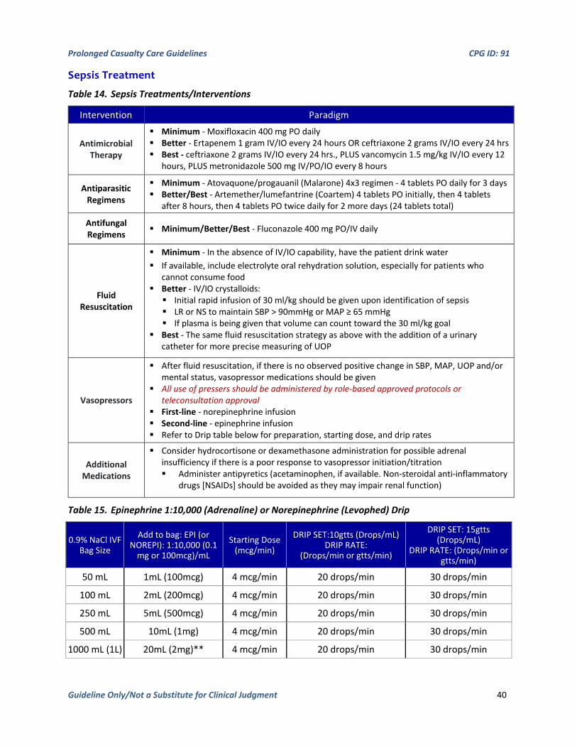
Prolonged Casualty Care Guidelines CPG ID: 91
Guideline Only/Not a Substitute for Clinical Judgment 40
Sepsis Treatment
Table 14. Sepsis Treatments/Interventions
Intervention Paradigm
Antimicrobial Therapy
Minimum - Moxifloxacin 400 mg PO daily Better - Ertapenem 1 gram IV/IO every 24 hours OR ceftriaxone 2 grams IV/IO every 24 hrs Best - ceftriaxone 2 grams IV/IO every 24 hrs., PLUS vancomycin 1.5 mg/kg IV/IO every 12
hours, PLUS metronidazole 500 mg IV/PO/IO every 8 hours
Antiparasitic Regimens
Minimum - Atovaquone/progauanil (Malarone) 4x3 regimen - 4 tablets PO daily for 3 days Better/Best - Artemether/lumefantrine (Coartem) 4 tablets PO initially, then 4 tablets
after 8 hours, then 4 tablets PO twice daily for 2 more days (24 tablets total)
Antifungal Regimens Minimum/Better/Best - Fluconazole 400 mg PO/IV daily
Fluid Resuscitation
Minimum - In the absence of IV/IO capability, have the patient drink water If available, include electrolyte oral rehydration solution, especially for patients who
cannot consume food Better - IV/IO crystalloids: Initial rapid infusion of 30 ml/kg should be given upon identification of sepsis LR or NS to maintain SBP > 90mmHg or MAP ≥ 65 mmHg If plasma is being given that volume can count toward the 30 ml/kg goal
Best - The same fluid resuscitation strategy as above with the addition of a urinary catheter for more precise measuring of UOP
Vasopressors
After fluid resuscitation, if there is no observed positive change in SBP, MAP, UOP and/or mental status, vasopressor medications should be given
All use of pressers should be administered by role-based approved protocols or teleconsultation approval
First-line - norepinephrine infusion Second-line - epinephrine infusion Refer to Drip table below for preparation, starting dose, and drip rates
Additional Medications
Consider hydrocortisone or dexamethasone administration for possible adrenal insufficiency if there is a poor response to vasopressor initiation/titration Administer antipyretics (acetaminophen, if available. Non-steroidal anti-inflammatory
drugs [NSAIDs] should be avoided as they may impair renal function)
Table 15. Epinephrine 1:10,000 (Adrenaline) or Norepinephrine (Levophed) Drip
0.9% NaCl IVF Bag Size
Add to bag: EPI (or NOREPI): 1:10,000 (0.1
mg or 100mcg)/mL
Starting Dose (mcg/min)
DRIP SET:10gtts (Drops/mL) DRIP RATE:
(Drops/min or gtts/min)
DRIP SET: 15gtts (Drops/mL)
DRIP RATE: (Drops/min or gtts/min)
50 mL 1mL (100mcg) 4 mcg/min 20 drops/min 30 drops/min
100 mL 2mL (200mcg) 4 mcg/min 20 drops/min 30 drops/min
250 mL 5mL (500mcg) 4 mcg/min 20 drops/min 30 drops/min
500 mL 10mL (1mg) 4 mcg/min 20 drops/min 30 drops/min
1000 mL (1L) 20mL (2mg)** 4 mcg/min 20 drops/min 30 drops/min

Prolonged Casualty Care Guidelines CPG ID: 91
Guideline Only/Not a Substitute for Clinical Judgment 41
**This is the least recommended approach as it commits a high volume of epinephrine to a large bag. If the patient’s vital signs (BP/MAP/HR) stabilize, the bag must be discontinued and the medic risks wasting some of their resources – “you can mix a drug in an IV bag, but you can’t take it out.”
Ancillary Medications During PCC, additional medications may be required during the extended treatment of casualties, in addition to pain and antibiotic medications. These medications may have synergistic effects to further reduce pain or fever. Some medications may be utilized to treat side-effects of medications, to include nausea or other GI related issues.
Deep vein thrombosis (DVT) prophylaxis is also recommended for patients that are expected to be in a PCC setting for greater than 48 hours that have achieved hemostasis from wounds or are not at risk for further hemorrhage.
Table 16. Ancillary Medications
Minimum Better Best Airway Albuterol MDI
Suctioning: Sterile water or 0.9% saline
Albuterol (Neb) Albuterol (Neb) + Atrovent (Neb)
*Antipyretic Meloxicam Acetaminophen PO/PR or Ibuprofen
Acetaminophen IV/IO or Ketoralac IM/IV/IO
Anxiety / Behavioral See “Pain and Sedation” DVT Prophylaxis Aspirin PO Heparin SQ Lovenox SQ Hydration (PO) Water Water + salt + sugar Water + Gatorade (or other
oral rehydration salt) Hydration (IV/IO) 0.9% Saline or Lactated Ringers Plasma-Lyte Nausea / Vomiting Alcohol Pad (inhale vapor) Ondansetron PO or ODT
Promethazine Ondansetron IV/IO or Metoclopramide IV/IO
GI Medications Ranitidine PO Prilosec PO Protonix IV/IO H1/H2 Blockers IV/IO
GI - Constipation Bisacodyl PO Glycerin Suppository
Mirilax PO Senna PO
Enema
Sleep Melatonin PO Diphenhydramine PO Zolpidem PO Temazepam PO
Other Medications: Oral Care (toothbrush/tooth paste and chapstick) Eye drops (intubated/sedated) Multi-Vitamins (PO daily) Animal Bites: Rabies Vaccine and Rabies Immunoglobulin HIV Prophylaxis (exposure from combat: civilians or enemy forces): PEP Guidance Regional Medications: Ensure continuing prophylaxis (malaria, etc)
*Antipyretic: Use caution with NSAIDs with urgent or priority patients. Ensure patient can void normally (no impaired renal function).
*Link to Infection Prevention in Combat-related Injuries, 27 Jan 2021 CPG 18
*Link to Sepsis Management in Prolonged Field Care, 28 Oct 2020 CPG 19

Prolonged Casualty Care Guidelines CPG ID: 91
Guideline Only/Not a Substitute for Clinical Judgment 42
WOUND CARE AND NURSING - PCC
Background Nursing interventions may not appear important to the medical professionals caring for a patient, but such interventions greatly reduce the possibility of complications such as DVT, pneumonia, pressure sores, wound infection, and urinary tract infection; therefore, essential nursing and wound care should be prioritized in the training environment. Critically ill and injured casualties are at high risk for complications that can lead to adverse outcomes such as increased disability and death. Nursing care is a core principle of PCC to reduce the risk of preventable complications and can be provided without costly or burdensome equipment.20
Using a nursing care checklist assists with developing a schedule for performing appropriate assessments and interventions.
Cross training all team members on these interventions prior to deployment will lessen the demand on the medic, especially when caring for more than one patient.
Prolonged Casualty Care Flowsheets, Nursing Care Checklists, Nursing Care Plans, Assessment/Intervention Packing List, and Recommended Nursing Skill Checklist for Clinical Rotations are included as a PCC Guidelines Appendix. (Also located in JTS Nursing Intervention in Prolonged Field Care CPG, 22 Jul 2018 18).
Pre-deployment, Mission Planning, and Training Considerations Hands-on experience is optimal; simulation is a reasonable substitute
Practice with minimal technology so you are prepared when you lose access to electricity, water
Regular monitoring, reassessment, and intervention is lifesaving but can be resource-intensive
Utilize the Recommended Nursing Skill Checklist for Clinical Rotations included in Appendix B to maximize training opportunities.

Prolonged Casualty Care Guidelines CPG ID: 91
Guideline Only/Not a Substitute for Clinical Judgment 43
Table 17. PCC Role-based Guidelines for Nursing Care and Wound Management
PCC Role-based Guidelines for Nursing Care and Wound Management T C C C - A S M
T C C C - C L S
T C C C - C M C
T C C C - C P P
* All Personnel - Complete Basic TCCC Management Plan for Nursing/Wound Management then: Many “nursing” interventions are actually basic soldier skills that need to be performed on those
casualties who cannot perform them on themselves. Therefore, many traditional non-medical tasks are listed at the Tier 1 level since they can
essentially be performed by anyone, but the activity can be overseen by medical personnel.
Interventions Frequency Paradigm Lip care Every hour Minimum: Commercial lip balm
Better: Moisturizing lotion Best: Petroleum jelly
Oral/Nasal Care
24 hours Minimum: Rotate site around mouth/nares, as feasible. Better: Rotate site and suction. Best: Rotate and suction with commercial device.
Oral/Dental Care
Every 12 hours
Minimum: Brush with gauze, water and gloved finger Better: Brush with tooth brush with toothpaste. Best: Use tooth brush with Chlorhexidine rinse.
Cough/Deep Breathing
Every hour Minimum: Encourage deep breathing/forced cough x 10. Better: Sit up. Encourage deep breathing/forced cough x 10. Best: Sit up, turn, and encourage deep breathing with incentive
spirometer/forced cough x 10. Repositioning/
Check Padding Every 2 hours
Minimum: Turn to opposite side, pad with clothing or textiles. Better: Turn to opposite side, pad with pillows or blankets. Best: Turn to opposite side, pad with pillows to all bony
prominences and between legs. Splint Care Every 2
hours Minimum: Use improvised splints (i.e. wood fence, plank). Better: Use commercial splinting device (e.g., SAM splint). Best: Use ortho-fiberglass splint with fluffing and elastic wrap. ** Re-check all pulses after splint placemen.t
Hypothermia Prevention
Continuous Minimum: Wrap patient in dry clothes or blankets. Better: Wrap patient in commercially available hypothermia
prevention kit, using air-activated heating element. Best: As above, add use of warmed, forced air and infusion of
warmed fluids using commercially available devices. Head Injury Continuous Elevate head of bed 30 degrees and then:
Minimum: Lay patient against ruck sack/backpack Better: Pillows or blankets Best: NATO litter back rest
Non-medical Interventions
Every hour Minimum: Distract the patient and perform guided imagery. Better: Splint wounds, pad boney prominences, provide ice
packs to injured/swollen areas (or, alternate with warm packs). Best: As above, combine both elements.
Psycho-social Needs
Continuous Minimum: Speak in calm tone, addressing casualty concerns, to reduce fear and anxiety.
Better: Support with caring touch, listening to fears/concerns; explain all procedures.
Best: Institute rest/sleep cycle system to minimize delirium.

Prolonged Casualty Care Guidelines CPG ID: 91
Guideline Only/Not a Substitute for Clinical Judgment 44
Nutrition Every 4-6 hours
Minimum: If patient is alert, encourage oral food/water intake. Better: As above, use MRE protein powder mixed with water. Best: As above, use commercially available tube feeding
products or protein shakes. Hygiene Every 24
hours Minimum: Rinse face, armpits, and groin with warm water, soap,
and gauze roll. Better: As above, use baby wipes or wash cloth. Best: As above, use chlorhexidine-impregnated cleansing wipes.
Bowel Management
As required Minimum: Cleanse soiled skin as described for bath; reapply new dressings/hypothermia management as appropriate.
Better: As above, add a cloth/linen/plastic barrier to protect wounds/hypothermia management kit from future soiling.
Best: As above, add barrier cream to skin for protection against breakdown.
Perform all recommended interventions from guidelines for above Tier level. Additional interventions include:
Intervention Frequency Paradigm IV/IO Site Care Minimum: Flush intravenous catheter every 12 hours; change
intravenous infusion tubing every 96 hours. Better: Flush intravenous catheter every 8 hours; change
intravenous infusion tubing every 72 hours. Best: Flush intravenous catheter every 4 hours. Change
intravenous infusion tubing every 48 hours. For IO: monitor the site closely for skin compromise (underneath
the hub of the IO); if possible, convert to an IV within 24 hours. Wound
Irrigation Every 24 hours
Minimum: Irrigate wound with potable water (cooled before use if boiled) poured across wound
Better: As above, use 10cc syringe and 18-gauge angio-catheter. Best: As above, using sterile saline or sterile water or
appropriate antimicrobial cleaning solution (i.e. Dankins). Dressing
Change Minimum: Reinforce dressings.
Better: Replace when soiled. Best: Change every 24 hours.
Ensure above nursing interventions are completed by non-medical TCCC ASM and CLS personnel. Conduct inventory of all resources. Document all pertinent information on PCC Flowsheet (attached). Additional interventions include:
Intervention Frequency Paradigm Suction
mouth/airway, if indicated
As often as required
Minimum: Toomey syringe attached to thin tubing Better: Manual suction device Best: Powered suction device
Monitor assisted
ventilation
Continuous: every hour
Minimum: Use bag-valve-mask ventilation. Better: Mechanical ventilator (without oxygen support), titrate
settings based on pulse oximetry. Best: Mechanical ventilator (with oxygen support).
IV Fluid Calculation
Minimum: Estimate fluid rate using infusion drip rate calculation.
Better: Use “dial-a-flow” technology to control rate of infusion. Best: Use commercial infusion pump.

Prolonged Casualty Care Guidelines CPG ID: 91
Guideline Only/Not a Substitute for Clinical Judgment 45
Deep Vein Thrombosis Prevention
**Pay attention to any wounds to
the affected limb**
Every 1-2 hours
Minimum: Massage lower extremities Better: As above; add application of compression stockings or
elastic bandages to improve venous return. Best: As above; add application of commercial mechanical
compression stockings.
Head Injury (Serial Neuro
Exams)
Minimum: Assess pupillary response, GCS and level of consciousness/orientation, every 8-12 hours; MACE Exam x 1.
Better: Neuro exam (as above) every 4 hrs; MACE exam every 24 hrs. Best: Neuro exam (as above) every 1 hr, MACE exam every 24 hrs.
Hyperthermia Prevention/ Treatment
Minimum: Expose skin to air. Better: Place cold, wet cloths to groin, neck, armpits (ice packs
may cause hypothermia). Best: Use of cooled, forced air and infusion of cooled fluids using
commercially available devices. Administer
Antibiotics Minimum: Provide oral or intramuscular injection of antibiotics
per CPG. Better: Administer intravenous infusion of broad-spectrum
antibiotics, per CPG. Best: Administer wound- or mechanism-specific antibiotics via
intravenous infusion, as directed by provider oversight. Pain Control Minimum: Intermittent dosing of analgesics, given:
oral/intramuscular/intravenous/subcutaneous Better: Continuous infusion of analgesics Best: Regional nerve blocks
Ensure nursing interventions noted above are completed by non-medical TCCC ASM and CLS personnel
Conduct inventory of all resources Document all pertinent information on PCC Flowsheet (attached) Additional interventions include:
Intervention Frequency Paradigm Suction
Advanced Airway
Every hour
Minimum: Manual suction device or improvised suction device, such as a 25cm length portion of IV tubing connected to a 60mL syringe
Better: Open suction tube, suction machine Best: Closed inline suction tube, suction machine
Oro/naso-gastric Tube
Management
Minimum: Cleanse area and rotate position every 12 hours; flush with water every 12 hours (check residuals prior)
Better: As above, every 8 hours (check residuals prior) Best: As above, every 4 hours (check residuals prior)
Foley Care 24 hours Minimum: Cleanse around catheter insertion site as part of bath, every 24 hours.
Better: Cleanse around catheter insertion site using soap and water, every 12 hours.
Best: Cleanse around catheter insertion site using chlorhexidine-impregnated cleansing wipes, every 12 hours.
*Link to Nursing Intervention in Prolonged Field Care, 22 Jul 2018 CPG 20 *Link to Acute Traumatic Wound Care in the Prolonged Field Care Setting, 24 Jul 2017 CPG 21
Legend: TCCC - ASM TCCC-CLS TCCC-CMC TCCC-CPP

Prolonged Casualty Care Guidelines CPG ID: 91
Guideline Only/Not a Substitute for Clinical Judgment 46
SPLINTING AND FRACTURE MANAGEMENT - PCC
Table 18. Splinting and Fracture Treatment
Intervention Paradigm
Litter Padding Minimum - Excess uniforms or other textiles Better - Blankets or military sleep pad Best - Blankets or military sleep pad
Splint Placement
Minimum - Improvised splints (wood fence, metal plank, etc.) Better - Commercial splinting device (e.g., SAM splint) Best - Commercial splinting device (e.g., SAM splint) Re-check all pulses after splint placement
Pressure Injury Prevention
Examine skin, including nares and mouth, for changes and ensure splints are fitted properly and pulses are present below splint.
Monitor for allergic reactions to tape, developing erythema, excessive dryness, pressure indenting the skin, cracking, or breakdown.
Minimum - As described above, every 2 hours Better - As above, adding padding to elevate bony prominences off of ground/litter/bed Best - As above, adding commercial barrier creams and pressure injury dressings (e.g.,
Mepilex) to bony prominences
Straps
Patient secured for transport with padding/hypothermia considerations All patient care items secured for flight or seaboard transport Waterproof outer shell (HPMK) Packaged to resist heavy wind from rotor wash and wind
Litter Padding
Minimum - Allow casualty to maintain airway Better - Facial burns may be associated with inhalation injury. Aggressively monitor
airway status and place the casualty in a recovery position IAW TCCC Guidelines Best - Given a trauma casualty who is unresponsive or has an airway obstruction,
perform a Head-Tilt Chin Lift or Jaw-thrust maneuver to open the airway IAW with TCCC guidelines
Link to JTS Orthopaedic Trauma: Extremity Fractures CPG, 26 Feb 202022

Prolonged Casualty Care Guidelines CPG ID: 91
Guideline Only/Not a Substitute for Clinical Judgment 47
BURN TREATMENT - PCC
Background Interrupt the burning process
Address any life-threatening process based on MARCH assessment as directed by TCCC.
A burned trauma casualty is a trauma casualty first
All TCCC skills can be performed through burned tissue
Burn Characteristics Superficial burns (1st degree) appear red, do not blister, and blanch readily.
Partial thickness burns (2nd degree) are moist and sensate, blister, and blanch.
Full thickness burns (3rd degree) appear leathery, dry, non-blanching, are insensate, and often contain thrombosed vessels
Table 19. PCC Role-Based Guidelines for Burn Management
PCC Role-based Guidelines for Burn Management T C C C - A S M
T C C C - C L S
T C C C - C M C
T C C C - C P P
Perform primary and secondary surveys for any trauma patient. Acute injuries found in the primary and secondary survey should be addressed as per standard trauma protocols
Avoid becoming distracted by the appearance of burned tissues.
Intervention Paradigm Airway
(Roles 1a/1b/1c)
Minimum - Allow casualty to maintain airway. Better - Facial burns may be associated with inhalation injury.
Aggressively monitor airway status and place the casualty in a recovery position IAW TCCC Guidelines.
Best - Given a trauma casualty who is unresponsive or has an airway obstruction, perform a Head-Tilt Chin Lift or Jaw-thrust maneuver to open the airway in accordance with TCCC guidelines.
Fluid Resuscitation (Roles 1a/1b/1c)
Estimate body total surface area (TBSA) burned using the Rule of Nines initially (DD Form 1380).
Note - Superficial (First-degree burns) are NOT used in the TBSA calculation.
If burns >20% TBSA, fluid resuscitation should be initiated as soon as IV/IO access is established.
Minimum - Oral intake of water Better - Oral intake of electrolyte solution Best - Oral intake of electrolyte solution
Hypothermia (Roles 1a/1b/1c)
Hypothermia prevention is extremely important for burn patients. For Burns >20%, place the casualty in the Heat-Reflective Shell or
Blizzard Survival blanket for the Hypothermia Prevention Kit to both cover the burned areas and prevent hypothermia.

Prolonged Casualty Care Guidelines CPG ID: 91
Guideline Only/Not a Substitute for Clinical Judgment 48
Pain Control Analgesia in accordance with the PCC Guidelines may be administered to treat burn pain.
Wounds (Roles 1a/1b)
Minimum - Cover with clean sheet or dry gauze. Leave blisters intact. Avoid wet dressings.
Better - Clean wounds by washing with any clean water (preferably with antibacterial soap if available), dress wounds with any available dressings; optimize wound and patient hygiene to the extent possible given the environment.
Best - Clean wounds by scrubbing gently with gauze and clean water, followed by gauze dressing.
Wounds (Role 1c)
Best - Clean wounds by scrubbing gently with gauze and chlorhexidine gluconate solution (if available) in clean water, followed by gauze dressing. Repeat daily.
Monitor vital signs. Ensure all interventions noted above are completed by TCCC ASM and CLS personnel.
Conduct inventory of all resources. Document all pertinent information on PCC Flowsheet (attached). Additional interventions include:
Intervention Paradigm Airway
(Roles 1a/1b/1c)
Minimum - Allow casualty to maintain airway. Better - Facial burns may be associated with inhalation injury.
Aggressively monitor airway status and consider early surgical airway for respiratory distress or oxygen saturation and/or EtCO2 (purple-gold colorimetric device).
Best - Given a trauma casualty who is unresponsive or has an airway obstruction, consider early surgical airway.
Fluid Resuscitation (Roles 1a/1b/1c)
Minimum - Oral intake of water. Rectal infusion of up to 500mL/h can be supplemented with oral hydration.
Better - Oral intake of electrolyte solution. Best - Start intravenous (IV) or intraosseous (IO) administration
immediately. NOTE - an IV/IO can be placed through burned skin if necessary.
Use isotonic crystalloids (i.e. Lactated Ringers). DO NOT circumferentially tape lines around extremities; this may
further impede circulation and cause limb ischemia as extremities swell during resuscitation.
NO bolus (unless hypotensive, in which case, bolus only until palpable pulses are restored).
Initial IV rate 500mL/h; start while completing initial assessment Give fluids per TCCC burn treatment guidelines. If resuscitation is delayed, DO NOT try to “catch up” by giving extra
fluids. Blood products may be used in major burn resuscitation due to
coagulopathy, anemia, and bleeding from escharotomy sites or other traumatic injuries.
Maintain a UOP of 30-50mL/hr. in adults; decrease or increase isotonic fluid rate by 20-25% per hour.
If UOP > 50 mL/hr., then decrease the fluid rate by 20-25% for the next hour and reassess.
Minimize fluid administration while maintaining organ perfusion; hour-to-hour fluid management is critical.

Prolonged Casualty Care Guidelines CPG ID: 91
Guideline Only/Not a Substitute for Clinical Judgment 49
8-12 hours post-burn, if the hourly IV fluid rate exceeds 1500mL/hr. or if the projected 24- hour total fluid volume approaches 250 mL/kg consult burn team or medical director.
24–48 hours post burn, plasma is lost into the burned and unburned tissues, causing hypovolemic shock (when burn size is >20%). The goal of burn-shock resuscitation is to replace these ongoing losses while avoiding over-resuscitation.
48-72 hours post-burn, completion of the resuscitation is marked by stabilizing hemodynamic parameters and reduction of IV fluid rate to a maintenance level.
Hypothermia (Roles 1a/1b/1c)
Hypothermia prevention is extremely important for burn patients. For Burns >20%, place the casualty in the Heat-Reflective Shell or
Blizzard Survival blanket for the Hypothermia Prevention Kit to both cover the burned areas and prevent hypothermia.
Use Blood/Fluid Warmer as needed and if available. Pain Control
(Roles 1a/1b/1c)
Analgesia in accordance with the PCC Guidelines may be administered to treat burn pain.
Medications (Roles 1a/1b)
Prophylactic antibiotics (oral or IV) are not indicated for burn injury in the absence of infection.
Penetrating wounds or open fractures should be treated with antibiotics according to current TCCC guidelines.
Medications (Role 1c)
After several days, if the patient develops cellulitis (spreading erythema around edges of burn), treat for gram-positive organisms, (e.g., cefazolin or clindamycin).
If patient develops invasive burn wound infection (signs: sepsis/septic shock, changes in color of wound, possible foul smell of wound), treat with broad-spectrum antibiotics.
Wounds (Role 1a)
Minimum - Cover with clean sheet or dry gauze. Leave blisters intact. Avoid wet dressings.
Better - Clean wounds by washing with any clean water (preferably with antibacterial soap if available), dress wounds with any available dressings; optimize wound and patient hygiene to the extent possible given the environment.
Best - Clean wounds by scrubbing gently with gauze and clean water, followed by gauze dressing.
DO NOT debride blisters until the patient has reached a facility with surgical capability.
Every patient with facial burns should have a thorough eye exam. Conduct an eye exam early, before edema begins.
If a corneal injury is identified, use a rigid shield to cover the eyes and apply ophthalmic erythromycin or neomycin ointment every 2 hours.
Wounds (Roles 1b/1c)
Better - Clean wounds and debride loose skin by washing with any clean water (preferably with antibacterial soap if available), dress wounds with any available dressings; optimize wound and patient hygiene to the extent possible given the environment.
Best - Clean wounds by scrubbing gently with gauze and chlorhexidine gluconate solution (if available) in clean water, apply topical antimicrobial cream followed by gauze dressing.

Prolonged Casualty Care Guidelines CPG ID: 91
Guideline Only/Not a Substitute for Clinical Judgment 50
Monitoring Monitor vital signs and urine output (UOP) closely. Minimum - Use other measures If unable to measure UOP, adjust IV
rate to maintain HR less than 140, palpable peripheral pulses, good capillary refill, intact mental status.
Better - Capture all spontaneously voided urine in premade or improvised (i.e. Nalgene® water bottle) graduated cylinder; >180mL every 6 hours is adequate for adults.
Best - Measure UOP with Foley catheter (burns to the penis are NOT a contraindication to catheter placement).
Ensure all above interventions are completed by TCCC ASM, CLS and CMC personnel. Conduct inventory of all resources. Document all pertinent information on PCC Flowsheet (attached.) Additional interventions include:
Intervention Paradigm Airway
(Roles 1a/1b/1c)
Minimum - Allow casualty to maintain airway. Edema after burn injury causes most supraglottic airway devices such as LMAs to be inadequate.
Better - Facial burns may be associated with inhalation injury. Aggressively monitor airway status and consider early surgical airway for respiratory distress or oxygen saturation and/or EtCO2 (purple-gold colorimetric device).
Best - Indications for endotracheal intubation include: a comatose patient, symptomatic inhalation injury, deep facial burns, and burns over 40% TBSA. Utilize an EMMA (or other Capnography) EtCO2 device if possible. Use a large-bore endotracheal tube if inhalation injury is suspected
(Size 8 ETT or larger is preferred for adults). Secure ETT with cotton umbilical ties (standard adhesive ETT holders
do not work around burned skin). Frequently reassess position of the ETT during the acute resuscitation
period as edema waves and wanes.
*Link to Burn Wound Management in Prolonged Field Care, 13 Jan 2017 CPG 23
Legend: TCCC - ASM TCCC-CLS TCCC-CMC TCCC-CPP

Prolonged Casualty Care Guidelines CPG ID: 91
Guideline Only/Not a Substitute for Clinical Judgment 51
Special Considerations in Burn Injuries
Chemical Burns
NOTE: Refer to the JTS Inhalation Injury and Toxic Industrial Chemical Exposure CPG for additional information.
Expose body surfaces, brush off dry chemicals, and copiously irrigate with clean water. Large volume (>20L) serial irrigations may be needed to thoroughly cleanse the skin of residual agents. Do not attempt to neutralize any chemicals on the skin.
Use personal protective equipment to minimize exposure of medical personnel to chemical agents.
White phosphorous fragments ignite when exposed to air. Clothing may contain white phosphorous residue and should be removed. Fragments embedded in the skin and soft tissue should be irrigated out if possible or kept covered with soaking wet saline dressings or hydrogels.
Seek early consultation from the USAISR Burn Center (DSN 312-429-2876 (BURN); Commercial (210) 916-2876 or (210) 222-2876; email [email protected]).
Electrical Burns
TCCC ASM and CLS personnel should remove the patient from the electricity source while avoiding injury themselves.
For cardiac arrest due to arrhythmia after electrical injury, follow advanced cardiac life support (ACLS) protocol and provide hemodynamic monitoring if spontaneous circulation returns.
Small skin contact points (cutaneous burns) can hide extensive soft tissue damage.
Observe the patient closely for clinical signs of compartment syndrome.
Tissue that is obviously necrotic must be surgically debrided.
NOTE: Escharotomy, which relieves the tourniquet effect of circumferential burns, will not necessarily relieve elevated muscle compartment pressure due to myonecrosis associated with electrical injury; therefore, fasciotomy is usually required.
Compartment syndrome and muscle injury may lead to rhabdomyolysis, causing pigmenturia and renal injury.
Pigmenturia typically presents as red-brown urine. In patients with pigmenturia, fluid resuscitation requirements are much higher than those predicted for a similar-sized thermal burn.
Isotonic fluid infusion should be adjusted to maintain UOP 75-100 mL/hr. in adult patients with pigmenturia.
If the pigmenturia does not clear after several hours of resuscitation consider IV infusion of mannitol, 12.5 g per liter of lactated Ringer’s solution, and/or sodium bicarbonate (150 mEq/L in D5W). These infusions may be given empirically; it is not necessary to monitor urinary pH. In patients receiving mannitol (an osmotic diuretic), close monitoring of intravascular status via CVP and other parameters is required.

Prolonged Casualty Care Guidelines CPG ID: 91
Guideline Only/Not a Substitute for Clinical Judgment 52
Seek early consultation from the USAISR Burn Center (DSN 312-429-2876 (BURN); Commercial (210) 916-2876 or (210) 222-2876; email [email protected]).
Pediatric Burn Injuries
Children with acute burns over 15% of the body surface usually require a calculated resuscitation.
Place a bladder catheter if available (size 6 Fr for infants and 8 Fr for most small children).
The Modified Brooke formula (3 mL/kg/%TBSA LR or other isotonic fluid divided over 24 hours, with one-half given during the first 8 hours) is a reasonable starting point. This only provides a starting point for resuscitation, which must be adjusted based on UOP and other indicators of organ perfusion. Goal UOP for children is 0.5-1mL/kg/hr.
Very young children do not have adequate glycogen stores to sustain themselves during resuscitation. Administer a maintenance rate of D5LR to children weighing < 20 kg. Utilize the 4-2-1 rule: 4ml/kg for the first 10kg + 2ml/kg 2nd 10kg + 1ml/kg over 20kg.
In children with burns > 30% TBSA, early administration may reduce overall resuscitation volume.
Monitor resuscitation in children, like adults, based on physical examination, input and output measurements, and analysis of laboratory data.
The well-resuscitated child should have alert sensorium, palpable pulses, and warm distal extremities; urine should be glucose negative.
Cellulitis is the most common infectious complication and usually presents within 5 days of injury. Prophylactic antibiotics do not diminish this risk and should not be used unless other injuries require antimicrobial coverage (penetrating injury or open fracture).
Most antistreptococcal antibiotics such as penicillin are successful in eradicating infection. Initial parenteral administration is advised for most children presenting with fever or systemic toxicity.
Nutrition is critical for pediatric burn patients. Nasogastric feeding may be started immediately at a low rate in hemodynamically stable patients and tolerance monitored. Start with a standard pediatric enteral formula (i.e. Pediasure) targeting 30-35 kcal/kg/day and 2g/kg/day of protein.
Children may rapidly develop tolerance to analgesics and sedatives; dose escalation is commonly required. Ketamine and propofol are useful procedural adjuncts.
When burned at a young age, many children will develop disabling contractures. These are often very amenable to correction which may be performed in theater with adequate staff and resources.
Seek early consultation from the USAISR Burn Center (DSN 312-429-2876 (BURN); Commercial (210) 916-2876 or (210) 222-2876; email [email protected]).
Opportunities for pediatric surgical care provided by Non-Governmental Organizations (NGOs) may be the best option but require the coordinated efforts of the military, host nation, and NGOs.

Prolonged Casualty Care Guidelines CPG ID: 91
Guideline Only/Not a Substitute for Clinical Judgment 53
Rule of Nines
On the DD Form 1380 the percentage of coverage on the casualty’s body will need to be documented. The Rule of Nines will help with the estimation. The below figure shows the approximation for each area of the body:
Eleven areas each have 9% body surface area (head, upper extremities, front and backs of lower extremities, and front and back of the torso having two 9% areas each).
General guidelines are that the size of the palm of the hand represents approximately 1% of the burned area.
When estimating, it is easiest to round up to the nearest 10.
If half of the front or rear area is burned, the area would be half of the area value.
For example, if half of the front upper/lower extremity is burned, it would be half of 9%, or 4.5%. If half of the front torso is burned, say either the upper or lower part of the front torso, then it would be half of 18%, or 9%.
Remember, the higher the percentage burned, the higher the chance for hypothermia.
For children, the percentage of BSA is calculated differently due to the distinctive proportion of major areas.
Figure 2. Adult Rule of Nines Figure 3. Pediatric Rule of Nines
Link to Burn Wound Management in Prolonged Field Care, 13 January 2017 CPG 23

Prolonged Casualty Care Guidelines CPG ID: 91
Guideline Only/Not a Substitute for Clinical Judgment 54
LOGISTICS - PCC
Background Reducing the time to required medical or surgical interventions prevents death in potentially survivable illness, injuries and wounds. When evacuation times are extended, en route care (ERC) capability must be adequately expanded to mitigate the delay. In January 2010, the Joint Force Health Protection Joint Patient Movement Report stated “the current success of the medical community is colored by the valiant ability to overcome deficiencies through ‘just-in-time workarounds;’ many systemic shortfalls are resolved and become transparent to patient outcomes. However, future operations may not tolerate current deficiencies.” 24
Patient packaging is highly dependent upon the transportation or evacuation platform that is available
If possible, rehearse patient packaging internally and with the external resources.
Train with all possible assets, familiarizing them with standard operating procedures
Ensure the patient is stable before initiating a critical patient transfer
Table 20. Logistics Interventions
Intervention Paradigm
Prepare Documentation
Minimum - TCCC Card - DA1380 Better - Prolonged Field Care Casualty Work Sheet Best - PCC Card with TCCC Card and any additional information, reference DA Form 4700
(SMOG 2021) for transport documentation standard.
Prepare Report
Report should give highlights, expected course, and possible complications during transport.
The hand-off is the most dangerous time for the patient; it is as important as treatments or medications.
If it is rushed, things can easily be missed. Make sure you highlight non-obvious interventions and aspects of care (drugs given,
repeat doses, etc.). Minimum - Verbal report describing the patient from head to toe with interventions or a
SOAP note. Better - MIST (Mechanism, Interventions, Symptoms, Treatments) Best - MIST with appropriate SBAR (Situation, Background, Assessment,
Recommendations) and pertinent labs and other diagnostic information
Prepare Medications
Minimum - Prepare medication list with doses and time of next dose. Better - Above with additionally preparing next dose of medication for transport crew
appropriately labeled. Best - Above with fresh IV fluids if indicated and fresh bags of drip medications with
appropriate labeling and 72 hours of antibiotic for extended transports..
Hypothermia Management
Minimum - Blankets Better - Sleep system and blankets. Best - HPMK with Ready Heat or Absorbent Patient Litter System (APLS). If possible, identify with tape the location of interventions or access points on top of
hypothermia management to allow transport teams quick identification of location.

Prolonged Casualty Care Guidelines CPG ID: 91
Guideline Only/Not a Substitute for Clinical Judgment 55
Intervention Paradigm
Flight Stressor/ Altitude
Management
Minimum - Ear Protection and Eye Protection, if nothing available sunglasses and gauze may be used, if patient is sedated and intubated eyes can be taped shut.
Better - Ear Pro and Eye Pro and blankets in all bony areas, Ear Protection and Eye Protection – foam ear plugs or actual hearing protection inserts, goggles.
Best - Above with gastric tube (NG/OG) or chest tube for decompression, if indicated. Depending on altitude/platform, consider bleeding air of out bags of fluid.
Secure Interventions and
Equipment
Minimum - Tape: Securely tape all interventions to include IVs, IOs, airway interventions, gastric tubes
and TQs). Oxygen tanks should be placed between the patients’ legs and the monitor should be
secured on the oxygen cylinder to prevent injury to the patient. Pumps should be secured to the litter.
Better - Additional litter straps to secure equipment and extend the litter with back support as indicated for vented patients to prevent VAP
Best - Above. Use the Special Medical Emergency Evacuation Device (SMEED) to keep the monitor and other transport equipment off patient.
Prepare Dressings
AE and Other MEDEVAC assets do not routinely change dressings during transport; therefore, ensure all dressings are changed, labeled, and secured before patient pick up.
Minimum - Secure and reinforce dressings with tape, date, and time all dressings. Better - Change dressings within 24 hours of departure, secure as above. Best - Change and reinforce dressings within 4 hours of departure. Ensure additional Class
VIII is available for any unforeseen issues in flight. CAUTION - Circumferential/constricting dressings MUST be limited/monitored due to
swelling during prolonged aerial transport.
Secure the Patient
Minimum - Litter with minimum of 2 litter straps. Better - Litter with padding (example: AE pad or Sleep Mat) with minimum of 3 litter
straps. Best - Litter with padding and flight approved litter headrest with minimum of 3 litter
straps. Additional litter straps can be used to secure patient or equipment.
Moving a Critical Care Patient
Minimum - Two-person litter carry to CASEVAC/MEDEVAC platform. Better - Three-person litter carry to CASEVAC/MEDEVAC platform. Best - Four-person litter carry to CASEVAC/MEDEVAC platform.
*Link to Interfacility Transport of Patients between Theater Medical Treatment Facilities, 24 Apr 2018 CPG

Prolonged Casualty Care Guidelines CPG ID: 91
Guideline Only/Not a Substitute for Clinical Judgment 56
REFERENCES
1. TCCC Guidelines, 15 Dec 2021.https://deployedmedicine.com/market/31/content/40
2. Kotwal RS, Howard JT, Orman JA, Tarpey BW, Bailey JA, Champion HR, Mabry RL, Holcomb JB, Gross KR. The effect of a Golden Hour Policy on the morbidity and mortality of combat casualties. JAMA Surg. 2016 Jan;151(1):15-24. https://jamanetwork.com/journals/jamasurgery/fullarticle/2446845 Accessed Nov 2021
3. Kuckelman, J., Derickson, M., Long, W.B. et al. MASCAL Management from Baghdad to Boston: Top Ten Lessons Learned from Modern Military and Civilian MASCAL Events. Curr Trauma Rep 4, 138–148 (2018). https://doi.org/10.1007/s40719-018-0128-0
4. Gurney JM, Spinella PC. Blood transfusion management in the severely bleeding military patient. Curr Opin Anesthesiol. 2018;31: 207–214. https://journals.lww.com/co-anesthesiology/Fulltext/2018/04000/Blood_transfusion_management_in_the_severely.15.aspx Accessed Nov 2021.
5. JTS, Damage Control Resuscitation (DCR) in Prolonged Field Care, 01 Oct 2018 CPG.https://jts.amedd.army.mil/assets/docs/cpgs/Damage_Control_Resuscitation_PFC_01_Oct_2018_I D73.pdf
6. Eastridge BJ, Mabry RL, Sequin P, et al. Death on the battlefield (2001-2011): implications for the future of combat casualty care. J Trauma Acute Care Surg. 2012;73(6 Suppl 5):S431-7.https://www.east.org/content/documents/MilitaryResources/b/TCCC%20Eastridge%20Death%20o n%20the%20Battlefield%20J%20Trauma%202012.pdf Accessed Nov 2021.
7. Hudson I, Blackburn MB, Mann-salinas EA, et al. Analysis of casualties that underwent airway management before reaching Role 2 facilities in the Afghanistan conflict 2008-2014. Mil Med. 2020;185(Suppl 1):10-18. https://pubmed.ncbi.nlm.nih.gov/32074383/
8. Blackburn MB, April MD, Brown DJ, et al. Prehospital airway procedures performed in trauma patients by ground forces in Afghanistan. J Trauma Acute Care Surg 2018;85(1S Suppl 2):S154-S160. https://journals.lww.com/jtrauma/Fulltext/2018/07002/Prehospital_airway_procedures_performe d_in_trauma.23.aspx Accessed Nov 2021.
9. JTS, Airway Management in Prolonged Field Care, 01 May 2020 CPGhttps://jts.amedd.army.mil/assets/docs/cpgs/Airway_Management_in_Prolonged_Field_Care_01_ May_2020_ID80.pdf
10. JTS, Documentation in Prolonged Field Care, 13 Nov 2018 CPGhttps://jts.amedd.army.mil/assets/docs/cpgs/Documentation_Prolonged_Field_Care_13_Nov_201 8_ID72.pdf
11. JTS, Documentation Requirements for Combat Casualty Care, 18 Sep 2020 CPGhttps://jts.amedd.army.mil/assets/docs/cpgs/Documentation_Requirements_for_Combat_Casualt y_Care_18_Sep_2020_ID11.pdf
12. JTS, Hypothermia Prevention, Monitoring, and Management, 18 Sep 2012 CPGhttps://jts.amedd.army.mil/assets/docs/cpgs/Hypothermia_Prevention_Monitoring_and_Manage ment_20_Sep_2012_ID23.pdf

Prolonged Casualty Care Guidelines CPG ID: 91
Guideline Only/Not a Substitute for Clinical Judgment 57
13. Marr AL, Coronado VG, eds. Central Nervous System Injury Surveillance. Data Submission Standards-2002. Atlanta, GA: Centers for Disease Control and Prevention; 2004. http://citeseerx.ist.psu.edu/viewdoc/download?doi=10.1.1.194.2694&rep=rep1&type=pdf Accessed Nov 2021
14. JTS, Traumatic Brain Injury Management in Prolonged Field Care, 06 Dec 2017 CPG https://jts.amedd.army.mil/assets/docs/cpgs/Traumatic_Brain_Injury_PFC_06_Dec_2017_ID63.pdf
15. JTS, Analgesia and Sedation Management During Prolonged Field Care, 11 May 2017 CPG https://jts.amedd.army.mil/assets/docs/cpgs/Analgesia_and_Sedation_Management_during_Prolonged_Field_Care_11_May_2017_ID61.pdf
16. JTS, Pain, Anxiety and Delirium, 26 Apr 2021 CPG https://jts.amedd.army.mil/assets/docs/cpgs/Pain_Anxiety_Delirium_26_Apr_2021_ID29.pdf
17. Keep JW, Messmer AS, Sladden R et al. National Early Warning Score at Emergency Department triage may allow earlier identification of patients with severe sepsis and septic shock: a retrospective observational study. Emerg Med J 2016;33:37–41. https://emj.bmj.com/content/33/1/37 Accessed 01 Dec 2021.
18. JTS, Infection Prevention in Combat-Related Injuries, 27 Jan 2021 CPG https://jts.amedd.army.mil/assets/docs/cpgs/Infection_Prevention_in_Combat-related_Injuries_27_Jan_2021_ID24.pdf
19. JTS, Sepsis Management in Prolonged Field Care, 28 Oct 2020 CPG https://jts.amedd.army.mil/assets/docs/cpgs/Sepsis_Management_PFC_28_Oct_2020_ID83.pdf
20. JTS, Nursing Intervention in Prolonged Field Care, 22 Jul 2018 CPG https://jts.amedd.army.mil/assets/docs/cpgs/Nursing_Intervention_Prolonged_Field_Care_22_Jul_2018_ID70.pdf
21. JTS, Acute Traumatic Wound Management in the Prolonged Field Care Setting, 24 Jul 2017 CPG https://jts.amedd.army.mil/assets/docs/cpgs/Wound_Management_PFC_24_Jul_2017_ID62.pdf
22. JTS Orthopaedic Trauma: Extremity Fractures CPG, 26 Feb 2020 https://jts.amedd.army.mil/assets/docs/cpgs/Orthopaedic_Trauma_Extremity_Fractures_26_Feb_2020_ID56.pdf
23. JTS, Burn Wound Management in Prolonged Field Care, 13 Jan 2017 CPG https://jts.amedd.army.mil/assets/docs/cpgs/Burn_Management_PFC_13_Jan_2017_ID57.pdf
24. Walrath, B. Searching for systems-based solutions to enhance readiness. Navy Medicine Live online blog.

Prolonged Casualty Care Guidelines CPG ID: 91
Guideline Only/Not a Substitute for Clinical Judgment 58
APPENDIX A: TCCC GUIDELINES
TCCC Guidelines Open the attachment on the side menu or open the below link to print or fill out electronically.
https://deployedmedicine.com/market/31/content/40
APPENDIX B: AIRWAY RESOURCES
Nursing Care Checklist Open the attachment on the side menu or open the below link to print or fill out electronically.
https://prolongedfieldcare.org/wp-content/uploads/2018/05/PFC-Nursing-Care-Plan_.pdf

Prolonged Casualty Care Guidelines CPG ID: 91
Guideline Only/Not a Substitute for Clinical Judgment 59
APPENDIX C: MASCAL RESOURCES
Triage Guiding Principles Priorities change based on time from injury
Activities in first hour are CRITICAL
Don’t waste time with formal triage tools Just extricate/stop threat, stop external bleeding, clear airway
Transfusion and ventilator support within the first hour identify a resource-intensive patient
Damage control surgery has little impact after the first hour
Figure 4. TRIAGE cheat cards START
START TRIAGE: Assess, Treat (use bystanders) When you have a color: STOP – TAG – MOVE ON
M I N O R
Move walking wounded
D E C E A S E D
No RESPIRATIONS after head tilt
I M M E D I A T E
Breathing but UNCONSCIOUS
Respirations over 30
Perfusion capillary refill > 2 or NO RADIAL PULSE Control bleeding
Mental Status: unable to follow simple commands
D E L A Y E D
Otherwise
Remember: Respirations – 30 Perfusion – 2 Mental Status – Can Do

Prolonged Casualty Care Guidelines CPG ID: 91
Guideline Only/Not a Substitute for Clinical Judgment 60
Table 21. Triage Assessment
Each Patient Triage Assessment Should Be Complete in Less Than 60 Seconds
Category Examples
Category I: Immediate (red chemlite) (Any MARCH issue) Airway obstruction Flail/open chest wound Tension- Pneumothorax/hemothorax Massive hemorrhage 20-70% Burns Unstable Vital Signs Severe TBI (unconscious alive Pt)
Category II: Delayed (green chemlite) Open fractures w/PMS intact Soft tissue injuries Moderate TBI (stable vital signs) Open abdominal wounds
*Category III: Minimal (no chemlite) remain armed continue to engage
Minor abrasions, burns, sprains lacerations Moderate/Mild anxiety Fractures/dislocations w/PMS Mild TBI
**Category IV: Expectant or Hero (blue chemlite)
Massive head or spinal injury Third degree burns > 70% BSA Injuries incompatible with life
* In combat, it is assumed that minimals will continue to stay armed/engaged if no mental status altering pharmaceuticals are given for pain. **Expectant category is ONLY used in combat operations and/or when the requirements to adequately treat these patients exceed the available resources. In peacetime, it is generally assumed that all patients have a chance of survival. Source: Special Operations Force Medic Handbooks (PJ, Ranger)
Triage Class 1 (MASCAL) Adequate medics to treat critical patients and handle the rest
Many casualties
Threat controlled
Resources not severely limited
Medical personnel can arrive
Evacuation possible

Prolonged Casualty Care Guidelines CPG ID: 91
Guideline Only/Not a Substitute for Clinical Judgment 61
Table 22. Triage Class 1 Actions and Goals
<1 Hour After Injury 1 – 4 Hours After Injury >4 Hours After Injury Goals Goals Goals Eliminate Threat Establish CCP Blood transfusion within 30 minutes Evacuate to DCR/DCS within 1 hour
DCR/DCS as soon as possible Use advanced resuscitation to
“extend the Golden Hour”
Evacuate
Actions Actions Actions Stop external bleeding Clear airway Ensure ventilation Formal triage Transfuse
MARCH PAWS Transfuse
Use prolonged care to optimize outcomes
Triage Class 2 (MASCAL) Unable to manage the number of critical patients
Numerous casualties or MASCAL (i.e. < 100 Casualties)
Threat has been controlled or partially controlled
Resources are very limited
Medical personnel can arrive (may be delayed > 1 hour)
Evacuation is possible (may be delayed > 1 hour)
Table 23. Triage Class 2 Actions and Goals
<1 Hour After Injury 1 – 4 Hours After Injury >4 Hours After Injury Goals Goals Goals Eliminate threat Get medical personnel on scene Begin evacuation of urgent but
survivable patients
Evacuate urgent and priority patients
DCR/DCS as soon as possible
Evacuate remainder of patients
Actions Actions Actions Stop external bleed Clear airway Reserve intubation/transfusion CCP if able, otherwise get a
count
Formal triage MARCH PAWS if able Transfuse Establish CCP Utilize minimals/returns to
duty
Re-triage Complete MARCH PAWS Use prolonged care to
optimize outcomes Wound/fracture management

Prolonged Casualty Care Guidelines CPG ID: 91
Guideline Only/Not a Substitute for Clinical Judgment 62
Triage Class 3 (Ultra-MASCAL) Absolutely overwhelming number of casualties
Ultra-MASCAL (i.e. >100, possibly thousands of casualties)
Threat is ongoing
Resources are severely limited
Medical personnel unable to arrive in < 1 Hour
Evacuation not possible in < 1 Hour
Table 24. Triage Class 3 Actions and Goals
<1 Hour After Injury 1 – 4 hours After Injury >4 Hours After InjuryGoals Goals Goals Respond to threat Self-aide, buddy care Separate ambulatory/
non-ambulatory
Eliminate threat Get medical personnel on scene Begin evacuation
Evacuate Distribute patients
Action Action Action Stop external bleed Clear airway Reverse intubation/
transfusion Get a count
Stop external bleed Reserve intubation/transfusion Begin to establish CCPs Utilize minimals/return to Duty
Formal triage Use prolonged care to optimize
outcomes Wound/fracture management Utilize minimals/return to duty
MASCAL/Austere Team Resuscitation Record Open the attachment on the side menu or open the below link to print or fill out electronically.
https://jts.amedd.army.mil/assets/docs/forms/MASCAL_Austere_Trauma_20_Jan_2020.pdf
Instructions: https://jts.amedd.army.mil/assets/docs/forms/MASCAL_Form_Instructions.pdf

Prolonged Casualty Care Guidelines CPG ID: 91
Guideline Only/Not a Substitute for Clinical Judgment 63
Tactical Triage Protocol (algorithm) Figure 5. Tactical Triage Protocol

Prolonged Casualty Care Guidelines CPG ID: 91
Guideline Only/Not a Substitute for Clinical Judgment 64
APPENDIX D: DOCUMENTATION RESOURCES
The following resources and associated links are included in this CPG as attachments.
DD 1380 TCCC Card and accompanying POI TCCC After Action Report
DD 3019 Resuscitation Record
DA 4700 TACEVAC form
Nursing care grid (See Appendix B.)
Teleconsultation Script
DD 1380 TCCC Card Open the attachment on the side menu or open the below link to print or fill out electronically.
https://jts.amedd.army.mil/assets/docs/forms/DD_Form_1380_TCCC_Card_Jun_2014.pdf
DD 1380 - POI TCCC After Action Report Open the attachment on the side menu or open the below link to print or fill out electronically.
https://jts.amedd.army.mil/assets/docs/forms/POI_TCCC_AAR.pdf
DD 3019 Resuscitation Record Open the attachment on the side menu or open the below link to print or fill out electronically.
https://www.esd.whs.mil/Portals/54/Documents/DD/forms/dd/dd3019.pdf
DA 4700 TACEVAC Form Open the attachment on the side menu or open the below link to print or fill out electronically.
https://jts.amedd.army.mil/assets/docs/forms/DA4700_OP5_JTS_TACEVAC-AAR&PCR.pdf
Instructions: https://jts.amedd.army.mil/assets/docs/forms/DA4700_OP4_JTS_TACEVAC-AAR-PCR_Instruction_20141002.pdf
Prolonged Field Care Casualty Card v22.1, 01 Dec 2020 Open the attachment on the side menu or open the below link to print or fill out electronically.
https://prolongedfieldcare.org/2019/06/27/version-22-of-the-prolonged-field-care-card/
https://jts.amedd.army.mil/assets/docs/forms/DA4700_OP4_JTS_TACEVAC-AAR-PCR_Instruction_20141002.pdf

Prolonged Casualty Care Guidelines CPG ID: 91
Guideline Only/Not a Substitute for Clinical Judgment 65
Virtual Critical Care Consultation Guide Guide is to be used with the Prolonged Field Care Card.
1. Before calling, E-mail image of the casualty (wounds, environment, etc.), "capabilities" (back of page), & vital signs trends to ___________________________ 2. If call not answered: a) call next number on PACE or call back in 5 – 10 min. 3. If unable to provide information due to operational security, state so.
P: A: C: E:
This is ___________________________________ I am a (job/ position) _______________________________ My best contact info is: ______________________________________________________________ YOUR best contact info is (Consultant’s number): ___________________Alternate e-mail: ___________________
*** PAUSE POINT to CONFIRM CONTACT INFO*** I have a _____ year–old ______(sex) ___________ (active duty/foreign national/OGA,etc.), who has the following:
Mechanism of Injury or known diagnosis(es) O that occurred in (location)
The injury/start of care occurred hours ago. Anticipated evacuation time is (range) O
Injuries/Problems/Symptoms: O
O
O
Treatments: O
O
O
He/she is currently (circle) stable/ unstable, getting better/ getting worse/ getting worse rapidly
Known Medication Allergies/Past medical/Surgical history is:
O
I need help with (be specific if possible, i.e. “I need help reading this ECG,” or “I need help stabilizing this patient,” etc.)
O
Other Consultants have recommended: O *** PAUSE POINT for Remote Consultant to ask clarification questions ***
VITALS (current & trend as of ): HR BP RR SpO2 ETCO2 Temp……….
UOP(ml/hr) over (# hours) Mental Status (GCS/ AVPU) O
EXAM: Neuro Ext/ MSK O
Heart Pulses O
Lungs Skin/ Wounds O
Abd O
LABS: ABG: Lactate: Other: O
*** PAUSE POINT for Remote Consultant to ask clarification questions **

Prolonged Casualty Care Guidelines CPG ID: 91
Guideline Only/Not a Substitute for Clinical Judgment 66
Virtual Critical Care Consultation Guide – page 2
TO-DO/ FOLLOW-UP/TO-STOP NOTES 1.
2.
3.
4.
5.
6. *** PAUSE POINT, for Medic/Local Caregiver to ask clarification questions/READBACK***
Available “kit” (supplies, equipment, medications) !! IF POSSIBLE PHOTOGRAPH AND SEND VIA EMAIL BEFORE CALLING !! IV access: IV Central line IO (location) Other:_____________________________________ Monitor: Propaq Tempus Foley Graduated urinal PulseOx only Exam Only Other:________________________________________________________ Commo: Tempus i2i ID: _______________THIAB: ___ SAT# ___ Local Cell#_____________
Web VTC Address_____________________________________________________________________
Other (e.g. “FaceTime, VSee, Skype, etc.):__________________________________________________ IV Fluids: Plasma-Lyte LR Normal Saline 3% saline Other:____________________ Colloids: Hetastarch Albumin Other:_________________________________________________ Blood products: Whole blood PRBC Plasma FDP Platelets Other:_____________ Medications: Antibiotics: name/route/dose_____________________________________________ Morphine IV/ PO Other opioid (name/ IV/ PO): ______________________________ Fentanyl IV/ PO (pop) Ketamine Midazolam Diazepam (IV/ PO) TXA Other(s): ______________________________________________ Airway supplies: ETT Cric kit LMA Ventilator BVM O2 Suction Misc: O
Plans/Recommendations
PRIORITY SYSTEM/PROBLEM RECOMMENDATION Neuro or problem #1
CV or problem #2
Pulm or problem #3
GI or problem #4
Renal or problem #5
Endocrine or problem #6
MSK/ Wound or problem #7
Tubes, lines, drains or problem #8
Prophylaxis/prevention or prob#9
Other

Prolonged Casualty Care Guidelines CPG ID: 91
Guideline Only/Not a Substitute for Clinical Judgment 67
APPENDIX E: TBI RESOURCES
Neurological Examination MENTAL STATUS Level of Consciousness: Note whether the patient is: Alert/responsive Not alert but arouses to verbal stimulation Not alert but responds to painful stimulation Unresponsive Orientation: Assess the patient’s ability to provide: Name Current location Current date Current situation (e.g., ask the patient what
happened to him/her) Language: Note the fluency and appropriateness of the patient’s response to questions. Note patient’s ability to follow commands when assessing other functions (e.g., smiling, grip strength, wiggling toes). Ask the patient to name a simple object (e.g., thumb, glove, watch). Speech: Observe for evidence of slurred speech. CRANIAL NERVES All patients: Assess the pupillary response to light. Assess position of the eyes and note any
movements (e.g., midline, gaze deviated left or right, nystagmus, eyes move together versus uncoupled movements).
Noncomatose patient: Test sensation to light touch on both sides of the
face. Ask patient to smile and raise eyebrows, and
observe for symmetry. Ask the patient to say “Ahhh” and directly observe
for symmetric palatal elevation. Comatose patient: Check corneal reflexes; stimulation should trigger
eyelid closure. Observe for facial grimacing with painful stimuli. Note symmetry and strength. Directly stimulate the back of the throat and look
for a gag, tearing, and/or cough.
MOTOR Tone: Note whether resting tone is increased (i.e. spastic or rigid), normal, or decreased (flaccid). Strength: Observe for spontaneous movement of extremities and note any asymmetry of movement (i.e. patient moves left side more than right side). Lift arms and legs, and note whether the limbs fall immediately, drift, or can be maintained against gravity. Push and pull against the upper and lower extremities and note any resistance given. Note any differences in resistance provided between the left and right sides. (NOTE: it is often difficult to perform formal strength testing in TBI patients. Unless the patient is awake and cooperative, reliable strength testing is difficult.) Involuntary movements: Note any involuntary movements (e.g., twitching, tremor, myoclonus) involving the face, arms, legs, or trunk. SENSORY If patient is not responsive to voice, test central pain and peripheral pain. Central pain: Apply a sternal rub or supraorbital pressure, and note the response (e.g., extensor posturing, flexor posturing, localization). Peripheral pain: Apply nail bed pressure or take muscle between the fingers, compress, and rotate the wrist (do not pinch the skin). Muscle in the axillary region and inner thigh is recommended. Apply similar stimulus to all four limbs and note the response (e.g., extensor posturing, flexor posturing, withdrawal, localization). NOTE: In an awake and cooperative patient, testing light touch is recommended. It is unnecessary to apply painful stimuli to an awake and cooperative patient. GAIT If the patient is able to walk, observe his/her casual gait and note any instability, drift, sway, and so forth.

Prolonged Casualty Care Guidelines CPG ID: 91
Guideline Only/Not a Substitute for Clinical Judgment 68
Ultrasonic Assessment of Optic Nerve Sheath Diameter If a patient is unconscious (i.e. does not follow commands or open eyes spontaneously), they may have elevated ICP. There is no reliable test for elevated ICP available outside of a hospital; however, optic nerve sheath diameter (ONSD) measurement is a rapid, safe, and easy-to-perform ultrasonographic assessment that may help identify elevated ICP when more definitive monitoring devices are not available. The optic nerve sheath directly communicates with the intracranial subarachnoid space. Increased
ICP, therefore, displaces cerebrospinal fluid along this pathway. Normal ONSD is 4.1–5.9mm.30 A 10–5-MHz linear ultrasound probe can be used to obtain ONSDs. ONSD is measured from one side
of the optic nerve sheath to the other at a distance of 3mm behind the eye immediately below the sclera.31
In general, ONSDs >5.2mm should raise concern for clinically significant elevations in ICP in unconscious TBI patients.5,32 The ONSD can vary significantly in normal individuals, so one single measurement may not be helpful; however, repeated measurements that detect gradual increases in ONSD over time may be more useful than a single measurement.
ONSD changes rapidly when the ICP changes, so it can be measured frequently.33 If ONSD is used, it is best to check hourly along with the neurologic examination.
Technique 1. Check to make sure there is no eye injury. A penetrating injury to the eyeball is an absolute contraindication to
ultrasound because it puts pressure on the eye. 2. Ensure the head and neck are in a midline position. Gentle sedation and/or analgesia may be necessary to
obtain accurate measurements. 3. Ensure the eyelids are closed. 4. If available, place a thin, transparent film (e.g., Tegaderm; 3M, http://www.3m.com) over the closed eyelids. 5. Apply a small amount of ultrasound gel to closed eyelid. 6. Place the 10(–5) MHz linear probe over the eyelid. The probe should be applied in a horizontal orientation
(Figure 1) with as little pressure as possible applied to the globe. 7. Manipulate the probe until the nerve and nerve sheath are visible at the bottom of screen. An example of a
proper ultrasonagraphic image of the optic nerve sheath can be seen in Figure 2. 8. Once the optic nerve sheath is visualized, freeze the image on the screen. 9. Using the device’s measuring tool, measure 3mm back from the optic disc and then obtain a second
measurement perpendicular to the first. The second measurement should cover the horizontal width of the optic nerve sheath (Figure 2). An abnormal ONSD is shown in Figure 3.
10. Repeat the previous sequence in the opposite eye. Annotate both ONSDs on the PFC Casualty Card. 11. ONSDs should be obtained, when possible, at regular intervals to help assess changes in ICP, particularly when
the neurologic examination is poor and/or unreliable (i.e. with sedation). Serial measurements with progressive diameter enlargement and/or asymmetry in ONSDs should be considered indicative of worsening intracranial hypertension.
CAUTION: ONSD measurements are contraindicated in eye injuries. NEVER apply pressure to an injured eye.

Prolonged Casualty Care Guidelines CPG ID: 91
Guideline Only/Not a Substitute for Clinical Judgment 69
Figure 1. Appropriate placement of the linear probe.
Ultrasound gel is placed over a closed eyelid and the probe placed horizontally over the eyelid, applying as little pressure to the globe as possible. If available, Tegaderm or other thin covering (e.g., Latex glove) should be placed over a closed eyelid for further protection.
Figure 2 An ultrasonographic view of a normal eye and optic nerve sheath.
To measure ONSD, apply the ultrasound measuring device to the optic disc and measure back 3mm along the length of the optic nerve. A second, perpendicular measurement is obtained at the previously measured point that spans the horizontal width of the optic nerve sheath. In this image, ONSD was determined to be 5.1mm, a normal value.
Figure 3. Ultrasound image of the right optic nerve sheath of a 61-year-old man with a traumatic subdural hematoma.
The optic nerve sheath measured 6.8mm in diameter. Elevated ICP was subsequently confirmed (26mmHg) after the placement of an ICP bolt monitor.
Spontaneous Venous Pulsations Spontaneous venous pulsations (SVPs) are subtle, rhythmic variations
in retinal vein caliber on the optic disc and have an association with ICP.
It is difficult to see SVPs without advanced equipment; however, if a handheld ophthalmoscope is available, it is worth an attempt to visualize the retinal veins.
Don’t worry if you cannot see SVPs; this may actually be normal. However, if you do see them, it is very reassuring that ICP is normal.10
If SVPs are initially present and can no longer be seen on subsequent examinations, the provider should be concerned for increasing ICP.
Technique 1. Gently lift the eyelid until the pupil is in view. 2. Using a handheld ophthalmoscope, the provider should maneuver
himself or herself to a position where the optic disc can be visualized. 3. Identify the retinal veins as they emerge from the optic disc. Retinal
veins are typically slightly larger and darker than retinal arteries. Figure at right demonstrates the typical appearance of the retina.
4. Observe the retinal veins for pulsations. Note the presence or absence of spontaneous venous pulsations
5. Repeat the step 1–4 sequence in the contralateral eye.
Figure 6. Typical appearance of a healthy retina.
The retinal vessels can be seen emerging from the optic disc. Retinal veins can be identified by their slightly larger, thicker size and darker color. Retinal arteries are small, thin, and lighter in color than retinal veins.

Prolonged Casualty Care Guidelines CPG ID: 91
Guideline Only/Not a Substitute for Clinical Judgment 70
Glasgow Coma Scale TBI severity classification using the GCS score:
Mild: 13–15
Moderate: 9–12
Severe: 3–8
Richmond Agitation Sedation Scale (RASS)
Eye Opening Verbal Response Motor Response 4 – Spontaneous 3 – To verbal command 2 – To painful stimuli 1 – No response
5 – Oriented 4 – Confused 3 – Inappropriate words 2 – Incomprehensible sounds 1 – No response
6 – Obeys commands 5 – Localizes to painful stimuli 4 – Withdraws from pain 3 – Flexion to pain 2 – Extension to pain 1 – No response
Score Term Description +4 Combative Overtly combative, violent, immediate danger to staff.
Verbal Stimulation Physical Stimulation
+3 Very Agitated Pulls or removes tube(s) or catheter(s); aggressive. +2 Agitated Frequent non-purposeful movement, fights ventilator. +1 Restless Anxious but movements not aggressive vigorous. 0 Alert, Calm -1 Drowsy Not fully alert, but has sustained awakening (eye-
opening/eye contact) to voice (>10 seconds). -2 Light Sedation Briefly awakens with eye contact to voice (<10 seconds). -3 Moderate Sedation Movement or eye opening to voice (but no eye contact). -4 Deep Sedation No response to voice, but movement or eye opening to
physical stimulation. -5 Unarousable No response to voice or physical stimulation.
Procedure for RASS Assessment 1. Observe patient: Patient is alert, restless, or agitated. Score 0 to+4 2. If not alert, state patient’s name and say to open eyes and look at speaker
- Patient awakens with sustained eye opening and eye contact. - Patient awakens with eye opening and eye contact, but not sustained. - Patient has any movement in response to voice but no eye contact.
Score -1 Score -2 Score -3
3. When no response to verbal stimulation, physically stimulate patient by shaking shoulder and/or rubbing sternum. - Patient has any movement to physical stimulation. - Patient has no response to any stimulation.
Score -4 Score -5
*Sessler CN, Gosnell M. Grap MJ, Brophy GT, O’Neal PV, Keane KA et al. The Richmond Agitation-Sedation Scale: validity and reliability in adult intensive care patients. Am J Respir Crit Care Med 2002; 166:1338-1344. *Ely EW, Truman B, Shintani A., Thomason JWW, Wheeler AP, Gordon S et al. Monitoring sedation status over time in ICU patients: the reliability and validity of the Richmond Agitation Sedation Scale (RASS). JAMA 2003; 289:2983-2991.

Prolonged Casualty Care Guidelines CPG ID: 91
Guideline Only/Not a Substitute for Clinical Judgment 71
Signs and Symptoms of Elevated Intracranial Pressure GCS<8 and suspected TBI
Rapid decline in mental status
Fixed dilated pupils(s)
Cushing’s triad hemodynamics (hypertension, bradycardia, altered respirations)
Motor posturing (unilateral or bilateral)
Penetrating brain injury and GCS <15
Open skull fracture
Hypertonic Saline (HTS) Protocol (goal Na 140-165 meq/L) 3% HTS: 250-500 cc bolus, then 50 ml/hr infusion, rebolus as needed for clinical signs
7.5% HTS: decrease above doses by 50%
23.4%: dilute to 3% and use as above. If unable to dilute, can be given as 30 ml bolus and re-dose as needed.
Central venous line (CVL) preferred for 3% (can be given initially via peripheral IV/IO)
CVL REQUIRED for 7.5% or higher concentration
Military Acute Concussion Evaluation 2 (MACE 2) Form, 2021 Open the attachment on the side menu or open the below link to print or fill out electronically.
https://www.health.mil/Reference-Center/Publications/2020/07/30/Military-Acute-Concussion-Evaluation-MACE-2
MHS Progressive Return to Activity Following Acute Concussion/Mild TBI Open the attachment on the side menu or open the below link to print or fill out electronically.
https://jts.amedd.army.mil/assets/docs/cpgs/Progressive_Return_to_Activity_Following_Acute_Concussion_mTBI_Clinical_Recommendation_2021.pdf

Prolonged Casualty Care Guidelines CPG ID: 91
Guideline Only/Not a Substitute for Clinical Judgment 72
APPENDIX F: LOGISTICS RESOURCES
Prolonged Field Care – Patient Packaging, 11 Aug 2021 Patient packaging is highly dependent upon the Casualty Evacuation (CASEVAC) / Medical Evacuation (MEDEVAC) platform that is operationally available. If possible, rehearse patient packaging internally and with the external resources. Train with MEDEVAC assets understand transporting teams’ standard operating procedures in order to best prepare the patient for transport. (Example some teams want to secure the patient and interventions themselves while others may be okay with a fully wrapped patient).
Ensure the patient is stable before initiating a critical patient transfer. For POI/unstable patients ensure the appropriate transport team (MEDEVAC with en route critical care nurse or advanced provider). Interfacility transfers should meet the following minimum:
1. Hemorrhage control
2. Resuscitation adequate (SBP 70-80 mmHg, MAP >60, or UOP >0.5ml/kg/hr)
3. Initial post-op recovery as indicated
4. Stabilization of fractures
Prepare Documentation
Good: TCCC Card - DA1380
Better: Prolonged Field Care Casualty Work Sheet
Best: PFC Card with TCCC Card and any additional information, reference DA Form 4700 (SMOG 2021) for transport documentation standard
*preference: secure to patient strip of 3in Tape with medications administered attached to blanket or HPMK
Prepare Report
Report should give highlights, expected course, and possible complications during transport. The hand-off is the most dangerous time for the patient it is as important as treatments or medications. If it is rushed things can easily be missed.
Good: Verbal report describing the patient from head to toe with a SOAP note.
Best: MIST (Mechanism, Interventions, Symptoms, Treatments)
Better: MIST with appropriate SBAR (Situation, Background, Assessment, Recommendations) and pertinent labs and other diagnostic information
Prepare Medications
Good: Prepare medication list with doses and time of next dose
Better: Above with additionally preparing next dose of medication for transport crew appropriately labeled.
Best: Above with fresh IV fluids if indicated and fresh bags of drip medications with appropriate labeling and 72 hours of antibiotic for extended transports.

Prolonged Casualty Care Guidelines CPG ID: 91
Guideline Only/Not a Substitute for Clinical Judgment 73
Hypothermia Management
Good: Blankets
Better: Sleep system and blankets
Best: HPMK with Ready Heat or Absorbent Patient Litter System (APLS)
Flight Stressor/ Altitude Management
Good: Ear Protection and Eye Protection, if nothing available sunglasses and gauze may be used, if patient is sedated and intubated eyes can be taped shut
Better: Ear Pro and Eye Pro and blankets in all bony areas, Ear Protection and Eye Protection – foamies or actual hearing protection inserts, goggles
Best: Above with gastric tube (NG/OG) or chest tube for decompression, if indicated. Depending on altitude/platform, consider bleeding air of out bags of fluid.
Secure Interventions and Equipment
Good: Tape (securely tape all interventions to include IVs, IOs, Airway interventions, Gastric Tubes and TQs). Oxygen tanks should be placed between the patients legs and the monitor should be secured on the oxygen cylinder to prevent injury to the patient. Pumps should be secured to the litter
Better: Additional litter straps to secure equipment and extend the litter with back support as indicated for vented patients to prevent VAP.
Best: Above and use the SMEED to keep the monitor and other transport equipment off patient
*if possible, identify with tape the location of interventions or access points on top of hypothermia management to allow transport teams quick identification of location.
Prepare Dressings
Air Evacuation and other MEDEVAC assets do not routinely change dressings during transport; therefore, ensure all dressings are changed, labeled, and secured before patient pick up
Good: Secure and reinforce dressings with tape, date, and time all dressings.
Better: Change dressings within 24 hours of departure, secure as above.
Best: Change and reinforce dressings within 4 hours of departure. Ensure additional Class VIII is available for any unforeseen issues in flight.
Secure the Patient
Good: Litter with minimum of 2 litter straps
Better: Litter with padding (example: AE pad or Sleep Mat) with minimum of 3 litter straps
Best: Litter with padding and flight approved litter headrest with minimum of 3 litter straps (additional litter straps can be used to secure patient or equipment)

Prolonged Casualty Care Guidelines CPG ID: 91
Guideline Only/Not a Substitute for Clinical Judgment 74
Moving a Critical Care Patient
Good: Two person little carry to CASEVAC/MEDEVAC platform
Better: Three person little carry on a rickshaw to CASEVAC/MEDEVAC platform
Best: Four person little carry on a rickshaw to CASEVAC/MEDEVAC platform
Prolonged Casualty Care Patient Packaging Flowchart Equipment: Litter with at least three litter straps Three channel IV pump (airworthy) Cardiac monitor and cables Suction Device
Possible Complications: Inadequate medications Injuries not addressed before transport Inexperienced provider on flight Equipment issues
Patient is a critical care transfer
Patient in critical
condition? NO YES
Patient on vasopressors?
Patient have head injury? Prevent hyperthermia with an
HPMK/blizzard wrap
Ensure the patient meets MEDEVAC transfer criteria. Consider transferring the patient with an en route critical care nurse or advanced provider.
When appropriate, return to Tactical Evaluation Guideline
Arrange CASEVAC/MEDEVAC
Consider taking a triple channel pump (airworthy).
Consider elevating the HOB on the litter with a back support
Ensure the patient transport team has a triple channel pump (airworthy) with a backup pump.
YES
YES
NO
Prevent hyperthermia with an HPMK/blizzard wrap
Rapid transfer to nearest MTF
NO

Prolonged Casualty Care Guidelines CPG ID: 91
Guideline Only/Not a Substitute for Clinical Judgment 75
Pearls: Document all times – TCCC Card or DA4700.
Assist Ensure the patient is stable before initiating a critical patient transfer.
POI/unstable patients ensure the appropriate transport team (MEDEVAC W/ECCN or Advancedprovider)
Interfacility transfers should meet the following minimum:
Hemorrhage control
Resuscitation adequate (SBP 70-80 mmHg, MAP >60, or UOP >0.5ml/kg/hr)
Initial post-op recovery as indicated
Stabilization of fractures

JOINT TRAUMA SYSTEM CLINICAL PRACTICE GUIDELINE (JTS CPG)
Sepsis Management in Prolonged Field Care (CPG ID:83) This CPG focuses on the most common etiologies of sepsis, and the treatments of those forms of sepsis that the austere provider can reasonably manage.
Contributors
SFC Justin Rapp, 18D, MC, USASOC Sean Keenan, MD, COL (ret.), USA Daniel Taylor, EMT-P, TP-C. Andrea Rapp, RN, 68W SGT Michael Turconi, PCP, FMR. OR5 (Italian Special Forces) CAPT Ryan Maves, MC, USN CDR Michael Kavanaugh, MC, USN
Devan Makati, MD LTC Doug Powell, MD, MC, USAR SFC Paul Loos, 18D, MC, USASOC CPT Simon Sarkisian, MC, USA Ankit Sakhuja, MD LTC (ret.) Dan Mosely, MC, USA, Col Stacy Shackelford, USAF, MC
Publication Date: 28 Oct 2020
TABLE OF CONTENTS Introduction ................................................................................................................................................................... 3 Sepsis Definitions ........................................................................................................................................................... 3 Sepsis Management Goals ............................................................................................................................................. 4 Early Suspicion and Recognition .................................................................................................................................... 4 History and Physical Exam ............................................................................................................................................. 4
Subjective Assessment .............................................................................................................................................. 6 Objective Assessment ............................................................................................................................................... 6
Source Identification...................................................................................................................................................... 7 Treatment ...................................................................................................................................................................... 8
Antimicrobial Therapy ............................................................................................................................................... 8 Antiparasitic Regimens .............................................................................................................................................. 8 Antifungal Regimens ................................................................................................................................................. 9
Resuscitation ................................................................................................................................................................. 9 Fluid Resuscitation Considerations ........................................................................................................................... 9 Drug Considerations ................................................................................................................................................ 10
Source Control ............................................................................................................................................................. 10 Monitor the Patient ..................................................................................................................................................... 11 Early Telemedicine ....................................................................................................................................................... 12 Evacuate to Definitive Care: ........................................................................................................................................ 12
Considerations ......................................................................................................................................................... 12 Nutrition Considerations ............................................................................................................................................. 12 Performance Improvement (PI) Monitoring ................................................................................................................ 14
Population of Interest ............................................................................................................................................. 14

Sepsis Management in Prolonged Field Care CPG ID: 83
Guideline Only/Not a Substitute for Clinical Judgment 2
Intent (Expected Outcomes) ................................................................................................................................... 14 Performance/Adherence Measures ........................................................................................................................ 14 Data Sources ............................................................................................................................................................ 14 System Reporting & Frequency ............................................................................................................................... 14 Responsibilities ........................................................................................................................................................ 14
References ................................................................................................................................................................... 14 Appendix A: Sepsis Patient Management Checklist .................................................................................................... 17 Appendix B: I-STAT Lab Values .................................................................................................................................... 18 Appendix C: Abnormalities Detection ......................................................................................................................... 19 Appendix D: Malaria Rapid Test and Microscopy ........................................................................................................ 20 Appendix E: Lactate ..................................................................................................................................................... 23 Appendix F: Epinephrine (Norepinephrine) - Vasopressor Guide ............................................................................... 24 Appendix G: Additional Information Regarding Off-Label Uses In CPGs ..................................................................... 26

Sepsis Management in Prolonged Field Care CPG ID: 83
Guideline Only/Not a Substitute for Clinical Judgment 3
I N T R O D U C T I O N
This Role 1 prolonged field care (PFC) guideline is intended for use in the austere environment when evacuation to higher level of care is not immediately possible. A provider must first be an expert in Tactical Combat Casualty Care (TCCC). The intent of this guideline is to provide a functional, evidence-based and experience-based solution to those individuals who must manage patients suspected of having or diagnosed with sepsis in an austere environment. Emphasis is placed on the basics of diagnosis and treatment using the tools most familiar to a Role 1 provider. Ideal hospital techniques are adapted to meet the limitations of austere environments while still maintaining the highest standards of care possible.
Sepsis and septic shock are medical emergencies. Patients suspected of having either of these conditions should be immediately evacuated out of the austere environment to higher echelons of care. These patients are often complex, requiring 24-hour monitoring, critical care skills, and a great deal of resources to treat. Obtaining evacuation is the highest treatment priority for these patients. This Clinical Practice Guideline (CPG) utilizes the minimum, better, best paradigm familiar to PFC, and gives medics of varying capabilities and resources options for treatment.
S E P S I S D E F I N I T I O N S
Sepsis: Life-threatening organ dysfunction caused by a dysregulated host response to infection (Sepsis-3 definition, adopted by Surviving Sepsis Campaign in 2017).1
Septic Shock: Persistent hypotension requiring vasopressors to maintain mean arterial pressure (MAP) > 65 mmHg and having a serum lactate > 2 mmol/L despite adequate volume resuscitation.
Shock is one of the most common complications of severe injury or illness. Shock is separated into four different classes: 1) hypovolemic (including hemorrhagic), 2) cardiogenic, 3) obstructive, and 4) distributive. Septic shock is a form of distributive shock. The hallmark of managing shock is:
1. early recognition (including an awareness that the magnitude of injury or illness has the potential to lead to shock);
2. identification of the cause of shock; and
3. early, decisive treatment of the cause and initiation of cause-specific resuscitation.
Severe infection is often a greater risk than trauma in the PFC environment. Trauma-associated sepsis is an important subset of sepsis in the military population, with a potential for casualties to develop severe wound, respiratory, urinary, and bloodstream infections related to their initial injury and initial treatment procedures (IV/IO catheters, foley catheters, etc.). Up to 38% of trauma-related sepsis is bloodstream-related.2 In the trauma patient, the manifestation of sepsis is often several days after initial presentation which makes sepsis particularly relevant in the PFC environment over a Role 1 with greater medevac capability.3 Early antibiotic therapy and hemodynamic resuscitation with fluids and vasopressors are the key initial therapies for the septic patient. Source control is similarly critical and may require surgery. When this is not possible in the PFC environment, the patient must be supported and transported to a location with surgical capability as rapidly as possible.

Sepsis Management in Prolonged Field Care CPG ID: 83
Guideline Only/Not a Substitute for Clinical Judgment 4
S E P S I S M A N A G E M E N T G O A L S
Sepsis management goals should include the following:1
Early suspicion and recognition.
Source identification.
Early antimicrobial therapy.
Resuscitation.
Source control.
Patient monitoring through trending patient information.
Early telemedicine consultation.
Evacuate to definitive care.
E A R L Y S U S P I C I O N A N D R E C O G N I T I O N
GOALS: Recognize sepsis and review the differential diagnosis; identify systemic infection before it progresses to decompensated shock; and identify patients who require evacuation.
Infection may take many forms in initial presentation. Common examples include viral upper respiratory infections, gastroenteritis, urinary tract infections, cellulitis, and pneumonia. In addition, infections associated with travel, to include diarrhea, vector-borne diseases (malaria, dengue fever, etc.) and some respiratory pathogens will be particularly common in certain areas of the world. A comprehensive history of illness, to include travel history, should be obtained initially. Many infections may be managed adequately without evacuation or need for higher levels of medical care.
Differential diagnoses of presentations attributed to infections must be considered as well. Sepsis “mimics” anaphylaxis, gastrointestinal emergency, pulmonary disease including pulmonary embolism, metabolic abnormality including hyperthyroidism and adrenal insufficiency, toxin ingestion, toxin withdrawal, vasculitis, and spinal injury.4 If the condition progresses to septic shock, “mimics” may include other causes of shock such as myocardial infarction, gastrointestinal bleeding, dehydration, heat injury, and hypovolemia secondary to gastrointestinal losses. If infection and sepsis are still prominent in the differential diagnosis after considering these other causes of shock, a focused assessment should be pursued, as detailed below. Any patient showing evidence of sepsis or septic shock should immediately be classified as an urgent evacuation priority.
H I S T O R Y A N D P H Y S I C A L E X A M
Minimum: “SAMPLER” history (Symptoms/subjective complaints: Allergies to medications; Medications taken or prescribed; Past medical and surgical history; Last meal/oral intake; Events leading up to presentation; and Recent travel). Initial vital signs on presentation. Trending of vital signs (e.g., on PFC flow sheet found on the JTS Forms website) when looking for signs of severe infection are: fever or hypothermia; increase in heart rate; increase in respiratory rate; and, generally later rather than earlier, a decrease in blood pressure. Additionally, monitor mental status, SpO2, and capillary refill. Complete secondary survey physical exam. Look for Systemic Inflammatory Response Syndrome (SIRS), quick

Sepsis Management in Prolonged Field Care CPG ID: 83
Guideline Only/Not a Substitute for Clinical Judgment 5
Sepsis-related Organ Failure Assessment (qSOFA) criteria5 and/or a high NEWS2 score6, listed below. Look for potential sources of infection. Establish blood type of patient using an Eldon card.
Better: Above, plus: Addition of simple labs: urine dipstick, BinaxNOW (malaria), i-STAT (or other point-of-care laboratory) values with vitals monitoring mentioned above. See i-STAT values in Appendix B. If available, monitor lactate.
Best: Above, plus: Thick and thin smear (malaria), laboratory values (including lactate), culture data from likely source(s).
The more indicators of systemic infection, the higher the suspicion for the treating clinician. This is the “whole-patient” approach. A comprehensive problem list will be important to organize patient care. If sepsis is suspected, telemedicine should immediately be initiated (if possible) to help guide both diagnosis and therapy. Monitor vital signs constantly to guide treatments.
The Systemic Inflammatory Response Syndrome (SIRS) is a sign of an inflammatory reaction to a severe physiologic insult. Although no longer part of the definition of sepsis, SIRS is a useful tool in identifying patients at risk of acute decompensation. The SIRS criteria consist of four indicators:
1. tachycardia (heart rate >90 beats/minute)
2. tachypnea (respiratory rate >20 breaths/minute or PaCO2 < 32 mmHg)
3. fever or hypothermia (temperature >38°C/100.4°F or <36°C/96.8°F)
4. leukocytosis or leukopenia (white blood cell count >12,000/mm³, <4,000/mm³, or > 10% bands).7
A practitioner in an austere environment may not be able to measure all these indicators (especially WBC), but an understanding of these features of early sepsis may prompt earlier identification of disease and direct more-timely therapeutic interventions.
qSOFA is the more recent sepsis screening tool; it seeks to identify patients at an increased risk of death.5 The presence of two or more of the following three qSOFA indicators should increase the clinician’s index of suspicion for sepsis: 1) altered mental status, 2) tachypnea (>22 breaths per minute), and 3) hypotension (SBP <100mmHg).
A more in-depth tool used in the prediction of ICU admission shown to be more effective than qSOFA is the NEWS2 score which is based on only clinical measurements and assessment. NEWS2 incorporates more variables, making it less “quick” than the qSOFA. Although not applied in clinical practice to sepsis evaluation exclusively, it may be a very useful tool in predicting clinical deterioration. The clinical variables required to compute a NEWS2 score include respiratory rate, hypercapnic respiratory failure (yes/no), oxygen saturation (and need for supplemental oxygen), temperature, heart rate, systolic blood pressure, and general level of consciousness. These parameters are recorded using a scale system, assigning points between 0-3 for each parameter. This tool has been shown to be superior to qSOFA for detecting sepsis with organ dysfunction in the emergency department.8
W A R N I N G
Hypotension is a late sign in sepsis. Do not wait for blood pressure to fall before initiating treatment and resuscitation in a patient who is showing multiple signs of systemic infection (fever, tachycardia, tachypnea, altered mental status, decreased urine output). Delaying intervention until blood pressure falls can make it harder to get the systemic inflammatory reaction under control and increase risk of death.

Sepsis Management in Prolonged Field Care CPG ID: 83
Guideline Only/Not a Substitute for Clinical Judgment 6
The following clinical assessment – history and physical exam findings – should raise the suspicion for early sepsis:
S U B J E C T I V E A S S E S S M E N T
Patients with sepsis may report the below complaints, with no other obvious non-infectious source (i.e. bleeding, traumatic brain injury, heat injury).
Chills and rigors
Confusion
Malaise: feeling weak, with little or no energy
O B J E C T I V E A S S E S S M E N T
Physical exam findings of patients with sepsis may include:
Concerning (or obvious) sources of infection:
Wound(s) displaying signs of infection (i.e. pain, redness warmth, purulent drainage, swelling).
Indwelling catheters (IV/IO/urinary) or devices in less-than-sterile/field environment.
Temperature of >38°C/100.4°F or < 36°C/96.8°F.
Cool skin, cyanosis, delayed capillary refill time (>3 seconds).
Abnormal vital signs, particularly tachycardia (> 90 bpm), sustained hypotension (systolic blood pressure <90 mmHg), tachypnea (>20/min).
Low urine output (oliguria).
Altered or worsening mental status (confusion, lethargy).
Rash, purpura (meningococcemia, Rocky Mountain spotted fever, other rare etiologies).
Meets the SIRS Criteria; qSOFA criteria and/or high NEWS2 score (see above).
Lab specific values. (See Appendix B.)
NOTE: Typical symptoms may not be present in some patient populations. Signs of sepsis may be attenuated and/or muted in the elderly, delayed manifestation in the very young, or masked in pregnancy due to the normal physiologic changes and absence of a febrile response in up to 50% of pregnant patients.

Sepsis Management in Prolonged Field Care CPG ID: 83
Guideline Only/Not a Substitute for Clinical Judgment 7
S O U R C E I D E N T I F I C A T I O N
GOAL: Locate probable cause of infection to most appropriately address the source.
Approach to Source Identification:
Gather a complete patient history of recent and/or ongoing illnesses.
Conduct a thorough head-to-toe exam to look for evidence of infection – wounds, bites, etc. Be sure to examine the genitourinary and perirectal areas, as these are common sites of missed infections.
Perform rapid malaria, dengue, and point of care source tests if in an endemic area, as well as urine dipstick and i-STAT labs.9
Sepsis may be of bacterial, fungal, viral, or parasitic origin. Bacterial infections are the most common causes of sepsis followed by parasitic (mainly malaria), viral (e.g., dengue, influenza, COVID-19), and finally, fungal diseases.10 The prevalence of each is directly related to a given region.11 Identifying the cause of sepsis is challenging in the PFC environment where advanced diagnostic tests are unavailable, and antiviral and antifungal therapies are rarely available. This CPG focuses on the most common etiologies of sepsis, and the treatments of those forms of sepsis that the austere provider can reasonably manage. Advanced preparation is important, and a medical area study and/or medical threat-model analysis should be done prior to traveling to gather data on microbes specific to that given region.
For a given region, bacterial sepsis is most often due to a limited number of common pathogens to include streptococci (including S. pneumoniae, which causes pneumonia and meningitis), Staphylococci, Neisseria meningitidis (also a cause of meningitis, particularly common in regions of sub-Saharan Africa), and gram-negative bacteria arising in the gut. Treatment of many of these infections is complicated by rising rates of antibiotic resistance worldwide.12
Malaria must be considered high on a differential diagnosis list as a leading cause of any febrile illness in an endemic region. In the typical patient from an industrialized country without prior exposure to the infection, malaria can be rapidly fatal in the absence of early and specific therapy.13
A significant minority of sepsis cases (up to 15%) are due to fungal infections. Candida species are normal flora in the human gut and vaginal canal and are the most common causes of fungal sepsis, followed by invasive mold infections such as Aspergillus and by endemic fungi causing community-acquired disease, including Histoplasma, Coccidioides, and Talaromyces. In the PFC environment, Candida infections may follow penetrating abdominal trauma with perforation of the gut, while invasive molds may arise after blast injuries with devitalized tissue (such as after a high lower extremity amputation). Therapeutic options for both of these scenarios are very limited in a PFC setting.
Because war wounds are considered grossly contaminated wounds, they must be attended to meticulously. Unattended wounds can lead to acute infection and sepsis within days (or possibly within hours for very large and contaminated wounds).14 Quality wound care is essential to infection and sepsis prevention as detailed in the JTS Acute Traumatic Wound Management in the Prolonged Field Care Setting CPG.15

Sepsis Management in Prolonged Field Care CPG ID: 83
Guideline Only/Not a Substitute for Clinical Judgment 8
T R E A T M E N T
A N T I M I C R O B I A L T H E R A P Y
GOAL: Use targeted and most appropriate antibiotic therapy when possible.
Antibiotic regimens: See the JTS Acute Traumatic Wound Management in the Prolonged Field Care Setting CPG.
Minimum: Moxifloxacin 400 mg PO daily (or levofloxacin 750 mg PO daily to provide better coverage of bacteria found in wet terrain/jungle environment).
Better: Ertapenem (Invanz) 1 gram IV/IO once per day (q24 hrs) given over 5 to 10 minutes or IM (not preferred), OR ceftriaxone (Rocephin) 2 grams IV/IO given over 10 minutes q24hrs.
Best: Ceftriaxone (Rocephin) 2 gm IV/IO q24 hrs given over 30 minutes, PLUS vancomycin (Vancocin) 1.5 mg/kg IV/IO q12 hrs. (given after ceftriaxone, given over 2 hours) PLUS metronidazole (Flagyl) 500 mg IV/PO/IO q8hrs, given over one hour.
A N T I P A R A S I T I C R E G I M E N S
If sepsis is suspected in a malaria-endemic area and there is no other clearly identified source, conduct a malaria point-of-care test BINAX Now® and thick and thin smears, if available. If positive, administer both antibiotics and antimalarials. If unable to test for malaria, empiric antimalarial therapy can also be considered. Additionally, in a malaria-endemic area, when a patient is initially unresponsive to antibiotic therapy, add antimalarials.
Minimum: Atovaquone/progauanil (Malarone) 4x3 regimen – 4 tablets PO once a day for 3 days.
Best: Artemether/lumafantrine (Coartem) 4 tablets PO initially, then 4 tablets after 8 hours, then 4 tablets PO twice daily for 2 more days (24 tablets total).
Severe Malaria: The optimal treatment for severe malaria (defined as malaria with associated findings such as altered mental status, acidosis with lactate >5 mmol/L, prostration, hypoglycemia, parasitemia >10%, hemoglobin <7 g/dL, creatinine >3 mg/dL, pulmonary edema, shock, or pathologic bleeding), the drug of choice is IV artesunate 2.4 mg/kg IV/IO at 0, 12, 24, and 48 hours (4 doses), followed by 3 days of either Malarone (4 tablets PO, for 3 days) or Coartem (4 tablets initial dose, followed by 4 tablets given 8 hours later, followed by 4 tablets twice daily for the next 2 days; 24 total tablets).
NOTE 1: Although artesunate may be available in tropical countries, the quality of formulation may not be at FDA standards and telemedicine assistance and initiation of medevac should be initiated for severe malaria.
NOTE 2: If unable to obtain artesunate, we recommend Coartem or Malarone with teleconsultation assistance and initiation of medevac.

Sepsis Management in Prolonged Field Care CPG ID: 83
Guideline Only/Not a Substitute for Clinical Judgment 9
A N T I F U N G A L R E G I M E N S
Do NOT administer without telemedicine. When advised, antifungal drugs are administered in conjunction with the antibiotic regimen mentioned above.
Minimum: Fluconazole 400 mg PO or IV daily (note that PO is equipotent to IV).
Better/Best: Due to the complexity and toxicity of many anti-fungal medications, this CPG will not extend beyond the minimum recommendation.
R E S U S C I T A T I O N
GOAL: Meet perfusion goals by replenishing intravascular volume and using adjunctive medications (i.e. vasopressors) as indicated.
***CAVEAT: If you are treating sepsis in the setting of trauma, resuscitate with whole blood or blood component therapy as you would for trauma. (See JTS Damage Control Resuscitation – PFC CPG.16)
F L U I D R E S U S C I T A T I O N C O N S I D E R A T I O N S
Minimum: In the absence of IV/IO capability, have the patient drink water and monitor urine output with a goal of averaging 0.3-0.5 mL/kg/hr. Include an oral rehydration solution for those patients who cannot consume food but can drink water (use either proprietary mixtures or fabricated – 6 teaspoons of granulated sugar with ½ teaspoon of table salt in 1 liter of water. See the JTS Nursing Intervention – PFC CPG.).17 Rectal fluids (begin at 100mL/hour and increase rate to a maximum of 500mL/hour) may be used for patients who cannot tolerate oral fluids. Use oral rehydration solution mentioned above for rectal administration.
Better: IV/IO crystalloid bolus (up to 30mL/kg of IV crystalloid within the first 3 hours) until a urine output of 0.3-0.5 mL/kg/hr is reached. This output can be measured using any external measuring device – water bottles, graduated collecting bottle, etc.
Best: The same fluid resuscitation strategy as above with the addition of a urinary catheter in place for more precise measuring of urine output (ensure the first catch from a recently placed catheter is discarded and not documented in total output calculations).
The first step in resuscitation of the sepsis/septic shock patient is the replenishing of intravascular volume (“fill the tank”), ideally with IV/IO administration of crystalloids. The preferred fluids for IV/IO resuscitation in order of preference are: Lactated Ringers (LR) (or Plasmalyte A), then normal saline. Chloride-restricted IVF (LR/Plasmalyte A) are ideal for larger boluses (>3-4L) or ongoing resuscitation.18 When administering large amounts of IV/IO fluids, monitor for over-resuscitation and pulmonary edema, indicated by lung auscultation (rales or “wet lungs”), increased work of breathing/decreased oxygen saturations, and evidence of edema on a plain chest film or ultrasound (if available). Urine output, with a goal of 0.3-0.5mL/kg/hr, should indicate adequate fluid resuscitation in a patient with normal kidney function, however, some patients in septic shock may have acute kidney injury (AKI). As such, initial bolus of crystalloids should not exceed 2-3 liters maximum, and further fluid administration should be based on blood pressure and measures of perfusion (capillary refill), as well as measured losses (gastrointestinal, etc.). Higher urine output (> 0.5mL/kg/hr) indicates over-resuscitation. Additionally, if labs are available, resuscitation to a normalized lactate level would be further indication

Sepsis Management in Prolonged Field Care CPG ID: 83
Guideline Only/Not a Substitute for Clinical Judgment 10
of positive improvement. It is also important to monitor serum electrolytes as abnormalities of sodium and potassium levels are particularly common during fluid resuscitation.
Negative trends of sepsis-induced hypoperfusion may include low and/or steadily decreasing SBP and/or delayed capillary refill (>3 seconds), an important indicator for measuring perfusion. In response to negative shifts in the patient’s hemodynamic status – a SBP that drops below 90 mmHg, and/or a Mean Arterial Pressure (MAP) that drops below 65 mmHg – initially increase the fluid resuscitation by 20 percent (watching for signs of over-resuscitation above).
Key point: Over-resuscitation carries considerable risk including acidosis, dilution of clotting factors, pulmonary edema, ascites, hypernatremia and peripheral edema. If the patient’s blood pressure and organ dysfunction (e.g., urine output and mentation) is not responding to recommended maximum resuscitation of 30 mL/kg over the first 2-4 hours of initiating treatment, seek telemedicine guidance on whether to cautiously give more fluids or start vasopressors.19
D R U G C O N S I D E R A T I O N S
Vasopressors: Consider, if, after initial fluid resuscitation (reaching initial urine output goals or maximum of 30mL/kg IVF bolus in the first 2-4 hours), there is no observed positive change in SBP, MAP, urine output and/or mentation (fluid-refractory shock). If vasopressors are used after initial fluid resuscitation, do not administer more fluids as this can likely cause dangerous fluid shifts; implement telemedicine. Consider starting low dose vasopressors ONLY under guidance of telemedicine consultation, with norepinephrine (first choice) or epinephrine (alternate)20 See Appendix F.
S O U R C E C O N T R O L
GOAL: Eliminate the source of infection.
Some infection sources are treated only with antibiotics, while others may require surgery to remove or drain the infection source. A full patient exam should be performed to look for sources of infection that may not have been identified on the initial patient survey and serial exams should be performed as part of ongoing patient assessment to track physical signs of infection spread or response to therapy. With wound sepsis, quality wound care is critical to infection and sepsis treatment and prevention as previously detailed in the JTS Acute Wound Management - PFC CPG.
Any identified potential infection sources such as foreign bodies, old indwelling catheters (to include IV/IO, urinary, etc.) and dead or dying tissue must be evaluated and attended to immediately. All previously inserted urinary or other indwelling catheters and IV/IO catheters should be removed and replaced if possible. Those that are not needed to immediately care for the patient should be considered for removal. All wound dressings should be removed, and any sign of infection requires surgical wound exploration. All dead infected tissue must be removed, up to and including amputation when needed. Any abscesses or infected spaces must be completely drained. Any suspected intrathoracic or intraperitoneal infection sources must be attended to by a surgeon urgently. Always initiate antibiotics and fluid resuscitation prior to attempting surgical interventions.
Aggressive and adequate surgical source control is essential to the patient’s survival. Obtain telemedicine consultation from a surgeon.

Sepsis Management in Prolonged Field Care CPG ID: 83
Guideline Only/Not a Substitute for Clinical Judgment 11
M O N I T O R T H E P A T I E N T
Monitor the patient for trend available vital signs, urine output, capillary refill, and adjuncts (ultrasound and/or labs if available).
GOAL: Establish a baseline and measure response to interventions.
Minimum: Serial vital signs measurement, mental status assessment, and urine output. Vital signs trends and interventions recorded on flowsheet.
Better: Add point-of-care lactate every 6 hours until normal.
Best: Add ultrasound monitoring to assess and trend inferior vena cava (IVC) and left ventricular (LV) filling, decrease in “hyperdynamic” cardiac physiology.
Employing focused, goal-oriented interventions as early as possible has shown to decrease mortality. The following goals will direct treatment initiatives. Medical providers should develop and prioritize a problem list with an accompanying treatment-solutions plan to meet the below therapy goals.
Systolic blood pressure (SBP) > 90 mmHg and/or palpable radial pulse (if blood pressure monitoring is not available). SBP is the most appropriate and feasible modality for monitoring a patient’s perfusion status.
Mean Arterial Pressure (MAP) > 65. MAP is manually calculated using the formula (Systolic BP + (2 x Diastolic BP))/3. Most electronic monitors will automatically calculate the MAP. At times, the MAP may be adequate even when the SBP is low, however, SBP may be used when it is easier to monitor.
Capillary refill is an easy physical exam skill and important indicator of adequate perfusion as a high-fidelity marker for effective resuscitation. It should be performed and recorded as a trend at least every 30 minutes for the unstable septic patient. The proper way to measure capillary refill is to apply pressure down on the fingernail until it blanches (turns white), and after the removal of the pressure, time the nail’s return to normal color as compared to the other nails. Normal values are a return to baseline color in three seconds or less. Longer times indicate normal perfusion has not been restored.
If a soft tissue infection is suspected, inspect and monitor the identified site (e.g., cellulitis) for improvement. Use a marker to outline any redness; time and date it. Monitor for any progression beyond the marked lines. If redness worsens after 24 hours or failure to improve within 48 hours, consider changing antibiotics or the need for surgical debridement after consulting telemedicine.
Urine output (UOP) should be maintained at an average of 0.3-0.5mL/kg/hr.
Perform and repeat available labs as indicated. Diagnostics: urine dipstick, malaria tests. Trend: i-STAT, lactate.
Initiate telemedicine early and often and report trends.
Monitor overall respiratory status. Many patients who are critically ill with sepsis will need ventilatory support at some point in their management – see the Airway Management - PFC CPG,21 as well as the PFC reference paper: “MSMAID” Applying an Anesthesia Checklist to SOF Medicine.22 Always be prepared to manage a complex airway with septic patients. Also, see the CCAT Mechanical Ventilation CPG. 23

Sepsis Management in Prolonged Field Care CPG ID: 83
Guideline Only/Not a Substitute for Clinical Judgment 12
E A R L Y T E L E M E D I C I N E
GOAL: Gather appropriate and complete information and enlist the help of a critical care and/or surgical expert early in your management.24
The following information, if obtainable, should be prioritized to enable the most effective telemedicine support: subjective comments; objective – vital signs (HR, RR, Systolic BP, MAP, urine output); mental status; skin exam, lactate (if available); treatments administered or available – antibiotics; any other drugs, and the amount and type of fluids given so far.
Ensure the complete medical history and documentation of any preceding events.
Communicate capabilities available where you are managing the patient (e.g., by completing the “capabilities checklist” on the PFC WG Telemedicine Template on prolongedfieldcare.org and emailing/texting if possible before calling the consultant).
Document any infected wounds, bites, or other potential sources of infection, etc. Send photographs or real-time video if possible.
E V A C U A T E T O D E F I N I T I V E C A R E
GOAL: Patients identified as being septic should be assigned the highest priority of evacuation to higher levels of medical care.
Patients with sepsis or with conditions concerning for the deterioration to sepsis will be best managed in a clinical setting conducive to monitoring and addressing multi-organ failure and associated complications. Also, see the JTS Nursing Intervention - PFC CPG and JTS Analgesia and Sedation Management - PFC CPG.25
N U T R I T I O N C O N S I D E R A T I O N S
Septic patients are most often in a hypermetabolic state due to the body’s efforts to fight off the infection. Nutrition is not the most important consideration in the early treatment. However, given that some patients may require continued treatment in an austere environment, may have pre-existing malnutrition, or may present for treatment after being septic for a period of time, attention must be paid to the patient’s nutrition as a part of a treatment plan. This can be difficult if the patient is nauseated and/or vomiting, has an intraabdominal source of infection, resources are severely limited, or the patient’s mental status is not conducive to eating and drinking.
Most patients do not require nutritional support when evacuation is anticipated within 72 hours. When evacuation is delayed beyond 72 hours or not possible, adequate nutrition should be sustained as outlined below.

Sepsis Management in Prolonged Field Care CPG ID: 83
Guideline Only/Not a Substitute for Clinical Judgment 13
GOAL: Goal 25-30kcal/kg/day + 1-1.2gm/kg protein. Most patients with a normal mental status can feed him or herself without the placement of a feeding tube.
1. Enteral nutrition (oral or administered by orogastric or nasogastric tube) should be withheld in hemodynamically unstable patients (i.e. those on high or increasing doses of vasopressors) due to the risk of causing ischemic GI injury to include perforation.
2. Nasogastric Tube (NGT) should be placed in patients deemed in critical for gastric decompression. If medical evacuation is significantly delayed (greater than 48 – 72 hours) or the patient has been without significant caloric intake for over 3 days (due to delayed presentation), consider starting enteral nutrition (orally if they can take PO safely, via tube if not). If the patient requires continuous vasopressors, avoid the bolus nutrition sources, and opt for a lower volume hourly rate of infusion (10 – 20cc/hr.).
3. At a minimum, confirm presence of gastric placement with auscultation over both lung fields and the abdomen, along with aspiration of gastric contents. Best recommendation is obtaining plain film radiography to confirm proper placement PRIOR to instilling any substances through the tube.
4. Ensure presence of normal bowel sounds prior to initiating any enteral feeding.
5. Enteral feeding is contraindicated in the presence of severe abdominal distension, abdominal pain and/or gastro-intestinal bleeding.
6. Meal supplement drinks are sufficient. 1x Muscle Milk Light bottle contains 150kcal and 28gm protein in 500mL.
7. A more concentrated alternative is to use commercially available protein powder (with similar caloric/protein content per scoop) at 1/4 the recommended concentration and mix until no clumps are visible.
8. Administer tube feeds in small volume boluses (e.g., 60mL via Toomey syringe) 2-4 hours for a goal of 1gm/kg/day protein content.
9. If vomiting or increased abdominal distension occur, hold tube feeds for 6 hours and then try again.
10. For more information, see JTS Nutrition Support Using Enteral and Parenteral Methods CPG.26
O r a l F e e d v s T u b e F e e d If a patient has a normal mental status they likely can take oral nutrition - do not put a tube down their nose. If they cannot cooperate or tolerate oral feeds, and evacuation is not likely or not possible within 72 hours, consider tube feeds. If tube feeds are initiated without an ability to obtain x-ray verification of placement, monitor for signs of misplacement or GI obstruction very closely (e.g., nausea, increasing abdominal distension or pain) and stop feeding if they occur. Patients require continuous hydration, but those who receive adequate hydration will not experience clinically significant adverse effects from starvation for several days to over a week.
S e v e r e M a l n u t r i t i o n If someone has signs of severe malnutrition (e.g., local nationals in setting of famine, hostages), obtain telemedicine consultation before starting enteral nutrition. Refeeding syndrome is a life-threatening metabolic and electrolyte derangement that can develop from feeding severely malnourished patients too much, too quickly. It is very difficult to detect in the PFC environment. The first sign may be cardiac collapse from electrolyte derangements which is nearly impossible to resuscitate.

Sepsis Management in Prolonged Field Care CPG ID: 83
Guideline Only/Not a Substitute for Clinical Judgment 14
P E R F O R M A N C E I M P R O V E M E N T ( P I ) M O N I T O R I N G
P O P U L A T I O N O F I N T E R E S T
Casualties receiving PFC or Prolonged Casualty Care (PCC).
I N T E N T ( E X P E C T E D O U T C O M E S )
Prehospital documentation is received by JTS on all PFC and PCC patients.
P E R F O R M A N C E / A D H E R E N C E M E A S U R E S
Sepsis patient with >2hr prehospital care that arrives intubated to Role 2 or 3.
Sepsis patient with >2hr prehospital care requiring dialysis at Role 3 or 4.
Sepsis patient with >2hr prehospital care that dies in first 30 days.
D A T A S O U R C E S
Patient Record
Department of Defense Trauma Registry (DoDTR)
PFC Flowsheet
S Y S T E M R E P O R T I N G & F R E Q U E N C Y
The above constitutes the minimum criteria for PI monitoring of this CPG. System reporting will be performed annually; additional PI monitoring and system reporting may be performed as needed.
The system review and data analysis will be performed by the JTS Chief and the JTS PI Branch.
R E S P O N S I B I L I T I E S
It is the trauma team leader’s responsibility to ensure familiarity, appropriate compliance and PI monitoring at the local level with this CPG.
R E F E R E N C E S
1. Rhodes, A., Evans, L.E., Alhazzani, W. et al. Surviving sepsis campaign: international guidelines for management of sepsis and septic shock: 2016. Intensive Care Med 43, 304–377 (2017).
2. Ma XY, Tian LX, Liang HP. Early prevention of trauma-related infection/sepsis. Mil Med Res. 2016;3:33. Published 2016 Nov 8. doi:10.1186/s40779-016-0104-3
3. Chung S, Choi D, Cho J, et al. Timing and associated factors for Sepsis-3 in severe trauma patients: a 3-year single trauma center experience. Acute Crit Care. 2018;33(3):130-134.

Sepsis Management in Prolonged Field Care CPG ID: 83
Guideline Only/Not a Substitute for Clinical Judgment 15
4. Long B, Koyfman A. Clinical mimics: an emergency medicine-focused review of sepsis mimics. J Emerg Med 2017 Jan;52(1):34-42
5. Quick sepsis related organ failure assessment. qSOFA.org.
6. National Early Warning Score (NEWS) 2. Determines the degree of illness of a patient and prompts critical care intervention. https://www.mdcalc.com/national-early-warning-score-news-2 Accessed Oct 2020.
7. Balk RA. Systemic Inflammatory Response Syndrome (SIRS): where did it come from and is it still relevant today? Virulence. 2014;5(1):20-26.
8. Mellhammar L, Linder A, Tverring J, et al. NEWS2 is superior to qSOFA in detecting sepsis with organ dysfunction in the emergency department. J Clin Med. 2019 Jul 29; 8(8):1128.
9. CCAT. The i-STAT Portable Blood Analyzer in Austere Locations CPG. Joint Trauma System, 2020. https://jts.amedd.army.mil/index.cfm/PI_CPGs/cpgs. Accessed Oct 2020.
10. Lin GL, McGinley JP, Drysdale SB, Pollard AJ. Epidemiology and immune pathogenesis of viral sepsis. Front Immunol. Sep 27 2018; 9:2147.
11. Rudd, K.E., Kissoon, N., Limmathurotsakul, D, et al. The global burden of sepsis: barriers and potential solutions .Crit Care 22, 2018; 232.
12. Falagas ME, Karageorgopoulos DE, Leptidis J, Korbila IP. MRSA in Africa: filling the global map of antimicrobial resistance. PLoS One. 2013; 8(7):e68024. Jul 29 2013
13. World Health Organization Authors. World Malaria Report, 2018. World Health Organization.
14. Petersen K, Riddle MS, Danko JR, et al. Trauma-related infections in battlefield casualties from Iraq. Ann Surg. May 2007; 245(5):803-11.
15. JTS Acute Traumatic Wound Management in the Prolonged Field Care Setting CPG. Jul 24 2017. https://jts.amedd.army.mil/index.cfm/PI_CPGs/cpgs Accessed Oct 2020.
16. Fisher AD, Washburn G, Powell D, et al. Damage control resuscitation in prolonged field care. Journal of Special Operations Medicine. Sep 1, 2018; 109-119
17. JTS. Nursing Intervention in Prolonged Field Care CPG, Jul 22 2018. https://jts.amedd.army.mil/index.cfm/PI_CPGs/cpgs Accessed Oct 2020.
18. Finfer S, Bellomo R, Boyce N, et al. A comparison of albumin and saline for fluid resuscitation in the intensive care unit. N Engl J Med. 2004 May 27;350(22):2247-56.
19. Levy MM, Evans LE, Rhodes A. The surviving sepsis campaign bundle: 2018 update. Intensive Care Med. 2018 Jun;44(6):925-928.
20. Chawla, L.S., Ostermann, M., Forni, L. et al. Broad spectrum vasopressors: A new approach to the initial management of septic shock? Crit Care 23 Apr 16 2019; 124.
21. JTS. Airway Management in Prolonged Field Care CPG, May 1 2020. https://jts.amedd.army.mil/index.cfm/PI_CPGs/cpgs Accessed Oct 2020.
22. Prolonged Field Care Working Group. M.S.M.A.I.D. Applying the Pre- Anesthesia Checklist to SOF Medicine MSMAID Applying the Pre- anesthesia Checklist to SOF Medicine, 2015. https://docplayer.net/29637654-M-s-m-a-i-d-applying-the-pre-anesthesia-checklist-to-sof-medicine-msmaid-applying-the-pre-anesthesia-checklist-to-sof-medicine.html. Accessed Oct 2020.

Sepsis Management in Prolonged Field Care CPG ID: 83
Guideline Only/Not a Substitute for Clinical Judgment 16
23. CCAT Mechanical Ventilation CPG, Oct 22 2013. https://jts.amedd.army.mil/index.cfm/PI_CPGs/cpgs Accessed Oct 2020.
24. Vasios WN 3rd, Pamplin JC, Powell D, et al. Teleconsultation in prolonged field care position paper. J Spec Oper Med. 2017 Fall; 17(3):141-144.
25. JTS Analgesia and Sedation Management during Prolonged Field Care CPG, May 11 2017. https://jts.amedd.army.mil/index.cfm/PI_CPGs/cpgs Accessed Oct 2020.
26. JTS Nutrition Support Using Enteral and Parenteral Methods CPG, Aug 4 2016. https://jts.amedd.army.mil/index.cfm/PI_CPGs/cpgs Accessed Oct 2020.

Sepsis Management in Prolonged Field Care CPG ID: 83
Guideline Only/Not a Substitute for Clinical Judgment 17
APPENDIX A: SEPSIS PATIENT MANAGEMENT CHECKLIST
If sepsis is suspected, the following checklist should be employed to begin treatment and set up a safety net for ongoing patient care:
� Placement of 2 IV access points (Large bore IV/IO).
� Collect blood cultures x2 (if available).
� Start IVF with goal resuscitation of adequate urine output, up to maximum bolus of 30ml/kg (usually about 2-3 Liters). See Resuscitation recommendations.
� Consider placement of advanced monitoring equipment (if available).
� Place a Foley catheter.
� Monitor all vital signs and continuously trend heart rate, blood pressure, respiratory rate, and mental status every 15 minutes on a flowsheet. (See JTS Documentation PFC CPG). Temperature and urine output should be documented hourly.
� Place on oxygen if indicated and available (for SpO2 < 92%).
� Ensure a secure airway is in place, or the equipment for a definitive airway is on hand along with the elements of M.S.M.A.I.D. (See JTS Airway Management for Prolonged Field Care CPG.)
� Search for the source of infection. Address this as appropriate (wound care, removal of infected catheters, surgical consultation, etc.)
� Give the most appropriate antibiotics.
� Call telemedicine consultant early and often.
� Take/Give Approach: Take – Vital signs, neurologic assessment, wound/ skin infection exams, serum lactate level, urine output (Foley catheter), rapid tests. Give – Antibiotics, Fluids, Oxygen (if available), Vasopressors (with telemedicine).
� If providing nutrition, monitor for signs of GI upset or obstruction: nausea, abdominal pain, and abdominal distension.

Sepsis Management in Prolonged Field Care CPG ID: 83
Guideline Only/Not a Substitute for Clinical Judgment 18
APPENDIX B: I-STAT LAB VALUES
The laboratory values shown in the table are a composite of the laboratory values, by cartridge, that i-STAT has available. Lab values that may indicate sepsis are elevated lactic acid (lactate), procalcitonin (PCT), and white blood cell (WBC) count. See the i-STAT Portable Blood Analyzer in Austere Locations CPG. The values represent a peripheral venous collection. Lab results should be compared to the i-STAT normal range shown in the reference table. (Preprinted with permission from Abbott Labs, July 2020)

Sepsis Management in Prolonged Field Care CPG ID: 83
Guideline Only/Not a Substitute for Clinical Judgment 19
APPENDIX C: ABNORMALITIES DETECTION
Urine Dipstick: A dipstick — a thin, plastic stick with strips of chemicals on it — is placed in the urine to detect abnormalities. The chemical strips change color if certain substances are present or if their levels are not normal. The color variation often is indicative of how severe the abnormality is. Urine dipsticks are another tool and indicator to measure a patient’s well-being. Its values should be included in the “whole patient” approach to the medical evaluation. A dipstick test checks for:
Acidity (pH). The pH level indicates the amount of acid in urine. Abnormal pH levels may indicate a kidney or urinary tract disorder or a potential systemic infection – sepsis.
Concentration. A measure of concentration, or specific gravity, shows how concentrated particles are in your urine. A higher than normal concentration often is a result of not drinking enough fluids. In the setting of sepsis, it is a sign of clinically significant volume depletion.
Protein. Low levels of protein in urine are normal. Small increases in protein in urine usually are not a cause for concern, but larger amounts may indicate a kidney problem.
Sugar. Normally the amount of sugar (glucose) in urine is too low to be detected. Very high levels of glucose, especially when ketones are also elevated and pH decreased, can indicate diabetic ketoacidosis which may mimic sepsis. This can occur in individuals who were not previously aware they are diabetic and is a life-threatening condition, obtain telemedicine consult.
Ketones. As with sugar, high levels of ketones detected in your urine could be a sign of diabetic ketoacidosis. Ketones in urine may also be a sign of malnutrition and protein-calorie deficit in the septic patient that should prompt consideration of starting caloric intake or enteral nutrition.
Bilirubin. Bilirubin is a product of red blood cell breakdown or bile synthesized in the liver. Normally, bilirubin is carried in the blood and passed into the liver, where it is conjugated and becomes part of bile. Bilirubin in your urine may indicate liver damage, bile duct obstruction, or hemolysis (e.g., distributed intravascular coagulopathy).
Evidence of infection. If either nitrites or leukocyte esterase — a product of white blood cells — is detected in your urine, it may be a sign of a urinary tract infection or systemic infection – sepsis.
Blood. Blood in your urine requires additional testing — it may be a sign of kidney damage, infection, kidney or bladder stones, kidney or bladder cancer, or blood disorders.

Sepsis Management in Prolonged Field Care CPG ID: 83
Guideline Only/Not a Substitute for Clinical Judgment 20
APPENDIX D: MALARIA RAPID TEST & MICROSCOPY
Reprinted with permission from Abbott Labs, July 2020.
BINAXNOW®
The BinaxNOW® Malaria test is a rapid and simple diagnostic test that can differentiate a deadly P. falciparum infection from a pan-malarial infection caused by P. vivax, P. ovale or P. malariae in 3 simple steps using 1 reagent. Typically, the limiting factor for mass-testing is the amount of reagent on-hand.
Results appear in 15 minutes
Sensitivity: 99.7% (P.f.); 93.5% (P.v.)
Specificity: 94.2% (P.f.); 99.8% (P.v.)
Directions for BinaxNOW Use
Step 1 Step 2 Step 3 Step 4
Apply 15uL of blood to the bottom half of the purple pad.
Apply 2 drops of Reagent to the white pad immediately below where the blood was applied.
Apply 4 drops of Reagent to the pad located at the top of the left-hand side of the test card.
When the blood sample reaches the base of the white absorbent pad at the top of the test strip, close device and read after 15 minutes.
Binaxnow Results Table
Positive Positive Positive Negative P. Falciparum Mixed
P. Falciparum P. Vivax P. Ovale P. Malariae

Sepsis Management in Prolonged Field Care CPG ID: 83
Guideline Only/Not a Substitute for Clinical Judgment 21
MICROSCOPY
When done correctly, microscopic examination of thick and thin blood smears is the most reliable test for malaria. Blood smears are taken most often from a finger prick and a few drops of blood. Thick and thin blood smears allow direct visualization of parasites and their reproductive derivatives – schizonts in malaria. See U.S. Department of Defense. Special Operations Forces Medical Handbook. 2011.
A thick blood smear is a drop of blood on a glass slide. Thick blood smears are most useful for detecting the presence of parasites, because they examine a larger sample of blood. (Often there are few parasites in the blood at the time the test is done).
A thin blood smear is a drop of blood that is spread across a large area of the slide. Thin blood smears help providers discover what species of malaria is causing the infection.
The two smears can work in tandem if a thick and thin smear is made. This method allows the provider to observe both thick and thin smears and find the blood density that is most likely to yield visualization of the parasite to the given observer.
If a high index of suspicion exists for malaria in a given patient, but microscopy does not reveal an obvious malarial infection, serial thick and thin smears can be repeated every 8 or 24 hours depending on the severity of the case.
Making a thick and thin smear
Step 1 Step 2 Step 3 Step 4 Step 5
Bring a clean spreader slide, held at a 45-deg. angle, toward the drop of blood on the specimen slide.
Wait until the blood spreads along the entire width of the spreader slide.
While holding the spreader slide at the same angle, push it forward rapidly and smoothly.
Wait until the smear is completely dry. Fix the thin film with 100% (absolute) methanol.
When the film is completely dry, stain with 7.5% Giemsa stain for 15 minutes.
The thick and thin film should be air-dried, fixed with 100% (absolute) methanol, and allowed to dry before staining with 7.5% Giemsa stain for 15 minutes. Plasmodium parasites are always intracellular, and they demonstrate, if stained correctly, blue cytoplasm with a red chromatin dot (see images 1-4 below). Common errors in reading malaria films can be caused by platelets overlying a red blood cell, concern regarding missing a positive slide, and misreading of artifacts as parasites. The slide is best read by starting at the thin end of the slide and moving it towards the thick side until the RBCs are side by side, but NOT overlapping. This will give the observer the highest concentration of RBCs in view, giving him or her the highest probability of identifying the parasite. Move the slide along this plane of side-by-side RBCs to accurately rule out or in a malarial parasitic infection (Image 1). If the malaria parasite is identified, begin appropriate anti-malaria/anti-parasite treatment immediately in conjunction with the antibiotic therapies (minimum, better, best) mentioned above.

Sepsis Management in Prolonged Field Care CPG ID: 83
Guideline Only/Not a Substitute for Clinical Judgment 22
Malaria parasites
Images 2-4 demonstrate plasmodium within cells with the characteristic “signet ring” sign.
Image 1. Malaria parasites are identifiable by their blue stain inside the cells. This picture shows an excellent example of a properly prepared thick/thin smear – the cells are side by side without overlapping.
Image 2
Image 3 Image 4
Images 2-4 from cdc.gov; image 3 from pathpedia.com.

Sepsis Management in Prolonged Field Care CPG ID: 83
Guideline Only/Not a Substitute for Clinical Judgment 23
APPENDIX E: LACTATE
Lactate can be measured with the CG4 i-STAT cartridge. This specific cartridge must be stored in a temperature-controlled environment. Lactate can also be measured using a lactate-specific device but many are not approved for medical management of critical patients.1
Adding lactate to other resuscitation endpoints is beneficial, however patients should be monitored using a “whole patient” approach. He or she could be doing well clinically with an adequate blood pressure and capillary refill, good urine output, and down-titrating vasopressors, but continues to have a persistently elevated lactate level. There are many reasons for this, some of which do not require a clinical response, only further monitoring. Lactate levels have a direct impact on a patient’s pH (acidosis), however the main adverse effects of acidosis are a) decreased cardiac output and vascular “tone,” which will be seen as persistent hypotension and b) impaired coagulation, which will be seen as bleeding despite adequate blood product resuscitation and source control. In general, these effects do not occur at a pH above 7.2 (which does not correlate directly to a specific lactate level).
If lactic acid levels are available, they should be integrated into patient monitoring as follows:
Trend lactic acid levels at a frequency of every 4 – 6 hours. Lactate improvement will lag behind improvement in vital signs, mental status, and urine output.
If patient is under-resuscitated, or resistant to resuscitation, vital signs and organ dysfunction assessments will continue to be abnormal, with no appreciable response, and lactate will remain elevated.
If other markers of resuscitation are improving and the patient is clinically improving, but lactate is not decreasing or is increasing, it could be an indication of tissue reperfusion (flushing out vascular beds that have not been perfused due to shock). Keep trending lactate, and if it remains elevated or is increasing, this could be an indication of a source of infection that has not been adequately addressed and may require a change in antibiotic regimen, and/or surgical intervention. Obtain telemedicine consult.
1. Bonaventura JM, Sharpe K, Knight E, et al. Reliability and accuracy of six hand-held blood lactate analysers. J Sports Sci Med. 2015 Mar 1;14(1):203-14.

Sepsis Management in Prolonged Field Care CPG ID: 83
Guideline Only/Not a Substitute for Clinical Judgment 24
APPENDIX F: EPINEPHRINE (NOREPINEPHRINE) - VASOPRESSOR GUIDE
Consider starting low dose vasopressors – either epinephrine, or norepinephrine if available -after 30mL/kg of IVF and no changes in MAP, urine output or mentation.1 Epinephrine can improve blood pressure by a) vasoconstriction and b) increasing cardiac contractility, thus improving cardiac output.2 Vasopressors are rarely used outside of a critical care setting and use in an austere environment indicates a dire situation and must be monitored extremely closely. The dose of either epinephrine or norepinephrine are the same for drip calculations. Epinephrine is presented as it is far more available in austere practice settings. These are presented as low-dose starting points and any adjustments should be directly under telemedicine guidance. Of note: if monitoring, levels of lactate may rise with use of epinephrine as a vasopressor.
Epinephrine, as an IV or IO push-dose: A 10mL syringe consisting of 9mL of Normal Saline (0.9% NaCl) with 1mL of cardiac epinephrine (1:10,000 or 100mcg/mL). Administer to acutely correct a blood pressure indicative of shock (systolic <90). Administer a lower-end dose (0.5 – 1mL) while preparing a longer-term IVF drip (below).
Concentration: 10mcg epinephrine/mL
Onset: 1 minute
Duration: 5 – 10 minutes
Dose: 0.5 – 2mL every 2 – 5 minutes (5 – 20mcg)
Administration: very slow IV push. 1 cc/min for 10 minutes. Rapid administration can cause rebound tachycardia, hypertensive emergency, and cardiac arrest.
Epinephrine as a vasopressor drip: Epinephrine; 4mcg/min bag reference chart. This dose is a starting/maintenance point in the application of an epinephrine (vasopressor) IV drip bag – Epinephrine Challenge. Gold-standard hospital practice utilizes a central line for this intervention; however, in an austere setting, peripheral antecubital access is acceptable (humeral, tibial, and sternal IO are also acceptable if flow rate can be precisely managed). The drip rate should be adjusted up, down or discontinued depending on the perfusion or vitals status of the patient. Once a vasopressor is started, the patient must be constantly monitored. Whenever possible, telemedicine consultation is required when vasopressor support is initiated.
Epinephrine 1:10,000 (Adrenaline) or Norepinephrine (Levophed) Drip Table
0.9% NaCl IVF Bag Size
Add to bag: EPI (or NOREPI): 1:10,000
(0.1 mg or 100mcg)/mL
Starting Dose (mcg/min)
DRIP SET:10gtts (Drops/mL) DRIP RATE:
(Drops/min or gtts/min)
DRIP SET: 15gtts (Drops/mL) DRIP RATE:
(Drops/min or gtts/min)
50mL 1mL (100mcg) 4 mcg/min 20 drops/min 30 drops/min 100mL 2mL (200mcg) 4 mcg/min 20 drops/min 30 drops/min 250mL 5mL (500mcg) 4 mcg/min 20 drops/min 30 drops/min 500mL 10mL (1mg) 4 mcg/min 20 drops/min 30 drops/min
1000mL (1L) 20mL (2mg)** 4 mcg/min 20 drops/min 30 drops/min

Sepsis Management in Prolonged Field Care CPG ID: 83
Guideline Only/Not a Substitute for Clinical Judgment 25
**This is the least recommended approach as it commits a high volume of epinephrine to a large bag. If the patient’s vital signs (BP/MAP/HR) stabilize, the bag must be discontinued and the medic risks wasting some of his or her resources – “you can mix a drug in an IV bag, but you can’t take it out.”
Key point: If administering epinephrine infusion via a peripheral IV, monitor the IV site with every vital signs check for signs of redness, swelling or induration (firm, chord-like feeling of vessel above IV site). If any of these are present, epinephrine may be leaking out of the IV (“extravasating”) which can cause permanent scarring and damage to the vessel. Stop the infusion immediately and seek telemedicine consultation.
Use IV hydrocortisone, 100 mg every 8 hours, for at least 3 days in a military-aged male to treat septic shock in patients if adequate fluid resuscitation and vasopressor therapy are not able to restore hemodynamic stability. Due to the low volume and quality of evidence with this intervention, telemedicine is required before IV hydrocortisone is initiated as a treatment.3
References
1. Russell J, Gordon A, Walley K. Early may be better: early low-dose norepinephrine in septic shock. American Journal of Respiratory and Critical Care Medicine Vol. 199, No. 9 May 01, 2019; 1049–1051.
2. Farkas J. Epinephrine Challenge in Sepsis: An Empiric Approach to Catecholamines. EMCrit 2016. https://emcrit.org/pulmcrit/epi/ Accessed Oct 2020.
3. Gibbison B, López-López JA, Higgins JP, et al. Corticosteroids in septic shock: a systematic review and network meta-analysis. Crit Care. 2017 Mar 28;21(1):78.
WARNING Vasopressors are extremely potent medications. Besides the risk associated with peripheral infusions, dosing must be strictly monitored, and care must be taken to ensure proper dose is being consistently delivered. Providers should expect to see increases in both heart rate and blood pressure, but extreme tachycardia (as a sign of malignant cardiac rhythms) should prompt stopping the infusion and consulting telemedicine advice. Careful medical history should be obtained and patients with hypertension and/or coronary artery disease should only be administered these medications under strict guidance.

Sepsis Management in Prolonged Field Care CPG ID: 83
Guideline Only/Not a Substitute for Clinical Judgment 26
APPENDIX G: ADDITIONAL INFORMATION REGARDING OFF-LABEL USES IN CPGS
PURPOSE
The purpose of this Appendix is to ensure an understanding of DoD policy and practice regarding inclusion in CPGs of “off-label” uses of U.S. Food and Drug Administration (FDA)–approved products. This applies to off-label uses with patients who are armed forces members.
BACKGROUND
Unapproved (i.e. “off-label”) uses of FDA-approved products are extremely common in American medicine and are usually not subject to any special regulations. However, under Federal law, in some circumstances, unapproved uses of approved drugs are subject to FDA regulations governing “investigational new drugs.” These circumstances include such uses as part of clinical trials, and in the military context, command required, unapproved uses. Some command requested unapproved uses may also be subject to special regulations.
ADDITIONAL INFORMATION REGARDING OFF-LABEL USES IN CPGS
The inclusion in CPGs of off-label uses is not a clinical trial, nor is it a command request or requirement. Further, it does not imply that the Military Health System requires that use by DoD health care practitioners or considers it to be the “standard of care.” Rather, the inclusion in CPGs of off-label uses is to inform the clinical judgment of the responsible health care practitioner by providing information regarding potential risks and benefits of treatment alternatives. The decision is for the clinical judgment of the responsible health care practitioner within the practitioner-patient relationship.
ADDITIONAL PROCEDURES
Balanced Discussion
Consistent with this purpose, CPG discussions of off-label uses specifically state that they are uses not approved by the FDA. Further, such discussions are balanced in the presentation of appropriate clinical study data, including any such data that suggest caution in the use of the product and specifically including any FDA-issued warnings.
Quality Assurance Monitoring
With respect to such off-label uses, DoD procedure is to maintain a regular system of quality assurance monitoring of outcomes and known potential adverse events. For this reason, the importance of accurate clinical records is underscored.
Information to Patients
Good clinical practice includes the provision of appropriate information to patients. Each CPG discussing an unusual off-label use will address the issue of information to patients. When practicable, consideration will be given to including in an appendix an appropriate information sheet for distribution to patients, whether before or after use of the product. Information to patients should address in plain language: a) that the use is not approved by the FDA; b) the reasons why a DoD health care practitioner would decide to use the product for this purpose; and c) the potential risks associated with such use.

Guideline Only/Not a Substitute for Clinical Judgment 1
JOINT TRAUMA SYSTEM CL INICAL PRACTICE GUIDELINE ( JTS CPG)
Traumatic Brain Injury Management in Prolonged Field Care (CPG ID: 63) This CPG provide medical professionals who encounter traumatic brain injury (TBI) in austere environments with evidence-based guidance.
Contributors
David Van Wyck, DO Paul Loos, 18D Nathan Friedline, MD Drew Stephens, MD Brian Smedick, PA-C
Randall McCafferty, MD Stephen Rush, MD Sean Keenan, MD Doug Powell, MD Stacy Shackelford, MD
Publication Date: 06 Dec 2017
TABLE OF CONTENTS
Introduction ................................................................................................................................................................................. 2 Neurologic Assessment ............................................................................................................................................................... 2 Monitoring ................................................................................................................................................................................... 4 Management ............................................................................................................................................................................... 5
Hemodynamic Control ............................................................................................................................................................ 5 Airway, Oxygenation/Ventilation Management ..................................................................................................................... 6 ICP Management ..................................................................................................................................................................... 7 Infection Control ..................................................................................................................................................................... 8 Seizure Prophylaxis and Management .................................................................................................................................... 9 Fever Control......................................................................................................................................................................... 10 Sodium Management ............................................................................................................................................................ 11 Blood Glucose Control .......................................................................................................................................................... 11
Transportation Considerations .................................................................................................................................................. 12 Performance Improvement (PI) Monitoring ............................................................................................................................. 12
Population of Interest ........................................................................................................................................................... 12 Intent ..................................................................................................................................................................................... 12 Metrics .................................................................................................................................................................................. 13 Data Source ........................................................................................................................................................................... 13 System Reporting & Frequency ............................................................................................................................................. 13
References ................................................................................................................................................................................. 14 Appendix A: Neurological Examination ..................................................................................................................................... 16 Appendix B: Ultrasonic Assessment of Optic Nerve Sheath Diameter ...................................................................................... 17 Appendix C: Spontaneous Venous Pulsations ........................................................................................................................... 18 Appendix D: Recommended Packing List .................................................................................................................................. 19 Appendix E: Management of Traumatic Brain Injury Summary Table ..................................................................................... 20 Appendix F: Additional Information Regarding Off-label Uses in CPGs .................................................................................... 22

Traumatic Brain Injury Management in Prolonged Field Care CPG ID: 63
Guideline Only/Not a Substitute for Clinical Judgment 2
INTRODUCTION
This Role 1, prolonged field care (PFC) guideline is intended to be used after Tactical Combat Casualty Care (TCCC) Guidelines when evacuation to a higher level of care is not immediately possible. A provider of PFC must first be an expert in TCCC. This clinical practice guideline (CPG) is meant to provide medical professionals who encounter traumatic brain injury (TBI) in austere environments with evidence-based guidance. Recommendations follow a “best, better, minimum” format that provides alternate or improvised methods when optimal hospital options are unavailable. A more comprehensive guideline for TBI management is available in the Joint Trauma System Clinical Practice Guideline for Neurosurgery and Severe Head Injury at https://jts.amedd.army.mil/index.cfm/PI_CPGs/cpgs.
TBI occurs when external mechanical forces impact the head and cause an acceleration/deceleration of the brain within the cranial vault which results in injury to brain tissue. TBI may be closed (blunt or blast trauma) or open (penetrating trauma).1 Signs and symptoms of TBI are highly variable and depend on the specific areas of the brain affected and the injury severity. Alteration in consciousness and focal neurologic deficits are common. Various forms of intracranial hemorrhage (ICH), such as epidural hematoma, subdural hematoma, subarachnoid hemorrhage, and hemorrhagic contusion can be components of TBI. The vast majority of TBIs are categorized as mild and are not considered life threatening; however, it is important to recognize this injury because if a patient is exposed to a second head injury while still recovering from a mild TBI, they are at risk for increased long-term cognitive effects. Moderate and severe TBIs are life-threatening injuries.
Prompt evaluation and intervention are necessary to reduce disability and mortality. Rapid evacuation and neurosurgical evaluation, while desirable, are not always feasible in austere environments. Nevertheless, recent data from the conflicts in Iraq and Afghanistan have shown improved mortality among military TBI casualties when compared with similar, propensity score–matched civilian TBIs. This is due partly to the aggressive resuscitation that began at the point of injury.2 PFC providers, therefore, should be prepared to use resources at hand for aggressive medical management in these patients until additional medical and surgical assets can be made available.
Regardless of mechanism, two categories of injury occur with TBI: primary and secondary. Primary injury occurs at the time of injury and results in irreversible damage to brain tissue.
There are no effective treatments for primary injury. Secondary injury, in contrast, occurs as a result of a complex inflammatory cascade that results in rapid development of brain swelling, rise in intracranial pressure, and subsequent decrease in cerebral perfusion. When severe, this can lead to massive swelling, compression of the brainstem, and, ultimately, death. Thus, the primary focus of TBI management is on limiting the effects of secondary brain injury. The brain possesses minimal cellular oxygen reserve and, therefore, is highly dependent on a continuous supply of oxygenated blood. A systolic blood pressure (SBP) <90mmHg or oxygen saturation via pulse oximetry (SpO2) <90% more than doubles the risk of death from brain injury.3 The management of hypotension, hypoxia, hypocarbia or hypercarbia, hypoglycemia, and signs of elevated intracranial pressure (ICP) is essential.
Telemedicine – Management of TBI is complex. Establish a telemedicine consultation as soon as possible.
NEUROLOGIC ASSESSMENT
Goal: Rapidly identify the clinical signs and symptoms of TBI and associated traumatic injuries and assess TBI severity.
Track the progression of brain injury over time and be vigilant for the early signs of rising ICP: worsening headache, focal neurologic deficits and declining neurologic examination.
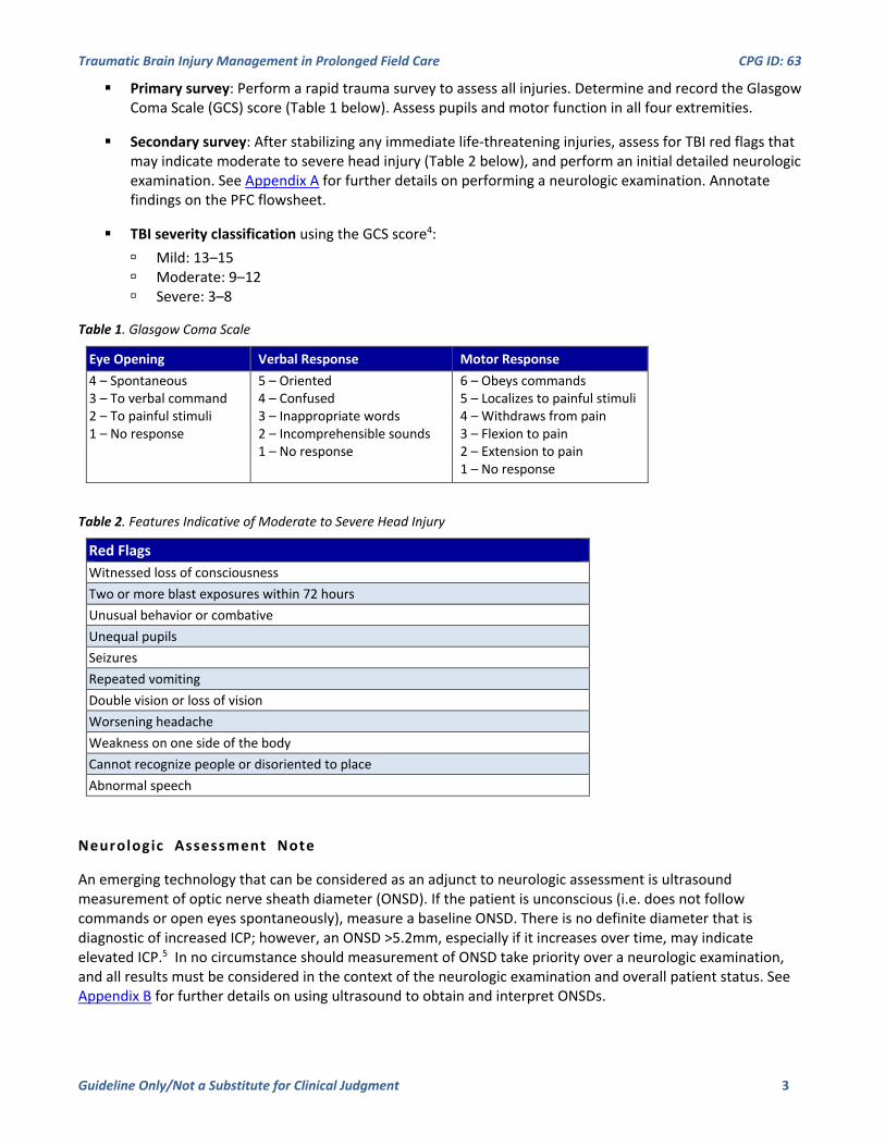
Traumatic Brain Injury Management in Prolonged Field Care CPG ID: 63
Guideline Only/Not a Substitute for Clinical Judgment 3
Primary survey: Perform a rapid trauma survey to assess all injuries. Determine and record the Glasgow Coma Scale (GCS) score (Table 1 below). Assess pupils and motor function in all four extremities.
Secondary survey: After stabilizing any immediate life-threatening injuries, assess for TBI red flags that may indicate moderate to severe head injury (Table 2 below), and perform an initial detailed neurologic examination. See Appendix A for further details on performing a neurologic examination. Annotate findings on the PFC flowsheet.
TBI severity classification using the GCS score4: Mild: 13–15 Moderate: 9–12 Severe: 3–8
Table 1. Glasgow Coma Scale
Table 2. Features Indicative of Moderate to Severe Head Injury
Neurologic Assessment Note
An emerging technology that can be considered as an adjunct to neurologic assessment is ultrasound measurement of optic nerve sheath diameter (ONSD). If the patient is unconscious (i.e. does not follow commands or open eyes spontaneously), measure a baseline ONSD. There is no definite diameter that is diagnostic of increased ICP; however, an ONSD >5.2mm, especially if it increases over time, may indicate elevated ICP.5 In no circumstance should measurement of ONSD take priority over a neurologic examination, and all results must be considered in the context of the neurologic examination and overall patient status. See Appendix B for further details on using ultrasound to obtain and interpret ONSDs.
Eye Opening Verbal Response Motor Response 4 – Spontaneous 3 – To verbal command 2 – To painful stimuli 1 – No response
5 – Oriented 4 – Confused 3 – Inappropriate words 2 – Incomprehensible sounds 1 – No response
6 – Obeys commands 5 – Localizes to painful stimuli 4 – Withdraws from pain 3 – Flexion to pain 2 – Extension to pain 1 – No response
Red Flags Witnessed loss of consciousness Two or more blast exposures within 72 hours Unusual behavior or combative Unequal pupils Seizures Repeated vomiting Double vision or loss of vision Worsening headache Weakness on one side of the body Cannot recognize people or disoriented to place Abnormal speech

Traumatic Brain Injury Management in Prolonged Field Care CPG ID: 63
Guideline Only/Not a Substitute for Clinical Judgment 4
ONSD should NOT be attempted in any patient who has sustained an open globe injury where any pressure on the globe is contraindicated. As many as 30% of head injuries in combat may also have an eye injury.6
MONITORING
Goal: Prevent secondary brain injury by maintaining adequate oxygenation and ventilation, avoiding hypotension, observing for signs and symptoms of elevated ICP, and trending the response to resuscitation. Detect changes in vital signs and neurologic examination as early as possible.
Minimum: Blood pressure cuff, stethoscope, pulse oximeter, method to monitor urine output. If an advanced airway is in place, monitor EtCO2 with capnometer. Check pupillary response and GCS as often as possible. Document vital signs, GCS score, and urine output on the PFC Casualty Card available at https://prolongedfieldcare.org.
Best: Portable monitor providing continuous vital-signs display, Foley catheter to monitor urine output. If an advanced airway is in place, monitor end-tidal carbon dioxide (EtCO2) with capnography. Check pupillary response and GCS score every hour. Document vital signs, GCS, and urine output on the PFC Casualty Card available at https://prolongedfieldcare.org.
ASSESSMENT AND MONITORING NOTES
Perform an initial assessment according to TCCC/MARCH (Massive hemorrhage, Airway, Respirations, Circulation, Head injury/Hypothermia) algorithms.
Severe head injury is associated with additional trauma in 60% of patients.7
If ONSD is used to evaluate for increased ICP and response of ICP to interventions, repeated ultrasound examinations should be performed if there is any change in neurologic examination and at regular intervals (30 minutes) after performing ICP-lowering interventions.
When possible, a pocket ophthalmoscope can be used to assess for the presence or absence of spontaneous venous pulsations (SVPs). SVPs are only present when ICP is normal. Visualization of SVPs can reassure the provider that ICP is not critically elevated.8 See Appendix C for additional information on the rapid assessment of SVPs.
Consider early C-spine immobilization. The incidence of concomitant brain and spinal cord injury in trauma ranges from 25% to 60%, with motor vehicle crashes and falls having the highest incidence of co-occurence.9 Ensure the cervical collar does not compress the jugular veins in the neck, because that could worsen ICP.
The neurologic examination is essential to identify deterioration in a TBI patient. Treat for elevated ICP for any deterioration in neurologic examination findings.
Pain medication and sedation are usually required for TBI patients; however, these medications also make it difficult to follow the neurologic examination.
Obtain a telemedicine consultation if possible. If in doubt, treat for elevated ICP according to ICP management outlined in the next section.
Close control of EtCO2 is critical for severe TBI patients. Plan and ensure the capability to monitor EtCO2 is available whenever advanced airway is placed. Goal EtCO2 is 35–40mmHg.

Traumatic Brain Injury Management in Prolonged Field Care CPG ID: 63
Guideline Only/Not a Substitute for Clinical Judgment 5
Cushing’s triad (i.e., increased SBP and/or widening pulse pressure, bradycardia, and irregular respirations) is a physiologic response that can occur with elevated ICPs, resulting in medullary compression. It is a late finding of severe brain injury with brainstem herniation. Cushing’s triad should be viewed as a sign of cerebral herniation and addressed immediately when recognized (see ICP Management).
MANAGEMENT
HEMODYNAMIC CONTROL
Goal: Maintain SBP >110mmHg. In polytrauma patients with ongoing bleeding, aggressively control hemorrhage using all means available and restore circulating blood volume by using blood products. Note: An SBP >90mmHg has traditionally been targeted in TBI patients, though recent literature has suggested better outcomes may occur when SBP is maintained above 110mmHg in TBI patients.10,11
Minimum: Stop all external bleeding. Manage internal bleeding to the extent possible with available resources. Administer tranexamic acid (TXA) per TCCC guidelines. Avoid medications that may lower the blood pressure (e.g., narcotics).
Better: If there is evidence of bleeding and no blood products are available, administer 1L 0.9% sodium chloride (NaCl). Target is an SBP >110mmHg.
Best: If there is evidence of bleeding, transfuse whole blood or, if not available, transfuse blood products. Greater emphasis on the use of Low Titer O Whole Blood is the optimal strategy. Target is an SBP >110mmHg.
Hemodynamic Control Notes
Do not neglect scalp bleeding. This can become a significant source of blood loss. Scalp lacerations should be sutured or stapled as soon as possible.
Take caution if an underlying skull fracture is present or if there is obvious penetrating trauma. DO NOT tightly pack or irrigate an open head wound. Suture or staple the skin closed, if bleeding. A pressure dressing may be placed if needed to control bleeding.
Hypotension is usually not caused by TBI except as a late finding due to herniation.12 Always look for other causes of hypotension, such as ongoing bleeding or tension pneumothorax.
Urine output (UOP) provides an important assessment of blood supply to the organs. Monitoring continuously by a Foley catheter is ideal. If a Foley catheter is not available, monitor by a graduated cylinder. Goal UOP in a polytrauma patient is 30–50mL/h.13
The role of TXA in TBI patients is currently under investigation in the CRASH-3 trial. Limited data suggest TXA limits ICH expansion and may improve outcomes in TBI patients.14 Until more definitive data are available, TXA can be used in TBI patients.
Colloids (e.g., albumin) have demonstrated a trend toward worsening outcomes in brain-injured patients.15 Hetastarches are associated with coagulopathy and increased risk of kidney injury in trauma patients.
Avoid colloids and hetastarches in TBI patients; however, they can be used if no alternative is available.

Traumatic Brain Injury Management in Prolonged Field Care CPG ID: 63
Guideline Only/Not a Substitute for Clinical Judgment 6
Avoid hypotonic fluids (including lactated Ringer’s) whenever possible; they can make brain swelling worse.
Most trauma patients with moderate or severe TBI will have other traumatic injuries. A careful search for bleeding should be performed in any hypotensive trauma patient.
Brain injury with associated hemorrhagic shock is a complicated scenario with a high risk of death. Balancing hemorrhage control (which is easier with lower blood pressure) with maintaining cerebral perfusion pressure (which requires higher blood pressure) should be guided with expert teleconsultation (i.e. critical care, neurocritical care, neurosurgical) whenever possible.
AIRWAY, OXYGENATION/VENTILATION MANAGEMENT
Goal: Manually maintain or secure the patient’s airway and avoid hypoxia, hypocapnia, or hypercapnia to reduce the risk of secondary brain injury. If GCS score is ≤8 or there is facial trauma with compromised airway, a definitive airway is most likely needed. The provider should place the type of airway (i.e. cricothyroidotomy or endotracheal tube [ETT]) that they have the most confidence in placing, based on their training and practice.
Minimum: Nasopharyngeal airway and bag-valve-mask with PEEP valve as needed. Use supplemental oxygen, if available. Maintain SpO2 >90%.
Better: Perform a cricothyroidotomy/ETT placement or place a supraglottic airway (e.g., laryngeal mask airway [LMA], King laryngeal tube [LT]) followed by continuous sedation and airway maintenance, supplemental oxygen via oxygen concentrator, and portable ventilator to maintain an SpO2 >95% and EtCO2 of 35–40mmHg.
Best: Cricothyroidotomy or ETT followed by continuous sedation and airway maintenance, supplemental oxygen, portable ventilator. Targets: SpO2 >95% and EtCO2 35–40mmHg. Check arterial blood gas results and correlate with EtCO2 within 30 minutes of intubation if laboratory capability is available. A positive end-expiratory pressure (PEEP) of 5cmH2O should be used routinely. PEEP can be safely increased up to 15cmH2O if needed to improve SpO2, but be alert for problems caused by increased intrathoracic pressure (e.g., lower blood pressure or increased ICP).16
Airway Management Notes
Patients with a GCS score ≤8 should undergo placement of an advanced airway (i.e. cricothyroidotomy or ETT) unless arrival to a higher level of care will occur in a timely manner or the airway can be manually maintained. The risk versus benefit of advanced airway placement should be carefully considered and discussed by telemedicine consultation whenever possible.
Airway interventions may cause transient hypoxia during the procedure. Every effort should be made to optimize airway placement on the first attempt by preoxygenating with supplemental O2, selecting the most experienced provider available, and using the technique most familiar to the provider.
Patients typically require less sedation after cricothyroidotomy than after ETT placement. This may help conserve resources if medications are limited.
Monitor EtCO2 and adjust ventilations to achieve the target range. Avoid hyperventilation (EtCO2 <35mmHg) except in extreme cases where imminent herniation is suspected, because hyperventilation worsens cerebral ischemia. Also avoid hypoventilation (EtCO2 >45mmHg) that will increase ICP.

Traumatic Brain Injury Management in Prolonged Field Care CPG ID: 63
Guideline Only/Not a Substitute for Clinical Judgment 7
Gastric decompression with a nasogastric tube (NGT) or oral gastric tube (OGT) will decrease the risk of aspiration in unconscious patients. If patients required bag-masking, they may have a distended stomach, which, in some patients, contributes to bradycardia. NGT and OGT cannot be placed with a supraglottic airway.
ICP MANAGEMENT
Goal: Suspect high ICP in any head injury patient with GCS score ≤8 OR declining findings on neurologic examination (unless explained by sedation, hypotension, hypoxia, hypercarbia, high fever). Minimize factors that contribute to elevated ICP, such as pain, anxiety, and fever. Rapidly recognize and manage elevated ICP, and maintain an adequate cerebral perfusion pressure.
Minimum: Use general measures to reduce ICP.
Elevate head of bed (HOB) 30°–60°.
Maintain neck in midline position.
Maintain arterial blood oxygen saturation (SpO2) >90% (or >95% if receiving ventilatory support).
Maintain EtCO2 between 35mmHg and 40mm Hg.
Maintain core temperature between 96°F and 99.5°F.
Maintain SBP >100mmHg, ideally at >110mmHg.
Prevent or rapidly manage seizure activity.
If concerned for impending herniation (e.g., unresponsive patient with unilateral dilated pupil, presence of Cushing’s triad), hyperventilate the patient for no more than 20 minutes to an EtCO2 target of 30mmHg.17 Seek expert consultation immediately.
The optimum duration of hyperventilation and frequency that can be repeated are not known. If performed, assess response (i.e. pupils, GCS score, and so forth). If patient responds, consider performing again if needed, guided by expert teleconsultation, if possible
Better: Even unconscious patients may experience pain and anxiety, manifested by hypertension (i.e., SBP >160mmHg) and/or agitation. Anxiety and agitation can increase ICP. In addition to all minimum measures, ensure adequate sedation and analgesia by targeting a Richmond Agitation and Sedation Score of −1 to −2. Refer to Joint Trauma System CPG on PFC Analgesia and Sedation.18
Ketamine 20mg IV/IO
Hydromorphone 0.5–2mg IV/IO
Fentanyl 25–50μg IV/IO
In addition to analgesics, consider administration of a rapid-onset, short-duration anxiolytic. Midazolam 1–2mg IV/IO as needed for agitation or anxiety.
Best: In addition to all minimum and better measures, administer osmotic therapy via peripheral intravenous (IV) or intraosseous (IO) access:
Hypertonic saline (HTS): 3% NaCl 250mL bolus over 20 minutes; repeat every 3 hours as needed when concerned for elevated ICP.

Traumatic Brain Injury Management in Prolonged Field Care CPG ID: 63
Guideline Only/Not a Substitute for Clinical Judgment 8
Mannitol can be used if there is no sign of bleeding and the SBP is >110mmHg. Mannitol 1g/kg IV/IO over 20 minutes. Repeat at 0.5g/kg IV/IO every 3 hours as needed when concerned for elevated ICP.
Seek additional medical direction as soon as possible and evacuate to neurosurgical care at the earliest opportunity.
ICP Management Notes
ICP cannot be directly measured without advanced intracranial monitoring devices. Therefore, vigilant clinical observation and the use of noninvasive ICP assessment modalities are critical to monitoring TBI patients until neurosurgical placement of neuromonitoring devices can occur.
HTS bolus lowers ICP and has a duration of action of approximately 3 hours.19,20
Mannitol, although effective, has several potentially adverse complications. It is a diuretic and might lower the blood pressure. Also, after repeated use, it can cross a damaged blood–brain barrier and potentially worsen ICP.20 For these reasons, HTS is preferred to mannitol in TBI and polytrauma patients.
Some institutions have reported ICP-lowering benefits from vertical positioning of patients, particularly when high intraabdominal or intrathoracic pressure is suspected. Lower intraabdominal and intrathoracic pressures may facilitate venous drainage from the intracranial compartment. If safe to do so, this can be attempted when other measures have failed.21
Always treat hypotension before treating elevated ICP. Cerebral blood flow is more affected by a decrease in blood pressure than an increase in ICP.
Cerebral perfusion pressure (CPP) = mean arterial pressure (MAP; mmHg) − ICP (mmHg)
Hyperventilation reduces CO2 and rapidly lowers ICP by causing cerebral vasoconstriction and decreasing the overall cerebral blood volume. However, hyperventilation also damages the brain by causing ischemia and should only be performed for brief periods. Avoid hyperventilation unless all other interventions have been ineffective.22
Although there are invasive interventions to help assess and treat elevated ICP, evacuate hematomas, and so forth, such as decompressive craniectomy, extraventricular drains, intracranial bolt monitors, and burr holes, such procedures are not recommended unless the PFC provider has training and experience in performing these procedures and is directed by expert teleconsultation.
INFECTION CONTROL
Goal: Dress all wounds to prevent further exposure to environmental pathogens and administer antibiotic prophylaxis to all patients with penetrating TBI.
Minimum: Dress all wounds to prevent further introduction of infectious materials. Optimize wound and patient hygiene to the extent possible given the environmental and situational conditions.
For penetrating head wounds, apply superficial dressings and seal the dressing to the extent possible.

Traumatic Brain Injury Management in Prolonged Field Care CPG ID: 63
Guideline Only/Not a Substitute for Clinical Judgment 9
Bleeding head injuries must be sutured or stapled to control bleeding.
DO NOT introduce any material into the wound cavity.
DO NOT attempt to flush the wound.
Antibiotics are not necessary in TBI without open or penetrating trauma.
Better: Antibiotics should be used for open or penetrating TBI.
Ertapenem 1g IV/IO every 24 hours and moxifloxacin 400mg PO every 24 hours for 5 days.
Ertapenam and moxifloxacin may increase the risk of seizure and ertapenam may not penetrate an intact blood-brain barrier. This combination, while commonly available, should only be used in TBI patients when the antibiotics with proven CNS penetration are not available.
Best: Administer CNS-penetrating antibiotics for open or penetrating TBI.
Ceftriaxone 2 gm IV/IO every 24 hours or cefazolin 2g IV/IO every 8 hours for 5 days. .
Add metronidazole 500mg IV/IO every 8 hours for 5 days for wounds that are grossly contaminated with organic debris (e.g., dirt, debris, clothing).17
Infection Control Notes
Moxifloxacin may be replaced with levofloxacin 750 mg PO daily to provide better coverage of bacteria found in wet terrain/ jungle environment.
If recommended antibiotics are not available or significant drug allergies are present, obtain teleconsultation to discuss alternative medications. Additional detailed recommendations may found in Forgione et al.23
SEIZURE PROPHYLAXIS AND MANAGEMENT
Goal: Rapidly identify and manage seizure activity in TBI patients.
Minimum: For witnessed convulsive seizure activity, place the patient on his/her side and clear the area of any potentially harmful objects. Suction the mouth if possible, but DO NOT attempt to place anything inside a seizing patient’s mouth. Treat any witnessed or suspected seizures with a rapid-acting benzodiazepine.
Midazolam 5mg IV/IO/IM every 5 minutes until seizure stops.
An alternate benzodiazepine can be used if available (diazepam 5mg IV every 5 minutes until seizure stops; lorazepam 4mg IV every 5 minutes until seizure stops).
Best: For witnessed or suspected seizures, administer a rapidly acting benzodiazepine (midazolam 5mg IV/IO/intramuscularly [IM]) plus a maintenance antiepilepsy drug. Broad-spectrum IV agents such as levetiracetam (Keppra; UCB Pharma, http://www.ucb.com/) are preferred.
Maintenance antiepilepsy-drug dosing: Levetiracetam: 2000mg IV/IO loading dose over 15 minutes followed by maintenance dosing of 500mg IV/IO every 12 hours.
Alternate maintenance antiepilepsy drugs:

Traumatic Brain Injury Management in Prolonged Field Care CPG ID: 63
Guideline Only/Not a Substitute for Clinical Judgment 10
Phenytoin (loading dose: 1.5g IV over 1 hour, then 100mg PO/IV/IO every 8 hours)
Phenobarbital (loading dose: 1.5g IV/IO over 1 hour, then 100mg PO/IV/IO daily). Be ready to support ventilation if phenobarbital is used.
Seizure Notes
Not all seizures are easy to see. At times, the findings may be obvious with generalized convulsions, or they may be subtle (e.g., persistent twitching of facial muscles, fingers).
Risk factors for seizures after TBI include: GCS score <10, skull fractures, penetrating injuries, prolonged length of coma (>24 hours).24
Non-convulsive seizures (NCSs) should be considered in any TBI patient with a GCS score ≤8 and who does not improve with appropriate resuscitation and/or ICP management. NCSs may persist after convulsive seizures are stopped and may be associated with higher morbidity and mortality.25 The most common signs of patients with NCS are coma, delirium, agitation, aphasia (impairment of language affecting production and/or comprehension of speech, reading, and/or writing) and/or “blank staring.”
Prompt initiation of seizure prophylaxis reduces early seizures after TBI.26 In PFC settings, where possible, an anti-epilepsy drug should be used early after injury to help prevent seizure.
Midazolam has a high rate of seizure control and works rapidly to terminate seizure activity.27 Midazolam is preferred because it is a short-acting medication (elimination time: 2–4 hours) and will allow for more regular and comprehensive neurologic examinations. It can be given IM in patients who do not yet have IV/IO access.
If not already placed, strong consideration should be given to placing an advanced airway (cricothyroidotomy or ETT) in any TBI patient who experiences seizures (place airway after seizures are controlled).
FEVER CONTROL
Goal: Maintain core temperature between 96°F and 99.5°F. Treat fever aggressively in TBI patients with a combination of medication, cold fluid boluses, and surface cooling techniques.
Minimum: Ensure patient has been removed from heat or sun. Remove clothes to allow evaporative cooling, Use surface-cooling measures (e.g., evaporative heat loss by misting and fan cooling) to reduce core body temperature.
Better: Apply cold packs to axillary regions, posterior cervical region, and the groin.
Best: Acetaminophen 650mg every 4 hours orally (PO) or rectally as needed for rectal temperatures >99.5°F. Additionally, cold saline IV fluid bolus can be used for refractory fever, if available.
Fever Notes
Fever will increase cerebral metabolism and may increase ICP.
Although targeted temperature management (previously referred to as therapeutic hypothermia) is used to reduce ICP in a critical care setting, hypothermia is part of the “lethal triad” in trauma patients, along with coagulopathy and acidosis. Targeted temperature management strategies beyond what is outlined in TCCC should NOT be attempted in the field or Role 1 setting.

Traumatic Brain Injury Management in Prolonged Field Care CPG ID: 63
Guideline Only/Not a Substitute for Clinical Judgment 11
Hypothermia prevention and management kits should continue to be used in all trauma patients. In TBI patients, however, warming measures should be avoided when the core body temperature is above the target range.
Avoid non-steroidal anti-inflammatory drugs (NSAIDs), such as ibuprofen, naproxen, ketorolac. Although these agents can effectively lower temperature, their antiplatelet effect may increase bleeding in TBI if intracranial hemorrhage is present (e.g., epidural hematomas, subdural hematomas).
SODIUM MANAGEMENT
Goal: Avoid hyponatremia, which can worsen brain swelling. The target serum sodium level in patients with severe TBI is slightly above normal, between 145mmol/L and 160mmol/L.
Minimum: Avoid the administration of any free water or hypotonic fluids that will lower serum sodium levels.
Best: Monitor serum sodium level via blood sampling. In a stable patient, check sodium level every 6 hours. In an unstable patient, or in a patient receiving HTS, check sodium level every 3 hours. Adjust fluids as needed to meet the sodium goals.
Sodium Management Notes
Several conditions can develop rapidly in brain-injured patients that can lower serum sodium levels (e.g., cerebral salt wasting, syndrome of inappropriate antidiuretic hormone secretion) or raise serum sodium levels (e.g., diabetes insipidus). Sodium levels, fluid intake, and urine output should be monitored closely.
If laboratory testing for serum sodium level is not available, then, as a reference, 250mL of 3% saline can be expected to raise the serum sodium level of an 80kg patient approximately 2–3mmol/L. Assuming a normal serum level of 140mmol/L before starting HTS therapy, it would take six 250mL bags of 3% HTS to raise the serum sodium to concerning levels (i.e. >160mmol/L). This is without factoring in the regulation of serum sodium by the kidneys. If patient is urinating, it will be difficult to raise serum sodium above 160mmol/L with 3% HTS. If patient is not urinating, more caution should be used because sodium levels can build up more quickly.
Obtain telemedicine consultation, preferably from a critical care or neurocritical care expert, before giving more than two 250mL boluses of 3% NaCl HTS.
BLOOD GLUCOSE CONTROL
Goal: Avoid both hypolgycemia and hyperglycemia. Target a blood glucose level of 180mg/dL via handheld glucometer.
Minimum: Monitor for clinical signs and symptoms of hypoglycemia (e.g., sweating, confusion, tremor, generalized weakness, generalized lethargy). If patient is hungry and able to safely swallow, allow the patient to eat to avoid hypoglycemia. Avoid the administration of any substances that are excessively high in sugar or carbohydrate content to prevent hyperglycemia.
Best: Check blood glucose level every 6 hours. If glucose level is <100mg/dL, give 20g of oral glucose (5 teaspoons of sugar or 4 teaspoons of honey) PO or by NG tube. Or administer 25g (50mL) dextrose 50% in water (D50) solution IV/IO. Recheck blood glucose in 1 hour, then continue to check every 6 hours.

Traumatic Brain Injury Management in Prolonged Field Care CPG ID: 63
Guideline Only/Not a Substitute for Clinical Judgment 12
Blood Glucose Control Notes
Hypoglycemia is more harmful to the brain than hyperglycemia.
Hyperglycemia may occur in TBI as an acute stress response or as a result of brain-induced catecholamine release. Early hyperglycemia (i.e., >180mg/dL) has been associated with poor neurologic outcome in severe TBI.28
The treatment of hyperglycemia requires insulin. If the appropriate medications, laboratory capability, and expertise are available, hyperglycemia may be treated; however, the treatment of hyperglycemia is not covered in this CPG.
TRANSPORTATION CONSIDERATIONS
Goal: Safely prepare TBI patients for ground and/or air transportation to higher levels of care.
Minimum: Dose sedative/analgesic medications, osmotic agent, and/or benzodiazepine before any significant ground or air transportation, as resources permit.
Neuromuscular blocking agents will mask seizures and clinical examination changes; therefore, the risk versus benefit of use during transport must be considered.
Best: Ensure transport assets are aware of the patient’s brain injury in advance so appropriate accommodations and/or alterations to the travel plan can be made.
Most patients are transported in a supine position. Every effort should be made to transport TBI patients with the HOB elevated to at least 30°.
For fixed-wing transport, TBI patients should be loaded with their head toward the front of the aircraft to minimize G-forces transmitted to the brain.
Air transport of TBI patients requires additional considerations. For air movements that involve altitudes >8,000 ft, TBI patients are at risk for additional complications that may worsen brain injury, such as hypobaric hypoxia and high-altitude cerebral edema.29 Preplanning with air assets is highly recommended.
Recommended Packing List. See Appendix D.
Management of Traumatic Brain Injury Summary Table. See Appendix E.
PERFORMANCE IMPROVEMENT (PI) MONITORING
POPULATION OF INTEREST
1. All patients with a diagnosis of traumatic brain injury and an initial GCS of 3-8.
2. All patients who receive a cranial procedure (ICP monitor, craniectomy, craniotomy).
INTENT
1. All patients in population of interest avoid hypotension and hypoxia: SBP never < 100 mmHg, MAP never < 60, SaO2 never < 93%.

Traumatic Brain Injury Management in Prolonged Field Care CPG ID: 63
Guideline Only/Not a Substitute for Clinical Judgment 13
2. All patients in population of interest have PaCO2 monitored at every role of care.
3. All patients in population of interest have a head CT performed within 4 hours of injury.
4. All patients with a ventriculostomy have hourly documentation of ICP/CPP and ventriculostomy output.
5. Patients in population of interest unable to be monitored clinically (e.g., unable to hold sedation for Q1 hour neuro exam) have an ICP monitor or ventriculostomy placed prior to transport out of theater.
METRICS
1. Number and percentage of patients in the population of interest with lowest SBP<100 within first 3 days after injury.
2. Number and percentage of patients in the population of interest with MAP<60 within first 3 days after injury.
3. Number and percentage of patients in the population of interest with SaO2<93% within first 3 days after injury.
4. Number and percentage of patients in population of interest who have PaCO2 documented at every role of care (POI, POI MEDEVAC, ROLE 2-4, interfacility MEDEVAC).
5. Number and percentage of patients in the population of interest who maintain PaCO2=35-40
6. Number and percentage of patients who had a head CT performed within 4 hours of injury.
7. Number and percentage of patients with a ventriculostomy who had hourly documentation of ICP/CPP and ventriculostomy output.
8. Number and percentage of patients in the population of interest unable to be monitored clinically (eg. unable to hold sedation for Q1 hour neuro exam) who have an ICP monitor or ventriculostomy placed prior to transport out of theater.
DATA SOURCE
Patient Record and the ASIA or Combat Neuro Exam worksheet
Department of Defense Trauma Registry (DoDTR)
SYSTEM REPORTING & FREQUENCY
The above constitutes the minimum criteria for PI monitoring of this CPG. System reporting will be performed annually; additional PI monitoring and system reporting may be performed as needed.
The system review and data analysis will be performed by the Joint Trauma System (JTS) Chief and the PI Branch.

Traumatic Brain Injury Management in Prolonged Field Care CPG ID: 63
Guideline Only/Not a Substitute for Clinical Judgment 14
REFERENCES
1. Marr AL, Coronado VG, eds. Central Nervous System Injury Surveillance. Data Submission Standards-2002. Atlanta, GA: Centers for Disease Control and Prevention; 2004.
2. DuBose JJ, Barmparas G, Inaba K, et al. Isolated severe traumatic brain injuries sustained during combat operations: demographics mortality outcomes, and lessons to be learned from contrasts to civilian counterparts. J Trauma. 2011;70:11–18.
3. McHugh GS, Engel DC, Butcher I, et al. Prognostic value of secondary insults in traumatic brain injury: results from the IMPACT study. J Neurotrauma. 2007;24(2):287–293.
4. Teasdale G, Murray G, Parker L, et al. Adding up the Glasgow Coma score. Acta Neurochir Suppl (Wien). 1979;28:13–16.
5. Raffiz M, Abdullah JM. Optic nerve sheath diameter measurement: a means of detecting raised ICP in adult traumatic and nontraumatic neurosurgical patients. Am J Emerg Med. 2017:35(1):150–153.
6. Cho R, Bakken H, Reynolds M, et al. Concomitant cranial and ocular combat injuries during Operation Iraqi Freedom. J Trauma. 2009;34(3):516–520.
7. Groswasswer Z, Cohen M, Blankstein E. Polytrauma associated with traumatic brain injury: incidence, nature and impact on rehabilitation outcome. Brain Inj. 1990;4(2):161–166.
8. Bruce B. State of the Art Review: non-invasive assessment of cerebrospinal fluid pressure. J Neuroophthalmol. 2014;34(3): 288–294.
9. Kushner DS, Alvarez G. Dual diagnosis: traumatic brain injury with spinal cord injury. Phys Med Rehabil Clin N Am. 2014;25(3):681–696.
10. Berry C, Ley EJ, Bukur M, et al. Redefining hypotension in traumatic brain injury. Injury. 2012;43(11):1833–1837.
11. Brenner M, Stein DM, Hu PF, et al. Traditional systolic blood pressure targets underestimate hypotension-induced secondary brain injury. J Trauma Acute Care Surg. 2012;72(5):1135–1139.
12. Van Wyck DW, Grant GA, Laskowitz DT. Penetrating traumatic brain injury: a review of current evaluation and management concepts. J Neurol Neurophysiol. 2015;6(6):1–7.
13. Baker B, Powell D, Riesberg J, et al. Prolonged Field Care Working Group fluid therapy recommendations. J Spec Oper Med. 2016;Spring;16(1):117–122.
14. Zehtabchi S, Abdel Baki SG, Falzon L, et al. Tranexamic acid for traumatic brain injury: a systematic review and meta-analysis. Am J Emerg Med. 2014;32(12):1503–1509.
15. SAFE Study Investigators, Australian and New Zealand Intensive Care Society Clinical Trials Group, Australian Red Cross Blood Service, et al. Saline or albumin for fluid resuscitation in patients with traumatic brain injury. N Engl J Med. 2007;357:874-884.
16. Davis DP, Peay J, Sise MJ, et al. Pre-hospital airway and ventilation management: a trauma score and injury severity score-based index analysis. J Trauma. 2010;69(2):294–301.
17. Diringer MN, Yundt K, Videen TO, et al. No reduction in cerebral metabolism as a result of early moderate hyperventilation following severe traumatic brain injury. J Neurosurg. 2000;92(1):7–13.

Traumatic Brain Injury Management in Prolonged Field Care CPG ID: 63
Guideline Only/Not a Substitute for Clinical Judgment 15
18. Joint Trauma System, Analgesia and sedation management during prolonged field care CPG, 11 May 2017. https://jts.amedd.army.mil/index.cfm/PI_CPGs/cpgs Accessed Mar 2018.
19. Mortazavi M, Romeo A, Deep A, et al. Hypertonic saline for treating raised intracranial pressure: literature review with metaanalysis. J Neurosurg. 2012;116:210–221.
20. Burgess S, Abu-Laban RB, Slavik RS, et al. A systematic review of randomized controlled trials comparing hypertonic sodium solutions and mannitol for traumatic brain injury: implications for emergency department management. Ann Pharmacother. 2016; 50(4):291–300.
21. Weingart S. Podcast 95 – Thomas Scalea on cutting-edge ICP management. EMCrit Blog. 2 April 2013. https://emcrit.org/podcasts/cutting-edge-icp-management/Accessed 15 May 2017.
22. Stocchetti N, Maas A, Chieregato A, et al. Hyperventilation in head injury. Chest. 2005;127(5):1812–1827.
23. Forgione MA, Moores LE, Wortmann GW. Prevention of infections associated with combat-related central nervous system injuries. J Trauma 2011;71:S258-S263.
24. Temkin NR. Risk factors for posttraumatic seizures in adults. Epilepsia. 2003;44 (suppl 10):18–20.
25. Alroughani R, Javidan M, Qasem A, et al. Non-convulsive status epilepticus; the rate of occurrence in a general hospital. Seizure. 2009;18(1):38–42.
26. Temkin NR, Dikmen SS, Wilensky AJ, et al. A randomized double-blind study of phenytoin for the prevention of post-traumatic seizures. N Engl J Med. 1990;323:497–502.
27. Silbergleit R, Durklaski V, Lowenstein D, et al. Intramuscular versus intravenous therapy for prehospital status epilepticus. N Engl J Med. 2012;366(7):591–600.
28. Jeremitsky E, Omert LA, Dunham CM, et al. The impact of hyperglycemia on patients with severe brain injury. J Trauma. 2005;58(1):47–50.
29. Basnyat B, Wu T, Gertsch JH. Neurological conditions at altitude that fall outside the usual definition of altitude sickness. High Alt Med Biol. 2004;5:171–179.
30. Vaiman M, Abuita R, Bekerman I. Optic nerve sheath diameters in healthy adults measured by computed tomography. Int J Opthalmol. 2015;8(6):1240–1244.
31. Amini A, Kariman H, Arhami Dolatabadi A, et al. Use of the sonographic diameter of optic nerve sheath to estimate intracranial pressure. Am J Emerg Med. 2013;31(1):236–239.
32. Soldatos T, Chatzmichail K, Papathanasiou M, et al. Optic nerve sonography: a new window for the non-invasive evaluation of intracranial pressure in brain injury. Emerg Med J. 2009;26(9):630–634.
33. Maissan I, Perjan J, Dirven AC, et al. Ultrasonographic measured optic nerve sheath diameter as an accurate and quick monitor for changes in intracranial pressure. J Neurosurg. 2015;123(3):743–747.

Traumatic Brain Injury Management in Prolonged Field Care CPG ID: 63
Guideline Only/Not a Substitute for Clinical Judgment 16
APPENDIX A: NEUROLOGICAL EXAMINATION I. MENTAL STATUS
Level of Consciousness: Note whether the patient is: Alert/responsive Not alert but arouses to verbal stimulation Not alert but responds to painful stimulation Unresponsive
Orientation: Assess the patient’s ability to provide: Name Current location Current date Current situation (e.g., ask the patient what happened to
him/her) Language: Note the fluency and appropriateness of the patient’s response to questions. Note patient’s ability to follow commands when assessing other functions (e.g., smiling, grip strength, wiggling toes). Ask the patient to name a simple object (e.g., thumb, glove, watch).
Speech: Observe for evidence of slurred speech. II. CRANIAL NERVES All patients: Assess the pupillary response to light. Assess position of the eyes and note any movements (e.g.,
midline, gaze deviated left or right, nystagmus, eyes move together versus uncoupled movements).
Noncomatose patient: Test sensation to light touch on both sides of the face. Ask patient to smile and raise eyebrows, and observe for
symmetry. Ask the patient to say “Ahhh” and directly observe for
symmetric palatal elevation. Comatose patient: Check corneal reflexes; stimulation should trigger eyelid
closure. Observe for facial grimacing with painful stimuli. Note symmetry and strength. Directly stimulate the back of the throat and look for a
gag, tearing, and/or cough.
III. MOTOR Tone: Note whether resting tone is increased (i.e. spastic or rigid),
normal, or decreased (flaccid). Strength: Observe for spontaneous movement of extremities and
note any asymmetry of movement (i.e. patient moves left side more than right side). Lift arms and legs, and note whether the limbs fall immediately, drift, or can be maintained against gravity. Push and pull against the upper and lower extremities and note any resistance given. Note any differences in resistance provided between the left and right sides. (NOTE: it is often difficult to perform formal strength testing in TBI patients. Unless the patient is awake and cooperative, reliable strength testing is difficult.)
Involuntary movements: Note any involuntary movements (e.g., twitching, tremor, myoclonus) involving the face, arms, legs, or trunk.
IV. SENSORY If patient is not responsive to voice, test central pain and peripheral pain. Central pain: Apply a sternal rub or supraorbital pressure, and
note the response (e.g., extensor posturing, flexor posturing, localization).
Peripheral pain: Apply nail bed pressure or take muscle between the fingers, compress, and rotate the wrist (do not pinch the skin). Muscle in the axillary region and inner thigh is recommended. Apply similar stimulus to all four limbs and note the response (e.g., extensor posturing, flexor posturing, withdrawal, localization). NOTE: In an awake and cooperative patient, testing light touch is recommended. It is unnecessary to apply painful stimuli to an awake and cooperative patient.
V. GAIT If the patient is able to walk, observe his/her casual gait and note any instability, drift, sway, and so forth.

Traumatic Brain Injury Management in Prolonged Field Care CPG ID: 63
Guideline Only/Not a Substitute for Clinical Judgment 17
APPENDIX B: ULTRASONIC ASSESSMENT OF OPTIC NERVE SHEATH DIAMETER If a patient is unconscious (i.e. does not follow commands or open eyes spontaneously), they may have elevated ICP. There is no reliable test for elevated ICP available outside of a hospital; however, optic nerve sheath diameter (ONSD) measurement is a rapid, safe, and easy-to-perform ultrasonographic assessment that may help identify elevated ICP when more definitive monitoring devices are not available. The optic nerve sheath directly communicates with the intracranial subarachnoid space. Increased ICP, therefore, displaces
cerebrospinal fluid along this pathway. Normal ONSD is 4.1–5.9mm.30 A 10–5-MHz linear ultrasound probe can be used to obtain ONSDs. ONSD is measured from one side of the optic nerve sheath to the
other at a distance of 3mm behind the eye immediately below the sclera.31 In general, ONSDs >5.2mm should raise concern for clinically significant elevations in ICP in unconscious TBI patients.5,32 The ONSD
can vary significantly in normal individuals, so one single measurement may not be helpful; however, repeated measurements that detect gradual increases in ONSD over time may be more useful than a single measurement.
ONSD changes rapidly when the ICP changes, so it can be measured frequently.33 If ONSD is used, it is best to check hourly along with the neurologic examination.
TECHNIQUE
1. Check to make sure there is no eye injury. A penetrating injury to the eyeball is an absolute contraindication to ultrasound because it puts pressure on the eye.
2. Ensure the head and neck are in a midline position. Gentle sedation and/or analgesia may be necessary to obtain accurate measurements.
3. Ensure the eyelids are closed. 4. If available, place a thin, transparent film (e.g., Tegaderm; 3M, http://www.3m.com) over the closed eyelids. 5. Apply a small amount of ultrasound gel to closed eyelid. 6. Place the 10(–5)MHz linear probe over the eyelid. The probe should be applied in a horizontal orientation (Figure 1) with as little
pressure as possible applied to the globe. 7. Manipulate the probe until the nerve and nerve sheath are visible at the bottom of screen. An example of a proper
ultrasonagraphic image of the optic nerve sheath can be seen in Figure 2. 8. Once the optic nerve sheath is visualized, freeze the image on the screen. 9. Using the device’s measuring tool, measure 3mm back from the optic disc and then obtain a second measurement perpendicular
to the first. The second measurement should cover the horizontal width of the optic nerve sheath (Figure 2). An abnormal ONSD is shown in Figure 3.
10. Repeat the previous sequence in the opposite eye. Annotate both ONSDs on the PFC Casualty Card. 11. ONSDs should be obtained, when possible, at regular intervals to help assess changes in ICP, particularly when the neurologic
examination is poor and/or unreliable (i.e. with sedation). Serial measurements with progressive diameter enlargement and/or asymmetry in ONSDs should be considered indicative of worsening intracranial hypertension.
CAUTION: ONSD measurements are contraindicated in eye injuries. NEVER apply pressure to an injured eye. Figure 1. Appropriate placement of the linear probe.
Ultrasound gel is placed over a closed eyelid and the probe placed horizontally over the eyelid, applying as little pressure to the globe as possible. If available, Tegaderm or other thin covering (e.g., Latex glove) should be placed over a closed eyelid for further protection.
Figure 2 An ultrasonographic view of a normal eye and optic nerve sheath.
To measure ONSD, apply the ultrasound measuring device to the optic disc and measure back 3mm along the length of the optic nerve. A second, perpendicular measurement is obtained at the previously measured point that spans the horizontal width of the optic nerve sheath. In this image, ONSD was determined to be 5.1mm, a normal value.
Figure 3. Ultrasound image of the right optic nerve sheath of a 61-year-old man with a traumatic subdural hematoma.
The optic nerve sheath measured 6.8mm in diameter. Elevated ICP was subsequently confirmed (26mmHg) after the placement of an ICP bolt monitor.

Traumatic Brain Injury Management in Prolonged Field Care CPG ID: 63
Guideline Only/Not a Substitute for Clinical Judgment 18
APPENDIX C: SPONTANEOUS VENOUS PULSATIONS Spontaneous venous pulsations (SVPs) are subtle, rhythmic
variations in retinal vein caliber on the optic disc and have an association with ICP.
It is difficult to see SVPs without advanced equipment; however, if a handheld ophthalmoscope is available, it is worth an attempt to visualize the retinal veins.
Don’t worry if you cannot see SVPs; this may actually be normal. However, if you do see them, it is very reassuring that ICP is normal.10
If SVPs are initially present and can no longer be seen on subsequent examinations, the provider should be concerned for increasing ICP.
TECHNIQUE
1. Gently lift the eyelid until the pupil is in view. 2. Using a handheld ophthalmoscope, the provider should
maneuver himself or herself to a position where the optic disc can be visualized.
3. Identify the retinal veins as they emerge from the optic disc. Retinal veins are typically slightly larger and darker than retinal arteries. Figure 1 demonstrates the typical appearance of the retina.
4. Observe the retinal veins for pulsations. Note the presence or absence of spontaneous venous pulsations
5. Repeat the step 1–4 sequence in the contralateral eye.
Figure 1. Typical appearance of a healthy retina.
The retinal vessels can be seen emerging from the optic disc. Retinal veins can be identified by their slightly larger, thicker size and darker color. Retinal arteries are small, thin, and lighter in color than retinal veins.

Traumatic Brain Injury Management in Prolonged Field Care CPG ID: 63
Guideline Only/Not a Substitute for Clinical Judgment 19
APPENDIX D: RECOMMENDED PACKING LIST Assumptions: One patient with a moderate to severe traumatic brain injury. To calculate the amount of fluid or medication you would need for a single TBI patient, use your worst case, longest possible evacuation extrapolated from your mission planning. For example, if you think you might have a 36-hour evacuation, you might need 3,500mg levetiracetam (a 2,000mg loading dose and 500mg every 12 hours)
MINIMUM
Equipment: vital-sign trending chart, BP cuff, stethoscope, wrist watch, pulse oximeter, capnometer, cricothyroidotomy kit, bag-valve-mask with PEEP valve, nasopharyngeal airway, disposable thermometer, Nalgene bottle to measure urine
Medications/Fluids: ketamine, midazolam, lorazepam, acetaminophen, ceftriaxone, 3% hypertonic saline
BETTER
Equipment: portable vital sign monitor, capnometer, cricothyroidotomy kit and/or ETT plus laryngoscope/glidescope and/or laryngeal mask airway, cold/ice packs, graduated cylinder to measure urine, oxygen concentrator
Medications/Fluids: ketamine, IV hydromorphone, IV fentanyl, midazolam
BEST
Equipment: portable monitor providing continuous vital-signs display with capnography, cricothyroidotomy kit, and/or ETT plus laryngoscope/glidescope, portable point-of care-testing device such as an iStat (Abbott Point of Care; https://www.pointofcare.abbott) or Epoch (Alere, http://www.alere.com) for arterial blood gas samples, and electrolyte monitoring, blood glucose monitor, Foley catheter kit, and supplemental oxygen or oxygen concentrator
Medications/Fluids: Fresh whole blood drawing supplies or stored blood products, 3% hypertonic saline, mannitol, ceftriaxone, metronidazole, levetiracetam or phenytoin, acetaminophen, dextrose 50% in water
OTHER PACKING L IST CONSIDERATIONS
Equipment: Portable ultrasound, nasogastric tube, red- (or red/yellow-speckled) top test tubes to test for electrolytes if a host-nation laboratory is available
Medications/Fluids: TXA

Traumatic Brain Injury Management in Prolonged Field Care CPG ID: 63
Guideline Only/Not a Substitute for Clinical Judgment 20
APPENDIX E: MANAGEMENT OF TRAUMATIC BRAIN INJURY SUMMARY TABLE
Management of Traumatic Brain Injury Summary
GOAL MINIMUM BETTER BEST
Neurological Assessment Goal: Rapidly identify TBI and associated injuries; assess TBI severity.
Consider: If patient is unconscious, measure a baseline optic nerve sheath diameter with ultrasound using a 10(–5)MHz linear probe.
Secondary survey: Evaluate for TBI red flags. Perform detailed neurologic examination.
Primary survey: Perform rapid trauma survey to assess all injuries. Determine and record Glasgow Coma Score. Assess pupils and motor function in all four extremities. Recognize Cushing’s triad.
ONSD should not be attempted on any patient with an open globe injury to the eye. Cushing’s triad: increased SBP/widened pulse pressure, bradycardia, irregular breathing (typically rapid/shallow)
Monitoring
Prevent secondary brain injury, maintain adequate oxygenation and ventilation, avoid hypotension, detect elevated ICP.
Blood pressure cuff, stethoscope, pulse oximeter, method to monitor urine output. If advanced airway is in place, monitor EtCO2 with capnometer. Check pupils and GCS hourly or as often as possible.
Portable monitor with continuous vital-signs display, Foley catheter to monitor urine output. If advanced airway in place, monitor end-tidal CO2 (EtCO2) with capnography. Check pupils and GCS hourly.
Neurologic examination and vital-sign trends are essential to identifying a deteriorating patient with TBI. Monitoring EtCO2 is critical for severe TBI patients. Bring the correct equipment whenever possible.
Management Hemodynamic Control
Maintain systolic pressure >110mmHg
Stop all external bleeding. Manage internal bleeding to the extent possible with available resources. Administer TXA per TCCC guidelines. Avoid medications that may lower the blood pressure.
If there is evidence of bleeding and no blood products are available, 0.9% sodium chloride 1L. Target SBP: >110mmHg
If evidence of bleeding, transfuse whole blood or, if not available, transfuse blood products per TCCC guidelines. Target SBP: >110mmHg
Brain injury in the presence of hemorrhagic shock requires balancing hemorrhage control with cerebral perfusion. Telemedicine consultation is strongly encouraged. Do not neglect scalp bleeding. Take care to inspect for skull fractures. DO NOT tightly pack an open head wound. Hypotonic fluids (to include lactated Ringer’s) should be avoided whenever possible because they can worsen cerebral edema.
Management: Airway, Oxygenation/Ventilation
Maintain or secure airway. If GCS score ≤8 or there is facial trauma or other airway compromise, consider definitive airway placement.
Nasopharyngeal airway and bag-valve-mask with PEEP. Supplemental oxygen, if available. Maintain SpO2 >90%.
Perform cricothyroidotomy/ETT or place supraglottic airway followed by continuous sedation, supplemental O2, portable ventilator. Target SpO2: >95%, EtCO2: 35–40mmHg.
Perform cricothyroidotomy/ETT followed by continuous sedation, supplemental O2, portable ventilator. Target SpO2 >95%, EtCO2 35–40. Check arterial blood gas values. PEEP: 5cmH2O (increase up to 15cmH2O if needed).
Avoid hyperventilation except in extreme cases where imminent cerebral herniation is suspected.
Management ICP Suspect high ICP in any head injury patient with GCS score ≤8 or declining findings on neurologic examination. Minimize factors that could contribute to elevated ICP, such as pain, anxiety, and fever.
Elevate HOB 30–45° Neck midline, loosen collar SBP >110mmHg (or at least > 100mmHg) SpO2 >90% or 95% on ventilator EtCO2 35–40mmHg Core temp 96–99.5°F Prevent/treat seizure Last choice if sign of herniation:
hyperventilate to EtCO2 30mmHg × 20 minutes.
In addition to minimum steps, ensure adequate sedation and analgesia. If SBP >160mmHg or agitated: Ketamine 20mg IV/IO Hydromorphone 0.5–2mg IV/IO Fentanyl 25–50μg IV/IO Midazolam 1–2mg IV/IO
In addition to minimum and better steps, give osmotic therapy IV/IO: HTS 3% 250mL over 20 minutes. Repeat
every 3 hours if needed. Mannitol (If no bleeding and SBP
>110mmHg) 1g/kg IV/IO over 20 minutes, repeat 0.5g/kg every 3 hours, if needed.
Telemedicine consultation early and often in the patient with elevated ICP.
Management: Infection Control
Dress all wounds and administer antibiotic prophylaxis for penetrating brain injuries and open wounds.
Dress all wounds to prevent further introduction of infection. Optimize wound care and patient hygiene to extent possible.
Ertapenem 1g IV/IO every 24 hours and moxifloxacin 400mg PO every 24 hours for 5 days.
Use an antibiotic with strong CNS penetration. Ceftriaxone 2 gm IV/IO every 24 hours or
cefazolin 2g IV/IO every 8 hours for 5 days
Add metronidazole 500mg IV/IO every 8 hours if wounds contaminated with organic debris.
Moxifloxacin may be replaced with levofloxacin 750 mg PO daily to provide better coverage of bacteria found in wet terrain/ jungle environment. Ertapenam and moxifloxacin may increase the risk of seizure and ertapenam may not penetrate an intact blood-brain barrier. If recommended antibiotics are not available or significant drug allergies are present, obtain teleconsultation to discuss alternative medications.

Traumatic Brain Injury Management in Prolonged Field Care CPG ID: 63
Guideline Only/Not a Substitute for Clinical Judgment 21
Management of Traumatic Brain Injury Summary
GOAL MINIMUM BETTER BEST Management: Seizures
Rapidly identify and manage seizures.
For a witnessed or suspected seizure, ensure safety and airway is clear. Treat with rapid-acting benzodiazepine: Midazolam 5mg IV/IO/IM every 5 minutes
until seizure stops
Levetiracetam 2,000mg IV/IO loading dose over 15 minutes + 500mg every 12 hours. Alternate therapy: Phenytoin 1.5g IV/IO load + 100mg IV/IO
every 8 hours Phenobarbital 1.5g IV/IO load + 100mg
IV/IO daily.
Consider nonconvulsive seizures in any TBI patient with GCS score ≤8 who is not improving with appropriate treatments. If not already placed, consider a definitive airway in any patient who experiences seizure. Perform after seizures are controlled.
Management: Fever Control
Maintain core temperature between 96°F and 99.5°F. Treat fever aggressively.
Remove patient from heat or sun. Remove clothing. Use surface cooling measures with misting and fan cooling
Apply cold packs to axillary, posterior cervical, and groin regions.
Acetaminophen 650mg every 4 hours PO or rectally for rectal temperature >99.5°F. Cold saline IV bolus if available.
Avoid NSAIDs, such as ibuprofen, naproxen, and ketorolac, because these agents may increase intracranial hemorrhage, if present. Management: Sodium Control Avoid hyponatremia. Mild hypernatremia optimal. Target sodium level: 145–160mmol/L.
Avoid administration of free water or hypotonic fluids.
Monitor serum sodium via laboratory blood samples. If patient is stable, check levels every 6 hours. In an unstable patient or one receiving HTS, check sodium level every 3 hours and adjust fluids as needed.
Numerous conditions can rapidly affect sodium levels in TBI patients. Monitor sodium and urine output whenever possible.
Management: Blood Glucose Control Avoid both hypo- and hyperglycemia. Target blood glucose 100–180mg/dL.
Monitor for signs and symptoms of hypoglycemia. Allow patient to eat as long as they are able.
Check blood glucose every 6 hours with handheld glucometer. If <100mg/dL, give 25g (50mL) dextrose 50% (D50) IV/IO or 5tsp sugar/4tsp honey PO/NG.
Transportation
TBI patients for ground and/or air transport to higher levels of care.
Dose sedative/analgesic medications, osmotic therapies, and/or benzodiazepines before any significant ground or air transport if possible.
Provide a detailed brief to the transport assets specifically highlighting any neurologic deficits and treatments and/or accommodations required during transport.
Neuromuscular blockers should only be used when the benefit outweighs the risks. Most patients should be transported in the supine position with the HOB elevated at least 30°. **For fixed-wing aircrafts: load patient with head to the front of the aircraft to minimize G-forces transmitted to the brain. **For altitudes >8000 ft, TBI patients are at risk of hypobaric hypoxia and high-altitude cerebral edema (HACE), which can worsen brain injuries.

Traumatic Brain Injury Management in Prolonged Field Care CPG ID: 63
Guideline Only/Not a Substitute for Clinical Judgment 22
APPENDIX F: ADDITIONAL INFORMATION REGARDING OFF-LABEL USES IN CPGS
PURPOSE
The purpose of this Appendix is to ensure an understanding of DoD policy and practice regarding inclusion in CPGs of “off-label” uses of U.S. Food and Drug Administration (FDA)–approved products. This applies to off-label uses with patients who are armed forces members.
BACKGROUND
Unapproved (i.e., “off-label”) uses of FDA-approved products are extremely common in American medicine and are usually not subject to any special regulations. However, under Federal law, in some circumstances, unapproved uses of approved drugs are subject to FDA regulations governing “investigational new drugs.” These circumstances include such uses as part of clinical trials, and in the military context, command required, unapproved uses. Some command requested unapproved uses may also be subject to special regulations.
ADDITIONAL INFORMATION REGARDING OFF-LABEL USES IN CPGS
The inclusion in CPGs of off-label uses is not a clinical trial, nor is it a command request or requirement. Further, it does not imply that the Military Health System requires that use by DoD health care practitioners or considers it to be the “standard of care.” Rather, the inclusion in CPGs of off-label uses is to inform the clinical judgment of the responsible health care practitioner by providing information regarding potential risks and benefits of treatment alternatives. The decision is for the clinical judgment of the responsible health care practitioner within the practitioner-patient relationship.
ADDITIONAL PROCEDURES
Balanced Discussion
Consistent with this purpose, CPG discussions of off-label uses specifically state that they are uses not approved by the FDA. Further, such discussions are balanced in the presentation of appropriate clinical study data, including any such data that suggest caution in the use of the product and specifically including any FDA-issued warnings.
Quality Assurance Monitoring
With respect to such off-label uses, DoD procedure is to maintain a regular system of quality assurance monitoring of outcomes and known potential adverse events. For this reason, the importance of accurate clinical records is underscored.
Information to Patients
Good clinical practice includes the provision of appropriate information to patients. Each CPG discussing an unusual off-label use will address the issue of information to patients. When practicable, consideration will be given to including in an appendix an appropriate information sheet for distribution to patients, whether before or after use of the product. Information to patients should address in plain language: a) that the use is not approved by the FDA; b) the reasons why a DoD health care practitioner would decide to use the product for this purpose; and c) the potential risks associated with such use.

Guideline Only/Not a Substitute for Clinical Judgment 1
JOINT TRAUMA SYSTEM CL INICAL PRACTICE GUIDELINE ( JTS CPG)
Acute Traumatic Wound Management in the Prolonged Field Care Setting (CPG ID: 62) The intent of this guideline is to provide evidence- and experience-based solutions to those who manage both simple and complex wounds in an austere environment.
Contributors
SFC Justin Rapp, 18D, USA MAJ Timothy Plackett, MC, USA SGT Jonathon Crane, 68D, USA SFC Jonathan Lu, 18D, USA LTC David Hardin, MC, USA
SSG Paul Loos, 18D, USA SFC (Ret) Richard Kelly, 18D, USA Maj Andrew Hall, MC, USAF COL Clinton Murray, MC, USA COL Sean Keenan, MC, USA Col Stacy Shackelford, USAF, MC
First Publication Date: 24 Jul 2017 Publication Date: 24 Jul 2017
TABLE OF CONTENTS
Introduction ................................................................................................................................................................................. 2 Special Considerations in Austere Environment .......................................................................................................................... 2 Types of Wounds ......................................................................................................................................................................... 2 Analgesia ...................................................................................................................................................................................... 3 Irrigation ...................................................................................................................................................................................... 4 Debridement ................................................................................................................................................................................ 5
Debridement Notes ................................................................................................................................................................. 6 Dressings ...................................................................................................................................................................................... 8
Shallow Wounds or Abrasions ................................................................................................................................................. 8 Closure ......................................................................................................................................................................................... 9 Infection Prevention .................................................................................................................................................................. 10 Infection Treatment ................................................................................................................................................................... 11 Special Considerations ............................................................................................................................................................... 11
Face and Scalp ....................................................................................................................................................................... 11 Open Globe Injury ................................................................................................................................................................. 12 Animal Bites ........................................................................................................................................................................... 12 Aquatic Exposure ................................................................................................................................................................... 12 Chronic Nonhealing Wounds ................................................................................................................................................. 12
References ................................................................................................................................................................................. 13 Appendix A: International Committee of the Red Cross Surgical Wound Management Flow Chart......................................... 15 Appendix B: Instrument Sterilization (Standard and Nonstandard) .......................................................................................... 17
Definitions ............................................................................................................................................................................. 17 Notes ..................................................................................................................................................................................... 17 Dry Heat ................................................................................................................................................................................. 18
Appendix C: Suture Chart .......................................................................................................................................................... 19 Appendix D: Field-expedient Wound Vacuum-assisted Closure ................................................................................................ 20 Appendix E: Tetanus Immunization Chart ................................................................................................................................. 21 Appendix F: Packing and Planning Considerations .................................................................................................................... 22 Appendix G: Wound Management Summary Table .................................................................................................................. 23 Appendix H: Additional Information Regarding Off-label Uses in CPGs .................................................................................... 25

Acute Traumatic Wound Management- Prolonged Field Care CPG ID: 62
Guideline Only/Not a Substitute for Clinical Judgment 2
INTRODUCTION
This Role 1, prolonged field care (PFC) guideline is intended for use after Tactical Combat Casualty Care (TCCC) guidelines when evacuation to higher level of care is not immediately possible. A provider must first be an expert in TCCC. The intent of this guideline is to provide evidence- and experience-based solutions to those who manage both simple and complex wounds in an austere environment. Emphasis is placed on the basics of wound care, using the tools most familiar to a Role 1 provider. Ideal hospital techniques are adapted to meet the PFC environment while still maintaining the highest standards of care possible.
SPECIAL CONSIDERATIONS IN AUSTERE ENVIRONMENT
Specific considerations for wound care in the PFC context: although all 10 of the PFC core capabilities should be implemented,1 the following have areas of emphasis specific to longer-term wound care and management.
Physical examination: Techniques specific to prolonged wound management include inspection of the wound and surrounding skin for evidence of necrosis or infection, passive and active range of motion to assess level of disability, ultrasound to assist in evaluating critical anatomy and/or guide regional anesthesia, distal pulse trending to assess for vascular compromise, and laboratory studies as an adjunct to determine the amount of blood loss or look for evidence of a developing infection.
Nursing: Scheduled dressing changes are necessary to ensure proper wound healing and infection management. The bandage plan should include scheduled changes, materials to be used, as well as analgesia.
Surgical intervention: Efforts are most likely geared toward irrigation and debridement. They should be scheduled and planned with evacuation timeline or inability to evacuate taken into consideration.
Telemedicine: Should be initiated as early as possible so the consultant can properly guide the patient’s management and monitor the patient’s status. Photographs play a vital role in a consultant’s ability to advise the Role 1 provider on wound care, especially in the assessment of viable tissue.
TYPES OF WOUNDS
Abrasion: A wound created by scraping or wearing away of the skin. These are partial-thickness wounds.
Burn: A wound created by thermal or chemical destruction of the skin and underlying tissues. These can be partial- or full-thickness wounds. Management of burn wounds is addressed in a separate guideline.
Puncture: A wound caused by a narrow object and results in a relatively small skin opening relative to the depth.
Laceration: A deep cut or tear in the skin.
Gunshot: A wound caused by a bullet; involves a high transfer of energy and extensive damage to tissues.
Blast: A complex wound that may involve penetrating fragments, extensive tissue damage or loss, and massive contamination with dirt and debris.
Crush injury: A wound caused by blunt trauma and may include injury to skin, muscle, or bone.

Acute Traumatic Wound Management- Prolonged Field Care CPG ID: 62
Guideline Only/Not a Substitute for Clinical Judgment 3
The overall principles of wound management are the same for each type of wound. The size of the wound and the evacuation timeline, as well as resources available and level of training of the medical provider, will influence the wound management plan. In most cases, open wounds with contamination, devitalization, loss of tissue (skin, muscle, or bone), or infection will require a more complex treatment strategy because of the invasive nature of the wound, especially in the context of war wounds. Large abrasions still require careful cleansing and dressing with a high index of suspicion for skin infection. Careful evaluation of the wound should lead to development of a problem list in the event PFC will be required. The care provider should focus on anesthesia, debridement and irrigation, wound dressing, antimicrobial therapy, scheduled dressing changes, and pain control. In select cases, delayed primary closure (DPC) may be considered.
ANALGESIA
Goal: Irrigating, debriding, repairing, and bandaging wounds can be painful for the patient. Analgesia is often necessary to facilitate wound care and sedation may be required for more severe wounds.
Best: Regional nerve block (for extremity wounds, ONLY if fully trained and equipped); and/or ketamine drip augmented with versed IV push as needed.
Better: Local anesthetic and/or ketamine drip augmented with versed IV push as needed.
Minimum: Local anesthetic and/or any pain medication.
1. 1% or 2% lidocaine (with or without epinephrine)
2. Oral (i.e., by mouth [PO]) acetaminophen and meloxicam
3. PO acetaminophen/oxycodone (e.g., Percocet; Endo Pharmaceuticals, http://www.endo.com)
4. Oral transmucosal fentanyl
5. IV/IM ketamine
6. IV morphine, hydromorphone, or fentanyl
Local anesthetic notes:
For smaller wounds that require painful interventions, direct injection of local anesthetic into the wound margins with 1% or 2% lidocaine can provide enough analgesic effect for the patient’s comfort.
The maximum dose of lidocaine WITHOUT epinephrine is 3mg/kg (1% lidocaine has 10mg/mL and 2% lidocaine has 20mg/mL).
The maximum dose of lidocaine WITH epinephrine is 7mg/kg (1% lidocaine has 10mg/mL and 2% lidocaine has 20mg/mL).
Epinephrine injected with lidocaine has several properties that are advantageous: reduces bleeding, increases the duration of the local anesthetic effect by at least 50% depending on the site, and raises the maximum dose.
Injection of epinephrine-containing anesthetics should be used with caution in the tip of the nose, ear, penis, fingers and toes, because of possible risk of ischemia.

Acute Traumatic Wound Management- Prolonged Field Care CPG ID: 62
Guideline Only/Not a Substitute for Clinical Judgment 4
Buffering lidocaine with sodium bicarbonate using a 9:1 ratio (9mL lidocaine with 1mL bicarbonate), keeping the lidocaine at room temperature, and using smaller gauge needles can lessen the burning sensation associated with injection.
Sodium bicarbonate buffers the acidity of local anesthetics, allowing for a more rapid analgesic onset.
If properly trained and equipped with an ultrasound machine, the provider can perform ultrasound-guided regional blocks for those wounds in which local anesthesia will not manage the pain effectively during wound care. See Analgesia and Sedation CPG for PFC.
For wounds that require the patient to be on a surgical plane, see Analgesia and Sedation CPG for PFC.
See the Analgesia and Sedation CPG for PFC for a more comprehensive summary.
IRRIGATION
Goal: Because war wounds are most often grossly contaminated, proper irrigation techniques should be implemented as early as possible after wounding to reduce the possibility of infection. Pressure, fluid choice, and fluid amount are the tenants of irrigation: the solution to pollution is dilution.
Best: Irrigate wounds with Dakin’s solution or sterile isotonic solution.
Better: Irrigate wounds with clean, potable tap water.
Minimum: Irrigate wounds with the cleanest water available (nonpotable water should be boiled for a minimum of 3 minutes and then cooled to body temperature).
Irrigation notes:
Cleansing the entire patient (shower or cloth bath) will reduce the risk of infection.
Bacterial loads drop significantly with increases in wound irrigation fluid volumes.2
Evaluate the wound size and contamination level to determine the optimal amount of irrigation: 1–3L for small or clean wounds, 4–8L for intermediate or dirty wounds, and ≥9L for large or very dirty wounds.
Dakin’s solution (0.025%) may be used for irrigation of large, very dirty, or infected wounds to help treat wound infection and prevent fungal infection. A diluted concentration of one-tenth the strength of “half-strength” Dakin’s solution has been shown to be toxic to microorganisms without damaging tissues. The recipe is given below.3–5
Half-strength Dakin’s Solution
1. 1L water, sterile or boiled
2. 5mL household bleach (5.25% hypochlorite solution, unscented)
3. Sodium bicarbonate: 1.5mL (1/2 tsp) of baking soda or 4 ampules (200mL) of 8.5% sodium bicarbonate injection (preferred, but can leave out if not available)
4. Once mixed, Dakin’s solution can be stored. The half-strength solution should be diluted 1:10 with water for wound irrigation solution.

Acute Traumatic Wound Management- Prolonged Field Care CPG ID: 62
Guideline Only/Not a Substitute for Clinical Judgment 5
Additives such as iodine, bacitracin, or alcohol are not associated with reduced infection.6
Very high-pressure (>15 psi) irrigation is not associated with reduced infection and has been associated with tissue damage and injection of bacteria deeper into the tissues.7 For smaller wounds, a large-bore catheter on the end of a syringe will provide high-pressure (6–15 psi) flow and is the ideal pressure for maintaining tissue viability and irrigation in a contaminated wound.8 A 19-gauge catheter with a 35mL syringe will deliver approximately 8 psi. For moderate sized wounds, a bulb syringe or bottle with puncture holes in the top can be used for low pressure (<6 psi) irrigation. For large wounds, bags of IV fluids can be connected to IV tubing to facilitate large-volume irrigation or any large container may be used to pour water onto the wound while gently scrubbing the wound.9
Debris made of wood, vegetative material, clothing, and any debris in the foot should be excised and/or removed if possible; however, foreign bodies penetrating into vital structures, including the eye, should be left in place until the patient can reach a surgical capability. Most retained metal fragments do not need to be removed, particularly if doing so creates additional tissue damage.
DEBRIDEMENT
Goal: Removal of devitalized tissue and debris reduces both infection and morbidity. Devitalized tissue acts as a medium for bacterial growth and inhibits leukocyte function. The goal is to remove this tissue while preserving as much perfused tissue as possible. A basic knowledge of debriding skills—cutting, clamping, grasping, ligation, and tying—allows the provider to effectively manage the balance between salvageable and unsalvageable tissue.
Identification of viable tissue may be challenging. Tissue that is clearly dead should be removed, whereas tissue of uncertain viability should be preserved and re-examined in 24–48 hours. Use the four Cs, along with clinical judgment, when excising muscle tissue: color, contraction, consistency, and circulation.
Caution: Debridement of large wounds or wounds associated with other injuries will result in significant blood loss. Be prepared to give blood transfusion.
Best: Use an assistant; remove nonviable tissue using sharp dissection (sterile scalpel or scissors) and control bleeding using tourniquet, topical hemostatic dressings, direct pressure, clamping, suture ligation, and/or electrocautery. Questionably viable tissue may be retained with repeat debridement once every 24–48 hours until arrival at surgical facility.10
Better: Remove nonviable tissue using sharp dissection (sterile scalpel or scissors) and control bleeding using tourniquet, topical hemostatic dressings, direct pressure, clamping, and/or suture ligation. Repeat debridement or delayed primary closure in 3–5 days.
Minimum: When unable to evacuate and resources for serial debridements are not available, remove all nonviable and questionably viable tissue, using sharp dissection (clean blade or scissors) and control bleeding with tourniquet and/or direct pressure. Leave clean dressing in place, then perform delayed primary closure in 4–7 days if no sign of infection.11,12 See Appendix A for further details on the International Committee of the Red Cross (ICRC) method of wound care.

Acute Traumatic Wound Management- Prolonged Field Care CPG ID: 62
Guideline Only/Not a Substitute for Clinical Judgment 6
DEBRIDEMENT NOTES
Exposure
Deeper wounds should be exposed through generous skin incisions in the long axis of the extremity.
Avoid exposing major blood vessels, tendons, and nerves, unless they are injured.
Anatomic Considerations
Review the anatomy of the injured body part before debridement. Extremity wounds are by far the most common and the need for reference anatomic diagrams should be anticipated.
If a major blood vessel is injured, it will require ligation, repair, or shunting. In the extremity, ligation may result in amputation; however, vascular repair or shunting are advanced surgical skills.
Damaged parts of tendons do not need much debridement and should be preserved when possible. Tendons may be repaired later as a delayed procedure.
Damaged nerves should not be debrided. Some nerves may be repaired as a delayed procedure.
Loose, unattached fragments of bone should be removed; however, any bone fragments still attached to muscle should be left in the wound. After debridement and dressing placement, a fractured extremity should be immobilized with a splint.
After debridement, attempt to ensure that vital structures (i.e. blood vessels, nerves, tendons, bone) are covered with soft tissue. This may require partial wound closure or mobilization of soft tissue to cover the structure.
Mangled extremities may require amputation. Amputation is best left to a surgical facility and the wound kept as clean as possible while awaiting evacuation. If evacuation is not possible, amputation may be necessary.
An injured extremity may require fasciotomy. Be vigilant for signs of compartment syndrome.
Wounds penetrating the chest cavity should not be probed and should be minimally debrided to avoid creating a pneumothorax. If a pneumothorax is present, a chest tube should be placed through a separate, clean skin incision.
Wounds penetrating the abdominal cavity require abdominal exploration (i.e. laparotomy) if associated with hemodynamic instability or peritonitis. Otherwise, these wounds may be debrided and dressed as any other wound.
Small wounds or puncture wounds can be managed using local anesthesia, washing (scrubbing) the wound with soap and water or disinfectant soap, irrigating, dressing, and then allowing the wound to close by secondary intent. Some minor excision of the wound edges may be indicated.
Large wounds require careful attention to hemostasis, wound care, and consideration for repeat debridements or delayed closure.
Complex wounds are best managed with a telemedicine consultation. Photos or video of the wound should be transmitted.

Acute Traumatic Wound Management- Prolonged Field Care CPG ID: 62
Guideline Only/Not a Substitute for Clinical Judgment 7
Assessment of T issue Viabi l ity
Color is the least reliable trait for measuring tissue viability. Evaluate the tissue’s color relative to similar tissue deemed viable. Nonviable tissue appears pale, bluish, gray, or black. Bruised tissue that is viable may also have a dark coloration.
Contractility refers to the retraction of muscle tissue when tension is placed on it by grasping. Nonviable muscle tissue does not contract.
Consistency evaluation involves comparing potentially dead tissue with live tissue in terms of texture, density, and rigidity. Viable tissue has elasticity and firmness, whereas nonviable tissue is friable and/or is in a state of liquefaction.
Circulation may be the best indicator of salvageable tissue because any tissue that bleeds when very minor trauma is applied can almost always be judged as perfused and, therefore, viable.
Debridement Techniques
Cutting: Meticulous sharp surgical technique using scalpel and scissors should be the starting point for almost all war wounds that involve penetrating foreign bodies. Typically, #10 or #15 scalpel blades (or #11, as an alternative) are used for the removal of skin tissue, and sharp scissors (e.g., Metzenbaum, Mayo) are used for the removal of deeper tissues below the dermis.
Grasping: Forceps, such as the Adson, Rat Tooth, and DeBakey, are used for grasping and tissue manipulation to fully explore a wound.
Ideally, equipment used for debridement should be sterile. However, if this is not available, disinfected or clean instruments will suffice (see Appendix B for nonstandard tool sterilization/disinfection).
Hemorrhage Control Techniques
Consideration for hemorrhage control should be given before starting debridement. For larger areas of debridement to an extremity, place a tourniquet before debridement. Elevate the extremity before tourniquet placement to facilitate venous return and reduce bleeding. Monitor the tourniquet times closely and attempt to keep the total tourniquet time <90 minutes.
Direct pressure to a wound, preferably in combination with a hemostatic dressing, will stop most minor bleeding encountered during wound care.
Hemostatic dressings should be applied along with direct pressure for a minimum of 3 minutes. At times, up to 10 minutes of direct pressure may be required.
Basic hemostatic dressings include Combat Gauze (Z-Medica; www.z-medica.com/healthcare), Celox gauze (Med-trade Products; http://www.celoxmedical.com), ChitoGauze (HemCon Medical Technologies, http://www.hemcon.com), and Surgicel (Johnson & Johnson, https://www.jnj.com).
Advanced hemostatic agents include FloSeal (Baxter Healthcare, http://www.floseal.com) and Evarrest (Johnson & Johnson, http://www.ethicon.com), among many others.
Clamping bleeding vessels provides a temporary hemostatic maneuver. When applying a clamp, try to avoid crushing the surrounding tissues. The bleeding vessel can then be ligated and tied off. Clamps can be left on for an extended time, if necessary.
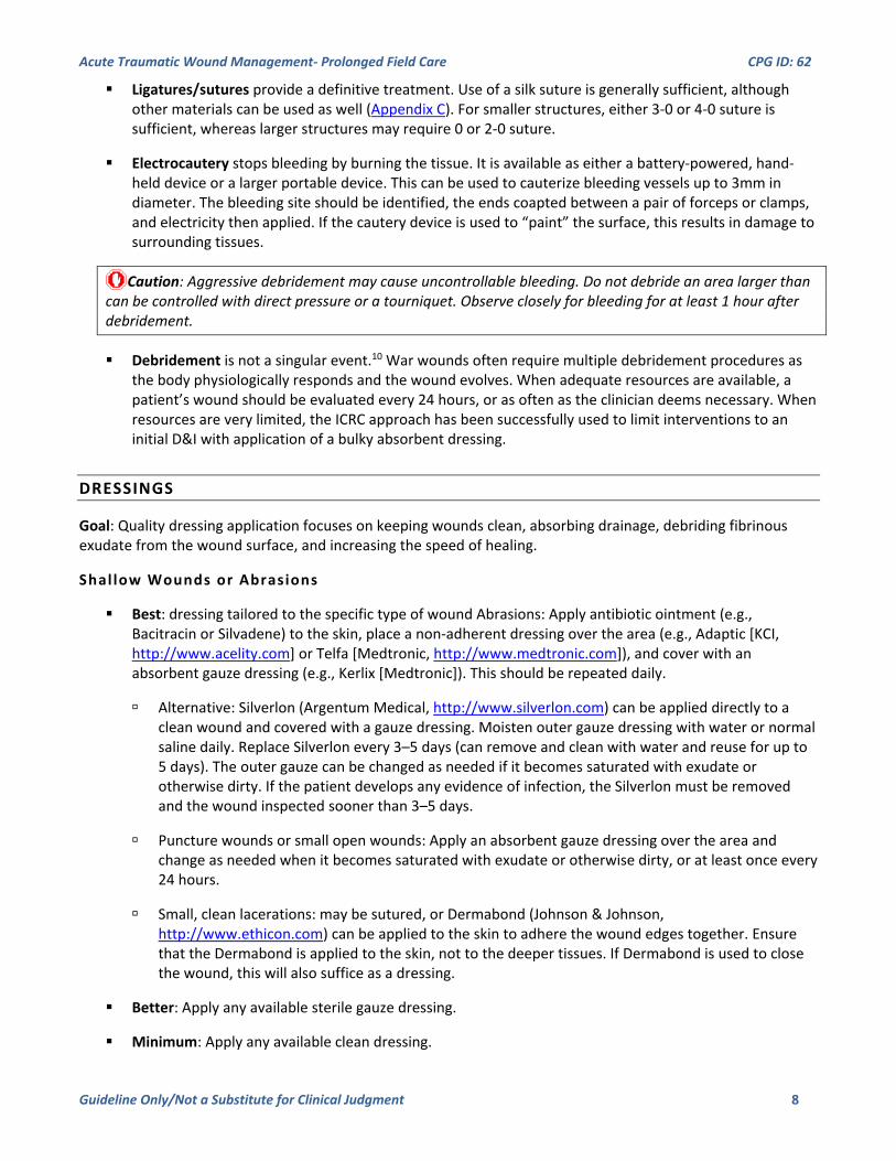
Acute Traumatic Wound Management- Prolonged Field Care CPG ID: 62
Guideline Only/Not a Substitute for Clinical Judgment 8
Ligatures/sutures provide a definitive treatment. Use of a silk suture is generally sufficient, although other materials can be used as well (Appendix C). For smaller structures, either 3-0 or 4-0 suture is sufficient, whereas larger structures may require 0 or 2-0 suture.
Electrocautery stops bleeding by burning the tissue. It is available as either a battery-powered, hand-held device or a larger portable device. This can be used to cauterize bleeding vessels up to 3mm in diameter. The bleeding site should be identified, the ends coapted between a pair of forceps or clamps, and electricity then applied. If the cautery device is used to “paint” the surface, this results in damage to surrounding tissues.
Caution: Aggressive debridement may cause uncontrollable bleeding. Do not debride an area larger than can be controlled with direct pressure or a tourniquet. Observe closely for bleeding for at least 1 hour after debridement.
Debridement is not a singular event.10 War wounds often require multiple debridement procedures as the body physiologically responds and the wound evolves. When adequate resources are available, a patient’s wound should be evaluated every 24 hours, or as often as the clinician deems necessary. When resources are very limited, the ICRC approach has been successfully used to limit interventions to an initial D&I with application of a bulky absorbent dressing.
DRESSINGS
Goal: Quality dressing application focuses on keeping wounds clean, absorbing drainage, debriding fibrinous exudate from the wound surface, and increasing the speed of healing.
Shallow Wounds or Abrasions
Best: dressing tailored to the specific type of wound Abrasions: Apply antibiotic ointment (e.g., Bacitracin or Silvadene) to the skin, place a non-adherent dressing over the area (e.g., Adaptic [KCI, http://www.acelity.com] or Telfa [Medtronic, http://www.medtronic.com]), and cover with an absorbent gauze dressing (e.g., Kerlix [Medtronic]). This should be repeated daily.
Alternative: Silverlon (Argentum Medical, http://www.silverlon.com) can be applied directly to a clean wound and covered with a gauze dressing. Moisten outer gauze dressing with water or normal saline daily. Replace Silverlon every 3–5 days (can remove and clean with water and reuse for up to 5 days). The outer gauze can be changed as needed if it becomes saturated with exudate or otherwise dirty. If the patient develops any evidence of infection, the Silverlon must be removed and the wound inspected sooner than 3–5 days.
Puncture wounds or small open wounds: Apply an absorbent gauze dressing over the area and change as needed when it becomes saturated with exudate or otherwise dirty, or at least once every 24 hours.
Small, clean lacerations: may be sutured, or Dermabond (Johnson & Johnson, http://www.ethicon.com) can be applied to the skin to adhere the wound edges together. Ensure that the Dermabond is applied to the skin, not to the deeper tissues. If Dermabond is used to close the wound, this will also suffice as a dressing.
Better: Apply any available sterile gauze dressing.
Minimum: Apply any available clean dressing.

Acute Traumatic Wound Management- Prolonged Field Care CPG ID: 62
Guideline Only/Not a Substitute for Clinical Judgment 9
Deep or Large Open Wounds
Best: Negative pressure wound therapy (NPWT)
Commercially available kits such as the KCI Wound vacuum-assisted closure (V.A.C. Therapy, KCI, http://www.kci-medical.com) device can be used to both cover the wound and promote wound healing.13 For exposed soft tissue, the pressure is usually set at 75–125mmHg of continuous suction.
Field-expedient wound VAC devices can be improvised from gauze, Tegaderm or Ioban, and suction tubing hooked up to a suction device (Appendix D).
NPWT dressings are generally changed every 2–3 days. Although they can be left in place for a longer time, the degree of granulation that can occur after 3 days generally leads to some bleeding with their removal.
NPWT dressings placed over infected wounds should be changed daily, along with wound debridement and irrigation, until the infection is controlled.
Better: Wet-to-dry dressing
Wet-to-dry dressings can be created using either a sterile isotonic fluid or clean water, if there is no sterile fluid available. A gauze dressing should be moistened (not soaked) and applied to all open, exposed tissues and loosely packed into the wound cavities. An absorbent gauze dressing or Army Battle Dressing pad is placed over top to absorb any extra fluids or exudate. This may be secured with tape or wrapped with a gauze bandage roll or elastic bandage.
Removal of dressings from a wound provides its own form of mechanical debridement; exudate adheres to the bandages and is then pulled from the wound during changing. Inspect old bandages during a dressing change for signs of infection by observing the color and odor of the exudate; the color should be off-white or slightly yellow, and the bandage should not smell sweet or foul. Do not moisten the gauze being removed to make the dressing change easier, otherwise the debridement effect will be lost.
Ideally, the dressing should be changed one or two times per day, unless the situation dictates a less frequent approach.
For wounds that are already draining heavily, a dry-to-wet dressing may be used instead.
Minimum: Place a bulky dressing with dry gauze, absorbent padding, and elastic bandage; leave in place 4–7 days if wound is clean and without sign of infection. See Appendix A for further details on the ICRC method of wound care.
CLOSURE
Goal: In most cases, war wounds should be closed by a surgical facility, where infection can be more effectively mitigated and the wound can be explored more effectively. However, when appropriate, wound closure simplifies the overall management of the wound.
Puncture wounds, in general, should not be primarily closed. Small wounds should be allowed to freely drain as needed and close by secondary intent. Larger puncture wounds may require debridement and irrigation, as described above.

Acute Traumatic Wound Management- Prolonged Field Care CPG ID: 62
Guideline Only/Not a Substitute for Clinical Judgment 10
Lacerations with minimal tissue loss/destruction and minimal need for debridement can generally be primarily closed provided that the wound is clean, there is not significant tension with the closure, and there has been <12 hours from injury to closure (this can be extended to 24 hours for lacerations to the face or scalp).
Lacerations that do not meet the above criteria can be considered for a DPC. These patients are provided 3–7 days of wound care to assure the wound is clean and without complications, and then is closed with either widely spaced sutures or staples.
Undermine wound margins if needed to reverse the retracted and inverted edges, and relieve any tension to allow skin closure.
Wounds with tissue loss or damage are generally not closed primarily. Instead, these wounds are treated with dressing changes and allowed to heal by secondary intent. However, there is occasionally a role for DPC in select wounds.
When in doubt, it is better to leave a wound open and plan for healing by secondary intent.
If a wound becomes infected after closure, it should be reopened and debrided to drain pus and remove infected, dead tissue.
Caution: Wound closure may speed healing; however, closure will also greatly increase the risk of infection. Only wounds that are very clean and have been well-debrided should be closed.
INFECTION PREVENTION
Goal: Antimicrobial drug therapy is a critical complement to debridement and irrigation in the fight against infection. This includes prophylaxis against Clostridium tetani (the causal agent of tetanus), as appropriate.
Guidance on specific antibacterial regimens can be found in the Joint Trauma System CPG Infection Control.
In general, cefazolin 2g in 250 mL NS IV over 5 min every 8 hours for 24 hours is adequate for most dirty wounds of the head and neck, torso, and extremities.
Oral antibiotics may be used: moxifloxacin 400mg PO once or clindamycin 300mg PO every 8 hours for 24 hours.
Note: Moxifloxacin may be replaced with levofloxacin 750 mg PO daily to provide better coverage of bacteria found in wet terrain/ jungle environment.
Ertapenem is commonly carried by Role 1 providers and gives broad-spectrum gram-positive, gram-negative, and anaerobic coverage. It is an acceptable choice when more-specific antibiotics are not available. Give 1g in 250 mL NS IV over 5 min (provides 24 hours of coverage).
If following the ICRC wound care method (Appendix A), continue antibiotics for 5 days or until DPC.
Prophylaxis against tetanus should be initiated, as appropriate, based upon the condition of the wound and the individual’s immunization history.14 (Appendix E).

Acute Traumatic Wound Management- Prolonged Field Care CPG ID: 62
Guideline Only/Not a Substitute for Clinical Judgment 11
INFECTION TREATMENT
Goal: If a wound develops signs of infection (e.g., increasing pain, erythema, purulent drainage) or the patient becomes systemically ill (e.g., fever, tachycardia, hypotension, lethargy, decreased mental status), wound infection must be identified and treated. Treat infected wounds with a combination of local wound care and systemic antibiotics.
Remove all dressings and inspect wound.
Wounds that have been closed must be reopened.
Repeat wound debridement to drain pus and remove all dead, infected tissue. If necessary, extend the wound with an incision in the long axis of the extremity.
Worst-case scenario may require amputation of an extremity to control life-threatening infection.
Increase the frequency of dressing changes to once or twice daily.
Continue antibiotics for 7–10 days, moxifloxacin 400mg PO daily, levofloxacin 750mg PO daily, or ertapenam 1g IV daily, or alternative broad-spectrum antibiotic as available (if possible, change antibiotic from prevention regimen).
Wounds with known or suspected infections can be treated using Dakin’s solution (dilute bleach) for irrigation and wet-to-dry dressing. This can be an effective adjunct, particularly for suspected fungal and Pseudomonal infections.3,15
Daily sugar or honey dressings for infected wounds may successfully treat infection in very austere settings.12 This involves irrigating the wound with saline or the cleanest water available and then filling it with sugar or honey, which is left in place under a dry dressing, repeating every 24 hours.
Severe wound infection is life threatening and may require aggressive debridement, up to and including amputation. Evacuate patient. If evacuation is not possible, obtain telemedicine consultation and initiate infection treatments above.
SPECIAL CONSIDERATIONS
FACE AND SCALP
These wounds rarely become infected because of the high blood flow of the face and scalp.
Most face and scalp wounds can be closed after irrigation. Debridement should be minimized to reduce cosmetic deformity. If the patient will arrive at a surgical facility within 24 hours, it is better to defer wound closure.
The clinician should take into consideration the low probability of infection when planning closure of a facial wound because of the poor cosmetic outcomes associated with delayed primary closure.
When primary closure is acceptable, sutures should only remain in place for 4–5 days for the face and 7 days for the scalp.

Acute Traumatic Wound Management- Prolonged Field Care CPG ID: 62
Guideline Only/Not a Substitute for Clinical Judgment 12
OPEN GLOBE INJURY
Apply an eye shield to prevent further damage.
Do not place any type of dressing in contact with the globe or underneath the eye shield.
Moxifloxacin 400mg PO daily to prevent endophthalmitis
Ondansetron 4mg PO/IV every 4 hours as needed for associated nausea
Pain control as needed (narcotics as well as ketamine are approved for use)
Position the patient with the head elevated, if possible.
Ultrasound evaluation is contraindicated when open globe injury is suspected.
The best results are achieved if surgery occurs within 24 hours of injury.
ANIMAL BITES
All animal bite treatments should begin with immediate, thorough cleaning of all wounds with soap and water.
Most animal bites should be allowed to heal by secondary intent (especially puncture wounds).16
Moxifloxacin 400mg PO daily OR Augmentin (amoxicillin/clavulanic acid; GlaxoSmithKline, http://us.gsk.com/) 875/125mg PO every 12 hours for 7 days are the best choices for most animal bites. Doxycycline (100mg PO twice a day) or clindamycin (300mg PO 3 times a day) with levofloxacin (750mg PO daily) are acceptable alternatives.
Consider the need for rabies vaccination (Appendix E).
AQUATIC EXPOSURE
Management of wounds exposed to fresh or salt water is unchanged; however, additional agents are occasionally required for adequate antimicrobial coverage.17
Levofloxacin 750mg PO daily (second-line doxycycline 100mg PO twice daily) to cover for Aeromonas species and Vibrio species.
Given the risk of atypical infections, telemedicine consultation should be sought if the wound worsens.
CHRONIC NONHEALING WOUNDS
If a wound has not healed by 2 weeks, it is likely the result of underlying infection (consider osteomyelitis as a potential cause), inadequate tissue perfusion, or inadequate nutrition.18 Often it is multifactorial and so more than one source needs to be addressed.
Consider telemedicine consultation if the wound is not healing.
Packing and Planning Considerations. See Appendix F.
Wound Management Summary Table. See Appendix G.

Acute Traumatic Wound Management- Prolonged Field Care CPG ID: 62
Guideline Only/Not a Substitute for Clinical Judgment 13
REFERENCES
1. Ball JA, Keenan S. Prolonged Field Care Working Group position paper: prolonged field care capabilities. J Spec Oper Med. 2015;15(3):76–77.
2. Anglen JO. Wound irrigation in musculoskeletal injury. J Am Acad Orthop Surg. 2001;9:219–226.
3. Lewandowski L, Purcell R, Fleming M, et al. The use of dilute Dakin’s solution for the treatment of angioinvasive fungal infection in the combat wounded: a case series. Mil Med. 2013;178:e503–e507.
4. Barsoumian A, Sanchez CJ, Mende K, et al. In vitro toxicity and activity of Dakin’s solution, mafenide acetate, and amphotericin B on filamentous fungi and human cells. J Orthop Trauma. 2013;27:428–436.
5. Vick LR, Propst RC, Bozeman R, et al. Effect of Dakin’s solution on components of a dermal equivalent. J Surg Res. 2009;155:54–64.
6. Owens BD, White DW, Wenke JC. Comparison of irrigation solutions and devices in a contaminated musculoskeletal wound survival model. J Bone Joint Surg Am. 2009;91:92–98.
7. Chatterjee JS. A critical review of irrigation techniques in acute wounds. Int Wound J. 2005;2:258–265.
8. Singer AJ, Hollander JE, Subramanian S, et al. Pressure dynamics of various irrigation techniques commonly used in the emergency department. Ann Emerg Med. 1994;24:36–40.
9. Svoboda SJ. Bice TG, Gooden HA, et al. Comparison of bulb syringe and pulse lavage irrigation with use of bioluminescent musculoskeletal wound model. J Bone Joint Surg Am. 2006;88:2167–2174.
10. Erdle NJ, Verwiebe EG, Wenke JC, et al. Debridement and irrigation: evolution and current recommendations. J Orthop Trauma. 2016;30:S7–S10.
11. Gianno C, Baldan M. War surgery: working with limited resources in armed conflict and other situations of violence, Vol. 1. 2010. International Committee of the Red Cross. https://shop.icrc.org/reference-publications-military.html. Accessed 11 April 2017.
12. Gianno C, Baldan M, Molde A. War surgery: working with limited resources in armed conflict and other situations of violence, Vol. 2. 2013. International Committee of the Red Cross. https://shop.icrc.org/reference-publications-military.html. Accessed 11 April 2017.
13. Hinck D, Franke A, Gatzka F. Use of vacuum-assisted closure negative pressure wound therapy in combat-related injuries—literature review. Mil Med. 2010;175:173–181.
14. Chapman LE, Sullivent EE, Grohskopf LA, et al. Postexposure interventions to prevent infection with HBV, HCV, or HIV, and tetanus in people wounded during bombings and other mass casualty events—United States, 2008: recommendations of the Centers for Disease Control and Prevention and Disaster Management and Public Health Preparedness. Disaster Med Public Health Prep. 2008;2:150–165.
15. McCullough M, Carlson GW. Dakin’s solution: historical perspective and current practice. Ann Plast Surg. 2014;73: 254–256.
16. Brook I. Management of human and animal bite wounds: an overview. Adv Skin Wound Care. 2005;18:197–203.
17. Noonburg GE. Management of extremity trauma and related infections occurring in the aquatic environment. J Am Acad Orthop Surg. 2005;13:243–253.

Acute Traumatic Wound Management- Prolonged Field Care CPG ID: 62
Guideline Only/Not a Substitute for Clinical Judgment 14
18. Guo S, DiPietro LA. Factor affecting wound healing. J Dent Res. 2010;89:219–229.
19. Simon RR, Strome G, Halbert RJ. Sterilization techniques in underground surgical units in Afghanistan. Ann Emerg Med. 1988;17:785–787.
20. Willard M, Alexander A. Comparison of sterilizing properties of formaldehyde-methanol solutions with formaldehyde-water solutions. Appl Microbiol. 1964;12:229–233.
21. MMWR 57 (RR-3): 1, 2008; http://wwwnc.cdc.gov/travel/yellowbook/2012/chapter-3-infections-diseases-related-to-travel/rabies.htm.

Acute Traumatic Wound Management- Prolonged Field Care CPG ID: 62
Guideline Only/Not a Substitute for Clinical Judgment 15
APPENDIX A : INTERNATIONAL COMMITTEE OF THE RED CROSS SURGICAL WOUND MANAGEMENT FLOW CHART
Notes on wound care according to the ICRC method: The ICRC wound care methods are most applicable for management of war wounds in austere conditions and with limited resources that prohibit serial (follow-on) debridements with associated postoperative care:
1. Dirty environment 2. Limited supplies 3. Limited manpower 4. Limited time (mission dictated) 5. Wounds older than 24 hours 6. Inability to evacuate (includes host-nation patient care where care provided in the austere environment is the definitive level of
care available) Serial debridement every 24–48 hours is not possible in this environment. Therefore, the initial debridement must remove all tissue that is nonviable or questionably viable to remove dead tissue that may act as a culture medium. The wound is then managed with a bulky absorbent dressing that can stay in place for 4–7 days with a low likelihood of infection. Continue antibiotics for 5 days or until delayed primary closure.

Acute Traumatic Wound Management- Prolonged Field Care CPG ID: 62
Guideline Only/Not a Substitute for Clinical Judgment 16
Wounds that become infected or have exposed vital structures, such as blood vessels, nerves, or bone, should be treated with more frequent debridement and dressing changes.
After 4–7 days, remove the dressing and evaluate both the dressing and the wound using adequate pain control and sedation, as needed. The bandage and dressing should be dry, stained (greenish-black), and have an ammonia-like odor (good-bad odor), and the dressing should be adherent to the wound. The wound base should bleed slightly and the muscle should contract. Any small area of collagen that was not removed with the dressing can be scraped off with an instrument. This reveals a clean wound that is ready for closure.
If the wound is infected, the dressing may slide easily off the wound without resistance because there is a layer of pus between the dressing and the wound surface. The wound may contain areas of necrotic tissue and the surface of the wound is dull or greyish-red, or may give off the “bad-bad smell” of wound sepsis. Such an infected wound requires further excision and DPC is delayed or the wound is allowed to close by secondary intent. Closure by secondary intent may take several weeks. ICRC recommends a change of dry bulky gauze dressing and gentle washing with normal saline every 4–5 days for closure by secondary intent.
The ICRC dressing change algorithm for closure by secondary intent is as follows:
Question 1: Is the wound clean?
Yes: infrequent (every 4–5 days) dressing with dry bulky gauze.
No: continue
Question 2: Is there erythema and tenderness around the wound?
Yes: antibiotics and continue
No: continue
Question 3: Is there a large volume of exudate?
Yes: daily sugar or honey dressings; this involves rinsing the wound with saline and then filling it with sugar or honey, which is left in place under a dry dressing. This is repeated after 24 hours.
No: infrequent dressing with dry bulky gauze
Question 4: Has there been a response to sugar/honey dressings?
Yes: Continue sugar/honey dressings or convert to infrequent dry bulky gauze.
No: Consider antiseptic solution or further surgical debridement
If the wound exudate continues, consider exploring the wound surgically.

Acute Traumatic Wound Management- Prolonged Field Care CPG ID: 62
Guideline Only/Not a Substitute for Clinical Judgment 17
APPENDIX B: INSTRUMENT STERILIZATION (STANDARD AND NONSTANDARD)
DEFINITIONS Cleaning: Removes dirt, debris, and biological material from surgical instruments Disinfect: Cleaning with a disinfectant to destroy bacteria Sterilize: Making free from bacteria or other living organisms
NOTES Cleaning, disinfecting, and sterilizing are the three levels of instrument care. Critical tools are those that make contact with open wounds, open vasculature, and/or organs. Semicritical tools are
those that contact the mucosa, and noncritical tools are those that contact intact skin. Sterilization is the goal for any tools that will be used for surgical intervention; however, if life depends on it and
sterilization is not an option; disinfection can be achieved using one of the options listed below as the “Better” option. The goal of sterilization is to eliminate the presence of bacteria, viruses, parasites, and particulates; bacterial spores are
the most difficult to destroy. For all nonstandard sterilization techniques, gross contamination should be removed from all instruments/equipment
by scrubbing with soap and water first. Chemical sterilization elements should never be combined. Steam with distilled water is the preferred sterilization technique.
BEST: Remove gross contamination, using soap and water, then sterilize by steam or dry heat using one of these techniques:
Steam sterilization using one of the following techniques and the following temperatures: 121°C (250°F) for 30 minutes or 132°C (270°F) for 4 minutes
Autoclave (portable or standard size) and distilled water Steam sterilization using the SF medical Tac-Set portable sterilizer pot (NSN: 6530-00-926-2022), according to the
manufacturer’s operating instructions: 1. Remove the cover by loosening the wing nuts in a counterclockwise motion. Always undo two opposite wing nuts
at a time. 2. Lubricate the metal-to-metal seal where side wall and bevel meet on the inside of the sterilizer. Use petroleum
jelly or mineral oil. 3. Remove the aluminum inner container. Pour clean (preferably distilled) water into sterilizer to a depth of not less
than three-fourths of an inch, but no more than 1 inch. 4. Place inner container rack into inner container with the lip or edge side downward. Place articles to be sterilized on
container rack. Only steam should make contact with articles. Replace packed inner container into sterilizer, ensuring the air exhaust tube channel is in position on the right side of the container when it is placed into the unit.
5. Place sterilizer cover on unit, making sure that the index alignment arrow on the cover aligns with index line/arrow on the side of the bottom. Ensure that the flexible tube is inserted into the guide channel on the inside wall of the aluminum container. Tighten the wing nuts evenly, always tightening down two opposite nuts simultaneously to assure a proper seal.
6. Place unit onto a heat source. Open the control valve by placing valve lever in an upright position. It is important that steam be permitted to escape vigorously for at least 7 minutes or until the flow of steam is continuous. Afterwards, close the control valve. When pressure gauge reaches 17–19 psi, reduce heat as necessary to maintain a constant pressure of 17–19 psi within the unit.
7. The sterilization period begins when the pressure steam gauge needle registers in the green sterilization band shown on the face of the gauge (i.e., 17–21 psi). This should be no less than 35 minutes. Note: At altitudes higher than sea level, settings need to be adequately adjusted to compensate for the effect of altitude on water’s boiling point. The manufacturer suggests increasing pressure by 0.5 psi for every 1,000 feet of elevation above sea level.
8. At the end of the sterilization period, remove unit from heat source and move the lever on the control valve to an upright position to allow the steam to escape. With caution, loosen wing nuts and remove the cover to retrieve sterilized items.

Acute Traumatic Wound Management- Prolonged Field Care CPG ID: 62
Guideline Only/Not a Substitute for Clinical Judgment 18
DRY HEAT Dry heat may also be used to sterilize surgical instruments. Instruments can be wrapped in aluminum foil or placed in sterilization trays before putting them in the oven. Allow the instruments to cool to room temperature inside the oven before use or storage. Instruments can be heated to any of the following to be considered sterilized:
180°C (356°F) for 30 minutes 170°C (338°F) for 1 hour 160°C (320°F) for 2 hours 149°C (300°F) for 2.5 hours 141°C (286°F) for 3 hours
BETTER: Remove gross contamination using soap and water, then sterilize with improvised autoclave or chemical disinfection.
Improvised autoclave using a pressure cooker that can reach the steam sterilization temperatures listed above. Place wire rack in the bottom to prevent the instruments from becoming submerged. Only steam should make
contact with the instruments. Begin timing for sterilization once steam starts to vent from the pressure cooker. Pressure cookers operate at various pressures and temperatures. Sterilization time of 60 minutes is recommended.
Glutaraldehyde (Cidex©; Johnson & Johnson): Widely available and accepted as a high-quality disinfectant in healthcare facilities. Can be used to sterilize aluminum, brass, copper, stainless steel, plastics, and elastomers. Once the container is open, the chemical has a limited shelf life—a minimum of 14 days, usually 28–30 days; refer
to the specific product’s manual for the exact shelf life. Test strips can indicate the efficacy of an opened container of glutaraldehyde. Alkaline glutaraldehyde mixes are more effective than acidic glutaraldehyde mixes. Items should be soaked for 60 minutes for maximum efficacy. Long-term exposure can cause irritation to tissues; rinse tools before use.
Formaldehyde: Prepare 5% formaldehyde solution. Both metallic and plastic medical equipment can be sterilized using formaldehyde. Wrap a 5mg tablet of formaldehyde in gauze and crush the tablet. Place the gauze-wrapped tablet in 100mL of potable water (can be cold water). This solution can be stored for up to 30 days without losing its potency. Lower concentrations of formaldehyde can be used, but the amount of time the instruments are submerged in the
solution needs to be increased to compensate for this change. Clean the instruments with soap and water, and remove any blood and tissue. Soak the instruments in the 5% formaldehyde solution for at least 1 hour but preferably for 12–24 hours.19,20 Duration should be closer to 24 hours if 1% formaldehyde solutions are used (i.e., 5mg tablet in 500mL of water). The instruments and formaldehyde solution can be kept at room temperature. Remove the instruments and rinse with water or saline before use.
Alcohol: 60% to 90% minimum; tools need 3 hours of contact time. Consumable alcohol must be a minimum of 120 proof. This method is not sporicidal and, therefore, not recommended as a primary modality for surgical items.
Sodium hypochlorite (household bleach) This disinfectant is caustic to eyes, oropharynx, esophagus, and gastric tissue. Undiluted (5.25%) sodium hypochlorite should be used to disinfect instruments categorized as critical. Instruments should have a minimum contact time of 5 minutes.
MINIMUM: Remove gross contamination, using soap and water, then boil instruments in at least 100°C (212°F) water for at least 1 minute.
NOTE: This method does not sterilize instruments and should be used as a last resort for surgical instruments.

Acute Traumatic Wound Management- Prolonged Field Care CPG ID: 62
Guideline Only/Not a Substitute for Clinical Judgment 19
APPENDIX C: SUTURE CHART
When supplies are limited, any suture may be used for any purpose; however, as a general guideline, absorbable suture should be used to approximate tissues deep to the skin, silk may be used to ligate blood vessels, and a monofilament suture (nylon/Prolene; Johnson & Johnson) should be used for the skin.
Most suturing/ligating can be accomplished with 2-0 sutures. Finer suture (3-0 or 4-0) may be used on the face for better cosmetic result. Heavier suture (0 or 1) may be used for DPC of larger wounds or for the scalp.
Tapered needles are best for suturing soft-tissue layers; cutting needles are best for suturing skin; strait needles do not require a needle holder.
Undesired tissue reactivity is greater with: Multifilament (compared with monofilament) Larger suture gauge Natural material (compared with synthetic)
Recommended multipurpose sutures: 2-0 Vicryl suture with CT-1 needle, 0 Prolene or Nylon with FSLX needle, 0 silk with strait needle.
Absorbable Sutures Suture Effective wound
support, days Time to complete absorption, days
Comments Usual indications
Surgical gut 8–9 30 Rarely used; high tissue reactivity
Approximate soft-tissue layers deep to the skin
Chromic gut 10–21
>90 Gut treated with chromium to decrease tissue reactivity and slow absorption
Approximate soft-tissue layers deep to the skin
Fast-absorbing gut 5–7 14–28 Gut treated with heat to speed absorption
Skin closure when suture removal will be difficult or impossible
Polyglactin (Vicryl) 21 90 Less reactive than gut; synthetic Approximate soft-tissue layers deep to the skin
Vicryl Rapide (Johnson & Johnson)
10 42 Gamma-irradiated to speed absorption
Skin closure when suture removal will be difficult or impossible
Non Absorbable Sutures Suture Tensile Strength Tissue Reactivity Comments Usual Indications
Silk Low High Multifilament, pliable Ligate blood vessels
Nylon (e.g., Ethelon; Johnson & Johnson)
High Low Monofilament, stiff Skin closure
Polypropylene (e.g., Prolene)
Moderate Very Low Monofilament, less knot security (may need extra knot)
Skin closure, repair blood vessels
Suture Removal Time Frames Body Part Number of Days After Suturing
Face 4-5
Scalp 7
Chest, extremities 10-14
High tension areas, joint, back 14-21

Acute Traumatic Wound Management- Prolonged Field Care CPG ID: 62
Guideline Only/Not a Substitute for Clinical Judgment 20
APPENDIX D: FIELD-EXPEDIENT WOUND VACUUM-ASSISTED CLOSURE
1. Copious irrigation with debridement of necrotic tissue
and removal of foreign debris. 2. Placement of gauze or reticulated sponge at base of wound.
3. Placement of suction tubing (e.g., Blake, Jackson-Pratt,
nasogastric, red rubber with holes) that can be attached to a suction source.
4. Placement of additional layer of reticulated sponge or gauze over suction tubing to separate suction from occlusive layer.
5. Placement of occlusive impermeable dressing, such as
Ioban or Tegaderm, over the gauze/sponge and suction tubing.
6. Connect to suction source.

Acute Traumatic Wound Management- Prolonged Field Care CPG ID: 62
Guideline Only/Not a Substitute for Clinical Judgment 21
APPENDIX E: TETANUS IMMUNIZATION CHART
(Special Operations Forces Medical Handbook; US Department of Defense; Washington, DC: Government Printing Office)
Tetanus Immunization Status Minor Clean Wound
Major Clean Wound
Contaminated Wound (War Wounds)
Fully immunized recent Td booster N/A N/A N/A Fully immunized Td booster 5–10 years ago
N/A Tdap Tdap
Fully immunized, no booster for >10 years Tdap Tdap Tdap
Unknown, none, or incomplete immunization
Tdap Tdap and TIG (250U)
Tdap and TIG (500U)
N/A, not applicable; Td, tetanus and diphtheria; Tdap, tetanus-diphtheria-acellular pertussis; TIG, tetanus immune globulin. Note: Tetanus vaccination of mother gives her protection and protects the newborn in the first few weeks of life.
Rabies Postexposure Immunization21 Animal Type Evaluation & Disposition of Animal Recommendations for Prophylaxis
Dogs, cats, ferrets Healthy and available for 10-day observation
Do not start unless animal develops symptoms, then immediately begin HRIG + vaccine
Rabid or suspected rabid Immediate HRIG + vaccine
Unknown (escaped) Consult public health officials
Skunks, raccoons, bats, foxes, coyotes, most carnivores
Regard as rabid Immediate vaccination
Livestock, horses, rodents, rabbits; includes hares, squirrels, hamsters, guinea pigs, gerbils, chipmunks, rats, mice, woodchucks
Consider case-by-case Consult public health officials. Bites of squirrels, hamsters, guinea pigs, gerbils, chipmunks, rats, mice, other small rodents, rabbits, and hares almost never require postexposure immunization.
If Not Previously Vaccinated
Treatment Regimen
Local wound cleaning All post-exposure treatment should begin with immediate, thorough Human rabies immune globulin (HRIG)
20 units per kg body weight given once on day 0. If anatomically feasible, the full dose should be infiltrated around the wound(s), or the rest should be administered IM in the gluteal area. If the calculated dose of HIRG is insufficient to inject all the wounds, it should be diluted with normal saline to allow infiltration around additional wound areas. HRIG should not be administered in the same syringe or into the same anatomical site as vaccine, or more than 7 days after the initiation of vaccine. Because HRIG may partially suppress active production of antibody, no more than the recommended dose should be given.
Vaccine Human diploid cell vaccine (HDCV), rabies vaccine adsorbed (RVA), or purified chick embryo cell vaccine (PCECV) 1 mL IM (deltoid area), one each days 0, 3, 7, 14.
If Previously Vaccinated
HRIG Do not give Vaccine HDCV or PCECV 1mL IM (deltoid area), one each on days 0 and 3.

Acute Traumatic Wound Management- Prolonged Field Care CPG ID: 62
Guideline Only/Not a Substitute for Clinical Judgment 22
APPENDIX F: PACKING AND PLANNING CONSIDERATIONS
This list is not all encompassing. Quantities of items listed are highly mission variable and up to provider discretion. All efforts were made to list items in common use.
Instruments Hemorrhage control Metzenbaum scissors Mayo scissors Rat Tooth/Adson/DeBakey forceps Kelly, Mosquito, Hemostat forceps Allis forceps Needle drivers #10/#11/#15 scalpels Volkmann or Weitlaner retractors Bovie pen
Combat Gauze/ChitoGauze C-A-Tb tourniquet/SOF Tactical tourniquet, widec/Esmarch
bandage/ sphygmomanometer with Esmarch bandage
General supplies Alcohol pads Betadine swabs 1mL, 5mL, 10mL, 35mL, 60mL syringes 19g Catheter 18g IV 1.5", 25g 1.5" hard needle Irrigation bulb Basin Chux pads Gloves of various sizes (small, medium, large) IV tubing Large-bore catheter Constricting band Saline lock Suture/ligature (see Appendix A) Skin stapler Cotton tip applicators Morgan lens Sterile drapes or draping material that can be sterilized Surgical masks Surgical gowns Sterile gloves Ioban 2–23" × 23" Tegaderm Suction tubing Suction device (SSCOR Quickdraw suction; TAC-SET)
Meds Bacitracin/Silvadene 2% Lidocaine with/without epinephrine 8.4% sodium bicarbonate 0.5% tetracaine hydrochloride (eye drops) IV ertapenem IV cefazolin Oral moxifloxacin 400mg or oral levofloxacin 750mg (Note: levofloxacin is preferred in wet/jungle environment) Oral clindamycin 300mg Pain and sedation medications NaCl (for irrigation)/potable water source Sterile water
Dressings Kerlix 6", 4" Ace wrap Cobana 3", 2", 1" medical tape Telfa/Adaptic Silverlon Dermabond/superglue Steristrips
a3M, http://www.3m.com; bCombat Application Tourniquet, Composite Resources, http://combattourniquet.com/; cTactical Medical Solutions, https://www.tacmedsolutions.com.

Acute Traumatic Wound Management- Prolonged Field Care CPG ID: 62
Guideline Only/Not a Substitute for Clinical Judgment 23
APPENDIX G: WOUND MANAGEMENT SUMMARY TABLE
Goals of Wound Care Hemorrhage control Minimize risk of infection Promote optimal healing Reduce discomfort Minimize disability/loss of function associated with treatment Implement definitive care when possible
Types of Wounds Abrasion Burn Puncture Laceration
Gunshot Blast Crush injury
Telemedicine Large or complex wounds Associated injury to bone, blood vessel, tendon, nerve Penetrating chest or abdominal wound Infected wounds Chronic, nonhealing wounds
Irrigation Best Irrigate wounds with dilute Dakin’s solution or sterile isotonic solution.
Better Irrigate wounds with clean potable tap water.
Minimum Irrigate wounds with the cleanest water available (nonpotable water should be boiled for a minimum of 3 minutes and then cooled to body temperature).
Debridement Best Use an assistant, remove nonviable tissue using sharp dissection (sterile scalpel or scissors) and control bleeding
using tourniquet, topical hemostatic dressings, direct pressure, clamping, suture ligation, and/or electrocautery. Questionably viable tissue may be retained with repeat debridement once every 24–48 hours until arrival at surgical facility.
Better Remove nonviable tissue using sharp dissection, control bleeding using tourniquet, topical hemostatic dressings, direct pressure, clamping, and/or suture ligation. Repeat debridement or delayed primary closure in 3–5 days.
Minimum When unable to evacuate and resources for serial debridement every 24–48 hours are not available, remove all nonviable and questionably viable tissue using sharp dissection (clean blade or scissors) and control bleeding with tourniquet and/or direct pressure. Leave clean dressing in place then delayed primary closure in 4–7 days if no sign of infection (ICRC wound care method).
Dressings – Shallow wounds or abrasions Best Abrasions: Apply antibiotic ointment (e.g., Bacitracin or Silvadene) to skin, place nonadherent dressing over the
area (e.g., Adaptic or Telfa), cover with absorbent gauze dressing, repeat daily. Alternative: Apply Silverlon dressing to a clean wound, cover with gauze, moisten gauze daily. Replace Silverlon every 3–5 days. Puncture wounds or small open wounds: Apply absorbent gauze dressing, change when saturated with
exudate or otherwise dirty, or at least once every 24 hours. Small, clean lacerations: suture or Dermabond
Better Apply any available sterile gauze dressing. Minimum Apply any available clean dressing.

Acute Traumatic Wound Management- Prolonged Field Care CPG ID: 62
Guideline Only/Not a Substitute for Clinical Judgment 24
Dressings – Deep or large open wounds Best NPWT
Commercial available kits such as the KCI Wound VAC device can be used to both cover the wound and promote wound healing. Field expedient wound VAC devices can be improvised from absorbent gauze, Tegaderm or Ioban, and suction
tubing hooked up to a suction device. Change NPWT dressings every 2–3 days. Change NPWT dressings placed over infected wounds, along with wound debridement and irrigation, daily.
Better Wet-to-dry dressing Wet-to-dry dressings using sterile isotonic fluid or clean water. Change the dressing 1–2 times per day unless the situation dictates a less frequent approach. For wounds that are draining heavily, a dry-to-wet dressing may be used instead.
Minimum Place a bulky dressing with dry gauze, absorbent padding, and elastic bandage; leave in place 4–7 days if wound is clean without sign of infection.
Infection prevention Tetanus prophylaxis if immunization status unknown In addition to wound care, treat open wounds with antibiotics as soon as possible after injury and continue for 24 hours with one
of the following antibiotics: Moxifloxacin 400mg PO × 1 dose or levofloxacin 750mg PO × 1 dose Note: levofloxacin is preferred in wet/jungle environment Clindamycin 300mg PO every 8 hours Cefazolin 2g IV every 8 hours Ertapenem 1g IV 1 dose
If following the ICRC wound care method, continue antibiotics for 5 days or until delayed primary closure.
Infection treatment Remove all dressings and inspect wound. Wounds that have been closed must be reopened. Repeat wound debridement to drain pus and remove all dead, infected tissue. If necessary, extend the wound with an incision in
the long axis of the extremity. Worst-case scenario may require amputation of an extremity to control life-threatening infection. Increase the frequency of dressing changes to once or twice daily. Continue antibiotics for 7–10 days, moxifloxacin 400 mg PO daily or levofloxacin 750mg PO daily or ertapenam 1g IV daily, or
alternative broad-spectrum antibiotic as available. Use Dakin’s solution for the wet-to-dry dressing, particularly for suspected fungal and pseudomonal infections. Daily sugar or honey dressings for infected wounds may successfully treat infection in very austere settings
NPWT, negative pressure wound therapy; VAC, vacuum-assisted closure.

Acute Traumatic Wound Management- Prolonged Field Care CPG ID: 62
Guideline Only/Not a Substitute for Clinical Judgment 25
APPENDIX H: ADDITIONAL INFORMATION REGARDING OFF-LABEL USES IN CPGS
PURPOSE
The purpose of this Appendix is to ensure an understanding of DoD policy and practice regarding inclusion in CPGs of “off-label” uses of U.S. Food and Drug Administration (FDA)–approved products. This applies to off-label uses with patients who are armed forces members.
BACKGROUND
Unapproved (i.e., “off-label”) uses of FDA-approved products are extremely common in American medicine and are usually not subject to any special regulations. However, under Federal law, in some circumstances, unapproved uses of approved drugs are subject to FDA regulations governing “investigational new drugs.” These circumstances include such uses as part of clinical trials, and in the military context, command required, unapproved uses. Some command requested unapproved uses may also be subject to special regulations.
ADDITIONAL INFORMATION REGARDING OFF-LABEL USES IN CPGS
The inclusion in CPGs of off-label uses is not a clinical trial, nor is it a command request or requirement. Further, it does not imply that the Military Health System requires that use by DoD health care practitioners or considers it to be the “standard of care.” Rather, the inclusion in CPGs of off-label uses is to inform the clinical judgment of the responsible health care practitioner by providing information regarding potential risks and benefits of treatment alternatives. The decision is for the clinical judgment of the responsible health care practitioner within the practitioner-patient relationship.
ADDITIONAL PROCEDURES
Balanced Discussion
Consistent with this purpose, CPG discussions of off-label uses specifically state that they are uses not approved by the FDA. Further, such discussions are balanced in the presentation of appropriate clinical study data, including any such data that suggest caution in the use of the product and specifically including any FDA-issued warnings.
Quality Assurance Monitoring
With respect to such off-label uses, DoD procedure is to maintain a regular system of quality assurance monitoring of outcomes and known potential adverse events. For this reason, the importance of accurate clinical records is underscored.
Information to Patients
Good clinical practice includes the provision of appropriate information to patients. Each CPG discussing an unusual off-label use will address the issue of information to patients. When practicable, consideration will be given to including in an appendix an appropriate information sheet for distribution to patients, whether before or after use of the product. Information to patients should address in plain language: a) that the use is not approved by the FDA; b) the reasons why a DoD health care practitioner would decide to use the product for this purpose; and c) the potential risks associated with such use.

1
Tactical Combat Casualty Care (TCCC) Guidelines for Medical Personnel
15 December 2021
RED text indicates new text in this year’s update to the TCCC Guidelines; BLUE text indicates text that did not change but was relocated within the guidelines. Recent changes include junctional and airway device recommendations, analgesia, and abdominal evisceration guidelines.
Basic Management Plan for Care Under Fire/Threat
1. Return fire and take cover.
2. Direct or expect casualty to remain engaged as a combatant if appropriate.
3. Direct casualty to move to cover and apply self-aid if able or when tactically feasible, move or drag casualty to cover.
4. Try to keep the casualty from sustaining additional wounds.
5. Casualties should be extracted from burning vehicles or buildings and moved to places of
relative safety. Do what is necessary to stop the burning process.
6. Stop life-threatening external hemorrhage if tactically feasible: a. Direct casualty to control hemorrhage by self-aid if able. b. Use a CoTCCC-recommended limb tourniquet for hemorrhage that is anatomically
amenable to tourniquet use. c. Apply the limb tourniquet over the uniform clearly proximal to the bleeding site(s). If
the site of the life-threatening bleeding is not readily apparent, place the tourniquet “high and tight” (as proximal as possible) on the injured limb and move the casualty to cover.
7. Airway management is generally best deferred until the Tactical Field Care phase.

TCCC Guidelines 2021
Basic Management Plan for Tactical Field Care
1. Establish a security perimeter in accordance with unit tactical standard operating procedures and/or battle drills. Maintain tactical situational awareness.
2. Triage casualties as required. Casualties with an altered mental status should have weapons and communications equipment taken away immediately.
3. Massive Hemorrhage
a. Assess for unrecognized hemorrhage and control all sources of bleeding. If not
already done, use a CoTCCC-recommended limb tourniquet to control life-threatening external hemorrhage that is anatomically amenable to tourniquet use or for any traumatic amputation. Apply directly to the skin 2-3 inches above the bleeding site. If bleeding is not controlled with the first tourniquet, apply a second tourniquet side-by-side with the first.
b. For compressible (external) hemorrhage not amenable to limb tourniquet use or as an adjunct to tourniquet removal, use Combat Gauze as the CoTCCC hemostatic dressing of choice.
• Alternative hemostatic adjuncts: Celox Gauze or ChitoGauze or XStat (best for deep, narrow-tract junctional wounds) iTClamp (may be used alone or in conjunction with hemostatic
dressing or XStat) • Hemostatic dressings should be applied with at least 3 minutes of direct
pressure (optional for XStat). Each dressing works differently, so if one fails to control bleeding, it may be removed and a fresh dressing of the same type or a different type applied. (Note: XStat is not to be removed in the field, but additional XStat, other hemostatic adjuncts, or trauma dressings may be applied over it.)
• If the bleeding site is amenable to use of a junctional tourniquet, immediately apply a junctional tourniquet. Do not delay in the application of the junctional tourniquet once it is ready for use. Apply hemostatic dressings with direct pressure if a junctional tourniquet is not available or while the junctional tourniquet is being readied for use.
c. For external hemorrhage of the head and neck where the wound edges can be easily re-approximated, the iTClamp may be used as a primary option for hemorrhage control. Wounds should be packed with a hemostatic dressing or XStat, if appropriate, prior to iTClamp application.
• The iTClamp does not require additional direct pressure, either when used alone or in combination with other hemostatic adjuncts.
• If the iTClamp is applied to the neck, perform frequent airway monitoring and evaluate for an expanding hematoma that may compromise the airway.

TCCC Guidelines 2021
Consider placing a definitive airway if there is evidence of an expanding hematoma
• DO NOT APPLY on or near the eye or eyelid (within 1cm of the orbit). d. Perform initial assessment for hemorrhagic shock (altered mental status in the
absence of brain injury and/or weak or absent radial pulse) and consider immediate initiation of shock resuscitation efforts.
4. Airway Management
a. Conscious casualty with no airway problem identified: • No airway intervention required
b. Unconscious casualty without airway obstruction: • Place casualty in the recovery position • Chin lift or jaw thrust maneuver or • Nasopharyngeal airway or • Extraglottic airway
c. Casualty with airway obstruction or impending airway obstruction: • Allow a conscious casualty to assume any position that best protects the
airway, to include sitting up and/or leaning forward. • Use a chin lift or jaw thrust maneuver • Use suction if available and appropriate • Nasopharyngeal airway or • Extraglottic airway (if the casualty is unconscious) • Place an unconscious casualty in the recovery position
d. If the previous measures are unsuccessful, perform a surgical cricothyroidotomy using one of the following:
• Bougie-aided open surgical technique using a flanged and cuffed airway cannula of less than 10 mm outer diameter, 6-7 mm internal diameter, and 5-8 cm of intratracheal length.
• Standard open surgical technique using a flanged and cuffed airway cannula of less than 10mm outer diameter, 6-7 mm internal diameter, and 5-8 cm of intra-tracheal length.
• Use lidocaine if the casualty is conscious. e. Cervical spine stabilization is not necessary for casualties who have sustained only
penetrating trauma. f. Monitor the hemoglobin oxygen saturation in casualties to help assess airway
patency. g. Always remember that the casualty’s airway status may change over time and
requires frequent reassessment.
Airway Notes: • If an extraglottic airway with an air-filled cuff is used, the cuff pressure must be
monitored to avoid overpressurization, especially during TACEVAC on an aircraft with the accompanying pressure changes.

TCCC Guidelines 2021
• Extraglottic airways will not be tolerated by a casualty who is not deeply unconscious. If an unconscious casualty without direct airway trauma needs an airway intervention, but does not tolerate an extraglottic airway, consider the use of a nasopharyngeal airway.
• For casualties with trauma to the face and mouth, or facial burns with suspected inhalation injury, nasopharyngeal airways and extraglottic airways may not suffice and a surgical cricothyroidotomy may be required.
• Surgical cricothyroidotomies should not be performed on unconscious casualties who have no direct airway trauma unless use of a nasopharyngeal airway and/or an extraglottic airway have been unsuccessful in opening the airway.
5. Respiration/Breathing
a. Assess for tension pneumothorax and treat, as necessary. • Suspect a tension pneumothorax and treat when a casualty has significant
torso trauma or primary blast injury and one or more of the following: − Severe or progressive respiratory distress − Severe or progressive tachypnea − Absent or markedly decreased breath sounds on one side of the chest − Hemoglobin oxygen saturation < 90% on pulse oximetry − Shock − Traumatic cardiac arrest without obviously fatal wounds
If not treated promptly, tension pneumothorax may progress from
respiratory distress to shock and traumatic cardiac arrest.
• Initial treatment of suspected tension pneumothorax: − If the casualty has a chest seal in place, burp or remove the chest seal. − Establish pulse oximetry monitoring. − Place the casualty in the supine or recovery position unless he or she is
conscious and needs to sit up to help keep the airway clear as a result of maxillofacial trauma.
− Decompress the chest on the side of the injury with a 14-gauge or a 10-gauge, 3.25-inch needle/catheter unit. Either the 5th intercostal space (ICS) in the anterior axillary line
(AAL) or the 2nd ICS in the mid-clavicular line (MCL) may be used for needle decompression (NDC.) If the anterior (MCL) site is used, do not insert the needle medial to the nipple line.
The needle/catheter unit should be inserted at an angle perpendicular to the chest wall and just over the top of the lower rib at the insertion site. Insert the needle/catheter unit all the way to the hub and hold it in place for 5-10 seconds to allow decompression to occur.
After the NDC has been performed, remove the needle and leave the catheter in place.

TCCC Guidelines 2021
− If a casualty has significant torso trauma or primary blast injury and is in traumatic cardiac arrest (no pulse, no respirations, no response to painful stimuli, no other signs of life), decompress both sides of the chest before discontinuing treatment.
• The NDC should be considered successful if:
− Respiratory distress improves, or − There is an obvious hissing sound as air escapes from the chest when
NDC is performed (this may be difficult to appreciate in high-noise environments), or
− Hemoglobin oxygen saturation increases to 90% or greater (note that this may take several minutes and may not happen at altitude), or
− A casualty with no vital signs has return of consciousness and/or radial pulse.
• If the initial NDC fails to improve the casualty’s signs/symptoms from the suspected tension pneumothorax:
− Perform a second NDC on the same side of the chest at whichever of the two recommended sites was not previously used. Use a new needle/catheter unit for the second attempt.
− Consider, based on the mechanism of injury and physical findings whether decompression of the opposite side of the chest may be needed.
− Continue to re-assess! • If the initial NDC was successful, but symptoms later recur:
− Perform another NDC at the same site that was used previously. Use a new needle/catheter unit for the repeat NDC
− Continue to re-assess! • If the second NDC is also not successful: continue on to the Circulation
section of the TCCC Guidelines.
b. All open and/or sucking chest wounds should be treated by immediately applying a vented chest seal to cover the defect. If a vented chest seal is not available, use a non-vented chest seal. Monitor the casualty for the potential development of a subsequent tension pneumothorax. If the casualty develops increasing hypoxia, respiratory distress, or hypotension and a tension pneumothorax is suspected, treat by burping or removing the dressing or by needle decompression.
c. Initiate pulse oximetry. All individuals with moderate/severe TBI should be monitored with pulse oximetry. Readings may be misleading in the settings of shock or marked hypothermia.
d. Casualties with moderate/severe TBI should be given supplemental oxygen when available to maintain an oxygen saturation > 90%.

TCCC Guidelines 2021
6. Circulation
a. Bleeding • A pelvic binder should be applied for cases of suspected pelvic fracture:
− Severe blunt force or blast injury with one or more of the following indications: Pelvic pain Any major lower limb amputation or near amputation Physical exam findings suggestive of a pelvic fracture Unconsciousness Shock
• Reassess prior tourniquet application. Expose the wound and determine if a tourniquet is needed. If it is needed, replace any limb tourniquet placed over the uniform with one applied directly to the skin 2-3 inches above the bleeding site. Ensure that bleeding is stopped. If there is no traumatic amputation, a distal pulse should be checked. If bleeding persists or a distal pulse is still present, consider additional tightening of the tourniquet or the use of a second tourniquet side-by-side with the first to eliminate both bleeding and the distal pulse. If the reassessment determines that the prior tourniquet was not needed, then remove the tourniquet and note time of removal on the TCCC Casualty Card.
• Limb tourniquets and junctional tourniquets should be converted to hemostatic or pressure dressings as soon as possible if three criteria are met: the casualty is not in shock; it is possible to monitor the wound closely for bleeding; and the tourniquet is not being used to control bleeding from an amputated extremity. Every effort should be made to convert tourniquets in less than 2 hours if bleeding can be controlled with other means. Do not remove a tourniquet that has been in place more than 6 hours unless close monitoring and lab capability are available.
• Expose and clearly mark all tourniquets with the time of tourniquet application. Note tourniquets applied and time of application; time of re-application; time of conversion; and time of removal on the TCCC Casualty Card. Use a permanent marker to mark on the tourniquet and the casualty card.
b. Assess for hemorrhagic shock (altered mental status in the absence of brain injury and/or weak or absent radial pulse).
c. IV/IO Access • Intravenous (IV) or intraosseous (IO) access is indicated if the casualty is in
hemorrhagic shock or at significant risk of shock (and may therefore need fluid resuscitation), or if the casualty needs medications, but cannot take them by mouth.
− An 18-gauge IV or saline lock is preferred. − If vascular access is needed but not quickly obtainable via the IV
route, use the IO route.

TCCC Guidelines 2021
d. Tranexamic Acid (TXA)
• If a casualty will likely need a blood transfusion (for example: presents with hemorrhagic shock, one or more major amputations, penetrating torso trauma, or evidence of severe bleeding) OR
• If the casualty has signs or symptoms of significant TBI or has altered metal status associated with blast injury or blunt trauma:
− Administer 2 gm of tranexamic acid via slow IV or IO push as soon as possible but NOT later than 3 hours after injury.
e. Fluid Resuscitation
• Assess for hemorrhagic shock (altered mental status in the absence of brain injury and/or weak or absent radial pulse.
• The resuscitation fluids of choice for casualties in hemorrhagic shock, listed from most to least preferred, are:
(1) Cold stored low titer O whole blood (2) Pre-screened low titer O fresh whole blood (3) Plasma, red blood cells (RBCs) and platelets in a 1:1:1 ratio (4) Plasma and RBCs in a 1:1 ratio (5) Plasma or RBCs alone
NOTE: Hypothermia prevention measures [Section 7] should be
initiated while fluid resuscitation is being accomplished.
• If not in shock: − No IV fluids are immediately necessary. − Fluids by mouth are permissible if the casualty is conscious and can
swallow.
• If in shock and blood products are available under an approved command or theater blood product administration protocol:
− Resuscitate with cold stored low titer O whole blood, or, if not available
− Pre-screened low titer O fresh whole blood, or, if not available − Plasma, RBCs, and platelets in a 1:1:1 ratio, or, if not available − Plasma and RBCs in a 1:1 ratio, or, if not available − Reconstituted dried plasma, liquid plasma or thawed plasma alone or
RBCs alone − Reassess the casualty after each unit. Continue resuscitation until a
palpable radial pulse, improved mental status or systolic BP of 100 mmHg is present.
− Discontinue fluid administration when one or more of the above end points has been achieved.

TCCC Guidelines 2021
− If blood products are transfused, administer one gram of calcium (30 ml of 10% calcium gluconate or 10 ml of 10% calcium chloride) IV/IO after the first transfused product.
• Given increased risk for a potentially lethal hemolytic reaction, transfusion of unscreened group O fresh whole blood or type specific fresh whole blood should only be performed under appropriate medical direction by trained personnel.
• Transfusion should occur as soon as possible after life-threatening hemorrhage in order to keep the patient alive. If Rh negative blood products are not immediately available, Rh positive blood products should be used in hemorrhagic shock.
• If a casualty with an altered mental status due to suspected TBI has a weak or absent radial pulse, resuscitate as necessary to restore and maintain a normal radial pulse. If BP monitoring is available, maintain a target systolic BP between 100-110 mmHg.
• Reassess the casualty frequently to check for recurrence of shock. If shock recurs, re-check all external hemorrhage control measures to ensure that they are still effective and repeat the fluid resuscitation as outlined above.
f. Refractory Shock • If a casualty in shock is not responding to fluid resuscitation, consider
untreated tension pneumothorax as a possible cause of refractory shock. Thoracic trauma, persistent respiratory distress, absent breath sounds, and hemoglobin oxygen saturation < 90% support this diagnosis. Treat as indicated with repeated NDC or finger thoracostomy/chest tube insertion at the 5th ICS in the AAL, according to the skills, experience, and authorizations of the treating medical provider. Note that if finger thoracostomy is used, it may not remain patent and finger decompression through the incision may have to be repeated. Consider decompressing the opposite side of the chest if indicated based on the mechanism of injury and physical findings.
7. Hypothermia Prevention
a. Take early and aggressive steps to prevent further body heat loss and add external
heat when possible for both trauma and severely burned casualties. b. Minimize casualty’s exposure to cold ground, wind and air temperatures. Place
insulation material between the casualty and any cold surface as soon as possible. Keep protective gear on or with the casualty if feasible.
c. Replace wet clothing with dry clothing, if possible, and protect from further heat loss. d. Place an active heating blanket on the casualty’s anterior torso and under the arms in
the axillae (to prevent burns, do not place any active heating source directly on the skin or wrap around the torso).
e. Enclose the casualty with the exterior impermeable enclosure bag.

TCCC Guidelines 2021
f. As soon as possible, upgrade hypothermia enclosure system to a well-insulated enclosure system using a hooded sleeping bag or other readily available insulation inside the enclosure bag/external vapor barrier shell.
g. Pre-stage an insulated hypothermia enclosure system with external active heating for transition from the non-insulated hypothermia enclosure systems; seek to improve upon existing enclosure system when possible.
h. Use a battery-powered warming device to deliver IV/IO resuscitation fluids, in accordance with current CoTCCC guidelines, at flow rate up to 150 ml/min with a 38°C output temperature.
i. Protect the casualty from exposure to wind and precipitation on any evacuation platform.
8. Penetrating Eye Trauma • If a penetrating eye injury is noted or suspected:
− Perform a rapid field test of visual acuity and document findings. − Cover the eye with a rigid eye shield (NOT a pressure patch). − Ensure that the 400 mg moxifloxacin tablet in the Combat Wound
Medication Pack (CWMP) is taken if possible and that IV/IO/IM antibiotics are given as outlined below if oral moxifloxacin cannot be taken.
9. Monitoring • Initiate advanced electronic monitoring if indicated and if monitoring
equipment is available.
10. Analgesia
a. TCCC non-medical first responders should provide analgesia on the battlefield achieved by using:
• Mild to Moderate Pain • Casualty is still able to fight
− TCCC Combat Wound Medication Pack (CWMP) Acetaminophen – 500 mg tablet, 2 PO every 8 hours Meloxicam – 15 mg PO once a day
b. TCCC Medical Personnel:
Option 1
• Mild to Moderate Pain • Casualty is still able to fight
− TCCC Combat Wound Medication Pack (CWMP) Acetaminophen – 500 mg tablet, 2 PO every 8 hours Meloxicam – 15 mg PO once a day

TCCC Guidelines 2021
Option 2 • Mild to Moderate Pain • Casualty IS NOT in shock or respiratory distress AND Casualty IS NOT at
significant risk of developing either condition. − Oral transmucosal fentanyl citrate (OTFC) 800 μg
May repeat once more after 15 minutes if pain uncontrolled by first
TCCC Combat Paramedics or Providers: − Fentanyl 50 mcg IV/IO 0.5-1 mcg/kg
May repeat q 30 min − Fentanyl 100 mcg IN
May repeat q 30 min Option 3
• Moderate to Severe Pain • Casualty IS in hemorrhagic shock or respiratory distress OR • Casualty IS at significant risk of developing either condition:
− Ketamine 20-30 mg (or 0.2 - 0.3 mg/kg) slow IV or IO push Repeat doses q 20min prn for IV or IO End points: Control of pain or development of nystagmus
(rhythmic back-and-forth movement of the eyes). − Ketamine 50-100 mg (or 0.5-1 mg/kg) IM or IN
Repeat doses q20-30 min prn for IM or IN
Option 4 TCCC Combat Paramedics or Providers:
• Sedation required: significant severe injuries requiring dissociation for patient safety or mission success or when a casualty requires an invasive procedure; must be prepared to secure the airway:
− Ketamine 1-2 mg/kg slow IV/IO push initial dose
Endpoints: procedural (dissociative) anesthesia − Ketamine 300 mg IM (or 2-3 mg/kg IM) initial dose
Endpoints: procedural (dissociative) anesthesia
If an emergence phenomenon occurs, consider giving 0.5-2 mg IV/IO midazolam.
If continued dissociation is required, move to the Prolonged Casualty Care (PCC) analgesia and sedation guidelines.
• If longer duration analgesia is required: − Ketamine slow IV/IO infusion 0.3 mg/kg in 100 ml 0.9% sodium
chloride over 5-15 minutes.

TCCC Guidelines 2021
Repeat doses q45min prn for IV or IO End points: Control of pain or development of nystagmus
(rhythmic back-and-forth movement of the eyes).
c. Analgesia and sedation notes: • Casualties need to be disarmed after being given OTFC, IV/IO fentanyl,
ketamine, or midazolam. • The goal of analgesia is to reduce pain to a tolerable level while still
protecting their airway and mentation. • The goal of sedation is to stop awareness of painful procedures. • Document a mental status exam using the AVPU method prior to
administering opioids or ketamine. • For all casualties given opioids, ketamine or benzodiazepines – monitor
airway, breathing, and circulation closely. • Directions for administering OTFC:
− Place lozenge between the cheek and the gum. − Do not chew the lozenge. − Recommend taping lozenge-on-a-stick to casualty’s finger as an added
safety measure OR utilizing a safety pin and rubber band to attach the lozenge (under tension) to the patient’s uniform or plate carrier.
− Reassess in 15 minutes. − Add second lozenge, in other cheek, as necessary to control severe
pain. − Monitor for respiratory depression.
• Ketamine comes in different concentrations; the higher concentration option (100 mg/ml) is recommended when using IN dosing route to minimize the volume administered intranasally.
• Naloxone (0.4 mg IV/IO/IM/IN) should be available when using opioid analgesics.
• TBI and/or eye injury does not preclude the use of ketamine. However, use caution with OTFC, IV/IO fentanyl, ketamine, or midazolam in TBI patients as this may make it difficult to perform a neurologic exam or determine if the casualty is decompensating.
• Ketamine may be a useful adjunct to reduce the amount of opioids required to provide effective pain relief. It is safe to give ketamine to a casualty who has previously received a narcotic. IV Ketamine should be given over 1 minute.
• If respirations are reduced after using opioids or ketamine, reposition the casualty into a “sniffing position”. If that fails, provide ventilatory support with a bag-valve-mask or mouth-to-mask ventilations.
• Ondansetron, 4 mg Orally Dissolving Tablet (ODT)/IV/IO/IM, every 8 hours as needed for nausea or vomiting. Each 8-hour dose can be repeated once after 15 minutes if nausea and vomiting are not improved. Do not give more than 8 mg in any 8-hour interval. Oral ondansetron is NOT an acceptable alternative to the ODT formulation.

TCCC Guidelines 2021
• The use routine of benzodiazepines such as midazolam is NOT recommended for analgesia. When performing procedural sedation, benzodiazepines may also be considered to treat behavioral disturbances or unpleasant (emergence) reactions. Benzodiazepines should not be used prophylactically and are not commonly needed when the correct pain or sedation dose of ketamine is used.
• Polypharmacy is not recommended; benzodiazepines should NOT be used in conjunction with opioid analgesia.
• If a casualty appears to be partially dissociated, it is safer to administer more ketamine than to use a benzodiazepine.
11. Antibiotics a. Antibiotics recommended for all open combat wounds. b. If able to take PO meds:
• Moxifloxacin (from the CWMP), 400 mg PO once a day. c. If unable to take PO meds (shock, unconsciousness):
• Ertapenem, 1 gm IV/IO/IM once a day. 12. Inspect and dress known wounds.
a. Inspect and dress known wounds. b. Abdominal evisceration – [Control bleeding]; rinse with clean (and warm if possible)
fluid to reduce gross contamination. Hemorrhage control – apply combat gauze or CoTCCC recommended hemostatic dressing to uncontrolled bleeding. Cover exposed bowel with a moist, sterile dressing or sterile water-impermeable covering.
• Reduction: do not attempt if there is evidence of ruptured bowel (gastric/intestinal fluid or stool leakage) or active bleeding.
• If no evidence of bowel leakage and hemorrhage is visibly controlled, a single brief attempt (<60 seconds) may be made to replace/reduce the eviscerated abdominal contents.
• If unable to reduce; cover the eviscerated organs with water impermeable non-adhesive material (transparent preferred to allow ability to re-assess for ongoing bleeding); examples include a bowel bag, IV bag, clear food wrap, etc. and secure the impermeable dressing to the patient using adhesive dressing (examples: ioban, chest seal).
• Do NOT FORCE contents back into abdomen or actively bleeding viscera. • The patient should remain NPO.
13. Check for additional wounds.
14. Burns.
a. Assess and treat as a trauma casualty with burns and not burn casualty with injuries. b. Facial burns, especially those that occur in closed spaces, may be associated with
inhalation injury. Aggressively monitor airway status and oxygen saturation in such

TCCC Guidelines 2021
patients and consider early surgical airway for respiratory distress or oxygen desaturation.
c. Estimate total body surface area (TBSA) burned to the nearest 10% using the Rule of Nines.
d. Cover the burn area with dry, sterile dressings. For extensive burns (>20%), consider placing the casualty in the Heat-Reflective Shell or Blizzard Survival Blanket from the Hypothermia Prevention Kit in order to both cover the burned areas and prevent hypothermia.
e. Fluid resuscitation (USAISR Rule of Ten): • If burns are greater than 20% of TBSA, fluid resuscitation should be initiated
as soon as IV/IO access is established. Resuscitation should be initiated with Lactated Ringer’s, normal saline, or Hextend. If Hextend is used, no more than 1000 ml should be given, followed by Lactated Ringer’s or normal saline as needed.
• Initial IV/IO fluid rate is calculated as %TBSA x 10 ml/hr for adults weighing 40- 80 kg.
• For every 10 kg ABOVE 80 kg, increase initial rate by 100 ml/hr. • If hemorrhagic shock is also present, resuscitation for hemorrhagic shock
takes precedence over resuscitation for burn shock. Administer IV/IO fluids per the TCCC Guidelines in Section (6).
• Consider oral fluids for burns up to 30% TBSA if casualty is conscious and able to swallow.
f. Analgesia in accordance with the TCCC Guidelines in Section (10) may be administered to treat burn pain.
g. Prehospital antibiotic therapy is not indicated solely for burns, but antibiotics should be given per the TCCC guidelines in Section (11) if indicated to prevent infection in penetrating wounds.
h. All TCCC interventions can be performed on or through burned skin in a burn casualty.
i. Burn patients are particularly susceptible to hypothermia. Extra emphasis should be placed on barrier heat loss prevention methods.
15. Splint fractures and re-check pulses.
16. Cardiopulmonary resuscitation (CPR). a. Resuscitation on the battlefield for victims of blast or penetrating trauma who have no
pulse, no ventilations, and no other signs of life will not be successful and should not be attempted.
b. However, casualties with torso trauma or polytrauma who have no pulse or respirations during TFC should have bilateral needle decompression performed to ensure they do not have a tension pneumothorax prior to discontinuation of care. The procedure is the same as described in section (5a) above.

TCCC Guidelines 2021
17. Communication.
a. Communicate with the casualty if possible. Encourage, reassure and explain care. b. Communicate with tactical leadership as soon as possible and throughout casualty
treatment as needed. Provide leadership with casualty status and evacuation requirements to assist with coordination of evacuation assets.
c. Communicate with the evacuation system (the Patient Evacuation Coordination Cell) to arrange for TACEVAC. Communicate with medical providers on the evacuation asset if possible and relay mechanism of injury, injuries sustained, signs/symptoms, and treatments rendered. Provide additional information as appropriate.
18. Documentation of Care.
• Document clinical assessments, treatments rendered, and changes in the casualty’s status on a TCCC Card (DD Form 1380). Forward this information with the casualty to the next level of care.
19. Prepare for Evacuation. a. Complete and secure the TCCC Card (DD 1380) to the casualty. b. Secure all loose ends of bandages and wraps. c. Secure hypothermia prevention wraps/blankets/straps. d. Secure litter straps as required. Consider additional padding for long evacuations. e. Provide instructions to ambulatory patients as needed. f. Stage casualties for evacuation in accordance with unit standard operating
procedures. g. Maintain security at the evacuation point in accordance with unit standard operating
procedures.

TCCC Guidelines 2021
Principles of Tactical Evacuation Care (TACEVAC) * The term “Tactical Evacuation” includes both Casualty Evacuation (CASEVAC) and Medical Evacuation (MEDEVAC) as defined in Joint Publication 4-02. Basic Management Plan for Tactical Evacuation Care
1. Transition of Care a. Tactical force personnel should establish evacuation point security and stage
casualties for evacuation. b. Tactical force personnel or the medic should communicate patient information and
status to TACEVAC personnel as clearly as possible. The minimum information communicated should include stable or unstable, injuries identified, and treatments rendered.
c. TACEVAC personnel should stage casualties on evacuation platforms as required. d. Secure casualties in the evacuation platform in accordance with unit policies,
platform configurations and safety requirements. e. TACEVAC medical personnel should re-assess casualties and re-evaluate all injuries
and previous interventions.
2. Massive Hemorrhage (same as Tactical Field Care)
3. Airway Management Endotracheal intubation may be considered in lieu of cricothyroidotomy if trained.
4. Respiration/Breathing
Most combat casualties do not require supplemental oxygen, but administration of oxygen may be of benefit for the following types of casualties:
• Low oxygen saturation by pulse oximetry • Injuries associated with impaired oxygenation • Unconscious casualty • Casualty with TBI (maintain oxygen saturation > 90% • Casualty in shock • Casualty at altitude • Known or suspected smoke inhalation
5. Circulation (same as Tactical Field Care)

TCCC Guidelines 2021
6. Traumatic Brain Injury a. Casualties with moderate/severe TBI should be monitored for:
• Decreases in level of consciousness • Pupillary dilation • SBP should be >90 mmHg • O2 sat > 90 • Hypothermia • End-tidal CO2 (If capnography is available, maintain between 35-40 mmHg) • Penetrating head trauma (if present, administer antibiotics) • Assume a spinal (neck) injury until cleared.
b. Unilateral pupillary dilation accompanied by a decreased level of consciousness may signify impending cerebral herniation; if these signs occur, take the following actions to decrease intracranial pressure:
• Administer 250 ml of 3 or 5% hypertonic saline IV/IO bolus. • Elevate the casualty’s head 30 degrees. • Hyperventilate the casualty.
− Respiratory rate 20 − Capnography should be used to maintain the end-tidal CO2 between
30-35 mmHg. − The highest oxygen concentration (FIO2) possible should be used for
hyperventilation. − Do not hyperventilate the casualty unless signs of impending
herniation are present. Casualties may be hyperventilated with oxygen using the bag-valve-mask technique.
7. Hypothermia Prevention (same as Tactical Field Care)
8. Penetrating Eye Trauma (same as Tactical Field Care)
9. Monitoring (same as Tactical Field Care)
10. Analgesia (same as Tactical Field Care)
11. Antibiotics (same as Tactical Field Care)
12. Inspect and dress known wounds (same as Tactical Field Care)
13. Check for additional wounds. (same as Tactical Field Care)
14. Burns (same as Tactical Field Care)
15. Splint fractures and re-check pulses (same as Tactical Field Care)

TCCC Guidelines 2021
16. Cardiopulmonary resuscitation (CPR) in TACEVAC a. Casualties with torso trauma or polytrauma who have no pulse or respirations during
TACEVAC should have bilateral needle decompression performed to ensure they do not have a tension pneumothorax. The procedure is the same as described in Section (4a) above.
b. CPR may be attempted during this phase of care if the casualty does not have obviously fatal wounds and will be arriving at a facility with a surgical capability within a short period of time. CPR should not be done at the expense of compromising the mission or denying lifesaving care to other casualties.
17. Communication
a. Communicate with the casualty if possible. Encourage, reassure and explain care. b. Communicate with medical providers at the next level of care as feasible and relay
mechanism of injury, injuries sustained, signs/symptoms, and treatments rendered. Provide additional information as appropriate
18. Documentation of Care (same as Tactical Field Care)

TCCC Guidelines 2021
Committee on Tactical Combat Casualty Care (CoTCCC) in 2021.
CAPT Brendon Drew, DO – Chairman Mr Harold Montgomery – Vice Chair
CMSgt Shawn Anderson CAPT Sean Barbabella, DO HMC Kevin Baskin SFC Hunter Black HMCM Mark Boyle SGM Curt Conklin CAPT Travis Deaton, MD CAPT John Devlin, MD Col John Dorsch, MD COL Brian Eastridge, MD Dr Erin Edgar, MD MAJ Andrew Fisher, MD LtCol Brian Gavitt, MD Mr William Gephart, PA, RN MAJ Christopher Gonzales, PA COL Kirby Gross, MD COL Jennifer Gurney, MD COL Bonnie Hartstein, MD CDR Shane Jensen, MD COL Jay Johannigman, MD Mr Win Kerr LTC Ryan Knight, MD
CDR Joseph Kotora, MD CAPT Lanny Littlejohn, MD CPT John Maitha, PA MSgt Billie Nored Maj D. Marc Northern, MD Mr Keith O’Grady CDR Dana Onifer, MD Dr Edward Otten, MD SFC Justin Rapp MSG Michael Remley COL Jamie Riesberg, MD HMCM Tyler Scarborough COL Jason Seery, MD Col Stacy Shackelford, MD CMSgt Travis Shaw CSM Timothy Sprunger Mr Richard Strayer LtCol Matthew Streitz, MD CAPT Matthew Tadlock, MD SFC Dominic Thompson HMCM Jeremy Torrisi
CoTCCC Staff Dr Frank Butler, MD Mr Matthew Adams Ms Danielle Davis
Subject Matter Expert Advisors Paul Allen, DSc, PA James Bagian, MD Brad Bennett, PhD Jeffrey Cain, MD David Callaway, MD Andre Cap, MD Howard Champion, MD Cord Cunningham, MD James Czarnik, MD William Donovan, PA Warren Dorlac, MD John Gandy, MD
James Geracci, MD John Holcomb, MD Donald Jenkins, MD Russ Kotwal, MD Robert Mabry, MD Ethan Miles, MD Kevin O’Connor, DO Peter Rhee, MD Thomas Rich, NRP Steve Rush, MD Marty Schreiber, M Jeffrey Timby, MD

19
SUMMARY OF 2021 CHANGES: 3. Massive Hemorrhage
b. “CoTCCC-Recommended” is removed from junctional tourniquets. No specific products are recommended by the CoTCCC. End users should select any FDA-approved device that is indicated for junctional hemorrhage control.
4. Airway Management
d. Removes Cric-Key technique as preferred option for surgical cricothyroidotomy and remove “least desirable option” from the standard open surgical technique. Units and end users should use the technique they are best trained to execute.
Airway Notes: Removes iGel as the preferred extraglottic airway. Units may still use iGel if mission are at high elevation or evacuation is at high altitudes.
6. Analgesia – adjust Ketamine IV/IO dosing to 20-30 mg (or 0.2 – 0.3 mg/kg) 12. Inspect and dress known wounds b. Adds the preference of cleaning abdominal evisceration with clean and warm water if possible; clarifies guidance on conditions to attempt reduction of abdominal contents; that patient should remain NPO and NOT be administered oral medicals (Combat Wound Medication pack) and removes prolonged care considerations (now covered in separate PCC guidelines).

INSTRUCTIONS: DA Form 4700 OP4, Tactical Evacuation (TACEVAC) After Action Report (AAR) and Patient
Care Record (PCR)
[MEDICAL RECORD-SUPPLEMENTAL MEDICAL DATA]
2 Oct 2014 Page 1 of 6
1. BACKGROUND and PURPOSE
a. Tactical Evacuation (TACEVAC) Pre-Hospital, Pre-Medical Treatment Facility (pre-MTF), or intra-MTF
documentation of medical interventions by TACEVAC medical personnel is critical to ensuring continuity of care
and providing meaningful analyses of medical interventions, techniques, tactics, and procedures rendered during
transport.
b. As medical providers, it is critically important to document patient care for follow on providers in order to achieve
the best patient outcomes. Additionally, well documented care can improve not only individual care, but as part of a
Process Improvement system, good documentation can identify places where casualty care can be improved on a
system-wide level.
c. Use of the DA Form 4700 OP3, Tactical Evacuation (TACEVAC) After Action Report (AAR) and Patient Care
Record (PCR) will allow for individual care improvement as well as a method for process improvement and quality
assurance for TACEVAC medical providers. It is designed specifically for use by TACEVAC medical personnel in
order to document all evaluation and care provided for casualties.
2. POLICY
a. Commanders will ensure that all TACEVAC providers use the PCR to document TACEVAC care. Such care relates
to both battle and non-battle injuries.
b. Once completed, the PCR will be included with the patient’s medical record and the trauma system’s trauma
registry. TACEVAC unit commanders must establish a clear process to ensure entry of the medical information
recorded in the electronic health record through the Joint Trauma System.
c. TACEVAC medical personnel will complete all entries as fully as possible.
d. Detailed instructions for preparing the PCR are provided in Table 1 and Table 2.
e. All abbreviations authorized for use in DoD health records or DoD trauma registries may also be used on the PCR.
f. Ideally, all entries on the PCR will be made electronically after care is provided, with digital signatures. Manual
entries may be made using a non-smearing pen or marker.
g. All entries on the PCR should be printed clearly, including the TACEVAC provider’s name.
Table 1 DA Form 4700 Tactical Evacuation After Action Report and Patient Care
Record Instructions for completing Footer of Page 1 & 2
Item Instruction
Demographics/Injury/Evacuation
Last Name, First Name, MI Record patient’s name (Last, First and Middle Initial).
Battle Roster #
Record first letter of patient’s first name, then first letter of patient’s last name,
then record the last four numbers of patient’s Social Security number. For
example, John Doe 123-12-1234 is Battle Roster # ‘JD1234’.
Rank Record patient’s rank.
Unit Record patient’s unit name.
Pt Cat Select the patient’s category from the dropdown list. Choices are: USA
(U.S. Army), USAF (U.S. Air Force), USMC (U.S. Marine Corp), USN (U.S.
Navy), USCG (U.S. Coast Guard), USPHS (U.S. Public Health Services),
Civilian-Local (includes Host Nation), Civilian Other (includes Host Nation
Police), Contractor, EPW (Enemy Prisoner of War), NATO-Coalition (joining
military forces), NonNATO-Coalition (opposing military forces), or Other.
SSN Record patient’s Social Security number or ID number.
DOB Record patient’s Date Of Birth.
Gender Mark an ‘X’ on the patient’s gender (Male (M) or Female (F)).
Allergy Select patient’s known drug allergies from dropdown list. Choices are: NKDA
(no known drug allergies), Opiates, Penicillin, Sulphur, or Other. If ‘Other’ is
selected, record specific allergy on ‘Other’ adjacent line.
Note: This form, DA Form 4700 Tactical Evacuation After Action Report and Patient Care Record, is intended for
Treatment documentation. Treatment is always marked with an ‘X’ and is not editable.

INSTRUCTIONS: DA Form 4700 OP4, Tactical Evacuation (TACEVAC) After Action Report (AAR) and Patient
Care Record (PCR)
[MEDICAL RECORD-SUPPLEMENTAL MEDICAL DATA]
2 Oct 2014 Page 2 of 6
Table 2 DA Form 4700 Tactical Evacuation After Action Report and Patient Care
Record Instructions for completing Page 1.
Item Instruction
Event Date/Time Date. Record the date of injury. Select date from calendar popup or manually type
the date (mmddyyyy). Date auto formats with slashes.
Time. Record 24-hour time of injury (hhmm). Time auto formats hh:mm.
Time Zone Mark an ‘X’ (L (Local) or Z (Zulu)) for time zone in which all times are recorded
on this form
MM Record the medical mission number, for example (S)01-16A. MM auto populates
on page 2.
Pt # ___ of ___ Record the patient’s number (first blank) of the total number of patients (second
blank) for this evacuation.
Tail to Tail Mark an ‘X’ Y (yes) or N (no) for tail-to-tail evacuation.
Leg # ___ of ____ Record the leg number (first blank) of the total number of legs (second blank) for
this evacuation.
9-Line Time. Record 24-hour time of 9-Line (hhmm) in the same time zone as marked in
‘Time Zone’ above. Time auto formats hh:mm.
Platform. Select the platform (aircraft) from the dropdown list.
Dispatch Cat. Select the level of urgency: urgent, priority, or routine, from the
dropdown list.
Assessed Cat. Select the level of urgency: urgent, priority, or routine, from the
dropdown list.
Disease Diagnosis If the evacuation is due to disease, record the diagnosis of the disease. Record
N/A if evacuation is not due to disease.
MIST Report M. Select the dominant/primary Mechanism of injury from the dropdown list. If
more than one Mechanism, specify additional mechanisms in Comments.
I. Select the type of Injury from the dropdown list.
S. Record the patient’s Signs and Symptoms.
T. Record the Treatment given to the patient.
Comments Record clarifying 9-Line comments.
Pickup Time. Record 24-hour time of pickup. Time auto formats hh:mm.
Role. Select the Role (level of care) from which the patient is picked up, from the
dropdown list:1-POI (Point of Injury), 1-Aid Station, Role 2, Role 3, Role 4,
Other. If ‘Other’, record the level of care/facility.
Region. Select the region in which the pickup occurred. If ‘Other’, record the
region name.
Location. Record the specific geographic location of the pickup.
Dropoff Time. Record 24-hour time of dropoff. Time auto formats hh:mm.
Role. Select the Role (level of care) at which the patient is dropped off, from the
dropdown list: 1-Aid Station, Role 2, Role 3, Role 4, Other. If ‘Other’, record
the level of care/facility.
Region. Select the region in which the dropoff occurred. If ‘Other’, record the
region name.
Location. Record the specific geographic location of the dropoff.
Capability Mark an ‘X’ for each capability present for this patient/mission. If ‘Other’, record
the other capability present.
Circulation-Hemorrhage Control
Direct Pressure
Hemostatic Dressing
Kerlix Dressing
Pressure Dressing
Other
Mark an ‘X’ for each type of dressing used to control bleeding. If ‘Other’ type of
dressing, record the type.
Prior TQ: Reassess/tighten Mark an ‘X’ (Y (yes), N (no), or N/A (not applicable) for previously applied
tourniquet assessment/adjustment.

INSTRUCTIONS: DA Form 4700 OP4, Tactical Evacuation (TACEVAC) After Action Report (AAR) and Patient
Care Record (PCR)
[MEDICAL RECORD-SUPPLEMENTAL MEDICAL DATA]
2 Oct 2014 Page 3 of 6
Table 2 DA Form 4700 Tactical Evacuation After Action Report and Patient Care
Record Instructions for completing Page 1.
Item Instruction
Tourniquet Time On. Record 24-hour time (hhmm) of all new tourniquet applications in the
same time zone as marked in ‘Time Zone’ above. Time auto formats hh:mm.
Extremity Tourniquet.
Mark an ‘X’ on all types of extremity tourniquet applied, CAT (Combat
Application Tourniquet), SOFTT (Special Operations Forces Tactical
Tourniquet) and/or Other. If ‘Other’, record the extremity tourniquet type.
Mark an ‘X’ on all locations, RUE (patient’s right arm (right upper extremity),
LUE (patient’s left arm (left upper extremity), RLE (patient’s right leg
(right lower extremity), LLE (patient’s left leg, (left lower extremity).
Junctional Tourniquet.
Mark an ‘X’ on type of truncal/junctional tourniquet applied, AAJT
(Abdominal Aortic Junctional Tourniquet), CRoC (Combat Ready
Clamp), JETT (Junctional Emergency Treatment Tool), SAM (Junctional
tourniquet by SAM Medical Products), and/or Other. If ‘Other’, record the
junctional tourniquet type.
Note: Type of junctional tourniquet inherently describes tourniquet location.
#. Record the number of tourniquets applied at the documented ‘Time On’ time.
TQ Comments Record clarifying notes for tourniquets.
Airway
Treatment Types Mark an ‘X’ for all types of airway treatment given. Self (none, patient breathes
without assistance), NPA (nasopharyngeal airway), OPA (oropharyngeal
airway), Cric (cricothyroidotomy), Trach (tracheotomy), ETT (endotracheal
tube), SGA (supraglottic airway).
Type. Record type of supraglottic airway treatment.
Tube Size. Record the size of tube.
Pos __ @ ___. Record the position (first blank) and select the Gums, Nare, or
Teeth (second blank) from the dropdown list.
Confirmed Mark an ‘X’ for all methods used to confirm breathing, BS (breath sounds), Vis
(visualization/chest rise), ETCO2 (End Tidal CO2 device).
O2 Source Mark an ‘X’ for all sources used to deliver oxygen, NC (nasal cannula and nasal
catheters), NRB (non-rebreather mask), BVM (bag valve mask), Vent
(mechanical ventilator).
LPM. Record flow of oxygen in liters per minute.
Intubated Mark an ‘X’ for Prior to transport (intubation occurred prior to transport); By
transport crew (intubation occurred during transport).
Suction Mark an ‘X’ for ETT (Endotracheal tube), Yaunker (Oral suction tube).
Annotate Injuries
Annotate Injuries Record type of injury and location on the body map. Position the cursor over the
location of the injury and type acronym for dominant injuries. For example,
‘GSW’ for gunshot wound.
Note: Press the Tab key (or Shift+Tab) to position the cursor over the location.
The cursor moves from Anterior Head to Posterior Head, Posterior
Back/Buttocks to Anterior Chest/Abdomen, and then right arm, left arm, right
leg, and left leg.
Breathing
Needle Decompression Time. Record 24-hour time of all needle decompressions (ND) in the same time
zone as marked in ‘Time Zone’ above.
Mark an ‘X’ for R (right), L (left), Mid ax ( mid axillary), Mid clav (mid clavicle)
locations of NDs.
Chest Tube Time. Record 24-hour time of chest tube insertion in the same time zone as
marked in ‘Time Zone’ above.
Mark an ‘X’ for R (right) and/or L (left) chest tube location.

INSTRUCTIONS: DA Form 4700 OP4, Tactical Evacuation (TACEVAC) After Action Report (AAR) and Patient
Care Record (PCR)
[MEDICAL RECORD-SUPPLEMENTAL MEDICAL DATA]
2 Oct 2014 Page 4 of 6
Table 2 DA Form 4700 Tactical Evacuation After Action Report and Patient Care
Record Instructions for completing Page 1.
Item Instruction
Chest Equal Rise and Fall Mark a ‘’for Y (yes), N (no) or N/A (not applicable) of equal chest rise and fall.
Respiratory Effort Mark an ‘X’ for Unlabored, Labored, Agonal, and Assisted respiratory effort.
Vent Settings Time. Record 24-hour time of initial and subsequent vent settings in the same
time zone as marked in ‘Time Zone’ above.
Record initial and subsequent vent setting values for Mode, Rate, TV, FiO2,
PEEP, PIP and ETCO2.
Circulation Assessment
Rhythm/Ectopy Mark an ‘X’ for NSR (normal sinus rhythm), SVT (supraventricular tachycardia),
ST (sinus tachycardia), VT (ventricular tachyarrhythmias), SB (sinus
bradycardia), VF (ventricular fibrillation), PEA (pulseless electrical activity),
Paced, Asystole, A-Fib (atrial fibrillation), A-FLUT (atrial flutter) of heart
rhythm/ecytopy.
Pulses Select A, D, +1, +2, +3 from the dropdown list for RAD, BRAC, CAR, FEM,
PED, TEMP.
Circulation Resuscitation
Transfusion Indication Mark an ‘X’ for Amputation, HR (heart rate) > 120, SBP (systolic blood pressure)
< 90. Mark all that apply.
Blood Infusion Time. Record 24-hour time infusion began in the same time zone as marked in
‘Time Zone’ above.
Component. Select the infusion component, FDP (Freeze Dried Plasma), FFP
(Fresh Frozen Plasma), PRBC (Packed Red Blood Cells), or Whole Blood
from the dropdown list.
ABO/RH. Select A+, A-, AB+, AB-. B+, B-, O+, or O- blood type from the
dropdown list.
Unit Number. Record the blood unit number, for example W012014000129P.
Exp. Date. Record the blood expiration date. Select date from calendar popup or
manually type the date (mmddyyyy). Date auto formats with slashes.
Blood Age. Record the age of blood.
IV Lines Peripheral. Mark an ‘X’ for R (right), L (left) Hand; R (right), L (left) Arm; R
(right), L (left) EJ (external jugular) of all intravenous line sites. Record the
gauge of all lines.
IO Type/Site.
Mark an ‘X’ for Fast-1, EZ IO intraosseous (IO) types used.
If ‘Other’, record the IO type used.
Mark an ‘X’ for (R (right), L (left) Humerus; (R (right), L (left) Tibia,
Sternum IO sites.
Central Line. Mark an ‘X’ for Triple lumen and/or Cordis central lines. Select
Fem-R, Fem L, IJ-R, IJ-L, Subclav-R, Subclav-L site from the dropdown list.
Arterial Line. Mark an ‘X’ for R (right), L (left) Wrist; R (right), L (left) Groin
sites.
Prepared By/Department/Date
Prepared By Record your name, rank and title, the person who completed the form.
Department/Service/Clinic Record the department, service, and/or clinic that provided treatment. This entry
auto populates entry on page 2.
Date Record the date the form was completed. Select date from calendar popup or
manually type the date (mmddyyyy). Date auto formats with slashes. The date
auto populates on page 2.

INSTRUCTIONS: DA Form 4700 OP4, Tactical Evacuation (TACEVAC) After Action Report (AAR) and Patient
Care Record (PCR)
[MEDICAL RECORD-SUPPLEMENTAL MEDICAL DATA]
2 Oct 2014 Page 5 of 6
Table 3 DA Form 4700 Tactical Evacuation After Action Report and Patient Care
Record Instructions for completing Page 2.
Item Instruction
Vital Signs
Vital Sign Time/Values Time. Record 24-hour time (hhmm) vital signs were obtained, in the same time
zone as marked in ‘Time Zone’ page 1. Time auto formats hh:mm.
Values. Record values for all known vital signs.HR (Heart Rate). BP (Blood
Pressure): record the systolic value in first blank and diastolic value in second
blank. Alternatively, record the P value. RR (Respiratory Rate). SpO2
(Oxygen saturation level). ETCO2 (End Tidal CO2). Temp (Temperature) and
select F (Fahrenheit) or C (Celsius). AVPU, select patient’s level of
consciousness: Alert, Verbal, Pain or Unresponsive. GCS, Eyes, Verbal, and
Motor, select a value from the dropdown list, with 1 being the worst score,
and 4, 5, or 6 respectively being the best score. GCS Total auto calculates the
sum. 15 is the best score. Pain, select the patient’s level of pain from the
dropdown list, with 0 being no pain, and 10 being the worst pain.
PERRLA Mark an ‘X’ for R (right eye) and/or L (left eye) pupils equal, round, reactive to
light and accommodation. Then record size in mm.
Field Ultrasound Results. Record ultrasound results.
Other Diagnostics. Record any other diagnostic results not otherwise specified.
Additional Interventions
Time Record 24-hour time (hhmm) of each intervention in the same time zone as
marked in ‘Time Zone’ page 1. Time auto formats hh:mm.
Foley Record comments specific to Foley.
Gastric Tube Mark an ‘X’ for Oral and/or Nasal. Record comments specific to gastric tube.
Protection Mark an ‘X’ for Eye Shield, Protective Eyewear and for R (right), L (left) eye.
Record comments specific to eye protection.
Immobilization Mark an ‘X’ for C-Collar, C-Spine, Spine Board, Pelvic Splint, Pelvic Binder
and/or Splint. If Pelvic Binder, record the type. If Splint, record type and location.
Warming Mark an ‘X’ for hypothermia prevention administered. Record the product
type/name.
Other Interventions Record other interventions not otherwise specified.
Medications and Fluids
Medications and Fluids Record name, dose, route, and 24-hour time of medications and fluids given.
Documents Received
Mark an ‘X’ for all documents received with the patient. TCCC (TCCC, DD1380
Tactical Combat Casualty Care Card), Patient Chart, None. If Other
documentation was received, record the type, document title and/or description.
Narrative Summary
Narrative Summary Record a summary of the care provided for the medical record. Do not include
items documented previously. Do not include classified information.
Enroute Care Provider
Provider Name Record the name (last, first) rank of the enroute care provider(s). Select the
provider’s capability from the dropdown list (EMT-B, EMT-I, EMT-P, EMT-
FPC, RN, CRNA, PA, MD/DO).
Provider Signature
Provider(s) digitally sign the form, recording the provider’s name, capability, date
and time of signature.
WARNING! Signature locks and prevents edits to Provider Name information.
Prepared By/Department/Date
Prepared By
The person who prepared the form digitally signs, recording his/her name,
capability, date and time of signature.
WARNING! Signature locks and prevents edits to entire form.
Department/Service/Clinic Type/Record the department, service, and/or clinic that provided treatment. Auto
populates field on page1/2.

INSTRUCTIONS: DA Form 4700 OP4, Tactical Evacuation (TACEVAC) After Action Report (AAR) and Patient
Care Record (PCR)
[MEDICAL RECORD-SUPPLEMENTAL MEDICAL DATA]
2 Oct 2014 Page 6 of 6
3. ISSUANCES a. DoDI 6490.03, Deployment Health
b. DoDI 6040.45, Service Treatment Record (ST) and Non-Service Treatment Records (NSTR)
c. DHB Memorandum Tactical Evacuation Care Improvements within the Department of Defense 2011-03 (August 8,
2011)
d. AR 40-66, Medical Record Administration and Healthcare Documentation

12 Hour Care Plan
Time T+0.5 T+1.0 T+1.5 T+2.0 T+2.5 T+3.0 T+3.5 T+4.0 T+4.5 T+5.0 T+5.5 T+6.0 T+6.5 T+7.0 T+7.5 T+8.0 T+8.5 T+9.0 T+9.5 T+10.0 T+10.5 T+11.0 T+11.5 T+12.0Action (suggested interval) IntervalCheck BP/HR/RR/T/SPO2/ETCO2 (Q1H)Check Peripheral Pulses (Q1H)Check Skin Temp and Color (Q1H)Check Lactate (Q4H)Check Blood Glucose (Q8H)
Check Drip Rates/Fluids In (Q1H)Check Urine Output (Q1H)Check Urine Dipstick (Q1H)Perform NG/OG Tube Care (Q2H)Perform Foley Care (Q24H)Flush PRN Locks (Q8H)
Check GCS/RASS/PAIN (Q1H)Give Pain Rx (per Rx)Give Sedation Rx (per Rx)
Perform Tube Suctioning (PRN)Perform Oral Suctioning (PRN)Perform Nasal Care/Moisten (Q4H)Perform Oral Care/Moisten (Q4H)Apply Lip Balm (Q1H)Apply Eye Ointment/Drops (per Rx)Brush Teeth (Q12H)Change All Tape (Q24H)
Check Ventilator Settings (Q1H)Auscultate Lungs (Q1H)Turn, Cough, Deep Breathe (Q1H)Check Chest Drainage (Q1H)
Check S/S Compartment Syndrome (Q2H)Reposition (Q2H)Check Padding (Q2H)Perform LE Massage (Q2H)Check Dressings (Q4H)Do A/P Limb ROM (Q8H)Wash and Dry Skin (Q24H)Perform Burn Skin Care (Q24H)Irrigate Wounds (Q24H)Debride Wounds (Q24H)Change Dressings (Q24H)Give Antibiotics Rx (Q24H)
Give PPI Rx if Indicated (per Rx)Give Antiemetic Rx (per Rx)Auscultate Abdomen (Q2H)Palpate Abdomen (Q2H)Give Food/Nutrition (Q8H)
Check O2 SupplyCheck/change batteriesCompression Socks/Stockings
Extr
a St
uff
Patient ID:
HEE
NT
Resp
irato
ryIn
tegu
men
tary
Gas
troi
ntes
tinal
Vita
lsIn
s/O
uts
Pain
/Sed
atio
n

24 Hour Care Plan
Time T+1hr T+2hr T+3hr T+4hr T+5hr T+6hr T+7hr T+8hr T+9hr T+10hr T+11hr T+12hr T+13hr T+14hr T+15hr T+16hr T+17hr T+18hr T+19hr T+20hr T+21hr T+22hr T+23hr T+24hrAction (suggested interval) IntervalCheck BP/HR/RR/T/SPO2/ETCO2 (Q1H)Check Peripheral Pulses (Q1H)Check Skin Temp and Color (Q1H)Check Lactate (Q4H)Check Blood Glucose (Q8H)
Check Drip Rates/Fluids In (Q1H)Check Urine Output (Q1H)Check Urine Dipstick (Q1H)Perform NG/OG Tube Care (Q2H)Perform Foley Care (Q24H)Flush PRN Locks (Q8H)
Check GCS/RASS/PAIN (Q1H)Give Pain Rx (per Rx)Give Sedation Rx (per Rx)
Perform Tube Suctioning (PRN)Perform Oral Suctioning (PRN)Perform Nasal Care/Moisten (Q4H)Perform Oral Care/Moisten (Q4H)Apply Lip Balm (Q1H)Apply Eye Ointment/Drops (per Rx)Brush Teeth (Q12H)Change All Tape (Q24H)
Check Ventilator Settings (Q1H)Auscultate Lungs (Q1H)Turn, Cough, Deep Breathe (Q1H)Check Chest Drainage (Q1H)
Check S/S Compartment Syndrome (Q2H)Reposition (Q2H)Check Padding (Q2H)Perform LE Massage (Q2H)Check Dressings (Q4H)Do A/P Limb ROM (Q8H)Wash and Dry Skin (Q24H)Perform Burn Skin Care (Q24H)Irrigate Wounds (Q24H)Debride Wounds (Q24H)Change Dressings (Q24H)Give Antibiotics Rx (Q24H)
Give PPI Rx if Indicated (per Rx)Give Antiemetic Rx (per Rx)Auscultate Abdomen (Q2H)Palpate Abdomen (Q2H)Give Food/Nutrition (Q8H)
Check O2 SupplyCheck/change batteries Compression Socks/Stockings
Patient ID:
Inte
gum
enta
ryEx
tra
Stuf
fVi
tals
Ins/
Out
sPa
in/S
edat
ion
HEE
NT
Resp
irato
ryG
astr
oint
estin
al

24 Hour Care Plan
Time T+1hr T+2hr T+3hr T+4hr T+5hr T+6hr T+7hr T+8hr T+9hr T+10hr T+11hr T+12hr T+13hr T+14hr T+15hr T+16hr T+17hr T+18hr T+19hr T+20hr T+21hr T+22hr T+23hr T+24hrAction (suggested interval) Interval 0230 0330 0430 0530 0630 0730 0830 0930 1030 1130 1230 1330 1430 1530 1630 1730 1830 1930 EVACCheck BP/HR/RR/T/SPO2/ETCO2 (Q1H) 1 CC DJ DJ DJ DJ PL PL PL CC CCCheck Peripheral Pulses (Q1H)Check Skin Temp and Color (Q1H)Check Lactate (Q4H) 8 CC CCCheck Blood Glucose (Q8H) 8 CC CC
Check Drip Rates/Fluids In (Q1H) 1 CC DJ DJ DJ DJ PL PL PL CC CCCheck Urine Output (Q1H)Check Urine Dipstick (Q1H)Perform NG/OG Tube Care (Q2H)Perform Foley Care (Q24H)Flush PRN Locks (Q8H) 8 CC CC
Check GCS/RASS/PAIN (Q1H) 1 CC DJ DJ DJ DJ PL PL PL CC CCGive Pain Rx (per Rx)Give Sedation Rx (per Rx)
Perform Tube Suctioning (PRN)Perform Oral Suctioning (PRN)Perform Nasal Care/Moisten (Q4H)Perform Oral Care/Moisten (Q4H)Apply Lip Balm (Q1H)Apply Eye Ointment/Drops (per Rx)Brush Teeth (Q12H)Change All Tape (Q24H) 24 CCCheck Pupils - PERRL 1 CC DJ DJ DJ DJ PL PL PL CC CC
Check Ventilator Settings (Q1H)Auscultate Lungs (Q1H) 4 CC DJ CCTurn, Cough, Deep Breathe (Q1H)Check Chest Drainage (Q1H)
Check S/S Compartment Syndrome (Q2H)Reposition (Q2H) 2 CC DJ DJ PL CCCheck Padding (Q2H) 2 CC DJ DJ PL CCPerform LE Massage (Q2H)Check Dressings (Q4H)Do A/P Limb ROM (Q8H) 8 CC CCWash and Dry Skin (Q24H)Perform Burn Skin Care (Q24H)Irrigate Wounds (Q24H)Debride Wounds (Q24H)Change Dressings (Q24H)Give Antibiotics Rx (Q24H) 24 CC
Give PPI Rx if Indicated (per Rx)Give Antiemetic Rx (per Rx) 4 CC DJ CCAuscultate Abdomen (Q2H)Palpate Abdomen (Q2H)Give Food/Nutrition (Q8H)
Check O2 SupplyCheck/change batteries Compression Socks/StockingsAssisted patient ambulation 8 CC CC
Inte
gum
enta
ryG
astr
oint
estin
alEx
tra
Stuf
f
Patient ID: JD7837 "John" (#1)
Vita
lsIn
s/O
uts
Pain
/Sed
atio
nH
EEN
TRe
spira
tory

DISCLAIMER! This nursing care guide is designed to be a tool for the medic to consider as many tasks as possible that MAY be necessary for any particular patient. It is not all encompassing! Some patients may only need a few of the tasks
to be performed, some may need every task to be performed. This guide is not a substitution for adequate cross training. It should emphasize the REQUIREMENT for medical cross training to properly provide long term care for a patient. There are some tasks that only a medic SHOULD do (ascultate lungs or administer certain medications for example) however, it may not be tactically or practically feasible for the medic to be present to accomplish these tasks, hence the importance of thorough cross training. Reference the individual sections for more detailed information about the listed nursing care
tasks.

Military Acute Concussion Evaluation 2 (MACE 2)
DateTime
Traumatic Brain Injury Center of Excellence
UNCLASSIFIED “Medically Ready Force…Ready Medical Force”

Presenters
Insert picture
Name, credentialsDisciplineAffiliation
Insert picture
“Medically Ready Force…Ready Medical Force”UNCLASSIFIED
Name, credentialsDisciplineAffiliation
2

Disclosures
The views expressed in this presentation are those of the authors and do not necessarily represent the official policy or position of the Defense Health Agency, Department of Defense, or any other U.S. government agency. This work was prepared under Contract HT0014-19-C-0004 with DHA Contracting Office (CO-NCR) HT0014 and, therefore, is defined as U.S. Government work under Title 17 U.S.C.§101. Per Title 17 U.S.C.§105, copyright protection is not available for any work of the U.S. Government. Oral presentation at the ~~Event Name, Month, Year~~. For more information, please contact [email protected].
UNCLASSIFIED “Medically Ready Force…Ready Medical Force” 3

Learning Objectives
At the conclusion of this training, participants will be able to:
Utilize the Military Acute Concussion Evaluation 2 (MACE 2) as an acute assessment tool in order to screen for and identify concussion/mTBI
Identify improvements to the MACE 2
Understand how the MACE 2 improves the standard of care for traumatic brain injury (TBI) identification among service members
Use the Tri-Service Workflow (TSWF) mTBI AIM form to document the MACE2 in AHLTA and review accurate mTBI ICD-10 coding
UNCLASSIFIED “Medically Ready Force…Ready Medical Force” 4
Presenter
Presentation Notes
Confirm diagnosis and guide initial assessment-but not replace full clinical assessment (neuro exam by provider)

DoD Definition of Traumatic Brain Injury
A traumatically induced structural injury or physiological disruption of brain function, as a result of an external force, that is indicated by new onset or worsening of at least one of the following clinical signs immediately following the events:
Loss of Consciousness (LOC)
Alteration of Consciousness (AOC)
Post-Traumatic Amnesia (PTA)
UNCLASSIFIED “Medically Ready Force…Ready Medical Force” 5
Presenter
Presentation Notes
The Department of Defense (DoD) definition of TBI. It’s important to remember that to have a traumatic brain injury there MUST be an injury event as well as presence of at least one of the subsequent clinical indicators. One of these clinical indicators is a loss of or a decreased level of consciousness, or LOC. Another possible clinical sign is alteration of consciousness which is sometimes referred to as having your bell rung or seeing stars. It’s an alteration in mental state that can be displayed as confusion, disorientation, slowed thinking, etc. The last possible clinical sign is post-traumatic amnesia which is any loss of memory for events immediately before or after the injury event. Remember only one clinical sign needs to be present with the injury event to meet the criteria for having sustained a TBI per the DoD.

Traumatic Brain Injury Severity
UNCLASSIFIED “Medically Ready Force…Ready Medical Force” 6
Presenter
Presentation Notes
TBI severity is noted AT THE TIME OF INJURY and not based on the severity of symptoms present during a clinical evaluation. Concussion, another word for mild TBI is the most common type of TBI in the military population. More than 80% of all documented TBI in the military are considered mild.

When to complete the MACE 2
After sustaining a direct blow to your head or your head hitting an object
After involvement in a potentially concussive event (PCE)
• Motor vehicle collision or rollover accident
• Presence within 50m of a blast• Involvement in a vehicle blast event• Sustaining a direct blow to the head or
your head hitting an object• Witnessed LOC
Command Directed
Photo by Army Spc. Marcus Floyd, Fort Hood, TX
Photo by Lance Cpl. Austin Long, Marine Corps Base Camp Lejeune, NC
UNCLASSIFIED “Medically Ready Force…Ready Medical Force” 7
Presenter
Presentation Notes
Most of the events are outlined in the Department of Defense Instruction (DoDI) 6490.11 which denotes PCEs that require mandatory medical evaluation, reporting of exposure, & 24-hour rest period, when in a deployed environment. If these events occur in a non-deployed setting, they should still warrant evaluation and completion of a MACE 2 screening Concussive events (per the CPG) may include any of the following events: the head being struck by an object, the head striking an object, the brain undergoing an acceleration/deceleration movement without direct external trauma to the head, a foreign body penetrating the brain, forces generated from events such as a blast or explosion, or other forces. Common causes of concussions in the military include falls, motor vehicle crashes, being struck by an object, assault, sports, guns or explosive devices; only about eight percent are battle injuries. Images from DVIDS Sgt. Phil Platt (left) and Sgt. James Burner (right), combatives instructors with the Phantom Warrior Academy at Kieschnick Physical Fitness Center on Fort Hood, Texas, practice in the tactical enclosure May 7, 2014, at the Kieschnick Physical Fitness Center. Spc. Marcus Floyd , 7th Mobile Public Affairs Detachment Marines and their instructor brace against the blast of a door charge, January 10, 2013 at Engineer Training Area 3. Breacher Photo by Lance Cpl. Austin Long , II Marine Expeditionary Force

Improvements to the MACE 2
Concussion screening: Red flags Observable signs checklist Inclusion of symptom checklist More detailed history and follow-up instructions
Neurologic exam:
Expanded speech and balance testingVestibular/Ocular-Motor Screening (VOMS)
Updated diagnostic codes
UNCLASSIFIED “Medically Ready Force…Ready Medical Force” 8
Presenter
Presentation Notes
With the latest update in 2018, there were many improvements to the MACE 2 screening. Within the concussion screening there was the addition of red flags, an observable signs checklist, and movement of the symptom checklist to the screening portion. It includes a more detailed history assessment as well as providing follow up instructions for the patient and guidance to the examiner on how to proceed. The Neurological exam was expanded to include speech and balance testing. The Vestibular Ocular Motor screening was added and diagnostic coding guidance was updated to reflect ICD-10 guidance.

Administration of the MACE 2
MACE 2 is used to identify concussion and guide initial assessment
• Most effective when used as close to time of injury as possible• May be repeated to evaluate recovery
Administer in sequence, using scripts as indicated
Clinical judgment always supersedes any MACE 2 findings
Use in conjunction with clinical recommendations
Only complete if Glasgow Coma Scale is between 13–15
UNCLASSIFIED “Medically Ready Force…Ready Medical Force” 9
Presenter
Presentation Notes
The MACE 2 should be used to identify concussion and guide the initial assessment of the patient involved in a potentially concussive event. *If GCS is <13, immediately consult a higher level of care and consider urgent evacuation according to evacuation precedence/Tactical Combat Casualty Care (TCCC). Factors such as sleep deprivation, medications, or pain may affect MACE 2 results

Conducting Concussion Screening
Patient DemographicsRed FlagsConcussion Screening
1. Description of Incident2. Alteration of Consciousness
or Memory3. Symptoms4. History
UNCLASSIFIED “Medically Ready Force…Ready Medical Force” 10
Presenter
Presentation Notes
Patient Demographics-collects basic patient information: name, DoD ID, date/time of screening, date/time of injury event Red Flags-determines if emergent care should be provided. A new addition was the inclusion of use of a structural brain injury detection device. Concussion Screening-determines if DoD Concussion criteria has been met through identification of injury event and presence of clinical indicators/signs. The concussion screening begins with documenting a description of the incident which establishes details of the potentially concussive event. You complete this section to describe the event as reported by the SM and/or witnesses, in as much detail as they can provide. In this section you will also note any observable signs, which as mentioned previously, was an improvement to the MACE 2. These signs are additional visual cues or indicators that a possible concussion did indeed occur. (The observable signs checklist was taken form the Concussion Recognition Tool). You will also document the type of event that occurred, noting estimated distance from an explosion of blast if that was the event; and finally confirming if there was indeed a blow/jolt to the head. This next section, Alteration of Consciousness or Memory, is where you will identify if there was presence of any clinical signs or indicators of concussion. Was there an alteration of consciousness, loss of consciousness or post-traumatic amnesia and if so, was it was witnessed? You will also want to ensure you document the duration of any of these clinical signs that the patient or witness reports The concussion screening continues with an assessment of commonly reported symptoms after a concussion. Headache is the most commonly reported symptom. Note whatever symptoms are present or if negative for all symptoms. Finally, the concussion screening concludes with assessing for any history of prior concussions within the last 12 months, prior history of headaches or migraines, and if they’ve ever been diagnosed with depression, anxiety or other behavior disorders. This section was expanded as research has shown that a positive answer to these questions can indicate a potential for a more complicated and lengthy recovery; History of previous concussions (literature shows multiple concussions can have more symptoms reported, more severe symptoms reported and may require closer follow up) History of headaches or migraine (literature shows they may have more intense and longer reporting of headache following concussion) History of depression, anxiety or other behavioral health concerns (literature shows they may have refractory or protracted recovery following concussion and therefore would require a closer observation)

Concussion Screening Results
Confirmation of concussion by identification of injury event and presence of at least one of the clinical indicators of concussion.
Examiner instructions Rest recommendations Leadership communication Documentation
UNCLASSIFIED “Medically Ready Force…Ready Medical Force” 11
Presenter
Presentation Notes
Finally the concussion screening concludes with guidance and instructions for follow up.

Cognitive Exam
5. Orientation Assesses how aware the SM is of the time,
where they are, and who they are This is most reliable within 12 hours
of injury6. Immediate Memory Tests the ability to remember a small
amount of information over a few seconds/minutes
Chosen list used for remainder of screening
Use scripts for each of the three required trials
UNCLASSIFIED “Medically Ready Force…Ready Medical Force” 12
Presenter
Presentation Notes
The Cognitive exam begins with assessing for basic orientation. To maintain standardization, anything in italics should be read to the patient exactly as written. Orientation is most reliable when assessed within 12 hours of the injury event. The SM should be within one hour of the actual time in order for their answer to be counted as correct. Next, immediate memory is tested, where you are evaluating if the SM is able to remember a small amount of information over a short period of time. Whatever list is chosen will be used for the remainder of the exam. There are a total of 3 trials required in which a script is read, followed by the recital of the word list at a rate of 1 word per second. The SM receives one point for each word they are able to recall in each trial.

Neurological Exam
7. Speech Fluency Stuttering or struggling to speak is
abnormal
8. Word Finding Inability to identify common objects or
repeat a sentence is abnormal
9. Grip Strength Unequal or weak grip strength is abnormal
10. Pronator Drift Hold for 5–10 seconds, any arm or palm
drift is abnormal
UNCLASSIFIED “Medically Ready Force…Ready Medical Force” 13
Presenter
Presentation Notes
The neurological exam tests overall sensory and motor functions and is placed between the two cognitive sections in order to allow sufficient time to pass to assess for delayed recall in the 2nd cognitive portion. 7. Speech fluency: Assess speech throughout the entire MACE 2, you can note their speech during initial intake questions or consider asking open ended questions such as ‘tell me about your home-town.” One word answers are not sufficient for this assessment. 8. Word finding: Point to a common object/gear and ask them to name the item. You can also have them repeat a sentence, such as “I got a haircut today and they did it way too short.” 9. Grip Strength: It should be strong and equal on both sides. To increase the sensitivity of the grip strength (after feeling a strong equal grip) you can twist your fingers, causing torque, to see if the patient can maintain their hold. 10. Pronator Drift: Patient stands with eyes closed, lifts arms up parallel to the ground with palms up, and holds for 5-10 seconds. Make sure their eyes are closed before asking to lift arms so that visual cues don't artificially cause a normal exam. You would document an abnormal exam if you observe either palm turning inward or either arm drifting down.

Neurological Exam (cont.)
11. Single Leg Stance Hold for 15 seconds, a loss of balance before 8 seconds is abnormal
12. Tandem Gait Stumbling or shifting feet is abnormal
13. Pupil Response Unequal pupil size, dilation or constriction delay is abnormal
14. Eye Tracking Assess extra-ocular movements, noting that both eyes move together Note if the head tilts or any abnormal eye movements such as
repetitive, uncontrolled movements or nystagmus
UNCLASSIFIED “Medically Ready Force…Ready Medical Force” 14
Presenter
Presentation Notes
11. Single leg stance: This is a new addition to the MACE 2 which can assist in measuring postural stability or balance, potentially indicating a cerebellar dysfunction. Have patient stand on one leg with eyes open initially. When patient is balanced, have them close their eyes. Count how long they are able to maintain their balance (hold for 15 seconds) and then repeat on the opposite leg. It is considered abnormal if they lose their balance before eight seconds. 12. Tandem gate: Tandem Gait was added because it provides a more dynamic assessment and has increased sensitivity compared to the Tandem Romberg in the original MACE exam. Ensure that the SM removes his shoes, if possible, before the exam. Then have him take 6 steps, heel to toe with one foot in front of the other, and arms at side. 13. Pupil response: Pupils should be round, equal in size (2-6 mm) and briskly constrict to a direct, bright light. They should become larger with dim light/darkness. 14. Assess extra-ocular movements in all visual fields with both an “H” and “X” pattern. You want to ensure that both eyes move together without any uncontrolled movements (such as nystagmus) or head tilts. That concludes the Neurological Exam. For scoring purposes, if all the sub-tests of the neurological exam were normal, you would check the “all normal” box and if any section was abnormal, you would check the “any abnormal” box.

Cognitive ExamConcentration15. Concentration
A. Reverse Digits Tests concentration by having the SM
repeat back a string of numbers in reverse order
Trial 2 only required if Trial 1 incorrect If both attempts at a digit string are
incorrect, STOP and record 0 for that string and any remaining
B. Months in Reverse Order Tests central processing speed for
both focused and sustained attention
UNCLASSIFIED “Medically Ready Force…Ready Medical Force” 15
Presenter
Presentation Notes
Now we return to complete the cognitive exam as sufficient time has now passed to perform the delayed recall. Concentration is assessed first with reverse digits and the months in reverse order. For reverse digits, you use the same colored number list that matches the one you previously used in immediate memory. You will read the scripts and ensure the SM understands the directions. You can have the patient verbalize directions for clarify if needed. Then, read the digits at a rate of one per second starting with row 1 of trial one. Make sure you read the numbers slowly and allow adequate time for read-back. Do not group the digits in any way. The SM has two attempts (if needed) at repeating each digit string length (trial 1 and trial 2). If correct on Trial 1 of the digit string, you would proceed to the next longer string in the Trial 1 column. If incorrect on Trial 1 for that digit string, move across to Trial 2 of the same level digit string. If correct on the 1st or 2nd attempt they get one point for that digit string length and you would move on to the next longer string length. If incorrect on a digit string in both Trials 1 and 2, STOP and record score as zero for that string AND all the remaining digit strings. Proceed with example: Trial 1, first string length: 4-9-3, repeated as 3-9-4 (1 point) then moving on to 3-8-1-4 repeated 4-2-8-3 so trial 2 given: 3-2-7-9 and repeated as 9-7-2-3 (1 point) so moving on to 6-2-9-7-1 which was repeated as 1-7-9-2-1 so next trial (trial 2 given) 1-5-2-8-5 repeated as 5-8-9-2-5 so both incorrect and a zero for this so total score would be 2/4. 15B. The next part of concentration is the months in reverse order. As before, you read the script as written and if the SM correctly recites the entire sequence, you record 1 point. For the concentration total score, you add the points for reverse digits (maximum of 4) to the points for months in reverse order (maximum of 1), for a total possible concentration score of 5.

Cognitive ExamDelayed Recall16. Delayed Recall Tests the ability to recall
previously learned information Using script and no
additional prompts, identify if SM can recall the same words from list previously used in the immediate memory test
UNCLASSIFIED “Medically Ready Force…Ready Medical Force” 16
Presenter
Presentation Notes
We finish the 2nd part of the cognitive exam with assessing for delayed recall. Remember to read the words at a rate of 1 per second and that the order of recall does not matter.

ConductingVestibular/Ocular-Motor Screening
VOMS Contraindication: Unstable Cervical Spine
UNCLASSIFIED “Medically Ready Force…Ready Medical Force” 17
Presenter
Presentation Notes
The Vestibular/Ocular-Motor Screening or VOMS is a major addition to the revised MACE 2. It was added as it has been shown to detect subtle abnormalities of eye functioning and/or vestibular functioning following concussion. The Vestibular Ocular Motor system is responsible for integrating balance, gaze stabilization, and visual and spatial orientation. So anytime there is a potential injury that may impact the VOM system, you may see symptoms associated with dysfunction of that particular system. Some of these symptoms can include; dizziness, headaches, blurred vision, impaired eye movements and ability to coordinate head and eye movements together, reading difficulty, balance problems, motion sickness, inability to interpret motion (perhaps unable to see the curb, or move in busy crowded environments or grocery aisles).

What is theVestibular/Ocular-Motor System?
Complex network that connects sensory organs of inner ear to the brain stem, cerebellum, cerebral cortex, ocular system and postural muscles
Responsible for integrating balance, gaze stabilization, and visual and spatial orientation
UNCLASSIFIED “Medically Ready Force…Ready Medical Force” 18
Presenter
Presentation Notes
Comprised of: Vestibulo-ocular system: which maintains visual stability during head movements Vestibulospinal system: is responsible for postural control

Why Complete the VOMS?
Among concussed patients Dizziness is reported by
nearly 50% Visual problems are reported in
approximately 30%Early identification of vestibular and ocular motor symptoms allows for development of targeted treatment and early referral if needed
U.S. Army photo by Sgt. Paige Behringer, Fort Bragg, NC
UNCLASSIFIED “Medically Ready Force…Ready Medical Force” 19
Presenter
Presentation Notes
Image from DVIDS. FORT BRAGG, NC, UNITED STATES, 12.15.2017, Photo by Sgt. Paige Behringer , 49th Public Affairs Detachment

Vestibular/Ocular-Motor Screening (VOMS)
Goal: Symptom provocation
VOMS consists of these seven tests afterevaluating for baseline symptoms (HDNF): Smooth Pursuits Saccades
• Horizontal Saccades • Vertical Saccades Convergence Vestibular-Ocular Reflex (VOR)
• Horizontal VOR Test • Vertical VOR Test Visual Motion Sensitivity (VMS)
UNCLASSIFIED “Medically Ready Force…Ready Medical Force” 20
Presenter
Presentation Notes
Goal is to determine if the performing the actions in a VOMS test aggravates the patient’s symptoms. Any increase above the baseline ratings is considered abnormal. With practice and more familiarity, the 7 tests in the VOMS should take about 5-7 minutes to complete. They should always be completed in the order in which they appear on the table. 1. Smooth Pursuits (tests ability to follow a slowly moving target)-the voluntary movement when you follow an object 2. Saccades (tests ability of eyes to move quickly between targets)-it is the rapid simultaneous movement of both eyes in same direction. This is performed horizontally and then vertically. 3. Convergence (tests ability to view a near target without double vision)-the simultaneous movement of eyes in opposite directions 4. Vestibular/Ocular-Reflex (VOR) (tests ability of eyes to focus on a stationary target as head moves to a predetermined pace)-this reflex stabilizes the eyes related to the external world, so any issue with this reflex will result in blurred vision when the head is in motion (moving). This is tested both horizontally and vertically. 5. Visual Motion Sensitivity (VMS) (tests ability of eyes to track moving target as upper body quickly rotates)-this is testing the ability of the eyes to stop movement when the vestibular system is working. Impairments here might cause dizziness, vertigo, nausea, or disequilibrium in busy environments like grocery aisles and shopping malls.

Vestibular/Ocular-Motor ScreeningEquipmentThe following equipment is recommended in order to perform the VOMS tests:
A target with a 14-point font (for the Convergence and VOR tests)
A tape measure with centimeterincrements (for the Convergence test)
A metronome (for the VOR and VMS tests)
Note: a 14pt target and a cm scale can be found on the last page of the MACE 2.
UNCLASSIFIED “Medically Ready Force…Ready Medical Force” 21

Vestibular/Ocular-Motor Screening Instructions
VOMS Contraindication: Unstable Cervical Spine
Perform each of the VOMS tests in the order they appear in the table. VOMS must be completed before return to duty. Any increase in symptom score above baseline, or
convergence > 5 centimeters (cm) is abnormal. Patient should be wearing glasses or corrective lenses
during VOMS tests.
UNCLASSIFIED “Medically Ready Force…Ready Medical Force” 22

Vestibular/Ocular-Motor ScreeningScoring Chart
UNCLASSIFIED “Medically Ready Force…Ready Medical Force” 23
Presenter
Presentation Notes
Point out the following: -Not tested column on the left -Comment section on the right to record any observed difficulty or abnormalities such as if patient is unable to hold head still when instructed to only move eyes, or observed head tilt when tracking an object, or if the speed of rotation or movement declines, etc. -Convergence requires obtaining 3 measurements -There is no total score, and VOMS results are either noted as all normal or any abnormal As you can see on this table the VOMS assessment begins with assessing for the patient’s baseline symptoms. You will first ask the patient to rate their headache, dizziness, nausea, and fogginess on a scale of 0 to 10. They should rate their symptoms at the current time, not their symptoms prior to the concussive injury.

Vestibular/Ocular-Motor Screening Smooth Pursuits
Service member and examiner are seated. Hold fingertip 3 feet from patient. Service member focuses on target as examiner moves
target smoothly horizontally 1.5 feet right and left of midline at rate requiring two seconds to go fully fromleft to right and right to left. Perform twice. Repeat in vertical direction 1.5 feet above and
1.5 feet below midline, up and down moving eyestwo seconds fully up and two seconds down. Perform twice. Re-assess and record HDNF on a scale of zero to 10.
UNCLASSIFIED “Medically Ready Force…Ready Medical Force” 24

Vestibular/Ocular-Motor Screening Horizontal Saccades
Service member and examiner are seated.
Hold 2 fingertips horizontally at a distance of three feet from service member, and move hand 1.5 feet to the right and 1.5 feet to the left of midline, so service member must gaze 30° to the left and 30° to the right.
Instruct service member to move eyes as quickly as possible from point-to-point.
Perform 10 times from point to point.
Re-assess and record HDNF on a scale of zero to 10.
UNCLASSIFIED “Medically Ready Force…Ready Medical Force” 25

Vestibular/Ocular-Motor Screening Vertical Saccades
Service member and examiner are seated. Repeat with two fingertips held vertically three
feet from service member, and 1.5 feet aboveand 1.5 feet below midline, so service member gazes 30° upward and then 30° downward. Service member moves eyes as quickly as
possible from point-to-point. Perform 10 times from point to point. Re-assess and record HDNF on a scale of zero
to 10.
UNCLASSIFIED “Medically Ready Force…Ready Medical Force” 26

Vestibular/Ocular-Motor Screening Convergence Seated service member faces examiner. Service member focuses on target (MACE 2 page
14) at arm’s length and slowly brings toward tip of nose. Service member stops target when two distinct
images are seen, or when outward deviation of one eye is observed. Repeat and measure threetimes. Record centimeters between target and tip of
nose for each trial. A near point of convergence > 5 centimeters from the tip of the nose is considered abnormal. Re-assess and record HDNF on a scale of 0 to 10.
UNCLASSIFIED “Medically Ready Force…Ready Medical Force” 27

Vestibular/Ocular-Motor ScreeningHorizontal Vestibular-ocular Reflex
1) Horizontal VOR test: Service member and examiner are seated. Examiner holds font target three feet in front of
service member at midline, and sets metronome to 180 beats per minute (bpm). While focusing on target, service member
turns head 20° to each side (horizontally) in time to the beat of the metronome. Perform 10 times. Wait 10 seconds, re-assess and record
HDNF on a scale of zero to 10.
UNCLASSIFIED “Medically Ready Force…Ready Medical Force” 28

Vestibular/Ocular-Motor Screening Vertical Vestibular-ocular Reflex Test2) Vertical VOR test: Service member and examiner are seated. Examiner holds target three feet in front of
service member at midline, and sets the metronome to 180 beats per minute (bpm). While focusing on target, service member
moves head up 20° and down 20°(vertically) in time to the beat of the metronome. Perform 10 times. Wait 10 seconds, re-assess and record
HDNF on a scale of zero to 10.
UNCLASSIFIED “Medically Ready Force…Ready Medical Force” 29

Vestibular/Ocular-Motor Screening VMS Test
Service member stands with feet shoulder width apart, facing a busy area.
Examiner stands next to and slightly behind service member. Sets metronome to 50 bpm.
Service member outstretches arm. Focusing on their thumb, the service member turns head, eyes and trunk as a unit 80° right and 80° left in time to the metronome.
Perform five times.
Record HDNF on a scale of zero to 10.
UNCLASSIFIED “Medically Ready Force…Ready Medical Force” 30

Final MACE 2 Scoring
Exam SummaryGuides provider through scoring
Cognitive ResultsScoring and results = __/30
Neurological Results Check + (Abnormal) or - (Normal)
Symptom Results Check + (1 or more symptoms) or– (No symptoms)
History ResultsCheck + (Positive) or - (Negative)
VOMS ResultsCheck + (Positive) or - (Negative) or (Deferred)
MACE 2 RESULTSCheck + (Positive) or - (Negative)
UNCLASSIFIED “Medically Ready Force…Ready Medical Force” 31

TSWF mTBI AIM Form
The MACE 2 should be documented in the mTBI Tri-Service Workflow (TSWF) AIM form*
*For AHLTA users only.Please checkback for details on MHS Genesis.
Consider adding both the TSWF Navigator and the TSWF mTBI form to your favorites.
UNCLASSIFIED “Medically Ready Force…Ready Medical Force” 32
Presenter
Presentation Notes
The mTBI TSWF form

MACE2 on TSWF mTBI AIM Form
The MACE 2 fields are located in the “Initial Visit” Tab of the TSWF mTBI AIM form
UNCLASSIFIED “Medically Ready Force…Ready Medical Force” 33
Presenter
Presentation Notes
The form opens straight to the Military Acute Concussion Evaluation (MACE 2), the same tool used in the field by medics and in the clinic by support staff. The MACE 2 ribbon contains a place to document the results of all 17 assessment questions. Utilize the MACE 2 for the initial visit only.

TBI Coding
Accurate coding is important for:• Surveillance• Continuity of Care• Reimbursement for care• Disability determination• Identification of trends• Improvement in patient outcomes
Mild TBI 72-hour follow-up required after initial visit S06.ELS “E”
• A = initial visit • D = subsequent/follow-up visit• S = sequela
Medical Provider Screening and Diagnostic Coding
TBI Screening
Positive TBI
screen?
No
Coding Sequence:1. Code Z13.8502. Primary symptom code, if
applicable 3. Deployment code, if
applicable**4. Place of occurrence code, if
applicable5. Activity code, if applicableYes
Visit Type
Initial
Coding Sequence:1. Primary TBI diagnostic code: S 0 6 . E L S E2. Primary symptom code, if applicable: (e.g., H53.2 -
diplopia)3. Deployment status code, if applicable:*** (e.g.,
Z56.82 for deployed or Z91.82 for history of military deployment)
4. TBI external cause of morbidity code: ( For example, Y36.290A [A- use for initial visit] for war operations involving other explosions and fragments, military personnel, initial encounter)
5. Place of occurrence code, if applicable6. Activity code, if applicable7. Personal history of TBI: (e.g., Z87.820- personal
history)
Subsequent
Coding Sequence:1. TBI diagnostic code: S06. E L S E** Code first if
asymptomatic. If symptomatic, code after symptom code.2. Primary symptom code, if applicable: Code first if
symptomatic starting with the most significant symptom (e.g.,G43.9 - migraine, unspecified)
3. Deployment status code, if applicable:***4. TBI external cause of morbidity code: (For example,
Y36.290D [D- use for subsequent visit] for war operations involving other explosions and fragments, military personnel, subsequent encounter)
5. Personal history of TBI: (e.g., Z87.820- personal history)
UNCLASSIFIED “Medically Ready Force…Ready Medical Force” 34
Presenter
Presentation Notes
ICD-10 Coding Guidance for TBI Provider Education Slides can be found online at www.health.mil/TBICoE under Provider Education.

Key Takeaways
MACE 2 should be completed as close to the time of injury as possible, cognitive assessment is most reliable within 12 hours of injury event
MACE 2 confirms concussion diagnosis and guides initial assessment, but does not replace comprehensive medical examination
History impacts rest and recovery MACE 2 identifies presence of symptom clusters to guide treatment Use in conjunction with clinical judgment and clinical
recommendations Improvements to the MACE 2 screening improve the standard of care
for concussed patients Accurate coding and utilization of the mild TBI TSWF form is essential
for continuity of care
UNCLASSIFIED “Medically Ready Force…Ready Medical Force” 35
Presenter
Presentation Notes
History may also impact presentation of symptom clusters in concussed patient. Improvements to the MACE 2 screening such as the addition of red flags, inclusion of speech and balance testing and identification of presence of symptom clusters all improve the standard of care for concussed patients.

Questions ?
The MACE 2 in PDF format can be downloaded from the TBICoE website: www.health.mil/TBICoE
If you have questions regarding the content of this presentation,please contact: [email protected]
UNCLASSIFIED “Medically Ready Force…Ready Medical Force” 36
Presenter
Presentation Notes
Is the email correct??

Contact
Insert presenter contact information
UNCLASSIFIED “Medically Ready Force…Ready Medical Force” 37

References
McCrea, M. (2001). Standardized mental status testing on the sideline after sport-related concussion.
Journal of Athletic Training, 36(3), 274–279. Retrieved from www.journalofathletictraining.org
McCrory, P., Meeuwisse, W., Dvorak, J., Aubry, M., Bailes, J., Broglio, S.,… Vos, P. E. (2017). Consensus
statement on concussion in sport—The 5th international conference on concussion in sport held in
Berlin, British Journal of Sports Medicine. Retrieved from https://doi.org/10.1136/bjsports-2017-
097699
Mucha, A., Collins, M. W., Elbin, R., Furman, J. M., Troutman-Enseki, C., DeWolf, R. M., ... Kontos, A. P.
(2014). A brief vestibular/ocular motor screening (VOMS) assessment to evaluate concussions
preliminary findings. American Journal of Sports Medicine, 40(10), 2479-2486. Retrieved from
https://doi.org/10.1177/0363546514543775
UNCLASSIFIED “Medically Ready Force…Ready Medical Force” 38

References (2)
Steinhafel, N. W., Mucha, A., (2015) Evaluation and treatment of visual dysfunctions following concussion
—Concepts for the vestibular physical therapist APTA Combined Sessions Meeting [PowerPoint
presentation] retrieved from http://www.neuropt.org/docs/default-source/csm-2015-
handouts/vestibular-sig-evaluation-and-treatment-of-visual-dysfunction-following-
concussion.pdf?sfvrsn=2
Released March 2021 by the Traumatic Brain Injury Center of Excellence. This product is reviewed annually and is current until superseded. 800-870-9244 Health.mil/TBICoE5XXX.1.0.20
UNCLASSIFIED “Medically Ready Force…Ready Medical Force” 39
Presenter
Presentation Notes
Need to include symptom cluster resources here if included in presentation.

DD Form XXXX, 25 Feb 2020 v2.0 Page of
TACTICAL COMBAT CASUALTY CARE AFTER ACTION REPORT (TCCC AAR)Complete within 72hrs after mission and submit to the Joint Trauma System via email: [email protected]
Annotate Injuries
Event Date: Time: Country: TheaterBattle Injury (BI): WIA KIA DOW Non-Battle Injury (NBI): DeadAlive Injury
Evacuation Category URG PRI ROULitterGround VehicleAircraft
Type:Type:
Time of Pick Up:
Local ZULU
Watercraft Type:Type:
Time of Pick Up:Time of Pick Up:Time of Pick Up:
BR#:Unit:
Rank:
SSN/DoD ID: DOB:
Casualty Demographics (mini. requirement:last name & last 4 SS# Last Name: First Name:
Mission #Gender M F
Point-of-Injury (POI) Provider Info
Non-Medic (NM) First Responder Last Name: First Name: Rank/Title:
Other POI Provider (OP) Last Name: First Name: Rank/Title:
Medic (M) Last Name: First Name: Rank/Title:
Airborne OperationAircraft CrashBlast – Dismounted IED or Mine Blast – Mounted IED or MineBlast – RPG or GrenadeBlast – Indirect Fire (Mortar/Artillery/Missile) Blast – Other Collapse/Crush/ Compartment from StructureFire/ExplosionFall, Height: ft
Fragmentation / Shrapnel GSW – Gunshot WoundVehicle Accident/Collision
Other:Environmental:
M - Mechanism of Injury
S - Signs Initial Check
A V P U GCS: /15 (E
V /5, M /6) RR: HR: BP:
pOx (%): Pain level (_/10): EtCO2 (mmHG):
A UPV GCS: /15 (E
V /5, M /6) BP:HR:RR:
pOx (%): Pain level (_/10): EtCO2 (mmHG):
Last Check
Eye Opening - 4: spontaneous, 3: to speech, 2: to pain, 1: no response
Motor Response - 6: follows commands, 5: localizes pain, 4: withdraws from pain, 3: decorticate flexion, 2: decerebrate extension, 1: no response
Verbal Response - 5: alert and oriented, 4: disoriented conversation, 3: speaking but nonsensical, 2: moans, unintelligible sounds, 1: no response
Time Time
/4 /4
(A)mputation(B)leeding(Bu)rn, TBSA: %(C)repitus(D)eformity
(FX)Fracture(E)cchymosis
(H)ematoma(GSW) Gun Shot Wound
(DG)Degloving
(P)ain(LAC)eration
(PW)Puncture Wound(PP)Peppering
I - Injuries
T - Treatments Massive Hemorrhage Control (TQ/Hemostatic Adjunct)
TypeLocationTime
LocationTime
LocationTime
LocationTime
Time off
Time off
Time off
Time off
Type
Type
Type
Airway
TypeTime Size Depth @
Type Size Depth @
Type Size Depth @
TypeTime Size Depth @
Time
Time
Type:MNM Chest SealOP
OP
OP
NM Needle Decompression Location 2ICS/MCL 5ICS/AAL # of attempts Cath/Needle sizeM
NM
AssistedLaboredSpontaneous Respiration/Breathing Assisted with BVM
Chest Tube
Time
Output Air Blood (ml)Finger ThoracostomyM

DD Form XXXX, 25 Feb 2020 v2.0 Page of
TACTICAL COMBAT CASUALTY CARE AFTER ACTION REPORT (TCCC AAR)Complete within 72hrs after mission and submit to the Joint Trauma System via email: [email protected]
OPMNM Saline Lock
OPMNM IO-Intraosseous Device, Type
OPMNM TXA-Tranexamic Acid Dose
OPMNM Blood products Type Volume
OPMNM IV Fluids Type Volume
Time Circulation - Resuscitation
OPMNM Pelvic Binder Type Outcome:NoYesSuccessful?
OPMNM Hypothermia Prev. Type Outcome:NoYesSuccessful?
OPMNM Eye Shield Left Right Outcome:YesSuccessful? No
OPMNM C-Collar Spine Board Outcome:NoYesSuccessful?
Interventions - Other Time
OPMNM Splint Type Outcome:NoYesSuccessful?
OPMNM Tourniquet Conversion Outcome:Location NoYesSuccessful?
OPMNM Antibiotic Name: Route:Dose: Outcome:
OPMNM Antibiotic Name: Route:Dose: Outcome:
OPMNM Analgesic Name: Route:Dose: Outcome:
OPMNM Analgesic Name: Route:Dose: Outcome:
OPMNM Analgesic Name: Route:Dose: Outcome:
OPMNM Analgesic Dose: Route:Name: Outcome:OPMNM Combat Wound Medication Pack Outcome:
OPMNM Other Med Name: Route:Dose: Outcome:
OPMNM Other Med Name: Route:Dose: Outcome:
Medications - Pain, Infection, Other Time
Sustains (Treatment, Equipment, Evacuation, Operations):
Improves (Treatment, Equipment, Evacuation, Operations):
Comments-Additional Treatment
SSN/DoD ID: Last Name:

DD FORM 3019, OCT 2015 Page 1 of 5
R E S U S C I T A T I O N R E C O R D P a r t I , N u r s i n g F l o w S h e e t
PATIENT IDENTIFICATION Last
Patient ID/SSN DOB Age
Deployed/Assigned Unit
Nurse Name Nurse Signature
Medical Record # M FGender
First RankName: MI
Facility Name Facility Location MOS/AFSC/NEC
BRN
2 . C A R E D O N E P R I O R T O A R R I V A L
Upper Extremities:Type:
CAT SOFTT
Other
L
Effective? NY
Effective? Y N
R 3How many?
142
3142
How many?
Time on
2.1 PREHOSPITAL TOURNIQUET
Lower Extremities:Type:
CAT SOFTT
Other
1
L
Effective? NY
Effective? Y N
R 342
3142
How many?
How many?
OffTime onOff
T
BP /
RR
O2 Sat
P
2.2 PREHOSPITAL VITALS
/4 Eye
/5 Verbal
/6Motor
GCS
Total /15
2.5 PREHOSPITAL MEDS
2.3 PREHOSPITAL HEMORRHAGE CONTROL MEASURES
Other (specify):
CeloxChitoFlexCombat GauzeDirect PressureField DressingHemConQuikClotNoneUnknown
2.4 PREHOSPITAL WARMING
Blanket
Body Bag
Other
HPMK
Space Blanket
2.6 PREHOSPITAL INTERVENTIONS
NY
Intubated....... NY
Cric ............... NY
NY
C-spine immobilized NY
Trach............. NY
Prehospital airway
Needle decompression
Pelvic Binder NYIO Infusions NY
CPR prior to arrival
Eye ShieldOD
OS
YNYN
Y N
Stridor
ObstructedDrooling
BVM
Other
IntubatedCombi Tube
3.2 AIRWAYPatent
Oral/Nasal Airway
3 . P R I M A R Y S U R V E Y
3.4 CPR IN ED Y N
Start time Stop time
3.3 HYPO / HYPERTHERMIA CONTROL MEASURES
Arrival Temp
Date
F C
Temperature Control Procedure:
Bair Hugger Warming Blanket
Cooling BlanketFluid Warmer
Oral Axillary RectalRoute
Other
Time
3.1 VITALS
P
RR
BP /
O2 Sat
Pain scale (1-10)
FlailRL
UnlaboredLabored
RetractionAbsent
Breath Sounds:ClearRalesWheezeAbsent
LR
R LLR
R L
3.5 BREATHING
Flaring
Chest Symmetry:Equal Right >Left >
Trachea:
MidlineDeviated
WarmPink
Cool
Dry
Pale
Moist
HotCyanotic
Skin:
Diaphoretic
< 2 Seconds (normal)
3.6 CIRCULATION
Heart Sounds:
Clear Muffled
Capillary Refill:
> 2 Seconds (delayed)
3.7 DEFICIT / NEURO
Responds to verbal stimuli
Alert-obeys commands
Unresponsive to painful stimuli
Responds to painful stimuli
Eye
Verbal
Total
GCS:
/15
/4
/5
/6Motor
Pediatric Broselow Tape Color
Notes/Comments
Battle
1.6 INJURY CLASSIFICATION
Non-Battle
Unknown
1 . P A T I E N T I N F O R M A T I O N1.10 INJURY CAUSE
Building CollapseBullet/GSW/FirearmBurnEFP
Fire/FlameIEDInhalation InjuryMineMortar / rocket /artillery shellMulti-fragMVC
UXOSports
Other (specify):
Fall
1.8 VALUABLES FOUND
None
Secured by PAD
Given to Patient
Time
1.7 TRIAGE CATEGORY
Immediate
Delayed
Minimal
Expectant
Date
Time of Arrival
Time of Injury
Transit Time minutes
1.2 ARRIVAL
Date of Injury
1.9 PATIENT CATEGORY
Other (specify:)
USA
USAF
USMC
USN
USCG
Civilian - Local
Civilian - Other
Contractor
EPW
NATO Coalition
Non-NATO Coalition
USPHS
1.3 EVAC FROM
Theater Hospital
Forward Resuscitative Care
1st Responder
Location
ED Physician
ServiceTime
CalledTime
Arrived Name
Anesthesiology
Radiology
Lab/Blood Bank
Respiratory Therapy
Trauma Surgeon
Pharmacy
1.1 TRAUMA TEAM DATA
Consult (i.e. Ortho)
Blunt BurnPenetrating
1.5 INJURY TYPE
Walked/Carried
1.4 MODE OF ARRIVAL
CCATT
CASEVAC - Air
MEDEVAC - Air
MEDEVAC - Ground
CASEVAC - Ground
Ship EVAC
AE
Other (specify):
Mission #
Mission #

R E S U S C I T A T I O N R E C O R D P a r t I , N u r s i n g F l o w S h e e t
DD FORM 3019, OCT 2015 Page 2 of 5
Patient ID/SSN
BRN
MIFirstLastName:PATIENT IDENTIFICATION
Nurse SignatureNurse NameFacility Location
4 . S E C O N D A R Y S U R V E Y4.1 HEAD / NECK ENT
Drainage:
CSF (Halo Test)
/+ -
C-spine Tender Y N
Dental Injury Y N
Nasal (Color)
Y NJVD
Ear (Color)
Right:Y N
Reactive PupilsLeft:
Y N
NRSluggishBrisk
NRSluggishBrisk
4.5 ALLERGIES
Unknown NKDA Other
4.6 CURRENT MEDICATIONS
UnknownNone
Last Tetanus Date
Current Meds: (List med, dose, & route)
4.4 EXTREMITIES
RUE
RLE
LUE
LLE
Deformities Motor
Y
Y
Y
Y
N
N
N
N
Sensory
Y
Y
Y
Y
N
N
N
N
Pulses Present
Pulses Present: indicate S=Strong W=Weak D=Doppler A=Absent
A-line Time L R
SitesTypesTourniquet Time
Thoracotomy L RClamshell
Time
TeethET Intubation Oral ETCO2 Change
BBS Post IntubationNasal(Note changes in remarks) cmTime
Meatus
Suprapubic
Amount
Color
Foley SizeResults cc
Urinary Heme Dip /- +Time
L R Blood (cc)AirChest Tube #1 Time
OralNasal
Gastric TubeNYSuctionNYVerified
Time
Blood (cc)AirChest Tube #2 Time L R
Needle Decompression Blood (cc)AirTime L R
ResultsProcedure Time Size/Type Site Performed ByO2 Therapy Nasal Cannula
NRB MaskLpm Oral Airway
Nasal AirwayBVM%
On
Off
Describe
Describe
Other Procedure
Other Procedure
Time
Time
C-Collar Placed C-Collar RemovedTime Time
Hemorrhage Control Measures Other
Celox
ChitoFlex
Combat Gauze
Direct Pressure
Field Dressing
HemCon
QuikClot
None
Unknown
4.7 PROCEDURES
Eye Shield OS BothODTime
ResultsPerformed BySiteSize/TypeTimeProcedure
4.2 HEART / THORACIC
RhythmNSR
V-fib / V-tach
PEA
Asystole
Other
Pulses S = Strong W = Weak D = Doppler A = Absent
R LPedal
R LCarotid
R LFemoral
LRRadial
R LBrachial
Tachy/Brady
Blood at meatus/vagina NY
4.3 ABDOMINAL/GU
Pelvic binder Y N
Open Wound
Flat
Distended
Obese
Tender
Non-Tender
Rebound Tenderness
Rigid
Guarding
Unable to Assess
FAST
Equivocal-+ describe
Last meal @

R E S U S C I T A T I O N R E C O R D P a r t I , N u r s i n g F l o w S h e e t
DD FORM 3019, OCT 2015 Page 3 of 5
Patient ID/SSN
BRN
MIFirstLastName:PATIENT IDENTIFICATION
Nurse SignatureNurse NameFacility Location
PEEP:
TV:
4.8 INTUBATION MECH/VENT
MODE:
RATE:
FiO2:
Time
4 . S E C O N D A R Y S U R V E Y , c o n t i n u e d
4.10 INTRAVENOUS ACCESS AND FLUIDS
Time Rate Gauge Site Amount Up Amount InIVF Type Stop
Total Amount Infused:
4.11 BLOOD PRODUCTS
Unit # Type Start Stop Volume Initials
4.14 LABS
Time Test
ABG
Chemistry
PT/PTT
TEG
CBC
VBG
H&H
Specify Other:
Other
Chest
4.16 X-RAYType
Abd
C-Spine
Ext
Time
RUE LUE
RLE LLE
Pelvis
Head
4.15 CTTimeType
Chest
Abd
C-Spine
Pelvis
Pan Scan*
* Select Pan Scan only if all of the above requested
4.12 MEDICATIONSDoseDrug Route Time Initials
T&S
T&C x ____
UA
HCG
INR
4.13 VITAL SIGNSGCS
Pain Scale (0-10)TempRR SaO2PBP Other (ICP)Time
Time of Death Mortuary Affairs Notified? Y N Time to Morgue
Death Remarks
4.18 DEATH INFORMATION
4.19 REMARKS
FiO2 pH pCO2 pO2
4.9 ABGs / VBGs
BE SATHCO3
VBGABG or
ABG or VBG
VBGABG or
VBGABG or
VBGABG or
Time
Admit
OR ICU ICW
Time:
4.17 DISPOSITION
RTDFull Quarters Profile
W/C
RTD Mode of Transport:
Ambulatory
RTD Unit:
Evac to Host Nation Coalition CASFFacility Name:
Evac Priority Routine Priority Urgent
Non-MedicalGround: Medical
MEDEVAC: Rotary Wing - MedTech Critical CareFixed Wing - AE CCATT
Evac Transport Vehicle
W/CEvac Mode of Transport AmbulatoryVacuum Spine BoardLitter
Date:

R E S U S C I T A T I O N R E C O R D P a r t I I , P h y s i c i a n H & P
DD FORM 3019, OCT 2015 Page 4 of 5
PATIENT IDENTIFICATION Last
Facility Location Physician Signature
First RankName: MI
Facility Name
BRNPatient ID/SSN DOB AgeMedical Record # M FGender
1. HISTORY & PHYSICAL - INJURY DESCRIPTION
1.3 CHIEF COMPLAINT, HISTORY AND PRESENTING ILLNESS
Pulses Present S= Strong W= Weak D= Doppler A=Absent
1.4 INJURY DESCRIPTION
ANTERIOR POSTERIOR
R L RL(AB)rasion (AMP)utation (AV)ulsion (BL)eeding (B)urn %TBSA_____ (C)repitus (D)eformity (DG)Degloving (E)cchymosis (FX)Fracture (F)oreign Body (GSW)Gun Shot Wound (H)ematoma (LAC)eration (PW)Puncture Wound (SS)Seatbelt Sign (SW)Stab Wound (P)ain (PP)Peppering
1.2 TRIAGE CATEGORY
ImmediateDelayedMinimalExpectant
Chest: R L
Output Blood (cc)Air
Needle Decompression Pericardial FAST +/
Pericardiocentesis
describe:-
Stable UnstablePelvis: Binder Prostate
Gyn
Interventions Prior to Arrival:
Seizure Protocol
Chemical Paralyze
Sedated 3% Saline
Mannitol
LocCntrl Line Site
IO Loc Site
A-Line Loc Site
1.6 PRE / INITIAL PROCEDURES / DIAGNOSTICS 1.5 HISTORY AND PHYSICAL
1.7 PUPILS / VISION
Right mmSize
No Light Perception R L
Light Perception R LHand Motion R L
NR R LSluggish R LBrisk R L
Left mm
1st
1.8 BURN
2nd 3rd
%TBSA
>20% Use the Burn Flow SheetCause
Head & Neck :Cric
ICP Monitor
Ventric
Pre / Initial Pre / InitialC-Collar/
R L
Time Removed
Cantholysis & Canthotomy
Tympanic MembranesRR LRupture
Blood L
Eye Shield LR
Lower Extremities: Closed Reduction EXT Fixation
Wound Washout Splint
TourniquetR #
L #
Upper Extremities: Closed Reduction EXT Fixation
Wound Washout Splint
TourniquetR #
L #
Abdomen/Back and Spine: DPL
Back Exam WNL ABNL describe
Gross Blood: / + describe-
Gross Blood: +/-Weak/Absent ToneRectal Exam WNL
Log Roll Time
1.9 EXTREMITIES
RUE
LUE
RLE
LLE
+ / -
+ / -
+ / -
+ / -
ROM+ / -
+ / -
+ / -
+ / -
Motor+ / -
+ / -
+ / -
+ / -
Sens
Time of Arrival
Date
1.1 ARRIVAL

R E S U S C I T A T I O N R E C O R D P a r t I I , P h y s i c i a n H & P
DD FORM 3019, OCT 2015 Page 5 of 5
MIFirstLastName:PATIENT IDENTIFICATION
Physician SignaturePhysician Name
Patient ID/SSN
BRN Facility Location
Head
C-Spine
Chest
Abd/Pelvis
Pan Scan** Select Pan Scan only if all of the above requested
2.1 CT OBTAINED 2.4 RESULTS (include TEG/Rotem results)
2. X-RAYS and CT2.3 PENDING STUDIES2.2 X-RAYS OBTAINED
C-Spine
Pelvis
Other
LLE
RLE
RUE
LUE
Spine
Chest/Upright
Other
Extremity
2.5 C-SPINE RESULTS
CT Scan NormalCT Scan Abnormal
C-Spine cleared based on: Normal Exam, reliable PtNormal CT scan, normal exam
Abnormal imagingNeuro c/o, abnormal exam
C-Spine not cleared based on:
Unreliable Pt
3.1 CBC 3.2 CHEMISTRY 7
3.3 PT / INR / PTT / /
SpGr
pH
Chem
Micro
RBCWBC
Bact
HCG
3.5 URINALYSIS
4. IMPRESSION
3.4 LFT
BiliAmylase
Alk Phos
LDH
Other
SGPT
SGOT
3. LABORATORY RESULTS
1
2
3
4
5
6
5. DIAGNOSES
6. PLAN6.1 PLAN
Injury, Sports
Injury, OtherInjury, MVC
SurgicalInjury, Work/Training
7. DNBI / NBI CATEGORY
6.2 TRIAD INDICATORS UPON ARRIVAL IN ED
Temp < 96F/36C NoYes INR >1.4 NoYes Base Deficit >5 NoYes Damage Control NoYes
FWB Requested NoYes
Airway
Chest PelvisHead
Abdomen ExtremityNeck
Other, Specify
8.1 ANATOMIC
LU / MOF
CNS
Sepsis
Hemorrhage
8.2 PHYSIOLOGIC
Total Body Disruption
Breathing
Other, Specify
8. CAUSE OF DEATH
6.3 DISPOSITION ICUOR TransferICW Time:Date:

INFORMATION REGARDING OFF-LABEL USES OF FDA-APPROVED PRODUCTS
PURPOSE
The purpose of this Appendix is to ensure an understanding of DoD policy and practice regarding inclusion in CPGs of “off-label” uses of U.S. Food and Drug Administration (FDA)–approved products. This applies to off-label uses with patients who are armed forces members.
BACKGROUND
Unapproved (i.e. “off-label”) uses of FDA-approved products are extremely common in American medicine and are usually not subject to any special regulations. However, under Federal law, in some circumstances, unapproved uses of approved drugs are subject to FDA regulations governing “investigational new drugs.” These circumstances include such uses as part of clinical trials, and in the military context, command required, unapproved uses. Some command requested unapproved uses may also be subject to special regulations.
ADDITIONAL INFORMATION REGARDING OFF-LABEL USES IN CPGS
The inclusion in CPGs of off-label uses is not a clinical trial, nor is it a command request or requirement. Further, it does not imply that the Military Health System requires that use by DoD health care practitioners or considers it to be the “standard of care.” Rather, the inclusion in CPGs of off-label uses is to inform the clinical judgment of the responsible health care practitioner by providing information regarding potential risks and benefits of treatment alternatives. The decision is for the clinical judgment of the responsible health care practitioner within the practitioner-patient relationship.
ADDITIONAL PROCEDURES
Balanced Discussion
Consistent with this purpose, CPG discussions of off-label uses specifically state that they are uses not approved by the FDA. Further, such discussions are balanced in the presentation of appropriate clinical study data, including any such data that suggest caution in the use of the product and specifically including any FDA-issued warnings.
Quality Assurance Monitoring
With respect to such off-label uses, DoD procedure is to maintain a regular system of quality assurance monitoring of outcomes and known potential adverse events. For this reason, the importance of accurate clinical records is underscored.
Information to Patients
Good clinical practice includes the provision of appropriate information to patients. Each CPG discussing an unusual off-label use will address the issue of information to patients. When practicable, consideration will be given to including in an appendix an appropriate information sheet for distribution to patients, whether before or after use of the product. Information to patients should address in plain language: a) that the use is not approved by the FDA; b) the reasons why a DoD health care practitioner would decide to use the product for this purpose; and c) the potential risks associated with such use.

General Instructions for Resuscitation Record DD Form 3019
DD FORM OCT2015 3019 1 of Page 5
PURPOSE: The Resuscitation Record is for documenting a trauma patient's injuries and related medical treatment
and resuscitation care provided at DoD medical treatment facilities (MTFs). It is to be used at all DoD
MTFs which have a surgical capability or emergency department (ED). A trauma patient is defined as a
person who has an injury with the potential of requiring a surgical intervention. The form is comprised of
two parts. Part I, Nursing Flow Sheet is completed by the nurse fulfilling the role as a scriber or the nurse
providing bed side care. Part II, Physician H&P (History and Physical) is completed by the trauma
physician providing care for the patient. The Resuscitation Record becomes part of the patient’s
permanent DoD medical record.
PART I: NURSING FLOW SHEET
GENERAL INSTRUCTIONS
To be completed by the nurse fulfilling the role as a scriber or the nurse providing bed side care.
Time Zones: Record all time local 24 hour military format, hh:mm
A+ (plus sign) means positive test result; a - (minus sign) means negative test result.
PATIENT IDENTIFICATION (at bottom of each page). As stated.
FACILITY NAME. Record your MTF unit identifier
FACILITY LOCATION. Record FOB, COB, or geographic site
BRN. Battle Roster Number
MOS. Military Occupational Specialty
AFSC. Air Force Specialty Code
NEC. Navy Enlisted Classification
1.0 PATIENT INFORMATION
1.1 TRAUMA TEAM DATA. As stated. Record all time local 24 hour military format, hh:mm
1.2 ARRIVAL. As stated.
1.3 EVAC FROM. Check all that apply. Location is the facility name.
1.4 MODE OF ARRIVAL. Check one. MEDEVAC Air includes DUSTOFF. If Other, describe the method by which the patient arrived, such as PJ or MERT, but not DUSTOFF.
1.5 INJURY TYPE. Check all that apply.
1.6 INJURY CLASSIFICATION. Check one.
1.7 TRIAGE CATEGORY. Check one.
Immediate - Patients who require rapid, immediate intervention in order to preserve life and/or limb AND are
likely to survive because of the intervention--damage control surgery (ex: respiratory obstruction,
unstable casualty with chest or abdominal injuries, uncontrolled hemorrhage, hypovolemic shock,
emergency amputation)
Delayed - Patients who require surgery or other specific therapeutic intervention, but who will not be severely
compromised if the intervention is delayed to a later time (ex: closed fx without neurovascular
compromise, moderate burns of < 50% TBSA, large muscle wounds, intra-abdominal and/or
thoracic wounds)
Minimal - Non-Urgent: Minor Injuries; patient can safely care for themselves or be helped by non-medical
personnel. (ex: Minor lacerations, abrasions, fractures of small bones, and minor burns). Can safely
wait 12-24 hours or longer for care.

General Instructions for Resuscitation Record DD Form 3019
DD FORM OCT2015 3019 2 of Page 5
Expectant - Patients whose injuries are so severe that even with the benefit of optimal medical resources,
their survival would be unlikely (ex: massive open head injury with brain matter present, high spinal
cord injuries, mutilating explosive wounds involving multiple anatomical sites and organs,
second/third degree burns in excess of 60% TBSA, profound shock with multiple injuries and agonal
respirations)
1.8 VALUABLES FOUND. Check one. Time correlates to checked item.
1.9 PATIENT CATEGORY. Check one. If Other, describe the patient’s classification as it relates to military, government or civilian organizations.
USA. United States Army
USAF. United States Air Force
USMC. United States Marine Corp
USN. United States Navy
USCG. United States Coast Guard
USPHS. United States Public Health Services
Civilian – Local. Includes Host Nation.
Civilian – Other. Includes Host Nation Police
EPW. Enemy Prisoner of War
NATO-Coalition. Joining military forces
Non-NATO Coalition. Opposing military forces
Other. Describe not otherwise specified category.
1.10 INJURY CAUSE. Check all that apply. If Other, describe cause of the injury.
EFP. Explosively Formed Projectile/Penetrator
IED. Improvised Explosive Device
Mortar/Rocket/Artillery Shell. Includes Indirect and Direct Fire
MVC. Motor Vehicle Crash
UXO. Unexploded Ordnance
2.0 CARE DONE PRIOR TO ARRIVAL
2.1 PREHOSPITAL TOURNIQUET. Check all that apply.
SOFTT. Special Operations Forces Tactical Tourniquet
CAT. Combat Application Tourniquet
If Other. Describe the type of tourniquet.
Effective. An effective tourniquet controls active hemorrhage. May be combined with a dressing.
2.2 PREHOSPITAL VITALS. As stated.
2.3 PREHOSPITAL HEMORRHAGE CONTROL MEASURES – Check all that apply.
Celox. Granules, applicator or gauze. Stops bleeding by bonding with red blood cells and gelling with fluids to
produce a sticky pseudo clot. This clot sticks to moist tissue to plug the bleeding site. Celox is made
with chitosan, a natural polysaccharide.
ChitoFlex. A stuffable wound dressing conducive to narrow wound tracks.
Combat Gauze. Combat Gauze™ is a 3-inch x 4-yard roll of sterile gauze. The gauze is impregnated with
kaolin, a material that causes the blood to clot.

General Instructions for Resuscitation Record DD Form 3019
DD FORM OCT2015 3019 3 of Page 5
Direct Pressure. Pressure applied directly to a wound, usually with sterile, low-adherent gauze between the
wound and source of bleeding.
Field Dressing. A casualty’s dressing applied to a wound to control hemorrhaging.
HemCon. Bandage or patch that becomes sticky when in contact with blood, seals the wound and controls
the bleeding. HemCon products are made from chitosan, a naturally occurring, bio-compatible
polysaccharide.
QuikClot. Emergency dressing, combat gauze, interventional bandage, QuikClot ACS+™, QuikClot 1st
Response™. When QuikClot® comes into contact with blood in and around a wound, it takes in the
smaller water molecules from the blood. The larger platelet and clotting factor molecules remain in
the wound in a concentrated form. This promotes rapid natural clotting and prevents severe blood
loss.
None. Check if no hemorrhage control measures.
Unknown. Check if hemorrhage control measures are unknown.
Other. Describe the not otherwise specified hemorrhage control measure.
2.4 PREHOSPITAL WARMING. Check all that apply.
HPMK. Hypothermia Prevention and Management Kit. Check only if all three components were used: Hat/Hood, Activated Liner, and Outer Shell.
If Other. Describe the not otherwise specified warming device.
2.5 PREHOSPITAL MEDS. Enter medication, dose and route.
2.6 PREHOSPITAL INTERVENTIONS. As stated.
3.0 PRIMARY SURVEY
3.1 VITALS. As stated. For Pain Scale, enter level that patient indicates their pain to be. Zero indicates the least pain; 10 is the most severe pain.
3.2 AIRWAY. As stated. If Other, describe the not otherwise specified type of airway.
3.3 HYPO/HYPERTHERMIA CONTROL MEASURES. As stated. Other includes Body Bag.
3.4 CPR IN ED. As stated.
3.5 BREATHING. As stated.
3.6 CIRCULATION. As stated.
3.7 DEFICIT/NEURO. As stated.
Pediatric Broselow Tape Color: Pediatric is a patient less than 15 years old at the time of injury. A patient 15 years old or older is considered an adult.
Color Patient Weight
Grey/Pink 3 - 7 Kg
Red/Purple/Yellow 8-14 Kg
White 15 - 18 Kg
Blue 19- 23 Kg
Orange 24 - 29 Kg
Green 30 - 35 Kg
4.0 SECONDARY SURVEY
4.1 HEAD/NECK ENT. As stated.
4.2 HEART / THORACIC.
Rhythm. As stated. If Other, describe not otherwise specified rhythm.

General Instructions for Resuscitation Record DD Form 3019
DD FORM OCT2015 3019 4 of Page 5
Pulses. Enter S, W, D, A as appropriate. Doppler includes non-palpable, but detected with Doppler. Absent means no pulse, non-palpable and not detected with Doppler.
4.3 ABDOMINAL/GU. As stated. Unable to Assess includes TAC (Temporary Abdominal Closure).
Last meal @. Enter date and time.
4.4 EXTREMITIES. Check all that apply. For Pulses Present (positive) enter S, W, D, or A. Doppler includes non-palpable, but detected with Doppler. Absent means no pulse, non-palpable and not detected with Doppler.
4.5 ALLERGIES. Check one. NKDA is No Known Drug Allergies. If Other, describe not otherwise specified allergy.
4.6 CURRENT MEDICATIONS. As stated. Current Meds: List medication, dose and route.
4.7 PROCEDURES. As stated. Hemorrhage Control Measures. Refer to Prehospital Hemorrhage Control Measures.
4.8 INTUBATION MECH/VENT. As stated.
4.9 ABGs/VBGs. As stated.
4.10 INTRAVENOUS ACCESS AND FLUIDS. As stated.
4.11 BLOOD PRODUCTS. As stated. Initials: Legible initials of person who performed task.
4.12 MEDICATIONS. As stated. Initials: Legible initials of person who performed task.
4.13 VITAL SIGNS. As stated.
4.14 LABS. Enter time as stated.
4.15 CT. As stated.
4.16 X-RAY. As stated.
4.17 DISPOSITION. As stated.
4.18 DEATH INFORMATION. If death, as stated. Leave blank if patient is alive.
4.19 REMARKS. Enter additional information relevant to the patient’s nursing care.
PART II: PHYSICIAN H&P
GENERAL INSTRUCTIONS:
• To be completed by the trauma physician providing care for the patient.
• Time Zones: Record all time local 24 hour military format, hh:mm
• A+ (plus sign) means positive test result; a - (minus sign) means negative test result.
PATIENT IDENTIFICATION (at bottom of each page). As stated.
FACILITY NAME. Record your MTF unit identifier
FACILITY LOCATION. Record FOB, COB, or geographic site
BRN. Battle Roster Number
1.0 HISTORY & PHYSICAL – INJURY DESCRIPTION
1.1 ARRIVAL. As stated.
1.2 TRIAGE CATEGORY. Check one. Refer to 1.7 for definitions from Part I Nursing Flow Sheet.
1.3 CHIEF COMPLAINT, HISTORY AND PRESENTING ILLNESS. As stated.
1.4 INJURY DESCRIPTION. As stated. Doppler includes non-palpable, but detected with Doppler. Absent means no pulse, non-palpable and not detected with Doppler.
1.5 HISTORY AND PHYSICAL. As stated. Interventions Prior to Arrival is any intervention performed in a prehospital or transferring facility.
1.6 PRE/INITIAL PROCEDURES/DIAGNOSTICS. As stated. Pre means prior to arrival. Cntrl Line is Central Line.

General Instructions for Resuscitation Record DD Form 3019
DD FORM OCT2015 3019 5 of Page 5
1.7 PUPILS/VISION. As stated.
1.8 BURN. As stated. Describe the cause of burn.
1.9 EXTREMITIES. As stated.
2.0 X-RAYS AND CT
2.1 CT OBTAINED. As stated.
2.2 X-RAYS OBTAINED. As stated.
2.3 PENDING STUDIES. As stated.
2.4 RESULTS. Include TEG/Rotem results.
2.5 C-SPINE RESULTS. As stated.
3.0 LABORATORY RESULTS
CBC. As stated. See example for format. 3.1
CHEMISTRY 7. As stated. See example for format. 3.2
3.3 PT/INT/PTT. As stated.
3.4 LFT. As stated. Other, describe not otherwise specified findings.
3.5 URINALYSIS. As stated.
4.0 IMPRESSION Enter impressions and findings.
5.0 DIAGNOSES Enter diagnoses and findings, up to six. If more than six, record the most life-threatening findings.
6.0 PLAN
6.1 PLAN. Enter the treatment plan.
6.2 TRIAD INDICATORS UPON ARRIVAL IN ED. As stated. For FWB Requested, indicate whether Fresh Whole Blood was requested.
6.3 DISPOSITION. As stated.
7.0 DNBI/NBI CATEGORY Check all Disease Non Battle Injuries/Non Battle Injuries that apply. Describe any injury not otherwise specified.
8.0 CAUSE OF DEATH. If death, complete sections. Leave blank if patient is alive.
8.1 ANATOMIC. As stated. If Other, describe not otherwise specified anatomy.
8.2 PHYSIOLOGIC. As stated. If Other, Specify, describe not otherwise specified physiology.

TQ 1 time on: TQ 2 time on: TQ 4 time on: TXA Dose 1 on:
TQ 1 Converted: TQ 2 Converted: TQ 4 Converted: TXA Dose 2 on:
Time Art Ven : : : : : :pH 7.4 7.35
pCO2 35-45 41-51
pO2
80-100
40
HCO3SO2
>95
70-75
BENA+K+Ca++Cl-AgapINRPTWBCPLTHCTcHgbBUN
GlucLactCreat
Treatments - PlansStable or Unstable
TQ 3 Converted:
Notes, Telemedicine Recommendations/red flags and Standing Orders:
TQ 3 time on:
Newest version available at prolongedfieldcare.org
S:
A:Injuries / Illness / Problems
5k-10k
11-13.5
45
Weight lbs:
Weight kg:
Prolonged Field Care Casualty Card v22.2 (1Dec2020)
Date:Name:ID Number:
Triage Category:EVAC Category:
Blood type:Titer
Time:Time Zone:
Ideal Body Weight:Height:
MOI:M.I.S.T. Report Time:
6-24
15
0.7-1.4
.5-1
80-100
150k-400k
8.5-10.2
102
M:
P:
L:
E:
3.5-5
135-145
1.0
8-16
-2 to 2
22-26

D
H Treatment Checklist
M Stop Massive Bleeding
Other Open Airway
200 200 Thoracostomy or Needle D
190 190 Initiate Blood Transfusion
180 180 1st TXA dose (ASAP)
170 170 Calcium
160 160 Send MIST Report
150 150 Reassess Tx
BP 140 140 Expose
s Systolic ˅ 130 130 Detailed Exam
d Diastolic ˄ 120 120 Pelvic Binder
110 110 Hypothermia Tx
100 100 Monitors
t Temp X 99 99 2nd IV/IO
98 98 GCS/Neuro/MACE
97 97 Analgesia
96 96 Sedation
95 95 NG / OG tube
2 SPO2 ◊ 94 94 Upgrade Airway
93 93 Post Cric Checklist
92 92 Vent or BVM w/ PEEP
91 91 Recalc TBSA and Fluids
p Pulse ● 90 90 Ultrasound EFAST
85 85 Teleconsult
80 80 Pressors for distributive Shock
75 75 Convert TQ <4hrs
70 70 Foley / Bladder Tap
m MAP ∆ 65 65 Adjust Vent Settings
60 60 UA Dipstick
55 55 Clear C-Spine
50 50 Position Pad Patient
45 45 Peripheral Pulses
40 40 Compartment Syndrome
35 35 Escharotomy
30 30 Reduce / Splint Fx
25 25 DVT Prophylaxis
20 20 Antibiotic War Wound Tx
15 15 Tetanus
10 10 Labs
5 5 X-Ray / Imaging
0 0 PreOp Eval
No Read No Rea Debridement
Amputation
Fasciotomy
Shunt
Preperitoneal Pelvic Packing
Eye response 4 Nursing Care Reminders
Oral Response 5 Vitals
Motor Response 6 Flush Saline Locks
GCS Total 15 Suction ET Tube
Reposition (30˚ Each side)
Oral Care / Hygeine
Foley Care
Check Peripheral Pulses
Tidal Volume Vt
Vent Rate RRPercent O2 FiO2
PEEP PEEP
Peak Pressure PiP
Plateau Pressure pPlat
In to Out Ratio I:E
Flow Rate IFR
Drug/Intervention:
Drug/Intervention:
Drug/Intervention:
Hour
Minute
RASS/ Pain Scale
AVPU/Neuro/MACE
Fluid Input
e ETCO2 ▪
Urine Output
r Respirations ○
Drug/Intervention:
Drug/Intervention:
Newest version available at prolongedfieldcare.org
Drug/Intervention:
Ventilator Mode
Day
Prolonged Field Care Casualty Card v22.2 (1Dec2020)

Please wait... If this message is not eventually replaced by the proper contents of the document, your PDF viewer may not be able to display this type of document. You can upgrade to the latest version of Adobe Reader for Windows®, Mac, or Linux® by visiting http://www.adobe.com/go/reader_download. For more assistance with Adobe Reader visit http://www.adobe.com/go/acrreader. Windows is either a registered trademark or a trademark of Microsoft Corporation in the United States and/or other countries. Mac is a trademark of Apple Inc., registered in the United States and other countries. Linux is the registered trademark of Linus Torvalds in the U.S. and other countries.

M A S S C A S U A L T Y ( M A S C A L ) / A U S T E R E T R A U M A T E A M R E S U S C I T A T I O N R E C O R D
DD Form #, 20 Jan 2020 Page of
Date of Arrival:
Time of Arrival:
Time of Injury:
ARRIVAL
Date of Injury:
EVAC FROM
Theater Hospital
Forward Resuscitative Care
1st Responder
Location:
INJURY CAUSE
Bullet/GSW
Building Collapse
IED NFS/VBIED/PBIED
OtherFall
Mine
Mortar/Rocket/Artillery
Knife/Other Sharp Obj
Burn Fire/Flame/Hot Obj
MVC
Sports
TRIAGE CATEGORY
Immediate
Delayed
Minimal
Expectant
PATIENT CATEGORY
USA
USAF
USMC
USN
USCG
Civilian - Local
Contractor
NATO - Coalition
Non-NATO - Coalition
Local Partner Forces
EPW/Detainee
Other:
INJURY TYPE
Blunt
Burn
Penetrating
ALLERGIES: NKDA/:
PATIENT MEDICAL INFORMATION
PMH: UNK/:
UNK/:PSH:
History of Present Illness / Notes
Walked/Carried
MODE OF ARRIVAL
CCATT
CASEVAC: Air/ground
MEDEVAC:Air/ground
Ship EVAC
Air EVAC
Other (specify):
I N T E R V E N T I O N S P R I O R T O A R R I V A L
PREHOSPITAL MEDICATIONS GIVEN:
Other (specify):
Fentanyl:
Ketamine:
Propofol:
Versed:
Rocuronium:
Succinylcholine:
Total Dose
Tetanus Given: Yes No
TIG Given: Yes No
Antibiotic (specify):
Other (specify):
TXA (grams IV):
Morphine:
Route
PREHOSPITAL AIRWAY/BREATHING INTERVENTIONS
Other
OPA/NPA
Intubated: ETT LMA King LT
Cric: Size
SizeTrach:
Needle Decompression: Right Loc:
Left Loc:
Chest Tube: Right Size
Left
Output (mL)
Size Output (mL)
Vent Settings
Mode: RR: TV: Peep:
FiO2: Other:
Other (specify):
BVM w/o intubation
Chest seal
LINE ACCESS:
IVs: Type/ Loc(s)/ Size
1.
2.
3.
Central Line:
Trip Lumen Cordis
Loc:
Type:
Other:
IO Infusion: Loc(s):
1. 2.
OTHER INTERVENTIONS:
Eye Shield: OS OD OU
C-Spine Immobilized
C-Spine Cleared PTA? Yes No
Splints Placed: (location)
1. 2.
Pelvic Binder
Foley Catheter
CPR Prior to Arrival: Start Time:
End Time:
VITALS SIGNS
BP: /
RR:
SpO2
Temp:
Eyes:
Verbal:
Total
GCS:
/15
/4
/5
/6Motor:
HR:
WARMING
Blanket
Body Bag
Other (specify):
HPMK
Space Blanket
N/A
HEMORRHAGE CONTROL MEASURES
Other (specify):
Combat Gauze
Direct Pressure
Field Dressing
Celox
Chitogauze
BLOOD PRODUCTS GIVEN
Other (specify):
Whole Blood: #
RBCs: #
FFP/LP: #
Plts: #
None/Unknown
U
U
U
U
Yes-Reaction No-Reaction
# Type Time On Time OffExtremity
Location(s):
Junctional
Time On:
Time Off:
TOURNIQUET Type: C = CAT / S = SOFTT
RUE:
LUE:
RLE:
LLE:
Unit: Facility/Loc:
RN/Medic Name: Signature:
Provider Name: Signature:
Team Type (i.e. SRT, GHOST):
Date
Date
Patient Last Name:
Patient ID/SSN: DOB:
First Name:
Rank:
MI:
Age: M FGender: MOS/AFSC/NEC:
Comments

M A S S C A S U A L T Y ( M A S C A L ) / A U S T E R E T R A U M A T E A M R E S U S C I T A T I O N R E C O R D
DD Form #, 20 Jan 2020 Page of
INITIAL VITALS SIGNS
BP: /
RR:
SpO2:
HR:
Temp: F C
T O A R
Time:
Pain: /10
AIRWAY:
Patent
Stridor
Drooling
Obstructed
OPA/NPA BVM
Intubated
Other (specify):
Type:
BREATHING
Unlabored
Labored (describe):
Absent
Clear:
Rales:
Wheeze:
Absent:
L BR
L BR
L BR
L BR
Breath Sounds:
Midline
Deviated
Equal
Left >
Trachea
Chest Symmetry
Right >
Flail: LRDiminished: L BR
CIRCULATIONSkin
Heart Sounds
Capillary Refill
Cool HotWarm
Pale CyanoticPink
Moist DiaphoreticDry
MuffledClear
> 2 Sec (delayed)< 2 Sec (nml)
Normal
HEENT:Pupils/Vision
Reactive:
NR:
Size: mm mm
TMs:Rupture
Blood
Eye Shield:
LR B
LR B
LR B
LR B
LR B
Unable to access
Pulses Present S= Strong W= Weak D= Doppler A=Absent
R L
INJURY DESCRIPTION
ANTERIOR POSTERIOR
R RL(AB)rasion (AMP)utation (AV)ulsion (BL)eeding (B)urn %TBSA_____ (C)repitus (D)eformity (DG)Degloving (E)cchymosis (EV)isceration (FX)Fracture (F)oreign Body (GSW)Gun Shot Wound (H)ematoma (LAC)eration (PW)Puncture Wound (SS)Seatbelt Sign (SW)Stab Wound (P)ain (PP)Peppering
L
/BP
/
/
Time HR RR SpO2 Pain GCS
VITAL SIGNS:
/
/
/
Temp
/
Type #UnitsTotal
PTA+now
BLOOD PRODUCTS/FLUIDS:
WB:
RBCs:
Plasma:
Plts:
Total PTA+now#UnitsType
Fluids: Type
Type
Volume
Volume
C-SPINE
Cleared:
Normal Exam, Reliable Pt
Time placed:
Normal CT, Normal Exam
Not Cleared
C-Collar Placed
LAB RESULTS: Time:attach iStat labs if prefer
Eyes:
Verbal:
Total
GCS:
/15
/4
/5
/6Motor:
NEURO
Deficit:
Extremities:
Neuro Intact
Time IV/IO Size LocIV/IO ACCESS:
g
g
g
g
MEDS GIVEN:Medication Dose RouteTime
ABG/VBG (circle/select)
pH
pCO2 HCO3
BE
iCapO2
Other
Blood Type:
RADIOLOGY
Lower right
Upper right
Pan
Head
C-Spine
Chest
Abd/Pelvis
CTXraysC-Spine
CXR
KUB
Pelvis
Extremity:
Other:Upper left
Lower left
PROCEDURES:O2 Tx
ET Intubation
Needle D LR
Time
Chest Tube R
L
ETCO2 Change BBS Post Intubation
Air Blood mL
Air Blood mL
Tourniquet Loc(s): / Time off:
Gastric Tube NasalOral
LR ClamshellThoracotomy
A-Line Loc(s): /
Urinary Cath Size: Amt:
Lpm NC NRB BVM
FSize: cm @ teeth
RR:
Vent Mode:
TV: PEEP: FiO2:
REBOA
Cric
DISPOSITION
Admit OR ICU ICW
Time:Date:
RTD Full Qtrs Profile
DEATH: Time:
Mechanism of Death
Diagnoses/Plan
EVACUATION:
Prioirity:
Urgent
Priority
Routine
Mode:Ambulatory
Litter
Other:
Type
AE MEDEVAC
CCATT
Ambulance
CASEVAC
Evacuated to: HN/Coal Fac.: Patient Last Name: ID/SSNFirst Name: MI
RN/Medic Name: Signature:
Provider Name: Signature:
Date
Date
Unit: Facility/Loc: Team Type (i.e. SRT, GHOST):
Radiology Results
Indeterminate
Pos, describe:
NegeFASTFAST Neg Indeterminate
Pos, describe: %
P R I M A R Y S U R V E Y
S E C O N D A R Y S U R V E Y

General Instructions for JTS MASCAL/Trauma Resuscitation Form
18 Feb 2020 1
PURPOSE: The MASCAL/Austere Trauma Team Resuscitation Record is an abridged version of the Resuscitation Record DD 3019 to be used in austere trauma scenarios and during mass casualty events when an individual cannot be dedicated to documentation. The Resuscitation Record DD 3019 should be used preferentially for trauma evaluations.
This form documents a trauma patient's injuries and related medical treatment and resuscitation care provided at medical treatment facilities (MTFs). It is to be used at all DoD MTFs which have a surgical capability or emergency department. The form is broken into prehospital interventions, initial – primary – survey and the more comprehensive secondary survey. The completed MASCAL Form becomes part of the patient’s permanent DoD medical record.
GENERAL INSTRUCTIONS
This instructions only address the section which require explanation or clarification.
The nurse fulfilling the role as a scriber or the nurse providing bed side care will complete the Transport and Interventions Prior to Arrival sections.
The trauma care physician tending to the patient will complete the Primary Survey and Secondary Survey.
Time Zones: Record all time local 24 hour military format, hh:mm
A + (plus sign) means positive test result; a - (minus sign) means negative test result.
PATIENT IDENTIFICATION (at bottom of page). As stated. FACILITY NAME. Record your MTF unit identifier
FACILITY LOCATION. Record FOB, COB, or geographic site
MOS. Military Occupational Specialty
AFSC. Air Force Specialty Code
NEC. Navy Enlisted Classification
PATIENT/TRANSPORT INFORMATION NOTES
EVAC FROM. Check all that apply. Location is the facility name.
INJURY CAUSE. Check all that apply. If Other, describe cause of the injury.
Building Collapse: Entire building or wall collapses and causes injury
Bullet/GSW: Ballistic Injury
Fall: Injury caused by fall from differing or same level of height
MVC: Injured while inside a moving vehicle prior to the occurrence causing harm to the vehicle occupants.
Burn/Fire/Frame/Hot Obj: Burn injury with no documented mechanism (Use by exception only when sources exhausted)
Fire/Flame: Injury caused by direct contact with fires or flames
IED NFS/VBIED/PBIED: Improvised explosive device (Add P=Person, V=Vehicle, NFS=Not Further Specified) Inhalation Injury: Injury causing damage to the airway from the nasal cavity to the lungs
Knife/Other Sharp Obj
Mortar/Rocket/Artillery: Indirect or Direct Fire (Circle specified Cause)
Mine: Injury resulting from detonation of ordnance
Sports: Any type of sports related injuries (Enter Specific Cause: i.e. Twisted ankle playing soccer)
Other: Describe Other Cause of Injury (i.e. RPG, Hand Grenade, Aerial Bomb, UAV)
TRIAGE CATEGORY. Check one.
Immediate - Patients who require rapid, immediate intervention in order to preserve life and/or limb AND are likely to survive because of the intervention--damage control surgery (ex: respiratory obstruction, unstable casualty with chest or abdominal injuries, uncontrolled hemorrhage, hypovolemic shock, emergency amputation)
Delayed - Patients who require surgery or other specific therapeutic intervention, but who will not be severely compromised if the intervention is delayed to a later time (ex: closed fx without neurovascular compromise, moderate burns of < 50% TBSA, large muscle wounds, intra-abdominal and/or thoracic wounds)
Minimal - Non-Urgent: Minor Injuries; patient can safely care for themselves or be helped by non-medical personnel. (ex: Minor lacerations, abrasions, fractures of small bones, and minor burns). Can safely wait 12-24 hours or longer for care.
Expectant - Patients whose injuries are so severe that even with the benefit of optimal medical resources, their survival would be unlikely (ex: massive open head injury with brain matter present, high spinal cord injuries, mutilating explosive wounds involving multiple anatomical sites and organs, second/third degree burns in excess of 60% TBSA, profound shock with multiple injuries and agonal respirations)
PATIENT CATEGORY. Check one. If Other, describe the patient’s classification as it relates to military, government or civilian organizations. Not all acronyms are defined below.

General Instructions for JTS MASCAL/Trauma Resuscitation Form
18 Feb 2020 2
PATIENT MEDICAL INFORMATION
NKDA. No Known Drug Allergies. Other. Describe not otherwise specified allergy.
MEDEVAC Air includes DUSTOFF.
Other: Describe the method by which the patient arrived, such as PJ or MERT, but not DUSTOFF.
HPI/NOTES: Notes regarding History of Present Illness
INTERVENTIONS PRIOR TO ARRIVAL NOTES
PREHOSPITAL WARMING. Check all that apply.
HPMK. Hypothermia Prevention and Management Kit. Check only if 3 components were used: Hat/Hood, Activated Liner, and Outer Shell.
Other. Describe the not otherwise specified warming device.
TOURNIQUET. Check all that apply.
HEMORRHAGE CONTROL MEASURES – Check all that apply.
Combat Gauze. Combat Gauze is a 3-inch x 4-yard roll of sterile gauze. The gauze is impregnated with kaolin, a material that causes the blood to clot.
Celox. Granules, applicator or gauze. Stops bleeding by bonding with red blood cells and gelling with fluids to produce a sticky pseudo clot. This clot sticks to moist tissue to plug the bleeding site.
ChitoGauze. Hemostatic gauze applied directly to external wounds (moderate to severe hemorrhage)
Direct Pressure. Pressure applied directly to a wound, usually with sterile, low-adherent gauze between the wound and source of bleeding.
Field Dressing. A casualty’s dressing applied to a wound to control hemorrhaging.
Other. Describe the not otherwise specified hemorrhage control measure.
PREHOSPITAL AIRWAY/BREATHING INTERVENTIONS. As stated.
OPA/NPA. Oropharyngeal Airway/Nasopharyngeal Airway basic airway adjuncts
ETT. Endotracheal tube
LMA. Laryngeal mask airway
King LT. King laryngeal tube
Other. Describe the not otherwise specified type of airway.
Cric. Cricothyrotomy
Trach. Tracheostomy
MEDICATIONS GIVEN. Enter medication, dose and route.
PRIMARY SURVEY NOTES As stated.
SECONDARY SURVEY NOTES
INJURY DESCRIPTION. As stated. Doppler includes non-palpable, but detected with Doppler. Absent means no pulse, non-palpable and not detected with Doppler.
LAB RESULTS. As stated. See example for format at right.
MEDS GIVEN. List current medication, dose and route.
RADIOLOGY. Check all that apply.
RADIOLOGY RESULTS. Note the results.
DIAGNOSES/PLAN. Enter diagnoses and findings, up to six. If more than six, record the most life-threatening findings. Enter the treatment plan.
USPHS. United States Public Health Services Civilian – Local. Includes Host Nation Contractor – Local. Includes Host Nation EPW/Detainee. Enemy Prisoner of War Other. Describe not otherwise specified category
Non-NATO Coalition. Coalition recognized military who do not belong to NATO.
NATO-Coalition. Non US Coalition recognized military whose country belongs to NATO
NATO Countries Include: Albania, Belgium, Bulgaria, Canada, Croatia, Czech Republic, Denmark, Estonia, France, Germany, Greece, Hungary, Iceland, Italy, Latvia, Lithuania, Luxembourg, Montenegro, Netherlands, Norway, Poland, Portugal, Romania, Slovakia, Slovenia, Spain, Turkey, United Kingdom

JTS APPROVED (Date) (12 Jul 2018) -V4.1
REPORT TITLE Tactical Evacuation After Action Report & Patient Care Record
MEDICAL RECORD-SUPPLEMENTAL MEDICAL DATA For use of this form, see AR 40-66; the proponent agency is the Office of the Surgeon General
APD PE v1.01ESEDITION OF MAY 78 IS OBSOLETE.DA FORM 4700, FEB 2003 JTS TACEVAC AAR & PCR OP 05 (MCMR-SRJ) NOV 2014
DATEDEPARTMENT/SERVICE/CLINIC (Treating Unit)
Last Name First Name MI
PATIENT'S IDENTIFICATION (Name: last, first, middle; grade; date; hospital or medical facility)
Rank UnitBR# Pt Cat
DOBSSN M FGender Allergy Other
OTHER EXAMINATION OR EVALUATION
HISTORY/PHYSICAL
DIAGNOSTIC STUDIES
OTHER, Specify
FLOW CHART
TREATMENT
Y NTail to Tail ofLeg #)MM ( ofPt #Event: Date Time L ZTime Zone
9-Line: Time Platform Dispatch Cat Assessed Cat
M I S T
Trauma MIST Report: M=Mechanism of Injury, I=Injury, S=Signs & Symptoms, T=Treatments / Disease Diagnosis:
Comments
RolePickup: Time Other LocationRegion Other
Dropoff: Time Role Other Region LocationOther
EMT-B EMT-I EMT-P EMT-FPC RN CRNA PA MD/DO OtherCapability
Circulation-Hemorrhage Control
Other
Direct Pressure
Hemostatic Dressing
Kerlix Dressing
Pressure Dressing
Tourniquet
Prior TQ: Reassess/tighten
Y
N
N/A
CAT SOFTT RUE LUE RLE LLEOtherTime On #
LLERLELUERUECAT SOFTT OtherTime On #
LLERLELUERUECAT SOFTT OtherTime On #
LLERLELUERUECAT SOFTT OtherTime On #
Time On #AAJT CRoC JETT SAM Other Junctional
TQ Comments
AirwayTypeSelf NPA OPA SGACric Trach ETT
Tube Size @Pos BSConfirmed
LPMO2 Source VentNRB BVMNC
ETCO2Vis
By transport crewPrior to transportIntubated Suction YaunkerETT
Time R L Mid-ax Mid-clav
Time R L Mid-ax Mid-clav
Time R L Mid-ax Mid-clav
Time R L Mid-ax Mid-clav
Needle Decompression
Chest Tube LRTime
Respiratory Effort
LaboredUnlabored
Agonal Assisted
Y N N/A
Chest Equal Rise and Fall
Breathing
Vent Settings Time Mode Rate TV FiO2 PEEP PIP ETCO2Initial
Change
Change
Change
Circulation - AssessmentRhythm / Ectopy
NSR SVT
VTST
A-FIB
A-FLUT
SB VF
PEA
Paced
Asystole
PulsesA, D, +1, +2, +3
RAD
BRAC
CAR
FEM
PED
TEMP
Circulation - ResuscitationTransfusion Indication
AmputationHR > 120SBP < 90
IO Type / Site
Fast-1 EZ IO
LRHumerus
LRTibia
Sternum
Other
Peripheral
LR gaHand
L R gaEJ
LR gaArm
Central Line
Triple lumen
Cordis
Location Arterial Line
Wrist L R
LRGroin
IV Lines
Blood Infusion Time Component Exp. Date Blood AgeUnit NumberABO/RH
Annotate Injuries
(AMP)utation
(C)repitus
(D)eformity
(DG)Degloving
(FX)Fracture
(GSW)Gunshot Wound
(H)ematoma
(LAC)eration
(PW)Puncture Wound
(PP)Peppering
(TBI)Suspect Other
(B)urn % TBSA
(E)cchymosis
(BL)eeding
(IMP)Impaled Object
(P)ain
(SQA)Subcutaneous Air
PREPARED BY (Name, Rank & Title)
, Page 1

JTS APPROVED (Date) (12 Jul 2018) -V4.1
REPORT TITLE Tactical Evacuation After Action Report & Patient Care Record
MEDICAL RECORD-SUPPLEMENTAL MEDICAL DATA For use of this form, see AR 40-66; the proponent agency is the Office of the Surgeon General
APD PE v1.01ESEDITION OF MAY 78 IS OBSOLETE.DA FORM 4700, FEB 2003 JTS TACEVAC AAR & PCR OP 05 (MCMR-SRJ) NOV 2014
DATEDEPARTMENT/SERVICE/CLINIC (Treating Unit)
Last Name First Name MI
PATIENT'S IDENTIFICATION (Name: last, first, middle; grade; date; hospital or medical facility)
Rank UnitBR# Pt Cat
DOBSSN M FGender Allergy Other
OTHER EXAMINATION OR EVALUATION
HISTORY/PHYSICAL
DIAGNOSTIC STUDIES
OTHER, Specify
FLOW CHART
TREATMENT
/First
/
/
/Last
CFTime GCS: Pain 0-10HR BP RR SpO2 AVPU Eyes 1-4Temp Verbal 1-5 Motor 1-6 TotalETCO2Vital Signs
Size (mm)RPERRLA Size (mm)L
Field Ultrasound Results Other Diagnostics
Protection Eye Shield Right LeftProtective Eyewear Comment
Warming
Hypothermia Prevention, Product
Hypothermia Prevention, Product
Immobilization C-Collar Spine Board Pelvic Binder, Type C-Spine Pelvic Splint
Splint, Type/Location
Other Interventions
Additional Interventions
Foley Comment Gastric Tube NasalOral Comment
Time Time
Medications and Fluids Route = IM, IN, IO, IV, PO, PR, SL, SQ
Time Drug / Fluid Dose RouteMedications and Fluids Route = IM, IN, IO, IV, PO, PR, SL, SQ
Time Drug / Fluid Dose Route
Documents Received TCCC Card Patient Chart None Other
Narrative Summary of Care
Enroute Care ProviderSignatureRank CapabilityLast Name First Name
Email PCR to: [email protected] )MM (PREPARED BY (Signature & Title)
, Page 2

TACTICAL EVACUATION-AFTER ACTION REPORT & PATIENT CARE RECORD Page 3
IAW AR 40-68 (RAR) 22 May 2009 Paragraph 3–7. This page is a quality assurance document. Do not file in medical records.
The National Defense Authorization Act for fiscal year 1987 (Public Law (PL) No. 99-661), section 1102, Title 10, (10USC 1102) this document was created by or for the DOD in a medical QA program and is confidential and privileged. PL 99-661 and subsequent guidance predicated on this law (10 USC 1102) preclude disclosure of, or testimony about, any records or findings, recommendations, evaluations, opinions, or actions taken as part of a QA program except in limited situations. Under the provisions of 10 USC 1102, this information is exempt from release in accordance with Exemption 3 of the FOIA. Additional detailed information regarding the confidentiality of QA documents and records is contained in appendix B.
PATIENT'S IDENTIFICATION (Name: last, first, middle; grade; date; hospital or medical facility)
Date OtherAllergy
Last Name First Name MI
UnitRankBR#
DOB GenderSSN M F Pt Cat
ImprovesSustains
Event Date Tactical situation complicated care (Explain in discussion)AAR Discussion
Casualty's Protective Equipment (Check all worn)
Ear Protection
Helmet, Ballistic
Eye ProtectionTactical Vest (IOTV)
Plate FrontPlate BackPlate Right SidePlate Left Side
Deltoid RightDeltoid Left
Neck Protector (Back)Throat Protector (Front) Pelvic Undergarment Tier 1
Pelvic Undergarment Tier 2
Groin Shield Blast GaugeBlast Sensor HelmetBlast Sensor Other