airway protective behaviors and mealtime performance in
107
AIRWAY PROTECTIVE BEHAVIORS AND MEALTIME PERFORMANCE IN CHILDREN WITH SPASTIC CEREBRAL PALSY AND TYPICALLY DEVELOPING CONTROLS AVINASH MISHRA Submitted in partial fulfillment of the requirements for the degree of Doctor of Philosophy under the Executive Committee of the Graduate School of Arts and Sciences COLUMBIA UNIVERSITY 2017
-
Upload
khangminh22 -
Category
Documents
-
view
0 -
download
0
Transcript of airway protective behaviors and mealtime performance in

AIRWAY PROTECTIVE BEHAVIORS AND MEALTIME PERFORMANCE IN
CHILDREN WITH SPASTIC CEREBRAL PALSY AND TYPICALLY DEVELOPING
CONTROLS
AVINASH MISHRA
Submitted in partial fulfillment of the
requirements for the degree of Doctor of
Philosophy
under the Executive Committee
of the Graduate School of Arts and Sciences
COLUMBIA UNIVERSITY
2017

©2017
Avinash Mishra
All rights reserved
ABSTRACT
AIRWAY PROTECTIVE BEHAVIORS AND MEALTIME PERFORMANCE IN CHILDREN
WITH SPASTIC CEREBRAL PALSY AND TYPICALLY DEVELOPING CONTROLS
Avinash Mishra
This investigation aimed to objectively measure physiological deficiencies
across the continuum of airway protection and clinical feeding and swallowing severity
in children with spastic cerebral palsy (SCP), and to make comparisons with a healthy
control group. Eleven children with SCP (GMFCS I-V) and 10 controls (mean age: 7+ 2
years) were tested. Results revealed significantly impaired oropharyngeal function and
greater clinical feeding and swallowing severity for children with SCP. These children
also demonstrated impaired respiratory-swallow coordination evidenced by frequent
post-swallow inhalation across all bolus types. Reduced cough volume acceleration was
also observed in children with SCP compared to controls. In the SCP group, significant
correlations with clinical feeding and swallowing severity were observed with oral
praxis and post-swallow inhalation. Additionally, clinical feeding and swallowing
severity and peak expiratory flow rate were significantly correlated with gross motor
function. This is the first study to objectively identify the co-occurrence of dysphagia
and dystussia in children with SCP, and to quantify factors underlying these deficits.
Future research should address functional deficits along the continuum of airway
protection in both assessment and treatment paradigms in order to maximize the quality
of life for this population.

TABLE OF CONTENTS
List of Figures ............................................................................................................... iii
List of Tables ................................................................................................................. v
List of Abbreviations .................................................................................................... vi
Acknowledgements ..................................................................................................... vii
Dedication ................................................................................................................... viii
I. INTRODUCTION AND LITERATURE REVIEW ......................................... 1
i. Cerebral Palsy (General) ......................................................................... 1
ii. Cerebral Palsy: Definition, Etiology and Classification ......................... 4
iii. Cerebral Palsy: Neuromechanistic and Developmental Components .... 7
iv. Normal Development of Feeding and Swallowing ................................. 9
v. Cerebral Palsy: Feeding and Swallowing Dysfunction ........................ 13
vi. Oral Praxis ........................................................................................... 16
vii. Abnormal Oral Praxis ........................................................................... 19
viii. Submental Amplitude .......................................................................... 20
ix. Abnormal Amplitude ............................................................................ 21
x. Airway Protective Behaviors ................................................................ 22
xi. Aims and Predictions ............................................................................ 28
xii. Specific Aim 1 ...................................................................................... 28
xiii. Specific Aim 2 ...................................................................................... 29
xiv. Specific Aim 3 ...................................................................................... 29
II. METHODS ...................................................................................................... 31
i. Study Design ......................................................................................... 31
ii. Participants ............................................................................................ 31
iii. Screening Procedures ............................................................................ 32
iv. Data Collection ..................................................................................... 33
v. Data Analysis ........................................................................................ 41
vi. Statistical Analyses ............................................................................... 45
III. RESULTS ........................................................................................................ 47
i. Reliability .............................................................................................. 47

ii. Participant Characteristics .................................................................... 47
iii. Between-Group Comparisons: Oropharyngeal Function and
Clinical Feeding and Swallowing Severity ........................................... 52
iv. Relationships Between Oropharyngeal Function and Clinical
Feeding and Swallowing Severity......................................................... 57
v. Between-Group Comparisons: Respiratory-Swallow Coordination
and Clinical Feeding and Swallowing Severity .................................... 60
vi. Relationships Between Respiratory-Swallow Coordination and
Clinical Feeding and Swallowing Severity ........................................... 64
vii. Between-Group Comparisons on Measures of Cough Effectiveness
and their Relationship with Clinical Feeding and Swallowing
Severity ................................................................................................. 66
IV. DISCUSSION .................................................................................................. 71
i. Overview of Results .............................................................................. 71
ii. Oropharyngeal Function and Clinical Feeding and Swallowing
Severity ................................................................................................. 71
iii. Respiratory-Swallow Coordination and Clinical Feeding and
Swallowing Severity ............................................................................. 74
iv. Cough Effectiveness and Gross Motor Function ................................. 76
v. Challenges and Limitations ................................................................... 79
vi. Conclusions and Future Directions ....................................................... 80
REFERENCES ............................................................................................................ 82

iii
LIST OF FIGURES
1. Biopsychosocial model of feeding and swallowing competencies ......... 12
2. Cough waveform .................................................................................... 26
3. Participant seated in standard chair and connected to surface
electrodes and respiratory/abdominal bands. .......................................... 39
4. Participant seated in standard chair during clinical feeding and
swallowing task. ..................................................................................... 40
5. The ideal exhale-swallow-exhale pattern ............................................... 44
6. The abnormal inhale-swallow-inhale pattern ......................................... 44
7. Differences between typically developing controls and children with
spastic cerebral palsy on measures of oral praxis. .................................. 54
8. Differences between typically developing controls and children with
spastic cerebral palsy on measures of clinical feeding and swallowing
severity .................................................................................................... 55
9. Differences between typically developing controls and children with
spastic cerebral palsy on normalized sEMG values across bolus
consistency .............................................................................................. 56
10. Negative correlation between the Kaufman test and the Dysphagia
Disorder Survey Total score in children with spastic cerebral palsy ...... 59
11. Differences between typically developing controls and children with
spastic cerebral palsy on duration of swallow apnea across bolus
consistency…………………… .............................................................. 61
12. Differences between typically developing controls and children with
spastic cerebral palsy on respiratory swallow pattern across bolus
consistency……………. ......................................................................... 62
13. An exhale-swallow-exhale pattern produced by a typically developing
control……. ............................................................................................ 63
14. An inhale-swallow-inhale pattern produced by a child with spastic
cerebral palsy… ...................................................................................... 63
15. Association between respiratory swallow pattern for solids and the
Dysphagia Disorder Survey in children with spastic cerebral palsy ...... 65
16. Cough epoch produced by a typically developing control. ................... 68
17. Cough epoch produced by a child with spastic cerebral palsy. ............. 68
18. Positive correlation between the Dysphagia Disorder Survey and GMFCS
in children with spastic cerebral palsy. ................................................... 69

iv
LIST OF FIGURES
19. Negative correlation between PEFR and GMFCS for cough response
1 in children with spastic cerebral palsy. ................................................ 70
20. Negative correlation between PEFR and GMFCS for cough response
2 in children with spastic cerebral palsy. ................................................ 70

v
LIST OF TABLES
1. Development of feeding and swallowing from 0-24 months of life ....... 11
2. Abnormalities by swallow phase in children with cerebral palsy .......... 15
3. Common oral praxis tasks by structure .................................................. 18
4. Objective measures of cough airflow ..................................................... 27
5. Intra- and inter-rater reliability results .................................................... 48
6. Participant characteristics ....................................................................... 49
7. Neurological insult and functional classification in children with
spastic cerebral palsy .............................................................................. 50
8. Statistical comparisons of demographic characteristics between
typically developing controls and children with spastic cerebral
palsy. ....................................................................................................... 51
9. Results of the Kaufman Test and Dysphagia Disorder Survey for
typically developing controls and children with spastic cerebral
palsy ........................................................................................................ 53
10. Correlations between the Kaufman Test and the Dysphagia Disorder
Survey for all participants ....................................................................... 58
11. Correlations between respiratory swallow pattern and the Dysphagia
Disorder Survey for all participants. ....................................................... 65
12. Means and standard deviations of measures of voluntary cough airflow
for typically developing controls and children with spastic cerebral. .... 67
13. Statistical comparisons between typically developing controls and
children with spastic cerebral palsy on measures of voluntary cough
airflow. .................................................................................................... 67

vi
LIST OF ABBREVIATIONS
ADL Activities of Daily Living
CP Cerebral Palsy
SCP Spastic Cerebral Palsy
USCP Unilateral Spastic Cerebral Palsy
BSCP Bilateral Spastic Cerebral Palsy
TDC Typically Developing Control
SSB Suck-Swallow-Breathe
SLP Speech-Language Pathologist
KSPT Kaufman Speech Praxis Test
IOPI Iowa Oral Performance Instrument
sEMG surface electromyography
MUAP Motor Unit Action Potential
SAD Swallow Apnea Duration
RSP Respiratory Swallow Pattern
CIV Cough Inspired Volume
IPD Inspiratory Phase Duration
CPD Compression Phase Duration
PEFRT Peak Expiratory Flow Rate Rise Time
PEFR Peak Expiratory Flow Rate
CVA Cough Volume Acceleration
CEV Cough Expired Volume
DDS Dysphagia Disorder Survey
TACL Test for Auditory Comprehension of Language
GMFCS Gross Motor Function Classification System
MACS Manual Ability Classification System
EDACS Eating and Drinking Ability Classification System
SPSS Statistical Package for the Social Sciences

vii
ACKNOWLEDGEMENTS
My doctoral program was certainly unique. It was filled with surprises, challenges, and
accomplishments along the way. Throughout this journey, I was always able to rely on my
family and friends for support.
I feel extremely blessed to have been mentored by the best professionals in our field. I
thank Shawn Goodman for giving me my first exposure to research as an undergraduate student.
I thank Michael Karnell for developing my clinical research acumen as I completed my senior
thesis. This experience permanently fueled my passion for clinical research. During my graduate
program, I was mentored in both research design and clinical practice by Jeri Logemann. She not
only helped me with academic inquiries, but was a strong support during all difficulties. Her
enthusiasm and passion for our field will forever be an inspiration to me.
I am very thankful to all members of my dissertation committee. Michelle Troche is a
pioneer in our field. She invested her time and energy into challenging me and shaping me into a
competent researcher. She challenged me to think critically and creatively. Georgia Malandraki
is a consummate professional. She trained me in the fundamentals of research and teaching while
always being a source of support and guidance. She amazes me with her work ethic and has
encouraged me throughout my doctoral program. Joan Sheppard is one of the best pediatric
dysphagia educators in our field. She introduced me to world of pediatrics and is the reason I
have chosen my professional path. I thank Andy Gordon and Erika Levy, both of whom are
prolific researchers. Their assistance and guidance has been invaluable.

viii
DEDICATION
I dedicate this work to all of the children who participated in this novel research. Their
spirit and contribution has made this project most fulfilling.

1
I. INTRODUCTION AND LITERATURE REVIEW
i. Cerebral Palsy (General)
Cerebral palsy (CP) describes “a group of permanent disorders of the development of
movement and posture causing activity limitation that are attributed to non-progressive
disturbances that occurred in the developing fetal or infant brain. The motor disorders of cerebral
palsy are often accompanied by disturbances of sensation, perception, cognition, communication,
and behavior, by epilepsy, and by secondary musculoskeletal problems” (Rosenbaum et al.,
2007, p. 9). Cerebral palsy is the most common neuromotor disability of childhood (Accardo,
2008). In CP, one observes a non-progressive encephalopathy involving damage to the
neuromotor pathways occurring during the early stages of development. Children with CP must
learn to develop and acquire feeding and swallowing skills on an abnormal motor system that
may involve spasticity, dyskinesia, and/or ataxia (Bax et al., 2005). Among CP types, spastic
cerebral palsy (SCP) is the most common and comprises nearly 80% of all cases (Himmelmann
& Uvebrant, 2014; Kirby et al., 2011). Additionally, within the umbrella of spastic cerebral palsy
(SCP), roughly half of the children demonstrate unilateral involvement and half demonstrate
bilateral involvement (Himmelmann & Uvebrant, 2014; Kirby et al., 2011).
Children across CP types demonstrate difficulties with feeding and swallowing as well as
underlying competencies that can result in malnutrition and detrimental psychosocial impacts
(Berlni et al., 2009; Morrow, Quine, Loughlin, & Craig, 2008; Sheppard, 2011). Feeding skills
and mobility are the most powerful prognostic indicators for survival in this population (Strauss
et al., 1999). Deficiencies across the oral preparatory, oral transport, pharyngeal and esophageal
phases of swallowing have been well documented in this population (Rogers et al., 1994; Mirrett
et al., 1994; Arvedson et al., 1994; Del Giudice et al., 1999). In particular, children with CP

2
demonstrate swallowing problems that result in aspiration of food and liquids. Aspiration, which
is food or liquid traveling below the true vocal folds and into the airway, can result from deficits
in the continuum of behaviors that are involved in airway protection (Troche et al., 2014). This
continuum of airway protective behaviors includes swallowing and cough at either end; with
swallowing being essential for preventing the bolus from entering the airway and cough being
necessary for ejecting food or liquid from the airway. Clinically, it has been reported that
children with CP exhibit abnormal cough (dystussia) or no cough response to aspiration (atussia).
The cause of this is likely multifactorial and may be influenced by poor posture, reduced motor
control, respiratory muscle weakness, and possible reduced airway sensitivity (Seddon & Khan,
2003; Sullivan, 2013). The decreased ability to clear secretions through an effective cough
response would increase the likelihood of lung disease (Arvedson, Rogers, Buck, Smart, &
Msall, 1994). The effect of impaired airway protective behaviors increases the risk of long-term
health problems in individuals with CP.
Despite the prevalence of reduced airway protection and mealtime deficiencies in
children with cerebral palsy, little work has objectively investigated these deficiencies in this
population. In fact, there have been no studies objectively testing cough effectiveness in these
children. Additionally, there is a paucity of literature assessing the underlying factors
contributing to mealtime deficiencies in this vulnerable population. This scientific gap in our
knowledge has, in part, resulted in speech-language pathologists (SLPs) implementing
assessment and treatment protocols for children with CP that are largely based on anecdotal
evidence. In fact, Snider, Majnemer, and Darsaklis (2011) found that many feeding and
swallowing interventions for children with CP are not evidence-based and are not guided by
objective data. Therefore, the present study aims to objectively assess oropharyngeal function,

3
respiratory-swallow coordination, cough effectiveness, and clinical feeding and swallowing
severity in children with SCP and typically developing controls (TDCs). Specific characteristics
of cerebral palsy and a review of the literature are presented below, followed by the specific aims
and predictions. The implemented methodology, statistical tests, and results for all aims are then
provided followed by an in-depth discussion that highlights the insights gained from the present
investigation.

4
ii. Cerebral Palsy: Definition, Etiology, and Classification
Cerebral palsy (CP) describes a static encephalopathy to the developing brain that affects
neuromotor function. The prevalence of cerebral palsy is approximately 2-3/1000 births (Kirby
et al., 2011; Reid et al., 2011; Nordmark et al., 2001; Wichers et al., 2001; Colver et al., 2000).
Goldstein (2004) described injuries of the developing brain observed in CP to include poor cell
migration, reduced myelination of nerve fibers, reduced synapses and gray matter cell death. A
variety of pre-, peri-, and post-natal risk factors have been associated with cerebral palsy, and
these may include prematurity/low birth weight, asphyxia, neurological malformations,
hemorrhages, strokes, and infections (Wu & Colford, 2000; Wu et al., 2003; Schendel et al.,
2002; O’Shea et al., 2000; Suvanand et al., 1997). For children with low birth weight (i.e., <2500
g), the most common causes include intraventricular hemorrhage, periventricular hemorrhage,
periventricular leukomalacia, and cerebral infarction; for children having normal birth weight
(>2500 g), the most common cause appears to be hypoxic-ischemic encephalopathy (Meberg &
Broch, 2004). Certainly, multiple etiologies may coexist in relation to cerebral palsy. Once CP
has been determined, it is often further divided into subtypes.
Cerebral palsy types have traditionally been classified by extremity involvement (e.g.,
quadriplegia, diplegia, hemiplegia) or by neurologic movement disorder (e.g., spastic, dyskinetic,
ataxic, mixed) (Cans, 2000; Sanger et al., 2003). In terms of limb involvement, quadriplegia
refers to the involvement of all four limbs, diplegia refers to greater involvement of the lower
extremities, and hemiplegia refers to primarily unilateral weakness. However, it has been
recommended that the terms quadriplegia and diplegia not be used for separate classifications
given their ambiguous interpretations, but that categorization of CP should be based on unilateral
versus bilateral involvement as this has shown high reliability of classification (Cans, 2000;

5
Colver & Sethumadhavan, 2003; Rosenbaum et al., 2007). Bilateral spastic cerebral palsy
(BSCP) is defined as encompassing both sides of the body and often results from a multifocal
cystic encephalomalacia leading to extensive damage to the cerebral cortex (Kerrigan et al.,
1991). Greater lower limb involvement is often associated with periventricular leukomalacia
(Leviton & Gilles, 1984). Unilateral spastic cerebral palsy (USCP) has single sided involvement
and is characterized by primarily unilateral deficits involving grey and white matter damage
(Korzeniewski et al., 2008; Stallings et al., 1995).
CP is also categorized based on neurologic movement disorder. Specifically, the
underlying movement disorders associated with cerebral palsy have been documented in relation
to spasticity, dyskinesia, and ataxia. Spasticity is the result of abnormal muscle tone and
function. It has been defined as when “resistance to externally imposed movement increases with
increasing speed of stretch and varies with the direction of joint movement, and/or resistance to
externally imposed movement rises rapidly above a threshold speed or joint angle” (Sanger et al.,
2003, p. e91). It is associated with hypertonia (i.e., resistance to passive stretch), hyperreflexia
(exaggerated myotatic reflex), and may lead to impaired voluntary (praxis) movement (Albright,
1995; Katz & Rymer, 1989; Young & Delwaide, 1981). Spasticity is thought to arise from
excessive excitability of alpha motor neurons caused by impairments in descending
corticospinal, corticobulbar, reticulospinal, and vestibulospinal tracts (Sanger et al., 2003;
Harrison, 1989). Though spastic CP is most common, other clinical subtypes include dyskinetic
CP and ataxic CP. Dyskinetic cerebral palsy is characterized by active primitive reflexes (e.g.,
asymmetric tonic neck, tonic labyrinthine), fluctuating muscle tone, and involuntary movements
(Krägeloh-Mann et al., 2005). It is further divided into dystonic cerebral palsy and choreo-
athetoid cerebral palsy. This type of CP results from damage to the basal ganglia and/or the
6
thalamus, often during the perinatal period, which can include intracranial hemorrhage and/or
stroke, hypoxic-ischemic encephalopathy, or cerebral infections (Himmelmann et al., 2007).
Ataxic CP is evidenced by abnormal posture with disrupted muscular coordination resulting in
abnormal accuracy, sequencing, and force of movements (Rosenbaum, 2003). In ataxic CP, there
is predominately white matter damage (Korzeniewski et al., 2008) with cerebellar hypoplasia and
cerebellar malformations (Aisen et al., 2011).
The aforementioned classifications are often used clinically and have been useful for
research purposes; however, many children do not fall into a specific category and may show
signs and symptoms characteristic of multiple CP types. Regardless of the classification, one can
clearly recognize the influence of CP on multiple sensorimotor functions including the
development of feeding and swallowing. The effects of cerebral palsy on feeding and swallowing
will be described following a review of typical development.

7
iii. Cerebral Palsy: Neuromechanistic and Developmental Components
Children with CP must develop motor control on an abnormal physiological system.
These children demonstrate abnormal regulation of muscle activity resulting in impaired
volitional motor control and coordination (praxis) (Damiano et al., 2000). This is observed
across gross and fine motor skills and certainly includes feeding and swallowing. Functional
deficits are dependent on degree of motor impairment. While emphasis in the current
manuscript will be given to motoric function, it is known that there are sensory deficits which
can contribute to co-contraction of agonist/antagonist musculature that further hinder voluntary
movement (Gibbs, Harrison, Stephens, & Evans, 1999).
The Neuronal Group Selection Theory (NGST) accounts for the combination of
neuromechanistic and developmental deficits in CP (Edelman, 1993; Hadders-Algra, 2000a).
According to NGST, networks within the cortex and subcortex are based on physiological and
developmental components. Each network is comprised of collections of neurons (functional
units) that selectively respond to movement patterns. The responses are based on experiences
early in life. Therefore, the developmental experiences in CP contribute to aberrant neuronal
responses to movement. Specifically, the cortiobulbar and corticospinal pathways are of interest
given that they are not completely myelinated until 2 years of age. There is clear disruption in
pathway development, and the timing of neurological insult would dictate further
developmental experiences. These pathways contribute to developing tone and posture,
mediating flexion/extension patterns and reflexes, and promoting muscle development (Sarnat,
1989). Children with CP develop abnormal physiological bases of motor control. This
disruption can be applied to early (suck-swallow-breathe) and later (mastication) skills. There is
increased motor variation with more complex skills and varying amounts of adaptation that

8
must occur in order to effectively preform the skill. According to the NGST, the motor control
processes do not achieve complete maturation until adolescence. It is at this time that healthy
populations are able to adapt in an efficient and precise manner to task-specific situations
(Hadders-Algra, 2000b). However, in CP, the age at which maturation of skills are achieved are
known to be delayed (Jan, 2006). An intact motor system requires precise communication
among the cerebral cortex, thalamus, brain stem, cerebellum and spinal cord. Ascending and
descending neural pathways must be functional. Children with CP demonstrate limited range of
adaptation (reduced flexibility) when performing goal-directed movement as a result of
programming deficits and neurological damage. This limited ability to adapt within a task has
been associated with white matter damage (Vaal et al. 2000). Pathways that travel through the
pyramids of the medulla, with the majority of fibers decussating and traveling down the
contralateral spinal cord and synapsing, are compromised. Pyramidal tract damage often results
in oral motor discoordination (Russman & Ashwal, 2004). The integrity of the presynaptic
fibers (upper motor neurons) is compromised in CP and results in spasticity and impaired
volitional movement as difficulties in modulating motor responses are observed given motor
programming dysfunction. Additionally, at the level of the neurotransmitters, one can identify a
decrease in inhibitory function (GABA, glycine) resulting in prolonged muscle contractions
with uncontrolled movement. A primary and life threatening consequence of discoordination is
dysphagia. However, the precise mechanisms as related to oropharyngeal and airway protective
components have yet to be explored in relation to motor programming deficits in children with
cerebral palsy.

9
iv. Normal Development of Feeding and Swallowing
Consistent with definitions from Delaney & Arvedson (2008) and Logemann
(2007), feeding is defined as “anticipatory events, such as motivation and readiness for eating,
food acceptance, and also the important interactions developed between the infant and the
caregiver/feeder during the feeding process” (Sheppard & Malandraki, 2015, p. 167-168).
Swallowing, on the other hand, is defined as “the complex sensorimotor sequence of events that
are initiated by recognizing the presence (touch), taste, temperature, and viscosity of food or
fluid in the oral cavity, followed by the preparation, in the case of food, to a consistency that can
be swallowed, and finalized by its safe transportation through the oral, pharyngeal, and
esophageal anatomic structures to the stomach” (Sheppard & Malandraki, 2015, p. 167-168).
Following birth, the healthy infant receives nutrition through the nipple (breast or bottle).
During nipple feeds, an efficient and coordinated suck-swallow-breathe (SSB) pattern is
necessary for airway protection while providing nutrition necessary for growth and development
(Koenig et al., 1990). In typically developing children, developmental milestones with
subsequent feeding and swallowing skills are acquired from birth through three years of age and
are then refined with experience (Arvedson & Brodsky, 2002). Sheppard (2008) highlighted the
importance of motor learning in skills acquisition with emphasis on the interactions between the
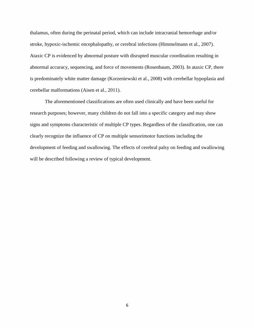
individual, environment and task. Table 1 presents a chronology of typical feeding and
swallowing development over the first two years of life (Arvedson & Brodsky, 2002; Ayoob &
Barresi, 2007; Carruth et al., 2004; Delaney & Arvedson, 2008; Morris & Klein, 2000; Institute
of Medicine-Food and Nutrition Board, 2001).
Feeding and swallowing require numerous underlying biological, psychological and
social competencies. Figure 1 displays the competencies as drawn from the biopsychosocial

10
model of feeding and swallowing (Berlin et al., 2009; Sheppard, 2011). Oropharyngeal function
and respiratory-swallow coordination were selected for inclusion in the present investigation
based on physiological and clinical rationales. Physiologically, these competencies are required
for accurate and precise movement of the oropharyngeal structures during the preparatory, oral
and pharyngeal stages of swallowing. These competencies are vital to the gradual and safe
development of feeding and swallowing. In addition, these competencies provide insight into
neuroanatomical and neurophysiological functioning as it relates to feeding and swallowing
(Martin, Logemann, Shaker, & Dodds,1994; Rogers & Arvedson, 2005; Leder et al., 2013).
Oropharyngeal function and respiratory-swallow coordination are relatively easy to assess
through non-invasive instrumentation and can provide objective data in this population.
Furthermore, despite the importance of these competencies, data on these measures in children
with neurodevelopmental disabilities are extremely sparse. Data on these competencies are
important when considering the airway protective deficits experienced by children with CP.
11
Table 1. Development of feeding and swallowing from 0-24 months of life.
Months
of Life
Recommended
Caloric Intake
Modality Bolus Milestone Emerging
Skill
Birth – 6 40-45
calories/pound/day
520-270
calories/day
Nipple Liquid Dissociated
voluntary
movements
SSB Pattern
6 – 8 35-40
calories/pound/day
646-743
calories/day
Spoon /
Cup
Puree Coordinated
movements
Munch chew
8 – 12 35-40
calories/pound/day
646-743
Utensils /
Cup
Puree and
Chopped
Solids
Adequate lip
seal +
sustained
bite
Lingual
lateralization
12 - 15 Dependent on BMI
and weight/height
growth charts
Utensils /
Cup
Solids
and
Liquids
Oral
functioning
for all bolus
types
Diagonal
rotary chew
15 - 24 Dependent on BMI
and weight/height
growth charts
Utensils /
Cup
(straw)
Solids
and
Liquids
Complete
self-feeding
with utensils
Rotary chew
+ sequential
sips

12
Figure 1. Biopsychosocial model of feeding and swallowing competencies.
• Respiration
• Motor Control and Planning (Praxis)
• Posture/Positioning
• Strength
Biological
• Attention
• Motivation
• Task Persistence
• Regulation
Psychological
• Dyad
• Independence
• EnvironmentSocial

13
v. Cerebral Palsy: Feeding and Swallowing Dysfunction
Children across CP types are prone to having feeding and swallowing disorders (Adams
et al., 2012; Arvedson, 2013; Benfer et al., 2015; Dahlseng et al., 2012; Gangil et al., 2001;
Rogers et al., 1994). Feeding and swallowing difficulties may be the first identifiable symptom
of cerebral palsy and are related to gross motor function (Aisen et al., 2011; Kim et al., 2013;
Weir et al., 2013). In contrast to typical development, children with CP are developing on an
abnormal motor system, usually involving pathological reflexes and disrupted underlying
competencies (Santos & Nogueria, 2005; Sheppard, 1964). The abnormal system arising from
CP negatively affects all phases of swallowing (Calis et al., 2008; Jones et al., 2007). Specific
abnormalities by phase of swallowing are presented in Table 2, as determined through
radiographic studies (Rogers et al., 1994; Mirrett et al., 1994; Arvedson et al., 1994; Del Giudice
et al., 1999). As can be seen, there are numerous disorders that may disrupt the oral preparatory,
oral transport, pharyngeal and esophageal phases of swallowing. These disorders impact the
safety and efficiency of oral intake required for adequate nutrition and hydration.
A comprehensive literature review spanning the years 1965-2004 reviewed the
prevalence of signs and symptoms of dysphagia in children across CP types and showed that
56% of children exhibited choking, 28% had extended feeding time, 59% experienced
constipation, and 22% demonstrated emesis (Odding et al, 2006). In children presenting
primarily with spastic CP, 77.3% had aspiration with 68.2% having silent aspiration (food or
liquid entering the airway without a response from the individual) and 68.2% displayed
gastroesophageal reflux (Mirret et al., 1994). These feeding and swallowing deficits in
conjunction with an abnormal cough response may result in malnutrition and reduced hydration
leading to abnormal growth and poor outcomes (Feng et al., 2002).

14
Literature has generally focused on children with a greater degree of motor involvement
such as those with BSCP as these children are known to have more severe feeding and
swallowing difficulties. Yet, current evidence shows that children with milder involvement, as
seen in USCP, also demonstrate feeding and swallowing difficulties (Benfer et al., 2013;
Malandraki et al., 2014; Malandraki et al., 2015; Mishra et al., 2014). Children with USCP are
found to exhibit difficulties in bolus reception, containment, oral transport, mastication, and the
pharyngeal swallow in addition to showing signs and symptoms of aspiration risk (coughing and
wet voice) during meals (Malandraki et al., 2014; Mishra et al., 2014). Though authors have
described the behavioral symptoms of dysphagia seen in these children, information on the
underlying coordinative competencies that lead to these deficits have not yet been investigated.

15
Table 2. Abnormalities by swallow phase in children with cerebral palsy.
Oral Preparatory Oral Transport Pharyngeal Esophageal
Prolonged oral
preparatory time
Tongue rest position
not at midline
Lingual
Discoordination
Prolonged oral
transit time
Piecemeal
deglutition
Lingual
Discoordination
Prolonged
pharyngeal transit
time
Delayed triggering
of the pharyngeal
swallow
Nasal regurgitation
Pharyngeal
dysmotility
Reduced upper
esophageal
sphincter opening
Overt and/or Silent
Aspiration
Esophageal
distention
Esophageal
backflow

16
vi. Oral Praxis
Praxis has been defined as “the ability to perform skilled movements on command or
demonstration” (Kools & Tweedy, 1975, p. 11). Praxis, viewed as an umbrella term, has also
been equated to motor control, planning and coordination (Murray et al., 1990). The skilled
movements that define praxis should be meaningful and goal oriented. In the context of
swallowing, skilled movements of the oropharyngeal musculature are required for a variety of
components including bolus reception, containment in the oral cavity, lingual propulsion during
the oral phase of swallowing, and timely triggering of the pharyngeal swallow (Rothi &
Heilman, 2014). Typical oral praxis tasks are presented in Table 3 (Kaufman, 1995; Kools &
Tweedy, 1975). These tasks often involve lingual and labial musculature. For functional feeding
and swallowing, age appropriate praxis of the oral system is necessary. The development of a
competent SSB pattern relies on motor planning and coordination of the lingual and labial
musculature as well as intact airway protective mechanisms to ensure adequate and safe
nutritional intake. As the child ages, a mature rotary chew develops and involves numerous
praxis components including lingual lateralization and biomechanical stability of the mandible
along with timely labial seal to prevent anterior spillage of the masticated bolus (Wilson &
Green, 2009). With maturation, the developing child must become an independent feeder and
must have the praxis skills required for the intake of various bolus volumes and viscosities.
Clinicians have noted the interaction between feeding and swallowing and the praxis
system. This has led to the development of clinical tools that assess praxis-related skills (Palmer,
2002; Palmer et al., 1992; Reilly et al., 1995; Skuse et al., 1995). Despite the clinical importance
of the praxis system to feeding and swallowing, research on the development of oral praxis in
healthy children is limited. Robbins and Klee (1987) assessed the development of oral praxis in
17
90 typically developing children across nine age groups and found maturation of skills to occur
by 4 years of age. However, decreases in variability of performance were observed through 6;11
years of age suggesting a process of praxis refinement from 4 to 7 years of age. Similarly, Kools
and Tweedy (1975) used a 20-item test to study oral and limb praxis in 87 healthy male children
and found that praxis competencies developed over time and reached maturation near 6 years of
age. Variability of praxis tasks was reduced as the child aged. It appears that oral praxis
development demonstrates a steady progression of development until 4 years of age, with
refinement processes occurring at approximately 6 years of age. This is in agreement with
Bearzotti and Fabbro (2007), who studied 93 healthy children and noted that an objective
assessment of oral praxis, including activities such as coughing, blowing, biting, lingual
lateralization, and yawning, could yield insight into motor skill level and possible disorders.
Following maturation of oral praxis skills, Fucile et al. (1998) demonstrated that these skills
remain intact throughout the healthy adult lifespan.

18
Table 3. Common oral praxis tasks by structure.
Mandible Tongue Lips Larynx
Depression Protrusion Protrusion Voicing
Lateralization Lateralization Retraction Cough
Elevation Elevation Rounding Pitch Variation

19
vii. Abnormal Oral Praxis
Information about the praxis system in cerebral palsy is sparse. The motor programming
deficits secondary to neurological insult overlaid on development provide support for the
abnormal praxis that is frequently observed in CP. Abnormal praxis (i.e., apraxia) is broad and
describes numerous “neurologically induced, acquired, and developmental disorders including
buccofacial apraxia, constructional apraxia, dressing apraxia, gait apraxia, gaze apraxia, limb
apraxia, speech apraxia, truncal apraxia, and swallowing apraxia” (Rothi & Heilman, 2014, p. 1).
Buccofacial apraxia is often used synonymously with oral apraxia (Watson et al., 1986). The
apraxia types are named according to the structures or functions that are motorically disrupted. It
is best to define apraxia with reference to normal function. Given that oral praxis is defined as
the ability to perform movements of the oral structures (e.g., lips, tongue, mandible, cheeks) by
imitation or on command (Bodison & Mailloux, 2006), oral apraxia can be defined as any
disruption or inability of oral praxis. The etiology and characteristics of oral apraxia are herein
presented.
The etiology of oral apraxia has been linked to reduced cerebellar volume and cerebellar
damage, specifically involving the vermis (Daily et al., 1995). It has also been linked to reduced
basal ganglia volume, particularly in the striatum (i.e., caudate nucleus and putamen), as well as
disruption in thalamic function (Berger, 2012; Krishnan et al., 2015). These neurological deficits
result in abnormal oral movements characterized by “disturbances in initiation of movement,
spatial targeting coordination of motor subsystems, rate of movement, additive motor behaviors,
disturbances of sequencing, and perseverative behavior” (Daniels, 2000, p. 161; Daniels et al.,
1999). Research on the development of oral praxis in children with cerebral palsy is sparse. It is
known that the primitive reflexes should be considered in identifying children with cerebral

20
palsy (Blasco, 1994). The persistence of primitive reflexes, thereby making them pathological in
nature, is one of many signs observed in children with CP (Jones et al., 2007). These reflexes
negatively affect praxis by disrupting volitional movement (Blasco, 1994). Pathological reflexes
have often been linked to frontal lobe damage (Isakov et al., 1984). They disrupt gross and fine
motor development and control (Berker & Yalçın, 2008). The infantile patterned responses to
stimuli, as they pertain to oral function, have been termed cranio-oral motor patterns; they have
been observed across CP types and have been found to interfere with volitional movements (i.e.,
praxis) required for oral hygiene, feeding and swallowing (Santos & Nogueria, 2005; Sheppard,
1964). Reilly et al (1996) observed disorders of oral motor control in > 90% of preschool
children with CP leading to difficulty in sucking, swallowing, and overall coordination.
Interestingly, the observed deficits were made prior to the diagnosis of CP in over half of the
children. Research regarding oral praxis development and disorders over the past decade has
demonstrated that approximately 80% of children with CP have some degree of oral involvement
(Wilson & Hustad, 2009). The difficulties have been associated with lingual movements, oral
sequencing and repetition of movement. These deficits have manifested in functional
deficiencies in biting and mastication (Gisel et al., 2000; Benfer, 2015), which have further been
associated with reduced nutrition and a reduction in eating efficiency (Santos et al., 2012).
viii. Submental Amplitude
The pharyngeal component of swallowing is critical in protecting the airway. The
primary muscle group that is of significance to swallowing is the submental musculature (i.e.,
mylohyoid, geniohyoid, anterior belly of the digastric). Current data assessing submental
amplitude utilizes surface electromyography (sEMG). These muscles are known to be active in
infants and children during many components of feeding and swallowing such as labial seal,

21
mastication, lingual propulsion, and the pharyngeal swallow (Gomes et al., 2006; Green et al.,
1997; Tamura et al., 1996; Inoque et al., 1995). The submental region is highly active during
triggering of the pharyngeal swallow as it maintains laryngeal elevation in order to protect the
airway (Perlman et al., 1999; Ertekin & Aydogdu, 2003). Subsequently, activation of the
submental muscle group during swallowing also contributes to opening of the upper esophageal
sphincter to allow the bolus to pass through the esophagus (Ertekin & Aydogdu, 2003).
Submental swallow amplitude in typically developing children and adults has been assessed
through sEMG. This technique is based on the summation of motor unit action potentials
(MUAPs) caused by electrophysiological activity (Sommerich et al., 2000). Amplitude is best
assessed through the integral of the smoothed sEMG corresponding to the pharyngeal swallow.
In terms of submental amplitudes during saliva and water swallows, children between 4 – 12
years of age produce approximately 20 – 40 microvolts per swallow. Notably, these values are
not significantly different from healthy adults between 18 – 30 years of age (Vaiman et al.,
2004b). However, it must be noted that studies often fail to report normalized sEMG values
which are highly valid and allow for individual and group comparisons.
ix. Abnormal Amplitude
A large body of evidence reporting reduced and discoordinated muscle activity in
cerebral palsy is derived from literature regarding skeletal limb musculature (Damiano et al.,
1995; Stackhouse et al., 2005). In children with spastic CP, reduced activation and recruitment of
MUAPs and reduced contraction of the upper (i.e., wrist flexors, wrist extensors) and lower (i.e.,
iliopsoas, rectus femoris, gluteus maximus, hip abductors, hip adductors, hamstrings, quadriceps,
gastrocnemius, soleus, anterior tibialis) limbs have been documented (Rose and McGill, 2005;
Wiley & Damiano, 1998; Vaz et al., 2006). Children with bilateral CP were also found to have

22
significantly reduced voluntary muscle activation (Stackhouse et al., 2005). Muscle weakness in
children with spastic CP has been attributed to the failure of muscle activation coupled with co-
activation of antagonist muscles (Elder et al., 2003).
Data on specific submental muscle amplitudes during swallowing in children with CP is
limited. In this population, sEMG has been used in the analysis of chewing or has been coupled
with other instrumentation to assist in determining respiratory-swallow interactions
(Briesemeister, Schmidt, & Ries, 2013; Casas, Kenny, & McPherson, 1994). Given the observed
pharyngeal phase deficits in CP, research is needed to determine submental activation and
variability during the swallow across bolus consistencies in order to determine the level of
impairment in these children and its impact on clinical feeding and swallowing severity.
x. Airway Protective Behaviors
The study of airway protective behaviors is a rapidly evolving area of research. Troche et
al. (2014) has outlined the framework for airway protection which involves a continuum of
behaviors from swallow to cough that require reconfiguration of the respiratory cycle (Troche et
al., 2014). Swallowing is one airway protective behavior and much research on its phases,
temporal sequence, and neural components has been previously described in the literature
(Ertekin & Aydogdu, 2003, Jean, 2001; Malandraki, Sutton, Perlman, Karampinos, & Conway,
2009). During normal swallowing, there is a period of breathing cessation known as the
swallow apnea duration (SAD). It is an airway protective mechanism that allows for the safe
temporal cascade of swallowing events to occur. Of relation to SAD is the respiratory swallow
pattern (RSP), which is another airway protective behavior. The RSP can be one of the
following four types: Exhale-Swallow-Exhale, Inhale-Swallow-Exhale, Exhale-Swallow-Inhale,
or Inhale-Swallow-Inhale. There are developmental changes in both SAD and RSP. In infants,
23
the apneic period is more pronounced as a means to prevent aspiration. With maturation there is
a shift to coughing as the primary behavior to prevent aspiration (Loughlin, 1989). In children
with feeding and swallowing dysfunction, the cough was determined to be a more significant
predictor of aspiration than gagging or changes in vocal quality (DeMatteo, Matovich, &
Hjartarson, 2005). Additionally, the infant RSP varies early in life, but the main pattern is for
the swallow to occur following the inhalation, which is likely associated with the infant
oropharyngeal anatomy (Bamdord, Taciak, & Gewolb, 1992; Kelly, Huckabee, Jones &
Frampton, 2007). Typically developing children over one year of age predominantly swallow at
the peak of inspiration and exhibit a post-swallow expiration (McPherson et al., 1992). In
healthy adults, the RSP of exhale-swallow-exhale is most frequently demonstrated, occurring
between 71-100% of the time, and is believed to serve as an airway protective behavior (Klahn
& Perlman, 1999; Martin-Harris et al., 2005; Martin et al., 1994; Preiksaitis, Mayrand, Robins,
& Diamant, 1992; Preiksaitis & Mills, 1996).
Research on airway protective behaviors in children with CP is necessary given that
respiratory dysfunction is the leading cause of death in individuals with CP (Strauss et al., 1999).
Research regarding RSP and SAD has been conducted in children with CP. Specifically,
McPherson et al. (1992) found that children with CP (5-12 years of age) frequently exhibited a
post-swallow inspiration following liquid swallows more often than solid swallows. Similarly,
Rempel and Moussavi (2005) found that individuals with CP (13-30 years of age) exhibited post-
swallow inspiration 50% of the time following liquid consumption and had a range of SAD
between .84-1.03 seconds across all liquids and puree boluses. This was significantly longer than
TDCs, whose average SAD was .63 seconds. These findings point to the need to analyze airway
protective behaviors and their relationship with clinical feeding and swallowing severity in

24
children with cerebral palsy.
It is known that children with SCP present with a high frequency of aspiration events
(Mirret et al. 2994; Odding et al., 2006). The primary airway protective behavior in response to
aspiration is the cough. It is known that cough and swallow have shared neural substrates, and
that both responses involve exertion over their respective central pattern generators by the
behavioral control assembly in order to reconfigure the respiratory cycle and allow for sufficient
airway protection (Troche et al., 2014). Cough is necessary to protect and clear the airways,
especially following aspiration. While this has been appreciated clinically in measures such as
the penetration-aspiration scale (Rosenbek, Robbins, Roecker, Coyle, & Wood, 1996), objective
research on cough is still emerging. Cough can be analyzed in its reflexive form or in its
voluntary form through the cough waveform (Figure 2) (Hegland, et al., 2014; Pitts et al.,
2010). In our population of interest, experimentally inducing the vagally mediated reflex cough
would require the use of a tussigenic stimulus such as capsaicin, citric acid, and/or
ultrasonically nebulized distilled water. Children with CP may have a history of respiratory tract
infections and reduced ability to clear the airways due to purportedly diminished function of
sensory receptors in the airway (Seddon & Khan, 2003; Sullivan, 2013). Therefore, voluntary
cough function was determined to be the most appropriate measure for this study.
Voluntary cough is modulated by the cerebral cortex. Areas of activation may be similar
to that of volitional inspiration and can include: premotor cortex, superior motor cortex,
supplementary motor area, inferolateral sensorimotor cortex, prefrontal cortex, and the striatum
(Evans, Shea, & Saykin, 1999). Voluntary cough has been delineated into the following three
phases: inspiration, compression, and expiration (Simonyan, Saad, Loucks, Poletto, & Ludlow,
2007). The inspiratory phase occurs when alveolar pressure is lower than the atmospheric

25
pressure. It involves the rapid influx of air through the abducted vocal folds along with
contraction of inspiratory musculature, including the diaphragm and the external intercostals
(Fontana, 2003). The compression phase involves adduction of the vocal folds causing a brief
cessation of breathing necessary to build subglottic pressure required for the final expiratory
phase. During the expiratory phase of the cough, the vocal folds rapidly abduct along with
contraction of the abdominal and thoracic musculature to generate a burst of airflow that is
necessary to clear material from the airways (Fontana, 2003). The main expiratory muscles
include the internal intercostals, external oblique, internal oblique, transversus abdominis and
rectus abdominis. As with swallowing, cough must be accurately and motorically sequenced to
maintain airway protection. Common measures of the cough motor response are listed in Table
4.
Voluntary cough has been found to be associated with penetration/aspiration in diseased
populations (Hegland, Okun, & Troche, 2014; Pitts, Bolser, Rosenbek, Troche, & Sapienza,
2008; Pitts et al., 2010). While a single voluntary cough is often elicited by the SLP during a
clinical examination, it is typically observed that individuals who cough in response to
aspiration do so multiple times. Additionally, voluntary sequential cough measures including
compression phase duration, peak expiratory flow rate, and cough expired volume were found
to be different between individuals with and without dysphagia. Therefore, the assessment of
voluntary sequential cough is ideal in children with SCP as it reflects the ecological validity of
the cough motor response without requiring exposure to tussigenic stimuli. Additionally,
assessment of voluntary sequential cough is especially pertinent in individuals with motor
impairment, as there may be differences between the first and subsequent cough responses in a
cough epoch.

26
Figure 2. Cough waveform.
A, Cough Inspired Volume, B, Inspiratory Phase Duration, C, Compression Phase Duration, D,
Peak Expiratory Flow Rate Rise Time, E, Peak Expiratory Flow Rate, F, Cough Expired Volume
Lit
ers/
seco
nd
(L
/s)
Time (seconds)
← AB →
C
D
E Cough Response Total = 4
← F →

27
Table 4. Objective measures of cough airflow.
Cough Airflow
Measure
Abbreviation Unit of
Measurement
Definition
Cough Inspired Volume CIV Liters (L) Amount of air inspired
during the inspiratory
phase of cough
Inspiratory Phase
Duration
IPD seconds (s) Time between post-
tidal volume breathing
inspiratory onset and
compression phase
onset
Compression Phase
Duration
CPD seconds (s) Time between
inspiratory phase
conclusion and
expiratory phase onset
Peak Expiratory Flow
Rate Rise Time
PEFRT seconds (s) Time between
expiratory phase onset
and peak expiratory
flow
Peak Expiratory Flow
Rate
PEFR Liters/second (L/s) Peak flow of the
expiratory phase
Cough Volume
Acceleration
CVA Liters/second2 (L/s2) Peak Expiratory Flow
Rate/ Peak Expiratory
Flow Rate Rise Time
Cough Expired Volume CEV Liters (L) Amount of air expired
during the expiratory
phase of cough

28
xi. Aims and Predictions
It has clinically recognized that children with SCP exhibit dysfunctional feeding and
swallowing as well as deficits in airway protection. Given the importance of these behaviors in
maintaining safe and efficient nutrition and hydration, it is necessary to obtain objective data
regarding the underlying deficiencies as related to functional outcomes. Literature that exists
often contains samples of children with mixed CP-types without controlling for age or gender.
Furthermore, when typically developing controls (TDC) are included they are often not age-
matched nor equivalent in size to the cerebral palsy group. This makes it difficult to draw
conclusions and to confidently translate results of these studies to clinical domains. Empirical
evidence is sparse and, at times, non-existent in regards to the underlying competencies of
feeding and swallowing as well as airway protective behaviors. These areas need to be explored
in order to further identify the impact and consequences of impaired neuromuscular development
and function in this population. Objective data will allow clinicians to rely less on subjective,
clinical judgment. The knowledge gained from the present investigation would allow for a better
understanding of the etiology of deficits in this understudied population and help guide evidence-
based assessment. To that end, the overarching aim of the present investigation was to
objectively examine oropharyngeal function and airway protective behaviors, and clinical
feeding and swallowing severity in children with SCP and TDCs.
xii. Specific Aim 1
a) To test the differences in measures of oropharyngeal function and clinical feeding and
swallowing severity in children with SCP and TDCs

29
It is predicted that children with SCP would have reduced oral praxis and
abnormal submental activation compared to TDCs.
b) To test the relationship between oropharyngeal function and clinical feeding and
swallowing severity in children with SCP and TDCs
It is predicted that oral praxis would be more significantly correlated to clinical
feeding and swallowing severity than submental activation in children with SCP
and TDCs.
xiii. Specific Aim 2
a) To test the differences in measures of respiratory-swallow coordination in children
with SCP and TDCs.
It is predicted that children with SCP would demonstrate prolonged SAD and
abnormal RSPs compared to TDCs.
b) To test the relationship between respiratory-swallow coordination and clinical feeding
and swallowing severity in children with SCP and TDCs
It is predicted that respiratory-swallow pattern would be more significantly
correlated to clinical feeding and swallowing severity than swallow apnea
duration in children with SCP and TDCs.
xiv. Specific Aim 3
To test the differences between children with SCP and TDCs on measures of cough
effectiveness and their relationship on clinical feeding and swallowing severity.

30
It is predicted that children with SCP would demonstrate reduced cough
effectiveness compared to TDCs as indicated by increased IPD, PEFRT and CPD,
and decreased CIV, PEFR, CVA, and CEV.
Cough effectiveness, as measured by PEFRT, PEFR, and CVA, would be
significantly correlated with clinical feeding and swallowing severity.

31
II. METHODS
i. Study Design
This prospective investigation examined a cohort including 11 children with spastic CP
and 10 age-matched controls. All participants participated in a single session of data collection,
which comprised a comprehensive and non-invasive evaluation of swallow and cough function.
ii. Participants
Participants were recruited from the annual cerebral palsy camp at Teachers College,
Columbia University and organizations in NYC through flyers, phone calls, emails and site
visits. For children with SCP, inclusion criteria included:
Diagnosis of SCP by a physician as documented by caregiver-report and pediatric intake
form
Elementary school-age (4-11 years)
Caregiver consent
Child consent (if > 8 years of age)
Exclusion criteria included:
Neurological conditions unrelated to CP
Current respiratory infections
Asthma
Unaided auditory-visual impairments
History of tracheostomy and/or head-neck surgery

32
For the control group, inclusion criteria included:
Typically developing as documented by caregiver-report and pediatric intake form
Elementary school-age (4-11 years)
Caregiver consent
Child consent (if > 8 years of age)
Exclusion criteria included:
Neurological condition
Respiratory condition
Unaided auditory-visual impairment
History of feeding and/or swallowing disorders
History of tracheostomy and/or head-neck surgery
iii. Screening Procedures
Participants were required to meet all of the inclusion criteria and none of the exclusion
criteria expounded above. An IRB approved screening form consisting of questions relating to all
criteria were presented to caregivers and were specific to the assigned group (SCP or TDC). All
participants were administered Subtest I (Vocabulary) of the Test for Auditory Comprehension
of Language (TACL) (Carrow-Woolfolk, 1998), which has been used in children with CP and
provides insight into participants’ comprehension skills (Levy et al., 2015).

33
iv. Data Collection
Data was collected in the Upper Airway Dysfunction Laboratory (955 Thorndike Hall) by
the primary investigator and a trained graduate research assistant. The primary investigator
provided the research assistant with detailed training in data acquisition and analysis for all
measured variables. Given investigator guidance, caregivers completed a thorough intake form
documenting previous/ongoing evaluations/treatments, medical history, neurological reports,
developmental history, feeding/swallowing development, current feeding/swallowing function,
and mealtime behaviors. Additionally, the investigator assigned each participant a level on the
gross motor function classification system (GMFCS) (Rosembaum et al., 1997), manual ability
classification system (MACS) (Eliasson et al., 2006), and the eating and drinking ability
classification system for individuals with cerebral palsy (EDACS) (Sellers et al., 2014). The
assigned levels were based on careful review of the classification system guidelines and were
assigned using live and video-recorded observations of each participant. Furthermore, given that
the data collection phase provided a snapshot of each child’s performance, communication with
caregivers was done to ensure accurate documentation of function. Data collection occurred in
two blocks. The first block consisted of completion of the intake form as well as measures of
receptive language, oral praxis and clinical feeding and swallowing severity. Tasks within the
second block consisted of the simultaneous measuring of submental amplitude, SAD, and RSP
across bolus consistencies. This was then followed by measures of voluntary sequential cough
airflow: CIV, IPD, CPD, PEFRT, PEFR, CVA, and CEV. A detailed overview of each variable
addressed in aims 1-3 is provided below.
Oral Praxis: This was measured through the Oral Movement portion of the Kaufman
Speech Praxis Test (KSPT) (Kaufman, 1995). This standardized assessment has been used
34
extensively in clinical settings to assess the oral praxis system in children between 2.6 – 5.11
years of age and was administered within 5-15 minutes. Given that the participants in the present
investigation were between 4-11 years of age, the KSPT was used to obtain descriptive
information regarding praxis.
The Oral Movement portion of the KSPT contained 11 items and has shown high
reliability of measurement (Cronbach’s alpha = .84) and high test-retest reliability (Cronbach’s
alpha =.93) (Kaufman, 1995). Construct, convergent and discriminant validity was measured on
the KSPT-A, the original test model prior to slight revisions in developing the final KSPT.
Strong validity was evidenced by unidimensionality measures such as principal component
analysis, Guttman scale analysis, and correlational modeling (Kaufman, 1995).
Per the KSPT guidelines, a complete oral mechanism examination was first conducted on
all participants in order to rule out structural abnormalities related to performance. According to
the KSPT administration manual, all participants were asked to imitate the investigator (“Do
what I do”) as the 11 components of the KSPT were performed and scored in a binary manner.
Submental amplitude, Swallow Apnea Duration, and Respiratory Swallow Pattern: These
were measured simultaneously as participants consumed 2 trials of 5ml water, 2 trials of 5cc
pudding/yogurt, and 2 trials of ¼ cookie. The trials were presented using a randomized block
design. The chosen volumes of consistencies and number of trials has been typically used during
clinical, research and instrumental protocols involving pediatric patients (Casas, McPherson, &
Kenny, 1995; Leder & Karas, 2000; Morgan, Ward, Murdoch, & Bilbie, 2002). Figure 3 displays
the configuration of the equipment and participant.
Surface electromyography (sEMG), which has been used in children with cerebral palsy,
was selected as the optimal measure of submental amplitude in our pediatric population (Casas et

35
al., 1994). The parameters were assessed with a bio-amp system and LabChart 7 Pro software
(PowerLab 16/30, ADInstruments, Inc., Colorado Springs, CO). Surface electromyography is an
objective and noninvasive tool that has been used extensively to study swallow amplitude across
pediatric and adult populations for both clinical and research purposes (Vaiman et al., 2004a;
Vaiman et al., 2004b; Perlman, 1993; Gupta et al., 1996; Crary & Baldwin, 1997; Spiro et al.,
1994; Palmer, 1989). In addition, the validity (kappa = .80) and inter-rater reliability (kappa =
.78) of sEMG in the identification of swallow related events have been well documented (Crary
et al., 2007).
The procedure for sEMG data acquisition was consistent with commonly employed
techniques (Ferdjallah et al., 2000; Reaz et al., 2006; Vaiman et al., 2004b; Stepp, 2012).
Participants were seated comfortably with a strong base of support. Bipolar pediatric electrodes
were placed bilaterally over the submental muscle group (mylohyoid, geniohyoid, anterior belly
of the digastric), and a ground electrode was placed on the clavicle. The inter-electrode distance
was fixed at 1.5cm. The bio-amp was calibrated to record at a 2KHz-sampling rate with a 500µV
range. A mains filter was used to attenuate extraneous noise in the environment in order to
maximize the signal to noise ratio. Following a one-minute rest period needed to establish a
clean baseline signal, participants were instructed to perform maximum submental contraction
tasks for normalization and the establishment of a maximum reference value (Vaiman et al.,
2004b; Stepp, 2012). Three maximum tasks were conducted as follows:
1. Participants performed lingual elevation against the Iowa Oral Performance
Instrument (IOPI) bulb (Northwest, 2005). The IOPI is a standard non-invasive tool
with excellent validity and reliability that provides manometric values in kilopascals
(kPa) (Adams et al., 2014; Potter & Short, 2009). The IOPI digitally displayed

36
pressure levels on a feedback screen. The device was connected, via a thin 2-inch
tube, to the IOPI stem and bulb. The primary investigator held the IOPI stem to
maintain consistency of positioning. The bulb was positioned longitudinally along the
hard palate and posteriorly to the central incisors (Potter& Short, 2009). Participants
were instructed to push against the air filled bulb toward the roof of their mouth as
hard as possible with their tongue for three seconds. Following familiarization with
the device, three trials with 30 seconds of rest between trials were obtained.
2. Participants performed three trials of maximum mandibular depression after being
provided with visual and verbal cues by the investigator.
3. Participants performed three effortful swallows with one minute of rest in between
trials. Visual and verbal cues were provided.
Surface EMG recordings of maximum submental contraction during these tasks were
obtained. The task that yielded maximum amplitude values and was performed most efficiently
by all children was maximum mandibular depression. Therefore, the signal corresponding to this
task was selected as the basis for normalization across participants. Throughout the sEMG task,
swallows were tagged according to trial number and bolus type.
SAD and the RSP were measured using pediatric thoracic and abdominal cotton elastic
bands. Thoracic and abdominal movements during breathing have been frequently measured
using elastic bands and provide valid and reliable data (Hegland, Huber, Pitts, & Sapienza, 2009;
Hegland, Huber, Pitts, Davenport, & Sapienza, 2011). Consistent with Martin et al. (1994), the
thoracic band was placed beneath the axilla and the abdominal band was placed at the umbilicus
level. Movement of the rib cage and abdomen were detected. Observation of 20 seconds of tidal
breathing was first conducted and was followed by tasks to ensure that the elastic bands were

37
accurately detecting movement required for the acquisition of SAD and RSP. These tasks
included having each participant inhale deeply and exhale, and then maximally displace the
abdomen inwardly and outwardly. The following RSP patterns were possible: Exhale-Swallow-
Exhale, Inhale-Swallow-Exhale, Exhale-Swallow-Inhale, and Inhale-Swallow-Inhale.
Clinical Feeding and Swallowing Severity: This was evaluated using the Dysphagia
Disorder Survey (DDS), a standardized mealtime assessment (Sheppard et al., 2014). This was
chosen over an invasive tool such as fiberoptic endoscopic evaluation of swallowing (FEES),
which would subject the child to risks that may include gagging, vasovagal syncope, epistaxis
and laryngospasm (Hiss & Postma, 2003). It was also determined that videofluoroscopy would
not be used as it would be clinically unethical to expose our pediatric control and non-referred
patient groups to harmful ionizing radiation (Hiorns & Ryan, 2006).
The DDS has been standardized on 654 individuals (age range: 8-82 years) with
developmental and intellectual disabilities including children with CP (Sheppard et al., 2014). It
has been used reliably in children with CP as young as 2.1 years of age (Calis et al., 2008).
Sensitivity and specificity of the DDS are as follows: Part 1 (.88 and .85) and Part 2 (.94 and
.87); Kaiser-Meyer-Olkin measure (>.90) and Spearman correlations (>.85). Reliability was
evidenced through moderate kappa values (.53-.71) (Sheppard et al., 2014). The DDS has two
parts comprising the DDS Total score. DDS Part 1 contains seven components indirectly related
to feeding and swallowing (e.g., independence, positioning). DDS Part 2 contains eight
components specifically related to feeding and swallowing competencies. The eight components
of Part 2 are scored using a binary system of zero to signify competence and one to signify
deficiency. Five trials of liquid, semisolid and solid boluses are required to derive an accurate
task analysis of feeding and swallowing using the DDS. A factor analysis of DDS components

38
revealed that the Body Mass Index (BMI) and the report of emesis/rumination were not of strong
significance in reference to the other components (Sheppard et al., 2014). However, given their
clinical importance, these items were obtained during the present investigation.
The children were seated comfortably to allow for a natural eating position. The
following items were placed on the table: one cup of water, one pudding cup, and one two
cookies. The investigator positioned a Canon HD video camera three feet in front of the
participant, with the view that allowed for visualization of the child and food items (Figure 4).
Participants were told to eat and drink in a regular manner. The participants ate at their normal
pace. The children were only interrupted to facilitate eating/drinking of a particular bolus type
or for safety intervention. Following five trials of each bolus type, the recording continued until
meal conclusion.
Voluntary sequential cough: Voluntary sequential cough was elicited and measured in a
manner similar to what has been described in the literature (Hegland et al., 2014). However,
adaptations were made given the pediatric population of interest. Participants were seated
comfortably with arms at the side. A facemask was placed over the participant’s mouth and
nose and was coupled to a pneumotachograph system that input differential pressure change to a
digital spirometer (ADInstruments). The airflow signal was digitized at 2KHz with a range of
2V, and samples were low pass filtered at 150Hz. Recording was obtained in Liters/seconds.
Following 20 seconds of tidal breathing, the investigator modeled and instructed the participants
to take a deep breath and cough sequentially (or several times in a row) as if something was
stuck in the throat. Three trials of sequential cough were performed with a 20 second rest period
between each cough epoch.

39
Figure 3. Participant seated in standard chair and connected to surface electrodes and
respiratory/abdominal bands.

40
Figure 4. Participant seated in standard chair during clinical feeding and swallowing task.
.

41
v. Data Analysis
The primary investigator analyzed all data. Intra- and inter-rater reliability was assessed
across all measures by the primary investigator (Rater 1) and co-investigator (Rater 2) using data
sets from four randomly selected participants (2 TDCs, 2 SCP). Rater 2 was blinded to
participant group.
Demographic Information: Information obtained from the intake form was used to
characterize the sample, with emphasis on neurological function and developmental milestones.
The TACL, GMFCS, MACS, and EDACS were scored according to their respective guidelines,
in conjunction with parent reports, and were used to assign levels of ability related to gross
motor, manual, and feeding/swallowing function. Outcomes included age equivalency on the
TACL as well as a rating on functional scales (GMFCS, MACS, EDACS) that ranged from level
I (mild impairment) to V (severe impairment).
Oral Praxis: The Oral Movement portion of the KSPT was scored according to the KSPT
scoring manual, which provided detailed instructions on how to interpret and score each of the
11 components. The principal investigator has administered and scored the KSPT numerous
times in clinical practice and trained the co-investigator in its usage and scoring. The raw score
(total number of components scored correctly) was used for analysis.
Submental Amplitude: LabChart 7 Pro software (ADInstruments) was utilized for all
sEMG analysis. Channel 5 was assigned to the left submental swallow signal and Channel 6 was
assigned to the right submental swallow signal. Maximum functional swallows and six tagged,
bolus swallow signals were analyzed by taking the average of both channels. For participants
who swallowed more than one time per bolus trial, the first swallow was analyzed. Using the
arithmetic feature of LabChart, raw sEMG swallow signals were rectified (transformed to a

42
positive polarity frequency) and smoothed through the root mean square equation for the left
[RMS (CH5, .2)] and right [RMS (CH6, .2)] swallow signals. Within the channel settings
feature, standard integral channels were created and corresponded to the sEMG signal. Start and
end values corresponding to each swallow signal were determined based on the respitrace signal
(flat slope signifying apnea) as this signal was synchornized with the sEMG signal. Using the
data pad feature in LabChart, integral values for each swallow were obtained.
Swallow Apnea Duration and Respiratory Swallow Pattern: LabChart software
(ADInstruments) was used to determine the duration of swallow apnea. The SAD and RSP
demonstrated by each child were analyzed through analysis of the respitrace signal. The positive
slope represents inhalation, the zero slope represents the period of swallow apnea, and the
negative slope represents exhalation. Figures 5 and 6 display the exhale-swallow-exhale pattern
and the inhale-swallow-inhale pattern, respectively. Further verification of apnea duration across
bolus swallows was assessed with the simultaneous sEMG swallow signal.
Clinical Feeding and Swallowing Severity: The primary and co-investigator are certified
in the administration, scoring and interpretation of the DDS. DDS scoring was done in real time
during the mealtime assessment using the DDS scoring form and instruction manual. This
allowed for Part 1, Part 2 and Total scores to be determined immediately following the mealtime
assessment. The investigators could not be blinded to group during the live DDS assessment;
however, video recording analysis using the DDS was conducted in a blinded fashion by the co-
investigator. Outcomes included DDS Part 1 (0-15), DDS Part 2 (0-19), and DDS Total Score (0-
34). The DDS Total Score is the sum of DDS Part 1 and DDS Part 2. A score of 34 indicates the
most severe feeding and swallowing impairment.
Voluntary sequential cough: All measures of voluntary sequential cough airflow (CIV,

43
IPD, CPD, PEFRT, PEFR, CVA, and CEV) were analyzed using LabChart7 Pro software
(ADInstruments). Thirty seconds of tidal breathing was recorded to ensure accurate signal
detection and to establish the signal baseline (0 liters/second). The Data Pad feature was utilized
to assess volume, duration, and peak value parameters corresponding to the airflow channel. The
area between the point at which the cough signal fell below 0 L/s to the point at which it returned
to 0 L/s was taken as the CIV. The time between those same two points was taken as the IPD.
The time between the end of the inspiratory phase to the point at which the signal began to
sharply increase was taken as the CPD. The time taken for the signal to travel from the end of the
compression phase to the maximum value was taken as the PEFRT. The maximum signal value
of the cough waveform was taken as the PEFR. The CVA was calculated by dividing the PEFR
by the PEFRT. Finally, the area under the cough response, or the area between the end of the
compression phase to the start of the subsequent compression phase, was taken as the CEV.

44
Figure 5. The ideal exhale-swallow-exhale pattern.
Figure 6. The abnormal inhale-swallow-inhale pattern.
Vo
lta
ge
(V)
Vo
lta
ge
(V)
Time (seconds)
Time (seconds)

45
vi. Statistical Analyses
Based on the results of power analysis using G*Power 3 software (Faul, Erdfelder, Lang,
& Buchner, 2007), non-parametric statistical analyses were conducted with SPSS statistical
software (IBM Corp. Released 2016. IBM SPSS Statistics for Windows, Version 24.0. Armonk,
NY: IBM Corp). Intra-and inter-rater reliability of all variables was assessed using data sets from
four randomly selected participants. Reliability statistics included intra-class correlation
coefficients for continuous variables and Cohen’s Kappa statistic for binary variables.
A Bonferroni corrected Mann Whitney test was used to determine between-group
differences in demographic data including age, sex, body mass index, and age-equivalency on
the TACL. Kruskal-Wallis tests were used to assess between-group differences in measures of
feeding/swallowing competencies and clinical feeding and swallowing severity. Specifically,
group differences in oral praxis, submental amplitudes across bolus swallows, swallow apnea
duration, respiratory swallow patterns, and DDS Part 1, DDS Part 2, and DDS Total scores were
evaluated. Spearman’s Rank Order Correlations were used to assess relationships between oral
praxis and all DDS scores, post swallow inhalation pattern and all DDS scores, and submental
amplitudes across bolus swallows and all DDS scores. These correlations were assessed in the
total sample (n=21) and in the SCP group (n=11).
The Mann Whitney test was used to evaluate between-group differences in the number of
coughs produced per cough epoch. Kruskal-Wallis tests were used to assess between-group
differences in measures of cough effectiveness. Specifically, group differences in CIV, IPD,
CPD, PEFRT, PEFR, CVA, and CEV were evaluated. Within the SCP group (n=11), Spearman’s
Rank Order Correlations were used to assess relationships between PEFR and DDS Total score,
and between CVA and DDS Total score, for the first (Cr1) and second (Cr2) cough of the cough

46
epoch. Spearman’s Rank Order Correlations were also used to determine relationships between
DDS Total score and GMFCS in addition to PEFR and GMFCS, and CVA and GMFCS, for the
first (Cr1) and second (Cr2) cough of the cough epoch.
Effect sizes were calculated for all applicable measures. Specifically, the effect size (r)
was used with Mann Whitney tests, and the effect size (η2) was used with Kruskal-Wallis tests.
The alpha level of significance for all statistical tests was set at p < .05.

47
III. RESULTS
i. Reliability
Intra- and inter-rater reliability was excellent for all continuous and binary responses as
evidenced by intraclass correlation coefficients and Cohen’s kappa statistic (Table 5). All values
exceeded .80.
ii. Participant Characteristics
A total of 23 children were enrolled in the study, but two children withdrew as they were
unable to complete the evaluation. Therefore, 21 children completed the protocol (10 controls
and 11 children with SCP). Demographic information for all participants is presented in Table 6.
Table 7 presents information specific to the SCP group. This group comprised 8 children with
unilateral impairment and 3 children with bilateral impairment. The most common central
nervous system damage in this sample was intra- and peri-ventricular hemorrhage. The mode,
median, and range of functional classification scores were as follows: GMFCS (I, II, I-V),
MACS (II, II, I-V), and EDACS (II, II, I-IV). This indicates mild functional impairment for this
sample. The two groups were well matched with no significant between-group differences in
age, sex, body mass index, or age equivalency on the TACL (Table 8).

48
Table 5. Intra- and inter-rater reliability results.
Measure Intra-rater
(ICC)
Inter-rater
(ICC)
DDS .992 .961
KT .980 .935
sEMG (Left) .978 .863
sEMG (Right) .989 .987
SAD .989 .906
CIV .999 .999
IPD .996 .918
CPD 1.00 .841
PEFRT 1.00 .868
PEFR 1.00 1.00
CVA .995 .982
CEV .999 .996
Measure Intra-rater (κ) Inter-rater (κ)
RSP (Pre-swallow) 1.00 .895
RSP (Post-swallow) 1.00 .882
ICC, intraclass correlation coefficient, κ, Cohen’s kappa, DDS, Dysphagia Disorder Survey, KT,
Kaufman Test, sEMG, surface electromyography, SAD, swallow apnea duration, CIV, cough
inspired volume, IPD, inspiratory phase duration, CPD, compression phase duration, PEFRT,
peak expiratory flow rate rise time, PEFR, peak expiratory flow rate, CVA, cough volume
acceleration, CEV, cough expired volume, RSP, respiratory-swallow pattern

49
Table 6. Participant characteristics.
Variable TDC group
(n=10)
SCP group
(n=11)
Mean age
(SD) (years)
7.6 (2) 7.7 (2.4)
Median
TACL (IQR)
11 (2.3) 7 (2.5)
Mean BMI
(SD)
15.5 (3) 18.2 (3.8)
Males (%)
(n)
50 (5) 72.7 (8)
TDC, typically developing control, SCP, spastic cerebral palsy, SD, standard deviation, TACL,
test of auditory comprehension of language, IQR, interquartile range, BMI, body mass index

50
Table 7. Neurological insult and functional classification in children with spastic cerebral palsy.
Participant
ID
Neurological
Insult
Motor
Involvement
GMFCS MACS EDACS
SCP1 PVL Unilateral 1 2 2
SCP2 PVH II Unilateral 2 2 2
SCP3 PVH II Unilateral 1 2 2
SCP4 PVL Bilateral 4 5 4
SCP5 PVH II Unilateral 2 2 2
SCP6 IVH IV Bilateral 5 3 2
SCP7 IVH II Unilateral 2 3 2
SCP8 PVL Unilateral 1 1 1
SCP9 PAIS Unilateral 1 1 2
SCP10 IVH II Unilateral 1 2 1
SCP11 HIE Bilateral 4 5 3
SCP, spastic cerebral palsy, GMFCS, Gross Motor Function Classification Scale, MACS, Manual Ability Classification Scale, EDACS, Eating and Drinking Ability Classification Scale, PVL, periventricular leukomalacia, PVH, periventricular hemorrhage, IVH, intraventricular hemorrhage, PAIS, perinatal arterial ischemic stroke, HIE, hypoxic-ischemic encephalopathy

51
Table 8. Statistical comparisons of demographic characteristics between typically developing
controls and children with spastic cerebral palsy.
Age Sex BMI TACL Age Equivalency
(years)
Mann
Whitney U
54 42.5 32 28.5
Z -.071 -1.045 -1.621 -1.878
p-value .973 .378 .114 .061
Effect size (r) .025 .369 .573 .664
BMI, body mass index, TACL, test of auditory comprehension of language

52
iii. Between-Group Comparisons: Oropharyngeal Function and Clinical
Feeding and Swallowing Severity
Table 9 shows group means and standard deviations for the Kaufman Test and all
components of the DDS. Figure 7 displays oral praxis performance by group. Compared to
children with SCP, the TDC group had significantly higher oral praxis scores (χ2 = 8.573, p =
.003, η2 = .429) as well as significantly lower scores on the DDS Part 1 (χ2 = 17.098, p<.0001, η2
= .855), DDS Part 2 (χ2 = 14.291, p < .0005, η2 = .715) and DDS Total (χ2 = 15.421, p < .0001, η2
= .771). A group comparison of the feeding/swallowing task analysis portion of the DDS is
presented in Figure 8. Compared to controls, children with SCP had greater difficulties across all
feeding/swallowing tasks. No child in the TDC group displayed overt signs of aspiration,
whereas three children with CP exhibited a single instance of cough.
Significant between-group differences in normalized sEMG values were observed (χ2
=11.731, p = .001). Specifically, children with SCP required a significantly greater percent of
maximum amplitude during puree (χ2 = 6.091, p = .014, η2 = .305) and solid swallows (χ2 =
10.555, p = .001, η2 = .528), but not for liquid swallows (χ2 = .077, p = .782, η2 = .004) (Figure
9). Further analysis within the SCP group revealed no significant differences in swallow
amplitude values between the less impaired and more impaired side of function (χ2 = .838, p =
.360, η2 = .084).

53
Table 9. Results of the Kaufman Test and Dysphagia Disorder Survey for typically developing
controls and children with spastic cerebral palsy.
Variable TDC group
(n=10)
SCP group
(n=11)
Mean KT (SD)
(Maximum = 11)
9.9 (1.9) 6.6 (2.2)
Mean DDS1 (SD)
(Maximum = 15)
0 (0) 3.7 (2.3)
Mean DDS2 (SD)
(Maximum = 19)
0.8 (1.3) 7.6 (3.6)
Mean DDST (SD)
(Maximum = 34)
0.8 (1.3) 11.4 (5.7)
TDC, typically developing control, SCP, spastic cerebral palsy, KT, Kaufman Test, DDS1,
Dysphagia Disorder Survey Part 1, DDS2, Dysphagia Disorder Survey Part 2, DDST, Dysphagia
Disorder Survey Total

54
Figure 7. Differences between typically developing controls and children with spastic cerebral
palsy on measures of oral praxis.
TDC, typically developing control, SCP, spastic cerebral palsy
0
10
20
30
40
50
60
70
80
90
100
Per
cen
t o
f P
art
icip
an
ts w
ith
Dy
sfu
nct
ion
Kaufman Test of Oral Praxis
Tasks Analysis by Group
TDC Group SCP Group

55
Figure 8. Differences between for typically developing controls and children with spastic
cerebral palsy on measures of clinical feeding and swallowing severity.
TDC, typically developing control, SCP, spastic cerebral palsy
0
10
20
30
40
50
60
70
80
90
100
Liq
uid
Pu
ree
So
lid
Liq
uid
Pu
ree
So
lid
Liq
uid
Pu
ree
So
lid
Liq
uid
Pu
ree
So
lid
So
lid
Liq
uid
Pu
ree
So
lid
Liq
uid
Pu
ree
So
lid
Orientation Reception Containment Transport Chew Swallow Post Swallow
Per
cen
t o
f P
art
icip
an
ts w
ith
Dy
sfu
nct
ion
(%
)
Dysphagia Disorder Survey
Task Analysis by Group
TDC Group SCP Group

56
Figure 9. Differences between typically developing controls and children with spastic cerebral
palsy on normalized submental swallow amplitude across bolus consistency.
*p<0.05
TDC, typically developing control, SCP, spastic cerebral palsy
0
10
20
30
40
50
60
70
80
90
100
Liquid Puree* Solid*
Per
cen
t o
f M
axim
um
Am
pli
tud
e
Normalized sEMG Values by Group
TDC Group SCP Group

57
iv. Relationships Between Oropharyngeal Function and Clinical Feeding and
Swallowing Severity
Results of the Spearman Rank Order Correlation revealed significant, negative
correlations between the Kaufman Test and the DDS (Table 10). Age was not correlated with
oral praxis or any component of the DDS. Normalized sEMG values were not correlated with the
DDS. When looking at the SCP group only, significant, negative correlations between the
Kaufman Test and the DDS Part 1 (rs=-.664, p=.026), DDS Part 2 (rs=-.633, p=.036) and DDS
Total (rs=-.671, p=.024) were observed. Figure 10 displays the association between the Kaufman
Test and the DDS Total for children with SCP. No significant correlations between normalized
sEMG values and DDS scores were observed for children with SCP.

58
Table 10. Correlations between the Kaufman Test and the Dysphagia Disorder Survey for all
participants.
DDS Component Value KT
DDS1 rs -0.733***
DDS2 rs -0.815***
DDST rs -0.802***
*p<0.05, **p<0.005, ***p<0.0005
DDS, Dysphagia Disorder Survey, DDS1, Dysphagia Disorder Survey Part 1, DDS2, Dysphagia
Disorder Survey Part 2, DDST, Dysphagia Disorder Survey Total, KT, Kaufman Test

59
Figure 10. Negative correlation between the Kaufman Test and the Dysphagia Disorder Survey
Total score in children with spastic cerebral palsy.
DDS, Dysphagia Disorder Survey
02468
10121416182022242628303234
0 1 2 3 4 5 6 7 8 9 10 11
DD
S T
ota
l S
core
Kaufman Test Score
Oral Praxis Skills and Clinical Feeding and Swallowig Severity

60
v. Between-Group Comparisons: Respiratory-Swallow Coordination and
Clinical Feeding and Swallowing Severity
No significant between-group differences in SAD were observed across liquid (χ2 =
1.698, p = .193, η2 = .085), puree (χ2 = .179, p = .672, η2 = .009), or solid (χ2 = 1.983, p = .159, η2
= .099) bolus consistencies. Figure 11 displays the group means and standard deviations across
consistencies. Respiratory-swallow patterns for both groups are presented in Figure 12, and a
significantly greater number of participants in the SCP group (>50%) demonstrated a post-
swallow inhalation on liquids (χ2 = 8.015, p = .005, η2 = .401), purees (χ2 = 6.086, p = .014, η2 =
.304), and solids (χ2 = 4.455, p = .035, η2 = .223) compared to controls. No significant pre-
swallow differences were observed. Representative examples of the respiratory-swallow pattern
from a TDC and a child with SCP are presented in Figures 13 and 14, respectively.

61
Figure 11. Differences between typically developing controls and children with spastic cerebral
palsy on duration of swallow apnea across bolus consistency.
TDC, typically developing control, SCP, spastic cerebral palsy
0.0
0.2
0.4
0.6
0.8
1.0
1.2
Liquid Puree Solid
Consistency
Tim
e (s
eco
nd
s)
Swallow Apnea Duration by Group
TDC Group SCP Group

62
Figure 12. Differences between typically developing controls and children with spastic cerebral
palsy on respiratory swallow pattern across bolus consistency.
*p<0.05
TDC, typically developing control, SCP, spastic cerebral palsy
0
10
20
30
40
50
60
70
80
90
100
Liquid Puree Solid Liquid* Puree* Solid*
Pre-Swallow Post-Swallow
Pa
rtic
ipa
nt
Inh
ala
tio
n (
%)
Respiratory-Swallow Pattern by Group
TDC Group SCP Group

63
Figure 13. An exhale-swallow-exhale pattern produced by a typically developing control.
Figure 14. An inhale-swallow-inhale pattern produced by a child with spastic cerebral palsy.
Vo
lta
ge
(V)
Vo
lta
ge
(V)
Time (seconds)
Time (seconds)

64
vi. Relationships Between Respiratory-Swallow Coordination and Clinical
Feeding and Swallowing Severity
Results of the Spearman Rank Order Correlation revealed significant correlations
between RSP and all components of the DDS (Table 11). In the SCP group, significant
correlations between the post-swallow respiratory pattern for solids and the DDS Part 1 (rs=.734,
p=.010), DDS Part 2 (rs=.610, p=.046) and DDS Total (rs=.673, p=.023) were observed. Figure
15 displays the relationship between RSP for solids and DDS scores for all 11 children with SCP
as grouped by post-swallow exhalation versus post-swallow inhalation. Neither age nor gross
motor impairment were correlated with RSP. No correlations between swallow apnea duration
and DDS components were observed.

65
Figure 15. Association between respiratory-swallow pattern for solids and the Dysphagia
Disorder Survey in children with spastic cerebral palsy.
DDS, Dysphagia Disorder Survey
0
5
10
15
20
25
Exhale Exhale Exhale Exhale Exhale Inhale Inhale Inhale Inhale Inhale Inhale
1 2 3 4 5 6 7 8 9 10 11
DD
S T
ota
l S
core
Post-Swallow Pattern
Relationship Between Post-Swallow Pattern for Solids and
Clinical Feeding and Swallowing Severity

66
vii. Between-Group Comparisons on Measures of Cough Effectiveness and their
Relationship with Clinical Feeding and Swallowing Severity
The means and standard deviations of cough response totals across all 3 trials of
cough were TDC group (3.24 + 1.53) and SCP group (2.95 + .701). The median number of
coughs produced per epoch for both groups was three. A Mann Whitney Test revealed no
significant between-group differences in number of coughs produced per epoch.
Table 12 presents group means and standard deviations of voluntary cough airflow measures
(CIV, IPD, CPD, PEFRT, PEFR, CVA, and CEV) for cough response 1 and cough response 2,
while Table 13 presents statistical comparisons across these measures. No significant between-
group differences were found on measures of CIV, IPD, CPD, PEFRT, PEFR or CEV.
However, the SCP group demonstrated significantly reduced CVA compared to the TDC group
for the first cough (Cr1) (p=.019) of the cough epoch. A cough epoch from a TDC and a child
with SCP is presented in Figures 16 and 17, respectively.
In children with SCP, no significant correlations were observed between the DDS and
PEFR for the first cough (Cr1) (rs=-.475, p=.165) or second cough (Cr2) (rs=-.438, p=.205) of
the cough epoch. Additionally, no significant correlations were observed between the DDS and
CVA for Cr1 (rs=-.043, p=.906) or Cr2 (rs=.043, p=.906) of the cough epoch. In the SCP group,
the DDS was positively correlated with the GMFCS (rs=.749, p=.008) (Figure 18). Correlations
between measures of cough effectiveness and the GMFCS were assessed. Though no significant
correlations were found between the GMFCS and CVA for Cr1 (rs=-.492, p=.149) or Cr2 (rs=-
.479, p=.162) of the cough epoch, significant, negative correlations were observed between the
GMFCS and PEFR for Cr1 (rs=-.702, p=.024) and Cr2 (rs=-.715, p=.020) of the cough epoch
(Figures 19 and 20).

67
Table 12. Means and standard deviations of measures of voluntary cough airflow for typically developing controls and children with
spastic cerebral palsy.
Cough Response 1 CIV IPD CPD PEFRT PEFR CVA CEV
Group TDC SCP TDC SCP TDC SCP TDC SCP TDC SCP TDC SCP TDC SCP
Mean .389 .543 .798 .998 .282 .283 .044 .107 2.14 1.79 52.7 31.5 .360 .265
Standard Deviation .300 .417 .525 .528 .203 .171 .015 .182 .658 .182 18.5 19.3 .240 .264
Cough Response 2 CIV IPD CPD PEFRT PEFR CVA CEV
Group TDC SCP TDC SCP TDC SCP TDC SCP TDC SCP TDC SCP TDC SCP
Mean NA NA NA NA .141 .148 .040 .059 1.49 1.48 43.3 33.8 .180 .187
Standard Deviation NA NA NA NA .107 .109 .022 .050 .381 .661 18.3 19.8 .060 .116
CIV, cough inspired volume, IPD, inspiratory phase duration, CPD, compression phase duration, PEFRT, peak expiratory flowrate rise time, PEFR, peak
expiratory flow rate, CVA, cough volume acceleration, CEV, cough expired volume, TDC, typically developing control, SCP, spastic cerebral palsy, NA, not
applicable
Table 13. Statistical comparisons between typically developing controls and children with spastic cerebral palsy on measures of
voluntary cough airflow.
Cough Response 1 CIV IPD CPD PEFRT PEFR CVA CEV
χ2 .463 .091 .463 3.158 .823 5.491 .691
p-value .196 .762 .496 .076 .364 .019 .406
Effect size (η2) .024 .005 .024 .166 .043 .289 .036
Cough Response 2 CIV IPD CPD PEFRT PEFR CVA CEV
χ2 NA NA .092 1.751 .051 .691 .036
p-value NA NA .762 .186 .821 .406 .850
Effect size (η2) NA NA .005 .092 .003 .036 .002 CIV, cough inspired volume, IPD, inspiratory phase duration, CPD, compression phase duration, PEFRT, peak expiratory flowrate rise time, PEFR, peak
expiratory flow rate, CVA, cough volume acceleration, CEV, cough expired volume, NA, not applicable

68
Figure 16. Cough epoch produced by a typically developing control.
Figure 17. Cough epoch produced by a child with spastic cerebral palsy.
Lit
ers/
seco
nd
(L
/s)
Lit
ers/
seco
nd
(L
/s)
Time (seconds)
Time (seconds)

69
Figure 18. Positive correlation between the Dysphagia Disorder Survey and GMFCS in children
with spastic cerebral palsy.
0
1
2
3
4
5
0 2 4 6 8 10 12 14 16 18 20 22 24 26 28 30 32 34
Gro
ss M
oto
r F
un
ctio
n C
lass
ific
ati
on
Sca
le
Dysphagia Disorder Survey (Total Score)
Dysphagia Disorder Survey and Gross Motor Function

70
Figure 19. Negative correlation between PEFR and GMFCS for cough response 1 in children
with spastic cerebral palsy.
Figure 20. Negative correlation between PEFR and GMFCS for cough response 2 in children
with spastic cerebral palsy.
0
1
2
3
4
5
0 0.5 1 1.5 2 2.5 3
Gro
ss M
oto
r F
un
ctio
n C
lass
ific
ato
in S
cale
Peak Expiratory Flow Rate (Liters/second)
Peak Expiratory Flow Rate and Gross Motor Function
Cough Response 1
0
1
2
3
4
5
0 0.5 1 1.5 2 2.5 3Gro
ss M
oto
r F
un
ctio
n C
lass
ific
ati
on
Sca
le
Peak Expiratory Flow Rate (Liters/second)
Peak Expiratory Flow Rate and Gross Motor Function
Cough Response 2

71
IV. DISCUSSION
i. Overview of Results
This investigation aimed to objectively examine airway protective behaviors and clinical
feeding and swallowing severity in children with SCP and healthy controls by testing the
differences in oropharyngeal function, respiratory-swallow coordination, and measures of cough
effectiveness between children with SCP and TDCs. It further aimed to identify and measure
physiological factors contributing to clinical feeding and swallowing severity. Differences in
the SCP and TDC groups were found on measures of clinical feeding and swallow severity,
oropharyngeal function, respiratory-swallow coordination, and cough volume acceleration.
Factors that correlated with clinical feeding and swallowing severity in children with SCP
included oral praxis and RSP. Additionally, a positive correlation between the DDS and gross
motor function was observed, and a negative correlation between PEFR and gross motor
function for children with SCP was demonstrated.
ii. Oropharyngeal Function and Clinical Feeding and Swallowing Severity
Children with SCP demonstrated reduced oral praxis and impaired clinical feeding and
swallowing severity compared to controls. In a population characterized by motoric deficits, the
present finding of oral motor dysfunction in the SCP group was expected. Oral praxis was
significantly correlated to clinical feeding and swallowing severity in children with SCP. This is
in agreement with previous literature demonstrating the high prevalence of oral-motor
dysfunction in younger children with cerebral palsy and its interference with meals (Wilson &
Hustad, 2009; Reilly, Skuse, & Poblete, 1996). The Kaufman Test was originally designed to
assess speech praxis, though the first portion of this test (oral praxis) was selected as it

72
contained tasks that frequently occur during mealtime. Tasks including lingual, labial, and
mandibular movement must be completed accurately and precisely in order to have safe and
efficient feeding and swallowing. The oral praxis test was also deemed to be the most time
efficient as compared to other tests. All children completed the tasks within 5-10 minutes,
thereby reducing the possibility of fatigue during task completion.
An in-depth analysis of specific components of oral praxis revealed that children with
SCP had greatest difficulty in lingual lateralization/elevation, labial protrusion, and alternating
lingual and labial movements. These difficulties are translated to functional skills required by
children during meals. The reduced and inaccurate lingual and labial movements seen in this
sample are supported by the NGST. The planning deficits and limited range of movement
hinder accuracy of production. The observed deficits were well reflected in the task analysis of
the DDS. Children showed clear deficits in bolus reception, containment, oral transport,
mastication, and the oropharyngeal swallow. All components of the DDS require adequate
coordination of movement. This indicates that these children are exhibiting non-productive oral
movement patterns. These patterns are likely voluntary attempts to manipulate and transport the
bolus. However, there is also a likely sensory level deficit coupled to the oral motor dysfunction
which exacerbates the problem during meals and can explain the lack of timely swallows as
well as reduced clearance of post-swallow residue from the oral cavity observed in this sample
through the DDS.
The majority of children with SCP in this sample were reported to have feeding
difficulties early in development requiring diet or environmental modifications. However, only
3 out of the 11 children received feeding therapy, and these were bilaterally impaired (GMFCS
IV or V). Children in this sample may have developed adaptive feeding patterns in order to

73
compensate for the oral praxis deficits. Patterns observed included placing the utensil deeper
into the oral cavity while titling the head posteriorly to avoid anterior spillage, positioning solid
consistencies on the molar surfaces to assist with chewing, and exhibiting multiple swallows to
clear the bolus given inefficient bolus manipulation.
Oral motor dysfunction may also have contributed to increased mealtime duration.
Though not a specific outcome in the present study, children with SCP had an average, parent-
reported mealtime duration of 37 minutes compared to 22 minutes for the control group. As
mealtimes increase, there is a presumed increase in energy expenditure and fatigue. Though
children in this sample presently displayed an appropriate body mass index, they may be at risk
for reduced growth in the future (Fung et al., 2002).
This is the first study to obtain measures of submental muscle activity during swallowing
across bolus consistencies in children with SCP and TDCs. Children with SCP demonstrated
greater normalized amplitude values across consistencies, with the puree and solid bolus
swallows being significantly greater than controls. Children with cerebral palsy are known to
have impaired swallowing manifesting as reduced oral preparation, delayed pharyngeal
triggering of the swallow, reduced hyolaryngeal excursion and reduced pharyngeal contraction
(Casas et al., 1995; Mirrett et al., 1994).
The increased amplitudes in the SCP group may, in part, be reflective of generalized
postural variability (de Graaf-Peters et al., 2007). Sitting with the hips at 90º reduces extensor
hypertonicity in children with SCP, while angles of 70º or 120º increase tonicity (Nwaobi,
Brubaker, Cusick, & Sussman, 1983). In healthy populations, it has been shown that increased
submental and infrahyoid muscle activity at rest corresponds to both neck extension and flexion
(Forsberg, Hellsing, Linder-Aronson, & Sheikholeslam, 1985). In the present study, children

74
with SCP were provided with a base of support allowing for improved stabilization of the feet
and back.
A more likely explanation for the increased swallow amplitudes for children with SCP is
grounded in neuromechanistic and developmental deficits. These children present with
difficulty in motor programming and in modulating volitional motor responses. They also have
reduced adaptability to stimuli, which in this case consists of the varying bolus consistencies.
The inability to modulate the swallow according to bolus consistency is likely reflected in the
heightened amplitudes of submental musculature. Considering the developmental aspect, the
amplitude values observed in this sample may also be viewed as a compensatory means of
prioritizing safety and efficiency of the swallow. Children with SCP may be exerting additional
effort across thicker consistencies in an attempt to improve bolus flow through the oropharynx.
Increased amplitudes have been demonstrated in previous literature with respect to masseter and
temporal muscle activation (Briesemeister, Schmidt, and Ries (2013). This increased effort may
serve to counter balance oral muscular and mandibular dysfunction as well as compromised
motor planning abilities (Ries and Berzin, 2005; Rosenbaum et al., 2007). The swallow
amplitudes may have been further increased as children with SCP have difficulty inhibiting
muscle activity and often present with co-activation of agonist/antagonist musculature (Dietz
and Sinkjaer, 2007). Whether or not the increased muscular contraction corresponds to
improved swallow physiology in this population remains to be explored.
iii. Respiratory-Swallow Coordination and Clinical Feeding and Swallowing
Severity
Adequate SAD, coinciding with airway closure, is part of the normal temporal cascade of

75
swallow events which occur in most safe and efficient swallows (Hiss, Treole, & Stuart, 2011).
In the present study, between-group differences in SAD were not statistically significance.
Rempel & Moussavi (2005) demonstrated prolonged apnea duration in adults with CP at larger
bolus volumes (180ml). Certainly, exploration is warranted to determine if children with SCP
are unable to regulate the onset and offset of the swallow and to see if SAD coincides with
swallow safety for this population.
Pre- and post-swallow exhalation is the predominant pattern observed in healthy adults
and is considered to be an effective airway protective behavior (Martin-Harris, Brodsky, Price,
Michel, & Walters, 2003; Preiksaitis et al., 1992). The post-swallow exhalation is considered to
facilitate the clearance of residue from the airway, thereby decreasing the likelihood and/or
severity of penetration/aspiration events (Troche, Huebner, Rosenbek, Okun, & Sapienza,
2011). Children with SCP exhibited significantly abnormal RSPs compared to controls.
Specifically, greater than 50% of children with SCP displayed post-swallow inhalation for all
consistencies, and greater than 70% of children with SCP displayed this pattern for liquids. This
is in agreement with McPherson et al. (1992) who demonstrated that children with CP
frequently exhibit a post-swallow inhalation for liquids. Rempel and Moussavi (2005) also
observed post-swallow inhalation in adolescents and adults with CP for liquids.
The post-swallow inhalation pattern in children with SCP was also significantly correlated
with clinical feeding and swallowing severity. All three children who coughed during the DDS
also exhibited a post-swallow inhalation pattern. Post-swallow inhalation of solids was highly
correlated with the DDS. The post-swallow exhalation pattern serves as a safety mechanism to
expel material that has erringly entered the upper airway. Children with SCP are prone to
shallow and aberrant breathing patterns and may not have the required reserve to produce post-

76
swallow exhalations, particularly in the context of prolonged respiratory cessation (Seddon &
Khan., 2003). Additionally, the coordination of breathing and swallowing is likely disrupted
due to the impaired ability of children with CP to rapidly adjust their swallow according to
bolus type given deficits in oropharyngeal and respiratory function (Calis et al., 2008; Seddon
& Khan., 2003). Thicker consistencies require greater manipulation prior to the swallow, and
these consistencies were most correlated to clinical feeding and swallowing severity in this
sample. These factors may increase the risk of penetration and aspiration.
iv. Cough Effectiveness and Gross Motor Function
The primary airway protective behavior in response to aspiration is the cough. In the
current sample, voluntary sequential coughs were measured as they reflect the ecological
validity of the cough motor response without requiring exposure to tussigenic stimuli. It was
predicted that children with SCP would have reduced cough effectiveness, and that this
reduction would be correlated with clinical feeding and swallowing severity. Children with SCP
demonstrated reduced CVA compared to controls for the first cough response in the cough
epoch. Cough Volume Acceleration is defined as PEFR/PEFRT. Children with SCP
demonstrated lower PEFR and longer PEFRT compared to TDCs, though these differences
were not statistically significant. However, the differences were large enough to yield a
significant CVA difference. CVA relates to the shearing force potential of the cough to clear the
airway (Pitts et al., 2009). Children in the SCP group had a range of impairment in gross motor
function and mobility. Impairment in mobility with subsequent activity limitation coincides
with deconditioning that can worsen over time (Vaz Fragozo et al., 2014). In addition to
deconditioning, spasticity of the respiratory muscles has been shown to impair pulmonary

77
function (Da Silva et al., 2005). An effective CVA requires adequate strength and coordination
of the respiratory musculature. It may be influenced by postural stability (upright vs.
forward/recline) and lung volume at initiation of the cough. Children with SCP are known to
exhibit variable posture as well as chest wall rigidity that can restrict intake of air during
respiration (Seddon & Khan., 2003). The shallow inhalation and discoordination of the
respiratory muscles likely contributed to the generation of intrathoracic pressure needed for
adequate CVA.
The measures of cough effectiveness were not correlated with clinical feeding and
swallowing severity. However, the DDS only contains a single item accounting for post-
swallow signs of aspiration. It may not be sensitive enough to capture deficits in cough
function. Given that Benfer et al. (2013) demonstrated a stepwise relationship between
oropharyngeal dysphagia and gross motor function, and the fact that the GMFCS is a robust
classification system, this scale was used as an outcome measure in relation to cough
effectiveness measures. PEFRT and CVA were not significantly correlated to the GMFCS in
the present sample. PEFRT was similar across level of gross motor function. While children
with more impaired gross motor function generally displayed lower CVA than those with
milder impairment, it was observed that one child with GMFCS I demonstrated a near identical
CVA to a child with GMFCS V. Knowing that CVA is determined by dividing PEFR by
PEFRT, the findings suggest that PEFR is the contributing factor to the between-group
differences in CVA identified earlier. Knowing that children with SCP have reduced PEFR, it
follows that their time to reach the peak flow would not be prolonged.
Significant correlations between PEFR and the GMFCS were found across cough
responses, thus documenting the first reported relationship between dystussia and gross motor

78
function in children with SCP. In this sample, children with GMFCS I had the highest PEFR
while children with GFMCS IV had the lowest PEFR. Children with more impaired gross motor
function are less likely to be ambulatory, and thus experience deconditioning over time.
Furthermore, the abnormal and shallow breathing that is frequently observed in this population
may shorten the respiratory muscles and stiffen the costovertebral joints (Ersöz et al., 2006).
Therefore, the correlation between PEFR and GMFCS was expected.
PEFR is the maximum flow rate generated during the cough and requires strength of the
expiratory muscles and individual effort. Children with SCP have decreased chest mobility,
expiratory muscle weakness, reduced pulmonary capacity, and postural abnormalities
(Papastamelos, Panitch, & Allen, 1996). Additionally, the present study examined PEFR in the
context of voluntary sequential cough which requires the precise coordination between
inspiratory, compression, and expiratory phases of the cough. Cerebral palsy is a neuromotor
disorder that adversely affects motor performance. Though maximum visual and verbal
modeling of the cough was provided to participants in this study, the coordination deficits likely
contributed, in part, to the reduced peak flow rates.
Peak expiratory flow rate is known to be lower in individuals with pulmonary dysfunction
and is predictive of pulmonary morbidity in individuals with dysphagia (Bianchi, Baiardi,
Khirani, & Cantarella, 2012). PEFR is also predictive of penetration/aspiration events in
neurologically impaired populations (Pitts et al., 2009; Pitts et al., 2010). In children with SCP,
CVA and PEFR are particularly important given the observed deficits oral motor function,
respiratory-swallow pattern, and clinical swallow function. All of these deficits would require
that an individual has adequate PEFR and CVA to clear the airways from endogenous or
aspirated material.

79
v. Challenges and Limitations
Though this study successfully acquired data from a sample of children with SCP and
controls, certain challenges arose. Specifically, two children were unable to complete the
protocol. This may be attributed to age as these children were 4 years old and at the youngest
end of the age range for inclusion in the study. Generally, challenges in the younger-aged
children included acclimating them to the equipment in a timely manner and ensuring proper
comprehension of tasks. This was addressed through the use of props and reinforcers as well as
frequent modeling throughout the protocol.
This study was not without limitations. The sample size consisted of 11 children with
spastic cerebral palsy and 10 healthy controls. The limited power and non-normal distribution
of the data required the use of non-parametric statistical analyses. This limitation may have
been a factor in detecting further between-group differences across variables.
This investigation focused on a minimally impaired sample in terms of gross motor,
manual, and feeding/swallow function. Three children presented with bilateral SCP and although
their gross motor function was worse than the children with unilateral CP, gross motor function
did not correlate with respiratory-swallow coordination. However, given the small sample size, it
cannot be ruled-out that the performance of the children with BSCP influenced the
oropharyngeal function and cough findings. The majority of children in the present sample
consisted of independent, self-feeders on unrestricted oral diets. Children self-fed during the
assessment of clinical feeding and swallowing severity, but were fed by the investigator during
the sEMG-related bolus swallows. This was done to increase standardization of task procedures,
though may have disrupted the respiratory-swallow coordination by reducing proprioceptive cues
and control over bolus volume and rate of eating (Logemann, 2000). Though clear deficits were

80
observed across research variables, these deficits are likely understated with respect to more
impaired children that clinicians often encounter in medical settings. This study utilized small
volumes based on previous pediatric literature (Casas, McPherson, & Kenny, 1995; Leder &
Karas, 2000; Morgan, Ward, Murdoch, & Bilbie, 2002). The use of a range of bolus volumes and
additional trials across tasks may have allowed for improved observations, specifically as related
to respiratory-swallow coordination. However, given that this was the first investigation to
objectively explore these relationships, it was deemed best to begin with smaller and safer
boluses.
Finally, this investigation did not incorporate an instrumental swallow evaluation.
However, the use of videofluoroscopic or endoscopic swallow assessments would be unethical in
the present sample (Arvedson, 2008). The participants did not present with a history of
respiratory infections or frequent signs of aspiration. Assessment of feeding and swallowing
severity was based on a standardized clinical assessment (DDS). The DDS is a standardized tool
that has been frequently utilized in prior research to evaluate clinical swallow function in
children with CP (Benfer et al., 2013; Calis et al., 2008; Sheppard et al., 2014).
vi. Conclusions and Future Directions
This prospective investigation successfully tested children with SCP and healthy controls
on objective, physiological measures of oropharyngeal function, respiratory-swallow
coordination, clinical feeding and swallowing severity, and cough effectiveness. Deficits in
oropharyngeal function and respiratory-swallow coordination correlated with clinical feeding
and swallowing severity. Therefore, evaluation of these domains must be conducted during the
clinical swallow examination. Dysfunctional clinical feeding and swallowing is compounded

81
by deficits in cough function. This highlights the importance of objectively assessing both
dysphagia and dystussia in children with CP to allow for quantification of clinically observed
deficits.
Future research in this population should utilize larger samples with greater representation
and equal balance across CP severity. Further exploration of electromyographic laterality indices
should be conducted to detect possible compensations and adaptations of the swallow.
Exploration of cough airflow measures and cough sensitivity in the context of standard reflex-
cough testing is also warranted. Finally, for children presenting with overt signs of aspiration and
significant medical history, instrumental assessment is required to assess swallow physiology
and to determine relationships between clinically observed deficits and swallow safety.

82
REFERENCES
Accardo, P. J. (Ed.). (2008). Capute & Accardo's neurodevelopmental disabilities in
infancy and childhood. Paul H. Brookes.
Adams, M. S., Khan, N. Z., Begum, S. A., Wirz, S. L., Hesketh, T., & Pring, T. R. (2012).
Feeding difficulties in children with cerebral palsy: low‐ cost caregiver training in Dhaka,
Bangladesh. Child: care, health and development, 38(6), 878-888.
Adams, V., Mathisen, B., Baines, S., Lazarus, C., & Callister, R. (2014). Reliability of
measurements of tongue and hand strength and endurance using the Iowa Oral
Performance Instrument with healthy adults. Dysphagia, 29(1), 83-95.
Aisen, M. L., Kerkovich, D., Mast, J., Mulroy, S., Wren, T. A., Kay, R. M., & Rethlefsen,
S. A. (2011). Cerebral palsy: clinical care and neurological rehabilitation. The Lancet
Neurology, 10(9), 844-852.
Albright, A. L. (1995). Spastic cerebral palsy. CNS drugs, 4(1), 17-27.
Arvedson, J. C. (2008). Assessment of pediatric dysphagia and feeding disorders: Clinical
and instrumental approaches. Developmental Disabilities Research Reviews, 14, 118-127.
Arvedson, J. C. (2013). Feeding children with cerebral palsy and swallowing difficulties.
European journal of clinical nutrition, 67, S9-S12.
Arvedson, J. C., & Brodsky, L. (2002). Pediatric swallowing and feeding: Assessment and
management. Cengage Learning.
Arvedson, J., Rogers, B., Buck, G., Smart, P., & Msall, M. (1994). Silent aspiration
prominent in children with dysphagia. International journal of pediatric
otorhinolaryngology, 28(2), 173-181.
Ayoob, K. T., & Barresi, I. (2007). Feeding Disorders in Children. Pediatric annals, 36(8),
479.
Bax, M., Goldstein, M., Rosenbaum, P., Leviton, A., Paneth, N., Dan, B., ... & Damiano,
D. (2005). Proposed definition and classification of cerebral palsy, April 2005.
Developmental Medicine & Child Neurology, 47(08), 571-576.
Bearzotti, F., Tavano, A., & Fabbro, F. (2007). DEVELOPMENT OF OROFACIAL
PRAXIS OF CHILDREN FROM 4 TO 8 YEARS OF AGE 1. Perceptual and motor
skills, 104(3c), 1355-1366.
Benfer, K. A., Weir, K. A., Bell, K. L., Ware, R. S., Davies, P. S., & Boyd, R. N. (2015).
Clinical signs suggestive of pharyngeal dysphagia in preschool children with cerebral
palsy. Research in developmental disabilities, 38, 192-201.

83
Benfer, K. A., Weir, K. A., Bell, K. L., Ware, R. S., Davies, P. S., & Boyd, R. N. (2013).
Oral dysphagia and gross motor skills in children with cerebral palsy. Pediatrics, 131(5),
e1553-e1562.
Berger, K. A. (2012). Praxis and autism: the psychomotor regulation sensory processing
dimension—a report from the field. Frontiers in integrative neuroscience, 6.
Berker, A. N., & Yalçın, M. S. (2008). Cerebral palsy: orthopedic aspects and
rehabilitation. Pediatric Clinics of North America, 55(5), 1209-1225.
Berlin, K. S., Davies, W. H., Lobato, D. J., & Silverman, A. H. (2009). A biopsychosocial
model of normative and problematic pediatric feeding. Children's Health Care, 38(4),
263-282.
Bianchi, C., Baiardi, P., Khirani, S., & Cantarella, G. (2012). Cough peak flow as a predictor of
pulmonary morbidity in patients with dysphagia. American Journal of Physical Medicine
& Rehabilitation, 91(9), 783-788.
Bodison, S., & Mailloux, Z. Z. (2006). The Sensory Integration and Praxis Tests. OT
PRACTICE, 11(17).
Brooke, M. H., & Engel, W. K. (1969). The histographic analysis of human muscle
biopsies with regard to fiber types I. Adult male and female. Neurology, 19(3), 221-221.
Briesemeister, M., Schmidt, K. C., & Ries, L. G. K. (2013). Changes in masticatory muscle
activity in children with cerebral palsy. Journal of Electromyography and Kinesiology,
23(1), 260-266.
Burkhead, L. M., Sapienza, C. M., & Rosenbek, J. C. (2007). Strength-training exercise in
dysphagia rehabilitation: principles, procedures, and directions for future research.
Dysphagia, 22(3), 251-265.
Calis, E. A., Veugelers, R., Sheppard, J. J., Tibboel, D., Evenhuis, H. M., & Penning, C.
(2008). Dysphagia in children with severe generalized cerebral palsy and intellectual
disability. Developmental Medicine & Child Neurology, 50(8), 625-630.
Cans, C. (2000). Surveillance of cerebral palsy in Europe: a collaboration of cerebral palsy
surveys and registers. Developmental Medicine & Child Neurology, 42(12), 816-824.
Carrow-Woolfolk, E. (1985). Test for auditory comprehension of language. DLM Teaching
Resources.
Carruth, B. R., Ziegler, P. J., Gordon, A., & Hendricks, K. (2004). Developmental
milestones and self-feeding behaviors in infants and toddlers. Journal of the American
Dietetic Association, 104, 51-56.

84
Casas, M. J., Kenny, D. J., & McPherson, K. A. (1994). Swallowing/ventilation
interactions during oral swallow in normal children and children with cerebral palsy.
Dysphagia, 9(1), 40-46.
Casas, M. J., McPherson, K. A., & Kenny, D. J. (1995). Durational aspects of oral swallow
in neurologically normal children and children with cerebral palsy: an ultrasound
investigation. Dysphagia, 10(3), 155-159.
Colver, A. F., Gibson, M., Hey, E. N., Jarvis, S. N., Mackie, P. C., & Richmond, S. (2000).
Increasing rates of cerebral palsy across the severity spectrum in north-east England
1964–1993. Archives of Disease in Childhood-Fetal and Neonatal Edition, 83(1), F7-
F12.
Colver, A. F., & Sethumadhavan, T. (2003). The term diplegia should be abandoned.
Archives of disease in childhood, 88(4), 286-290.
Crary, M. A., & Baldwin, B. O. (1997). Surface electromyographic characteristics of
swallowing in dysphagia secondary to brainstem stroke. Dysphagia, 12(4), 180-187.
Crary, M. A., Carnaby, G. D., & Groher, M. E. (2007). Identification of swallowing events from
sEMG signals obtained from healthy adults. Dysphagia, 22(2), 94-99
Dahlseng, M. O., Finbråten, A. K., Júlíusson, P. B., Skranes, J., Andersen, G., & Vik, T.
(2012). Feeding problems, growth and nutritional status in children with cerebral palsy.
Acta Paediatrica, 101(1), 92-98.
Damiano, D. L., Martellotta, T. L., Sullivan, D. J., Granata, K. P., & Abel, M. F. (2000). Muscle
force production and functional performance in spastic cerebral palsy: relationship of
cocontraction. Archives of Physical Medicine and Rehabilitation, 81(7), 895-900.
Damiano, D. L., Vaughan, C. L., & Abel, M. E. (1995). Muscle response to heavy resistance
exercise in children with spastic cerebral palsy. Developmental Medicine & Child
Neurology, 37(8), 731-739.
Daniels, S. K. (2000). Swallowing apraxia: A disorder of the praxis system? Dysphagia, 15(3),
159-166.
Daniels, S. K., Brailey, K., & Foundas, A. L. (1999). Lingual discoordination and
dysphagia following acute stroke: analyses of lesion localization. Dysphagia, 14(2), 85-
92.
Da Silva, A. C., Steinberg, L. L., Coutinho, A. V., Lima, R. I., Fernandez, A. C., Neder, J. A., ...
& Mello, M. T. (2005). Breathing pattern of athletes with cerebral palsy. Developmental
Medicine & Child Neurology, 47(04), 286-287
de Graaf-Peters, V. B., Blauw-Hospers, C. H., Dirks, T., Bakker, H., Bos, A. F., &

85
Hadders-Algra, M. (2007). Development of postural control in typically developing
children and children with cerebral palsy: possibilities for intervention? Neuroscience &
Biobehavioral Reviews, 31(8), 1191-1200.
Delaney, A. L., & Arvedson, J. C. (2008). Development of swallowing and feeding:
prenatal through first year of life. Developmental disabilities research reviews, 14(2),
105-117.
Del Giudice, E., Staiano, A., Capano, G., Romano, A., Florimonte, L., Miele, E., ... &
Crisanti, A. F. (1999). Gastrointestinal manifestations in children with cerebral palsy.
Brain and Development, 21(5), 307-311.
DeMatteo, C., Matovich, D., & Hjartarson, A. (2005). Comparison of clinical and
videofluoroscopic evaluation of children with feeding and swallowing difficulties.
Developmental Medicine & Child Neurology, 47(03), 149-157.
Dodds, W. J. (1989). The physiology of swallowing. Dysphagia, 3(4), 171-178.
Duchateau, J., & Hainaut, K. (1984). Isometric or dynamic training: differential effects on
mechanical properties of a human muscle. Journal of applied physiology, 56(2), 296-301.
Edelman, G. M. (1993). Neural Darwinism: selection and reentrant signaling in higher brain
function. Neuron, 10(2), 115-125.
Elder, G. C., Kirk, J., Stewart, G., Cook, K., Weir, D., Marshall, A., & Leahey, L. (2003).
Contributing factors to muscle weakness in children with cerebral palsy. Developmental
Medicine & Child Neurology, 45(08), 542-550.
Eliasson, A. C., Krumlinde-Sundholm, L., Rösblad, B., Beckung, E., Arner, M., Öhrvall,
A. M., & Rosenbaum, P. (2006). The Manual Ability Classification System (MACS) for
children with cerebral palsy: scale development and evidence of validity and reliability.
Developmental Medicine & Child Neurology, 48(07), 549-554.
Ersöz, M., Selcuk, B., Gündüz, R., Kurtaran, A., & Akyüz, M. (2006). Decreased chest mobility
in children with spastic cerebral palsy. The Turkish journal of pediatrics, 48(4), 344.
Ertekin, C., & Aydogdu, I. (2003). Neurophysiology of swallowing. Clinical
Neurophysiology, 114(12), 2226-2244.
Evans, K. C., Shea, S. A., & Saykin, A. J. (1999). Functional MRI localisation of central
nervous system regions associated with volitional inspiration in humans. The Journal of
Physiology, 520(2), 383-392.
Faul, F., Erdfelder, E., Lang, A. G., & Buchner, A. (2007). G* Power 3: A flexible
statistical power analysis program for the social, behavioral, and biomedical sciences.
Behavior research methods, 39(2), 175-191.
86
Ferdjallah, M., Wertsch, J. J., & Shaker, R. (2000). Spectral analysis of surface
electromyography (EMG) of upper esophageal sphincter-opening muscles during head
lift exercise. Journal of rehabilitation research and development, 37(3), 335-340.
Fontana, G. A. (2003). Motor mechanisms and the mechanics of cough. Cough: Causes,
Mechanisms and Therapy, 193-205.
Forsberg, C. M., Hellsing, E., Linder-Aronson, S., & Sheikholeslam, A. (1985).
EMG activity in neck and masticatory muscles in relation to extension and flexion of the
head. The European Journal of Orthodontics, 7(3), 177-184.
Fucile, S., Wright, P. M., Chan, I., Yee, S., Langlais, M. E., & Gisel, E. G. (1998).
Functional oral-motor skills: do they change with age? Dysphagia, 13(4), 195-201.
Fung, E. B., Samson-Fang, L., Stallings, V. A., Conaway, M., Liptak, G., Henderson, R.
C., ... & D STEVENSON, R. I. C. H. A. R. D. (2002). Feeding dysfunction is associated
with poor growth and health status in children with cerebral palsy. Journal of the
American Dietetic Association, 102(3), 361-373.
Gangil, A., Patwari, A. K., Aneja, S., Ahuja, B., & Anand, V. K. (2001). Feeding problems
in children with cerebral palsy. Indian pediatrics, 38(8), 839-846.
Gibbs, J., Harrison, L. M., Stephens, J. A., & Evans, A. L. (1999). Does abnormal branching of
inputs to motor neurones explain abnormal muscle cocontraction in cerebral
palsy?. Developmental Medicine & Child Neurology, 41(07), 465-472.
Gisel, E. G., Alphonce, E., & Ramsay, M. (2000). Assessment of ingestive and oral praxis
skills: children with cerebral palsy vs. controls. Dysphagia, 15(4), 236-244.
Goldstein, M. (2004). The treatment of cerebral palsy: what we know, what we don't know.
The Journal of pediatrics, 145(2), S42-S46.
Gomes, C. F., Trezza, E., Murade, E., & Padovani, C. R. (2006). Surface electromyography
of facial muscles during natural and artificial feeding of infants. Jornal de pediatria,
82(2), 103-109.
Gowitzke, B. A., & Milner, M. (1988). Scientific bases of human movement. Williams &
Wilkins.
Green, J. R., Moore, C. A., Ruark, J. L., Rodda, P. R., Morvée, W. T., & Vanwitzenburg,
M. J. (1997). Development of chewing in children from 12 to 48 months: longitudinal
study of EMG patterns. Journal of Neurophysiology, 77(5), 2704-2716.
Gupta, V., Reddy, N. P., & Canilang, E. P. (1996). Surface EMG measurements at the
throat during dry and wet swallowing. Dysphagia, 11(3), 173-179.

87
Hadders‐Algra, M. (2000a). The neuronal group selection theory: a framework to explain
variation in normal motor development. Developmental Medicine & Child
Neurology, 42(8), 566-572.
Hadders-Algra, M. (2000b). The neuronal group selection theory: promising principles for
understanding and treating developmental motor disorders. Developmental Medicine &
Child Neurology, 42(10), 707-715.
Hammond, C. S., Goldstein, L. B., Zajac, D. J., Gray, L., Davenport, P. W., & Bolser, D.
C. (2001). Assessment of aspiration risk in stroke patients with quantification of
voluntary cough. Neurology, 56(4), 502-506.
Harrison, A. (1988). Spastic cerebral palsy: possible spinal interneuronal contributions.
Developmental Medicine & Child Neurology, 30(6), 769-780.
Hegland, K. W., Huber, J. E., Pitts, T., Davenport, P. W., & Sapienza, C. M. (2011). Lung
volume measured during sequential swallowing in healthy young adults. Journal of
Speech, Language, and Hearing Research, 54(3), 777-786.
Hegland, K. M. W., Huber, J. E., Pitts, T., & Sapienza, C. M. (2009). Lung volume during
swallowing: single bolus swallows in healthy young adults. Journal of Speech, Language,
and Hearing Research, 52(1), 178-187.
Hegland, K. W., Okun, M. S., & Troche, M. S. (2014). Sequential voluntary cough and
aspiration or aspiration risk in Parkinson’s disease. Lung, 192(4), 601-608.
Hegland, K. W., Troche, M. S., Brandimore, A. E., Davenport, P. W., & Okun, M. S.
(2014). Comparison of voluntary and reflex cough effectiveness in Parkinson's
disease. Parkinsonism & related disorders, 20(11), 1226-1230.
Himmelmann, K., Hagberg, G., Wiklund, L. M., Eek, M. N., & Uvebrant, P. (2007).
Dyskinetic cerebral palsy: a population‐ based study of children born between 1991 and
1998. Developmental Medicine & Child Neurology, 49(4), 246-251.
Himmelmann, K., & Uvebrant, P. (2014). The panorama of cerebral palsy in Sweden. XI.
Changing patterns in the birth‐ year period 2003–2006. Acta paediatrica, 103(6), 618-
624.
Hiorns, M. P., & Ryan, M. M. (2006). Current practice in paediatric videofluoroscopy.
Pediatric radiology, 36(9), 911-919.
Hiss, S. G., Treole, K., & Stuart, A. (2001). Effects of age, gender, bolus volume, and trial on
swallowing apnea duration and swallow/respiratory phase relationships of normal
adults. Dysphagia, 16(2), 128-135.
Iannuzzi-Sucich, M., Prestwood, K. M., & Kenny, A. M. (2002). Prevalence of sarcopenia
88
and predictors of skeletal muscle mass in healthy, older men and women. The Journals of
Gerontology Series A: Biological Sciences and Medical Sciences, 57(12), M772-M777.
Inoque, N., Sakashita, R., & Kamegai, T. (1995). Reduction of masseter muscle activity in
bottle-fed babies. Early human development, 42(3), 185-193.
Institute of Medicine. Food and Nutrition Board. (2001). DRI, Dietary Reference Intakes
for Vitamin A, Vitamin K, Arsenic, Boron, Chromium, Copper, Iodine, Iron, Manganese,
Molybdenum, Nickel, Silicon, Vanadium, and Zinc: A Report of the Panel on
Micronutrients and of Interpretation and Uses of Dietary Reference Intakes, and the
Standing Committee on the Scientific Evaluation of Dietary Reference Intakes. National
Academy Press.
Isakov, E., Sazbon, L., Costeff, H., Luz, Y., & Najenson, T. (1984). The diagnostic value
of three common primitive reflexes. European neurology, 23(1), 17-21.
Jan, M. M. (2006). Cerebral palsy: comprehensive review and update. Annals of Saudi
medicine, 26(2), 123.
Jones, M. W., Morgan, E., Shelton, J. E., & Thorogood, C. (2007). Cerebral palsy:
introduction and diagnosis (part I). Journal of Pediatric Health Care, 21(3), 146-152.
Katz, R. T., & Rymer, W. Z. (1989). Spastic hypertonia: mechanisms and measurement.
Arch Phys Med Rehabil, 70(2), 144-55.
Kaufman, N. R. (1995). The Kaufman Speech Praxis: Test for Children. Wayne State
University Press.
Kelly, B. N., Huckabee, M. L., Jones, R. D., & Frampton, C. M. (2007). The early impact
of feeding on infant breathing–swallowing coordination. Respiratory physiology &
neurobiology, 156(2), 147-153.
Kerrigan, J. F., Chugani, H. T., & Phelps, M. E. (1991). Regional cerebral glucose
metabolism in clinical subtypes of cerebral palsy. Pediatric neurology, 7(6), 415-425.
Kim, J. S., Han, Z. A., Song, D. H., Oh, H. M., & Chung, M. E. (2013). Characteristics of
dysphagia in children with cerebral palsy, related to gross motor function. American
Journal of Physical Medicine & Rehabilitation, 92(10), 912-919.
Kirby, R. S., Wingate, M. S., Braun, K. V. N., Doernberg, N. S., Arneson, C. L., Benedict,
R. E., ... & Yeargin-Allsopp, M. (2011). Prevalence and functioning of children with
cerebral palsy in four areas of the United States in 2006: a report from the Autism and
Developmental Disabilities Monitoring Network. Research in developmental disabilities,
32(2), 462-469.
Klahn, M. S., & Perlman, A. L. (1999). Temporal and durational patterns associating

89
respiration and swallowing. Dysphagia, 14(3), 131-138.
Koenig, J. S., Davies, A. M., & Thach, B. T. (1990). Coordination of breathing, sucking,
and swallowing during bottle feedings in human infants. Journal of Applied Physiology,
69(5), 1623-1629.
Kools, J. A., & Tweedie, D. (1975). Development of praxis in children. Perceptual and
motor skills, 40(1), 11-19.
Korzeniewski, S. J., Birbeck, G., DeLano, M. C., Potchen, M. J., & Paneth, N. (2008). A
systematic review of neuroimaging for cerebral palsy. Journal of child neurology, 23(2),
216-227.
Krägeloh-Mann, I., Petruch, U., & Weber, P. M. (2005). Reference and Training Manual
(R&TM) of the SCPE. Grenoble, Switzerland: Surveillance of Cerebral Palsy in Europe
(SCPE).
Krigger, K. W. (2006). Cerebral palsy: an overview. Am Fam Physician, 73(1),
91-100.
Krishnan, S., Bergström, L., Alcock, K. J., Dick, F., & Karmiloff-Smith, A. (2015).
Williams syndrome: A surprising deficit in oromotor praxis in a population with
proficient language production. Neuropsychologia, 67, 82-90.
Kroemer, K. E. (1970). Human strength: terminology, measurement, and interpretation of
data. Human Factors: The Journal of the Human Factors and Ergonomics Society, 12(3),
297-313.
Leder, S. B., & Karas, D. E. (2000). Fiberoptic endoscopic evaluation of swallowing in
the pediatric population. The Laryngoscope, 110(7), 1132-1136.
Leder, S. B., Suiter, D. M., Murray, J., & Rademaker, A. W. (2013). Can an oral
mechanism examination contribute to the assessment of odds of aspiration? Dysphagia,
28(3), 370-374.
Leviton, A., & Gilles, F. H. (1984). Acquired perinatal leukoencephalopathy. Annals of
neurology, 16(1), 1-8.
Levy, E. S., Leone, D., Moya-Gale, G., Hsu, S. C., Chen, W., & Ramig, L. O. (2015).
Vowel Intelligibility in Children With and Without Dysarthria: An Exploratory Study.
Communication Disorders Quarterly, 1525740115618917.
Löe, H. (2000). Oral hygiene in the prevention of caries and periodontal disease.
International dental journal, 50(3), 129-139.
Logemann, J. A. (2000). Therapy for children with swallowing disorders in the educational

90
setting. Language, Speech, and Hearing Services in Schools, 31(1), 50-55.
Logemann, J. A. (2007). Oral dysphagia and nutritional management. Current Opinion in
Clinical Nutrition & Metabolic Care, 10(5), 611-614.
Loughlin, G. M. (1989). Respiratory consequences of dysfunctional swallowing and
aspiration. Dysphagia, 3(3), 126-130.
Malandraki, G.A., Freel, K., Mishra, A., Kantarcigil, C., Gordon, A.M., Sheppard, J.J.
(2015, March). Swallowing disorders in pediatric hemiplegia: frequency, types and
associations with neurological findings. Dysphagia Research Society, Chicago, IL.
Malandraki, G.A., Mishra, A., Gordon, A.M., & Sheppard, J.J. (2014, November).
Dysphagia in pediatric hemiplegia: more prevalent than expected. American Speech-
Language-Hearing Association, Orlando, FL.
Malandraki, G. A., Sutton, B. P., Perlman, A. L., Karampinos, D. C., & Conway, C.
(2009). Neural activation of swallowing and swallowing‐ related tasks in healthy young
adults: an attempt to separate the components of deglutition. Human brain mapping,
30(10), 3209-3226.
Martin, B. J., Logemann, J. A., Shaker, R., & Dodds, W. J. (1994). Coordination between
respiration and swallowing: respiratory phase relationships and temporal integration.
Journal of Applied Physiology, 76(2), 714-723.
Martin-Harris, B., Brodsky, M. B., Michel, Y., Ford, C. L., Walters, B., & Heffner, J.
(2005). Breathing and swallowing dynamics across the adult lifespan. Archives of
Otolaryngology–Head & Neck Surgery, 131(9), 762-770.
Martin-Harris, B., Brodsky, M.B., Price, C. C., Michel, Y., & Walters, B. (2003). Temporal
coordination of pharyngeal and laryngeal dynamics with breathing during swallowing:
single liquid swallows. Journal of Applied Physiology, 94(5), 1735-1743.
Martin-Harris, B., McFarland, D., Hill, E. G., Strange, C. B., Focht, K. L., Wan, Z., ... &
McGrattan, K. (2015). Respiratory-swallow training in patients with head and neck
cancer. Archives of physical medicine and rehabilitation, 96(5), 885-893
McPherson, K. A., Kenny, D. J., Koheil, R., Bablich, K., Sochaniwskyj, A., & Milner, M.
(1992). Ventilation and swallowing interactions of normal children and children with
cerebral palsy. Developmental Medicine & Child Neurology, 34(7), 577-588.
Meberg, A., & Broch, H. (2004). Etiology of cerebral palsy. Journal of perinatal medicine,
32(5), 434-439.
Mishra, A., Sheppard, J.J., Rajappa, A., Gordon, A.M., & Malandraki, G.A. (2014, April).
Feeding and swallowing behaviors in children with congenital or acquired hemiplegia: A

91
pilot observational study. New York State Speech-Language-Hearing Association,
Saratoga Springs, NY.
Mirrett, P. L., Riski, J. E., Glascott, J., & Johnson, V. (1994). Videofluoroscopic
assessment of dysphagia in children with severe spastic cerebral palsy. Dysphagia, 9(3),
174-179.
Morgan, A., Ward, E., Murdoch, B., & Bilbie, K. (2002). Acute characteristics of pediatric
dysphagia subsequent to traumatic brain injury: videofluoroscopic assessment. The
Journal of head trauma rehabilitation, 17(3), 220-241.
Morris, S. E., & Klein, M. D. (2000). Pre-feeding skills: a comprehensive resource for
mealtime development. Pro-ed.
Morrow, A. M., Quine, S., Loughlin, E. V., & Craig, J. C. (2008). Different priorities: a
comparison of parents’ and health professionals’ perceptions of quality of life in
quadriplegic cerebral palsy. Archives of disease in childhood, 93(2), 119-125.
Murray, E. A., Cermak, S. A., & O’Brien, V. (1990). The relationship between form and
space perception, constructional abilities, and clumsiness in children. American Journal
of Occupational Therapy, 44(7), 623-628.
Nordmark, E., Hagglund, G., & Lagergren, J. (2001). Cerebral palsy in southern Sweden I.
Prevalence and clinical features. Acta Paediatrica, 90(11), 1271-1276.
Northwest, I. O. P. I. (2005). Iowa oral performance instrument: user’s manual. Carnation,
WA: IOPI Northwest.
Nwaobi, O. M., Brubaker, C. E., Cusick, B., & Sussman, M. D. (1983). Electromyographic
Investigation of Extensor Activity in Cerebral‐palsied Children in Different Seating
Positions. Developmental Medicine & Child Neurology, 25(2), 175-183.
Odding, E., Roebroeck, M. E., & Stam, H. J. (2006). The epidemiology of cerebral palsy:
incidence, impairments and risk factors. Disability and rehabilitation, 28(4), 183-191.
Oertel, G. (1988). Morphometric analysis of normal skeletal muscles in infancy, childhood
and adolescence: an autopsy study. Journal of the neurological sciences, 88(1), 303-313.
O'shea, T. M., & Dammann, O. (2000). Antecedents of cerebral palsy in very low–birth
weight infants. Clinics in perinatology, 27(2), 285-302.
Palmer, J. B. (1989). Electromyography of the muscles of oral swallowing: basic concepts.
Dysphagia, 3(4), 192-198.
Palmer, M. M. (2002). Recognizing and resolving infant suck difficulties. Journal of
human lactation: official journal of International Lactation Consultant Association,
18(2), 166-discussion.

92
Palmer, M. M., Crawley, K., & Blanco, I. A. (1992). Neonatal Oral-Motor Assessment
scale: a reliability study. Journal of perinatology: official journal of the California
Perinatal Association, 13(1), 28-35.
Papastamelos, C., Panitch, H. B., & Allen, J. L. (1996). Chest wall compliance in infants and
children with neuromuscular disease. American journal of respiratory and critical care
medicine, 154(4), 1045-1048.
Perlman, A. L. (1993). Electromyography and the study of oral swallowing. Dysphagia,
8(4), 351-355.
Perlman, A. L., Palmer, P. M., McCulloch, T. M., & Vandaele, D. J. (1999).
Electromyographic activity from human laryngeal, pharyngeal, and submental muscles
during swallowing. Journal of Applied Physiology, 86(5), 1663-1669.
Pitts, T., Bolser, D., Rosenbek, J., Troche, M., Okun, M. S., & Sapienza, C. (2009).
Impact of expiratory muscle strength training on voluntary cough and swallow function
in Parkinson disease. CHEST Journal, 135(5), 1301-1308.
Pitts, T., Bolser, D., Rosenbek, J., Troche, M., & Sapienza, C. (2008). Voluntary cough
production and swallow dysfunction in Parkinson’s disease. Dysphagia, 23(3), 297-301.
Pitts, T., Troche, M., Mann, G., Rosenbek, J., Okun, M. S., & Sapienza, C. (2010). Using
voluntary cough to detect penetration and aspiration during oropharyngeal swallowing in
patients with Parkinson disease. CHEST Journal, 138(6), 1426-1431.
Potter, N. L., & Short, R. (2009). Maximal tongue strength in typically developing children
and adolescents. Dysphagia, 24(4), 391-397.
Prasse, J. E., & Kikano, G. E. (2009). An overview of pediatric dysphagia. Clinical
pediatrics, 48(3), 247-251.
Preiksaitis, H. G., Mayrand, S., Robins, K., & Diamant, N. E. (1992). Coordination of
respiration and swallowing: effect of bolus volume in normal adults. American Journal of
Physiology-Regulatory, Integrative and Comparative Physiology, 263(3), R624-R630.
Preiksaitis, H. G., & Mills, C. A. (1996). Coordination of breathing and swallowing: effects
of bolus consistency and presentation in normal adults. Journal of Applied Physiology,
81(4), 1707-1714.
Reaz, M. B. I., Hussain, M. S., & Mohd-Yasin, F. (2006). Techniques of EMG signal
analysis: detection, processing, classification and applications. Biological procedures
online, 8(1), 11-35.
Reid, S. M., Carlin, J. B., & Reddihough, D. S. (2011). Distribution of motor types in
cerebral palsy: how do registry data compare? Developmental Medicine & Child

93
Neurology, 53(3), 233-238.
Reilly, S., Skuse, D., Mathisen, B., & Wolke, D. (1995). The objective rating of oral-motor
functions during feeding. Dysphagia, 10(3), 177-191.
Reilly, S., Skuse, D., & Poblete, X. (1996). Prevalence of feeding problems and oral motor
dysfunction in children with cerebral palsy: a community survey. The Journal of
pediatrics, 129(6), 877-882.
Rempel, G., & Moussavi, Z. (2005). The effect of viscosity on the breath–swallow pattern
of young people with cerebral palsy. Dysphagia, 20(2), 108-112.
Ries, L.G.K. and Bérzin, F. Signs and symptoms of temporomandibular disorders in
children with cerebral palsy. Rev Bras Fisioter. 2005; 9: 341–346.
Rosenbek, J. C., Robbins, J. A., Roecker, E. B., Coyle, J. L., & Wood, J. L. (1996). A
penetration-aspiration scale. Dysphagia, 11(2), 93-98.
Robbins, J., & Klee, T. (1987). Clinical assessment of oral motor development in young
children. Journal of Speech and Hearing Disorders, 52(3), 271-277.
Rogers, B., & Arvedson, J. (2005). Assessment of infant oral sensorimotor and swallowing
function. Mental retardation and developmental disabilities research reviews, 11(1), 74-
82.
Rogers, B., Arvedson, J., Buck, G., Smart, P., & Msall, M. (1994). Characteristics of
dysphagia in children with cerebral palsy. Dysphagia, 9(1), 69-73.
Rose, J., & McGill, K. C. (2005). Neuromuscular activation and motor-unit firing
characteristics in cerebral palsy. Developmental Medicine & Child Neurology, 47(05),
329-336.
Rosenbaum, P. (2003). Cerebral palsy: what parents and doctors want to know. Bmj,
326(7396), 970-974.
Rosenbaum, P., Paneth, N., Leviton, A., Goldstein, M., Bax, M., Damiano, D., ... &
Jacobsson, B. (2007). A report: the definition and classification of cerebral palsy April
2006. Dev Med Child Neurol Suppl, 109(suppl 109), 8-14.
Rothi, L. J. G., & Heilman, K. M. (2014). Apraxia: the neuropsychology of action.
Psychology Press.
Russman, B. S., & Ashwal, S. (2004, March). Evaluation of the child with cerebral palsy.
In Seminars in pediatric Neurology (Vol. 11, No. 1, pp. 47-57). WB Saunders.
Sanger, T. D., Delgado, M. R., Gaebler-Spira, D., Hallett, M., & Mink, J. W. (2003).

94
Classification and definition of disorders causing hypertonia in childhood. Pediatrics,
111(1), e89-e97.
Santos, M. T., Batista, R., Previtali, E., Ortega, A., Nascimento, O., & Jardim, J. (2012).
Oral motor performance in spastic cerebral palsy individuals: are hydration and
nutritional status associated? Journal of Oral Pathology & Medicine, 41(2), 153-157.
Santos, M. T. B. R., & Nogueira, M. L. G. (2005). Infantile reflexes and their effects on
dental caries and oral hygiene in cerebral palsy individuals. Journal of oral
rehabilitation, 32(12), 880-885.
Sarnat, H. B. (1989). Do the corticospinal and corticobulbar tracts mediate functions in the
human newborn?. Canadian Journal of Neurological Sciences/Journal Canadien des
Sciences Neurologiques, 16(02), 157-160.
Scannapieco, F. A. (1999). Role of oral bacteria in respiratory infection. Journal of
periodontology, 70(7), 793-802.
Schendel, D. E., Schuchat, A., & Thorsen, P. (2002). Public health issues related to
infection in pregnancy and cerebral palsy. Mental retardation and developmental
disabilities research reviews, 8(1), 39-45.
Seddon, P. C., & Khan, Y. (2003). Respiratory problems in children with neurological
impairment. Archives of disease in childhood, 88(1), 75-78.
Sellers, D., Mandy, A., Pennington, L., Hankins, M., & Morris, C. (2014). Development
and reliability of a system to classify the eating and drinking ability of people with
cerebral palsy. Developmental Medicine & Child Neurology, 56(3), 245-251.
Selley, W. G., Parrott, L. C., Lethbridge, P. C., Flack, F. C., Ellis, R. E., Johnston, K. J., &
Tripp, J. H. (2000). Non‐invasive technique for assessment and management planning of
oral‐pharyngeal dysphagia in children with cerebral palsy. Developmental Medicine &
Child Neurology, 42(9), 617-623.
Sheppard, J. J. (1964). Cranio-oral motor patterns in dysarthria associated with cerebral
palsy. Journal of Speech, Language, and Hearing Research, 7(4), 373-380.
Sheppard, J. J. (2011). Motor learning approaches for improving negative eating-related
behaviors and swallowing and feeding skills in children. In Handbook of Behavior, Food
and Nutrition (pp. 3271-3284). Springer New York.
Sheppard, J. J. (2008). Using motor learning approaches for treating swallowing and
feeding disorders: a review. Language, speech, and hearing services in schools, 39(2),
227-236.

95
Sheppard, J. J., Hochman, R., & Baer, C. (2014). The Dysphagia Disorder Survey:
Validation of an assessment for swallowing and feeding function in developmental
disability. Research in developmental disabilities, 35(5), 929-942.
Sheppard, J. J., & Malandraki, G. A. (2015). Pediatric Dysphagia. In Swallowing–
Physiology, Disorders, Diagnosis and Therapy (pp. 161-188). Springer India.
Skuse, D., Stevenson, J., Reilly, S., & Mathisen, B. (1995). Schedule for oral-motor
assessment (SOMA): methods of validation. Dysphagia, 10(3), 192-202.
Snider, L., Majnemer, A., & Darsaklis, V. (2011). Feeding interventions for children with
cerebral palsy: a review of the evidence. Physical & occupational therapy in pediatrics,
31(1), 58-77.
Sommerich, C. M., Joines, S. M., Hermans, V., & Moon, S. D. (2000). Use of surface
electromyography to estimate neck muscle activity. Journal of Electromyography and
Kinesiology, 10(6), 377-398.
Spiro, J., Rendell, J. K., & Gay, T. (1994). Activation and coordination patterns of the
suprahyoid muscles during swallowing. The Laryngoscope, 104(11), 1376-1382.
Stackhouse, S. K., Binder‐ Macleod, S. A., & Lee, S. C. (2005). Voluntary muscle
activation, contractile properties, and fatigability in children with and without cerebral
palsy. Muscle & nerve, 31(5), 594-601.
Stallings, V. A., Cronk, C. E., Zemel, B. S., & Charney, E. B. (1995). Body composition in
children with spastic quadriplegic cerebral palsy. The Journal of pediatrics, 126(5), 833-
839.
Staron, R. S. (1997). Human skeletal muscle fiber types: delineation, development, and
distribution. Canadian Journal of Applied Physiology, 22(4), 307-327.
Stepp, C. E. (2012). Surface electromyography for speech and swallowing systems:
Measurement, analysis, and interpretation. Journal of Speech, Language, and
Hearing Research, 55(4), 1232-1246.
Strauss, D., Cable, W., & Shavelle, R. (1999). Causes of excess mortality in cerebral
palsy. Developmental Medicine & Child Neurology, 41(09), 580-585.
Sullivan, P. B., Juszczak, E., Bachlet, A. M., Lambert, B., Vernon‐ Roberts, A., Grant, H.
W., ... & Thomas, A. G. (2005). Gastrostomy tube feeding in children with cerebral
palsy: a prospective, longitudinal study. Developmental Medicine & Child Neurology,
47(2), 77-85.
Suvanand, S., Kapoor, S. K., Reddaiah, V. F., Singh, U., & Sundaram, K. R. (1997). Risk
factors for cerebral palsy. The Indian Journal of Pediatrics, 64(5), 677-685.

96
Tamura, Y., Horikawa, Y., & Yoshida, S. (1996). Co-ordination of tongue movements and
peri-oral muscle activity during nutritive sucking. Developmental Medicine & Child
Neurology, 38(6), 503-510.
Troche, M. S., Brandimore, A. E., Godoy, J., & Hegland, K. W. (2014). A framework for
understanding shared substrates of airway protection. Journal of Applied Oral Science,
22(4), 251-260.
Troche, M. S., Huebner, I., Rosenbek, J. C., Okun, M. S., & Sapienza, C. M. (2011).
Respiratory-swallowing coordination and swallowing safety in patients with Parkinson’s
disease. Dysphagia, 26(3), 218-224.
Troughton, K. E. V., & Hill, A. E. (2001). Relation between objectively measured feeding
competence and nutrition in children with cerebral palsy. Developmental Medicine &
Child Neurology, 43(3), 187-190.
Vaal, J., Van Soest, A. J., Hopkins, B., Sie, L. T. L., & Van der Knaap, M. S. (2000).
Development of spontaneous leg movements in infants with and without periventricular
leukomalacia. Experimental brain research, 135(1), 94-105.
Vaiman, M., Eviatar, E., & Segal, S. (2004a). Evaluation of normal deglutition with the
help of rectified surface electromyography records. Dysphagia, 19(2), 125-132.
Vaiman, M., Segal, S., & Eviatar, E. (2004b). Surface electromyographic studies of
swallowing in normal children, age 4–12 years. International journal of pediatric
otorhinolaryngology, 68(1), 65-73.
Vaz Fragoso, C. A., Beavers, D. P., Hankinson, J. L., Flynn, G., Berra, K., Kritchevsky, S. B., ...
& Gill, T. M. (2014). Respiratory Impairment and Dyspnea and Their Associations with
Physical Inactivity and Mobility in Sedentary Community‐Dwelling Older
Persons. Journal of the American Geriatrics Society, 62(4), 622-628.
Wiley, M. E., & Damiano, D. L. (1998). Lower‐Extremity strength profiles in spastic cerebral
palsy. Developmental Medicine & Child Neurology, 40(2), 100-107.
Wilson, E. M., Green, J. R., Yunusova, Y., & Moore, C. A. (2008, November).
Task specificity in early oral motor development. In Seminars in speech and
language (Vol. 29, No. 04, pp. 257-266). © Thieme Medical Publishers.
Wilson, E. M., & Hustad, K. C. (2009). Early feeding abilities in children with cerebral
palsy: a parental report study. Journal of medical speech-language pathology,
nihpa57357.