Mismatch repair protein expression and colorectal cancer in Hispanics from Puerto Rico
20
Mismatch repair protein expression and colorectal cancer in Hispanics from Puerto Rico Wilfredo E. De Jesus-Monge, Master of Science in Clinical Research Program, University of Puerto Rico, San Juan, Puerto Rico; Program in Gene Function and Expression, University of Massachusetts Medical School, Worcester, Massachusetts; Department of Medicine, University of Puerto Rico, San Juan, Puerto Rico; Clinical Research Center, Medical Sciences Campus, P.O. Box 365067, San Juan, Puerto Rico 00936-5067 Carmen Gonzalez-Keelan, Department of Pathology and Laboratory Medicine, University of Puerto Rico, San Juan, Puerto Rico Ronghua Zhao, Comprehensive Cancer Center, University of Puerto Rico, San Juan, Puerto Rico; Department of Surgery, University of Saskatchewan College of Medicine, Saskatoon, SK, Canada Stanley R. Hamilton, Division of Pathology and Laboratory Medicine, The University of Texas M.D. Anderson Cancer Center, Houston, Texas Miguel Rodriguez-Bigas, and Department of Surgical Oncology, The University of Texas M.D. Anderson Cancer Center, Houston, Texas Marcia Cruz-Correa Department of Medicine, University of Puerto Rico, San Juan, Puerto Rico; Clinical Research Center, Medical Sciences Campus, P.O. Box 365067, San Juan, Puerto Rico 00936-5067 telephone: (787) 759-0306, fax: (787) 759-0305, [email protected]; Comprehensive Cancer Center, University of Puerto Rico, San Juan, Puerto Rico; Department of Surgical Oncology, The University of Texas M.D. Anderson Cancer Center, Houston, Texas; Department of Biochemistry, University of Puerto Rico, San Juan, Puerto Rico Abstract Colorectal cancer (CRC) is a leading cause of morbidity and mortality and alterations in mismatch repair (MMR) genes, leading to absent protein (negative) expression, are responsible for approximately 20% of CRC cases. Immunohistochemistry is a tool for prescreening of MMR protein expression in CRC but the literature on its use on Hispanics is scarce. However, Hispanics represent the second leading ethnicity in the United States (US) and CRC is a public health burden in this group. Our objectives were to determine the frequency of MMR protein-negative CRC and to evaluate its association with clinical and pathological characteristics among Hispanics from Puerto Rico, for the first time to our knowledge. A retrospective observational study of unselected CRC patients from the Puerto Rico Medical Center from 2001 to 2005 was done. MLH1 and MSH2, the most commonly altered MMR genes, protein expression was evaluated using immunohistochemistry, with microsatellite instability (MSI) and BRAF gene analyses in the absence of MLH1 protein expression. One-hundred sixty-four CRC patients were evaluated: the overall MMR protein-negative Correspondence to: Marcia Cruz-Correa. NIH Public Access Author Manuscript Fam Cancer. Author manuscript; available in PMC 2010 June 1. Published in final edited form as: Fam Cancer. 2010 June ; 9(2): 155–166. doi:10.1007/s10689-009-9310-4. NIH-PA Author Manuscript NIH-PA Author Manuscript NIH-PA Author Manuscript
Transcript of Mismatch repair protein expression and colorectal cancer in Hispanics from Puerto Rico
Mismatch repair protein expression and colorectal cancer inHispanics from Puerto Rico
Wilfredo E. De Jesus-Monge,Master of Science in Clinical Research Program, University of Puerto Rico, San Juan, Puerto Rico;Program in Gene Function and Expression, University of Massachusetts Medical School,Worcester, Massachusetts; Department of Medicine, University of Puerto Rico, San Juan, PuertoRico; Clinical Research Center, Medical Sciences Campus, P.O. Box 365067, San Juan, PuertoRico 00936-5067
Carmen Gonzalez-Keelan,Department of Pathology and Laboratory Medicine, University of Puerto Rico, San Juan, PuertoRico
Ronghua Zhao,Comprehensive Cancer Center, University of Puerto Rico, San Juan, Puerto Rico; Department ofSurgery, University of Saskatchewan College of Medicine, Saskatoon, SK, Canada
Stanley R. Hamilton,Division of Pathology and Laboratory Medicine, The University of Texas M.D. Anderson CancerCenter, Houston, Texas
Miguel Rodriguez-Bigas, andDepartment of Surgical Oncology, The University of Texas M.D. Anderson Cancer Center, Houston,Texas
Marcia Cruz-CorreaDepartment of Medicine, University of Puerto Rico, San Juan, Puerto Rico; Clinical ResearchCenter, Medical Sciences Campus, P.O. Box 365067, San Juan, Puerto Rico 00936-5067telephone: (787) 759-0306, fax: (787) 759-0305, [email protected]; Comprehensive CancerCenter, University of Puerto Rico, San Juan, Puerto Rico; Department of Surgical Oncology, TheUniversity of Texas M.D. Anderson Cancer Center, Houston, Texas; Department of Biochemistry,University of Puerto Rico, San Juan, Puerto Rico
AbstractColorectal cancer (CRC) is a leading cause of morbidity and mortality and alterations in mismatchrepair (MMR) genes, leading to absent protein (negative) expression, are responsible forapproximately 20% of CRC cases. Immunohistochemistry is a tool for prescreening of MMR proteinexpression in CRC but the literature on its use on Hispanics is scarce. However, Hispanics representthe second leading ethnicity in the United States (US) and CRC is a public health burden in thisgroup. Our objectives were to determine the frequency of MMR protein-negative CRC and toevaluate its association with clinical and pathological characteristics among Hispanics from PuertoRico, for the first time to our knowledge. A retrospective observational study of unselected CRCpatients from the Puerto Rico Medical Center from 2001 to 2005 was done. MLH1 and MSH2, themost commonly altered MMR genes, protein expression was evaluated using immunohistochemistry,with microsatellite instability (MSI) and BRAF gene analyses in the absence of MLH1 proteinexpression. One-hundred sixty-four CRC patients were evaluated: the overall MMR protein-negative
Correspondence to: Marcia Cruz-Correa.
NIH Public AccessAuthor ManuscriptFam Cancer. Author manuscript; available in PMC 2010 June 1.
Published in final edited form as:Fam Cancer. 2010 June ; 9(2): 155–166. doi:10.1007/s10689-009-9310-4.
NIH
-PA Author Manuscript
NIH
-PA Author Manuscript
NIH
-PA Author Manuscript
frequency was 4.3%, with 0.6% frequency of co-occurrence of MLH1-protein negative expression,MSI-high, and normal BRAF gene. MMR protein-negative expression was associated with proximalcolon location (p = 0.02) and poor histological tumor differentiation (p = 0.001), but not with othercharacteristics. The frequency of MMR protein-negative CRC in Hispanics from Puerto Rico waslower than reported in other populations. This finding may explain the lower CRC incidence rateamong US Hispanics as compared to US non-Hispanic whites and blacks.
Keywordscolorectal cancer; genetics; Hispanics; immunohistochemistry; mismatch repair
IntroductionColorectal cancer (CRC) is a preventable and highly curable disease if detected in the earlystages of tumorigenesis. However, it is the third most common cancer and the fourth mostfrequent cause of cancer death worldwide [1]. Furthermore, CRC will be the third mostcommon cancer in incidence and cause of death for both sexes in the United States (US) in2009 [2]. CRC arising as a result of microsatellite instability (MSI) have distinct clinical andpathological features that distinguish them from those with microsatellite stability [3]. MSIoccurs when a germline repeated nucleotide sequence (microsatellite) allele has gained or lostrepeated units, and has, thus, undergone a somatic change in length [4]. MSI is the result ofinactivation of both alleles of a DNA nucleotide mismatch repair (MMR) gene: MLH1,MSH2, MSH6, PMS1, or PMS2. These genes are implicated in post-replication repair, DNAdamage signaling, and apoptosis when repair is overwhelmed by DNA damage [5–7].
A germline mutation in any of the MMR genes is associated with hereditary nonpolyposiscolorectal cancer (HNPCC) or Lynch syndrome [8–13]. HNPCC comprises 2–4% of all CRC[8,14,15], is the most common form of hereditary CRC [16], and probably the most frequentcause of hereditary cancer [17]. More than 90% of patients with HNPCC have MMR defectscausing MSI [18,19], with MLH1 and MSH2 germline mutations accounting for more than90% of [20,21] or most [14,15,22] HNPCC cases.
MSI associated with MMR gene alterations account for 20% of all CRC cases [23], 15–20%of sporadic CRC [15,19,24], and 85% of hereditary CRC [25]. Specifically, MMR germlinemutation is associated with a 70–80% lifetime risk of developing CRC [26,27], compared to5–6% in the general population [20]. Tumors with a MMR gene defect show absence of MMRprotein expression [28], which may be secondary to either a germline mutation (3–5% of allCRC cases, expressed as HNPCC) or a somatic mutation, usually hypermethylation ofMLH1 promoter (15–20% of all CRC cases) [29]. Additionally, BRAF (gene involved in growthfactor signaling [30]) V600E activating (BRAFV600E) mutation is evaluated in MLH1-proteinnegative expression because it is often present when the MLH1 promoter is methylated [14].
MSI or immunohistochemical testing (with or without BRAFV600E mutation testing) of thetumor tissue are strategies to select patients for subsequent diagnostic testing [14,31–33].Immunohistochemistry is a simple, accessible, rapid, and relatively inexpensive prescreeningmethod [22,34–36] to evaluate tumor tissue for MMR protein expression as compared topolymerase chain reaction (PCR)-based MSI assays. Absence of protein expression alsoindicates the MMR gene most appropriate for DNA analysis [15,34,35,37,38]. For screeningof HNPCC among patients with CRC, immunohistochemistry is almost equally sensitive asMSI [34,39]. If restricted to experienced pathologists [35], immunohistochemistry is a validtool to identify patients at risk for HNPCC and patients with sporadic microsatellite instableCRC [40]. For these reasons, immunohistochemistry of these colorectal tumors has gained
De Jesus-Monge et al. Page 2
Fam Cancer. Author manuscript; available in PMC 2010 June 1.
NIH
-PA Author Manuscript
NIH
-PA Author Manuscript
NIH
-PA Author Manuscript
popularity as the first step in checkingfor MMR gene mutations, even in community hospitals[15].
Hispanics or Latinos are individuals of Mexican, Puerto Rican, Cuban, Central or SouthAmerican, or other Spanish culture or origin, regardless of race [41]. Hispanics represent anotable fraction of the world population [42], the second leading ethnicity in the US [43], andalmost all the population of Puerto Rico [44]. Indeed, CRC basically has the second highestincidence rate and the third highest mortality rate among cancer sites in Hispanics [2,45,46](Figure 1). These are reasons why it is important to study this highly preventable but deadlydisease in this ethnicity.
Studies of MMR protein expression in unselected CRC patients have been performed in variouscountries or ethnicities [37,47–58]. However, literature in Hispanic patients is scarce, showinga frequency of MMR protein-negative expression of approximately 7% with no documentationon its association with clinical and pathological characteristics [57,58]. Furthermore, most ofthe literature about MMR protein expression in CRC is from selected high risk cancer clinicsand registries, including HNPCC cohorts [59–66], or sporadic colorectal tumors excludingHNPCC patients [67]. Hence, we designed a study of unselected patients with the objective ofevaluating the frequency of MMR protein-negative CRC and the association with clinical andpathological characteristics in Hispanics from Puerto Rico. Moreover, to our knowledge andconsidering that immunohistochemistry is a relatively inexpensive prescreening method, thisis the first study evaluating MMR protein expression by immunohistochemistry in HispanicCRC patients from Puerto Rico.
Material and methodsStudy design and population
A retrospective observational study of unselected patients that visited the Puerto Rico MedicalCenter (PRMC, San Juan, Puerto Rico), which includes the Hospital Oncologico IsaacGonzalez-Martinez (HOIGM) between 2001 and 2005 was conducted. These hospitals providetertiary and supra-tertiary medical care and are the main referral centers for patients in PuertoRico in need of specialized health care. Hispanic patients with the diagnosis of CRC who were21 years of age or older and who had pathological tissue from biopsy or surgery at the PRMCor HOIGM were included. Individuals with familial adenomatous polyposis phenotype basedon surgical or pathological report were excluded because it is a genetically different disease[68–70]. Patients were identified originally by evaluation of pathology reports and the HOIGMcancer registry for the diagnosis of colorectal carcinoma in situ or adenocarcinoma. The studywas approved by the University of Puerto Rico Medical Sciences Campus and the HOIGMinstitutional review boards.
Hispanics comprised 98.5% of the population in Puerto Rico for 2005 [71]. CRC was diagnosedin approximately 7000 patients in Puerto Rico in 1999–2003: 53% men and 47% women, withmore than 90% of patients being older than 50 years old. Additionally, CRC was the cause ofdeath in 2907 patients in Puerto Rico in 2000–2004: 54% men and 46% women [46].
Tumor MLH1 and MSH2 protein expressionTumor tissue blocks were retrieved from the departments of pathology of both institutions.Hematoxylin and eosin slides from each block were evaluated by a pathologist (C.G.K.) toassess adequacy of the sample and confirm the diagnosis. Coded slides were prepared fromeach block for blind assessment.
Formalin-fixed, paraffin-embedded tumor tissues were sectioned at 4 μm and affixed toFisherbrand Colorfrost / PLUS microscope slides (No. 12-550-19, Fisher Scientific, Pittsburgh,
De Jesus-Monge et al. Page 3
Fam Cancer. Author manuscript; available in PMC 2010 June 1.
NIH
-PA Author Manuscript
NIH
-PA Author Manuscript
NIH
-PA Author Manuscript

PA) and heated at 60°C for at least 1 hour. The slides were deparaffinized and rehydrated withxylene, descending graded alcohols, and distilled water. Endogenous peroxidase activity wasquenched using 3% hydrogen peroxide in methanol solution for 5 minutes. The slidesunderwent antigen retrieval with 10X Tris-ethylenediamine tetraacetic acid buffer, pH 7.5(Code MB-006, Rockland Immunochemicals, Inc., Gilbertsville, PA) for 45 minutes on boilingwater in a Flavor Scenter Steamer Plus (SKU HS900, Black and Decker, Hunt Valley, MD).The sections were cooled for 20 minutes and transferred to DakoCytomation wash buffer(Product S3006, DakoCytomation, Inc., Carpinteria, CA). The subsequentimmunohistochemical staining protocol was performed on an automated immunostainer (DakoAutostainer Plus Universal Staining System, Dako North America, Inc., Carpinteria, CA). Thesections were incubated with mouse monoclonal antibodies against MLH1 (Clone G168-728,1:60; Catalog No. 554073, BD Pharmingen, San Jose, CA) or MSH2 (Clone FE11, 1:30;Catalog No. NA27, Calbiochem, La Jolla, CA) for 60 minutes. The antibodies were dilutedwith antibody diluent with background reducing components (Product S3022,DakoCytomation, Inc., Carpinteria, CA). Wash buffer (Product S3006, DakoCytomation, Inc.,Carpinteria, CA) was used for rinsing. Immunoreactivity was detected using theDakoCytomation EnVision+ System-HRP kit (Code K4007, DakoCytomation, Inc.,Carpinteria, CA). Mayer’s hematoxylin was used as counterstain for 8 minutes. The sectionswere dehydrated through ascending graded alcohols, cleared in xylene, and mounted.
Two independent pathologists (C.G.K. and S.R.H.) evaluated the tissue slide staining. Proteinexpression was scored as positive if both the tumor and internal control (normal germinalcenters and basal crypt epithelium) showed nuclear staining; negative if the tissue lackedstaining in tumor while the internal control was stained, or uninterpretable if noimmunostaining of tumor or internal control could be shown.
Microsatellite instability (MSI)To evaluate for suggestion of germline mutation or hypermethylation of the MLH1 promoter,any patient with MLH1-protein negative expression was evaluated for MSI by fluorescentlylabeled PCR reaction amplification with five microsatellite markers from the panel describedby the National Cancer Institute conference on MSI: BAT-26, BAT-40, D2S123, D5S346, andD17S250 [72]. MSI-high (MSI-H) was defined by shifts of bands compared with control DNAin ≥ 30% of evaluable markers and / or in a mononucleotide repeat, MSI-low was defined byshifts in one dinucleotide marker representing < 30% of evaluable markers, and microsatellite-stable was defined by absence of shifts in any marker.
Sequencing of BRAF geneIn the case of MLH1-protein negative expression, BRAFV600E mutation was evaluated byamplification of BRAF gene exon 15 by genomic PCR using intronic primers and a commercialDNA sequencing kit according to the manufacturer’s instructions (BigDye Terminator v1.1Cycle Sequencing kit, Part 4337449, Applied Biosystems, Foster City, CA). The PCR productswere analyzed with an automated sequencer (3730 DNA Analyzer, Applied Biosystems, FosterCity, CA) using forward and reverse primers. All mutations were confirmed by an independentPCR amplification and sequencing.
Medical record review and independent covariatesMedical records were examined for the following clinical and pathological variables: sex, ageat diagnosis, follow up time, status on follow up (alive no vs. yes), personal history of anyother cancer, family history of any cancer, tumor location, tumor size (greatest dimension),history of surgery for the primary tumor, history of chemotherapy, history of radiotherapy, andtumor recurrence. The tumor location was classified as proximal (from cecum to transverse
De Jesus-Monge et al. Page 4
Fam Cancer. Author manuscript; available in PMC 2010 June 1.
NIH
-PA Author Manuscript
NIH
-PA Author Manuscript
NIH
-PA Author Manuscript
colon, also known as right-sided) or distal (from splenic flexure to rectum, also known as left-sided).
Tumor histological differentiation was classified as well / moderately differentiated (more than50% gland formation) or poorly differentiated (less than 50% gland formation). The tumor wasclassified according to the American Joint Committee on Cancer staging (I vs. II vs. III vs. IVvs. not available or not applies) [73]. Stages were I (tumor confined within the muscularispropria of the wall of the large bowel), II (tumor penetrates through muscularis propria of thebowel wall), III (tumor has lymph node involvement), and IV (tumor spread to systemicorgans).
StatisticsThe primary outcome variable for MMR protein expression was either positive or negative.Hypothesis testing for the association of MMR protein-negative expression with clinical andpathological characteristics was done using Pearson Χ2 test, two-sample t test with equalvariances, or Fisher’s exact test as appropriate for small cell sizes. The follow up time wasdetermined from date of diagnosis until May 31, 2007, when patients’ survival was sought inthe Social Security Death Index at http://www.rootsweb.com if unknown otherwise. Statisticalanalysis was performed using STATA 9.0 software (Stata Corporation, College Station, TX).A significance value less than 0.05 was considered statistically significant.
ResultsPatients
Two-hundred twenty-six potential patients were identified during the study period. Of these,39 patients were excluded because the block did not include tumor tissue, 20 due to lack ofavailability of tissue blocks, and 3 due to failure to meet eligibility criteria. Thus, 164 patientswere eligible and were included in the study. Table 1 shows the clinical and pathologicalfeatures of these patients. Our study population included 94 (57.3%) women, had a mean ageat diagnosis of 62.72 years, and a mean tumor size of 4.4 cm. At a mean follow up time of 39.2months, 73.8% of patients were alive. Most of the patients had no personal history of any othercancer (90.2%), had tumor localized in distal colon (70.4%), had well / moderate tumorhistological differentiation (81.1%), were stage II (40.2%), had surgery (92.1%), receivedchemotherapy (56.1%), did not receive radiotherapy (70.7%), and did not have tumorrecurrence (90.0%). There were no statistically significant differences between the final cohortevaluated (n = 164) and those excluded (n = 62) with regards to basic demographic and clinicaland pathological characteristics (data not shown).
Tumor MLH1 and MSH2 protein expressionFigure 2 shows representative examples of immunohistochemistry for MLH1 and MSH2protein expression. MLH1 protein was expressed in all but one tumor (0.6%). MSH2 proteinwas expressed in all but 6 tumors (3.7%). One patient had uninterpretable results. Overall, 7of 164 tumors evaluated for MMR protein expression showed absence of expression (4.3%).
MLH1 evaluation for MSI and sequencing for BRAFV600E mutationThe only patient who showed absence of MLH1 protein expression was evaluated for bothMSI and BRAFV600E mutation. Two of 5 (40%) microsatellite markers were altered in thetumor, thus classifying the tumor as MSI-H. BRAF exon 15 sequencing did not reveal V600Eactivating mutation. Thus, the BRAF gene was considered to be wild type or normal.
De Jesus-Monge et al. Page 5
Fam Cancer. Author manuscript; available in PMC 2010 June 1.
NIH
-PA Author Manuscript
NIH
-PA Author Manuscript
NIH
-PA Author Manuscript
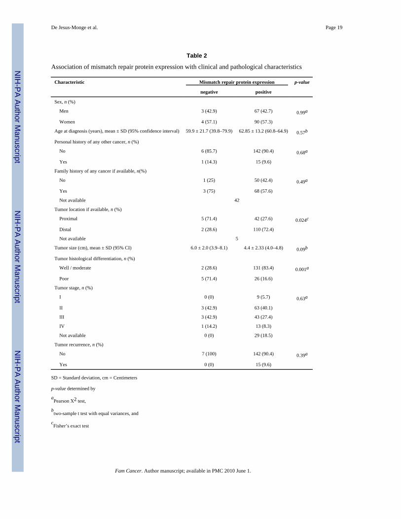
Clinical and pathological characteristicsTumor location and histological differentiation were the only statistically significant clinicaland pathological differences observed between colorectal tumors based on MMR proteinexpression (Table 2). Colorectal tumors with absent MMR protein expression were morecommonly located in the proximal colon compared to tumors that expressed both MMRproteins, which were more commonly located in the distal colon (Fisher’s exact p = 0.024).Colorectal tumors with MMR protein expression were more commonly well or moderatelydifferentiated compared to those with absence of protein expression, which more commonlywere poorly differentiated (Pearson X2 p = 0.001). There was no significant differenceaccording to MMR protein status with regards to sex, age at diagnosis of cancer, personalhistory of any other cancer, family history of any cancer, mean tumor size, tumor stage or tumorrecurrence.
DiscussionCRC is a deadly illness of great concern in Hispanic populations and the island of Puerto Ricois a great site to study this ethnicity. During 1998–2002, in both sexes, Puerto Ricans had CRCincidence and mortality rates similar to those for US Hispanics, but their rates were lower thanthose for non-Hispanic whites and non-Hispanic blacks. However, Puerto Rican men andwomen with ages 40–59 years had a greater risk of incidence and mortality than their USHispanic counterparts [74].
Evaluation of colon cancers for MMR defects may have significant therapeutic implications.In patients with stage II or III CRC, adjuvant 5-fluorouracil chemotherapy provides survivalimprovement in patients with MMR-competent tumors while the survival of patients withMMR-defective tumors does not improve with the adjuvant chemotherapy [75–77]. Also, lossof tumor MMR function may predict improved outcome in stage III colon cancer patientspostoperatively treated with a weekly bolus irinotecan, fluorouracil, and leucovorin regimenas compared with those receiving weekly bolus of fluorouracil and leucovorin [78].
HNPCC is characterized by young onset CRC [8,9,15], an increased risk for gynecologic,urinary tract, and gastrointestinal cancers [9,27,79–88], and most commonly occurs in men[89]. In HNPCC, a single mutation is inherited in the germline, and MSI occurs only afterinactivation of the other allele [90]. Most sporadic MSI-H cancers are caused by methylationand silencing of the MLH1 gene [91,92], most probably develop in women, and have theirorigin within serrated polyps [89]. Colorectal tumors with MSI are associated with locationproximal to the splenic flexure and poor histological differentiation [67,93,94].
MSI occurs in approximately 20% of CRC cases [23]. These tumors typically fail to expressMMR protein as seen on immunohistochemistry [28]. In the present study, only 4.3% of 164patients lacked protein MMR expression. Lack of protein expression as a surrogate for MSI isclinically important to patients. A patient with absence of MMR protein expression may be amember of a Lynch syndrome kindred, hence results may have implications for themselvesand other family members. Furthermore, individuals belonging to a HNPCC-kindred have ahigher risk of developing other non-colorectal cancers such as endometrial, ovarian, and gastric[9,27,79–88]. Of the seven tumors in our study that lacked MMR protein expression, six tumorslacked expression of MSH2, which usually indicates a germline mutation in the MSH2 gene,and one patient had absence of MLH1 with MSI-H.
The tumor with absence of MLH1 protein expression with MSI-H may have a either a germlinemutation or hypermethylation of the MLH1 promoter. Additionally, this patient had normalBRAF gene, which may be suggestive of HNPCC-associated MLH1 germline mutation.Virtually 100% of individuals with HNPCC do not carry the BRAFV600E mutation [14,15],
De Jesus-Monge et al. Page 6
Fam Cancer. Author manuscript; available in PMC 2010 June 1.
NIH
-PA Author Manuscript
NIH
-PA Author Manuscript
NIH
-PA Author Manuscript
whereas 68% of those without HNPCC do [14]. Moreover, BRAFV600E mutation is associatedwith sporadic CRC with MSI [30,95]. MLH1 promoter hypermethylation leads to geneinactivation [96] and, if present, may be secondary to CpG island methylation phenotype(CIMP), progressing via the MSI-H pathway. CpG islands are regions rich in cytosine-guanosine dinucleotide repeats at the 5′ region of approximately half of all human genes.Methylation of cytosine residues within CpG islands of promoters and proximal exons isassociated with loss of gene expression [97]. CIMP tumors have similarities to tumors withMSI-H, such as right-sided location and poor histological differentiation [98]. However,BRAF mutation is frequently seen in sporadic MSI-H CRC, associated with DNA methylationsecondary to CIMP-high (3–4 methylated in tumor markers are methylated) status [99]. Forthese reasons, it is unlikely that a MLH1 promoter hypermethylation secondary to CIMP mayhave occurred. However, none of the patients underwent germline genetic testing.
In our study of Hispanics from Puerto Rico, the frequency of MMR protein-negative tumorswas far lower than in other studies of unselected CRC patients in other countries or ethnicities,especially regarding MLH1-protein negative expression (Table 3) [37,47–58]. Moreover, thefrequency of MMR protein-negative tumors in our study was lower than in Spain, includingMLH1-protein negative expression [57,58].
As seen in other studies [4,15,37,56,67,93,94,100–102], colorectal tumors with lack of MMRprotein expression, suggesting MMR deficiency, were associated with a proximal location inthe colon and poor histological differentiation. However, opposing previous publications [4,17,19,37,55,72,79,101–104], we did not observe associations of MMR protein-negativetumors with gender, earlier age at diagnosis of cancer, positive personal and family history ofcancer, or earlier tumor stage, when compared to tumors with MMR protein-positiveexpression.
We acknowledge that our study has several limitations. First, the study was retrospective, whichlimited the information to that present in the medical record. This limited our ability toaccurately evaluate family history of cancer and other factors, like environmental exposure.Second, we were only able to include patients that had tumor blocks available for analysis.However, the percentage of patients excluded from pathological analysis was small and didnot statistically differ with regards to basic demographic characteristics from the patientsincluded in this investigation. As such, we believe our results are representative of the referralpopulation seen at our Center. Third, the sample size was relatively small. The sample sizemay have limited the power of the study to identify important differences according to MMRprotein expression status. This is also a pilot retrospective study and a larger prospective studyis undergone to verify our findings. Fourth, only MLH1 and MSH2 protein expression wereevaluated. However, over 90% [20,21] or most [14,15,22] of genetically characterized HNPCCcases are accounted for by germline mutations in any of them.
Additionally, a fifth limitation is that all patients were not subsequently evaluated for MSI toconfirm the suggestive findings from immunohistochemistry. However, for screening ofHNPCC among patients with CRC, immunohistochemistry is almost equally sensitive as MSI[33,34,39]. Indeed, MSI testing is not available in the majority of routine pathology servicelaboratories, and these rely on immunohistochemistry to detect loss of MMR protein expressionas a surrogate marker for the presence of MSI [105]. In consequence, immunohistochemistryis a valid tool to identify patients at risk for HNPCC and patients with sporadic microsatelliteinstable CRC [40] when evaluated by experienced pathologists [35] like the authors.Additionally, many consider immunohistochemistry and MSI testing as complementary. In asequential approach, immunohistochemistry is done first and, if informative, may result in costsavings. In the other hand, if immunohistochemistry is not informative, MSI testing can thenbe performed [15]. Nonetheless, this investigation provides insightful information about the
De Jesus-Monge et al. Page 7
Fam Cancer. Author manuscript; available in PMC 2010 June 1.
NIH
-PA Author Manuscript
NIH
-PA Author Manuscript
NIH
-PA Author Manuscript

frequency and clinical and pathological characteristics associated with MMR proteindeficiency in Hispanics from Puerto Rico, which may be ethnically related.
To our knowledge, the present study is the first one to evaluate the frequency of MMR protein-negative expression in unselected Hispanic CRC patients from Puerto Rico using the relativelyinexpensive [35] prescreening method of immunohistochemistry and its association withclinical and pathological characteristics.
In conclusion, this investigation of unselected Hispanic CRC patients from Puerto Ricorevealed a low frequency of MMR protein-negative tumors. Similar to other populations,Hispanic MMR protein-negative tumors were proximally located and exhibited poorhistological differentiation. The results may be relevant to the evaluation and management ofHispanic patients with CRC from Puerto Rico in immigrant as well as native populations. Therelatively low frequency of MMR protein-negative CRC in unselected Hispanic patients fromPuerto Rico may be a reflection of, or explain, the lower CRC incidence rate among USHispanics as compared to non-Hispanic whites and blacks. Our study is significant on sheddinglight on the scarce literature on MMR protein expression in CRC.in Hispanic populations.
AcknowledgmentsThis study was supported by the National Institutes of Health (NIH) National Center for Research Resources, grantsR25 RR 017589, P20 RR 011126-13, and G12 RR 003051-22; and NIH National Cancer Institute, grants K07 CA092445, K22 CA 115913-02, and U54 CA 096297. S.R.H. is the Frederick F. Becker Distinguished University Chairin Cancer Research from The University of Texas. We thank the Department of Pathology of the PRMC and theHOIGM for tissue access; the Department of Medical Records of the PRMC and the HOIGM and the HOIGM cancerregistry for data access; Mr. Nelson Santiago and Mrs. Janet E. Quiñones for laboratory technical support; Mr. AlexisA. Florian-Ayala and Mr. Alejandro Lopez-Araujo for data collection and laboratory technical support; Kerry Siegerfor MSI evaluation and BRAF gene sequencing; and Dr. Sara S. Strom, Dr. Francis M. Giardiello, Dr. Elena Martinez-Stoffel, Prof. Vivianna M. De Jesus-Monge, and the faculty and scholars of the University of Puerto Rico Master ofScience in Clinical Research Program for critical feedback. This work was done as fulfillment for the studies for agraduate degree from the University of Puerto Rico Medical Sciences Campus by Wilfredo E. De Jesus-Monge.
Abbreviations
CIMP CpG island methylation phenotype
CRC Colorectal cancer
HNPCC Hereditary nonpolyposis colorectal cancer
HOIGM Hospital Oncologico Isaac Gonzalez-Martinez
MMR Mismatch repair
MSI Microsatellite instability
MSI-H Microsatellite instability-high
PCR Polymerase chain reaction
PRMC Puerto Rico Medical Center
US United States
References1. Steward, BW.; Kleihues, P., editors. World cancer report. IARC Press; Lyon: 2003. Colorectal cancer.2. Jemal A, Siegel R, Ward E, et al. Cancer statistics. CA Cancer J Clin 2009;59:225–249. [PubMed:
19474385]
De Jesus-Monge et al. Page 8
Fam Cancer. Author manuscript; available in PMC 2010 June 1.
NIH
-PA Author Manuscript
NIH
-PA Author Manuscript
NIH
-PA Author Manuscript

3. Raut CP, Pawlik TM, Rodriguez-Bigas MA. Clinicopathologic features in colorectal cancer patientswith microsatellite instability. Mutat Res 2004;568:275–282. [PubMed: 15542114]
4. Soreide K, Janssen EA, Soiland H, et al. Microsatellite instability in colorectal cancer. Br J Surg2006;93:395–406. [PubMed: 16555243]
5. Wu J, Gu L, Wang H, et al. Mismatch repair processing of carcinogen-DNA adducts triggers apoptosis.Mol Cell Biol 1999;19:8292–8301. [PubMed: 10567554]
6. Hickman MJ, Samson LD. Role of DNA mismatch repair and p53 in signaling induction of apoptosisby alkylating agents. Proc Natl Acad Sci U S A 1999;96:10764–10769. [PubMed: 10485900]
7. Koessler T, Azzato EM, Perkins B, et al. Common germline variation in mismatch repair genes andsurvival after a diagnosis of colorectal cancer. Int J Cancer 2009;124:1887–1891. [PubMed: 19115210]
8. Wijnen JT, Brohet RM, van Eijk R, et al. Chromosome 8q23.3 and 11q23.1 variants modify colorectalcancer risk in Lynch syndrome. Gastroenterology 2009;136:131–137. [PubMed: 19010329]
9. Lynch HT, Smyrk T. Hereditary nonpolyposis colorectal cancer (Lynch syndrome). An updated review.Cancer 1996;78:1149–1167. [PubMed: 8826936]
10. Boland CR, Koi M, Chang DK, et al. The biochemical basis of microsatellite instability and abnormalimmunohistochemistry and clinical behavior in Lynch syndrome: from bench to bedside. Fam Cancer2008;7:41–52. [PubMed: 17636426]
11. Raevaara TE, Korhonen MK, Lohi H, et al. Functional significance and clinical phenotype ofnontruncating mismatch repair variants of MLH1. Gastroenterology 2005;129:537–549. [PubMed:16083711]
12. Hampel H, Stephens JA, Pukkala E, et al. Cancer risk in hereditary nonpolyposis colorectal cancersyndrome: later age of onset. Gastroenterology 2005;129:415–421. [PubMed: 16083698]
13. Shin YK, Heo SC, Shin JH, et al. Germline mutations in MLH1, MSH2 and MSH6 in Koreanhereditary non-polyposis colorectal cancer families. Hum Mutat 2004;24:351. [PubMed: 15365995]
14. Recommendations from the EGAPP Working Group. genetic testing strategies in newly diagnosedindividuals with colorectal cancer aimed at reducing morbidity and mortality from Lynch syndromein relatives. Genet Med 2009;11:35–41. [PubMed: 19125126]
15. Lynch HT, Boland CR, Rodriguez-Bigas MA, et al. Who should be sent for genetic testing inhereditary colorectal cancer syndromes? J Clin Oncol 2007;25:3534–3542. [PubMed: 17687158]
16. Lynch HT, Boland CR, Gong G, et al. Phenotypic and genotypic heterogeneity in the Lynch syndrome:diagnostic, surveillance and management implications. Eur J Hum Genet 2006;14:390–402.[PubMed: 16479259]
17. Ricciardiello L, Boland CR. Lynch syndrome (hereditary non-polyposis colorectal cancer): currentconcepts and approaches to management. Curr Gastroenterol Rep 2005;7:412–420. [PubMed:16168241]
18. De Jong AE, Morreau H, Van Puijenbroek M, et al. The role of mismatch repair gene defects in thedevelopment of adenomas in patients with HNPCC. Gastroenterology 2004;126:42–48. [PubMed:14699485]
19. Soreide K. Molecular testing for microsatellite instability and DNA mismatch repair defects inhereditary and sporadic colorectal cancers--ready for prime time? Tumour Biol 2007;28:290–300.[PubMed: 17962726]
20. Chung DC, Rustgi AK. The hereditary nonpolyposis colorectal cancer syndrome: genetics and clinicalimplications. Ann Intern Med 2003;138:560–570. [PubMed: 12667026]
21. Rouleau E, Lefol C, Bourdon V, et al. Quantitative PCR high-resolution melting (qPCR-HRM) curveanalysis, a new approach to simultaneously screen point mutations and large rearrangements:application to MLH1 germline mutations in Lynch syndrome. Hum Mutat 2009;30:867–875.[PubMed: 19224586]
22. Lindor NM, Burgart LJ, Leontovich O, et al. Immunohistochemistry versus microsatellite instabilitytesting in phenotyping colorectal tumors. J Clin Oncol 2002;20:1043–1048. [PubMed: 11844828]
23. Hampel H, Frankel WL, Martin E, et al. Screening for the Lynch syndrome (hereditary nonpolyposiscolorectal cancer). N Engl J Med 2005;352:1851–1860. [PubMed: 15872200]
24. Pawlik TM, Raut CP, Rodriguez-Bigas MA. Colorectal carcinogenesis: MSI-H versus MSI-L. DisMarkers 2004;20:199–206. [PubMed: 15528785]
De Jesus-Monge et al. Page 9
Fam Cancer. Author manuscript; available in PMC 2010 June 1.
NIH
-PA Author Manuscript
NIH
-PA Author Manuscript
NIH
-PA Author Manuscript
25. Cruz-Correa M, Giardiello FM. Diagnosis and management of hereditary colon cancer. GastroenterolClin North Am 2002;31:537–549. x. [PubMed: 12134617]
26. Bleiker EM, Menko FH, Taal BG, et al. Screening behavior of individuals at high risk for colorectalcancer. Gastroenterology 2005;128:280–287. [PubMed: 15685539]
27. Guillem JG, Wood WC, Moley JF, et al. ASCO/SSO review of current role of risk-reducing surgeryin common hereditary cancer syndromes. J Clin Oncol 2006;24:4642–4660. [PubMed: 17008706]
28. Balmana J, Balaguer F, Castellvi-Bell S, et al. Comparison of predictive models, clinical criteria andmolecular tumour screening for the identification of patients with Lynch syndrome in a population-based cohort of colorectal cancer patients. J Med Genet 2008;45:557–563. [PubMed: 18603628]
29. Sidelnikov E, Bostick RM, Flanders WD, et al. MutL-Homolog 1 expression and risk of incident,sporadic colorectal adenoma: search for prospective biomarkers of risk for colorectal cancer. CancerEpidemiol Biomarkers Prev 2009;18:1599–1609. [PubMed: 19423536]
30. De Jesus-Monge WE, Gonzalez-Keelan C, Cruz-Correa M. Serrated adenomas. CurrentGastroenterology Reports 2009;11:420–427. [PubMed: 19765371]
31. Vasen, HF.; Moslein, G.; Alonso, A., et al. Recommendations to improve identification of hereditaryand familial colorectal cancer in Europe. Fam Cancer. 2009. Available via INTERNET.http://www.springerlink.com/content/u165m5k773817528/Cited 28 Oct 2009
32. Ponz de Leon M, Bertario L, Genuardi M, et al. Identification and classification of hereditarynonpolyposis colorectal cancer (Lynch syndrome): adapting old concepts to recent advancements.Report from the Italian Association for the Study of Hereditary Colorectal Tumors Consensus Group.Dis Colon Rectum 2007;50:2126–2134. [PubMed: 17899274]
33. Lynch HT, Fusaro RM, Lynch PM. Sebaceous skin lesions as clues to hereditary non-polyposiscolorectal cancer. J Invest Dermatol 2006;126:2158–2159. [PubMed: 16983324]
34. Shia J. Immunohistochemistry versus microsatellite instability testing for screening colorectal cancerpatients at risk for hereditary nonpolyposis colorectal cancer syndrome. Part I. The utility ofimmunohistochemistry. J Mol Diagn 2008;10:293–300. [PubMed: 18556767]
35. de La Chapelle A. Microsatellite instability phenotype of tumors: genotyping orimmunohistochemistry? The jury is still out. J Clin Oncol 2002;20:897–899. [PubMed: 11844809]
36. Morales-Burgos A, Sanchez JL, Figueroa LD, et al. MSH-2 and MLH-1 protein expression in MuirTorre syndrome-related and sporadic sebaceous neoplasms. P R Health Sci J 2008;27:322–327.[PubMed: 19069357]
37. Molaei, M.; Mansoori, BK.; Ghiasi, S., et al. Colorectal cancer in Iran: immunohistochemical profilesof four mismatch repair proteins. Int J Colorectal Dis. 2009. Available via INTERNET.http://www.springerlink.com/content/932045231028jj4k/Cited 28 Oct 2009
38. Evans DG, Walsh S, Hill J, et al. Strategies for identifying hereditary nonpolyposis colon cancer.Semin Oncol 2007;34:411–417. [PubMed: 17920896]
39. Hampel H, Frankel WL, Martin E, et al. Feasibility of screening for Lynch syndrome among patientswith colorectal cancer. J Clin Oncol 2008;26:5783–5788. [PubMed: 18809606]
40. Overbeek LI, Ligtenberg MJ, Willems RW, et al. Interpretation of immunohistochemistry formismatch repair proteins is only reliable in a specialized setting. Am J Surg Pathol 2008;32:1246–1251. [PubMed: 18677806]
41. Recommendations from the Interagency Committee for the Review of the Racial and Ethnic Standardsto the Office of Management and Budget concerning changes to the standards for the classificationof federal data on race and ethnicity. US Office of Management and Budget. 1997. Available viaINTERNET. http://www.census.gov/population/www/socdemo/race/Directive_15.htmlCited 17Oct 2009
42. International Data Base. US Census Bureau, Population Division; Washington. : 2009.http://www.census.gov/ipc/www/idb/region.phpCited 18 Oct 2009
43. US American Community Survey demographic and housing estimates: 2008. US Census Bureau,American Community Survey; Washington: 2008.http://factfinder.census.gov/servlet/ADPTable?_bm=y&-geo_id=01000US&-qr_name=ACS_2008_1YR_G00_DP5&-ds_name=ACS_2008_1YR_G00_&-_lang=en&-_caller=geoselect&-state=adp&-format=Cited 16 Oct 2009
De Jesus-Monge et al. Page 10
Fam Cancer. Author manuscript; available in PMC 2010 June 1.
NIH
-PA Author Manuscript
NIH
-PA Author Manuscript
NIH
-PA Author Manuscript
44. Puerto Rico American Community Survey demographic and housing estimates: 2008. US CensusBureau, American Community Survey; Washington: 2008.http://factfinder.census.gov/servlet/ADPTable?_bm=y&-state=adp&-context=adp&-qr_name=ACS_2008_1YR_G00_DP5&-ds_name=ACS_2008_1YR_G00_&-tree_id=308&-_caller=geoselect&-geo_id=04000US72&-format=&-_lang=enCited 17 Oct 2009
45. Curado, MP.; Edwards, B.; Shin, HR., et al. Cancer incidence in five continents. International Agencyfor Research on Cancer; Lyon: 2007. http://www-dep.iarc.fr/Cited 20 Oct 2009
46. Figueroa-Valles NR. Boletin del Registro de Cancer. Puerto Rico Cancer Central Registry. 2008.Available via INTERNET.http://www.salud.gov.pr/RCancer/Documents/Vol.%201%20No.%202.pdfCited 17 Oct 2009
47. Brim H, Mokarram P, Naghibalhossaini F, et al. Impact of BRAF, MLH1 on the incidence ofmicrosatellite instability high colorectal cancer in populations based study. Mol Cancer 2008;7:68.[PubMed: 18718023]
48. Ashktorab H, Smoot DT, Farzanmehr H, et al. Clinicopathological features and microsatelliteinstability (MSI) in colorectal cancers from African Americans. Int J Cancer 2005;116:914–919.[PubMed: 15856472]
49. Pandey V, Prabhu JS, Payal K, et al. Assessment of microsatellite instability in colorectal carcinomaat an Indian center. Int J Colorectal Dis 2007;22:777–782. [PubMed: 17160686]
50. Ashktorab H, Brim H, Al-Riyami M, et al. Sporadic colon cancer: mismatch repairimmunohistochemistry and microsatellite instability in Omani subjects. Dig Dis Sci 2008;53:2723–2731. [PubMed: 18299982]
51. Erdamar S, Ucaryilmaz E, Demir G, et al. Importance of MutL homologue MLH1 and MutShomologue MSH2 expression in Turkish patients with sporadic colorectal cancer. World JGastroenterol 2007;13:4437–4444. [PubMed: 17724798]
52. Jin HY, Liu X, Lin VK, et al. Detection of mismatch repair gene germline mutation carrier amongChinese population with colorectal cancer. BMC Cancer 2008;8:44. [PubMed: 18257912]
53. Jensen LH, Lindebjerg J, Byriel L, et al. Strategy in clinical practice for classification of unselectedcolorectal tumours based on mismatch repair deficiency. Colorectal Dis 2008;10:490–497. [PubMed:17868408]
54. Boardman LA, Lanier AP, French AJ, et al. Frequency of defective DNA mismatch repair in colorectalcancer among the Alaska Native people. Cancer Epidemiol Biomarkers Prev 2007;16:2344–2350.[PubMed: 18006922]
55. Wright CL, Stewart ID. Histopathology and mismatch repair status of 458 consecutive colorectalcarcinomas. Am J Surg Pathol 2003;27:1393–1406. [PubMed: 14576472]
56. Coggins RP, Cawkwell L, Bell SM, et al. Association between family history and mismatch repairin colorectal cancer. Gut 2005;54:636–642. [PubMed: 15831908]
57. Xicola RM, Llor X, Pons E, et al. Performance of different microsatellite marker panels for detectionof mismatch repair-deficient colorectal tumors. J Natl Cancer Inst 2007;99:244–252. [PubMed:17284719]
58. Piñol V, Castells A, Andreu M, et al. Accuracy of revised Bethesda guidelines, microsatelliteinstability, and immunohistochemistry for the identification of patients with hereditary nonpolyposiscolorectal cancer. JAMA 2005;293:1986–1994. [PubMed: 15855432]
59. Terdiman JP, Gum JR Jr, Conrad PG, et al. Efficient detection of hereditary nonpolyposis colorectalcancer gene carriers by screening for tumor microsatellite instability before germline genetic testing.Gastroenterology 2001;120:21–30. [PubMed: 11208710]
60. Christensen M, Katballe N, Wikman F, et al. Antibody-based screening for hereditary nonpolyposiscolorectal carcinoma compared with microsatellite analysis and sequencing. Cancer 2002;95:2422–2430. [PubMed: 12436451]
61. Wahlberg SS, Schmeits J, Thomas G, et al. Evaluation of microsatellite instability andimmunohistochemistry for the prediction of germ-line MSH2 and MLH1 mutations in hereditarynonpolyposis colon cancer families. Cancer Res 2002;62:3485–3492. [PubMed: 12067992]
62. Park JG, Vasen HF, Park YJ, et al. Suspected HNPCC and Amsterdam criteria II: evaluation ofmutation detection rate, an international collaborative study. Int J Colorectal Dis 2002;17:109–114.[PubMed: 12014418]
De Jesus-Monge et al. Page 11
Fam Cancer. Author manuscript; available in PMC 2010 June 1.
NIH
-PA Author Manuscript
NIH
-PA Author Manuscript
NIH
-PA Author Manuscript
63. Hitchins M, Williams R, Cheong K, et al. MLH1 germline epimutations as a factor in hereditarynonpolyposis colorectal cancer. Gastroenterology 2005;129:1392–1399. [PubMed: 16285940]
64. Bermejo JL, Eng C, Hemminki K. Cancer characteristics in Swedish families fulfilling criteria forhereditary nonpolyposis colorectal cancer. Gastroenterology 2005;129:1889–1899. [PubMed:16344057]
65. Woerner SM, Kloor M, Mueller A, et al. Microsatellite instability of selective target genes in HNPCC-associated colon adenomas. Oncogene 2005;24:2525–2535. [PubMed: 15735733]
66. Plaschke J, Engel C, Kruger S, et al. Lower incidence of colorectal cancer and later age of diseaseonset in 27 families with pathogenic MSH6 germline mutations compared with families with MLH1or MSH2 mutations: the German Hereditary Nonpolyposis Colorectal Cancer Consortium. J ClinOncol 2004;22:4486–4494. [PubMed: 15483016]
67. Lim SB, Jeong SY, Lee MR, et al. Prognostic significance of microsatellite instability in sporadiccolorectal cancer. Int J Colorectal Dis 2004;19:533–537. [PubMed: 15175889]
68. Cruz-Correa M, Giardiello FM. Familial adenomatous polyposis. Gastrointestinal Endoscopy2003;58:885–894. [PubMed: 14652558]
69. Half ED, Bercovich D, Rozen P. Familial adenomatous polyposis. Orphanet J Rare Dis 2009;4:22.[PubMed: 19822006]
70. Rozen P, Macrae F. Familial adenomatous polyposis: The practical applications of clinical andmolecular screening. Fam Cancer 2006;5:227–235. [PubMed: 16998668]
71. Puerto Rico general demographic characteristics: 2005. US Census Bureau, American CommunitySurvey; Washington: 2005.http://factfinder.census.gov/servlet/ADPTable?_bm=y&-state=adp&-context=adp&-qr_name=ACS_2005_EST_G00_DP1&-ds_name=ACS_2005_EST_G00_&-tree_id=305&-redoLog=true&-all_geo_types=N&-_caller=geoselect&-geo_id=04000US72&-format=&-_lang=enCited 16 Oct 2009
72. Umar A, Boland CR, Terdiman JP, et al. Revised Bethesda Guidelines for hereditary nonpolyposiscolorectal cancer (Lynch syndrome) and microsatellite instability. J Natl Cancer Inst 2004;96:261–268. [PubMed: 14970275]
73. Fleming, ID.; Cooper, JS.; Henson, DE., et al., editors. AJCC cancer staging manual. 5. Lippincott-Raven; Philadelphia: 1997.
74. Soto-Salgado M, Suarez E, Calo W, et al. Incidence and mortality rates for colorectal cancer in PuertoRico and among Hispanics, non-Hispanic whites, and non-Hispanic blacks in the United States,1998–2002. Cancer 2009;115:3016–3023. [PubMed: 19402167]
75. Jover R, Zapater P, Castells A, et al. The efficacy of adjuvant chemotherapy with 5-fluorouracil incolorectal cancer depends on the mismatch repair status. Eur J Cancer 2009;45:365–373. [PubMed:18722765]
76. Carethers JM, Smith EJ, Behling CA, et al. Use of 5-fluorouracil and survival in patients withmicrosatellite-unstable colorectal cancer. Gastroenterology 2004;126:394–401. [PubMed:14762775]
77. Ribic CM, Sargent DJ, Moore MJ, et al. Tumor microsatellite-instability status as a predictor of benefitfrom fluorouracil-based adjuvant chemotherapy for colon cancer. N Engl J Med 2003;349:247–257.[PubMed: 12867608]
78. Bertagnolli MM, Niedzwiecki D, Compton CC, et al. Microsatellite instability predicts improvedresponse to adjuvant therapy with irinotecan, fluorouracil, and leucovorin in stage III colon cancer:Cancer and Leukemia Group B Protocol 89803. J Clin Oncol 2009;27:1814–1821. [PubMed:19273709]
79. Grover S, Syngal S. Genetic testing in gastroenterology: Lynch syndrome. Best Pract Res ClinGastroenterol 2009;23:185–196. [PubMed: 19414145]
80. Beiner ME, Rosen B, Fyles A, et al. Endometrial cancer risk is associated with variants of the mismatchrepair genes MLH1 and MSH2. Cancer Epidemiol Biomarkers Prev 2006;15:1636–1640. [PubMed:16985024]
81. Yoon SN, Ku JL, Shin YK, et al. Hereditary nonpolyposis colorectal cancer in endometrial cancerpatients. Int J Cancer 2008;122:1077–1081. [PubMed: 17973265]
De Jesus-Monge et al. Page 12
Fam Cancer. Author manuscript; available in PMC 2010 June 1.
NIH
-PA Author Manuscript
NIH
-PA Author Manuscript
NIH
-PA Author Manuscript
82. Park JG, Kim DW, Hong CW, et al. Germ line mutations of mismatch repair genes in hereditarynonpolyposis colorectal cancer patients with small bowel cancer: International Society forGastrointestinal Hereditary Tumours Collaborative Study. Clin Cancer Res 2006;12:3389–3393.[PubMed: 16740762]
83. Scaife CL, Rodriguez-Bigas MA. Lynch syndrome: implications for the surgeon. Clin ColorectalCancer 2003;3:92–98. [PubMed: 12952564]
84. Watson P, Riley B. The tumor spectrum in the Lynch syndrome. Fam Cancer 2005;4:245–248.[PubMed: 16136385]
85. Watson P, Lynch HT. Cancer risk in mismatch repair gene mutation carriers. Fam Cancer 2001;1:57–60. [PubMed: 14574017]
86. Manchanda R, Menon U, Michaelson-Cohen R, et al. Hereditary non-polyposis colorectal cancer orLynch syndrome: the gynaecological perspective. Curr Opin Obstet Gynecol 2009;21:31–38.[PubMed: 19125001]
87. Brown GJ, St John DJ, Macrae FA, et al. Cancer risk in young women at risk of hereditarynonpolyposis colorectal cancer: implications for gynecologic surveillance. Gynecol Oncol2001;80:346–349. [PubMed: 11263929]
88. Schmeler KM, Lu KH. Gynecologic cancers associated with Lynch syndrome/HNPCC. Clin TranslOncol 2008;10:313–317. [PubMed: 18558577]
89. Dionigi G, Bianchi V, Villa F, et al. Differences between familial and sporadic forms of colorectalcancer with DNA microsatellite instability. Surg Oncol 2007;16:S37–S42. [PubMed: 18023569]
90. Aaltonen LA, Peltomaki P, Mecklin JP, et al. Replication errors in benign and malignant tumors fromhereditary nonpolyposis colorectal cancer patients. Cancer Res 1994;54:1645–1648. [PubMed:8137274]
91. Herman JG, Umar A, Polyak K, et al. Incidence and functional consequences of hMLH1 promoterhypermethylation in colorectal carcinoma. Proc Natl Acad Sci U S A 1998;95:6870–6875. [PubMed:9618505]
92. Kuismanen SA, Holmberg MT, Salovaara R, et al. Genetic and epigenetic modification of MLH1accounts for a major share of microsatellite-unstable colorectal cancers. Am J Pathol 2000;156:1773–1779. [PubMed: 10793088]
93. Gryfe R, Gallinger S. Microsatellite instability, mismatch repair deficiency, and colorectal cancer.Surgery 2001;130:17–20. [PubMed: 11436007]
94. Lothe RA, Peltomaki P, Meling GI, et al. Genomic instability in colorectal cancer: relationship toclinicopathological variables and family history. Cancer Res 1993;53:5849–5852. [PubMed:8261392]
95. O’Brien MJ. Hyperplastic and serrated polyps of the colorectum. Gastroenterol Clin North Am2007;36:947–968. viii. [PubMed: 17996799]
96. Esteller M, Fraga MF, Guo M, et al. DNA methylation patterns in hereditary human cancers mimicsporadic tumorigenesis. Hum Mol Genet 2001;10:3001–3007. [PubMed: 11751682]
97. Chirieac LR, Chen L, Catalano PJ, et al. Phenotype of microsatellite-stable colorectal carcinomaswith CpG island methylation. Am J Surg Pathol 2005;29:429–436. [PubMed: 15767794]
98. van Rijnsoever M, Grieu F, Elsaleh H, et al. Characterisation of colorectal cancers showinghypermethylation at multiple CpG islands. Gut 2002;51:797–802. [PubMed: 12427779]
99. Kambara T, Simms LA, Whitehall VL, et al. BRAF mutation is associated with DNA methylation inserrated polyps and cancers of the colorectum. Gut 2004;53:1137–1144. [PubMed: 15247181]
100. Al-Kuraya KS, Bavi PP, Ezzat AA, et al. Colorectal carcinoma from Saudi Arabia. Analysis ofMLH-1, MSH-2 and p53 genes by immunohistochemistry and tissue microarray analysis. SaudiMed J 2006;27:323–328. [PubMed: 16532091]
101. Samowitz WS, Curtin K, Ma KN, et al. Microsatellite instability in sporadic colon cancer isassociated with an improved prognosis at the population level. Cancer Epidemiol Biomarkers Prev2001;10:917–923. [PubMed: 11535541]
102. Chen JR, Chiang JM, Changchien CR, et al. Mismatch repair protein expression in Amsterdam IIcriteria-positive patients in Taiwan. Br J Surg 2008;95:102–110. [PubMed: 18064717]
103. Gryfe R, Kim H, Hsieh ET, et al. Tumor microsatellite instability and clinical outcome in youngpatients with colorectal cancer. N Engl J Med 2000;342:69–77. [PubMed: 10631274]
De Jesus-Monge et al. Page 13
Fam Cancer. Author manuscript; available in PMC 2010 June 1.
NIH
-PA Author Manuscript
NIH
-PA Author Manuscript
NIH
-PA Author Manuscript
104. Mecklin JP, Jarvinen HJ. Clinical features of colorectal carcinoma in cancer family syndrome. DisColon Rectum 1986;29:160–164. [PubMed: 3943429]
105. Watson N, Grieu F, Morris M, et al. Heterogeneous staining for mismatch repair proteins duringpopulation-based prescreening for hereditary nonpolyposis colorectal cancer. J Mol Diagn2007;9:472–478. [PubMed: 17652638]
De Jesus-Monge et al. Page 14
Fam Cancer. Author manuscript; available in PMC 2010 June 1.
NIH
-PA Author Manuscript
NIH
-PA Author Manuscript
NIH
-PA Author Manuscript
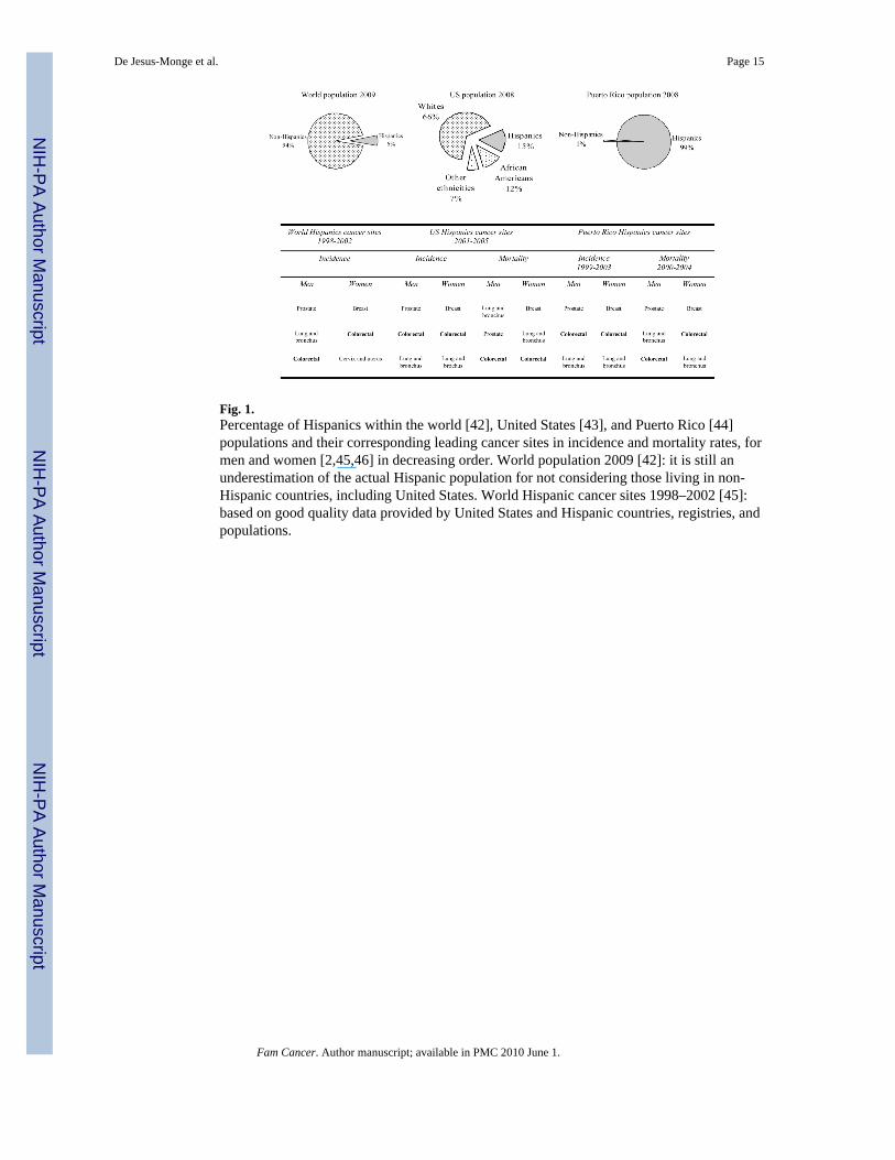
Fig. 1.Percentage of Hispanics within the world [42], United States [43], and Puerto Rico [44]populations and their corresponding leading cancer sites in incidence and mortality rates, formen and women [2,45,46] in decreasing order. World population 2009 [42]: it is still anunderestimation of the actual Hispanic population for not considering those living in non-Hispanic countries, including United States. World Hispanic cancer sites 1998–2002 [45]:based on good quality data provided by United States and Hispanic countries, registries, andpopulations.
De Jesus-Monge et al. Page 15
Fam Cancer. Author manuscript; available in PMC 2010 June 1.
NIH
-PA Author Manuscript
NIH
-PA Author Manuscript
NIH
-PA Author Manuscript
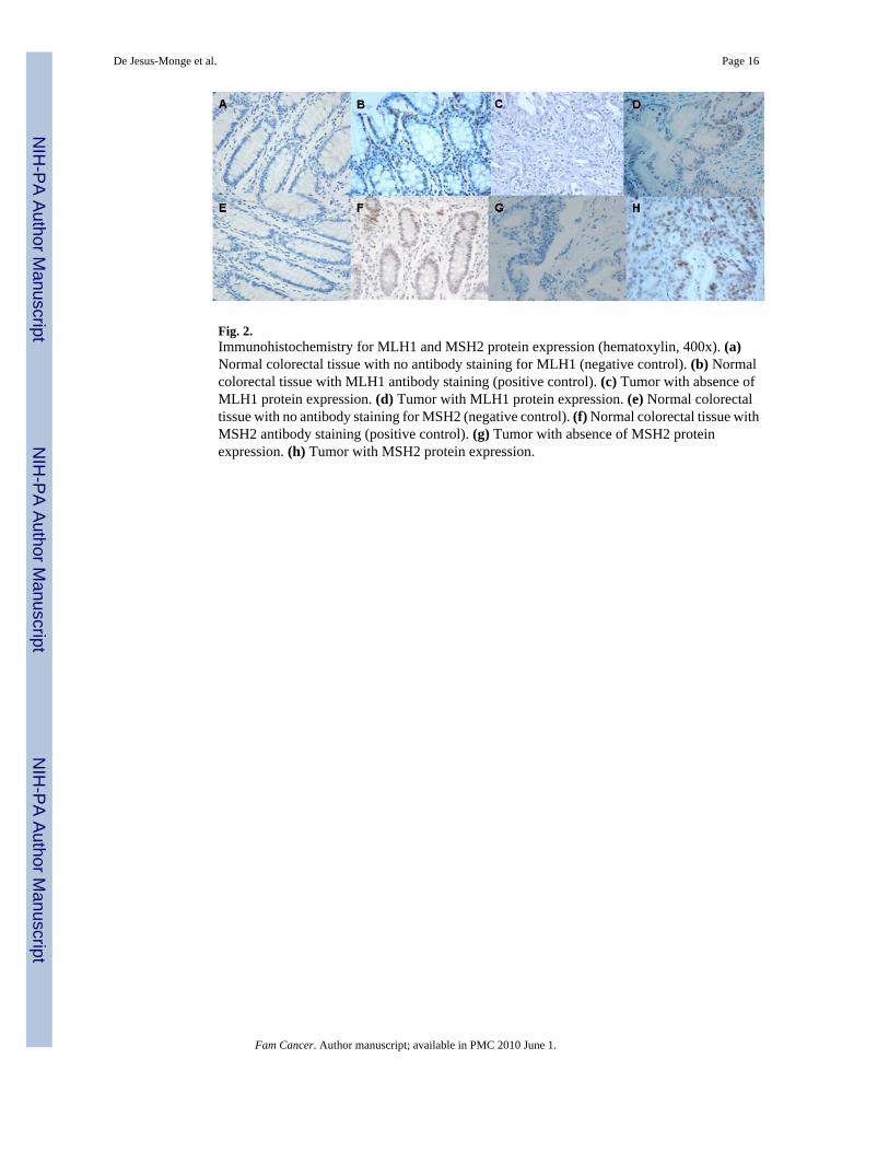
Fig. 2.Immunohistochemistry for MLH1 and MSH2 protein expression (hematoxylin, 400x). (a)Normal colorectal tissue with no antibody staining for MLH1 (negative control). (b) Normalcolorectal tissue with MLH1 antibody staining (positive control). (c) Tumor with absence ofMLH1 protein expression. (d) Tumor with MLH1 protein expression. (e) Normal colorectaltissue with no antibody staining for MSH2 (negative control). (f) Normal colorectal tissue withMSH2 antibody staining (positive control). (g) Tumor with absence of MSH2 proteinexpression. (h) Tumor with MSH2 protein expression.
De Jesus-Monge et al. Page 16
Fam Cancer. Author manuscript; available in PMC 2010 June 1.
NIH
-PA Author Manuscript
NIH
-PA Author Manuscript
NIH
-PA Author Manuscript

NIH
-PA Author Manuscript
NIH
-PA Author Manuscript
NIH
-PA Author Manuscript
De Jesus-Monge et al. Page 17
Table 1
Clinical and pathological characteristics of study population
Characteristic
Patients, n 164
Sex, n (%)
Men 70 (42.7)
Women 94 (57.3)
Age at diagnosis (years), mean ± SD (range) 62.72 ± 13.54 (23-9)
Follow up time (months), mean ± SD (range) 39.2 ± 22 (0.67–154.1)
Alive on follow up, n (%)
No 43 (26.2)
Yes 121 (73.8)
Personal history of any other cancer, n (%)
No 148 (90.2)
Yes 16 (9.8)
Family history of any cancer if available, n (%)
No 51 (41.8)
Yes 71 (58.2)
Tumor location if available, n (%)
Proximal 47 (29.6)
Distal 112 (70.4)
Tumor size if available (cm), mean ± SD (range) 4.42 ± 2.34 (1–13.2)
Tumor histological differentiation, n (%)
Well / moderate 133 (81.1)
Poor 31 (18.9)
Tumor stage, n (%)
I 9 (5.5)
II 66 (40.2)
III 46 (28.1)
IV 14 (8.5)
Not available or not applies 29 (17.7)
Surgery received, n (%)
No 13 (7.9)
Yes 151 (92.1)
Chemotherapy received, n (%)
No 72 (43.9)
Yes 92 (56.1)
Radiotherapy received, n (%)
No 116 (70.7)
Yes 48 (29.3)
Tumor recurrence, n (%)
No 149 (90.9)
Fam Cancer. Author manuscript; available in PMC 2010 June 1.

NIH
-PA Author Manuscript
NIH
-PA Author Manuscript
NIH
-PA Author Manuscript
De Jesus-Monge et al. Page 18
Characteristic
Yes 15 (9.2)
SD = Standard deviation, cm = Centimeters
Fam Cancer. Author manuscript; available in PMC 2010 June 1.
NIH
-PA Author Manuscript
NIH
-PA Author Manuscript
NIH
-PA Author Manuscript
De Jesus-Monge et al. Page 19
Table 2
Association of mismatch repair protein expression with clinical and pathological characteristics
Characteristic Mismatch repair protein expression p-value
negative positive
Sex, n (%)
Men 3 (42.9) 67 (42.7) 0.99a
Women 4 (57.1) 90 (57.3)
Age at diagnosis (years), mean ± SD (95% confidence interval) 59.9 ± 21.7 (39.8–79.9) 62.85 ± 13.2 (60.8–64.9) 0.57b
Personal history of any other cancer, n (%)
No 6 (85.7) 142 (90.4) 0.68a
Yes 1 (14.3) 15 (9.6)
Family history of any cancer if available, n(%)
No 1 (25) 50 (42.4) 0.49a
Yes 3 (75) 68 (57.6)
Not available 42
Tumor location if available, n (%)
Proximal 5 (71.4) 42 (27.6) 0.024c
Distal 2 (28.6) 110 (72.4)
Not available 5
Tumor size (cm), mean ± SD (95% CI) 6.0 ± 2.0 (3.9–8.1) 4.4 ± 2.33 (4.0–4.8) 0.09b
Tumor histological differentiation, n (%)
Well / moderate 2 (28.6) 131 (83.4) 0.001a
Poor 5 (71.4) 26 (16.6)
Tumor stage, n (%)
I 0 (0) 9 (5.7) 0.63a
II 3 (42.9) 63 (40.1)
III 3 (42.9) 43 (27.4)
IV 1 (14.2) 13 (8.3)
Not available 0 (0) 29 (18.5)
Tumor recurrence, n (%)
No 7 (100) 142 (90.4) 0.39a
Yes 0 (0) 15 (9.6)
SD = Standard deviation, cm = Centimeters
p-value determined by
aPearson Χ2 test,
btwo-sample t test with equal variances, and
cFisher’s exact test
Fam Cancer. Author manuscript; available in PMC 2010 June 1.

NIH
-PA Author Manuscript
NIH
-PA Author Manuscript
NIH
-PA Author Manuscript
De Jesus-Monge et al. Page 20
Table 3
Comparison of our study with others in other countries or ethnicities for mismatch repair protein expression inunselected colorectal cancer patients
Authors Country or ethnic group Evaluated patients, nMLH1-protein negative
expression, n (%)MSH2-protein negative
expression, n (%)
Lindor et al [22]a Minnesota, US 255 48 (18.8) 3 (1.2)
De Jesus-Monge et al Hispanics from Puerto Rico 164 1 (0.6) 6 (3.7)
Brim et al [47] Iran 25 10 (40) 1 (4)
Ashktorab et al [48] African b 16 of 34 (47.1)c 12 of 31 (47.1)c
Americans 3 of 34 (8.8)d 1 of 31 (8.8)d
Pandey et al [49] India 46 7 (15.2) 1 (2.2)
Ashktorab et al [50] Oman 49 5 (10.2)e 3 (6.1)e
Brim et al [47] Oman 61 19 (31.1) 5 (8.2)
Erdamar et al [51] Turkey 74 29 (39.2) 7 (9.5)
Brim et al [47] African Americans b 26 of 76 (34.2) 8 of 74 (10.8)
Jin et al [52] China 146 17 (11.6) 9 (6.2)
Jensen et al [53] Denmark 262 37 (14.1) 3 (1.1)
Boardman et al [54] Alaska Natives 329 42 (12.8) 3 (0.9)
Molaei et al [37] Iran 343 19 (5.5) 24 (7)
Wright et al [55] New Zealand 458 80 (17.5) 9 (2)
Coggins et al [56] England 732 52 (7.1) 5 (0.7)
Xicola et al [57] Spain 1058 59 (5.6) 22 (2.1)
Piñol et al [58] Spain 1222 60 (4.9) 21 (1.7)
aRepresentative sample for United States
bSee columns to the right to see the number of evaluated patients for each protein
cPartially negative expression
dCompletely negative expression
ePartially or completely negative expression
Fam Cancer. Author manuscript; available in PMC 2010 June 1.