The replication origin of a repABC plasmid
14
RESEARCH ARTICLE Open Access The replication origin of a repABC plasmid Ramón Cervantes-Rivera, Francisco Pedraza-López, Gabriela Pérez-Segura and Miguel A Cevallos * Abstract Background: repABC operons are present on large, low copy-number plasmids and on some secondary chromosomes in at least 19 a-proteobacterial genera, and are responsible for the replication and segregation properties of these replicons. These operons consist, with some variations, of three genes: repA, repB, and repC. RepA and RepB are involved in plasmid partitioning and in the negative regulation of their own transcription, and RepC is the limiting factor for replication. An antisense RNA encoded between the repB-repC genes modulates repC expression. Results: To identify the minimal region of the Rhizobium etli p42d plasmid that is capable of autonomous replication, we amplified different regions of the repABC operon using PCR and cloned the regions into a suicide vector. The resulting vectors were then introduced into R. etli strains that did or did not contain p42d. The minimal replicon consisted of a repC open reading frame under the control of a constitutive promoter with a Shine- Dalgarno sequence that we designed. A sequence analysis of repC revealed the presence of a large A+T-rich region but no iterons or DnaA boxes. Silent mutations that modified the A+T content of this region eliminated the replication capability of the plasmid. The minimal replicon could not be introduced into R. etli strain containing p42d, but similar constructs that carried repC from Sinorhizobium meliloti pSymA or the linear chromosome of Agrobacterium tumefaciens replicated in the presence or absence of p42d, indicating that RepC is an incompatibility factor. A hybrid gene construct expressing a RepC protein with the first 362 amino acid residues from p42d RepC and the last 39 amino acid residues of RepC from SymA was able to replicate in the presence of p42d. Conclusions: RepC is the only element encoded in the repABC operon of the R. etli p42d plasmid that is necessary and sufficient for plasmid replication and is probably the initiator protein. The oriV of this plasmid resides within the repC gene and is located close to or inside of a large A+T region. RepC can act as an incompatibility factor, and the last 39 amino acid residues of the carboxy-terminal region of this protein are involved in promoting this phenotype. Background Proteins that are involved in the initiation of DNA repli- cation are essential to cells. These proteins recognize the origin of replication, destabilize double-stranded DNA, and recruit the replisome, which is the machinery directly involved in DNA replication [1]. Both the activity and concentration of the initiator proteins are highly regulated because the genetic mate- rial needs to be replicated only once per generation. A failure in this process could accelerate the production of new DNA molecules with a concomitant increase in the number of new origins of replication, which could be used in new rounds of replication and leading to cell death (i.e., “runaway replication”) [2]. Initiator proteins control the replication rate using several mechanisms that limit either their own synthesis or their availability. The initiator proteins can directly auto-regulate the transcription of their own genes or trigger the production of negative regulators, antisense- RNAs or proteins, which are co-transcribed with the initiator genes. The activity of the initiator proteins can be controlled by covalent modifications or by titrating out their availability using DNA sites that resemble ori- gins of replication. In addition, the DNA initiation rate can be controlled by blocking or hiding the origins of replication [3,4]. The initiation of replication of the Escherichia coli chromosome and of some of its plasmids has been * Correspondence: [email protected] Programa de Genómica Evolutiva, Centro de Ciencias Genómicas, Universidad Nacional Autónoma de México, Apartado Postal 565-A, Cuernavaca, Morelos, México Cervantes-Rivera et al. BMC Microbiology 2011, 11:158 http://www.biomedcentral.com/1471-2180/11/158 © 2011 Cervantes-Rivera et al; licensee BioMed Central Ltd. This is an Open Access article distributed under the terms of the Creative Commons Attribution License (http://creativecommons.org/licenses/by/2.0), which permits unrestricted use, distribution, and reproduction in any medium, provided the original work is properly cited.
Transcript of The replication origin of a repABC plasmid
RESEARCH ARTICLE Open Access
The replication origin of a repABC plasmidRamón Cervantes-Rivera, Francisco Pedraza-López, Gabriela Pérez-Segura and Miguel A Cevallos*
Abstract
Background: repABC operons are present on large, low copy-number plasmids and on some secondarychromosomes in at least 19 a-proteobacterial genera, and are responsible for the replication and segregationproperties of these replicons. These operons consist, with some variations, of three genes: repA, repB, and repC.RepA and RepB are involved in plasmid partitioning and in the negative regulation of their own transcription, andRepC is the limiting factor for replication. An antisense RNA encoded between the repB-repC genes modulates repCexpression.
Results: To identify the minimal region of the Rhizobium etli p42d plasmid that is capable of autonomousreplication, we amplified different regions of the repABC operon using PCR and cloned the regions into a suicidevector. The resulting vectors were then introduced into R. etli strains that did or did not contain p42d. The minimalreplicon consisted of a repC open reading frame under the control of a constitutive promoter with a Shine-Dalgarno sequence that we designed. A sequence analysis of repC revealed the presence of a large A+T-richregion but no iterons or DnaA boxes. Silent mutations that modified the A+T content of this region eliminated thereplication capability of the plasmid. The minimal replicon could not be introduced into R. etli strain containingp42d, but similar constructs that carried repC from Sinorhizobium meliloti pSymA or the linear chromosome ofAgrobacterium tumefaciens replicated in the presence or absence of p42d, indicating that RepC is an incompatibilityfactor. A hybrid gene construct expressing a RepC protein with the first 362 amino acid residues from p42d RepCand the last 39 amino acid residues of RepC from SymA was able to replicate in the presence of p42d.
Conclusions: RepC is the only element encoded in the repABC operon of the R. etli p42d plasmid that is necessaryand sufficient for plasmid replication and is probably the initiator protein. The oriV of this plasmid resides withinthe repC gene and is located close to or inside of a large A+T region. RepC can act as an incompatibility factor,and the last 39 amino acid residues of the carboxy-terminal region of this protein are involved in promoting thisphenotype.
BackgroundProteins that are involved in the initiation of DNA repli-cation are essential to cells. These proteins recognize theorigin of replication, destabilize double-stranded DNA,and recruit the replisome, which is the machinerydirectly involved in DNA replication [1].Both the activity and concentration of the initiator
proteins are highly regulated because the genetic mate-rial needs to be replicated only once per generation. Afailure in this process could accelerate the production ofnew DNA molecules with a concomitant increase in thenumber of new origins of replication, which could be
used in new rounds of replication and leading to celldeath (i.e., “runaway replication”) [2].Initiator proteins control the replication rate using
several mechanisms that limit either their own synthesisor their availability. The initiator proteins can directlyauto-regulate the transcription of their own genes ortrigger the production of negative regulators, antisense-RNAs or proteins, which are co-transcribed with theinitiator genes. The activity of the initiator proteins canbe controlled by covalent modifications or by titratingout their availability using DNA sites that resemble ori-gins of replication. In addition, the DNA initiation ratecan be controlled by blocking or hiding the origins ofreplication [3,4].The initiation of replication of the Escherichia coli
chromosome and of some of its plasmids has been
* Correspondence: [email protected] de Genómica Evolutiva, Centro de Ciencias Genómicas,Universidad Nacional Autónoma de México, Apartado Postal 565-A,Cuernavaca, Morelos, México
Cervantes-Rivera et al. BMC Microbiology 2011, 11:158http://www.biomedcentral.com/1471-2180/11/158
© 2011 Cervantes-Rivera et al; licensee BioMed Central Ltd. This is an Open Access article distributed under the terms of the CreativeCommons Attribution License (http://creativecommons.org/licenses/by/2.0), which permits unrestricted use, distribution, andreproduction in any medium, provided the original work is properly cited.

studied extensively. However, our knowledge of otherbacterial replication systems is limited. Research onnew replicons that are not found in E. coli or itsclose relatives would yield new insights into the regu-lation of initiation of replication in bacteria. The pre-sent work concerns repABC replicons, which arepresent on large, low copy-number plasmids and onsome secondary chromosomes in at least 19 a-pro-teobacterial genera. Some bacterial strains containmore than one repABC replicon, indicating that thisplasmid family encompasses several incompatibilitygroups [5-7].The basic replicon of repABC plasmids is compact
because all of the elements required for replication andsegregation are encoded in a single operon, the repABCoperon [8,9]. However, this operon is controlled by acomplex regulatory mechanism. The first two genes ofthe repABC operon encode for proteins belonging to atype Ia segregation system [10]. RepA and RepB havebeen implicated in the negative transcriptional regula-tion of the repABC operon [9,11].RepC is a limiting replication factor and thus has been
suggested to be the initiator protein [8,12,13]. Themembers of the repABC family contain a centromeric-like sequence (parS) in three possible locations: down-stream of and close to the stop codon of repC [14,15],between repA and repB, or upstream of repA [16,17]. Aconserved sequence between the repB and repC genes ispresent in all known repABC replicons and contains anantisense RNA (ctRNA) gene, the product of whichnegatively modulates the expression of RepC [18-20].Regulatory role of the ctRNA depends on its pairingwith the repABC mRNA. In the absence of the ctRNA,the mRNA section corresponding to the repB-repCintergenic region folds into a large stem-loop structureso that the predicted repC Shine-Dalgarno (SD)sequence and the repC initiation codon remain single-stranded, allowing repC translation. In contrast, whenthe ctRNA hybridizes with the repABC mRNA, the repCleader sequence forms an intrinsic terminator, blockingrepC transcription [21].Many aspects of the biology of these plasmids remain
unknown, especially the details of the replication or seg-regation of these genetic elements. In this paper, wedemonstrate the following: A) RepC is the only elementencoded in the repABC operon of the Rhizobium etlip42d plasmid (formally pRetCFN42d) that is necessaryand sufficient for plasmid replication. B) RepC is anincompatibility factor. C) The RepC carboxy-terminalregion is involved in the incompatibility phenotype. D)The origin of replication of the repABC plasmid residesin a large A+T-rich region located at the central sectionof the repC gene.
MethodsPlasmids, bacterial strains and growth conditionsThe bacterial strains and the plasmids used in this workare described in Table 1. E. coli strains were grown at37°C in Luria-Bertani medium. Rhizobium strains weregrown at 30°C in PY medium supplemented with 1 mMCaCl2 [22]. Nalidixic acid (20 μg/ml) and chlorampheni-col (30 μg/ml) were added when required. Growthkinetics were made in 500 ml flasks containing, 50 ml ofPY medium without antibiotics. Incubation was per-formed at 30°C and 250 rpm.
Bacterial matingpDOP derivatives were introduced into Rhizobium byconjugation using E. coli S17-1 as a donor strain [23].The strains were grown in the proper antibiotic-freeliquid medium to stationary phase, mixed in a donor-recipient ratio of 1:2 on antibiotic-free PY plates, andincubated at 30°C overnight. The cells were resuspendedin PY medium, and serial dilutions were plated on theappropriate selective PY medium.
ConstructionsThe primers used in this work are presented in Table 2.The plasmid pDOP-H3 replicon was obtained by clon-ing a 5.6 Kb HindIII fragment containing the completerepABC operon from pH3 into the HindIII site of pDOP[24]. The inserts of the plasmids used in this work,unless otherwise indicated, were PCR amplified fromrepABC operon cloned in pH3 [24]. Inserts were intro-duced into pDOP in the sense orientation with respectto the promoter, using restriction sites that wereincluded in the primer sequences.Constructs used to determine the minimal repliconInsert of plasmid pDOP-aC was generated by amplify-ing the inca-repC region with the primers ALFAU2and Mal-C2Kpn. The repC (p42d) gene present inpDOP-C was amplified by PCR with the primers RBS-C and Mal-C2. The repC gene of pSymA, present inconstruct pDOP-CsA, was obtained by PCR with theprimers C-SymA and K-Syma-L and the genomic DNAof S. meliloti 2011 as the template. The repC gene ofthe linear chromosome of Agrobacterium tumefaciensC58 was obtained by PCR with the primers repCAT-BamU and repCATHinL and genomic DNA as thetemplate.Constructs carrying repC deletions or repC fragmentsThe insert of the plasmid pDOP-C/D1UM with a dele-tion in its 5’-end was obtained with the oligonucleotidesrepC-D1U and Mal-C2. The repC gene present in theplasmid pDOP-C/RD1L was amplified with the primersRBS-C and repC-D1L. Six plasmids carrying fragmentsof the repC gene were constructed: pDOP-C/F1 insert
Cervantes-Rivera et al. BMC Microbiology 2011, 11:158http://www.biomedcentral.com/1471-2180/11/158
Page 2 of 14

was obtained with primers repC-F1U and repC-F1L.The insert of plasmid pDOP-C/F1-F2 was obtained withprimers repC-F1U and repC-F2L. Inserts of plasmidspDOP-C/F1-F3, pDOP-C/F4, pDOP-C/F4-F3, andpDOP-C/F4-F2 were obtained with the following primerpairs: repC-F1U and repC-F3L, repC-F4U and repC-
F4L, repC-F3U and repC-F4L, repC-F2U and repC-F4L,respectively.Construction of a repC mutant lacking its Shine-DalgarnosequenceThe insert of the plasmid pDOP-Cs/SD was acquired byPCR with the primers repCd-sSDU and Mal-C2.
Table 1 Bacterial strains and plasmid used in this work
Strain Relevant characteristics Reference
Rhizobium etliCE3
Streptomycin resistant derivative of CFN42 strain [20]
R. etliCFNX101
recA::Ω-Spectinomycin derivative of CE3 [46]
R. etliCFNX107
recA:: Ω-Spectinomycin derivative of CE3, laking plasmid p42a and p42d. [46]
E. coli S17-1 Plasmid donor in conjugations [23]
Plasmid Relevant characteristics Reference
pDOP A chloramphenicol resistant suicide vector derived from pBC SK(+), and containing oriT This work
pDOP-E’ pDOP derivative with the intergenic region repB-repC, the complete repC gene under Placpromoter, and 500 pbdownstream repC stop codon.
[22]
pDOP-H3 pDOP derivative carrying a 5.6 Kb HindIII with repABC operon of R. etli plasmid p42d. This work
pDOP-aC pDOP derivative with the intergenic region repB-repC and the complete repC gene under Plac promoter. This work
pDOP-C pDOP carrying repC gen of plasmid p42d, with a SD sequence (AGGA) and under Plac promoter. This work
pDOP-C/D1UM
Similar to pDOP-C but with a repC gene carrying a deletion from codon 2 to codon 29 This work
pDOP-C/RD1L Similar to pDOP-C but with a repC gene carrying a deletion from codon 372 to codon 401 This work
pDOP-F1 pDOP containing a repC fragment from codon 2 to codon 110, with a SD consensus sequence under Plac promoter. This work
pDOP-C/F1-F2 pDOP containing a repC fragment from codon 2 to codon 209, with a SD consensus sequence under Plac promoter. This work
pDOP-C/F1-F3 pDOP containing a repC fragment from codon 2 to codon 309, with a SD consensus sequence under Plac promoter. This work
pDOP-C/F4 pDOP containing a repC fragment from codon 310 to codon 403, with a SD consensus sequence under Plac promoter. This work
pDOP-C/F4-F3 pDOP containing a repC fragment from codon 210 to codon 403, with a SD consensus sequence under Plac promoter. This work
pDOP-C/F4-F2 pDOP containing a repC fragment from codon 111 to codon 403, with a SD consensus sequence under Plac promoter. This work
pDOP-C s/SD Similar to pDOP-C but without the SD sequence This work
pDOP-TtMC Similar to pDOP-C but with a mutant repC gene carrying This work
silent mutations to increase its CG content
pDOP-CBbglll Similar to pDOP-C but with repC gene, carrying a frameshift mutation at the BglII restriction site This work
pDOP-CSphI Similar to pDOP-C but with repC gene, carrying a frameshift mutation at the SphI restriction site This work
pDOP-CAtLC pDOP derivative carrying repC gen of the Agrobacterium This work
tumefaciens C58 linear chromosome, with a SD sequence (AGGA) and under Plac promoter.
pDOP-CsA pDOP derivative carrying repC gen of the Sinorhizobium meliloti 1021 pSymA, with a SD sequence (AGGA) and underPlac promoter.
This work
pDOP/C420-1209
pDOP with a hybrid repC gene, encoding the first 140 amino acid residues of the pSymA RepC protein and the rest ofp42d.
This work
pDOP/C1-420 pDOP with a hybrid repC gene, encoding the first 140 amino acid residues of the p42d RepC protein and the rest ofpSymA.
This work
pDOP/C421-840
pDOP with a hybrid repC gene encoding, the first 140 amino acid residues of the pSymA RepC protein, the next 140amino acid residues from the p42d RepC protein and the rest from the pSymA RepC protein.
This work
pDOP/Cs421-840
pDOP witha hybrid repC gene, encoding the first 140 amino acid residues of the p42d RepC protein, the next 140amino acid residues from the pSymA RepC protein and the rest from the p42d RepC protein.
This work
pDOP/C841-1209
pDOP derivative with a hybrid repC gene, encoding the first 280 aminoacid residued of pSymA RepC and the rest ofp42d RepC protein.
This work
pDOP/C1-990 pDOP derivative with a hybrid repC gene, encoding the first 330 amino acid residues of p42d RepC protein and the restof pSymA RepC protein.
This work
pDOP/C1-1086 pDOP derivative with a hybrid repC gene, encoding the first 362 amino acid residues of p42d RepC protein and the restof pSymA RepC protein.
This work
Cervantes-Rivera et al. BMC Microbiology 2011, 11:158http://www.biomedcentral.com/1471-2180/11/158
Page 3 of 14

Constructs carrying repC frame-shift mutationsPlasmid pDOP-CBglII, was constructed digesting pDOP-C with BglII and filling in 5’-overhangs with T4 DNApolymerase (Fermentas). The blunted plasmid wasligated again with T4 ligase (Fermentas). PlasmidpDOP-CSphI was constructed in a similar way butdigesting pDOP-C with SphI.
Construction of a repC gene carrying synonomous mutatiosin the A+T rich regionThe pDOP-TtMC insert was obtained by an overlapextension PCR as described by Horton et al. (1989) [25].The first PCR was performed using the primersTtrack1-U and Mal-C2Kpn, and pH3 DNA as initialtemplate. This product was purified and used as
Table 2 Oligonucleotides used in these work
Name Sequence
ALFAU2 5’-AGGGTACCCCGCAAAAGAAAAGA
Mal-C2Kpn 5’-TCGGTACCTTACCCAGCCCTCAAACC
RBS-C 5’-GGATCCAAGGAAACAGCTATGCAGTCGGGGAATG
repC-F1U 5’-GCGGCCGCGGATCCAATGCAGTCGGGGAATGTA
repC-F1L 5’-ACTAGTCCCGGGAACCCCGACTCCACCAGA
repC-F2U 5’-GCGGCCGCGGATCCAATGGATCGTCCGTAAGGATAG
repC-F2L 5’-ACTAGTCCCGGGGCGCGGAATTCTGCTCGC
repC-F3U 5’-GCGGCCGCGGATCCAATGGTTCCGACCCTTGAAGGG
repC-F3L 5’-ACTAGTCCCGGGGCGCGGTGCATAGTCGCT
repC-F4U 5’-GCGGCCGCGGATCCAATGGGCGTGGGAAGCTGGCGA
repC-F4L 5’-ACTAGTCCCGGGTTACCCAGCCCTCAAACC
repC-D1U 5’-GGATCCAAGGAAACAGCTATGACGCTTGCGCTCGTGC
repC-D1L 5’-GCCAAGCTTTTATATCATCGGGCCAAGC
repC-D2U 5’-GGATCCAAGGAAACAGCTATGGGAAAAGCTGCCGATA
repC-D2L 5’-GCCAAGCTTTTATAGATCCCGCAGATAG
repC-D3U 5’-GGATCCAAGGAAACAGCTATGGAACTGCTCAAGA
repC-D3L 5’-GCCAAGCTTTTATTCCAGGATGCACGCA
repCd-sSDU 5’-GAAAAGAGCTCCCTCAACGT
Cd-StopC-U 5’-GGATCCTAACAGTCGGGGAATG
Ttrack1-U 5’AGCCCGAGTCCGTGAACGAGTCCGAGCCGCGCTCCGAGAAG
GAGCAGCACATACAGAATTCAAAACCC
Ttrack1-L 5’-CTCGAGGTTCTCCAAGCGGTTCAGCACCTCCTCGCGGAG
CATCTCCATCTCGTTGAGCACGCTAGTGACCCCTTCAAG
Ttrack2-U 5’-ATCGAGCAGCACATCCAGAACTCCAAGCCCGAGTCCGTG
Ttrack2-L 5’-GGAGTTCTGGATGTGCTGCTCGATGTGGGCGGCGTTGGT
GCTGATCTTCTCGGCGTTCTCGAGGTTCTCCAAGCGGTT
repCATBamU 5’-GGATCCAAGGAAACAGCTATGGACAGCACATGTGTAACG
repCATHindL 5’-AAGCTTCTAACCCGCCATGCCCACCTC
K-Syma-L 5’-GGTACCTCACGACACCCCCCGCCC
C-SymA 5’-GGATCCAAGGAAACAGCTATGGAGATTGGAAGTGTGACG
Mal-C2 5’-TCAAGCTTTTACCCAGCCCTCAAACC
AL-2U 5’-ATCGGCACAGCGTTCGGCTTTTCGTCGCCCCTC CTGGCGCGATCGGAA
1L-BU 5’-GAGAATGCTTTTGGCTTCGACCTGCTCGCACCAC TGCTGGCCCGCTCC
2L-CU 5’-GAGCAGGGGGCAAAGGCGAGCTTGGAACCGGCC AACAAGGCAAAAAGG
BL-3U 5’-GAGCGGTTAGACGGCCAAGCCATTAGCCTTCAGC CGAAGAATGAATCG
AL-2Uc 5’-CGAAAAGCCGAACGCTGTGCCGAT
1L-BUc 5’-CAGGTCGAAGCCAAAAGCATTCTC
2L-CUc 5’-CAAGCTCGGCTTTGCCCCCTGCTC
BL-3Uc 5’-AATGGCTTGGCCGTCTAACCGCTC
Cd-1086L 5’-GTTGATGAAGTTAGCCCTTTCCAG
SaU-CdL 5’-AACGCCAAGCATAGATCGTACCGT CCCCAGCATAGATCGAACCACCAC
SaL-CdU 5’-CTGTTGAGCTTCTATCCGGAGAAC GAGTTGCGTCAGGATGCACAATTG
Cs-1087U 5’-CTGGAAAGGGCTAACTTCATCAA CTCTGCTGGCGGCTATCTGCGCGAT
Cervantes-Rivera et al. BMC Microbiology 2011, 11:158http://www.biomedcentral.com/1471-2180/11/158
Page 4 of 14

template for a second PCR with the oligonucleotides Mal-C2Kpn and Ttrack2-U; the amplification product wasnamed T2-U. A third PCR amplification product obtainedwith the primers RBS-C and Ttrack1-L, and pH3 DNA asthe template, was purified and used as a template in a newPCR reaction with the primers RBS-C and Ttrack2-L. Theamplification product was named T2-L.Finally, PCR products T2-U and T2-L were then
mixed and used as the template for the last PCR. In thisreaction, the primers Mal-C2Kpn and RBS-C were used,and the final PCR product was cloned into pDOP.Construction of repC hybrid genesOverlap extension PCR was also employed to obtainrepC hybrid genes. RepC gene amplification productsfrom pSymA were obtained using pDOP-CsA as thetemplate, and the repC p42d products were obtainedusing pH3 as the template. Most of the hybrid genesdescribed here required the overlap of two PCR pro-ducts. The insert of plasmid pDOP/C420-1209 wasobtained using the primers C-SymA and AL-2Uc for thefirst PCR product and AL-2U and Mal-C2 for the sec-ond product. The final PCR product was obtained withthe external primers C-SymA and Mal-C2. The insert ofplasmid pDOP/C1-420 was constructed with primersRBS-C and 1L-B2c and the primers 1L-B2 and K-SymAL for the first and second PCR products, respec-tively. These products were combined using the primersRBS-C and K-SymAL. The pDOP/C841-1209 insert wasconstructed with the primers C-SymA and BL-3Uc forthe first PCR product and BL-3U and Mal-C2 for thesecond. These products were joined in a third PCR withthe primers C-SymA and Mal-C2. The hybrid gene inpDOP/C1-990 was acquired with the primers RBS-Cand Sal-CdL for the first PCR product and Sal-CdU andMal-C2 for the second. These PCR products were inte-grated in a third PCR with the primers RBS-C and Mal-C2. Similarly, the hybrid gene of pDOP/C1-990 wasobtained with the primers RBS-C and Cd-1086 for thefirst amplification product. To obtain the second PCRproduct, the primers Cs-1087U and Mal-C2 were used,and both PCR products were fused with the primersRBS-C and Mal-C2. The inserts of two of the con-structs, pDOP/C421-840 and pDOP/Cs421-840,required the fusion of three PCR products. The hybridgene located in pDOP/C421-840 required the primersC-SymA and AL-2Uc for the first PCR product, the pri-mers AL-2U and AL-2Uc for the second PCR product,and the primers 2L-CU and K-SymA for the third PCRproduct. The three PCR products were fused in the finalPCR with the primers C-SymA and K-SymA. The hybridgene present in pDOP/Cs421-840 was obtained usingthe primers RBS-C and 1L-B2c for the first PCR pro-duct, the primers 1L-B2 and B2-3Uc for the secondPCR product, and the primers BL-2U and Mal-C2 for
the third PCR product. These PCR products were linkedusing the primers RBS-C and Mal-C2 in the final PCR.DNA sequences of the inserts of all constructs wereobtained to corroborate their correctness.
Plasmid profilesThe plasmid profiles of four transconjugants from eachcross were visualized on agarose gels according to theprotocol described by Hynes and McGregor [26].
DNA isolation and manipulationPlasmid DNA was isolated using the High Pure PlasmidIsolation Kit (Roche) according to the manufacturer’sinstructions. Restriction and ligation reactions were per-formed under the conditions specified by the enzymemanufacturer (Fermentas). PCR was performed using Pla-tinum High Fidelity Taq Platinum Polymerase or Therma-lAce™ DNA Polymerase (Invitrogen). PCR products werecloned using the TOPO TA Cloning Kit (Invitrogen).
Plasmid incompatibilityThe incompatibility properties of the constructs weredetermined as described in Ramírez-Romero et al. [7].
Plasmid replication in R. etliTo determine the replication capabilities of the pDOPderivatives in R. etli, the plasmids were introduced intoCFNX107 by conjugation. The plasmid profiles of atleast four transconjugants from each cross were ana-lyzed. A recombinant plasmid was considered capable ofreplicating in R. etli if the plasmid profiles of the trans-conjugants showed a new band of the expected size.
Plasmid copy-number determinationThe plasmid copy numbers of the CFNX107 transconju-gants containing pDOP derivatives were evaluated asfollows: the total DNA of each transconjugant was iso-lated, digested with HindIII endonuclease, resolved in a1% agarose gel and transferred to Hybond-N+ mem-branes (Amersham). The blot was then simultaneouslyhybridized with an Ω- spectinomycin cassette locatedwithin the recA gene (chromosome-encoded) and with afragment of pDOP; both probes were of the same sizeand GC content. The hybridization signals were quanti-fied with a PhosphorImager SI (Molecular Dynamics).The plasmid copy-number was calculated from the ratioof the integrated hybridization signal of the recombinantplasmid and the integrated hybridization signal of thechromosome.
BioinformaticsAlignments were performed with Clustal-W [27] at theWWW service of the European Bioinformatics Institutehttp://www2.ebi.ac.uk/clustalw. Protein secondary
Cervantes-Rivera et al. BMC Microbiology 2011, 11:158http://www.biomedcentral.com/1471-2180/11/158
Page 5 of 14

structure predictions were made with PSIPRED [28] atthe WWW service of the Bioinformatics Group, UCLDepartment Of Computer Science http://bioinf.cs.ucl.ac.uk/psipred/. The DNA duplex helical stability profile wascalculated using WEB-THERMODYN: sequence analysissoftware for profiling DNA helical stability http://www.gsa.buffalo.edu/dna/dk/WEBTHERMODYN/[29].
ResultsThe oriV of p42d is located within the repC codingsequenceThe basic replicon of Rhizobium etli p42d, defined asthe smallest DNA region that contains all of the ele-ments required to replicate with the same stability andplasmid copy-number as the parental plasmid, consistsof the complete repABC operon plus 500 bp down-stream of the repC stop codon (inc-beta region, contain-ing parS) and 86 bp upstream of the repA initiationcodon [8] (Figure 1). pDOP-E’, a construct which carriesthe complete repC gene, the intergenic sequencebetween repB and repC (inc-alpha), and the 500 bpdownstream of the repC stop codon under a constitutivepromoter (Plac promoter), can replicate but does sowith a slightly higher plasmid copy-number than theparental plasmid. However, derivatives that lack parts ofthe gene encoding the antisense RNA were unable toreplicate [20].To identify the minimal region of p42d that is capable
of independent replication (putting aside the propertiesof the parental plasmid), we further explored the regionbetween the repB stop codon and the 500 bp down-stream of the repC stop codon. Three PCR productsthat possessed parts of this region were amplified andcloned into pDOP, a mobilizable suicide vector, underthe control of the Plac promoter, which behaves as aconstitutive promoter in Rhizobium. The first construct(pDOP-aC) contained the repB-repC intergenic region(inc-alpha) and the complete repC gene. The secondconstruct, pDOP-SDnC, contained the repC open read-ing frame (ORF), including its putative repC Shine-Dal-garno (SD) sequence (AGGUG). The third constructcontained the repC ORF but with a SD sequence thatwas more similar to the Rhizobium etli SD consensus(AGGAA) positioned 6 bp prior to the repC initiationcodon (pDOP-C). As a control, we introduced a HindIIIfragment of 5.6 Kb that carried the entire repABC ofp42d into pDOP conferring it the ability to replicate inRhizobium (Figure 1) [24].These constructs were introduced by mating into a
recA Rhizobium etli CFN42 derivative lacking the p42dand p42a plasmids (CFNX107) (Figure 1). Only con-structs pDOP-H3, pDOP-aC and pDOP-C were intro-duced with similar conjugation frequencies, from 1.6x10⁻³ to 6x10⁴. However, CFNX107/pDOP-C transconjugants
formed colonies after a longer time period (6-7 days),which was slower than the CFNX107/pDOP-aC andCFNX107/pDOP-H3 transconjugants and the receptorstrain CFNX107 (3-4 days). Plasmid profile analyses ofthe transconjugants showed that the introduced plasmidsreplicated independently (Figure 2). The analyses alsoshowed that pDOP-C replicated with a higher plasmidcopy-number than pDOP-H3 carrying the complete p42drepABC operon. This observation was corroborated bymeasuring the plasmid copy-number of the transconju-gants: 6 copies of pDOP-C were present per chromosomeinstead of 1-2 copies of the control plasmid pDOP-H3(Figure 3).These results indicate that the minimal replicon of
p42d consists of a repC gene under a constitutive pro-moter (Plac) and the SD sequence that we introducedand that the origin of replication resides within therepC-coding region. However, the growth rate ofCFNX107 strain was negatively influenced by the intro-duction of pDOP-C (see Figure 4).To prove that RepC is essential for replication, two
repC deletions and two frame-shift mutants were con-structed and cloned into pDOP under the control of thePlac promoter. Plasmid pDOP-C/D1UM contained arepC gene with a deletion of 14 codons (from codon 2to 14), and plasmid pDOP-C/RD1L contained a repCgene with a deletion at its 3’end of 14 codons (fromcodon 388 to 401). The construct pDOP-CBglII pos-sessed a repC gene with a frame-shift mutation atnucleotide 948, while plasmid pDOP-CSphI carried aframe-shift mutation at nucleotide 277. All of these con-structs contained the same SD sequence as constructpDOP-C and were in the same relative orientation withrespect to PLac in the vector. All plasmids were matedinto the R. etli CFNX107 strain, but no transconjugantswere obtained, indicating that the complete RepC pro-duct is crucial for replication.To demonstrate that these observations were not spe-
cific to the p42d repC sequence, the repC genes of S.meliloti 1021 pSymA and the A. tumefaciens C58 linearchromosome were amplified by PCR and introducedinto pDOP under Plac control and downstream of a SDsequence. The recombinant plasmids were conjugatedinto R. etli strain CFNX107, and the plasmid profiles ofthe transconjugants were analyzed. Both recombinantplasmids were capable of replication in Rhizobium, aswas pDOP-C (Figure 2). These results clearly suggestthat the presence of an origin of replication (oriV)within repC is a general property of repABC operons.
Analysis of the repC sequence: the role of the high A+Tcontent regionTo circumscribe the origin of replication (oriV) of therepABC plasmids, we performed an in silico analysis to
Cervantes-Rivera et al. BMC Microbiology 2011, 11:158http://www.biomedcentral.com/1471-2180/11/158
Page 6 of 14

Figure 1 Linear representation of the constructs used in this work. a) At the top of the figure the p42d repABC operon is shown. Greyarrows represent genes encoding the partitioning proteins and parS and the grey ellipse represents the centromeric-like region parS. A whitearrow shows the relative position of the gene encoding RepC, a protein essential for replication. Dashed arrow represents a gene encoding asmall antisense RNA that modulates repC expression. Boxed P1 and P2, indicate the position and transcription directions of the promoters foundwithin the repABC operon. Brackets indicate regions involved in plasmid incompatibility. Below, graphic representation of the genetic elementspresent in each one of the constructs used in this work, using the same symbols than above. Square filled with horizontal lines shows therelative position of pLac, a constitutive promoter in Rhizobium. b) A magnification of the repC gene and repC gene fragments present in theconstructs, including the genetic elements introduced by us: white vertical rectangle represent a Shine-Dalgarno (SD) sequence, while the blackvertical rectangle shows the initiation codon. Crossed rectangle indicates that the SD sequence was eliminated in that particular construction.Crosses within the white arrows, marked with SphI or BglII, indicate that inserts of those constructs possess a frame-shift mutation in that specificpoint. Construct names are listed in the left column and their replication capabilities in strains CFNX101 and CFNX107 are listed in the columnsin the right: (+) indicates that the construct is capable of autonomous replication and (-) that the construct does not have this property.
Cervantes-Rivera et al. BMC Microbiology 2011, 11:158http://www.biomedcentral.com/1471-2180/11/158
Page 7 of 14

search for three sequence features that are characteristicof the oriV in low copy-number plasmids: a set of tan-dem direct repeat sequences (iterons), a region of highA+T content, and DnaA boxes. We only detected aregion of high A+T content between positions 450 and850 of the repC coding region. However, we did notfind any trace of even highly degenerated direct repeatsequences or of DnaA boxes.
To determine if the high A+T content region has arole in plasmid replication, we constructed a repC deri-vative in which a group of silent mutations were intro-duced with the aim of altering the A+T content andincrease the DNA duplex stability of this region, withoutdisrupting the repC product (Figure 5). This repCmutant was cloned into pDOP under the Plac promoterand a SD sequence, generating the plasmid pDOP-TtMC. This plasmid could not replicate in Rhizobium
Figure 2 Plasmid profiles of Rhizobium etli CFNX101, andRhizobium etli CFNX107 transconjugants, carrying thefollowing plasmids: pDOP-H3, pDOP-aC, pDOP-C, pDOP-CAtLC,pDOP-CsA. Brackets at right show the positions of the residentlarge plasmids, broken DNA, and of the incoming plasmids. Arrowat left shows the location of plasmid p42d, in R. etli CFNX101.Negative image of Ethidium bromide stained gel.
Figure 3 Plasmid copy number. Autoradiogram of a Southernblot of total DNA digested with HindIII and probed simultaneouslywith The Ω-Spc cassette, located within recA gene (chromosomaldetector) and with a pDOP vector (incoming plasmid detector). Theplasmid copy number of each strain was calculated as the ratio ofthe integrated hybridization signal of repC (incoming plasmid) andthe integrated hybridization signal of Ω-Spc cassette (chromosome).Lane 1, CFNX107; lane 2, CFNX107/pDOP-C; lane 3, CFNX107/pDOP-aC; lane 4, pDOP-H3. Numbers at the bottom indicate the plasmid/chromosome ratio.
Figure 4 Growth kinetics of R. etli CFNX107 (red line), andR. etli CFNX107/pDOP-C (blue line), in PY medium withoutantibiotics, incubated at 30°C, and 250 rpm (see Methods).
Cervantes-Rivera et al. BMC Microbiology 2011, 11:158http://www.biomedcentral.com/1471-2180/11/158
Page 8 of 14
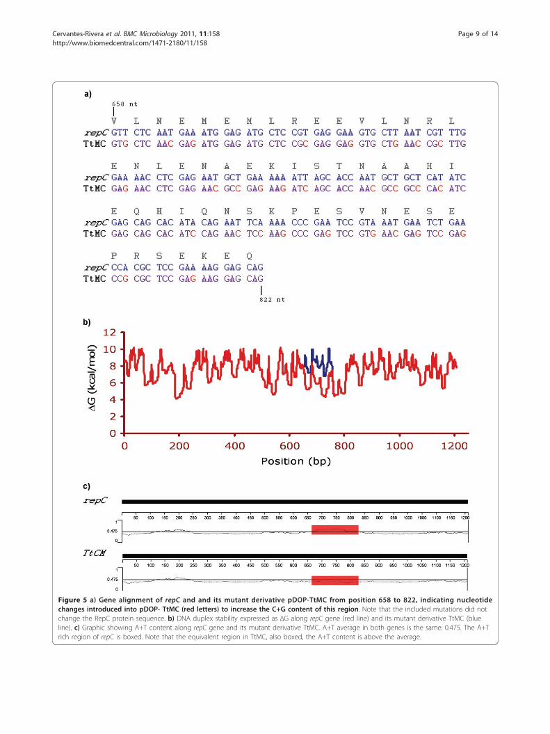
Figure 5 a) Gene alignment of repC and and its mutant derivative pDOP-TtMC from position 658 to 822, indicating nucleotidechanges introduced into pDOP- TtMC (red letters) to increase the C+G content of this region. Note that the included mutations did notchange the RepC protein sequence. b) DNA duplex stability expressed as ΔG along repC gene (red line) and its mutant derivative TtMC (blueline). c) Graphic showing A+T content along repC gene and its mutant derivative TtMC. A+T average in both genes is the same: 0.475. The A+Trich region of repC is boxed. Note that the equivalent region in TtMC, also boxed, the A+T content is above the average.
Cervantes-Rivera et al. BMC Microbiology 2011, 11:158http://www.biomedcentral.com/1471-2180/11/158
Page 9 of 14

strains with or without p42d, indicating that the A+Trich region plays a major role in replication.
RepC exerts its action in cisThe identification of an oriV sequence is generally basedon its ability to facilitate replication when present on aplasmid that otherwise could replicate only if the appro-priate replication factors (e.g., an initiator protein) wereprovided in trans. To more precisely locate the oriVwithin repC, we cloned a collection of internal segmentsof repC into the suicide vector pDOP (Figure 1). Thiscollection was conjugated into an R. etli strain contain-ing the parental plasmid (CFNX101) as the source of allthe trans elements required for replication, but we wereunable to obtain transconjugants.To determine if the activation of oriV requires tran-
scription (i.e., the repC mRNA also acts as a replicationprimer), we constructed a pDOP derivative that con-tained a repC gene but lacked a SD sequence (pDOP-Cs/SD) (Figure 1). This plasmid was also incapable ofreplicating in R. etli CFNX101. Similarly, the two plas-mids with repC frame-shift mutations, pDOP-CBglIIand pDOP-CSphI, were also conjugated into R. etliCFNX101 without success. Overall, these results indicatethat RepC exerts its action in cis.
RepC as an incompatibility factorPlasmid incompatibility, or the inability of two repliconsto coexist in the same cell line, results from the sharingof elements involved in plasmid replication, partitioningor control [30]. The repC open reading frame of p42d,when cloned in a vector capable of replicating in R. etli,CFNX101, can coexist with p42d [8]. However, all of ourattempts to introduce the construct pDOP-C into R. etliCFNX101 failed. In contrast, CFNX101 transconjugantscarrying a similar construct (pDOP-CsA) that containedthe repC gene pSymA of S. meliloti 2011 were easilyobtained. The frequencies with which CFNX101/pDOP-CsymA and CFNX107/pDOP-CsymA transconjugantswere obtained were similar (average 5 × 10⁻³). Moreover,the plasmid profiles of the transconjugants showed thatpDOP-CsA replicated in these strains as an independententity. These observations indicate that pDOP-C and itsparental plasmid p42d are incompatible, while that ofpDOP-CSymA and p42d are compatible.The RepC protein of S. meliloti 2011 pSymA shares
54% identity with the p42d RepC protein, and both pro-teins have very similar secondary structures (Figure 6).To map the RepC regions of p42d that are involved inplasmid incompatibility, a collection of hybrid genes con-taining fragments of the repC genes from S. melilotipSymA and R. etli p42d were constructed. A schematicrepresentation of the hybrid genes and their properties isshown in Figure 7. The hybrid genes were designed so
that none of the predicted alpha-helix and beta regions ofthe repC products were disturbed. The hybrid genes werecloned into pDOP under the Plac promoter and trans-ferred by conjugation into R. etli CFNX107 to determinetheir ability to replicate autonomously and into R. etliCFNX101 to test if they were able to replicate withoutthe interference of p42d. Two constructs were capable ofreplicating in both genetic backgrounds: pDOP-C1-990and pDOP-C1-1086. The rest of the constructs failed toreplicate in both strains. The plasmid pDOP-C1-1086expresses a hybrid protein containing the first 362 aminoacid residues (aa) of the p42d RepC protein and the last39 aa carboxy-terminal region of the pSymA RepC pro-tein. With respect to plasmid incompatibility, this recom-binant plasmid behaved the same as plasmid pDOP-CSymA, i.e., it replicated similarly in the strainsCFNX101 and CFNX107. This result indicates that theRepC region involved in plasmid incompatibility residesin the last 39 amino acid residues of the protein.
DiscussionPlasmids in which the oriV is located in the gene encodingan initiation protein are uncommon but not exceptional.The Enterococcus faecalis pheromone-responding plasmidpAD1 [31] (Francia, et al., 2004), the Staphylococcus xylo-sus plasmid pSX267 [32], the plasmids pAMb1 and pLS32from Bacillus subtilis [33-35], and the Staphylococcus aur-eus multiresistance plasmids pSK1 and pSK41 [36,37] fallinto this category. However, the origins of replication inall of these plasmids have recognizable iterons, and aninsert that contains some or all of the iterons from theseplasmids is usually capable of driving plasmid replication ifthe initiator protein is provided in trans. The minimalreplicon of the p42d plasmid is the repC ORF sequencedriven by a constitutive promoter (Plac) with an SDsequence that we designed. Frame shift and deletionmutants of the repC gene disrupted the capacity for repli-cation of the minimal replicon, indicating that RepC isessential for replication and is likely the initiator protein.To confirm this function, it will be necessary to demon-strate that this protein binds the oriV, melts the double-stranded DNA, and recruits the initiation host factor.A DNA sequence analysis of the repC gene clearly
showed the absence of iterons or other large, perfect orimperfect, repetitive sequences (>8 bp), which are the typi-cal DNA-binding sites of plasmid initiator proteins [1].The replication of several bacterial plasmids, such as
P1, F, R6K, RK2, Rts1, pMU720, and pSC101, requires acrucial and concerted participation of DnaA and theplasmid-encoded initiator protein. These plasmids con-tain at least one DnaA box in their oriV sequences[38-43]. For other plasmids, DnaA participates only asan accessory, but these plasmids also contain DnaAboxes in their origins of replication (e.g., pR1) [44].
Cervantes-Rivera et al. BMC Microbiology 2011, 11:158http://www.biomedcentral.com/1471-2180/11/158
Page 10 of 14

However, we failed to identify such DnaA boxes withinthe repC-coding region, suggesting that DnaA does nothave a role in p42d replication.A common property of theta-replicating plasmids is
an A+T rich region close to the origin of replication,
which is necessary for strand melting and the assemblyof host initiation factors [1]. The repC ORF sequence ofp42d contains a large A+T rich region that is crucial forplasmid replication. A construct carrying silent muta-tions that partially eliminated the A+T rich region was
Figure 6 Protein alignment of p42d RepC from R. etli CFN42 and pSymA RepC from S. meliloti 1021 and where identical amino acidresidues are marked in red. The secondary structures of these proteins are also shown. Coiled regions are marked with C; helical regions are markedwith boxed H letters; and with letter E, the stranded regions. Arrows with an associated numbers indicates the positions where the genes were swap, inthe hybrid genes (see table 1).
Cervantes-Rivera et al. BMC Microbiology 2011, 11:158http://www.biomedcentral.com/1471-2180/11/158
Page 11 of 14

unable to promote replication in R. etli strains with orwithout the symbiotic plasmid, indicating that thisregion is an essential part of the oriV. However, asequence analysis of other repC genes located in repABCoperons revealed that an A+T rich region was present inall of the analyzed plasmids but its relative location wasnot conserved (data not shown).The p42d minimal replicon (pDOP-C) has two intri-
guing properties. First, the construct resulted in enhan-cing the plasmid copy-number to around six, in contrastparental plasmid, which was maintained at 1-2 copiesper chromosome. Second, the strain carrying this con-struct has a longer duplication time and a lower yieldwhen the cells reach stationary phase than the strainwithout this construct.While describing the observed increase in the plasmid
copy-number, we must bear in mind that the repC genein pDOP-C was expressed by a constitutive promoter.In addition, the negative transcriptional regulation of
the repC gene expression mediated by RepA and RepBwas eliminated, and the antisense RNA (ctRNA), whichalso plays a negative role in the expression of repC, wasremoved. In the absence of these layers of negative regu-lation, it is expected that the plasmid replication wouldaccelerate resulting in the production of new DNAmolecules with a concomitant increase in the number ofnew origins of replication, which in turn, could be usedto promote new rounds of replication, leading to celldeath. However, in the present study, with the use of
the minimal replicon (pDOP-C) we did not observe celldeath, and the plasmid copy-number increased onlymoderately. This observation suggests the existence of aposttranslational mechanism that limits RepC activity,thus preventing over-initiation.Growth kinetics of CFNX101 and CFNX107 were
identical (data not shown), however, when pDOP-C wasintroduced into CFNX1017 growth of the bacteriumwas inhibited. The growth rate and yield diminutionobserved in strain CFNX107/pDOP-C relative toCFNX107 is not likely caused by the metabolic burdenimposed by pDOP-C replication. The size of the paren-tal plasmid (p42d) is approximately 374 Kb, while thesize of pDOP-C is approximately 5.57 Kb; even if wetake into consideration the 6-fold increase in plasmidcopy-number, the amount of DNA required for replica-tion in CFNX107/pDOP-C is several fold lower than theamount of DNA required for replication in CFNX101.Based on these observations it can be hypothesized thatRepC, being an initiator protein, must perform threetasks: recognize the origin of replication, unwind theDNA at the origin, and recruit the replisome. An excessof RepC could lead to the formation of more of replica-tion “bubbles”. However, if one or more elements of thereplisome are suboptimal in the growing cell, then,some replication forks will be stalled resulting in inhibi-tion of cell division and growth.We demonstrated that pDOP-C was capable of
autonomous replication in an R. etli strain lacking the
Figure 7 a) Plasmid profiles of CFNX101 (lane 1) and CFNX101/pDOP-CsA (lane 2), showing that plasmid p42d and pDOP-CsA arecompatible. b) Linear representation of constructs containing SymA repC gene (blue arrow), p42d repC gene (red arrow) and SymA/p42d hybridderivatives (blue/red arrows), and their associated replication capabilities when introduced into R. etli CFNX101 (with p42d) and CFNX107 (a p42dcured derivative) strains (table at left). “+” Symbols indicate that the construct are capable to replicate, and “-” that the construct is incapable todo that. Construct names are listed at the right of the figure. Black squares indicate the relative position of the Plac promoter, and the whiterectangles the position of the Shine-Dalgarno (SD) sequences. Numbers at top indicate the positions where the SymA/p42d regions were swap.
Cervantes-Rivera et al. BMC Microbiology 2011, 11:158http://www.biomedcentral.com/1471-2180/11/158
Page 12 of 14

parental plasmid (p42d). However, we could not intro-duce this construct into an R. etli strain harboring theparental plasmid. In contrast, a similar construct thatcontained the repC gene of S. meliloti pSymA repli-cated autonomously with the same behavior in bothstrains. This result indicates that RepC is an incompat-ibility factor that prevents the coexistence of p42d andpDOP-C and that the incompatibility phenomenon isreplicon-specific. Additionally, a construct (pDOP-C1-1086) expressing a chimeric protein consisting of theamino-terminal region of p42 RepC and 39 aa residuesof the carboxy-terminal region of the pSymA RepCprotein was capable of replicating as an independententity with the same efficiency in R. etli strains, withor without p42d. This result indicates that the last 39aa residues of the RepC carboxy-terminal region aredirectly involved in the incompatibility phenotype. Aclose inspection of this region in the RepC proteins ofpSymA and p42d shows that they share 62.5% of iden-tity, indicating that 15 amino acid residues or less arecritical in promoting the incompatibility phenotype.Interestingly, however, in spite of the variations in 15aa residues, RepC proteins of p42d and pSymA have asimilar secondary structure: both possess two alphahelices of ten amino acid residues each, separated by acoiled region of six amino acid residues, in the samerelative positions.Our current hypothesis linking incompatibility and the
RepC posttranslational regulation is as follows: RepC,like many other plasmid-encoded initiator proteins,exists in two forms, an active monomer and an inactivedimer, and protein thermodynamics favors dimer forma-tion [1]. The RepC carboxy-terminal region is involvedin dimer formation, and the dimerization process isreplicon-specific. The introduction of pDOP-C into astrain containing p42d displaces the RepC monomer-dimer equilibrium that favors the inactive form, prevent-ing the establishment of the incoming plasmid. A similarintroduction of a construct with the RepC of a compati-ble plasmid will not affect the monomer-dimer equili-brium and will allow the establishment of the newplasmid.Another unusual observation was the inability to com-
plement the repC ORF in trans for replication. One pos-sibility is that the repC transcript acts as an RNA primerfor replication or assists in DNA melting at the oriV.However, the construct pDOP-Cs/SD, which lacks a SDsequence, could not replicate in CFNX101, suggestingthat translation is required for the newly synthesizedRepC protein to be located at the oriV. To the best ofour knowledge, the only initiator protein that functionsonly in cis is RepA from prophage N15 [45]. At this
stage we cannot determine which of these possibilities ismore likely, and further experiments are needed toresolve these questions.
ConclusionsRepC is the only element encoded in the repABCoperon of the Rhizobium etli p42d plasmid that isnecessary and sufficient for plasmid replication and islikely the initiator protein. The oriV of this plasmidresides within the repC gene and is located close to orinside of a large A+T region. This architecture is sharedby other repABC plasmids. Our results also indicate thatRepC can act as an incompatibility factor and that thelast 39 aa of the carboxy-terminal region of this proteinare involved in this phenotype.
Acknowledgements and FundingThis work was supported by the Consejo Nacional de Ciencia y Tecnología(CONACyT, México) (Grant number: 000000000100099); and by the Programade Apoyo a Proyectos de Investigación e Inovación Tecnológica (PAPIIT-UNAM, México) (Grant number IN205611-3) to M.A. C. R. C-R, F. P-L and G P-S were supported during the Ph.D. program (Programa de Doctorado enCiencias Biomédicas-Universidad Nacional Autónoma de México) withscholarships from Consejo Nacional de Ciencia y Tecnología and DirecciónGeneral de Estudios de Posgrado (México). We are greatly indebted toÁngeles Pérez-Oseguera for her technical support, and to Dr. PallavoluMaheswara Reddy for his critical review of the manuscript.
Authors’ contributionsR C-R conducted the bulk of the experiments and made the constructions; FP-L and G P-S made growth kinetics, plasmid profiles and incompatibilityexperiments. MAC designed and coordinated the study, and wrote themanuscript. All authors read and approved the final manuscript.
Competing interestsThe authors declare that they have no competing interests.
Received: 15 April 2011 Accepted: 30 June 2011Published: 30 June 2011
References1. del Solar G, Giraldo R, Ruiz-Echevarría MJ, Espinosa M, Díaz-Orejas R:
Replication and control of circular bacterial plasmids. Microbiol Mol BiolRev 1998, 62:434-464.
2. Nordström K, Molin S, Light J: Control of replication of bacterial plasmids:genetics, molecular biology, and physiology of the plasmid R1 system.Plasmid 1984, 12:71-90.
3. Paulsson J, Chattoraj DK: Origin inactivation in bacterial DNA replicationcontrol. Mol Microbiol 2006, 61:9-15.
4. Zakrzewska-Czerwinska J, Jakimowicz D, Zawilak-Pawlik A, Messer W:Regulation of the initiation of chromosomal replication in bacteria. FEMSMicrobiol Rev 2007, 31:378-387.
5. Cevallos MA, Cervantes-Rivera R, Gutiérrez-Ríos RM: The repABC plasmidfamily. Plasmid 2008, 60:19-37.
6. Castillo-Ramírez S, Vázquez-Castellanos JF, González V, Cevallos MA:Horizontal gene transfer and diverse functional constrains within acommon replication-partitioning system in Alphaproteobacteria: therepABC operon. BMC Genomics 2009, 10:536.
7. Pappas KM: Cell-cell signaling and the Agrobacterium tumefaciens Tiplasmid copy number fluctuations. Plasmid 2008, 60:89-107.
8. Ramírez-Romero MA, Soberón N, Pérez-Oseguera A, Téllez-Sosa J, Cevallos MA:Structural elements required for replication and incompatibility of theRhizobium etli symbiotic plasmid. J Bacteriol 2000, 182:3117-3124.
Cervantes-Rivera et al. BMC Microbiology 2011, 11:158http://www.biomedcentral.com/1471-2180/11/158
Page 13 of 14

9. Pappas KM, Winans SC: The RepA and RepB autorepressors and TraR playopposing roles in the regulation of a Ti plasmid repABC operon. MolMicrobiol 2003, 49:441-455.
10. Gerdes K, Moller-Jensen J, Jensen RB: Plasmid and chromosome partition:surprises from phylogeny. Mol Microbiol 2000, 37:455-466.
11. Ramírez-Romero MA, Téllez-Sosa J, Barrios H, Pérez-Oseguera A, Rosas V,Cevallos MA: RepA negatively autoregulates the transcription of therepABC operon of the Rhizobium etli symbiotic plasmid basic replicon.Mol Microbiol 2001, 42:195-204.
12. Tabata S, Hooykaas PJ, Oka A: Sequence determination andcharacterization of the replicator region in the tumor-inducing plasmidpTiB6S3. J Bacteriol 1989, 171:1665-1672.
13. Bartosik D, Baj J, Wlodarczyk M: Molecular and functional analysis ofpTAV320, a repABC-type replicon of the Paracoccus versutus compositeplasmid pTAV1. Microbiology 1998, 144:3149-3157.
14. Bartosik D, Szymanik M, Wysocka E: Identification of the partitioning sitewithin the repABC-type replicon of the composite Paracoccus versutusplasmid pTAV1. J Bacteriol 2001, 183:6234-6243.
15. Soberón N, Venkova-Canova T, Ramírez-Romero MA, Téllez-Sosa J,Cevallos MA: Incompatibility and the partitioning site of the repABC basicreplicon of the symbiotic plasmid from Rhizobium etli. Plasmid 2004,51:203-216.
16. Chai Y, Winans SC: RepB protein of an Agrobacterium tumefaciens Tiplasmid binds to two adjacent sites between repA and repB for plasmidpartitioning and autorepression. Mol Microbiol 2005, 58:1114-1129.
17. MacLellan SR, Zaheer R, Sartor AL, MacLean AM, Finan TM: Identification ofa megaplasmid centromere reveals genetic structural diversity withinthe repABC family of basic replicons. Mol Microbiol 2006, 59:1559-1575.
18. Chai Y, Winans SC: A small antisense RNA downregulates expression ofan essential replicase protein of an Agrobacterium tumefaciens Tiplasmid. Mol Microbiol 2005, 56:1574-1585.
19. MacLellan SR, Smallbone LA, Sibley CD, Finan TM: The expression of anovel antisense gene mediates incompatibility within the large repABCfamily of alpha-proteobacterial plasmids. Mol Microbiol 2005, 55:611-623.
20. Venkova-Canova T, Soberón NE, Ramírez-Romero MA, Cevallos MA: Twodiscrete elements are required for the replication of a repABC plasmid:an antisense RNA and a stem-loop structure. Mol Microbiol 2004,54:1431-1444.
21. Cervantes-Rivera R, Romero-López C, Berzal-Herranz A, Cevallos MA:Analysis of the mechanism of action of the antisense RNA that controlsthe replication of the repABC plasmid p42d. J Bacteriol 2010,192:3268-3278.
22. Noel KD, Sanchez A, Fernandez L, Leemans J, Cevallos MA: Rhizobiumphaseoli symbiotic mutants with transposon Tn5 insertions. J Bacteriol1984, 158:148-155.
23. Simon R, Priefer U, Pühler A: A broad host-range mobilization system forin vivo genetic engineering transposon mutagenesis in Gram negativebacteria. Bio/Technology 1983, 1:784-791.
24. Ramírez-Romero MA, Bustos P, Girard L, Rodríguez O, Cevallos MA, Dávila G:Sequence, localization and characteristics of the replicator region of thesymbiotic plasmid of Rhizobium etli. Microbiology 1997, 143:2825-2831.
25. Horton RM, Hunt HD, Ho SN, Pullen JK, Pease LR: Engineering hybridgenes without the use of restriction enzymes: gene splicing by overlapextension. Gene 1989, 77:61-68.
26. Hynes MF, McGregor NF: Two plasmids other than the nodulationplasmid are necessary for formation of nitrogen-fixing nodules byRhizobium leguminosarum. Mol Microbiol 1990, 4:567-574.
27. Thompson JD, Higgins DG, Gibson TJ: CLUSTAL W: improving thesensitivity of progressive multiple sequence alignment throughsequence weighting, position-specific gap penalties and weight matrixchoice. Nucleic Acids Res 1994, 22:4673-4680.
28. Jones DT: Protein secondary structure prediction based on position-specific scoring matrices. J Mol Biol 1999, 292:195-202.
29. Huang Y, Kowalski D: WEB-THERMODYN: sequence analysis software forprofiling DNA helical stability. Nucl Acids Res 2003, 31:3819-3821.
30. Novick RP: Plasmid incompatibility. Microbiol Rev 1987, 51:381-395.31. Francia MV, Fujimoto S, Tille P, Weaver KE, Clewell DB: Replication of
Enterococcus faecalis pheromone-responding plasmid pAD1: location ofthe minimal replicon and oriV site and RepA involvement in initiation ofreplication. J Bacteriol 2004, 186:5003-5016.
32. Gering M, Götz F, Brückner R: Sequence and analysis of the replicationregion of the Staphylococcus xylosus plasmid pSX267. Gene 1996,182:117-122.
33. Bruand C, Ehrlich SD: Transcription-driven DNA replication of plasmidpAMbeta1 in Bacillus subtilis. Mol Microbiol 1998, 30:135-145.
34. Tanaka T, Ogura M: A novel Bacillus natto plasmid pLS32 capable ofreplication in Bacillus subtilis. FEBS Lett 1998, 422:243-246.
35. Tanaka T, Ishida H, Maehara T: Characterization of the replication regionof plasmid pLS32 from the Natto strain of Bacillus subtilis. J Bacteriol2005, 187:4315-4326.
36. Kwong SM, Skurray RA, Firth N: Staphylococcus aureus multiresistanceplasmid pSK41: analysis of the replication region, initiator proteinbinding and antisense RNA regulation. Mol Microbiol 2004, 51:497-509.
37. Kwong SM, Skurray RA, Firth N: Replication control of staphylococcalmultiresistance plasmid pSK41: an antisense RNA mediates dual-levelregulation of Rep expression. J Bacteriol 2006, 188:4404-4412.
38. Betteridge T, Yang J, Pittard AJ, Praszkier J: Role of RepA and DnaAproteins in the opening of the origin of DNA replication of an IncBplasmid. J Bacteriol 2004, 186:3785-3793.
39. Gaylo PJ, Turjman N, Bastia D: DnaA protein is required for replication ofthe minimal replicon of the broad-host-range plasmid RK2 in Escherichiacoli. J Bacteriol 1987, 169:4703-4709.
40. Hansen EB, Yarmolinsky MB: Host participation in plasmid maintenance:dependence upon dnaA of replicons derived from P1 and F. Proc NatlAcad Sci USA 1986, 83:4423-4427.
41. Hasunuma K, Sekiguchi M: Replication of plasmid pSC101 in Escherichiacoli K12: requirement for dnaA function. Mol Gen Genet 1977,154:225-230.
42. Itoh Y, Terawaki Y: Replication properties of mini-Rts1 derivatives deletedfor DnaA boxes in the replication origin. Plasmid 1989, 21:242-246.
43. Kline BC, Kogoma T, Tam JE, Shields MS: Requirement of the Escherichiacoli dnaA gene product for plasmid F maintenance. J Bacteriol 1986,168:440-443.
44. Ortega-Jiménez S, Giraldo-Suárez R, Fernández-Tresguerres ME, Berzal-Herranz A, Díaz-Orejas R: DnaA dependent replication of plasmid R1occurs in the presence of point mutations that disrupt the dnaA box oforiR. Nucleic Acids Res 1992, 20:2547-2551.
45. Mardanov AV, Ravin NV: Functional characterization of the repAreplication gene of linear plasmid prophage N15. Res Microbiol 2006,157:176-183.
46. Martínez-Salazar J, Romero D, Girard ML, Dávila G: Molecular cloning andcharacterization of the recA gene of Rhizobium phaseoli andconstruction of recA mutants. J Bacteriol 1991, 173:3035-3040.
doi:10.1186/1471-2180-11-158Cite this article as: Cervantes-Rivera et al.: The replication origin of arepABC plasmid. BMC Microbiology 2011 11:158.
Submit your next manuscript to BioMed Centraland take full advantage of:
• Convenient online submission
• Thorough peer review
• No space constraints or color figure charges
• Immediate publication on acceptance
• Inclusion in PubMed, CAS, Scopus and Google Scholar
• Research which is freely available for redistribution
Submit your manuscript at www.biomedcentral.com/submit
Cervantes-Rivera et al. BMC Microbiology 2011, 11:158http://www.biomedcentral.com/1471-2180/11/158
Page 14 of 14