Cell cycle regulation of chromatin at an origin of DNA replication
12
Cell cycle regulation of chromatin at an origin of DNA replication Jing Zhou 1 , Charles M Chau 1 , Zhong Deng 1 , Ramin Shiekhattar 1 , Mark-Peter Spindler 2 , Aloys Schepers 2 and Paul M Lieberman* ,1 1 The Wistar Institute, Philadelphia, PA, USA and 2 Department of Gene Vectors, GSF-National Research Center for Environment and Health, Munich, Germany Selection and licensing of mammalian DNA replication origins may be regulated by epigenetic changes in chro- matin structure. The Epstein–Barr virus (EBV) origin of plasmid replication (OriP) uses the cellular licensing ma- chinery to regulate replication during latent infection of human cells. We found that the minimal replicator se- quence of OriP , referred to as the dyad symmetry (DS), is flanked by nucleosomes. These nucleosomes were subject to cell cycle-dependent chromatin remodeling and histone modifications. Restriction enzyme accessibility assay in- dicated that the DS-bounded nucleosomes were remodeled in late G1. Remarkably, histone H3 acetylation of DS- bounded nucleosomes decreased during late G1, coincid- ing with nucleosome remodeling and MCM3 loading, and preceding the onset of DNA replication. The ATP-depen- dent chromatin-remodeling factor SNF2h was also re- cruited to DS in late G1, and formed a stable complex with HDAC2 at DS. siRNA depletion of SNF2h reduced G1- specific nucleosome remodeling, histone deacetylation, and MCM3 loading at DS. We conclude that an SNF2h– HDAC1/2 complex coordinates G1-specific chromatin remodeling and histone deacetylation with the DNA replication initiation process at OriP . The EMBO Journal (2005) 24, 1406–1417. doi:10.1038/ sj.emboj.7600609; Published online 17 March 2005 Subject Categories: chromatin & transcription; genome stability & dynamics Keywords: DNA replication; EBV; histone; OriP; SNF2h Introduction Eukaryotic replication origins are highly regulated by cell cycle-dependent activities to ensure they initiate DNA repli- cation once and only once during the S phase of each division cycle (reviewed in Blow and Hodgson, 2002; DePamphilis, 2003; Mendez and Stillman, 2003). The selection, activation, and deactivation of replication origins follow a strict regula- tory program that involves an ordered cascade of checkpoints and licensing events. In lower eukaryotes, like budding yeast, DNA replication origins are discrete genetic elements that bind directly to the origin recognition complex (ORC) (re- viewed in Bell and Dutta, 2002). Origin selection in higher eukaryotes is less well understood and it is currently not clear whether replication initiates at specific sites or in large initiation zones. Replication origins might be delineated by poorly defined epigenetic factors, like chromatin boundaries and nuclear domains (reviewed in Gilbert, 2001; McNairn and Gilbert, 2003). In human cells, both ORC binding and MCM protein loading are highly regulated by numerous post-translational modifications and accessory proteins, including cdk/cyclins, cdt1, cdc6, and geminin (reviewed in DePamphilis, 2003; Mendez and Stillman, 2003). However, the contribution of chromatin structure and histone modifi- cations to the cell cycle regulation of replication origin function has not been explored in detail. Viral origins of DNA replication provide valuable models for investigating basic cellular regulatory processes. The Epstein–Barr virus (EBV) is a human lymphotropic herpes- virus that persists as a chromatin-associated episomal gen- ome during latent infections (Kieff, 1996). The EBV origin of plasmid replication (OriP) is licensed to replicate once and only once in latently infected cells that express the viral origin binding protein EBNA1 (Adams, 1987; Yates and Guan, 1991; Hirai and Shirakata, 2001; reviewed in Sugden and Leight, 2001). EBNA1 binds to a minimal replicator sequence, re- ferred to as the dyad symmetry (DS) region, which consists of two pairs of EBNA1 binding sites punctuated by three telo- mere repeat factor (TRF) binding sites (Yates et al, 2000; Bashaw and Yates, 2001; Koons et al, 2001; Deng et al, 2002). This minimal replicator has been shown to recruit ORC complex and to be genetically dependent upon ORC2 expres- sion for replication function (Chaudhuri et al, 2001; Dhar et al, 2001; Schepers et al, 2001; Ritzi et al, 2003). MCM proteins are recruited to DS in a cell cycle-dependent manner, and ectopic expression of geminin downregulates OriP-de- pendent replication (Chaudhuri et al, 2001; Dhar et al, 2001). Based on these observations, it is thought that OriP uses the cellular licensing and replication machinery to regulate sin- gle-round replication and maintain a stable copy number of viral genomes in proliferating cells latently infected with EBV. The contribution of chromatin structure and histone mod- ification to nuclear events has become increasingly apparent (Strahl and Allis, 2000; Jenuwein and Allis, 2001). ATP- dependent chromatin-remodeling complexes are thought to be essential for repositioning nucleosomes that preclude the binding of DNA replication and transcription factors (Varga- Weisz, 2001; Becker and Horz, 2002; Bozhenok et al, 2002; Collins et al, 2002; Peterson, 2002; Lusser and Kadonaga, 2003). The SNF2h member of the ISWI family of chromatin- remodeling proteins has been implicated in cellular DNA replication, by facilitating DNA synthesis of higher-ordered heterochromatin in mammalian cells (Bozhenok et al, 2002; Collins et al, 2002; Poot et al, 2004) and facilitating DNA synthesis-associated chromatin assembly in Drosophila Received: 16 August 2004; accepted: 8 February 2005; published online: 17 March 2005 *Corresponding author. The Wistar Institute, 3601 Spruce Street, Philadelphia, PA 19104, USA. Tel.: þ 1 215 898 9491; Fax: þ 1 215 898 0663; E-mail: [email protected] The EMBO Journal (2005) 24, 1406–1417 | & 2005 European Molecular Biology Organization | All Rights Reserved 0261-4189/05 www.embojournal.org The EMBO Journal VOL 24 | NO 7 | 2005 & 2005 European Molecular Biology Organization EMBO THE EMBO JOURNAL THE EMBO JOURNAL 1406
-
Upload
independent -
Category
Documents
-
view
1 -
download
0
Transcript of Cell cycle regulation of chromatin at an origin of DNA replication

Cell cycle regulation of chromatin at an originof DNA replication
Jing Zhou1, Charles M Chau1,Zhong Deng1, Ramin Shiekhattar1,Mark-Peter Spindler2, Aloys Schepers2
and Paul M Lieberman*,1
1The Wistar Institute, Philadelphia, PA, USA and 2Department of GeneVectors, GSF-National Research Center for Environment and Health,Munich, Germany
Selection and licensing of mammalian DNA replication
origins may be regulated by epigenetic changes in chro-
matin structure. The Epstein–Barr virus (EBV) origin of
plasmid replication (OriP) uses the cellular licensing ma-
chinery to regulate replication during latent infection of
human cells. We found that the minimal replicator se-
quence of OriP, referred to as the dyad symmetry (DS), is
flanked by nucleosomes. These nucleosomes were subject
to cell cycle-dependent chromatin remodeling and histone
modifications. Restriction enzyme accessibility assay in-
dicated that the DS-bounded nucleosomes were remodeled
in late G1. Remarkably, histone H3 acetylation of DS-
bounded nucleosomes decreased during late G1, coincid-
ing with nucleosome remodeling and MCM3 loading, and
preceding the onset of DNA replication. The ATP-depen-
dent chromatin-remodeling factor SNF2h was also re-
cruited to DS in late G1, and formed a stable complex
with HDAC2 at DS. siRNA depletion of SNF2h reduced G1-
specific nucleosome remodeling, histone deacetylation,
and MCM3 loading at DS. We conclude that an SNF2h–
HDAC1/2 complex coordinates G1-specific chromatin
remodeling and histone deacetylation with the DNA
replication initiation process at OriP.
The EMBO Journal (2005) 24, 1406–1417. doi:10.1038/
sj.emboj.7600609; Published online 17 March 2005
Subject Categories: chromatin & transcription; genome
stability & dynamics
Keywords: DNA replication; EBV; histone; OriP; SNF2h
Introduction
Eukaryotic replication origins are highly regulated by cell
cycle-dependent activities to ensure they initiate DNA repli-
cation once and only once during the S phase of each division
cycle (reviewed in Blow and Hodgson, 2002; DePamphilis,
2003; Mendez and Stillman, 2003). The selection, activation,
and deactivation of replication origins follow a strict regula-
tory program that involves an ordered cascade of checkpoints
and licensing events. In lower eukaryotes, like budding yeast,
DNA replication origins are discrete genetic elements that
bind directly to the origin recognition complex (ORC) (re-
viewed in Bell and Dutta, 2002). Origin selection in higher
eukaryotes is less well understood and it is currently not clear
whether replication initiates at specific sites or in large
initiation zones. Replication origins might be delineated by
poorly defined epigenetic factors, like chromatin boundaries
and nuclear domains (reviewed in Gilbert, 2001; McNairn
and Gilbert, 2003). In human cells, both ORC binding
and MCM protein loading are highly regulated by numerous
post-translational modifications and accessory proteins,
including cdk/cyclins, cdt1, cdc6, and geminin (reviewed in
DePamphilis, 2003; Mendez and Stillman, 2003). However,
the contribution of chromatin structure and histone modifi-
cations to the cell cycle regulation of replication origin
function has not been explored in detail.
Viral origins of DNA replication provide valuable models
for investigating basic cellular regulatory processes. The
Epstein–Barr virus (EBV) is a human lymphotropic herpes-
virus that persists as a chromatin-associated episomal gen-
ome during latent infections (Kieff, 1996). The EBV origin of
plasmid replication (OriP) is licensed to replicate once and
only once in latently infected cells that express the viral origin
binding protein EBNA1 (Adams, 1987; Yates and Guan, 1991;
Hirai and Shirakata, 2001; reviewed in Sugden and Leight,
2001). EBNA1 binds to a minimal replicator sequence, re-
ferred to as the dyad symmetry (DS) region, which consists of
two pairs of EBNA1 binding sites punctuated by three telo-
mere repeat factor (TRF) binding sites (Yates et al, 2000;
Bashaw and Yates, 2001; Koons et al, 2001; Deng et al, 2002).
This minimal replicator has been shown to recruit ORC
complex and to be genetically dependent upon ORC2 expres-
sion for replication function (Chaudhuri et al, 2001; Dhar
et al, 2001; Schepers et al, 2001; Ritzi et al, 2003). MCM
proteins are recruited to DS in a cell cycle-dependent manner,
and ectopic expression of geminin downregulates OriP-de-
pendent replication (Chaudhuri et al, 2001; Dhar et al, 2001).
Based on these observations, it is thought that OriP uses the
cellular licensing and replication machinery to regulate sin-
gle-round replication and maintain a stable copy number of
viral genomes in proliferating cells latently infected with EBV.
The contribution of chromatin structure and histone mod-
ification to nuclear events has become increasingly apparent
(Strahl and Allis, 2000; Jenuwein and Allis, 2001). ATP-
dependent chromatin-remodeling complexes are thought to
be essential for repositioning nucleosomes that preclude the
binding of DNA replication and transcription factors (Varga-
Weisz, 2001; Becker and Horz, 2002; Bozhenok et al, 2002;
Collins et al, 2002; Peterson, 2002; Lusser and Kadonaga,
2003). The SNF2h member of the ISWI family of chromatin-
remodeling proteins has been implicated in cellular DNA
replication, by facilitating DNA synthesis of higher-ordered
heterochromatin in mammalian cells (Bozhenok et al, 2002;
Collins et al, 2002; Poot et al, 2004) and facilitating DNA
synthesis-associated chromatin assembly in DrosophilaReceived: 16 August 2004; accepted: 8 February 2005; publishedonline: 17 March 2005
*Corresponding author. The Wistar Institute, 3601 Spruce Street,Philadelphia, PA 19104, USA. Tel.: þ 1 215 898 9491;Fax: þ 1 215 898 0663; E-mail: [email protected]
The EMBO Journal (2005) 24, 1406–1417 | & 2005 European Molecular Biology Organization | All Rights Reserved 0261-4189/05
www.embojournal.org
The EMBO Journal VOL 24 | NO 7 | 2005 &2005 European Molecular Biology Organization
EMBO
THE
EMBOJOURNAL
THE
EMBOJOURNAL
1406
(Fyodorov et al, 2004). SNF2h is an ATPase that has been
isolated in several multiprotein complexes, including ACF
(Ito et al, 1997; LeRoy et al, 2000), CHRAC (Varga-Weisz et al,
1997), WCRF (Bochar et al, 2000; Bozhenok et al, 2002), and
a large HDAC1/2–SNF2h–cohesin complex (Hakimi et al,
2002). These distinct complexes suggest that SNF2h performs
multiple functions in chromatin regulation.
Post-translational modifications of histone tails have been
mechanistically linked to numerous changes in gene regula-
tion, including transcription complex assembly, histone de-
position at the DNA replication fork, and response to DNA
damage (Carrozza et al, 2003). Typically, histone acetylation
correlates well with increased DNA access, while histone
deacetylation and histone H3 K9 methylation correlate with
the formation of transcriptionally silent chromatin (Strahl
and Allis, 2000; Jenuwein and Allis, 2001). Acetylation of
histone tails has been mechanistically linked to the recruit-
ment of bromodomain-containing chromatin-remodeling
complexes during transcription activation (Agalioti et al,
2000; Hassan et al, 2002; Neely and Workman, 2002). More
recently, histone H3 K4 methylation has been linked to
recruitment of SNF2h-containing complexes (Santos-Rosa
et al, 2003). However, the chromatin events regulating DNA
replication initiation have not been extensively investigated.
Cell cycle changes in chromatin remodeling and histone
modification at eukaryotic origins may be an important
regulatory feature controlling replication and licensing factor
access to DNA. In this work, we investigate the nucleosome
organization and histone tail modifications at OriP. We found
that the DS region of OriP is flanked by nucleosomes that
undergo chromatin remodeling and histone deacetylation at
the G1/S border of the cell cycle. We also show that an
SNF2h–HDAC1/2-dependent chromatin-remodeling activity
physically associates with OriP during G1/S and contributes
to OriP replication activity.
Results
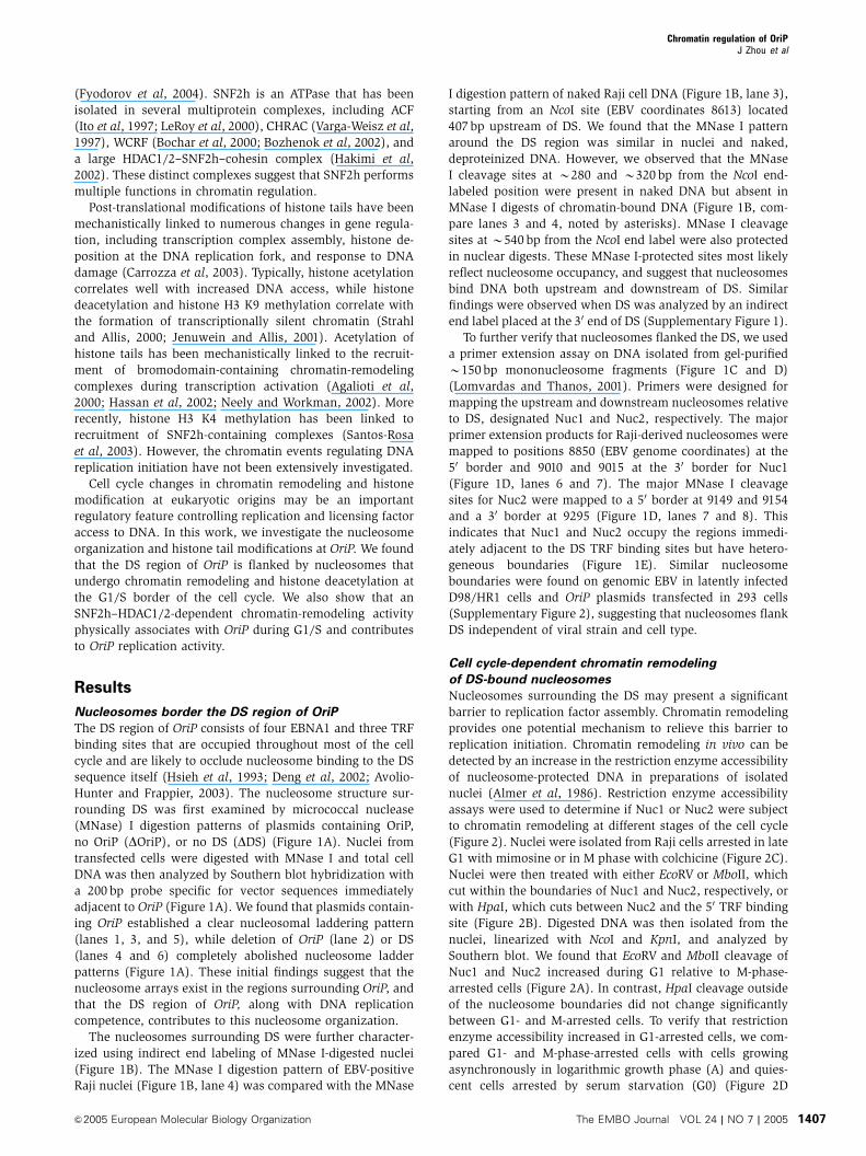
Nucleosomes border the DS region of OriP
The DS region of OriP consists of four EBNA1 and three TRF
binding sites that are occupied throughout most of the cell
cycle and are likely to occlude nucleosome binding to the DS
sequence itself (Hsieh et al, 1993; Deng et al, 2002; Avolio-
Hunter and Frappier, 2003). The nucleosome structure sur-
rounding DS was first examined by micrococcal nuclease
(MNase) I digestion patterns of plasmids containing OriP,
no OriP (DOriP), or no DS (DDS) (Figure 1A). Nuclei from
transfected cells were digested with MNase I and total cell
DNA was then analyzed by Southern blot hybridization with
a 200 bp probe specific for vector sequences immediately
adjacent to OriP (Figure 1A). We found that plasmids contain-
ing OriP established a clear nucleosomal laddering pattern
(lanes 1, 3, and 5), while deletion of OriP (lane 2) or DS
(lanes 4 and 6) completely abolished nucleosome ladder
patterns (Figure 1A). These initial findings suggest that the
nucleosome arrays exist in the regions surrounding OriP, and
that the DS region of OriP, along with DNA replication
competence, contributes to this nucleosome organization.
The nucleosomes surrounding DS were further character-
ized using indirect end labeling of MNase I-digested nuclei
(Figure 1B). The MNase I digestion pattern of EBV-positive
Raji nuclei (Figure 1B, lane 4) was compared with the MNase
I digestion pattern of naked Raji cell DNA (Figure 1B, lane 3),
starting from an NcoI site (EBV coordinates 8613) located
407 bp upstream of DS. We found that the MNase I pattern
around the DS region was similar in nuclei and naked,
deproteinized DNA. However, we observed that the MNase
I cleavage sites at B280 and B320 bp from the NcoI end-
labeled position were present in naked DNA but absent in
MNase I digests of chromatin-bound DNA (Figure 1B, com-
pare lanes 3 and 4, noted by asterisks). MNase I cleavage
sites at B540 bp from the NcoI end label were also protected
in nuclear digests. These MNase I-protected sites most likely
reflect nucleosome occupancy, and suggest that nucleosomes
bind DNA both upstream and downstream of DS. Similar
findings were observed when DS was analyzed by an indirect
end label placed at the 30 end of DS (Supplementary Figure 1).
To further verify that nucleosomes flanked the DS, we used
a primer extension assay on DNA isolated from gel-purified
B150 bp mononucleosome fragments (Figure 1C and D)
(Lomvardas and Thanos, 2001). Primers were designed for
mapping the upstream and downstream nucleosomes relative
to DS, designated Nuc1 and Nuc2, respectively. The major
primer extension products for Raji-derived nucleosomes were
mapped to positions 8850 (EBV genome coordinates) at the
50 border and 9010 and 9015 at the 30 border for Nuc1
(Figure 1D, lanes 6 and 7). The major MNase I cleavage
sites for Nuc2 were mapped to a 50 border at 9149 and 9154
and a 30 border at 9295 (Figure 1D, lanes 7 and 8). This
indicates that Nuc1 and Nuc2 occupy the regions immedi-
ately adjacent to the DS TRF binding sites but have hetero-
geneous boundaries (Figure 1E). Similar nucleosome
boundaries were found on genomic EBV in latently infected
D98/HR1 cells and OriP plasmids transfected in 293 cells
(Supplementary Figure 2), suggesting that nucleosomes flank
DS independent of viral strain and cell type.
Cell cycle-dependent chromatin remodeling
of DS-bound nucleosomes
Nucleosomes surrounding the DS may present a significant
barrier to replication factor assembly. Chromatin remodeling
provides one potential mechanism to relieve this barrier to
replication initiation. Chromatin remodeling in vivo can be
detected by an increase in the restriction enzyme accessibility
of nucleosome-protected DNA in preparations of isolated
nuclei (Almer et al, 1986). Restriction enzyme accessibility
assays were used to determine if Nuc1 or Nuc2 were subject
to chromatin remodeling at different stages of the cell cycle
(Figure 2). Nuclei were isolated from Raji cells arrested in late
G1 with mimosine or in M phase with colchicine (Figure 2C).
Nuclei were then treated with either EcoRV or MboII, which
cut within the boundaries of Nuc1 and Nuc2, respectively, or
with HpaI, which cuts between Nuc2 and the 50 TRF binding
site (Figure 2B). Digested DNA was then isolated from the
nuclei, linearized with NcoI and KpnI, and analyzed by
Southern blot. We found that EcoRV and MboII cleavage of
Nuc1 and Nuc2 increased during G1 relative to M-phase-
arrested cells (Figure 2A). In contrast, HpaI cleavage outside
of the nucleosome boundaries did not change significantly
between G1- and M-arrested cells. To verify that restriction
enzyme accessibility increased in G1-arrested cells, we com-
pared G1- and M-phase-arrested cells with cells growing
asynchronously in logarithmic growth phase (A) and quies-
cent cells arrested by serum starvation (G0) (Figure 2D
Chromatin regulation of OriPJ Zhou et al
&2005 European Molecular Biology Organization The EMBO Journal VOL 24 | NO 7 | 2005 1407

and E). We found that restriction enzyme accessibility at the
MboII site within Nuc2 was enhanced only in mimosine-
treated G1-arrested cells, and not detected in asynchronous or
resting cells (Figure 2D).
Enriched histone H3 K4 methylation at DS-bound
nucleosomes
Histone modifications may also regulate chromatin dynamics
at OriP. We next used the chromatin immunoprecipitation
(ChIP) assay to characterize DS-associated proteins and
histone post-translational modifications in Raji cells
(Figure 3). As expected, EBNA1 was highly enriched at DS
relative to the inactive viral lytic origin OriLyt located over
30 kb from OriP (Figure 3A). ORC2 and MCM3 proteins were
modestly enriched at DS, consistent with published findings
(Chaudhuri et al, 2001; Dhar et al, 2001; Schepers et al, 2001;
Ritzi et al, 2003). Most significantly, ChIP with antibodies to
several histone tail modifications revealed that DS-associated
nucleosomes were highly enriched for histone H3 dimethyl-
K4 (H3mK4) relative to OriLyt (Figure 3A). Acetylated his-
tone H3 (AcH3) was also enriched at DS, although to a lesser
extent than that seen for H3mK4. In contrast, acetylated
histone H4 (AcH4) was slightly more enriched at OriLyt
relative to DS.
G1-specific histone deacetylation at DS-bound
nucleosomes
Histone modifications at OriP were examined for changes at
various stages of the cell cycle (Figure 3B). To measure more
accurately the histone modifications at Nuc1 and Nuc2, we
enriched for mononucleosomes by digesting DNA with
MNase I and performed ChIP with primer sets located within
the boundary of each nucleosome. Remarkably, we found
that AcH3 was significantly reduced at Nuc1 and Nuc2 in
mimosine-treated G1-arrested cells (Figure 3B). This loss of
AcH3 modification in G1 was specific for DS nucleosomes
since we did not observe a similar loss at control region Zp,
located B40 kb from DS. In contrast to AcH3, H3mK4 was
not reduced in mimosine-arrested cells, suggesting that his-
tones still occupy the DS region during G1. A more extensive
analysis of Nuc1 and Nuc2 shows that the association with
EBNA1, AcH4, and H3mK4 did not change significantly at
various stages of the cell cycle, while AcH3 was consistently
reduced at both nucleosome positions (Figure 3C).
Modification of the K9 position of H3 has been associated
EBNA1 EBNA1 EBNA1 EBNA1Nuc1 TRF TRF TRF Nuc2
ab
cd9020 9136
8839 9144 92889009
88509015 9149
92959010 9154
B
DS
Nuc2
Nuc1
Nco1−5′
*ProbeNcoI
21761766
1230
1033
653
517453394
298
234
154
1 2 3 4
− ++MNase IDNA N G A T C a b c d Primer
Raji
1 2 3 4 5 6 7 8
C
MN
ase
dige
st
150 bp
1 2 3
PE
tem
plat
e D
Nuc3*
*
**
8613
10 789
pOriP
p∆O
riP
MNase
1 2
A
E
pOriP
p∆D
S
3 4 5 6
15 U/mlMNase 15 75 U/ml
pOriP
p∆D
S
Figure 1 Nucleosomal organization at OriP. (A) Nucleosomal laddering assay of D98/HR1 cells transfected with plasmids N503 (OriP), N530(DOriP), or N564 (DDS) as indicated. Plasmid DNA was detected by Southern blot hybridization with a 200 bp probe specific to GFP-vectorsequence adjacent to DS. (B) Nucleosome locations at the DS region of OriP were analyzed by indirect end labeling. Purified EBVþ Raji cellDNA (lanes 2 and 3) or nuclei (lane 4) were treated with 0 U/ml (lane 2), 15 U/ml (lane 3), or 75 U/ml (lane 4) MNase I, and subsequentlydigested with NcoI. The DNA products were then analyzed by Southern blot and probed with a 200 bp fragment covering EBV sequences 8613–8813. Molecular weights of DNA markers (lane 1) are indicated at the left. Asterisks indicate MNase I sites protected in nuclear digest. Deducedpositions of nucleosomes (Nuc1 and Nuc2) and EBNA1 binding sites in the DS are indicated by shaded and black ovals in the schematic to theright. EBV coordinates are provided at the bottom and top of the schematic. (C) MNase I-digested mononucleosomal DNA fraction (left lane)and the gel-purified DNA used for primer extension assays (right lane) were visualized with ethidium bromide staining of agarose gels. (D)Primer extension analysis of mononucleosomal DNA isolated from EBVþ Raji cells. Primers a (lane 5), b (lane 6), c (lane 7), and d (lane 8) areindicated above the lane and their position in DS is shown in the schematic below. Sequencing reactions are shown in lanes 1–4. (E) Schematicof DS and primer extension products mapped in (B–D) show Raji nucleosome boundaries (lower thin arrows) and D98/HR1 nucleosomeboundaries (upper thick arrows) at the DS region of OriP. EBV genome coordinates are indicated, along with EBNA1 and TRF binding sites.Primer annealing positions a–d are indicated by horizontal arrows.
Chromatin regulation of OriPJ Zhou et al
The EMBO Journal VOL 24 | NO 7 | 2005 &2005 European Molecular Biology Organization1408

with changes in gene expression and heterochromatin for-
mation (Jenuwein and Allis, 2001). Consistent with the
results found for the hyperacetylated H3 antibody, ChIP
analysis revealed a strong reduction in H3acK9 at Nuc1 and
Nuc2 in G1 as well as in M (Figure 3D). Conversely, H3mK9
showed a modest increase at Nuc2 in M-phase-arrested cells.
No significant change in EBNA1 binding could be detected,
suggesting that MNase I digestion was incomplete or that
Nuc1 and Nuc2 interact with neighboring EBNA1 or DS-
associated proteins throughout the cell cycle.
Cell cycle association of SNF2h with DS
Since DS-associated nucleosomes were subject to chromatin
remodeling during G1 (Figure 2), we reasoned that an ATP-
dependent chromatin-remodeling complex is likely to be
responsible for this activity. In mammalian cells, the predo-
minant chromatin-remodeling complexes belong to the
SNF2h, BRG1, or Mi-2/NuRD families (Varga-Weisz, 2001;
NcoI KpnI
MboII
TETE EETNuc1 Nuc2
EcoRV
v
HpaI
E5′ M3′
N/K
EcoRV MboII
MG1
*
M5′
*
E3′
MG1
G1 M
2n 2n 4n4n
*H3′
H5′
HpaI
MG1N/K N/K
MG1G0A
M5′
*
M3′
MboII
N/K
A
B
C
D
E A G0 G1 M
4n2n 4n2n 4n2n 4n2n
Figure 2 Cell cycle-dependent restriction enzyme accessibility atthe DS. (A) Restriction enzyme accessibility assay was performedon nuclei isolated from Raji cells arrested with mimosine (G1) orcolchicine (M). Nuclei were digested with EcoRV (left panel), HpaI(middle panel), or MboII (right panel) for 5 min, deproteinized, andthen digested with NcoI/KpnI prior to Southern blotting and detec-tion with an OriP-specific probe. The MboII (M), HpaI (H), andEcoRV (E) 50 and 30 restriction products are indicated by arrows.Asterisks indicate a control cleavage product outside of nucleosome1 or 2. (B) Schematic indicating position of MboII, HpaI, and EcoRVrelative to DS. EBNA1 (E) and TRFs (T) are indicated by the shadedovals. (C) FACS profile of cell cycle-arrested Raji cells used for theexperiments described above. (D) MboII restriction enzyme acces-sibility in asynchronous (A), quiescent (G0), mimosine-arrested(G1), or colchicine-arrested (G2) Raji cells. (E) FACS profile of cellcycle for cells used in (D).
Nuc1
Nuc2
Input IgG EBNA1 H3mK9 H3acK9
AcH3Input
Nuc1
Nuc2
Zp
A M
H3mK4
G0
IgG
G1
G0 G1
G1 G1 G1 G1 G1 G1
A MG0 G1 A MG0 G1 A MG0 G1
Nuc1
Nuc2
A M A M A M A M A M A M
G1A M G1A M G1A M G1A M G1A M
Input IgG EBNA1 AcH3 AcH4 H3mK4
0
20
40
60
80
100
IgG
EBNA1AcH
3AcH
4
H3mK4
ORC2
MCM
3
% R
elat
ive
ChI
P
DSOriLyt
A M
120
140A
B
C
D
E
2n 4n 2n 4n 2n 4n 2n 4n
Figure 3 Cell cycle-associated changes in histone modifications atOriP. (A) Protein interactions at DS were determined by ChIP assaywith either control IgG or antibodies specific to EBNA1, AcH3,AcH4, H3mK4, ORC2, and MCM3. Chromatin immunoprecipitatedDNA was analyzed by real-time PCR with primers specific for DS orOriLyt. (B) ChIP assay of MNase I-treated Raji cell nuclei derivedfrom asynchronous (A), serum-starved (G0), mimosine-arrested(G1), or colchicine-arrested (M) cells. Immunoprecipitation withantibodies specific for acetylated H3 or H3mK4 was analyzed byPCR with primers specific for DS-associated leftward nucleosome(Nuc1), the DS-associated rightward nucleosome (Nuc2), or theBZLF1 promoter region (Zp). (C) Same as (B), but with antibodiesspecific for EBNA1, AcH3, AcH4, H3mK4, and control IgG. (D)Same as above, but with IgG, EBNA1, H3mK9, and histone H3acetyl K9 (H3acK9). (E) FACS profile of propidium iodide-treatedcells after treatments for cell cycle arrests shown above.
Chromatin regulation of OriPJ Zhou et al
&2005 European Molecular Biology Organization The EMBO Journal VOL 24 | NO 7 | 2005 1409

Peterson, 2002; Lusser and Kadonaga, 2003; Bowen et al,
2004). Antibodies specific to SNF2h, BRG1, or the BRG1-
associated protein Ini1 were compared for their ability to
precipitate DS DNA in a ChIP assay (Figure 4A). We found
that SNF2h specifically associated with DS relative to GAPDH
DNA, and to a greater extent than BRG1 or Ini1 (Figure 4A).
We next asked whether SNF2h associated with OriP in a cell
cycle-dependent manner using real-time PCR analysis of ChIP
DNA (Figure 4B and C). Raji cells grown asynchronously (A)
or cell cycle arrested with serum starvation (G0), mimosine
(G1), hydroxyurea (S), or colchicine (M) were assayed with
antibodies specific for AcH3, SNF2h, MCM3, EBNA1, or
control IgG (Figure 4B). First, we confirmed our previous
observations that AcH3 levels were reduced at DS in G1 (B4-
fold relative to M-phase levels). Next, we found that SNF2h
was significantly enriched at DS in G1 stage (B5-fold relative
to M-phase levels). SNF2h was not similarly enriched at the
control DNA site of OriLyt. We also demonstrate that MCM3
was enriched at DS in G1 (B8-fold relative to M-phase
levels). EBNA1 remained bound to DS through all stages of
the cell cycle examined and IgG did not precipitate significant
levels of DS or OriLyt in any condition. These results indicate
that SNF2h bound DS in G1-arrested cells where nucleosome
remodeling was detected (Figure 2), AcH3 levels decreased
(Figures 3 and 4B), and MCM3 binding increased (Figure 4B)
at DS.
Cell cycle arrest experiments also revealed that histone
AcH3 levels oscillate through the cell cycle (Figure 4B).
Remarkably, we found that AcH3 levels decreased four-fold
in G1/S, followed by a rapid increase in S (B7-fold relative to
G1-phase levels), and then a return to median levels in M-
phase-arrested cells. This oscillation in AcH3 levels was not
observed at control OriLyt DNA (Figure 4B), or at the Zp
region of EBV (Figure 3B). Similar patterns of oscillation were
not observed for SNF2h, MCM3, EBNA1, or IgG. These
observations suggest that DS-associated histone H3 acetyla-
tion has a markedly distinct cell cycle pattern.
HDAC2 colocalizes with SNF2h at DS during G1
SNF2h interacts with DS in G1-arrested cells, which coincides
with the loss of AcH3 (Figures 3 and 4). SNF2h has been
isolated as a multiprotein complex with HDAC1 and 2
(Hakimi et al, 2002), and we sought to determine whether
HDAC1 and 2 may also interact with DS in a cell cycle- and
SNF2h-associated manner. We first determined whether
SNF2h and HDAC2 could be isolated as a stable complex in
EBV-positive Raji cells (Figure 5A). Raji cell nuclear extracts
were subject to immunoprecipitation with SNF2h or HDAC2,
0
20
40
60
80
100
120
A G0 G1 S M0
20
40
60
80
100
120140
A G0 G1 S M0
20
40
60
80
100
A G0 G1 S M
05
101520253035404550
A G0 G1 S M
DS
OriLyt
SNF2h
% R
elat
ive
ChI
P
% R
elat
ive
ChI
P
0
50
100
150
200
250
300
350
A G0 G1 S M
AcH3
MCM3 EBNA1 IgG
A G1 MS
M Inpu
t
IgG
EB
NA
1
BR
G1
Ini1
OriP
GAPDH
SN
F2h
1 2 3 4 5 6 7
A
B
C
2n 4n 2n 4n 2n 4n 2n 4n
Figure 4 SNF2h is recruited to OriP in a cell cycle-dependent manner and is required for DNA replication. (A) ChIP assays with antibodiesspecific for EBNA1, BRG1, Ini1, SNF2h, or control IgG were analyzed for DNA from OriP or GAPDH. (B) Chromatin dynamics were analyzed inasynchronous (A), serum-starved (G0), mimosine-treated (G1), hydroxyurea-treated (S), or colchicine-treated (M) Raji cells. Real-time PCRanalyses of DS (black box) or OriLyt (gray box) DNA precipitated by ChIP assay with antibodies specific for AcH3, SNF2h, MCM3, EBNA1, orcontrol IgG are indicated. (C) FACS analysis of propidium iodide-stained cells used in the experiments shown in (B).
Chromatin regulation of OriPJ Zhou et al
The EMBO Journal VOL 24 | NO 7 | 2005 &2005 European Molecular Biology Organization1410
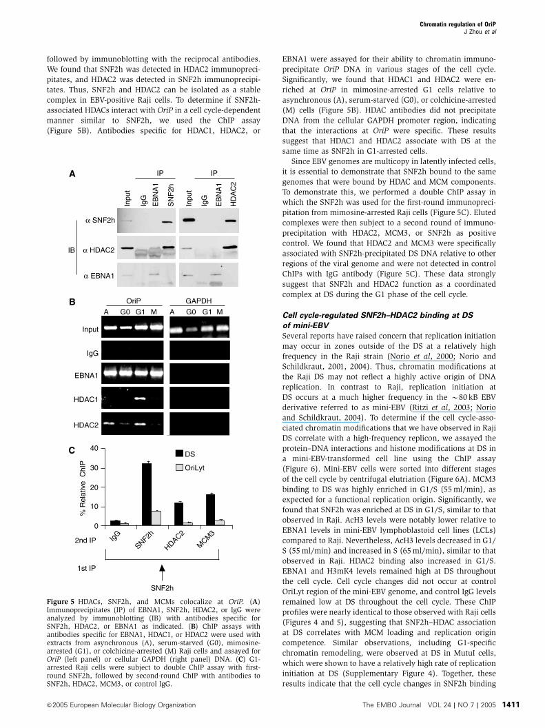
followed by immunoblotting with the reciprocal antibodies.
We found that SNF2h was detected in HDAC2 immunopreci-
pitates, and HDAC2 was detected in SNF2h immunoprecipi-
tates. Thus, SNF2h and HDAC2 can be isolated as a stable
complex in EBV-positive Raji cells. To determine if SNF2h-
associated HDACs interact with OriP in a cell cycle-dependent
manner similar to SNF2h, we used the ChIP assay
(Figure 5B). Antibodies specific for HDAC1, HDAC2, or
EBNA1 were assayed for their ability to chromatin immuno-
precipitate OriP DNA in various stages of the cell cycle.
Significantly, we found that HDAC1 and HDAC2 were en-
riched at OriP in mimosine-arrested G1 cells relative to
asynchronous (A), serum-starved (G0), or colchicine-arrested
(M) cells (Figure 5B). HDAC antibodies did not precipitate
DNA from the cellular GAPDH promoter region, indicating
that the interactions at OriP were specific. These results
suggest that HDAC1 and HDAC2 associate with DS at the
same time as SNF2h in G1-arrested cells.
Since EBV genomes are multicopy in latently infected cells,
it is essential to demonstrate that SNF2h bound to the same
genomes that were bound by HDAC and MCM components.
To demonstrate this, we performed a double ChIP assay in
which the SNF2h was used for the first-round immunopreci-
pitation from mimosine-arrested Raji cells (Figure 5C). Eluted
complexes were then subject to a second round of immuno-
precipitation with HDAC2, MCM3, or SNF2h as positive
control. We found that HDAC2 and MCM3 were specifically
associated with SNF2h-precipitated DS DNA relative to other
regions of the viral genome and were not detected in control
ChIPs with IgG antibody (Figure 5C). These data strongly
suggest that SNF2h and HDAC2 function as a coordinated
complex at DS during the G1 phase of the cell cycle.
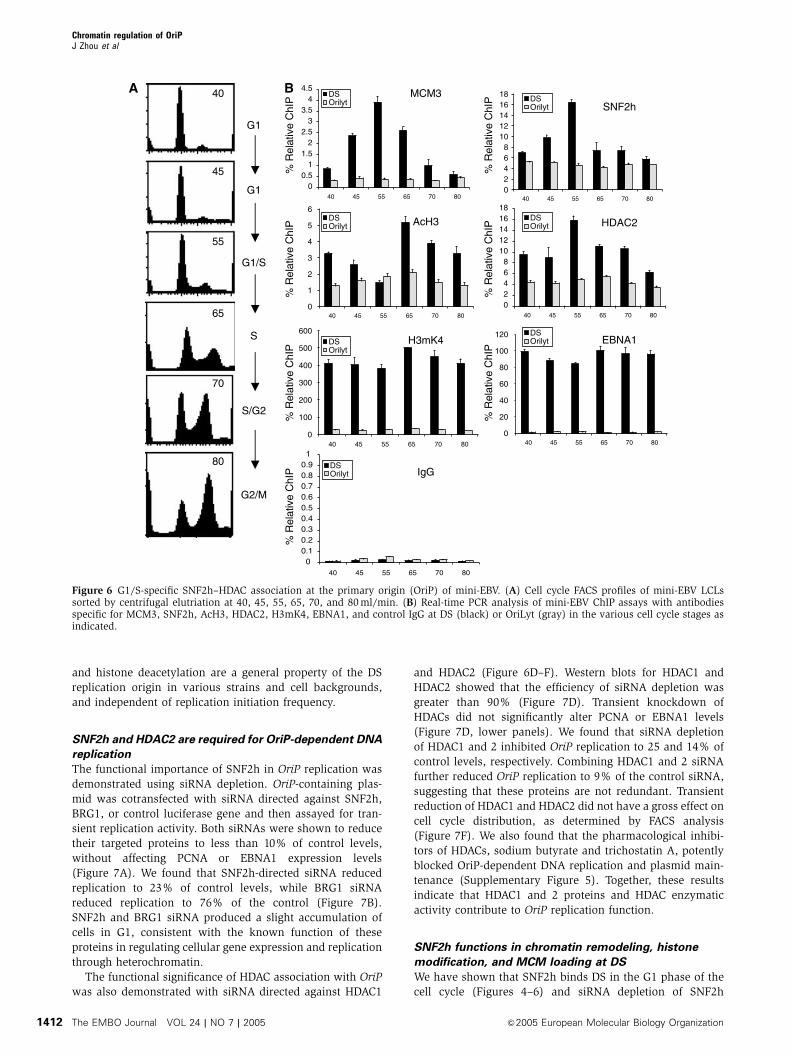
Cell cycle-regulated SNF2h–HDAC2 binding at DS
of mini-EBV
Several reports have raised concern that replication initiation
may occur in zones outside of the DS at a relatively high
frequency in the Raji strain (Norio et al, 2000; Norio and
Schildkraut, 2001, 2004). Thus, chromatin modifications at
the Raji DS may not reflect a highly active origin of DNA
replication. In contrast to Raji, replication initiation at
DS occurs at a much higher frequency in the B80 kB EBV
derivative referred to as mini-EBV (Ritzi et al, 2003; Norio
and Schildkraut, 2004). To determine if the cell cycle-asso-
ciated chromatin modifications that we have observed in Raji
DS correlate with a high-frequency replicon, we assayed the
protein–DNA interactions and histone modifications at DS in
a mini-EBV-transformed cell line using the ChIP assay
(Figure 6). Mini-EBV cells were sorted into different stages
of the cell cycle by centrifugal elutriation (Figure 6A). MCM3
binding to DS was highly enriched in G1/S (55 ml/min), as
expected for a functional replication origin. Significantly, we
found that SNF2h was enriched at DS in G1/S, similar to that
observed in Raji. AcH3 levels were notably lower relative to
EBNA1 levels in mini-EBV lymphoblastoid cell lines (LCLs)
compared to Raji. Nevertheless, AcH3 levels decreased in G1/
S (55 ml/min) and increased in S (65 ml/min), similar to that
observed in Raji. HDAC2 binding also increased in G1/S.
EBNA1 and H3mK4 levels remained high at DS throughout
the cell cycle. Cell cycle changes did not occur at control
OriLyt region of the mini-EBV genome, and control IgG levels
remained low at DS throughout the cell cycle. These ChIP
profiles were nearly identical to those observed with Raji cells
(Figures 4 and 5), suggesting that SNF2h–HDAC association
at DS correlates with MCM loading and replication origin
competence. Similar observations, including G1-specific
chromatin remodeling, were observed at DS in MutuI cells,
which were shown to have a relatively high rate of replication
initiation at DS (Supplementary Figure 4). Together, these
results indicate that the cell cycle changes in SNF2h binding
Inpu
t
Inpu
t
IgG
IgG
EB
NA
1
EB
NA
1
SN
F2h
HD
AC
2
α SNF2h
α HDAC2
α EBNA1
IB
IP IP
0
10
20
30
40
IgG
SNF2h
HDAC2
MCM
3
DS
OriLyt
2nd IP
1st IP
SNF2h
% R
elat
ive
ChI
P
A G0 G1 M
OriP
A G0 G1 M
GAPDH
Input
IgG
EBNA1
HDAC1
HDAC2
A
B
C
Figure 5 HDACs, SNF2h, and MCMs colocalize at OriP. (A)Immunoprecipitates (IP) of EBNA1, SNF2h, HDAC2, or IgG wereanalyzed by immunoblotting (IB) with antibodies specific forSNF2h, HDAC2, or EBNA1 as indicated. (B) ChIP assays withantibodies specific for EBNA1, HDAC1, or HDAC2 were used withextracts from asynchronous (A), serum-starved (G0), mimosine-arrested (G1), or colchicine-arrested (M) Raji cells and assayed forOriP (left panel) or cellular GAPDH (right panel) DNA. (C) G1-arrested Raji cells were subject to double ChIP assay with first-round SNF2h, followed by second-round ChIP with antibodies toSNF2h, HDAC2, MCM3, or control IgG.
Chromatin regulation of OriPJ Zhou et al
&2005 European Molecular Biology Organization The EMBO Journal VOL 24 | NO 7 | 2005 1411
and histone deacetylation are a general property of the DS
replication origin in various strains and cell backgrounds,
and independent of replication initiation frequency.
SNF2h and HDAC2 are required for OriP-dependent DNA
replication
The functional importance of SNF2h in OriP replication was
demonstrated using siRNA depletion. OriP-containing plas-
mid was cotransfected with siRNA directed against SNF2h,
BRG1, or control luciferase gene and then assayed for tran-
sient replication activity. Both siRNAs were shown to reduce
their targeted proteins to less than 10% of control levels,
without affecting PCNA or EBNA1 expression levels
(Figure 7A). We found that SNF2h-directed siRNA reduced
replication to 23% of control levels, while BRG1 siRNA
reduced replication to 76% of the control (Figure 7B).
SNF2h and BRG1 siRNA produced a slight accumulation of
cells in G1, consistent with the known function of these
proteins in regulating cellular gene expression and replication
through heterochromatin.
The functional significance of HDAC association with OriP
was also demonstrated with siRNA directed against HDAC1
and HDAC2 (Figure 6D–F). Western blots for HDAC1 and
HDAC2 showed that the efficiency of siRNA depletion was
greater than 90% (Figure 7D). Transient knockdown of
HDACs did not significantly alter PCNA or EBNA1 levels
(Figure 7D, lower panels). We found that siRNA depletion
of HDAC1 and 2 inhibited OriP replication to 25 and 14% of
control levels, respectively. Combining HDAC1 and 2 siRNA
further reduced OriP replication to 9% of the control siRNA,
suggesting that these proteins are not redundant. Transient
reduction of HDAC1 and HDAC2 did not have a gross effect on
cell cycle distribution, as determined by FACS analysis
(Figure 7F). We also found that the pharmacological inhibi-
tors of HDACs, sodium butyrate and trichostatin A, potently
blocked OriP-dependent DNA replication and plasmid main-
tenance (Supplementary Figure 5). Together, these results
indicate that HDAC1 and 2 proteins and HDAC enzymatic
activity contribute to OriP replication function.
SNF2h functions in chromatin remodeling, histone
modification, and MCM loading at DS
We have shown that SNF2h binds DS in the G1 phase of the
cell cycle (Figures 4–6) and siRNA depletion of SNF2h
00.5
11.5
22.5
33.5
44.5
40 45 55 65 70 80
DSOrilyt DS
Orilyt
DSOrilyt
DSOrilyt
DSOrilyt
DSOrilyt
DSOrilyt
MCM3
0
1
2
3
4
5
6
40 45 55 65 70 80
AcH3
0
100
200
300
400
500
600
40 45 55 65 70 80
H3mK4
00.10.20.30.40.50.60.70.80.91
40 45 55 65 70 80
IgG
0
20
40
60
80
100
120
40 45 55 65 70 80
EBNA1
02468
1012141618
40 45 55 65 70 80
SNF2h
02468
1012141618
40 45 55 65 70 80
HDAC2
G1
G1
G1/S
S
S/G2
G2/M
% R
elat
ive
ChI
P%
Rel
ativ
e C
hIP
% R
elat
ive
ChI
P
% R
elat
ive
ChI
P%
Rel
ativ
e C
hIP
% R
elat
ive
ChI
P
% R
elat
ive
ChI
P
40
45
55
65
70
80
A B
Figure 6 G1/S-specific SNF2h–HDAC association at the primary origin (OriP) of mini-EBV. (A) Cell cycle FACS profiles of mini-EBV LCLssorted by centrifugal elutriation at 40, 45, 55, 65, 70, and 80 ml/min. (B) Real-time PCR analysis of mini-EBV ChIP assays with antibodiesspecific for MCM3, SNF2h, AcH3, HDAC2, H3mK4, EBNA1, and control IgG at DS (black) or OriLyt (gray) in the various cell cycle stages asindicated.
Chromatin regulation of OriPJ Zhou et al
The EMBO Journal VOL 24 | NO 7 | 2005 &2005 European Molecular Biology Organization1412

inhibits OriP replication (Figure 7). We next determined
whether SNF2h depletion disrupted chromatin dynamics at
DS (Figure 8). EBV-positive D98/HR1 cells were transfected
with control or SNF2h-specific siRNA, arrested in G1 with
mimosine, and then assayed for restriction enzyme accessi-
bility at the Nuc1 site using EcoRV (Figure 8A and B). We
found that cells depleted for SNF2h had significantly less
EcoRV restriction enzyme accessibility at Nuc1 relative to
cells transfected with control siRNA (Figure 8B). We next
asked whether SNF2h depletion led to a loss of MCM3 protein
association at DS in G1-arrested cells using the ChIP assay
(Figure 8C). We found that SNF2h depletion led to an B3-
fold decrease in MCM3 binding to DS. We also observed that
SNF2h depletion resulted in a corresponding increase in
histone H3 acetylation at DS, but not at control OriLyt region
of the EBV genome (Figure 8D). Surprisingly, we also
observed that SNF2h depletion led to an B2-fold decrease
in histone H3mK4 at DS, with no significant change at
Con
trol
α HDAC1
HD
AC
1
Con
trol
HD
AC
2
α HDAC2
α EBNA1
α PCNA
Con
trol
HD
AC
1
HD
AC
2
HD
AC
1+2
α BRG1
α PCNA
α EBNA
α SNF2h Dpn1/BamHI
BamHI
BamHI
OriP
Dpn1/BamHIOriP
OriP
Replication 100 76 23 (wt %)
Replication 100 25 14 9 (wt %)
Control siSNF2h siBRG1
492422
631714
521921
% G1% S% G2/M
Control siHDAC1 siHDAC2
% G1% S% G2/M
552515
602214
572013
SN
F2h
BR
G1
Con
trol
Con
trol
Con
trol
BR
G1
SN
F2hA
C
D
F
E
B
Figure 7 SNF2h and HDACs promote replication and maintenance of OriP. (A) Western blots of cells transfected with siRNA specific for SNF2h(left) or BRG1 (right) were analyzed with antibodies to SNF2h, BRG1, or against control proteins EBNA1 and PCNA (as indicated). (B)Replication assays of OriP in cells cotransfected with control, BRG1, or SNF2h siRNAs. Transfected DNA was analyzed by Dpn1/BamHI orBamHI resistance followed by Southern blot. (C) FACS analysis of propidium iodide-stained cells transfected with siRNAs for SNF2h or BRG1 asindicated. (D) Western blots of extracts derived from D98/HR1 cells transfected with HDAC1 or HDAC2 siRNAs (upper panels), or with controlantibodies specific to EBNA1 or cellular protein PCNA (lower panels). (E) OriP-dependent DNA replication was assayed in D98/HR1 cellstransfected with OriP plasmid and siRNA specific for HDAC1, HDAC2, or HDAC1þ 2 as indicated above each lane. (F) FACS analysis ofpropidium iodide-stained cells transfected with siRNA specific for HDAC1 or HDAC2 as indicated.
Chromatin regulation of OriPJ Zhou et al
&2005 European Molecular Biology Organization The EMBO Journal VOL 24 | NO 7 | 2005 1413
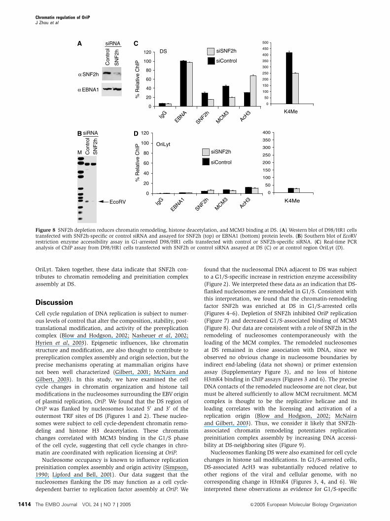
OriLyt. Taken together, these data indicate that SNF2h con-
tributes to chromatin remodeling and preinitiation complex
assembly at DS.
Discussion
Cell cycle regulation of DNA replication is subject to numer-
ous levels of control that alter the composition, stability, post-
translational modification, and activity of the prereplication
complex (Blow and Hodgson, 2002; Nasheuer et al, 2002;
Hyrien et al, 2003). Epigenetic influences, like chromatin
structure and modification, are also thought to contribute to
prereplication complex assembly and origin selection, but the
precise mechanisms operating at mammalian origins have
not been well characterized (Gilbert, 2001; McNairn and
Gilbert, 2003). In this study, we have examined the cell
cycle changes in chromatin organization and histone tail
modifications in the nucleosomes surrounding the EBV origin
of plasmid replication, OriP. We found that the DS region of
OriP was flanked by nucleosomes located 50 and 30 of the
outermost TRF sites of DS (Figures 1 and 2). These nucleo-
somes were subject to cell cycle-dependent chromatin remo-
deling and histone H3 deacetylation. These chromatin
changes correlated with MCM3 binding in the G1/S phase
of the cell cycle, suggesting that cell cycle changes in chro-
matin are coordinated with replication licensing at OriP.
Nucleosome occupancy is known to influence replication
preinitiation complex assembly and origin activity (Simpson,
1990; Lipford and Bell, 2001). Our data suggest that the
nucleosomes flanking the DS may function as a cell cycle-
dependent barrier to replication factor assembly at OriP. We
found that the nucleosomal DNA adjacent to DS was subject
to a G1/S-specific increase in restriction enzyme accessibility
(Figure 2). We interpreted these data as an indication that DS-
flanked nucleosomes are remodeled in G1/S. Consistent with
this interpretation, we found that the chromatin-remodeling
factor SNF2h was enriched at DS in G1/S-arrested cells
(Figures 4–6). Depletion of SNF2h inhibited OriP replication
(Figure 7) and decreased G1/S-associated binding of MCM3
(Figure 8). Our data are consistent with a role of SNF2h in the
remodeling of nucleosomes contemporaneously with the
loading of the MCM complex. The remodeled nucleosomes
at DS remained in close association with DNA, since we
observed no obvious change in nucleosome boundaries by
indirect end-labeling (data not shown) or primer extension
assay (Supplementary Figure 3), and no loss of histone
H3mK4 binding in ChIP assays (Figures 3 and 6). The precise
DNA contacts of the remodeled nucleosome are not clear, but
must be altered sufficiently to allow MCM recruitment. MCM
complex is thought to be the replicative helicase and its
loading correlates with the licensing and activation of a
replication origin (Blow and Hodgson, 2002; McNairn
and Gilbert, 2003). Thus, we consider it likely that SNF2h-
associated chromatin remodeling potentiates replication
preinitiation complex assembly by increasing DNA accessi-
bility at DS-neighboring sites (Figure 9).
Nucleosomes flanking DS were also examined for cell cycle
changes in histone tail modifications. In G1/S-arrested cells,
DS-associated AcH3 was substantially reduced relative to
other regions of the viral and cellular genome, with no
corresponding change in H3mK4 (Figures 3, 4, and 6). We
interpreted these observations as evidence for G1/S-specific
0
50
100
150
200
250
300
350
400
K4Me0
20
40
60
80
100
120
IgG
EBNA1
SNF2h
MCM
3AcH
3
0
20
40
60
80
100
120
IgG
EBNA
SNF2h
MCM
3AcH
3
0
50
100
150
200
250
300
350
400
450
500
K4Me
α SNF2h
α EBNA1
Con
trol
SN
F2h
siRNA
Con
trol
SN
F2h
siRNA
EcoRV
M
DS
OriLyt
siSNF2h
siControl
siSNF2h
siControl
% R
elat
ive
ChI
P%
Rel
ativ
e C
hIP
A
B D
C
Figure 8 SNF2h depletion reduces chromatin remodeling, histone deacetylation, and MCM3 binding at DS. (A) Western blot of D98/HR1 cellstransfected with SNF2h-specific or control siRNA and assayed for SNF2h (top) or EBNA1 (bottom) protein levels. (B) Southern blot of EcoRVrestriction enzyme accessibility assay in G1-arrested D98/HR1 cells transfected with control or SNF2h-specific siRNA. (C) Real-time PCRanalysis of ChIP assay from D98/HR1 cells transfected with SNF2h or control siRNA assayed at DS (C) or at control region OriLyt (D).
Chromatin regulation of OriPJ Zhou et al
The EMBO Journal VOL 24 | NO 7 | 2005 &2005 European Molecular Biology Organization1414

histone H3 deacetylation. This interpretation was further
supported by finding G1/S-specific enrichment of HDAC1
and HDAC2 at DS (Figures 5 and 6). We also observed that
histone H3 acetylation levels increased at DS in S-phase-
arrested cells, and then returned to intermediate levels in
M-phase-arrested cells. While the oscillation of histone acet-
ylation and deacetylation across the cell cycle is not un-
expected (Verreault, 2003), we were surprised to find histone
deacetylation at DS in G1/S, when prereplication factors are
assembling. Histone deacetylation is typically associated with
repressive chromatin, and histone acetylation has been
linked to activation of transcription and DNA replication in
other systems (Sterner and Berger, 2000; Aggarwal and Calvi,
2004; Stedman et al, 2004). In Drosophila follicle cells,
histone acetylation enhanced, while deacetylation repressed
replication initiation frequency (Aggarwal and Calvi, 2004).
Similarly, constitutively high levels of histone H3 acetylation
were found at the origin of plasmid replication in the related
viral genome of HHV8/KSHV (Stedman et al, 2004).
Paradoxically, chromatin remodeling and replication factor
assembly at EBV OriP correlated with histone deacetylation
and not histone hyperacetylation.
The atypical pattern of histone modification at DS may be
linked to the complex epigenetic behavior of OriP. Replication
initiation at OriP occurs at relatively low frequency in Raji
cells, but at high frequency in mini-EBV-transformed LCLs
(Norio and Schildkraut, 2001, 2004). Despite the difference in
replication initiation frequency at OriP, we found no signifi-
cant difference in the G1/S-specific histone H3 deacetylation,
or SNF2h, HDAC1/2, or MCM3 binding (Figure 6). It has been
reported that OriP-dependent DNA replication initiates in late
S phase (Carroll et al, 1991). Histone deacetylation at OriP
may delay replication firing until the burst of histone acetyla-
tion observed at DS in later S phase (Figures 4 and 6). This
would suggest that late S-phase firing origins load MCM, but
remain inactive due to histone deacetylation. Histone deace-
tylation and late S-phase firing at OriP may also explain the
reduced replication initiation frequency observed in Raji
cells, where alternative replication zones may fire in early
S phase. Future experiments will be required to determine
whether these and other epigenetic factors regulate the
replication timing and initiation frequency at OriP in
the Raji and min-EBV genomes.
Histone modifications may also create a protein-recogni-
tion code that regulates replication activity (Strahl and Allis,
2000; Jenuwein and Allis, 2001). We found that DS nucleo-
somes were enriched for H3mK4 relative to other regions of
the EBV genome at all stages of the cell cycle examined.
H3mK4 has been shown to serve as a recognition site for
SNF2h-containing complexes (Santos-Rosa et al, 2003).
However, the constitutively elevated levels of H3mK4 cannot
account for the G1-specific recruitment of SNF2h to DS. Other
histone modifications may also modulate SNF2h binding, as
was shown for the inhibition of Drosophila ISWI to acetylated
histone H4 K16 (Corona et al, 2002). Thus, it is possible that
histone H3 deacetylation may be required in combination
with H3 K4 methylation to provide the necessary specificity
for the cell cycle-dependent recruitment of SNF2h to DS.
SNF2h can be isolated in several distinct multiprotein
complexes (Varga-Weisz, 2001). SNF2h can be recruited to
replication foci through a physical interaction with WSTF and
PCNA (Poot et al, 2004). SNF2h can also be isolated in a
multiprotein complex containing HDAC1 and 2 (Hakimi et al,
2002). We found that SNF2h and HDAC2 co-immunoprecipi-
tated and were colocalized to DS in double ChIP assays
(Figure 5). Histone deacetylation and nucleosome remodeling
colocalized spatially and temporally with MCM3 binding to
DS, indicating that these changes correlate with preinitiation
complex assembly at DS. The physical association of SNF2h
with HDACs at DS suggests that histone deacetylation is
functionally linked to chromatin remodeling. Chromatin re-
modeling during G1/S provides a mechanism for MCM load-
ing and replication factor assembly at DNA sites occluded by
DS-flanked nucleosomes. However, the precise function of
G1/S-specific histone H3 deacetylation at DS remains unclear.
It is possible that G1/S-specific histone deacetylation, in
combination with high histone H3 K4 methylation, provides
a unique epigenetic mark on the origin-associated nucleo-
somes that modulates SNF2h remodeling, or replication pre-
initiation complex assembly. It is also possible that histone
deacetylation of the remodeled nucleosomes at OriP restricts
replication initiation to late S phase or to once per cell cycle.
It will be important to further characterize these cell cycle-
dependent changes in chromatin structure at OriP and to
determine if these modifications are common features of
other viral and cellular replication origins, and how these
chromatin dynamics correlate with replication timing and
initiation frequency.
Materials and methods
Plasmids and cell cultureOriP plasmid (N503) has been described previously and consists ofOriP sequences, EBNA1, eGFP, and hygromycin genes as a pREP10(Invitrogen) derivative (Deng et al, 2002). pDOriP (N530) and pDDS
ORC
ORC
ORC
H3mK4 H3acK9
EBNA1 TRFs
G1/S
G1/G0
G2/M
OriP
ORC
ORC
MCMSNF2h/HDAC1/2
ORC
Replication
Figure 9 Model of cell cycle coordinated histone tail modificationand chromatin remodeling at OriP. An SNF2h/HDAC2 complex canbe colocalized to DS during G1, simultaneously with histone H3deacetylation, chromatin remodeling, and MCM loading.
Chromatin regulation of OriPJ Zhou et al
&2005 European Molecular Biology Organization The EMBO Journal VOL 24 | NO 7 | 2005 1415

(N564) are replication-incompetent derivatives of N503 lackingsequences of OriP and the DS region, respectively. Raji and MutuIcells are EBVþ suspension cells that were maintained in RPMIsupplemented with 10% FBS, glutamine, penicillin, and streptomy-cin sulfate (Cellgro). D98/HR1 (EBVþ adherent cells) and 293 cellswere maintained in DMEM with 10% FBS, glutamine, penicillin,and streptomycin sulfate (Cellgro). Adherent cells were transfectedwith Lipofectamine 2000, according to the manufacturer’s recom-mendations (Invitrogen). The A39 mini-EBV LCL was cultured asdescribed previously (Schepers et al, 2001; Ritzi et al, 2003).
Cell cycle arrestsAsynchronous Raji or D98/HR1 cells were arrested in G0 by growthin 0.5% FBS for 48 h, in G1/S by treatment with 400 mM mimosine(Sigma) for 24 h, in S phase by treatment with 100 mM hydroxyureafor 18 h, or in G2/M by treatment with 1mM colchicine (Sigma) for24 h. Mini-EBV LCL was fractionated according to cell cycle stage bycentrifugal elutriation using the same parameters as described (Ritziet al, 2003).
Primer extension analysis of mononucleosomesIsolation of nuclei and primer extension assay were performed asdescribed (Lomvardas and Thanos, 2001) with some modifications(see Supplementary Methods).
Indirect end-labeling assayNucleosome positions were analyzed by indirect end-labelingmethod described previously (Hager and Fragoso, 1999; Ryanet al, 1999).
Restriction enzyme accessibility assayRaji, MutuI, and siRNA transfected D98/HR1 cell nuclei wereprepared as described. Nuclei (107) were resuspended in 50mlrestriction digestion buffer for either MboII, HpaI, or EcoRV asspecified by the manufacturer (NEB). Restriction enzyme digestionwas performed at 371C for 10 min and stopped by the addition ofStop Buffer. After incubation at 501C for 2 h with proteinase K, DNAwas phenol/chloroform extracted, purified by ethanol precipitation,cut with KpnI and NcoI, and analyzed by Southern blotting. DNAwas detected using the DIG detection kit (Roche) with a 150 bp PCR-generated probe to DS region.
Chromatin immunoprecipitation assaysThe ChIP assay was a modification of the protocol provided byUpstate Biotechnology Inc., and has been described in detailelsewhere (Deng et al, 2002, 2003). Real-time PCR analysis of ChIPDNA was the average of three independent experiments, quantifiedusing the standard curve method on ABI 7000 thermocycler, andnormalized to EBNA1 bound to DS in asynchronous cells (set at100%).
OriP replication assayDNA replication and plasmid maintenance assays have beendescribed previously (Yates et al, 2000; Deng et al, 2002). Fortransient replication assays with siRNA, all cells were cotransfectedwith OriP plasmid and siRNA.
siRNAsiRNAs were synthesized as duplex RNA (Dharmacon Inc.) with thefollowing target sequences for Luciferase control (cgtacgcggaatacttcga) and SNF2h (aagaggaggaugaagagcuau) as described pre-viously (Collins et al, 2002). siRNAs for HDAC1, 2, and 3 werecommercially available as Smartpool products (Dharmacon Inc.).siRNA to BRG1 was generated by the Hanon method (Paddisonet al, 2002) with the following oligonucleotide primer (aaaaaagacgttaacgctgtcacagacgctaccgcaagcttccagtagcatctgtaacagcattaactgtcggtgtttcgtcctttccacaa). Plasmid or siRNA controls were used in parallel foreach siRNA experiment.
Additional details of methods can be found in SupplementaryMethods.
Supplementary dataSupplementary data are available at The EMBO Journal Online.
Acknowledgements
We thank Latasha Day for technical support and the Wistar CancerCenter Core Facilities for their assistance. This work was funded byNCI CA93606 and DOD BC022095 to PML and NIH (GM61204) toRS. Charles Chau was funded by the Wistar NCI postdoctoraltraining grant, and Zhong Deng is a fellow of the Leukemia-Lymphoma Society.
References
Adams A (1987) Replication of latent Epstein–Barr virus genomes inRaji cells. J Virol 61: 1743–1746
Agalioti T, Lomvardas S, Parekh B, Yie J, Maniatis T, Thanos D(2000) Ordered recruitment of chromatin modifying and generaltranscription factors to the IFN-beta promoter. Cell 103: 667–678
Aggarwal BD, Calvi BR (2004) Chromatin regulates origin activity inDrosophila follicle cells. Nature 430: 372–376
Almer A, Rudolph H, Hinnen A, Horz W (1986) Removal ofpositioned nucleosomes from the yeast PHO5 promoter uponPHO5 induction releases additional upstream activating DNAelements. EMBO J 5: 2689–2696
Avolio-Hunter TM, Frappier L (2003) EBNA1 efficiently assembleson chromatin containing the Epstein–Barr virus latent origin ofreplication. Virology 315: 398–408
Bashaw JM, Yates JL (2001) Replication from oriP of Epstein–Barrvirus requires exact spacing of two bound dimers of EBNA1which bend DNA. J Virol 75: 10603–10611
Becker PB, Horz W (2002) ATP-dependent nucleosome remodeling.Annu Rev Biochem 71: 247–273
Bell SP, Dutta A (2002) DNA replication in eukaryotic cells. AnnuRev Biochem 71: 333–374
Blow JJ, Hodgson B (2002) Replication licensing—defining theproliferative state? Trends Cell Biol 12: 72–78
Bochar DA, Savard J, Wang W, Lafleur DW, Moore P, Cote J,Shiekhattar R (2000) A family of chromatin remodeling factorsrelated to Williams syndrome transcription factor. Proc Natl AcadSci USA 97: 1038–1043
Bowen NJ, Fujita N, Kajita M, Wade PA (2004) Mi-2/NuRD: multiplecomplexes for many purposes. Biochim Biophys Acta 1677:52–57
Bozhenok L, Wade PA, Varga-Weisz P (2002) WSTF–ISWI chroma-tin remodeling complex targets heterochromatic replication foci.EMBO J 21: 2231–2241
Carroll SM, Trotter J, Wahl GM (1991) Replication timing controlcan be maintained in extrachromosomally amplified genes. MolCell Biol 11: 4779–4785
Carrozza MJ, Utley RT, Workman JL, Cote J (2003) The diversefunctions of histone acetyltransferase complexes. Trends Genet19: 321–329
Chaudhuri B, Xu H, Todorov I, Dutta A, Yates JL (2001) HumanDNA replication initiation factors, ORC and MCM, associatewith oriP of Epstein–Barr virus. Proc Natl Acad Sci USA 98:10085–10089
Collins N, Poot RA, Kukimoto I, Garcia-Jimenez C, Dellaire G,Varga-Weisz PD (2002) An ACF1–ISWI chromatin-remodelingcomplex is required for DNA replication through heterochroma-tin. Nat Genet 32: 627–632
Corona DF, Clapier CR, Becker PB, Tamkun JW (2002) Modulationof ISWI function by site-specific histone acetylation. EMBO Rep 3:242–247
Deng Z, Atanasiu C, Burg JS, Broccoli D, Lieberman PM (2003)Telomere repeat binding factors TRF1, TRF2, and hRAP1modulate replication of Epstein–Barr virus OriP. J Virol 77:11992–12001
Deng Z, Lezina L, Chen CJ, Shtivelband S, So W, Lieberman PM(2002) Telomeric proteins regulate episomal maintenance ofEpstein–Barr virus origin of plasmid replication. Mol Cell 9:493–503
DePamphilis ML (2003) The ‘ORC cycle’: a novel pathway forregulating eukaryotic DNA replication. Gene 310: 1–15
Chromatin regulation of OriPJ Zhou et al
The EMBO Journal VOL 24 | NO 7 | 2005 &2005 European Molecular Biology Organization1416
Dhar SK, Yoshida K, Machida Y, Khaira P, Chaudhuri B,Wohlschlegel JA, Leffak M, Yates J, Dutta A (2001) Replicationfrom oriP of Epstein–Barr virus requires human ORC and isinhibited by geminin. Cell 106: 287–296
Fyodorov DV, Blower MD, Karpen GH, Kadonaga JT (2004) Acf1confers unique activities to ACF/CHRAC and promotes the for-mation rather than disruption of chromatin in vivo. Genes Dev 18:170–183
Gilbert DM (2001) Making sense of eukaryotic DNA replicationorigins. Science 294: 96–100
Hager GL, Fragoso G (1999) Analysis of nucleosome positioning inmammalian cells. Methods Enzymol 304: 626–638
Hakimi MA, Bochar DA, Schmiesing JA, Dong Y, Barak OG,Speicher DW, Yokomori K, Shiekhattar R (2002) A chromatinremodelling complex that loads cohesin onto human chromo-somes. Nature 418: 994–998
Hassan AH, Prochasson P, Neely KE, Galasinski SC, Chandy M,Carrozza MJ, Workman JL (2002) Function and selectivity ofbromodomains in anchoring chromatin-modifying complexes topromoter nucleosomes. Cell 111: 369–379
Hirai K, Shirakata M (2001) Replication licensing of the EBV oriPminichromosome. Curr Top Microbiol Immunol 258: 13–33
Hsieh DJ, Camiolo SM, Yates JL (1993) Constitutive binding ofEBNA1 protein to the Epstein–Barr virus replication origin, oriP,with distortion of DNA structure during latent infection. EMBO J12: 4933–4944
Hyrien O, Marheineke K, Goldar A (2003) Paradoxes of eukaryoticDNA replication: MCM proteins and the random completionproblem. BioEssays 25: 116–125
Ito T, Bulger M, Pazin MJ, Kobayashi R, Kadonaga JT (1997) ACF,an ISWI-containing and ATP-utilizing chromatin assembly andremodeling factor. Cell 90: 145–155
Jenuwein T, Allis CD (2001) Translating the histone code. Science293: 1074–1080
Kieff E (1996) Epstein–Barr virus and its replication. In Field’sVirology, Knipe D, Howley PM (eds) Vol. 2, pp 2343–2396.Philadelphia: Lippincott-Raven Publishers
Koons MD, Scoy SV, Hearing J (2001) The replicator of the Epstein–Barr virus latent cycle origin of DNA replication, oriP, is com-posed of multiple functional elements. J Virol 75: 10582–10592
LeRoy G, Loyola A, Lane WS, Reinberg D (2000) Purification andcharacterization of a human factor that assembles and remodelschromatin. J Biol Chem 275: 14787–14790
Lipford JR, Bell SP (2001) Nucleosomes positioned by ORC facilitatethe initiation of DNA replication. Mol Cell 7: 21–30
Lomvardas S, Thanos D (2001) Nucleosome sliding via TBP DNAbinding in vivo. Cell 106: 685–696
Lusser A, Kadonaga JT (2003) Chromatin remodeling by ATP-dependent molecular machines. BioEssays 25: 1192–1200
McNairn AJ, Gilbert DM (2003) Epigenomic replication: linkingepigenetics to DNA replication. BioEssays 25: 647–656
Mendez J, Stillman B (2003) Perpetuating the double helix: mole-cular machines at eukaryotic DNA replication origins. BioEssays25: 1158–1167
Nasheuer HP, Smith R, Bauerschmidt C, Grosse F, Weisshart K(2002) Initiation of eukaryotic DNA replication: regulation andmechanisms. Prog Nucleic Acid Res Mol Biol 72: 41–94
Neely KE, Workman JL (2002) Histone acetylation and chromatinremodeling: which comes first? Mol Genet Metab 76: 1–5
Norio P, Schildkraut CL (2001) Visualization of DNA replication onindividual Epstein–Barr virus episomes. Science 294: 2361–2364
Norio P, Schildkraut CL (2004) Plasticity of DNA replication initia-tion in Epstein–Barr virus episomes. PLoS Biol 2: e152
Norio P, Schildkraut CL, Yates JL (2000) Initiation of DNA replica-tion within oriP is dispensable for stable replication of the latentEpstein–Barr virus chromosome after infection of established celllines. J Virol 74: 8563–8574
Paddison PJ, Caudy AA, Bernstein E, Hannon GJ, Conklin DS (2002)Short hairpin RNAs (shRNAs) induce sequence-specific silencingin mammalian cells. Genes Dev 16: 948–958
Peterson CL (2002) Chromatin remodeling enzymes: taming themachines. Third in review series on chromatin dynamics. EMBORep 3: 319–322
Poot RA, Bozhenok L, van den Berg DL, Steffensen S, Ferreira F,Grimaldi M, Gilbert N, Ferreira J, Varga-Weisz PD (2004) TheWilliams syndrome transcription factor interacts with PCNA totarget chromatin remodelling by ISWI to replication foci. Nat CellBiol 6: 1236–1244
Ritzi M, Tillack K, Gerhardt J, Ott E, Humme S, Kremmer E,Hammerschmidt W, Schepers A (2003) Complex protein–DNAdynamics at the latent origin of DNA replication of Epstein–Barrvirus. J Cell Sci 116: 3971–3984
Ryan MP, Stafford GA, Yu L, Cummings KB, Morse RH (1999)Assays for nucleosome positioning in yeast. Methods Enzymol304: 376–399
Santos-Rosa H, Schneider R, Bernstein BE, Karabetsou N, MorillonA, Weise C, Schreiber SL, Mellor J, Kouzarides T (2003)Methylation of histone H3 K4 mediates association of the Isw1pATPase with chromatin. Mol Cell 12: 1325–1332
Schepers A, Ritzi M, Bousset K, Kremmer E, Yates JL, Harwood J,Diffley JF, Hammerschmidt W (2001) Human origin recognitioncomplex binds to the region of the latent origin of DNA replica-tion of Epstein–Barr virus. EMBO J 20: 4588–4602
Simpson RT (1990) Nucleosome positioning can affect the functionof a cis-acting DNA element in vivo. Nature 343: 387–389
Stedman W, Deng Z, Lu F, Lieberman PM (2004) ORC, MCM, andhistone hyperacetylation at the Kaposi’s sarcoma-associated her-pesvirus latent replication origin. J Virol 78: 12566–12575
Sterner DE, Berger SL (2000) Acetylation of histones and transcrip-tion-related factors. Microbiol Mol Biol Rev 64: 435–459
Strahl BD, Allis CD (2000) The language of covalent histonemodifications. Nature 403: 41–45
Sugden B, Leight ER (2001) Molecular mechanisms of maintenanceand disruption of virus latency. In Epstein–Barr Virus and HumanCancer, Takada K (ed) Vol. 258, pp 3–11. Heidelberg: Springer
Varga-Weisz P (2001) ATP-dependent chromatin remodelingfactors: nucleosome shufflers with many missions. Oncogene20: 3076–3085
Varga-Weisz PD, Wilm M, Bonte E, Dumas K, Mann M, Becker PB(1997) Chromatin-remodelling factor CHRAC contains theATPases ISWI and topoisomerase II. Nature 388: 598–602
Verreault A (2003) Histone deposition at the replication fork: amatter of urgency. Mol Cell 11: 283–284
Yates JL, Camiolo SM, Bashaw JM (2000) The minimal replicator ofEpstein–Barr virus oriP. J Virol 74: 4512–4522
Yates JL, Guan N (1991) Epstein–Barr virus-derived plasmids repli-cate only once per cell cycle and are not amplified after entry intocells. J Virol 65: 483–488
Chromatin regulation of OriPJ Zhou et al
&2005 European Molecular Biology Organization The EMBO Journal VOL 24 | NO 7 | 2005 1417