Motor Contagion during Human-Human and Human-Robot Interaction
Upload
independentCategory
view
0download
0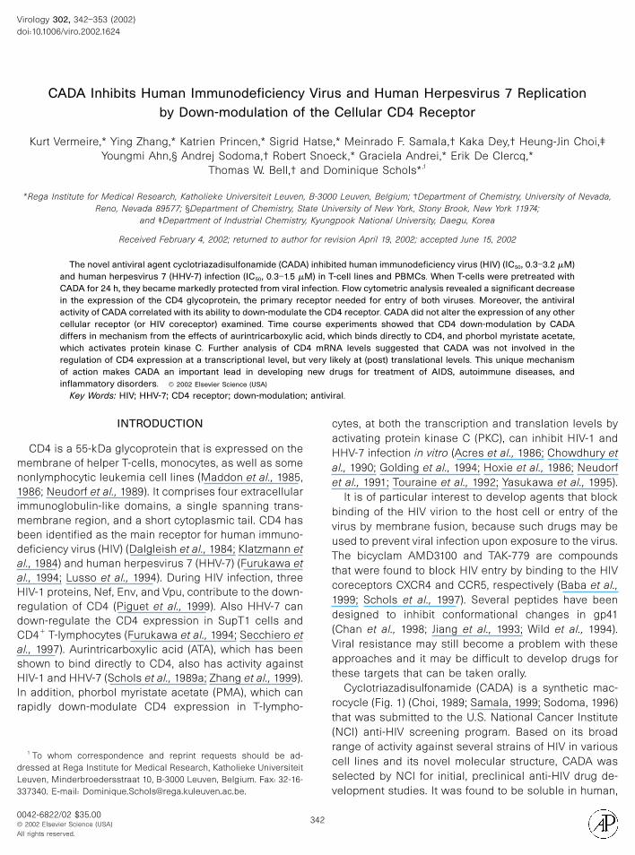
Virology 302, 342–353 (2002)
CADA Inhibits Human Immunodeficiency Virus and Human Herpesvirus 7 Replicationby Down-modulation of the Cellular CD4 Receptor
Kurt Vermeire,* Ying Zhang,* Katrien Princen,* Sigrid Hatse,* Meinrado F. Samala,† Kaka Dey,† Heung-Jin Choi,‡Youngmi Ahn,§ Andrej Sodoma,† Robert Snoeck,* Graciela Andrei,* Erik De Clercq,*
Thomas W. Bell,† and Dominique Schols*,1
*Rega Institute for Medical Research, Katholieke Universiteit Leuven, B-3000 Leuven, Belgium; †Department of Chemistry, University of Nevada,Reno, Nevada 89577; §Department of Chemistry, State University of New York, Stony Brook, New York 11974;
and ‡Department of Industrial Chemistry, Kyungpook National University, Daegu, Korea
Received February 4, 2002; returned to author for revision April 19, 2002; accepted June 15, 2002
The novel antiviral agent cyclotriazadisulfonamide (CADA) inhibited human immunodeficiency virus (HIV) (IC50, 0.3–3.2 �M)and human herpesvirus 7 (HHV-7) infection (IC50, 0.3–1.5 �M) in T-cell lines and PBMCs. When T-cells were pretreated withCADA for 24 h, they became markedly protected from viral infection. Flow cytometric analysis revealed a significant decreasein the expression of the CD4 glycoprotein, the primary receptor needed for entry of both viruses. Moreover, the antiviralactivity of CADA correlated with its ability to down-modulate the CD4 receptor. CADA did not alter the expression of any othercellular receptor (or HIV coreceptor) examined. Time course experiments showed that CD4 down-modulation by CADAdiffers in mechanism from the effects of aurintricarboxylic acid, which binds directly to CD4, and phorbol myristate acetate,which activates protein kinase C. Further analysis of CD4 mRNA levels suggested that CADA was not involved in theregulation of CD4 expression at a transcriptional level, but very likely at (post) translational levels. This unique mechanismof action makes CADA an important lead in developing new drugs for treatment of AIDS, autoimmune diseases, and
; antivi
INTRODUCTION
CD4 is a 55-kDa glycoprotein that is expressed on themembrane of helper T-cells, monocytes, as well as somenonlymphocytic leukemia cell lines (Maddon et al., 1985,1986; Neudorf et al., 1989). It comprises four extracellularimmunoglobulin-like domains, a single spanning trans-membrane region, and a short cytoplasmic tail. CD4 hasbeen identified as the main receptor for human immuno-deficiency virus (HIV) (Dalgleish et al., 1984; Klatzmann etal., 1984) and human herpesvirus 7 (HHV-7) (Furukawa etal., 1994; Lusso et al., 1994). During HIV infection, threeHIV-1 proteins, Nef, Env, and Vpu, contribute to the down-regulation of CD4 (Piguet et al., 1999). Also HHV-7 candown-regulate the CD4 expression in SupT1 cells andCD4� T-lymphocytes (Furukawa et al., 1994; Secchiero etal., 1997). Aurintricarboxylic acid (ATA), which has beenshown to bind directly to CD4, also has activity againstHIV-1 and HHV-7 (Schols et al., 1989a; Zhang et al., 1999).In addition, phorbol myristate acetate (PMA), which canrapidly down-modulate CD4 expression in T-lympho-
1 To whom correspondence and reprint requests should be ad-dressed at Rega Institute for Medical Research, Katholieke Universiteit
© 2002 Elsevier Science (USA)All rights reserved.
342
cytes, at both the transcription and translation levels byactivating protein kinase C (PKC), can inhibit HIV-1 andHHV-7 infection in vitro (Acres et al., 1986; Chowdhury etal., 1990; Golding et al., 1994; Hoxie et al., 1986; Neudorfet al., 1991; Touraine et al., 1992; Yasukawa et al., 1995).
It is of particular interest to develop agents that blockbinding of the HIV virion to the host cell or entry of thevirus by membrane fusion, because such drugs may beused to prevent viral infection upon exposure to the virus.The bicyclam AMD3100 and TAK-779 are compoundsthat were found to block HIV entry by binding to the HIVcoreceptors CXCR4 and CCR5, respectively (Baba et al.,1999; Schols et al., 1997). Several peptides have beendesigned to inhibit conformational changes in gp41(Chan et al., 1998; Jiang et al., 1993; Wild et al., 1994).Viral resistance may still become a problem with theseapproaches and it may be difficult to develop drugs forthese targets that can be taken orally.
Cyclotriazadisulfonamide (CADA) is a synthetic mac-rocycle (Fig. 1) (Choi, 1989; Samala, 1999; Sodoma, 1996)that was submitted to the U.S. National Cancer Institute(NCI) anti-HIV screening program. Based on its broadrange of activity against several strains of HIV in variouscell lines and its novel molecular structure, CADA was
inflammatory disorders. © 2002 Elsevier Science (USA)
Key Words: HIV; HHV-7; CD4 receptor; down-modulation
Leuven, Minderbroedersstraat 10, B-3000 Leuven, Belgium. Fax: 32-16-337340. E-mail: [email protected].
doi:10.1006/viro.2002.1624
0042-6822/02 $35.00
ral.
selected by NCI for initial, preclinical anti-HIV drug de-velopment studies. It was found to be soluble in human,
rat, and mouse plasma at 1.9–3.1 �M, to be stable inplasma (�200 h), and to be detectable in the blood-stream up to 2 h after intravenous injection in mice.
Here, we report that CADA specifically down-modu-lates cell surface and intracellular CD4 expression. For aperiod of up to 4 days, CADA completely blocked CD4expression in the MT-4 T-cell line, the SupT1 T-cell line,and peripheral blood mononuclear cells (PBMCs). ThisCD4 down-regulating effect resulted in a marked inhibi-tion of HIV and HHV-7 infection.
RESULTS
Anti-HIV and anti-HHV-7 activity of CADA
In order to confirm the antiviral data of CADA obtainedfrom the NCI anti-HIV screening program, we evaluatedCADA in our antiviral assays. First, the anti-HIV-1 activityof CADA was tested. Cells were given fivefold dilutions ofthe compound and infected with an optimized dose ofthe virus. HIV replication was monitored and whenstrong CPE was observed in untreated infected cells, allsamples were then scored for CPE and supernatant wascollected for HIV-1 core antigen (p24 Ag) ELISA. Assummarized in Table 1, CADA inhibits HIV-1 replication(strains IIIB, RF, and NL4.3) at an IC50 ranging from 0.3 to3.2 �M when evaluated in different CD4� T-cell lines(such as MT-4 and SupT1) and in PBMCs. Furthermore,for two HIV-2 strains (ROD and EHO), an IC50 of 0.2 �Mwas obtained in MT-4 cells.
Comparable data were obtained when CADA wasevaluated for its anti-HHV-7 activity. The IC50s of CADAfor HHV-7 were 0.3 and 1.5 �M when evaluated in SupT1cells and PBMCs, respectively. For CD8� cell-depletedPBMCs an IC50 of 1.0 �M was measured. The CC50
values of CADA in MT-4 cells, SupT1 cells, and PBMCwere 39, 142, and 73 �M, respectively. Thus, CADAequally inhibited HIV and HHV-7 infection.
Specific down-modulation of cell surface CD4 byCADA
Since the cellular CD4 receptor is the primary receptorneeded for entry of HIV and HHV-7, the effect of CADA onCD4 expression was evaluated. A selective down-mod-
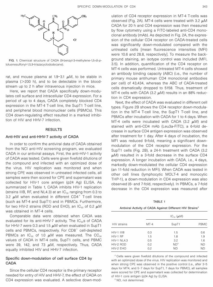
ulation of CD4 receptor expression in MT-4 T-cells wasobserved (Fig. 2A). MT-4 cells were treated with 3.2 �MCADA for 20 h and CD4 expression was then measuredby flow cytometry using a FITC-labeled anti-CD4 mono-clonal antibody (mAb). As depicted in Fig. 2A, the expres-sion of the cellular CD4 receptor on CADA-treated cellswas significantly down-modulated compared with theuntreated cells [mean fluorescence intensities (MFI)were 10.6 and 28.8, respectively]. To measure the back-ground staining, an isotype control was included (MFI,3.5). In addition, quantification of the CD4 receptor onMT-4 cells was performed. Untreated MT-4 cells showedan antibody binding capacity (ABC) (i.e., the number ofprimary mouse antihuman CD4 monoclonal antibodiesper cell) of 43,434, whereas the ABC of CADA-treatedcells dramatically dropped to 5158. Thus, treatment ofMT-4 cells with CADA (3.2 �M) results in an 88% reduc-tion in CD4 expression.
Next, the effect of CADA was evaluated in different celltypes. Figure 2B shows the CD4 receptor down-modula-tion in the MT-4 T-cell line, the SupT1 T-cell line, andPBMCs after incubation with CADA for 1 to 4 days. WhenMT-4 cells were incubated with CADA (3.2 �M) andstained with anti-CD4 mAb (Leu3a-FITC), a 6-fold de-crease in surface CD4 antigen expression was observedafter treatment for 1 day. After 4 days of incubation, theMFI was reduced 9-fold, meaning a significant down-modulation of the CD4 receptor expression. For theSupT1 cells (Fig. 2B), a 24-h treatment with CADA (3.2�M) resulted in a 3-fold decrease in the surface CD4expression. A longer incubation with CADA, i.e., 4 days,markedly down-modulated the cellular CD4 expression(an 11-fold reduction in MFI). When CADA was tested inother cell lines (lymphocytic MOLT-4 and monocyticTHP-1), a down-modulation in CD4 expression was alsoobserved (6- and 7-fold, respectively). In PBMCs, a 7-folddecrease in the CD4 expression was measured after
TABLE 1
Antiviral Activity of CADA Against Different HIV Strainsa
HIV strains
IC50 (�M)
MT-4 SupT1 PBMC
HIV-1 IIIB 0.3 1.5 0.6HIV-1 RF 1.5 1.8 1.9HIV-1 NL4.3 0.5 3.2 1.6HIV-2 ROD 0.2 NDb NDHIV-2 EHO 0.2 ND ND
a Cells were given fivefold dilutions of the compound and infectedwith an optimized dose of the virus. HIV replication was monitored andwhen strong CPE was observed in the positive control (i.e., after 3–5days for MT4, and 5–7 days for SupT1, 7 days for PBMC), all sampleswere scored for CPE and supernatant was collected for determinationof HIV-1 core antigen (p24 Ag) by ELISA.
b ND, not determined.
FIG. 1. Chemical structure of CADA (9-benzyl-3-methylene-1,5-di-p-toluenesulfonyl-1,5,9-triazacyclododecane).
343SPECIFIC DOWN-MODULATION OF CD4
incubation with CADA (16 �M) for 4 days (Fig. 2B). Fur-ther flow cytometric analysis of PBMCs revealed a CD4down-regulating effect of CADA not only in lymphocytesbut also in monocytes (7- and 8-fold reductions of MFI,respectively). Thus, CADA significantly down-modulatedCD4 expression when evaluated in different humanCD4� cell lines and in PBMCs. Comparable results wereobtained with two other anti-CD4 mAbs, OKT4a andOKT4; the latter also binds to a different region of CD4(data not shown).
CADA down-regulates intracellular CD4
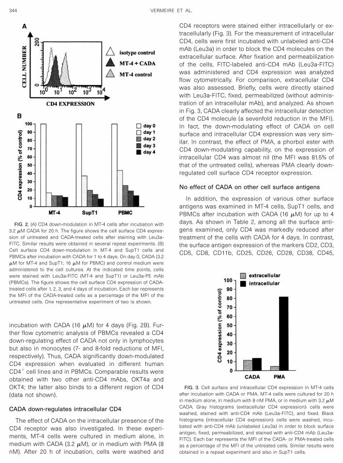
The effect of CADA on the intracellular presence of theCD4 receptor was also investigated. In these experi-ments, MT-4 cells were cultured in medium alone, inmedium with CADA (3.2 �M), or in medium with PMA (8nM). After 20 h of incubation, cells were washed and
CD4 receptors were stained either intracellularly or ex-tracellularly (Fig. 3). For the measurement of intracellularCD4, cells were first incubated with unlabeled anti-CD4mAb (Leu3a) in order to block the CD4 molecules on theextracellular surface. After fixation and permeabilizationof the cells, FITC-labeled anti-CD4 mAb (Leu3a-FITC)was administered and CD4 expression was analyzedflow cytometrically. For comparison, extracellular CD4was also assessed. Briefly, cells were directly stainedwith Leu3a-FITC, fixed, permeabilized (without adminis-tration of an intracellular mAb), and analyzed. As shownin Fig. 3, CADA clearly affected the intracellular detectionof the CD4 molecule (a sevenfold reduction in the MFI).In fact, the down-modulating effect of CADA on cellsurface and intracellular CD4 expression was very sim-ilar. In contrast, the effect of PMA, a phorbol ester withCD4 down-modulating capability, on the expression ofintracellular CD4 was almost nil (the MFI was 81.5% ofthat of the untreated cells), whereas PMA clearly down-regulated cell surface CD4 receptor expression.
No effect of CADA on other cell surface antigens
In addition, the expression of various other surfaceantigens was examined in MT-4 cells, SupT1 cells, andPBMCs after incubation with CADA (16 �M) for up to 4days. As shown in Table 2, among all the surface anti-gens examined, only CD4 was markedly reduced aftertreatment of the cells with CADA for 4 days. In contrast,the surface antigen expression of the markers CD2, CD3,CD5, CD8, CD11b, CD25, CD26, CD28, CD38, CD45,
FIG. 3. Cell surface and intracellular CD4 expression in MT-4 cellsafter incubation with CADA or PMA. MT-4 cells were cultured for 20 hin medium alone, in medium with 8 nM PMA, or in medium with 3.2 �MCADA. Gray histograms (extracellular CD4 expression): cells werewashed, stained with anti-CD4 mAb (Leu3a-FITC), and fixed. Blackhistograms (intracellular CD4 expression): cells were washed, incu-bated with anti-CD4 mAb (unlabeled Leu3a) in order to block surfaceantigen, fixed, permeabilized, and stained with anti-CD4 mAb (Leu3a-FITC). Each bar represents the MFI of the CADA- or PMA-treated cellsas a percentage of the MFI of the untreated cells. Similar results wereobtained in a repeat experiment and also in SupT1 cells.
FIG. 2. (A) CD4 down-modulation in MT-4 cells after incubation with3.2 �M CADA for 20 h. The figure shows the cell surface CD4 expres-sion of untreated and CADA-treated cells after staining with Leu3a-FITC. Similar results were obtained in several repeat experiments. (B)Cell surface CD4 down-modulation in MT-4 and SupT1 cells andPBMCs after incubation with CADA for 1 to 4 days. On day 0, CADA (3.2�M for MT-4 and SupT1; 16 �M for PBMC) and control medium wereadministered to the cell cultures. At the indicated time points, cellswere stained with Leu3a-FITC (MT-4 and SupT1) or Leu3a-PE mAb(PBMCs). The figure shows the cell surface CD4 expression of CADA-treated cells after 1, 2, 3, and 4 days of incubation. Each bar representsthe MFI of the CADA-treated cells as a percentage of the MFI of theuntreated cells. One representative experiment of two is shown.
344 VERMEIRE ET AL.
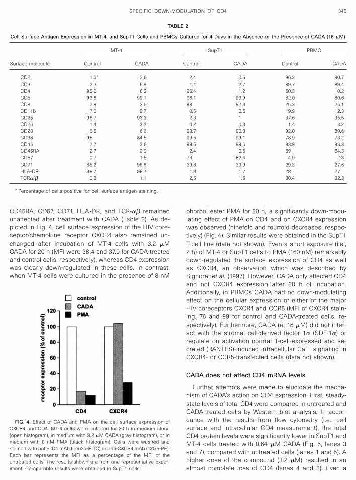
CD45RA, CD57, CD71, HLA-DR, and TCR-�� remainedunaffected after treatment with CADA (Table 2). As de-picted in Fig. 4, cell surface expression of the HIV core-ceptor/chemokine receptor CXCR4 also remained un-changed after incubation of MT-4 cells with 3.2 �MCADA for 20 h (MFI were 38.4 and 37.0 for CADA-treatedand control cells, respectively), whereas CD4 expressionwas clearly down-regulated in these cells. In contrast,when MT-4 cells were cultured in the presence of 8 nM
phorbol ester PMA for 20 h, a significantly down-modu-lating effect of PMA on CD4 and on CXCR4 expressionwas observed (ninefold and fourfold decreases, respec-tively) (Fig. 4). Similar results were obtained in the SupT1T-cell line (data not shown). Even a short exposure (i.e.,2 h) of MT-4 or SupT1 cells to PMA (160 nM) remarkablydown-regulated the surface expression of CD4 as wellas CXCR4, an observation which was described bySignoret et al. (1997). However, CADA only affected CD4and not CXCR4 expression after 20 h of incubation.Additionally, in PBMCs CADA had no down-modulatingeffect on the cellular expression of either of the majorHIV coreceptors CXCR4 and CCR5 (MFI of CXCR4 stain-ing, 76 and 99 for control and CADA-treated cells, re-spectively). Furthermore, CADA (at 16 �M) did not inter-act with the stromal cell-derived factor 1� (SDF-1�) orregulate on activation normal T-cell-expressed and se-creted (RANTES)-induced intracellular Ca2� signaling inCXCR4- or CCR5-transfected cells (data not shown).
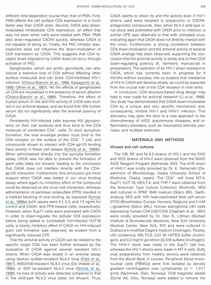
CADA does not affect CD4 mRNA levels
Further attempts were made to elucidate the mecha-nism of CADA’s action on CD4 expression. First, steady-state levels of total CD4 were compared in untreated andCADA-treated cells by Western blot analysis. In accor-dance with the results from flow cytometry (i.e., cellsurface and intracellular CD4 measurement), the totalCD4 protein levels were significantly lower in SupT1 andMT-4 cells treated with 0.64 �M CADA (Fig. 5, lanes 3and 7), compared with untreated cells (lanes 1 and 5). Ahigher dose of the compound (3.2 �M) resulted in analmost complete loss of CD4 (lanes 4 and 8). Even a
TABLE 2
Cell Surface Antigen Expression in MT-4, and SupT1 Cells and PBMCs Cultured for 4 Days in the Absence or the Presence of CADA (16 �M)
Surface molecule
MT-4 SupT1 PBMC
Control CADA Control CADA Control CADA
CD2 1.5a 2.6 2.4 0.5 96.2 90.7CD3 2.3 5.9 1.4 2.7 89.7 89.4CD4 95.6 6.3 96.4 1.2 60.3 0.2CD5 99.6 99.1 96.1 93.9 82.0 80.6CD8 2.8 3.5 98 92.3 25.3 25.1CD11b 7.0 9.7 0.5 0.6 19.9 12.3CD25 96.7 93.3 2.3 1 37.6 35.5CD26 1.4 3.2 0.2 0.3 1.4 3.2CD28 6.6 6.6 98.7 90.8 92.0 89.6CD38 95 84.5 99.5 99.1 78.9 73.2CD45 2.7 3.6 99.5 99.6 98.9 98.3CD45RA 2.7 2.0 2.4 0.5 69 64.3CD57 0.7 1.5 73 82.4 4.9 2.3CD71 85.2 98.8 39.8 33.9 29.3 27.6HLA-DR 98.7 98.7 1.9 1.7 28 27TCR�/� 0.8 1.1 2.5 1.6 80.4 82.3
a Percentage of cells positive for cell surface antigen staining.
FIG. 4. Effect of CADA and PMA on the cell surface expression ofCXCR4 and CD4. MT-4 cells were cultured for 20 h in medium alone(open histogram), in medium with 3.2 �M CADA (gray histogram), or inmedium with 8 nM PMA (black histogram). Cells were washed andstained with anti-CD4 mAb (Leu3a-FITC) or anti-CXCR4 mAb (12G5-PE).Each bar represents the MFI as a percentage of the MFI of theuntreated cells. The results shown are from one representative exper-iment. Comparable results were obtained in SupT1 cells.
345SPECIFIC DOWN-MODULATION OF CD4
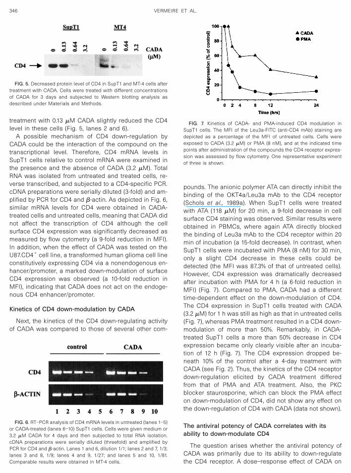
treatment with 0.13 �M CADA slightly reduced the CD4level in these cells (Fig. 5, lanes 2 and 6).
A possible mechanism of CD4 down-regulation byCADA could be the interaction of the compound on thetranscriptional level. Therefore, CD4 mRNA levels inSupT1 cells relative to control mRNA were examined inthe presence and the absence of CADA (3.2 �M). TotalRNA was isolated from untreated and treated cells, re-verse transcribed, and subjected to a CD4-specific PCR.cDNA preparations were serially diluted (3-fold) and am-plified by PCR for CD4 and �-actin. As depicted in Fig. 6,similar mRNA levels for CD4 were obtained in CADA-treated cells and untreated cells, meaning that CADA didnot affect the transcription of CD4 although the cellsurface CD4 expression was significantly decreased asmeasured by flow cytometry (a 9-fold reduction in MFI).In addition, when the effect of CADA was tested on theU87.CD4� cell line, a transformed human glioma cell lineconstitutively expressing CD4 via a nonendogenous en-hancer/promoter, a marked down-modulation of surfaceCD4 expression was observed (a 10-fold reduction inMFI), indicating that CADA does not act on the endoge-nous CD4 enhancer/promoter.
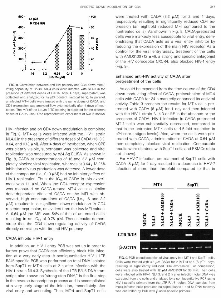
Kinetics of CD4 down-modulation by CADA
Next, the kinetics of the CD4 down-regulating activityof CADA was compared to those of several other com-
pounds. The anionic polymer ATA can directly inhibit thebinding of the OKT4a/Leu3a mAb to the CD4 receptor(Schols et al., 1989a). When SupT1 cells were treatedwith ATA (118 �M) for 20 min, a 9-fold decrease in cellsurface CD4 staining was observed. Similar results wereobtained in PBMCs, where again ATA directly blockedthe binding of Leu3a mAb to the CD4 receptor within 20min of incubation (a 15-fold decrease). In contrast, whenSupT1 cells were incubated with PMA (8 nM) for 30 min,only a slight CD4 decrease in these cells could bedetected (the MFI was 87.3% of that of untreated cells).However, CD4 expression was dramatically decreasedafter incubation with PMA for 4 h (a 6-fold reduction inMFI) (Fig. 7). Compared to PMA, CADA had a differenttime-dependent effect on the down-modulation of CD4.The CD4 expression in SupT1 cells treated with CADA(3.2 �M) for 1 h was still as high as that in untreated cells(Fig. 7), whereas PMA treatment resulted in a CD4 down-modulation of more than 50%. Remarkably, in CADA-treated SupT1 cells a more than 50% decrease in CD4expression became only clearly visible after an incuba-tion of 12 h (Fig. 7). The CD4 expression dropped be-neath 10% of the control after a 4-day treatment withCADA (see Fig. 2). Thus, the kinetics of the CD4 receptordown-regulation elicited by CADA treatment differedfrom that of PMA and ATA treatment. Also, the PKCblocker staurosporine, which can block the PMA effecton down-modulation of CD4, did not show any effect onthe down-regulation of CD4 with CADA (data not shown).
The antiviral potency of CADA correlates with itsability to down-modulate CD4
The question arises whether the antiviral potency ofCADA was primarily due to its ability to down-regulatethe CD4 receptor. A dose–response effect of CADA on
FIG. 5. Decreased protein level of CD4 in SupT1 and MT-4 cells aftertreatment with CADA. Cells were treated with different concentrationsof CADA for 3 days and subjected to Western blotting analysis asdescribed under Materials and Methods.
FIG. 6. RT–PCR analysis of CD4 mRNA levels in untreated (lanes 1–5)or CADA-treated (lanes 6–10) SupT1 cells. Cells were given medium or3.2 �M CADA for 4 days and then subjected to total RNA isolation.cDNA preparations were serially diluted (threefold) and amplified byPCR for CD4 and �-actin. Lanes 1 and 6, dilution 1/1; lanes 2 and 7, 1/3;lanes 3 and 8, 1/9; lanes 4 and 9, 1/27; and lanes 5 and 10, 1/81.Comparable results were obtained in MT-4 cells.
FIG. 7. Kinetics of CADA- and PMA-induced CD4 modulation inSupT1 cells. The MFI of the Leu3a-FITC (anti-CD4 mAb) staining aredepicted as a percentage of the MFI of untreated cells. Cells wereexposed to CADA (3.2 �M) or PMA (8 nM), and at the indicated timepoints after administration of the compounds the CD4 receptor expres-sion was assessed by flow cytometry. One representative experimentof three is shown.
346 VERMEIRE ET AL.
HIV infection and on CD4 down-modulation is combinedin Fig. 8. MT-4 cells were infected with the HIV-1 strainNL4.3 in the presence of different doses of CADA (16, 3.2,0.64, and 0.13 �M). After 4 days of incubation, when CPEwas clearly visible, supernatant was collected and viralreplication was measured by p24 Ag ELISA. As shown inFig. 8, CADA at concentrations of 16 and 3.2 �M com-pletely blocked viral replication, whereas at 0.64 �M 25%inhibition of virus production was detected. A lower doseof the compound (i.e., 0.13 �M) had no inhibitory effect onHIV-1 replication. Thus, the IC50 of CADA in this experi-ment was 1.1 �M. When the CD4 receptor expressionwas measured on CADA-treated MT-4 cells, a similardose-dependent effect of CADA on the MFI was ob-served. High concentrations of CADA (i.e., 16 and 3.2�M) resulted in a significant down-modulation in CD4receptor expression, as evident from the low MFI values.At 0.64 �M the MFI was 54% of that of untreated cells,resulting in an IC50 of 0.76 �M. These results demon-strate that the CD4 down-regulating activity of CADAdirectly correlates with its anti-HIV potency.
CADA inhibits HIV-1 entry
In addition, an HIV-1 entry PCR was set up in order tofurther prove that CADA can efficiently block HIV infec-tion at a very early step. A semiquantitative HIV-1 LTRR/U5-specific PCR was performed on total DNA isolatedfrom SupT1 or MT-4 cells at 2 h after infection with theHIV-1 strain NL4.3. Synthesis of the LTR R/U5 DNA tran-script, also known as “strong-stop DNA,” is the first stepin the reverse transcription process and is accomplishedat a very early stage of the infection, immediately afterviral entry and uncoating. Thus, MT-4 and SupT1 cells
were treated with CADA (3.2 �M) for 2 and 4 days,respectively, resulting in significantly reduced CD4 ex-pression (an eightfold reduced MFI compared to thenontreated cells). As shown in Fig. 9, CADA-pretreatedcells were markedly less susceptible to viral entry, dem-onstrating that CADA acts as a viral entry inhibitor byreducing the expression of the main HIV receptor. As acontrol for the viral entry assay, treatment of the cellswith AMD3100 (12 �M), a strong and specific antagonistof the HIV coreceptor CXCR4, also blocked HIV-1 entry(Fig. 9).
Enhanced anti-HIV activity of CADA afterpretreatment of the cells
As could be expected from the time course of the CD4down-modulating effect of CADA, preincubation of MT-4cells with CADA for 24 h markedly enhanced its antiviralactivity. Table 3 presents the results for MT-4 cells pre-treated with CADA (8 �M) for 1 day and then infectedwith the HIV-1 strain NL4.3 or RF in the absence or thepresence of CADA. HIV-1 infection in CADA-pretreatedMT-4 cells was substantially decreased, compared tothat in the untreated MT-4 cells (a 4.5-fold reduction inp24 core antigen levels). Also, when the cells were pre-treated with CADA, administration of CADA at 0.64 �Mthen completely blocked viral replication. Comparableresults were obtained with SupT1 cells and PBMCs (datanot shown).
For HHV-7 infection, pretreatment of SupT1 cells withCADA (8 �M) for 1 day resulted in a decrease in HHV-7infection of more than threefold compared to that in
FIG. 9. PCR-based detection of virus entry into MT-4 and SupT1 cells.Cells were treated with 3.2 �M CADA for 2 (MT-4) or 4 (SupT1) days,resulting in significantly reduced CD4 expression. For comparison,cells were also treated with 12 �M AMD3100 for 30 min. Then cellswere infected with HIV-1 NL4.3, and 2 h after infection total DNA wasextracted from the cells and analyzed by a semiquantitative PCR usingHIV-1-specific primers from the LTR R/U5 region. DNA samples frommock-infected cells produced no signal (lanes 1 and 5). DNA recoverywas controlled by PCR with �-actin-specific primers.
FIG. 8. Correlation between anti-HIV potency and CD4 down-modu-lating capability of CADA. MT-4 cells were infected with NL4.3 in thepresence of different doses of CADA. After 4 days, supernatant wascollected and analyzed for its p24 content (vertical bars). In parallel,uninfected MT-4 cells were treated with the same doses of CADA, andCD4 expression was analyzed flow cytometrically after 4 days of incu-bation. The MFI of the Leu3a-FITC staining is depicted for the differentdoses of CADA (line). One representative experiment of two is shown.
347SPECIFIC DOWN-MODULATION OF CD4

untreated SupT1 cells. Thus, as expected from the datapresented in Fig. 2, pretreatment of the cells with CADAfor 24 h clearly enhanced its antiviral potency.
DISCUSSION
Incubation of CD4� T cells with CADA resulted in adown-modulation of the cell surface CD4 expression. Inaddition, intracellular CD4 expression was also stronglyblocked after incubation with CADA. Further Westernblotting analysis of the total CD4 protein levels confirmedthe CD4 down-regulating activity of the compound. Ex-cept for the CD4 receptor, CADA did not have effect onany of the other cellular receptors evaluated so far. As inSupT1 cells, a significant down-modulating effect onCD4 expression was also detected in PBMCs. Thus, inall cell lines tested, CADA acted as a very specificdown-modulator of CD4. Interestingly, even in theU87.CD4� cell line, a transformed human glioma cell lineconstitutively expressing CD4 via a nonendogenous en-hancer/promoter (Clapham et al., 1991), a marked down-modulated of surface CD4 expression was observed.This suggested that interaction of CADA on CD4 mRNAregulation seemed very unlikely. Indeed, when the effectof CADA was evaluated on the transcriptional level ofCD4, no differences in CD4 mRNA production were mea-sured in CADA-treated T-cells compared to untreatedcells (Fig. 6). The data with the kinetics of CADA indicatethat the timing of the CD4 down-regulating effect ofCADA varied somewhat from one cell line to another (inMT-4 cells the CD4 down-modulation began to occurafter 12 h, whereas in SupT1 cells and PBMCs the effectof CADA was more pronounced after 4 days of incuba-tion). This relatively long incubation time before modula-tion of CD4 becomes clearly visible suggests that CADAmay affect CD4 expression at the translation level.
As three CD4 binding events are needed to efficientlyactivate HIV-1 Env trimers (Layne et al., 1990), multimericCD4 binding is required for HIV infection, further implyingthat receptor density plays a crucial role in the efficiency
of viral infectivity (Platt et al., 1997). Davis et al. reportedthat CD4� T-cells bind approximately 49,000 CD4 (Leu3a)antibody molecules and that this binding is bivalent,suggesting 98,000 CD4 antigen molecules on the sur-face of these cells (Davis et al., 1998). Our data concern-ing the quantification of the CD4 receptor showed asimilar result (approximately 43,000 CD4 antibody (Ab)molecules/cell). Interestingly, after treatment with CADAthe amount of CD4 Ab bound to the cells dropped toapproximately 5000/cell. Thus, CADA is able to reducethe CD4 receptor density by almost 90%. It has beenreported that CD4 receptor density will significantly im-pact the efficiency of viral entry and when the CD4 levelbecomes limiting (less than 10,000 CD4 molecules/cell) itstrongly inhibits viral infection (Kabat et al., 1994; Platt etal., 1997); primary HIV isolates especially seem to bemuch more dependent on the level of CD4 expression(Kabat et al., 1994; Platt et al., 2000). When evaluatedagainst six different primary HIV-1 isolates in PBMCs,CADA showed a potent (but variable) activity rangingfrom 0.002 to 2.3 �M. Although CD4-independent viruseshave been described (Hoffman et al., 1999), these aremuch more sensitive to neutralization by antibodies,which could explain the rarity of CD4-independent wild-type HIV variants (Edwards et al., 2001; Kolchinsky et al.,2001). A CD4-lowering drug, such as CADA, not onlyinhibits viral infection by blocking viral entry (Fig. 9), butalso can make the virus more prone to elimination by theneutralizing action of antibodies. Furthermore, as severaldomains of CD4 play an important role in regulating HIVentry of cells (Poulin et al., 1991), a specific down-mod-ulator of the complete CD4 molecule may be consideredan effective antiviral agent.
The anionic polymer ATA inhibits HIV-1 replication by adirect interaction with the CD4 receptor (Schols et al.,1989a). It can also inhibit HHV-7 infection, probably dueto its interference with the CD4 receptor (Zhang et al.,1999). Inhibition of CD4 expression by ATA can be de-tected immediately after incubating the cells with ATA. Incontrast, our data show that incubation of the cells withCADA for 1 h did not affect CD4 receptor expression,suggesting that the down-modulation of CD4 expressionby CADA is not due to direct binding to the surface CD4receptor. Previous studies have shown that the phorbolester PMA can down-regulate CD4 expression at thetranscriptional and translational levels (Neudorf et al.,1991). Phorbol esters can also down-modulate the CD4receptor by activating PKC (Chowdhury et al., 1990). Inaddition, it has been shown that PMA can inhibit HHV-7infection (Yasukawa et al., 1995) and HIV-1 infection(Touraine et al., 1992). Furthermore, phorbol esters inhibitHIV-induced syncytia formation through down-modula-tion of the surface CD4 receptor (Chowdhury et al., 1990)or by modulating an accessory component in the CD4�
cells (Golding et al., 1994). From our data we can con-clude that the down-modulation of CD4 by CADA has a
TABLE 3
Levels of p24 Antigen (pg/ml) in the Supernatant of MT-4 CellsInfected with HIV-1 Strains NL4.3 and RF
NL4.3 RF
Pretreateda Untreated Pretreateda Untreated
MediumCADA 34,839 158,627 42,853 100,002
(3.2 �M) �5 �5 �5 �5(0.64 �M) �5 63,098 �5 56,139(0.13 �M) 33,785 127,838 35,894 75,118
a Cells were pretreated with CADA (8 �M) for 24 h, and the com-pound was added again after HIV infection at the indicated concentra-tions.
348 VERMEIRE ET AL.
different time-dependent course than that of PMA. First,PMA affects the cell surface CD4 expression in a muchfaster way than CADA does. Second, CADA also down-modulated intracellular CD4 expression, an effect thatwas not seen when cells were treated with PMA. PMAalso down-regulated CXCR4 quickly, whereas CADA isnot capable of doing so. Finally, the PKC inhibitor stau-rosporine does not influence the down-modulation ofCD4 expression by CADA. This suggests that CD4 re-ceptor down-regulation by CADA does not occur throughactivation of PKC.
Gangliosides, which are acidic glycolipids, can alsoinduce a selective loss of CD4 without affecting othersurface molecules and can block CD4-mediated HIV-1infection (Chieco-Bianchi et al., 1989; Kawaguchi et al.,1989; Offner et al., 1987). Yet the effects of gangliosideson CD4 are neutralized in the presence of serum albumin(Chieco-Bianchi et al., 1989). Therefore, the effect ofhuman serum on the anti-HIV activity of CADA was stud-ied in our antiviral assays, and we found that 10% humanserum did not significantly alter the antiviral potency ofCADA.
Persistently HIV-infected cells express HIV glycopro-teins on their cell surfaces and thus bind to the CD4molecule of uninfected CD4� cells. To elicit syncytiumformation, the viral envelope protein must bind to theCD4 receptor on the surface of the CD4� T cell, andcompounds shown to interact with CD4–gp120 bindinghave activity in these cell assays (Schols et al., 1989b).When administered directly into the syncytia formationassay, CADA was not able to prevent the formation ofgiant cells (data not shown), leading to the conclusionthat CADA does not interfere directly with the CD4–gp120 interaction. Furthermore, this conclusion got moresupport when CADA was tested in our virus bindingassay, where again no inhibitory effect of the compoundcould be observed on the virus–cell interaction, whereasadministration of pentosan polysulfate (PPS) resulted ina marked blocking of viral binding, as expected (Scholset al., 1989a) (p24 values were 5.1, 5.4, and 1.3 ng/ml forcontrol and CADA- and PPS-treated cells, respectively).However, when SupT1 cells were pretreated with CADAin order to down-regulate the cellular CD4 expressionbefore being added to (untreated) HIV-infected HUT78cells, a clearly inhibitory effect of CADA on HIV-inducedgiant cell formation was observed, as evident from asignificantly reduced CPE.
That the antiviral activity of CADA can be related to thespecific target CD4 has been further stressed by theevaluation of CADA against several resistant HIV-1strains. When CADA was tested in an antiviral assayusing dextran sulfate-resistant NL4.3 virus (Este et al.,1997), AMD3100-resistant NL4.3 virus (De Vreese et al.,1996), or SDF-1�-resistant NL4.3 virus (Schols et al.,1998), no loss of activity was detected compared to thatin the wild-type NL4.3 virus (data not shown). Thus,
CADA seems to retain its anti-HIV activity even if HIV-1strains used were resistant to polyanionic or CXCR4-antagonistic compounds. Also, when NL4.3 wild-type vi-rus stock was pretreated with CADA prior to infection, asimilar CPE was observed to that with untreated virus,indicating again that CADA does not directly interact withthe virion. Furthermore, a strong correlation betweenCD4 down-modulation and the antiviral activity of severalCADA analogs has been observed, supporting the con-clusion that the antiviral activity is solely due to their CD4down-regulating potency (K. Vermeire, manuscript inpreparation). The selection of an HIV-1 strain resistant toCADA, which has currently been in progress for 4months without success, lets us suspect that resistanceof HIV to CADA will develop slowly, as could be expectedfrom the crucial role of the CD4 receptor in viral entry.
In conclusion, CD4 structure-based drug design maylead to new roads for antiviral research (Li et al., 1998).Our study has demonstrated that CADA down-modulatesCD4 by a unique and very specific mechanism and,consequently, inhibits HIV-1 and HHV-7 infection. Thisdiscovery may open the door to a new approach to thechemotherapy of AIDS, autoimmune diseases, and in-flammatory disorders, such as rheumatoid arthritis, pso-riasis, and multiple sclerosis.
MATERIALS AND METHODS
Viruses and cell cultures
The IIIB, RF, and NL4.3 strains of HIV-1 and the EHOand ROD strains of HIV-2 were obtained from the NIAIDAIDS Reagent Program (Bethesda, MD). The KHR strainof HHV-7 was kindly provided by Dr. K. Yamanishi (De-partment of Microbiology, Osaka University School ofMedicine, Osaka, Japan). The CD4� cell lines MT-4,SupT1, HUT-78, MOLT-4, and THP-1 were obtained fromthe American Type Culture Collection (Rockville, MD)and cultured in RPMI 1640 medium (Gibco BRL, Gaith-ersburg, MD) with 10% heat-inactivated fetal calf serum(FCS) (Biowhittaker Europe, Verviers, Belgium) and 2 mML-glutamine (Gibco BRL). Human astroglioma U87 cellsexpressing human CD4 (U87.CD4) (Clapham et al., 1991)were kindly provided by Dr. Dan R. Littman (SkirballInstitute of Biomolecular Medicine, New York UniversityMedical Center, New York, NY) and were cultured inDulbecco’s modified Eagle’s medium (Invitrogen, Paisley,UK) containing 10% FCS, 0.01 M HEPES buffer (Invitro-gen), and 0.2 mg/ml geneticin (G-418 sulfate) (Invitrogen).The HHV-7 stock was made in the SupT1 cell line,whereas the HIV-1 stocks were made in MT-4 cells. Buffycoat preparations from healthy donors were obtainedfrom the Blood Bank in Leuven. Peripheral blood mono-nuclear cells (PBMCs) were first isolated by densitygradient centrifugation over Lymphoprep (d � 1.077g/ml) (Nycomed, Oslo, Norway). CD8 magnetic beads(Dynal AS, Oslo, Norway) were added to remove the
349SPECIFIC DOWN-MODULATION OF CD4
CD8� T lymphocytes. The PBMCs and CD8� T-cell-de-pleted PBMCs were stimulated with 2 �g/ml of phytohe-magglutinin (PHA) (Sigma Chemical Co., Bornem, Bel-gium) for 3 days at 37°C.
Compounds and monoclonal antibodies
Aurintricarboxylic acid (ATA; M r 422.3), phorbol myris-tate acetate (PMA; M r 616.8), and pentosan polysulfate(PPS; M r 3100) were purchased from Sigma ChemicalCompany. The bicyclam AMD3100 was synthesized asdescribed previously (Bridger et al., 1995). The com-pound CADA (M r 618, Fig. 1) was synthesized by Dr.Thomas W. Bell (Department of Chemistry, University ofNevada, Reno, NV) by a modification of the methodsdeveloped in Stony Brook (Department of Chemistry,State University of New York, Stony Brook, NY). CADAwas dissolved at 16 mM in DMSO.
The mAbs labeled with phycoerythrin (PE) or FITCused were the following: CD2, CD3, CD4 (Leu3a,RPA-T4), CD5, CD8, CD11b, CD25, CD26, CD26, CD28,CD38, CD45, CD45RA, CD57, CD71, HLA-DR, TCR-��,CXCR4, and CCR5. All mAbs were purchased from BDBiosciences (Erembodegem, Belgium). The anti-CD4mAbs OKT4 and OKT4a were purchased from OrthoDiagnostic Systems (Beerse, Belgium). The HIV-1 p24antigen ELISA kit was purchased from NEN (Brussels,Belgium). The specific mAb to HHV-7 (RK-4) (AdvancedBiotechnologies, Columbia, MD) recognizing an earlyHHV-7 protein was used to detect HHV-7-infected cells.
Antiviral activity
For HIV-1, PBMCs were incubated with the HIV-1strains IIIB, RF, and NL4.3 for 2 h at 37°C. Then the cellswere washed with warm medium and seeded into 48-well flat bottom plates (Iwaki, Japan). Fivefold dilutions ofthe compound were added to each well, together with 1ng/ml of IL-2 (R&D Systems Europe, Abingdon, Oxon,UK). The anti-HIV-1 assays were also performed withMT-4 and SupT1 cells. Fivefold dilutions of CADA in 100�l were added to 96-well flat bottom plates (Iwaki). Then6 � 104 MT-4 cells or SupT1 cells were added in 50 �l ofmedium, and finally 50 �l of diluted HIV-1 stocks (strainsIIIB, RF, and NL4.3) was added to each well. Cytopathiceffect induced by the virus was checked regularly micro-scopically. When strong CPE was observed in the posi-tive control (i.e., untreated HIV-1-infected cells), all sam-ples were scored for CPE at the same time regardless ofthe CPE present, and the supernatant of each samplewas then collected (at the same time), stored at �20°C,and analyzed for HIV-1 core antigen by p24 Ag ELISA.
For HHV-7, fivefold dilutions of CADA were added in500 �l of culture medium in 24-well flat bottom plates(Iwaki), whereupon 2 � 105 SupT1 cells were added in400 �l of culture medium. After 30 min of incubation atroom temperature, 100 �l of HHV-7 stock was added to
each well. HHV-7-infected and mock-infected SupT1cells were cultured in a final volume of 1 ml of medium inthe absence of CADA. On day 4, half of the medium andthe cells were replaced and fresh medium without newcompound was added. This procedure was repeatedevery 2 or 3 days. In the anti-HHV-7 assay in PBMCs andCD8� T-cell-depleted PBMCs, fivefold dilutions of CADAwere added in 250 �l of culture medium in 24-wellplates, and then 2 � 106 cells were added in 400 �l ofculture medium. After 30 min of incubation at room tem-perature, 100 �l of HHV-7 stock was added together withIL-2 (1 ng/ml). At day 4, 1 ml of fresh medium containingIL-2 (1 ng/ml) was added to each well. The CPE waschecked regularly microscopically. When CPE was ob-served, the HHV-7 antigen expression was monitored byflow cytometry, as described previously (Zhang et al.,1999).
Flow cytometric analyses
To study the effect of CADA on surface CD4 antigenexpression, MT-4 cells, SupT1 cells, HHV-7-infectedSupT1 cells, and PBMCs were incubated with a serialfivefold dilution of CADA (16, 3.2, 0.64, and 0.13 �M) ormedium at 37°C. Cell surface CD4 antigen expressionwas analyzed at different time points (up to 4 days). Afterwashing with phosphate-buffered saline (PBS) contain-ing 2% fetal calf serum (FCS), cells were incubated withmAb for 20 min at 4°C. Then the cells were washed, fixedwith 1% formaldehyde, and analyzed by a FACScalibur(BD, San Jose, CA).
For comparative assessments on CD4 receptor ex-pression in T cells, experiments with ATA and PMA werealso conducted. Briefly, CADA (16 or 3.2 �M), ATA (118�M), and PMA (8 nM) were added to the cells for anincubation time between 20 min and 4 days, and cellsurface CD4 antigen expression was analyzed as de-scribed above.
In order to investigate the effect of CADA on intracel-lular CD4 protein expression, cells were incubated withCADA (16 or 3.2 �M), PMA (8 nM), or medium at 37°C.The intracellular CD4 antigen expression was evaluatedon day 1. Briefly, the cells were first washed with PBSA(PBS, 0.1% NaN3, 2% NCS) and then incubated with un-labeled CD4 mAb (Leu3a) to block the surface CD4expression. After 30 min of incubation at 4°C, the cellswere washed and resuspended in 1% formaldehyde for1 h at 4°C. Then the cell pellets were resuspended in0.2% Tween 20 at 37°C. After 15 min of incubation, thecells were centrifuged and incubated with CD4 mAb(Leu3a-FITC) for 30 min at 4°C, washed again, and an-alyzed by flow cytometry.
Quantitative analysis of CD4 receptor
DAKO QIFIKIT (DAKO, Glostrup, Denmark) was usedfor the quantitative determination of cell surface CD4
350 VERMEIRE ET AL.
receptor by flow cytometry. The antigen quantity is ex-pressed in ABC units, i.e., the number of primary mousemonoclonal antibodies per cell or microbead. Accordingto the manufacturer’s user manual, 4 � 105 MT-4 cellswere incubated (30 min, 4°C) with a saturating concen-tration (10 �l) of unconjugated primary mouse monoclo-nal antibody against human CD4 (Leu3a pure, BD). As anegative control, an irrelevant mouse monoclonal anti-body of the same isotype (IgG1) was included (negativecontrol, DAKO). Cells were washed and FITC conjugate(FITC-conjugated goat antimouse immunoglobulins,DAKO) was administered to the cells as well as to theSet-up and Calibration Beads. After an incubation of 45min (4°C), cells and beads were washed, fixed with 1%formaldehyde, and analyzed by a FACScalibur (BD). Forthe calculation of the ABC of the samples, the MFI ofeach bead population of the Calibration Beads (bearingdifferent but exact known numbers of monoclonal Abmolecules) was defined and used for the construction ofthe calibration curve.
Time-of-addition experiments
To further investigate the antiviral effect of CADA, time-of-addition assays were performed. Briefly, 5 � 106
SupT1 or MT-4 cells were pretreated with CADA at 8 �M.After 1 or 4 days of pretreatment with CADA, the cellswere evaluated for CD4 expression level and infectedwith HHV-7 or HIV-1, as described above.
Virus binding assay
MT-4 cells (5 � 105) were incubated with compound(16 �M CADA or 8 �M PPS) or medium for 30 min at37°C. Next, supernatant containing 1 � 105 pg of p24antigen of HIV-1 NL4.3 virus was added. One hour afterinfection, cells were washed three times with PBS (with1% FCS) and lysed and p24 antigen bound to the cellswas determined by a commercial p24 Ag ELISA test(NEN, Brussels, Belgium).
In order to investigate if CADA has a direct inhibitoryeffect on the virus, HIV-1 NL4.3 virus stock (650 ng/ml)was pretreated with 16 �M CADA for 2 h at 37°C prior toinfection. Next, the virus stock was diluted to 0.5 ng/mlfor the infection of MT-4 cells, leaving less than 16 nMresidual CADA in the supernatant of the infected cells.When strong CPE was observed in the positive control(i.e., untreated HIV-1-infected cells), all samples werescored for CPE at the same time regardless of the CPEpresent, and the supernatant of each sample was thencollected (at the same time), stored at �20°C, and ana-lyzed for HIV-1 core antigen by p24 Ag ELISA.
Syncytium formation assay
Fivefold dilutions of CADA or anti-CD4 mAb (RPA-T4)were added in 500 �l of culture medium in 24-well flatbottom plates. Then 1 � 106 SupT1 cells were added in
250 �l of culture medium, followed by 1 � 106 HIV-1(IIIB)-infected HUT-78 cells in 250 �l of culture medium.The HIV-1-induced cell fusion was checked microscopi-cally 24 h after the start of the coculture. In some exper-iments, only the SupT1 cells were pretreated with CADA(3.2 �M) for 4 days and added to the (untreated) IIIB-infected HUT-78 cells.
HIV entry PCR
Cells (SupT1 or MT-4) were seeded in 24-well plates at1 � 106 cells per well. Virus stocks (diluted to a p24 titerof 10,000 pg/ml) were treated with 500 U/ml of RNase-free DNase (Roche Molecular Biochemicals) for 1 h atroom temperature. Then the cells in each well wereinfected with 1000 pg of p24. After incubation at 37°C for2 h, the cells were washed twice with PBS and total DNAwas extracted from the infected cells using the QIAampDNA Mini Kit (QIAGEN, Hilden, Germany). The DNA waseluted from the QIAamp spin columns in a final volume of200 �l of elution buffer. Then 10 �l of each DNA samplewas subjected to 38 cycles of HIV-1 LTR R/U5-specificand 28 cycles of �-actin-specific PCR on a Biometra T3Thermocycler. Each cycle comprised a 45-s denaturationstep at 95°C, a 45-s annealing step at 61°C, and a 45-sextension step at 72°C. The primers used were LTR R/U5sense primer 5�-GGCTAACTAGGGAACCCACTG-3� (nu-cleotides 496 to 516, according to the HIV-1 HXB-2 DNAsequence, see Ratner et al., 1985), LTR R/U5 antisenseprimer 5�-CTGCTAGAGATTTTCCACACTGAC-3� (nucleo-tides 612 to 635), �-actin sense primer 5�-TCTGGCG-GCACCACCATGTACC-3� (nucleotides 2658 to 2679),�-actin antisense primer 5�-CGATGGAGGGGCCGGAC-TCG-3� (nucleotides 2961 to 2980). The reaction mixturescontained PCR buffer (supplied with the enzyme), 200�M concentrations of dATP, dGTP, dCTP, and dTTP (LifeTechnologies), 0.4 �M concentrations of the forward andreverse primers, and 0.5 U of SuperTaq DNA polymerase(HT Biotechnology, Cambridge, England) in a total vol-ume of 25 �l. After gel electrophoresis through a 2%agarose gel, the amplified DNA fragments were visual-ized by ethidium bromide. In preliminary experiments,the exponential range of the PCR amplification curvewas determined for both the HIV-1 LTR R/U5 and the�-actin PCRs by varying the amount of input DNA and thenumber of PCR cycles. Based on these experiments,appropriate conditions were chosen to perform thePCRs.
RT–PCR analysis of CD4 expression
Total RNA was isolated from approximately 5 � 106
cells using the RNeasy Mini Kit (QIAGEN). To eliminateany possible contamination by genomic DNA, DNasetreatment (RNase-free DNase Set, QIAGEN) was in-cluded in the RNA purification protocol. The RNA con-centration of the samples was determined spectropho-
351SPECIFIC DOWN-MODULATION OF CD4
tometrically at 260 nm. After a 1-min denaturation at95°C, 2 �g of RNA was reversed transcribed by 4 U ofRous-associated virus (RAV-2) reverse transcriptase (Am-ersham-Pharmacia Biotech, Uppsala, Sweden) in a 50-�lmixture consisting of reaction buffer (supplied with theenzyme) with 250 �M concentrations of dATP, dGTP,dCTP, and dTTP (Invitrogen), 6 �g of random hexanucle-otide primers (Invitrogen), and 130 U of human placentalribonuclease inhibitor (Amersham-Pharmacia Biotech).The reaction was allowed to proceed for 90 min at 45°C,whereafter the cDNA samples were stored frozen. Then,2 �l of each DNA sample was subjected to 38 cycles ofCD4-specific and 30 cycles of �-actin-specific PCR on aBiometra T3 Thermocycler (Biometra). For CD4, eachcycle comprised a 45-s denaturation step at 95°C, a 45-sannealing step at 67°C, and a 45-s extension step at72°C. For �-actin, each cycle comprised a 45-s denatur-ation step at 95°C, a 45-s annealing step at 63°C, and a45-s extension step at 72°C. Oligonucleotide primerpairs for CD4 consisted of the following nucleotide se-quences: 5� primer (5�–3�) GTGAACCTGGTGGTGAT-GAGAGC and 3� primer (5�–3�) GGGCTACATGTCTTCT-GAAACCGGTG. �-Actin oligonucleotide primer pairsconsisted of the 5� primer (5�–3�) ATCCTCACCCTGAAG-TACCCCA and the 3� primer (5�–3�) GAAGGTCTCAAA-CATGATCTGGGT. The reaction mixtures contained PCRbuffer (supplied with the enzyme), 100 �M concentra-tions of dATP, dGTP, dCTP, and dTTP (Life Technologies),0.5 �M concentrations of the forward and reverse prim-ers, and 0.5 U of SuperTaq DNA polymerase (HT Bio-technology) in a total volume of 25 �l. After gel electro-phoresis through a 2% agarose gel, the amplified DNAfragments were visualized by ethidium bromide. In pre-liminary experiments, the exponential range of the PCRamplification curve was determined for both the CD4 andthe �-actin PCRs by varying the amount of input DNA andthe number of PCR cycles. Based on these experiments,appropriate conditions were chosen to perform thePCRs.
Western blotting analysis
Cells were lysed in 100 �l of PBS–0.5% Triton X-100and sonnicated two times for 15 s at 0°C. Cell lysateswere next centrifuged 15 min at 4°C (15,000 g), and theprotein concentration in the supernatants was measuredusing the Bio-Rad Protein Assay (Bio-Rad LaboratoriesGmbH, Munchen, Germany), with BSA as a standard.Aliquots of nonreduced protein were subjected to elec-trophoresis on 12.5% polyacrylamide gels containingSDS. The proteins were electroblotted onto PVDF mem-branes (PVDF-Plus, MSI Corp.) and blocked overnight inTBST � 5% nonfat dry milk. Blots were probed with 0.66�g/ml of purified mouse antihuman CD4 mAb (BD Bio-sciences) for 1 h at RT. After incubation with a horserad-ish peroxidase-conjugated antimouse mAb (Jackson, PA,
U.S.A.) blots were developed using chemiluminescentdetection (ECL�, Amersham).
ACKNOWLEDGMENTS
We thank Sandra Claes, Eric Fonteyn, and Erik Martens for excellenttechnical assistance. We also thank Dr. J. Bader and Dr. R. Schultz ofthe NCI for facilitating initial drug development studies on CADA. Thiswork was supported by the Belgian Fonds voor WetenschappelijkOnderzoek Vlaanderen (Krediet No. G.0104.98) and the GeconcerteerdeOnderzoekacties Vlaamse Gemeenschap (Project No. 00/12).
REFERENCES
Acres, R. B., Conlon, P. J., Mochizuki, D. Y., and Gallis, B. (1986). Rapidphosphorylation and modulation of the T4 antigen on cloned helperT cells induced by phorbol myristate acetate or antigen. J. Biol.Chem. 261, 16210–16214.
Baba, M., Nishimura, O., Kanzaki, N., Okamoto, M., Sawada, H., Iizawa,Y., Shiraishi, M., Aramaki, Y., Okonogi, K., Ogawa, Y., Meguro, K., andFujino, M. (1999). A small-molecule, nonpeptide CCR5 antagonistwith highly potent and selective anti-HIV-1 activity. Proc. Natl. Acad.Sci. USA 96, 5698–5703.
Bridger, G. J., Skerlj, R. T., Thornton, D., Padmanabhan, S., Martellucci,S. A., Henson, G. W., Abrams, M. J., Yamamoto, N., De Vreese, K.,Pauwels, R., and De Clercq, E. (1995). Synthesis and structure-activityrelationships of phenylenebis(methylene)-linked bis-tetraazamacro-cycles that inhibit HIV replication. Effects of macrocyclic ring sizeand substituents on the aromatic linker. J. Med. Chem. 38, 366–378.
Chan, D. C., Chutkowski, C. T., and Kim, P. S. (1998). Evidence that aprominent cavity in the coiled coil of HIV type 1 gp41 is an attractivedrug target. Proc. Natl. Acad. Sci. USA 95, 15613–15617.
Chieco-Bianchi, L., Calabro, M. L., Panozzo, M., De Rossi, A., Amadori,A., Callegaro, L., and Siccardi, A. (1989). CD4 modulation and inhi-bition of HIV-1 infectivity induced by monosialoganglioside GM1 invitro. AIDS 3, 501–507.
Choi, H.-J. (1989). Ph.D. thesis. State University of New York, StonyBrook, NY.
Chowdhury, I. H., Koyanagi, Y., Kobayashi, S., Hamamoto, Y., Yoshiyama,H., Yoshida, T., and Yamamoto, N. (1990). The phorbol ester TPAstrongly inhibits HIV-1-induced syncytia formation but enhances vi-rus production: Possible involvement of protein kinase C pathway.Virology 176, 126–132.
Clapham, P. R., Blanc, D., and Weiss, R. A. (1991). Specific cell surfacerequirements for the infection of CD4-positive cells by human immu-nodeficiency virus types 1 and 2 and by Simian immunodeficiencyvirus. Virology 181, 703–715.
Dalgleish, A. G., Beverley, P. C., Clapham, P. R., Crawford, D. H.,Greaves, M. F., and Weiss, R. A. (1984). The CD4 (T4) antigen is anessential component of the receptor for the AIDS retrovirus. Nature312, 763–767.
Davis, K. A., Abrams, B., Iyer, S. B., Hoffman, R. A., and Bishop, J. E.(1998). Determination of CD4 antigen density on cells: Role of anti-body valency, avidity, clones, and conjugation. Cytometry 33, 197–205.
De Vreese, K., Kofler-Mongold, V., Leutgeb, C., Weber, V., Vermeire, K.,Schacht, S., Anne, J., De Clercq, E., Datema, R., and Werner, G. (1996).The molecular target of bicyclams, potent inhibitors of human immu-nodeficiency virus replication. J. Virol. 70, 689–696.
Edwards, T. G., Hoffman, T. L., Baribaud, F., Wyss, S., LaBranche, C. C.,Romano, J., Adkinson, J., Sharron, M., Hoxie, J. A., and Doms, R. W.(2001). Relationships between CD4 independence, neutralizationsensitivity, and exposure of a CD4-induced epitope in a humanimmunodeficiency virus type 1 envelope protein. J. Virol. 75, 5230–5239.
Este, J. A., Schols, D., De Vreese, K., Van Laethem, K., Vandamme, A. M.,
352 VERMEIRE ET AL.

Desmyter, J., and De Clercq, E. (1997). Development of resistance ofhuman immunodeficiency virus type 1 to dextran sulfate associatedwith the emergence of specific mutations in the envelope gp120glycoprotein. Mol. Pharmacol. 52, 98–104.
Furukawa, M., Yasukawa, M., Yakushijin, Y., and Fujita, S. (1994). Dis-tinct effects of human herpesvirus 6 and human herpesvirus 7 onsurface molecule expression and function of CD4� T cells. J. Immu-nol. 152, 5768–5775.
Golding, H., Manischewitz, J., Vujcic, L., Blumenthal, R., and Dimitrov,D. S. (1994). The phorbol ester phorbol myristate acetate inhibitshuman immunodeficiency virus type 1 envelope-mediated fusion bymodulating an accessory component(s) in CD4-expressing cells.J. Virol. 68, 1962–1969.
Hoffman, T. L., LaBranche, C. C., Zhang, W., Canziani, G., Robinson, J.,Chaiken, I., Hoxie, J. A., and Doms, R. W. (1999). Stable exposure ofthe coreceptor-binding site in a CD4-independent HIV-1 envelopeprotein. Proc. Natl. Acad. Sci. USA 96, 6359–6364.
Hoxie, J. A., Matthews, D. M., Callahan, K. J., Cassel, D. L., and Cooper,R. A. (1986). Transient modulation and internalization of T4 antigeninduced by phorbol esters. J. Immunol. 137, 1194–1201.
Jiang, S., Lin, K., Strick, N., and Neurath, A. R. (1993). HIV-1 inhibition bya peptide. Nature 365, 113.
Kabat, D., Kozak, S. L., Wehrly, K., and Chesebro, B. (1994). Differencesin CD4 dependence for infectivity of laboratory-adapted and primarypatient isolates of human immunodeficiency virus type 1. J. Virol. 68,2570–2577.
Kawaguchi, T., Nakakuma, H., Kagimoto, T., Shirono, K., Horikawa, K.,Hidaka, M., Iwamori, M., Nagai, Y., and Takatsuki, K. (1989). Charac-teristic mode of action of gangliosides in selective modulation ofCD4 on human T lymphocytes. Biochem. Biophys. Res. Commun.158, 1050–1059.
Klatzmann, D., Champagne, E., Chamaret, S., Gruest, J., Guetard, D.,Hercend, T., Gluckman, J. C., and Montagnier, L. (1984). T-lymphocyteT4 molecule behaves as the receptor for human retrovirus LAV.Nature 312, 767–768.
Kolchinsky, P., Kiprilov, E., and Sodroski, J. (2001). Increased neutraliza-tion sensitivity of CD4-independent human immunodeficiency virusvariants. J. Virol. 75, 2041–2050.
Layne, S. P., Merges, M. J., Dembo, M., Spouge, J. L., and Nara, P. L.(1990). HIV requires multiple gp120 molecules for CD4-mediatedinfection. Nature 346, 277–279.
Li, S., Satoh, T., Korngold, R., and Huang, Z. (1998). CD4 dimerizationand oligomerization: Implications for T-cell function and structure-based drug design. Immunol. Today 19, 455–462.
Lusso, P., Secchiero, P., Crowley, R. W., Garzino-Demo, A., Berneman,Z. N., and Gallo, R. C. (1994). CD4 is a critical component of thereceptor for human herpesvirus 7: Interference with human immu-nodeficiency virus. Proc. Natl. Acad. Sci. USA 91, 3872–3876.
Maddon, P. J., Dalgleish, A. G., McDougal, J. S., Clapham, P. R., Weiss,R. A., and Axel, R. (1986). The T4 gene encodes the AIDS virusreceptor and is expressed in the immune system and the brain. Cell47, 333–348.
Maddon, P. J., Littman, D. R., Godfrey, M., Maddon, D. E., Chess, L., andAxel, R. (1985). The isolation and nucleotide sequence of a cDNAencoding the T cell surface protein T4: A new member of theimmunoglobulin gene family. Cell 42, 93–104.
Neudorf, S., DeLaat, C., and Jones, M. (1989). Expression of the CD4molecule on acute nonlymphocytic leukemia (ANLL) cell lines. J. Clin.Lab. Anal. 3, 312–315.
Neudorf, S., Jones, M., Parker, S., Papes, R., and Lattier, D. (1991).Phorbol esters down-regulate transcription and translation of theCD4 gene. J. Immunol. 146, 2836–2840.
Offner, H., Thieme, T., and Vandenbark, A. A. (1987). Gangliosides
induce selective modulation of CD4 from helper T lymphocytes.J. Immunol. 139, 3295–3305.
Piguet, V., Schwartz, O., Le Gall, S., and Trono, D. (1999). The down-regulation of CD4 and MHC-I by primate lentiviruses: A paradigm forthe modulation of cell surface receptors. Immunol. Rev. 168, 51–63.
Platt, E. J., Kozak, S. L., and Kabat, D. (2000). Critical role of enhancedCD4 affinity in laboratory adaptation of human immunodeficiencyvirus type 1. AIDS Res. Hum. Retroviruses 16, 871–882.
Platt, E. J., Madani, N., Kozak, S. L., and Kabat, D. (1997). Infectiousproperties of human immunodeficiency virus type 1 mutants withdistinct affinities for the CD4 receptor. J. Virol. 71, 883–890.
Poulin, L., Evans, L. A., Tang, S. B., Barboza, A., Legg, H., Littman, D. R.,and Levy, J. A. (1991). Several CD4 domains can play a role in humanimmunodeficiency virus infection in cells. J. Virol. 65, 4893–4901.
Ratner, L., Haseltine, W., Patarca, R., Livak, K. J., Starcich, B., Josephs,S. F., Doran, E. R., Rafalski, J. A., Whitehorn, E. A., Baumeister, K., etal. (1985). Complete nucleotide sequence of the AIDS virus, HTLV-III.Nature (London) 313, 277–284.
Samala, M. F. (1999). Ph.D. thesis. University of Nevada, Reno, NV.Schols, D., Baba, M., Pauwels, R., Desmyter, J., and De Clercq, E.
(1989a). Specific interaction of aurintricarboxylic acid with the humanimmunodeficiency virus/CD4 cell receptor. Proc. Natl. Acad. Sci. USA86, 3322–3326.
Schols, D., Este, J. A., Cabrera, C., and De Clercq, E. (1998). T-cell-line-tropic human immunodeficiency virus type 1 that is made resistant tostromal cell-derived factor 1� contains mutations in the envelopegp120 but does not show a switch in coreceptor use. J. Virol. 72,4032–4037.
Schols, D., Pauwels, R., Baba, M., Desmyter, J., and De Clercq, E.(1989b). Syncytium formation and destruction of bystander CD4�
cells cocultured with T cells persistently infected with human immu-nodeficiency virus as demonstrated by flow cytometry. J. Gen. Virol.70, 2397–2408.
Schols, D., Struyf, S., Van Damme, J., Este, J. A., Henson, G., and DeClercq, E. (1997). Inhibition of T-tropic HIV strains by selective an-tagonization of the chemokine receptor CXCR4. J. Exp. Med. 186,1383–1388.
Secchiero, P., Gibellini, D., Flamand, L., Robuffo, I., Marchisio, M.,Capitani, S., Gallo, R. C., and Zauli, G. (1997). Human herpesvirus 7induces the down-regulation of CD4 antigen in lymphoid T cellswithout affecting p56 lck levels. J. Immunol. 159, 3412–3423.
Signoret, N., Oldridge, J., Pelchen-Matthews, A., Klasse, P. J., Tran, T.,Brass, L. F., Rosenkilde, M. M., Schwartz, T. W., Holmes, W., Dallas,W., Luther, M. A., Wells, T. N., Hoxie, J. A., and Marsh, M. (1997).Phorbol esters and SDF-1 induce rapid endocytosis and down mod-ulation of the chemokine receptor CXCR4. J. Cell Biol. 139, 651–664.
Sodoma, A. (1996). Ph.D. thesis. State University of New York, StonyBrook, NY.
Touraine, J. L., Sanhadji, K., and Benard, I. (1992). Phorbol ester inducesdown-regulation of CD4 molecule expression and resistance to invitro infection by HIV1. Thymus 20, 239–248.
Wild, C. T., Shugars, D. C., Greenwell, T. K., McDanal, C. B., andMatthews, T. J. (1994). Peptides corresponding to a predictive �-he-lical domain of human immunodeficiency virus type 1 gp41 arepotent inhibitors of virus infection. Proc. Natl. Acad. Sci. USA 91,9770–9774.
Yasukawa, M., Inoue, Y., Sada, E., Yakushijin, Y., Furukawa, M., andFujita, S. (1995). CD4 down-modulation by ganglioside and phorbolester inhibits human herpesvirus 7 infection. J. Gen. Virol. 76, 2381–2385.
Zhang, Y., Schols, D., and De Clercq, E. (1999). Selective activity ofvarious antiviral compounds against HHV-7 infection. Antiviral Res.43, 23–35.
353SPECIFIC DOWN-MODULATION OF CD4
Copyright © 2022 FDOKUMEN