Duration of protective immunity and antibody responses in cattle immunised against alcelaphine...
11
RESEARCH Open Access Duration of protective immunity and antibody responses in cattle immunised against alcelaphine herpesvirus-1-induced malignant catarrhal fever George C Russell 1* , Julio Benavides 2 , Dawn Grant 1 , Helen Todd 1 , David Deane 1 , Ann Percival 1 , Jackie Thomson 1 , Maira Connelly 1 and David M Haig 3 Abstract Protection of cattle from alcelaphine herpesvirus-1 (AlHV-1)-induced malignant catarrhal fever (MCF) has been described previously, using an attenuated virus vaccine in an unlicensed adjuvant. The vaccine was hypothesised to induce a protective barrier of virus-neutralising antibody in the oro-nasal region, supported by the observation of high titre neutralising antibodies in nasal secretions of protected animals. Here we describe further analysis of this vaccine strategy, studying the effectiveness of the vaccine formulated with a licensed adjuvant; the duration of immunity induced; and the virus-specific antibody responses in plasma and nasal secretions. The results presented here show that the attenuated AlHV-1 vaccine in a licensed adjuvant protected cattle from fatal intranasal challenge with pathogenic AlHV-1 at three or six months. In addition, animals protected from MCF had significantly higher initial anti-viral antibody titres than animals that succumbed to disease; and these antibody titres remained relatively stable after challenge, while titres in vaccinated animals with MCF increased significantly prior to the onset of clinical disease. These data support the view that a mucosal barrier of neutralising antibody blocks infection of vaccinated animals and suggests that the magnitude of the initial response may correlate with long-term protection. Interestingly, the high titre virus-neutralising antibody responses seen in animals that succumbed to MCF after vaccination were not protective. Introduction Malignant catarrhal fever (MCF) is a fatal disease of cat- tle and other ungulates caused by gamma-herpesviruses of the genus macavirus, including alcelaphine herpes- virus 1 (AlHV-1) and ovine herpesvirus 2 (OvHV-2) [1,2]. These viruses infect their reservoir hosts (wilde- beest for AlHV-1 and sheep for OvHV-2) efficiently and without apparent disease but cause lymphoproliferative disease that is generally fatal when they infect suscep- tible hosts such as cattle, deer and bison. MCF is found worldwide wherever susceptible hosts mix with reservoir species [1]. The AlHV-1 and OvHV-2 genomes have been com- pletely sequenced, revealing that they are highly-related viruses [3,4]. Both viruses cause MCF with similar path- ology in susceptible animals that include laboratory spe- cies such as hamster and rabbit [5-7]. In cattle, MCF is characterised by fever, generalised lymphadenopathy, nasal and ocular discharge, and erosions of mucosal epi- thelia throughout the gastrointestinal tract. Death can occur within a few days or weeks after the first clinical signs. Characteristic lesions of this disease include non- purulent vasculitis and interstitial infiltration of lymph- oid cells in most tissues, which may be associated with the presence of ulcers at epithelial surfaces [8,9]. Despite the severity of the lesions associated with MCF, virus-infected cells in the tissues have been difficult to de- tect, suggesting they were of low frequency [10]. However, recent results in AlHV-1 and OvHV-2-infected cattle, * Correspondence: [email protected] 1 Moredun Research Institute, Pentlands Science Park, Bush Loan, Penicuik EH26 0PZScotland, UK Full list of author information is available at the end of the article VETERINARY RESEARCH © 2012 Russell et al.; licensee BioMed Central Ltd. This is an Open Access article distributed under the terms of the Creative Commons Attribution License (http://creativecommons.org/licenses/by/2.0), which permits unrestricted use, distribution, and reproduction in any medium, provided the original work is properly cited. Russell et al. Veterinary Research 2012, 43:51 http://www.veterinaryresearch.org/content/43/1/51
-
Upload
independent -
Category
Documents
-
view
1 -
download
0
Transcript of Duration of protective immunity and antibody responses in cattle immunised against alcelaphine...
VETERINARY RESEARCHRussell et al. Veterinary Research 2012, 43:51http://www.veterinaryresearch.org/content/43/1/51
RESEARCH Open Access
Duration of protective immunity and antibodyresponses in cattle immunised againstalcelaphine herpesvirus-1-induced malignantcatarrhal feverGeorge C Russell1*, Julio Benavides2, Dawn Grant1, Helen Todd1, David Deane1, Ann Percival1, Jackie Thomson1,Maira Connelly1 and David M Haig3
Abstract
Protection of cattle from alcelaphine herpesvirus-1 (AlHV-1)-induced malignant catarrhal fever (MCF) has beendescribed previously, using an attenuated virus vaccine in an unlicensed adjuvant. The vaccine was hypothesised toinduce a protective barrier of virus-neutralising antibody in the oro-nasal region, supported by the observation ofhigh titre neutralising antibodies in nasal secretions of protected animals. Here we describe further analysis of thisvaccine strategy, studying the effectiveness of the vaccine formulated with a licensed adjuvant; the duration ofimmunity induced; and the virus-specific antibody responses in plasma and nasal secretions. The results presentedhere show that the attenuated AlHV-1 vaccine in a licensed adjuvant protected cattle from fatal intranasal challengewith pathogenic AlHV-1 at three or six months. In addition, animals protected from MCF had significantly higherinitial anti-viral antibody titres than animals that succumbed to disease; and these antibody titres remainedrelatively stable after challenge, while titres in vaccinated animals with MCF increased significantly prior to the onsetof clinical disease. These data support the view that a mucosal barrier of neutralising antibody blocks infection ofvaccinated animals and suggests that the magnitude of the initial response may correlate with long-termprotection. Interestingly, the high titre virus-neutralising antibody responses seen in animals that succumbed toMCF after vaccination were not protective.
IntroductionMalignant catarrhal fever (MCF) is a fatal disease of cat-tle and other ungulates caused by gamma-herpesvirusesof the genus macavirus, including alcelaphine herpes-virus 1 (AlHV-1) and ovine herpesvirus 2 (OvHV-2)[1,2]. These viruses infect their reservoir hosts (wilde-beest for AlHV-1 and sheep for OvHV-2) efficiently andwithout apparent disease but cause lymphoproliferativedisease that is generally fatal when they infect suscep-tible hosts such as cattle, deer and bison. MCF is foundworldwide wherever susceptible hosts mix with reservoirspecies [1].
* Correspondence: [email protected] Research Institute, Pentlands Science Park, Bush Loan, PenicuikEH26 0PZScotland, UKFull list of author information is available at the end of the article
© 2012 Russell et al.; licensee BioMed CentralCommons Attribution License (http://creativecreproduction in any medium, provided the or
The AlHV-1 and OvHV-2 genomes have been com-pletely sequenced, revealing that they are highly-relatedviruses [3,4]. Both viruses cause MCF with similar path-ology in susceptible animals that include laboratory spe-cies such as hamster and rabbit [5-7]. In cattle, MCF ischaracterised by fever, generalised lymphadenopathy,nasal and ocular discharge, and erosions of mucosal epi-thelia throughout the gastrointestinal tract. Death canoccur within a few days or weeks after the first clinicalsigns. Characteristic lesions of this disease include non-purulent vasculitis and interstitial infiltration of lymph-oid cells in most tissues, which may be associated withthe presence of ulcers at epithelial surfaces [8,9].Despite the severity of the lesions associated with MCF,
virus-infected cells in the tissues have been difficult to de-tect, suggesting they were of low frequency [10]. However,recent results in AlHV-1 and OvHV-2-infected cattle,
Ltd. This is an Open Access article distributed under the terms of the Creativeommons.org/licenses/by/2.0), which permits unrestricted use, distribution, andiginal work is properly cited.
Russell et al. Veterinary Research 2012, 43:51 Page 2 of 11http://www.veterinaryresearch.org/content/43/1/51
bison and rabbits suggest that virus-infected cells in MCFare more frequent [11,12]. These infected cells may bethe source of the large granular lymphocytes that can becultured from the tissues of MCF-affected animals. Thesecell lines comprise> 90% infected cells that have unre-stricted cytotoxic activity for a variety of target cells[13-16].Current control measures for MCF rely on minimizing
contact between carrier animals and susceptible live-stock. For AlHV-1 MCF, in areas close to wildebeest mi-gration routes in east Africa, this has the effect ofdriving pastoralist cattle to poorer grazing where therisks of other diseases such as trypanosomiasis or eastcoast fever are increased, with consequent poor product-ivity [17,18]. Thus, improved control measures for MCF,particularly an effective vaccine, would significantly im-prove the livelihoods of subsistence-level pastoralists.Similarly, OvHV-2 MCF is a problem in farmed bison,deer and susceptible cattle worldwide, and a vaccine iseagerly sought. Early attempts to immunise cattle usinglive or inactivated formulations of attenuated strains ofAlHV-1 were either unsuccessful or gave equivocalresults [19,20]. However, we have recently developed animmunisation strategy that uses prime and boost immu-nisations of attenuated AlHV-1 in adjuvant intramuscu-larly in the upper neck to stimulate a mucosal barrier ofvirus–neutralising antibody in the oro-nasal pharyngealregion [21]. This approach successfully protected cattlechallenged intranasally with a fatal dose of virulentAlHV-1. This original study used Freunds’ adjuvant anda relatively early time point for challenge (10 weeks afterpriming immunisation, 6 weeks after booster immunisa-tion) but demonstrated that reliable protection againstAlHV-1 MCF could be achieved.In the present study, we wished to determine: firstly,
whether the licensed oil-and-water adjuvant Emulsigenwould facilitate protective immunity to AlHV-1; sec-ondly, the duration of immunity; and thirdly, whetherthe degree of protective immunity correlated with virus-specific antibody responses locally (nasal secretions) orsystemically (blood plasma). In these immunisationexperiments AlHV-1 was used as, unlike OvHV-2, cell-free attenuated and virulent virus could be obtainedfrom tissue culture and used to immunise cattle andchallenge them respectively [20].
Materials and methodsViruses and adjuvantThe strains of AlHV-1 used for vaccination and chal-lenge were as described previously [21]. Briefly, the viru-lent C500 strain virus was collected from cultures ofbovine turbinate (BT) cells infected with a cell suspen-sion derived from pooled lymphoid tissue from rabbitsinfected with AlHV-1 C500 that had developed MCF.
Infected BT cell cultures were passaged onto fresh BTcells by a 1:4 split four times at peak cytopathic effect(approximately weekly) after which virulent virus washarvested from culture supernatants and cells followingthree rounds of freeze-thaw treatment. Cell-free virussupernatant was stored at -80°C in batches and repre-sentative aliquots of each batch were titrated to allowcalculation of the appropriate challenge dose. Titrationmeasured 50% tissue-culture-infectious dose (TCID50) asdescribed previously [21,22]. Pathogenic virus challengein this experiment was by intranasal inoculation of10 ml of virus suspension with titre approximately 104
TCID50/mL.The attenuated AlHV-1 C500 strain at passage> 1000
was used as the source of virus for immunisation [23].This cell-free virus was obtained from BT cell culturesupernatants, clarified by centrifugation and stored inbatches at -80 °C. Representative aliquots of attenuatedAlHV-1 were titrated as described for virulent AlHV-1.Emulsigen is an oil-in-water adjuvant that has no
ingredients of animal origin [24]. It contains micron-sized oil droplets with a high surface area available forantigen coating and does not cause adverse reactions atthe injection site. Titration of the attenuated AlHV-1virus in combination with adjuvant showed that 20%Emulsigen did not reduce the titre of the virus and thatthe adjuvant was not toxic to BT cells (data not shown).Thus, the AlHV-1-Emulsigen combination can beclassed as an adjuvanted live vaccine.
AnimalsDisease-free and MCF seronegative male Ayrshire, Hol-stein or Friesian-Holstein cross calves of approximately7 months of age were used in the experiments. The ani-mal experiments were carried out with the approval ofthe Moredun Research Institute’s experiments and ethicscommittee and complied fully with the Home Office ofGreat Britain and Northern Ireland “Animals (ScientificProcedures) Act 1986”. Animals were assigned to experi-mental groups randomly, after taking age and breed intoaccount. After virus challenge, animal rectal tempera-tures were measured daily and clinical scoring was donedaily after the onset of fever (temperature> 40°C). Clin-ical scoring, based on temperature, body condition, ocu-lar and nasal pathology, was used to ensure that allanimals were euthanized at the onset of moderate clin-ical signs with an overdose of intravenous sodiumpentabarbitone.
Immunisation study designTwo vaccine studies are presented here. In the firststudy, the efficacy of the licensed adjuvant Emulsigen(MVP, Omaha, USA) was addressed (Table 1; group 1).Eight calves in this group were intranasally challenged
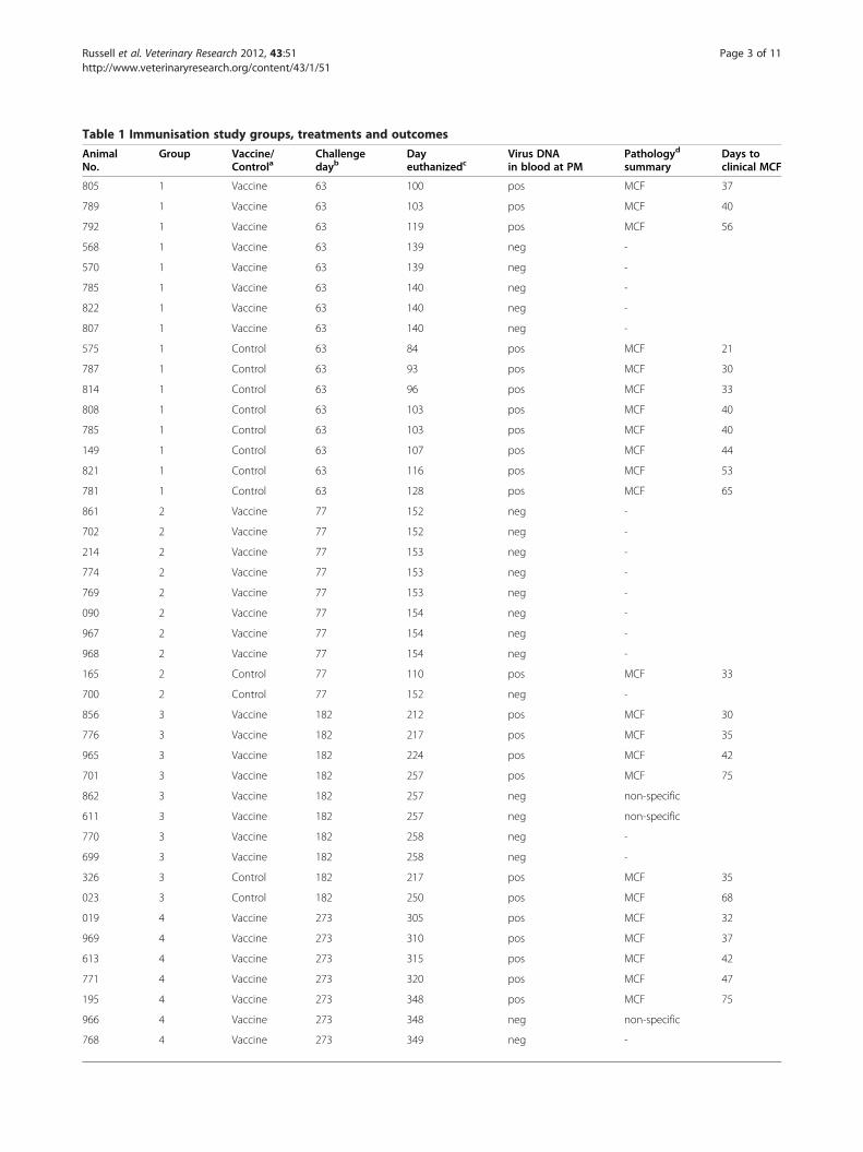
Table 1 Immunisation study groups, treatments and outcomes
AnimalNo.
Group Vaccine/Controla
Challengedayb
Dayeuthanizedc
Virus DNAin blood at PM
Pathologyd
summaryDays toclinical MCF
805 1 Vaccine 63 100 pos MCF 37
789 1 Vaccine 63 103 pos MCF 40
792 1 Vaccine 63 119 pos MCF 56
568 1 Vaccine 63 139 neg -
570 1 Vaccine 63 139 neg -
785 1 Vaccine 63 140 neg -
822 1 Vaccine 63 140 neg -
807 1 Vaccine 63 140 neg -
575 1 Control 63 84 pos MCF 21
787 1 Control 63 93 pos MCF 30
814 1 Control 63 96 pos MCF 33
808 1 Control 63 103 pos MCF 40
785 1 Control 63 103 pos MCF 40
149 1 Control 63 107 pos MCF 44
821 1 Control 63 116 pos MCF 53
781 1 Control 63 128 pos MCF 65
861 2 Vaccine 77 152 neg -
702 2 Vaccine 77 152 neg -
214 2 Vaccine 77 153 neg -
774 2 Vaccine 77 153 neg -
769 2 Vaccine 77 153 neg -
090 2 Vaccine 77 154 neg -
967 2 Vaccine 77 154 neg -
968 2 Vaccine 77 154 neg -
165 2 Control 77 110 pos MCF 33
700 2 Control 77 152 neg -
856 3 Vaccine 182 212 pos MCF 30
776 3 Vaccine 182 217 pos MCF 35
965 3 Vaccine 182 224 pos MCF 42
701 3 Vaccine 182 257 pos MCF 75
862 3 Vaccine 182 257 neg non-specific
611 3 Vaccine 182 257 neg non-specific
770 3 Vaccine 182 258 neg -
699 3 Vaccine 182 258 neg -
326 3 Control 182 217 pos MCF 35
023 3 Control 182 250 pos MCF 68
019 4 Vaccine 273 305 pos MCF 32
969 4 Vaccine 273 310 pos MCF 37
613 4 Vaccine 273 315 pos MCF 42
771 4 Vaccine 273 320 pos MCF 47
195 4 Vaccine 273 348 pos MCF 75
966 4 Vaccine 273 348 neg non-specific
768 4 Vaccine 273 349 neg -
Russell et al. Veterinary Research 2012, 43:51 Page 3 of 11http://www.veterinaryresearch.org/content/43/1/51
Table 1 Immunisation study groups, treatments and outcomes (Continued)
772 4 Vaccine 273 349 neg -
719 4 Control 273 300 pos MCF 27
620 4 Control 273 302 pos MCF 29a Vaccinated animals in all groups received the same dose (10 mL of 107 TCID50 per mL) of vaccine for both prime and boost, given intramuscularly with 20% (v/v)Emulsigen. Control animals in each group received virus-free culture medium with Emulsigen, prepared and administered in the same way.b Pathogenic AlHV-1 challenge for each group was as follows: group 1 received 10 mL of 104.7 TCID50/mL virus; group 2 received 10 mL of 103.8 TCID50/mL virus;and groups 3 and 4 received 10 mL of 104.05 TCID50/mL virus, administered intranasally in all groups.c All animals were monitored daily for clinical signs of MCF, including fever, loss of appetite, depression, nasal or ocular discharge and conjunctivitis. A clinicalscoring scheme (Additional file 1 Table S1) was used to ensure that all animals were euthanized at the onset of moderate clinical signs. Animal that did notdevelop clinical signs of MCF were euthanized for post-mortem at least 75 days after challenge.d Pathology was assessed as described in the text and is summarised as MCF, non-specific or negative (−). Tissue sections from animals with non-specificpathology were tested for presence of AlHV-1 DNA by real-time PCR. No viral DNA was detected in any sample with non-specific pathology.
Russell et al. Veterinary Research 2012, 43:51 Page 4 of 11http://www.veterinaryresearch.org/content/43/1/51
with pathogenic AlHV-1 after prime and boost intra-muscular (IM, upper neck) immunisation with attenu-ated AlHV-1 in Emulsigen (20% v/v), while theremaining eight calves were challenged after mock primeand boost immunisation with virus-free culture mediumin Emulsigen.In the second study, the longevity of protection
induced by the vaccine formulated with Emulsigen wastested by immunisation of three groups of eight calves(Table 1; groups 2–4) followed by challenge at varioustime points after immunisation. In each experiment,uncoagulated blood and nasal secretions were collectedfrom all animals just prior to primary immunisation,fortnightly until challenge and weekly thereafter asdescribed previously [21].At autopsy, the following tissues were collected: brain,
buccal mucosa, rumen, reticulum, liver, kidney, lung,prescapular lymph node, mesenteric lymph node (MLN),naso-pharyngeal tonsil and blood. Pieces of each tissue(except blood) were fixed in 10% formal saline beforeprocessing and embedding in paraffin wax. For patho-logical analysis, 4 μm sections of formalin-fixed tissueswere cut and stained with haematoxylin and eosin. Inaddition, total DNA was prepared from blood buffy coatcells or from sections of selected fixed tissues for PCRdetection of viral DNA.
Detection of viral DNA in blood and fixed tissuesViral DNA was assayed in pure genomic DNA samplesextracted from blood buffy coat cells or from 100 μm sec-tions of fixed tissues by quantitative (q)PCR as describedpreviously [25]. Briefly, ~50 ng of total DNA was assayedsimultaneously for the presence of AlHV-1 and genomicactin sequences by duplex real-time PCR analysis. Each 20μL assay contained 900 μM of each AlHV-1 forward andreverse primer (AlHV1-F, AlHV1-R) and 250 μM of thefluorogenic probe FAM-AlHV1 [25]; 450 μM of each gen-omic actin primer (gACT-F: 5′-CAC CTT CCA GCAGAT GTG GA-3′; gACT-R: 5′-CTA GAA GCA TTTGCG GTG GAC ) and 125 μM of the fluorogenic minor-groove binding (mgb) probe VIC-gACT (5′-VIC-AGC
AAG CAG GAG TAC G-mgb) (K. Willoughby; unpub-lished data); and real-time PCR reagents containing Plat-inum Taq polymerase and ROX control dye (Invitrogen).All assays were conducted using standard conditions onABI 7000 or 7500 sequence detection systems (AppliedBiosystems).
Analysis of antibody responses by ELISA and virus-neutralising antibody testTo quantify the humoral antibody response in bloodplasma and the mucosal antibody response in nasalsecretions (NS), an antibody ELISA was developed basedon a detergent extract of the vaccine virus. Virus fromcell-free culture fluid of BT cells infected with attenuatedAlHV-1 C500 was concentrated by centrifugation(35 000 × g, 3 h, 4°C). Virus pellets were resuspended inphosphate-buffered saline (PBS) containing 0.8% CHAPS(3-[(3-Cholamidopropyl) dimethylammonio]-1-propane-sulfonate, Sigma) and the insoluble virus pellet was col-lected by centrifugation as above. The virus pellet wasthen solubilised in 0.2% SDS before dialysing back into0.2% CHAPS in PBS. This virus-positive ELISA coatingantigen had a protein concentration of approximately2 mg/mL. To produce a negative antigen for compari-son, cell-free culture fluid from uninfected BT cells wastreated according to the same protocol and the resultingsolution used as negative ELISA antigen.Pairs of adjacent rows of 96-well microtitre plates
(Greiner, high protein binding) were coated with 50 μLof 5 μg/mL virus-positive or -negative antigen in 0.1 Mcarbonate buffer pH 9.6. Individual samples of bloodplasma or sterile-filtered nasal secretion fluid diluted inPBS were then applied in duplicate to positive and nega-tive antigen wells at serial dilutions from 1:100 to1:4000. A standard curve, comprising 1:100 to 1:4000dilutions of an AlHV-1-positive plasma pool wasincluded on each plate to ensure reproducibility in theassays. A negative serum at 1:500 dilution was alsoincluded with each test sample series. Antibody boundin each well was detected using 1:1000 rabbit anti-bovine IgG-Horseradish Peroxidase conjugate (Sigma).
Russell et al. Veterinary Research 2012, 43:51 Page 5 of 11http://www.veterinaryresearch.org/content/43/1/51
ELISA values (difference between means of positiveand negative antigen wells for each sample dilution)were used to calculate a relative titre for each test sam-ple, based on the standard curve produced from thepositive control serum wells on the same plate. Testsamples with ELISA values outside the range of thestandard curve were discarded.The virus neutralisation test was based upon inhibition
of AlHV-1-induced cytopathic effect in BT cells by dilu-tions of plasma or nasal secretion fluid as described[21,22]. Assays were carried out in 96 well tissue cultureplates with BT cells at greater than 80% confluence. Allassays used a high titre bovine anti-AlHV-1 serum as astandard and included non-specific toxicity control wellscontaining sample and cells without virus.
Pathology and histopathologyMCF was confirmed by clinical and pathological signs. Theclinical symptoms included fever, nasal and ocular dis-charge, conjunctivitis and development of corneal opacity.Clinical assessment was done using a system (Additionalfile 1 Table S1) which combined scores for pyrexia (1–3),nasal discharge (0–2), ocular pathology (1–3) and diar-rhoea (1–6). Any animal with a score over 6 was eutha-nized without delay. This system ensured that all animalswith clinical MCF were euthanized at a similar stage of thedisease, prior to the development of moderate clinicalsigns. All animals with MCF had clinical signs for fewerthan 5 days before euthanasia. Surviving animals wereeuthanized 11 weeks after pathogenic virus challenge.MCF histopathology in brain, kidney, liver, lung and digest-ive epithelium was examined and scored for the frequencyof lesions consistent with MCF as described [21]. Thepathology of each case was summarised as MCF (most ofthe organs studied contained significant numbers of lesionsconsistent with a diagnosis of MCF); non-specific (a smallnumber of lesions, present usually only in one or two stud-ied organs, and of mild intensity, without extensive infiltra-tion of lymphocytes or clear vasculitis); or negative (nosignificant lesions observed). A positive test for AlHV-1DNA in the blood on the day of post-mortem, in combin-ation with MCF histopathological signs, was taken as a de-finitive diagnosis of MCF. In cases with non-specificpathology observed in some tissues, DNA prepared fromadjacent sections was tested for the presence of virus DNAto assess whether the lesions observed could be due toAlHV-1 infection.
Statistical analysisData for post-challenge survival between groups wereanalysed by Fisher’s exact test [26,27] while comparisonof immune response data was done by students’ T-teston log(base2)-transformed titre data. p values less than0.05 were considered significant.
ResultsProtection of cattle from MCF by attenuated AlHV-1 inemulsigenIn these studies, four groups of eight cattle were vacci-nated with attenuated AlHV-1 C500 in the presence of20% (v/v) Emulsigen (MVP Labs, USA), while 14 controlanimals were mock-vaccinated with adjuvant in tissueculture fluid (Table 1). Both primary and boost immuni-sations, using the same material given four weeks apart,were administered by the intramuscular route, high inthe neck of each animal.Within groups 1 and 2 (Table 1) a total of 16 vacci-
nated animals were challenged with pathogenic AlHV-1by the intranasal route at either 9 or 11 weeks after pri-mary immunisation. Groups 3 and 4, each comprisingeight vaccinated animals and two unvaccinated controls,were challenged at 26 and 39 weeks, respectively, afterthe primary immunisation, by intranasal inoculation ofpathogenic AlHV-1 (Table 1).Animal rectal temperatures and general demeanour
(appetite, activity, nasal secretion) were recorded daily fol-lowing challenge and additional clinical scores (Additionalfile 1 Table S1) were recorded daily after the onset of fever(temperature≥ 40°C). A final blood sample was collectedbefore euthanasia and a range of tissues were collectedpost-mortem for diagnostic, pathological and molecular in-vestigation. Animals that did not succumb to MCF wereeuthanized for post-mortem examination between 75 and77 days after challenge (Table 1).In groups 1 and 2, challenged at 9 or 11 weeks post
vaccination, 13 animals were protected from clinicalMCF while all but one of the control animals suc-cumbed to disease. Analysis of these data by Fisher’sexact test showed that protection was statistically signifi-cant in each individual study (group 1, p= 0.025; andgroup 2, p= 0.003), while the combined results werehighly significant (p< 0.0001), demonstrating the effi-cacy of Emulsigen as an adjuvant in this system.In the groups challenged at later times after primary
immunisation, four of eight animals in group 3, chal-lenged at 26 weeks, and three of eight animals in group4, challenged at 39 weeks, survived with no disease andno detectable AlHV-1 DNA post-mortem (Table 1).Comparison of the occurrence of MCF in the vaccinatedand unvaccinated groups by Fisher’s exact test showedsignificant protection was afforded by the vaccineagainst MCF challenge at six months after immunisation(p= 0.04) but not at 9 months (p= 0.12).
Evidence of circulating viral DNA in infected animalsAnalysis of terminal blood and tissue samples showedthat all animals with clear clinical and histopathologicalsigns of MCF (Table 1) had detectable virus DNA inblood. In contrast, animals that showed no clinical
Russell et al. Veterinary Research 2012, 43:51 Page 6 of 11http://www.veterinaryresearch.org/content/43/1/51
disease or non-specific histopathological signs, asdefined above (Table 1), had no detectable AlHV-1 DNAin blood or in tissue sections containing lesions, con-firming that these animals did not have MCF at the timeof euthanasia. In MCF cases in either vaccinated or con-trol animals, virus DNA could be detected up to threeweeks before post mortem.
ELISA analysis of antibody responses followingvaccination and challengeThe relative titres of AlHV-1-specific antibodies in nasalsecretions and blood plasma were determined with re-spect to standard pools of MCF-positive plasma or nasalsecretions using the direct ELISA described above. Ani-mals in group 1 were assayed independently of the othergroups, using a different serum pool to calculate relativetitre. Thus the ELISA results from group 1 are not com-parable with the other groups. For animals in groups 2–4,samples from throughout the study period were testedtogether. Unvaccinated control animals challenged with
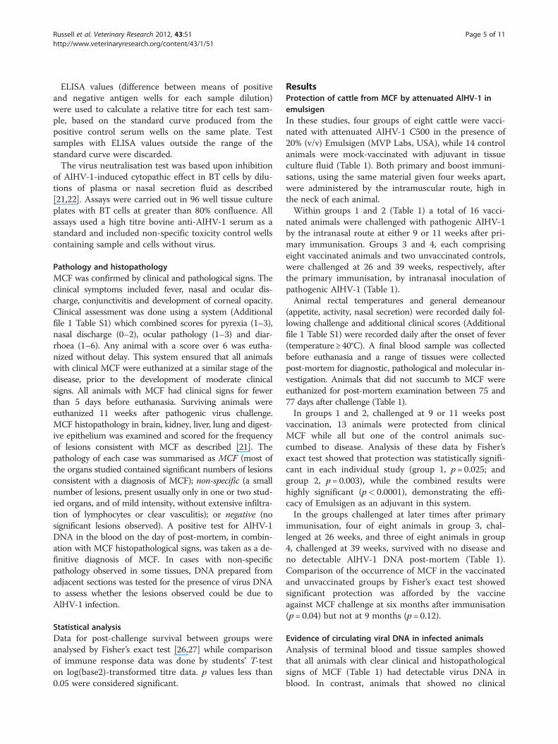
Figure 1 ELISA analysis of virus-specific antibody responses in vaccinspecific antibody titres in plasma (filled squares, solid line) and in nasal secanimals available at each time point. Error bars correspond to the standard
AlHV-1 showed no detectable titre of anti-viral antibodiesat any point before or during the progress of clinicaldisease.Following immunisation and boost, all vaccinated ani-
mals showed a peak in anti-virus antibody titre in bothblood plasma and nasal secretions between days 63 and91 after primary immunisation (Figure 1). Average titresthen reduced to less than 1/100 in plasma and nasalsecretions by day 120. Analysis of virus-specific antibodytitres in nasal secretions and plasma at a point withinthe peak response to vaccination (day 63), showed thatplasma antibody titres were significantly lower on day 63in animals that went on to develop MCF than in thosethat were protected (p= 0.006) while nasal secretionantibody titres were not significantly different betweenthese groups. After challenge, the patterns of antibodyresponse also differed between vaccinated animals thatsuccumbed to MCF and those which did not (Figures 2and 3). Animals protected by vaccine showed a generallystable virus-specific antibody response (Figure 2) with
ated animals from groups 2–4 prior to challenge. Average virus-retions (open squares, broken line) were plotted for all unchallengederror of the means.
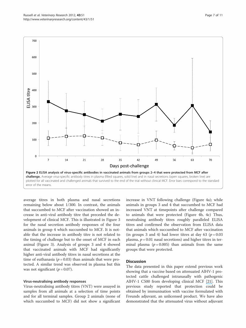
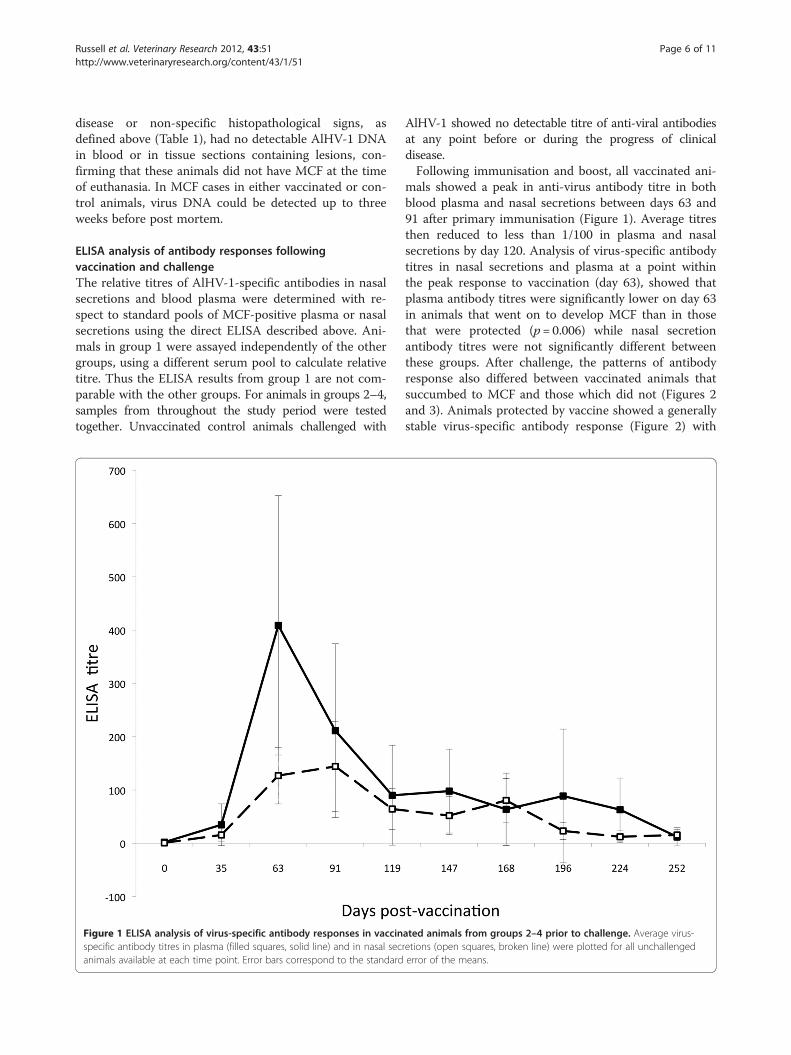
Figure 2 ELISA analysis of virus-specific antibodies in vaccinated animals from groups 2–4 that were protected from MCF afterchallenge. Average virus-specific antibody titres in plasma (filled squares, solid line) and in nasal secretions (open squares, broken line) areplotted for all vaccinated and challenged animals that survived to the end of the trial without clinical MCF. Error bars correspond to the standarderror of the means.
Russell et al. Veterinary Research 2012, 43:51 Page 7 of 11http://www.veterinaryresearch.org/content/43/1/51
average titres in both plasma and nasal secretionsremaining below about 1/300. In contrast, the animalsthat succumbed to MCF after vaccination showed an in-crease in anti-viral antibody titre that preceded the de-velopment of clinical MCF. This is illustrated in Figure 3for the nasal secretion antibody responses of the fouranimals in group 4 which succumbed to MCF. It is not-able that the increase in antibody titre is not related tothe timing of challenge but to the onset of MCF in eachanimal (Figure 3). Analysis of groups 3 and 4 showedthat vaccinated animals with MCF had significantlyhigher anti-viral antibody titres in nasal secretions at thetime of euthanasia (p= 0.03) than animals that were pro-tected. A similar trend was observed in plasma but thiswas not significant (p= 0.07).
Virus-neutralising antibody responsesVirus-neutralising antibody titres (VNT) were assayed insamples from all animals at a selection of time pointsand for all terminal samples. Group 2 animals (none ofwhich succumbed to MCF) did not show a significant
increase in VNT following challenge (Figure 4a); whileanimals in groups 3 and 4 that succumbed to MCF hadincreased VNT at timepoints after challenge comparedto animals that were protected (Figure 4b, 4c) Thus,neutralising antibody titres roughly paralleled ELISAtitres and confirmed the observation from ELISA datathat animals which succumbed to MCF after vaccination(in groups 3 and 4) had lower titres at day 63 (p= 0.03plasma, p= 0.01 nasal secretions) and higher titres in ter-minal plasma (p= 0.005) than animals from the samegroups that were protected.
DiscussionThe data presented in this paper extend previous workshowing that a vaccine based on attenuated AlHV-1 pro-tected cattle challenged intranasally with pathogenicAlHV-1 C500 from developing clinical MCF [21]. Thisprevious study reported that protection could beobtained by immunisation with vaccine formulated withFreunds adjuvant, an unlicensed product. We have alsodemonstrated that the attenuated virus without adjuvant
Figure 3 ELISA analysis of virus-specific antibodies in the four animals from group 3 that succumbed to MCF after challenge. Relativetitres of antibodies in the nasal secretions of each animal at a range of timepoints after challenge are plotted individually. Each animal isrepresented by a different symbol, joined by solid lines. The rightmost point in each line corresponds to the sample collected on the day of postmortem.
Russell et al. Veterinary Research 2012, 43:51 Page 8 of 11http://www.veterinaryresearch.org/content/43/1/51
did not protect from intranasal challenge with patho-genic AlHV-1 (G.C. Russell, unpublished data). Here weshow that the licensed oil-in-water adjuvant Emulsigencan facilitate protection from intranasal experimentalAlHV-1 challenge given up to six months after primaryimmunisation.Analysis of plasma and nasal secretion antibody
responses to vaccination and challenge showed that (i)unvaccinated animals with MCF had no ELISA-detectable or neutralising antibody response to AlHV-1following challenge; (ii) vaccinated animals that wereprotected from MCF had significantly higher titres ofvirus-neutralising antibodies in both plasma and nasalsecretions at about 1 month after boost than unpro-tected animals and these titres did not significantlychange after challenge (Figure 4); (iii) in contrast, vacci-nated animals that succumbed to MCF showed signifi-cant increases in both total virus-specific antibody and
virus-neutralising antibody during the development ofclinical MCF (Figure 3 and Figure 4).The lack of a detectable antibody response in unvac-
cinated cattle challenged with AlHV-1 could reflect therapid onset of MCF that might prevent the induction ofan immune response to combat the infection. However,in the experiments reported here, the appearance of clin-ical MCF in control animals ranged between 21 and68 days after challenge (Table 1), suggesting that therewas sufficient time for the development of an immuneresponse in at least some of the infected cattle. Thesedata confirm and extend the previous observation thatcontrol cattle challenged intranasally did not develop de-tectable titres of virus-neutralising antibody [21].In field cases of MCF, virus-specific antibody responses
are reported to be found in 70-80% of samples tested[22]. The lack of antibody response in some MCF caseswas confirmed in a comparative analysis of MCF-
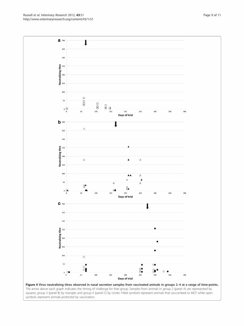
Figure 4 Virus neutralising titres observed in nasal secretion samples from vaccinated animals in groups 2–4 at a range of time-points.The arrow above each graph indicates the timing of challenge for that group. Samples from animals in group 2 (panel A) are represented bysquares, group 3 (panel B) by triangles and group 4 (panel C) by circles. Filled symbols represent animals that succumbed to MCF while opensymbols represent animals protected by vaccination.
Russell et al. Veterinary Research 2012, 43:51 Page 9 of 11http://www.veterinaryresearch.org/content/43/1/51
Russell et al. Veterinary Research 2012, 43:51 Page 10 of 11http://www.veterinaryresearch.org/content/43/1/51
specific PCR and serological diagnostic testing where 14of 39 OvHV-2 PCR-positive animals with clinical MCFwere considered serologically borderline or negative[28]. Additionally, sub-clinical infection with MCFviruses has been inferred from serological surveys inwhich animals from MCF-susceptible species (cattle,bison, deer) had detectable MCF-specific antibodies orviral DNA in the absence of disease [29-31]. More re-cently, experiments using OvHV-2 to infect cattle orbison showed sub-clinical infection of five cattle, withseroconversion, and of one bison without seroconver-sion; while evidence of previous infection was found insix bison of which two had both detectable anti-MCFantibodies and OvHV-2 DNA [32,33]. Previous subclin-ical infection did not appear to reduce susceptibility toMCF following intranasal challenge.These observations suggest that infection with MCF
viruses and the ensuing immune response and diseasemay be more complex than previously thought, leadingto the development of MCF in some animals without adetectable virus-specific antibody response. There is alack of good transmission and epidemiological work onMCF that requires to be done to address such issues.In contrast to the boost to AlHV-1-specific antibody
responses found in vaccinated animals that developedMCF, vaccine-protected animals did not show a signifi-cant increase in antibody titres after challenge. This islikely to be because no virus in immunogenic quantitywas able to penetrate the mucosal barrier of neutralisingantibody to boost the response. This supports the con-tention that the establishment of a mucosal barrier ofantibody is a mechanism of protection against MCF.This is reinforced by the observation that protected ani-mals in groups 3 and 4 had high virus-neutralising anti-body titres after immunisation compared to animals thatsuccumbed to MCF. The higher titres are likely to beassociated with longer duration of antibody responsesand hence protection at later time points.The development of high titre virus-specific antibody
responses prior to the onset of MCF among vaccinatedcattle in these experiments suggests that the develop-ment of clinical MCF includes the expression of virusgene products that stimulate an anamnestic immune re-sponse. Indeed, clinical MCF cases are often charac-terised by the development of a circulating antibodyresponse which includes antibodies specific for virusstructural antigens including the major glycoproteincomplex of the viral envelope [34,35]. This is in contrastto recent suggestions that MCF in rabbits and cattle isassociated with a latent infection of lymphocytes and alack of lytic gene expression that would include anti-genic capsid and envelope proteins [11,36,37]. The pres-ence of antibodies specific for virus structural proteinsin MCF-affected animals suggests that infection results
in a pattern of virus gene expression that includes anumber of lytic cycle antigens but without the produc-tion of infectious virus. It may therefore be inappropriateto discuss MCF in terms of lytic or latent gene expres-sion patterns since latency should be defined in the nat-ural host rather than in MCF-susceptible species thatcannot propagate these viruses.Current work aims to improve the magnitude and dur-
ation of the protective immune response by the strategicinclusion of toll-like receptor (TLR) agonists in the MCFvaccine. In addition, the role, if any, of cytotoxic T cellsin protection against MCF is currently not known andthis is being investigated. The six month window of pro-tection offered by the current immunisation regimeshould be adequate to protect cattle in Eastern andSouthern Africa exposed to wildebeest during the calv-ing period. This is being studied in field trials inTanzania.
Additional file
Additional file 1: Table S1. Clinical scoring scheme for cattle withMCF. Cattle were scored daily following the onset of fever > 40°C.Animals were euthanized when their daily score totalled more than 6.However, any animal with symptoms that compromised its welfarewould be euthanized immediately.
Competing interestsThe authors declare they have no competing interests
Authors’ contributionsExperimental design and planning: GCR, DG, HT, JT, JB, DMH; animalexperiments GCR, DG, HT, JT, JB; pathology analysis DG, HT, JB; serologicalanalysis DD, AP, MC, DG; data processing and statistical analysis DD, AP, DG,DMH, GCR; drafting of the manuscript GCR, DMH, DD, JB. All authors readand approved the manuscript.
AcknowledgementsThis work was supported by the Scottish Government Rural andEnvironment Research and Analysis Directorate. We are indebted to theMoredun Research Institute Bioservices Division for their excellent animalservices and to Biomathematics & Statistics Scotland (BIOSS) for advice onstatistical analysis.
Author details1Moredun Research Institute, Pentlands Science Park, Bush Loan, PenicuikEH26 0PZScotland, UK. 2Instituto de Ganaderia de Montaña (CSIC-ULE),24346, Grulleros (Leon), Spain. 3School of Veterinary Medicine and Science,Nottingham University, Sutton Bonington LE12 5RD, United Kingdom.
Received: 20 February 2012 Accepted: 23 May 2012Published: 11 June 2012
References1. Russell GC, Stewart JP, Haig DM: Malignant catarrhal fever: a review. Vet J
2009, 179:324–335.2. Davison A, Eberle R, Ehlers B, Hayward G, McGeoch D, Minson A, Pellett P,
Roizman B, Studdert M, Thiry E: The order Herpesvirales. Arch Virol 2009,154:171–177.
3. Ensser A, Pflanz R, Fleckenstein B: Primary structure of the alcelaphineherpesvirus 1 genome. J Virol 1997, 71:6517–6525.
4. Hart J, Ackermann M, Jayawardane G, Russell G, Haig DM, Reid H, StewartJP: Complete sequence and analysis of the ovine herpesvirus 2 genome.J Gen Virol 2007, 88:28–39.
Russell et al. Veterinary Research 2012, 43:51 Page 11 of 11http://www.veterinaryresearch.org/content/43/1/51
5. Anderson IE, Buxton D, Campbell I, Russell G, Davis WC, Hamilton MJ, HaigDM: Immunohistochemical study of experimental malignant catarrhalfever in rabbits. J Comp Pathol 2007, 136:156–166.
6. Buxton D, Jacoby RO, Reid HW, Goodall PA: The pathology of sheep-associated malignant catarrhal fever in the hamster. J Comp Pathol 1988,98:155–166.
7. Buxton D, Reid HW: Transmission of malignant catarrhal fever to rabbits.Vet Rec 1980, 106:243–245.
8. Liggitt HD, DeMartini JC: The pathomorphology of malignant catarrhalfever. I. Generalized lymphoid vasculitis. Vet Pathol 1980, 17:58–72.
9. Liggitt HD, DeMartini JC: The pathomorphology of malignant catarrhalfever. II. Multisystemic epithelial lesions. Vet Pathol 1980, 17:73–83.
10. Bridgen A, Munro R, Reid HW: The detection of Alcelaphine herpesvirus-1DNA by insitu hybridization of tissues from rabbits affected withmalignant catarrhal fever. J Comp Pathol 1992, 106:351–359.
11. Dewals B, Myster F, Palmeira L, Gillet L, Ackermann M, Vanderplasschen A:Ex vivo bioluminescence detection of alcelaphine herpesvirus 1 infectionduring malignant catarrhal fever. J Virol 2011, 85:6941–6954.
12. Simon S, Li H, O’Toole D, Crawford TB, Oaks JL: The vascular lesions of acow and bison with sheep-associated malignant catarrhal fever containovine herpesvirus 2-infected CD8(+) T lymphocytes. J Gen Virol 2003,84:2009–2013.
13. Burrells C, Reid HW: Phenotypic analysis of lymphoblastoid cell-linesderived from cattle and deer affected with sheep-associated malignantcatarrhal fever. Vet Immunol Immunopathol 1991, 29:151–161.
14. Reid HW, Buxton D, Pow I, Finlayson J: Isolation and characterization oflymphoblastoid-cells from cattle and deer affected with sheep-associated malignant catarrhal fever. Res Vet Sci 1989, 47:90–96.
15. Schock A, Collins RA, Reid HW: Phenotype, growth regulation andcytokine transcription in Ovine Herpesvirus-2 (OHV-2)-infected bovine T-cell lines. Vet Immunol Immunopathol 1998, 66:67–81.
16. Swa S, Wright H, Thomson J, Reid H, Haig D: Constitutive activation of Lckand Fyn tyrosine kinases in large granular lymphocytes infected withthe gamma-herpesvirus agents of malignant catarrhal fever. Immunology2001, 102:44–52.
17. Bedelian C, Nkedianye D, Herrero M: Maasai perception of the impact andincidence of malignant catarrhal fever (MCF) in southern Kenya. Prev VetMed 2007, 78:296–316.
18. Cleaveland S, Kusiluka L, ole Kuwai J, Bell C, Kazwala R: Assessing the impactof Malignant Catarrhal Fever in Ngorongoro District, Tanzania.: 2001. http://www.eldis.org/fulltext/cape_new/MCF_Maasai_Tanzania.pdf. (Accessed 13February 2012).
19. Edington N, Plowright W: The protection of rabbits against theherpesvirus of malignant catarrhal fever by inactivated vaccines. Rese VetSci 1980, 28:384–386.
20. Plowright W, Herniman KA, Jessett DM, Kalunda M, Rampton CS:Immunisation of cattle against the herpesvirus of malignant catarrhalfever: failure of inactivated culture vaccines with adjuvant. Res Vet Sci1975, 19:159–166.
21. Haig DM, Grant D, Deane D, Campbell I, Thomson J, Jepson C, Buxton D,Russell GC: An immunisation strategy for the protection of cattle againstalcelaphine herpesvirus-1-induced malignant catarrhal fever. Vaccine2008, 26:4461–4468.
22. Reid HW: Malignant catarrhal fever. In OIE Terrestrial Manual 2008, Volume2. 6th edition.: 2012. www.oie.int/fileadmin/Home/eng/Health_standards/tahm/2.04.15_MCF.pdf.
23. Handley JA, Sargan DR, Herring AJ, Reid HW: Identification of a region ofthe alcelaphine herpesvirus-1 genome associated with virulence forrabbits. Vet Microbiol 1995, 47:167–181.
24. : Emulsigen Technical Bulletin.: www.mvplabs.com/copy/adjuvant/Emulsigen.pdf (Accessed 23 April 2012).
25. Traul DL, Elias S, Taus NS, Herrmann LM, Oaks JL, Li H: A real-time PCRassay for measuring alcelaphine herpesvirus-1 DNA. J Virol Methods 2005,129:186–190.
26. Fisher RA: On the interpretation of X2 from contingency tables, and thecalculation of P. J R Stat Soc 1922, 85:87–94.
27. Fisher's Exact Test.: www.langsrud.com/fisher.htm (Accessed 23 April 2012).28. Muller-Doblies UU, Li H, Hauser B, Adler H, Ackermann M: Field validation
of laboratory tests for clinical diagnosis of sheep-associated malignantcatarrhal fever. J Clin Microbiol 1998, 36:2970–2972.
29. Li H, Shen DT, Jessup DA, Knowles DP, Gorham JR, Thorne T, O'Toole D,Crawford TB: Prevalence of antibody to malignant catarrhal fever virus inwild and domestic ruminants by competitive inhibition ELISA. J Wild Dis1996, 32:437–443.
30. Zarnke RL, Li H, Crawford TB: Serum antibody prevalence of malignantcatarrhal fever viruses in seven wildlife species from Alaska. J Wild Dis2002, 38:500–504.
31. Powers JG, VanMetre DC, Collins JK, Dinsmore RP, Carman J, Patterson G,Brahmbhatt D, Callan RJ: Evaluation of ovine herpesvirus type 2infections, as detected by competitive inhibition ELISA and polymerasereaction assay in dairy cattle without clinical signs of malignant catarrhalfever. J Am Vet Med Assoc 2005, 227:606–611.
32. Taus NS, Oaks JL, Gailbreath K, Traul DL, O'Toole D, Li H: Experimentalaerosol infection of cattle (Bos taurus) with ovine herpesvirus 2 usingnasal secretions from infected sheep. Vet Microbiol 2006, 116:29–36.
33. Gailbreath KL, O’Toole D, Taus NS, Knowles DP, Oaks J, Li H: Experimentalnebulization of American bison (Bison bison) with low doses of ovineherpesvirus 2 from sheep nasal secretions. Vet Microbiol 2010,143:389–393.
34. Herring A, Reid H, Inglis N, Pow I: Immunoblotting analysis of the reactionof wildebeest, sheep and cattle sera with the structural antigens ofalcelaphine herpesvirus-1 (malignant catarrhal fever virus). Vet Microbiol1989, 19:205–215.
35. Li H, McGuire TC, Muller-Doblies UU, Crawford TB: A simpler, moresensitive competitive inhibition enzyme-linked immunosorbent assay fordetection of antibody to malignant catarrhal fever viruses. J Vet DiagnInvest 2001, 13:361–364.
36. Dewals B, Boudry C, Farnir Fdr, Drion PV, Vanderplasschen A: Malignantcatarrhal fever induced by alcelaphine herpesvirus 1 is associated withproliferation of CD8+ T cells supporting a latent infection. PLoS One 2008,3:e1627.
37. Meier-Trummer CS, Rehrauer H, Franchini M, Patrignani A, Wagner U,Ackermann M: Malignant catarrhal fever of cattle is associated with lowabundance of IL-2 transcript and a predominantly latent profile of ovineherpesvirus 2 gene expression. PLoS One 2009, 4:e6265.
doi:10.1186/1297-9716-43-51Cite this article as: Russell et al.: Duration of protective immunity andantibody responses in cattle immunised against alcelaphineherpesvirus-1-induced malignant catarrhal fever. Veterinary Research 201243:51.
Submit your next manuscript to BioMed Centraland take full advantage of:
• Convenient online submission
• Thorough peer review
• No space constraints or color figure charges
• Immediate publication on acceptance
• Inclusion in PubMed, CAS, Scopus and Google Scholar
• Research which is freely available for redistribution
Submit your manuscript at www.biomedcentral.com/submit