Polymerase chain reaction amplification of wildebeest-associated and cervine-derived malignant...
10
Arch Virol (t994) 135:355-364 _Archives V rology © Springer-Verlag 1994 Printed in Austria Polymerase chain reaction amplification of wildebeest-associated and cervine-derived malignant catarrhal fever virus DNA K. M. Tham, K. Ng*, and L. W. Young Virology Section, Central Animal Health Laboratory, MAF Quality Management, New Zealand Accepted November 24, 1993 Summary. A polymerase chain reaction (PCR) assay was developed for the detection of alcelaphine herpesvirus 1 (AHV1), a causative agent of malignant catarrhal fever (MCF) of ruminants. A pair of 20-base primers was constructed based on the published nucleotide sequence of gene A of the WCll isolate of AHVt and was used to amplify a DNA fragment of 413 base pairs. The optimised PCR assay was highly sensitive, i.e. it detected 10 fg of genomic DNA of AHV1 (WC11 isolate). The amplified fragment was shown to be specific for AHV1 DNA by (i) cleavage with XbaI which yielded 2 subfragments of approximately 140 and 280 base pairs and (ii) chemiluminescence Southern blot hybfidisation with a digoxigenin-labelled 25-base internal probe. The PCR assay also amplified AHV1 gene sequences in tissue samples from deer and rabbits experimentally infected with materials derived from deer with clinical sheep-associated MCF. Introduction Malignant catarrhal fever (MCF) is a sporadic, often fatal, lymphoproliferative and degenerative disease of cattle, buffalo, deer and other wild ruminants [3, 7, 12, 16, 17]. The unique pathogenesis of MCF involves the proliferation of T lymphocytes I-7, 16, 17]. The disease is caused by a virus (MCFV) classified in the subfamily gammaherpesvirinae [17, 18]. MCFV or alcelaphine herpesvirus 1 (AHV1), which is indigenous to the blue wildebeest (Connochaetes taurinus), causes an inapparent infection in its natural host. In cattle and other susceptible ruminants, the virus causes a disease of low morbidity and high mortality. Epidemiological and virological studies have implicated sheep as a plausible second carrier of MCFV in countries such as New Zealand where sheep are associated with cattle and deer [4, 13, 19, 20, 23]. The causative agent of * Present address: AgResearch, Wallaceville Animal Research Centre, Upper Hutt, New Zealand.
Transcript of Polymerase chain reaction amplification of wildebeest-associated and cervine-derived malignant...

Arch Virol (t994) 135:355-364
_Archives
V rology © Springer-Verlag 1994 Printed in Austria
Polymerase chain reaction amplification of wildebeest-associated and
cervine-derived malignant catarrhal fever virus D N A
K. M. Tham, K. Ng*, and L. W. Young
Virology Section, Central Animal Health Laboratory, MAF Quality Management, New Zealand
Accepted November 24, 1993
Summary. A polymerase chain reaction (PCR) assay was developed for the detection of alcelaphine herpesvirus 1 (AHV1), a causative agent of malignant catarrhal fever (MCF) of ruminants. A pair of 20-base primers was constructed based on the published nucleotide sequence of gene A of the W C l l isolate of AHVt and was used to amplify a DNA fragment of 413 base pairs. The optimised PCR assay was highly sensitive, i.e. it detected 10 fg of genomic DNA of AHV1 (WC11 isolate). The amplified fragment was shown to be specific for AHV1 DNA by (i) cleavage with XbaI which yielded 2 subfragments of approximately 140 and 280 base pairs and (ii) chemiluminescence Southern blot hybfidisation with a digoxigenin-labelled 25-base internal probe. The PCR assay also amplified AHV1 gene sequences in tissue samples from deer and rabbits experimentally infected with materials derived from deer with clinical sheep-associated MCF.
Introduction
Malignant catarrhal fever (MCF) is a sporadic, often fatal, lymphoproliferative and degenerative disease of cattle, buffalo, deer and other wild ruminants [3, 7, 12, 16, 17]. The unique pathogenesis of MCF involves the proliferation of T lymphocytes I-7, 16, 17]. The disease is caused by a virus (MCFV) classified in the subfamily gammaherpesvirinae [17, 18]. MCFV or alcelaphine herpesvirus 1 (AHV1), which is indigenous to the blue wildebeest (Connochaetes taurinus), causes an inapparent infection in its natural host. In cattle and other susceptible ruminants, the virus causes a disease of low morbidity and high mortality.
Epidemiological and virological studies have implicated sheep as a plausible second carrier of MCFV in countries such as New Zealand where sheep are associated with cattle and deer [4, 13, 19, 20, 23]. The causative agent of
* Present address: AgResearch, Wallaceville Animal Research Centre, Upper Hutt, New Zealand.
356 K.M. Tham et al.
sheep-associated (SA)-MCF is still obscure, although increasing evidence showed that the agent is a virus resembling the AHV-1 [1, 4, 19, 20]. Based on its DNA homology with AHVI[18], SA-MCFV has been classified as ovine herpesvirus 2 (OHV2). Standard serological tests have not been able to detect the virus or viral antigens in proliferating lymphocytes and tissues and attempts to culture the causative agent from sheep or from animals with SA-MCF have consistently been unsuccessful [2, 14, 15, 19, 20, 23], Hence, a specific and sensitive diagnostic method for the detection of MCFV in clinical samples is essential for the better understanding of the pathogenesis and epidemiology of both the WA-MCF and the SA-MCF, the latter of which is currently diagnosed by clinical findings and histopathology in New Zealand.
Polymerase chain reaction (PCR) has proved to be a sensitive and specific molecular technique for the detection of specific gene sequences [22]. The detection of AHV1 in infected cell cultures and buffy coat of calves has recently been reported I-6, 9]. This paper describes the development of a PCR assay for the detection of MCFV DNA in cell cultures infected with WA-MCF and in tissue samples from SA-MCFV infected deer and rabbits.
Materials and methods
Viruses and cell cultures
The attenuated WC11 isolate of AHV1, bovine herpesvirus 1 and caprine herpesvirus 1 were propagated in bovine kidney (BK) cells grown in minimum essential medium supplemented with foetal calf serum and antibiotics. Other non-ruminant herpesviruses used in this study included suid herpesvirus 1, gallid herpesvirus 1 and canine herpesvirus 1. These viruses were propagated in their respective susceptible host cell lines using standard cell culture techniques.
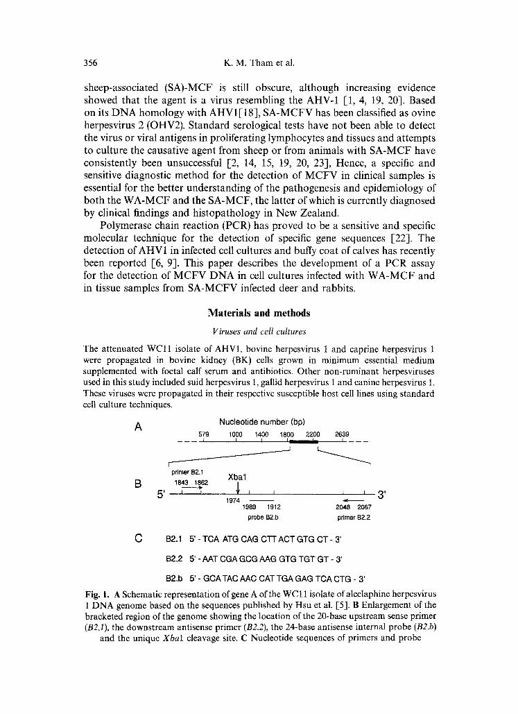
A Nucleotide number (bp)
579 1000 1400
B
r primer B2.1
Xbal 1843 1862
5 ' I I 1974
1989
1800 2200 2639 1 I I I - - - - - -
--J ---_>
I
1912
J = 3 ' 20~8 2067
p r o b e B2.b primer B2.2
C B2.1 5' - TCA ATG CAG C-l-r ACT GTG CT - 3'
B2.2 5' - AAT CGA GCG AAG GTG TGT GT - 3'
B2.b 5' - GCA TAC AAC CAT TGA GAG TCA CTG - 3'
Fig. 1. A Schematic representation of gene A of the WC 1 t isolate of alcelaphine herpesvirus 1 DNA genome based on the sequences published by Hsu et al. [5]. B Enlargement of the bracketed region of the genome showing the location of the 20-base upstream sense primer (B2.1), the downstream antisense primer (B2.2), the 24-base antisense internal probe (B2.b)
and the unique Xbal cleavage site. C Nucleotide sequences of primers and probe
PCR amplification of MCF virus DNA 357
Clinical samples
Mesenteric lymph nodes from 2 red deer experimentally infected with blood collected from deer showing clinical SA-MCF [ 13] and cell suspensions of spleen and/or mesenteric lymph nodes obtained from 7 rabbits experimentally infected with deer MCFV [14] were used in this study.
DNA extraction
Viral DNAs were prepared from infected cell cultures and tissue samples using the method described previously [24]. Mock-infected cultured cell, normal bovine and ovine buffy coat cell DNAs were similarly treated and used as negative controls.
Otigonucleotide primers and probe
A set of primers (B2.1 and B2.2) and an internal probe (B2.b) (Fig. 1), based on the published sequence of the WCl l strain of AHV1 [5], were synthesised and HPLC-purified by the Centre for Gene Technology, University of Auckland, New Zealand. The primers were selected to amplify a 413 bp target sequence of gene A of the AHV1 genome and contains a unique XbaI cleavage site at position 1974.
Polymerase chain reaction
Preliminary experiments were performed to optimise the MCFV PCR by varying (i) the concentrations of dNTP, (ii) the concentrations of the thermostable Tub DNA polymerase (Amersham, Buckinghamshire, England), (iii) the annealing temperatures, and (iv) the annealing times as described previously [24]. Briefly, the optimised PCR protocol for the WCl l isolate of AHV1 is as follows. Amplification was performed in a final volume of 50 gl that contained 2 mM each of dNTP, 1 gM of each primer, 2.5U of Tub polymerase and 10 gl of DNA template in the presence of 50 mM Tris-HC1 (pH 9.0), 20 mM ammonium sulphate, 1.5 mM magnesium chloride. The mixtures were overlaid with 50 gl sterile mineral oil and were first denatured at 95 °C for 2 rain. Using a programmable thermocycler (PHC-3 Dri-Block Cycler, Techne, Cambridge, U.K.), the amplification reaction was taken through 30 repetitive cycles of denaturation (95 °C for 1 min), annealing (56 °C for 2min), and extension (74 °C for 2 min) with a final extension of 10 min for completion of strand synthesis. This PCR protocol was used throughout the experiments described below, unless otherwise stated. A second 35-cycle amplification protocol was used in some PCR experiments under the following conditions: denaturation at 94 °C for 1.5 rain, annealing at 51 °C for 2.5 min and extension at 75 °C for 1 min. Stringent laboratory procedures were observed throughout the experiments to prevent carry-over contamination [10, 11, 24] whereas negative controls were included in each PCR assay to monitor potential contamination.
Analysis of PCR products
Aliquots of the amplified products were analysed by agarose gel electrophoresis and visualised by ethidium bromide staining and UV fluorescence as previously described [24]. The specificity of the amplified product was confirmed through (i) appearance of a 413 bp fragment of the expected size, (ii) appearance of a Xbal restriction pattern as expected from the DNA sequence of the WC1 t isolate of AHV1 and (iii) chemiluminescence Southern blot hybridisation. For enzymatic digestion, a 10 gl aliquot of the PCR product was cleaved with 10 U Xbat at 37 °C for 2 h and the digest was then analysed by agarose gel electro- phoresis as described above.

358 K.M. Tham et al.
The amplified products were transferred onto nylon membranes by Southern blotting [25], hybridised with a digoxigenin-labelled internal probe B2.b (Fig. 1) and analysed by chemiluminescence [24].
Sensitivity and specificity
The sensitivity of the optimised PCR assay was determined using AHV1 DNA extracted from WC11 virus-infected BK cells as target templates tested at serial 10-fold dilutions of 1 pg to 0.1 fg. The specificity of the MCFV PCR was determined using several herpesvirus and non-herpesvirus DNAs, DNAs extracted from mock-infected cell lines and bovine and ovine buffy coat cells described above.
Results
Optimisation of P C R parameters
Conditions for performing the PCR assays were optimised by using AHV1 (WC11 isolate) DNA extracted from infected BK cells. PCR amplification was successful with all concentrations of dNTPs, Tub polymerase and primers (Fig. 2). A distinct band of 413 bp in length was observed in 2% agarose gel following a 30-cycle amplification.
Analysis of amplified products
Digestion of the 413 bp amplified products with XbaI yielded two fragments of about 131 and 282 bp in length (Fig. 3) indicating that the PCR product was specific for AHV1.
Sensitivity and specificity
Using serial 10-fold dilutions of AHV1 ( W C l l isolate) DNA as target, the optimised PCR amplification assay had a sensitivity limit of detection of 10 fg
Fig. 2. Agarose gel electrophoresis of PCR products showing the 413bp amplified fragments (arrow). Amplification was performed using a 30-cycle protocol (see Materials and methods) with 1 mM (1, 4, 7, 10, 13, 16), 2mM (2, 5, 8, 11, 14, 17) or 4mM (3, 6, 9, 12, 15, 18) of each dNTPs, 101~M (1, 2, 3, 10, 11, 12), 1 ~tM (4, 5, 6, 13, 14, 15) or 0.1 ~tM (7, 8, 9, I6, 17, 18) of each primer and 2.5U (I-9) or 5U (10-I8) Tub polymerase. 19BK cell DNA amplified using 4mM of each dNTPs, 101aM of each primers and 5U Tub
polymerase. M Size marker in bp with sizes shown at right

PCR amplification of MCF virus DNA 359
Fig. 3. Restriction enzyme digestion of amplified products. I, 4 Undigested amplified products of PCR performed using the 30-cycle and 35-cycle protocols respectively. 2, 5
Amplified products of I and 4 digested with Xbal. 3 Size marker in bp
Fig. 4. Sensitivity and specificity of MCFV PCR. Electrophoresis of amplified products in 2~o agarose gel showing the 413 bp fragments in the sensitivity titration using 1 pg to 0.1 fg WC11 isolate of AHV1 DNA (I-5). Amplification was not detected with target templates of other viral DNAs such as bovine herpesvirus 1 (6), caprine herpesvirus 1 (7), suid herpesvirus 1 (8), canine herpesvirus 1 (9), and gallid herpesvirus 1 (I0). I 1 Bovine kidney
cell DNA. M Size marker in bp
DNA (Fig. 4). The PCR assay described above was shown to be specific for MCFV. Figure 4 shows that the MCFV primers B2.1 and B2.2 specifically amplified AHV1 DNA but failed to amplify other ruminant herpesvirus, nonherpes viral, mock-infected cultured cell and normal bovine and ovine buffy coat cell DNAs.
PCR amplification of tissue samples
Figures 5 and 6 show the results of PCR amplification of tissue samples from deer and rabbits experimentally infected with materials derived from deer with clinical SA-MCF. The expected amplified fragment of 413 bp was detected in

360 K.M. Tham et al.
Fig. 5. MCFV PCR test for tissue samples. Agarose gel electrophoresis of amplified products following the 35-cycle amplification protocol. 1-7 Lymphoid cell suspensions of rabbits experimentally infected with deer MCFV. 8 Mesenteric lymph node of a red deer experimentally infected with blood collected from a clinical case of deer MCF. 9 WC11 isolate of AHV1 DNA. 10 Bovine kidney cell DNA. 11, 12 Normal bovine and ovine buffy
coat cell DNA. M Size marker in bp
Fig. 6. Confirmatory PCR test for the detection of MCFV in lymphoid tissues of deer and rabbits. A Agarose gel electrophoresis of amplified products. DNAs extracted from lymphoid cell suspensions of two rabbits (2, 3) experimentally infected with deer MCFV and mesenteric lymph node of two red deer (4, 5) experimentally infected with blood collected from a clinical case of deer MCF were tested using the 35-cycle amplification protocol, 1 WCll isolate of AHV1 DNA. 6 Bovine kidney cell DNA. M Size marker in bp. B Chemiluminescence Southern blot hybridisation of amplified products. The numbers
correspond to those in A

PCR amplification of MCF virus DNA 361
tissue samples from 6 out of 7 rabbits, whereas tissue samples from 2 deer consistently yielded a distinct fragment of approximately 600 bp in length
Discussion
Diagnosis of MCF by conventional virus isolation methods is tedious and difficult. Viral infectivity decreases rapidly by repeated freezing and thawing and by specimen autolysis. AHV1 replicates slowly in cell cultures and may require repeated passage using cell-associated techniques, especially for primary virus isolations. Virus replication in cell cultures is initially noncytopathic and are not readily demonstrated in infected tissues by immunofluorescence or electron microscopy [3, 4, 15]. Recently, PCR assays of high sensitivity were described for the detection of AHV1 DNA in infected cell cultures and bovine buffy coat cells [-6, 9].
In this study, a PCR assay was developed for AHV1 following the opti- misation of some parameters known to affect the amplification reaction [-8, 21, 24]. The optimised PCR conditions described herein (Fig. 2) are in agreement with a previous study of optimisation of PCR conditions for another DNA virus [24]. The identity of the 413 bp amplified fragments was proven using restriction endonuclease cleavage and ethidium bromide-agarose gel electro- phoresis. Xbal was shown to cut at the expected site (Fig. 1), yielding two fragments of approximately 140 and 280 bp in length (Fig. 3, lane 2). The size of these two subfragments was close to the predicted sizes (131 and 282 bp) of the nucleotide sequence of the W C l l isolate of AHV1 DNA (Fig. 1B). The specificity of the 413 bp amplified fragment was also confirmed by chemilumi- nescence Southern blot hybridisation using a digoxigenin-labelled internal oligonucleotide probe (Fig. 6B, lanes 1 to 3).
The PCR assay described detects low copy number of AHV1 genome. Our results showed that 10fg of extracted AHV1 DNA could be amplified and the products could be resolved by ethidium bromide-agarose gel electrophoresis (Fig. 4). With a genome size of 160kbp [18], this level of sensitivity represents the detection of about six genomic copies of AHV1.
The PCR assay was shown to be highly specific for MCFV. Amplification of the target gene A of AHV1 was successful with the standard attenuated WC11 isolate, with lymphoid tissues from red deer and rabbits experimentally infected with blood collected from a deer with a clinical case of MCF but not with other ruminant and non-ruminant herpesviruses, mock-infected cell cultures and normal bovine and ovine buffy coat cells (Figs. 4 and 5). The specificity of the PCR assay was confirmed when the 413bp amplified products reacted positively with a digoxigenin-labelled internal probe in a chemiluminescence Southern blot hybridisation assay (Fig. 6B). This was exemplified by the ampli- fication of some MCFV gene sequences which yielded three unexpected distinct fragments of about 400, 600 and 900 bp in length (Figs. 5 and 6A) but these fragments were not detectable by the chemiluminescence Southern blot hybridisation (Fig. 6B).

362 K.M. Tham et al.
The demonstration of the additional amplified fragments of unexpected size, especially with lymphoid tissues from infected red deer, was reproducible (Figs. 5 and 6). While these additional amplified fragments were initially detected at low molar concentrations using the optimised PCR conditions based on the WCl l isolate of AHV1 DNA (data not shown) the fragments were amplified efficiently using the second PCR of 35-cycle protocol. The fragment of approximately 600 bp in length was consistently amplified to higher molar concentrations. One plausible explanation for the detection of these additional fragments, which failed to hybridise with the sequence specific internal probe (Fig. 6B), is the presence of high G + C content (61~) of the AHV1 genome[3] and the amplification reaction performed under relatively low annealing temperature of 56 °C in the 30-cycle protocol. This is further supported with the results of the amplification of target DNAs using the 35-cycle PCR protocol with an annealing temperature of 51 °C (Figs. 5 and 6). It is well established that the thermostable DNA polymerase is active over a broad range of temperatures, resulting in incorrect primer annealing and misextension of primers and extension of misincorporated nucleotides [8].
It is interesting to observe that the expected 413bp fragment was not detected with the lymphoid tissues from two red deer experimentally infected with material derived from a clinical case of deer MCF. Instead, the unexpected amplified fragment of approximately 600 bp in length was consistently present in the MCFV PCR. The optimised MCFV PCR assay is currently used to analyse tissue samples from field cases of MCF in cattle, buffalo and deer. Molecular cloning and/or sequencing of the amplified fragments would help to elucidate the origin and significance of the alcelaphine herpesviruses in wildebeest and the ruminants.
Acknowledgements
The authors thank Drs. R.W. Worthington and G.W. Horner for their support and encouragement. We also thank Dr. B. Buddle for the use of the PHC-3 thermocycler, Mr. A. Barkus for photographic assistance, and Mrs. M. Burns for secretarial services.
References
1. Bridgen A, Reid HW (1991) Derivation of a DNA clone corresponding to the viral agent of sheep-associated malignant catarrhal fever. Res Vet Sci 50:38-44
2. Castro A, Schramke, Ramsay E (1983) A diagnostic approach in the identification and isolation of malignant catarrhal fever virus from inapparent carriers in a wildebeest herd. In: Proc Third Int Syrup World Assoc Vet Lab Diagn, Vol 2. Iowa State University Press, Ames, pp 715-721
3. Edington N, Patel J, Russell PH, Plowright W (1979) The nature of the acute lymphoid proliferation in rabbits infected with the herpes virus of bovine malignant catarrhal fever. Eur J Cancer 15:1515-1522
4. Herring A J, Reid H, Inglis N, Pow I (1989) Immunoblotting analysis of the reaction

PCR amplification of MCF virus DNA 363
of wildebeest, sheep and cattle sera with the structural antigens of alcelaphine herpesvirus-1 (malignant catarrhal fever virus). Vet Microbiol 19:205-215
5. Hsu D, Shih LM, Zee YC (1990a) Nucleotide sequence of a 3.5 kilobase fragment of malignant catarrhal fever virus strain WCll. Arch Virol 113:53-60
6. Hsu D, Shih LM, Castro AE, Zee YC (1990b) A diagnostic method to detect alcelaphine herpesvirus-1 of malignant catarrhal fever using the polymerase chain reaction. Arch Virol 114:259-263
7. Hunt RD, Billup OH (1979) Wildebeest-associated malignant catarrhal fever in Africa: a neoplastic disease of cattle caused by an oncogenic herpesvirus? Comp Immunol Microbiol Infect Dis 2:275-283
8. Innis MA, Gelfand DH (1990) Optimization of PCRs. In: Innis MA, Gelfand DH, Sninsky J J, White TJ (eds) PCR protocols. A guide to methods and applications. Academic Press, New York, pp 3-12
9. Katz J, Seal B, Ridpath J (1991) Molecular diagnosis of alcelaphine herpesvirus (malignant catarrhal fever) infections by nested amplification of viral DNA in bovine blood buffy coat specimens. J Vet Diagn Invest 3:193-198
10. Kitchin PA, Szotyori Z, Fromholc C, Almond N (1990) Avoidance of false positives. Nature 344:201
11. Kwok S (1990) Procedures to minimize PCR-product carry-over. In: Innis MA, Gelfand DH, Sninsky J J, White TJ (eds) PCR protocols. A guide to methods and applications. Academic Press, New York, pp 142-145
12. Metzier A (1991) The malignant catarrhal fever complex. Comp Immunol Microbiol Infect Dis 14:107-124
13. 0liver RE, Beatson NS, Cathcart A, Poole WS (1983) Experimental transmission of malignant catarrhal fever to red deer (Cervus elaphus) NZ Vet J 31:209-212
14. Oliver RE, Beatson NS, Cathcart A, Poole WS (1985) Transmission of malignant catarrhal fever from red deer to rabbits. Roy Soc NZ Bull 22:139-142
15. Patel JR, Edington N (1981) The detection and behavior of the herpesvirus of malignant catarrhal fever in bovine lymphocytes. Arch Virol 68:321-326
16. Plowright W (1986) Malignant catarrhal fever. Rev Sci Tech Int Epizool 5:897-918 17. Reid HW, Buxton D (1989) Malignant catarrhal fever and the gammaherpesvirinae of
bovidae. In: Wittmann G (ed) Herpesvirus disease of cattle, horse and pigs. Kluwer, Norwell, pp t 16-162
18. Roizman B, Desrosiers RC, Fleckenstein B, Lopez C, Minson AC, Studdert MJ (1992) The family Herpesviridae: an update. Arch Virol 123:425-448
19. Rossiter PB (1981) Antibodies to malignant catarrhal fever virus in sheep sera. J Comp Pathol 91:303-311
20. Rossiter PB (1983) Antibodies to malignant catarrhal fever virus in cattle with non-wildebeest associated malignant catarrhal fever. J Comp Pathol 93:93-97
21. Saiki RK (1989) The design and optimization of the PCR. In: Erlich HA (ed) PCR technology: principles and applications for DNA amplification. Stockton/Macmillan, New York, pp 7-16
22. Saiki RK, Scharf S, Faloona F, Mullis KB, Horn GT, Erlich HA, Arnhaim N (1985) Enzymatic amplification of beta-globin genomic sequences and restriction site analysis for diagnosis of sickle cell anemia. Science 230:1350-1354
23. Snowdon WA (1985) The role of sheep in the transmission of bovine malignant catarrh. In: Della-Porta AJ (ed) Veterinary viral diseases: their significance in South East Asia and the Western Pacific. Academic Press, Australia, pp 455-458
24. Tham KM, Stanislawek WL (1992) Detection of chicken anaemia agent DNA sequences by the polymerase chain reaction. Arch Virol 127:245-255

364 K.M. Tham et al.: PCR amplification of MCF virus DNA
25. Tham KM, Chow VTK, Singh P, Tock EPC, Ching KC, Lim-Tan SK, Sng ITY, Bernard HU (1990) Diagnostic sensitivity of polymerase chain reaction and Southern blot hybridization for the detection of human papilloma-virus DNA in biopsy specimens from cervical lesions. Am J Clin Pathol 95:638-646
Authors' address: Dr. K. M. Tham, Virology Section, Central Animal Health Laboratory, MQM, P O Box 40-063, Upper Hutt, New Zealand.
Received November 8, 1993