PET in GIT Malignancies - The Egyptian Society of Nuclear ...
18
Egyptian J. Nucl. Med., Vol. 1, No. 1, 35-52 (2009) Oncology, Review Article INTEGRATION OF PET IN THE CLINICAL MANAGEMENT OF GASTROINTESTINAL TRACT MALIGNANCIES EL-MAGHRABY, T. Nuclear Medicine Department, Cairo University, Cairo, Egypt, INTRODUCTION 1 Positron emission tomography (PET) is a major paradigm shift in medical imaging as it is a molecular modality that images the metabolic activity of tissue. Recently, there has been a major expansion and move from research applications into clinical patient care. The majority of these PET scans are performed to evaluate cancer. Uses include cancer diagnosis, staging, restaging and monitoring response to therapy. There is evolving critical applications of PET in the management of Gastrointestinal (GI) tract malignancies. PET is highly sensitive in the detection of occult GIT tumors, nodal and metastatic involvement to liver and other distant sites, yet the role of CT is essential for anatomical delineation, defining tumor extent and resectability. This manuscript reviews the various indications of PET imaging in GI tract malignancies. This will demonstrate the literature and the wide clinical experiences of PET applications in esophagus, Stomach and colorectal. The primary tumors of abdominal solid organs like pancreas, liver ..etc were beyond the scope of this review. The fundamental role of CT imaging in GI tract malignancies is discussed with more emphasis on the added value of the recent fusion of PET and CT that leads to more precise and expansion of the molecular PET images. Correspondence Author: Tarek El- Maghraby e-mail: [email protected] Esophageal cancer Esophageal cancer has a very poor prognosis despite the advances in treatment because esophageal cancer is often diagnosed at advanced stage. There are two types of esophageal carcinoma, squamous cell type and adenocarcinoma which usually occur in the proximal and distal portion of the esophagus, respectively [1]. Staging work-up and imaging: Staging methods include computed tomography (CT), endoscopic ultrasonography (EUS) and magnetic resonance imaging (MRI) [2]. These morphologic imaging modalities, however, rely on structural changes and are often inaccurate resulting in failure of surgery with curative intent. EUS has limitations in patients with stenosis of the lumen of the esophagus caused by the tumor. CT has limitations in differentiating benign from malignant causes of thickening of the wall of the esophagus. Positron emission tomography (PET) using fluorodeoxyglucose (FDG-PET) has now been integrated into the staging and restaging algorithm of esophageal cancer. Patients with early disease have a good chance of survival as curative surgical resection of early stage esophageal cancer is the mainstay of therapy (figure 1). In advanced stages, however, neoadjuvant therapy is necessary prior to surgery to decrease tumor bulk and associated morbidity [1].
-
Upload
khangminh22 -
Category
Documents
-
view
3 -
download
0
Transcript of PET in GIT Malignancies - The Egyptian Society of Nuclear ...
Egyptian J. Nucl. Med., Vol. 1, No. 1, 35-52 (2009)
Oncology, Review Article
INTEGRATION OF PET IN THE CLINICAL MANAGEMENT OF
GASTROINTESTINAL TRACT MALIGNANCIES
EL-MAGHRABY, T.
Nuclear Medicine Department, Cairo University, Cairo, Egypt,
INTRODUCTION1
Positron emission tomography
(PET) is a major paradigm shift in
medical imaging as it is a molecular
modality that images the metabolic
activity of tissue. Recently, there has
been a major expansion and move from
research applications into clinical patient
care. The majority of these PET scans are
performed to evaluate cancer. Uses
include cancer diagnosis, staging,
restaging and monitoring response to
therapy. There is evolving critical
applications of PET in the management
of Gastrointestinal (GI) tract
malignancies. PET is highly sensitive in
the detection of occult GIT tumors, nodal
and metastatic involvement to liver and
other distant sites, yet the role of CT is
essential for anatomical delineation,
defining tumor extent and resectability.
This manuscript reviews the various
indications of PET imaging in GI tract
malignancies. This will demonstrate the
literature and the wide clinical
experiences of PET applications in
esophagus, Stomach and colorectal. The
primary tumors of abdominal solid
organs like pancreas, liver ..etc were
beyond the scope of this review. The
fundamental role of CT imaging in GI
tract malignancies is discussed with more
emphasis on the added value of the recent
fusion of PET and CT that leads to more
precise and expansion of the molecular
PET images.
Correspondence Author: Tarek El-
Maghraby e-mail: [email protected]
Esophageal cancer
Esophageal cancer has a very poor
prognosis despite the advances in
treatment because esophageal cancer is
often diagnosed at advanced stage. There
are two types of esophageal carcinoma,
squamous cell type and adenocarcinoma
which usually occur in the proximal and
distal portion of the esophagus,
respectively [1].
Staging work-up and imaging:
Staging methods include computed
tomography (CT), endoscopic
ultrasonography (EUS) and magnetic
resonance imaging (MRI) [2]. These
morphologic imaging modalities,
however, rely on structural changes and
are often inaccurate resulting in failure of
surgery with curative intent. EUS has
limitations in patients with stenosis of the
lumen of the esophagus caused by the
tumor. CT has limitations in
differentiating benign from malignant
causes of thickening of the wall of the
esophagus. Positron emission
tomography (PET) using
fluorodeoxyglucose (FDG-PET) has now
been integrated into the staging and
restaging algorithm of esophageal cancer.
Patients with early disease have a
good chance of survival as curative
surgical resection of early stage
esophageal cancer is the mainstay of
therapy (figure 1). In advanced stages,
however, neoadjuvant therapy is
necessary prior to surgery to decrease
tumor bulk and associated morbidity [1].

Egyptian J. Nucl. Med., Vol. 1, No. 1, 35-52 (2009)
36
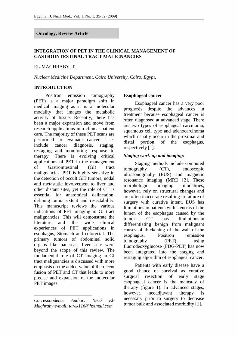
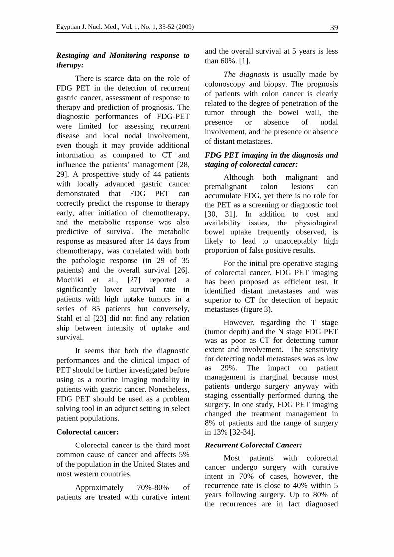
Figure (1): 65 years female patient with cancer esophagus, the PET showed
high uptake in the primary that was already known from the CT examination.
However, the combination of the PET and anatomical verification of CT
showed the tumor extension distal to gastro-esophageal junction with early
involvement of the stomach fundus. The rest of whole body showed
no distant metastases.
Diagnosis and Staging by FDG-PET:
FDG PET became an established
functional imaging modality for patients
with esophageal cancer. FDG PET is
highly sensitive in the detection of the
primary esophageal tumors, hepatic and
distant metastases [3-7].
Regional lymph node involvement
in esophageal cancer is the most
important prognostic factor. The
sensitivity of both PET and CT appears
limited for the detection of local lymph
node involvement with small burden of
tumor as well as lymph nodes that are in
close proximity of the primary tumor.
Nonetheless, studies have shown that
FDG PET significantly improves
preoperative lymph node staging. A
meta-analysis of 12 studies has
Egyptian J. Nucl. Med., Vol. 1, No. 1, 35-52 (2009)
37
demonstrated a pooled sensitivity and
specificity of 51% and 84%, respectively
for FDG PET in the detection of
locoregional disease [8]. However, FDG
PET is more sensitive than conventional
imaging for detection of distant
metastases therefore has an important
role in M staging. The overall pooled
sensitivity and specificity of FDG PET
for detection of distant metastases was
reported around 67% and 97%,
respectively [8]. In a prospective study of
74 patients with esophageal carcinoma,
FDG PET had a higher accuracy than the
combination of CT and EUS for
diagnosing stage IV disease (82% versus
64%) [9, 10]. EUS was more sensitive
than FDG PET for local lymph node
staging (81% versus 33%), but the
specificity of FDG PET was superior to
CT and EUS combined for staging local
and distant lymph nodes. FDG PET
changed the stage in 22% (16/74) of
patients, by upstaging two thirds and
downstaging one third [9]. Comparing
different strategies for preoperative
staging of patients with esophageal
cancer, Wallace et al [11] found that the
combination of PET + EUS with fine
needle aspiration biopsy was the most
effective strategy among various
combination strategies.
Prognosis:
FDG PET is promising for the
assessment of prognosis. In a study of 69
patients with esophageal cancer who
were undergoing curative surgery, the
SUV of the primary tumor, the number of
positive lymph nodes on FDG PET, the
length of the tumor and tumor stage were
independent prognostic predictors
compared to clinical features [12].
Assessment of response to therapy:
One of the strong predictors of
long-term survival is the degree of
response to chemotherapy and radiation
therapy [13]. In a study on 100 patients,
PET, CT and EUS were compared prior
to and 3-5 weeks after completion of
neoadjuvant therapy [14]. FDG PET
imaging was superior to both CT and
EUS with a sensitivity, specificity and
accuracy of 62%, 84% and 76%,
respectively. When the primary tumor,
regional and distant metastatic disease
were considered, the sensitivity,
specificity and accuracy of PET was
69%, 78% and 75% respectively. In this
study, a post-therapy SUV of the primary
tumor (equal or greater than 4.0) was an
independent predictor of long-term
survival. Other studies showed that the
response to therapy can be predicted
early after the initiation of chemotherapy
or chemoradiotherapy [15, 16]. Although,
larger prospective trials are necessary, it
appears that the degree of change in FDG
uptake during or after therapy is
predictive of the pathological response
and has long-term prognostic
significance.
Restaging:
Patients with esophageal carcinoma
present with distant metastases more
often at the time of recurrence than at the
time of initial diagnosis. As a whole body
metabolic imaging modality FDG PET is
superior to other imaging techniques in
the detection of distant metastases.
Hence, FDG PET may be most helpful in
restaging patients when they present with
recurrence [17, 18].
Summary:
So, PET in ca aesophus may
change the staging in up to 25%
of patients because of its superiority in
the detection of distant metastases when
compared to CT and EUS. Locoregional
nodal metastases are detected more
accurately with FDG PET than CT, but
not as compared to EUS. The utility of
FDG PET may be more important in the
evaluation of patients with recurrence or
suspected recurrence and in the
assessment of response to therapy.
Egyptian J. Nucl. Med., Vol. 1, No. 1, 35-52 (2009)
38
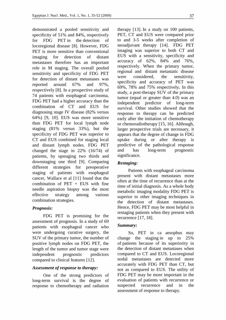
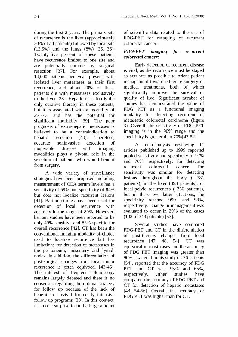
A B
Figure (2): 70 years male patient with gastric cancer. The PET images showed
the primary mass with intense uptake and 2 hepatic metastases. The fusion
PET/CT images delineates the malignant lesions more precisely.
Gastric Cancer:
Ninety-five percent of gastric cancers are adenocarcinomas. They have been classified into the intestinal and diffuse type. The diffuse type seems to have a genetic predisposition and affects younger individuals. Histologically, it is poorly differentiated and lacks glandular structures. The intestinal types develop in a transition from normal mucosa to dysplasia and adenocarcinoma, usually in the distal stomach. Histologically, the intestinal type forms gland-type structures [19, 20].
Staging:
The prognosis of patients with gastric cancer depends on the tumor extent and includes both nodal involvement and direct tumor extension beyond the gastric wall [21]. The diagnosis for the primary is usually made by endoscopy and biopsy, while CT and EUS are used for assessment of the locoregional extent of the disease [2]. For early gastric cancer, surgical resection with en bloc resection of the tumor and regional lymph nodes is associated with a 5-year survival of approximately 90% [22]. Unfortunately, most cases are
diagnosed at an advanced stage and are
treated with neoadjuvant chemotherapy
with a poor survival of approximately
10% [2]. The role of FDG PET for
staging of gastric cancer is still
controversial, yet the PET is highly
sensitive for showing hepatic and distant
metastases [23-25], as with the case
example shown in figure (2). Overall, for
gastric carcinomas, the sensitivity,
specificity and accuracy of PET for the
detection of the primary tumor, loco-
regional metastases, and distant
metastases is in the same range as for
esophageal carcinomas. The sensitivity
for detecting locally advanced gastric
carcinoma ranges from 60 to 80% [23,
26, 27]. Pathologic type (Nonintestinal
type) and depth of invasion and tumor
size have been reported as factors
influencing the detection rate [23, 27].
The false negatives for FDG PET are for
the detection of diffuse type of gastric
adenocarcinoma with a high mucin
content, in addition, normal diffuse
physiological uptake in the stomach may
obscure small tumors with low degree of
FDG uptake.
Egyptian J. Nucl. Med., Vol. 1, No. 1, 35-52 (2009)
39
Restaging and Monitoring response to
therapy:
There is scarce data on the role of
FDG PET in the detection of recurrent
gastric cancer, assessment of response to
therapy and prediction of prognosis. The
diagnostic performances of FDG-PET
were limited for assessing recurrent
disease and local nodal involvement,
even though it may provide additional
information as compared to CT and
influence the patients’ management [28,
29]. A prospective study of 44 patients
with locally advanced gastric cancer
demonstrated that FDG PET can
correctly predict the response to therapy
early, after initiation of chemotherapy,
and the metabolic response was also
predictive of survival. The metabolic
response as measured after 14 days from
chemotherapy, was correlated with both
the pathologic response (in 29 of 35
patients) and the overall survival [26].
Mochiki et al., [27] reported a
significantly lower survival rate in
patients with high uptake tumors in a
series of 85 patients, but conversely,
Stahl et al [23] did not find any relation
ship between intensity of uptake and
survival.
It seems that both the diagnostic
performances and the clinical impact of
PET should be further investigated before
using as a routine imaging modality in
patients with gastric cancer. Nonetheless,
FDG PET should be used as a problem
solving tool in an adjunct setting in select
patient populations.
Colorectal cancer:
Colorectal cancer is the third most
common cause of cancer and affects 5%
of the population in the United States and
most western countries.
Approximately 70%-80% of
patients are treated with curative intent
and the overall survival at 5 years is less
than 60%. [1].
The diagnosis is usually made by
colonoscopy and biopsy. The prognosis
of patients with colon cancer is clearly
related to the degree of penetration of the
tumor through the bowel wall, the
presence or absence of nodal
involvement, and the presence or absence
of distant metastases.
FDG PET imaging in the diagnosis and
staging of colorectal cancer:
Although both malignant and
premalignant colon lesions can
accumulate FDG, yet there is no role for
the PET as a screening or diagnostic tool
[30, 31]. In addition to cost and
availability issues, the physiological
bowel uptake frequently observed, is
likely to lead to unacceptably high
proportion of false positive results.
For the initial pre-operative staging
of colorectal cancer, FDG PET imaging
has been proposed as efficient test. It
identified distant metastases and was
superior to CT for detection of hepatic
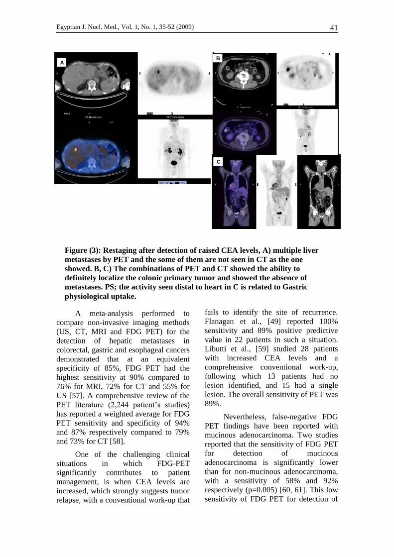
metastases (figure 3).
However, regarding the T stage
(tumor depth) and the N stage FDG PET
was as poor as CT for detecting tumor
extent and involvement. The sensitivity
for detecting nodal metastases was as low
as 29%. The impact on patient
management is marginal because most
patients undergo surgery anyway with
staging essentially performed during the
surgery. In one study, FDG PET imaging
changed the treatment management in
8% of patients and the range of surgery
in 13% [32-34].
Recurrent Colorectal Cancer:
Most patients with colorectal
cancer undergo surgery with curative
intent in 70% of cases, however, the
recurrence rate is close to 40% within 5
years following surgery. Up to 80% of
the recurrences are in fact diagnosed
Egyptian J. Nucl. Med., Vol. 1, No. 1, 35-52 (2009)
40
during the first 2 years. The primary site
of recurrence is the liver (approximately
20% of all patients) followed by local site
(12.5%) and the lungs (8%) [35, 36].
Twenty-five percent of these patients
have recurrence limited to one site and
are potentially curable by surgical
resection [37]. For example, about
14,000 patients per year present with
isolated liver metastases as their first
recurrence, and about 20% of these
patients die with metastases exclusively
to the liver [38]. Hepatic resection is the
only curative therapy in these patients,
but it is associated with a mortality of
2%-7% and has the potential for
significant morbidity [39]. The poor
prognosis of extra-hepatic metastases is
believed to be a contraindication to
hepatic resection [40]. Therefore,
accurate noninvasive detection of
inoperable disease with imaging
modalities plays a pivotal role in the
selection of patients who would benefit
from surgery.
A wide variety of surveillance
strategies have been proposed including
measurement of CEA serum levels has a
sensitivity of 59% and specificity of 84%
but does not localize recurrent lesions
[41]. Barium studies have been used for
detection of local recurrence with
accuracy in the range of 80%. However,
barium studies have been reported to be
only 49% sensitive and 85% specific for
overall recurrence [42]. CT has been the
conventional imaging modality of choice
used to localize recurrence but has
limitations for detection of metastases in
the peritoneum, mesentery and lymph
nodes. In addition, the differentiation of
post-surgical changes from local tumor
recurrence is often equivocal [43-46].
The interest of frequent colonoscopy
remains largely debated and there is no
consensus regarding the optimal strategy
for follow up because of the lack of
benefit in survival for costly intensive
follow up programs [30]. In this context,
it is not a surprise to find a large amount
of scientific data related to the use of
FDG-PET for restaging of recurrent
colorectal cancer.
FDG-PET imaging for recurrent
colorectal cancer:
Early detection of recurrent disease
is vital, as the recurrence must be staged
as accurate as possible to orient patient
management toward either re-surgery or
medical treatments, both of which
significantly improve the survival or
quality of live. Significant number of
studies has demonstrated the value of
FDG PET as a functional imaging
modality for detecting recurrent or
metastatic colorectal carcinoma (figure
3). Overall, the sensitivity of FDG PET
imaging is in the 90% range and the
specificity is greater than 70%[47-52].
A meta-analysis reviewing 11
articles published up to 1999 reported
pooled sensitivity and specificity of 97%
and 76%, respectively, for detecting
recurrent colorectal cancer The
sensitivity was similar for detecting
lesions throughout the body ( 281
patients), in the liver (393 patients), or
local-pelvic recurrences ( 366 patients),
but in these two latter situations, the
specificity reached 99% and 98%,
respectively. Change in management was
evaluated to occur in 29% of the cases
(102 of 349 patients) [53].
Several studies have compared
FDG-PET and CT in the differentiation
of post-therapy changes from local
recurrence [47, 48, 54]. CT was
equivocal in most cases and the accuracy
of FDG PET imaging was greater than
90%. Lai et al in his study on 76 patients
[54], reported that the accuracy of FDG
PET and CT was 95% and 65%,
respectively. Other studies have
compared the accuracy of FDG-PET and
CT for detection of hepatic metastases
[48, 54-56]. Overall, the accuracy for
FDG PET was higher than for CT.
Egyptian J. Nucl. Med., Vol. 1, No. 1, 35-52 (2009)
41
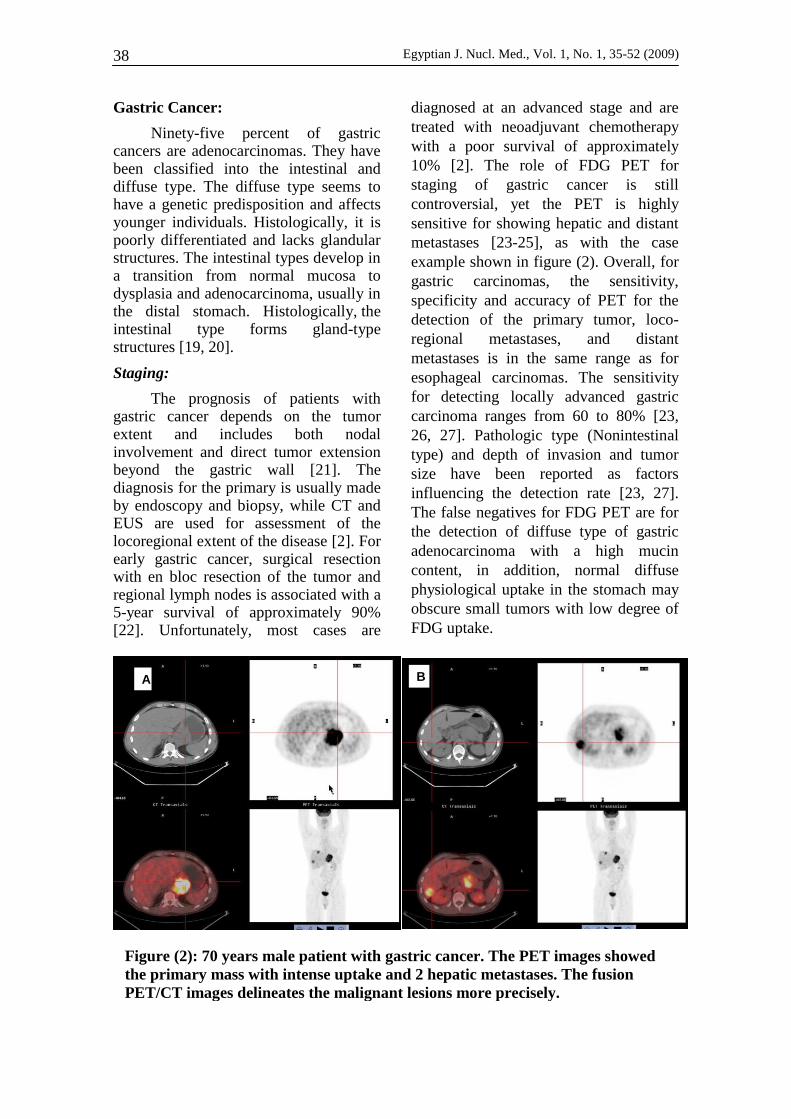
A
C
B
Figure (3): Restaging after detection of raised CEA levels, A) multiple liver
metastases by PET and the some of them are not seen in CT as the one
showed. B, C) The combinations of PET and CT showed the ability to
definitely localize the colonic primary tumor and showed the absence of
metastases. PS; the activity seen distal to heart in C is related to Gastric
physiological uptake.
A meta-analysis performed to
compare non-invasive imaging methods
(US, CT, MRI and FDG PET) for the
detection of hepatic metastases in
colorectal, gastric and esophageal cancers
demonstrated that at an equivalent
specificity of 85%, FDG PET had the
highest sensitivity at 90% compared to
76% for MRI, 72% for CT and 55% for
US [57]. A comprehensive review of the
PET literature (2,244 patient’s studies)
has reported a weighted average for FDG
PET sensitivity and specificity of 94%
and 87% respectively compared to 79%
and 73% for CT [58].
One of the challenging clinical
situations in which FDG-PET
significantly contributes to patient
management, is when CEA levels are
increased, which strongly suggests tumor
relapse, with a conventional work-up that
fails to identify the site of recurrence.
Flanagan et al., [49] reported 100%
sensitivity and 89% positive predictive
value in 22 patients in such a situation.
Libutti et al., [59] studied 28 patients
with increased CEA levels and a
comprehensive conventional work-up,
following which 13 patients had no
lesion identified, and 15 had a single
lesion. The overall sensitivity of PET was
89%.
Nevertheless, false-negative FDG
PET findings have been reported with
mucinous adenocarcinoma. Two studies
reported that the sensitivity of FDG PET
for detection of mucinous
adenocarcinoma is significantly lower
than for non-mucinous adenocarcinoma,
with a sensitivity of 58% and 92%
respectively (p=0.005) [60, 61]. This low
sensitivity of FDG PET for detection of
Egyptian J. Nucl. Med., Vol. 1, No. 1, 35-52 (2009)
42
mucinous adenocarcinoma is due to the
relative hypocellularity of these tumors.
Assessment of response to therapy:
One of the earliest applications for
FDG-PET was to differentiate scar tissue
following therapy from recurrent tumor
in the pelvic area [62, 63]. In cases of
advanced cancer colon, systemic
chemotherapy with 5-fluorouracil, often
in combination with radiotherapy, has
demonstrated effective palliation and
improved survival [64]. A study on 44
patients demonstrated that FDG PET
imaging can differentiate local recurrence
from post-therapy changes (scarring)
after radiation therapy, However, post
radiation inflammatory changes many
lead to increase in FDG uptake [65]. In
fact, the longer the interval of time
between completions of radiation
therapy, the higher is the accuracy of
PET for assessing recurrent or persistent
disease [66]. The time course of post-
irradiation FDG activity has not been
studied systematically; it is, however,
generally accepted that FDG activity
present 6 months after completion of
radiation therapy most likely represents
tumor recurrence. A case-controlled
study of 60 FDG-PET studies performed
6 months following external beam
radiation therapy for rectal cancer found
a sensitivity of 84% and specificity of
88% for detection of local pelvic
recurrence [66]. Some data indicates that
FDG PET assessment of locally
advanced rectal cancer response to
preoperative chemoradiation may predict
long term outcomes [67-70].
Systemic chemotherapy or regional
therapies are used to treat hepatic
metastases. The regional therapy
modalities for hepatic metastases include
chemotherapy administered through the
hepatic artery using infusion pumps,
selective chemoembolization,
radiofrequency ablation, cryoablation,
alcohol ablation and radiolabeled 90
Y-
microspheres [71]. There are preliminary
reports suggesting that the response to
chemotherapy in patients with hepatic
metastases can be predicted using FDG
PET. Responders may be discriminated
from non-responders after 4 to 5 weeks
of chemotherapy with fluorouracil by
measuring FDG uptake before and during
therapy [72]. Data suggest that FDG PET
imaging accurately monitors the efficacy
of radiofrequency ablation for treatment
of hepatic metastases and it detects
incomplete tumor ablation not detectable
on CT. FDG uptake decreases in
responding lesions and the presence of
residual uptake in some lesions can help
in guiding further regional therapy [73].
Overall, the current data suggest that
FDG PET imaging may be able to
effectively monitor the efficacy of
regional therapy to hepatic metastases
but, it seems that much a larger series of
patients before considering FDG-PET as
a routine clinical tool in this indication.
Impact of FDG PET on Patient
Management:
The greater sensitivity of FDG PET
compared to CT in the diagnosis and
staging of recurrent tumor results from
two factors: early detection of abnormal
tumor metabolism, before changes
become apparent by anatomic imaging,
and the whole body imaging which
permits detection of metastases in
unusual and/or unexpected sites. FDG
PET imaging allows detection of
unsuspected metastases in 13%-36% of
patients and has a clinical impact in 14%-
65% [52, 56, 74-76]. In the study of
Delbeke et al, [56] surgical management
was altered by FDG PET in 28% of
patients, in one-third by initiating surgery
and in two thirds by avoiding surgery.
Kalff et al., [78] compared the
treatment plan according to the results of
the conventional work-up with the actual
management, decided after performing
PET. Treatment changes occurred in 60
(59%) of 102 patients; in particular,
surgery was cancelled in 26 (60%) of 43

Egyptian J. Nucl. Med., Vol. 1, No. 1, 35-52 (2009)
43
patients because PET found additional,
unsuspected, lesions. Other groups found
even higher figures, such as Staib et al.,
[74] who reported in a series of 100
patients additional information in 86% of
the cases and modification of the surgical
decision in 61%. Other investigators
reported changes in management ranging
from 21% to 48% [51, 79-81]. The
comprehensive review of the FDG PET
literature has reported a weighted average
change of management related to FDG
PET findings in 32% of 915 patients [58].
Fernandez et al., reported 5-year
survival data after resection of metastasis
from colorectal carcinoma [82]. They
established a 5-year survival rate using
conventional diagnostic imaging from the
literature by pooling the data from 19
studies with a total of 6,090 patients.
The 5-year survival rate was 30% and
appeared not to have changed over time.
These results were compared to their
group of 100 patients with hepatic
metastases, who were pre-operatively
staged for resection with curative intent
with the addition of FDG PET imaging.
The 5-year survival rate improved to
58%, indicating that they were able to
define a subgroup after conventional
imaging that has a better prognosis. The
main contribution was to be able to detect
occult disease, leading to a reduction of
unnecessary surgeries.
So; PET has established itself as an
essential diagnostic tool in patients with
colorectal cancer. It is valuable in these
indications: diagnosis and staging of
recurrence especially before re-surgery
with curative intent, and differentiation
of post treatment changes from
recurrence, differentiate nature of
indeterminate lymph nodes, hepatic and
pulmonary lesions. Most importantly,
evaluation of patients with rising CEA
tumor marker levels with inconclusive
work-up. FDG PET proves useful for
assessing the response to treatment and
as a systemic screening tool in follow-up
after curative surgery especially in
patients with high risks of recurrence.
The addition of FDG PET imaging
reduces overall treatment costs by
accurately identifying patients who will
and will not benefit from surgical
procedures. It is particularly useful if
surgery can be avoided in cases where
FDG PET demonstrates metastases.
Hybrid PET/CT in GIT malignancies:
Combined PET/CT imaging with
an integrated system is especially
important in the abdomen and pelvis.
FDG PET images alone may be difficult
to interpret. Bowel activity may be high,
with various patterns that can either
mimic disease or mask peritoneal or
intestinal lesions. The absence of
anatomical landmarks with physiological
excretion by kidneys and ureteric activity
make it difficult confidently to locate a
focus of increased uptake as peritoneal,
nodal, or even bony.
A study of 45 patients with
colorectal cancer referred for FDG PET
imaging using an integrated PET/CT
system demonstrated that PET/CT
imaging increases the accuracy of
interpretation and certainty of locating
lesions. In their study, the frequency of
equivocal and probable lesion
characterization was reduced by 50%
with PET/CT compared to PET alone, the
number of definite locations was
increased by 25%, and the overall correct
staging increased from 78% to 89% [83].
In 204 patients (34 with GIT
tumors) studied with integrated PET/CT
system, the diagnostic accuracy of PET is
improved in approximately 50% of
patients. The results of PET/CT images
had an impact on management of 14%
(28/204) of all patients, 7/28 patients
with a change of management had
colorectal cancer representing 20% (7/34)
of patients with GIT tumors. The impact
on management in the 7 patients with
colorectal cancer included guiding
Egyptian J. Nucl. Med., Vol. 1, No. 1, 35-52 (2009)
44
colonoscopy and biopsy for a local
recurrence (n=2), guiding surgery to
localized metastatic lymph nodes (n=3)
and referral to chemotherapy (n=2) [84].
Selzner et al., [85]
compared
contrast CT and PET/CT imaging in 76
patients evaluated for resection of liver
metastases. CT with contrast and
PET/CT provide similar sensitivity in the
detection of hepatic metastases (95% and
91% respectively). However, the
specificity of PET/CT was superior for
detection of intrahepatic recurrences in
patients with prior hepatectomy
(specificity of 50% versus 100%). The
sensitivity of PET/CT was 93% for
detection of local recurrence at the
primary colorectal resection site
compared to only 53% for contrast CT.
The PET/CT findings changed the
therapeutic strategy in 21% of patients.
The added value of PET/CT over
dedicated PET was reviewed in 62 patients
who underwent abdomino-perineal or low
anterior resection for colorectal cancer. The
sensitivity and specificity for FDG PET
was 82% and 65% while it was much
higher (98% and 96% respectively) for
hybrid PET/CT [86].
Though, presently, there are no
published literature regarding the
incremental value of PET/CT for staging
esophageal cancer, it is highly
recommended based on the improved
detection and characterization of
equivocal and suspicious lesions [87, 88].
The CT addition to the PET have
the marvelous advantages of using the
superior CT data for attenuation
correction, and the potential to provide
better maps than CT alone to modulate
field and dose of radiation therapy in GIT
malignancies [89].
Limitations of PET in GIT
Malignancies:
Interpretation of FDG PET images
needs familiarity with the normal
distribution of FDG, physiological
variations, and benign conditions that
accumulate FDG, which can mimic
malignant processes. FDG uptake is
normally present in the esophagus,
stomach and bowel. Incidental colonic
FDG uptake in 27 patients without
colorectal carcinoma has been correlated
with colonoscopic and/or
histolopathologic findings [90]. In most
patients, diffuse uptake was normal,
segmental uptake was due to colitis, and
focal uptake was associated with benign
adenomas. Agress et al., [91] reviewed
FDG PET studies of 1,750 patients
referred for evaluation of known or
suspected malignancies. The authors
found 58 unexpected focal areas of FDG
uptake and 42 lesions were
pathologically confirmed, 30 (71%) of
which were malignant or premalignant
including 18 colonic adenomas and three
colon carcinoma.
False positive high FDG uptake is
seen in inflamed tissues due to the active
metabolism in macrophages, neutrophils,
fibroblasts and granulation tissue. Mild to
moderate FDG activity seen early after
radiation therapy, along recent incisions,
infected incisions, drainage tubing and
catheters, as well as colostomy sites can
lead to errors in interpretation. Post-
radiotherapy FDG high uptake may
persist for several months and
comparison with baseline FDG images
and knowledge of the radiation port are
imperative. Some inflammatory lesions,
especially granulomatous ones, may be
markedly FDG-avid and can be mistaken
for malignancies; this includes
inflammatory bowel disease,
diverticulitis, acute cholangitis, acute
cholecystitis, acute pancreatitis,
tuberculosis, sarcoidosis, histoplasmosis
and aspergillosis among others [92].
The size of the tumor and the
degree of FDG avidity determine tumor
delectability. False-negative lesions are
caused by partial volume averaging,
Egyptian J. Nucl. Med., Vol. 1, No. 1, 35-52 (2009)
45
leading to underestimation of the uptake
in small lesions (less than twice the
resolution of the imaging system) or in
necrotic or mucinous lesions, falsely
classifying these lesions as benign.
The FDG is extremely sensitive but
the specificity can be compromised in
various circumstances as noted. Other
improvements may be expected from the
development of alternative tracers that
ideally retain the high sensitivity of FDG
while improving the specificity for
tumors. A review of these alternative
PET tracers is beyond the scope of this
article, but data obtained with 18
F-Deoxy-
Fluorothymidine (FLT) are worth
mentioning, given the high expectations
generated by this compound. Francis et
al., [93] demonstrated a strong
correlation between FLT uptake in
colorectal cancer lesions and their level
of proliferation, as measured by immuno-
histochemistry. In a series of 17 patients
with colorectal cancer, the same
investigators reported that both FDG and
FLT demonstrated all primary tumors
were visualized but FDG uptake was on
average two-fold higher when compared
to FLT. Pulmonary and peritoneal
metastases were visualized with both
tracers, but the sensitivity of FLT for
hepatic metastases was only 34%
compared to 97% for FDG due to the
high physiologic hepatic activity with
FLT [94]. Prognosis and therapy
assessment should be the major
indications for FLT tracer, provided
further studies establish its clinical value.
CONCLUSIONS
The clinical applications of FDG-
PET imaging in GIT tumors is now
firmly established in various situations
that include preoperative staging of
esophageal cancer and revealing
unexpected metastases in gastric and
colorectal carcinoma. More importantly
is the detection and staging of recurrent
colorectal cancer when there is a clinical
or biological suspicion with inconclusive
conventional findings. The literature
showed encouraging results in the
evaluation of the therapeutic response of
various gastrointestinal malignancies,
either during the treatment or after its
completion.
PET and CT are complimentary
modalities and quite useful in the
abdomen where there is abundant
physiologic FDG uptake. The diagnostic
implications of integrated PET/CT
imaging include improved detection of
lesions on both CT and FDG PET
images, better differentiation of
physiologic from pathologic foci, and
better localization of pathologic foci.
This advanced hybrid technology
provides more accurate interpretation of
both CT and FDG PET images and
therefore affect the clinical management
by guiding further procedures (biopsy,
surgery, radiation therapy) or excluding
unnecessary additional imaging leading
to optimal patient care in GIT
malignancies.
REFERENCES
1. Jemal, A, Murray T, Ward E, Samuels
A, Tiwari RC, Ghafoor A, Feuer EJ,
Thun MJ. Cancer statistics, 2005. CA
Cancer J Clin. 2005, 55 , 10-30.
2. Dehdashti, F, Siegel BA. Neoplasms
of the esophagus and stomach. Semin
Nucl Med. 2004, 34 , 198-208.
3. Flanagan, FL, Dehdashti F, Siegel
BA, Trask DD, Sundaresan SR,
Patterson GA, Cooper JD. Staging of
esophageal cancer with 18F-
fluorodeoxyglucose positron emission
tomography. AJR Am J Roentgenol.
1997, 168 , 417-24.
4. McAteer, D, Wallis F, Couper G,
Norton M, Welch A, Bruce D, Park K,
Nicolson M, Gilbert FJ, Sharp P.
Evaluation of 18F-FDG positron
emission tomography in gastric and
oesophageal carcinoma. Br J Radiol.
1999, 72 , 525-9.

Egyptian J. Nucl. Med., Vol. 1, No. 1, 35-52 (2009)
46
5. Luketich, JD, Friedman DM, Weigel
TL, Meehan MA, Keenan RJ,
Townsend DW, Meltzer CC.
Evaluation of distant metastases in
esophageal cancer: 100 consecutive
positron emission tomography scans.
Ann Thorac Surg. 1999, 68 , 1133-6;
discussion 1136-7.
6. Rice, TW. Clinical staging of
esophageal carcinoma. CT, EUS, and
PET. Chest Surg Clin N Am. 2000, 10
, 471-85.
7. Hoegerle, S, Altehoefer C, Nitzsche
EU. Staging an esophageal carcinoma
by F-18 fluorodeoxyglucose whole-
body positron emission tomography.
Clin Nucl Med. 2000, 25 , 219-20.
8. van Westreenen, HL, Westerterp M,
Bossuyt PM, Pruim J, Sloof GW, van
Lanschot JJ, Groen H, Plukker JT.
Systematic review of the staging
performance of 18F-
fluorodeoxyglucose positron emission
tomography in esophageal cancer. J
Clin Oncol. 2004, 22 , 3805-12.
9. Flamen, P, Lerut A, Van Cutsem E,
De Wever W, Peeters M, Stroobants
S, Dupont P, Bormans G, Hiele M, De
Leyn P, Van Raemdonck D,
Coosemans W, Ectors N, Haustermans
K, Mortelmans L. Utility of positron
emission tomography for the staging
of patients with potentially operable
esophageal carcinoma. J Clin Oncol.
2000, 18 , 3202-10.
10. Lerut, T, Flamen P, Ectors N, Van
Cutsem E, Peeters M, Hiele M, De
Wever W, Coosemans W, Decker G,
De Leyn P, Deneffe G, Van
Raemdonck D, Mortelmans L.
Histopathologic validation of lymph
node staging with FDG-PET scan in
cancer of the esophagus and
gastroesophageal junction: A
prospective study based on primary
surgery with extensive
lymphadenectomy. Ann Surg. 2000,
232 , 743-52.
11. Wallace, MB, Nietert PJ, Earle C,
Krasna MJ, Hawes RH, Hoffman BJ,
Reed CE. An analysis of multiple
staging management strategies for
carcinoma of the esophagus:
computed tomography, endoscopic
ultrasound, positron emission
tomography, and thoracoscopy/
laparoscopy. Ann Thorac Surg. 2002,
74 , 1026-32.
12. Choi, JY, Jang HJ, Shim YM, Kim K,
Lee KS, Lee KH, Choi Y, Choe YS,
Kim BT. 18F-FDG PET in patients
with esophageal squamous cell
carcinoma undergoing curative
surgery: prognostic implications. J
Nucl Med. 2004, 45 , 1843-50.
13. Thomas, CR, Jr. Biology of
esophageal cancer and the role of
combined modality therapy. Surg Clin
North Am. 1997, 77 , 1139-67.
14. Swisher, SG, Maish M, Erasmus JJ,
Correa AM, Ajani JA, Bresalier R,
Komaki R, Macapinlac H, Munden
RF, Putnam JB, Rice D, Smythe WR,
Vaporciyan AA, Walsh GL, Wu TT,
Roth JA. Utility of PET, CT, and EUS
to identify pathologic responders in
esophageal cancer. Ann Thorac Surg.
2004, 78 , 1152-60; discussion 1152-
60.
15. Weber, WA, Ott K, Becker K, Dittler
HJ, Helmberger H, Avril NE,
Meisetschlager G, Busch R, Siewert
JR, Schwaiger M, Fink U. Prediction
of response to preoperative
chemotherapy in adenocarcinomas of
the esophagogastric junction by
metabolic imaging. J Clin Oncol.
2001, 19 , 3058-65.
16. Wieder, HA, Brucher BL,
Zimmermann F, Becker K, Lordick F,
Beer A, Schwaiger M, Fink U,
Siewert JR, Stein HJ, Weber WA.
Time course of tumor metabolic
activity during chemoradiotherapy of
esophageal squamous cell carcinoma
Egyptian J. Nucl. Med., Vol. 1, No. 1, 35-52 (2009)
47
and response to treatment. J Clin
Oncol. 2004, 22 , 900-8.
17. Flamen, P, Lerut A, Van Cutsem E,
Cambier JP, Maes A, De Wever W,
Peeters M, De Leyn P, Van
Raemdonck D, Mortelmans L. The
utility of positron emission
tomography for the diagnosis and
staging of recurrent esophageal
cancer. J Thorac Cardiovasc Surg.
2000, 120 , 1085-92.
18. Kato, H, Miyazaki T, Nakajima M,
Fukuchi M, Manda R, Kuwano H.
Value of positron emission
tomography in the diagnosis of
recurrent oesophageal carcinoma. Br J
Surg. 2004, 91 , 1004-9.
19. Lauren, P. The Two Histological Main
Types of Gastric Carcinoma: Diffuse
and So-Called Intestinal-Type
Carcinoma. an Attempt At a Histo-
Clinical Classification. Acta Pathol
Microbiol Scand. 1965, 64 , 31-49.
20. Zivny, J, Wang TC, Yantiss R, Kim
KH, Houghton J. Role of therapy or
monitoring in preventing progression
to gastric cancer. J Clin Gastroenterol.
2003, 36 , S50-60; discussion S61-2.
21. Greene, FL. TNM staging for
malignancies of the digestive tract:
2003 changes and beyond. Semin Surg
Oncol. 2003, 21 , 23-9.
22. Roukos, DH. Current advances and
changes in treatment strategy may
improve survival and quality of life in
patients with potentially curable
gastric cancer. Ann Surg Oncol. 1999,
6 , 46-56.
23. Stahl, A, Ott K, Weber WA, Becker
K, Link T, Siewert JR, Schwaiger M,
Fink U. FDG PET imaging of locally
advanced gastric carcinomas:
correlation with endoscopic and
histopathological findings. Eur J Nucl
Med Mol Imaging. 2003, 30 , 288-95.
Epub 2002 Nov 8.
24. Yoshioka, T, Yamaguchi K, Kubota
K, Saginoya T, Yamazaki T, Ido T,
Yamaura G, Takahashi H, Fukuda H,
Kanamaru R. Evaluation of 18F-FDG
PET in patients with a, metastatic, or
recurrent gastric cancer. J Nucl Med.
2003, 44 , 690-9.
25. Mochiki, E, Kuwano H, Katoh H,
Asao T, Oriuchi N, Endo K.
Evaluation of 18F-2-deoxy-2-fluoro-
D-glucose positron emission
tomography for gastric cancer. World
J Surg. 2004, 28 , 247-53. Epub 2004
Feb 17.
26. Ott, K, Fink U, Becker K, Stahl A,
Dittler HJ, Busch R, Stein H, Lordick
F, Link T, Schwaiger M, Siewert JR,
Weber WA. Prediction of response to
preoperative chemotherapy in gastric
carcinoma by metabolic imaging:
results of a prospective trial. J Clin
Oncol. 2003, 21 , 4604-10.
27. Mochiki, E, Kuwano H, Katoh H,
Asao T, Oriuchi N, Endo K.
Evaluation of 18F-2-deoxy-2-fluoro-
D-glucose positron emission
tomography for gastric cancer. World
J Surg. 2004, 28 , 247-53. Epub 2004
Feb 17.
28. De Potter, T, Flamen P, Van Cutsem
E, Penninckx F, Filez L, Bormans G,
Maes A, Mortelmans L. Whole-body
PET with FDG for the diagnosis of
recurrent gastric cancer. Eur J Nucl
Med Mol Imaging. 2002, 29 , 525-9.
Epub 2002 Feb 23.
29. Jadvar, H, Tatlidil R, Garcia AA,
Conti PS. Evaluation of recurrent
gastric malignancy with [F-18]-FDG
positron emission tomography. Clin
Radiol. 2003, 58 , 215-21.
30. Ohlsson, B, Palsson B. Follow-up
after colorectal cancer surgery. Acta
Oncol. 2003, 42 , 816-26.
31. Chen, YK, Kao CH, Liao AC, Shen
YY, Su CT. Colorectal cancer
screening in asymptomatic adults: the
Egyptian J. Nucl. Med., Vol. 1, No. 1, 35-52 (2009)
48
role of FDG PET scan. Anticancer
Res. 2003, 23 , 4357-61.
32. Abdel-Nabi, H, Doerr RJ, Lamonica
DM, Cronin VR, Galantowicz PJ,
Carbone GM, Spaulding MB. Staging
of primary colorectal carcinomas with
fluorine-18 fluorodeoxyglucose
whole-body PET: correlation with
histopathologic and CT findings.
Radiology. 1998, 206 , 755-60.
33. Mukai, M, Sadahiro S, Yasuda S,
Ishida H, Tokunaga N, Tajima T,
Makuuchi H. Preoperative evaluation
by whole-body 18F-
fluorodeoxyglucose positron emission
tomography in patients with primary
colorectal cancer. Oncol Rep. 2000, 7
, 85-7.
34. Kantorova, I, Lipska L, Belohlavek O,
Visokai V, Trubac M, Schneiderova
M. Routine (18)F-FDG PET
preoperative staging of colorectal
cancer: comparison with conventional
staging and its impact on treatment
decision making. J Nucl Med. 2003,
44 , 1784-8.
35. Kievit, J. Follow-up of patients with
colorectal cancer: numbers needed to
test and treat. Eur J Cancer. 2002, 38
, 986-99.
36. Longo, WE, Johnson FE. The
preoperative assessment and
postoperative surveillance of patients
with colon and rectal cancer. Surg
Clin North Am. 2002, 82 , 1091-108.
37. August, DA, Ottow RT, Sugarbaker
PH. Clinical perspective of human
colorectal cancer metastasis. Cancer
Metastasis Rev. 1984, 3 , 303-24.
38. Foster, JH, Lundy J. Liver Metastases.
Curr Probl Surg. 1981, 18 , 157-202.
39. Holm, A, Bradley E, Aldrete JS.
Hepatic resection of metastasis from
colorectal carcinoma. Morbidity,
mortality, and pattern of recurrence.
Ann Surg. 1989, 209 , 428-34.
40. Hughes, KS, Rosenstein RB,
Songhorabodi S, Adson MA, Ilstrup
DM, Fortner JG, Maclean BJ, Foster
JH, Daly JM, Fitzherbert D, et al.
Resection of the liver for colorectal
carcinoma metastases. A multi-
institutional study of long-term
survivors. Dis Colon Rectum. 1988,
31 , 1-4.
41. Moertel, CG, Fleming TR, Macdonald
JS, Haller DG, Laurie JA, Tangen C.
An evaluation of the
carcinoembryonic antigen (CEA) test
for monitoring patients with resected
colon cancer. Jama. 1993, 270 , 943-
7.
42. Chen, YM, Ott DJ, Wolfman NT,
Gelfand DW, Karsteadt N, Bechtold
RE. Recurrent colorectal carcinoma:
evaluation with barium enema
examination and CT. Radiology.
1987, 163 , 307-10.
43. Sugarbaker, PH, Gianola FJ, Dwyer
A, Neuman NR. A simplified plan for
follow-up of patients with colon and
rectal cancer supported by prospective
studies of laboratory and radiologic
test results. Surgery. 1987, 102 , 79-
87.
44. Steele, G, Jr., Bleday R, Mayer RJ,
Lindblad A, Petrelli N, Weaver D. A
prospective evaluation of hepatic
resection for colorectal carcinoma
metastases to the liver:
Gastrointestinal Tumor Study Group
Protocol 6584. J Clin Oncol. 1991, 9 ,
1105-12.
45. Granfield, CA, Charnsangavej C,
Dubrow RA, Varma DG, Curley SA,
Whitley NO, Wallace S. Regional
lymph node metastases in carcinoma
of the left side of the colon and
rectum: CT demonstration. AJR Am J
Roentgenol. 1992, 159 , 757-61.
46. Charnsangavej, C, Whitley NO.
Metastases to the pancreas and
peripancreatic lymph nodes from

Egyptian J. Nucl. Med., Vol. 1, No. 1, 35-52 (2009)
49
carcinoma of the right side of the
colon: CT findings in 12 patients. AJR
Am J Roentgenol. 1993, 160 , 49-52.
47. Falk, PM, Gupta NC, Thorson AG,
Frick MP, Boman BM, Christensen
MA, Blatchford GJ. Positron emission
tomography for preoperative staging
of colorectal carcinoma. Dis Colon
Rectum. 1994, 37 , 153-6.
48. Ogunbiyi, OA, Flanagan FL,
Dehdashti F, Siegel BA, Trask DD,
Birnbaum EH, Fleshman JW, Read
TE, Philpott GW, Kodner IJ.
Detection of recurrent and metastatic
colorectal cancer: comparison of
positron emission tomography and
computed tomography. Ann Surg
Oncol. 1997, 4 , 613-20.
49. Flanagan, FL, Dehdashti F, Ogunbiyi
OA, Kodner IJ, Siegel BA. Utility of
FDG-PET for investigating
unexplained plasma CEA elevation in
patients with colorectal cancer. Ann
Surg. 1998, 227 , 319-23.
50. Flamen, P, Stroobants S, Van Cutsem
E, Dupont P, Bormans G, De Vadder
N, Penninckx F, Van Hoe L,
Mortelmans L. Additional value of
whole-body positron emission
tomography with fluorine-18-2-fluoro-
2-deoxy-D-glucose in recurrent
colorectal cancer. J Clin Oncol. 1999,
17 , 894-901.
51. Imdahl, A, Reinhardt MJ, Nitzsche
EU, Mix M, Dingeldey A, Einert A,
Baier P, Farthmann EH. Impact of
18F-FDG-positron emission
tomography for decision making in
colorectal cancer recurrences.
Langenbecks Arch Surg. 2000, 385 ,
129-34.
52. Kalff, VV, Hicks R, Ware R, Binns D,
McKenzie A. 29. F-18 FDG PET for
Suspected or Confirmed Regional
Recurrence of Colon Cancer. A
Prospective Study of Impact and
Outcome. Clin Positron Imaging.
2000, 3 , 183.
53. Huebner, RH, Park KC, Shepherd JE,
Schwimmer J, Czernin J, Phelps ME,
Gambhir SS. A meta-analysis of the
literature for whole-body FDG PET
detection of recurrent colorectal
cancer. J Nucl Med. 2000, 41 , 1177-
89.
54. Schiepers, C, Penninckx F, De Vadder
N, Merckx E, Mortelmans L, Bormans
G, Marchal G, Filez L, Aerts R.
Contribution of PET in the diagnosis
of recurrent colorectal cancer:
comparison with conventional
imaging. Eur J Surg Oncol. 1995, 21 ,
517-22.
55. Vitola, JV, Delbeke D, Sandler MP,
Campbell MG, Powers TA, Wright
JK, Chapman WC, Pinson CW.
Positron emission tomography to stage
suspected metastatic colorectal
carcinoma to the liver. Am J Surg.
1996, 171 , 21-6.
56. Delbeke, D, Vitola JV, Sandler MP,
Arildsen RC, Powers TA, Wright JK,
Jr., Chapman WC, Pinson CW.
Staging recurrent metastatic colorectal
carcinoma with PET. J Nucl Med.
1997, 38 , 1196-201.
57. Kinkel, K, Lu Y, Both M, Warren RS,
Thoeni RF. Detection of hepatic
metastases from cancers of the
gastrointestinal tract by using
noninvasive imaging methods (US,
CT, MR imaging, PET): a meta-
analysis. Radiology. 2002, 224 , 748-
56.
58. Gambhir, SS, Czernin J, Schwimmer
J, Silverman DH, Coleman RE, Phelps
ME. A tabulated summary of the FDG
PET literature. J Nucl Med. 2001, 42 ,
1S-93S.
59. Libutti, SK, Alexander HR, Jr.,
Choyke P, Bartlett DL, Bacharach SL,
Whatley M, Jousse F, Eckelman WC,
Kranda K, Neumann RD, Carrasquillo
JA. A prospective study of 2-[18F]
fluoro-2-deoxy-D-glucose/positron
emission tomography scan, 99mTc-

Egyptian J. Nucl. Med., Vol. 1, No. 1, 35-52 (2009)
50
labeled arcitumomab (CEA-scan), and
blind second-look laparotomy for
detecting colon cancer recurrence in
patients with increasing
carcinoembryonic antigen levels. Ann
Surg Oncol. 2001, 8 , 779-86.
60. Whiteford, MH, Whiteford HM, Yee
LF, Ogunbiyi OA, Dehdashti F, Siegel
BA, Birnbaum EH, Fleshman JW,
Kodner IJ, Read TE. Usefulness of
FDG-PET scan in the assessment of
suspected metastatic or recurrent
adenocarcinoma of the colon and
rectum. Dis Colon Rectum. 2000, 43 ,
759-67; discussion 767-70.
61. Berger, KL, Nicholson SA, Dehdashti
F, Siegel BA. FDG PET evaluation of
mucinous neoplasms: correlation of
FDG uptake with histopathologic
features. AJR Am J Roentgenol. 2000,
174 , 1005-8.
62. Beets, G, Penninckx F, Schiepers C,
Filez L, Mortelmans L, Kerremans R,
Aerts R, De Roo M. Clinical value of
whole-body positron emission
tomography with
[18F]fluorodeoxyglucose in recurrent
colorectal cancer. Br J Surg. 1994, 81
, 1666-70.
63. Strauss, LG, Clorius JH, Schlag P,
Lehner B, Kimmig B, Engenhart R,
Marin-Grez M, Helus F, Oberdorfer F,
Schmidlin P, et al. Recurrence of
colorectal tumors: PET evaluation.
Radiology. 1989, 170 , 329-32.
64. Bertino, JR. Biomodulation of 5-
fluorouracil with antifolates. Semin
Oncol. 1997, 24 , S18-52-S18-56.
65. Haberkorn, U, Strauss LG,
Dimitrakopoulou A, Engenhart R,
Oberdorfer F, Ostertag H, Romahn J,
van Kaick G. PET studies of
fluorodeoxyglucose metabolism in
patients with recurrent colorectal
tumors receiving radiotherapy. J Nucl
Med. 1991, 32 , 1485-90.
66. Moore, HG, Akhurst T, Larson SM,
Minsky BD, Mazumdar M, Guillem
JG. A case-controlled study of 18-
fluorodeoxyglucose positron emission
tomography in the detection of pelvic
recurrence in previously irradiated
rectal cancer patients. J Am Coll Surg.
2003, 197 , 22-8.
67. Guillem, JG, Puig-La Calle J, Jr.,
Akhurst T, Tickoo S, Ruo L, Minsky
BD, Gollub MJ, Klimstra DS,
Mazumdar M, Paty PB, Macapinlac
H, Yeung H, Saltz L, Finn RD, Erdi
Y, Humm J, Cohen AM, Larson S.
Prospective assessment of primary
rectal cancer response to preoperative
radiation and chemotherapy using 18-
fluorodeoxyglucose positron emission
tomography. Dis Colon Rectum.
2000, 43 , 18-24.
68. Guillem, JG, Moore HG, Akhurst T,
Klimstra DS, Ruo L, Mazumdar M,
Minsky BD, Saltz L, Wong WD,
Larson S. Sequential preoperative
fluorodeoxyglucose-positron emission
tomography assessment of response to
preoperative chemoradiation: a means
for determining longterm outcomes of
rectal cancer. J Am Coll Surg. 2004,
199 , 1-7.
69. Amthauer, H, Denecke T, Rau B,
Hildebrandt B, Hunerbein M, Ruf J,
Schneider U, Gutberlet M, Schlag PM,
Felix R, Wust P. Response prediction
by FDG-PET after neoadjuvant
radiochemotherapy and combined
regional hyperthermia of rectal cancer:
correlation with endorectal ultrasound
and histopathology. Eur J Nucl Med
Mol Imaging. 2004, 31 , 811-9. Epub
2004 Feb 5.
70. Calvo, FA, Domper M, Matute R,
Martinez-Lazaro R, Arranz JA, Desco
M, Alvarez E, Carreras JL. 18F-FDG
positron emission tomography staging
and restaging in rectal cancer treated
with preoperative chemoradiation. Int
Egyptian J. Nucl. Med., Vol. 1, No. 1, 35-52 (2009)
51
J Radiat Oncol Biol Phys. 2004, 58 ,
528-35.
71. Liu, LX, Zhang WH, Jiang HC.
Current treatment for liver metastases
from colorectal cancer. World J
Gastroenterol. 2003, 9 , 193-200.
72. Findlay, M, Young H, Cunningham D,
Iveson A, Cronin B, Hickish T, Pratt
B, Husband J, Flower M, Ott R.
Noninvasive monitoring of tumor
metabolism using fluorodeoxyglucose
and positron emission tomography in
colorectal cancer liver metastases:
correlation with tumor response to
fluorouracil. J Clin Oncol. 1996, 14 ,
700-8.
73. Langenhoff, BS, Oyen WJ, Jager GJ,
Strijk SP, Wobbes T, Corstens FH,
Ruers TJ. Efficacy of fluorine-18-
deoxyglucose positron emission
tomography in detecting tumor
recurrence after local ablative therapy
for liver metastases: a prospective
study. J Clin Oncol. 2002, 20 , 4453-
8.
74. Staib, L, Schirrmeister H, Reske SN,
Beger HG. Is (18)F-
fluorodeoxyglucose positron emission
tomography in recurrent colorectal
cancer a contribution to surgical
decision making? Am J Surg. 2000,
180 , 1-5.
75. Strasberg, SM, Dehdashti F, Siegel
BA, Drebin JA, Linehan D. Survival
of patients evaluated by FDG-PET
before hepatic resection for metastatic
colorectal carcinoma: a prospective
database study. Ann Surg. 2001, 233 ,
293-9.
76. Ruers, TJ, Langenhoff BS, Neeleman
N, Jager GJ, Strijk S, Wobbes T,
Corstens FH, Oyen WJ. Value of
positron emission tomography with
[F-18]fluorodeoxyglucose in patients
with colorectal liver metastases: a
prospective study. J Clin Oncol.
2002, 20 , 388-95.
77. Meta, J, Seltzer M, Schiepers C,
Silverman DH, Ariannejad M,
Gambhir SS, Phelps ME, Valk P,
Czernin J. Impact of 18F-FDG PET
on managing patients with colorectal
cancer: the referring physician's
perspective. J Nucl Med. 2001, 42 ,
586-90.
78. Kalff, V, Hicks RJ, Ware RE, Hogg
A, Binns D, McKenzie AF. The
clinical impact of (18)F-FDG PET in
patients with suspected or confirmed
recurrence of colorectal cancer: a
prospective study. J Nucl Med. 2002,
43 , 492-9.
79. Arulampalam, T, Costa D, Visvikis D,
Boulos P, Taylor I, Ell P. The impact
of FDG-PET on the management
algorithm for recurrent colorectal
cancer. Eur J Nucl Med. 2001, 28 ,
1758-65. Epub 2001 Oct 20.
80. Simo, M, Lomena F, Setoain J, Perez
G, Castellucci P, Costansa JM,
Setoain-Quinquer J, Domenech-Torne
F, Carrio I. FDG-PET improves the
management of patients with
suspected recurrence of colorectal
cancer. Nucl Med Commun. 2002, 23
, 975-82.
81. Desai, DC, Zervos EE, Arnold MW,
Burak WE, Jr., Mantil J, Martin EW,
Jr. Positron emission tomography
affects surgical management in
recurrent colorectal cancer patients.
Ann Surg Oncol. 2003, 10 , 59-64.
82. Fernandez, FG, Drebin JA, Linehan DC, Dehdashti F, Siegel BA, Strasberg SM. Five-year survival after resection of hepatic metastases from colorectal cancer in patients screened by positron emission tomography with F-18 fluorodeoxyglucose (FDG-PET). Ann Surg. 2004, 240 , 438-47; discussion 447-50.
83. Cohade, C, Osman M, Leal J, Wahl RL. Direct comparison of (18)F-FDG PET and PET/CT in patients with

Egyptian J. Nucl. Med., Vol. 1, No. 1, 35-52 (2009)
52
colorectal carcinoma. J Nucl Med. 2003, 44 , 1797-803.
84. Bar-Shalom, R, Guralnik L, Tsalic M, Leiderman M, Frenkel A, Gaitini D, Ben-Nun A, Keidar Z, Israel O. The additional value of PET/CT over PET in FDG imaging of oesophageal cancer. Eur J Nucl Med Mol Imaging. 2005, 32 , 918-24. Epub 2005 Apr 19.
85. Selzner, M, Hany TF, Wildbrett P, McCormack L, Kadry Z, Clavien PA. Does the novel PET/CT imaging modality impact on the treatment of patients with metastatic colorectal cancer of the liver? Ann Surg. 2004, 240 , 1027-34; discussion 1035-6.
86. Even-Sapir, E, Parag Y, Lerman H, Gutman M, Levine C, Rabau M, Figer A, Metser U. Detection of recurrence in patients with rectal cancer: PET/CT after abdominoperineal or anterior resection. Radiology. 2004, 232 , 815-22. Epub 2004 Jul 23.
87. Yuan, S, Yu Y, Chao KS, Fu Z, Yin Y, Liu T, Chen S, Yang X, Yang G, Guo H, Yu J. Additional value of PET/CT over PET in assessment of locoregional lymph nodes in thoracic esophageal squamous cell cancer. J Nucl Med. 2006, 47 , 1255-9.
88. Guo, H, Zhu H, Xi Y, Zhang B, Li L, Huang Y, Zhang J, Fu Z, Yang G, Yuan S, Yu J. Diagnostic and prognostic value of 18F-FDG PET/CT for patients with suspected recurrence from squamous cell carcinoma of the esophagus. J Nucl Med. 2007, 48 , 1251-8. Epub 2007 Jul 13.
89. Pan, T, Mawlawi O, Nehmeh SA, Erdi YE, Luo D, Liu HH, Castillo R, Mohan R, Liao Z, Macapinlac HA.
Attenuation correction of PET images with respiration-averaged CT images in PET/CT. J Nucl Med. 2005, 46 , 1481-7.
90. Tatlidil, R, Jadvar H, Bading JR, Conti PS. Incidental colonic fluorodeoxyglucose uptake:
correlation with colonoscopic and histopathologic findings. Radiology. 2002, 224 , 783-7.
91. Agress, H, Jr., Cooper BZ. Detection of clinically unexpected malignant and premalignant tumors with whole-body FDG PET: histopathologic comparison. Radiology. 2004, 230 , 417-22. Epub 2003 Dec 29.
92. Kubota, R, Yamada S, Kubota K, Ishiwata K, Tamahashi N, Ido T. Intratumoral distribution of fluorine-18-fluorodeoxyglucose in vivo: high accumulation in macrophages and granulation tissues studied by microautoradiography. J Nucl Med. 1992, 33 , 1972-80.
93. Francis, DL, Freeman A, Visvikis D, Costa DC, Luthra SK, Novelli M, Taylor I, Ell PJ. In vivo imaging of cellular proliferation in colorectal cancer using positron emission tomography. Gut. 2003, 52 , 1602-6.
94. Francis, DL, Visvikis D, Costa DC, Arulampalam TH, Townsend C, Luthra SK, Taylor I, Ell PJ. Potential impact of [18F]3'-deoxy-3'-fluorothymidine versus [18F]fluoro-2-deoxy-D-glucose in positron emission tomography for colorectal cancer. Eur J Nucl Med Mol Imaging. 2003, 30 , 988-94. Epub 2003 May 9.