The N-terminal domain of APJ, a CNS-based coreceptor for HIV1, is essential for its receptor...
11
The N-terminal domain of APJ, a CNS-based coreceptor for HIV-1, is essential for its receptor function and coreceptor activity Naiming Zhou, a Xiaoling Zhang, a Xuejun Fan, b Elias Argyris, a Jianhua Fang, a Edward Acheampong, a Garrett C. DuBois, b and Roger J. Pomerantz a, * a Dorrance H. Hamilton Laboratories, Center for Human Virology and Biodefense, Division of Infectious Diseases and Environmental Medicine, Department of Medicine, Jefferson Medical College, Philadelphia, PA 19107, USA b Kimmel Cancer Center, Thomas Jefferson University, Philadelphia, PA 19107, USA Received 2 June 2003; returned to author for revision 16 July 2003; accepted 19 August 2003 Abstract The human APJ, a G protein-coupled seven-transmembrane receptor, has been found to be dramatically expressed in the human central nervous system (CNS) and also to serve as a coreceptor for the entry of human immunodeficiency virus type 1 (HIV-1) and simian immunodeficiency virus (SIV). Studies with animal models suggested that APJ and its natural ligand, apelin, play an important role in the central control of body fluid homeostasis, and in regulation of blood pressure and cardiac contractility. In this study, we characterize the structural and functional determinants of the N-terminal domain of APJ in interactions with its natural ligand and HIV-1 envelope glycoprotein. We demonstrate that the second 10 residues of the N-terminal domain of APJ are critical for association with apelin, while the first 20 amino acids play an important role in supporting cell– cell fusion mediated by HIV-1 gp120. With site-directed mutagenesis, we have identified that the negatively charged amino acid residues Glu20 and Asp23 are involved in receptor and coreceptor functions, but residues Tyr10 and Tyr11 substantially contribute to coreceptor function for both T-tropic (CXCR4) and dual-tropic (CXCR4 and CCR5) HIV-1 isolates. Thus, this study provides potentially important information for further characterizing APJ-apelin functions in vitro and in vivo and designing small molecules for treatment of HIV-1 infection in the CNS. © 2003 Elsevier Inc. All rights reserved. Keywords: APJ receptor; Mutagenesis; Apelin; Signaling; HIV-1; CNS Introduction APJ, an orphan G protein-coupled, seven-transmem- brane receptor, was originally identified from human genomic DNA by O’Dowd et al. (1993) and later was also isolated from mice (Devic et al., 1999) and rats (De Mota et al., 2000; Hosoya et al., 2000; O’Carroll et al., 2000). The human APJ gene encoding a 380 amino acid resembled the angiotensin II receptor, sharing an identity of more than 30% in amino acid sequence, but angiotensin II was unable to interact with APJ (O’Dowd et al., 1993; Tatemoto et al., 1998). Recently the endogenous ligand for APJ receptor, apelin, was first isolated from bovine stomach extracts (Tatemoto et al., 1998), and the protein sequences of hu- man, rat, and mouse apelin preproproteins were deduced from the cDNAs (Tatemoto et al., 1998; Habata et al., 1999; Hosoya et al., 2000; Lee et al., 2000). Studies on the distribution of APJ and apelin demon- strated that both were abundantly expressed in the central nervous system (CNS) and periphery of human and rat (O’Dowd et al., 1993; Matsumoto et al., 1996; Edinger et al., 1998; Lee et al., 2000; Reaux et al., 2001, 2002). In addition, significant similarities of sequence and tissue dis- tributions between APJ/apelin and the AT1/angiotensin II suggested that APJ and apelin would play an important physiological role as a neurotransmitter or neuromodulator (Lee et al., 2000; Reaux et al., 2001, 2002). In a rat model, APJ/apelin has been shown to play an important role in the hypothalamic regulation of water intake and the endocrine * Corresponding author. Fax: 1-215-503-2624. E-mail address: [email protected] (R.J. Pomerantz). R Available online at www.sciencedirect.com Virology 317 (2003) 84 –94 www.elsevier.com/locate/yviro 0042-6822/$ – see front matter © 2003 Elsevier Inc. All rights reserved. doi:10.1016/j.virol.2003.08.026
Transcript of The N-terminal domain of APJ, a CNS-based coreceptor for HIV1, is essential for its receptor...
The N-terminal domain of APJ, a CNS-based coreceptor for HIV-1,is essential for its receptor function and coreceptor activity
Naiming Zhou,a Xiaoling Zhang,a Xuejun Fan,b Elias Argyris,a Jianhua Fang,a
Edward Acheampong,a Garrett C. DuBois,b and Roger J. Pomerantza,*a Dorrance H. Hamilton Laboratories, Center for Human Virology and Biodefense, Division of Infectious Diseases and Environmental Medicine,
Department of Medicine, Jefferson Medical College, Philadelphia, PA 19107, USAb Kimmel Cancer Center, Thomas Jefferson University, Philadelphia, PA 19107, USA
Received 2 June 2003; returned to author for revision 16 July 2003; accepted 19 August 2003
Abstract
The human APJ, a G protein-coupled seven-transmembrane receptor, has been found to be dramatically expressed in the human centralnervous system (CNS) and also to serve as a coreceptor for the entry of human immunodeficiency virus type 1 (HIV-1) and simianimmunodeficiency virus (SIV). Studies with animal models suggested that APJ and its natural ligand, apelin, play an important role in thecentral control of body fluid homeostasis, and in regulation of blood pressure and cardiac contractility. In this study, we characterize thestructural and functional determinants of the N-terminal domain of APJ in interactions with its natural ligand and HIV-1 envelopeglycoprotein. We demonstrate that the second 10 residues of the N-terminal domain of APJ are critical for association with apelin, whilethe first 20 amino acids play an important role in supporting cell–cell fusion mediated by HIV-1 gp120. With site-directed mutagenesis, wehave identified that the negatively charged amino acid residues Glu20 and Asp23 are involved in receptor and coreceptor functions, butresidues Tyr10 and Tyr11 substantially contribute to coreceptor function for both T-tropic (CXCR4) and dual-tropic (CXCR4 and CCR5)HIV-1 isolates. Thus, this study provides potentially important information for further characterizing APJ-apelin functions in vitro and invivo and designing small molecules for treatment of HIV-1 infection in the CNS.© 2003 Elsevier Inc. All rights reserved.
Keywords: APJ receptor; Mutagenesis; Apelin; Signaling; HIV-1; CNS
Introduction
APJ, an orphan G protein-coupled, seven-transmem-brane receptor, was originally identified from humangenomic DNA by O’Dowd et al. (1993) and later was alsoisolated from mice (Devic et al., 1999) and rats (De Mota etal., 2000; Hosoya et al., 2000; O’Carroll et al., 2000). Thehuman APJ gene encoding a 380 amino acid resembled theangiotensin II receptor, sharing an identity of more than30% in amino acid sequence, but angiotensin II was unableto interact with APJ (O’Dowd et al., 1993; Tatemoto et al.,1998). Recently the endogenous ligand for APJ receptor,apelin, was first isolated from bovine stomach extracts
(Tatemoto et al., 1998), and the protein sequences of hu-man, rat, and mouse apelin preproproteins were deducedfrom the cDNAs (Tatemoto et al., 1998; Habata et al., 1999;Hosoya et al., 2000; Lee et al., 2000).
Studies on the distribution of APJ and apelin demon-strated that both were abundantly expressed in the centralnervous system (CNS) and periphery of human and rat(O’Dowd et al., 1993; Matsumoto et al., 1996; Edinger etal., 1998; Lee et al., 2000; Reaux et al., 2001, 2002). Inaddition, significant similarities of sequence and tissue dis-tributions between APJ/apelin and the AT1/angiotensin IIsuggested that APJ and apelin would play an importantphysiological role as a neurotransmitter or neuromodulator(Lee et al., 2000; Reaux et al., 2001, 2002). In a rat model,APJ/apelin has been shown to play an important role in thehypothalamic regulation of water intake and the endocrine
* Corresponding author. Fax: �1-215-503-2624.E-mail address: [email protected] (R.J. Pomerantz).
R
Available online at www.sciencedirect.com
Virology 317 (2003) 84–94 www.elsevier.com/locate/yviro
0042-6822/$ – see front matter © 2003 Elsevier Inc. All rights reserved.doi:10.1016/j.virol.2003.08.026
axes (Lee et al., 2000; Reaux et al., 2001; Taheri et al.,2002), in regulation of blood pressure via a nitric oxide-dependent mechanism (Lee et al., 2000; Tatemoto et al.,2001), and cardiac contractility (Reaux et al., 2002; Szokodiet al., 2002).
Recently the APJ receptor has been found to supportenv-mediated membrane fusion or viral entry as an alterna-tive coreceptor for human and simian immunodeficiencyviruses (HIV and SIV) (Choe et al., 1998; Edinger et al.,1998; Zhang et al., 1998; Puffer et al., 2000). Consistentlyapelin has been reported to be able to block the activity ofAPJ as an HIV-1 coreceptor (Cayabyab et al., 2000; Pufferet al., 2000; Zou et al., 2000). Although the absence of APJexpression in microglia and macrophages, the principalCD4-positive cell types in CNS, suggested initially that APJis unlikely to mediate HIV-1 infection in the CNS (Choe etal., 2000), a recent study has shown that HIV-1 infection inneural cells involves a chemokine-receptor-dependent butCD4-independent entry (Alvarez Losada et al., 2002). Inaddition, it was reported that some viral envelope proteinscould mediate fusion with APJ-positive, CD4-negative cells(Puffer et al., 2000). With the detection of abundant expres-sion of APJ in the CNS, these observations raise the possi-bility that the APJ receptor might contribute to HIV-1 in-fection and pathogenesis in the CNS.
Although many studies have investigated the physiolog-ical role of APJ receptor, there is no report on the charac-terization of the structure and function of APJ as a receptorand a coreceptor. In the present study, we used deletion andsite-directed mutagenesis combined with binding and func-tional assays to investigate structural determinants of APJfor its ligand activation and coreceptor function. Our studyhas revealed the residues in the N-terminal domain of APJcritical for biological function and coreceptor activity andprovided information for further understanding the physio-logical and pathological roles of APJ and elucidating mech-anisms of HIV-1 viral entry.
Results
Effects of deletion of amino acids in the N-terminaldomain of APJ on functions of receptor and HIV-1coreceptor activities
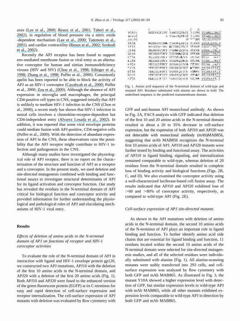
To evaluate the role of the N-terminal domain of APJ ininteraction with ligand and HIV-1 envelope protein gp120,we constructed two APJ mutations, APJ10 with the deletionof the first 10 amino acids in the N-terminal domain, andAPJ20 with a deletion of the first 20 amino acids (Fig. 1).Both APJ10 and APJ20 were fused to the enhanced versionof the green fluorescent protein (EGFP) at its C-terminus foreasy and rapid detection of cell-surface expression andreceptor internalization. The cell-surface expression of APJmutants with deletion was evaluated by flow cytometry with
GFP and anti-human APJ monoclonal antibody. As shownin Fig. 2A, FACS analysis with GFP indicated that deletionof the first 10 and 20 amino acids in the N-terminal domainresulted in about a 20 to 35% decrease in cell-surfaceexpression, but the expression of both APJ10 and APJ20 wasnot detectable with monoclonal antibody (mAb)MAB856,suggesting that mAb MAB856 recognizes the N-terminalfirst 10 amino acids of APJ. APJ10 and APJ20 mutants werefurther tested by binding and functional assay. The activitiesof APJ10 in ligand binding, signaling, and internalizationremained comparable to wild-type, whereas deletion of 20residues from the N-terminal domain resulted in completeloss of binding activity and biological functions (Figs. 2B,C, and D). We also examined the coreceptor activity usinga well-characterized luciferase-based cell fusion assay. Theresults indicated that APJ10 and APJ20 exhibited lose of�60 and �80% of coreceptor activity, respectively, ascompared to wild-type APJ (Fig. 2E).
Cell-surface expression of APJ site-directed mutants
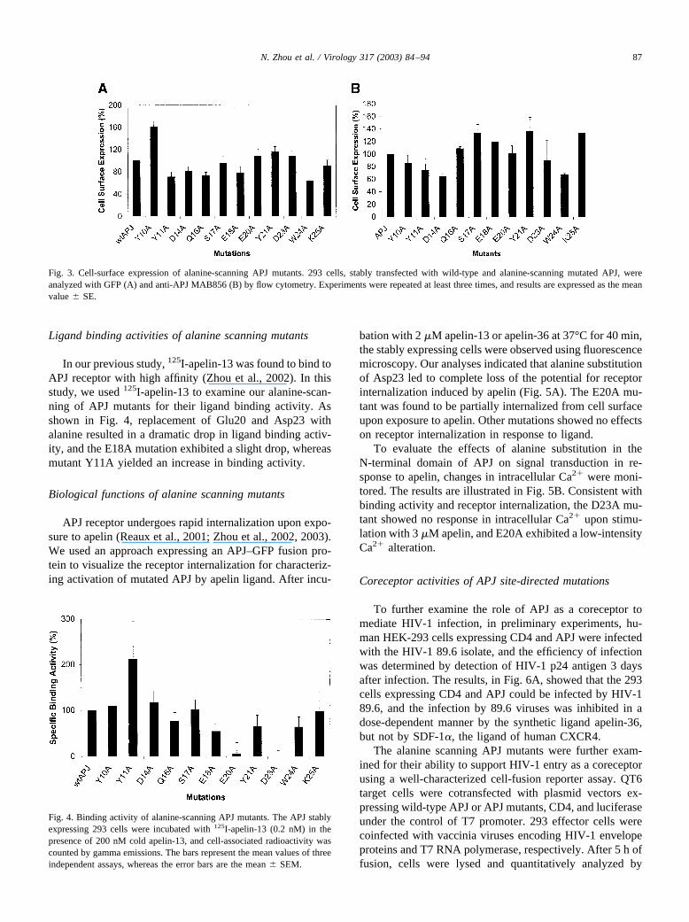
As shown in the APJ mutations with deletion of aminoacids in the N-terminal domain, the second 10 amino acidsof the N-terminus of APJ plays an important role in ligandbinding and function. To further identify amino acid sidechains that are essential for ligand binding and function, 11residues located within the second 10 amino acids of theN-terminal domain were selected for site-directed mutagen-esis studies, and all of the selected residues were individu-ally substituted with alanine (Fig. 1). All alanine-scanningmutants were stably transfected into 293 cells, and cell-surface expression was analyzed by flow cytometry withboth GFP and mAb MAB865. As illustrated in Fig. 3, themutant Y10A showed a higher expression level with detec-tion of GFP, but similar expression levels to wild-type APJwith mAb MAB865, while all other mutants exhibited ex-pression levels comparable to wild-type APJ in detection byboth GFP and mAb MAB865.
Fig. 1. Amino acid sequence of the N-terminal domain of wild-type andmutated APJ. Residues substituted with alanine are shown in bold. Theunderlined sequence is the predicted transmembrane domain.
85N. Zhou et al. / Virology 317 (2003) 84–94
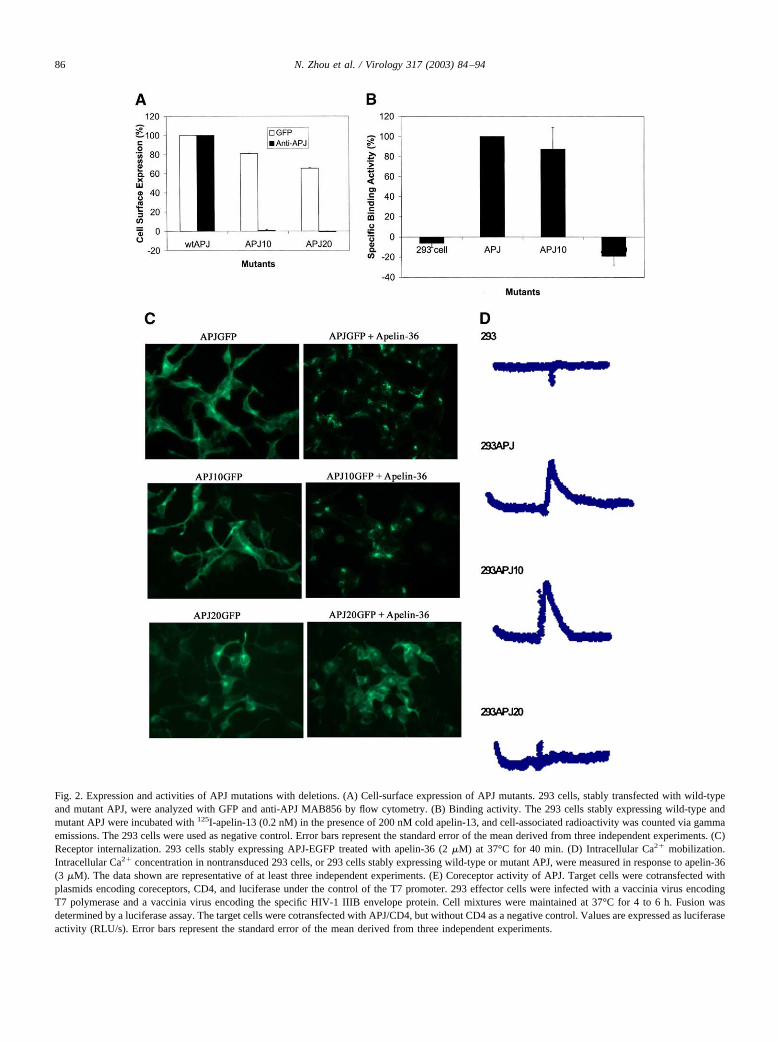
Fig. 2. Expression and activities of APJ mutations with deletions. (A) Cell-surface expression of APJ mutants. 293 cells, stably transfected with wild-typeand mutant APJ, were analyzed with GFP and anti-APJ MAB856 by flow cytometry. (B) Binding activity. The 293 cells stably expressing wild-type andmutant APJ were incubated with 125I-apelin-13 (0.2 nM) in the presence of 200 nM cold apelin-13, and cell-associated radioactivity was counted via gammaemissions. The 293 cells were used as negative control. Error bars represent the standard error of the mean derived from three independent experiments. (C)Receptor internalization. 293 cells stably expressing APJ-EGFP treated with apelin-36 (2 �M) at 37°C for 40 min. (D) Intracellular Ca2� mobilization.Intracellular Ca2� concentration in nontransduced 293 cells, or 293 cells stably expressing wild-type or mutant APJ, were measured in response to apelin-36(3 �M). The data shown are representative of at least three independent experiments. (E) Coreceptor activity of APJ. Target cells were cotransfected withplasmids encoding coreceptors, CD4, and luciferase under the control of the T7 promoter. 293 effector cells were infected with a vaccinia virus encodingT7 polymerase and a vaccinia virus encoding the specific HIV-1 IIIB envelope protein. Cell mixtures were maintained at 37°C for 4 to 6 h. Fusion wasdetermined by a luciferase assay. The target cells were cotransfected with APJ/CD4, but without CD4 as a negative control. Values are expressed as luciferaseactivity (RLU/s). Error bars represent the standard error of the mean derived from three independent experiments.
86 N. Zhou et al. / Virology 317 (2003) 84–94
Ligand binding activities of alanine scanning mutants
In our previous study, 125I-apelin-13 was found to bind toAPJ receptor with high affinity (Zhou et al., 2002). In thisstudy, we used 125I-apelin-13 to examine our alanine-scan-ning of APJ mutants for their ligand binding activity. Asshown in Fig. 4, replacement of Glu20 and Asp23 withalanine resulted in a dramatic drop in ligand binding activ-ity, and the E18A mutation exhibited a slight drop, whereasmutant Y11A yielded an increase in binding activity.
Biological functions of alanine scanning mutants
APJ receptor undergoes rapid internalization upon expo-sure to apelin (Reaux et al., 2001; Zhou et al., 2002, 2003).We used an approach expressing an APJ–GFP fusion pro-tein to visualize the receptor internalization for characteriz-ing activation of mutated APJ by apelin ligand. After incu-
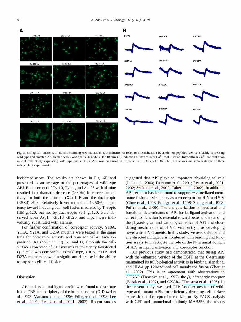
bation with 2 �M apelin-13 or apelin-36 at 37°C for 40 min,the stably expressing cells were observed using fluorescencemicroscopy. Our analyses indicated that alanine substitutionof Asp23 led to complete loss of the potential for receptorinternalization induced by apelin (Fig. 5A). The E20A mu-tant was found to be partially internalized from cell surfaceupon exposure to apelin. Other mutations showed no effectson receptor internalization in response to ligand.
To evaluate the effects of alanine substitution in theN-terminal domain of APJ on signal transduction in re-sponse to apelin, changes in intracellular Ca2� were moni-tored. The results are illustrated in Fig. 5B. Consistent withbinding activity and receptor internalization, the D23A mu-tant showed no response in intracellular Ca2� upon stimu-lation with 3 �M apelin, and E20A exhibited a low-intensityCa2� alteration.
Coreceptor activities of APJ site-directed mutations
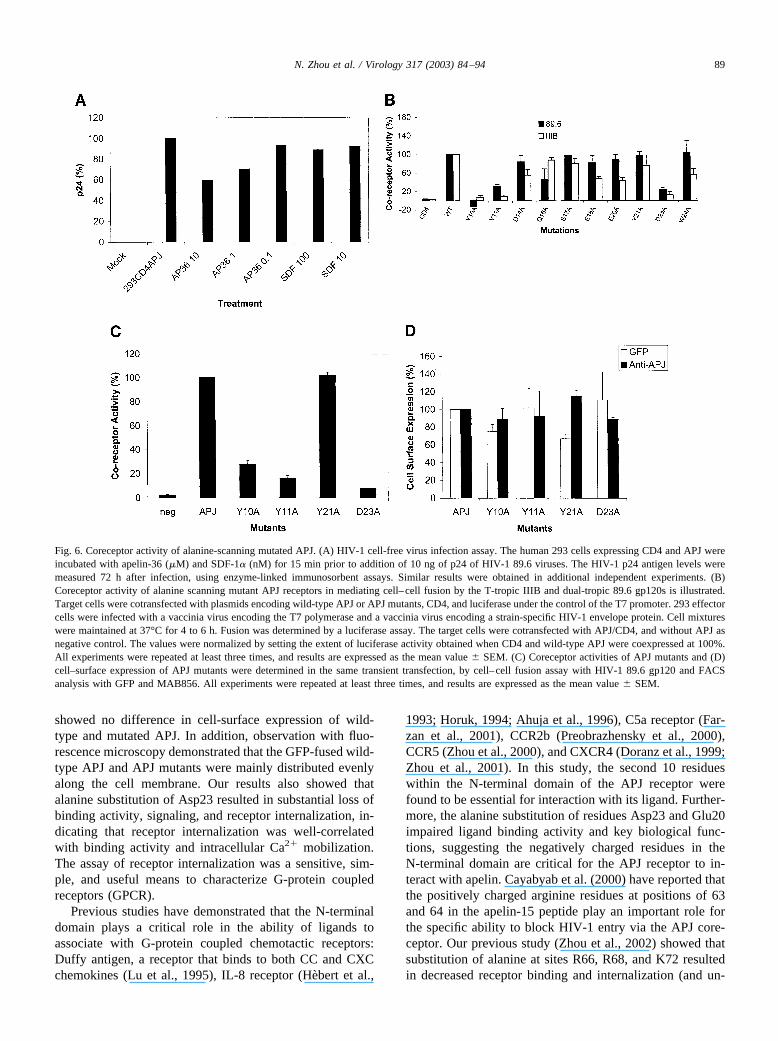
To further examine the role of APJ as a coreceptor tomediate HIV-1 infection, in preliminary experiments, hu-man HEK-293 cells expressing CD4 and APJ were infectedwith the HIV-1 89.6 isolate, and the efficiency of infectionwas determined by detection of HIV-1 p24 antigen 3 daysafter infection. The results, in Fig. 6A, showed that the 293cells expressing CD4 and APJ could be infected by HIV-189.6, and the infection by 89.6 viruses was inhibited in adose-dependent manner by the synthetic ligand apelin-36,but not by SDF-1�, the ligand of human CXCR4.
The alanine scanning APJ mutants were further exam-ined for their ability to support HIV-1 entry as a coreceptorusing a well-characterized cell-fusion reporter assay. QT6target cells were cotransfected with plasmid vectors ex-pressing wild-type APJ or APJ mutants, CD4, and luciferaseunder the control of T7 promoter. 293 effector cells werecoinfected with vaccinia viruses encoding HIV-1 envelopeproteins and T7 RNA polymerase, respectively. After 5 h offusion, cells were lysed and quantitatively analyzed by
Fig. 3. Cell-surface expression of alanine-scanning APJ mutants. 293 cells, stably transfected with wild-type and alanine-scanning mutated APJ, wereanalyzed with GFP (A) and anti-APJ MAB856 (B) by flow cytometry. Experiments were repeated at least three times, and results are expressed as the meanvalue � SE.
Fig. 4. Binding activity of alanine-scanning APJ mutants. The APJ stablyexpressing 293 cells were incubated with 125I-apelin-13 (0.2 nM) in thepresence of 200 nM cold apelin-13, and cell-associated radioactivity wascounted by gamma emissions. The bars represent the mean values of threeindependent assays, whereas the error bars are the mean � SEM.
87N. Zhou et al. / Virology 317 (2003) 84–94
luciferase assay. The results are shown in Fig. 6B andpresented as an average of the percentages of wild-typeAPJ. Replacement of Tyr10, Tyr11, and Asp23 with alanineresulted in a dramatic decrease (�80%) in coreceptor ac-tivity for both the T-tropic (X4) IIIB and the dual-tropic(R5X4) 89.6. Relatively lower reductions (�50%) in po-tency toward inducing cell–cell fusion mediated by T-tropicIIIB gp120, but not by dual-tropic 89.6 gp120, were ob-served when Asp14, Glu18, Glu20, and Trp24 were indi-vidually substituted with alanine.
For further confirmation of coreceptor activity, Y10A,Y11A, Y21A, and D23A mutants were tested at the sametime for coreceptor activity and transient cell-surface ex-pression. As shown in Fig. 6C and D, although the cell-surface expression of APJ mutants in transiently transfectedQT6 cells was comparable to wild-type, Y10A, Y11A, andD23A mutants showed a significant decrease in the abilityto support cell–cell fusion.
Discussion
APJ and its natural ligand apelin were found to distributein the CNS and periphery of the human and rat (O’Dowd etal., 1993; Matsumoto et al., 1996; Edinger et al., 1998; Leeet al., 2000; Reaux et al., 2001, 2002). Recent studies
suggested that APJ plays an important physiological role(Lee et al., 2000; Tatemoto et al., 2001; Reaux et al., 2001,2002; Szokodi et al., 2002; Taheri et al., 2002). In addition,APJ receptor has been found to support env-mediated mem-brane fusion or viral entry as a coreceptor for HIV and SIV(Choe et al., 1998; Edinger et al., 1998; Zhang et al., 1998;Puffer et al., 2000). The characterization of structural andfunctional determinants of APJ for its ligand activation andcoreceptor function is essential toward better understandingthe physiological and pathological roles of APJ and eluci-dating mechanisms of HIV-1 viral entry plus developingnovel anti-HIV-1 agents. In this study, we used deletion andsite-directed mutagenesis combined with binding and func-tion assays to investigate the role of the N-terminal domainof APJ in ligand activation and coreceptor function.
Our previous study had demonstrated that fusing APJwith the enhanced version of the EGFP at the C-terminusmaintained its full biological activities in binding, signaling,and HIV-1 gp 120-induced cell membrane fusion (Zhou etal., 2002). This is in agreement with observations inCCKAR (Tarasova et al., 1997), the �2-adrenergic receptor(Barak et al., 1997), and CXCR4 (Tarasova et al., 1998). Inthe present study, we used GFP-fused expression of wild-type and mutant APJs for efficiently detecting cell-surfaceexpression and receptor internalization. By FACS analysiswith GFP and monoclonal antibody MAB856, the results
Fig. 5. Biological functions of alanine-scanning APJ mutations. (A) Induction of receptor internalization by apelin-36 peptides. 293 cells stably expressingwild type and mutated APJ treated with 2 �M apelin-36 at 37°C for 40 min. (B) Induction of intracellular Ca2� mobilization. Intracellular Ca2� concentrationin 293 cells stably expressing wild-type and mutated APJ was measured in response to 3 �M apelin-36. The data shown are representative of threeindependent experiments.
88 N. Zhou et al. / Virology 317 (2003) 84–94
showed no difference in cell-surface expression of wild-type and mutated APJ. In addition, observation with fluo-rescence microscopy demonstrated that the GFP-fused wild-type APJ and APJ mutants were mainly distributed evenlyalong the cell membrane. Our results also showed thatalanine substitution of Asp23 resulted in substantial loss ofbinding activity, signaling, and receptor internalization, in-dicating that receptor internalization was well-correlatedwith binding activity and intracellular Ca2� mobilization.The assay of receptor internalization was a sensitive, sim-ple, and useful means to characterize G-protein coupledreceptors (GPCR).
Previous studies have demonstrated that the N-terminaldomain plays a critical role in the ability of ligands toassociate with G-protein coupled chemotactic receptors:Duffy antigen, a receptor that binds to both CC and CXCchemokines (Lu et al., 1995), IL-8 receptor (Hebert et al.,
1993; Horuk, 1994; Ahuja et al., 1996), C5a receptor (Far-zan et al., 2001), CCR2b (Preobrazhensky et al., 2000),CCR5 (Zhou et al., 2000), and CXCR4 (Doranz et al., 1999;Zhou et al., 2001). In this study, the second 10 residueswithin the N-terminal domain of the APJ receptor werefound to be essential for interaction with its ligand. Further-more, the alanine substitution of residues Asp23 and Glu20impaired ligand binding activity and key biological func-tions, suggesting the negatively charged residues in theN-terminal domain are critical for the APJ receptor to in-teract with apelin. Cayabyab et al. (2000) have reported thatthe positively charged arginine residues at positions of 63and 64 in the apelin-15 peptide play an important role forthe specific ability to block HIV-1 entry via the APJ core-ceptor. Our previous study (Zhou et al., 2002) showed thatsubstitution of alanine at sites R66, R68, and K72 resultedin decreased receptor binding and internalization (and un-
Fig. 6. Coreceptor activity of alanine-scanning mutated APJ. (A) HIV-1 cell-free virus infection assay. The human 293 cells expressing CD4 and APJ wereincubated with apelin-36 (�M) and SDF-1� (nM) for 15 min prior to addition of 10 ng of p24 of HIV-1 89.6 viruses. The HIV-1 p24 antigen levels weremeasured 72 h after infection, using enzyme-linked immunosorbent assays. Similar results were obtained in additional independent experiments. (B)Coreceptor activity of alanine scanning mutant APJ receptors in mediating cell–cell fusion by the T-tropic IIIB and dual-tropic 89.6 gp120s is illustrated.Target cells were cotransfected with plasmids encoding wild-type APJ or APJ mutants, CD4, and luciferase under the control of the T7 promoter. 293 effectorcells were infected with a vaccinia virus encoding the T7 polymerase and a vaccinia virus encoding a strain-specific HIV-1 envelope protein. Cell mixtureswere maintained at 37°C for 4 to 6 h. Fusion was determined by a luciferase assay. The target cells were cotransfected with APJ/CD4, and without APJ asnegative control. The values were normalized by setting the extent of luciferase activity obtained when CD4 and wild-type APJ were coexpressed at 100%.All experiments were repeated at least three times, and results are expressed as the mean value � SEM. (C) Coreceptor activities of APJ mutants and (D)cell–surface expression of APJ mutants were determined in the same transient transfection, by cell–cell fusion assay with HIV-1 89.6 gp120 and FACSanalysis with GFP and MAB856. All experiments were repeated at least three times, and results are expressed as the mean value � SEM.
89N. Zhou et al. / Virology 317 (2003) 84–94
published data). It is noteworthy that the net charge of theN-terminal domain of APJ is �7. We hypothesized that thenegatively charged N-terminal domain of APJ is critical forforming favorable electrostatic interactions with the posi-tively charged residues in apelin.
HIV-1 infection of human cells requires the receptorCD4 and chemokine receptors as coreceptors. The majorcoreceptors used by HIV-1 viruses are CCR5 and CXCR4(Deng et al., 1996; Doranz et al., 1996; Dragic et al., 1996;Feng et al., 1996). Recently, the APJ receptor has beenidentified as an alternative coreceptor for HIV-1 and SIV tosupport env-mediated membrane fusion or viral entry invitro (Choe et al., 1998; Edinger et al., 1998; Zhang et al.,1998; Puffer et al., 2000). In our preliminary infectionassay, the human 293 cells expressing CD4 and APJ wereinfected with HIV-1 89.6 viruses, and the infection of 89.6virues could be inhibited by apelin-36, the natural ligand ofAPJ, by not by SDF-1�, ligand of CXCR4, suggesting thatAPJ receptor functioned as a coreceptor of HIV-1. Althoughsome reports argue for the significance of APJ as a core-ceptor during in vivo infection with HIV-1 (Choe et al.,2000; Puffer et al., 2000), identification of structural deter-minants of APJ in mediation of HIV-1 entry is helpful forfuller understanding of the interactions of HIV-1 env gly-coprotein with CD4 and coreceptor toward developingnovel anti-HIV-1 agents.
In the present study, we use a well-characterized cell–cell fusion assay to evaluate the coreceptor activities of APJmutations. The data showed that deletion of the first 10residues and 20 residues in the N-terminal domain resultedin 60 and 80% reduction of coreceptor activity, respectively,indicating that the N-terminal domain was required forinteractions with HIV-1 gp120. Further site-directed mu-tagenesis analysis demonstrated that residues Tyr10, Tyr11,and Asp23 were critical in supporting env glycoprotein-mediated membrane fusion for both T-tropic and dual-tropicviruses. It was interesting to note that replacement of neg-atively charged residues Asp20, Glu18, and Glu20 withalanine impaired cell–cell fusion activity mediated by T-tropic viral env glycoprotein. APJ has a negatively chargedN-terminal domain, similar to CXCR4. Mutagenesis studiesalso indicated that the negatively charged residues in theN-terminal domain were critical determinants for CXCR4coreceptor activity (Chabot et al., 1999; Brelot et al., 2000).In addition, the V3 loop of gp120 from T-tropic isolates hasbeen found to have a higher net positive charge than the V3loop from M-tropic strains (O’Brien et al., 1996). It is likelythat the electrostatic interactions of coreceptors with HIV-1gp120 play significant roles in CXCR4 and APJ coordinatedcell entry by T-tropic viruses. Our previous study demon-strated that ALX40-4C, initially identified as a small-mol-ecule antagonist of the chemokine receptor CXCR4, di-rectly bound to APJ and prevented use of APJ as a HIV-1coreceptor (Zhou et al., 2003). Taken together, APJ as areceptor and coreceptor, in structure and function, bears astrong resemblance to CXCR4, although APJ contains an
aminoterminal NYYG sequence, and a similar motif,NYYT, has been identified at the N-terminal domain ofCCR5, that is particularly important for coreceptor function(Farzan et al., 1998).
There are three tyrosine residues, Tyr10, Tyr11, andTyr21, within the N-terminal domain of APJ. The sametyrosine-rich N-terminal domain exists in most or all che-mokine receptors and all HIV-1 coreceptors (Farzan et al.,1999). CCR5 contains at least three sulfated tyrosineswithin the N-terminal domain that contribute to both recep-tor functions and coreceptor activity (Farzan et al., 1999;Bannert et al., 2001). The tyrosine sulfate moieties onCCR2b and the chemotactic receptor for C5a also have beenobserved to play an important role in association with ligand(Bannert et al., 2001; Farzan et al., 2001). In the case ofCXCR4, the sulfate group at tyrosine 21 makes a substantialcontribution to its interaction with ligand SDF-1�, but playsa less significant role in CXCR4-mediated HIV-1 entry thanin CCR5-dependent HIV-1 entry (Farzan et al., 2002). How-ever, in the present study, we observed that Tyr10 andTyr11 of APJ receptor were critical for the ability to supportcell–cell fusion mediated by both T-tropic and dual-tropicHIV-1 isolates, but none of the three tyrosine residuescontributed to receptor binding and function. We observedthat the deletion mutant APJ10 still showed some activity,although quite less, in support of IIIB gp120 induced cell–cell fusion. Nonetheless, the site-directed mutant Y10Amaintained little coreceptor activity for both 89.6 and IIIBgp120. This discrepancy may be accounted for by the con-formational change induced by alanine substitution atTyr10.
We have characterized the role of the N-terminal domainof APJ receptor in association with ligand and HIV-1 envglycoprotein. Our results have revealed the residues in theN-terminal domain of APJ critical for biological functionand coreceptor activity. In the case of most G-protein cou-pled receptors, the N-terminal domain, together with two orthree extracellular loops, are involved in interactions withligand, and also with HIV-1 envelope glycoproteins. How-ever, further studies are necessary to examine the role of theextracellular loops of APJ in ligand binding functions andcoreceptor activity.
Materials and methods
Materials
Dulbecco’s modified Eagle’s medium (DMEM), fetalbovine serum, and G418 were purchased from Life Tech-nologies, Inc. Rhodamine red concanavalin A (ConA), tet-ramethylrhodamine-transferrin, LysoTracker Red, furo-2,and Pluronic F-127 were purchased from Molecular ProbesInc. (Eugene, OR). Anti-human APJ monoclonal antibody(MAB856) was obtained from R&D Systems, Inc. PlasmidpCDNA-APJ, recombinant vaccinia viruses encoding two
90 N. Zhou et al. / Virology 317 (2003) 84–94
envelopes of HIV-1 vSC60 (IIIB) and vBD3 (89.6), andvTF1.1 encoding T7 RNA polymerase were generous giftsfrom Dr. Robert W. Doms, University of Pennsylvania. Thevector pEGFP-N1 was purchased from Clontech Laborato-ries, Inc. (Palo Alto, CA).
Cell and cell culture
The HEK-293 cell line was obtained from the AIDSResearch and Reference Reagent Program (Division ofAIDS, NIAID, NIH). The Japanese quail fibrosarcoma QT6cell line was kindly provided by Robert W. Doms (Univer-sity of Pennsylvania, Philadelphia, PA). HEK-293 and QT6cells were maintained in DMEM plus 10% fetal bovineserum.
DNA construction and mutagenesis
Plasmid pCDNA-APJ containing the human APJ genewas kindly provided by Robert W. Doms (University ofPennsylvania, Philadelphia, PA). The full open readingframe (ORF) of wild-type and N-terminally deleted APJwere amplified using polymerase chain reaction (PCR) andsubcloned in-frame into HindIII and BamHI sites of thepEGFP-N1 vector. All constructs were sequenced to con-firm the correct sequence and orientation.
Site-directed mutagenesis was performed on pAPJ-EGFP vector and prepared with QuikChange Site-DirectedMutagenesis Kit (Stratagene, La Jolla, CA), according to themanufacturer’s instructions. The mutations were confirmedby sequencing. The plasmid constructs of wild-type APJand mutants were transfected into 293 cells by the calcium-phosphate precipitation method. Twenty-four hours aftertransfection, selection for stable expression was initiated bythe addition of G418 (800 �g/ml). Transfected cells wereevaluated for expression levels at the cell surface by flowcytometry.
Radioligand binding assay
The radioiodinated human apelin-13 was purchased fromAmersham Biosciences, Inc. (Piscataway, NJ). The specificactivity of human 125I-apelin-13 was 2000 Ci/mmol. Thebinding assay was carried out as described previously (Zhouet al., 2002). The wild-type APJ and mutated APJ stablyexpressing 293 cells were harvested in phosphate-bufferedsaline (PBS) (Ca2� and Mg2� free) plus 0.5 mM EDTA andwashed twice with PBS. Ligand binding experiments wereperformed using a single concentration (0.2 nM) of 125I-apelin-13 in the absence or presence of 200 nM cold ape-lin-13 in a final volume of 100 �l of binding buffer (50 nMHEPES pH 7.4, 1 nM CaCl2, 5 nM MgCl2, 0.1% bovineserum albumin) containing 5 � 105 cells. Samples wereincubated for 60 min at room temperature. The incubationwas terminated by separating the cells from the bindingbuffer by centrifugation and washing once with 500 �l of
cold binding buffer. Bound ligands were determined bycounting gamma emissions. At least three independent ex-periments were performed.
Flow cytometry
APJ-EGFP and APJ mutant stably transfected 293 cells(2 � 105) were washed with fluorescence-activated cellsorting (FACS) buffer (0.3% bovine serum albumin, 0.05%sodium azide in PBS) and fixed in fixing buffer (2% para-formaldehyde in PBS). For detection of APJ expressionusing anti-APJ antibody, stably or transiently APJ express-ing cells were harvested with PBS containing 5 mM EDTA.After washing with FACS buffer, cells were incubated withan anti-APJ mAb (10 �g/ml) for 30 min at 4°C. Afterwashing with FACS buffer, cells were incubated with 10�g/ml PE-conjugated goat anti-mouse IgG (PharMingen)for 30 min at 4°C. After washing twice with FACS buffer,cells were fixed in fixing buffer (2% paraformaldehyde inPBS) and then analyzed on a FACScan flow cytometer(Coulter EPICS Elite, Coolten Corp., Hialeah, FL).
Receptor internalization and fluorescence microscopy
The stable APJ or mutated APJ expressing 293 cellswere seeded on Nunc two-chamber slides and incubated at37°C overnight. After treatment with apelin at 37°C for 40min, cells were washed in PBS and then fixed in PBScontaining 2% paraformaldehyde for 10 min. Fluorescencemicroscopy was performed on an Olympus System micro-scope, model BX60, with fluorescence attachment BX-FLA.
Intracellular calcium measurements
Intracellular Ca2� was monitored following a modifiedprocedure, as published by others (Donnadieu et al., 1994;Heveker et al., 1998). Briefly, untransduced 293 cells andwild-type APJ or APJ mutant stably transfected 293 cellswere cultured in the DMEM medium containing 10% FBS.Cells were harvested with PBS containing 5 mM EDTA andwashed twice with PBS. For Ca2� mobilization studies, 5 �106/ml cells were loaded with the fluorescent dye, fura-2 (3�M), and 0.05% F172, in Hank’s balanced salt solution(140 mM NaCl, 5 mM KCl, 10 mM HEPES pH 7.4, 1 mMCaCl2, 1 mM MgCl2, 1 mg/ml glucose, and 0.025% BSA),for 30 min at 37°C. The cells were washed three times andresuspended at a concentration of 30 to 40 � 106/ml, then1.5 to 2 � 106 cells were tested in the same buffer. Intra-cellular Ca2� mobilization was measured using excitation at340 and 380 nm in a fluorescence spectrometer (Perkin–Elmer LS50B, Beaconsfield, UK), upon stimulation withapelin. Intracellular Ca2� concentrations were calculatedusing a fluorescence spectrometer measurement program.
91N. Zhou et al. / Virology 317 (2003) 84–94
HIV-1 infection assay
To determine coreceptor activity of APJ receptor in me-diating HIV-1 infection, the human HEK-293 cells weretransfected with CD4 and APJ. The 293 cells expressingCD4/APJ were challenged with 10 ng of HIV-1 p24 antigen,isolate 89.6, for 6 h, and then washed three times. Forinhibition with ligand, the cells were incubated with indi-cated amounts of apelin-36 and SDF-1� for 15 min prior toaddition of HIV-1. Supernatants were collect for p24 anti-gen measurement by ELISA, 72 h after infection. TheHIV-1 p24 antigen levels were measured using enzyme-linked immunosorbent assays (ELISA; Zeptometrix).
Gene reporter fusion assay
A gene reporter fusion assay was used to determine thecoreceptor activity of APJ in mediating HIV-1 viral entryfollowing a modified procedure, as published by others(Nussbaum et al., 1994; Doranz et al., 1996; Rucker et al.,1997). Briefly, the effector 293 cells were infected withrecombinant vaccinia virus with diverse HIV-1 Env proteinsand T7 RNA polymerase for 2 h. Infected cells were thentrypsinized, washed with PBS, resuspended in medium, andincubated overnight at 32°C in the presence of rifampicin(100 �g/ml). Target QT6 cells were cotransfected in six-well plates with plasmids encoding CD4, wild-type, or mu-tant APJs and luciferase under control of the T7 promotor,using the calcium-phosphate precipitation method. Fourhours after transfection, cells were lifted, seeded in 24-wellplates, and incubated at 37°C overnight. To initiate fusion,105 effector cells were added to each well and incubated at37°C. After 5 h of fusion, cells were lysed in 100 �l ofreporter lysis buffer (Pharmingen) and assayed for lucif-erase activity by using commercially available reagents(Pharmingen) with FB12 Luminometer (Zylux Corp.,Maryville, TN).
Peptide synthesis
Peptide synthesis was carried out as described previously(Zhou et al., 2002). Briefly, apelin-13 and apelin-36 wereprepared by solid-phase synthesis using Fmoc-strategy on a430A peptide synthesizer (Applied Biosystems, Foster City,CA) and a 9050 Pepsynthesizer Plus (Perceptive Biosys-tems, Cambridge, MA). Crude peptides were purified bypreparative reverse-phase high-performance liquid chroma-tography using a Dynamax-300 Å C18 25 cm � 21.4 mm IDcolumn with flow rate of 9 ml/min and two solvent systemsof 0.1% TFA/H2O and 0.1% TFA/acetonitrile. Fractionscontaining the appropriate peptide were pooled together andlyophilized. The purity of the final product was assessed byanalytical reverse-phase high-performance liquid chroma-tography, capillary electrophoresis, and matrix-assisted la-ser desorption/ionization time-of-flight mass spectrometry.
Acknowledgments
We thank Rita M. Victor and Brenda O. Gordon forexcellent secretarial assistance, Dr. Andrew B. Maksy-mowych for assisting with the Ca2� mobilization assays,and Dr. Robert W. Doms for providing vaccinia viruses. Anumber of reagents were obtained from the AIDS Researchand Reference Reagent Program, Division of AIDS, NIAID,NIH. This work is supported in part by USPHS GrantsGM57761 and AI45414 to Z.H. and NS27405, MH58529,and NS41864 to R.J.P.
References
Ahuja, S., Lee, J., Murphy, P., 1996. CXC chemokines bind to unique setsof selectivity determinants that can function independently and arebroadly distributed on multiple domains of human interleukin-8 recep-tor B. Determinants of high affinity binding and receptor activation aredistinct. J. Biol. Chem. 271, 225–232.
Alvarez Losada, S., Canto-Nogues, C., Munoz-Fernandez, M., 2002. Anew possible mechanism of human immunodeficiency virus type 1infection of neural cells. Neurobiol. Dis. 11, 469–478.
Bannert, N., Craig, S., Farzan, M., Sogah, D., Santo, N., Choe, H.,Sodroski, J., 2001. Sialylated O-glycans and sulfated tyrosines in theNH2-terminal domain of CC chemokine receptor 5 contribute to highaffinity binding of chemokines. J. Exp. Med. 194, 1661–1673.
Barak, L., Ferguson, S., Zhang, J., Martenson, C., Meyer, T., Caron, M.,1997. Internal trafficking and surface mobility of a functionally intactbeta 2-adrenergic receptor-green fluorescent protein conjugate. Mol.Pharmacol. 51, 177–184.
Brelot, A., Heveker, N., Montes, M., Alizon, M., 2000. Identification ofresidues of CXCR4 critical for human immunofeficiency virus co-receptor and chemokine receptor activities. J. Biol. Chem. 275, 23736–23744.
Cayabyab, M., Hinuma, S., Farzan, M., Choe, H., Fukusumi, S., Kitada, C.,Nishizawa, N., Hosoya, M., Nishimura, O., Messele, T., Pollakis, G.,Goudsmit, J., Fujino, M., Sodroski, J., 2000. Apelin, the natural ligandof the orphan seven-transmembrane receptor APJ, inhibits human im-munodeficiency virus type 1 entry. J. Virol. 74, 11972–11976.
Chabot, D.J., Zhang, P-f., Quinnan, G.V., Broder, C.C., 1999. Mutagenesisof CXCR4 identifies important domains for human immunodeficiencyvirus type 1 X4 isolate envelope-mediated membrane fusion and virusentry and reveals cryptic co-receptor activity for R5. J. Virol. 73,6598–6609.
Choe, H., Farzan, M., Konkel, M., Martin, K., Sun, Y., Marcon, L.,Cayabyab, M., Berman, M., Dorf, M., Gerard, N., Gerard, C., Sodroski,J., 1998. The orphan seven-transmembrane receptor apj supports theentry of primary T-cell-line-tropic and dualtropic human immunodefi-ciency virus type 1. J. Virol. 72, 6113–6118.
Choe, W., Albright, A., Sulcove, J., Jaffer, S., Hesselgesser, J., Lavi, E.,Crino, P., Kolson, D., 2000. Functional expression of the seven-trans-membrane HIV-1 co-receptor APJ in neural cells. J. Neurovirol. 6,S61–S69.
De Mota, N., Lenkei, Z., Llorens-Cortes, C., 2000. Cloning, pharmacolog-ical characterization and brain distribution of the rat apelin receptor.Neuroendocrinology 72, 400–407.
Deng, H., Rong, L., Ellmeier, W., Choe, S., Unutmaz, D., Burkhart, M.,Marzio, P.D., Marmon, S., Sutton, R.E., Hill, C.M., Davis, C.B.,Peiper, S.C., Schall, T.J., Littman, D.R., Landau, N.R., 1996. Identifi-cation of a major co-receptor for primary isolates of HIV-1. Nature381, 661–666.
92 N. Zhou et al. / Virology 317 (2003) 84–94
Devic, E., Rizzoti, K., Bodin, S., Knibiehler, B., Audigier, Y., 1999.Amino acid sequence and embryonic expression of msr/apj, the mousehomolog of Xenopus X-msr and human APJ. Mech. Dev. 84, 199–203.
Donnadieu, E., Bismuth, G., Trautmann, A., 1994. Antigen recognition byhelper T cells elicits a sequence of distinct changes of their shape andintracellular calcium. Curr. Biol. 4, 584–595.
Doranz, B., Orsini, M., Turner, J., Hoffman, T., Berson, J., Hoxie, J.,Peiper, S., Brass, L., Doms, R., 1999. Identification of CXCR4 domainsthat support co-receptor and chemokine receptor functions. J. Virol. 73,2752–2761.
Doranz, B.J., Rucker, J., Yi, Y., Smyth, RJ., Samson, M., Peiper, S.C.,Parmentier, M., Collman, R.G., Doms, R.W., 1996. A dual-tropicprimary HIV-1 isolate that uses fusin and the beta-chemokine receptorsCKR-5, CKR-3, and CKR-2b as fusion cofactors. Cell 85, 1149–1158.
Dragic, T., Litwin, V., Allaway, G.P., Martin, S.R., Huang, Y., Nagashima,K.A., Cayanan, C., Maddon, P.J., Koup, R.A., Moore, J.P., Paxton,W.A., 1996. HIV-1 entry into CD4� cells is mediated by the chemo-kine receptor CC-CKR-5. Nature 381, 667–673.
Edinger, A., Hoffman, T., Sharron, M., Lee, B., Yi, Y., Choe, W., Kolson,D., Mitrovic, B., Zhou, Y., Faulds, D., Collman, R., Hesselgesser, J.,Horuk, R., Doms, R., 1998. An orphan seven-transmembrane domainreceptor expressed widely in the brain functions as a co-receptor forhuman immunodeficiency virus type 1 and simian immunodeficiencyvirus. J. Virol. 72, 7934–7940.
Fan, X., Zhou, N., Zhang, X., Mukhtar, M., Lu, Z., Fang, J., DuBois, G.C.,Pomerantz, R.J., 2003. Structural and functional study of the apelin-13peptide, an endogenous ligand of the HIV-1 coreceptors, APJ. Bio-chemistry 42, 10163–10168.
Farzan, M., Babcock, G., Vasilieva, N., Wright, P., Kiprilov, E., Mirza-bekov, T., Choe, H., 2002. The role of post-translational modificationsof the CXCR4 amino terminus in stromal-derived factor 1 alpha asso-ciation and HIV-1 entry. J. Biol. Chem. 277, 29484–29489.
Farzan, M., Choe, H., Vaca, L., Martin, K., Sun, Y., Desjardins, E.,Ruffing, N., Wu, L., Wyatt, R., Gerard, N., Gerard, C., Sodroski, J.,1998. A tyrosine-rich region in the N terminus of CCR5 is importantfor human immunodeficiency virus type 1 entry and mediates anassociation between gp120 and CCR5. J. Virol. 72, 1160–1164.
Farzan, M., Mirzabekov, T., Kolchinsky, P., Wyatt, R., Cayabyab, M.,Gerard, N.P., Gerard, C., Sodroski, J., Choe, H., 1999. Tyrosine sul-fation of the amino terminus of CCR5 facilitates HIV-1 entry. Cell 96,667–676.
Farzan, M., Schnitzler, C., Vasilieva, N., Leung, D., Kuhn, J., Gerard, C.,Gerard, N., Choe, H., 2001. Sulfated tyrosines contribute to the for-mation of the C5a docking site of the human C5a anaphylatoxinreceptor. J. Exp. Med. 193, 1059–1066.
Feng, Y., Broder, C.C., Kennedy, P.E., Berger, E.A., 1996. HIV-1 entrycofactor: functional cDNA cloning of a seven-transmembrane, G pro-tein-coupled receptor. Science 272, 872–877.
Habata, Y., Fujii, R., Hosoya, M., Fukusumi, S., Kawamata, Y., Hinuma,S., Kitada, C., Nishizawa, N., Murosaki, S., Kurokawa, T., Onda, H.,Tatemoto, K., Fujino, M., 1999. Apelin, the natural ligand of theorphan receptor APJ, is abundantly secreted in the colostrum. Biochim.Biophys. Acta 1452, 25–35.
Hebert, C.A., Chuntharapai, A., Smith, M., Colby, T., Kim, J., Horuk, R.,1993. Partial functional mapping of the human interleukin-8 type Areceptor identification of a major lignad binding domain. J. Biol. Chem.268, 18549–18553.
Heveker, N., Montes, M., Germeroth, L., Amara, A., Trautmann, A.,Alizon, M., Schneider-Mergener, J., 1998. Dissociation of the signal-ling and antiviral properties of SDF-1-derived small peptides. Curr.Biol. 8, 369–376.
Horuk, R., 1994. The interleukin-8-receptor family: from chemokines tomalaria. Immunol Today 15, 169–174.
Hosoya, M., Kawamata, Y., Fukusumi, S., Fujii, R., Habata, Y., Hinuma,S., Kitada, C., Honda, S., Kurokawa, T., Onda, H., Nishimura, O.,
Fujino, M., 2000. Molecular and functional characteristics of APJ.Tissue distribution of mRNA and interaction with the endogenousligand apelin. J. Biol. Chem. 275, 21061–21067.
Lee, D., Cheng, R., Nguyen, T., Fan, T., Kariyawasam, A., Liu, Y.,Osmond, D., George, S., O’Dowd, B., 2000. Characterization of apelin,the ligand for the APJ receptor. J. Neurochem. 74, 34–41.
Lu, Z., Wang, Z., Horuk, R., Hesselgesser, J., Lou, Y., Hadley, T., Peiper,S., 1995. The promiscuous chemokine binding profile of the Duffyantigen/receptor for chemokines is primarily localized to sequences inthe amino-terminal domain. J. Biol. Chem. 270, 26239–26245.
Matsumoto, M., Hidaka, K., Akiho, H., Tada, S., Okada, M., Yamaguchi,T., 1996. Low stringency hybridization study of the dopamine D4receptor revealed D4-like mRNA distribution of the orphan seven-transmembrane receptor, APJ, in human brain. Neurosci. Lett. 219,119–122.
Nussbaum, O., Broder, C.C., Berger, E.A., 1994. Fusogenic mechanism ofenvelope-virus glycoproteins by a novel recombinant vaccinia virus-based assay quantitating cell fusion-dependent reporter gene activation.J. Virol. 68, 5411–5422.
O’Brien, W., Sumner-Smith, M., Mao, S., Sadeghi, S., Zhao, J., Chen, I.,1996. Anti-human immunodeficiency virus type 1 activity of an oligo-cationic compound mediated via gp120 V3 interactions. J. Virol. 70,2825–2831.
O’Carroll, A., Selby, T., Palkovits, M., Lolait, S., 2000. Distribution ofmRNA encoding B78/apj, the rat homologue of the human APJ recep-tor, and its endogenous ligand apelin in brain and peripheral tissues.Biochem. Biophys. Acta 1492, 72–80.
O’Dowd, B.F., Heiber, M., Chan, A., Heng, H.H., Tsui, L.C., Kennedy,J.L., Shi, X., Petronis, A., George, S.R., Nguyen, T., 1993. A humangene that shows identity with the gene encoding the angiotensin recep-tor is located on chromosome 11. Gene 136, 355–360.
Preobrazhensky, A., Dragan, S., Kawano, T., Gavrilin, M., Gulina, I.,Chakravarty, L., Kolattukudy, P., 2000. Monocyte chemotactic pro-tein-1 receptor CCR2B is a glycoprotein that has tyrosine sulfation ina conserved extracellular N-terminal region. J. Immunol. 165, 5295–5303.
Puffer, B., Sharron, M., Coughlan, C., Baribaud, F., McManus, C., Lee, B.,David, J., Price, K., Horuk, R., Tsang, M., Doms, R., 2000. Expressionand co-receptor function of APJ for primate immunodeficiency viruses.Virology 276, 435–444.
Reaux, A., De Mota, N., Skultetyova, I., Z. L, El Messari, S., Gallatz, K.,Corvol, P., Palkovits, M., Llorens-Cortes, C. (2001) Physiological roleof a novel neuropeptide, apelin and its receptor in the rat brain.J. Neurol. 77:1085–1096.
Reaux, A., Gallatz, K., Palkovits, M., Llorens-Cortes, C., 2002. Distribu-tion of apelin-synthesizing neurons in the adult rat brain. Neuroscience113, 653–662.
Rucker, J., Doranz, B., Edinger, A., Long, D., Berson, J., Doms, R., 1997.Cell-cell fusion assay to study role of chemokine receptors in humanimmunodeficiency virus type I entry. Methods Enzymol. 288, 118–133.
Szokodi, I., Tavi, P., Foldes, G., Voutilainen-Myllyla, S., Ilves, M., To-kola, H., Pikkarainen, S., Piuhola, J., J. R, Toth, M, H. R (2002) Apelin,the novel endogenous ligand of the orphan receptor APJ, regulatescardiac contractility. Circ. Res. 91, 534–540.
Taheri, S., Murphy, K., Cohen, M., Sujkovic, E., Kennedy, A., Dhillo, W.,Dakin, C., Sajedi, A., Ghatei, M., Bloom, S., 2002. The effects ofcentrally administered apelin-13 on food intake, water intake and pi-tuitary hormone release in rats. Biochem. Biophys. Res. Commun. 291,1208–1212.
Tarasova, N., Stauber, R., Michejda, C., 1998. Spontaneous and ligand-induced trafficking of CXC-chemokine receptor 4. J. Biol. Chem. 273,15883–15886.
Tarasova, N.I., Stauber, R.H., Choi, J.K., Hudson, E.A., Czerwinski, G.,Miller, J.L., Pavlakis, G.N., Michejda, C.J., Wank, S.A., 1997. Visu-alization of G protein-coupled receptor trafficking with the aid of the
93N. Zhou et al. / Virology 317 (2003) 84–94
green fluorescent protein. Endocytosis and recycling of cholecystokininreceptor type A. J. Biol. Chem. 272, 14817–14824.
Tatemoto, K., Hosoya, M., Habata, Y., Fujii, R., Kakegawa, T., Zou, M.,Kawamata, Y., Fukusumi, S., Hinuma, S., Kitada, C., Kurokawa, T.,Onda, H., Fujino, M., 1998. Isolation and characterization of a novelendogenous peptide ligand for the human APJ receptor. Biochem.Biophys. Res. Commun. 251, 471–476.
Tatemoto, K., Takayama, K., Zou, M., Kumaki, I., Zhang, W., Kumano,K., Fujimiya, M., 2001. The novel peptide apelin lowers blood pressurevia a nitric oxide-dependent mechanism. Regul. Pept. 99, 87–92.
Zhang, Y., Dragic, T., Cao, Y., Kostrikis, L., Kwon, D., Littman, D.,KewalRamani, V., Moore, J., 1998. Use of co-receptors other thanCCR5 by non-syncytium-inducing adult and pediatric isolates of hu-man immunodeficiency virus type 1 is rare in vitro. J. Virol. 72,9337–9344.
Zhou, N., Fan, X., Mukhtar, M., Fang, J., Patel, C.A., DuBois, G.C.,
Pomerantz, R.J., 2002. Cell-cell fusion and internalization of the CNS-based, HIV-1 co-receptor; APJ. Virology 307, 22–36 2003.
Zhou, N., Fang, J., Acheampong, E., Mukhtar, M., Pomerantz, R.J., 2003.Binding of ALX40-4C to APJ, the CNS-based receptor, inhibits utili-zation as co-receptor by HIV-1. Virology 00, 000–000.
Zhou, N., Luo, Z., Hall, J.W., Luo, J., Han, X., Huang, Z., 2000. Molecularmodeling and site-directed muagenesis of CCR5 reveal residues criticalfor chemokine binding and signal transduction. Eur. J. Immunol. 30,164–173.
Zhou, N., Luo, Z., Luo, J., Liu, D., Hall, J., Pomerantz, R., Huang, Z.,2001. Structural and functional characterization of human CXCR4 as achemokine receptor and HIV-1 co-receptor by mutagenesis and mo-lecular modeling studies. J. Biol. Chem. 276, 42826–42833.
Zou, M., Liu, H., Haraguchi, Y., Soda, Y., Tatemoto, K., Hoshino, H.,2000. Apelin peptides block the entry of human immunodeficiencyvirus (HIV). FEBS Lett. 473, 15–18.
94 N. Zhou et al. / Virology 317 (2003) 84–94