Weak anti-HIV CD8+ T-cell effector activity in HIV primary infection
9
Introduction HIV primary infection (PI) is characterized by an intense viral replication and the subsequent induction of an immune response (1, 2). The cellular immune response appears to be the main effector involved in controlling viral replication at this stage, and several studies have reported that the rapid induction of an HIV-specific, cytotoxic T lymphocyte (CTL) response occurs at the same time as the decline in viral titers (3, 4). Direct evi- dence of the role of CD8 + T cells in controlling viral repli- cation in vivo was provided recently in the primary simi- an immunodeficiency virus (SIV) infection of Rhesus macaques (5). However, the immune response fails to eliminate the virus from the host during acute infection, which is then followed by persistent viral replication and chronic HIV infection. The reasons for this incomplete control of viral repli- cation are not clearly understood. A steady-state is grad- ually achieved when the production of new viral particles is offset by the control of infected cells. Whether this control results either from the cytolytic destruction of infected cells or from the noncytolytic suppressive antivi- ral activity of CD8 + T cells is not clearly established (6). The resulting viral replication then influences the prog- nosis of HIV infection (7). The factors that determine this virologic set point may be linked to quantitative or qualitative differences in the immune response (8). The mobilization of a broad CD8 + T-cell repertoire early in HIV PI is associated with slower progression of disease. This suggests that the ability to recruit a wide spectrum of HIV-specific CTL responses during PI leads to better control of viral replication (8). However, even in these subjects, the viral load usually remains readily detectable in the absence of therapy, despite the persist- ence of a broad and intense CD8 + T-cell response. This suggests that the induction of the cellular immune response has been inadequate in most subjects. Thus, a thorough analysis of the immune response during early HIV PI is essential for a clear understanding of the mech- anisms responsible for its lack of effectiveness. Therefore, we characterized the HIV-specific CD8 + T- cell effector response in 24 subjects during HIV-1 PI using IFN-γ enzyme-linked immunospot (ELISPOT) assay and intracellular staining. We evaluated the breadth of the CD8 + T-cell repertoire induced by using a wide variety of optimum HLA class-I epitopic peptides derived from the sequences of the env, gag, pol, and nef genes of HIV-1 as stimulator antigens. The antiviral repertoire of each subject was defined by the multispeci- ficity of the response (which was assessed as the number The Journal of Clinical Investigation | November 1999 | Volume 104 | Number 10 1431 Weak anti-HIV CD8 + T-cell effector activity in HIV primary infection Marc Dalod, 1 Marion Dupuis, 1 Jean-Christophe Deschemin, 1 Cécile Goujard, 2 Christiane Deveau, 3 Laurence Meyer, 3 Nicole Ngo, 4 Christine Rouzioux, 4 Jean-Gérard Guillet, 1 Jean-François Delfraissy, 2 Martine Sinet, 1 and Alain Venet 1 1 Laboratoire d’Immunologie des Pathologies Infectieuses et Tumorales, Unité Institut National de la Santé et de la Recherche Médicale (INSERM) 445, Institut Cochin de Génétique Moléculaire, Université René Descartes, 75014 Paris, France 2 Service de Médecine Interne, and 3 INSERM U292, Hôpital de Bicêtre, F 94276 Le Kremlin Bicêtre Cedex, France 4 Laboratoire de Virologie, Hôpital Necker, F 75015 Paris, France Cécile Goujard, Christiane Deveau, Laurence Meyer, and Jean-François Delfraissy were investigators for the PRIMO Cohort Study Group. Address correspondence to: Marc Dalod, INSERM U445, Institut Cochin de Génétique Moléculaire 27 rue du Faubourg St.-Jacques, 75014 Paris, France. Phone: 33-1-44-07-18-21; Fax: 33-1-44-07-14-25; E-mail: [email protected]. Received for publication April 22, 1999, and accepted in revised form September 30, 1999. HIV-specific CD8 + T cells play a major role in the control of virus during HIV primary infection (PI) but do not completely prevent viral replication. We used IFN-γ enzyme-linked immunospot assay and intra- cellular staining to characterize the ex vivo CD8 + T-cell responses to a large variety of HIV epitopic pep- tides in 24 subjects with early HIV PI. We observed HIV-specific responses in 71% of subjects. Gag and Nef peptides were more frequently recognized than Env and Pol peptides. The number of peptides recognized was low (median 2, range 0–6). In contrast, a much broader response was observed in 30 asymptomatic subjects with chronic infection: all were responders with a median of 5 peptides recognized (range 1–13). The frequency of HIV-specific CD8 + T cells among PBMC for a given peptide was of the same order of mag- nitude in both groups. The proportion of HIV-specific CD8 + CD28 – terminally differentiated T cells was much lower in PI than at the chronic stage of infection. The weakness of the immune response during HIV PI could partially account for the failure to control HIV. These findings have potential importance for defining immunotherapeutic strategies and establishing the goals for effective vaccination. J. Clin. Invest. 104:1431–1439 (1999).
-
Upload
independent -
Category
Documents
-
view
0 -
download
0
Transcript of Weak anti-HIV CD8+ T-cell effector activity in HIV primary infection

IntroductionHIV primary infection (PI) is characterized by an intenseviral replication and the subsequent induction of animmune response (1, 2). The cellular immune responseappears to be the main effector involved in controllingviral replication at this stage, and several studies havereported that the rapid induction of an HIV-specific,cytotoxic T lymphocyte (CTL) response occurs at thesame time as the decline in viral titers (3, 4). Direct evi-dence of the role of CD8+ T cells in controlling viral repli-cation in vivo was provided recently in the primary simi-an immunodeficiency virus (SIV) infection of Rhesusmacaques (5). However, the immune response fails toeliminate the virus from the host during acute infection,which is then followed by persistent viral replication andchronic HIV infection.
The reasons for this incomplete control of viral repli-cation are not clearly understood. A steady-state is grad-ually achieved when the production of new viral particlesis offset by the control of infected cells. Whether thiscontrol results either from the cytolytic destruction ofinfected cells or from the noncytolytic suppressive antivi-ral activity of CD8+ T cells is not clearly established (6).The resulting viral replication then influences the prog-nosis of HIV infection (7). The factors that determine
this virologic set point may be linked to quantitative orqualitative differences in the immune response (8).
The mobilization of a broad CD8+ T-cell repertoireearly in HIV PI is associated with slower progression ofdisease. This suggests that the ability to recruit a widespectrum of HIV-specific CTL responses during PI leadsto better control of viral replication (8). However, even inthese subjects, the viral load usually remains readilydetectable in the absence of therapy, despite the persist-ence of a broad and intense CD8+ T-cell response. Thissuggests that the induction of the cellular immuneresponse has been inadequate in most subjects. Thus, athorough analysis of the immune response during earlyHIV PI is essential for a clear understanding of the mech-anisms responsible for its lack of effectiveness.
Therefore, we characterized the HIV-specific CD8+ T-cell effector response in 24 subjects during HIV-1 PIusing IFN-γ enzyme-linked immunospot (ELISPOT)assay and intracellular staining. We evaluated thebreadth of the CD8+ T-cell repertoire induced by using awide variety of optimum HLA class-I epitopic peptidesderived from the sequences of the env, gag, pol, and nefgenes of HIV-1 as stimulator antigens. The antiviralrepertoire of each subject was defined by the multispeci-ficity of the response (which was assessed as the number
The Journal of Clinical Investigation | November 1999 | Volume 104 | Number 10 1431
Weak anti-HIV CD8+ T-cell effector activity in HIV primary infection
Marc Dalod,1 Marion Dupuis,1 Jean-Christophe Deschemin,1 Cécile Goujard,2
Christiane Deveau,3 Laurence Meyer,3 Nicole Ngo,4 Christine Rouzioux,4
Jean-Gérard Guillet,1 Jean-François Delfraissy,2 Martine Sinet,1 and Alain Venet1
1Laboratoire d’Immunologie des Pathologies Infectieuses et Tumorales, Unité Institut National de la Santé et de la Recherche Médicale (INSERM) 445, Institut Cochin de Génétique Moléculaire, Université René Descartes, 75014 Paris, France
2Service de Médecine Interne, and3INSERM U292, Hôpital de Bicêtre, F 94276 Le Kremlin Bicêtre Cedex, France4Laboratoire de Virologie, Hôpital Necker, F 75015 Paris, FranceCécile Goujard, Christiane Deveau, Laurence Meyer, and Jean-François Delfraissy were investigators forthe PRIMO Cohort Study Group.
Address correspondence to: Marc Dalod, INSERM U445, Institut Cochin de Génétique Moléculaire 27 rue du Faubourg St.-Jacques, 75014 Paris, France. Phone: 33-1-44-07-18-21; Fax: 33-1-44-07-14-25; E-mail: [email protected].
Received for publication April 22, 1999, and accepted in revised form September 30, 1999.
HIV-specific CD8+ T cells play a major role in the control of virus during HIV primary infection (PI) butdo not completely prevent viral replication. We used IFN-γenzyme-linked immunospot assay and intra-cellular staining to characterize the ex vivo CD8+ T-cell responses to a large variety of HIV epitopic pep-tides in 24 subjects with early HIV PI. We observed HIV-specific responses in 71% of subjects. Gag and Nefpeptides were more frequently recognized than Env and Pol peptides. The number of peptides recognizedwas low (median 2, range 0–6). In contrast, a much broader response was observed in 30 asymptomaticsubjects with chronic infection: all were responders with a median of 5 peptides recognized (range 1–13).The frequency of HIV-specific CD8+ T cells among PBMC for a given peptide was of the same order of mag-nitude in both groups. The proportion of HIV-specific CD8+CD28– terminally differentiated T cells wasmuch lower in PI than at the chronic stage of infection. The weakness of the immune response during HIVPI could partially account for the failure to control HIV. These findings have potential importance fordefining immunotherapeutic strategies and establishing the goals for effective vaccination.
J. Clin. Invest. 104:1431–1439 (1999).

of epitopic peptides recognized) and the response inten-sity (assessed as the frequency of IFN-γ–secreting cells).Expression of CD28 on HIV-specific CD8+ T cells wasalso analyzed. These values were then compared withthose obtained for asymptomatic subjects during thechronic stage of infection.
MethodsStudy population. We studied 24 subjects included in themulticenter French PRIMO cohort. The study wasapproved by the local Comité Consultatif de Protection desPersonnes dans la Recherche Biomédicale (Paris, France).
After giving informed consent, subjects were includedwhether the HIV PI was symptomatic or not. The HIV-1 PI was diagnosed by (a) an incomplete Western blot(i.e., a lack of anti-p68 and anti-p34 antibodies), or (b) apositive p24 antigenemia with a negative or indetermi-nate ELISA or negative Western blot. The date of infec-tion was estimated using the date of the onset of symp-toms minus 15 days, or, in case of asymptomatic PI, thedate of incomplete Western blot minus 1 month. Nosubject had been given any antiretroviral therapy whenincluded in the study.
For comparison, 30 untreated, asymptomatic HIV-seropositive individuals in the chronic stage of infection
were also studied.Cells and peptides. PBMC were isolated by density gradient
centrifugation (Ficoll-Paque; Pharmacia Biotech AB, Upp-sala, Sweden). Subjects were HLA-typed by genotype analy-sis (ACTGene, Evry, France). The sequences of HIV-1 LAIepitopes used may be consulted on the National Institutesof Health (Bethesda, Maryland, USA) HIV molecularimmunology database (9). Peptides were synthesized byNeosystem (Strasbourg, France) and supplied by theAgence Nationale de Recherches sur le SIDA (Paris,France). Lyophilized peptides were diluted to 1 mg/mL inwater plus 10% DMSO, aliquoted, and then stored at–20°C. They were used at a final concentration of 1 µg/mL.
ELISPOT assay. IFN-γsecretion by virus-specific CD8+ Tcells was quantitated by ELISPOT assay. Ninety-six–wellnitrocellulose plates (Millipore Corp., Bedford, Massachu-setts, USA) were coated with 1 µg/mL capture mouse anti-human IFN-γmAb (Mabtech AB, Nacka, Sweden). PBMCwere plated in triplicate at serial dilutions (3 × 10⁵ to 104
cells per well). Appropriate stimuli were then added, andthe plates were incubated for 20 hours at 37°C and 5%CO2. Wells were then washed, filled with 100 µL biotiny-lated mouse monoclonal anti-human IFN-γ (Mabtech),and then filled with alkaline phosphatase–labeled extra-vidin (Sigma Chemical Co., St. Louis, Missouri, USA).Spots were developed by adding chromogenic alkalinephosphatase conjugate substrate (Bio-Rad LaboratoriesInc., Hercules, California, USA). Colored spots were count-ed using a stereomicroscope. Positive controls consisted of6 wells containing 300–1,000 cells with 50 ng/mL PMAand 500 ng/mL ionomycin. Negative controls consisted ofcells cultured in medium alone or with irrelevant peptides(10). In most experiments, negative controls yielded 0–1spot per well. In experimental wells, the signal was consid-ered positive if (a) the number of spots was greater than themean ± 3 standard deviations observed with the negativecontrols, and (b) the number of spots obtained was pro-portional to the number of plated cells. Frequencies ofIFN-γspot-forming cells (SFC) were then calculated aftersubtracting negative control values.
Cell surface labeling and flow cytometry. The FACS analy-ses were performed using a FACScan cytofluorometer(Becton Dickinson Immunocytometry Systems, SanJose, California, USA) after staining of whole blood sam-ples with the following mAb’s: FITC-labeled anti-CD8mAb and phycoerythrin (PE)-labeled anti-CD38, -CD28,-CD45RO, or –HLA-DR mAb’s (all from Becton Dickin-son Immunocytometry Systems).
Intracellular staining of IFN-γ. Cells (1.8 × 10⁶) were cul-tured for 6 hours in 300 µL complete medium with 10µg/mL brefeldin A in the presence of various stimuli.Several HIV, Ebstein-Barr virus (EBV), and influenzapeptides were tested for each individual. For some sub-jects, autologous EBV-transformed B-lymphoblastoidcell line (B-LCL), which were infected with recombi-nant vaccinia viruses expressing the env or pol HIV-1LAI genes, were used as stimulators, as described pre-viously (10). Positive control consisted of activationwith 25 ng/mL PMA and 1 µg/mL ionomycin. Nega-tive controls consisted of medium alone, peptides thatwere negative in the ELISPOT assay, or autologous B-LCL infected with the wild-type vaccinia virus.
1432 The Journal of Clinical Investigation | November 1999 | Volume 104 | Number 10
Table 1Clinical and biological characteristics of the 24 subjects with primary HIVinfection
Subject Age SymptomsA Duration Estimated T-cell Log plasmaof time from countsC HIV RNAD
symptoms infectionB
(days) (days)
CD4 CD8
HIL 66 ++ 8 24 217 308 6.0KKK 51 ++ 10 26 329 785 6.5BKC 45 ++ 21 32 439 2,136 5.9DOX 47 ++ 17 34 156 751 5.9NUY 40 +++ 36 34 582 1,197 5.9COH 22 ++ 12 35 292 569 5.3EOD 27 0 35 548 973 5.5KXO 34 + 5 35 954 744 3.0LEU 28 ++ 7 37 734 1,205 4.8FQQ 25 +++ 7 39 788 913 4.6ADB 29 +++ 15 44 550 596 4.0OVU 43 +++ 30 44 783 910 2.7OUI 38 ++ 30 46 637 5,712 5.8BRI 49 0 52 600 537 5.2KOA 35 + 7 57 945 703 4.5OFP 43 0 57 453 821 4.7ASS 28 0 58 624 761 3.8IRO 28 +++ 20 60 696 1,183 5.6GVT 26 ++ 16 61 1,240 4,883 5.2KVG 25 ++ 15 63 600 1,340 4.7AWA 31 ++ 10 66 1,004 939 3.3GFR 28 ++ 8 83 554 1,224 4.9LLV 49 ++ 15 93 521 1,587 5.7KJA 26 0 97 760 615 3.5Median 33 15 45 600 910 4.9
AThe classification of symptom severity is based on the number of different symp-toms harbored by the patients: 1–3: +; 4–6: ++; ≥7: +++. The following suggestivesymptoms of acute HIV-1 infection were considered: fever, fatigue, rash,headache, lymphadenopathy, pharyngitis, myalgia or arthralgia, nausea, vomit-ing or diarrhea, oral and genital ulcers. BSee Methods for calculation of the esti-mated date of infection. CValues are given per microliter. DValues are given in log10
of viral titers in copies per milliliter.

Cells were stained directly in the culture tubes with 15µL anti-CD8 PercP and 15 µL anti-CD28 PE, or with istype-matched negative control reagents. Cells were thenpermeabilized and incubated with 30 µL anti-humanIFN-γ–FITC or with an isotype-matched negative controlreagent. Finally, cells were washed, resuspended in 100µL buffer containing 1% PFA, and then stored at 4°C inthe dark before flow cytometry analysis.
Five-parameter analysis (using foward scatter [FSC],SSC, FITC, PE, and peridinin chlorophyll protein [PerCP])was performed on a FACScan cytofluorometer using theCellquest software (Becton Dickinson ImmunocytometrySystems). For each sample 125,000–500,000 events wereacquired, gated on CD8 expression and a scatter gatedesigned to include only viable lymphocytes.
Statistical analyses. Data analyses were performed withthe StatView 4.5 software (Abacus Concepts, San Fran-cisco, California, USA). Comparisons between variableswere performed by using ANOVA or the Mann-WhitneyU-test. Correlations were identified by using simple lin-ear regression analysis and Spearman rank test. P ≤ 0.05was considered significant.
ResultsStudy population. The 24 subjects with HIV PI included21 men and 3 women, with a median age of 33 years.They had been infected recently (median 45 days sincethe estimated date of infection). HIV PI was sympto-matic in 19 of them (79%). The clinical, immunologic,and virological characteristics of these subjects areshown in Table 1 and are similar to those of cohortsdescribed previously (11–13).
The 30 asymptomatic subjects included 26 men and 4women. At the time of the test, these individuals had
been diagnosed HIV-positive for a median of 5 years(ranging from 9 months to 12 years). Their blood medi-an CD4+ and CD8+ T-cell counts were 340 and 958 perµL, respectively. Median plasma viral load was 4.5 log.Tissue HLA-typing data are shown in Table 2.
HIV-specific IFN-γ–secreting CD8+ T cells. HLA-restrictedCD8+ T-cell responses were evaluated by IFN-γELISPOTassays using a wide range of synthetic HIV peptidesderived from the Env, Gag, Pol, and Nef proteins. Amedian of 10 peptides were tested per individual (range2–19) depending on HLA type.
IFN-γ–secreting cells were detected in 17 subjects (71%)(Table 2). Studies performed with purified cells indicat-ed that CD8+ T cells were responsible for IFN-γ synthe-sis (data not shown). Gag and Nef peptides were morefrequently recognized (in 54% and 58% of subjects,respectively) than Env and Pol peptides (in 19% and 13%of subjects, respectively). To evaluate whether the lowrecognition of Env and Pol peptides could be due to abias in the selection of peptides tested, HIV-specificCD8+ T-cell responses were evaluated in 6 nonrespondersubjects by stimulating effector cells with autologous B-LCL expressing the entire env and pol genes (individualsDOX, EOD, GVT, IRO, KXO, OFP). Only 1 of these indi-viduals (subject OFP) expressed a reactivity against pol-expressing targets. However, no Pol peptide had beentested in this individual because of the lack of availablePol peptides known to be restricted by his HLA alleles.
All subjects recognized only a few peptides (median 2,range 0–6). However, because the numbers of peptidestested for each individual varied depending on their HLAhaplotype, the percentage of peptides tested that wererecognizable by each individual is a more accurate indi-cation of the breadth of HIV recognition; these percent-
The Journal of Clinical Investigation | November 1999 | Volume 104 | Number 10 1433
Table 2Characteristics of the ex vivo functional repertoire of antiviral CD8+ T lymphocytes in the 24 subjects with primary infection
Subject HLA No. of peptides positive/tested positive Nb SFCB
Env Gag Pol Nef TotalA peptides (%) MeanC RangeD TotalE
HIL A1/24 B8/18 1/3 1/2 0/1 1/4 3/10 30 1,233 400–2,200 3,700KKK A11/24 B8/15 0/2 0/6 0/4 2/4 2/16 13 340 30–650 680BKC A2/28 B18/44 0/1 0/2 0/4 0/1 0/8 0 .DOX A2/23 B44/49 0/1 0/2 0/3 0/1 0/7 0 .NUY A3/25 B8/51 0/3 2/6 0/2 1/2 3/13 23 328 25–560 985COH A2/2 B8/– 0/2 1/3 0/4 1/2 2/11 18 1130 380–1,880 2,260EOD A2/28 B8/49 0/1 1/3 0/5 1/3 2/12 16 365 30–700 730KXO A2/24 B22/40 0/2 0/1 0/5 1/2 1/10 10 420 420 420LEU A2/68 B35/51 0/1 1/2 3/5 0/4 4/12 33 338 30–1,200 1,350FQQ A1/26 B35/63 0/0 0/1 0/1 0/3 0/5 0 .ADB A2/23 B40/44 0/1 0/2 0/4 0/1 0/8 0 .OVU A1/24 B27/57 0/2 1/2 0/1 1/4 2/9 22 65 30–100 130OUI A2/3 B35/60 0/2 0/5 0/6 1/6 1/19 5 1,500 1,500 1,500BRI A29/66 B35/60 0/0 0/1 1/1 0/3 1/5 20 25 25 25KOA A2/33 B7/14 1/3 1/1 0/3 1/3 3/10 30 533 30–1,500 1,600OFP A30/33 B45/53 0/0 1/1 0/0 0/1 1/2 50 800 800 800ASS A2/3 B7/18 1/4 1/4 0/4 1/4 3/16 18 617 200–1,300 1,850IRO A1/2 B44/57 0/1 0/2 0/4 0/2 0/9 0 .GVT A2/24 B51/– 0/2 0/1 0/5 0/3 0/11 0 .KVG A1/24 B8/35 1/2 1/5 1/3 3/7 6/17 35 425 10–1,000 2,550AWA A1/– B8/52 0/1 1/3 0/1 1/3 2/8 25 550 100–1,000 1,100GFR A1/24 B18/37 0/1 1/1 0/1 1/2 2/5 40 765 540–990 1,530LLV A2/23 B7/49 0/3 0/1 0/3 0/3 0/10 0 .KJA A26/30 B8/63 0/1 3/4 0/1 2/2 5/8 63 1,550 215–3,630 7,752
ANumber of peptides recognized by a given individual per number of peptides tested for this individual. BNumber of IFN-γ SFC per 106 PBMC expressed as meanC andrangeD for each individual. ETotal intensity defined for each subject as the sum of the numbers of SFC per 106 PBMC for all viral peptides recognized.

ages ranged from 0% to 62% (mean: 19%; median: 18.5%).These data are remarkably different from those
obtained in the 30 asymptomatic subjects tested in thechronic stage of infection in whom HIV peptides elicit-ed a much broader response (Figure 1). All asympto-matic individuals were responders recognizing at least 1peptide. All viral proteins were frequently recognized:Env, Gag, Pol, and Nef peptides giving positive respons-es in 69%, 90%, 77%, and 90% of subjects, respectively (P< 0.0001, comparing chronically infected subjects andindividuals with PI for each protein) (Table 3). Althoughthe numbers of peptides tested were similar in bothgroups, a higher frequency of recognition was observedin chronically infected subjects; a median of 5 peptidesinduced IFN-γ synthesis with a mean percentage of rec-ognized peptides equaling 49% ± 19%, range 13%–93% (P< 0.0001 compared with PI subjects).
In contrast, the frequencies of CD8+ T cells respondingto a given peptide were of the same order of magnitudeat both stages of infection ranging from 10 to 3,630 per106 PBMC (30–1,500, 10th–90th percentile) during PI
and from 6 to 7,260 (50–2,100, 10th–90th percentile)during chronic infection (Tables 2 and 3).
The total SFC count, as an index of the total reactivityagainst HIV peptides, ranged from 25 to 7,552 per 106
cells in responding subjects with PI (median 1,350). Dur-ing chronic infection, these values ranged from 230 to16,413, with a significantly higher median value of 4,476(P < 0.0001). This higher global reactivity reflected thebroader response observed during the chronic stage.
We then looked for a relationship between HIV-induced IFN-γ synthesis and the virological andimmunological status of the subjects with PI. We firstanalyzed the relationship between the 3 parameters ofthe CD8+ T-cell response (percentage of peptides recog-nized, mean SFC, and total SFC counts) and the viralload; no statistical correlation was found. Similarly, wefound no correlation between the parameters of theCD8+ T-cell response and the immunological status,assessed by CD4+ T-cell counts (data not shown).
IFN-γ−inducing HIV peptides. We then analyzed the data atthe peptide level to look for different patterns of reactivity
1434 The Journal of Clinical Investigation | November 1999 | Volume 104 | Number 10
Figure 1Comparison of anti-HIV CD8+ T-cellresponses of HIV-infected subjects in PI(a) and during chronic infection (b). TheY axis represents the numbers of testedpeptides; black bars show numbers ofrecognized peptides; and white barsshow numbers of peptides not recog-nized. Each bar on the X axis representsthe data obtained for 1 individual.
Table 3Comparison between the ex vivo functional repertoire of antiviral CD8+ T lymphocytes in chronic and acute HIV-1 infection
Subjects Frequency of subjects responding to HIV-1 proteins % positive Mean SFC numberB Total SFC numberC
No. of responders/No. tested (%) peptidesA
Env Gag Pol Nef Total median median range median rangePrimary infection 4/21 13/24 3/23 14/24 17/24 19% 533 10–3,630 1,350 25–7,552n = 24 (19%) (54%) (13%) (58%) (71%)Chronic infection 20/29 26/29 23/30 27/30 30/30 49% 727 6–7,260 4,476 230–16,413n = 30 (69%) (90%) (77%) (90%) (100%)
AFor each individual, percent of positive peptides was calculated as described in Table 2; median of these values is given for each group. BFor each individual, mean SFC numberwas calculated as described in Table 2; median and range of these values is given for each group. CFor each individual, total SFC number was calculated as described in Table 2;median and range of these values is given for each group.

against the different HIV peptides. A total of 42 peptideswere tested in PI subjects, 21 of them (50%) eliciting a pos-itive response. In contrast, 43 out of the 45 peptides (96%)elicited a positive response in the chronic stage (Table 4).
In subjects with HIV PI, we observed a positive cor-
relation between the frequency at which a given pep-tide was recognized (among the individuals bearingthe corresponding HLA class I molecule) and theintensity of the response it induced, as expressed inSFC (R = 0.73; P = 0.0006).
The Journal of Clinical Investigation | November 1999 | Volume 104 | Number 10 1435
Table 4Frequencies of peptide recognition according to the HLA restriction molecule
Peptides Restriction Chronic infection Primary infection
MoleculesA Responding subjectsB SFC number/peptide Responding subjectsB SFC number/peptide
Frequency % Mean Range Frequency % Mean Range
Nef 121-128 A1 1/5 20% 10 — 0/5 0% — —Nef 184-191 A1 1/5 20% 800 — 0/4 0% — —Env 121-129 A2 4/14 29% 247 25–435 0/13 0% — —Gag 77-85 A2 10/14 71% 2,264 300–7,260 2/13 15% 615 30–1,200Pol 200-208 A2 2/12 17% 726 250–1,202 0/13 0% — —Pol 476-484 A2 10/14 71% 738 55–1,979 1/13 8% 30 —Pol 588-596 A2 1/12 8% 120 — 1/13 8% 60 —Pol 683-692 A2 0/14 0% — — 1/9 11% 60 —Nef 82-91 A2 0/3 0% — — 1/3 33% 1,500 —Nef 136-145 A2 4/14 29% 309 70–630 0/13 0% — —Env 775-785 A3 1/6 17% 106 — 0/3 0% — —Gag 18-26 A3 1/7 14% 15 — 0/3 0% — —Gag 20-28 A3 6/7 86% 286 59–450 2/3 67% 300 200–400Gag 266-275 A3 1/4 25% 6 — 0/3 0% — —Pol 325-333 A3 1/3 33% 63 — 0/3 0% — —Nef 73-82 A3 5/6 83% 1016 38–3,000 1/3 33% 350 —Gag 83-91 A11 1/5 20% 106 — 0/1 0% — —Gag 349-359 A11 2/3 67% 141 50–232 0/1 0% — —Pol 325-333 A11 5/5 100% 224 70–610 0/1 0% — —Pol 507-517 A11 5/5 100% 385 70–863 0/1 0% — —Nef 73-82 A11 5/5 100% 1413 105–3,106 0/1 0% — —Nef 84-92 A11 3/5 60% 231 32–420 1/1 100% 650 —Env 590-598 A24 4/8 50% 791 255–1,516 0/7 0% — —Gag 77-85 A24 — — — — 1/5 20% 540 —Pol 508-517 A24 6/8 75% 681 149–1,534 1/7 14% 40 —Nef 134-143 A24 6/8 75% 467 28–1,345 4/7 57% 702 400–1,000Gag 20-28 A30 1/2 50% 60 — 2/2 100% 2,215 800–3,630Nef 73-82 A30 0/1 0% — — 1/2 50% 1,005 —Env 775-785 A31 3/3 100% 406 130–743 — — — —Gag 20-28 A31 1/3 33% 1,177 — — — — —Gag 83-91 A31 1/1 100% 607 — — — — —Nef 84-92 A31 1/5 20% 106 — — — — —Env 424-432 A32 3/5 60% 310 52–650 — — — —Env 774-782 A32 1/5 20% 306 — — — — —Pol 559-568 A32 4/5 80% 450 60–1,172 — — — —Env 303-312 B7 3/8 38% 476 119–1,060 0/3 0% — —Env 848-856 B7 6/8 75% 942 8–1,884 2/3 67% 1,400 1,300–1,500Nef 128-137 B7 6/8 75% 397 17–1,017 0/3 0% — —Nef 68-76 B7 3/8 38% 106 19–250 1/3 33% 70 —Env 591-598 B8 3/8 38% 636 200–1,454 1/7 14% 10 —Env 791-800 B8 — — — — 1/1 100% 1,100 —Env 851-859 B8 2/6 33% 418 85–751 0/1 0% — —Gag 24-32 B8 2/7 29% 1310 370–2,250 1/6 17% 215 —Gag 259-267 B8 7/8 88% 1591 163–3,000 7/8 88% 867 30–2,500Gag 329-337 B8 2/6 33% 96 40–153 0/6 0% — —Pol 185-193 B8 2/6 33% 256 120–392 0/6 0% — —Nef 90-97 B8 8/8 100% 900 32–2,976 7/8 88% 719 25–1,880Nef 135-143 B18 1/1 100% 6,700 — — — — —Env 791-800 B27 2/2 100% 558 510–606 0/1 0% — —Gag 263-272 B27 3/3 100% 821 100–1,824 1/1 100% 100 —Nef 134-141 B27 2/2 100% 465 330–660 1/1 100% 30 —Gag 254-262 B35 1/4 25% 142 — 0/5 0% — —Pol 342-350 B35 3/4 75% 405 100–1,015 1/5 20% 25 —Nef 68-76 B35 1/3 33% 51 — 0/5 0% — —Nef 74-81 B35 1/4 25% 4,879 — 0/5 0% — —Nef 135-143 B35 2/4 50% 2,708 2,326–3,090 1/5 20% 200 —Gag 178-186 B44 7/11 64% 1,368 106–6,455 0/4 0% — —
ASome peptides may be restricted by several related HLA molecules such as the A3 superfamily (A3; A30; A31; A33); thus they were mentioned in the Table according tothe HLA haplotype of the responding patients. BNumber of subjects recognizing the peptide per number of subjects bearing the HLA restriction molecule who have beentested with this peptide.
The peptides recognized in more than 50% of subjectsduring PI induced positive responses in 84% of subjectswith chronic infection. The peptides that were rarely rec-ognized in PI elicited responses in only 46% of chroni-cally infected subjects. Peptides not recognized at all inPI elicited responses in 36% of chronically infected sub-jects. Most of these peptides were recognized with rela-tively high intensities during the chronic stage (Table 4).
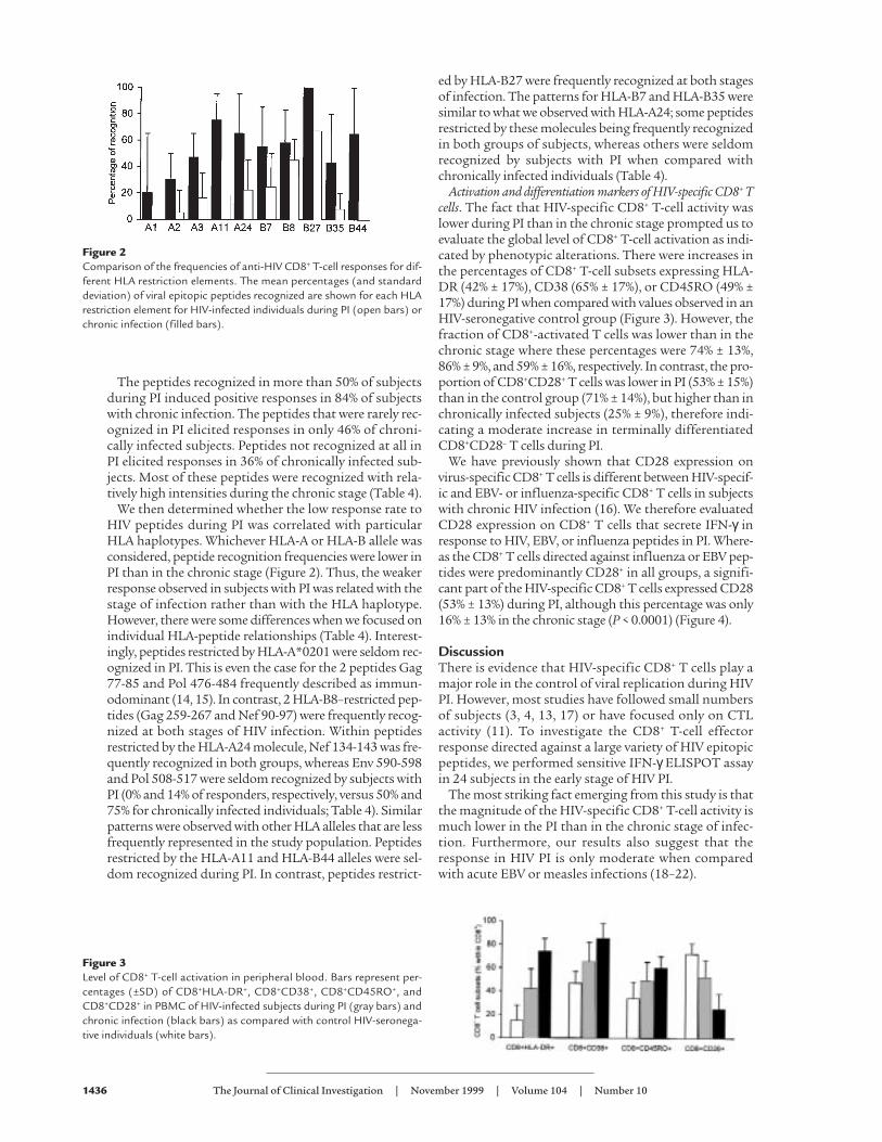
We then determined whether the low response rate toHIV peptides during PI was correlated with particularHLA haplotypes. Whichever HLA-A or HLA-B allele wasconsidered, peptide recognition frequencies were lower inPI than in the chronic stage (Figure 2). Thus, the weakerresponse observed in subjects with PI was related with thestage of infection rather than with the HLA haplotype.However, there were some differences when we focused onindividual HLA-peptide relationships (Table 4). Interest-ingly, peptides restricted by HLA-A*0201 were seldom rec-ognized in PI. This is even the case for the 2 peptides Gag77-85 and Pol 476-484 frequently described as immun-odominant (14, 15). In contrast, 2 HLA-B8–restricted pep-tides (Gag 259-267 and Nef 90-97) were frequently recog-nized at both stages of HIV infection. Within peptidesrestricted by the HLA-A24 molecule, Nef 134-143 was fre-quently recognized in both groups, whereas Env 590-598and Pol 508-517 were seldom recognized by subjects withPI (0% and 14% of responders, respectively, versus 50% and75% for chronically infected individuals; Table 4). Similarpatterns were observed with other HLA alleles that are lessfrequently represented in the study population. Peptidesrestricted by the HLA-A11 and HLA-B44 alleles were sel-dom recognized during PI. In contrast, peptides restrict-
ed by HLA-B27 were frequently recognized at both stagesof infection. The patterns for HLA-B7 and HLA-B35 weresimilar to what we observed with HLA-A24; some peptidesrestricted by these molecules being frequently recognizedin both groups of subjects, whereas others were seldomrecognized by subjects with PI when compared withchronically infected individuals (Table 4).
Activation and differentiation markers of HIV-specific CD8+ Tcells. The fact that HIV-specific CD8+ T-cell activity waslower during PI than in the chronic stage prompted us toevaluate the global level of CD8+ T-cell activation as indi-cated by phenotypic alterations. There were increases inthe percentages of CD8+ T-cell subsets expressing HLA-DR (42% ± 17%), CD38 (65% ± 17%), or CD45RO (49% ±17%) during PI when compared with values observed in anHIV-seronegative control group (Figure 3). However, thefraction of CD8+-activated T cells was lower than in thechronic stage where these percentages were 74% ± 13%,86% ± 9%, and 59% ± 16%, respectively. In contrast, the pro-portion of CD8+CD28+ T cells was lower in PI (53% ± 15%)than in the control group (71% ± 14%), but higher than inchronically infected subjects (25% ± 9%), therefore indi-cating a moderate increase in terminally differentiatedCD8+CD28– T cells during PI.
We have previously shown that CD28 expression onvirus-specific CD8+ T cells is different between HIV-specif-ic and EBV- or influenza-specific CD8+ T cells in subjectswith chronic HIV infection (16). We therefore evaluatedCD28 expression on CD8+ T cells that secrete IFN-γ inresponse to HIV, EBV, or influenza peptides in PI. Where-as the CD8+ T cells directed against influenza or EBV pep-tides were predominantly CD28+ in all groups, a signifi-cant part of the HIV-specific CD8+ T cells expressed CD28(53% ± 13%) during PI, although this percentage was only16% ± 13% in the chronic stage (P < 0.0001) (Figure 4).
DiscussionThere is evidence that HIV-specific CD8+ T cells play amajor role in the control of viral replication during HIVPI. However, most studies have followed small numbersof subjects (3, 4, 13, 17) or have focused only on CTLactivity (11). To investigate the CD8+ T-cell effectorresponse directed against a large variety of HIV epitopicpeptides, we performed sensitive IFN-γ ELISPOT assayin 24 subjects in the early stage of HIV PI.
The most striking fact emerging from this study is thatthe magnitude of the HIV-specific CD8+ T-cell activity ismuch lower in the PI than in the chronic stage of infec-tion. Furthermore, our results also suggest that theresponse in HIV PI is only moderate when comparedwith acute EBV or measles infections (18–22).
1436 The Journal of Clinical Investigation | November 1999 | Volume 104 | Number 10
Figure 2Comparison of the frequencies of anti-HIV CD8+ T-cell responses for dif-ferent HLA restriction elements. The mean percentages (and standarddeviation) of viral epitopic peptides recognized are shown for each HLArestriction element for HIV-infected individuals during PI (open bars) orchronic infection (filled bars).
Figure 3Level of CD8+ T-cell activation in peripheral blood. Bars represent per-centages (±SD) of CD8+HLA-DR+, CD8+CD38+, CD8+CD45RO+, andCD8+CD28+ in PBMC of HIV-infected subjects during PI (gray bars) andchronic infection (black bars) as compared with control HIV-seronega-tive individuals (white bars).

There was an HIV-specific response in 71% of subjects,which is consistent with the 74% responder rate report-ed by Musey et al. (11). The HIV-specific activity wasmuch lower than that observed in subjects at the chron-ic stage, where 100% of individuals responded to at least1 HIV peptide. In addition, responder subjects in PI onlyreacted to a limited number of peptides, whereas thoseat the chronic stage had a broad, specific response direct-ed at several peptides in the various HIV proteins.
We used a large variety of HIV epitopic peptides. How-ever, these peptides do not encompass entire sequencesof all HIV proteins, and they have been defined largelyfrom subjects with chronic HIV infection. As we cannotexclude the possibility that subjects with PI recognizeepitopes distinct from those identified in the chronicstage, we may have underestimated the breadth of theiranti-HIV CD8+ T-cell repertoire. This may explain whywe observed a low reactivity against Env even thoughmost responders recognized this protein in the studyreported by Musey et al. (11). However, experiments per-formed with B-LCL infected with recombinant vacciniaviruses and still showing a low T-cell reactivity againstEnv and Pol do not confirm this hypothesis. In addition,these approaches would only detect conserved epitopesbetween HIV-1 LAI and autologous isolates. Finally, HIVregulatory proteins may be preferential targets of theCD8+ T-cell response in the early phase of infection, asalready suggested by the high recognition frequency ofNef peptides. We are currently investigating whether Tat,Rev, and the other HIV regulatory proteins are targets ofresponse in the PI. In this context, it must be noted thatmost peptides recognized in acute EBV infection arederived from the immediate early and early proteins ofthe lytic cycle of EBV (21).
The low magnitude of the CD8+ T-cell response in bloodmay also be due to the sequestration of HIV-specific CD8+
T cells at sites of virus replication in the lymphoid organs.However, this seems unlikely in the light of recent studiesusing the SIV simian model of HIV infection (23, 24).
Differences in the breadth of the response between the2 stages of infection vary according to the HLA haplo-type. HLA-A*0201, HLA-A11, and HLA-B44 restrictedpeptides were seldom recognized during PI. In contrast,peptides restricted by HLA-B8 or HLA-B27 were fre-quently recognized at both stages of infection. More vari-able patterns were observed with HLA-A24, HLA-B7, andHLA-B35. Such differences between HLA molecules havealready been observed and the great efficiency of HLA-B8 and HLA-B27 alleles in presenting antigen has beendescribed, particularly during acute EBV infection (19,25). These data need to be confirmed in experiments onlarger numbers of subjects sharing the same HLA allelesto evaluate the relationship between these findings andthe reported prognostic value of HLA profiles.
Contrasting with differences observed in the numbersof peptides recognized between the 2 stages of infection,the frequencies of CD8+ T cells specific for a given peptidewere of the same order of magnitude in subjects withacute or chronic HIV infection. However, because acuteviral infections are usually characterized by a large expan-sion of antigen-specific cells, the frequencies observed inPI should be considered low. In subjects with PI, we
observed a positive correlation between the frequency atwhich a given peptide was recognized and the mean SFCcount induced by this peptide. This suggests that the pep-tides frequently recognized early in infection may beimmunodominant epitopes. Conversely, in chronic infec-tion, peptides recognized in a small proportion of indi-viduals may have high SFC counts. This may suggest adelayed expansion of cells recognizing subdominant epi-topes in response to chronic viral stimulation.
The frequencies of HIV-specific cells are of the sameorder of magnitude as those found using a similar tech-nique with EBV peptides in the latent phase of EBV
The Journal of Clinical Investigation | November 1999 | Volume 104 | Number 10 1437
Figure 4Comparison of the proportion of CD8+CD28+ T cells specific for HIV orother viral peptides among CD8+ T cells. (a) Representative experimentsfrom 3 HLA-B8–expressing individuals where PBMC were incubated inmedium alone or stimulated with peptides EBNA-3 325-333 derived fromEBV and Gag 259-267 from HIV-1. IFN-γ intracellular production (x axis)and CD28 expression (y axis) were analyzed on gated CD8+ T lymphocytes.200,000 events were acquired for subject KJA and 400,000 for the otherindividuals. Absolute numbers of events are indicated in the right quad-rants of each dot-plot. (b) Boxes representing the fraction of CD8+ CD28+
T cells (25th–75th percentile) specific for HIV peptides (gray boxes) or forinfluenza or EBV peptides (white boxes) in subjects with PI (n = 14) orchronic HIV infection (n = 14), and seronegative controls (n = 4).

infection (26). In contrast, the frequencies of EBV-spe-cific CD8+ T cells are much higher in acute EBV infec-tion. These frequency values ranged from 0.5% to 6.6%for HLA-A2–restricted T cells and from 29% to 44% forHLA-B8–restricted T cells (18). However, these frequen-cies have been assayed by tetramer staining, and it hasbeen recently shown that this method gives higher fre-quencies than those obtained by IFN-γ ELISPOT (26).Tetramer analysis may detect either cells without func-tional activity or cells that secrete cytokines other thanIFN-γ (27, 28). In acute measles infection, polyclonaldirect cytotoxic responses have been reported in 77% ofsubjects, which also suggests a high frequency of specif-ic CD8+ effector T cells (22).
The relative contribution of CD8+CD28+ T cells andCD8+CD28– T cells to the control of HIV is still unclearbut remains a major issue. In chronic HIV infection, theCD8+CD28+ T cells have noncytolytic suppressive activ-ity and have been associated with a good prognosis(29–31), whereas CD8+CD28– T cells are cytotoxic andincrease in parallel with the evolution towards terminalimmunodeficiency (29, 32). However, the CD8+CD28– Tcells may be a major player in early and efficient controlof viruses during acute infections. It has been reportedthat expression of CD28 is very low on the highly reac-tive CD8+ T cells during acute EBV infection (18). In thiscontext, the weak expansion of HIV-specific CD8+CD28–
T cells that we observed on HIV-specific CD8+ T cells insubjects with HIV PI is remarkable.
As a whole, there is a marked discrepancy betweenthe large global increase of CD8+CD28– T cellsobserved in blood and the relatively small numbers ofHIV-specific CD8+ T cells detected, particularly of theCD28– phenotype. Several hypotheses may explain thisdiscrepancy. First, some of the expanded CD8+CD28–
T cells may be directed at untested epitopes, derivedfrom autologous HIV-1 sequences different from thatof HIV-1 LAI, or present within some regulatory pro-teins, as already discussed. Second, an accumulation ofnonfunctional CD8+ T cells could occur during HIV PIas suggested by tetramer analysis in chronically HIV-infected individuals (27), and as demonstrated in lym-phocytic choriomeningitis virus–infected mice (28)and patients with melanoma (33). Further evidence forimpaired in vivo functionality of HIV-specific CD8+ Tcells has been provided recently (14, 34, 35). Bystanderactivation and expansion of CD8+ T cells directedagainst viruses other than HIV is unlikely becausemost of these cells express the CD28 molecule, at leastin absence of acute or recurrent infection (16).
During HIV PI and in chronically infected subjects, wefound no correlation between the total anti-HIV CD8+
T-cell activity and plasma viral RNA (10). First, in PI itmay be difficult to demonstrate any relationshipbetween the viral load and the immune response beforea steady-state has been achieved. Koup et al. (3) demon-strated that CTL levels increase as viral load declines toa set-point, and Musey et al. (11) found a correlationbetween the high frequencies of HIV-specific CTLp andlow plasma HIV RNA only after 6 months of infection.Second, at both stages of infection, CD8+ T cells with dif-ferent specificities may control the virus with different
efficiencies. This may account for the lack of correlationbetween viral load and the aggregate CD8+ T-cellresponses directed at all recognized epitopes. In this con-text, asymptomatic patients exhibit an inverse correla-tion between the viral load and the number of CD8+ Tcells directed at 2 HLA-A*0201 restricted, immun-odominant epitopes (Gag 77-85 and Pol 476-484) (36).This suggests that these CD8+ T cells are highly efficient,although the functional activity of cells binding totetrameric complexes containing these 2 peptides hasrecently been questioned (27, 34). In chronic infection,among the 10 HLA-A*0201–positive individuals whoseimmune response recognized these epitopes, only 4 hadavailable viral load determinations that precluded sta-tistical analysis. It is interesting to note that these 2immunodominant peptides were rarely recognized in PI.
There could be several explanations for the weakimmune response observed in HIV PI. A defect in CD4+ Thelper cells may be involved. The role of these cells hasbeen recently underscored in long-term nonprogressorsubjects (37–39) and in individuals treated with highlyactive antiretroviral treatment (HAART) during PI (37).Such responses are generally weak or absent before thera-py (manuscript in preparation). A qualitative or quantita-tive defect of dendritic cells, particularly those bearing theCD11c molecule (40), could be responsible for CD4+ andCD8+ T-cell defects, because these cells play an importantrole in inducing primary T-cell responses (41). Finally,HIV, like other viruses, may develop different strategies toavoid immune response. Various mechanisms have beenpostulated including a nef-mediated downregulation ofMHC class I expression that would directly hamper theCD8+ T-cell response (42, 43).
We conclude that a clear understanding of the immunemechanisms responsible for controlling HIV replicationduring PI is important for understanding the pathogene-sis of AIDS. We have described particular quantitative andqualitative features of the immune response in PI thatmay be responsible for the lack of control in HIV infec-tion. In particular, the number of CTL specificities may bea critical factor for the resolution of infection, as recentlydemonstrated in an acute model of hepatitis C virus infec-tion in chimpanzees (44). In addition, the immuneresponse further decreases during antiretroviral therapy(12, 13, 27, 45, 46). This decreased response could also beresponsible for the failure to eradicate the virus even whenthe viral load is extremely low because of effective therapy.This response could perhaps be strengthened to controlthe virus more effectively. Serial interruptions of antiviraltherapy have been shown to achieve this goal (47, 48).However, controlled immunotherapeutic protocols asso-ciated with HAART may be a safer option. The observa-tions made on the immune response during PI shouldhelp to improve the design of these protocols.
AcknowledgmentsWe thank the clinicians from all the participating centersof the PRIMO Cohort Study Group for their efficient col-laboration. We thank J. Choppin and J.-B. Hubert forhelpful and stimulating discussions; M. Levacher and F.Bouscarat for their contribution to FACS analysis; and F.Lemonnier and F. Gotch for critical reading of the man-
1438 The Journal of Clinical Investigation | November 1999 | Volume 104 | Number 10

uscript and constructive comments. The authors are alsoindebted to all the individuals who agreed to participatein the study and their referring physicians. The Englishtext has been edited by Owen Parkes. This work was fund-ed by the Agence Nationale de Recherches sur le SIDA. M.Dalod was supported by the Ministère de l’EducationNationale, de la Recherche et de la Technologie.
1. Pantaleo, G., Graziosi, C., and Fauci, A.S. 1997. Virologic and immuno-logic events in primary HIV infection. Springer Semin. Immunopathol.18:257–266.
2. Kahn, J.O., and Walker, B.D. 1998. Acute human immunodeficiencyvirus type 1 infection. N. Engl. J. Med. 339:33–39.
3. Koup, R.A., et al. 1994. Temporal association of cellular immuneresponses with the initial control of viremia in primary humanimmunodeficiency virus type 1 syndrome. J. Virol. 68:4650–4655.
4. Borrow, P., Lewicki, H., Hahn, B.H., Shaw, G.M., and Oldstone, M.B.A.1994. Virus-specific CD8+ cytotoxic T lymphocytes activity associatedwith control of viremia in primary human immunodeficiency virustype 1 infection. J. Virol. 68:6103–6110.
5. Schmitz, J.E., et al. 1999. Control of viremia in simian immunodefi-ciency virus infection by CD8+ lymphocytes. Science. 283:857–860.
6. Levy, J.A., Mackewicz, C.E., and Barker, E. 1996. Controlling HIVpathogenesis: the role of the noncytotoxic anti-HIV response of CD8+
T cells. Immunol. Today. 17:217–224.7. Mellors, J.W., Kingsley, L.A., and Rinaldo, C.R. 1995. Quantitation of
HIV-1 RNA in plasma predicts outcome after seroconversion. Ann.Intern. Med. 122:573–579.
8. Pantaleo, G., Demarest, J.F., and Schacker, T. 1997. The qualitativenature of the primary immune response to HIV infection is a prog-nosticator of disease progression independent of the initial level ofplasma viremia. Proc. Natl. Acad. Sci. USA. 94:254–258.
9. http://hiv-web.lanl.gov/immuno/index.html. HIV molecularimmunology database. National Institutes of Health, Bethesda, Mary-land, USA.
10. Dalod, M., et al. 1999. Broad, intense anti-HIV ex vivo CD8+ responsesin HIV type 1-infected patients: comparison with anti-EBV responsesand changes during antiretroviral therapy. J. Virol. 73:7108–7116.
11. Musey, L., et al. 1997. Cytotoxic T cell responses, viral load and diseaseprogression in early HIV-1 infection. N. Engl. J. Med. 337:1267–1274.
12. Dalod, M., et al. 1998. Evolution of cytotoxic T lymphocyte responsesto HIV-1 in patients with symptomatic primary infection on anti-retroviral triple therapy. J. Infect. Dis. 178:61–69.
13. Markowitz, M., et al. 1999. The effect of commencing combinationantiretroviral therapy soon after human immunodeficiency virus type1 infection on viral replication and antiviral immune responses. J.Infect. Dis. 179:525–537.
14. Goulder, P.J., et al. 1997. Patterns of immunodominance in HIV-1-spe-cific cytotoxic T lymphocyte responses in two human histocompati-bility antigens (HLA)-identical siblings with HLA-A*0201 are influ-enced by epitope mutation. J. Exp. Med. 185:1423–1433.
15. Brander, C., et al. 1998. Lack of strong immune selection pressure bythe immunodominant HLA-A*0201-restricted cytotoxic T lymphocyteresponse in chronic human immunodeficiency virus-1 infection. J. Clin.Invest. 101:2559–2566.
16. Dalod, M., et al. 1999. Altered ex vivo balance between CD28+ andCD28– cells within HIV-specific CD8+ T cells of HIV-seropositivepatients. Eur. J. Immunol. 29:38–44.
17. Lamhamedi-Cherradi, S., et al. 1995. Different patterns of HIV-1-spe-cific cytotoxic T-lymphocyte activity after primary infection. AIDS.9:421–426.
18. Callan, M.F.C., et al. 1998. Direct visualisation of antigen-specificCD8+ T cells during the primary immune response to Epstein-BarrVirus in vivo. J. Exp. Med. 187:1395–1402.
19. Steven, N.M., Leese, A.M., Annels, N.E., Lee, S.P., and Rickinson, A.B.1996. Epitope focusing in the primary cytotoxic T cell response toEpstein-Barr virus and its relationship to T cell memory. J. Exp. Med.184:1801–1813.
20. Mongkoslsapaya, J., et al. 1999. Antigen-specific expansion of cytotoxicT lymphocytes in acute measles virus infection. J. Virol. 73:67–71.
21. Steven, N.M., et al. 1997. Immediate early and early lytic cycle proteinsare frequent targets of the Epstein-Barr virus-induced cytotoxic T cellresponse. J. Exp. Med. 185:1605–1617.
22. Jaye, A., Magnusen, A.F., Sadiq, A.D., Corrah, T., and Whittle, H.C.
1998. Ex vivo analysis of cytotoxic T lymphocytes to measles antigensduring infection and after vaccination in Gambian children. J. Clin.Invest. 102:1969–1977.
23. Kuroda, M.J., et al. 1999. Comparative analysis of cytotoxic T lympho-cytes in lymph nodes and peripheral blood of simian immunodefi-ciency virus-infected rhesus monkeys. J. Virol. 73:1573–1579.
24. Kuroda, M.J., et al. 1999. Emergence of CTL coincides with clearanceof virus during primary simian immunodeficiency virus infection inrhesus monkeys. J. Immunol. 162:5127–5133.
25. Benjamin, R.J., Madrigal, J.A., and Parham, P. 1991. Peptide binding toempty HLA-B27 molecules of viable human cells. Nature. 351:74–77.
26. Tan, L.C., et al. 1999. A re-valuation of the frequency of CD8+ T cellsspecific for EBV in healthy virus carriers. J. Immunol. 162:1827–1835.
27. Gray, C.M., et al. 1999. Frequency of class I HLA-restricted anti-HIVCD8+ T cells in individuals receiving highly active antiretroviral ther-apy (HAART). J. Immunol. 162:1780–1788.
28. Zajac, A.J., et al. 1998. Viral immune evasion due to persistence of acti-vated T cells without effector function. J. Exp. Med. 12:2205–2213.
29. Choremi-Papadopoulou, H., et al. 1994. Downregulation of CD28 sur-face antigen on CD4+ and CD8+ T lymphocytes during HIV-1 infection.J. Acquir. Immune Defic. Syndr. 7:245–253.
30. Mackewicz, C.E., Ortega, H.W., and Levy, J.A. 1991. CD8+ cell anti-HIVactivity correlates with the clinical state of the infected individual. J.Clin. Invest. 87:1462–1466.
31. Landay, A.L., Mackewicz, C.E., and Levy, J.A. 1993. An activated CD8+
T cell phenotype correlates with anti-HIV activity and asymptomaticclinical status. Clin. Immunol. Immunopathol. 69:106–116.
32. Fiorentino, S., Dalod, M., Olive, D., Guillet, J.G., and Gomard, E. 1996.Predominant involvement of CD8+CD28- lymphocytes in humanimmunodeficiency virus-specific cytotoxic activity. J. Virol.70:2022–2026.
33. Lee, P.P., et al. 1999. Characterization of circulating T cells specific fortumor-associated antigens in melanoma patients. Nat. Med. 5:677–685.
34. Brander, C., et al. 1999. Persistent HIV-1-specific CTL clonal expansiondespite high viral burden post in utero HIV-1 infection. J. Immunol.162:4796–4800.
35. Hay, C.M., et al. 1999. Lack of viral escape and defective in vivo activa-tion of human immunodeficiency virus type 1-specific cytotoxic T lym-phocytes in rapidly progressive infection. J. Virol. 73:5509–5519.
36. Ogg, G.S., et al. 1998. Quantitation of HIV-1-specific cytotoxic T lym-phocytes and plasma viral load RNA. Science. 279:2103–2106.
37. Rosenberg, E.S., et al. 1997. Vigorous HIV-1-specific CD4+ T cellresponses associated with control of viremia. Science. 278:1447–1450.
38. Kalams, S.A., and Walker, B.D. 1998. The critical need for CD4 help inmaintaining effective cytotoxic T lymphocyte responses. J. Exp. Med.188:2199–2204.
39. Kalams, S.A., et al. 1999. Association between virus-specific cytotoxicT-lymphocyte and helper responses in human immunodeficiency virustype 1 infection. J. Virol. 73:6715–6720.
40. Grassi, F.R., et al. 1999. Depletion in blood CD11c-positive dendriticcells from human immunodeficiency infected patients. AIDS.13:759–766.
41. Rissoan, M.C., et al. 1999. Reciprocal control of T helper cell and den-dritic cell differentiation. Science. 283:1183–1186.
42. Collins, K.L., Chen, B.K., Kalams, S.A., Walker, B.D., and Baltimore, D.1998. HIV-1 Nef protein protects infected primary cells against killingby cytotoxic T lymphocytes. Nature. 391:397–401.
43. Schwartz, O., Marechal, V., Le Gall, S., Lemonnier, F., and Heard, J.M.1996. Endocytosis of major histocompatibility complex class I mole-cules is induced by the HIV-1 Nef protein. Nat. Med. 2:338–342.
44. Cooper, S., et al. 1999. Analysis of a successful immune responseagainst hepatitis C virus. Immunity. 10:439–449.
45. Ogg, G.S., et al. 1999. Decay kinetics of human immunodeficiencyvirus-specific effector cytotoxic T lymphocytes after combination anti-retroviral therapy. J. Virol. 73:797–800.
46. Kalams, S.A., et al. 1999. Levels of human immunodeficiency virus type1-specific cytotoxic T- lymphocyte effector and memory responsesdecline after suppression of viremia with highly active antiretroviraltherapy. J. Virol. 73:6721–6728.
47. Lori, F., et al. 1999. Intermittent drug therapy increases the time to HIVrebound in humans and induces the control of SIV after treatment inter-ruption in monkeys. In Program and Abstracts of the 6th Conference on Retro-viruses and Opportunistic Infections. Foundation for Retrovirology andHuman Health. Alexandria, VA. Late Breaker Abstract no. 5, p.206.
48. Ortiz, G.M., et al. 1999. HIV-1–specific immune responses in subjectswho temporarily contain virus replication after discontinuation ofhighly active antiretroviral therapy. J. Clin. Invest. 104:13–18.
The Journal of Clinical Investigation | November 1999 | Volume 104 | Number 10 1439