Interleukin-2 rescues helpless effector CD8+ T cells by diminishing the susceptibility to TRAIL...
15
Interleukin-2 rescues helpless effector CD8 + T cells by diminishing the susceptibility to TRAIL mediated death Monika C. Wolkers 1,3,4 , Steven J. Bensinger 1,3,4 , Douglas R. Green 1,4 , Stephen P. Schoenberger 1 , and Edith M. Janssen 2,4 1 Department of Cellular Immunology, La Jolla Institute for Allergy and Immunology, 9420 Athena Circle, La Jolla, CA 92037, USA 2 Department of Developmental Immunology, La Jolla Institute for Allergy and Immunology, 9420 Athena Circle, La Jolla, CA 92037, USA Abstract CD8 + T cells primed in the absence of CD4 + T cell help are programmed to produce TRAIL, which results in Death receptor (DR5) mediated apoptosis upon restimulation. Here, we studied whether these `helpless' effector CD8 + T cells are consigned to an apoptotic fate or whether their helpless program can be altered by inflammatory or growth cytokines. We found that helpless CD8 + T cells regained their full proliferative and functional capacity only when IL-2 was added to cell cultures, while IL-7 and IL-15, two common gamma chain cytokines associated with CD8 + T cell homeostasis and memory, could only partly restore secondary expansion in helpless CD8 + T cells. Recovery of functional CD8 + T cell immunity by IL-2 was concomitant with induction of IL2Rα (CD25) expression, downregulation of TRAIL, and the upregulation of anti-apoptotic molecules Bcl-2 and FLIP. The addition of IL-2 to helpless CD8 + T cells also interfered with DR5-mediated apoptosis induction, indicating that IL-2 affects several components of the TRAIL- DR5 pathway. Collectively, these data demonstrate that the helpless phenotype is not fixed, and that IL-2R signaling at the time of reactivation can play an important role in restoring CD8 + T cell function. Keywords T cells; cytotoxic; cytokines; CD8; TRAIL © 2011 Elsevier B.V. All rights reserved Corresponding author: Monika Wolkers. Department of Hematopoiesis, Sanquin Research/Landsteiner laboratory AMC/UvA, Plesmanlaan 125, 1066 CX Amsterdam, The Netherlands. Tel +31-20-5121379. Fax. +31-20-5123474. [email protected]. 3 authors contributed equally 4 Current addresses: Department of Hematopoiesis, Sanquin Research/Landsteiner laboratory AMC/UvA, Plesmanlaan 125, 1066 CX Amsterdam, The Netherlands (M.W.), Department of Pathology and Laboratory Medicine, University of California Los Angeles School of Medicine, 675 Charles E. Young Drive, Los Angeles, CA 90075, USA (S.B.), Department of Immunology, St. Jude Children's Research Hospital, 332 North Lauderdale St., Memphis, TN 38105, USA (D.G.), and Division of Molecular Immunology, Cincinnati Children's Hospital Research Foundation, and the University of Cincinnati College of Medicine, 3333 Burnet Avenue, Cincinnati, OH 45229 (E.J.). Publisher's Disclaimer: This is a PDF file of an unedited manuscript that has been accepted for publication. As a service to our customers we are providing this early version of the manuscript. The manuscript will undergo copyediting, typesetting, and review of the resulting proof before it is published in its final citable form. Please note that during the production process errors may be discovered which could affect the content, and all legal disclaimers that apply to the journal pertain. NIH Public Access Author Manuscript Immunol Lett. Author manuscript; available in PMC 2012 September 30. Published in final edited form as: Immunol Lett. 2011 September 30; 139(1-2): 25–32. doi:10.1016/j.imlet.2011.04.011. NIH-PA Author Manuscript NIH-PA Author Manuscript NIH-PA Author Manuscript
-
Upload
independent -
Category
Documents
-
view
1 -
download
0
Transcript of Interleukin-2 rescues helpless effector CD8+ T cells by diminishing the susceptibility to TRAIL...
Interleukin-2 rescues helpless effector CD8+ T cells bydiminishing the susceptibility to TRAIL mediated death
Monika C. Wolkers1,3,4, Steven J. Bensinger1,3,4, Douglas R. Green1,4, Stephen P.Schoenberger1, and Edith M. Janssen2,4
1Department of Cellular Immunology, La Jolla Institute for Allergy and Immunology, 9420 AthenaCircle, La Jolla, CA 92037, USA2Department of Developmental Immunology, La Jolla Institute for Allergy and Immunology, 9420Athena Circle, La Jolla, CA 92037, USA
AbstractCD8+ T cells primed in the absence of CD4+ T cell help are programmed to produce TRAIL,which results in Death receptor (DR5) mediated apoptosis upon restimulation. Here, we studiedwhether these `helpless' effector CD8+ T cells are consigned to an apoptotic fate or whether theirhelpless program can be altered by inflammatory or growth cytokines. We found that helplessCD8+ T cells regained their full proliferative and functional capacity only when IL-2 was added tocell cultures, while IL-7 and IL-15, two common gamma chain cytokines associated with CD8+ Tcell homeostasis and memory, could only partly restore secondary expansion in helpless CD8+ Tcells. Recovery of functional CD8+ T cell immunity by IL-2 was concomitant with induction ofIL2Rα (CD25) expression, downregulation of TRAIL, and the upregulation of anti-apoptoticmolecules Bcl-2 and FLIP. The addition of IL-2 to helpless CD8+ T cells also interfered withDR5-mediated apoptosis induction, indicating that IL-2 affects several components of the TRAIL-DR5 pathway. Collectively, these data demonstrate that the helpless phenotype is not fixed, andthat IL-2R signaling at the time of reactivation can play an important role in restoring CD8+ T cellfunction.
KeywordsT cells; cytotoxic; cytokines; CD8; TRAIL
© 2011 Elsevier B.V. All rights reservedCorresponding author: Monika Wolkers. Department of Hematopoiesis, Sanquin Research/Landsteiner laboratory AMC/UvA,Plesmanlaan 125, 1066 CX Amsterdam, The Netherlands. Tel +31-20-5121379. Fax. +31-20-5123474. [email protected] contributed equally4Current addresses: Department of Hematopoiesis, Sanquin Research/Landsteiner laboratory AMC/UvA, Plesmanlaan 125, 1066 CXAmsterdam, The Netherlands (M.W.), Department of Pathology and Laboratory Medicine, University of California Los AngelesSchool of Medicine, 675 Charles E. Young Drive, Los Angeles, CA 90075, USA (S.B.), Department of Immunology, St. JudeChildren's Research Hospital, 332 North Lauderdale St., Memphis, TN 38105, USA (D.G.), and Division of Molecular Immunology,Cincinnati Children's Hospital Research Foundation, and the University of Cincinnati College of Medicine, 3333 Burnet Avenue,Cincinnati, OH 45229 (E.J.).Publisher's Disclaimer: This is a PDF file of an unedited manuscript that has been accepted for publication. As a service to ourcustomers we are providing this early version of the manuscript. The manuscript will undergo copyediting, typesetting, and review ofthe resulting proof before it is published in its final citable form. Please note that during the production process errors may bediscovered which could affect the content, and all legal disclaimers that apply to the journal pertain.
NIH Public AccessAuthor ManuscriptImmunol Lett. Author manuscript; available in PMC 2012 September 30.
Published in final edited form as:Immunol Lett. 2011 September 30; 139(1-2): 25–32. doi:10.1016/j.imlet.2011.04.011.
NIH
-PA Author Manuscript
NIH
-PA Author Manuscript
NIH
-PA Author Manuscript
1. IntroductionIn the naïve host, antigen-specific CD8+ T cells are present in very low frequencies. Uponantigenic stimuli through infection or immunization, naïve CD8+ T cells undergo rapidclonal expansion that results in significant numbers of antigen-specific CD8+ T cells [1–4].After having reached a plateau for several days, T cell responses contract, and more than90% of the antigen-specific CD8+ T cells are eliminated via apoptosis. The survivingantigen-specific CD8+ T cells create a memory pool that provides durable protection whenre-challenged [5,6]. A number of studies have demonstrated that a relatively short durationof antigenic stimulation commits naïve CD8+ T cells to clonal expansion and effectordifferentiation. These data indicate that initial antigen encounter launches a genetic programin CD8+ T cells that stably modifies the phenotype and function of their daughter cells(progeny) [7–11].
Signals provided by CD4+ T helper cells during CD8+ T cell priming are critical for theacquisition and maintenance of functional immunity. Although primary CD8+ T cellresponses can proceed independently of CD4+ T cells, the lack of CD4+ T cells during CD8+
T cell priming results in loss of efficient CD8+ T cell recall responses [12–15]. Oneconsequence of CD8+ T cell priming in the absence of T cell help is the acquisition of atranscriptional program that results in the upregulation of TNF related apoptosis inducingligand (TRAIL) upon secondary antigen encounter. [16–18]. Those data showed that T cellreceptor triggering is required for the expression of TRAIL in helpless CD8+ T cells. Theexpression of TRAIL in reactivated helpless CD8+ T cells results in Death Receptor 5 (DR5)mediated apoptosis, providing a mechanistic explanation for the poor CD8+ T cell immunityobserved during recall responses. To date, it is not known if helpless CD8+ T cells areconsigned to an apoptotic fate upon secondary challenge, or if signals delivered in trans canprotect helpless CD8+ T cells from TRAIL/DR5 mediated death.
In this report we show that facets of the helpless program are not fixed and can be altered byγchain cytokines, in particular by IL-2. While IL-2, IL-7 and IL-15 provide survival signalssufficient to maintain effector function of restimulated helpless CD8+ T cells, only IL-2 hadthe capacity to restore robust secondary expansion. Consistent with an important role for theIL-2 signaling axis in regulating the helpless phenotype, we found that CD8+ T cells primedwithout CD4 help failed to upregulate CD25 in recall responses. Interestingly, the additionof IL-2, IL-7 and IL-15 to cultures during restimulation was sufficient to restore theexpression of CD25. Moreover, common γchain signaling influenced the apoptotic programof helpless CD8+ T cells by regulating the expression of DR-5, Trail, Flip and Bcl-2. Lastly,we show that IL-2 signals protected both helpless and helped CD8+ T cells from DR-5mediated apoptosis. Together, our data provide a molecular explanation how cytokines caninfluence the fitness of secondary CD8+ T cell responses.
2. Material and Methods2.1. Mice and cell lines
C57BL/6J mice were purchased from the Jackson laboratory (Bar Harbor, ME). Mice weremaintained at the La Jolla Institute for Allergy and Immunology under specific pathogenfree conditions in accordance with guidelines by the Association for Assessment andAccreditation of Laboratory Animal Care International. Tap sufficient and deficient mouseembryo cell lines (MEC) expressing the human adenovirus type 5 early region 1 (Ad5E1)[19] were cultured in IMDM supplemented with 10% fetal calf serum, 50μM 2-mercaptoethanol, 2mM L-glutamine, 20U/mL penicillin and 20μg/mL streptomycin.
Wolkers et al. Page 2
Immunol Lett. Author manuscript; available in PMC 2012 September 30.
NIH
-PA Author Manuscript
NIH
-PA Author Manuscript
NIH
-PA Author Manuscript
2.2. Immunization and antibody treatmentMice were immunized subcutaneously in the flank with 1×10⁷ irradiated (3000 rad) TAP−/−-Ad5E1-MECs. CD4+ cells were depleted on the first three days prior to immunization byintraperitoneal administration of 100 μg GK1.5 (CD4-depleted at the time of priming:`helpless'; untreated: `helped'). At day 3 post immunization, all mice were treated withGK1.5 [12].
2.3. Isolation and stimulation of CD8+ T cellsCD8+ T cells were purified from spleens and lymph nodes (LN) of TAP−/−-Ad5E1-MEC-immunized mice by negative selection with the MACS CD8 isolation kit (Miltenyi Biotec,Germany). Purity of isolated CD8+ T cells was confirmed by FACS analysis and wasroutinely greater than 95% with less than 0.1% CD4+ T cell contamination. CD8+ T cellswere restimulated in vitro for 6 days with irradiated syngeneic TAP+/+-Ad5E1-MECs (10:1ratio). Recombinant mIL-1β, hIL-2, mIL-4, mIL-5, mIL-6, mIL-7, mIL-10, mIL-12,mIL-15, mIL-18, mTNF-α, mIFN-γ, mIFN-α, mGM-CSF, DR5-Fc or Fas-Fc (Peprotech,New Jersey; PBL, New Jersey; R and D Systems, Minnesota) were added to cultures at thetime of in vitro re-stimulation at indicated concentrations. In some experiments, CD8+ Tcells from spleens and LN cells from TAP−/−-Ad5E1-MEC immunized mice were culturedwith biotinylated anti-DR5 at 5μg/mL (clone MD5-1, eBioscience, California) for 30minutes as previously described [20]. Purified streptavidin (Invitrogen, California) wassubsequently added to cultures at 5μg/mL to cross link the anti-DR5 antibody. After 30 minof incubation, CD8+ T cells were cultured for 6 days with TAP+/+-Ad5E1-MECs (10:1 ratio)in the presence or absence of IL-2.
2.4. Enumeration of antigen-specific CD8+ T cellsSpleen and LN cells were incubated for 5 h with the E1B192–200 peptide or control peptideOVA257–264 (aa:VNIRNCCYI and aa:SIINFEKL, resp.; A&A labs LLC, California) at 5μg/mL final concentration in the presence of Brefeldin A (BDbiosciences, California) eitherdirectly ex vivo, or after in vitro culture. Cells were stained for surface expression of CD8and CD44, fixed and permeabilized using Cytofix/Cytoperm kit (BDbiosciences, California)and stained for intracellular IFN-γ, or IFN-γ in combination with antibodies to CD25,CD122 or CD127 (eBioscience, California) according to manufacturer's protocol. The foldexpansion of E1B192–200-specific CD8+ T cells was calculated by dividing the absolutenumber of IFN-γ+CD8+ T cells after in vitro culture by the absolute number of IFN-γ+CD8+
T cells placed into culture [12,16]. The cytolytic activity of E1B192–200-specific CD8+ Tcells was determined by the JAM assay using 3H-thymidine labeled EL-4 as target cells asdescribed before [12,21].
2.5. Real-time reverse transcription-PCR (RT-PCR)Purified CD8+ T cells were stimulated with the E1B192–200 peptide for the indicated time,and total RNA was isolated using TriZol (Gibco BRL, Maryland) according to themanufacturer's protocol. RNA was reverse transcribed with the M-MLV reversetranscriptase (Gibco BRL, Maryland) using random hexamers (Gibco BRL, Maryland).Sequence-specific primers for murine IL-2Rα (CD25), TRAIL, TRAIL-R2/DR5, FLIP,BcL-2, IL-2, IL-7, IL-15, L32 and 18S were previously described [16]. Real Time PCR™was performed with Amplitaq Gold™ polymerase in a PE biosystems 5700 thermocyclerusing SyBr Green™ detection protocol as outlined by the manufacturer. L32 and 18S wereused as internal controls.
Wolkers et al. Page 3
Immunol Lett. Author manuscript; available in PMC 2012 September 30.
NIH
-PA Author Manuscript
NIH
-PA Author Manuscript
NIH
-PA Author Manuscript
2.6. Statistical methodsUnless stated otherwise, data are expressed as mean ± standard error of the mean (S.E.M),and evaluated using a two-tailed analysis of variance (ANOVA) followed by a Dunnett test.A probability value of p<0.05 was considered statistically significant.
3. Results3.1. Helpless effector CD8+ T cells die a TRAIL mediated death
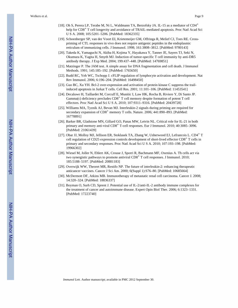
Immunization in the absence of CD4+ T cells results in compromised `helpless' CD8+ Tcells that undergo TRAIL induced cell death upon secondary antigen encounter [12–16]. Tostudy this in further detail, we immunized intact `helped' and CD4-depleted “helpless” micewith TAP-deficient MECs that were transfected with the human adenovirus type 5 earlyregion 1 (designated TAP−/−-Ad5E1-MECs). Antigen-specific, IFNγ-producing CD8+ Tcells were enumerated ex vivo one week after immunization, and secondary expansion ofhelped and helpless CD8+ T cells was assessed upon a six day in vitro restimulation withirradiated TAP+/+-Ad5E1-MECs [12,16,19]. Consistent with previous reports, helplessCD8+ T cells failed to expand upon secondary antigen encounter, as opposed to helpedCD8+ T cells (Fig. 1). Importantly, the secondary expansion of helpless CD8+ T cells couldbe restored by blocking the TRAIL/DR5 pathway, but not by inhibiting the FAS/FASLinteraction (p<0.05).
3.2. Common γchain cytokines can restore secondary responses of helpless CD8+ T cellsWe next assessed if cytokines had the potential to alter the helpless T cell phenotype duringsecondary antigenic stimulation. To this end, we added a broad range of cytokines producedby lymphocytes or antigen presenting cells (APCs) to helpless CD8+ T cell cultures. Theinflammatory cytokines IL-1β, TNF-α, IFNα and IFNβ, and the growth factors IL-6 andGM-CSF had no effect on viability and secondary expansion of helpless CD8+ T cells,whereas APC-derived proinflammatory IL-12 and IL-18 modestly improved survival (Fig.2a, data not shown). The Th1-associated cytokine IFN-γ, and the Th2 associated cytokinesIL-4, IL-5 and IL-10 provided little rescue. In contrast, the common γ chain cytokines IL-2,IL-7, and IL-15 could significantly improve survival and expansion of helpless CD8+ T cellcultures. Specifically, the addition of exogenous IL-2 (20ng/mL) markedly increased thesecondary expansion of helpless CD8+ T cells (p<0.002), ultimately exceeding theexpansion of helped CD8+ T cells (without exogenous IL-2) by 10-fold (Fig. 2a; data notshown). The addition of IL-7 (100ng/mL, p<0.02) and IL-15 (100ng/mL, p<0.05) alsorestored secondary expansion of antigen-specific helpless CD8+ T cells, albeit to a muchlesser extent (Fig. 2a). The rescue of helpless CD8+ T cells was not a universal feature of allγc cytokines; IL-4 failed to protect helpless CD8+ T cells, at a dose range from 2–500ng/mL(Fig. 2a; data not shown).
Examination of a dose response curve to IL-2, IL-7 and IL-15 revealed that as little as 1ng/mL of IL-2 added at the time of antigenic restimulation resulted in a 5-fold expansion ofhelpless CD8+ T cells over control cells (i.e. helped CD8+ T cells cultured without IL-2),with a maximal expansion between 10–100ng/mL (Fig. 2b, p<0.0001). For IL-7 and IL-15,100ng/ml was required to achieve a more modest secondary expansion that was below theexpansive capacity of helped CD8+ T cells (data not shown).
IL-2, IL-7 and IL-15 were also capable to maintain the effector function of restimulatedhelpless CD8+ T cells. We found that IL-2 and IL-7 significantly restored the IFNγproduction of helpless CD8+ T cells on a per cell basis (Fig. 2c). Furthermore, helplessCD8+ T cells regained their capacity to kill target cells after 6 days of in vitro restimulation(Fig. 2d). The marked increase of cytolytic function resembled that of helped CD8+ T cells,
Wolkers et al. Page 4
Immunol Lett. Author manuscript; available in PMC 2012 September 30.
NIH
-PA Author Manuscript
NIH
-PA Author Manuscript
NIH
-PA Author Manuscript
in particular when IL-2 was added to the culture. In contrast, helpless CD8+ T cells thatwere restimulated without cytokines exhibited cytotoxicity levels that were similar to that ofnaïve polyclonal CD8+ T cells (Fig. 2d) [12]. These data indicate that IL-2, IL-7 and IL-15have the capacity to maintain the effector function of helpless CD8+ T cells, however, IL-2alone is sufficient to support full secondary expansion. Together, this suggests that IL-2,IL-7 and IL-15 play overlapping and divergent roles in the restoration of secondaryexpansion and maintenance of effector function of helpless CD8+ T cells in vitro.
3.3. Regulation of CD25 expression by common γchain cytokinesWe next sought to determine how IL-2, IL-7, and IL-15 restore helpless CD8+ T cellexpansion during secondary antigen encounter. Gene expression studies indicated that bothhelped and helpless CD8+ T cells express similar levels of IL-2, IL-7, and IL-15 in responseto antigenic stimulation (Fig. 3a). Hence, it appears unlikely that restoration of secondaryexpansion and effector function by cytokines is the product of an autocrine feedback loop.Therefore, we considered the possibility that common γchain receptor signaling was alteredin helpless CD8+ T cells. To address this hypothesis, we first examined the expressionpattern of the common γc cytokine receptors on helped and helpless CD8+ T cells ex vivo.After 24 and 48 hours of in vitro restimulation, we could not detect differences in theexpression levels of the IL-2/15 shared β chain (CD122) and the IL-7Rα chain (CD127) onantigen-specific CD8+ T cells (Fig. 3b and data not shown). In contrast, helpless CD8+ Tcells expressed substantially lower levels of the high affinity IL-2Rα chain (CD25) afterantigenic stimulation (MFI 441 v 21, Fig. 3b). The addition of IL-2 during antigenicrestimulation restored CD25 expression on helpless CD8+ T cells that was comparable toreactivated helped CD8+ T cells. We also observed modest increase in CD25 levels onhelpless CD8+ T cells upon addition of IL-15, and to a lesser extent of IL-7 (MFI 148 and106, respectively).
The expression of CD25 on helped CD8+ T cells upon antigenic re-stimulation appears to beregulated by de novo synthesis of CD25 rather than by redistribution of preformed protein.This is reflected by the fact that restimulation of CD8+ T cells in the presence of Brefeldin Aresulted in significant accumulation of CD25 protein in the endoplasmic reticulum and thatsurface CD25 expression remained unaltered (Fig 3b,c). In addition, quiescent helplessCD8+ T cells had little or no detectable surface and intracellular CD25 expression directlyex vivo (Fig. 3c, left panel). This also correlated with low CD25 mRNA levels (Fig. 3d).Activation of helpless CD8+ T cells, however, led to rapid production of CD25 transcripts(p<0.001). The induction of CD25 transcripts was specific for helped CD8+ T cells, ashelpless CD8+ T cells failed to upregulate CD25 message and protein upon antigenicrestimulation (Fig. 3b, d). This transcriptional defect could be modestly restored by addingIL-2, IL-7, and IL-15 to the culture (Fig. 3d; p<0.005) and correlated with increasedexpression of CD25 protein in the cells (Fig. 3b). Thus, the differential regulation of CD25as a consequence of the presence or absence of CD4+ T cell help at priming suggests thatCD25 expression, like TRAIL expression, is part of the transcriptional program of helplessCD8+ T cells.
3.4. Modifications of the helpless program by IL-2, IL-7, and IL-15The key characteristic feature of the helpless transcriptional program is TRAIL-mediatedactivation-induced cell death (AICD) that is reflected by induction of TRAIL mRNA uponrestimulation exclusively in helpless, but not helped CD8+ T cells ([16]; Fig 4a, p<0.0001).Given that IL-2, IL-7 and IL-15 can restore helpless CD8+ T cell responses, we addressedwhether they can also modify TRAIL expression. As we have recently shown, IL-2 (20ng/mL) significantly decreased the induction of TRAIL mRNA in helpless CD8+ T cells(Wolkers et al., manuscript in preparation and Fig. 4a, p<0.02). The addition of IL-15
Wolkers et al. Page 5
Immunol Lett. Author manuscript; available in PMC 2012 September 30.
NIH
-PA Author Manuscript
NIH
-PA Author Manuscript
NIH
-PA Author Manuscript
(100ng/mL) also resulted in a modest, but not significant downregulation of TRAIL inhelpless CD8+ T cells, while IL-7 did not affect TRAIL induction (at a concentrationsufficient to partly restore secondary expansion and effector function; 100ng/mL, Fig. 2).
Because IL-2 significantly, but not completely, suppressed TRAIL expression in reactivatedhelpless CD8+ T cells, we hypothesized that IL-2R signaling may prevent AICD byadditional effects, i.e. by inducing anti-apoptotic genes. The Flice inhibitory protein (FLIP)can inhibit Death Receptor mediated AICD by blocking the intracellular caspase cascade[22]. Flip is induced in helped CD8+ T cells after antigenic stimulation, but not in helplessCD8+ T cells (Fig. 4b, p<0.001) [16]. Strikingly, all three common γ chain cytokinesrestored the induction of FLIP mRNA upon reactivation in helpless CD8+ T cells (Fig. 4b,p<0.001). Similarly, the anti-apoptotic factor Bcl-2 that can decrease the susceptibility toTRAIL mediated AICD [23] was upregulated in restimulated helpless CD8+ T cells upon theaddition of IL-2 and IL-15 (p<0.001), but not by IL-7 (Fig. 4c). Together, our findingsindicate that IL-2, IL-7, and IL-15 employ several pathways to decrease susceptibility toAICD in helpless CD8+ T cells. These include the reduction of TRAIL expression, and theconcomitant induction of Bcl-2 and FLIP, the latter possibly interfering with the DR5signaling pathway and/or decreasing DR5 sensitivity.
3.5. IL-2 protects helpless CD8+ T cells from DR5 mediated apoptosisThe upregulation of anti-apoptotic genes in response to exogenous cytokines led us tohypothesize that IL-2R signaling may directly influence TRAIL-DR5 mediated cell death.Indeed, administration of IL-2 (at 20ng/mL) during antigenic restimulation resulted in asignificant decrease in DR5 mRNA levels in helpless CD8+ T cells (p=0.002), while DR5mRNA in helped CD8+ T cells appeared unchanged (Fig. 5a). This divergent expressionpattern of DR5 in helped and helpless CD8+ T cells was only evident when IL-2 was addedduring restimulation, because DR5 mRNA expression was comparable directly ex vivo andupon peptide restimulation alone (Fig. 5a). Of note, DR5 and its downstream signaling isfully functional in helped CD8+ T cells, as restimulation in the presence of recombinantTRAIL completely abrogated secondary expansion (data not shown, and [16]).
To directly test whether IL-2 protects helpless CD8+ T cells from DR5 mediated cell death,we pretreated helped and helpless CD8+ T cells with the cross-linked agonistic anti-DR5antibody MD5-1 [20] before the CD8+ T cells were restimulated in vitro. As expected,helpless CD8+ T cells did not expand when treated with cross-linked MD5-1, confirmingthat MD5-1 is not acting as a DR5 blocking antibody in this system (Fig. 5b, c).Interestingly, helped CD8+ T cells cultures were significantly affected by cross-linkedMD5-1 as assessed by their reduced frequency and total number of IFN-γ+CD8+ T cells(p<0.02), further confirming that helped CD8+ T cells maintain their susceptibility toTRAIL/DR5 mediated death. Importantly, the addition of 20ng/ml IL-2 to cultures drovesecondary expansion in both helped and helpless CD8+ T cells regardless of MD5-1treatment (Fig. 5b, c; p<0.05). These data indicate that IL-2 signals interfere with DR-5mediated apoptosis both by decreasing the ligand production and by reducing the sensitivityto DR5-mediated cell death.
4. DiscussionCD8+ T cells primed in the absence of CD4 help fail to undergo robust secondary expansionwhen reactivated by cognate antigen. In this report, we sought to determine if helpless CD8+
T cells are consigned to TRAIL-mediated cell death, or if signals delivered in trans couldalter the helpless program. In support of the latter assertion, we found that IL-2, IL-7 andIL-15 significantly influenced helpless CD8+ T cell fate and function. These data indicatethat the genetic and functional program acquired by helpless CD8+ T cells during priming
Wolkers et al. Page 6
Immunol Lett. Author manuscript; available in PMC 2012 September 30.
NIH
-PA Author Manuscript
NIH
-PA Author Manuscript
NIH
-PA Author Manuscript
can be overwritten to some extent at the time of secondary antigen encounter if the correctsignals are delivered.
It has previously been shown that the common γ chain receptor is critical for CD8+ memoryT cell formation [24]. In particular, IL-2 signals during T cell priming are pivotal forproficient CD8+ T cell memory formation, both in terms of expansion and differentiationafter secondary challenge [25]. Moreover, IL-15 and recently IL-21 have been shown toprovide `help' signals during CD8+ T cell priming [18,26]. Here, we extend theseobservations to the secondary expansion phase of CD8+ T cells and demonstrate that thecommon γc cytokines IL-2, IL-7 and IL-15 preserve effector functions in reactivatedhelpless CD8+ T cells.
We observed that IL-2, and to a lesser extent IL-7 and IL-15, modulates the expression ofseveral genes involved in apoptosis including Bcl-2, Flip, Trail and DR-5. Therefore, itappears likely that a combination rather than one single gene mediates the rescue of helplessCD8+ T cells to significantly reduce TRAIL mediated death and DR5 sensitivity. Consistentwith this idea, Bcl-2 transgenic overexpression by itself (that is induced in reactivatedhelpless CD8+ T cells by IL-2 and IL-15 (Fig. 4c)) could not restore secondary expansion inhelpless CD8+ T cells [16].
Interestingly, while all three cytokines could equally well restore effector functions such asIFNγ production and cytotolytic activity, only IL-2 had the capacity to profoundly restorethe secondary expansion in helpless CD8+ T cells. This differential outcome was alsoreflected in their distinct effects on TRAIL production, sensitivity to DR5 signaling andCD25 expression. These data suggest that cytokines involved in lymphocyte maintenance(e.g. IL-7 and IL-15) may play an important role in allowing helpless cells to persist, butIL-2 appears to be the dominant cytokine in reprogramming helpless CD8+ T cells to fullyfunctional effector cells during antigenic restimulation. Therefore, it will be of interest todetermine if helpless CD8+ T cells that have been reactivated in the presence of IL-2maintain the helpless transcriptional program for their progeny, or if IL-2 can rewrite theCD8 program to ensure long-term CD8+ T cell immunity.
The molecular signature of CD8+ T cell immunity is not fully determined yet. Here we showthat the high affinity IL-2 receptor α unit (CD25) is an important component of secondaryexpansion. While the CD25 expression is low in helped and helpless CD8 T cells directly exvivo, CD25 levels were significantly increased by cytokines in reactivated helpless CD8+ Tcells, in particular by IL-2 via its IL-2/IL-2R positive feedback loop. Recent data haveprovided evidence that CD4+ T cell help during T cell priming increases CD25 expressionon CD8+ T cells indirectly through CD40L engagement on APCs [27,28]. We now showthat high CD25 expression levels on CD8+ T cells also appear pivotal during reactivation toprovide proficient secondary CD8+ T cell responses. Furthermore, our study indicates thatregulation of CD25 expression is part of the transcriptional program of helpless CD8+ Tcells. Therefore, we identified CD25 as a second marker to distinguish helped from helplessCD8+ T cells ex vivo, in addition to TRAIL expression and AICD upon restimulation ofhelpless CD8 T cells.
We found that antigenic restimulation of helpless CD8+ T cells in vivo did not result inincreased expression of CD25 (EM Janssen; unpublished data), suggesting that circulatingIL-2 levels are not sufficient to induce CD25 expression on the CD8+ T cells. In light ofthese findings, it is tempting to speculate that administration of IL-2 in vivo may restorehelpless CD8+ T cell responses, and that current IL-2 based therapies mediate their effectsthrough the modification of a relatively “helpless” program [29–31]. In conclusion, the datapresented herein provide important insights into the fate and function of CD8+ T cells
Wolkers et al. Page 7
Immunol Lett. Author manuscript; available in PMC 2012 September 30.
NIH
-PA Author Manuscript
NIH
-PA Author Manuscript
NIH
-PA Author Manuscript
primed in the absence of CD4 T cell help and could have important therapeutic implicationsfor vaccine strategies to infectious disease and cancer.
AcknowledgmentsThis work was supported in part by a Career Development Award from the Leukemia and Lymphoma Society(#3248-05 to E.J.), the National Institutes of Health (#RR021975 to S.B.), and the Cancer Research Institute/Irvington Institute (M.W.).
References[1]. Butz EA, Bevan MJ. Massive expansion of antigen-specific CD8+ T cells during an acute virus
infection. Immunity. 1998; 8:167–175. [PubMed: 9491998][2]. Callan MF, Tan L, Annels N, Ogg GS, Wilson JD, O'Callaghan CA, Steven N, McMichael AJ,
Rickinson AB. Direct visualization of antigen-specific CD8+ T cells during the primary immuneresponse to Epstein-Barr virus In vivo. J Exp Med. 1998; 187:1395–1402. [PubMed: 9565632]
[3]. Doherty PC. The numbers game for virus-specific CD8+ T cells. Science. 1998; 280:227.[PubMed: 9565533]
[4]. Murali-Krishna K, Altman JD, Suresh M, Sourdive DJ, Zajac AJ, Miller JD, Slansky J, Ahmed R.Counting antigen-specific CD8 T cells: a reevaluation of bystander activation during viralinfection. Immunity. 1998; 8:177–187. [PubMed: 9491999]
[5]. Dutton RW, Bradley LM, Swain SL. T cell memory. Annu Rev Immunol. 1998; 16:201–223.[PubMed: 9597129]
[6]. Sprent J, Tough DF. T cell death and memory. Science. 2001; 293:245–248. [PubMed: 11452113][7]. van Stipdonk MJ, Lemmens EE, Schoenberger SP. Naive CTLs require a single brief period of
antigenic stimulation for clonal expansion and differentiation. Nat Immunol. 2001; 2:423–429.[PubMed: 11323696]
[8]. van Stipdonk MJ, Hardenberg G, Bijker MS, Lemmens EE, Droin NM, Green DR, SchoenbergerSP. Dynamic programming of CD8+ T lymphocyte responses. Nat Immunol. 2003; 4:361–365.[PubMed: 12640451]
[9]. Badovinac VP, Porter BB, Harty JT. Programmed contraction of CD8+ T cells after infection. NatImmunol. 2002; 3:619–626. [PubMed: 12055624]
[10]. Kaech SM, Ahmed R. Memory CD8+ T cell differentiation: initial antigen encounter triggers adevelopmental program in naive cells. Nat Immunol. 2001; 2:415–422. [PubMed: 11323695]
[11]. Teixeiro E, Daniels MA, Hamilton SE, Schrum AG, Bragado R, Jameson SC, Palmer E.Different T cell receptor signals determine CD8+ memory versus effector development. Science.2009; 323:502–505. [PubMed: 19164748]
[12]. Janssen EM, Lemmens EE, Wolfe T, Christen U, von Herrath MG, Schoenberger SP. CD4+ Tcells are required for secondary expansion and memory in CD8+ T lymphocytes. Nature. 2003;421:852–856. [PubMed: 12594515]
[13]. Bourgeois C, Rocha B, Tanchot C. A role for CD40 expression on CD8+ T cells in the generationof CD8+ T cell memory. Science. 2002; 297:2060–2063. [PubMed: 12242444]
[14]. Shedlock DJ, Shen H. Requirement for CD4 T cell help in generating functional CD8 T cellmemory. Science. 2003; 300:337–339. [PubMed: 12690201]
[15]. Sun JC, Bevan MJ. Defective CD8 T cell memory following acute infection without CD4 T cellhelp. Science. 2003; 300:339–342. [PubMed: 12690202]
[16]. Janssen EM, Droin NM, Lemmens EE, Pinkoski MJ, Bensinger SJ, Ehst BD, Griffith TS, GreenDR, Schoenberger SP. CD4+ T-cell help controls CD8+ T-cell memory via TRAIL-mediatedactivation-induced cell death. Nature. 2005; 434:88–93. [PubMed: 15744305]
[17]. Hamilton SE, Wolkers MC, Schoenberger SP, Jameson SC. The generation of protectivememory-like CD8+ T cells during homeostatic proliferation requires CD4+ T cells. Nat Immunol.2006; 7:475–481. [PubMed: 16604076]
Wolkers et al. Page 8
Immunol Lett. Author manuscript; available in PMC 2012 September 30.
NIH
-PA Author Manuscript
NIH
-PA Author Manuscript
NIH
-PA Author Manuscript
[18]. Oh S, Perera LP, Terabe M, Ni L, Waldmann TA, Berzofsky JA. IL-15 as a mediator of CD4+
help for CD8+ T cell longevity and avoidance of TRAIL-mediated apoptosis. Proc Natl Acad SciU S A. 2008; 105:5201–5206. [PubMed: 18362335]
[19]. Schoenberger SP, van der Voort EI, Krietemeijer GM, Offringa R, Melief CJ, Toes RE. Cross-priming of CTL responses in vivo does not require antigenic peptides in the endoplasmicreticulum of immunizing cells. J Immunol. 1998; 161:3808–3812. [PubMed: 9780143]
[20]. Takeda K, Yamaguchi N, Akiba H, Kojima Y, Hayakawa Y, Tanner JE, Sayers TJ, Seki N,Okumura K, Yagita H, Smyth MJ. Induction of tumor-specific T cell immunity by anti-DR5antibody therapy. J Exp Med. 2004; 199:437–448. [PubMed: 14769851]
[21]. Matzinger P. The JAM test. A simple assay for DNA fragmentation and cell death. J ImmunolMethods. 1991; 145:185–192. [PubMed: 1765650]
[22]. Budd RC, Yeh WC, Tschopp J. cFLIP regulation of lymphocyte activation and development. NatRev Immunol. 2006; 6:196–204. [PubMed: 16498450]
[23]. Guo BC, Xu YH. Bcl-2 over-expression and activation of protein kinase C suppress the trail-induced apoptosis in Jurkat T cells. Cell Res. 2001; 11:101–106. [PubMed: 11453541]
[24]. Decaluwe H, Taillardet M, Corcuff E, Munitic I, Law HK, Rocha B, Riviere Y, Di Santo JP.Gamma(c) deficiency precludes CD8+ T cell memory despite formation of potent T celleffectors. Proc Natl Acad Sci U S A. 2010; 107:9311–9316. [PubMed: 20439728]
[25]. Williams MA, Tyznik AJ, Bevan MJ. Interleukin-2 signals during priming are required forsecondary expansion of CD8+ memory T cells. Nature. 2006; 441:890–893. [PubMed:16778891]
[26]. Barker BR, Gladstone MN, Gillard GO, Panas MW, Letvin NL. Critical role for IL-21 in bothprimary and memory anti-viral CD8+ T-cell responses. Eur J Immunol. 2010; 40:3085–3096.[PubMed: 21061439]
[27]. Obar JJ, Molloy MJ, Jellison ER, Stoklasek TA, Zhang W, Usherwood EJ, Lefrancois L. CD4+ Tcell regulation of CD25 expression controls development of short-lived effector CD8+ T cells inprimary and secondary responses. Proc Natl Acad Sci U S A. 2010; 107:193–198. [PubMed:19966302]
[28]. Wiesel M, Joller N, Ehlert AK, Crouse J, Sporri R, Bachmann MF, Oxenius A. Th cells act viatwo synergistic pathways to promote antiviral CD8+ T cell responses. J Immunol. 2010;185:5188–5197. [PubMed: 20881183]
[29]. Overwijk WW, Theoret MR, Restifo NP. The future of interleukin-2: enhancing therapeuticanticancer vaccines. Cancer J Sci Am. 2000; 6(Suppl 1):S76–80. [PubMed: 10685664]
[30]. McDermott DF, Atkins MB. Immunotherapy of metastatic renal cell carcinoma. Cancer J. 2008;14:320–324. [PubMed: 18836337]
[31]. Boyman O, Surh CD, Sprent J. Potential use of IL-2/anti-IL-2 antibody immune complexes forthe treatment of cancer and autoimmune disease. Expert Opin Biol Ther. 2006; 6:1323–1331.[PubMed: 17223740]
Wolkers et al. Page 9
Immunol Lett. Author manuscript; available in PMC 2012 September 30.
NIH
-PA Author Manuscript
NIH
-PA Author Manuscript
NIH
-PA Author Manuscript
Research highlights
• CD8 T cells require CD4 T cell help for proficient memory formation.
• Addition of cytokines rescues `helpless' CD8 T cells from TRAIL mediated celldeath.
• Cytokines change the transcriptional profile of `helpless' CD8 T cells.
Wolkers et al. Page 10
Immunol Lett. Author manuscript; available in PMC 2012 September 30.
NIH
-PA Author Manuscript
NIH
-PA Author Manuscript
NIH
-PA Author Manuscript
Figure 1. TRAIL regulates the secondary expansion of helpless CD8+ T cellsUntreated (helped) or CD4 depleted (helpless) C57BL/6J mice were immunized with 1×10⁷irradiated TAP−/− Ad5E1 MECs. At day 7 after immunization CD8+ T cells were purifiedand cultured for 6 days together with TAP+/+-Ad5E1 MECs cells in the absence or presenceof Fas-Fc or DR5-FC (5ug/ml). The frequency of antigen-specific CD8+ T cells ex vivo, andafter in vitro restimulation was determined by quantifying E1B192-200-specific IFN-γproducing cells by flow cytometry. Data of one representative mouse of three mice isshown. The frequency of IFN-γ producing cells within the CD8+ T cell population isdepicted within the FACS plot. Numbers below the FACS plots show secondary expansionof E1B192-200-specific IFN-γ producing CD8+ T cells at the end of the culture as mean ±S.E.M. of 3–4 mice per group (representative of 4 experiments). *** p<0.005 compared tohelped CD8+ T cells; * P<0.05 compared to helpless CD8+ T cells).
Wolkers et al. Page 11
Immunol Lett. Author manuscript; available in PMC 2012 September 30.
NIH
-PA Author Manuscript
NIH
-PA Author Manuscript
NIH
-PA Author Manuscript

Figure 2. Common γc cytokines IL-2, IL-7 and IL-15 can rescue the helpless phenotypePurified helped and helpless CD8+ T cells from mice immunized with TAP−/−-Ad5E1MECs were isolated on day 7 post immunization and cultured with TAP+/+ -Ad5E1 MECsin the presence of the indicated cytokine. Unless indicated otherwise, cytokines were used atconcentrations of 10ng/ml (IL-2, GM-CSF), or 200 ng/ml (IL-4,-7,-15,-10, IFN-γ). (A, B)After 6 days of in vitro culture the secondary expansion of E1B192-200-specific IFN-γproducing CD8+ T cells was assessed as described above (Fig. 1). (C) The meanfluorescence intensity (MFI) of IFNγ of in vitro restimulated E1B192-200-specific CD8+ Tcells was determined after 6 days in vitro culture in the presence or absence of indicatedcytokines. (D) Cytolytic activity of purified naive, or helped and helpless CD8+ T cells fromimmunized mice 6 days after restimulation with TAP+/+-Ad5E1 MECs in the presence ofindicated cytokine. Cytolytic activity was assessed using the JAM assay by determining totalloss in 3H-thymidine from E1B192-200-pulsed or OVA257-264-pulsed (control) target cells.Data are presented as mean ± S.E.M. * p<0.05; ** p<0.02; *** p<0.005; (n= 3–4 mice pergroup; repeated 3 times).
Wolkers et al. Page 12
Immunol Lett. Author manuscript; available in PMC 2012 September 30.
NIH
-PA Author Manuscript
NIH
-PA Author Manuscript
NIH
-PA Author Manuscript
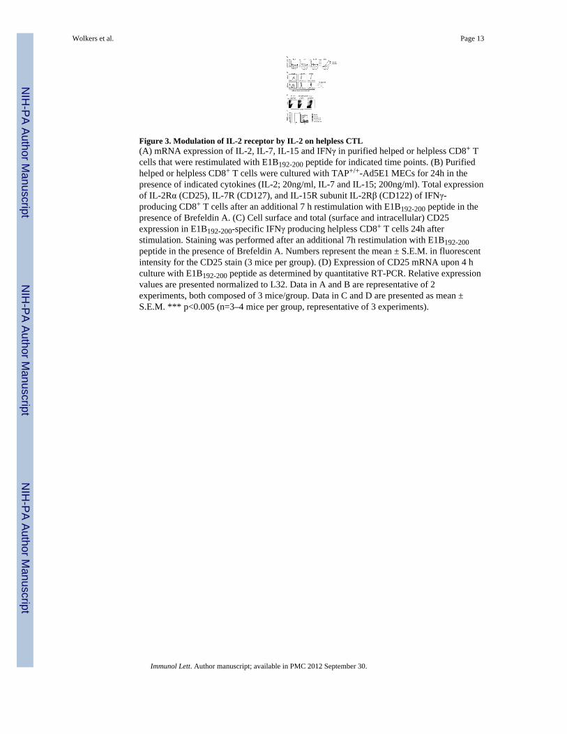
Figure 3. Modulation of IL-2 receptor by IL-2 on helpless CTL(A) mRNA expression of IL-2, IL-7, IL-15 and IFNγ in purified helped or helpless CD8+ Tcells that were restimulated with E1B192-200 peptide for indicated time points. (B) Purifiedhelped or helpless CD8+ T cells were cultured with TAP+/+-Ad5E1 MECs for 24h in thepresence of indicated cytokines (IL-2; 20ng/ml, IL-7 and IL-15; 200ng/ml). Total expressionof IL-2Rα (CD25), IL-7R (CD127), and IL-15R subunit IL-2Rβ (CD122) of IFNγ-producing CD8+ T cells after an additional 7 h restimulation with E1B192-200 peptide in thepresence of Brefeldin A. (C) Cell surface and total (surface and intracellular) CD25expression in E1B192-200-specific IFNγ producing helpless CD8+ T cells 24h afterstimulation. Staining was performed after an additional 7h restimulation with E1B192-200peptide in the presence of Brefeldin A. Numbers represent the mean ± S.E.M. in fluorescentintensity for the CD25 stain (3 mice per group). (D) Expression of CD25 mRNA upon 4 hculture with E1B192-200 peptide as determined by quantitative RT-PCR. Relative expressionvalues are presented normalized to L32. Data in A and B are representative of 2experiments, both composed of 3 mice/group. Data in C and D are presented as mean ±S.E.M. *** p<0.005 (n=3–4 mice per group, representative of 3 experiments).
Wolkers et al. Page 13
Immunol Lett. Author manuscript; available in PMC 2012 September 30.
NIH
-PA Author Manuscript
NIH
-PA Author Manuscript
NIH
-PA Author Manuscript
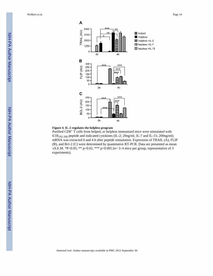
Figure 4. IL-2 regulates the helpless programPurified CD8+ T cells from helped, or helpless immunized mice were stimulated withE1B192-200 peptide and indicated cytokines (IL-2; 20ng/ml, IL-7 and IL-15; 200ng/ml).mRNA was extracted 0 and 4 h after peptide stimulation. Expression of TRAIL (A), FLIP(B), and Bcl-2 (C) were determined by quantitative RT-PCR. Data are presented as mean±S.E.M. *P<0.05; ** p<0.02, *** p<0.005 (n= 3–4 mice per group; representative of 3experiments).
Wolkers et al. Page 14
Immunol Lett. Author manuscript; available in PMC 2012 September 30.
NIH
-PA Author Manuscript
NIH
-PA Author Manuscript
NIH
-PA Author Manuscript
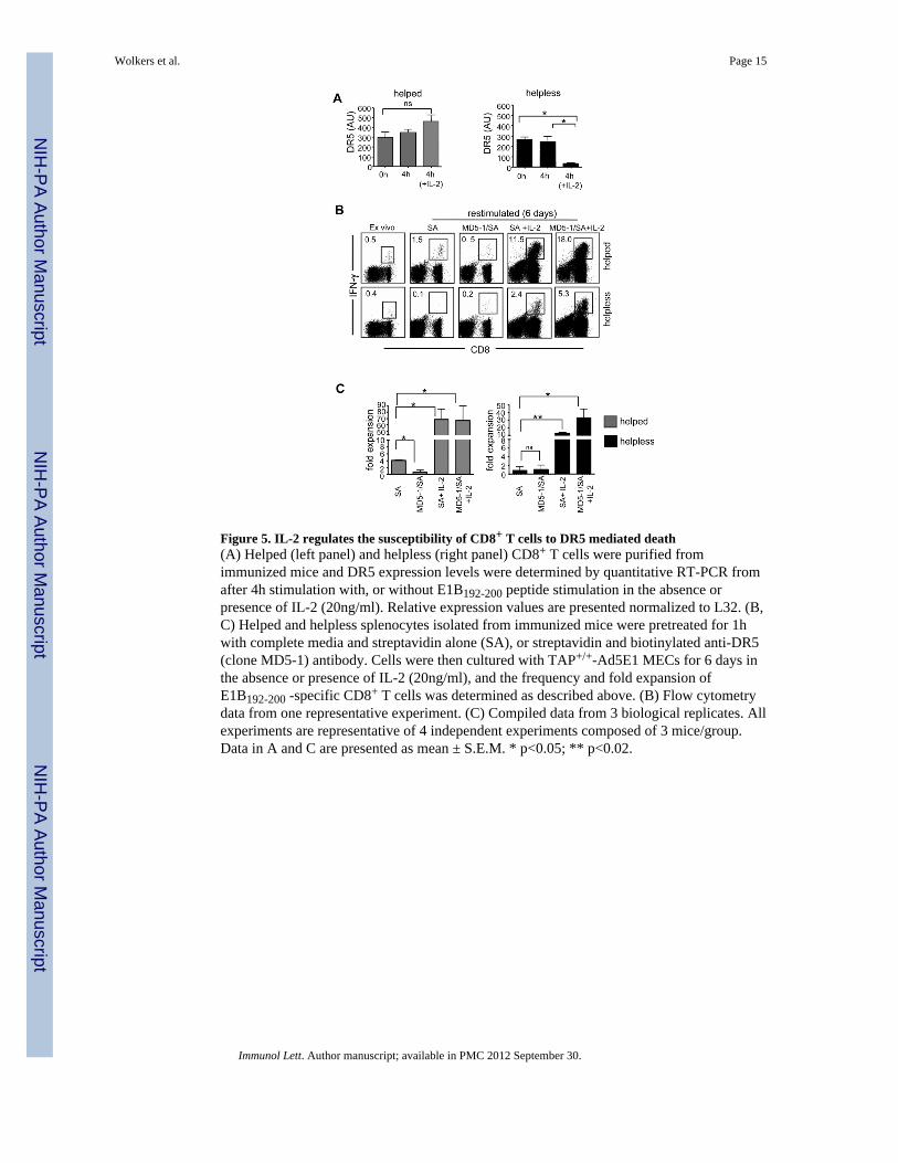
Figure 5. IL-2 regulates the susceptibility of CD8+ T cells to DR5 mediated death(A) Helped (left panel) and helpless (right panel) CD8+ T cells were purified fromimmunized mice and DR5 expression levels were determined by quantitative RT-PCR fromafter 4h stimulation with, or without E1B192-200 peptide stimulation in the absence orpresence of IL-2 (20ng/ml). Relative expression values are presented normalized to L32. (B,C) Helped and helpless splenocytes isolated from immunized mice were pretreated for 1hwith complete media and streptavidin alone (SA), or streptavidin and biotinylated anti-DR5(clone MD5-1) antibody. Cells were then cultured with TAP+/+-Ad5E1 MECs for 6 days inthe absence or presence of IL-2 (20ng/ml), and the frequency and fold expansion ofE1B192-200 -specific CD8+ T cells was determined as described above. (B) Flow cytometrydata from one representative experiment. (C) Compiled data from 3 biological replicates. Allexperiments are representative of 4 independent experiments composed of 3 mice/group.Data in A and C are presented as mean ± S.E.M. * p<0.05; ** p<0.02.
Wolkers et al. Page 15
Immunol Lett. Author manuscript; available in PMC 2012 September 30.
NIH
-PA Author Manuscript
NIH
-PA Author Manuscript
NIH
-PA Author Manuscript