Computational Design of Peptides That Target Transmembrane Helices
Upload
independentCategory
view
2download
0
ChemicalScience
EDGE ARTICLE
Ope
n A
cces
s A
rtic
le. P
ublis
hed
on 2
0 N
ovem
ber
2014
. Dow
nloa
ded
on 2
9/05
/201
6 20
:29:
45.
Thi
s ar
ticle
is li
cens
ed u
nder
a C
reat
ive
Com
mon
s A
ttrib
utio
n 3.
0 U
npor
ted
Lic
ence
.
View Article OnlineView Journal | View Issue
Rational coupled
aCurriculum in Bioinformatics and Comp
Carolina, Chapel Hill, NC 27599, USA. E-mbProgram in Molecular and Cellular Biophys
Hill, NC 27599, USAcDepartment of Biochemistry and Biophysic
Hill, NC 27599, USAdCystic Fibrosis Treatment and Research Cen
Hill, NC 27599, USA
† Electronic supplementary informa10.1039/c4sc01320d
‡ These authors contributed equally to th
Cite this: Chem. Sci., 2015, 6, 1237
Received 7th May 2014Accepted 14th November 2014
DOI: 10.1039/c4sc01320d
www.rsc.org/chemicalscience
This journal is © The Royal Society of C
dynamics network manipulationrescues disease-relevant mutant cystic fibrosistransmembrane conductance regulator†
Elizabeth A. Proctor,‡ab Pradeep Kota,‡bc Andrei A. Aleksandrov,cd Lihua He,cd
John R. Riordancd and Nikolay V. Dokholyan*abcd
Many cellular functions necessary for life are tightly regulated by protein allosteric conformational change,
and correlated dynamics between protein regions has been found to contribute to the function of proteins
not previously considered allosteric. The ability to map and control such dynamic coupling would thus
create opportunities for the extension of current therapeutic design strategy. Here, we present an
approach to determine the networks of residues involved in the transfer of correlated motion across a
protein, and apply our approach to rescue disease-causative mutant cystic fibrosis transmembrane
regulator (CFTR) ion channels, DF508 and DI507, which together constitute over 90% of cystic fibrosis
cases. We show that these mutations perturb dynamic coupling within the first nucleotide-binding
domain (NBD1), and uncover a critical residue that mediates trans-domain coupled dynamics. By
rationally designing a mutation to this residue, we improve aberrant dynamics of mutant CFTR as well as
enhance surface expression and function of both mutants, demonstrating the rescue of a disease
mutation by rational correction of aberrant protein dynamics.
Introduction
Protein allostery is a ubiquitous mechanism central to theregulation of many cellular processes, such as enzyme catalysisand signal transduction.1,2 An allosteric change involves theprotein population ensemble redistributing itself among theavailable conformations.2–4 NMR studies of dynamic couplingbetween residues in proteins support the idea that allostery is acommon intrinsic property of many proteins.3,5,6 Exertingcontrol over such dynamic coupling interactions could open thedoor for novel therapeutic strategies in diseases such as cysticbrosis. However, predicting and designing the effect ofcoupling interactions on protein structure and functionremains a challenge. Determining interaction networks thatcouple distant sites of the protein and identifying hub (“hotspot”) residues that control the dynamics of such couplinginteractions are essential steps to uncovering the molecular
utational Biology, University of North
ail: [email protected]
ics, University of North Carolina, Chapel
s, University of North Carolina, Chapel
ter, University of North Carolina, Chapel
tion (ESI) available. See DOI:
is work.
hemistry 2015
mechanisms of protein allostery.7 Previous efforts toward theseends have focused mainly on searching for new allosteric sitesand mechanisms; X-ray crystallographic studies of bound andunbound structures have offered particularly important struc-tural insights into allosteric regulation.7 However, analysis ofthe static structures of allosteric end states cannot provide acomplete picture of the inter-residue dynamics and interactionsinvolved in allosteric conformational change or the coupling ofdynamics at distal sites. NMR studies of protein dynamics havebeen pivotal in identifying “hidden” networks of residues withstrong dynamic coupling.6,8 Thermodynamic mutation cycles,9
which measure the coupling between two mutation sites bytheir mutual contribution to protein stability, provide a directmethod to systematically probe such relations between proteinsites. However, due to experimental limitations and practicalconsiderations, a large-scale study using these methods wouldbe prohibitively laborious and time consuming.
Computational methods can be used to probe the couplingof amino acids and to identify networks of residues controllingprotein conformational changes. For example, sequence-basedapproaches can reveal co-evolving residues likely to be ener-getically or functionally coupled.10–14 However, the applicationof sequence-based approaches, which rely on evolutionaryinformation, is limited by the availability of homologoussequences and complicated by the fact that evolutionaryconservation is oen driven by factors other than function (e.g.stability, folding kinetics).15,16
Chem. Sci., 2015, 6, 1237–1246 | 1237

Fig. 1 Deletion of F508 or I507 results in altered coupling betweenregions of NBD1. (a) The root mean square fluctuations (RMSF) overthe simulation of each residue in WT NBD1 compared with those inDF508- and DI507-NBD1 suggests increased flexibility in severalregions upon deletion mutation. (b) Difference map derived from thecorrelation maps of WT and DF508 NBD1. Blue denotes lost correla-tion upon deletion mutation, while red denotes gained correlation.Tube representation of NBD1 protein dynamics highlights changes influctuations in NBD1 upon deletion of F508. The thickness and color ofthe tube represent the extent of change in dynamics of the corre-sponding region during the simulation. Warmer colors indicate greaterincrease in flexibility, while colder colors indicate no change. (c) Sameas (b), for DI507-NBD1.
Chemical Science Edge Article
Ope
n A
cces
s A
rtic
le. P
ublis
hed
on 2
0 N
ovem
ber
2014
. Dow
nloa
ded
on 2
9/05
/201
6 20
:29:
45.
Thi
s ar
ticle
is li
cens
ed u
nder
a C
reat
ive
Com
mon
s A
ttrib
utio
n 3.
0 U
npor
ted
Lic
ence
.View Article Online
Recently, we have proposed that coupled dynamics plays acritical role in the pathophysiology of cystic brosis (CF).17,18
Over 1500 mutations in the CFTR gene have been identied inpatients with CF. The protein product of this gene, CFTR, playsa fundamental role in epithelial ion transport, providing a rate-limiting step in the regulation of salt secretion and reabsorp-tion. In approximately 90% of CF patients, the deletion of asingle phenylalanine (DF508) in its rst nucleotide-bindingdomain (NBD1) results in misfolding and misassembly of theprotein (DF508-CFTR).17 Cheng et al. determined that the DF508mutation prevents maturation and trafficking of CFTR to thecell membrane,19 although maturation and partial function ofDF508-CFTR have been observed at sub-physiological temper-ature (<30 �C) in various cell types.20–23 Second site mutations,like the commonly used I539T substitution, can improvematuration,24,25 but DF508-CFTR rescued in this mannerexhibits poor function at physiological temperature, indicatingthat the fundamental defect in DF508-CFTR maturation andfunction is a consequence of reduced thermal stability.26 Thishypothesis is further supported by the fact that F508 plays acritical role in interfacing NBD1 with the fourth cytoplasmicloop of CFTR, thus contributing signicantly to the structuralintegrity and stability of the protein.27,28 Therefore, effectivetherapeutic rescue of mutant CFTR requires restoration ofthermal stability, oen mediated by protein uctuations anddynamics, making mutant CFTR an ideal system for develop-ment of methods for dynamic control.
Here, we develop a widely applicable computational metho-dology that utilizes concepts from graph theory to identifyspecic residues that propagate dynamic coupling effectsbetween structurally distant sites in proteins. We apply ourapproach to the rescue of DF508-CFTR, using data from discretemolecular dynamics simulations, performable on a personalcomputer, to identify hot spot sites. We then utilize mutagen-esis to demonstrate control over dynamic coupling between twodistant regions of NBD1. We show that rational mutagenesis ofthe identied bottleneck sites dramatically rescues aberrantdynamics, as well as dramatically improving maturation andfunction of DF508-CFTR in mammalian cells. We furtherdemonstrate the ability of our method to identify sites impor-tant to wild-type protein function and stability by showing thatour designed mutation can also rescue the adjacent but distinctand more severe disease mutant, DI507-CFTR.
ResultsHeightened thermal uctuations in the regulatory insertionpropagate to the SDR and ATP-binding sub-domain
We and others have reported that the NBD1 regulatory insertion(RI, residues 404-435) inuences the dynamics of NBD1 inCFTR,18,26,29 but the nature and mechanism of this inuence arestill unknown. To elucidate the effects of RI within NBD1, weperform discrete molecular dynamics (DMD) simulations30,31 ofwild type (WT), DF508-, and DI507-NBD1. We nd that thermaluctuations increase and/or are shied signicantly in severalkey regions of NBD1 upon deletion of F508 or I507: the RI,structurally diverse region (SDR), residues 532-552,29 RI-SDR
1238 | Chem. Sci., 2015, 6, 1237–1246
bridge (residues 492-502), F508-loop (residues 507-514), andpart of the ATP-binding sub-domain (residues 570-600) (regionsindicated by color-coded arrows, Fig. 1a). These heightened
This journal is © The Royal Society of Chemistry 2015

Edge Article Chemical Science
Ope
n A
cces
s A
rtic
le. P
ublis
hed
on 2
0 N
ovem
ber
2014
. Dow
nloa
ded
on 2
9/05
/201
6 20
:29:
45.
Thi
s ar
ticle
is li
cens
ed u
nder
a C
reat
ive
Com
mon
s A
ttrib
utio
n 3.
0 U
npor
ted
Lic
ence
.View Article Online
uctuations suggest the possibility of dynamic couplingbetween the affected regions, which would transfer a pertur-bation in one region to any of the others. For example, deletionof F508 or I507 in the F508 loop affects the dynamics of RI, theRI-SDR bridge, the SDR, and the ATP-binding sub-domain.
F508 deletion results in non-native internal coupling of thea-subdomain and ATP-binding subdomain
In order to conrm our hypothesis of coupled dynamicsbetween key structural regions of NBD1, we perform covariationanalysis of dynamic uctuations observed in simulations.32 InDF508-NBD1, we nd a marked decoupling of the RI from theentire a-subdomain and parts of the ATP-binding subdomain ascompared to the wild type (Fig. 1b-i). In addition, we observe anincrease in coupling between the various key regions of thea-subdomain and the ATP-binding subdomain (Fig. 1b-ii), andan increase in internal coupling in the ATP-binding subdomain(Fig. 1b-iii). From these ndings, we conclude that the F508loop, the SDR, the RI-SDR bridge, the RI, and the ATP-bindingsubdomain are inter-connected in a network of coupleddynamics within NBD1. These changes suggest that the deletionof F508 from the F508 loop could cause dynamic instability andincreased uctuations in this region, which lead to decouplingfrom the RI. The RI decouples from the entire a-subdomain,leading to the gain of non-native coupling within that region.Ultimately, the observed increase in uctuations and changes todynamic coupling within NBD1may result in domain instabilityand consequent misprocessing of DF508-CFTR. Deletion of theRI eliminates this transfer of dynamics through the domain,rescuing the protein as observed.26
I507 deletion results in non-native coupling between the SDRand the ATP-binding sub-domain
Instead of the complete decoupling of the RI from the a-sub-domain seen with the F508 deletion (discussed above), DI507-NBD1 features a shi in the regions of RI that undergocoupling, losing coupling in one area only to gain it in adjacentresidues (Fig. 1c-i). We detect a similar shi of coupling inter-actions in the SDR, with its coupling to the RI (Fig. 1c-i), and inaddition note that the SDR loses coupling within itself (Fig. 1c-ii), likely due to the increase in uctuations in this area (Fig. 1a).The perturbation to the SDR also includes a gained non-nativecoupling with part of the ATP-binding subdomain (Fig. 1c-ii),which decouples from the a-subdomain (Fig. 1c-iii). Lastly, wend a strong non-native coupling of the SDR with the F508 loop(Fig. 1c-iv), likely the perturbation caused by the deletionmutation. Combining these ndings, we conclude that thedeletion of I507 causes a perturbation to coupled dynamics thatis transferred by coupled dynamics through the SDR to the RIand the ATP-binding subdomain, shiing coupling interactionsall throughout NBD1, which severely affects the dynamicstability of the domain. Likewise, the non-native coupling of theATP-binding subdomain to the SDR and the loss of internalcoupling in the ATP-binding subdomain could affect thebinding of ATP, which is stabilizing to NBD1 under normalconditions. In the absence of ATP-binding, NBD1 is
This journal is © The Royal Society of Chemistry 2015
signicantly destabilized, potentially compounding the effectsof the DI507 mutation.
Network formalism of NBD1 dynamics reveals bottleneckresidues that control dynamic coupling
In order to deduce the pathways through which these changesin dynamic coupling occur, for each system of wild type,DF508, and DI507 NBD1, we represent the pairwise correlationcoefficients between residues as a complete graph, with thecorrelation between every possible pair of residues repre-sented as a connection between nodes (an edge). We thenweight this graph (G(N,E), where N is the set of nodes and E isthe set of edges) such that each edge is enumerated by thecorrelation coefficient between the corresponding pair ofresidues. Weighting enables us to isolate those edges havingthe most signicant impact on the transduction of uctua-tions across the protein. To identify the specic residuesmediating dynamic coupling within NBD1, we impose acorrelation cutoff to our graph, eliminating edges with aweight below our cutoff until the largest component of theresulting disconnected sub-graph comprises approximately50% of the total number of nodes in the graph (Fig. S1†). Thiscutoff is at the critical threshold, where the network exhibitscritical properties33 and transitions from connected todisconnected.34 Our rationale with this construction is tocreate an algorithm for network mapping that is without freeparameters. From the resulting network, we determinewhether a node is critical for the connectivity of the largestcomponent in the sub-graph by monitoring the topologicalchanges in the sub-graph as we iteratively remove each nodefrom the sub-graph. If removing node a splits the largest sub-graph into two or more components, then node a is a criticalnode, or “bottleneck,” in the network. A bottleneck residue is aresidue with the ability to affect the entire network because ofits positioning as the lone connection between at least tworegions of a network (i.e., an unavoidable step in the couplingof dynamics between one region and the other). Mutation ormodication of a bottleneck residue can affect the dynamics,stability, and allosteric or binding behavior of a protein ordomain. We nd several such bottleneck residues in thedynamic networks of DF508, DI507, and wild type NBD1,which we perturb to ameliorate the pathological dynamics ofthe mutant NBD1s.
Network bottlenecks control the transfer of heightenedthermal uctuations from the RI of DF508-NBD1
We have previously reported that thermal uctuations areincreased in the SDR and dynamic coupling is lost between theF508 loop and the ATP-binding sub-domain upon deletion ofF508, both of which are ameliorated upon deletion of the RI.26
We hypothesize that manipulating the coupling between the RIand the 508-loop and between RI and the SDR may ameliorateuctuations and improve dynamic stability in these regions. Inorder to identify bottleneck residues that mediate the transferof uctuations in the RI across the domain, we conduct anetwork analysis of the correlated dynamics between residues
Chem. Sci., 2015, 6, 1237–1246 | 1239

Chemical Science Edge Article
Ope
n A
cces
s A
rtic
le. P
ublis
hed
on 2
0 N
ovem
ber
2014
. Dow
nloa
ded
on 2
9/05
/201
6 20
:29:
45.
Thi
s ar
ticle
is li
cens
ed u
nder
a C
reat
ive
Com
mon
s A
ttrib
utio
n 3.
0 U
npor
ted
Lic
ence
.View Article Online
in NBD1 (Methods). We conclude from this analysis that theresidues K464, T465, L468, S492, I601, and V603 are criticalnodes involved in the transfer of the thermal uctuations fromthe RI to other regions of NBD1 via dynamic coupling (Fig. 2a).K464 and V603mediate coupling with the b-strands forming theATP-binding core sub-domain (Fig. S2,† red residues), L468 andL601 with the regulatory extension (RE) of NBD1 (Fig. S2,† blueresidues), and T465 with both of these regions (Fig. 2b and S2†).However, S492 in b5 is the only residue mediating dynamiccoupling between the RI and the 508-loop and SDR (Fig. 2b:yellow nodes; Fig. S2:† green residues). In addition, we note thatS492 has been identied as the site of a rescue mutation forCFTR in an independent study.18 We conclude that S492 is anetwork bottleneck that dynamically couples the RI with theseregions in NBD1.
Fig. 2 S492 is a critical node in DF508-NBD1. (a) Number of nodes inthe largest component (y-axis) of the disconnected sub-graph afterdisconnecting the corresponding residue (x-axis). We consider resi-dues that partition the sub-graph into connected components of tenor more residues as critical nodes. Different colors represent differentcomponents. (b) Network representation of the disconnected sub-graph. S492 forms a critical node that partitions the sub-graph intotwo connected components of nearly equal size (yellow and graycircles). Yellow nodes with black outline represent residues in the RI.Smaller components are represented as white circles.
1240 | Chem. Sci., 2015, 6, 1237–1246
DI507-NBD1 network bottlenecks reside in regions essentialfor CFTR maturation and function
Unlike DF508-CFTR, removal of the RI in NBD1 does not rescueDI507-CFTR, so we instead adopt the strategy of amelioratingthe non-native coupling of the SDR and ATP-binding sub-domain. Using network analysis of correlated dynamicsbetween DI507-NBD1 residues (Methods), we nd that residuesT547, L548, K564, D565, A566, D567, and L568 are critical nodesin the cross-domain coupling of dynamics in DI507-NBD1, andalso that each of these critical nodes is central to couplingbetween the SDR and the ATP-binding sub-domain (Fig. 3a andb). However, residues T547 and L548 reside directly adjacent toor within the signature motif responsible for ATP binding,35 andresidues D565, A566, and D567 comprise the di-acidic exit coderecognized by coat complex II (COPII) for transport of CFTRfrom the endoplasmic reticulum to the membrane,36,37 withK564 and L568 directly adjacent (Fig. 3c). Although severalreversion mutations exist in the signature motif,24,38,39 T547 andL548 appear to be in position to participate in the binding ofATP (Fig. 3c), one of the major contributing factors to CFTRstability.40,41 Using computational methods, we nd potentialstabilizing mutations at some of these sites (although onlymarginally stabilizing, DDG �1–2 kcal mol�1), but experimentsconrm that these mutations result in lack of mature CFTR(Fig. S3†), reecting the crucial nature of these conserved resi-dues to full-length CFTR in the cell. While we therefore cannotutilize mutagenesis at these positions to rescue DI507-CFTR, wedemonstrate that these important sites are hubs of coupleddynamics networks capable of affecting domain dynamics.
S492P substitution restores dynamic stability and function toDF508-CFTR
To conrm that DF508-NBD1 dynamics can be improved byrestoring coupling between the F508 loop and the ATP-bindingsub-domain, we perform computational mutagenesis of S492 inthe DF508 background. In an alignment of NBD1 with NBD2and nucleotide-binding domains from many other ABCproteins, we nd that the position corresponding to S492contains exclusively either serine or proline (Fig. S4†).18 Wetherefore test the effect of a proline substitution at this positionin NBD1. We perform DMD simulations and RMSF analysis ofthe NBD1 variant DF508-S492P in the context of I539T, apreviously identied maturation reversion mutation24,25 (DF508-PT, generated using Medusa15,42,43). We nd that DF508-PTNBD1 exhibits a thermal uctuation prole comparable to thatof wild type NBD1, suggesting potential thermal rescue ofDF508-CFTR (Fig. 4a, in comparison to Fig. 1a). We demonstratethat this rescue of dynamics is due to the S492P substitution, asI539T mutation alone does not return wild type dynamics toDF508-NBD1 (Fig. S5†). Notably, the network of correlateddynamics in DF508-PT does not contain any bottlenecks; nosingle residue is so crucial to the dynamics that without it thenetwork splits into non-communicating pieces (Fig. 4b). Suchradical topological rearrangement could be a consequence ofdecreased uctuations in the RI due to strengthened dynamiccoupling. Alternately, in DF508-PT NBD1 the uctuations
This journal is © The Royal Society of Chemistry 2015

Fig. 3 Critical nodes connect the SDR and ATP-binding subdomain inDI507-NBD1. (a) Number of nodes in the largest component (y-axis) ofthe disconnected sub-graph after disconnecting the correspondingresidue (x-axis). We consider residues that partition the sub-graph intoconnected components of ten or more residues as critical nodes.Different colors represent different components. (b) Network repre-sentation of the disconnected sub-graph. Several residues form criticalnodes that partition the sub-graph into two connected components ofnearly equal size (yellow and blue circles; grey circles represent nodesthat switch components depending on which critical node isremoved). (c) Structure of NBD1 with critical nodes highlighted in redstick representation. Color-coded by sub-structure, the RI is pink,ATP-binding subdomain is green, SDR is blue, and F508 loop is orange.I539 (blue) and S492 (grey) are highlighted in stick representation.
Edge Article Chemical Science
Ope
n A
cces
s A
rtic
le. P
ublis
hed
on 2
0 N
ovem
ber
2014
. Dow
nloa
ded
on 2
9/05
/201
6 20
:29:
45.
Thi
s ar
ticle
is li
cens
ed u
nder
a C
reat
ive
Com
mon
s A
ttrib
utio
n 3.
0 U
npor
ted
Lic
ence
.View Article Online
originating from the RI might dissipate throughout the domainsuch that they are not channeled towards the 508-loop orthe SDR.
This journal is © The Royal Society of Chemistry 2015
To determine whether DF508-PT CFTR is functionallyrescued at physiologically relevant temperatures, we performexperimental characterization of the DF508-PT variant of CFTR.As predicted from our simulations, Western blot analysis oflysates from HEK293 cells transiently expressing wild type andmutant (DF508, DF508-PT) CFTR reveals that maturation ofDF508-PT is improved to a level similar to that of wild type CFTR(Fig. 4c). Using single channel recordings, we found in apreviously published study18 that DF508-PT CFTR is functionalat 35 �C while DF508-I539T is not, suggesting that the S492Psubstitution not only improves maturation but also restoresfunction to DF508-CFTR (Fig. 4d). Furthermore, this resultindicates that it is the dynamic stability of the protein, notevasion of the cell's quality control machinery such as attainedby the I539T substitution, that guarantees CFTR function. Thesesingle channel tracings reveal that the fast, ickering gatingmode characteristic of DF508-CFTR is interrupted only rarely bynormal full conductance gating transitions in the I539T variant,whereas normal gating transitions are the dominant mode inDF508-PT CFTR. When assessed near human physiologicaltemperature, DF508-PT features a level of channel activity thatprovides conductance similar to wild type CFTR (Fig. 4d). Inparallel, we monitor iodide efflux from BHK cells stablyexpressing wild type, DF508, or DF508-PT CFTR (Fig. 4e).Together, the relative single channel properties of DF508-PTand wild type CFTR (Fig. 4d) and iodide efflux behavior (Fig. 4e)provide substantial evidence that DF508-PT CFTR trafficked tothe plasma membrane is functional, and that the S492P muta-tion identied with our graph theoretical approach successfullyrescues DF508-CFTR.
S492P substitution restores partial wild-type function inDI507-CFTR
Because of the similarities in increased uctuations and non-native coupling of the a-subdomain between DI507-NBD1 andDF508-NBD1, we hypothesize that the S492Pmutation may havea similar rescue effect in DI507-NBD1. In simulations of DI507-PT NBD1, we nd that uctuations are ameliorated incomparison with DI507-NBD1, returning to a wild type-likeprole (Fig. 4a). Notably, this dynamic stabilization results inwild type-like maturation (Fig. 4c). These results suggest thatstiffening the RI-SDR bridge with a proline mutation at position492 prevents the transfer of correlated dynamics from the SDRto both the RI and the ATP-binding subdomain, alleviating thechanges in coupled motions and rescuing the mutant.
Discussion
Allostery in proteins is viewed as an effect that propagates tensof angstroms across the structure, an action caused by aperturbation at a distant site. Described 50 years ago, thisphenomenon is ubiquitously observed in various biologicalprocesses, from gene regulation to protein biogenesis. However,the mechanism of coupled dynamics transfer (caused by ligandbinding, for example) over large distances (>10 A, spanningmultiple bond lengths) remains unknown, and determining
Chem. Sci., 2015, 6, 1237–1246 | 1241
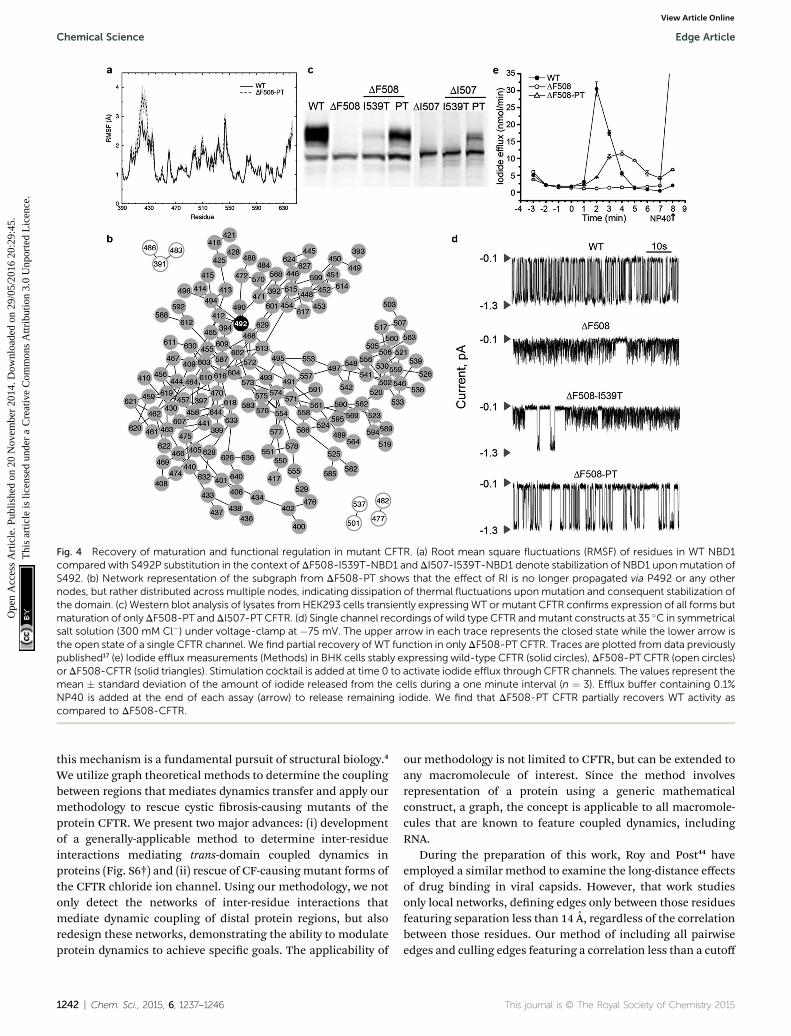
Fig. 4 Recovery of maturation and functional regulation in mutant CFTR. (a) Root mean square fluctuations (RMSF) of residues in WT NBD1compared with S492P substitution in the context of DF508-I539T-NBD1 and DI507-I539T-NBD1 denote stabilization of NBD1 uponmutation ofS492. (b) Network representation of the subgraph from DF508-PT shows that the effect of RI is no longer propagated via P492 or any othernodes, but rather distributed across multiple nodes, indicating dissipation of thermal fluctuations upon mutation and consequent stabilization ofthe domain. (c) Western blot analysis of lysates fromHEK293 cells transiently expressingWT or mutant CFTR confirms expression of all forms butmaturation of only DF508-PT and DI507-PT CFTR. (d) Single channel recordings of wild type CFTR andmutant constructs at 35 �C in symmetricalsalt solution (300 mM Cl�) under voltage-clamp at �75 mV. The upper arrow in each trace represents the closed state while the lower arrow isthe open state of a single CFTR channel. We find partial recovery of WT function in only DF508-PT CFTR. Traces are plotted from data previouslypublished17 (e) Iodide effluxmeasurements (Methods) in BHK cells stably expressing wild-type CFTR (solid circles), DF508-PT CFTR (open circles)or DF508-CFTR (solid triangles). Stimulation cocktail is added at time 0 to activate iodide efflux through CFTR channels. The values represent themean � standard deviation of the amount of iodide released from the cells during a one minute interval (n ¼ 3). Efflux buffer containing 0.1%NP40 is added at the end of each assay (arrow) to release remaining iodide. We find that DF508-PT CFTR partially recovers WT activity ascompared to DF508-CFTR.
Chemical Science Edge Article
Ope
n A
cces
s A
rtic
le. P
ublis
hed
on 2
0 N
ovem
ber
2014
. Dow
nloa
ded
on 2
9/05
/201
6 20
:29:
45.
Thi
s ar
ticle
is li
cens
ed u
nder
a C
reat
ive
Com
mon
s A
ttrib
utio
n 3.
0 U
npor
ted
Lic
ence
.View Article Online
this mechanism is a fundamental pursuit of structural biology.4
We utilize graph theoretical methods to determine the couplingbetween regions that mediates dynamics transfer and apply ourmethodology to rescue cystic brosis-causing mutants of theprotein CFTR. We present two major advances: (i) developmentof a generally-applicable method to determine inter-residueinteractions mediating trans-domain coupled dynamics inproteins (Fig. S6†) and (ii) rescue of CF-causing mutant forms ofthe CFTR chloride ion channel. Using our methodology, we notonly detect the networks of inter-residue interactions thatmediate dynamic coupling of distal protein regions, but alsoredesign these networks, demonstrating the ability to modulateprotein dynamics to achieve specic goals. The applicability of
1242 | Chem. Sci., 2015, 6, 1237–1246
our methodology is not limited to CFTR, but can be extended toany macromolecule of interest. Since the method involvesrepresentation of a protein using a generic mathematicalconstruct, a graph, the concept is applicable to all macromole-cules that are known to feature coupled dynamics, includingRNA.
During the preparation of this work, Roy and Post44 haveemployed a similar method to examine the long-distance effectsof drug binding in viral capsids. However, that work studiesonly local networks, dening edges only between those residuesfeaturing separation less than 14 A, regardless of the correlationbetween those residues. Our method of including all pairwiseedges and culling edges featuring a correlation less than a cutoff
This journal is © The Royal Society of Chemistry 2015

Edge Article Chemical Science
Ope
n A
cces
s A
rtic
le. P
ublis
hed
on 2
0 N
ovem
ber
2014
. Dow
nloa
ded
on 2
9/05
/201
6 20
:29:
45.
Thi
s ar
ticle
is li
cens
ed u
nder
a C
reat
ive
Com
mon
s A
ttrib
utio
n 3.
0 U
npor
ted
Lic
ence
.View Article Online
value gives the dual advantage of increased network coveragewith the inclusion of distal correlations and faster computationof shortest path, since weak connections are eliminated and arenot included in calculations of shortest path.
In the present study, we apply our computational method-ology to study dynamic coupling within NBD1 of CFTR. Weidentify hubs in NBD1 that mediate dynamic coupling betweenvarious sites across the protein (Fig. S2†), and nd that theresidue S492 alone mediates dynamic coupling between thedynamic RI region and the F508 loop where disease-relevantdeletion occurs, making this residue a hot spot for rationaldesign via mutagenesis. We further nd that mutation of S492to proline (an evolutionarily-conserved amino acid in thisposition among NBDs) successfully rescues DF508- and DI507-CFTR. To evaluate whether the rescue of CFTR upon S492Pmutation is due simply to thermodynamic stabilization, wecalculate the DDG of mutation at this position using Eris(Methods).42,43 Intriguingly, we nd that the thermodynamicstability of DF508-NBD1 is not improved by proline substitu-tion, nor by any amino acid substitution at this position,emphasizing the role of dynamic coupling within NBD1 inCFTR structural stability (Fig. S7†). The conservation of S492 inCFTR among various species further indicates an evolutionarybasis for an dynamic coupling mechanism of regulation ofNBD1 dynamics (Fig. S4†). Furthermore, we nd both compu-tationally and experimentally that mutagenesis of S492 in thecontext of I539T (referred to as DXXXX-PT), a previously iden-tied maturation reverting mutation,24 signicantly improvesthe dynamic stability, maturation, and function of CFTR dele-tionmutants. The biological signicance of the combined I539Tand S492P substitutions is also apparent in nature. Forinstance, some non-mammalian (e.g. Xenopus laevis and Squa-lus acanthias) CFTR orthologs naturally possess T539 and P492,and theDF508mutation does not compromise their folding andmaturation (Fig. S4 and S8†). Notably, mammalian species (e.g.Equus caballus and Ovis aries) do not contain these variations,and their folding and maturation is affected by F508 deletion(Fig. S4 and S8†).
In order to demonstrate the importance of dynamic behaviorin the trans-domain “communication” between various regions,for contrast we perform a similar analysis of the static structureof human NBD1, using the number of contacts made betweeneach pair of residues as the weight of each edge in the graph. Inthe graph of the static system, S492 does not appear in any ofthe paths between the RI and the F508 loop, further supportingthe idea that an understanding of protein dynamics is critical torational manipulation of proteins.
In the case of CF, the main cause of disease (approximately90% of cases) is the deletion of F508 from NBD1 of CFTR.17 Thedeletion of I507, the adjacent residue, also results in disease.45
We nd that deletion of these residues from the F508 loopsubstructure leads to non-native dynamic coupling interactionswithin NBD1. In DF508-NBD1, the deletion mutation causes aperturbation that decouples the RI from the rest of NBD1, andthe loss of coupling with the RI results in non-native couplingwithin the a-subdomain and the ATP-binding domain. InDI507-CFTR, the deletion of I507 changes the interactions of the
This journal is © The Royal Society of Chemistry 2015
mutation site with the SDR, from which perturbations affectcoupling throughout the domain. This difference explains whythe removal of RI rescues DF508-CFTR, but does not affectDI507-CFTR (Fig. S9†). However, in both CFTR constructs, theperturbed SDR plays a major role in mediating transfer ofaberrant dynamics from the mutation site. The substitution of aproline for a serine at position 492 in NBD1 stiffens the proteinbackbone and inhibits uctuations, inhibiting the couplingbetween the SDR and both the RI and the ATP-binding sub-domain, interrupting the transfer of non-native dynamics andrescuing both deletion mutants. Notably, the phenomenon oflost native coupling in the ATP-binding sub-domain of DI507-NBD1, but not in DF508-NBD1, can potentially explain why thelatter, but not the former, mutant can be rescued by decreasingthe temperature from 37 �C to 27 �C (Fig. S9†); because thebinding of ATP to the NBDs is known to be a signicant stabi-lizing factor in CFTR,40,41 the loss of coupling between thedifferent areas of the ATP-binding subdomain could interferewith the binding of this important ligand.
Given these ndings and conclusions, we are faced with thequestion: if the S492P mutation rescues both DF508- and DI507-CFTR, why do we not identify S492 in our computational anal-ysis of the DI507-NBD1 dynamic network? We note that coupledmotions are markedly lower in DI507-NBD1 as opposed toDF508, as evidenced by the much lower correlation coefficientcutoff in our networks (0.50 for DF508, 0.32 for DI507). Wehypothesize that the low level of correlation in the DI507-NBD1domain and sharp transition in node inclusion (Fig. S1†) makethe choice of network cutoff much more critical to networkproperties. We nd that S492 is indeed located in the bottleneckregion, in fact correlated (correlation coefficient 0.30, ascompared to the cutoff 0.32) with one of the identied bottle-neck residues, L568. We note that in using our method, anexpert researcher could manually ne-tune the correlationcoefficient cutoff based on prior knowledge or on regions orresidues of interest. Manual intervention may be particularlyuseful in systems such as DI507-NBD1, where the sharp tran-sition in network composition with correlation coefficientcutoff makes choice of this parameter particularly critical.
The sensitivity of the DI507-NBD1 system leads us toemphasize that our method serves as a guide to narrow the wideeld of possible rescue mutations, highlighting residues withpotential inuence over protein dynamics, and that not allidentied residues will be the site of viable rescue mutations,nor that all rescue mutation sites will be identied. Thecomputational methods that we employ necessarily are molec-ular in scale, and cannot integrate all cellular processes thatinuence the expression, maturation, and interactions ofparticular mutants. Also due to molecular detail, our compu-tational method is only applicable in cases where the structureof the protein or protein domain of interest has been solved tohigh resolution (<4 A). Lower resolution structures wouldrequire additional computational methods to model atomicresolution before our approach could be applied. Finally, thestructure must be of a size that is practical for extensivedynamic sampling during molecular simulations.
Chem. Sci., 2015, 6, 1237–1246 | 1243

Chemical Science Edge Article
Ope
n A
cces
s A
rtic
le. P
ublis
hed
on 2
0 N
ovem
ber
2014
. Dow
nloa
ded
on 2
9/05
/201
6 20
:29:
45.
Thi
s ar
ticle
is li
cens
ed u
nder
a C
reat
ive
Com
mon
s A
ttrib
utio
n 3.
0 U
npor
ted
Lic
ence
.View Article Online
MethodsNBD1 models and discrete molecular dynamics simulations
Crystal structures exist of wild type NBD1 (PDB ID: 2BBO) andDF508-NBD1 (PDB ID: 2BBT), but because of the inherent ex-ibility of the RI this region is not resolved in either structure. Wereconstruct the missing residues in the RI ab initio usingdiscrete molecular dynamics (DMD) simulations30 with theMedusa force eld.15 We perform multiple iterations of replicaexchange simulation and energetic minimization using Chi-ron46 to obtain nal minimized models of full-length wild typeand DF508-NBD1. No structure of DI507-NBD1 is currentlyavailable, therefore starting from our minimized model of full-length wild type NBD1, we manually delete the I507 residue andreseal the protein backbone using DMD simulations. We resealthe protein backbone by imposing peptide-bonding constraintson the cleaved ends until the backbone is once again intact. Wehold the majority of the protein static during this process,allowing only three residues on either side of the deletion tomove freely and reseal the gap caused by deletion. From thesethree initial full-length models (wild type, DF508, and DI507),we utilize the Eris suite42,43 to create S492P/I539T mutants ofeach construct. We perform energy minimization of allconstructs using Chiron.
We perform long timescale single-temperature DMD simu-lations using the nal minimized models of full-length NBD1.We perform 10 randomized simulations for each construct at atemperature of 0.4 kcal mol�1 kB
�1, with each individualsimulation having a length of 106 DMD time steps (approxi-mately 50 ns), summing to a total of approximately 500 ns foreach construct. The results that we present here are averagedover all simulations for each construct, unless otherwisespecied.
Construction of dynamic networks and determination ofoptimal paths
We utilize the resulting simulation trajectories to determinedynamic coupling between the various regions of NBD1 bycomputing correlation coefficients of motion between all resi-dues.32 We represent the pairwise correlation map as a completeweighted graph G(N,E), with the Ca atoms in NBD1 representingthe nodes (N) of the graph and the edges (E) being theconnections between these nodes. The edge weight Eij betweenany two nodes i and j is the correlation coefficient Cij betweenthe corresponding pair of residues.
To determine optimal paths of dynamics transfer throughcoupled residues, we use the same network with edge weights ofEij ¼ 1 � |Cij| and apply Dijkstra's algorithm.47 Since dynamiccoupling is mediated by physical interactions, we reduce thecomplete graph by removing edges between nodes representingresidues that are not in contact. We consider two residues to bein contact if their Cb atoms (Ca for Glycine) are within 7.5 A ofone another. Our aim is to determine the transfer of correlatedmotion across the protein. Hence, we emphasize the effect oflocal interactions by retaining only those edges in which the
1244 | Chem. Sci., 2015, 6, 1237–1246
participating nodes feature a contact frequency (uc) of at least0.5 over the simulation.
Flexible backbone redesign
We perform iterative exible backbone redesign and structuralrelaxation using the Medusa suite15 in order to ensure optimumbackbone conguration for computational mutagenesis ofhuman NBD. We perform 15 randomly-seeded iterations ofbackbone redesign Monte Carlo simulations with 20 MonteCarlo iterations per replicate. We select the lowest energystructure from each iteration to use as the starting point for thenext iteration. Finally, we choose the structure with lowestenergy and smallest Kabsch root mean square deviation(KRMSD) from the initial structure for xed-backbone mutationanalysis using the Eris suite.42,43
CFTR construction and expression
We express human CFTR cDNAs encoding wild type or mutantproteins transiently in HEK 293 cells or stably in BHK-21 cells,with pcDNA3 or pNUT vectors, respectively, as previouslydescribed.28 We use the QuickExchange protocol (Stratagene) togenerate mutant CFTR constructs in pcDNA3 and pNUT vectorsfrom human WT CFTR cDNA, and conrm sequences by auto-mated DNA sequencing (UNC-CH Genome Analysis Facility).We carry out transfection using jetPEI transfection reagent(Fermentas, Glen Burnie, MD) according to the manufacturer'sinstructions. For stable cell line establishment, we select andmaintain BHK cells expressing CFTR in methotrexate-contain-ing media as previously described.48
Western blotting
We harvest HEK or BHK cells overexpressing CFTR in radio-immunoprecipitation assay (RIPA) buffer without SDS (50 mMTris, 150 mM NaCl, 1% Triton X-100, 1% deoxycholate, pH 7.4)plus protease inhibitor cocktail (1 mg ml�1 leupeptin, 2 mg ml�1
aprotinin, 3.57 mg ml�1 E64, 156.6 mg ml�1 benzamidine and 2mM Pefablock). We subject equal amounts of proteins in SDS-PAGE sample buffer to 7.5% SDS-PAGE and Western blotanalysis with mAb596 in order to determine CFTR expressionand maturation.49
Membrane isolation
We harvest BHK or HEK 293 cells expressing CFTR or its vari-ants by scraping, and then homogenize the cells on ice in 10mM Hepes, pH 7.2, 1 mM EDTA (ethylenediaminetetraaceticacid) containing a protease inhibitor cocktail (benzamidine at120 mg ml�1, E64 at 3.5 mg ml�1, aprotinin at 2 mg ml�1, leu-peptin at 1 mg ml�1 and Pefablock at 50 mg ml�1). We centrifugethe resulting samples at 600 g for 15 minutes to remove nucleiand undisrupted cells, followed by centrifugation at 100 000gfor 60 minutes to pellet the membranes, which we then resus-pend in phosphorylation buffer (10 mM Hepes, pH 7.2, con-taining 0.5 mM EGTA (ethylene glycol bis(b-aminoethyl ether),N,N0-tetraacetic acid), 2 mM MgCl2, and 250 mM sucrose). Weutilize brief (3 � 20 s) bath sonications to generate vesicles of
This journal is © The Royal Society of Chemistry 2015

Edge Article Chemical Science
Ope
n A
cces
s A
rtic
le. P
ublis
hed
on 2
0 N
ovem
ber
2014
. Dow
nloa
ded
on 2
9/05
/201
6 20
:29:
45.
Thi
s ar
ticle
is li
cens
ed u
nder
a C
reat
ive
Com
mon
s A
ttrib
utio
n 3.
0 U
npor
ted
Lic
ence
.View Article Online
uniform size. For single-channel recordings, we phosphorylatemembrane vesicles by incubating with 50 nM PKA catalyticsubunit (Promega) and 2 mM Na2ATP (Sigma) in phosphoryla-tion buffer for 20 min at +4 �C. We then aliquot the membranesand store at �80 �C for later use.
Single-channel measurements
To prepare planar lipid bilayers, we drill a 0.2 mm hole in aTeon cup and paint the hole with a phospholipid solutioncontaining a 3 : 1 mixture of 1-palmitoyl-2-oleoyl-sn-glycero-3-phosphoethanolamine and 1-palmitoyl-2-oleoyl-sn-glycero-3-phosphoserine (Avanti Polar Lipids) in n-decane. The lipidbilayer separates 1 ml of solution in the Teon cup (cis side)from 5 ml of solution in the outer glass chamber (trans side).Both chambers are magnetically stirred and thermally insu-lated. We utilize a temperature control system (TC2BIP, CellMicro Controls).
We transfer CFTR ion channels into the pre-formed lipidbilayer by spontaneous fusion of membrane vesicles containingthe CFTR variants. To maintain uniform orientation and func-tional activity of the CFTR channels transferred into the bilayer,we add 2 mM ATP, 50 nM PKA, and membrane vesicles into thecis compartment only. We perform all measurements insymmetrical salt solution (300 mM Tris–HCl, pH 7.2, 3 mMMgCl2 and 1 mM EGTA) under voltage-clamp conditions usingan Axopatch 200B amplier. We maintain a membrane voltagepotential of �75 mV, the difference between cis and trans(ground) compartments. We analyze the resulting data aspreviously described.50
Iodide efflux assay
We grow BHK cells stably expressing wild type and mutantCFTR to�100% conuence in six-well plates and incubate in aniodide loading buffer (136 mM NaI, 3 mM KNO3, 2 mMCa(NO3)2, 11 mM glucose, and 20 mM Hepes, pH 7.4) for onehour at room temperature. We rinse the cells with iodide-freeefflux buffer (which is the same as the loading buffer except thatNaI is replaced by NaNO3) to remove extracellular iodide. Wecollect samples by completely replacing the efflux buffer (1 mlvolume) with fresh solution at one-minute intervals. We useresults from the rst four samples to establish a baseline. Wemeasure iodide efflux upon stimulation with PKA agonists (10mM forskolin, 100 mM dibutyl-cAMP and 1 mM 3-isobutyl-1-methylxanthine) using an iodide-selective electrode LIS-1461CM (Lazar Res. Lab., Inc.), as previously described.48
Conclusions
We demonstrate here that NBD1 of CFTR, for which trans-domain coupled dynamics was not previously described,features patterns of correlated motions that form a networkthroughout the domain and allow structural uctuations to betransferred to distal sites through dynamic coupling, support-ing the general notion that all folded proteins are to some extentallosteric in nature.3 Moreover, we show that these networks canbe rationally redesigned to affect a desired outcome. Identifying
This journal is © The Royal Society of Chemistry 2015
the residues involved in trans-domain dynamics can provide amap to greatly improve the stability and/or modulate thefunction of enzymes for numerous biotechnological applica-tions, as well as contribute to our understanding of the manyhuman diseases caused by protein dysfunction. Determiningdynamic coupling networks and hot spots is also of practicalimportance in rational drug design.3 Conventionally, structure-based virtual screening utilizes experimentally-validatedbinding sites, such as an enzyme catalytic site or a ligand-boundpocket, but the determination of pre-existing dynamiccommunication pathways has great potential for identifyingnovel “druggable” sites that can be targeted to modulate proteinfunction in human diseases. Thus, beyond the signicantadvance in understanding the fundamental defect in cysticbrosis, our approach has broad application in the elucidationof mechanisms of protein function and dysfunction in disease.
Acknowledgements
We thank Dr Feng Ding and Dr Srinivas Ramachandran forinsightful suggestions and discussions, and Rachel Cohen forexpression and testing of DI507-CFTR constructs. This work wassupported by grants from the National Institutes of Health (toNVD: GM080742, and to JRR: DK051619) and the Cystic FibrosisFoundation. EAP was supported by a predoctoral fellowshipfrom the National Institutes of Health National Institute onAging (F31AG039266).
Notes and references
1 J.-P. Changeux and S. J. Edelstein, Science, 2005, 308, 1424–1428.
2 N. M. Goodey and S. J. Benkovic, Nat. Chem. Biol., 2008, 4,474–482.
3 K. Gunasekaran, B. Ma and R. Nussinov, Proteins, 2004, 57,433–443.
4 C.-J. Tsai and R. Nussinov, PLoS Comput. Biol., 2014, 10,e1003394.
5 N. Popovych, S. Sun, R. H. Ebright and C. G. Kalodimos, Nat.Struct. Mol. Biol., 2006, 13, 831–838.
6 E. J. Fuentes, S. A. Gilmore, R. V. Mauldin and A. L. Lee, J.Mol. Biol., 2006, 364, 337–351.
7 J. A. Hardy and J. A. Wells, Curr. Opin. Struct. Biol., 2004, 14,706–715.
8 S.-R. Tzeng and C. G. Kalodimos, Nature, 2009, 462, 368–372.9 G. Schreiber and A. R. Fersht, J. Mol. Biol., 1995, 248, 478–486.
10 E. A. Proctor, P. Kota, S. J. Demarest, J. A. Caravella andN. V. Dokholyan, Proteins, 2013, 81, 884–895.
11 S. W. Lockless and R. Ranganathan, Science, 1999, 286, 295–299.
12 M. Socolich, S. W. Lockless, W. P. Russ, H. Lee,K. H. Gardner and R. Ranganathan, Nature, 2005, 437,512–518.
13 G. M. Suel, S. W. Lockless, M. A. Wall and R. Ranganathan,Nat. Struct. Biol., 2003, 10, 59–69.
Chem. Sci., 2015, 6, 1237–1246 | 1245
Chemical Science Edge Article
Ope
n A
cces
s A
rtic
le. P
ublis
hed
on 2
0 N
ovem
ber
2014
. Dow
nloa
ded
on 2
9/05
/201
6 20
:29:
45.
Thi
s ar
ticle
is li
cens
ed u
nder
a C
reat
ive
Com
mon
s A
ttrib
utio
n 3.
0 U
npor
ted
Lic
ence
.View Article Online
14 W. Zheng, B. R. Brooks and D. Thirumalai, Proc. Natl. Acad.Sci. U. S. A., 2006, 103, 7664–7669.
15 F. Ding and N. V. Dokholyan, PLoS Comput. Biol., 2006, 2,e85.
16 N. V. Dokholyan and E. I. Shakhnovich, J. Mol. Biol., 2001,312, 289–307.
17 J. R. Riordan, J. M. Rommens, B. Kerem, N. Alon,R. Rozmahel, Z. Grzelczak, J. Zielenski, S. Lok, N. Plavsic,J. L. Chou, et al., Science, 1989, 245, 1066–1073.
18 A. A. Aleksandrov, P. Kota, L. Cui, T. Jensen, A. E. Alekseev,S. Reyes, L. He, M. Gentzsch, L. A. Aleksandrov,N. V. Dokholyan and J. R. Riordan, J. Mol. Biol., 2012, 419,41–60.
19 S. H. Cheng, R. J. Gregory, J. Marshall, S. Paul, D. W. Souza,G. A. White, C. R. O'Riordan and A. E. Smith, Cell, 1990, 63,827–834.
20 M. L. Drumm, D. J. Wilkinson, L. S. Smit, R. T. Worrell,T. V. Strong, R. A. Frizzell, D. C. Dawson and F. S. Collins,Science, 1991, 254, 1797–1799.
21 C. Li, M. Ramjeesingh, E. Reyes, T. Jensen, X. Chang,J. M. Rommens and C. E. Bear, Nat. Genet., 1993, 3, 311–316.
22 G. M. Denning, M. P. Anderson, J. F. Amara, J. Marshall,A. E. Smith and M. J. Welsh, Nature, 1992, 358, 761–764.
23 M. E. Egan, E. M. Schwiebert and W. B. Guggino, Am. J.Physiol., 1995, 268, C243–C251.
24 A. C. V. DeCarvalho, L. J. Gansheroff and J. L. Teem, J. Biol.Chem., 2002, 277, 35896–35905.
25 H. Hoelen, B. Kleizen, A. Schmidt, J. Richardson,P. Charitou, P. J. Thomas and I. Braakman, PLoS One,2010, 5, e15458.
26 A. A. Aleksandrov, P. Kota, L. A. Aleksandrov, L. He,T. Jensen, L. Cui, M. Gentzsch, N. V. Dokholyan andJ. R. Riordan, J. Mol. Biol., 2010, 401, 194–210.
27 A. W. Serohijos, T. Hegedus, A. A. Aleksandrov, L. He, L. Cui,N. V. Dokholyan and J. R. Riordan, Proc. Natl. Acad. Sci. U. S.A., 2008, 105, 3256–3261.
28 L. He, A. A. Aleksandrov, A. W. Serohijos, T. Hegedus,L. A. Aleksandrov, L. Cui, N. V. Dokholyan andJ. R. Riordan, J. Biol. Chem., 2008, 283, 26383–26390.
29 H. A. Lewis, C. Wang, X. Zhao, Y. Hamuro, K. Conners,M. C. Kearins, F. Lu, J. M. Sauder, K. S. Molnar,S. J. Coales, P. C. Maloney, W. B. Guggino, D. R. Wetmore,P. C. Weber and J. F. Hunt, J. Mol. Biol., 2010, 396, 406–430.
30 F. Ding, D. Tsao, H. Nie and N. V. Dokholyan, Structure, 2008,16, 1010–1018.
1246 | Chem. Sci., 2015, 6, 1237–1246
31 N. V. Dokholyan, S. V. Buldyrev, H. E. Stanley andE. I. Shakhnovich, Fold Des, 1998, 3, 577–587.
32 S. Sharma, F. Ding and N. V. Dokholyan, Biophys. J., 2007, 92,1457–1470.
33 N. V. Dokholyan, B. Shakhnovich and E. I. Shakhnovich,Proc. Natl. Acad. Sci., 2002, 99, 14132–14136.
34 N. V. Dokholyan, Y. Lee, S. V. Buldyrev, S. Havlin, P. R. Kingand H. E. Stanley, J. Stat. Phys., 1998, 93, 603–613.
35 J. E. Moody, L. Millen, D. Binns, J. F. Hunt and P. J. Thomas,J. Biol. Chem., 2002, 277, 21111–21114.
36 N. Nishimura and W. E. Balch, Science, 1997, 277, 556–558.37 X. Wang, J. Matteson, Y. An, B. Moyer, J.-S. Yoo, S. Bannykh,
I. A. Wilson, J. R. Riordan and W. E. Balch, J. Cell Biol., 2004,167, 65–74.
38 J. L. Teem, H. A. Berger, L. S. Ostedgaard, D. P. Rich,L. C. Tsui and M. J. Welsh, Cell, 1993, 73, 335–346.
39 J. L. Teem, M. R. Carson and M. J. Welsh, Recept. Channels,1996, 4, 63–72.
40 I. Protasevich, Z. Yang, C. Wang, S. Atwell, X. Zhao,S. Emtage, D. Wetmore, J. F. Hunt and C. G. Brouillette,Protein Sci., 2010, 19, 1917–1931.
41 C. Wang, I. Protasevich, Z. Yang, D. Seehausen, T. Skalak,X. Zhao, S. Atwell, J. Spencer Emtage, D. R. Wetmore,C. G. Brouillette and J. F. Hunt, Protein Sci., 2010, 19,1932–1947.
42 S. Yin, F. Ding and N. V. Dokholyan, Structure, 2007, 15,1567–1576.
43 S. Yin, F. Ding and N. V. Dokholyan, Nat. Methods, 2007, 4,466–467.
44 A. Roy and C. B. Post, Proc. Natl. Acad. Sci., 2012, 109, 5271–5276.
45 M. Schwarz, C. Summers, L. Heptinstall, C. Newton,A. Markham and M. Super, Adv. Exp. Med. Biol., 1991, 290,393–398.
46 S. Ramachandran, P. Kota, F. Ding and N. V. Dokholyan,Proteins, 2011, 79, 261–270.
47 E. W. Dijkstra, Numer. Math., 1959, 1, 269–271.48 X. B. Chang, J. A. Tabcharani, Y. X. Hou, T. J. Jensen,
N. Kartner, N. Alon, J. W. Hanrahan and J. R. Riordan, J.Biol. Chem., 1993, 268, 11304–11311.
49 L. Cui, L. Aleksandrov, Y.-X. Hou, M. Gentzsch, J.-H. Chen,J. R. Riordan and A. A. Aleksandrov, J. Physiol., 2006, 572,347–358.
50 A. A. Aleksandrov, L. Cui and J. R. Riordan, J. Physiol., 2009,587, 2875–2886.
This journal is © The Royal Society of Chemistry 2015
Copyright © 2022 FDOKUMEN