Transmembrane Domain Determinants of CD4 Downregulation by HIV-1 Vpu
17
Published Ahead of Print 16 November 2011. 2012, 86(2):757. DOI: 10.1128/JVI.05933-11. J. Virol. Javier G. Magadán and Juan S. Bonifacino CD4 Downregulation by HIV-1 Vpu Transmembrane Domain Determinants of http://jvi.asm.org/content/86/2/757 Updated information and services can be found at: These include: SUPPLEMENTAL MATERIAL http://jvi.asm.org/content/suppl/2011/12/16/86.2.757.DC1.html REFERENCES http://jvi.asm.org/content/86/2/757#ref-list-1 at: This article cites 86 articles, 40 of which can be accessed free CONTENT ALERTS more» articles cite this article), Receive: RSS Feeds, eTOCs, free email alerts (when new http://journals.asm.org/site/misc/reprints.xhtml Information about commercial reprint orders: http://journals.asm.org/site/subscriptions/ To subscribe to to another ASM Journal go to: on July 12, 2012 by NAT INST OF HEALTH LIB http://jvi.asm.org/ Downloaded from
-
Upload
independent -
Category
Documents
-
view
0 -
download
0
Transcript of Transmembrane Domain Determinants of CD4 Downregulation by HIV-1 Vpu
Published Ahead of Print 16 November 2011. 2012, 86(2):757. DOI: 10.1128/JVI.05933-11. J. Virol.
Javier G. Magadán and Juan S. Bonifacino CD4 Downregulation by HIV-1 VpuTransmembrane Domain Determinants of
http://jvi.asm.org/content/86/2/757Updated information and services can be found at:
These include:
SUPPLEMENTAL MATERIAL http://jvi.asm.org/content/suppl/2011/12/16/86.2.757.DC1.html
REFERENCEShttp://jvi.asm.org/content/86/2/757#ref-list-1at:
This article cites 86 articles, 40 of which can be accessed free
CONTENT ALERTS more»articles cite this article),
Receive: RSS Feeds, eTOCs, free email alerts (when new
http://journals.asm.org/site/misc/reprints.xhtmlInformation about commercial reprint orders: http://journals.asm.org/site/subscriptions/To subscribe to to another ASM Journal go to:
on July 12, 2012 by NA
T IN
ST
OF
HE
ALT
H LIB
http://jvi.asm.org/
Dow
nloaded from
Transmembrane Domain Determinants of CD4 Downregulation byHIV-1 Vpu
Javier G. Magadán and Juan S. Bonifacino
Cell Biology and Metabolism Program, Eunice Kennedy Shriver National Institute of Child Health and Human Development, National Institutes of Health,Bethesda, Maryland, USA
The transmembrane domains (TMDs) of integral membrane proteins do not merely function as membrane anchors but playactive roles in many important biological processes. The downregulation of the CD4 coreceptor by the Vpu protein of HIV-1 is aprime example of a process that is dependent on specific properties of TMDs. Here we report the identification of Trp22 in theVpu TMD and Gly415 in the CD4 TMD as critical determinants of Vpu-induced targeting of CD4 to endoplasmic reticulum(ER)-associated degradation (ERAD). The two residues participate in different aspects of ERAD targeting. Vpu Trp22 is requiredto prevent assembly of Vpu into an inactive, oligomeric form and to promote CD4 polyubiquitination and subsequent recruit-ment of the VCP-UFD1L-NPL4 dislocase complex. In the presence of a Vpu Trp22 mutant, CD4 remains integrally associatedwith the ER membrane, suggesting that dislocation from the ER into the cytosol is impaired. CD4 Gly415, on the other hand,contributes to CD4-Vpu interactions. We also identify two residues, Val20 and Ser23, in the Vpu TMD that mediate retention ofVpu and, by extension, CD4 in the ER. These findings highlight the exploitation of several TMD-mediated mechanisms by HIV-1Vpu in order to downregulate CD4 and thus promote viral pathogenesis.
Human immunodeficiency virus type 1 (HIV-1) targets helperT cells and macrophages/monocytes through interaction of
the viral envelope glycoprotein (Env) with a combination of theCD4 and CCR5/CXCR4 cell surface receptors (59). Once insidethe cell, the genetic material of the virus directs rapid and sus-tained downregulation of CD4 (26, 62), a phenomenon that pro-motes viral spread by preventing superinfection, enabling the re-lease of progeny virions, and interfering with the host immuneresponse (3, 6, 34, 53). The ability of HIV-1 to downregulate CD4depends on two accessory proteins encoded in the viral genome,Nef and Vpu (32, 38, 45, 61). Nef is a myristoylated protein thatattaches to the cytosolic leaflet of the plasma membrane, where itfunctions to link the cytosolic tail of CD4 to the clathrin-associated adaptor protein 2 (AP-2) complex (1, 12, 16, 17, 19, 24,39). These interactions lead to rapid internalization of cell surfaceCD4 by a clathrin-dependent pathway (14, 16, 30, 60). Nef exertsa second function in endosomes, namely, the targeting of inter-nalized CD4 to the multivesicular body pathway for eventual deg-radation in lysosomes (20).
Vpu is a type III integral membrane protein that, in contrast toNef, acts on newly synthesized CD4, causing its retention in theendoplasmic reticulum (ER) (42) and subsequent delivery to theER-associated degradation (ERAD) pathway (7, 42, 63, 79).The mechanism of CD4 downregulation by Vpu involves a phys-ical interaction of the cytosolic domains of both proteins (11, 47).A phosphoserine (pS)-based motif, DpSGxxpS, in the cytosolicdomain of Vpu then acts as a binding site for the �-TrCP1/2 com-ponent of the SCF�-TrCP1/2 E3 ubiquitin (Ub)-ligase complex (15,25, 48), which in turn mediates lysine- and serine/threonine-dependent polyubiquitination of the CD4 cytosolic tail (42).Polyubiquitination partly arrests CD4 in the ER while enablingrecruitment of the VCP-UFD1L-NPL4 dislocase complex, whichextracts CD4 from the ER membrane into the cytosol (7, 42).Dislocated CD4 is subsequently delivered to the proteasome fordegradation (7, 42, 63).
Although most studies on Vpu-induced CD4 downregulation
have focused on reactions involving the cytosolic domains of theproteins, some studies have also implicated the correspondingtransmembrane domains (TMDs) in this process (13, 42, 57, 73).Indeed, replacement of the vesicular stomatitis virus G glycopro-tein (VSV-G) TMD for the CD4 TMD abolished Vpu-induceddegradation of the resulting chimeric protein (13). Moreover, werecently showed that substitution of the VSV-G TMD for the VpuTMD abrogated both CD4 retention in the ER and ERAD target-ing induced by Vpu (42). These findings indicate that the TMDs ofboth CD4 and Vpu play important roles in the process of Vpu-induced CD4 downregulation.
In this study, we have examined the specific features of the Vpuand CD4 TMDs that are required for CD4 downregulation. Wereport the presence of a cluster of amino acids on the Vpu TMD,centered on Trp22, which is critical for Vpu-mediated CD4 deg-radation. Mutation of Trp22 does not prevent interaction of Vpuwith CD4 but enhances Vpu oligomerization and also reducesCD4 polyubiquitination and recruitment of the VCP-UFD1L-NPL4 dislocase complex. In the presence of a Vpu Trp22 mutant,CD4 remains integrated into the ER membrane, suggesting thatdislocation from the ER to the cytosol is impaired. We also definean additional determinant comprising Gly415 in the CD4 TMDthat is required for both Vpu-CD4 interaction and induction ofCD4 degradation by Vpu. Finally, we identify Val20 and Ser23 inthe Vpu TMD as components of a separate determinant of Vpuand CD4 retention in the ER. These findings highlight the crucial
Received 8 August 2011 Accepted 3 November 2011
Published ahead of print 16 November 2011
Address correspondence to Juan S. Bonifacino, [email protected].
Supplemental material for this article may be found at http://jvi.asm.org/.
Copyright © 2012, American Society for Microbiology. All Rights Reserved.
doi:10.1128/JVI.05933-11
0022-538X/12/$12.00 Journal of Virology p. 757–772 jvi.asm.org 757
on July 12, 2012 by NA
T IN
ST
OF
HE
ALT
H LIB
http://jvi.asm.org/
Dow
nloaded from
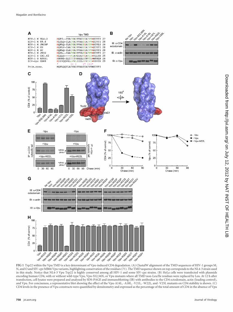
FIG 1 Trp22 within the Vpu TMD is a key determinant of Vpu-induced CD4 degradation. (A) ClustalW alignment of the TMD sequences of HIV-1 groups M,N, and O and SIV-cpz MB66 Vpu variants, highlighting conservation of the residues (71). The TMD sequence shown on top corresponds to the NL4-3 strain usedin this study. Notice that NL4-3 Vpu Trp22 is highly conserved among all HIV-1 and some SIV-cpz strains. (B) HeLa cells were transfected with plasmidsencoding human CD4, with or without wild-type Vpu, Vpu-S52,56N, or Vpu mutants where all TMD non-Leu/Ile residues were replaced by Leu. At 12 h aftertransfection, cell lysates were prepared and analyzed by SDS-PAGE and immunoblotting (IB) with antibodies to the CD4 ectodomain, actin (loading control),and Vpu. For conciseness, a representative blot showing the effect of the Vpu-A14L, -A18L, -V21L, -W22L, and -V25L mutants on CD4 stability is shown. (C)CD4 levels in the presence of Vpu constructs were quantified by densitometry and expressed as the percentage of the total amount of CD4 in the absence of Vpu
Magadán and Bonifacino
758 jvi.asm.org Journal of Virology
on July 12, 2012 by NA
T IN
ST
OF
HE
ALT
H LIB
http://jvi.asm.org/
Dow
nloaded from
requirement of specific TMD residues at several steps in the mech-anisms of CD4 downregulation by Vpu.
MATERIALS AND METHODSRecombinant DNA constructs. pFLAG-CMV2-human �-TrCP1 was agift from Y. Ben-Neriah (Hadassah Medical School, Hebrew University).pcDNA3.1-FLAG-Ub was already described (42). Site-directed mutagen-esis (QuikChange II kit; Stratagene, Cedar Creek, TX) was performed onpcDNA3.1-codon-optimized HIV-1 NL4-3 Vpu (54) and pCMV-humanCD4 (11). A nucleotide sequence encoding a hemagglutinin (HA) epitopewas also added to the 3= terminus of the codon-optimized Vpu cDNA bysite-directed mutagenesis. Alternatively, the codon-optimized Vpu cDNA(without a stop codon) was amplified by PCR from the pcDNA3.1-codon-optimized Vpu construct and cloned as an EcoRI/XhoI fragment into thepcDNA3.1/myc-His A vector (Invitrogen, Carlsbad, CA). All mutagenesisand cloning products were verified by DNA sequencing.
Antibodies. The following mouse monoclonal antibodies were used:anti-human CD4, clone 4B12 (Leica Microsystems, Bannockburn, IL)and clone OKT4 (eBiosciences, San Diego, CA); anti-actin (clone Ab-5)and anti-VCP (clone 18) (BD Biosciences, San Jose, CA); anti-humantransferrin receptor (TfR), clone H68.4 (Zymed, San Francisco, CA); un-conjugated/horseradish peroxidase (HRP)-conjugated anti-FLAG (cloneM2) and anti-myc (clone 9E10) (Sigma-Aldrich, Saint Louis, MO); anti-HA, clone 16B12 (Covance, Berkeley, CA); and anti-RGS-His (Qiagen,Valencia, CA). Rabbit polyclonal antibodies to the human CD4 (residues420 to 447) and Vpu (residues 32 to 81) were previously described (44,63). Goat polyclonal antibody to GAPDH (glyceraldehyde-3-phosphatedehydrogenase) and HRP-conjugated donkey anti-goat IgG were pur-chased from Santa Cruz Biotechnology (Santa Cruz, CA). HRP-conjugated donkey anti-mouse IgG and donkey anti-rabbit IgG werefrom GE Healthcare Biosciences (Piscataway, NJ).
Cell culture and transfections. Media and reagents for cell culturewere purchased from Mediatech, Inc. (Manassas, VA). HeLa cells (Amer-ican Type Culture Collection, Manassas, VA) were grown in Dulbecco’smodified Eagle’s medium, supplemented with 10% heat-inactivated fetalbovine serum, 100 U/ml penicillin, 100 �g/ml streptomycin and 2 mML-glutamine at 37°C in a humidified atmosphere of 5% carbon dioxide inair. Cells were transiently transfected using Lipofectamine 2000 (Invitro-gen). Plasmids encoding human CD4, codon-optimized Vpu, andFLAG-Ub were transfected at a 1:1:0.5 ratio (42). Cells were analyzed at 8to 16 h after transfection.
Pulse-chase analysis, immunoprecipitation, electrophoresis,and immunoblotting. Cells were pulse-labeled with [35S]methionine-cysteine and chased in complete medium, and cell lysates were subjectedto immunoprecipitation as described previously (9). Immunoprecipi-tated proteins were analyzed by SDS-PAGE (42) and visualized by fluo-rography on a Typhoon 9200 PhosphorImager (Amersham Biosciences).Data analysis and quantification were performed using the ImageQuantsoftware (GE Healthcare). Immunoblotting studies were carried out asreported previously (42) and analyzed using the Image J software (http://rsbweb.nih.gov/ij).
In vivo ubiquitination. Cells expressing FLAG-tagged Ub were ex-tracted in denaturing lysis buffer as described previously (42). Ubiquiti-nated CD4 was immunoprecipitated using a conformation-independentantibody to CD4 and analyzed by immunoblotting using an HRP-conjugated antibody to the FLAG epitope (42).
Fractionation of permeabilized cells. Cells grown on 10-cm plateswere detached with trypsin, washed twice with ice-cold phosphate-buffered saline (PBS) supplemented with 0.9 mM CaCl2, and permeabil-ized in ice-cold permeabilization buffer (PB) (25 mM HEPES [pH 7.3],115 mM potassium acetate, 5 mM sodium acetate, 2.5 mM MgCl2, 0.5mM EGTA) containing 10 mM �-iodoacetamide, 5 mM N-ethyl-maleimide, 0.028% digitonin, and the complete Mini protease inhibitorcocktail (Roche Diagnostics, Indianapolis, IN) for 5 min on ice. Cytosolwas extracted from permeabilized cells by centrifugation at 20,000 � g for10 min at 4°C. The pellet was resuspended in ice-cold PB, and all fractionswere adjusted to 1% SDS and 10 mM dithiothreitol (DTT). Samples wereheated for 10 min at 100°C and processed for in vivo ubiquitination aspreviously described (42).
Isolation of microsomes. Cells (5 � 106) were scraped off the platesand incubated with ice-cold 20 mM HEPES (pH 7.2), 10 mM�-iodoacetamide, 5 mM N-ethylmaleimide, and the complete Mini pro-tease inhibitor cocktail for 10 min before passage through a 1-ml syringefitted with a 21-gauge needle (�20 strokes). Cell lysates were supple-mented with 5 mM MgCl2 and 100 mM potassium acetate and then se-quentially centrifuged at 600 � g for 2 min and 15,000 � g for 10 min at4°C. The resulting supernatants were ultracentrifuged at 200,000 � g for 1h at 4°C, and pelleted microsomes were resuspended in membrane buffer(50 mM HEPES [pH 7.4], 150 mM potassium acetate, 10 mM magnesiumacetate, 250 mM sucrose, 10 mM �-iodoacetamide, 5 mM N-ethyl-maleimide) supplemented (or not) with 100 mM Na2CO3, pH 11.3. Thesamples were fractionated into soluble and microsomal membrane frac-tions by ultracentrifugation at 200,000 � g for 10 min at 4°C. All fractionswere adjusted to 1% SDS and 10 mM dithiothreitol (DTT), heated for 10min at 100°C, and processed for in vivo ubiquitination as described pre-viously (42).
Cell surface biotinylation. Cells grown on 6-well plates were incu-bated with 2 mM sulfo-NHS-LC biotin (Thermo Scientific, Rockford, IL)in ice-cold PBS supplemented with 0.1 mM CaCl2 and 1 mM MgCl2 for 30min at 4°C. After extensive washing, cells were treated with 50 mM Tris-HCl (pH 7.5) for 10 min at 4°C and then extracted with ice-cold lysisbuffer (0.5% Triton X-100, 50 mM Tris-HCl [pH 7.5], 150 mM NaCl, 5mM EDTA) supplemented with the complete Mini protease inhibitorcocktail. Equivalent amounts of cell lysates were incubated with aNeutrAvidin-coupled resin (Thermo Scientific) for 2 h at 4°C. Pulled-down biotinylated proteins or whole-cell lysates (10 �g of protein) weresubjected to SDS-PAGE and immunoblotting with antibodies to CD4,Vpu, and TfR (loading control) as previously reported (42).
Endo H and PNGase F digestion. Cell lysates made under denaturingconditions were processed for endoglycosidase H (endo H) and peptide:N-glycosidase F (PNGase F) digestion as described previously (42).
RESULTSMutational analysis of the Vpu TMD identifies Trp22 as a criti-cal determinant for induction of CD4 degradation. The aminoacid sequence of the Vpu TMD is highly conserved among strainsof the pandemic HIV-1 group M, which account for more than90% of all HIV-1 infections (4, 33) (Fig. 1A). To determine therequirement for specific amino acid residues within this TMD forinduction of CD4 downregulation, we mutated each residue otherthan Leu/Ile to Leu in the context of codon-optimized Vpu from
(100% control). Values represent the means � standard errors of the means (SEM) from three independent experiments. (D) PyMOL images of the solid-stateNMR structure of the NL4-3 Vpu TMD (56) (RSCB protein database entry 1PJE). Val21, Trp22, and Val25 (red) form a patch on the same face of the Vpu TMD�-helix. (E) HeLa cells expressing human CD4, with or without wild-type Vpu or Vpu-W22L, were labeled with [35S]methionine-cysteine for 2 min and thenchased for the indicated times at 37°C. Cells extracts were subjected to immunoprecipitation using antibodies to the CD4 or Vpu cytosolic tails. Immunopre-cipitated proteins were analyzed by SDS-PAGE and fluorography. (F) Percentage of CD4 or Vpu at each chase time relative to CD4 at time zero (100% control).(G) HeLa cells were transfected with plasmids encoding human CD4 and no Vpu (empty vector), wild-type Vpu, or an array of Vpu mutants where Trp22 wasreplaced by all amino acids. Cell lysates were treated as for panel B. (H) CD4 levels in the presence of Vpu were quantified as for panel C.
Transmembrane Determinants of CD4 Downregulation
January 2012 Volume 86 Number 2 jvi.asm.org 759
on July 12, 2012 by NA
T IN
ST
OF
HE
ALT
H LIB
http://jvi.asm.org/
Dow
nloaded from
the HIV-1 group M strain NL4-3 (54) (Fig. 1A). These mutants,along with wild-type Vpu and an inactive Vpu mutant (Vpu-S52,56N) bearing substitutions of the Ser52 and Ser56 residuesthat mediate interaction with �-TrCP1/2 (64) (positive and neg-ative controls, respectively), were coexpressed with human CD4by transient transfection of HeLa cells. The ability of the differentVpu constructs to induce CD4 degradation was determined byimmunoblot analysis of total CD4 levels (42) (Fig. 1B). As ex-pected, wild-type Vpu induced CD4 degradation whereas Vpu-S52,56N did not (Fig. 1B and C). Most mutations in the Vpu TMDhad no effect on Vpu-mediated CD4 degradation (Fig. 1B and Cand unpublished observations). However, mutation of Trp22 al-most completely abolished CD4 degradation by Vpu, whereasmutation of Val21 or Val25 had intermediate effects (Fig. 1B andC). These three residues form a patch on the Vpu TMD �-helix(56), at a location proximal to the membrane-cytosol interface(Fig. 1D). Notably, the Trp residue is conserved in virtually allHIV-1 Vpu variants, including those of HIV-1 groups M, N, andO, as well as in some simian immunodeficiency virus (SIV) vari-ants (77) (Fig. 1A). Val21 and Val25 are also highly conserved inHIV-1 group M but are replaced by other hydrophobic residues inmost other variants (77) (Fig. 1A). A number of HIV-1 group Msubtype C variants have Ala in place of Val at position 25. Replace-ment of Val25 by Ala had no effect on the ability of NL4-3 Vpu toinduce CD4 degradation (see Fig. S1A and B in the supplementalmaterial), indicating that this is a conservative substitution.
To confirm that the effect of the Vpu Trp22 mutation was onthe ability of Vpu to induce degradation of newly synthesizedCD4, we performed pulse-chase analysis (Fig. 1E). HeLa cells ex-pressing human CD4, with or without wild-type Vpu or Vpu-W22L, were labeled for 2 min with [35S]methionine-cysteine andchased for up to 90 min in complete medium. Cells were extractedwith Triton X-100, and CD4 was isolated by immunoprecipitation(42). Using this methodology, we observed that Vpu caused rapiddegradation of CD4, with only �25% remaining after 90 min ofchase (42) (Fig. 1E and F). In contrast, Vpu-W22L failed to pro-mote CD4 degradation (Fig. 1E and F). This failure was not due todestabilization of Vpu by the mutation, since both wild-type andVpu-W22L were equally stable over the time course of the exper-iment (Fig. 1E and F). These findings thus demonstrated a crucialrequirement of Vpu Trp22 for CD4 targeting to degradation.
Trp has a bulky, aromatic side chain—the most hydrophobicside chain among all amino acids, according to some scales (52,66). However, the Trp side chain also has an indole nitrogen atomthat can act as a hydrogen bond donor (43). This dual characterendows Trp with a particular affinity for membrane-water inter-faces (80, 84), where Trp22 in the Vpu TMD is likely located (Fig.1A and D). To determine whether these unique features of Trp areessential for Vpu-induced CD4 degradation, we mutated VpuTrp22 to each of the other amino acids and tested for the ability ofthe resulting mutants to decrease total CD4 levels. We observedthat with the exception of Cys, none of the other amino acidscould substitute for Trp in this assay (Fig. 1G and H). These find-ings demonstrated the critical requirement for Trp at this posi-tion, likely explaining the strict conservation of this residue in allVpu variants (Fig. 1A).
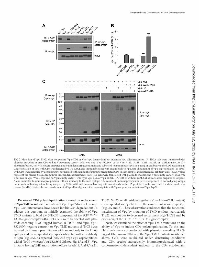
Mutation of Vpu Trp22 does not prevent Vpu-CD4 interac-tion but enhances Vpu oligomerization. The cytosolic domainsof Vpu and CD4 are known to interact with one another and toharbor critical determinants of Vpu-induced CD4 downregula-
tion (11, 36, 47, 78). The Vpu and CD4 TMDs are also essential forVpu-induced CD4 degradation as well as ER retention of CD4 (13,42, 57, 73), but their roles in mediating protein interactions havenot been addressed. To investigate whether Trp22 and other res-idues located on the same face of the Vpu TMD are involved ininteraction with CD4, HeLa cells were transfected with plasmidsencoding human CD4, with or without wild-type or mutant Vpuconstructs. CD4 was isolated by immunoprecipitation under non-denaturing conditions and associated Vpu detected by immuno-blotting (Fig. 2A). Because wild-type Vpu promotes CD4 degra-dation, coprecipitation of these two proteins could be visualizedonly after prolonged exposure of the films (unpublished observa-tions). The inability of Vpu-S52,56N to induce CD4 degradation,on the other hand, allowed visualization of its coprecipitation withCD4 upon short exposure times (11) (Fig. 2A and B). Surprisingly,replacement of Leu for Trp22 in the Vpu TMD greatly increasedthe amount of Vpu that coprecipitated with CD4 (Fig. 2A and B).All other mutations in the Vpu TMD had little or no effect onVpu-CD4 interactions (Fig. 2A and B).
The increased coprecipitation of Vpu-W22L with CD4 couldbe due to an enhanced affinity between the two proteins or to achange in the stoichiometry of the interaction. The latter interpre-tation is warranted by nuclear magnetic resonance (NMR) studiesindicating that the Vpu TMD can occur in both monomeric andoligomeric forms (22, 55, 68). In this context, mutations in theTMD could alter the Vpu monomer/oligomer ratio and thuschange the amount of Vpu coprecipitated with CD4. To deter-mine the effect of the Trp22-to-Leu mutation on Vpu-Vpu inter-actions, HeLa cells were transfected with combinations of plas-mids encoding wild-type Vpu and Vpu-W22L tagged at the Ctermini with the myc or HA epitopes, with or without CD4. TritonX-100 extracts of these cells were subjected to immunoprecipita-tion with an antibody to the myc epitope, followed by SDS-PAGEwithout a reducing agent and without boiling and immunoblot-ting with an antibody to the HA peptide (Fig. 2C). Under theseconditions, CD4 was efficiently degraded when at least one of theVpu constructs corresponded to the wild-type version, but itfailed to be degraded when both constructs had the W22L muta-tion (Fig. 2C). Coexpression of wild-type Vpu-HA and Vpu-mycresulted in only a small amount of coprecipitation (Fig. 2C), sug-gesting that Vpu monomers predominate or that Vpu oligomersare unstable under these conditions. Interestingly, coexpression ofwild-type Vpu with Vpu-W22L-myc or Vpu-W22L-HA resultedin intermediate levels of coprecipitation (Fig. 2C). Finally, coex-pression of Vpu-W22L-myc with Vpu-W22L-HA resulted inmuch greater coprecipitation as evidenced by the detection of co-precipitating species that migrate as both monomers and stableoligomers on SDS-PAGE (Fig. 2C). Similar results were obtainedin the absence or presence of CD4 (Fig. 2C), indicating that thesechanges are intrinsic to Vpu. Substitution of Ala or Gly for Trp22also increased coprecipitation of epitope-tagged Vpu pairs (seeFig. S2A and B in the supplemental material), indicating that thiseffect is due to the removal of Trp rather than the addition ofanother specific residue at position 22. From these experiments weconcluded that Vpu Trp22 hinders Vpu-Vpu interactions and thatits mutation stabilizes Vpu oligomers. The above results thus in-dicated that the Vpu Trp22 residue is not required for either Vpu-CD4 or Vpu-Vpu interactions. Rather, mutation of this aminoacid increases coprecipitation of Vpu with CD4 due to enhancedVpu oligomerization.
Magadán and Bonifacino
760 jvi.asm.org Journal of Virology
on July 12, 2012 by NA
T IN
ST
OF
HE
ALT
H LIB
http://jvi.asm.org/
Dow
nloaded from
Decreased CD4 polyubiquitination caused by replacementof Vpu TMD residues. If mutation of Vpu Trp22 does not preventVpu-CD4 interactions, how does it inhibit CD4 degradation? Toaddress this question, we initially examined the ability of VpuTMD mutants to bind the �-TrCP1 component of the SCF�-TrCP1/2
E3 Ub-ligase complex (48). HeLa cells were transfected with plas-mids encoding FLAG-tagged human �-TrCP1 and Vpu, Vpu-S52,56N (negative control), or Vpu TMD mutants. �-TrCP1 wasisolated by immunoprecipitation with an antibody to the FLAGepitope and coprecipitated Vpu species detected with an antibodyto Vpu (Fig. 3A). As expected (48), wild-type Vpu coprecipitatedwith �-TrCP1 whereas Vpu-S52,56N did not (Fig. 3A and B). Vpumutants having TMD substitutions of Leu for Ala14, Ala18, Val21,
Trp22, Val25, or all residues together (Vpu-A14¡V25L mutant)coprecipitated with �-TrCP1 to the same extent as wild-type Vpu(Fig. 3A and B). These observations indicated that the functionalinactivation of Vpu by mutation of TMD residues, particularlyTrp22, was not due to decreased recruitment of �-TrCP1 and, byextension, of the SCF�-TrCP1/2 E3 Ub-ligase complex.
Next, we examined the effect of Vpu TMD mutations on theability of Vpu to induce CD4 polyubiquitination. To this end,HeLa cells were cotransfected with plasmids encoding FLAG-tagged Ub, human CD4, and the Vpu TMD mutants mentionedabove. Cells were solubilized under denaturing conditionsand CD4 species subsequently immunoprecipitated with aconformation-independent antibody to the CD4 ectodomain.
FIG 2 Mutation of Vpu Trp22 does not prevent Vpu-CD4 or Vpu-Vpu interactions but enhances Vpu oligomerization. (A) HeLa cells were transfected withplasmids encoding human CD4 and no Vpu (empty vector), wild-type Vpu, Vpu-S52,56N, or the Vpu-A14L, -A18L, -V21L, -W22L, or -V25L mutant. At 12 hafter transfection, cell lysates were prepared under nondenaturing conditions and subjected to immunoprecipitation using an antibody to the CD4 ectodomain.Coprecipitation of Vpu with CD4 was detected by SDS-PAGE and immunoblotting with an antibody to Vpu. (B) The amount of Vpu coprecipitated (co-IPed)with CD4 was quantified by densitometry, normalized to the amount of immunoprecipitated CD4 in each sample, and expressed as arbitrary units (a.u.). Valuesrepresent the means � SEM from three independent experiments. (C) HeLa cells were transfected with plasmids encoding no Vpu (empty vector), wild-typeVpu-myc or Vpu-W22L-myc and no Vpu (empty vector), wild-type Vpu-HA, or Vpu-W22L-HA, with or without CD4. Cell extracts were prepared as for panelA and subjected to immunoprecipitation with an antibody to the myc epitope. The resultant immunoprecipitates were resuspended in nonreducing samplebuffer without boiling before being analyzed by SDS-PAGE and immunoblotting with an antibody to the HA peptide. Numbers on the left indicate molecularmasses (in kDa). Notice the increased amount of Vpu-HA oligomers that coprecipitate with Vpu-myc upon mutation of Vpu Trp22.
Transmembrane Determinants of CD4 Downregulation
January 2012 Volume 86 Number 2 jvi.asm.org 761
on July 12, 2012 by NA
T IN
ST
OF
HE
ALT
H LIB
http://jvi.asm.org/
Dow
nloaded from
762 jvi.asm.org Journal of Virology
on July 12, 2012 by NA
T IN
ST
OF
HE
ALT
H LIB
http://jvi.asm.org/
Dow
nloaded from
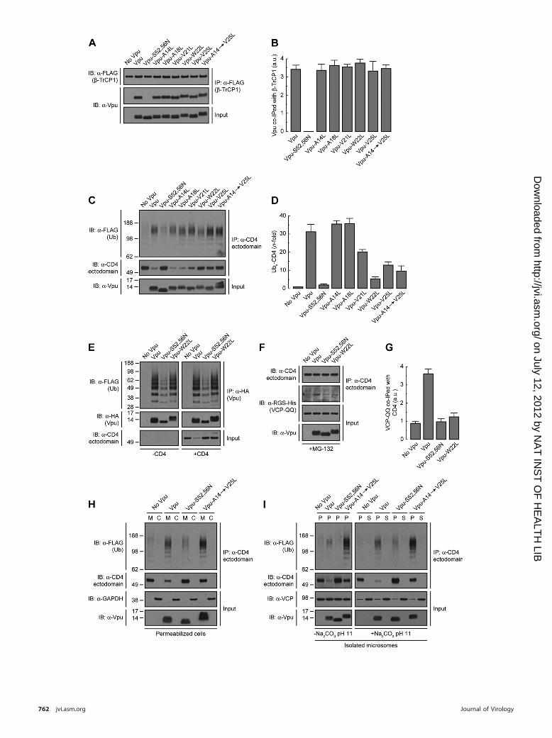
Polyubiquitinated CD4 in the immunoprecipitates was detectedby immunoblotting using an antibody to the FLAG epitope (42)(Fig. 3C). The amount of polyubiquitinated CD4 was normalizedto the amount of nonubiquitinated CD4 in each sample (42) (Fig.3C and D). Using this procedure, we observed that wild-type Vpuincreased CD4 polyubiquitination by 32-fold (Fig. 3C and D),consistent with previous results (42). The Vpu-S52,56N mutantelicited only background CD4 polyubiquitination (Fig. 3C andD), as expected from the inability of this mutant to recruit theSCF�-TrCP1/2 E3 Ub-ligase complex (42, 48) (Fig. 3A and B). Singlemutation of Val21, Trp22, or Val25 or combined mutation ofAla14, Ala18, Val21, Trp22, and Val25 (Vpu-A14¡V25L), re-duced the extent of Vpu-induced CD4 polyubiquitination to 5- to20-fold, whereas single mutation of Ala14 and Ala18 had no effect(Fig. 3C and D). We also tested for polyubiquitination of HA-tagged Vpu under identical experimental conditions. We ob-served that Vpu was polyubiquitinated in a SCF�-TrCP1/2-dependent fashion, as previously described (5) (Fig. 3E), andindependently of CD4 coexpression (Fig. 3E). Importantly,polyubiquitination of Vpu was not affected by replacement ofTrp22 by Leu (Fig. 3E). These experiments demonstrated thatmutation of Val21, Trp22, and/or Val25 decreased CD4 polyubiq-uitination without affecting SCF�-TrCP1/2 recruitment to Vpu aswell as Vpu polyubiquitination.
Accumulation of CD4 in the ER membrane in the presence ofa Vpu TMD mutant. Vpu-mediated CD4 polyubiquitination al-lows recruitment of the VCP-UFD1L-NPL4 dislocase complex,promoting CD4 dislocation from the ER membrane for eventualtargeting to the cytosolic proteasome (7, 42, 87). We observed thatmutation of Vpu Trp22 to Leu reduced the amount of thesubstrate-trapping VCP-QQ mutant (42, 85) that coprecipitatedwith CD4 (Fig. 3F and G), in line with the reduction of CD4polyubiquitination caused by this mutation (Fig. 3C and D). De-creased recruitment of the VCP-UFD1L-NPL4 complex to CD4 in
the presence of the Vpu-W22L mutant would be expected to resultin accumulation of CD4 in the ER membrane. To test this predic-tion, we developed a permeabilized cell system in which HeLa cellsexpressing FLAG-tagged Ub and human CD4, with or withoutVpu, Vpu-S52,56N, or Vpu-A14¡V25L were treated with a lowconcentration of digitonin and soluble, cytosolic proteins wereextruded from the cells by centrifugation. The resulting pellet andsupernatant fractions were fully denatured prior to immunopre-cipitation with a conformation-independent antibody to CD4 andimmunoblotting with an antibody to the FLAG epitope (42) (Fig.3H). This procedure was efficient at separating membrane andcytosolic proteins as demonstrated by detection of transmem-brane Vpu and cytosolic GAPDH in the corresponding fractions(Fig. 3H). As expected, CD4 was completely associated with mem-branes in the absence of Vpu (Fig. 3H). The small amount ofpolyubiquitinated and nonubiquitinated CD4 that remained inthe presence of wild-type Vpu was also found to be membraneassociated, as was the case for nonubiquitinated CD4 that accu-mulated in the presence of the Vpu-S52,56N mutant (Fig. 3H).Importantly, we observed that both polyubiquitinated and nonu-biquitinated CD4 remained membrane associated upon expres-sion of the Vpu-A14¡V25L construct having mutations in theTrp22-centered patch of residues (Fig. 3H). Moreover, neitherCD4 species could be removed from isolated microsomal mem-branes by treatment with 100 mM Na2CO3, pH 11.3 (23, 42),indicating that they behaved as integral membrane proteins (Fig.3I). Therefore, both polyubiquitinated and nonubiquitinatedCD4 that cannot be targeted to degradation by the Vpu TMDmutant (Fig. 3C and H) accumulate as integral membrane pro-teins rather than being released into the cytosol. These findingssuggest that Vpu TMD residues contribute to dislocation of CD4from the ER membrane.
Gly415 within the CD4 TMD is required for both Vpu-induced CD4 degradation and Vpu-CD4 interaction. Since the
FIG 3 The Vpu TMD promotes polyubiquitination and dislocation of CD4 from the ER membrane. (A) HeLa cells expressing FLAG-tagged human �-TrCP1,with or without wild-type Vpu, Vpu-S52,56N, or the Vpu-A14L, -A18L, -V21L, -W22L, -V25L, or -A14,A18,V21,W22,V25L (Vpu-A14¡V25L) mutant, werelysed under nondenaturing conditions and extracts subjected to immunoprecipitation using an antibody to the FLAG peptide. Coprecipitation of Vpu withFLAG-�-TrCP1 was detected by SDS-PAGE and immunoblotting with an antibody to Vpu. Notice that only phosphorylatable wild-type Vpu and Vpu TMDmutants coprecipitate with FLAG-�-TrCP1. (B) The amount of coprecipitated Vpu with FLAG-�-TrCP1 was quantified by densitometry, normalized to theamount of immunoprecipitated FLAG-�-TrCP1 in each sample, and expressed as arbitrary units (a.u.). Values represent the means � SEM from threeindependent experiments. (C) HeLa cells were transfected with plasmids encoding FLAG-tagged Ub and human CD4, with or without wild-type Vpu, Vpu-S52,56N, or the Vpu-A14L, -A18L, -V21L, -W22L, -V25L, or -A14¡V25L mutant. At 12 h after transfection, equivalent amounts of cell extracts made underdenaturing conditions were subjected to immunoprecipitation with a conformation-independent antibody to the CD4 ectodomain. Ubiquitination of CD4 wasdetected by immunoblotting with an HRP-conjugated antibody to the FLAG epitope. (D) Ubn-CD4 levels in the presence of Vpu were quantified by densitometryand expressed as the percentage of the total amount of Ubn-CD4 in the absence of Vpu (100% control). These values were normalized to the remainingnonubiquitinated CD4 (i.e., 1 for Ubn-CD4 in the absence of Vpu). Values are the means � SEM from three independent experiments. (E) Denatured cell lysatesfrom HeLa cells expressing FLAG-Ub and no Vpu or HA-tagged versions of wild-type Vpu, Vpu-S52,56N, or Vpu-W22L, with or without human CD4, weresubjected to immunoprecipitation using an antibody to the HA peptide. Ubiquitination of Vpu was detected by immunoblotting with an HRP-conjugatedantibody to the FLAG epitope. (F) HeLa cells were transfected with plasmids encoding human CD4 and RGS-His-tagged VCP-QQ plus no Vpu (empty vector),wild-type Vpu, Vpu-S52,56N, or Vpu-W22L. At 12 h after transfection, cells were treated with a 20 �M concentration of the proteasome inhibitor MG-132 incomplete medium for 8 h at 37°C, lysed, and then subjected to immunoprecipitation with an antibody to the CD4 ectodomain. Coprecipitation of RGS-His-tagged VCP-QQ with CD4 in the presence of wild-type Vpu was detected by immunoblotting with an antibody to the RGS-His epitope. (G) The amount ofcoprecipitated RGS-His-VCP-QQ with CD4 was quantified by densitometry, normalized to the amount of immunoprecipitated CD4 in each sample, andexpressed as arbitrary units (a.u.). Values represent the means � SEM from three independent experiments. (H) HeLa cells expressing FLAG-Ub and humanCD4, with or without wild-type Vpu, Vpu-S52,56N, or Vpu-A14¡V25L, were permeabilized with 0.028% digitonin before being fractionated into membrane(M) and cytosol (C) by centrifugation. Samples were adjusted to 1% SDS and 10 mM DTT and then processed for in vivo ubiquitination as described for panelC. Transmembrane Vpu and cytosolic GAPDH were used as fractionation controls. (I) Microsomes from HeLa cells described for panel H were resuspended inmembrane buffer supplemented (or not) with 100 mM Na2CO3, pH 11.3. After ultracentrifugation of the samples, membrane (P) and supernatant (S) fractionswere collected and adjusted to 1% SDS and 10 mM DTT before being processed for in vivo ubiquitination as described for panel C. Transmembrane Vpu and theperipherally associated membrane protein VCP were used as fractionation controls. Numbers on the left in panels C, H, and I indicate molecular masses (in kDa).Notice that both nonubiquitinated and polyubiquitinated CD4 species are membrane anchored in the presence of the Vpu-A14¡V25L mutant even after thehigh-pH treatment.
Transmembrane Determinants of CD4 Downregulation
January 2012 Volume 86 Number 2 jvi.asm.org 763
on July 12, 2012 by NA
T IN
ST
OF
HE
ALT
H LIB
http://jvi.asm.org/
Dow
nloaded from
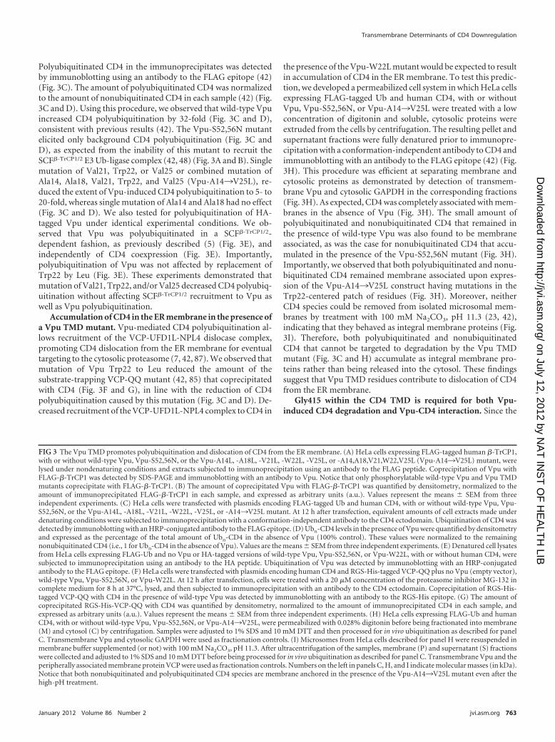
FIG 4 CD4 Gly415 is a key determinant for both Vpu-CD4 interaction and Vpu-induced CD4 degradation. (A) ClustalW alignment of CD4 TMD sequences,highlighting conservation of the residues. Notice that human CD4 Gly403, Gly404, Gly407, and Gly413 are highly conserved among all mammals, whereasGly415 is present only in humans (CD4_HUMAN) and monkeys (CD4_PANTR; chimpanzee). (B) PyMOL images of the solid-state NMR structure of thehuman CD4 TMD (81) (RSCB protein database entry 2KLU). Gly residues within the CD4 TMD are shown in red. (C) HeLa cells were transfected with plasmidsencoding human CD4 or the CD4 TMD mutant CD4-G403,404,407L, -G413,415L, -G413L, or -G415L, with or without wild-type Vpu or Vpu-S52,56N. At 12h after transfection, cell lysates were processed as described in the legend to Fig. 1B. (D) CD4 levels were quantified as described in the legend to Fig. 1C. Values
Magadán and Bonifacino
764 jvi.asm.org Journal of Virology
on July 12, 2012 by NA
T IN
ST
OF
HE
ALT
H LIB
http://jvi.asm.org/
Dow
nloaded from
TMD of CD4 is also important for Vpu-induced CD4 downregu-lation (13, 57), we next examined the requirement for specificresidues within this domain. A notable feature of the CD4 TMD isthe occurrence of several Gly residues at regular intervals within itssequence (Fig. 4A). Of these, Gly403, Gly404, Gly407, and Gly413are highly conserved in all mammals (Fig. 4A), whereas Gly415 ispresent only in humans and monkeys (Fig. 4A), the natural hostsfor HIV and SIV, respectively. All of these Gly residues exceptGly413 are located on the same face of the CD4 TMD �-helix (81)(Fig. 4B). This arrangement of Gly residues has been previouslyshown to promote interhelical interactions that are stabilized byvan der Waals forces (18, 31, 41) and C�—H · · · O hydrogenbonds (65). To analyze the requirement of these Gly residues, wemutated each of them to Leu and examined the susceptibility ofthe mutant proteins to Vpu-induced degradation (Fig. 4C). Weobserved that mutation of Gly415 completely abrogated the de-crease in CD4 levels elicited by Vpu, whereas mutation of theother Gly residues, either singly or in combinations, had little orno effect (Fig. 4C and D). Pulse-chase analysis confirmed the sta-bility of newly synthesized CD4-G415L in the presence of Vpu(Fig. 4E and F). Mutation of each of the other non-Gly residuesalso had no effect on the susceptibility of CD4 to Vpu-mediateddegradation (unpublished observations). From these experi-ments, we concluded that CD4 Gly415 is a critical determinant forVpu-induced CD4 targeting to degradation.
To determine whether CD4 Gly415 and other TMD Gly resi-dues are required for interaction with Vpu, we transfected HeLacells with plasmids encoding wild-type CD4 or CD4 TMD Glymutants plus Vpu-S52,56N, which binds to CD4 but does notinduce its degradation (11). Interactions were analyzed by copre-cipitation under nondenaturing conditions as described above(Fig. 2A). We observed that wild-type CD4 as well as CD4 TMDconstructs bearing mutations of Gly403, Gly404, Gly407, andGly413 equally coprecipitated with Vpu-S52,56N (Fig. 4G and H).However, coprecipitation of Vpu-S52,56N with CD4 was greatlyimpaired when CD4 Gly415 was replaced by Leu (Fig. 4G and H).The interaction between Vpu and the CD4 Gly415 mutants wasnot completely abrogated, presumably due to the contribution ofthe cytoplasmic domains of the proteins to their mutual binding(11, 47). These experiments indicated that the requirement ofCD4 Gly415 for Vpu-CD4 degradation is at least in part due to itsinvolvement in Vpu-CD4 TMD interaction.
Vpu Trp22 and CD4 Gly415 are not involved in Vpu-mediated inhibition of CD4 export to the Golgi complex. Werecently uncovered a new function of Vpu in preventing transportof CD4 from the ER to the Golgi complex, which is distinct fromits ability to target CD4 to the ERAD pathway (42). Because thisnew function is also dependent on the Vpu TMD (42), we testedthe effect of mutating several Vpu TMD residues on its ability tocause CD4 retention in the ER. HeLa cells were transfected withplasmids encoding CD4, with or without various Vpu TMD mu-tants, and the intracellular transport of CD4 was analyzed by en-
doglycosidase H (endo H) treatment (Fig. 5A) and cell surfacebiotinylation (Fig. 5C). Newly synthesized CD4 contains twoN-linked oligosaccharides within its ectodomain, one of whichacquires endo H resistance when CD4 is transported through theGolgi complex (72) (Fig. 5A and B). In cells expressing wild-typeVpu, the small amount of remaining CD4 was �75% sensitive toendo H, consistent with its predominant localization to the ER(42) (Fig. 5A and B). In cells expressing the Vpu-W22L mutant,CD4 was not degraded, but it remained mostly sensitive to endo H(Fig. 5A and B), indicating that Vpu Trp22 is required for CD4targeting to ERAD but not for CD4 retention in the ER.
The effects of Vpu on CD4 at the ER were reflected in changesof CD4 levels at the cell surface as observed in surface biotinylationexperiments (Fig. 5C). HeLa cells were transfected with plasmidsencoding human CD4, with or without Vpu or the Vpu-A14L,-A18L, -V21L, -W22L, -V25L, and -A14¡V25L mutants. Cellsurface proteins were biotinylated at 4°C, followed by cell lysis andpulldown with a NeutrAvidin-coupled resin. CD4, Vpu, and TfRwere then visualized by immunoblotting using specific antibodies(Fig. 5C). Since TfR stability and maturation are not affected byVpu (42), this protein was used as a loading control (Fig. 5C). Forquantification purposes, the amounts of CD4 present at the cellsurface were normalized to the amounts of CD4 in each sample(100% for the control without Vpu) (Fig. 5D). No CD4 could bedetected at the cell surface in the presence of Vpu (Fig. 5C and D),as expected from the ER retention and ERAD targeting of CD4under these conditions. Despite the inhibition of ERAD targetingcaused by mutation of Vpu Trp22, however, only a small fraction(up to �25% of the control) of CD4 reached the cell surface in thepresence of the Vpu-W22L or Vpu-A14¡V25L mutant (Fig. 5Cand D). This result is consistent with Vpu Trp22 not being in-volved in CD4 retention in the ER.
We next investigated the contribution of CD4 Gly415 to Vpu-mediated ER retention of CD4 by performing endo H sensitivity(Fig. 5E) and cell surface biotinylation (Fig. 5G) assays, as de-scribed above. In the absence of Vpu, both CD4 and CD4-G415Lefficiently exited the ER en route to the plasma membrane (Fig. 5Eto H). In contrast, in the presence of Vpu, most of the stableCD4-G415L mutant remained in the ER (Fig. 5E and F) and didnot reach the plasma membrane (�25% of the control) (Fig. 5Gand H). The same result was obtained when not only Gly415 butalso all non-Leu residues on the same face of the CD4 TMD�-helix were replaced by Leu (CD4-G403¡F418L mutant) (Fig.5G and H). Altogether, these results demonstrated that Vpu con-served the ability to retain CD4 in the ER when a key CD4 TMDresidue required for its degradation, Gly415, was mutated.
Val20 and Ser23 within the Vpu TMD are critical for Vpu-induced CD4 retention in the ER. Since the Vpu TMD contrib-utes to CD4 retention in the ER (42) and Vpu Trp22 (and to alesser extent Val21 and Val25) is required for CD4 ERAD targetingbut not ER retention (this study), we hypothesized that other res-idues in the Vpu TMD must be involved in ER retention of CD4.
are the means � SEM from three independent experiments. (E) HeLa cells expressing human CD4-G415L, with or without wild-type Vpu, were labeled with[35S]methionine-cysteine for 2 min and chased for the indicated times at 37°C. Radiolabeled CD4 was detected as described in the legend to Fig. 1E. (F)Percentage of CD4 at each chase time relative to CD4 at time zero (100% control). (G) HeLa cells were transfected with plasmids encoding no CD4 (emptyvector), human CD4, or CD4 TMD mutants bearing mutations on Gly 403, 404, 407, 413, or 415 or all glycines together (CD4-G403¡G415L), plus theVpu-S52,56N mutant. Cell lysates were processed as described in the legend to Fig. 2A. (H) The amount of coprecipitated Vpu-S52,56N with CD4 was quantifiedas described in the legend to Fig. 2B. Values are the means � SEM from three independent experiments. Notice that Vpu-S52,56N is poorly coprecipitated withCD4 in the absence of CD4 Gly415 compared to wild-type CD4 or the other CD4 TMD mutants.
Transmembrane Determinants of CD4 Downregulation
January 2012 Volume 86 Number 2 jvi.asm.org 765
on July 12, 2012 by NA
T IN
ST
OF
HE
ALT
H LIB
http://jvi.asm.org/
Dow
nloaded from
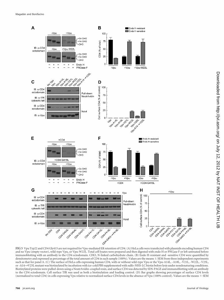
FIG 5 Vpu Trp22 and CD4 Gly415 are not required for Vpu-mediated ER retention of CD4. (A) HeLa cells were transfected with plasmids encoding human CD4and no Vpu (empty vector), wild-type Vpu, or Vpu-W22L. Total cell lysates were prepared and then digested with endo H or PNGase F or left untreated beforeimmunoblotting with an antibody to the CD4 ectodomain. CHO, N-linked carbohydrate chain. (B) Endo H-resistant and -sensitive CD4 were quantified bydensitometry and expressed as percentage of the total amount of CD4 in each sample (100%). Values are the means � SEM from three independent experimentssuch as that for panel A. (C) The surface of HeLa cells expressing human CD4, with or without wild-type Vpu or the Vpu-A14L, -A18L, -V21L, -W22L, -V25L,or -A14¡V25L mutant was biotinylated by incubation with ice-cold PBS supplemented with sulfo-NHS-LC biotin before lysis under nondenaturing conditions.Biotinylated proteins were pulled-down using a NeutrAvidin-coupled resin, and surface CD4 was detected by SDS-PAGE and immunoblotting with an antibodyto the CD4 ectodomain. Cell surface TfR was used as both a biotinylation and loading control. (D) Bar graphs showing percentages of surface CD4 levels(normalized to total CD4) in cells expressing Vpu relative to normalized surface CD4 levels in the absence of Vpu (100% control). Values are the means � SEM
Magadán and Bonifacino
766 jvi.asm.org Journal of Virology
on July 12, 2012 by NA
T IN
ST
OF
HE
ALT
H LIB
http://jvi.asm.org/
Dow
nloaded from
We focused our analysis on Vpu Val20 and Ser23, two residuesthat are contiguous on a face opposite to that displaying Val21,Trp22, and Val25 (Fig. 6A). Vpu Val20 is conserved in mostHIV-1 group M variants (Fig. 1A), whereas Ser23 is highly con-served only in HIV-1 group M subtype B Vpu sequences (Fig. 1A).Mutational analysis showed that replacement of Val20 or Ser23 byLeu had little or no effect on the ability of Vpu to cause CD4degradation (Fig. 6B and C). Substitution of Leu for Val21, Trp22,and Val25 (Vpu-V21¡V25L mutant) in addition to Val20 andSer23 [Vpu-V21¡V25L (�V20,S23L) mutant] had the expectedeffect of preventing CD4 degradation (Fig. 6B and C). We nextexamined the effect of replacing Val20 and Ser23 on the ability ofVpu to induce CD4 retention in the ER, as determined by endo Hdigestion analysis (Fig. 6D). Interestingly, CD4 became mostlyendo H resistant (�80%) in the presence of Vpu-V21¡V25L(�V20,S23L) (Fig. 6D and E). This was in contrast to the endo Hsensitivity of CD4 in the presence of the Vpu-V21¡V25L mutant(Fig. 6D and E). Individual mutation of Val20 or Ser23 to Leu[Vpu-V21¡V25L (�V20L) and Vpu-V21¡V25L (�S23L) mu-tants, respectively] had intermediate effects (�50 to 60% of CD4remained resistant to endo H) (Fig. 6D and E). Interestingly, mostCD4 remained endo H sensitive when Vpu Ser23 was replaced byIle [Vpu-V21¡V25L (�S23I) mutant] instead of Leu (Fig. 6Dand E). The latter finding is consistent with a significant percent-age of HIV-1 group M Vpu sequences carrying Ile at position 23.These differential effects of the Vpu TMD mutants on the ERretention of CD4 were reflected in the expression of CD4 at theplasma membrane, as measured by the surface biotinylation assay(Fig. 6F and G). We observed undetectable levels of surface CD4 incells expressing wild-type Vpu, low levels in cells expressing Vpu-V21¡V25L or Vpu-V21¡V25L (�S23I) (�25% of control), in-termediate levels in cells expressing Vpu-V21¡V25L (�V20L) orVpu-V21¡V25L (�S23L) (�50 to 60% of control), and highlevels in cells expressing Vpu-V21¡V25L (�V20,S23L) (�90%of control) (Fig. 6F and G), even though none of these Vpu TMDmutants induced CD4 degradation (Fig. 6F). Remarkably, re-placement of Leu for Vpu Val20 and Ser23 together or in isolationalso enhanced Vpu trafficking from the ER to the cell surface in aCD4-independent fashion (Fig. 6F). From these experiments weconcluded that Vpu Val20 and Ser23 are both components of aTMD determinant for retention of not only CD4 but also Vpu inthe ER, which is distinct from the ERAD-targeting TMD determi-nant conformed by Val21, Trp22, and Val25.
DISCUSSION
It is now well established that TMDs do not merely act as mem-brane anchors or connectors between the luminal/extracellulardomains (i.e., ectodomains) and cytosolic domains (i.e., endodo-mains) of integral membrane proteins. Rather, they play activeroles in processes such as transport of molecules across mem-branes (58), intramembrane proteolysis (82), assembly of multi-
protein complexes (46), protein localization to intracellular or-ganelles (67), and targeting to the ERAD pathway (8, 10, 76). Forthis reason, many TMDs are not just random sequences of hydro-phobic amino acid residues but possess specific characteristics,including conserved residues at defined positions within thetransmembrane �-helices. The results presented in this studyshow that Vpu exploits these properties of TMDs in order todownregulate CD4. Indeed, we demonstrate that the TMDs ofboth Vpu and CD4 contain specific residues that are required forVpu-induced CD4 targeting to ERAD and retention in the ER, aswell as for assembly of Vpu-CD4 and Vpu-Vpu complexes.
A cluster of residues centered on Trp22 in the Vpu TMD isrequired for CD4 targeting to the ERAD pathway. Although ini-tially overlooked, the role of TMDs in Vpu-induced CD4 degra-dation has now been demonstrated by several studies (13, 42, 57,73). The specific features of the Vpu and CD4 TMDs that arerequired for this process, however, have remained poorly under-stood. Here we show that a cluster of residues centered on Trp22(i.e., Val21, Trp22, and Val25) on the Vpu TMD is required forCD4 targeting to ERAD. Vpu Trp22 is the most critical residue onthis cluster, since it cannot be replaced by any residue other thanCys without complete loss of activity (Fig. 1G and H). The fact thatCys can substitute for Trp at position 22 is puzzling, since thesetwo amino acids have very different chemical properties. Trp22 isat a position near the membrane-cytosol interface, the most fre-quent location for Trp within TMDs (80, 84). The affinity of TMDTrp residues for membrane-cytosol interfaces stems from the dualbulky-hydrophobic/hydrogen-bonding nature of the indole ring(43, 52, 66). Vpu Trp22 could participate in CD4 targeting to theERAD pathway by mediating protein-protein interactions or byfixing the position of the Vpu TMD relative to the lipid bilayer.These properties could be conferred on CD4 through assemblywith Vpu.
An earlier study showed that scrambling of some residues inthe Vpu TMD did not affect the ability of Vpu to downregulateCD4 (VpuRD mutant) (64), an observation that seems at oddswith the conclusion from this and previous studies (42, 73). How-ever, the scrambled VpuRD mutant has Val22, Trp24, and Val27residues at positions that are roughly equivalent to those of Val21,Trp22, and Val25 in the wild-type Vpu TMD. The location ofTrp24 near the membrane-cytosol interface in VpuRD could ex-plain why this mutant, like wild-type Vpu, remains active for CD4downregulation.
Effects of the Vpu Trp22 mutation on CD4 polyubiquitina-tion and recruitment of the VCP-UFD1L-NPL4 dislocase com-plex. How might Trp22 participate in the mechanism by whichVpu induces targeting of CD4 to ERAD? We initially hypothesizedthat Vpu Trp22 could be required for Vpu-CD4 interaction at thelevel of the TMDs. However, we found that this was not the case, asmutation of Trp22 to Leu did not abrogate, but instead enhanced,coprecipitation of Vpu with CD4 (Fig. 2A and B). This enhance-
from three independent experiments. Notice that CD4 is not expressed at the cell surface even in the absence of those Vpu TMD residues that are required forERAD targeting of CD4. (E) HeLa cells were transfected with plasmids encoding human CD4 or CD4-G415L, with or without wild-type Vpu. Detergent extractswere prepared and processed as for panel A. (F) Endo H-resistant and -sensitive CD4 were quantified as for panel B. Values are the means � SEM from threeindependent experiments such as that for panel E. (G) The surface of HeLa cells expressing no CD4 (empty vector), human CD4, or the CD4-G403L, -G404L,-G407L, -F411L, -G415L, -F418L, or -G403,G404,G407,F411,G415,F418L (CD4-G403¡F418L) mutant, with or without wild-type Vpu, was biotinylated asdescribed for panel C before lysis under nondenaturing conditions. Surface biotinylated CD4 was detected as for panel C. (H) Quantification of surface CD4 levelswas performed as for panel D. Values are the means � SEM from three independent experiments. Notice that CD4 Gly415 is not required for Vpu-mediated ERretention of CD4, even though this residue is a critical determinant for CD4 ERAD targeting by Vpu (Fig. 4).
Transmembrane Determinants of CD4 Downregulation
January 2012 Volume 86 Number 2 jvi.asm.org 767
on July 12, 2012 by NA
T IN
ST
OF
HE
ALT
H LIB
http://jvi.asm.org/
Dow
nloaded from
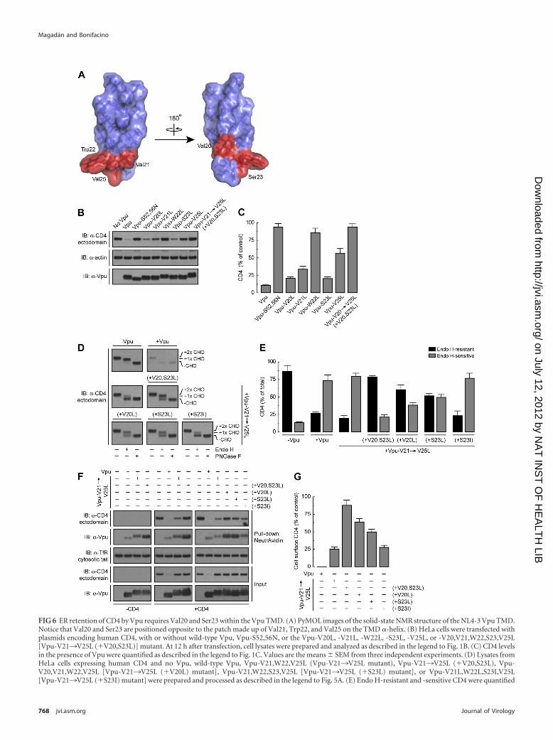
FIG 6 ER retention of CD4 by Vpu requires Val20 and Ser23 within the Vpu TMD. (A) PyMOL images of the solid-state NMR structure of the NL4-3 Vpu TMD.Notice that Val20 and Ser23 are positioned opposite to the patch made up of Val21, Trp22, and Val25 on the TMD �-helix. (B) HeLa cells were transfected withplasmids encoding human CD4, with or without wild-type Vpu, Vpu-S52,56N, or the Vpu-V20L, -V21L, -W22L, -S23L, -V25L, or -V20,V21,W22,S23,V25L[Vpu-V21¡V25L (�V20,S23L)] mutant. At 12 h after transfection, cell lysates were prepared and analyzed as described in the legend to Fig. 1B. (C) CD4 levelsin the presence of Vpu were quantified as described in the legend to Fig. 1C. Values are the means � SEM from three independent experiments. (D) Lysates fromHeLa cells expressing human CD4 and no Vpu, wild-type Vpu, Vpu-V21,W22,V25L (Vpu-V21¡V25L mutant), Vpu-V21¡V25L (�V20,S23L), Vpu-V20,V21,W22,V25L [Vpu-V21¡V25L (�V20L) mutant], Vpu-V21,W22,S23,V25L [Vpu-V21¡V25L (�S23L) mutant], or Vpu-V21L,W22L,S23I,V25L[Vpu-V21¡V25L (�S23I) mutant] were prepared and processed as described in the legend to Fig. 5A. (E) Endo H-resistant and -sensitive CD4 were quantified
Magadán and Bonifacino
768 jvi.asm.org Journal of Virology
on July 12, 2012 by NA
T IN
ST
OF
HE
ALT
H LIB
http://jvi.asm.org/
Dow
nloaded from
ment was not likely due to increased affinity of the Vpu-CD4TMD interaction but rather to increased oligomerization of Vpuand coprecipitation of Vpu oligomers with CD4 (Fig. 2). Indeed,Vpu had previously been shown to occur in equilibrium betweenmonomeric and oligomeric forms (29, 40, 44, 56). We found thatmutation of Vpu Trp22 to Leu, Ala, or Gly shifts the equilibriumtoward oligomeric forms (Fig. 2C; see Fig. S2A and B in the sup-plemental material), implying that the Vpu TMD Trp residue hin-ders Vpu-Vpu interactions. In agreement with these findings, theVpu TMD containing a Trp-to-Leu substitution remains open asan ion channel for much longer than the wild type, behaving morelike a pore (50). A corollary of these findings is that monomeric,and not oligomeric, Vpu is the active form for CD4 targeting to theERAD pathway. Therefore, at a minimum, Vpu Trp22 functionsto maintain Vpu in its active, monomeric form.
To determine whether Vpu Trp22 has additional functions, weexamined the possible involvement of this residue in ERAD-targeting steps downstream of the Vpu-CD4 interaction. We ob-served that mutation of Vpu Trp22 did not affect the recruitmentof the cytosolic SCF�-TrCP1/2 E3 Ub-ligase complex to the Vpucytosolic domain (Fig. 3A and B). However, we found that Vpu-induced polyubiquitination of CD4 was reduced from 32-fold to5- to 20-fold by single or combined mutation of several Vpu TMDresidues, including Trp22 (Fig. 3C and D). A possible explanationof these findings is that Vpu TMD mutations, though not decreas-ing CD4-Vpu interactions, alter the orientation of the CD4 cyto-solic tail relative to the SCF�-TrCP1/2 complex such that CD4polyubiquitination is impaired. Vpu TMD mutations could alsoprevent the function of another transmembrane protein that con-tributes to CD4 polyubiquitination such as, for example, an E2Ub-conjugating enzyme (86). Finally, decreased CD4 polyubiq-uitination could be due to partial deubiquitination of the accumu-lated CD4 that cannot be targeted to degradation by the Vpu TMDmutants (74).
Binding of VCP (and, by extension, of the VCP-UFD1L-NPL4complex) to CD4 was also impaired by mutation of Vpu Trp22(Fig. 3F and G), likely as a consequence of reduced CD4 polyubiq-uitination. Since the VCP-UFD1L-NPL4 complex functions toextract CD4 from the ER membrane in the presence of Vpu (7,42), we expected that CD4 would remain associated with mem-branes upon expression of Vpu TMD mutants. Indeed, subcellu-lar fractionation experiments showed that both nonubiquitinatedand polyubiquitinated CD4 species accumulated as integral mem-brane proteins in the presence of Vpu having mutations of severalTMD residues, including Trp22 (Vpu-A14¡V25L mutant) (Fig.3H and I). This result should be interpreted with caution, how-ever, as CD4 that was protected from Vpu-induced degradationby incubation with the proteasomal inhibitor MG-132 also re-mained integrally associated with membranes (unpublished ob-servations). The latter observation indicates that proteasomal ac-tivity is a prerequisite for membrane dislocation of CD4 by Vpu, aspreviously shown for some ERAD substrates (35, 49, 83) but not
others (27, 28). Nevertheless, our observations are most consistentwith a role for the Vpu TMD, and in particular Trp22, in allowingextraction of CD4 from the ER membrane.
Gly415 in the CD4 TMD is critical for assembly with Vpu andfor Vpu-induced CD4 degradation. The TMD of CD4 is alsorequired for Vpu-mediated CD4 degradation (13, 57). In thisstudy, we found that mutation of a single CD4 TMD residue,Gly415, completely abrogated CD4 degradation induced by Vpu(Fig. 4C and D). Unlike mutation of Vpu Trp22, however, muta-tion of CD4 Gly415 impaired the Vpu-CD4 interaction (Fig. 4Gand H), thus establishing a clear basis for the inhibition of Vpu-induced CD4 degradation caused by this mutation. This findingrepresents the first demonstration that mutation of a TMD resi-due decreases Vpu-CD4 interactions. Thus, both the TMD (thisstudy) and the cytosolic domains (11, 47) contribute to the phys-ical interaction of Vpu with CD4. Based on the solid-state NMRstructure of the human CD4 TMD �-helix (81) (Fig. 4B), Gly415is predicted to be part of a “Gly strip,” a structural motif thatmediates TMD interactions (18, 31) (Fig. 4A and B). With theexception of CD4 Gly415, single substitution of each Gly residueon the strip (Fig. 4) or of other, non-Gly residues in the CD4 TMD(unpublished observations), however, had no effect on assemblywith Vpu (Fig. 4G and H) or Vpu-induced CD4 degradation (Fig.4C and D). These results do not rule out that other CD4 TMDresidues are involved, as inhibition of those functions could re-quire combined mutation of two or more residues.
An ER retention determinant comprising Val20 and Ser23 inthe Vpu TMD. In addition to promoting targeting of CD4 to theERAD pathway, the Vpu TMD contributes to retention of CD4 inthe ER (42). Mutation of Vpu TMD Trp22 prevents Vpu-inducedCD4 degradation but not CD4 ER retention (Fig. 5), an observa-tion that is in agreement with the ability of this mutant to decreaseexpression of CD4 at the cell surface as reported by one group (69)but not another (77). Additional mutagenesis revealed two otherVpu TMD residues, Val20 and Ser23, which are critical for CD4ER retention (Fig. 6). Val20 and Ser23 are located on a face of theTMD �-helix opposite to that of the cluster comprising Val21,Trp22, and Val25 (Fig. 6A). Moreover, mutation of Val20 andSer23 alone did not inhibit Vpu-induced CD4 degradation (Fig.6B and C). These observations indicate that the determinants forERAD targeting and ER retention of CD4 in the Vpu TMD arespatially and mechanistically separable. How does Vpu retain CD4in the ER? Vpu itself lacks a known ER retention motif, but re-placement of Val20 and Ser23 on its TMD increased Vpu trans-port out of the ER en route to the plasma membrane indepen-dently of CD4 (Fig. 6F). This indicates that Vpu might be at leastpartially retained in the ER by establishment of Val20- and Ser23-mediated TMD interactions with a putative ER resident protein.In this regard, calnexin has been shown to modulate ER localiza-tion and stability of a number of ERAD substrates by recognitionof specific features within their TMDs (37, 51, 70). It is also pos-sible that Val20 and Ser23 confer ER retention through interac-
as described in the legend to Fig. 5B. Values are the means � SEM from three independent experiments such as that for panel D. (F) Surface biotinylated CD4from HeLa cells transfected as for panel D was detected as described in the legend to Fig. 5C. The same experiment was also done with cells expressing no Vpu,wild-type Vpu, Vpu-V21¡V25L, or Vpu-V21¡V25L (�V20,S23L), without CD4 for comparison. Surface biotinylated Vpu was also detected by immunoblot-ting using an antibody to Vpu. (G) Quantification of surface CD4 levels was performed as described in the legend to Fig. 5D. Values are the means � SEM fromthree independent experiments. Notice that the Vpu-V21¡V25L and Vpu-V21¡V25L (�V20,S23L) mutants exhibit different abilities to retain CD4 in the EReven though neither mutant is able to induce CD4 degradation.
Transmembrane Determinants of CD4 Downregulation
January 2012 Volume 86 Number 2 jvi.asm.org 769
on July 12, 2012 by NA
T IN
ST
OF
HE
ALT
H LIB
http://jvi.asm.org/
Dow
nloaded from
tions with the ER lipid bilayer. In any case, CD4 appears to beretained in the ER in trans, through assembly with Vpu. It is worthnoting that Vpu has been localized not just to the ER (42) but alsoto the trans-Golgi network and endosomal compartments (21, 75,77). In fact, the post-ER population of Vpu is likely the form thatfunctions in BST-2 antagonism (2, 69).
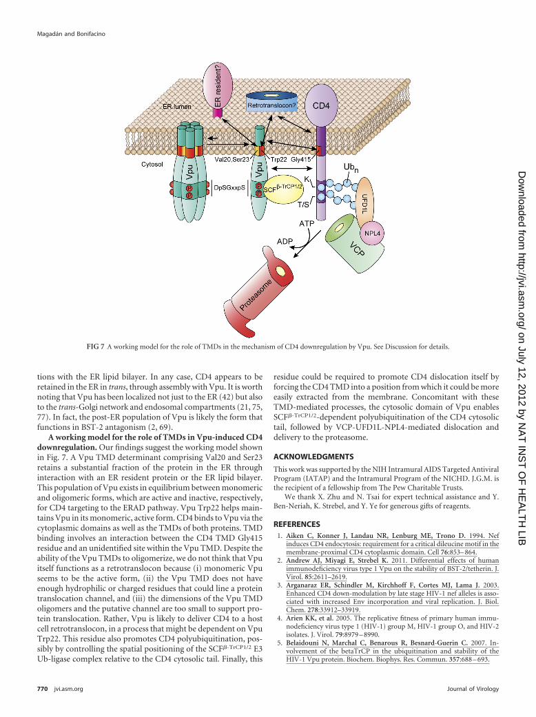
A working model for the role of TMDs in Vpu-induced CD4downregulation. Our findings suggest the working model shownin Fig. 7. A Vpu TMD determinant comprising Val20 and Ser23retains a substantial fraction of the protein in the ER throughinteraction with an ER resident protein or the ER lipid bilayer.This population of Vpu exists in equilibrium between monomericand oligomeric forms, which are active and inactive, respectively,for CD4 targeting to the ERAD pathway. Vpu Trp22 helps main-tains Vpu in its monomeric, active form. CD4 binds to Vpu via thecytoplasmic domains as well as the TMDs of both proteins. TMDbinding involves an interaction between the CD4 TMD Gly415residue and an unidentified site within the Vpu TMD. Despite theability of the Vpu TMDs to oligomerize, we do not think that Vpuitself functions as a retrotranslocon because (i) monomeric Vpuseems to be the active form, (ii) the Vpu TMD does not haveenough hydrophilic or charged residues that could line a proteintranslocation channel, and (iii) the dimensions of the Vpu TMDoligomers and the putative channel are too small to support pro-tein translocation. Rather, Vpu is likely to deliver CD4 to a hostcell retrotranslocon, in a process that might be dependent on VpuTrp22. This residue also promotes CD4 polyubiquitination, pos-sibly by controlling the spatial positioning of the SCF�-TrCP1/2 E3Ub-ligase complex relative to the CD4 cytosolic tail. Finally, this
residue could be required to promote CD4 dislocation itself byforcing the CD4 TMD into a position from which it could be moreeasily extracted from the membrane. Concomitant with theseTMD-mediated processes, the cytosolic domain of Vpu enablesSCF�-TrCP1/2-dependent polyubiquitination of the CD4 cytosolictail, followed by VCP-UFD1L-NPL4-mediated dislocation anddelivery to the proteasome.
ACKNOWLEDGMENTS
This work was supported by the NIH Intramural AIDS Targeted AntiviralProgram (IATAP) and the Intramural Program of the NICHD. J.G.M. isthe recipient of a fellowship from The Pew Charitable Trusts.
We thank X. Zhu and N. Tsai for expert technical assistance and Y.Ben-Neriah, K. Strebel, and Y. Ye for generous gifts of reagents.
REFERENCES1. Aiken C, Konner J, Landau NR, Lenburg ME, Trono D. 1994. Nef
induces CD4 endocytosis: requirement for a critical dileucine motif in themembrane-proximal CD4 cytoplasmic domain. Cell 76:853– 864.
2. Andrew AJ, Miyagi E, Strebel K. 2011. Differential effects of humanimmunodeficiency virus type 1 Vpu on the stability of BST-2/tetherin. J.Virol. 85:2611–2619.
3. Arganaraz ER, Schindler M, Kirchhoff F, Cortes MJ, Lama J. 2003.Enhanced CD4 down-modulation by late stage HIV-1 nef alleles is asso-ciated with increased Env incorporation and viral replication. J. Biol.Chem. 278:33912–33919.
4. Arien KK, et al. 2005. The replicative fitness of primary human immu-nodeficiency virus type 1 (HIV-1) group M, HIV-1 group O, and HIV-2isolates. J. Virol. 79:8979 – 8990.
5. Belaidouni N, Marchal C, Benarous R, Besnard-Guerin C. 2007. In-volvement of the betaTrCP in the ubiquitination and stability of theHIV-1 Vpu protein. Biochem. Biophys. Res. Commun. 357:688 – 693.
FIG 7 A working model for the role of TMDs in the mechanism of CD4 downregulation by Vpu. See Discussion for details.
Magadán and Bonifacino
770 jvi.asm.org Journal of Virology
on July 12, 2012 by NA
T IN
ST
OF
HE
ALT
H LIB
http://jvi.asm.org/
Dow
nloaded from
6. Benson RE, Sanfridson A, Ottinger JS, Doyle C, Cullen BR. 1993.Downregulation of cell-surface CD4 expression by simian immunodefi-ciency virus Nef prevents viral super infection. J. Exp. Med. 177:1561–1566.
7. Binette J, et al. 2007. Requirements for the selective degradation of CD4receptor molecules by the human immunodeficiency virus type 1 Vpuprotein in the endoplasmic reticulum. Retrovirology 4:75.
8. Bonifacino JS, Cosson P, Klausner RD. 1990. Colocalized transmem-brane determinants for ER degradation and subunit assembly explain theintracellular fate of TCR chains. Cell 63:503–513.
9. Bonifacino JS, Dell’Angelica EC. 2001. Immunoprecipitation. Curr. Pro-toc. Cell Biol. 7:Unit 7.2.
10. Bonifacino JS, Suzuki CK, Klausner RD. 1990. A peptide sequenceconfers retention and rapid degradation in the endoplasmic reticulum.Science 247:79 – 82.
11. Bour S, Schubert U, Strebel K. 1995. The human immunodeficiencyvirus type 1 Vpu protein specifically binds to the cytoplasmic domain ofCD4: implications for the mechanism of degradation. J. Virol. 69:1510 –1520.
12. Bresnahan PA, Yonemoto W, Greene WC. 1999. SIV Nef protein utilizesboth leucine- and tyrosine-based protein sorting pathways for down-regulation of CD4. J. Immunol. 163:2977–2981.
13. Buonocore L, Turi TG, Crise B, Rose JK. 1994. Stimulation of heterol-ogous protein degradation by the Vpu protein of HIV-1 requires the trans-membrane and cytoplasmic domains of CD4. Virology 204:482– 486.
14. Burtey A, et al. 2007. Dynamic interaction of HIV-1 Nef with theclathrin-mediated endocytic pathway at the plasma membrane. Traffic8:61–76.
15. Butticaz C, Michielin O, Wyniger J, Telenti A, Rothenberger S. 2007.Silencing of both beta-TrCP1 and HOS (beta-TrCP2) is required to sup-press human immunodeficiency virus type 1 Vpu-mediated CD4 down-modulation. J. Virol. 81:1502–1505.
16. Chaudhuri R, Lindwasser OW, Smith WJ, Hurley JH, Bonifacino JS.2007. Downregulation of CD4 by human immunodeficiency virus type 1Nef is dependent on clathrin and involves direct interaction of Nef withthe AP2 clathrin adaptor. J. Virol. 81:3877–3890.
17. Chaudhuri R, Mattera R, Lindwasser OW, Robinson MS, Bonifacino JS.2009. A basic patch on alpha-adaptin is required for binding of humanimmunodeficiency virus type 1 Nef and cooperative assembly of a CD4-Nef-AP-2 complex. J. Virol. 83:2518 –2530.
18. Cosson P, Bonifacino JS. 1992. Role of transmembrane domain interac-tions in the assembly of class II MHC molecules. Science 258:659 – 662.
19. Craig HM, Reddy TR, Riggs NL, Dao PP, Guatelli JC. 2000. Interactionsof HIV-1 nef with the mu subunits of adaptor protein complexes 1, 2, and3: role of the dileucine-based sorting motif. Virology 271:9 –17.
20. daSilva LL, et al. 2009. Human immunodeficiency virus type 1 Nef pro-tein targets CD4 to the multivesicular body pathway. J. Virol. 83:6578 – 6590.
21. Dube M, et al. 2009. Suppression of tetherin-restricting activity uponhuman immunodeficiency virus type 1 particle release correlates with lo-calization of Vpu in the trans-Golgi network. J. Virol. 83:4574 – 4590.
22. Ewart GD, Sutherland T, Gage PW, Cox GB. 1996. The Vpu protein ofhuman immunodeficiency virus type 1 forms cation-selective ion chan-nels. J. Virol. 70:7108 –7115.
23. Fujiki Y, Hubbard AL, Fowler S, Lazarow PB. 1982. Isolation of intra-cellular membranes by means of sodium carbonate treatment: applicationto endoplasmic reticulum. J. Cell Biol. 93:97–102.
24. Greenberg ME, et al. 1997. Co-localization of HIV-1 Nef with the AP-2adaptor protein complex correlates with Nef-induced CD4 down-regulation. EMBO J. 16:6964 – 6976.
25. Guatelli JC. 2009. Interactions of viral protein U (Vpu) with cellularfactors. Curr. Top. Microbiol. Immunol. 339:27– 45.
26. Hoxie JA, et al. 1986. Alterations in T4 (CD4) protein and mRNA syn-thesis in cells infected with HIV. Science 234:1123–1127.
27. Hughes EA, Hammond C, Cresswell P. 1997. Misfolded major histo-compatibility complex class I heavy chains are translocated into the cyto-plasm and degraded by the proteasome. Proc. Natl. Acad. Sci. U. S. A.94:1896 –1901.
28. Huppa JB, Ploegh HL. 1997. The alpha chain of the T cell antigen receptoris degraded in the cytosol. Immunity 7:113–122.
29. Hussain A, Das SR, Tanwar C, Jameel S. 2007. Oligomerization of thehuman immunodeficiency virus type 1 (HIV-1) Vpu protein—a genetic,biochemical and biophysical analysis. Virol. J. 4:81.
30. Jin YJ, et al. 2005. HIV Nef-mediated CD4 down-regulation is adaptorprotein complex 2 dependent. J. Immunol. 175:3157–3164.
31. Kim S, et al. 2005. Transmembrane glycine zippers: physiological andpathological roles in membrane proteins. Proc. Natl. Acad. Sci. U. S. A.102:14278 –14283.
32. Kirchhoff F. 2010. Immune evasion and counteraction of restriction fac-tors by HIV-1 and other primate lentiviruses. Cell Host Microbe 8:55– 67.
33. Korber B, et al. 2000. Timing the ancestor of the HIV-1 pandemic strains.Science 288:1789 –1796.
34. Lanzavecchia A, Roosnek E, Gregory T, Berman P, Abrignani S. 1988.T cells can present antigens such as HIV gp120 targeted to their ownsurface molecules. Nature 334:530 –532.
35. Lee RJ, et al. 2004. Uncoupling retro-translocation and degradation inthe ER-associated degradation of a soluble protein. EMBO J. 23:2206 –2215.
36. Lenburg ME, Landau NR. 1993. Vpu-induced degradation of CD4: re-quirement for specific amino acid residues in the cytoplasmic domain ofCD4. J. Virol. 67:7238 –7245.
37. Li Q, et al. 2010. Transmembrane segments prevent surface expression ofsodium channel Nav1.8 and promote calnexin-dependent channel degra-dation. J. Biol. Chem. 285:32977–32987.
38. Lindwasser OW, Chaudhuri R, Bonifacino JS. 2007. Mechanisms of CD4downregulation by the Nef and Vpu proteins of primate immunodefi-ciency viruses. Curr. Mol. Med. 7:171–184.
39. Lindwasser OW, et al. 2008. A diacidic motif in human immunodefi-ciency virus type 1 Nef is a novel determinant of binding to AP-2. J. Virol.82:1166 –1174.
40. Lu JX, Sharpe S, Ghirlando R, Yau WM, Tycko R. 2010. Oligomeriza-tion state and supramolecular structure of the HIV-1 Vpu protein trans-membrane segment in phospholipid bilayers. Protein Sci. 19:1877–1896.
41. MacKenzie KR, Prestegard JH, Engelman DM. 1997. A transmembranehelix dimer: structure and implications. Science 276:131–133.
42. Magadan JG, et al. 2010. Multilayered mechanism of CD4 downregula-tion by HIV-1 Vpu involving distinct ER retention and ERAD targetingsteps. PLoS Pathog. 6:e1000869.
43. Mahalakshmi R, Sengupta A, Raghothama S, Shamala N, Balaram P.2005. Tryptophan-containing peptide helices: interactions involving theindole side chain. J. Pept. Res. 66:277–296.
44. Maldarelli F, Chen MY, Willey RL, Strebel K. 1993. Human immuno-deficiency virus type 1 Vpu protein is an oligomeric type I integral mem-brane protein. J. Virol. 67:5056 –5061.
45. Malim MH, Emerman M. 2008. HIV-1 accessory proteins— ensuringviral survival in a hostile environment. Cell Host Microbe 3:388 –398.
46. Manolios N, Bonifacino JS, Klausner RD. 1990. Transmembrane helicalinteractions and the assembly of the T cell receptor complex. Science 249:274 –277.
47. Margottin F, et al. 1996. Interaction between the cytoplasmic domains ofHIV-1 Vpu and CD4: role of Vpu residues involved in CD4 interactionand in vitro CD4 degradation. Virology 223:381–386.
48. Margottin F, et al. 1998. A novel human WD protein, h-beta TrCp, thatinteracts with HIV-1 Vpu connects CD4 to the ER degradation pathwaythrough an F-box motif. Mol. Cell 1:565–574.
49. Mayer TU, Braun T, Jentsch S. 1998. Role of the proteasome in mem-brane extraction of a short-lived ER-transmembrane protein. EMBO J.17:3251–3257.
50. Mehnert T, et al. 2008. Biophysical characterization of Vpu from HIV-1suggests a channel-pore dualism. Proteins 70:1488 –1497.
51. Molinari M, Galli C, Piccaluga V, Pieren M, Paganetti P. 2002. Sequen-tial assistance of molecular chaperones and transient formation of cova-lent complexes during protein degradation from the ER. J. Cell Biol. 158:247–257.
52. Monera OD, Sereda TJ, Zhou NE, Kay CM, Hodges RS. 1995. Relation-ship of sidechain hydrophobicity and alpha-helical propensity on the sta-bility of the single-stranded amphipathic alpha-helix. J. Pept. Sci.1:319 –329.
53. Nethe M, Berkhout B, van der Kuyl AC. 2005. Retroviral superinfectionresistance. Retrovirology 2:52.
54. Nguyen KL, et al. 2004. Codon optimization of the HIV-1 vpu and vifgenes stabilizes their mRNA and allows for highly efficient Rev-independent expression. Virology 319:163–175.
55. Park SH, De Angelis AA, Nevzorov AA, Wu CH, Opella SJ. 2006.Three-dimensional structure of the transmembrane domain of Vpu fromHIV-1 in aligned phospholipid bicelles. Biophys. J. 91:3032–3042.
Transmembrane Determinants of CD4 Downregulation
January 2012 Volume 86 Number 2 jvi.asm.org 771
on July 12, 2012 by NA
T IN
ST
OF
HE
ALT
H LIB
http://jvi.asm.org/
Dow
nloaded from
56. Park SH, et al. 2003. Three-dimensional structure of the channel-formingtrans-membrane domain of virus protein “u” (Vpu) from HIV-1. J. Mol.Biol. 333:409 – 424.
57. Raja NU, Vincent MJ, M abdul Jabbar. 1994. Vpu-mediated proteolysisof gp160/CD4 chimeric envelope glycoproteins in the endoplasmicreticulum: requirement of both the anchor and cytoplasmic domains ofCD4. Virology 204:357–366.
58. Rapoport TA. 2007. Protein translocation across the eukaryotic endoplas-mic reticulum and bacterial plasma membranes. Nature 450:663– 669.
59. Ray N, Doms RW. 2006. HIV-1 coreceptors and their inhibitors. Curr.Top. Microbiol. Immunol. 303:97–120.
60. Rhee SS, Marsh JW. 1994. Human immunodeficiency virus type 1 Nef-induced down-modulation of CD4 is due to rapid internalization anddegradation of surface CD4. J. Virol. 68:5156 –5163.
61. Ruiz A, Guatelli JC, Stephens EB. 2010. The Vpu protein: new conceptsin virus release and CD4 down-modulation. Curr. HIV Res. 8:240 –252.
62. Salmon P, et al. 1988. Loss of CD4 membrane expression and CD4mRNA during acute human immunodeficiency virus replication. J. Exp.Med. 168:1953–1969.
63. Schubert U, et al. 1998. CD4 glycoprotein degradation induced by hu-man immunodeficiency virus type 1 Vpu protein requires the function ofproteasomes and the ubiquitin-conjugating pathway. J. Virol. 72:2280 –2288.
64. Schubert U, et al. 1996. The two biological activities of human immuno-deficiency virus type 1 Vpu protein involve two separable structural do-mains. J. Virol. 70:809 – 819.
65. Senes A, Ubarretxena-Belandia I, Engelman DM. 2001. The Calpha—H . . . O hydrogen bond: a determinant of stability and specificity in trans-membrane helix interactions. Proc. Natl. Acad. Sci. U. S. A. 98:9056 –9061.
66. Sereda TJ, Mant CT, Sonnichsen FD, Hodges RS. 1994. Reversed-phasechromatography of synthetic amphipathic alpha-helical peptides as amodel for ligand/receptor interactions. Effect of changing hydrophobicenvironment on the relative hydrophilicity/hydrophobicity of amino acidside-chains. J. Chromatogr. A 676:139 –153.
67. Sharpe HJ, Stevens TJ, Munro S. 2010. A comprehensive comparison oftransmembrane domains reveals organelle-specific properties. Cell 142:158 –169.
68. Sharpe S, Yau WM, Tycko R. 2006. Structure and dynamics of the HIV-1Vpu transmembrane domain revealed by solid-state NMR with magic-angle spinning. Biochemistry 45:918 –933.
69. Skasko M, et al. 2011. BST-2 is rapidly down-regulated from the cellsurface by the HIV-1 protein Vpu: evidence for a post-ER mechanism ofVpu-action. Virology 411:65–77.
70. Swanton E, High S, Woodman P. 2003. Role of calnexin in the glycan-independent quality control of proteolipid protein. EMBO J. 22:2948 –2958.
71. Thompson JD, Higgins DG, Gibson TJ. 1994. CLUSTAL W: improvingthe sensitivity of progressive multiple sequence alignment through se-
quence weighting, position-specific gap penalties and weight matrixchoice. Nucleic Acids Res. 22:4673– 4680.
72. Tifft CJ, Proia RL, Camerini-Otero RD. 1992. The folding and cellsurface expression of CD4 requires glycosylation. J. Biol. Chem. 267:3268 –3273.
73. Tiganos E, et al. 1998. Structural and functional analysis of themembrane-spanning domain of the human immunodeficiency virus type1 Vpu protein. Virology 251:96 –107.
74. Tsai YC, Weissman AM. 2011. Ubiquitylation in ERAD: reversing to goforward? PLoS Biol. 9:e1001038.
75. Varthakavi V, et al. 2006. The pericentriolar recycling endosome plays akey role in Vpu-mediated enhancement of HIV-1 particle release. Traffic7:298 –307.
76. Vembar SS, Brodsky JL. 2008. One step at a time: endoplasmic reticulum-associated degradation. Nat. Rev. Mol. Cell Biol. 9:944 –957.
77. Vigan R, Neil SJ. 2010. Determinants of tetherin antagonism in thetransmembrane domain of the human immunodeficiency virus type 1Vpu protein. J. Virol. 84:12958 –12970.
78. Vincent MJ, Raja NU, Jabbar MA. 1993. Human immunodeficiencyvirus type 1 Vpu protein induces degradation of chimeric envelope glyco-proteins bearing the cytoplasmic and anchor domains of CD4: role of thecytoplasmic domain in Vpu-induced degradation in the endoplasmic re-ticulum. J. Virol. 67:5538 –5549.
79. Willey RL, Maldarelli F, Martin MA, Strebel K. 1992. Human immu-nodeficiency virus type 1 Vpu protein induces rapid degradation of CD4.J. Virol. 66:7193–7200.
80. Wimley WC, White SH. 1996. Experimentally determined hydrophobic-ity scale for proteins at membrane interfaces. Nat. Struct. Biol. 3:842– 848.
81. Wittlich M, Thiagarajan P, Koenig BW, Hartmann R, Willbold D. 2010.NMR structure of the transmembrane and cytoplasmic domains of hu-man CD4 in micelles. Biochim. Biophys. Acta 1798:122–127.
82. Wolfe MS, Kopan R. 2004. Intramembrane proteolysis: theme and vari-ations. Science 305:1119 –1123.
83. Yang M, Omura S, Bonifacino JS, Weissman AM. 1998. Novel aspects ofdegradation of T cell receptor subunits from the endoplasmic reticulum(ER) in T cells: importance of oligosaccharide processing, ubiquitination,and proteasome-dependent removal from ER membranes. J. Exp. Med.187:835– 846.
84. Yau WM, Wimley WC, Gawrisch K, White SH. 1998. The preference oftryptophan for membrane interfaces. Biochemistry 37:14713–14718.
85. Ye Y, Meyer HH, Rapoport TA. 2003. Function of the p97-Ufd1-Npl4complex in retrotranslocation from the ER to the cytosol: dual recognitionof nonubiquitinated polypeptide segments and polyubiquitin chains. J.Cell Biol. 162:71– 84.
86. Ye Y, Rape M. 2009. Building ubiquitin chains: E2 enzymes at work. Nat.Rev. Mol. Cell Biol. 10:755–764.
87. Ye Y, Shibata Y, Yun C, Ron D, Rapoport TA. 2004. A membraneprotein complex mediates retro-translocation from the ER lumen into thecytosol. Nature 429:841– 847.
Magadán and Bonifacino
772 jvi.asm.org Journal of Virology
on July 12, 2012 by NA
T IN
ST
OF
HE
ALT
H LIB
http://jvi.asm.org/
Dow
nloaded from