Toll-like receptor 2 induces chemokine receptor CXCR2 downregulation and neutrophil migration...
47
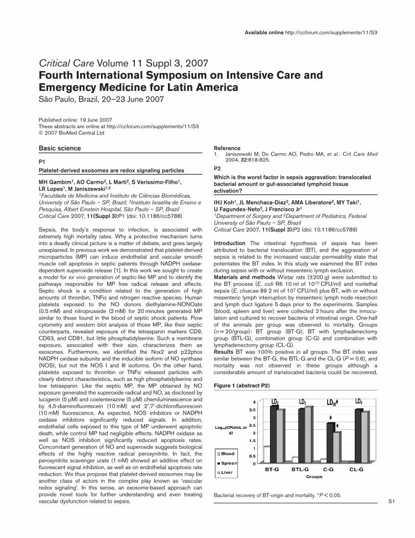
S1 Available online http://ccforum.com/supplements/11/S3 Critical Care Volume 11 Suppl 3, 2007 Fourth International Symposium on Intensive Care and Emergency Medicine for Latin America São Paulo, Brazil, 20–23 June 2007 Published online: 19 June 2007 These abstracts are online at http://ccforum.com/supplements/11/S3 © 2007 BioMed Central Ltd Basic science P1 Platelet-derived exosomes are redox signaling particles MH Gambim 1 , AO Carmo 2 , L Marti 2 , S Veríssimo-Filho 1 , LR Lopes 1 , M Janiszewski 1,2 1 Faculdade de Medicina and Instituto de Ciências Biomédicas, University of São Paulo – SP, Brazil; 2 Instituto Israelita de Ensino e Pesquisa, Albert Einstein Hospital, São Paulo – SP, Brazil Critical Care 2007, 11(Suppl 3):P1 (doi: 10.1186/cc5788) Sepsis, the body’s response to infection, is associated with extremely high mortality rates. Why a protective mechanism turns into a deadly clinical picture is a matter of debate, and goes largely unexplained. In previous work we demonstrated that platelet-derived microparticles (MP) can induce endothelial and vascular smooth muscle cell apoptosis in septic patients through NADPH oxidase- dependent superoxide release [1]. In this work we sought to create a model for ex vivo generation of septic-like MP and to identify the pathways responsible for MP free radical release and effects. Septic shock is a condition related to the generation of high amounts of thrombin, TNFα and nitrogen reactive species. Human platelets exposed to the NO donors diethylamine-NONOate (0.5 mM) and nitroprusside (2 mM) for 20 minutes generated MP similar to those found in the blood of septic shock patients. Flow cytometry and western blot analysis of those MP, like their septic counterparts, revealed exposure of the tetraspanin markers CD9, CD63, and CD81, but little phosphatidylserine. Such a membrane exposure, associated with their size, characterizes them as exosomes. Furthermore, we identified the Nox2 and p22phox NADPH oxidase subunits and the inducible isoform of NO synthase (NOS), but not the NOS I and III isoforms. On the other hand, platelets exposed to thrombin or TNFα released particles with clearly distinct characteristics, such as high phosphatidylserine and low tetraspanin. Like the septic MP, the MP obtained by NO exposure generated the superoxide radical and NO, as disclosed by lucigenin (5 µM) and coelenterazine (5 µM) chemiluminescence and by 4,5-diaminofluorescein (10 mM) and 2′,7′-dichlorofluorescein (10 mM) fluorescence. As expected, NOS inhibitors or NADPH oxidase inhibitors significantly reduced signals. In addition, endothelial cells exposed to this type of MP underwent apoptotic death, while control MP had negligible effects. NADPH oxidase as well as NOS inhibition significantly reduced apoptosis rates. Concomitant generation of NO and superoxide suggests biological effects of the highly reactive radical peroxynitrite. In fact, the peroxynitrite scavenger urate (1 mM) showed an additive effect on fluorescent signal inhibition, as well as on endothelial apoptosis rate reduction. We thus propose that platelet-derived exosomes may be another class of actors in the complex play known as ‘vascular redox signaling’. In this sense, an exosome-based approach can provide novel tools for further understanding and even treating vascular dysfunction related to sepsis. Reference 1. Janiszewski M, Do Carmo AO, Pedro MA, et al.: Crit Care Med 2004, 32:818-825. P2 Which is the worst factor in sepsis aggravation: translocated bacterial amount or gut-associated lymphoid tissue activation? IHJ Koh 1 , JL Menchaca-Diaz 2 , AMA Liberatore 2 , MY Taki 1 , U Fagundes-Neto 2 , J Francisco Jr 1 1 Department of Surgery and 2 Department of Pediatrics, Federal University of São Paulo – SP, Brazil Critical Care 2007, 11(Suppl 3):P2 (doi: 10.1186/cc5789) Introduction The intestinal hypothesis of sepsis has been attributed to bacterial translocation (BT), and the aggravation of sepsis is related to the increased vascular permeability state that potentates the BT index. In this study we examined the BT index during sepsis with or without mesenteric lymph exclusion. Materials and methods Wistar rats (±200 g) were submitted to the BT process (E. coli R6 10 ml of 10 10 CFU/ml) and nonlethal sepsis (E. cloacae 89 2 ml of 10 7 CFU/ml) plus BT, with or without mesenteric lymph interruption by mesenteric lymph node resection and lymph duct ligature 5 days prior to the experiments. Samples (blood, spleen and liver) were collected 2 hours after the innocu- lation and cultured to recover bacteria of intestinal origin. One-half of the animals per group was observed to mortality. Groups (n = 20/group): BT group (BT-G), BT with lymphadenectomy group (BTL-G), combination group (C-G) and combination with lymphadenectomy group (CL-G). Results BT was 100% positive in all groups. The BT index was similar between the BT-G, the BTL-G and the CL-G (P = 0.6), and mortality was not observed in these groups although a considerable amount of translocated bacteria could be recovered, Figure 1 (abstract P2) Bacterial recovery of BT-origin and mortality. *P < 0.05.
-
Upload
independent -
Category
Documents
-
view
3 -
download
0
Transcript of Toll-like receptor 2 induces chemokine receptor CXCR2 downregulation and neutrophil migration...
S1
Available online http://ccforum.com/supplements/11/S3
Critical Care Volume 11 Suppl 3, 2007Fourth International Symposium on Intensive Care andEmergency Medicine for Latin AmericaSão Paulo, Brazil, 20–23 June 2007
Published online: 19 June 2007These abstracts are online at http://ccforum.com/supplements/11/S3© 2007 BioMed Central Ltd
Basic science
P1Platelet-derived exosomes are redox signaling particles
MH Gambim1, AO Carmo2, L Marti2, S Veríssimo-Filho1, LR Lopes1, M Janiszewski1,2
1Faculdade de Medicina and Instituto de Ciências Biomédicas,University of São Paulo – SP, Brazil; 2Instituto Israelita de Ensino ePesquisa, Albert Einstein Hospital, São Paulo – SP, BrazilCritical Care 2007, 11(Suppl 3):P1 (doi: 10.1186/cc5788)
Sepsis, the body’s response to infection, is associated withextremely high mortality rates. Why a protective mechanism turnsinto a deadly clinical picture is a matter of debate, and goes largelyunexplained. In previous work we demonstrated that platelet-derivedmicroparticles (MP) can induce endothelial and vascular smoothmuscle cell apoptosis in septic patients through NADPH oxidase-dependent superoxide release [1]. In this work we sought to createa model for ex vivo generation of septic-like MP and to identify thepathways responsible for MP free radical release and effects.Septic shock is a condition related to the generation of highamounts of thrombin, TNFα and nitrogen reactive species. Humanplatelets exposed to the NO donors diethylamine-NONOate(0.5 mM) and nitroprusside (2 mM) for 20 minutes generated MPsimilar to those found in the blood of septic shock patients. Flowcytometry and western blot analysis of those MP, like their septiccounterparts, revealed exposure of the tetraspanin markers CD9,CD63, and CD81, but little phosphatidylserine. Such a membraneexposure, associated with their size, characterizes them asexosomes. Furthermore, we identified the Nox2 and p22phoxNADPH oxidase subunits and the inducible isoform of NO synthase(NOS), but not the NOS I and III isoforms. On the other hand,platelets exposed to thrombin or TNFα released particles withclearly distinct characteristics, such as high phosphatidylserine andlow tetraspanin. Like the septic MP, the MP obtained by NOexposure generated the superoxide radical and NO, as disclosed bylucigenin (5 µM) and coelenterazine (5 µM) chemiluminescence andby 4,5-diaminofluorescein (10 mM) and 2′,7′-dichlorofluorescein(10 mM) fluorescence. As expected, NOS inhibitors or NADPHoxidase inhibitors significantly reduced signals. In addition,endothelial cells exposed to this type of MP underwent apoptoticdeath, while control MP had negligible effects. NADPH oxidase aswell as NOS inhibition significantly reduced apoptosis rates.Concomitant generation of NO and superoxide suggests biologicaleffects of the highly reactive radical peroxynitrite. In fact, theperoxynitrite scavenger urate (1 mM) showed an additive effect onfluorescent signal inhibition, as well as on endothelial apoptosis ratereduction. We thus propose that platelet-derived exosomes may beanother class of actors in the complex play known as ‘vascularredox signaling’. In this sense, an exosome-based approach canprovide novel tools for further understanding and even treatingvascular dysfunction related to sepsis.
Reference1. Janiszewski M, Do Carmo AO, Pedro MA, et al.: Crit Care Med
2004, 32:818-825.
P2Which is the worst factor in sepsis aggravation: translocatedbacterial amount or gut-associated lymphoid tissueactivation?
IHJ Koh1, JL Menchaca-Diaz2, AMA Liberatore2, MY Taki1, U Fagundes-Neto2, J Francisco Jr1
1Department of Surgery and 2Department of Pediatrics, FederalUniversity of São Paulo – SP, BrazilCritical Care 2007, 11(Suppl 3):P2 (doi: 10.1186/cc5789)
Introduction The intestinal hypothesis of sepsis has beenattributed to bacterial translocation (BT), and the aggravation ofsepsis is related to the increased vascular permeability state thatpotentates the BT index. In this study we examined the BT indexduring sepsis with or without mesenteric lymph exclusion.Materials and methods Wistar rats (±200 g) were submitted tothe BT process (E. coli R6 10 ml of 1010 CFU/ml) and nonlethalsepsis (E. cloacae 89 2 ml of 107 CFU/ml) plus BT, with or withoutmesenteric lymph interruption by mesenteric lymph node resectionand lymph duct ligature 5 days prior to the experiments. Samples(blood, spleen and liver) were collected 2 hours after the innocu-lation and cultured to recover bacteria of intestinal origin. One-halfof the animals per group was observed to mortality. Groups(n = 20/group): BT group (BT-G), BT with lymphadenectomygroup (BTL-G), combination group (C-G) and combination withlymphadenectomy group (CL-G).Results BT was 100% positive in all groups. The BT index wassimilar between the BT-G, the BTL-G and the CL-G (P = 0.6), andmortality was not observed in these groups although aconsiderable amount of translocated bacteria could be recovered,
Figure 1 (abstract P2)
Bacterial recovery of BT-origin and mortality. *P < 0.05.

S2
Critical Care June 2007 Vol 11 Suppl 3 Fourth International Symposium on Intensive Care and Emergency Medicine for Latin America
particularly at the liver and spleen (Figure 1). When BT was addedto the sepsis without lymph exclusion (C-G), the BT index wasstatistically lower (P = 0.04); however, 50% (LD50) of mortalityoccurred within 30 hours (Figure 1).Conclusion These results show that, more than the amount oftranslocated bacteria, the gut-associated lymphoid systemactivation by the BT process played a pivotal role in the worseningof sepsis. Besides, BT occurred independently of mesentericlymph interruption, showing that the hematological route of BTmight be the principal route for bacterial dissemination into thebloodstream.
P3Toll-like receptor 2 induces chemokine receptor CXCR2downregulation and neutrophil migration impairment in severesepsis
JC Alves-Filho, A Freitas, F Spiller, FO Souto, H Paula-Neto, JS Silva, FQ CunhaDepartment of Pharmacology, School of Medicine of RibeirãoPreto, University of São Paulo – SP, BrazilCritical Care 2007, 11(Suppl 3):P3 (doi: 10.1186/cc5790)
There is a marked defect in neutrophil migration into the infectiousfocus during severe sepsis, which is associated with the severity ofdisease. Recently, we demonstrated that this phenomenon is aconsequence of downregulation of the chemokine receptorCXCR2 on the surface of circulating neutrophils. Toll-like receptorsare pattern-recognition receptors that are important in innateimmune responses to bacterial infection. Toll-like receptoractivation in phagocytes produces proinflammatory cytokines andchemokines that contribute directly to elimination of infectiousagents. A sustained inflammatory response, however, can result intissue damage and sepsis. Here, we address the role of Toll-likereceptor 2 (TLR2) in the downregulation of CXCR2 and theestablishment of neutrophil migration impairment in severe sepsis.TLR2-deficient (TLR2–/–) and C57BL/6 (WT) mice were subjectedto severe polymicrobial sepsis by the cecal ligation and puncturemodel, and neutrophil migration, bacteremia, CXCR2 expressionand cytokine levels were evaluated. It was observed that TLR2 iscritical for downregulation of CXCR2 expression on circulatingneutrophils during severe sepsis, since this event was prevented inTLR2–/– mice. In accordance, TLR2–/– mice did not present failureof neutrophil migration into the infectious focus and, consequently,they presented lower bacteremia and did not display systemicinflammation determined by reduced levels of circulating cytokines,showing an improve of survival rate. Furthermore, in vitro, TLR2agonist (lipoteichoic acid) was able to downregulate CXCR2expression and markedly to inhibit neutrophil chemotaxis inducedby CXCR2 ligand. The downregulation of CXCR2 was associated
with enhanced expression of G-protein-coupled receptor kinases-2(GRK-2), which is known to play an important role indesensitization and internalization of this chemokine receptor.Finally, we showed that in-vitro lipoteichoic acid-stimulatedneutrophils adoptively transferred into naïve WT mice display asignificantly reduced competence to migrate into peritoneal cavityin response to thioglycolate. Altogether, these findings suggestthat TLR2, through GRK2 signaling, downregulates CXCR2expression on the surface of circulating neutrophils, which is acritical determinant of impairment of neutrophil migration into theinfection focus during severe sepsis.
Hemodynamics/shock
P4Effects of hypertonic saline and lactated Ringer’s solutions onbacterial translocation in a rat model of intestinal obstructionand ischemia
FL Zanoni1, KV Greco1, ACR Moreno2, LF Poli de Figueiredo1, MR Silva1, P Sannomiya1
1Research Division, Heart Institute (InCor), LIM 11, University ofSão Paulo Medical School, São Paulo – SP, Brazil; 2Faculty ofPharmaceutical Sciences, Department of Clinical Analysis,University of São Paulo – SP, BrazilCritical Care 2007, 11(Suppl 3):P4 (doi: 10.1186/cc5791)
Introduction Clinical evidence suggests that bacterial trans-location (BT) may not be the primary cause in the development ofsepsis and multiple organ dysfunction. However, BT has animportant role in the activation of the immune system. Therapieshave been extensively investigated to improve tissue perfusion andreduce intestinal ischemia. The aim of this study is to evaluate theeffects of hypertonic saline (HSS) 7.5% and lactated Ringer’s (LR)solutions on intestinal BT in rats that underwent intestinalobstruction and ischaemia (IO).Methods Wistar rats (300 ± 50 g) underwent anesthesia withsodium pentobarbital (50 mg/kg, i.p.) and were submitted to IO: (i)cecum exposure, (ii) ileum ligation at 1.5 cm proximal to theileocecal valve, and (iii) ligation of the mesenteric vessels thatsupply a 7–10 cm length of the ileal loop. Two hours after surgicalprocedures, 4 ml/kg of 7.5% HSS or LR were administeredintravenously, during 5 minutes. Animals were killed 24 hours afterIO, and microbiological assays were performed in mesentericlymph nodes, liver, spleen, and blood.Results See Table 1.Conclusion HSS reduced the number of CFU/g in the liver,spleen, and blood after IO, resulting in improvement of the ‘gutbarrier function’.
Table 1 (abstract P4)
Microbiological assays
Mesenteric lymph nodes Liver Spleen Blood
Group +/n CFU/g +/n CFU/g +/n CFU/g +/n
Sham 1/7 57 0/7 NG 0/7 NG 0/7
IO 6/7 2,939 ± 1,751 6/7 953 ± 525 6/7 4,616 ± 1,973 4/7
LR 7/7 1,862 ± 1,178 5/7 3,080 ± 1,832 6/7 4,376 ± 2,836 6/7
HSS 6/7 2,371 ± 1,451 3/7 104 ± 67 4/7 174 ± 75 1/7
Sham group, false operated; +/n, number of animals with positive cultures for E. coli/total number of animals; CFU/g, colony formation units/g tissue (meanvalue ± SEM, n = 7 animals in each group); NG, no growth.

S3
Available online http://ccforum.com/supplements/11/S3
P5Experimental pulmonary microembolism: effects onhemodynamics, lung mechanics and histopathology
LCP Azevedo, DT Dolci, CB Fuentes, M Park, GPP SchettinoIntensive Care and Anesthesiology Research Laboratory, Researchand Education Institute, Hospital Sírio-Libanês, São Paulo – SP,BrazilCritical Care 2007, 11(Suppl 3):P5 (doi: 10.1186/cc5792)
Objectives To characterize an experimental model of pulmonaryembolism by studying hemodynamics, lung mechanics and histo-pathologic derangements caused by pulmonary microembolism inpigs. To identify lung alterations after embolism that may be similarto those evidenced in pulmonary inflammatory conditions.Materials and methods Ten Large White pigs (weight 35–42 kg)were instrumented with arterial and pulmonary catheters, andpulmonary embolism was induced in five pigs by injection ofpolystyrene microspheres (diameter ~300 µM), in order to obtain apulmonary mean arterial pressure of twice the baseline value. Fiveother animals injected with saline served as controls.Hemodynamic and respiratory data were collected and pressure xvolume curves of the respiratory system were performed by aquasi-static low flow method. Animals were followed for 12 hours,and after death lung fragments were dissected and sent topathology.Results Pulmonary embolism induced a significant reduction instroke volume (71 ± 18 ml/min/bpm pre vs 36 ± 9 ml/min/bpmpost, P < 0.05), an increase in pulmonary mean arterial pressure(27 ± 4 mmHg pre vs 39 ± 6 mmHg post, P < 0.05) and pulmo-nary vascular resistance (193 ± 122 mmHg/l/min pre vs451 ± 149 mmHg/l/min post, P < 0.05). Respiratory dysfunctionwas evidenced by significant reductions in the PaO2/FiO2 ratio(480 ± 50 pre vs 159 ± 55 post, P < 0.05), the dynamic lungcompliance (27 ± 6 ml/cmH2O pre vs 19 ± 5 ml/cmH2O post,P < 0.05), the increase in dead space ventilation (20 ± 4 pre vs47 ± 20 post, P < 0.05) and, the shift of pressure x volume curvesto the right, with reduction in pulmonary hysteresis. Pathologydepicted inflammatory neutrophil infiltrates, alveolar edema,collapse and hemorrhagic infarctions.Conclusion This model of embolism is associated with cardio-vascular dysfunction, as well as respiratory injury characterized bya decrease in oxygenation, lung compliance and hysteresis.Pathology findings were similar to those verified in inflammatorypulmonary injury conditions. This model may be useful to studypathophysiology, as well as pharmacologic and ventilatoryinterventions useful to treat pulmonary embolism.
P6Hemodynamic and metabolic features of a porcine systemiclow flow state model
LCP Azevedo1,2, AT Maciel1,2, D Noritomi1, GPP Schettino1, M Park1,2
1Intensive Care Laboratory, Research and Education Institute,Hospital Sírio-Libanês, São Paulo – SP, Brazil; 2Intensive CareUnit, Hospital das Clínicas, HCFMUSP, São Paulo – SP, BrazilCritical Care 2007, 11(Suppl 3):P6 (doi: 10.1186/cc5793)
Objective To describe a new experimental systemic low flow statemodel induced by cardiac tamponade.Methods Ten Large White pigs (43 ± 5 kg) were instrumented witharterial and pulmonary catheters, cystostomy and splenectomy, anda latex balloon was inserted anterior to the heart. Pigs wererandomized to a shock group or a control group. The shock grouphad the balloon inflated with 620 ± 344 ml to keep the mean arterialblood pressure at 45–55 mmHg (mean = 49 ± 4 mmHg) for 1 hour.Hemodynamic data were collected and shown as the mean ± SD.Two-way ANOVA was used with Bonferroni’s correction.Results During shock, the SvO2 was 34 ± 8%, the heart rate was173 ± 36 bpm, and the stroke volume was 18 ± 12 ml/min/beat.After shock, see Table 1.Conclusion In our model, transient cardiac tamponade causedpersistent hypotension and cardiovascular dysfunction.Hyperthermia was an interesting finding in the last hours of theexperiment in animals submitted to cardiac tamponade.
P7Effects of hypertonic saline solution and pentoxifylline on ratmesenteric microcirculation after hemorrhagicshock/reperfusion followed by cecal ligation/puncture: anintravital microscopic study
NK Nakagawa1,2, RA Nogueira1, CA Obuti2, CJ Correia1, JWMC Cruz3, LF Poli de Figueiredo1, M Rocha e Silva1, P Sannomiya1
1Research Division, Heart Institute (InCor), LIM-11 University ofSão Paulo Medical School, São Paulo – SP, Brazil; 2UNICID, SãoPaulo – SP, Brazil; 3Institute of Biomedical Sciences, University ofSão Paulo, BrazilCritical Care 2007, 11(Suppl 3):P7 (doi: 10.1186/cc5794)
Objective Hemorrhagic shock/reperfusion followed by sepsistriggers systemic microcirculatory disturbances that may inducemultiple organ failure. The present in-vivo study evaluated the
Table 1 (abstract P6)
Data Group Pre-shock Post-shock 1 hour 3 hours 6 hours P value
Mean arterial blood pressure (mmHg) Shock 111 ± 19 76 ± 13*,& 87 ± 22* 82 ± 10*,& 72 ± 7*,& <0.001#
Control 111 ± 16 115 ± 9 115 ± 9 102 ± 7 97 ± 8 <0.001$
Stroke volume (ml/min/beat) Shock 65 ± 9 22 ± 5*,& 44 ± 8*,& 38 ± 12 & 36 ± 8& <0.001#
Control 59 ± 16 59 ± 13 53 ± 7 54 ± 9 45 ± 12 <0.001$
SVO2 (%) Shock 73 ± 9 65 ± 8 69 ± 8 55 ± 15& 56 ± 13& 0.023#
Control 75 ± 7 72 ± 6 72 ± 6 68 ± 5& 62 ± 6& 0.002$
Lactate (mg/dl) Shock 15 ± 6 72 ± 19*,& 51 ± 22*,& 23 ± 19 14 ± 6 <0.001#
Control 25 ± 13 18 ± 8 12 ± 5 17 ± 13 14 ± 10 <0.001$
Temperature (ºC) Shock 36.8 ± 0.6 37.5 ± 0.4 38.7 ± 0.7*,& 39.6 ± 0.7*,& 39.7 ± 0.6*,& <0.001#
Control 36.5 ± 0.3 37.0 ± 0.6 37.5 ± 0.7 38.0 ± 0.6 38.4 ± 0.4 <0.001$
Two-way ANOVA between groups# and within groups$. Tukey *P < 0.05 vs control and &P < 0.05 vs baseline.

S4
effects of hypertonic saline solution (HSS) (7.5%, 4 ml/kg) andpentoxifylline (PTX) (4 mg/kg) on mesenteric microcirculation indouble-injured rats.Methods Thirty-three anesthetized Wistar rats (~250 g) wererandomly assigned to: (1) SHAM: no injury group; (2) HSS: hemor-rhagic shock/reperfusion with HSS; (3) LR: hemorrhagic shock/reperfusion with lactated Ringer’s solution (LR), three times shedblood volume; (4) HSS + PTX: hemorrhagic shock/reperfusionwith HSS associated with PTX; and (5) LR + PTX: hemorrhagicshock/reperfusion with LR associated with PTX. The animals weresubmitted to cecal ligation/puncture (second injury).Results Leukocyte–endothelium interactions (Table 1) wereassessed by intravital microscopy at mesenteric postcapillaricvenules (~21.07 µm diameter).Conclusion The in-vivo observation of the rat mesentericmicrocirculation showed that, in a double-injury model, reperfusionof the animals with HSS and PTX did attenuate the inflammationresponse compared with LR-reperfused animals.Acknowledgements Funded by FAPESP and PRONEX.
P8Increased pulse pressure variations observed in a pulmonaryexperimental thromboembolism model
GA Westphal1,2, ARR Gonçalves1,2, A Bedin1,2, R Steglich1,2, E Silva1,2, LF Poli de Figueiredo1,2
1Division of Experimental Surgery, Joinville University (Univille)Medical School, Joinville – SC, Brazil; 2Division of AppliedPhysiology, Heart Institute, InCor, University of São Paulo MedicalSchool, São Paulo – SP, BrazilCritical Care 2007, 11(Suppl 3):P8 (doi: 10.1186/cc5795)
Background Pulse pressure respiratory variation (PPV), which is thedifference between the maximal and minimal arterial pulse pressurevalues after each positive-pressure breath, is largely used for earlyidentification of hypovolemic status. Increased PPV observed inhypovolemia results from exaggerated respiratory variation intranspulmonary blood flow that results in corresponding left ventricularpreload variations during respiratory cycles. Hence, any modulationsthat affect the left ventricular preload would influence PPV.Objective To test the hypothesis that PPV amplification observedin hypovolemia can also be detected after pulmonary thrombo-embolism obtained with central venous injection of blood cloth.Methods PPV was studied in five anesthetized and mechanicallyventilated male rabbits weighing 1.6 ± 0.3 kg. The heart rate (HR)and mean arterial pressure (MAP) were monitored after centralvenous (jugular) and arterial (carotid) catheterization, and 1.5 ml/kgautologous blood cloth were injected slowly through the jugularcatheter into the central circulation. The HR, MAP and PPV wereregistered before and after blood cloth injection and comparedusing the Student t test.
Results The HR did not change, but the MAP was significantlylowered as much as PPV significantly increased after clothinjection. See Table 1.
Table 1 (abstract P8)
Before After P value
HR 249 ± 50 295 ± 22.9 0.2
MAP 55 ± 4.2 21 ± 3.4 <0.001
PPV 5.5 ± 1.8 30 ± 5.6 <0.009
Conclusion PPV amplification observed in hypovolemia can bealso detected after pulmonary thromboembolism obtained withcentral venous injection of blood cloth. It is possible to concludethat pulmonary hypertension should be assumed as a limitation forcardiovascular fluid responsiveness determination by PPV.
P9Pharmacological vasodilatation increased pulse pressurevariation mimicking hypovolemic status in rabbits
GA Westphal1,2, ARR Gonçalves1,2, A Bedin1,2, R Steglich1,2, E Silva1,2, LF Poli de Figueiredo1,2
1Division of Experimental Surgery, Joinville University (Univille)Medical School, Joinville – SC, Brazil; 2Division of AppliedPhysiology, Heart Institute, InCor, University of São Paulo MedicalSchool, São Paulo – SP, BrazilCritical Care 2007, 11(Suppl 3):P9 (doi: 10.1186/cc5796)
Background Pulse pressure respiratory variation (PPV), which isthe difference between the maximal and minimal arterial pulsepressure values after one positive-pressure breath, is largely usedfor early identification of hypovolemic status. Increased PPV, asseen in hypovolemia, results from exaggerated respiratory variationin transpulmonary blood flow that results in corresponding leftventricular preload variations during respiratory cycles. Hence, anyfactor that affects left ventricular preload can be associated withPPV amplification.Objective To test the hypothesis that PPV amplification observedin hypovolemia can also be observed during pharmacological vaso-dilatation, induced by sodium nitroprusside (SN).Methods Ten anesthetized, mechanically ventilated rabbits, under-went progressive hypotension by either controlled hemorrhage(CH) or intravenous SN infusion. CH group: five rabbits weresubmitted to graded hemorrhage of 10%, 20%, 30%, 40% and50% of their blood volume. Mean arterial pressure steps wereregistered and assumed as pressure targets. SN group: fiverabbits were submitted to a progressive SN dose infusion to reachsimilar pressure targets observed in the CH group (Table 1). PPVwas measured at each arterial pressure step.
Critical Care June 2007 Vol 11 Suppl 3 Fourth International Symposium on Intensive Care and Emergency Medicine for Latin America
Table 1 (abstract P7)
Leukocyte–endothelial interactions in mesenteric postcapillaric venules
Rollers Adherent cells Migrated cells Total white blood Group n (10 min) (100 µm venule length) (5,000 µm2) (cells/mm3)
SHAM 6 96 ± 12 3 ± 1 2 ± 1 12,367 ± 2,641
LR 7 207 ± 19a 17 ± 1a 17 ± 1a 16,771 ± 5,703a
HSS 8 108 ± 16 9 ± 3b 12 ± 3b 12,388 ± 6,629
HSS + PTX 7 102 ± 26 6 ± 1b,c 6 ± 1b,c 10,757 ± 2,483
LR + PTX 5 115 ± 20 7 ± 1b 7 ± 1b,c 11,650 ± 1,570
aP < 0.001 vs other groups, bP < 0.001 vs SHAM, cP < 0.001 vs HSS.

S5
Results The heart rate was significantly greater in the SN groupthan in the CH group (P < 0.05). PPVs were similar among theexperimental models in all steps (P = 0.17).Conclusion Pharmacologic vasodilatation by SN induced a PPVamplification similar to that observed in hypovolemia. Our resultsreinforce the idea that PPV amplification may be associated withpotential cardiovascular response and not necessarily hypovolemicstatus. Hence, caution should be exercised before assuming thatPPV is a marker of intravascular volume status.
P10Early fluid replacement with hypertonic isoncotic solutionguided by mixed venous oxygen saturation in experimentalhypodynamic sepsis
L Rahal, AG Garrido, RJ Cruz Jr, M Rocha e Silva, LF Poli de FigueiredoLIM 11, InCor, University of São Paulo School of Medicine, SãoPaulo – SP, BrazilCritical Care 2007, 11(Suppl 3):P10 (doi: 10.1186/cc5797)
Introduction Volume replacement is one of the cornerstones in themanagement of sepsis. The type and amount of fluid are stillcontroversial.Hypothesis A hypertonic isoncotic solution could promotesuperior hemodynamic benefits as the initial fluid regimen thanstandard crystalloid resuscitation, and mixed venous oxygensaturation could be useful to guide fluid administration in experi-mental sepsis.Methods Anesthetized mongrel dogs received an intravenousinfusion of 1.2 x 10¹⁰ cfu/kg live E. coli in 30 minutes (T0–T30).After 60 minutes (T90), the dogs were randomized to receiveisotonic saline solution, 32 ml/kg over 20 minutes (NS, n = 7) or7.5% hypertonic isoncotic solution (Hyper-Haes) 4 ml/kg over5 minutes (HH, n = 7). After 30 and 60 minutes (T120 and T150),additional isotonic saline solution 32 ml/kg was administered if
mixed venous oxygen saturation was below 70% in both groups.the mean arterial pressure (MAP), cardiac output (CO) and portalblood flow (PVBF) were monitored; blood gases and lactate levelswere analyzed at each timepoint.Results See Table 1. Data are expressed as the mean ± SEM.Conclusion Both solutions promoted similar and partial benefits atsystemic and regional levels in this hypodynamic sepsis model.Although initial fluid requirement after HH was lower than NS,overall fluid infused was not statistically different between groups(HH 31.4 ± 10.9 ml/kg vs NS 50.3 ± 6.5 ml/kg).Acknowledgement Supported by FAPESP 05/51176-5.
P11Dynamic evaluation of central venous pressure for fluidresponsiveness assessment in spontaneous breathing dogs
E Silva, P Rehder, GA WestphalDivision of Applied Physiology, Heart Institute, InCor, University ofSão Paulo Medical School, São Paulo – SP, BrazilCritical Care 2007, 11(Suppl 3):P11 (doi: 10.1186/cc5798)
Background Variations in intrathoracic pressure interfere withvenous return and cardiac output (CO). Inspiratory fall in centralvenous pressure (CVP) traces (ifCVP) during spontaneous breath-ing have been recommended for cardiovascular fluid responsive-ness (CFR) evaluation. We recently described the usefulness ofCVP wave amplitude variation (pressoric vena cava collapsibilityindex, Cvci) during mechanical ventilation for CFR estimation incritically ill patients. There are no data about the Cvci evaluationduring spontaneous breathing.Objective To test the hypothesis that Cvci can be used for CFRevaluation during spontaneous ventilation.Methods In six male, anesthetized, intubated and spontaneousbreathing dogs, CO measurements and CVP waves wereregistered through a Swan–Ganz catheter while the mean arterialpressure (MAP) was measured through an intraarterial catheter.
Available online http://ccforum.com/supplements/11/S3
Table 1 (abstract P9)
Pulse pressure respiratory variation values in every step in both groups
BL T1 T2 T3 T4 T5
Hemorrhage 3.9 ± 1.2 6.7 ± 1.8 9.7 ± 2.4 13.5 ± 1.6 15.1 ± 0.9 19.6 ± 2.4
Nitroprusside 5.6 ± 2.1 10.7 ± 2.4 10.7 ± 2.4 16.3 ± 4 22.1 ± 5.3 22.6 ± 5.4
Table 1 (abstract P10)
T0 T30 T90 T120 T150 T270
MAP (mmHg)
NS 102 ± 3.8 87 ± 5.4* 54 ± 7.3* 73 ± 9.5* 65 ± 10.2* 69 ± 15.7*
HH 105 ± 5.2 87 ± 7.8* 54 ± 7.3* 63 ± 8.1* 66 ± 9.3* 56 ± 9.4*
CO (l/min)
NS 2.1 ± 0.2 1.7 ± 0.1* 1.2 ± 0.2*,a,b 2.3 ± 0.3* 1.6 ± 0.2*,d 1.2 ± 0.2*
HH 2.2 ± 0.1 1.5 ± 0.1* 1.1 ± 0.1*,a,b 1.5 ± 0.2* 1.5 ± 0.2* 0.8 ± 0.1*
PVBF (ml/min)
NS 510 ± 91 324 ± 60 160 ± 44*,a 396 ± 85 275 ± 72* 167±53*
HH 561 ± 32 287 ± 31* 218 ± 39* 233 ± 47* 204 ± 29* 129 ± 29*
SpO2 (%)
NS 92 ± 1.2 86 ± 3.5 74 ± 4.6* 87 ± 3.2 79 ± 4.3 65 ± 6.9*
HH 89 ± 1.8 81 ± 3.9 68 ± 2.4* 70 ± 4.2 70 ± 3.6d 52 ± 6.9*
P < 0.05 for the following comparisons: *vs baseline, aT90 vs T120, bT90 vs T270, cT120 vs T150, dT150 vs T270.

S6
After baseline measurements a graded hemorrhage was performedin 10% quota until 50% of the estimated volemia. The total shedblood volume was then re-infused in the same quota. Measure-ments of the heart rate (HR), CO, MAP, CVP, ifCVP, and Cvci areperformed in every bleeding and re-infusion step. The Cvci wascalculated with the following formula: Cvci (%) = [(CVPPexp –CVPPins) / CVPPexp] x 100, using the inspiratory (CVPPins) andexpiratory central venous pulse pressure (CVPPexp). ifCVP = CVPmeasured in the ‘a’ wave base at expiration minus CVP measuredin the ‘a’ wave base at inspiration. Correlations among the CO andother variables were performed using the Spearman coefficienttest.Results See Table 1. The correlations encountered were: CO andSvO2 (r = 0.94, P < 0.001); CO and Cvci (r = 0.61, P < 0.04);and CO and ifCVP (r = –0.02, P < 0.9).Conclusion We conclude that a pressoric vena cava collapsibilityindex may be used to detect cardiovascular fluid responsivenessduring spontaneous ventilation.
P12Evaluation of tissue perfusion parameters and intravascularvolume, emphasizing the inferior vena cava diameter andcollapsibility
H Missaka, MA Lima, H Cal, NM Otto, D Moraes, P Rotava, MH Pereira, A Farias, J Abrantes, AC Malizia, S Divan Filho, JL Machado, J Campos, RP Confalonieri, PCT CostaIntensive Care Unit, Emergency Department, Hospital MunicipalSouza Aguiar (HMSA), Rio de Janeiro – RJ, BrazilCritical Care 2007, 11(Suppl 3):P12 (doi: 10.1186/cc5799)
Introduction Echocardiography in critically ill patients enablesdiagnosis of a large number of cardiac conditions, including life-threatening ones. Intensivists can use it as a powerful diagnostic tool.Objective A comparison of intravascular volume and tissueperfusion parameters in critically ill patients to enhance beneficialconduct in treatment and outcome using the inferior vena cavadiameter as guidance.Materials and methods Patients were enrolled from Novemberuntil December 2006 in the ICU of the Emergency Department atHMSA. Inclusion criteria: (a) hemodynamic instability ordependency on vasoactive drugs, at the first 6 hours; (b) age>18 years; (c) deep vein access in superior vena cava. Evaluationof the intravascular volume and tissue perfusion parametersfollowed after admission, with normal values being defined ascardiac rate (CR: 80–100 bpm); mean blood pressure (MBP:>90 mmHg); central venous pressure (CVP: 8–12 mmHg); serumlactate (Lac: < 1 mmol/l); arterial oxygen saturation (SaO2: >90%);central venous oxygen saturation (ScvO2: >75%); ∆PCO2(<4 mmHg indicates a cardiac index >2.5 l/min/m2); inferior venacava diameter (IVC: >15 mm) and its variation with inspiration(∆IVC: <50%).Results A total of 32 patients were investigated – of which fivepresented with the following apparent divergences:
1. CR: 98 bpm; MBP: 80 mmHg, in use of norepinephrine (NE);CVP: 12 mmHg; Lac: 1.6 mmol/l; SaO2: 98.1%; SvcO2:54.9%; ∆PCO2: 5 mmHg; IVC: 24 mm; ∆IVC: 10%. Proce-dure: patient with severe left ventricular dysfunction. IncreasedIVC demanded initiation of inotropic drugs.
2. CR: 128 bpm; MBP: 119 mmHg, in use of NE; CVP: 18.4 mmHg;Lac: 9.6 mmol/l; SaO2: 96.5%; SvcO2: 83.8%; ∆PCO2:1.7 mmHg; IVC: 3 mm; ∆IVC: 66%. Procedure: septic patient,hyperdynamic. Decreased IVC resulted in volume replacement.
3. CR: 86 bpm; MBP: 75 mmHg; CVP: 6.5 mmHg; Lac:1.1 mmol/l; SaO2: 87%; SvcO2: 81.2%; ∆PCO2: 6.2 mmHg;IVC: 7 mm; ∆IVC: 70%. Procedure: trauma victim with ARDS,in mechanical ventilation (PEEP: 12 cmH2O). Decreased IVCresulted in volume infusion.
4. CR: 106 bpm; MBP: 60 mmHg; CVP: 5.5 mmHg; Lac:1.6 mmol/l; SaO2: 95.9%; SvcO2: 74.3%; ∆PCO2: 3.4 mmHg;IVC: 12 mm; ∆IVC: 60%. Procedure: patient with sub-arachnoid hemorrhage. Normal IVC diameter and collapsibilityhelped to maintain MBP > 100 mmHg and prevent vasospasm.
5. CR: 128 bpm; MBP: 90 mmHg; CVP: 18.5 mmHg; Lac:1.4 mmol/l; SaO2: 80%; SvcO2: 71.2%; ∆PCO2: 3.2 mmHg;IVC: 25 mm; ∆IVC: 5%. Procedure: hypervolemic patient withARDS, in mechanical ventilation (APRV-Bilevel). Increased IVCresulted in volume restriction and use of diuretics to improveP/F.
Conclusion Cases reported in this study demonstrate how the IVChelped monitor hemodynamics in critically ill patients and led tofurther decisions in treatment. Other studies also recommend theincorporation of this technology as a routine in ICUs due to itsnoninvasivity, feasibility, accessibility and lower risks.References1. Feissel M, Michard F, Faller JP: The respiratory variation in infe-
rior vena cava diameter as a guide to fluid therapy. IntensiveCare Med 2004, 30:1834-1837.
2. Jardin F, Vieillard-Baron A: Ultrasound examination of the venaecavae. Intensive Care Med 2006, 134:203-206.
3. Price S, Nicol E, Gibson DG: Echocardiography in the criticallyill: current and potential roles. Intensive Care Med 2006, 32:48-59.
P13Base excess and early mortality in patients admitted to thegeneral intensive care unit at a university hospital in Fortaleza
FA de Meneses, ISAM Bezerra, E Ribeiro, AH Furtado Junior, AA Peixoto JuniorFederal University of Ceará – UFC, Intensive Care Unit, Fortaleza –CE, BrazilCritical Care 2007, 11(Suppl 3):P13 (doi: 10.1186/cc5800)
Introduction Base excess is considered an indicator of injury,shock and adequate resuscitation. We looked to establish arelation between base excess and serum bicarbonate obtained onadmission to the ICU and the prognostics of patients.Methods A retrospective study with analysis of 110 patientsadmitted consecutively to the ICU, during the period June–December 2006.
Critical Care June 2007 Vol 11 Suppl 3 Fourth International Symposium on Intensive Care and Emergency Medicine for Latin America
Table 1 (abstract P11)
Step 0 –10% –20% –30% –40% –50% –40% –20% 0
CO 2.8 ± 0.5 2.4 ± 0.4 2.2 ± 0.4 1.8 ± 0.5 1.6 ± 0.5 1.4 ± 0.5 1.8 ± 0.4 3.0 ± 0.7 3.5 ± 0.8
ifCVP 2.8 ± 0.4 3.1 ± 0.5 3.8 ± 0.7 3.3 ± 0.5 2.1 ± 0.2 2.7 ± 0.5 4.1 ± 1 3.0 ± 0.5 2.3 ± 0.2
Cvci –8 ± 6 –11 ± 13 –19 ± 8 –18 ± 15 –27 ± 10 –17 ± 7 –12 ± 20 –11 ± 7 –8 ± 4

S7
Results Of the 110 patients, there was a predominance of womenand mean age 54.2 ± 18.7 years. The length of stay in the ICU was6.5 ± 7.4 days and the mean APACHE II index, at the first 24 hoursof admission, was 21.0 ± 8.1 points. Most patients survived(71.9%), 9.3% died during the first 48 hours in the ICU and 18.6%after 48 hours from admission to this unit. The standardizedmortality ratio was 0.715. Patients with early mortality, during thefirst 48 hours in the ICU, had lower base excess (–7.75 ± 8.33 vs–3.17 ± 5.43) and serum bicarbonate (16.7 ± 6.2 vs 20.9 ± 5.6)than survivors (P < 0.05). Patients with permanence in the ICU upto 7 days and patients that stayed in this unit for more than 7 dayshad similar base excess and serum bicarbonate (–3.24 ± 5.37 vs–2.98 ± 5.72 and 20.9 ± 5.3 vs 20.9 ± 6.3) (P > 0.05).Conclusion Serum bicarbonate and base excess were associatedwith early mortality during the ICU stay. However, theseparameters did not correlate with ICU length of stay.
P14Specificity of the pulmonary artery catheter in classifying thetype of shock
SC Aranha, MH Fujino, FM Costa, CA Horta, SE Mataloun, M MoockGrajau State Hospital (HEG), UNISA Medical School, São Paulo –SP, BrazilCritical Care 2007, 11(Suppl 3):P14 (doi: 10.1186/cc5801)
Objective To evaluate the utility of the pulmonary artery catheter(PAC) to classify the type of shock in hemodynamic instabilitypatients with no known reason.
Materials and methods Nineteen patients from Grajau StateHospital ICU who had shock diagnosis and those who needed aPAC to diagnose were evaluated.Results The average age was 49 years and the APACHE IIaverage score was 17. The average catheter length of stay was3.68 days. The most common reason for inpatient admission wascardiovascular (42.1%), followed by respiratory (26.3%); 52.6% ofclinical diagnoses were of distributive shock, only 42.1% wereconfirmed by catheter. Cardiogenic shock was diagnosed in42.1% before the catheter, and after the PAC it was 26.3%.Hypovolemic shock had the same rate of 5.2% before and aftercatheter insertion.Conclusion Even with a clinical body being well trained to classifyshock and the low number of patients in this study, the PAC iscertainly useful to predict the type of shock.References1. Yu DT, Platt R, Lanken PN, et al.: Relationship of pulmonary
artery catheter use to mortality and resource utilization inpatients with severe sepsis. Crit Care Med 2003, 31:2734-2741.
2. Rapoport J, Teres D, Steingrub J, et al.: Patient characteristicsand ICU organizational factors that influence frequency ofpulmonary artery catheterization. JAMA 2000, 283:2559-2567.
3. American Society of Anesthesiologists Task Force on PulmonaryArtery Catheterization: Practice guidelines for pulmonary arterycatheterization: an update report by the American Society ofAnesthesiologists Task Force on Pulmonary Artery Catheteri-zation. Anesthesiology 2003, 99:988-1014.
P15Systemic and regional hemodynamic and metabolic changesin an experimental model of brain death
FA De Luca, RJ Cruz Jr, AG Garrido, R Prist, F Scuotto, M Rocha e SilvaHeart Institute – InCor, University of São Paulo – SP, BrazilCritical Care 2007, 11(Suppl 3):P15 (doi: 10.1186/cc5802)
Introduction Despite the evolution of transplant techniques, thegreat number of donated organs continues to proceed from donorsin brain death (BD). The need for stabilization in patients with BD,in the view of the triggered autonomic storm, is basic in such a waythat knowledge of the physiopathologic, hemodynamic and meta-bolic disturbances becomes essential.Objective We evaluated hemodynamic and metabolic changesinduced by experimental BD in dogs.Materials and methods Ten anesthetized and ventilated mongreldogs (17–25 kg) were subjected to BD, by brainstem herniation,induced through an intracerebral balloon filled to maintain intra-
Available online http://ccforum.com/supplements/11/S3
Figure 1 (abstract P13)
Table 1 (abstract P15)
Baseline T5 T15 T30 T60
Mean arterial pressure (mmHg) 115.3 ± 6.3 187.8 ± 13.6* 110.2 ± 11.1 57.4 ± 4.4* 53.8 ± 3.1*ICP (mmHg) 19 ± 4 274 ± 16.6* 194 ± 25.2* 137 ± 12.3* 37 ± 3.31*Cardiac index (l/min) 3 ± 0.2 3.6 ± 0.4 4 ± 0.4* 3.2 ± 0.4 2.6 ± 0.3PVBF (ml/min) 685.6 ± 114.2 883.6 ± 167.7 1074 ± 179.2* 846 ± 165 622.9 ± 130HABF (ml/min) 277.2 ± 33.1 521.2 ± 88* 332.1 ± 65.7 178.3 ± 39.7 134.9 ± 28*Cerebral perfusion pressure (mmHg) 96.5 ± 6.2 –55.5 ± 30.8* –64.3 ± 32.7* –65.9 ± 18.2* –15 ± 3.6*SvO2 89.4 ± 1.7 86.9 ± 2.2SpO2 94.3 ± 1.7 86.3 ± 2.5*Arterial lactate (mmol/l) 0.6 ± 0.3 0.9 ± 0.4Portal vein lactate (mmol/l) 0.6 ± 0.3 0.8 ± 0.3Cerebral lactate (mmol/l) 1 ± 0.3 1.1 ± 0.5
*P < 0.05 vs baseline.
S8
cranial pressure (ICP) > systolic arterial pressure for 30 minutes(baseline–T30). The animals were observed for 30 minutes there-after (T60). Systemic hemodynamics was evaluated by arterial andpulmonary artery catheters, while regional perfusion was assessedby portal vein blood flow (PVBF) and hepatic artery blood flow(HABF) with ultrasonic flow probes.Results See Table 1. The data are expressed as the mean ± SEM.Conclusion BD promoted an initial hyperkinetic state followed bymarked hypotension without systemic and regional lactic acidosis.In spite of the severe hypotension, the hepatosplachnic blood flowwas preserved.
Sepsis
P16Potential role of poly(ADP-ribose) activation in myocardialcontractile dysfunction of human septic shock
AC Nogueira, M Lins, W Hoshino, L Gonzaga, V Kawabata, J Barradas, S Cappi, A Duarte, P Bisele, F Maia, M Miranda, M Bernik, PA Lotufo, FG SorianoHospital Universitário da USP e Faculdade de Medicina USP, SâoPaulo – SP, BrazilCritical Care 2007, 11(Suppl 3):P16 (doi: 10.1186/cc5803)
Objective To study whether cardiodepression found in septicpatients is associated with plasma markers of myocardial necrosisand with myocardial polyADP(ribose)polymerase (PARP) activa-tion. Sepsis is associated with increased production of superoxideand nitric oxide with consequent peroxynitrite (ONOO–) genera-tion. Cardiodepression is induced in the heart during oxidativestress associated with septic shock. Oxidative and nitrosativestress can lead to activation of the nuclear enzyme PARP, withsubsequent loss of myocardial contractile function.Design A prospective and observational study.Setting A university hospital ICU for clinical and surgical patients.Participants We assigned 25 patients presenting severe sepsis orseptic shock.Interventions Patients were followed for 28 days, and data werecollected and analyzed a posteriori, separating into two groups:survivors and nonsurvivors.Measurements and main results Function of the heart in septicpatients correlates to PARP activation in dead patients. The studypopulation included 25 individuals, of whom 12 died during thefollow-up period of 6 days. The initial data of inflammation markerC-reactive protein and APACHE severity were similar in bothgroups. Overall, an increase in the plasma troponin level wasrelated to increased mortality risk. Patients that died presentedheart dysfunction, and histological analysis of the heart showedinflammatory infiltration, increased collagen in the interstitium, andderangement of mitochondrial cryptae. Immunohistochemicalstaining for poly(ADP-ribose) (PAR), the product of activated PARP,was demonstrated in septic hearts. There was a positivecorrelation between PAR staining score and troponin I (r2 = 0.81);and a correlation of PAR staining score and LVSSW (r2 = 0.61).Conclusion Septic patients with impaired cardiac functiondemonstrate inflammatory alterations and PARP activation. Wesuggest that PARP activation may be, in part, responsible for thecardiac function depression observed in patients with severesepsis.
P17Mitochondrial injury in sepsis
AC Nogueira, W Hoshino, L Gonzaga, V Reze, A Duarte, C Valeri, P Branquinho, M Seckler, E Estumano, V Kawabata, D Noritomi, S Cappi, M Lins, M Miranda, K Sichieri, F Maia, AS Colombo, EL Azevedo, BCS Martins, M Bernik, EG Caldini,PA Lotufo, FG SorianoHospital Universitário, Universidade de São Paulo – SP, BrazilCritical Care 2007, 11(Suppl 3):P17 (doi: 10.1186/cc5804)
Background Sepsis-induced multiple organ failure is the majorcause of mortality and morbidity in critically ill patients. However,the precise mechanisms by which this dysfunction is causedremain to be elucidated. It seems that, in sepsis, mitochondriadysfunction results in raised tissue oxygen tensions and organfailure. Possibly due to oxide nitric, that is produced in excess insepsis, and is known to inhibit mitochondrial respiration in vitro.Objective To analyze cellular damage to electronic microscopyand evaluated its possible relation with serum cardiac markers(troponin, MB-creatin phosphate kinase), and homodynamic data.Methods We selected all consecutive patients who met thecriteria for septic shock, and we collected blood samples from thefirst through the 12th day, or until death. We also analyzedhomodynamic parameters by pulmonary catheter. From thepatients that died, a fragment of the left ventricle was sent forelectronic microscopy. The exclusion criteria were previouscoronary artery disease or dilated miocardiopathy.Results We studied 22 patients, age 53 ± 4 years, APACHEscores 22 ± 2; mortality was 45%. The patients who died showeddata of cardiac damage from the first day. This was shown bytroponin (0.54 ± 0.08 U/Ml vs 1.7 ± 0.3 U/Ml) and the leftventricular systolic worth index (64.2 ± 3.7 vs 37.6 ± 1.3),respectively, in survivor and nonsurvivor groups. The electronicmicroscopy from the myocardial of the nonsurvivor group showed asignificant injury in the mitochondria, represented by an increase inits numbers. There was an alteration on organelle organization andmitochondria crest lesions. The histology of the heart demonstratedinflammatory infiltration and increases of collagen fibers.Conclusion Septic patients with impaired cardiac functiondemonstrate inflammatory alterations and mitochondrial damage.We hypothesize that mitochondrial damage may, in part, beresponsible for the cardiac depression seen in severe septicpatients.
P18Septic lipidic dysregulation is related to heart rate variabilityalteration
AC Nogueira, V Kawabata, P Biselli, J Barradas, M Lins, C Valeri, M Seckler, W Hoshino, L Gonzaga Jr, MMS Bernik, PA Lotufo, E Martins, R Curi, FG SorianoHospital Universitario USP, Emergencias Clinicas FMUSP, SãoPaulo – SP, BrazilCritical Care 2007, 11(Suppl 3):P18 (doi: 10.1186/cc5805)
Context Although observational studies have demonstrated analteration of heart rate variability (HRV) in septic patients, no singlestudy has systematically addressed the relationship of heartdamage by systemic inflammation and metabolic alterations.Objective To determine whether heart damage from sepsis iscaused by free fatty acids (FFA) and may be detected with HRVanalysis.Design A prospective and observational study of patientspresenting with severe sepsis or septic shock.
Critical Care June 2007 Vol 11 Suppl 3 Fourth International Symposium on Intensive Care and Emergency Medicine for Latin America

S9
Setting A university hospital ICU for clinical and surgical patients.Participants Thirty-one patients were included with sepsis.Exclusion criteria were previous myocardial dysfunction, coronaryartery disease and cancer. The data were collected and analyzed aposteriori, in two groups: survivors and nonsurvivors.Main outcome measures Association between troponin I elevation,FFA elevation and HRV reduction. Association between clinicalevolution and HRV index, troponin, and hemodynamic parameters.Results The study population included 31 individuals, of whom 19died during the follow-up of 6 days. The initial measurements of C-reactive protein and gravity APACHE score were similar in the twogroups. Overall, an increase in the plasma troponin level wasrelated to an increased mortality risk. From the first day thenonsurvivor group presented a reduced left ventricular stroke worksystolic index (LVSWI), and a reduced low frequency (LF) index.The correlation coefficient for LF values and troponin wasr2 = 0.75. Patients showed FFA elevation; survivors presented0.62 ± 0.08 mmol/l and nonsurvivors 1.05 ± 0.12 mmol/l.Conclusion Understanding damage to the heart from sepsisrequires specific analysis of biochemical markers such as troponinI, and of hemodynamic parameters such as the LVSWI or the HRVindex. Our results suggest that damage to the heart by systemicinflammation is the cause of an aberrant beat-to-beat response.FFA produce cell death (apoptosis and necrosis) through oxidativestress and induced LF alterations. FFA inducing LF alterations hasbeen shown in the literature for healthy and diabetic patients; thisis the first time it has been shown in septic patients.
P19Serum lipids analysis in septic shock patients
AC Nogueira, S Cappi, C Valeri, J Barradas, V Reze, D Noritomi,ER Borges, A Duarte, M Lins, E Comissario, K Sichieri, R Curi,H Takahashi, M Miranda, M Bernik, PA Lotufo, M Martins, JB Machado, A Colombo, FG SorianoHospital Universitário da USP e Faculdade Medicina USP, SãoPaulo – SP, BrazilCritical Care 2007, 11(Suppl 3):P19 (doi: 10.1186/cc5806)
Objective We conducted a prospective study to analyze serumlipids, glucose, triglycerides and C-reactive protein in septic shockpatients to evaluate its relation with outcome.Design Prospective observational analysis of septic shock patients.Setting A 28-bed medico-surgical ICU.Participants Eighteen patients were analyzed.Materials and methods We collected blood samples for analysis ondays 1, 3, 6, 9, 12 or until death. Statistical analysis All results arepresented as the mean with standard deviation. For analysis wedivided patients into survivors and nonsurvivors at day 12. Weperformed a paired Student t test for differences in continuousvariables, and correlation coefficients were determined according tomultiple-level regression analysis. P < 0.05 was considered significant.Results Our mortality rate was 60%.The two groups had similarAPACHE II scores. At day 1 there were no statistical differencesfor any of the substances analyzed. From day 3 onward, significantdifferences were found between groups for total cholesterol, HDLfraction, triglycerides, glycemia and C-reactive protein. As indepen-dent variables, we found glycemia and triglycerides.Conclusion Low levels of cholesterolemia, HDL fraction, hyper-triglyceridemia and hyperglycemia were statistically significantlyrelated to a poor prognosis.
P20Clinical factors associated with mortality in septic shock
FS Dias, M Eidt, RP Duquia, F Stringhi, C Schwartzman, F Sztiler, MO Guerreiro, MS Canabarro, C LeonhardtGeneral ICU, Hospital São Lucas (PUCRS), Porto Alegre – RS, BrazilCritical Care 2007, 11(Suppl 3):P20 (doi: 10.1186/cc5807)
Introduction Septic shock (SS) is a disease associated with highmortality worldwide. In Brazil, mortality in SS reaches 60%. Theaim of our study was to identify clinical variables easily accessed inthe presentation of SS and their correlation with mortality.Methods Between January 2003 and December 2004, all patientswith SS criteria according to the ACCP/SCCM were included inthis observational study. At the time of SS diagnosis the followingvariables were collected: age, gender, heart rate (HR) and meanarterial pressure (MAP). On the ICU admission day the APACHE IIand SOFA scores were calculated. Data were retrieved from thepatient chart by one of the investigators, then transferred to STATAversion 9.0 software, where all analyses were run. All patients werefollowed until ICU discharge or death.Results During the period of study, there were 794 admissions tothe ICU, of whom 239 (30%) presented SS. Sixty-seven percentwere male, mean age was 57.0 (SD = 17.7) years, mean HR was108 (SD = 26.3) bpm, and mean MAP was 64.5 (SD = 21.2)mmHg. The mean APACHE II score was 23.3 (SD = 8.6) and themean SOFA score was 9.7 (SD = 3.2). The ICU mortality rate was66.5%. In the analysis of the prevalence of mortality and its crudeassociation with independent variables, age and gender show noassociation. Patients with HR above 108 bpm presented amortality OR of 1.78 (0.98–3.24) compared with those patientswith HR equal to or less than 108 bpm (P < 0.05). An APACHE IIscore greater than 24 points was associated with a mortality OR of2.91 (1.52–5.78) compared with those patients with a score equalto or less than 24 (P < 0.001). A SOFA score greater than 8points was associated with a mortality OR of 1.89 (1.04–3.42),compared with patients with values equal to or less than 8. Theanalysis of MAP demonstrated a trend to a lower mortality, inassociation with a higher level.Conclusion Our study confirmed, as previously demonstrated, thata HR less than 110 bpm at SS presentation is associated with lowmortality, as well a higher level of MAP. The severity of illness(APACHE II score > 24 points) is indicative of high-risk mortality;multiple organ dysfunction (SOFA score > 8 points), and a worseoutcome.References1. Parker MM, Shelhamer JH, Natanson C, et al.: Serial cardiovas-
cular variables in survivors and nonsurvivors of human septicshock: heart rate as an early predictor of prognosis. Crit CareMed 1987, 15:923-929.
2. Bernard GR, Vincent JL, Laterre PF, et al.: Efficacy and safety ofrecombinant human activated protein C for severe sepsis. N Engl J Med 2001, 344:699-709.
Available online http://ccforum.com/supplements/11/S3

S10
P21Acid–base disturbances in critically ill septic patients: alongitudinal quantitative study
DT Noritomi1, M Park2, AB Liborio2, SB Cappi1, PJC Biselli1, WI Hoshino1, FG Soriano1
1University Hospital, University of São Paulo – SP, Brazil; 2Hospitaldas Clínicas, São Paulo Medical School, University of São Paulo –SP, BrazilCritical Care 2007, 11(Suppl 3):P21 (doi: 10.1186/cc5808)
Introduction Applying a quantitative methodology, we describedthe acid–base status of severe septic patients in the first 5 daysafter admission to the ICU.Patients and methods Patients were studied if they had adiagnosis of severe sepsis with less than 24 hours of organ dys-function. Data were prospectively collected daily until the fifth dayafter inclusion.Results Sixty patients were included in the study. At admission tothe ICU, septic patients presented a severe metabolic acidosiswith an average pH of 7.29; PCO2 = 36 mmHg and SBE = –8.0.Figure 1 presents the several components of the metabolic acid–base disturbances found on the first day in the ICU. We found thatthe magnitude of metabolic acidosis, measured by the SBE, wasgreater among the nonsurvivors than the survivors. However, thecomponents of acid–base disturbances are kept proportionallyconstant among different clinical outcome subgroups.During the study period, the survivor group presented an increasedSBE from –6.4 to –1.5 due to a significant decrease in serumlactate level and SIG. No change occurred in the albumin serumlevel, which persisted as an alkalinizing force. In contrast, thenonsurvivor group became even more acidemic due to an increasein the PCO2 and persistence of a highly negative SBE. From themetabolic point of view, no significant change occurred in thisgroup from the first to the last day of the study, except for a smallincrease in the phosphate serum level.Conclusion Severe septic patients present, on the first day in theICU, a complex metabolic acid–base disturbance marked by amixed high-degree acidosis partially attenuated by a hypo-albuminemic alkalosis. Over the study period, the survivor grouppartially corrected its acidosis mainly through the disappearance ofunmeasured anions and lactate. Nonsurvivors did not changesignificantly their metabolic acidosis over time.
P22Institutional evaluation of a new methodology for early sepsisrisk identification in hospitalized patients
GA Westphal, KFP Fujiwara, AA Kawate, AO Monteiro, MM Schroeder, N Ferrari, RR da Silva, IV Scremin, R BeimsCentro Hospitalar Unimed, Joinville – SC, BrazilCritical Care 2007, 11(Suppl 3):P22 (doi: 10.1186/cc5809)
Background The effectiveness of sepsis, severe sepsis and septicshock management on prognosis depends strongly on early clinicalsuspicion and rigorous diagnosis methods. Early clinical sugges-tive infection sign recognition is therefore also a cornerstone ofsuccessful treatment.Objective To evaluate a new institutional methodology for earlysepsis risk identification in hospitalized patients.Methods A before–after study design with prospectiveconsecutive data collection in a 124-bed private medical center.Twelve months after the institutional Surviving Sepsis Campaignimplementation and current use of the respective treatmentbundles, this medical center adopted a standardized hospitalmaneuver to anticipate the identification of two or more suggestiveinfection signs. Demographic data, the time interval for recognitionof two or more infection risk signs, and the mortality rate areevaluated during the next 5 months (phase II) and compared withthe same data obtained during the initial 12 months (phase I).Results A total of 85 patients with two or more suggestiveinfection signs were enrolled. Thirty-two patients were recognizedwith severe sepsis during phase I and 22 patients in phase II.Demographic variables and severity of illness measured by theAPACHE II score (P = 0.12) were similar for both groups. Thephase I severe sepsis patients were identified after 29 ± 32 hoursfrom the initial presentation of two or more infections signs. On theother hand, during phase II this time was lower: 14.5 ± 16 hours(P < 0.07). The hospital mortality was greater in the phase I group(50%) when compared with the phase II group (27.3%) (P < 0.08).Conclusion These preliminary data suggest that the imple-mentation of the proposed methodology for early sepsis riskidentification in hospitalized patients was associated with earlysevere sepsis recognition and reduced mortality.
P23Sepsis provokes host’s microbiota overgrowth of commensalGram-negative bacteria and subsequent induction of bacterialtranslocation in rats
AMA Liberatore1, JL Menchaca-Diaz1, RM Silva2, MY Taki3, MR Silva1, J Francisco Jr3, TH Koh3, MB Morais1, IHJ Koh3
1Department of Pediatrics, 2Department of Microbiology and3Department of Surgery, Federal University of São Paulo – SP, BrazilCritical Care 2007, 11(Suppl 3):P23 (doi: 10.1186/cc5810)
Introduction The literature has shown the participation of intestinalmicrobiota in the genesis of primary infections as well as of sepsis.In this study we examine the role of sepsis on the microbiota byexamining the most frequently recovered Gram-negative bacteria(G–).Materials and methods Adult Wistar rats (±200 g) weresubmitted to the induction of semi-lethal sepsis (S-G) (E. coli R61 ml of 108 CFU/ml/100 g body weight, i.v.). Firstly, fecal G–kinetic following sepsis induction was examined (6, 12, 24, 48, 72,120 and 216 hours) (n = 6). After sepsis induction, in other groups(n = 18), samples were harvested from the small bowel(duodenum, jejunum, ileum) and large bowel (cecum and fecesbefore and after sepsis) at 6, 12 and 24 hours, and the BT index
Critical Care June 2007 Vol 11 Suppl 3 Fourth International Symposium on Intensive Care and Emergency Medicine for Latin America
Figure 1 (abstract P21)
Components of SBE on the first day.

S11
was examined at the mesenteric lymph nodes (MLN), liver andspleen by culture in MacConkey medium. Control groups were thesham group (Sham-G, saline injection) (n = 18) and the naïvegroup (N-G, without any procedure) (n = 6).Results Overall data showed that, after sepsis induction, fecal G–microbiota increased progressively up to 24 hours (P < 0.05)returning to control level after 72 hours (data not shown). Gutsegment overgrowth was also found until 24 hours and BToccurred during this period (Figure 1).Conclusion Sepsis provoked G– overgrowth and this was able toinduce the BT process. Other factors, such as splanchnichypoperfusion, decreased peristalsis and gut immunity by sepsis,might have also contributed to this event.
P24Influence of bacterial translocation in the genesis of themicrocirculation: hypoperfusion in sepsis
JL Menchaca-Diaz1, AMA Liberatore1, R Salomão2, MY Taki3, L Vilela-Oliveira3, J Francisco Jr3, U Fagundes-Neto1, MB Morais1, IHJ Koh2
1Department of Pediatrics, 2Department of Infectology and3Department of Surgery, Federal University of São Paulo – SP, BrazilCritical Care 2007, 11(Suppl 3):P24 (doi: 10.1186/cc5811)
Introduction Increasing evidence suggests that bacterialtranslocation (BT) has been implicated in the pathogenesis ofsepsis and multiple organ failure. In this study we examined the roleof the mesenteric lymph during the BT process on the intestinaland systemic tissue perfusion in association with nonlethal sepsis.Materials and methods Adult Wistar rats (±200 g) were sub-mitted to the induction of BT (E. coli R6 10 ml of 1010 CFU/ml),sepsis (E. cloacae 89 2 ml of 107 CFU/ml, i.v.) and sepsis plus BT,with or without interruption of the mesenteric lymph flow bymesenteric lymph node resection and lymph duct ligature 5 daysprior to the experiments. The tissue perfusion (jejunum, ileum, liverand kidneys) was monitored (laser Doppler) before and 2 hoursafter the inoculation. Groups (n = 16/group): BT group (BT-G); BTwith lymphadenectomy group (BTL-G); sepsis group (S-G); sepsiswith lymphadenectomy group (SL-G); combination of sepsis plusBT group (C-G); combination with lymphadenectomy group(CL-G); sham BT group (SBT-G); sham sepsis group (SS-G); andsham combination group (SC-G).
Results Following BT induction, with or without sepsis orlymphadenectomy, the bacterial recovery was 100% in all groups.A significant and similar reduction of the tissue perfusion wasobserved in all organs in BT-G (P < 0.0001) and C-G(P < 0.0001). However, with lymph interruption (BTL-G and CL-G),the tissue perfusion drop was completely abrogated and was assimilar as the respective sham groups (Figure 1). Mortality of 50%(LD50) was observed only in C-G.Conclusion The components of the mesenteric lymph during theBT process were a determinant factor related to the impairment ofthe splachnic and systemic tissue perfusion index possibly by gut-associated tissue activation, suggesting a possible participation ofBT in the genesis of the hypodynamic state of sepsis.
P25Kinetic study of gut and systemic tissue perfusion followingone challenge of bacterial translocation
L Vilela-Oliveira1, JL Menchaca-Diaz2, R Salomão3, AMA Liberatore2, MY Taki1, J Francisco Jr1, U Fagundes-Neto2,IHJ Koh1
1Department of Surgery, 2Department of Pediatrics and3Department of Infectology, Federal University of São Paulo – SP,BrazilCritical Care 2007, 11(Suppl 3):P25 (doi: 10.1186/cc5812)
Introduction The pathogenesis of sepsis and multiple organ failurehas been associated with bacterial translocation (BT). In a previousstudy we observed intestinal and systemic tissue hypoperfusion 2hours after a BT process. In this study we examined the perfusionkinetics a longer period after one unique challenge of BT.Materials and methods Adult female Wistar rats (±200 g) weresubmitted to the induction of 2 hours of BT (E. coli R6 1010
CFU/ml, 5 ml/100 g weight by oroduodenal catheterization). Shamgroups received saline. The tissue perfusion (jejunum, ileum, liverand right and left kidneys) was monitored before BT and 2, 6, 24and 72 hours, 7 and 14 days after BT (n = 6/group).Results and discussion The tissue perfusions in BT groups werestatistically decreased at 2 and 24 hours in all organs, returning tonormal levels after 72 hours up to 14 days compared with shamgroups, except the ileum that remained with a high perfusion indexafter 72 hours onward. Interestingly, in the 6 hours BT group atransitory increased perfusion occurred in all organs, beingsignificant at gut tissues, denoting that at this time point transientinflammatory-response-dependent vasodilatation might haveoccurred (Figure 1). The BT-related hypoperfusion effect seems tobe related to a BT-induced host inflammatory response.Conclusion Single BT challenge provoked significant andenduring local and systemic tissue hypoperfusion. These findingscan support the hypothesis of BT-related sepsis aggravation.
Available online http://ccforum.com/supplements/11/S3
Figure 1 (abstract P23)
Intestinal microbiota kinetic of sepsis vs controls and BT index.*P < 0.05.
Figure 1 (abstract P24)
Mean tissue perfusion units (∆%) and mortality in all groups. *P < 0.05.

S12
P26Oxidative stress and serum concentrations of vitamin A inseptic patients
CR Nogueira, A Ramalho, E Lameu, CAS Franca, G Aguiar, C David, E AcciolyDepartamento de Nutrição Social e Aplicada, Universidade Federaldo Rio de Janeiro, Centro de Ciências da Saúde, Instituto deNutrição, Ilha do Fundão – RJ, BrazilCritical Care 2007, 11(Suppl 3):P26 (doi: 10.1186/cc5813)
Sepsis, which may be considered systemic inflammatory responsesyndrome facing an infectious stimulus, is the main cause of mortalityin patients in ICUs. As a result of the systemic inflammatory responseand of the decrease of the aerobic metabolism in sepsis, oxidativestress occurs. Vitamin A is recognized by the favorable effect that itexerts on the immune response to infections and antioxidant action.The aim of the present study was to assess the associationbetween serum concentrations of retinol, carotenoids and oxidativestress in septic patients in the ICU.The subjects were to hospitalized adult patients with diagnosis ofsepsis in the ICU from Hospital São Vicente de Paulo and fromHospital Universitário Clementino Fraga Filho, UFRJ, Rio de JaneiroState, Brazil, in the period from January to December 2006. Thediagnosis of sepsis was based on the definitions of theInternational Sepsis Definitions Conference. Serum levels ofretinol, total carotenoids and C-reactive protein (CRP) weremeasured. Oxidative stress was assessed through the lipidperoxidation dosage, which was estimated by thiobarbituric acidreactive substance (TBARS) levels. The APACHE II score wasassessed. Forty-six patients were studied and divided into twogroups: patients with diet (n = 24) and patients without diet(n = 22). The median age was 64.7 ± 19.4. Reduced levels ofretinol and carotenoids were found in 65.2% and 73.9% of thesample, respectively. The group with diet had an inadequacy ofretinol in 54% and carotenoids in 62.5%. CRP was high in 100%of the patients. The median vitamin A intake was 8,622 IU, theAPACHE II score was 16.1 ± 4.68 and TBARS was 4.48 ± 4.49nmol/ml. No significant difference was found related to retinollevels, TBARS and APACHE II score between the groups(P = 0.33/P = 0.24/P = 0.43). This was found between CRP levelsand carotenoids (P = 0.001/P = 0.047). The results bringsubsidies for the establishment/revision of the nutritional protocoldirected to the group, particularly as regards the intake of vitaminA, aiming at improvement of the prognosis, evolution and survival ofthese patients.
P27Evaluation of the source of infection in patients with severesepsis
IAM Kauss, AM Bonametti, CMC Grion, LB Nunes, MC Thomazini, CMDM Carrilho, LTQ CardosoUniversidade Estadual de Londrina, Hospital Universitário, Londrina –PR, BrazilCritical Care 2007, 11(Suppl 3):P27 (doi: 10.1186/cc5814)
Introduction The growing frequency of patients with severeinfection in the ICU, resulting in persistent high mortalityassociated with high costs, is a concern that calls for attention incritical care medicine. It is important to amplify knowledge aboutsevere sepsis and septic shock, in an attempt to prevent it, toidentify it early and to reduce mortality. The objective of this studyis to evaluate the source of infection and the evolution of patientswith severe infection in the ICU.Methods All patients admitted to the ICU of a public universityhospital in the period January–June 2004 were included. Thevariables collected were demographic data, admission diagnostic,SOFA and APACHE II scores, definition of sepsis and sepsis-related conditions were in accordance with the ACCP/SCCMdefinitions, and the source and site of infection were recorded foreach of first sepsis event. The length of stay and mortality werealso recorded. For statistical analysis, the program Epi Info version3.3.2 was used.Results During the study period 316 patients were admitted to theICU, the male sex was more frequent (65.8%), and the mean agewas 56.5 ± 20.4 years. At admission 141 patients (44.6%) had adiagnostic of severe infection, 86 (28.5%) being severe sepsis and55 (18.2%) septic shock. The most frequent admission diagnosesof these patients were sepsis, gastrointestinal surgery andintracranial hemorrhage. When comparing the group of patientswith severe infection with the other patients we found a higherAPACHE II score (25.09 ± 8.7 and 17.93 ± 6.7, respectively;P <0.0001), and a higher SOFA score (9.4 ± 4.3 and 5.5 ± 3.3,respectively; P < 0.0001). The sites of infection more frequentlyobserved were pulmonary (63.8%), abdominal (11.3%) and urinary(7.8%). The source of infection was in the community in 46.1% ofthe cases of severe infection and nosocomial in 53.2% (P = 0.23).The mortality stratified by the source of infection did not differamong patients (60% community and 62.6% in the nosocomialinfection group, P = 0.52).Conclusion Severe infection was a common cause of admission tothe ICU in this study. The patients with severe infection had ahigher severity of disease and more organ failure when comparedwith the other patients admitted to the ICU. The frequency ofcommunity and nosocomial infection was similar in the group ofpatients with severe infection, as was the associated mortality.
P28The critical role of heme oxygenase in neutrophil migrationimpairment in polymicrobial sepsis
A Freitas, JC Alves-Filho, D Dal-Secco, F Spiller, FQ CunhaDepartment of Pharmacology, FMRP-USP, São Paulo – SP, BrazilCritical Care 2007, 11(Suppl 3):P28 (doi: 10.1186/cc5815)
Introduction and objective During severe sepsis a markedimpairment of neutrophil migration into the infectious focus occurs,which is associated with dissemination of infection resulting in highmortality. We recently showed that heme oxygenase (HO)products, carbon monoxide and biliverdin, downregulate neutrophilrecruitment by reducing the neutrophil/endothelium rolling and
Critical Care June 2007 Vol 11 Suppl 3 Fourth International Symposium on Intensive Care and Emergency Medicine for Latin America
Figure 1 (abstract P25)
Mean tissue perfusion units (∆%) of sham and BT groups.

S13
adhesion in a noninfectious inflammatory model. This study aimedto investigate a possible role of the HO-1 pathway on the failure ofneutrophil recruitment in mice subjected to severe (S-CLP)polymicrobial sepsis induced by cecal ligation and puncture (CLP).Methods and results Balb/c mice were pretreated with vehicle orwith specific HO-1 inhibitor (ZnPPIX, 30 mg/kg, s.c.) and subjec-ted to S-CLP. Mice were killed 6 hours after CLP, and HO-1expression in the mesentery and in circulating neutrophils weredetermined. In another set of experiments, mice were sacrificed 6and 12 hours after sepsis induction, and intraperitoneal neutrophilmigration, bacteremia, lung neutrophil sequestration, cytokines andmean arterial pressure were evaluated. A significant increase inHO-1 expression was observed in the mesentery and in circulatingneutrophils of mice pretreated with vehicle and subjected toS-CLP. The inhibition of HO-1 prevents the failure of neutrophilendothelium rolling, adhesion and migration observed in animalspretreated with vehicle and submitted to S-CLP. As consequence,the HO-1 inhibition promoted a reduction of bacteremia, low levelsof circulating cytokine and lung neutrophil sequestration, andimproves the mean arterial pressure, resulting in an increase of thesurvival rate.Conclusion These data suggest that during an infectious processHO-1 displays a crucial role in the failure of neutrophil migration tothe infectious focus, and consequently in the susceptibility insevere sepsis.Acknowledgements Supported by FAPESP/CAPES/FAEPA.
P29Mortality-associated factors in old patients with severe sepsisor septic shock in the medical intensive care unit
R Bak, CS Salomão, RL Machado, GMM Oliveira, EB Lameu,PH GodoyProntocor Lagoa, Rio de Janeiro – RJ, BrazilCritical Care 2007, 11(Suppl 3):P29 (doi: 10.1186/cc5816)
Introduction With the aging of the population, the old-aged willrepresent a large portion of the patients admitted to the ICUpresenting singular characteristics that need to be studied.Methods A cohort of 104 old-aged patients with severe sepsis orseptic shock, according to the 1992 Consensus, during January2005–November 2006 was studied. The starting point wassystolic arterial pressure under 100 mmHg, and the exclusioncriteria were: presence of advanced neoplasia, excuse to sign thefree consent term and transfer from the ICU. The variables usedwere: SOFA score, CRP, lactate and albumin on days 1, 3, 5, 7,14 and 28, APACHE II score, troponin I, BNP, number of organicfailures, cardiovascular diseases before the sepsis, need formechanical ventilation, length of ICU stay, presence ofhypoglycemia and echocardiogram parameters. For the statisticalanalyses, we used Student’s t test and the Fisher Exact test, thechi-square test and Spearman’s correlation considering asignificant level of 5%.Results The average age was 83 ± 8 years (minimum = 60,maximum = 99) and 65% were female. Septic shock represented71% of cases and the mortality was 44%. The average length ofICU stay was 16 ± 9 days (minimum = 1, maximum = 28). Theaverage APACHE II score was 19 ± 6 (minimum = 6, maximum =44) and the average SOFA scores on days 1, 3, 5, 7, 14, 28 were8 ± 3, 8 ± 4, 7 ± 4, 7 ± 3, 8 ± 3, respectively. The variablesassociated with mortality were: SOFA score on days 1, 3, 5, 7, 14and 28 (P = 0.00010), CRP on days 5, 14 and 28 (P = 0.03,P = 0.005 and P = 0.02, respectively), lactate on days 14 and 28(P = 0.023 and P = 0.005), albumin on days 14 and 28(P = 0.00010), APACHE II score (P = 0.44), presence of two or
more organic failures (P = 0.0001), need for mechanical ventilation(P = 0.001) and length of ICU stay (P = 0.002).Conclusion The SOFA score, APACHE II score, number oforganic failures and the need for mechanical ventilation wereassociated with mortality from the beginning admission to the ICU,while the metabolic and inflammatory parameters were associatedwith late mortality. These variables should be studied as potentialcandidates for the models of prediction of death in the aged.
P30Mortality rate reduction associated with a severe sepsismanagement protocol implementation
AG de Sousa, CJ Fernandes Jr, G de Paula Dias Santos, E Silva, N Akamine, LF LisboaAlbert Einstein Hospital, São Paulo – SP, BrazilCritical Care 2007, 11(Suppl 3):P30 (doi: 10.1186/cc5817)
Introduction The Surviving Sepsis Campaign is an internationaleffort to reduce severe-sepsis-associated mortality. We havedecided to implement the recommendations proposed by theCampaign through a management protocol in our institution.Objective To describe the impact of the Surviving SepsisCampaign recommendations on mortality in severe sepsis patientsadmitted to the ICU.Methods The study was conducted within the emergencydepartment and ICU of a tertiary hospital in Brazil. A managementprotocol for the care of severe sepsis and septic shock based onthe Surviving Sepsis Campaign guidelines was implemented by a‘sepsis’ team comprising emergency department physicians,pharmacists, and critical care physicians, chaired by a coordinator.Also, we have used the individual collected data proposed by theSurviving Sepsis Campaign to obtain information about qualityindicators.Results A total of 160 patients with septic shock were identified.Ninety-four patients were managed before the implementation ofthe standardized protocol, constituting the Control group, and 66patients were evaluated after the implementation of thestandardized protocol (Intervention group). Demographic variablesand severity of illness scores (APACHE II and SOFA) were similarfor both groups. Patients in the Intervention group showedstatistically significant larger numbers of cultures obtained, earlierantibiotics and a more rigorous glucose control. In addition, thosepatients received more corticoids and activated protein C. The ICUand hospital lengths of stay were similar in both groups. Thehospital mortality rate was significantly lower in the after group(56.4% vs 37.9%, P < 0.05).Conclusion The implementation of the Surviving Sepsis Campaignguidelines through a standardized protocol was associated withimproved patient care and a reduction in severe-sepsis-relatedmortality.
P31Implementation strategy of a severe sepsis managementprotocol in a tertiary hospital
AG de Sousa, CJ Fernandes Jr, G de Paula Dias Santos, E Silva, N Akamine, LF LisboaAlbert Einstein Hospital, São Paulo – SP, BrazilCritical Care 2007, 11(Suppl 3):P31 (doi: 10.1186/cc5818)
Introduction Large variability in clinical practice, in addition to theincreasing awareness that certain processes of care are associa-ted with improved medical outcome, has led to the development ofclinical practice guidelines. Severe sepsis guidelines have been
Available online http://ccforum.com/supplements/11/S3

S14
developed and there is a multinational effort to implement them atbedside.Objective To describe the methodology of sepsis protocolimplementation in a tertiary hospital.Methods A team composed of a case manager, physicians, nurses,a clinical pharmacist and a respiratory therapist was created toorganize the flow of septic patients in our institution. Every severeseptic patient recognized by a physician was followed by the casemanager and every member of the team was alerted. The ICU, ER,central lab and imaging service receive simultaneously a messageabout this patient. Several tools were created in order to facilitatethe implementation process, such as patient flow (Figures 1 and2), continuous education by the web, multidepartmental trainingand sepsis kit (normal saline bags, arterial and central venouscatheter Presep). Also, we have used the individual collect dataproposed by the Surviving Sepsis Campaign to obtain informationabout quality indicators.Conclusion The management protocol is a useful and feasible toolto implement guidelines for severe sepsis. A specific team isnecessary to accomplish this task.
Infection
P32Trichosporon asahii as an emerging etiologic agent of fungalinfection and colonization in heart failure patients in theintensive care unit: epidemiologic characteristics of a44-patient cohort
RT Carvalho, AAP Fagundes Jr, RF Siciliano, L Kopel, JF Bastos, TV Strabelli, SG LageHeart Institute, University of São Paulo Medical School, São Paulo –SP, BrazilCritical Care 2007, 11(Suppl 3):P32 (doi: 10.1186/cc5819)
Introduction In the last years, the fungal infection incidence isincreasing progressively in severely ill patients in the ICU.Trichosporon asahii (TA) (formerly Trichosporon beigelii) reportedrisk factors for infection less usually include acquired immuno-deficiency caused by drugs, AIDS and critical illness in patientswith chronic comorbidities. There is no description of theseinfections in heart failure patients in the ICU.
Objective To describe the characteristics of a cohort of severely illpatients colonized or infected by TA in a medical ICU.Methods A 5.5-year (January 2000–July 2005) retrospective studyof all urinary tract infections/colonizations that occurred in a tertiarycare ICU of a teaching hospital.Results Among 585 urinary tract infections (UTI) and colonizationepisodes, 253 (43%) were caused by fungi. Of these, 44 (17%)were caused by TA. They were divided into two groups: GI,symptomatic UTI; and GII, colonization (NNISS – CDC Atlanta). InGI, n = 24 patients with age 68 ± 12 years, 71% were male. TheICU length of stay (LOS) before the diagnosis was 34 ± 39 days.These patients had an ejection fraction (EF) of 42 ± 19% (62%had EF <40%). Fifty percent had renal failure (Cr > 2.0), creatinine2.3 ± 1.3 mg/dl. Inhospital mortality was 83%. Three UTI episodeswere observed in 2000–2001, 14 in 2002–2003 and seven in2004–2005. One patient of GI had bloodstream infection. In GII,n = 20 patients with age 73 ± 11 years, 70% were male. The ICULOS before diagnosis was 26 ± 18 days. EF was 40 ± 14% (65%had EF <40%). Fifty-five percent had renal failure, creatinine2.3 ± 1.2 mg/dl. Inhospital mortality was 85%.Conclusion Although the overwhelming majority of cases of TAinfection had been described in haematologic patients, these datahighlight the importance of considering severely ill heart failurepatients as a risk group for TA infection/colonization in the ICU.High mortality in both groups, despite infection or colonization,reinforces TA as a marker of severity in these patients.
P33Vancomycin-resistant enterococci outbreak in an intensivecare unit: prevention and control
FP Almeida, MDV Martino, M Silva Jr, LC Lamblet, L Corrêa, S Silbert, C ValloneHospital Israelita Albert Einstein, São Paulo – SP, BrazilCritical Care 2007, 11(Suppl 3):P33 (doi: 10.1186/cc5820)
Introduction Infection caused by vancomycin-resistant entero-cocci (VRE) is associated with high morbidity and mortality rates; itposes a serious threat, in particular, to critically ill patients. Itgenerates high costs and challenges infection control programs.An important component of VRE control is the identification ofcolonized patients. Since December 2001 we have monitoredpatients in high-risk units, who would be most susceptible foracquiring VRE. Contact precautions are implemented for VRE-colonized or VRE-infected patients.Objective To describe an outbreak of VRE in an adult ICU with 30beds from a 450-bed tertiary care, private hospital.Methods A monthly surveillance for VRE was performed inpatients considered at risk for acquiring VRE in high-risk areas(adult ICU and semi-ICU). Patients with hospitalization longer than30 days were screened for the presence of VRE by the collectionand culture of stools or perirectal swab. In September 2005 wedetected an increase in the number of colonized and infectedpatients with VRE, and we expanded the VRE surveillance to every15 days. The positive samples were characterized for antimicrobialsusceptibility followed by pulsed-field gel electrophoresis.Results Nine of 231 patients (3.9% of screened patients) wereVRE-positive between August and November 2005. The sampleswere plated on chromogenic agar medium and all suspected VREwere confirmed by manual and automated methods. Pulsed-fieldgel electrophoresis identified four different patterns. One pattern(‘B’) was found in five different patients and another one (‘C’) intwo different patients, suggesting a clone dissemination. The othertwo patterns were isolated in one patient each. All patients with apositive stool were followed until three negative results, collected
Critical Care June 2007 Vol 11 Suppl 3 Fourth International Symposium on Intensive Care and Emergency Medicine for Latin America
Figure 1 (abstract P31)
Diagnosis flow for severe sepsis.
Figure 2 (abstract P31)
Treatment flow for severe sepsis.

S15
with an interval of 1 week or when they had been discharged. Atthe same time, revision of the basic procedure such as dealing withmaterials and equipments, training, an informative leaflet thoughthe web, and visits in the unit were performed in order to provideeducation and orientation to the staff. There was a reduction in thenumber of new cases of VRE after all measures, and the outbreakwas considered controlled in December 2005.Conclusion The active surveillance program among high-riskpatients resulted in the complete control of the VRE outbreak atour institution.
P34Application of a new Candida score for early antifungaltreatment in an adult medical–surgical critical care unit
M Santos, D Thielman, C Teles, JLF Costa, F Gutierrez, RC Costa-FilhoHospital Pró-Cardíaco, Rio de Janeiro – RJ, BrazilCritical Care 2007, 11(Suppl 3):P34 (doi: 10.1186/cc5821)
Introduction The incidence of infections caused by Candidaspecies in critical care has substantially increased in recent years.Recently, Candida infections have been increasing especially incritical care patients. Invasive candidiasis has been associated withsevere sepsis, septic shock and multiple organ failure. With aclinical scenario very similar to bacterial infections, this infection isa diagnostic challenge with an estimated mortality rate of 40%.Objective To evaluate the impact of a Candida score on earlyantifungal treatment in a general critical care unit.Methods The Candidemia incidence rate ratio from July toDecember 2005 was compared with the same period in 2006 afterthe implementation of a Candida score system (Software Stata 8.0).Surveillance cultures of urine, tracheal and digestive samples fromeach patient were obtained weekly. According to Leon andcolleagues [1], patients with at least two positive sites and sepsis orwith total parenteral nutrition or a recent surgical interventionreceived antifungal treatment (fluconazole or Caspofungin) –preemptive treatment. The incidence after the Candida scoreimplementation was reduced from 1.91 (2/1,049 patient-days) to0.92 (1/1,081 patient-days) with no statistical significance (P = 0.3).Conclusion In this preliminary report, the Candida score seems tobe a helpful tool to reduce the incidence of Candida infections in ageneral critical care unit. In a large population, the use of theCandida score system may assist in identifying candidates forpreemptive antifungal treatment among the critical care population.Reference1. Leon C, Ruiz-Santana S, Saavedra P, et al.: A bedside scoring
system (‘Candida score’) for early antifungal treatment in non-neutropenic critically ill patients with Candida colonization.Crit Care Med 2006, 34:730-737.
P35Utilization of a tool to help intensivists in the implementationand monitoring of the ventilator-associated pneumonia bundleprotocol running in an adult medical–surgical critical care unit
M Santos, M Manhães, T Castelões, F Saddy, F Gutierrez, RC Costa-FilhoHospital Pró-Cardíaco, Rio de Janeiro – RJ, BrazilCritical Care 2007, 11(Suppl 3):P35 (doi: 10.1186/cc5822)
Background Ventilator-associated pneumonia (VAP) is an airwaysinfection that must have developed more than 48 hours after thepatient was intubated. VAP is the leading cause of death amonghospital-acquired infections, exceeding the rate of death due tocentral line infections, severe sepsis, and respiratory tract
infections in the nonintubated patient. The hospital mortality ofventilated patients who develop VAP is 46%, compared with 32%for ventilated patients who do not develop VAP. Reducing mortalitydue to VAP requires an organized process that guarantees earlyrecognition of pneumonia and consistent application of the bestevidence-based practices. The Ventilator Bundle is a series ofinterventions related to ventilator care that, when implementedtogether, will achieve significantly better outcomes than whenimplemented individually.Objective To evaluate the implementation effect of a VAP bundlein a general ICU, with the utilization of homemade softwaredesigned for this purpose (http://www.bundle.com.br).Methods In a 15-bed general ICU, implementation of the bundlewas done over 3 months beginning in January 2006. The keycomponents of the VAP bundle are: elevation of the head of thebed; daily sedation interruptions; a ventilation tube with a subgloticaspiration system; peptic ulcer disease prophylaxis; deep venousthrombosis prophylaxis; an oral feeding tube instead of a nasalfeeding tube; and oral hygiene with chlorexidine twice a day. Wecompared the incidence density rate from April to December 2005with the same period in 2006 (Software Stata 8.0).Results The VAP incidence rate reduced from 21.15/1,000 to6.72/1,000 mechanical ventilation days (P < 0.01) – an incidencerate ratio of 3.15 (95% CI 1.2–9.5). After 5 months, the rate ofVAP was zero. This period was the lowest incidence of VAP everregistered in the ICU. The incidence of multiresistant Gram-negative bacteria infections was also lower than before bundleimplementation.Conclusion After 5 months of VAP bundle implementation with theaid of homemade software to help clinicians follow the results indaily basis, results have demonstrated an important reduction inthe incidence of VAP in our ICU. The impact of this systemimplementation for a longer period should be followed with the aidof homemade software.
P36Severe imported malaria: a case report
A Vianna, B Bockorny, S Adolphsson, G Carleti, G Cabral, A Alves, A Martins, ACP SouzaIntensive Care Unit, Clinica São Vicente, Rio de Janeiro – RJ,BrazilCritical Care 2007, 11(Suppl 3):P36 (doi: 10.1186/cc5823)
Purpose Malaria is still considered a major global health problem.The severity form of the disease is caused mainly by P. falciparumand may occur together with cerebral, kidney, pulmonary, hemato-logic, circulatory and hepatic complications. This report is about apatient with a case of severe imported malaria.Case report A 30-year-old male, mulatto, Philippine, sailor, from aship arriving from Nigeria, with a history of abdominal pain on theright hypochondrium, jaundice, fever, and a decrease in conscious-ness. Laboratory tests on admission showed hyperbilirubinemia ata level of 50 mg/dl, severe metabolic acidosis, thrombocytopenia,creatinine levels of 5.6 mg/dl and leukocytosis with deviationthrough metamyelocytes. The APACHE II score was 37, with adeath estimated risk of 88%. During the patient’s stay at thehospital, P. falciparum malaria was diagnosed through the thickdrop test. Even with the adequate antimalaria therapy, the patient’scondition evolved to an acute renal failure requiring hemodialysis,acute respiratory distress syndrome, septic shock, and ahematological disorder, forming a multiple organ dysfunctionsyndrome. After being discharged from the hospital, the patient didnot present any cerebral, pulmonary or kidney sequel.
Available online http://ccforum.com/supplements/11/S3

S16
Discussion From the criteria described in the medical literature todefine critical malaria, the patient fulfilled the following: acute renalfailure, acute respiratory distress syndrome, metabolic acidosis,altered level of consciousness, macroscopic hemoglobinuria,hyperparasitism and hyperbilirubinemia, related to a lethality rate ofover 10% depending on early treatment and available resources.Severe malaria requires fast diagnosis allied to quick access to anintensive care treatment, since any delay increases the morbidity–mortality of the disease.References1. World Health Organization: Severe falciparum malaria. Trans R
Soc Trop Med Hyg 2000, 94(Suppl 1):S1-S90.2. Bruneel F, Hocqueloux L, Alberti C, et al.: The clinical spectrum
of severe imported falciparum malaria in the ICU: report of188 cases in adults. Am J Respir Crit Care Med 2003, 167:684-689.
P37Catheter-related bloodstream infections in the intensive careunit
TPB Silva, RN Parrode, JPM Bittar, PM Tedeschi, HP Guimarães, LS Vendrame, AC LopesDisciplina de Clínica Médica, Universidade Federal de São Paulo(UNIFESP/EPM), São Paulo – SP, BrazilCritical Care 2007, 11(Suppl 3):P37 (doi: 10.1186/cc5824)
Background In ICUs, physicians insert many central venouscatheters every year. Central venous catheters allow measurementof hemodynamic variables, delivery medications, hemodialysis andnutritional support. Unfortunately, catheter-related bloodstreaminfections are common, costly, and potentially lethal. Infectioncomplication is reported to occur in 5–26% of patients.Objectives To identify rates of catheter-related infection in ICUpatients. To identify whether catheter-related infection prolongs thetime of hospitalization in the ICU.Methods A retrospective analysis of patients with catheter-relatedinfection was performed, including 132 patients admitted to theICU. All patients with catheter-related infection were identifiedregardless of the diagnosis at ICU admission.Results The average age was 58.27 years, 58% were male. Thirty-two patients (24.24%) had catheter-related bloodstream infection.The medium time of ICU stay in the infection group was 23.96days against 12.18 days in the control group.Conclusion The use of central venous catheters was associatedwith bloodstream infection and was hazardous to patients. In thesepatients, catheter-related infection prolongs hospitalization in theICU.
Cardiology
P38Is the widening of the QTc associated with mortality insepsis?
AC Nogueira, L Gonzaga, V Kawabata, P Bisele, D Noritomi, C Valeri, V Reze, W Hoshino, A Duarte, ER Borges, S Cappi, M Seckler, P Branquinho, E Estumano, F Maia, B Martins, A Colombo, M Bernik, PA Lotufo, FG SorianoHospital Universitário USP, São Paulo – SP, BrazilCritical Care 2007, 11(Suppl 3):P38 (doi: 10.1186/cc5825)
Introduction The widening of the QTc is a mortality predictor inacute coronary syndromes and cerebral vascular accident.
Objective To study the alterations on the correlation among QTc,troponin and the echocardiogram with sepsis mortality.Methods Holter and echocardiogram were performed, where wewere able to analyze the QTc space and the chamber’s size. Wechecked the troponin, CPK and CKMB levels on the 1st, 6th and12th day after admission. Magnesium and potassium levels werealso checked.Results Nineteen patients were studied. Ten of them died (52%).The APACHE score (29.8 ± 8.4 and 26.8 ± 6.5) and age(48 ± 6.4 and 58 ± 6.4 years) were similar in survivor andnonsurvivor groups, respectively. There was no meaningfuldifference in the daily dosage of vocative drips. Troponin wassignificantly elevated among those who died during the first12 days (day 1: 0.5 ± 0.3 and 1.4 ± 1.1; day 6: 0.4 ± 0.1 and1.4 ± 1.2; day 12: 0.3 ± 0.1 and 1.0 ± 0.8; P < 0.05). The QTcwas elevated in the nonsurvivor group (day 1: 0.44 ± 0.05 and0.46 ± 0.04; day 6: 0.45 ± 0.05 and 0.46 ± 0.08; day 12: 0.41 ± 0.02and 0.45 ± 0.09; P < 0.05 – survivors and nonsurvivors, respec-tively). There was an increase in acute events in the nonsurvivorgroup (40 ± 6 mm) on the 12-day trial.Conclusion QTc, troponin and acute event data were elevatedamong the nonsurvivor patients. There is therefore an evidentcorrelation of these parameters and their clinical evolution.
P39Implementation of evidence in clinical practice for preventionof thromboembolic events in intensive medicine
LF dos Reis Falcão, RM Vieira de Melo, HP Guimarães, F Gazoni, RC Lima, DB Miranda, R Tavares, AC LopesUniversidade Federal de São Paulo – SP, BrazilCritical Care 2007, 11(Suppl 3):P39 (doi: 10.1186/cc5826)
Objective To determine the association between the high risk toembolic events and the application of guidelines for their preven-tion in ICU patients.Methods A retrospective study evaluating the medical files of 200ICU patients. Inclusion criteria: patients ≥18 years old that did nothave diagnosis of deep-vein thromboembolism (DVP) and pulmo-nary embolism (PE) at internment. Exclusion criteria: use ofprevious or present anticoagulant, counterindication for use ofheparin in prophylactic doses. The patients were classified accor-ding to risk stratification to thromboembolism of low, moderate andhigh risk.Results One hundred and seven patients were included. The mostprevalent risk factors to PE were: age over 40 years old (72.0%),longer than 3 days in bed (49.5%), longer than 60 minutes ofsurgery (43.0%), central venous catheterization (32.7%) and cancer(21.5%). The risk stratification showed 15.9% low-risk patients,63.6% moderate-risk patients and 20.6% high-risk patients. Theperformance of any type of prophylaxis (physical or pharmaco-logical) was 64.8% (80 patients). Out of these 80 patients, 30patients (37.5%) received physical prophylaxis (26.7%precautious walking, 46.7% inferior limb elevation and 93.3%physiotherapy) and 75 patients (93.8%) received pharmacologicalprophylaxis (37.3% unfractionated heparin, 64% heparin with lowmolecular weight). The association between high risk andprophylaxis was not significant (P = 0.269, Fisher’s test).Conclusion This study showed a nonsignificant association(P = 0.269) between high risk for thromboembolism and theperformance of thromboprophylaxis, which is a potential risk factorfor mortality in the ICU.
Critical Care June 2007 Vol 11 Suppl 3 Fourth International Symposium on Intensive Care and Emergency Medicine for Latin America

S17
P40Critically ill patients with Takotsubo cardiomyopathy
JO Oishi, LC Thome, A Rea-Neto, MC Oliveira, JL Rocha, VG LemkeCEPETI, Curitiba – PR, BrazilCritical Care 2007, 11(Suppl 3):P40 (doi: 10.1186/cc5827)
Background A cardiac syndrome of ‘apical ballooning’, alsonamed Takotsubo cardiomyopathy, consists of an acute onset oftransient akinesia of apical and mid portions of the left ventricle,without significant coronary stenosis. It is considered to be triggedby emotional stress. Recently, it has also been described incritically ill patients. Cardiogenic shock can occur but is not usual.Objective To describe one HIV patient with Takotsubo cardio-myopathy in an ICU admission.Methods and results NSB, a Caucasian 54-year-old female, withprevious history of anemia and depression, was admitted to theICU due to altered mental status. Her family reported moodchanges and lethargy. Her physical examination showed oral candi-diasis and loss of consciousness. After ventilatory support, an MRIimage was obtained showing ring-enhancing mass lesionssuggesting intracerebral toxoplasmosis. Folinic acid, pyrimetha-mine and sulfadizine were initiated. Screening and confirmatorytests were positive for HIV.Over the following days the patient developed haemodynamicinstability requiring intravenous vasopressors. Troponin, CKMBand CK were normal and the ECG showed an inverted T wave inleads V2–V5. An echocardiography showed anteroapical akinesia.The cardiac catheterization did not reveal any obstructive coronarylesion; however, ventriculography demonstrated an abnormal leftventricle with anterioapical akinesia. After 3 days, the patient wasstable and without inotropic support. In a second echocardio-graphy, the left ventricular wall motion was normal.Conclusion Critically ill patients can present a cardiomyopathywith reversible anteroapical akinesia without coronary stenosis.This diagnosis should be considered among ICU patients.References1. Akashi YJ, Nakazawa K, Sakakibara M, et al.: The clinical fea-
tures of takotsubo cardiomyopathy. Q J Med 2003, 96:563-573.
2. Park JH, Kang SJ, Ong JK, et al.: Left ventricular apical balloon-ing due to severe physical stress in patients admitted to themedical ICU. Chest 2005, 128:296-302.
P41Cardiovascular complications related to cocaine use: a casereport
F Gazoni, A Truffa, C Kawamura, H Guimarães, R Lopes, L Vendrame, A LopesUniversidade Federal de São Paulo – SP, BrazilCritical Care 2007, 11(Suppl 3):P41 (doi: 10.1186/cc5828)
Background and objective Cocaine is the most commonly usedillicit drug and its acute and chronic effects are related to a varietyof physiological changes, mainly in the cardiovascular system. Thisstudy is a case report of a patient with cardiomyopathy related tococaine use.Case report A 19-year-old man, who has been using cocaine andcrack since he was 15 years old, was admitted to the EmergencyDepartment in February 2006 with progressive dyspnea duringminimal efforts and bloody expectoration. During the physicalexamination, leg edema, jugular stasis and dyspnea at rest were
observed. The echocardiogram demonstrated left ventricularhypocinesia, a 17 mm ventricular thrombus and a 12% ejectionfraction. Bleeding from the left upper lobe was identified during apulmonary bronchoscopy, which was treated with arterialembolization. After 48 hours of the procedure, the patient wasasymptomatic and antithrombotic treatment with warfarin andenoxaparin was started. No obstruction was found at thecineangiography and the patient was discharged after clinicalimprovement. The patient was admitted again to the ICU in Julywith intensive chest pain and dyspnea at rest. A new cineangio-graphy was performed and occlusion in the anterior descendentcoronary artery was observed.Conclusion The acute effects of cocaine are commonly seen inthe Emergency Department but the chronic effects, such as thecardiovascular manifestations, can take longer to be correlated asa side effect of cocaine use. Its prolonged use is related to leftventricular systolic dysfunction due to hypertrophy or myocardialdilation, atherosclerosis, arrhythmias, myocyte apoptosis andsympathetic damage.
P42Factors associated with the door-to-electrocardiogram time inpatients with acute myocardial infarction
AG Correa, T Tavares, M Vaidotas, V Oliveira, AP Mello, RG Souza, LMA Forlenza, MRP Makdisse, RB Magaldi, LF Lisboa, ME Knobel, E KnobelHospital Israelita Albert Einstein, São Paulo – SP, BrazilCritical Care 2007, 11(Suppl 3):P42 (doi: 10.1186/cc5829)
Introduction and objective The door-to-electrocardiogram (ECG)time is recommended to be 10 minutes or less in patients withchest pain presenting to the emergency department (ED). The aimof this study was to identify factors associated with delays in thedoor-to-ECG time in patients admitted to the ED with acutemyocardial infarction (AMI).Patients and methods A total of 186 patients (70% male, meanage: 65.0 ± 14.0 years) hospitalized for AMI were evaluated. Thedoor-to-ECG time was prospectively measured from the time ofpatient arrival in the ED to the time of initial ECG acquisition(minutes). Statistical analysis was performed using ANOVA andmultiple comparison tests (Bonferroni, Scheffé, Tukey, Duncan).P < 0.05 was considered statistically significant.Results The mean door-to-ECG time was 9.0 ± 12.3 minutes(1–60 min). See Table 1.
Table 1 (abstract P42)
Mean door-to-ECG Variable n time (min) P value
Chest pain on admission (yes/no) 132/53 7.7/12.4 0.037
STEMI/NONSTEMI 93/93 7.4/10.6 0.121
Age (<65/≥65 years) 91/95 10,0/8,1 0.244
Killip class (I–II/III–IV) 176/10 9.0/8.8 0.646
Gender (male/female) 146/40 8.8/9.7 0.925
Workday/weekend 132/54 9.14/8.7 0.941
Conclusion Our data show that among the analyzed factors onlythe absence of chest pain on admission was significantlyassociated with a prolonged door-to-ECG time. This findingsuggests that early identification of AMI patients with atypicalpresentation should facilitate appropriate and timely management.
Available online http://ccforum.com/supplements/11/S3

S18
P43Increase in prescription rate of angiotensin-converting enzymeinhibitors or angiotensin receptor blocker for hospitalizedpatients with acute myocardial infarction and left ventricularsystolic dysfunction
MRP Makdisse, AG Correa, M Knobel, S Lagudis, F Bacal, SS Morhy, CH Fischer, MLC Vieira, EBL Filho, PKO Yokota, E KnobelHospital Israelita Albert Einstein, São Paulo – SP, BrazilCritical Care 2007, 11(Suppl 3):P43 (doi: 10.1186/cc5830)
Introduction and objective The rate of angiotensin-convertingenzyme inhibitors/angiotensin receptor blocker (ACEi/ARB) atdischarge is a quality indicator for acute myocardial infarction (AMI)care. The aim of the study was to evaluate changes in drugprescription before and after the implementation of a managed AMIprotocol in patients hospitalized for AMI with moderate to severeleft ventricular systolic dysfunction (LVSD).Patients and methods A total of 578 consecutive AMI patients(mean age: 68.0 ± 14.4 years) were evaluated. Of these, 92 wereeligible for ACEi/ARB therapy at discharge (had LVEF <40%and/or narrative of LVSD and/or did not have a contraindication toACEi/ARB and had survived their hospital stay without transfer toanother facility). The managed AMI protocol was implemented in atertiary hospital on 1 March 2005. Quality indicators wereprospectively followed by a nurse case-manager, and periodicperformance feedback (reports) were given to local hospitalmanagers and clinical staff. Patients were divided into threegroups: G1, pre-protocol (March 2004–February 2005); G2, firstyear post-protocol (March 2005–February 2006); and G3, secondyear post-protocol (March 2006–February 2007). Statisticalanalysis was performed using the chi-square test and Fisher’sexact test. P < 0.05 was considered statistically significant.Results The results are presented in Table 1.
Table 1 (abstract P43)
Group n % ACEi/ARB prescription P
G1, n = 15 8/15 53.3 0.03 (G1 x G2)
G2, n = 31 26/31 83.8 0.0004 (G1 x G3)
G3, n= 46 44/46 95.6 0.05 (G2 x G3)
Conclusion A significant increase in the rate of ACEi/ARBprescription was observed both in the first and the second yearsafter AMI protocol implementation. A trend toward an increase wasalso observed when the first and second years post-protocol arecompared. These data suggest that managed protocols thatinclude continuous monitoring of quality indicators are useful toolsfor implementing scientific evidence into clinical practice.
P44Women with acute myocardial infarction are more likely thanmen to have a delayed door-to-electrocardiogram time
MRP Makdisse, AG Correa, A Pfeferman, LMA Forlenza, M Knobel, AP Mello, T Tavares, V Oliveira, S Ellovitch, COB Filho, A Pieri, E KnobelHospital Israelita Albert Einstein, São Paulo – SP, BrazilCritical Care 2007, 11(Suppl 3):P44 (doi: 10.1186/cc5831)
Introduction and objective Inhospital treatment delays experiencedby women may limit their potential to achieve the maximum benefitsof acute myocardial infarction (AMI) therapies. The door-to-
electrocardiogram (ECG) time is recommended to be 10 minutesor less in patients with chest pain presenting to the emergencydepartment (ED). The aim of this study was to examine genderdifferences in the door-to-ECG time for patients admitted to theED with AMI.Patients and methods A total of 384 patients hospitalized for AMIwere evaluated. Of those, 107 were female (27%) and the meanage was 67.1 ± 14.2 years. The door-to-ECG time was prospec-tively measured from the time of patient arrival in the ED to the timeof initial ECG acquisition (minutes). Statistical analysis wasperformed using the chi-square test and the Fisher exact test.P < 0.05 was considered statistically significant.Results The mean door-to-ECG time was 6.7 ± 12.6 minutes formen and 12.7 ± 21.8 minutes for women (P = 0.007). Womenwere older (72.3 ± 13.6 years vs 65.2 ± 13.9 years, P < 0.0001),had a lower prevalence of ST-elevation myocardial infarction(STEMI) (20.5% vs 79.5%, P < 0.0001) and tended to presentless often chest pain on admission (47.1% vs 58.2%, P = 0.05) incomparison with men.Conclusion Women with AMI had a door-to-ECG time twice ashigh compared with male patients. Factors such as older age,lower prevalence of STEMI and atypical clinical presentation, morecommon among women in this cohort, may have contributed to thelongest delay in the door-to-ECG time.
P45Electrocardiographic manifestations of hypothermia and the ‘J (Osborn) wave’
BF Belezia, LC da Paixão, TR Diniz, T Duarte, BCV Lemos, AP Antunes, I Dellandrea, AM Neiva, LT Carvalhido, MAB Cristino, CL Menezes, IFM PereiraDepartment of General Surgery, Division of Emergency and Trauma,Hospital Municipal Odilon Behrens, Belo Horizonte – MG, BrazilCritical Care 2007, 11(Suppl 3):P45 (doi: 10.1186/cc5832)
Background Hypothermia is defined as a core temperature lessthan 35°C. Critical trauma patients usually are hypothermic. Areversible coma simulating cerebral death could be one of theclinical manifestations of hypothermia. Life-threatening ventriculararrhythmias could be evident when moving the patient and duringthe rewarming process. Electrocardiographic manifestations ofhypothermia are: bradycardia, absence of atrial activity, narrowQRS complexes and a prolonged QT interval. The presence of the‘J (Osborn) wave’, a second upward wave immediately following Swaves, is pathognomonic. The ‘J (Osborn) wave’ is the result of thedifference of potential action between the epicarde and endocardeduring phases 1 and 2 of the ventricular repolarisation and isrelated to increase in mortality.Objective To report a case of penetrating thoracic gunshot woundwith electrocardiographic manifestations of hypothermia, includinga ‘J (Osborn) wave’, who died.Methods Case report and literature review.Results A 30-year-old male injured in the left hemithorax wastransferred to our emergency department 8 hours after aggressiveinitial resuscitative thoracotomy, total left pneumectomy andcardiopulmonary maneuvers. He was admitted in shock, midriasisand with core temperature of 32°C, after 1.5 hours of interhospitaltransportation. A ventricular fibrillation occurred and was treatedwith two biphasic shocks. An electrocardiogram showed: anabsence of P waves, a ventricular rate of 78 beats, narrow QRScomplexes, a prolonged QT interval and a ‘J (Osborn) wave’(Figure 1). The patient was resuscitated by the principles of earlygoal direct therapy and was submitted to external and internalrewarming processes. Although there was an effective and clear
Critical Care June 2007 Vol 11 Suppl 3 Fourth International Symposium on Intensive Care and Emergency Medicine for Latin America

S19
diuresis, an improvement in lactic acidosis and central venoussaturation, and a body temperature of 36°C, the patient had cerebraldeath declared on the second day and died on the third day.Conclusion The search for electrocardiographic manifestations ofhypothermia should be part of the routine for critical traumapatients and, when reported, should alert the surgical team aboutthe possibility of supporting a bad prognosis.
P46Simulation-based training on emergencies in cardiology:experience with 497 trainees
B Zawadzki, AC Figueiredo, D Paoli, S Gelbvaks, B Schubsky,FE NáculCentro de Treinamento Berkeley, Rio de Janeiro – RJ, BrazilCritical Care 2007, 11(Suppl 3):P46 (doi: 10.1186/cc5833)
Background Emergencies in cardiology are among the keyrequirements of appropriate therapy in emergency and critically illpatients. Medical simulation used in combination with traditionaltraining methods can provide a comprehensive learningopportunity that allows the clinician to safely learn, practice, andrepeat the procedures until proficiency is achieved.Objectives (1) To address the use of medical simulation as a wayfor medical learners to acquire and maintain skills needed tomanage emergencies in cardiology. (2) To evaluate the students’satisfaction with the course.Methods The study was performed at Berkeley Training Center –Brazil, between March 2002 and December 2006, with a totalnumber of 497 trainees. Trainees received a baseline evaluation(n = 283) followed by an 8-hour training session that involved anintroductory lecture, skills management with a mannequin simu-lator, clinical scenarios for the training ACLS algorithm, andinstructor-facilitated debriefings. After finishing the course, thetrainees were retested and completed a numerical scale survey(n = 497) of their perceptions about our course (1 = poor, 2 = fair,3 = good, and 4 = excellent).Results (a) Performance improved significantly after simulatortraining (76.7% vs 58.1%, P < 0.001). (b) Seventy-five percent ofparticipants scored less than 70% in the baseline evaluation, whileonly 25% scored less than 70% in the retest. (c) The course wasconsidered excellent by 63% of the participants and good by 36%.Conclusion The extremely positive response to simulation-basedtraining on emergencies in cardiology found in this pilot study
suggests that this training modality may be valuable in the trainingof medical students and physicians. Simulation-based training isexpected to become routine in many healthcare settings in thecoming decade.
P47Impact of a new triage tool for screening acute myocardialinfarction patients on the door-to-electrocardiogram time in anemergency department
T Tavares, LMA Forlenza, AG Correa, AP Mello, GC Borges, DS Brito, KCR Dias, KALD Endrigo, MRP Makdisse, FS Brito Jr,IM Gomes, M KnobelHospital Israelita Albert Einstein, São Paulo – SP, BrazilCritical Care 2007, 11(Suppl 3):P47 (doi: 10.1186/cc5834)
Background and objective Early diagnosis and treatment of acutemyocardial infarction (AMI) have direct implications on clinicaloutcome. Initial triage has the challenge of being able to identifyboth typical and atypical cases. The aim of this study was toinvestigate the impact of implementing a new triage tool in theemergency department on the door-to-electrocardiogram (ECG)time of patients admitted with AMI.Patients and methods A total of 50 consecutive AMI patients(mean age 71.0 ± 12.9 years, 76.0% male) were evaluated.Patients were divided into two groups: G1 consisted of 27patients evaluated in the 4 months before tool implementation(April–July 2006), and G2 consisted of 23 patients evaluated inthe first 4 months after tool implementation (August–November2006). This new triage tool was developed to guide the nurse’sdecision and was based on three key questions: presence ofcardiovascular risk factors, previous atherosclerotic disease(coronary artery disease, stroke, peripheral arterial disease, carotidstenosis, aortic aneurysms, renal artery stenosis), and symptomson admission. After triage team training, the tool was implementedon 1 August 2006. The door-to-ECG time was prospectivelymeasured from the time of patient arrival in the emergencydepartment to the time of initial ECG acquisition (minutes).Statistical analysis was performed using Student’s t test. P < 0.05was considered statistically significant.Results The comparison between groups found a significantdecrease in the door-to-ECG time in the post-implementationperiod (P = 0.00002) (Table 1).
Available online http://ccforum.com/supplements/11/S3
Figure 1 (abstract P45)
Electrocardiographic manifestations of Hypothermia: absence of P waves, ventricular rate of 78 beats, narrow QRS complexes, a prolonged QTinterval and the ‘J (Osborn) wave’ (white arrows).

S20
Conclusion The implementation of this new triage tool had asignificant impact on reducing the door-to-ECG time and it maybecome a useful tool for identifying atypical AMI patients.
P48Relationship between B-type natriuretic peptide plasma levelsand echocardiography parameters in compensated chronicheart failure patients treated with levosimendan
EJ de Almeida Figueiredo, A Perez, E Knobel, JMA Souza, MB Ferri, M Knobel, M Nigri, P Silveira, RD Morsch, RH PassosHospital Israelita Albert Einstein, São Paulo – SP, BrazilCritical Care 2007, 11(Suppl 3):P48 (doi: 10.1186/cc5835)
Background B-type natriuretic peptide (BNP) plasma levels haverecently been demonstrated as significant neurohormonal markersof chronic heart failure (CHF) progression and prognosis. Addition-ally, clinical studies have shown that the calcium sensitizerlevosimendan beneficially affects the central hemodynamics ofCHF patients and improves their long-term prognosis.Objective To investigate whether levosimendan-induced hemo-dynamic improvement, as confirmed by echocardiogram of CHFpatients, is related to respective changes in BNP levels.Methods Circulating levels of BNP were measured by ELISA in 37patients with decompensated advanced CHF at baseline and72 hours after the initiation of levosimendan treatment. Echocardio-graphic parameters – pulmonary artery pressure (PAP), end-diastolic volume, end-systolic volume and left ventricular ejectionfraction (LVEF) – were also measured at baseline and 72 hoursafter infusion initiation. We used the threshold of 500 pg/ml forBNP, 30 mmHg for PAP and 50% for LVEF to define patients ashaving altered results.Results We retrospectively analyzed 37 consecutive CHF patientsto whom levosimendan was prescribed by the attending physicianbesides standard measures. BNP levels were significantly lowerwithin 72 hours of levosimendan treatment (P < 0.01). A significantreduction of PAP (P < 0.05) was also found during the sameperiod. A good correlation between the levosimendan-inducedchanges in LVEF and the respective reduction of BNP levels(P < 0.01) was observed.Conclusion Our results indicate that changes in BNP levels maybe useful as biochemical markers of levosimendan-inducedimprovement in echocardiographic and clinical parameters.
P49Rotational thromboelastography in a patient with heparin-induced thrombocytopenia: a case report
EA Silva, ET Leite, C Teles, T Crochemore, S Machareth, F Gutierrez, RC Costa-FilhoHospital Pró-Cardíaco, Rio de Janeiro – RJ, BrazilCritical Care 2007, 11(Suppl 3):P49 (doi: 10.1186/cc5836)
Background Thrombocytopenia is a common problem in the ICUand in cardiovascular patients. It has been considered to play a
role in worsening the prognosis of ICU patients. Especially,patients submitted to cardiac surgery may be exposed toprolonged heparin infusions. After open-heart surgery, as opposedto other surgical procedures, the platelet count falls, primarily dueto platelet damage and destruction in the bypass circuit and hemo-dilution. Heparin is the most common drug to be implicated inthrombocytopenia in ICU patients. Determining the etiology for thelow platelet count is important for the implementation of appro-priate management. The use of a direct thrombin inhibitor in treat-ment should be considered early if a diagnosis of heparin-inducedthrombocytopenia is possible.Objective The aim of the study is to present one case of heparin-induced thrombocytopenia after a mitral valve replacement surgeryand to compare the rotational thromboelastography (roTEG) andcoagulation tests before and after argatroban use.Case report An 83-year-old female patient was hospitalizedbecause of acute mitral regurgitation secondary to chordal ruptureand was submitted to a mitral valve replacement. Past medicalhistory included hypertension, diabetes, chronic atrial fibrillationand mild renal failure. Before the surgery, a coronary angiographywas performed and revealed normal coronary arteries and a normalleft function. After 4 days using unfractionated heparin, the plateletcount dropped 30% and the anticoagulation was changed fromunfractionated heparin to fractionated heparin. Postoperatively, thepatient presented shock, acute renal failure and signs of peripheralhypoperfusion and increased abdominal pressure. Seven daysafter the surgery, the suspicion of heparin-induced thrombocyto-penia was confirmed by ELISA test for PF4–heparin antibodies.Heparin was stopped and argatroban was initiated. The patientdied from multiple organ failure 1 week later.Methods We evaluated the roTEG and coagulation tests (platelets;PTT; TAT; PAI; PTN-C; fibrinogen; D-dimer and antithrombin III)before and after argatroban use.Comments In this case the roTEG was as good as a widecoagulation profile test to evaluate the effects of anticoagulationusing argatroban in a heparin-induced thrombocytopenia patient.
P50Analysis of the TIMI score in patients admitted with ST-segmentelevation myocardial infarction in an intensive care unit
SLM Arruda, HJP Branisso, EC Figueiredo, VA Pereira, JA Luna,TPF AraujoHospital Santa Lúcia, Brasília – DF, BrazilCritical Care 2007, 11(Suppl 3):P50 (doi: 10.1186/cc5837)
Objective To correlate the TIMI score in patients admitted to anICU with ST-segment elevation myocardial infarction (STEMI) withage, treatment and mortality.Materials and methods From July 2004 to December 2006, thedata of 359 patients with ACS were collected prospectively. Onehundred and fifty-six patients were admitted with STEMI and wereclassified by TIMI score. Statistical analysis was performed withone-way ANOVA with the Tukey post test and Pearson correlation.Results The TIMI score was between 0 and 12; 76.9% of patientswere men. Details are presented in Table 1.Conclusion The patient age was significantly lower in TIMI scores0–3. The mortality increased proportionally with TIMI score.Nevertheless, there was no mortality in the higher TIMI scores,probably because of the small number of patients.
Critical Care June 2007 Vol 11 Suppl 3 Fourth International Symposium on Intensive Care and Emergency Medicine for Latin America
Table 1 (abstract P47)
Phase n Door-to-ECG time (minutes) ±SD
Before implementation 27 14.6 20.0
After implementation 23 6.9 6.8
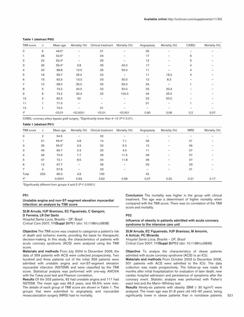
S21
P51Unstable angina and non-ST-segment elevation myocardialinfarction: an analysis by TIMI score
SLM Arruda, HJP Branisso, EC Figueiredo, C Gangoni, D Ferreira, LR Del SartoHospital Santa Lúcia, Brasília – DF, BrazilCritical Care 2007, 11(Suppl 3):P51 (doi: 10.1186/cc5838)
Objective The TIMI score was created to categorize a patient’s riskof death and ischemic events, providing the basis for therapeuticdecision-making. In this study, the characteristics of patients withacute coronary syndrome (ACS) were analyzed using the TIMIscore.Materials and methods From July 2004 to December 2006, thedata of 359 patients with ACS were collected prospectively. Twohundred and three patients out of the initial 359 patients wereadmitted with unstable angina and non-ST-segment elevationmyocardial infarction (NSTEMI) and were classified by the TIMIscore. Statistical analysis was performed with one-way ANOVAwith the Tukey post test and Pearson correlation.Results Of the 203 patients, 92 had unstable angina and 111 hadNSTEMI. The mean age was 66.3 years, and 65.5% were men.The details of each group of TIMI score are shown in Table 1. Thegroups that were submitted to angioplasty and myocardialrevascularization surgery (MRS) had no mortality.
Conclusion The mortality was higher in the group with clinicaltreatment. The age was a determinant of higher mortality whencompared with the TIMI score. There was no correlation of the TIMIscore and mortality.
P52Influence of obesity in patients admitted with acute coronarysyndrome to the intensive care unit
SLM Arruda, EC Figueiredo, HJP Branisso, M Amorim, A Achcar, PC MirandaHospital Santa Lúcia, Brasília – DF, BrazilCritical Care 2007, 11(Suppl 3):P52 (doi: 10.1186/cc5839)
Objective To analyze the characteristics of obese patientsadmitted with acute coronary syndrome (ACS) to an ICU.Materials and methods From October 2003 to December 2006,501 patients with ACS were admitted to the ICU. The datacollection was made prospectively. The follow-up was made 6months after initial hospitalization for evaluation of later death, newcardiac hospital admission and persistence of symptoms after thecoronary event. Statistic analysis was performed with Fisher’sexact test and the Mann–Whitney test.Results Ninety-six patients with obesity (BMI ≥ 30 kg/m2) wereanalyzed. The mean age was 61.8 years old (43–90 years), beingsignificantly lower in obese patients than in nonobese patients
Available online http://ccforum.com/supplements/11/S3
Table 1 (abstract P50)
TIMI score n Mean age Mortality (%) Clinical treatment Mortality (%) Angioplasty Mortality (%) CABG Mortality (%)
0 6 49.0* – 01 – 05 – – –
1 26 52.6* – 04 – 17 – 6 –
2 22 53.4* – 05 – 12 – 5 –
3 26 59.4* 3.8 05 40.0 17 – 4 –
4 20 68.8 10.0 05 50.0 11 – 4 –
5 16 69.7 25.0 02 – 11 18.2 4 –
6 15 63.3 13.3 03 50.0 12 8.3 – –
7 10 68.0 30.0 05 60.0 04 – 1 –
8 5 74.2 40.0 02 50.0 03 33.3 – –
9 6 74.2 33.3 02 100.0 04 25.0 – –
10 2 82.0 50 – – 02 50.0 – –
11 1 71.0 – – – 01 – 1 –
12 1 74.0 – 01 – – – – –
P <0.01 <0.0001 <0.01 <0.001 0.90 0.06 0.2 0.07
CABG, coronary artery bypass graft surgery. *Significantly lower than 4–12 (P < 0.01).
Table 1 (abstract P51)
TIMI score n Mean age Mortality (%) Clinical treatment Mortality (%) Angioplasty Mortality (%) MRS Mortality (%)
0 2 54.5 – 02 – – – – –
1 21 59.4* 4.8 14 7.1 06 – 01 –
2 35 59.2* 2.9 20 5.0 10 – 06 –
3 43 65.7 2.3 25 4.0 11 – 07 –
4 39 70.9 7.7 26 11.5 06 – 07 –
5 47 72.1 8.5 34 11.8 06 – 07 –
6 12 67.7 – 06 – 03 – 03 –
7 4 57.8 – 03 – – – 01 –
Total 203 66.3 4,9 130 42
P 0.0001 0.53 0.62 0.58 0.07 0.20 0.21 0.17
*Significantly different from groups 4 and 5 (P < 0.0001).

S22
(P < 0.001). Seventy-seven percent of the patients were men (74patients) (P < 0.0001); 35.4% of the patients had unstable angina,30.2% had non-ST-segment elevation myocardial infarction(NSTEMI) and 24.3% had ST-segment elevation myocardialinfarction (STEMI). The most common risk factors werehypertension (78.1%) (P = 0.062), followed by sedentarism(66.6%) (P = 0.01), dyslipidemia (43.7%) (P = 0.49) and diabetes(29.1%) (P = 0.70). Previous angina was related by 40 patients(P = 0.07) and previous myocardial infarction by 36 (P = 0.29).Nine patients had previous stent (P = 0.85) and 19 previouscoronary artery bypass graft surgery (CABG) (P = 0.36). Atadmission, 88.5% related precordial pain. Coronariography wasmade in 81 patients, with 36 submitted to angioplasty (34 withstent). The most affected coronary was the anterior descending(64.1%) followed by the right coronary (51.8%) and the circumflex(27.1%). Thrombolytic was used in 14 patients and 15 weresubmitted to CABG. Forty-four patients received only clinicaltreatment. The hospital mortality was significantly lower in obesepatients (2.0%) than in nonobese patients (11.6%). The mean timeof stay in the ICU was 2.38 days (P = 0.37) and the hospital staywas 9.53 days (P = 0.58). Follow-up was made with 78.3% (65 of83 patients). The later mortality by cardiac causes was 1.5%.There was 19 new hospital admissions for cardiac causes and 15had persistence of symptoms.Conclusion Obesity predisposes to a higher risk of cardiovasculardisease in the young population. Nevertheless, mortality was lower,probably because of the lower mean age of this obese group withACS.
P53Evaluation of the percutaneous coronary intervention as adiagnostic or therapeutic tool in 501 consecutive cases ofacute coronary syndrome
SLM Arruda, EC Figueiredo, HJP Branisso, P Rodrigues, V AmorimHospital Santa Lúcia, Brasília – DF, BrazilCritical Care 2007, 11(Suppl 3):P53 (doi: 10.1186/cc5840)
Objective The evaluation of the percutaneous coronary inter-vention (PCI) as a diagnostic or therapeutic tool in patients withacute coronary syndrome (ACS) admitted to an ICU.Materials and methods From October 2003 to December 2006,501 patients with ACS were admitted to the ICU. The datacollection was made prospectively using interviews with patients,chart reviews and examinations. The follow-up was made 6 monthsafter the coronary event for evaluation of later death and newhospital admission (351 patients) using telephone calls. Statisticalanalysis was performed with the chi-square test.Results A total of 419 patients with ACS were submitted to PCI.In this group, 124 patients (29.5%) had unstable angina, 112(26.7%) had non-ST-segment elevation myocardial infarction and183 (43.6%) had ST-segment elevation myocardial infarction. Themean age was 62.43 years old (27–95 years) and 71.1% weremen. The diagnostic PCI was made in 234 patients (55.9%) andthe therapeutic in 185 patients (44.1%). One hundred and fourpatients (56.2%) received a nonpharmacological stent and 73patients (39.4%) received a pharmacological stent. The mostaffected arteries were the anterior descending (58.2%), rightcoronary (44.8%), and circumflex (24.8%). Seventy-six patientswere submitted to coronary artery bypass graft after PCI. Hospitalmortality was 6.2%, corresponding to 5.5% of diagnostic PCI and7.0% of the angioplasty group (P = 0.54). Follow-up was madewith 304 or 86.6% of the patients at least 6 months after the initialhospitalization. The mean time of follow-up was 12.68 months
(6–31 months). The later mortality was 2.3%, corresponding to3.4% of diagnostic PCI and 0.7% from the angioplasty group (P =0.14). There were 33 (19.4%) new hospital admissions for cardiaccauses in the diagnostic group and 29 (21.9%) in the angioplastygroup (P = 0.56).Conclusion There were no significant differences in mortalitybetween diagnostic and therapeutic PCI.
P54Cardiac arrest and its epidemiologic variables in anemergency room
RE Martins, AM Baffa, CRG Silva, L Goes de SouzaPUC Campinas, Campinas, SP, BrazilCritical Care 2007, 11(Suppl 3):P54 (doi: 10.1186/cc5841)
Introduction Cardiac arrest is the sudden interruption ofventricular and respiratory activities, sufficient equipment for themaintenance of life. Atherosclerosis is the most frequent cause,although in Brazil there is Chagas disease and its arrhythmias.Methods A retrospective analysis of 66 adults admitted to ouremergency room in cardiac arrest in 2004. A series of clinical andepidemiologic variables was evaluated. They included: sex, age,causes, electrocardiographic rate, comorbidity, and time betweenthe arrest and the beginning of cardiopulmonary resuscitation.Comparison was made between the findings of the retrospectiveanalysis and the literature. The literature search was carried outthrough the electronic databases Medline and LILACS in January2007.Conclusion The average age, the causes and the electro-cardiographic rate are concordant with the studied literature. Wecould not correlate the time between the arrest and the beginningof cardiopulmonary resuscitation with the prognosis. Wedemonstrated a high rate of mortality, 83%, as in the literature. Theunsuitable filling in of the hospital cards did contribute to the smallnumber of analysed cases. However, the results demonstrate theurgency of accurate filling of the hospital charts and also for thepublication of this type of research, due to the lack of thisinformation in the literature.
P55Heart rate variability and pulmonary function behavior inpatients undergoing coronary artery bypass grafting andphysiotherapy intervention
RG Mendes, RP Simões, FSM Costa, CBF Pantoni, S Luzzi, AM Catai, A Borghi-SilvaNúcleo de Pesquisa em Exercício Físico, Irmandade Santa Casa deMisericórdia de Araraquara, Universidade Federal de São Carlos,São Paulo – SP, BrazilCritical Care 2007, 11(Suppl 3):P55 (doi: 10.1186/cc5842)
Objective To assess the behavior and relationship of heart ratevariability (HRV) and pulmonary function in patients undergoingcoronary artery bypass grafting (CABG) and physiotherapyintervention (PI).Methods Fourteen patients undergoing CABG and PI underwent aprospective study consisting of HRV analysis, spirometry andrespiratory muscle strength (RMS) evaluation before and after (1and 4 days) surgery. The heart rate (HR) and R–R intervals wererecorded by the cardiofrequencimeter (Polar S810i), beat-to-beat,in the resting condition and 10 minutes in a supine position. HRVwas evaluated in the time domain by the RMSSD index. Thespirometry (Vitalograph 2120) was evaluated and the forced vitalcapacity (FVC), the forced expiratory volume in 1 second (FEV1)
Critical Care June 2007 Vol 11 Suppl 3 Fourth International Symposium on Intensive Care and Emergency Medicine for Latin America

S23
and the maximal voluntary ventilation (MVV) were obtained. RMSwas measured by maximal inspiratory pressure (MIP) and maximalexpiratory pressure (MEP) obtained by an aneroid manovacuo-meter (Ger-Ar). All patients had initiated PI after 24 hours of theextubation, following a program of steps previously established.The Friedman test followed by the Dunn post-hoc test was utilizedto compare the variables among conditions before and aftersurgery, and Spearman correlation analysis to verify relationshipamong RMSSD and pulmonary parameters. The significance levelwas set at 5% for all analyses.Results Significant correlations were observed between theRMSSD and FVC, FEV1, MMV and MIP (r = 0.6) and the RMSSDand MEP (r = 0.7). Table 1 presents the comparisons amongconditions.
Table 1 (abstract P55)
Results of heart rate variability and pulmonary function on thepreoperative, first postoperative and fourth postoperative days ofCABG
1st 4th postoperative postoperative
Preoperative day day
RMSSD (ms) 21.2 9.2* 14.5
FVC (l) 3.2 0.7* 1.1
FEV1 (l) 2.7 0.6* 0.9†
MVV (l/min) 100.6 23.3* 33.9†
MIP (cmH2O) 76.4 25.4* 32.8†
MEP (cmH2O) 111.1 35.8* 39.3†
Values as median. *P < 0.05 preoperative vs 1st day postoperative,†P < 0.05 preoperative vs 4th day postoperative.
Conclusion The present findings showed that the RMSSD indexassociated positively with pulmonary function and that cardiacautonomic regulation is impaired after CABG. Additionally, the PIcan be a potential therapeutic to reestablish the parasympatheticactivity in these patients.Acknowledgements This work was supported by grants fromFAPESP and CNPq.
Nephrology
P56Preliminary report of an enoxaparin dose protocol based onanti-Xa activity in continuous renal replacement therapy
JLF Costa, J Ferreira, C Teles, F Gutierrez, A Mesquita, RC Costa-FilhoHospital Pró-Cardíaco, Rio de Janeiro – RJ, BrazilCritical Care 2007, 11(Suppl 3):P56 (doi: 10.1186/cc5843)
Background During continuous renal replacement therapy(CRRT), anticoagulation of the extracorporeal circuit is generallyrequired to prevent clotting of the circuit, preserve filter perfor-mance, optimize circuit survival, and prevent blood loss due tocircuit clotting. Unfractionated heparin and low-molecular-weightheparin are generally used to perform this strategy. This anti-coagulation may cause dangerous bleeding, however, especially inacute renal critical patients. In these patients, it is very difficult topredict bleeding or thrombosis correctly during CRRT.Objective To asses the safety and efficacy of the use of anenoxaparin dose protocol based on anti-Xa activity in CRRT.
Methodology From September 2005 to December 2006, 26patients were submitted to 55 CRRT sessions. All sessions usedan enoxparin dose protocol based on anti-Xa activity (target0.25–0.4 U/ml). The endpoints analyzed were the circuit time(hours) to judge efficacy, and death (30-day mortality) and seriousbleeding (red cell transfusion) to judge safety.Results Continuous veno-venous hemodiafiltration was a frequentlyused method in 53 sessions (96.4%). The average circuit time was41.6 ± 26.6 hours. Ten patients received red blood transfusion (19transfusions required) related to CRRT and four patients hadbleeding complications (retroperitoneal hemorrhage, hemothorax,puncture complication, acute gastric lesion). No death wasreported during 30 days of follow-up.Conclusion In this series, the use of an enoxaparin dose protocolbased on anti-Xa activity in CRRT was considered relatively safeand effective. The circuit time of 41 hours was acceptable ineffectiveness and efficiency.
P57Evaluation of acute renal failure in surgical patients in theintensive care unit
EHT Anami, T Matsuo, CMC Grion, TF Perazolo, LTQ CardosoHospital Universitário, Universidade Estadual de Londrina, Londrina –PR, BrazilCritical Care 2007, 11(Suppl 3):P57 (doi: 10.1186/cc5844)
Background Acute renal failure (ARF) is a common and seriouscomplication in the postoperative period of critically ill patients. Itoccurs, depending on specific definition, in up to 30% of patientssubmitted to cardiac surgery. Recent evidence suggests that evensmall oscillations in serum creatinine are associated withsignificant effects on mortality. The objective of this study is toevaluate the impact of ARF on the morbidity and mortality ofsurgical patients in the ICU.Methods A retrospective observational study conducted in theICU of a public university hospital during the period January2004–December 2004. The research was realized in an electronicdatabase and included demographic data, diagnostic, SOFA andAPACHE II scores, length of stay in the ICU and mortality. Therenal dysfunction was defined as a SOFA score ≥2 (creatinine≥2 mg/dl or oliguria <500 ml/day). Patients with chronic renalfailure were excluded. For statistical analysis, the Epi Info programversion 3.3.2 was used.Results One hundred and five surgical patients were admitted tothe ICU in the study period, male sex was more frequent (55.2%),with a mean age of 53.2 years, mean APACHE II score of 17 ± 7,and length of stay varying from 1 to 65 days (median 3 days). Themost frequent surgeries were gastrointestinal (16.2%), multipletrauma (13.3%), intracranial hemorrhage (9.5%) and metabolic/renal (9.5%). In this sample the cardiovascular surgery was notrepresentative (2.9%).ARF occurred in 19% (95% CI: 11.3–26.8%) of the surgicalpatients and the mortality in this group was greater than in thegroup of patients that did not develop this complication, respec-tively 63.2% and 15.1% (P < 0.001). The patients with ARF wereolder than the patients without this complication (64.9 ± 7.9 yearsvs 50.6 ± 21.14 years, P < 0.007) and the APACHE II score washigher (20.3 ± 4.9) in the ARF patients when compared withpatients without ARF (16.5 ± 7.4, P < 0.06). The median length ofstay was higher in the patients with ARF, being 6 days, varyingfrom 3 to 65 days, while in the patients without ARF the medianwas 2 days, varying from 1 to 33 days (P < 0.001).Conclusion The high frequency of ARF found in this study wasprobably due to the definition criteria adopted, including transient
Available online http://ccforum.com/supplements/11/S3

S24
oliguria and pre-renal ARF. The occurrence of renal dysfunctionresulted in higher morbidity and mortality in this group of patients.Several studies have been carried out to determine the patients athigh risk of developing ARF in the postoperative period, andprotective strategies have been developed, but the results are asyet inconclusive.
P58Acute renal failure patients submitted to conservative anddialytic treatment in an intensive care unit
SC Aranha, L Duailibe, FMS Costa, LR Faustino, MC Ichida, SE Mataloun, M MoockGrajau State Hospital (HEG), UNISA Medical School, São Paulo –SP, BrazilCritical Care 2007, 11(Suppl 3):P58 (doi: 10.1186/cc5845)
Twenty to 25% of ICU patients have acute renal failure (ARF).Dialytic treatment in ARF patients reduces mortality and is wellused worldwide. The objective of this study was to analyze ICUpatients with a creatinine rate above 1.3 ng/dl who were treatedand nontreated with dialysis. The epidemiologic study was of 392inpatients from the ICU of the HEG where 42% had ARFdiagnosed. Only 29.1% were submitted to hemodialysis asinpatients. We can realize the high creatinine rate before dialysistreatment and the lower ICU stay of patients that did not need thistreatment. The ARF incidence in the ICU of the HEG was higherthan that in the literature, but dialysis was less used. This can beeasily explained by the low availability of necessary equipment.Sepsis is the first cause of ARF, but in this study it is the numbertwo cause, especially in dialytic patients. Mortality was similar toother studies. In conclusion, the ARF mortality rate is still high,even with new dialytic treatments. Dialysis is therefore associatedwith better life quality and less time in hospital.References1. Gill N, Nally Jr JV, Fatica RA: Renal failure secondary to acute
tubular necrosis epidemiology, diagnosis and management.Chest 2005, 128:2847-2863.
2. Needham E: Management of acute renal failure. Am Fam Phys2005, 72:1739-1746.
3. Klenzak J, Himmelfarb J: Sepsis and kidney. Crit Care Clin 2005,21:211-222.
P59Acute renal failure and other complications induced bycocaine abuse
J Cheibub, H Missaka, MAMA Lima, T Boechat, A Andrade, AC Marques, J Pinheiro, PCT CostaUPG, CTI Emergência do Hospital Municipal Souza Aguiar, Rio deJaneiro – RJ, BrazilCritical Care 2007, 11(Suppl 3):P59 (doi: 10.1186/cc5846)
Introduction Cocaine is a social and medical problem. In theUnited States, 14.6% of a trialed population has already usedcocaine. The physiopathology of the renal injury is multifactorialand largely remains unknown, and rhabdomyolysis is mostfrequently responsible for the renal injury.Objectives To describe and analyze the case of a patient withanuric acute renal failure (ARF) due to cocaine overdose. Tocompare and identify more recent scientific evidence for thetreatment.Materials and methods A search and analysis of the case of apatient with anuric ARF treated in the ICU of a public emergencyhospital.
Results GC, male, 29 years old, a cocaine and marihuana user,presented a sudden condition of irritability, aggressiveness anddelirious after consuming the drugs. In the subsequent days hepresented hyperthermia, jaundice, oliguria, and respiratoryinsufficiency. He was admitted to the ICU in a severe conditionwith hypertension, hyperthermia and P/F = 128. Due to anuricARF, the patient went to daily hemodialysis.Discussion Physiopathologic effects include hemodynamic alter-ations, failure in the synthesis of glomerular matrix, degradation andoxidative stress and induced renal atherogenesis.Rhabdomyolysis is the main cause responsible for renal injury. Ithas a high index of mortality and its mechanisms remain unknown.There is evidence that it is intimately related to vasoconstrictiondue to ischemia, direct toxicity, hyperpyrexia and increased muscularactivity. A quick implementation of treatment for convulsions,hyperpyrexia and agitation improves prognosis and decreasescomplications.The therapeutic goals are removal of precipitating factors, handlingof complications and early dialysis. The treatment of rhabdo-myolysis is based on hydration, induced osmotic diuresis and urinealkalinization.Conclusion There are few renal injury cases reported in theliterature. These generally describe renal infarction after havinginhaled the drug, acute interstitial nephritis and cocaine-inducedARF with or without rhabdomyolysis. However, it is imperative thatwell-designed epidemiologic studies are designed to betterelucidate the physiopathology of cocaine-induced renal injuries.References1. Jaffe JA, Kimmel PL: Chronic nephropathies of cocaine and
heroin abuse: a critical review. American Society of Nephrol-ogy. Clin J Am Soc Nephrol 2006, 1:655-667.
2. Bemanian S, Motallebiand M, Nosrati SM: Cocaine-inducedrenal infarction: report of a case and review of the literature.BMC Nephrol 2005, 6:10.
3. Cami J, Farré M: Drug addiction. N Engl J Med 2003, 349:975-986.
Pneumology
P60The influence of different ventilatory modes on the intensity ofpulse pressure variation
PS Andrade, GA Westphal, M Caldeira FilhoHospital Municipal São José, Centro Hospitalar Unimed andHospital Dona Helena, Joinville – SC, BrazilCritical Care 2007, 11(Suppl 3):P60 (doi: 10.1186/cc5847)
Background Pulse pressure variation (PPV) has beenrecommended to evaluate the cardiac responsiveness to fluidinfusion in mechanically ventilated patients with sepsis shock orfollowing cardiac surgery. The recommendation is that PPV measure-ments must be performed during the volume-controlled ventilatorymode (VC) and not with the pressure control mode (PC).Objective To test the hypothesis that the PC should not cause animportant change on PPV when compared with the VC duringmechanical ventilation.Methods A prospective, nonrandomized, observational andcomparative study that compares effects on PPV of the VC withanother three PC ventilatory settings applied in sedated andmechanically ventilated critically ill patients with an arterial catheterin place. Initial/control setting (S1): VC with tidal volume (Vt) = 10ml/kg; setting 2 (S2): PC with the peak pressure (Pp) obtained inS1; setting 3 (S3): PC with the Pp set in the plateau pressure levelobtained in S1; setting 4 (S4): PC with pressure values
Critical Care June 2007 Vol 11 Suppl 3 Fourth International Symposium on Intensive Care and Emergency Medicine for Latin America
S25
determined with the Vt set at 10 ml/kg. All settings included PEEPof 5 cmH2O and a respiratory rate of 15 rpm. The mean arterialpressure and PPV obtained in each ventilatory set are comparedwith each other using the paired Student t test.Results Thirty-four patients were evaluated. The Pp wassignificantly lower in S3 and S4 (P < 0.001). The Vt wassignificantly greater in S2 (P < 0.001) with a parallel PPV incrementat this time (S2) (P < 0.001). We found strong clinical concordancebetween S1 and S4 (accuracy = 98.1%, kappa = 0.93, P < 0.001).Significant but weaker concordance was found between S1 and S2(accuracy = 91.2%, kappa = 0.80, P < 0.001) and between S1 andS3 (accuracy = 94.1%, kappa = 0.86, P < 0.001).Conclusion The pressure-controlled mode should not causeimportant changes or significant clinical misinterpretation on PPVwhen compared with the volume-controlled mode duringmechanical ventilation. These findings are especially evident whenthe pressure-controlled mode with the Vt limited at 10 ml/kg (S4set) is used, demonstrated by the best accuracy on reproducingthe PPV obtained during the volume-controlled mode.
P61Application of a mechanical ventilation weaning protocol in acoronary unit
RF Piotto, LN Maia, M de Nassau Machado, SP OrricoUnidade Coronariana do Hospital de Base/Faculdade de Medicinade São José do Rio Preto, São José do Rio Preto – SP, BrazilCritical Care 2007, 11(Suppl 3):P61 (doi: 10.1186/cc5848)
Introduction Weaning is the transitional period when a patientunder mechanical ventilation (MV) transfers to unassisted spon-taneous breathing. Failure in the discontinuation of ventilatorysupport is associated with an increase in the number ofcomplications. The use of standardized guidelines to carry outweaning is already well established in general ICUs. The condi-tions most commonly seen in the coronary unit of care (CUC),such as acute myocardial ischemia, left ventricular dysfunction andafter coronary artery bypass grafting surgery, however, causecompletely different hemodynamic and circulatory alterations tothose observed in other types of severely ill patients. The effects ofmechanical ventilation and of weaning should therefore be testedspecifically for these patients.Objective To compare MV weaning performed according to theapplication of a series of guidelines versus nonstandardizedweaning in patients hospitalized in a CUC.Method Initially a pilot study was performed with the aim ofestimating the failure rate of MV weaning of patients hospitalized inthe CUC. The results confirmed the necessity of improving themethod then employed by the multidisciplinary team. Hence, 36patients, who utilized MV for a period greater than 24 hours andwere ready for weaning, were prospectively included in the study.The average age of the patients was 59.5 ± 16.4 years. The numberof patients needed to include in the study was determined bycalculating the sample size. The patients were then randomly placedinto two groups: the experimental group (EG) and the control group(CG). In the EG, extubation was standardized according to thespontaneous respiratory test (SRT) of the American guidelines forweaning and was conducted by investigator in the study. For theCG, the SRT was also performed by the same investigator butwithout altering the extubation procedure employed, which wasdetermined by the multidisciplinary team.Results The groups were matched so there were no statisticallysignificant differences in respect to gender, age, diagnoses atadmission, ventilation parameters, physiological variables andAPACHE II score. The time necessary for weaning was significantly
shorter in the EG (2 hours and 24 minutes vs 70 hours; P = 0.0009).Sixteen patients in the CG were extubated, of which 11 (69%) didnot fulfill the clinical criteria of the SRT. Of these 16 cases, 12 (75%)were reintubated and four (25%) were successfully weaned with allthe successful cases among patients who passed the SRT. Of the18 patients in the EG, 11 fulfilled the criteria for SRT and wereextubated. Of these, eight (73%) cases were successful and three(27%) required reintubation. The reintubation rate was significantlyhigher in the CG (75% vs 25%; P = 0.0001).Conclusion The application of the weaning MV guidelines in heartdisease patients hospitalized in the CUC reduces the timenecessary to complete weaning, increases the success rate andreduces the reintubation rates.
P62Leukocyte–endothelial interactions in mesentericmicrocirculation of rats under different positive end expiratorypressure levels
P Aikawa1, LS Perucci2, SHP Farsky3, MA Oliveira4, T Mauad5,P Sannomiya1, NK Nakagawa1,2
1Heart Institute (InCor), University of São Paulo Medical School,LIM-11, São Paulo – SP, Brazil; 2UNICID, São Paulo – SP, Brazil;3School of Pharmaceutical Sciences, São Paulo – SP, Brazil;4Institute of Biomedical Sciences, University of São Paulo – SP,Brazil; 5Pathology Department, FMUSP, São Paulo – SP, BrazilCritical Care 2007, 11(Suppl 3):P62 (doi: 10.1186/cc5849)
Introduction Mechanical ventilation (MV) with positive endexpiratory pressure (PEEP) is widely used to improve oxygenationand to treat acute respiratory failure. PEEP can affect nearly everyorgan system of the body due to homeostatic interactions betweenthe lungs and other body systems. Although essential in themanagement of critically ill patients, many deleterious side effectsof positive pressure ventilation in the lungs and hemodynamicshave been reported.Methods This study investigated the effects of different PEEPlevels on leukocyte–endothelial interactions in postcapillaric venules(13–23 µm diameter) at the mesentery by intravital microscopyand lung histomorphometry. Nineteen male Wistar rats weresubmitted to inhaled isofluorane anesthesia and MV with a tidalvolume of 10 ml/kg, a respiratory rate of 70 breaths/min andFiO2 = 100%. Animals were randomly assigned to three groups:(1) PEEP0 (n = 6), no PEEP; (2) PEEP5, 5 cmH2O (n = 6); and (3)PEEP10, 10 cmH2O (n = 7).Results After 2 hours of MV, there were no differences betweenPEEP0 and PEEP5 in the number of rolling leukocytes/10 minutes(127 ± 16 and 147 ± 26, respectively), adherent leukocytes/100 µm (3 ± 1 and 4 ± 2, respectively), migrated leukocytes intothe perivascular tissue/5,000 µm2 (2 ± 1 and 2 ± 1, respectively)and total white blood cells/mm3 (11,730 ± 2,856 and10,200 ± 2,222, respectively). However, the PEEP10 group presen-ted an increased number of rolling leukocytes (188 ± 15 cells/10 min, P < 0.05), adherent leukocytes (8 ± 1 cells/100 µm,P < 0.05), migrated leukocytes (12 ± 1 cells/5,000 µm2, P < 0.05)at the mesentery, and an increased number of total white bloodcells (18,786 ± 4,207 cells/mm3, P < 0.05), basically neutrophils.Lung morphometric analysis showed some edema at theperivascular tissue with no neutrophil infiltration in the parenchymaof the PEEP10 group compared with the other groups (P < 0.05).There were no changes in mean arterial blood pressure in allgroups along the study period.Conclusion After 2 hours of MV, PEEP = 10 cmH2O induced aninflammatory response in rat mesenteric microcirculation.Acknowledgements Funded by FAPESP and PRONEX.
Available online http://ccforum.com/supplements/11/S3

S26
P63Does the cycling-off criteria of pressure support change therespiratory parameters in intensive care unit patients?
TD Correa1, RH Passos1, S Kanda1, C Taniguchi1, C Hoelz1, J Bastos1, GFJ Matos1, EC Meyer1, CSV Barbas1,2
1Adult Intensive Care Unit, Hospital Israelita Albert Einstein, SãoPaulo – SP, Brazil; 2Pulmonary Division of University of São Paulo –SP, BrazilCritical Care 2007, 11(Suppl 3):P63 (doi: 10.1186/cc5850)
Introduction In modern mechanical ventilators it is possible tomodify the flow cycling-off criteria of pressure support ventilation.The changes in the flow cycling-off criteria of pressure supportventilation can modify the synchrony between the mechanical andneural inspiration termination.Objective To compare the effects of two different flow cycling-offcriteria of pressure support ventilation on the respiratoryparameters of ICU mechanically ventilated patients.Methods We prospectively evaluated 20 intubated andmechanically ventilated adult ICU patients recovering from acuterespiratory failure that could be comfortably ventilated withpressure support of 15 cmH2O, PEEP of 5 cmH2O and FIO2 of40%. The patients were ventilated at two different cycling-offcriteria of pressure support of 25% and 40% of the peakinspiratory flow. We evaluated the respiratory rate (RR), expiratorytidal volume (VTe), minute ventilation (MV), VCO2, VTCO2, ETCO2,SpO2, mean arterial pressure (MAP) and heart rate (HR).Results Comparisons between different cycling-off values did notresult in any statistically significant changes for the evaluatedvariables. There were no significant changes in blood pressure orHR under any experimental conditions (Table 1).Conclusion The changes in cycling of criteria from 25% to 40% ofthe peak flow did not affect the respiratory parameters inmechanically ventilated patients in a mixed ICU.
P64Influence of the pressure support slope on the respiratoryparameters of intensive care unit patients
TD Correa1, RH Passos1, S Kanda1, C Taniguchi1, C Hoelz1, J Bastos1, GFJ Matos1, EC Meyer1, CSV Barbas1,2
1Adult Intensive Care Unit, Hospital Israelita Albert Einstein, SãoPaulo – SP, Brazil; 2Pulmonary Division of University of São Paulo –SP, BrazilCritical Care 2007, 11(Suppl 3):P64 (doi: 10.1186/cc5851)
Introduction The possibility of changing the pressure slope duringpressure support ventilation is a characteristic of the newgeneration of ICU ventilators. However, the influence of the slopechanges on the respiratory parameters in ICU patients is still underinvestigation.Objective To analyze the effects of two different pressure slopes(150 or 300 ms) of pressure support ventilation on the respiratoryparameters of ICU mechanically ventilated patients.Methods We prospectively evaluated ICU patients recoveringfrom acute respiratory failure that could be comfortably ventilatedwith pressure support of 15 cmH2O, PEEP of 5 cmH2O and FIO2of 40%. The patients were submitted to two different pressureslopes of pressure support with 150 and 300 ms delays. Therespiratory rate (RR), expiratory tidal volume (VTe), minute ventila-tion (MV), VCO2, VTCO2, ETCO2, SpO2, mean arterial pressure(MAP) and heart rate (HR) were measured in these differentconditions.Results Comparisons between different pressure slopes did notresult in any statistically significant changes for the evaluatedvariables. There were no significant changes in blood pressure orHR under any experimental conditions (Table 1).Conclusion The slope changes from 150 to 300 ms of pressuresupport ventilation did not affect the respiratory parameters ofmechanically ventilated ICU patients.
Critical Care June 2007 Vol 11 Suppl 3 Fourth International Symposium on Intensive Care and Emergency Medicine for Latin America
Table 1 (abstract P63)
Respiratory and hemodynamic variables measured with two cycling-off criteria (25% and 40%)
Cycling-off RR VTe MV VCO2 VTCO2 ETCO2 SpO2 MAP HR Slope (ms) (%) (beats/min) (ml) (l/min) (l/min) (l/min) (l/min) (%) (mmHg) (beats/min)
0.15 25 18.2 587.5 10.6 193.6 11.2 27.8 98.8 96.4 83.4
0.15 40 19.2 560.7 10.0 183.7 10.88 28 98.8 96.2 83.9
P value 0.21 0.31 0.2 0.19 0.45 0.83 1 0.92 0.44
0.30 25 19.4 588.4 10.5 192.8 11.54 28 98.7 98.2 83.6
0.30 40 19.7 565.2 10.2 187.8 10.61 28.2 98.9 97.7 83.6
P value 0.74 0.2 0.5 0.50 0.17 0.65 0.08 0.78 0.9
Table 1 (abstract P64)
Respiratory and hemodynamic variables measured with two pressure slopes
Cycling-off RR VTe MV VCO2 VTCO2 ETCO2 SpO2 MAP HR Slope (ms) (%) (beats/min) (ml) (l/min) (l/min) (l/min) (l/min) (%) (mmHg) (beats/min)
0.15 25 18.2 587.5 10.6 193.6 11.2 27.8 98.8 96.4 83.4
0.30 25 19.4 588.4 10.5 192.8 11.5 28 98.8 98.2 83.6
P value 0.28 0.96 0.7 0.89 0.45 0.81 1 0.54 0.79
0.15 40 19.2 560.7 10.1 183.7 10.9 28 98.8 96.2 83.9
0.30 40 19.7 565.3 10.2 187.8 10.6 28.2 98.9 97.7 83.6
P value 0.53 0.79 0.7 0.41 0.51 0.65 0.42 0.54 0.86
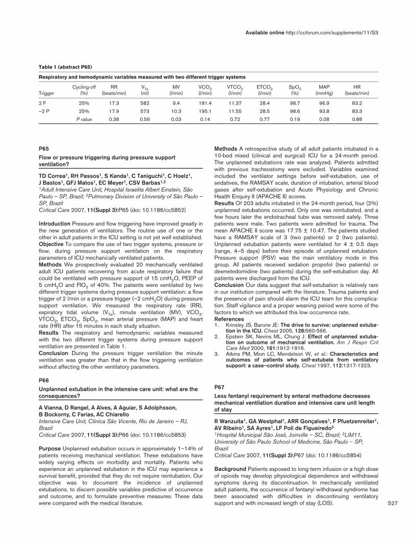
S27
P65Flow or pressure triggering during pressure supportventilation?
TD Correa1, RH Passos1, S Kanda1, C Taniguchi1, C Hoelz1, J Bastos1, GFJ Matos1, EC Meyer1, CSV Barbas1,2
1Adult Intensive Care Unit, Hospital Israelita Albert Einstein, SãoPaulo – SP, Brazil; 2Pulmonary Division of University of São Paulo –SP, BrazilCritical Care 2007, 11(Suppl 3):P65 (doi: 10.1186/cc5852)
Introduction Pressure and flow triggering have improved greatly inthe new generation of ventilators. The routine use of one or theother in adult patients in the ICU setting is not yet well established.Objective To compare the use of two trigger systems, pressure orflow, during pressure support ventilation on the respiratoryparameters of ICU mechanically ventilated patients.Methods We prospectively evaluated 20 mechanically ventilatedadult ICU patients recovering from acute respiratory failure thatcould be ventilated with pressure support of 15 cmH2O, PEEP of5 cmH2O and FIO2 of 40%. The patients were ventilated by twodifferent trigger systems during pressure support ventilation: a flowtrigger of 2 l/min or a pressure trigger (–2 cmH2O) during pressuresupport ventilation. We measured the respiratory rate (RR),expiratory tidal volume (VTe), minute ventilation (MV), VCO2,VTCO2, ETCO2, SpO2, mean arterial pressure (MAP) and heartrate (HR) after 15 minutes in each study situation.Results The respiratory and hemodynamic variables measuredwith the two different trigger systems during pressure supportventilation are presented in Table 1.Conclusion During the pressure trigger ventilation the minuteventilation was greater than that in the flow triggering ventilationwithout affecting the other ventilatory parameters.
P66Unplanned extubation in the intensive care unit: what are theconsequences?
A Vianna, D Rangel, A Alves, A Aguiar, S Adolphsson, B Bockorny, C Farias, AC ChiarelloIntensive Care Unit, Clinica São Vicente, Rio de Janeiro – RJ,BrazilCritical Care 2007, 11(Suppl 3):P66 (doi: 10.1186/cc5853)
Purpose Unplanned extubation occurs in approximately 1–14% ofpatients receiving mechanical ventilation. These extubations havewidely varying effects on morbidity and mortality. Patients whoexperience an unplanned extubation in the ICU may experience asurvival benefit, provided that they do not require reintubation. Ourobjective was to document the incidence of unplannedextubations, to discern possible variables predictive of occurrenceand outcome, and to formulate preventive measures. These datawere compared with the medical literature.
Methods A retrospective study of all adult patients intubated in a10-bed mixed (clinical and surgical) ICU for a 24-month period.The unplanned extubations rate was analyzed. Patients admittedwith previous tracheostomy were excluded. Variables examinedincluded the ventilator settings before self-extubation, use ofsedatives, the RAMSAY scale, duration of intubation, arterial bloodgases after self-extubation and Acute Physiology and ChronicHealth Enquiry II (APACHE II) scores.Results Of 203 adults intubated in the 24-month period, four (2%)unplanned extubations occurred. Only one was reintubated, and afew hours later the endotracheal tube was removed safely. Threepatients were male. Two patients were admitted for trauma. Themean APACHE II score was 17.75 ± 10.47. The patients studiedhave a RAMSAY scale of 3 (two patients) or 2 (two patients).Unplanned extubation patients were ventilated for 4 ± 0.5 days(range, 4–5 days) before their episode of unplanned extubation.Pressure support (PSV) was the main ventilatory mode in thisgroup. All patients received sedation propofol (two patients) ordexmetedomidine (two patients) during the self-extubation day. Allpatients were discharged from the ICU.Conclusion Our data suggest that self-extubation is relatively rarein our institution compared with the literature. Trauma patients andthe presence of pain should alarm the ICU team for this complica-tion. Staff vigilance and a proper weaning period were some of thefactors to which we attributed this low occurrence rate.References1. Krinsley JS, Barone JE: The drive to survive: unplanned extuba-
tion in the ICU. Chest 2005, 128:560-566.2. Epstein SK, Nevins ML, Chung J: Effect of unplanned extuba-
tion on outcome of mechanical ventilation. Am J Respir CritCare Med 2000, 161:1912-1916.
3. Atkins PM, Mion LC, Mendelson W, et al.: Characteristics andoutcomes of patients who self-extubate from ventilatorysupport: a case–control study. Chest 1997, 112:1317-1323.
P67Less fentanyl requirement by enteral methadone decreasesmechanical ventilation duration and intensive care unit lengthof stay
R Wanzuita1, GA Westphal1, ARR Gonçalves1, F Pfuetzenreiter1,AV Ribeiro1, SA Ayres1, LF Poli de Figueiredo2
1Hospital Municipal São José, Joinville – SC, Brazil; 2LIM11,University of São Paulo School of Medicine, São Paulo – SP,BrazilCritical Care 2007, 11(Suppl 3):P67 (doi: 10.1186/cc5854)
Background Patients exposed to long-term infusion or a high doseof opioids may develop physiological dependence and withdrawalsymptoms during its discontinuation. In mechanically ventilatedadult patients, the occurrence of fentanyl withdrawal syndrome hasbeen associated with difficulties in discontinuing ventilatorysupport and with increased length of stay (LOS).
Available online http://ccforum.com/supplements/11/S3
Table 1 (abstract P65)
Respiratory and hemodynamic variables measured with two different trigger systems
Cycling-off RR VTe MV VCO2 VTCO2 ETCO2 SpO2 MAP HR Trigger (%) (beats/min) (ml) (l/min) (l/min) (l/min) (l/min) (%) (mmHg) (beats/min)
2 F 25% 17.3 582 9.4 181.4 11.37 28.4 98.7 96.9 83.2
–2 P 25% 17.9 573 10.3 195.1 11.55 28.5 98.6 93.8 83.3
P value 0.38 0.56 0.03 0.14 0.72 0.77 0.19 0.08 0.88

S28
Objective We tested the hypothesis that enteral methadone canreduce fentanyl requirements and, thereby, decrease mechanicalventilation duration and ICU LOS.Methods A prospective, randomized and double-blind studyinvolving patients fulfilling criteria for weaning from mechanicalventilation but under high risk for fentanyl abstinence syndrome(defined as continuous fentanyl for more than 5 days or more than5 µg/kg/hour during 12 hours). Patients were randomized into twogroups, methadone (MET) group and control (CT) group, asfollows: for the first 24 hours both groups were given 80% of theoriginal dose of fentanyl and received, additionally, in the METgroup enteral methadone (10 mg each 6 hours) or in the CT groupenteral placebo. After the first 24 hours, the MET group receivedenteral methadone and intravenous placebo while the CT groupreceived enteral placebo and intravenous fentanyl. In both groups,the blinded intravenous solutions were reduced by 20% of theoriginal dose, every 24 hours. Any abstinence symptoms weretreated with a bolus of fentanyl. The Student t test was used tocompare groups in the following criteria: (1) days undermechanical ventilation and (2) ICU LOS.Results Sixteen patients were included, seven in the MET groupand nine in the CT group. The LOS was significantly lower in theMET group (13 ± 3 vs 27 ± 13 days, P < 0.02). Days undermechanical ventilation were also significantly decreased in patientsfrom the MET group (4 ± 0.8 vs 20 ± 21 days, P < 0.05).Conclusion These preliminary data show that, by replacingfentanyl infusion with methadone through the enteral route, it ispossible to decrease mechanical ventilation duration as well as theICU length of stay.
P68Bilevel plus PSV and nitric oxide as an alternative ventilatorystrategy in acute respiratory distress syndrome patients
F Saddy, TMF Castelões, HM Mesquita, JLF Costa, JRB Martins,PN Gomes, R Costa-FilhoIntensive Care Unit, Hospital Pró-Cardíaco, NUGESCO, Rio deJaneiro – RJ, BrazilCritical Care 2007, 11(Suppl 3):P68 (doi: 10.1186/cc5855)
The protective ventilatory strategy provided by usual ventilatorymodes is a cornerstone factor to determine prognosis in acuterespiratory distress syndrome (ARDS). However, there aresituations where alternative strategies must be used.We describe three patients, all females with a mean age of 76 ± 6years, admitted to the ICU presenting ARDS related to community-acquired pneumonia (two patients) and pneumonia secondary togastric aspiration (one patient). The mean APACHE II score was25 ± 2. The ventilator used was the Puritan-Bennet 840. The meanstatic compliance (Cst), paCO2 and PaO2/FiO2 ratio afterorotracheal intubation in the VCV mode and a PEEP level of10 cmH2O were 24.3 ± 3.1, 48 ± 4 mmHg and 120.6 ± 46,respectively. All patients presented septic shock and weremonitored with a Swan–Ganz catheter. The initial mean pulmonaryartery pressure was 56 ± 4 mmHg on vasopressors to maintain amean systemic arterial pressure above 65 mmHg. The patientswere submitted to a recruitment maneuver using a pressuregradient between the PEEP and inspiratory pressure of 15 cmH2Oduring 2 minutes each step to a limit of 60 cmH2O, and after thatthe mean level of PEEP used was 20 ± 3 cmH2O and theventilatory mode was switched to Bilevel plus PSV (derived modefrom APRV). The mean Vt/kg used was 5.6 ± 0.8 ml/kg, with an
inspiratory pressure level of 30 ± 2 cmH2O and I:E ratio of 1:1.The mean PaO2/FiO2 ratio was raised to 153 ± 25 (26.8%). Nitricoxide (NO) was started and the mean concentration used was37 ± 3 ppm. After 4 hours, the mean pulmonary artery pressureand PaCO2 decreased to 40 ± 4 mmHg (28.6%) and 42 ± 2 mmHg(12.5%), respectively, and the PaO2/FiO2 ratio increased to268 ± 22 (122.2%). After 24 hours NO was decreased to18 ± 4 ppm, and was discontinued 12 hours after. Two patientswere successfully weaned from mechanical ventilation after 8 and10 days, respectively, and discharged home afterwards. Onepatient died.This series of cases was the first that depicted the Bilevel plusPSV mode wholly with NO to control pulmonary hypertension andARDS. These interventions seems to be beneficial and feasible tosupport critically ill patients, mainly those who failed to respond torecruitment maneuver and conventional ventilatory modality.
P69Evaluation of a weaning protocol in an intensive care unit
RB Pivoto, M Weiss, F Cabral, T Ojopi, C Faccin, R Affonso, AC Maia, IC Neves, I Small, A Aguiar, A ViannaClínica São Vicente, Rio de Janeiro, RJ, BrazilCritical Care 2007, 11(Suppl 3):P69 (doi: 10.1186/cc5856)
Objective Current evidence suggests that patients spend about41% of their time in mechanical ventilation (MV) with weaning. Theapplication of weaning protocols brings better results over thetreatment based upon simple observation of the patient, reducingthe time of MV and its associated complications. The goal of thisstudy was to evaluate the results from the application of a weaningprotocol executed by the Physical Therapy team, aiming to reducethe weaning length and the MV length.Methods Fifty-eight inpatients were prospectively studied at theClinica São Vicente ICU, from June 2005 to February 2007. Thesepatients were ventilated through an endotracheal tube and were inMV ≥48 hours. The ventilatory status of these patients was evaluateddaily and after they fitted the protocol we started the weaningprogram, using pressure support ventilation (PSV). Patients thattolerated one spontaneous ventilation attempt were consideredeligible for extubation, considering PSV of 7 cmH2O and PEEP of4 cmH2O for 30 minutes. Higher levels of PSV and PEEP wereaccepted for patients with chronic obstructive pulmonary disease(COPD).Measurements and results The mean age of the patients was66.25 years (SD ± 20.21), with 52.6% males and 47.4% females.The average time of MV was 6.44 days (SD ± 3.17) and weaninglength was 1.52 days (SD ± 1.09). The indication for an artificialairway and MV was 15 postoperative patients (26.3%), 33 patientswith acute respiratory distress (57.9%) and nine neurologyaffected patents (15.8%). At the extubation time the average PSVwas 7.81 (SD ± 1.30) and the average PEEP was 4.54(SD ± 1.00). Forty-seven patients were successful (82.5%), while10 needed reintubation (17.5%). There was no correlationbetween nonsuccess and age, COPD, MV causes or the Tobinindex, considered a predictive index of success in the literature.Regarding the maximal inspiratory pressure, there was astatistically significant correlation (P = 0.01).Conclusion The time spent to weaning in our study was shorterthan the literature description, and the reintubation rate wascompatible with anterior publishing. The study showed that theinstitution of protocols can minimize the weaning length and theMV length in ICUs.
Critical Care June 2007 Vol 11 Suppl 3 Fourth International Symposium on Intensive Care and Emergency Medicine for Latin America

S29
P70Low tidal volume in association with low positive endexpiratory pressure in acute respiratory distress syndrome: asuboptimal strategy? A computed tomography-based analysis
GFJ de Matos1, JB Borges2, VN Okamoto2, F Stanzani1, EC Meyer1, CRR Carvalho2, CV Barbas1,2, MBP Amato2
1Hospital Albert Einstein, São Paulo – SP, Brazil; 2Respiratory ICU(LIM-09), Pulmonary Division, University of São Paulo MedicalSchool, São Paulo – SP, BrazilCritical Care 2007, 11(Suppl 3):P70 (doi: 10.1186/cc5857)
Introduction In a recent trial, ARDSNet investigators found nobenefit with the association of an intermediary PEEP to a low-tidal-volume (6 ml/kg) ventilatory strategy. The goal of maximal recruit-ment strategy (MRS) guided by thoracic CT scan is to minimizealveolar collapse and tidal recruitment (TR).Objectives To analyze the amount of lung collapse in ARDSpatients ventilated in three situations: A – minimum PEEP(10–20 cmH2O), B – maximum PEEP (35–50 cmH2O) and C –titrated PEEP (15–25 cmH2O) after maximal recruitment. To analyzethe occurrence of regional tidal recruitment during the MRS and intwo situations: minimum PEEP and titrated PEEP.Methods Thirty-one ARDS patients under mechanical ventilationwere transported to the CT room and sequences of CT scans atexpiratory and inspiratory pause were performed during therecruitment maneuver (RM). The RM consisted of 2-minute stepsof tidal ventilation with fixed ∆PCV = 15 cmH2O and progressivePEEP levels (10–50–30–10 cmH2O), RR = 15, I:E = 1:1, andFiO2 = 1.0. CT images between the carina and the diaphragmwere performed in all steps of the RM. The amount of lungparenchyma collapse as well as potentially recruitable lung werecalculated and compared.The lungs were divided into four regions according to thesternum–vertebral axis (one anterior and four posterior). Theamount of collapsed tissue as well as the difference of collapsedtissue between expiration and inspiration (TR) were calculated andcompared during all phases of the protocol.Results See Figures 1, 2 and 3.
Conclusion MRS and PEEP at 25 cmH2O were able to minimizecollapse and TR, while ventilation with PEEP 10 cmH2O did notprevent TR. This finding suggests that low PEEP, even associatedwith low volumes and pressures, is a suboptimal protectivestrategy.
P71Maximal recruitment strategy guided by thoracic computedtomography scan in acute respiratory distress syndromepatients: preliminary results of a clinical study
GFJ de Matos1, RH Passos1, EC Meyer1, C Hoelz1, M Rodrigues1, MB Ferri1, VN Okamoto2, JB Borges2, CRR Carvalho2, MBP Amato2, CV Barbas1,2
1Hospital Israelita Albert Einstein, São Paulo – SP, Brazil;2Respiratory ICU (LIM-09), Pulmonary Division, University of SãoPaulo Medical School, São Paulo – SP, BrazilCritical Care 2007, 11(Suppl 3):P71 (doi: 10.1186/cc5858)
Introduction There is great controversy concerning protectiveventilatory strategy in ARDS. Recruitment maneuvers and PEEP
Available online http://ccforum.com/supplements/11/S3
Figure 1 (abstract P70)
*Data expressed as the median and 25–75 percentiles.
Figure 2 (abstract P70)
Tidal recruitment region 3.
Figure 3 (abstract P70)
Tidal recruitment region 4.

S30
titration sufficient to avoid collapse and tidal recruitment in the lungare the major goals of the maximal recruitment strategy (MRS)guided by computed tomography (CT).Objectives To evaluate the gas exchange response before andafter MRS. To describe the ventilator parameters set by CT image.To describe patient characteristics at entry and the 7-day SOFAscore evolution after PEEP titration. To evaluate possible complica-tions related to transportations and barotrauma.Methods Forty-five patients with the diagnosis of ARDS weretransported to the CT room and submitted to the MRS, whichconsisted of 2-min steps of tidal ventilation with a fixed ∆PCV of15 cmH2O and progressive PEEP levels (10–45–25–10 cmH2O),RR = 10, I:E = 1:1, and FiO2 = 1.0. Opening (recruitment) andclosing (PEEP titration) pressures were determined according tothe least amount of collapse observed at the CT image, and wereused to ventilate the patients afterward. PEEP was maintained asset by CT for 48 hours.Results Clinical and laboratory data are presented in Table 1.There were no complications due to transportation to the CT room,and no barotrauma was detected.
Table 1 (abstract P71)
Clinical and laboratory data
Age 48.6 ± 17.2*
APACHE II score protocol day 19.9 ± 3.6*
SOFA score protocol day 9,6 ± 3,4
Maximal recruitment pressure 59.0 ± 5.2*
Titrated PEEP 24.2 ± 3.4*
Maximal plateau pressure 40.1 ± 4.8*
PaO2/FiO2 pre MRS 131.2 ± 43.4*,#
PaO2/FiO2 post MRS 315.7 ± 102.3*,#
Primary ARDS 82%
Mortality 28%
*Data expressed as the mean ± SD. #P < 0.0001.
Conclusion MRS was well tolerated in this series of patients,rendering the gas distribution through the lung morehomogeneous, improving gas exchange and being related to lowmortality. A RCT to compare MRS with the strategy proposed bythe ARDSNet investigators is necessary.Acknowledgement Funded by IEP Hospital Israelita AlbertEinstein.
P72Bi-level positive airway pressure produces an increase ofsympathetic response in healthy young men
CBF Pantoni, RG Mendes, MS Reis, A Borghi-Silva, AM CataiDepartamento de Fisioterapia e Núcleo de Pesquisa em ExercícioFísico, Universidade Federal de São Carlos, São Carlos – SP, BrazilCritical Care 2007, 11(Suppl 3):P72 (doi: 10.1186/cc5859)
Objective To evaluate healthy young men’s heart rate variabilitywith and without noninvasive positive pressure ventilation (NPPV).Methods Eleven healthy men (22 ± 2.2 years) were evaluated. Theheart rate and R–R intervals (R-Ri) were recorded in a sittingposition during spontaneous breathing and NPPV application(600 min each). The NPPV was delivered using a bi-level positiveairway pressure (BiPAP) applied via a nasal mask with inspiratory
and expiratory levels of 20 and 13 cmH2O, respectively. The heartrate and its variability were analyzed in the time domain by theRMSSD (root mean square of the squares of the differencesbetween successive R–Ri) and SDNN (standard deviation of allR–Ri) indexes of R–Ri (ms); and in the frequency domain by thelow-frequency (LF) and high-frequency (HF), in normalized units(nu), and the LF/HF ratio. For statistical analysis, the paired t testwas used with a level of significance of 5%.Results No significant differences were observed in the timedomain between two conditions. In the frequency domain,however, the LF bands presented significant higher values(0.6 ± 0.2 vs 0.8 ± 0.2) and HF bands lower values (0.4 ± 0.2 vs0.2 ± 0.2) during NPPV compared with spontaneous breathing.Conclusion The NPPV application produced autonomicmodulation adjustments, with a parasympathetic cardiac activityreduction and a sympathetic increase in healthy male youngsters.
P73Profile of deglutition speech evaluation in an intensive care unit
J Toniolo, R SoneghetHospital Israelita Albert Einstein, São Paulo – SP, BrazilCritical Care 2007, 11(Suppl 3):P73 (doi: 10.1186/cc5860)
Objective To characterize critically ill patients evaluated by aspeech deglutition therapist.Methods The files of 96 patients evaluated by the speech therapyservice in the ICU from July 2005 to July 2006 were analyzed,focusing on data about age, gender, main diagnosis (admissioncategory), alternative feeding ways (presence or not), and severityof dysphagia at the moment of evaluation.Results From July 2005 to July 2006, 96 evaluations wereperformed, enabling identification of cases through 89 digitalizedfiles. In the 89 patients, 58 (65%) were male and 31 (35%) werefemale, with ages varying from 14 to 96 years (average 62 years).As for the main diagnosis, we found: (1%) greatly burned, onenephrologic disorder (1%), seven (7.8%) oncologic, eight (8.9%)gastroenterologic, nine (10.1%) cardiologic, 14 (15.7%)pneumologic disorder, 23 (25.8%) post-liver transplant, and 27(30.3%) neurologic disorder. In 89 evaluations, 72 (80%) hadalternative feeding ways, (nasogastric tube, gastrostomy orparenteral nutrition) and 17 (20%) did not. According to theadapted classification of O’Neil and colleagues’ scale [1] (used inour service), we found that 21 (24%) patients had functionaldeglutition, 34 (38.2%) mild dysphagia, six (6.7%) mild/moderatedysphagia, 13 (14.6%) moderate dysphagia, seven (7.8%)moderate/severe dysphagia, and eight (8.9%) severe dysphagia.Conclusion We observed that the major amount of patientsrequesting speech therapist evaluation in the ICU had neurologicor pneumologic origin diseases or were post-liver transplantspatients (65% male patients and 80.8% with alternative feedingmethods). In 68 (76.4%) of 89 cases, some degree of dysphagiawas detected.We would like to emphasize that speech deglutition evaluationcontributed to identifying dysphagia and consequently wasindicated to begin the rehabilitation process or to reintroduce oralfeeding with safety. More findings of this analysis will be discussedfurther.Reference1. O’Neil KH, Purdy M, Falk J, Gallo L: The dysphagia outcome
and severity scale. Dysphagia 1999, 14:139-145.
Critical Care June 2007 Vol 11 Suppl 3 Fourth International Symposium on Intensive Care and Emergency Medicine for Latin America

S31
P74Epidemiology of patients admitted to a chronic ventilatory careunit
AF Thompson, MF Gago, AR Misiara, J Pantoja, M Sabóia, F Saddy, RB Serafim, ALX SilvaHospital Copa dor, Rio de Janeiro – RJ, BrazilCritical Care 2007, 11(Suppl 3):P74 (doi: 10.1186/cc5861)
Introduction The evolution of intensive care and its results relatedto the survival of very critically ill patients produce a group ofsurvivors characterized by complex co-morbidities and prolongeddependence on mechanical ventilation (more than 21 days). Thesesurvivors require specialized intensive care support for improvedresults and better resource allocation.Objective To describe the epidemiologic profile of patientsadmitted to a tertiary care hospital’s chronic ventilatory care unit.Materials and methods In this retrospective study we evaluatedeach patient’s admission form during the period between January2006 and February 2007 in a seven-bed chronic ventilatory careunit. The collected data consist of: sex, age, APACHE II score,diagnosis, frequency and type of infection, antibiotic utilization,frequency of hemodialysis, ventilatory parameters, length of stay(LOS), frequency of transference to the ICU and mortality. Resultsare presented as the mean ± SD and percentage.Results Sixty-eight patients were enrolled in the study. There were35 females and 33 males. The mean age and APACHE II scorewere 74.99 ± 13.97 years and 14.46 ± 5.16, respectively. Themain diagnosis was chronic obstructive pulmonary disease (COPD)(42.64%). Pneumonia associated with mechanical ventilation (PAV)was the main source of infection (38.2%), followed by urinary tractinfection (2.9%) and bloodstream infection (2.9%). A total of57.4% of patients were using intravenous antibiotics; 67.64% ofpatients were colonized with multidrug-resistant bacteria; and20.58% of patients were on hemodialysis. Noninvasive mechanicalventilation was used in 7.4% of patients. In total, 86.76% ofpatients were tracheotomized. The most frequent ventilatory modeused was continuous positive airway pressure + pressure supportventilation in 88.2% of patients. The mean inspiratory pressure andPEEP used were 21 ± 3.41 cmH2O and 8.41 ± 2.03 cmH2O,respectively. A total 29.41% of patients needed to be transferredto the ICU. The mean LOS was 27.76 ± 25.39 days. The mortalityrate was 16.17%.Conclusion COPD on prolonged mechanical ventilation was themost frequent cause of admission. PAV was the most frequentsource of infection. More than one-half of patients were usingantibiotics. There was a high prevalence of multidrug-resistantbacteria colonizing patients. Hemodialysis was used in one-fifth ofpatients. The majority of patients were ventilated invasively and byspontaneous mode. Almost one-third of patients had to betransferred to the ICU. There was a high LOS, explained by thechronicity of disease. The mortality was less than expected fromthe APACHE II score.
Neurology
P75Applicability of four sedation/agitation scales in Portuguese
AP Nassar Jr, W Figueiredo, RC Pires Neto, DM Lima, M ParkHospital das Clínicas, FMUSP, Guarulhos – SP, BrazilCritical Care 2007, 11(Suppl 3):P75 (doi: 10.1186/cc5862)
Introduction Sedation scales are widely used to guide sedationprotocols in the ICU. However, no scale in Portuguese has everbeen evaluated.Methods Four sedation/agitation scales (RASS, Glasgow ComaScore, Ramsay and SAS) were translated into Portuguese andwere applied to 29 patients by four different members of the criticalcare team (a nurse, a physiotherapist, a physician and a resident).Interobserver agreement was evaluated by weighted kappa.Results See Table 1.Conclusion All scales had a substantial agreement, but the RASSand SAS had the best agreement. We believe that the Portugueseversion of these two scales can be used in the ICU to evaluatepatients’ sedation and agitation.
P76Hemicraniectomy for large middle cerebral artery territoryinfarction: outcome and clinical variables in 10 patients
SVC da Silva1, FS Machado2, AC Neto1, AC Feraz1, RD Morsch1, AL Baptiston Nunes2
1Hospital Israelita Albert Einstein, São Paulo – SP, Brazil; 2Hospitale Maternidade São Camilo Santana, São Paulo – SP, BrazilCritical Care 2007, 11(Suppl 3):P76 (doi: 10.1186/cc5863)
Background Historically, large space-occupying middle cerebralartery (MCA) infarction carries a mortality of 50–80%. Severalstudies have shown that decompressive hemicraniectomy candecrease mortality, morbidity and improve functional outcome,especially in younger patients. Other important variables are thetime of decompressive hemicraniectomy and the severity ofneurologic damage.Methods A retrospective review of clinical variables of 10 patientswho underwent decompressive craniectomy for space-occupyingMCA infarction, from July 2002 to October 2003. Clinical outcomewas evaluated using the Glasgow Outcome Scale (GOS).Statistical analysis was performed by chi-square and proportionanalysis.Results There were seven males and three females with a meanage of 56.6 years (range 38–73 years). The time between hospitaladmission and craniectomy was 4 days (range 1–13 days). Theduration of orotracheal intubation was 8 days (range 5–12 days).The length of stay in the ICU was 14 days (range 8–22 days). Fivepatients were submitted to hypothermia and four used thiopental.GOS scores at 6 months were: one patient with GOS 1; three
Available online http://ccforum.com/supplements/11/S3
Table 1 (abstract P75)
RASS Glasgow Ramsay SAS Comparison/weighted kappa (95% confidence interval) (95% confidence interval) (95% confidence interval) (95% confidence interval)
Nurse vs physiotherapist 0.86 (0.77–0.94) 0.81 (0.67–0.94) 0.83 (0.71–0.94) 0.83 (0.67–0.97)
Nurse vs physician 0.89 (0.81–0.97) 0.72 (0.56–0.89) 0.82 (0.69–0.96) 0.89 (0.77–0.99)
Nurse vs resident 0.85 (0.77–0.94) 0.86 (0.74–0.98) 0.75 (0.61–0.89) 0.86 (0.73–0.99)
Physiotherapist vs physician 0.87 (0.79–0.96) 0.82 (0.68–0.95) 0.68 (0.50–0.87) 0.87 (0.74–0.99)
Physiotherapist vs resident 0.86 (0.78–0.94) 0.86 (0.76–0.95) 0.78 (0.64–0.92) 0.90 (0.80–0.99)
Physician vs resident 0.82 (0.73–0.92) 0.73 (0.56–0.89) 0.82 (0.70–0.94) 0.90 (0.78–0.99)

S32
patients with GOS 3; four patients with GOS 4; and two patientswith GOS 5.Conclusion This small group showed low mortality and goodfunctional outcome. Age was the unique variable associated with agood outcome.
P77Implementation of the Protocol of Early Treatment of AcuteIschemic Stroke established for the first time at a publichospital in Rio de Janeiro
H Missaka, AR Tanaka, SL Fernandes, MAM Lima, CG Ponde,TM Gomes, LO Rodrigues, FL Barros, J Abrantes, J Salame,PCT Costa, RGD Martins, JA Andrade, AC Malizia, S Divan FilhoUPG – Intensive Care Unit, Emergency Department, HospitalMunicipal Souza Aguiar (HMSA), Rio de Janeiro – RJ, BrazilCritical Care 2007, 11(Suppl 3):P77 (doi: 10.1186/cc5864)
Introduction Cerebrovascular diseases constitute an importantchapter in medicine, and amongst them acute ischemic stroke(AIS) deserves special attention since it possesses few therapeuticoptions, which effectively reduce the high mortality and morbidityrates. Thrombolytic therapy appears an important option, despitethe narrow therapeutic window, and its use is reinforced by theWorld Health Organization and Brazilian Society of Cerebro-vascular diseases (2001) and by extensive scientific evidence. How-ever, there are still few Brazilian public health units that have lookedto adjusting their structure for the accomplishment of treatment.Objective To analyze the cases from the first 12 months afterimplementation of the protocol for early treatment of AIS at a largepublic hospital in Rio de Janeiro.Methods An observational series study was conducted includingcases of all patients admitted to an ICU with signs of stroke in aninterval of up to 3 hours after the start of the symptoms, with amultidisciplinary team specially trained at Albert Einstein Hospitaland Mãe de Deus Hospital.
Results During the period of study, 24 patients were admitted withsigns of stroke inside the 3-hour period. Amongst them, fourpresented with signs of hemorrhagic stroke at computerizedtomography, while the remaining 17 did not possess evidence ofbleeding, suggesting AIS. Thrombolytic therapy, in accordancewith the protocol, was implemented in 17 of the cases, leavingthree excluded from the thrombolytic protocol – one of these threedue to the family’s refusal to go through with the protocol, anotherone for possessing a history of AIS in less than 3 months and thelast one due to reversion of the symptoms. Amongst the patientsthat underwent thrombolysis, the mean Glasgow Coma Scale was10.4 ± 1.5 and mean National Institute of Health Stroke Scale(NIHSS) was 12.6 ± 5. There were two casualties, including apatient that presented with hemorrhagic transformation. Three didnot show significant clinical improvement; however, 12 presentedwith important improvement, with force restoration and aphasiainvolution according to the NIHSS, modified Ranking, and Barthel’sIndex.Conclusion Implementation of the protocol for early treatment ofthe AIS in a large public emergency hospital assumes a series ofchallenges, but constitutes the main entrance to patient who arevictims of acute stroke. During implementation and training therewas a need for greater consciousness and involvement of allsectors in order to make the process effective: starting atprehospital until ICU admittance, with an exclusive bed reservedfor AIS. Facing the impressive number of stroke victims, there arefew patients who could benefit from treatment. In spite of this smallsample size, among the deceased patients one presented with anAIS located in the brainstem and the other suffered nonsurgicalbleeding with complications due to severe prior coronary arterydisease. With the consolidation and divulging of the protocol, thenumber of beneficiaries might be greater.References1. Brazilian consensus for the thrombolysis in acute ischemic
stroke. Arq Neuropsiquiatr 2002, 60:675-680.2. National Institute of Neurological Disorders and Stroke (NINDS):
rt-PA Stroke Study Group. Tissue plasminogen activator foracute ischemic stroke. N Engl J Med 1995, 333:1581-1587.
3. WHO Task Force on Stroke and Other Cerebrovascular Disor-ders: Recommendations on stroke prevention, diagnosis, andtherapy. Stroke 1989, 20:1407-1431.
P78Delirium impact in a chronic ventilatory care unit
E Almeida, F Costa, MF Gago, J Pantoja, M Sabóia, F Saddy, A Schettini, R Serafim, A Thompson, B TuraHospital Copa dor, Rio de Janeiro – RJ, BrazilCritical Care 2007, 11(Suppl 3):P78 (doi: 10.1186/cc5865)
Background Delirium has been associated with poor hospitaloutcomes, including increased morbidity and mortality, prolongedlength of stay and functional decline. Recently published clinicalpractice guidelines from the Society of Critical Care Medicinerecommended monitoring for the presence of delirium in all ICUpatients, but it has never before been assessed in a group ofchronically ventilated patients.Objectives To compare the usual clinical assessment for deliriumand the Confusion Assessment Method for the Intensive Care Unit(CAM-ICU), to describe its characteristics in chronically ventilatedpatients and to evaluate the incidence, associated clinicalconditions, length of stay and late mortality.Patients and methods A prospective observational study.Delirium was evaluated on a daily basis and followed by a group ofpreviously trained nurses. Twenty-one tracheotomized, mechanicallyventilated, awake and cooperative subjects admitted to the
Critical Care June 2007 Vol 11 Suppl 3 Fourth International Symposium on Intensive Care and Emergency Medicine for Latin America
Table 1 (abstract P76)
Clinical variables
GOS 4–5 GOS 1–3 Total P
Patients 6 (60%) 4 (40%) 10 0.97
Age (average) 47 (8.3) 70.75 (3.2) <0.006
Male 5 2 7
Female 1 2 3
Glasgow at admission 9 to 12 6 to 11 0.22
Herniation 1 1 2
Absence of herniation 5 3 8
Territory (right MCA) 4 1 5 0.78
Territory (left MCA) 1 3 4
Craniectomy performed 4 2 3 0.52<48 hours
Craniectomy performed 2 2 4>48 hours
Hypothermia 4 1 5 0.52
Normothermia 2 3 5
Thiopental (+) 3 1 0.50
Thiopental (+) 3 3
Noradrenaline (+) 5 3 1.0
Noradrenaline (–) 1 1

S33
ventilatory care unit (VCU) during a period of 12 months wereincluded in the study protocol. The CAM-ICU tool was applied 5days a week at same time in the afternoon and its data werecompared with intensivist and psychiatric evaluations. The resultsare expressed as the mean ± standard deviation. The chi-squaretest was used to evaluate differences in proportions; P < 0.05 wasconsidered statistically significant.Results Seven females and 14 males were studied. Deliriumoccurred in 10 (47%) patients during the VCU stay, and the CAM-ICU tool detected 65.38% of it. The mean onset was14.22 ± 17.27 days and the mean duration was 1.74 ± 0.92 days.There was no difference related to age and APACHE II scorebetween patients with and without delirium: 75.10 vs 67.36(P = 0.082) and 16.40 vs 16.36 (P = 0.81), respectively. Therewas a strong association between new infection and deliriumdiagnosed with the CAM-ICU in spite of a significant differencebetween the mean titulated C reactive protein level and insulindose used during the study with delirium patients (D group) andpatients with no delirium (ND group): 9.09 ± 5.30 vs 7.10 ± 3.9and 61.96 ± 77.7 vs 57.69 ± 73.89, respectively. All patients withsensorial deficit presented delirium. The hospital mortality andlength of stay did not show any statistically significant differencebetween both groups.Conclusion The incidence of delirium in this study was less thanexpected. The CAM-ICU demonstrated inferior sensibility to thatdescribed in the literature. The presence of delirium was related toearly onset of new infection despite normal inflammatory markers.The main limitation of this study was the low number of patientsenrolled.
P79Cerebral salt-wasting syndrome in children: a case report
OVB Andrade, JLD Gherpelli, MLM Andrea, A Stape, EJ TrosterPediatric Intensive Care Unit, Hospital Israelita Albert Einstein, SãoPaulo – SP, BrazilCritical Care 2007, 11(Suppl 3):P79 (doi: 10.1186/cc5866)
Hyponatremia is an electrolyte disturbance presenting the potentialfor morbidity and mortality in patients with neurologicalcomplications secondary to brain injury, trauma-related or not. Insuch patients, hyponatremia is frequently accompanied by the syn-drome of inappropriate secretion of antidiuretic hormone (SIADH).However, in rare cases, hyponatremia can be accompanied by acontroversial pathology known as cerebral salt-wasting syndrome(CSWS), which is generally associated with subarachnoidhemorrhage in adults. In this report, we describe the clinicalevolution and treatment of a 47-month-old male patient withventriculo-peritoneal shunt resulting from congenital hydro-cephalus. The patient had developed severe hyponatremia(121 mEq/l) accompanied by signs of dehydration, intracranialhypertension and hypouricemia, as well as elevated urinary sodiumand osmolality. In addition to intravenous fluid replacement andinfusion of 3% saline, high levels of sodium replacement (up to25 mEq/kg/day), together with fluorocortisone administration, wererequired in order to maintain appropriate serum levels of sodium.The diagnosis of CSWS was confirmed on the basis of the highserum level of atrial natriuretic peptide. The patient showedprogressive improvement and resolution of the condition afterconfirmation of intracranial hypertension and clearance of theventriculo-peritoneal shunt obstruction. We emphasize theimportance of recognizing CSWS in patients with hyponatremiaaccompanied by central nervous system disturbances, as well asthe differential diagnosis with SSIADH.
Nutrition/metabolism
P80Mortality-associated factors in elderly patients with septicshock and severe sepsis
R Bak, PH Godoy, EB Lameu, R de L Machado, GM de O MoraesProntocor-Lagoa, Rio de Janeiro – RJ, BrazilCritical Care 2007, 11(Suppl 3):P80 (doi: 10.1186/cc5867)
Background Some beneficial effects of early enteral feeding (EF)have been reported for the immune response, the infectiouscomplications rate, multiple organ failure and antibiotic usage andthe length of hospital stay.Objective To determine the relationship among early feedingpractice, mortality and multiple organ failure, considering theprognostic indices such as APACHE II score, SOFA score, andplasma concentration of albumin, CRP, glucose and lactate.Design A cohort study in a general ICU using 65 medical patientsrequiring intensive care after diagnosis of septic shock or severesepsis were studied during 18 consecutive months.Methods Enteral tube feeding was initiated as soon as possible,considering the absence of abdominal distention, gastric stasis,hyperglycemia >300 mg or clinical signs of severe hypoxia. Overthe first day in the ICU, blood levels of albumin, CRP, glucose andlactate were evaluated and the APACHE and SOFA scores wereperformed.Results The mean age was 83 ± 8.2 years, the APACHE scorewas 19.4 ± 6.2, the SOFA score was 9.31 ± 2.9 and the period oftime to initiate EF (tEF) was 0.97 ± 1.1 days. The seriousness ofclinical conditions was demonstrated by a death rate of 50.7% andthis was not reduced either by tEF nor the use of EF itself.However, it was related to the APACHE II score (P = 0.17), theSOFA score over the first day (P = 0.04), the length of stay in theICU (5.1 ± 8.9 days) (P = 0.002), the initial diagnosis of septicshock (P = 0.01) and number of organ failures. The difficulty ofinitiating EF was not associated with the APACHE or SOFAscores over the first day, blood lactate levels, CRP, albumin ornumber of organ failures. However, hyperglycemia was a factorthat retarded the initiation of EF (P = 0.02). It was not possible toinitiate EF in 46.1% of patients due to several factors. Pulmonarysepsis was associated with the number of organ failures. Urinarysepsis was not associated with such failures.Conclusion Septic shock has a high mortality associated withmultiple organ failure. EF, even when early tolerated in patients withsevere sepsis or septic shock, was not able to reduce mortality orthe number of organ failures. Hyperglycemia was a retarding factorin the initiation of enteral nutrition.
P81Impact of early enteral nutrition on the morbi-mortality ofpatients with severe acute pancreatitis
JR Azevedo1,2, LSS Passos1,2, PCO Moreira1,2, RP Azevedo1,2
1Hospital São Domingos, São Luiz – MA, Brazil; 2Hospital DrClementino Moura, São Luiz – MA, BrazilCritical Care 2007, 11(Suppl 3):P81 (doi: 10.1186/cc5868)
Background and objective For many years total parenteralnutrition was considered the mainstay of nutritional support inpatients with severe acute pancreatitis [1]. Recent studies with alimited number of cases have shown that early enteral nutrition isfeasible and associated with a reduction of morbidity in thesepatients [2,3]. The objective of this study was to evaluate the
Available online http://ccforum.com/supplements/11/S3

S34
impact of early enteral nutrition on the morbi-mortality of patientswith severe acute pancreatitis.Methods We compared two groups of patients with acute severepancreatitis admitted to a general ICU in the period from 1994 to2004. Patients admitted between 1994 and August 1999systematically received total parenteral nutrition (TPN), while thoseadmitted from September 1999 to December 2004 receivedenteral nutrition through a nasojejunal tube. All other aspects oftreatment were similar between the two groups.Results Forty-four patients were included in the study. Eighteen ofthem, admitted between 1994 and 1999, received parenteralnutrition (TPN group), and 26 patients admitted between 1999and 2004 received enteral nutrition (EN group). The two groupswere comparable regarding age, gender, etiology of pancreatitisand APACHE III score. Thirteen patients were qualified as ‘C’ ofBalthazar (seven in the TPN group and six in the EN group).Twenty-seven patients were qualified as ‘D’ or ‘E’ (eight in the TPNgroup and 19 in the EN group). Severe pancreatic necrosis wasseen in 14 patients (three in the TPN group and 11 in the ENgroup). Morbidity and mortality data are presented in Table 1.
Table 1 (abstract P81)
Morbidity and mortality
EN group TPN group (n = 26) (n = 18) P value
ICU stay (days), 11.5 15.5 0.07median (interquartile (9.7–20.5) (12–25)range)
Surgical interventions 1 2Acute respiratory failure 1 4 0.07 (RR = 0.17;
95% CI = 0.021–1.42)Hemodynamic dysfunction 1 4 0.07 (RR = 0.17;
95% CI = 0.021–1.42)Pancreatic abscess 0 2 0.6Pseudocyst 3 1 0.6 (RR = 2.07;
95% CI = 0.23–18.40)Sepsis 6 9 0.06 (RR = 0.46;
95% CI = 0.19–1.06)Mortality 0 3 0.06
Conclusion This study contributes to reinforcing the conclusionsfrom other authors that have shown a trend towards the reductionof morbi-mortality of patients with acute pancreatitis that receivedearly enteral nutrition.References1. Grant JP, James S, Grabowxki V, et al.: Total parenteral nutrition
in pancreatic disease. Ann Surg 1984; 200:627-631.2. McClave SA, Greene LM, Snider HL, et al.: Comparison of the
safety of early enteral vs parenteral nutrition in mild acutepancreatitis. J Parenteral Enteral Nutr 1997, 21:14-20.
3. Windsor ACJ, Kanwar S, Li AGK, et al.: Compared with par-enteral nutrition, enteral feeding attenuates the acute phaseresponse and improves disease severity in acute pancreatitis.Gut 1998; 42:431-435.
P82Intensive insulin therapy versus glycemic control in critically illpatients: a prospective controlled trial
JR Azevedo, LO de Araujo, RP Azevedo, WS Silva, FL CruzHospital São Domingos, São Luiz – MA, Brazil and Hospital DrClementino Moura, São Luiz – MA, BrazilCritical Care 2007, 11(Suppl 3):P82 (doi: 10.1186/cc5869)
Background and objective Intensive insulin therapy significantlyreduced morbi-mortality in a population of critically ill surgical
patients [1]. The results in medical ICU patients were less clear[2]. The objective of this study was to determine whether intensiveinsulin therapy is more safe and efficient than a glycemic controlstrategy without the use of high doses of insulin in aheterogeneous population of critically ill adult patients.Methods Included in the study were all adult patients admittedfrom 1 July 2004 to 31 December 2006 to a 20-bedmultidisciplinary ICU of a general hospital and to an 11-bed traumacenter ICU that had at least two blood glucose levels above150 mg/dl from three measurements obtained in the first 12 hoursafter admission. Patients were randomly assigned to strict normali-zation of blood glucose levels (80–120 mg/dl) with the use ofinsulin infusion or to glycemic control through glucose-free venoushydration, hypoglycidic nutritional formula and subcutaneousinsulin if the blood glucose level was higher than 180 mg/dl in themeasurements taken every 6 hours.Results Three hundred and thirty-seven patients were enrolled inthe study. At admission the two groups were comparableregarding age, sex, APACHE III score, prevalence of diabetesmellitus and nosologies. Patients in group 1 (n = 168) received 52(35–74.5) units regular insulin per day, while group 2 (n = 169)received 2 (0–6.5) units insulin/day (P < 0.001). The medianglucose level during treatment was 133.6 (119.7–153.3) mg/dl ingroup 1 and was 144 (123–174.2) mg/dl in group 2 (P = 0.003).ICU mortality was 22.6% in group 1 and 25.0% in group 2(P = 0.6). There was no difference between the two groupsregarding length of ICU stay, infectious complications and organdysfunctions. Hypoglycemia occurred in 27 patients (16%) ingroup 1 and six patients (3.5%) in group 2 (P < 0.001). Nopermanent cognitive defects were recorded in patients withhypoglycemia. When a subgroup of patients who stayed in the ICUfor more than 5 days was analyzed, although a small trend towardmortality reduction was noted (25.5% in group 1 and 30.3% ingroup 2; relative reduction of 16%), this difference did not reachstatistical significance.Conclusion This study demonstrates the need to use protocols tocontrol hyperglycemia that allows a less strict blood glucosecontrol. With this approach it is possible to limit the hazards ofhypoglycemia and, at the same time, to maintain the benefits ofglycemic control. With such an approach it would be possible toextend the benefits of blood glucose control to ward patients.References1. Van Den Berghe G, Wouters P, Weekers F, et al.: Intensive
insulin therapy in critically ill patients. N Engl J Med 2001,345:1359-1367.
2. Van den Berghe G, Wilmer A, Hermans G, et al.: Intensiveinsulin therapy in the medical ICU. N Engl J Med 2006,354:449-461.
P83Intensive insulin therapy versus conventional glycemic controlin patients with acute neurological injury: a prospectivecontrolled trial
JR Azevedo, ERM Lima, RJD Cossetti, RP AzevedoHospital São Domingos, São Luiz – MA, Brazil and Hospital DrClementino Moura, São Luiz – MA, BrazilCritical Care 2007, 11(Suppl 3):P83 (doi: 10.1186/cc5870)
Objective To compare intensive insulin therapy with conventionalglycemic control in patients with acute neurological injury,evaluating neurological outcome and morbi-mortality.Methods Patients with two glycemias above 150 mg/dl 12 hoursafter admission were randomized to receive intensive insulintherapy (G1) or conventional treatment (G2). We evaluated a
Critical Care June 2007 Vol 11 Suppl 3 Fourth International Symposium on Intensive Care and Emergency Medicine for Latin America

S35
subgroup of patients with acute brain injury from July 2004 to June2006.Results G1 patients (n = 31) received 70.5 (45.1–87.5) units insulin/day while G2 patients (n = 19) received 2 (0.6–14.1) units/day(P < 0.0001). The median glycemia was comparable in bothgroups (P = 0.16). Hypoglycemia occurred in two patients (6.4%)in G1 and in one patient (5.8%) in G2 (P = 1.0). Mortality in G1was 25.8%, and it was 35.2% in G2 (relative reduction of 27%).Neurological outcome was similar in both groups.Conclusion A less strict intensive insulin therapy can reducehypoglycemia and still maintain its benefits.
P84A computer-guided insulin protocol causes less hypoglycemiathan a strict glycemic control protocol: a randomizedcontrolled trial
AB Cavalcanti1, J Eluf-Neto2, AJ Pereira1, M Caldeira3, FP Almeida1, G Westphal3, R Beims3, CC Fernandes4, TD Correa1, MR Gouvea1, E Silva1
1Hospital Israelita Albert Einstein, São Paulo – SP, Brazil;2University of São Paulo – SP, Brazil; 3Hospital Dona Helena,Joinville – SC, Brazil; 4Hospital Estadual Mario Covas, São Paulo –SP, BrazilCritical Care 2007, 11(Suppl 3):P84 (doi: 10.1186/cc5871)
Introduction Strict glycemic control has been recommended forcritically ill patients. However, its implementation may facedifficulties with increased nursing workload, inadequate glucosecontrol and higher risk of hypoglycemia.Objective We report the results of a randomized controlled trial toevaluate the efficacy and safety of three insulin algorithms inmedical ICU (MICU) patients.Methods MICU patients with at least one blood glucose≥150 mg/dl and who were on mechanical ventilation, or had SIRS,or were admitted because of trauma or burn were randomized toone of the following treatments: algorithm A – continuous insulininfusion with adjustments guided by a handheld device or desktopsoftware targeting glucose levels between 100 and 130 mg/dl;algorithm B – continuous insulin aiming at glucose levels between80 and 110 mg/dl; algorithm C – conventional treatment of inter-mittent subcutaneous administration of insulin if blood glucoselevels exceeded 150 mg/dl. Efficacy was measured by the mean ofpatients’ median blood glucose and safety was measured by theincidence of hypoglycemia (≤40 mg/dl).Results One hundred and nine patients were included. TheAPACHE II score was similar in the groups (20.5 ± 7.9). Theefficacy and safety of the algorithms to attain glucose control arepresented in Table 1.Conclusion A computer-guided insulin infusion protocol causesless episodes of hypoglycemia than, and is as efficacious as, thestandard strict glycemic control protocol for controlling glucose atnormal nonfasting levels (80–140 mg/dl) in MICU patients.
P85Reliability of arterial, capillary and venous point-of-careglucose measurements in the intensive care unit setting:evaluation of two glucometers
AJ Pereira, AB Cavalcanti, TD Correa, FP Almeida, EJA Figueiredo, E SilvaIntensive Care Unit, Hospital Albert Einstein, São Paulo – SP,BrazilCritical Care 2007, 11(Suppl 3):P85 (doi: 10.1186/cc5872)
Introduction Increased risk of hypoglycemia is the major drawbackof strict glycemic control, which has been extensively used incritically ill patients. Fast and precise glucose measurements aretherefore mandatory. Our aim was to evaluate the accuracy of twomethods of bedside point-of-care testing for glucose measurementsusing arterial, capillary and venous blood samples in ICU patients.Methods A cross-sectional study with prospective data collection,including 86 patients admitted to a 40-bed clinical–surgical ICU ofa tertiary care hospital. Results from two different methods ofglucose measurement were compared with central laboratoryarterial blood measurements: (1) AccuChek Advantage® (Roche),arterial, venous and capillary samples; (2) Precision PCx® (Abbott),arterial sample. All samples were collected simultaneously.Agreement between measurements was tested with the Bland–Altman method.Results Comparisons between pairs of measurements arepresented in Table 1.
Table 1 (abstract P85)
Arterial central laboratory Mean difference 95% limits of agreement measurement minus (mg/dl) (mg/dl)
Precision PCx® (arterial) –17.8 –50.0 and 14.2AccuCheck® (arterial) –6.7 –35.3 and 22.0AccuCheck® (fingerstick) –6.4 –44.1 and 31.2AccuCheck® (venous) –8.2 –56.4 and 41.0
Conclusion The two glucose meters evaluated might not besufficiently reliable to be used in the ICU setting, especially forpatients under strict blood glucose control. Moreover, there aremarked differences between equipments and a decrease inprecision if capillary or venous samples are used.
P86Clinical pharmacist intervention in reviewing prescriptions ofdrugs administered by an enteral feeding tube in adult andpediatric intensive care units
G Lima, SM Almeida, NMM Negrini, FT Ferracini, A Stape, LR FerrazAlbert Einstein Jewish Hospital, São Paulo – SP, BrazilCritical Care 2007, 11(Suppl 3):P86 (doi: 10.1186/cc5873)
Introduction Administration of drugs by an enteral feeding tube isa common practice in critically ill patients, since it provides an easyaccess for those unable to swallow.
Available online http://ccforum.com/supplements/11/S3
Table 1 (abstract P84)
Algorithm A (n = 36) Algorithm B (n = 36) Algorithm C (n = 37) P all groups P A vs B
Glucose median (mg/dl) 124.8 128.5 161.3 <0.001 0.45
Hypoglycemia (n (%)) 8 (22.2) 17 (47.2) 0 (0.0) <0.001 0.03
MICU death (n (%)) 18 (51.4) 19 (54.3) 15 (40.5) 0.47 0.81

S36
By monitoring the administration of drugs by an enteral feedingtube in the adult and pediatric ICUs, we have found several factorsthat could lead to an unsuccessful practice. Since most patientsare given continuous enteral feeding, problems such as drug–nutrient interactions, tube obstruction, changes in drug bio-availability, and biological risk for the nursing team may occur.In a survey conducted from April to June 2006 in the ICU, 83 pharma-cist interventions relating to problems with drugs administered byfeeding tube were found. Of these 83 interventions, 23 wereassociated with absorption problems, 44 with obstruction problems,15 with drug–nutrient interactions, and one with a biological risk forthe nursing team.Objective To implement a procedure to increase clinicalpharmacist interventions for drug administration by feeding tube inorder to avoid problems with this mode of administration.Materials and methods A patient follow-up form was prepared inJune 2006. As of this date, all medical prescriptions containingdrugs to be administered by feeding tube were reviewed. A newsurvey from July to September was carried out to assess theresults of the new procedure.Results After the implementation of the follow-up procedure,clinical pharmacist interventions were increased by 100%.Conclusion Complete follow-up of prescriptions containing drugsto be administered by feeding tube by the clinical pharmacistreduces the possible risks related to this practice.
P87Enteral nutrition therapy in intensive care units: a comparativestudy between prescribed and administered diet
KG Padilha, MJA PoltronieriEscola de Enfermagem da Universidade de São Paulo – SP, BrazilCritical Care 2007, 11(Suppl 3):P87 (doi: 10.1186/cc5874)
Enteral nutritional therapy (ENT) has a solid and important role inthe treatment of severely ill patients in ICUs. The multidisciplinaryteam therefore has to assure a safe nutritional therapy free fromfailures. The objectives of this study were to compare theprescribed and administrated volume of enteral diet; to comparedaily caloric needs (DCN) with the prescribed calories (PC) andthe administrated calories (AC); and to identify the factorsassociated with failures in the administration of enteral therapy.This is a descriptive, comparative and prospective study carriedout in two general ICUs of a private hospital in the city of SãoPaulo, Brazil, in 2005. The data were collected daily based oninformation from medical records. Descriptive statistics as well asthe Student t test, kappa rate and logistic regression model(stepwise forward) were used to analyze the data. P < 0.05 wasconsidered statistically significant. The sample was composed of61 patients (636 enteral diet daily administration). The timebetween the admission to the ICU and ENT was, on average,2.5 days; most diets (57.6%) were special and were administeredthrough an enteral catheter placed into the stomach (56.9%). Thevolume of diet administered was usually smaller than the pres-cribed one, respectively 1,111.8 ± 400.4 ml and 1,257.2 ± 306.9ml (P = 0.000). Concerning calories, PC (1,302.6 ± 481.9) as wellas AC (1,164.8 ± 508.2) were statistically smaller than the DCN(1,797.1 ± 292.7). The comparison between volume and caloriesaccording to the intervals showed a moderate concordancebetween the prescribed and administered volumes (kappa =0.614) and low concordance between the DCN and PC (kappa =0.191) and between the DCN and AC (kappa = 0.100). From atotal of 308 reasons for failure while administering the prescribedvolume, the wrong calculus of the infusion speed by the nursingteam was predominant (20.8%) followed by diagnostic or
therapeutic examinations and surgical procedures (14.9%); 70.6%of the reasons were avoidable. The factors associated with failurein volume administration were age, infusion speed and DCN. Theresults indicate the importance of further studies that look intoadverse events related to the administration of enteral therapyaiming to guarantee the actual nutritional needs of severely illpatients in ICUs.
Epidemiology/quality of life/administration
P88Prognostic evaluation of critically ill patients from the intensivecare unit of the Hospital Beneficence Portuguese of RibeirãoPreto
MA Martins, EB MassaHospital Benefeciência Portuguesa of Ribeirão Preto, University ofRibeirão Preto, UNAERP, Ribeirão Preto – SP, BrazilCritical Care 2007, 11(Suppl 3):P88 (doi: 10.1186/cc5875)
Introduction A prognostic evaluation system was developed tomeasure the clinical severity of patients and to evaluate assistancequality, among other objectives. The APACHE II score (APII)analyses 12 clinical, physiological and laboratorial variables, throughwhich the risk of death can be obtained, translating the patientseverity into numerical values. The evaluation of the patientprognosis and the prediction of the risk of death for seriously illpatients are of great importance, requiring adequate intensiveassistance.Objectives To characterize the severity of patients, comparing theobserved versus expected mortality, and to evaluate the ICUperformance regarding assistance.Method A prospective study in which the APII was calculated andthe outcome of the patients admitted to our ICU from 24 June2006 to 19 October 2006 was studied. For such calculation, thelargest discrepancy from the reference values was considered inthe first 24 hours in the ICU. The study included 202 patients, 116men (57.4%) and 86 women (42.6%), with age varying between22 and 94 years (mean age: 63.9 years). The most prevalentdiseases were: postoperative cardiac care (14.8%), congestiveheart failure (13.4%), acute arterial insufficiency (11.9%) andpneumonia (9.4%). The length of stay in the ICU varied from 0 to63 days (average = 5.7 days). The cutoff value of the APII was 25and the results of RO were 36.8%.Results According to the ROC curve, a sensitivity of 87.5% and aspecificity of 77% was observed for an APII cutoff value of 25;thus, 87% of the patients who died had APII ≥25 and 77% of thepatients classified as high severity presented APII <25. The curvealso showed that 88% of the patients who died presented RO≥36.8% and 74% of patients classified as high severity had a RO<36.8%. When correlating the cutoff value of 25 from the APII with36.8% for the RO, it was noticed that 96.9% of patients with APII≥25 and RO ≥36.8% evolved to death and that 93.7% of patientswith APII <25 and RO <36.8% were classified as high severity;there was only 4.95% of inconsistency. The global expectedmortality was 44% while the observed mortality was 37%. APII<12 excluded death, and APII ≥45 confirmed death.Conclusion The population studied included patients of higherseverity when compared with those described in the generalliterature. The observed mortality was less than the expectedmortality, suggesting adequate assistance. The APII is a goodprognostic index, and when used in the first 24 hours of internmentpresents high specificity and sensitivity to calculate the death risk.
Critical Care June 2007 Vol 11 Suppl 3 Fourth International Symposium on Intensive Care and Emergency Medicine for Latin America

S37
P89Clinical pharmacy in the intensive care unit of a privatehospital in Brazil
SM Almeida, G Lima, NMM Negrini, FT Ferracini, A Stape, EJ Troster, MS Durão Jr, EC Meyer, C Hoelz, CJ Fernandes JrAlbert Einstein Jewish Hospital, São Paulo – SP, BrazilCritical Care 2007, 11(Suppl 3):P89 (doi: 10.1186/cc5876)
Introduction Patient safety is one of the major concerns ofhealthcare professionals, especially in an intensive care setting. Foridentifying risk factors for adverse events and drug-relatedproblems (DRPs), a clinical pharmacy (CP) service in the adult andpediatric ICUs of the Albert Einstein Jewish Hospital (HIAE) wascreated to work directly with medical prescriptions. This service,started in 2001, assesses factors such as: the route and frequencyof administration, dose, compatibility, dilution, drug interaction,adverse drug reactions, allergy, infusion time, and indication. Afterthe acceptance of a pharmacist in this team, in 2005, the clinicalpharmacy has been expanded, with one pharmacist in each ICU,and in 2006 the clinical pharmacist has also started to act inprocedures managed by the institution.Objective To show progressively the role of a clinical pharmacist inthe hospital ICU, and to identify and classify DRPs in these units.Materials and methods A prospective study from 2004 to 2006of the daily follow-up of patient prescriptions and medical charts atICUs, identifying and intervening with DRPs.Results A total of 583 interventions were recorded in 2004, 1,970in 2005, and 5,800 in 2006. We have seen an increase ininterventions during this period of 994%, especially in compatibilitystudies (4,017%), drug interactions (2,150%), and adverse drugreactions (1,000%).Conclusion The clinical pharmacy program that was implemented,besides providing a direct addition to the quality of the criticalpatient’s care, enables the identification of the DRP profile andmake the prevention of adverse events feasible. We concludedthat the program was successful, well accepted, and furtherexpanded, evidencing the pharmacist’s role as a member of ateam.
P90Evaluation of the nurse care given to critically ill patientsthrough quality indicators
AC Chiarello, C Farias, M MondainiClínica São Vicente, Rio de Janeiro – RJ, BrazilCritical Care 2007, 11(Suppl 3):P90 (doi: 10.1186/cc5877)
Objective To improve nurse care given to patients in the ICUthrough the systematic collection of data from six previouslydefined quality indicators.Methods Retrospective analysis of six quality indicators (phlebitis,falls out of the bed, accidental extubation, drug-relatedcomplications, pressure ulcer and accidental nasoenteral catheterdisplacement) from a 10-bed medical ICU in a private hospital,which were collected daily by nurses from the data found in thepatient’s medical register. A monthly critical and statistic evaluationof these data was accomplished in order to develop an action plan.Results According to the goal established by the institution, someindicators such as phlebitis and fall out of the bed were found withinthe expected rates. The accidental extubation and development ofpressure ulcers were very close to the expected numbers. Accordingto Souza and colleagues [1] the daily evaluation and systematicintervention in order to prevent and treat pressure ulcers is a nurse’stask. There is a proven relation between assistance quality and
pressure ulcer care. The drug-related complications and nasoenteralcatheter displacement were above the established limits, requiringspecific actions from the ICU team to reduce these problems.Conclusion The rates presented through the analyses of thequality indicators shows specific failures in nurse care. The actionplan to correct these failures consists of continued education withclose monitoring of the quality indicators to assure better results,regarding increased patient safety.Reference1. Souza DMST, et al.: Prevalência de úlcera de pressão em
pacientes hospitalizados. Enfermagem Atual 2004, 22:21-26.
P91Elderly patients and nursing workload in intensive care units
KG Padilha, CR Souza, K Leão, LA Gonçalves, MC TofolletoEscola de Enfermagem da Universidade de São Paulo – SP, BrazilCritical Care 2007, 11(Suppl 3):P91 (doi: 10.1186/cc5878)
As the population ages, the number of diseases related to theelderly increases, having as a consequence an acute complicationstatus that requires ICU admission. Considering that these unitsare complex and costly, admission of the elderly to ICUs iscontroversial and is presumed to have a high nursing workload.Based on that, the aims of this study were to compare the nursingworkload (Nursing Activities Score (NAS)) and the severity ofillness (SAPS II) among elderly patients and to identify predictorsof the nursing workload in ICUs. In a prospective study, data werecollected from 71 elderly patients (≥60 years) admitted to ICUs inthree hospitals in the city of São Paulo, Brazil, in 2004. The samplewas divided according to age into three groups: 60–69 years,70–79 years and ≥80 years. Statistic analyses were carried out toverify the relation among the variables. The Student t test andANOVA tests, with Bonferroni correction, and multiple logisticforward regression were used. The level of significance wasP < 0.05. Most of the patients (n = 71) were males (53.5%), andtheir mean age was 75.8 years (range: 60–97 years). Medicaltreatment (74%) and admissions from the emergency room(40.8%) were predominant. The mean length of stay in the ICUwas 13.9 days (range: 1–53 days) and the mortality rate was17.0%. The mean NAS according to the groups (that is,60–69 years, 70–79 years and ≥80 years) were, respectively,72.4%, 74.3% and 71.9%, and the mean risks of mortality were15.8%, 24.8% and 35.6%. The NAS major average was foundamong patients aged 70–79 years, but was not statisticallysignificant (P = 0.842). Among the nursing workload predictorswere found age, type of admission and severity. It was observedthat the severity and risk of mortality increased as patients aged.This was not the same for nursing workload. Surgical treatmentand the least LOS increased the workload for the elderly in theICU. The results indicate the need for discussing intensivetreatment of the elderly so that age is not a discriminating factor.
P92Comparison between the SAPS 3 and APACHE II score in ageneral intensive care unit in Brazil
CJ Alves, GPP Franco, MSD Coelho, MS Gallina, PB Santos, M Andraus, SP SouzaGrupo Cuiabano de Medicina Intensiva, Hospital Jardim Cuiabá,Cuiabá – MT, BrazilCritical Care 2007, 11(Suppl 3):P92 (doi: 10.1186/cc5879)
Objective Specific features of different populations may influenceprognostic index results. The literature shows differences in the
Available online http://ccforum.com/supplements/11/S3

S38
standardized mortality rate (SMR), calibration, and discrimination ofsuch indexes. This study intends to evaluate the capacity ofSAPS 3 on predicting ICU patient outcome, using two of itsequations – global and Central/South American – and to compareit with the APACHE II score in a general ICU in Brazil.Methods We analyzed prospectively collected data from 1 Januaryto 31 August 2006. From the 544 admitted patients, 42 (7.72%)were excluded due to readmission. In the remaining 502 patientswe analyzed the SMR, calibration through the Hosmer–LemeshowC test, and discrimination through the area under the ROC curve(aROC). The evaluated endpoint was death or hospital discharge.We also evaluated the index performance through the SMR forsubgroups of patients – clinical, surgical and according toAPACHE II diagnostic categories. Calculation was performedusing EXCEL 2000 software (Microsoft Corporation) and MedCalcVersion 7.0.0.4 (Frank Schoonjans).Results The SMR approached one when calculated through theglobal SAPS 3 equation and the APACHE II score (1.0644 and1.0765, respectively). The SAPS 3 Central/South American equationoverestimated mortality (SMR = 0.8182). Calibration was adequatefor the SAPS 3 global and Central/South American equations (13.02,P = 0.1110 and 15.07, P = 0.0578) and was inadequate for theAPACHE II score (19.53, P = 0.0123). Both indexes showedexcellent discrimination (aROC > 0.8). In the evaluated subgroupsthere was great SMR variation (0.5419–1.5754).Conclusion The global SAPS 3 equation showed the bestperformance in this group of patients.
P93APACHE II and SOFA scores for intensive care and hospitaloutcome prediction in oncologic patients
DN Forte, OT Ranzani, N Stape, LU Taniguchi, A Toledo-Maciel,M ParkMedical Emergency Department, University of São Paulo – SP, BrazilCritical Care 2007, 11(Suppl 3):P93 (doi: 10.1186/cc5880)
Introduction The number of acute organ failures has been shownto be an important determinant of prognosis in critically ill cancerpatients admitted to an ICU [1]. Although the SOFA score is usefulin analyzing the number and the severity of acute organ failuresrelated to ICU mortality, it is not validated to predict outcomes inthe ICU. On the other hand, general prognostic models have failedto accurately predict outcomes in the oncologic population [2,3].Given this, we propose to analyze the ability of the SOFA scorecompared with the APACHE II score in predicting ICU andinhospital mortality in oncologic patients.Methods ICU data from a tertiary university hospital wereprospectively collected from March 2003 to November 2005.Oncologic patients with an ICU admission longer than 24 hourswere selected. The SOFA and APACHE II scores were retrievedfrom our prospectively acquired database. The accuracy of theAPACHE II, first-day SOFA (SOFA1) and maximum SOFA duringthe entire ICU admission (mSOFA) scores were analyzed throughthe area under the ROC curve (AUC).Results Seventy out of 793 patients had an oncologic diagnosis.Eleven patients were excluded due to an ICU length of stay lessthan 24 hours. One patient had missing SOFA data. Of the 58analyzed patients, the mean age was 52 ± 18 years, male gender53%, medical admission 74%, hematological malignancies 50%and mean APACHE II score 20 ± 8. The ICU mortality was 43%and the inhospital mortality was 65%. The accuracy of the scoresfor mortality prediction is presented in Table 1.Conclusion The accuracy of the APACHE II score to predict ICUand inhospital mortality in critically ill cancer patients was modest
and similar to the described in the literature. The severity ofmultiple organ failure evaluated through the SOFA score on day 1and the maximum SOFA score reached a better accuracy topredict both ICU and hospital mortality in an oncologic population.References1. Soares M, Carvalho MS, Salluh JI, et al.: Effect of age on survival
of critically ill patients with cancer. Crit Care Med 2006,34:715–721.
2. Soares M, Fontes F, Dantas J, et al.: Performance of six sever-ity-of-illness scores in cancer patients requiring admission tothe intensive care unit: a prospective observational study. CritCare 2004, 8:R194-R203.
3. Berghmans T, Paesmans M, Sculier JP: Is a specific oncologicalscoring system better at predicting the prognosis of cancerpatients admitted for an acute medical complication in anintensive care unit than general gravity scores? Support CareCancer 2004, 12:234–239.
P94Intensive care unit bed shortage leading to a delay in patientadmission to public intensive care units
LTQ Cardoso, CMC Grion, AM Bonametti, LMD Seko, HB Zampa, GL FerreiraHospital Universitário da Universidade Estadual de Londrina – PR,BrazilCritical Care 2007, 11(Suppl 3):P94 (doi: 10.1186/cc5881)
Introduction ICU bed shortage is a daily problem that leads todelayed ICU treatment for those with an extended waiting time.This population is thought to have a bad prognosis, mainly thosefrom wards. The objective of this study is to identify whether thedelay between reference and ICU admissions caused by an ICUbed shortage could lead to a higher mortality rate in ER patients.Materials and methods All referrals consecutively made to the ICUof a public university hospital and not immediately admitted in theperiod June–December 2005 were included. Patients wereevaluated prospectively to the final outcome regarded asadmittance/nonadmittance. Each patient was described bydemographic data, origin sector and waiting time. The vacancyreservation criterion adopted was hierarchy through a request order.Data from external requests to the hospital were not collected. Forstatistical analysis the Epi Info version 3.3.2 program was used.Results Throughout the observation period, 629 referrals to theICU were made, 42.4% of patients being immediately admitted.Out of 362 clinical referrals initially refused because of bedshortage, the male sex was more frequent (58.6%), and the meanage was 57.9 ± 19.0 years. Of all patients initially refused, 20.2%were cancelled due to patients’ clinical recovery within 36 hoursand 15.2% died before an available bed in a mean waiting time of23.4 hours. Only 21 patients (5.8%) were transferred to anotherhospital, in a mean time of 17.9 hours. Of clinical patients lateradmitted to the ICU, the median waiting time was 24 hours(9–26.5 hours). Their mean age was no different from those whodied before admission (57.8 ± 18.33 years). The origin sector was
Critical Care June 2007 Vol 11 Suppl 3 Fourth International Symposium on Intensive Care and Emergency Medicine for Latin America
Table 1 (abstract P93)
ICU Hospital
95% confidence 95% confidence Score AUC interval AUC interval
APACHE II 0.710 0.578–0.843 0.655 0.491–0.819
SOFAm 0.779 0.656–0.901 0.721 0.588–0.854
SOFA1 0.925 0.859–0.991 0.835 0.734–0.934

S39
the ER in 62.5% of the requests. There was no difference betweenmortality of requests from the ER and from the ward (P = 0.17).Conclusion In this population, 57.6% of the demand for ICU bedswas not admitted immediately. The waiting time for clinicaladmissions was very high, suggesting that time-sensitive diseaseslike sepsis had a worst prognosis. The mortality among ER patientswaiting for ICU beds was no higher then ward patients. Wesuggest a study to evaluate the impact of delay on the prognosis ofadmitted patients.
P95Burnout syndrome and quality of life in intensivists
D de Souza Barros1, MOS Tironi2, CLN Sobrinho3, EJF Borgesdos Reis4, ESM Filho5, A Almeida6, A Bitencourt6, AIR Feitosa6,FS Neves7, ICC Mota8, J França8, LG Borges8, MBJ Lordão8, MV Trindade8, MS Teles6, MBT Almeida8, YG de Souza6
1Hospital Santa Izabel, Salvador – BA, Brazil; 2UniversidadeSalvador, Salvador – BA, Brazil; 3Universidade Estadual de Feirade Santana, Salvador – BA, Brazil; 4Universidade Federal daBahia, Salvador – BA, Brazil; 5Hospital Santa Izabel, Salvador –BA, Brazil; 6UFBA, Salvador – BA, Brazil; 7FBDC, Salvador – BA,Brazil; 8UNIFACS, Salvador – BA, BrazilCritical Care 2007, 11(Suppl 3):P95 (doi: 10.1186/cc5882)
Introduction Burnout is a prolonged response to chronicemotional and interpersonal stressors on the job, and is defined bythree dimensions: exhaustion, cynism (depersonalization), andinefficacy [1]. ICU physicians are exposed to several stress factorsand are particularly predisposed to this syndrome [2].Objective To describe the prevalence of burnout syndrome amongintensivists and its relation to their quality of life.Methods An epidemiological cross-sectional survey conducted toevaluate all adult ICU physicians in Salvador, BA (Brazil), fromOctober to December 2006. The quality of life and burnoutsyndrome were evaluated respectively by the WHOQOL-Brefinstrument [3] and the Maslach Burnout Inventory (MBI) [1]. Burnoutwas classified into low, moderate and high levels for the threestudied dimensions, according the MBI classification, and it wasdefined by the presence of a high level in at least one dimension. Thequality of life was evaluated in four domains: physical, psychological,social relationships and environment, graduated from 0 to 100, withhigher scores denoting higher quality-of-life.Results A total of 297 intensivists were enrolled (88.4% of theeligible population). The mean age was 34.2 ± 6.9 years and 71.7%were male. The mean time since graduation was 10.0 ± 6.7 years.Burnout syndrome was observed in 63.3% (n = 188) of intensi-vists. A high burnout level was observed in 7.4%, 22.2% and33.7%, for three, two and one dimensions, respectively. Moderateto high levels of exhaustion, depersonalization and inefficacy werefound in 79.9%, 51.0% and 54.8%, respectively. The mean scorefound in the four domains of quality of life were: physical,68.0 ± 15.6; psychological, 64.5 ± 14.5; social relationships,62.0 ± 19.6; environment, 60.0 ± 13.7. Intensivists with burnoutsyndrome had lower mean scores of quality of life in the physical(75.8 ± 13.8 vs 63.5 ± 14.7), psychological (72.3 ± 11.3 vs59.9 ± 14.0), social relationships (70.6 ± 16.9 vs 57.0 ± 19.4)and environment (66.4 ± 12.7 vs 56.3 ± 12.8) domains (P < 0.001).Conclusion Intensivists presented high prevalence of burnoutsyndrome, which was related to lower quality of life. These dataindicate that strategies must be discussed to prevent thissyndrome in this population.References1. Maslach C, Schaufeli WB, Leiter MP: Job burnout. Annu Rev
Psychol 2001, 52:397-422.
2. Embriaco N, Azoulay E, Barrau K, et al.: High level of burnout inintensivists: prevalence and associated factors. Am J RespirCrit Care Med 2007, 175:686-692.
3. WHOQOL Group: Development of the World Health Organiza-tion WHOQOL-Bref quality of life assessment. Psychol Med1998, 28:551-558.
P96SOFA-derived variables and sepsis survival in a Brazilianuniversity hospital intensive care unit
DM Lima, BFC Almeida, RL Cordioli, ETA Moura, I Schmidtbauer, APN Junior, FMQ Silva, R Zigaib, D Forte, F Giannini, J Coelho, AT Maciel, M ParkHospital das Clínicas da Faculdade de Medicina da Universidadede São Paulo – SP, BrazilCritical Care 2007, 11(Suppl 3):P96 (doi: 10.1186/cc5883)
Introduction Organ dysfunction is a major determinant ofmorbidity and mortality in the critically ill septic patient. We tried toestablish the mortality prediction accuracy of SOFA-derivedvariables (maximum SOFA, 48-hour ∆SOFA and highest SOFA) ina Brazilian sample of ICU patients.Methods Patients with severe sepsis or septic shock admitted forat least 5 days to a seven-bed medicosurgical ICU from a Brazilianuniversity hospital were studied. The daily SOFA score for eachpatient was calculated during the first 5 days of admission.Relevant data were prospectively acquired from March 2003 toMay 2006, the latter retrieved from an electronic database.Medians and interquartile ranges (IQR) were used to describe thesample. The accuracy of each SOFA-derived variable to predictICU mortality was calculated as the area under the receiveroperator characteristics curve (AUROC). Noncollinear SOFA-derived variables were entered into a logistic regression model.P < 0.05 was defined as the significance limit.Results One hundred and seventy-six patients were studied: 71male (56%), median age 51 years (IQR 36–67 years), 78 withsevere sepsis (44%), median length of ICU stay 10 days (IQR7–16 days), median admission SOFA score 6 (IQR 4–9), medianAPACHE II score 19 (IQR 13–26), ICU mortality 27.84%. Thehighest SOFA score had the largest AUROC (0.79; SE = 0.04,95% CI = 0.72–0.87) followed closely by the maximum SOFA andthe admission SOFA. The 48-hour ∆SOFA had an AUROC notdifferent from 0.5 (P = 0.09). The 48-hour ∆SOFA score was notcorrelated with the other SOFA-derived variables. The highcorrelation found among the admission SOFA, the maximum SOFAand the highest SOFA scores suggests collinearity (R2 above 0.9for each comparison). The logistic regression model displayedsimilar mortality likelihood for all the variables studied.Conclusion Some SOFA-derived variables attained during the first5 days of admission have a reasonable accuracy to predictmortality in a Brazilian sample of ICU patients. It seems that organdysfunction acquired during the first days of sepsis is adeterminant factor in the evolution of these patients.
P97Clinical characteristics, evolution and prognosis of elderlypatients admitted to a medical intensive care unit
LS Harima, CH dos Anjos, MS Silva, LU Taniguchi, VS IssaDisciplina de Emergências Clínicas do Hospital das Clínicas daFaculdade de Medicina da Universidade de São Paulo – SP, BrazilCritical Care 2007, 11(Suppl 3):P97 (doi: 10.1186/cc5884)
Background The elderly currently represent up to 52% of patientsadmitted to ICUs, and have poorer prognosis when compared with
Available online http://ccforum.com/supplements/11/S3

S40
younger patients. The clinical characteristics, evolution andprognosis of elder patients admitted to the ICU have not beenstudied in a recent national series of patients.Patients and methods We selected 112 (29.5%) patients olderthan 65 years out of 380 patients admitted to an ICU. Wecompared their clinical characteristics, evolution and prognosiswith that of younger patients. The Acute Physiologic and ChronicHealth Evaluation (APACHE II) and Sequential Organ FailureAssessment (SOFA) were used as prognostic indexes.Results Fifty-four (48.1%) patients were women and 58 (51.8%)were men, and the mean age was 73.8 ± 5.9 years. The averageICU stay was 15.8 ± 19.2 days and 56 (50%) patients died.Compared with younger patients, elderly patients had a higherAPACHE II score (21.9 ± 9.0 vs 15.9 ± 8.7, P < 0.001) and meanSOFA score (7.1 ± 4.56 vs 4.5 ± 4.80, P < 0.001). Moreover,urinary catheters were used more frequently (75% vs 63.7%,P = 0.002), as well as central venous catheters (74.1% vs 63.7%,P = 0.004), mechanical ventilation (63.4% vs 47.6%, P < 0,001),and pulmonary artery catheters (21.4% vs 13.7%, P = 0.012).Elder patients had a lower rate of pneumonias (5.4% vs 10.0%,P = 0.025) and a higher rate of urinary infections (18.8% vs12.1%, P = 0.023). The mortality of elderly patients was higher(50% vs 31.3%, P < 0.001). Among elder patients, the APACHE IIscore (26.1 ± 9.5 vs 17.7 ± 6.0, P = 0.000), mean SOFA score(10.55 ± 3.41 vs 3.52 ± 2.29, P < 0.001), use of a centralcatheter (87.5% vs 60.7%, P = 0.001), dialysis (21.4% vs 5.4%,P = 0.013), mechanical ventilation (91.1% vs 35.7%, P < 0.001)and presence of septic shock (32.1% vs 8.9%, P = 0.002) wereassociated with worse prognosis.Conclusion Elder patients admitted to the ICU are more severely illand have a higher number of organ dysfunctions as compared withyounger patients. They have a worse prognosis despite theincreased number of medical interventions.
P98Morbidity and mortality of the old and oldest-old patients inthe intensive care unit
FA de Meneses, MM de Alemida, RQ de Souza, AA Peixoto JuniorIntensive Care Unit, Federal University of Ceará – UFC, Fortaleza –CE, BrazilCritical Care 2007, 11(Suppl 3):P98 (doi: 10.1186/cc5885)
Introduction Demographic changes, new drugs and interventionsincrease population survival. Elderly patients make up over 25% ofpatients in the ICU. However, the mortality of elderly ICU patientsis controversial. The survival of elderly patients admitted to an ICUin Brazil has not been studied.Methods A retrospective study was conducted in a six-bedmedical ICU in a tertiary university hospital. We reviewed allpatients admitted from August 2003 to January 2007; 1,179patients were included in the study. The APACHE II score andpreview mortality were assessed at the first 24 hours in the ICU.Demographic data, previous medical history, reasons for admissionto the ICU, length of stay in the ICU, survival rate and standardizedmortality ratio (SMR) were also researched.Results The mean age was 54.0 ± 19.8 years, 52.3% were femaleand most of patients originated from hospital (51.8%). TheAPACHE II score was 18.3 ± 8.3 points. The length of stay in theICU was 6.6 ± 7.4 days and mortality was 34.9%, 3.9% before48 hours. The SMR was 1.04. In the group of old patients(≥65 years old, n = 416), the mean APACHE II score was19.5 ± 7.6 points. Respiratory insufficiency, sepsis and acutecoronary syndrome were the most frequent reasons for admission
(18%, 16.8% and 14.4%, respectively). Mortality in this group was37.0% (chi-square test; P = 0.2506), 4.3% at the first 48 hours,and the SMR was 1.06. Length of stay in the ICU was6.6 ± 5.8 days (P > 0.05). In the oldest-old patient group(≥85 years old, n = 37), the mean APACHE II score was18.4 ± 4.9. Sepsis and respiratory insufficiency were the principalreasons for admission (21.6% and 13.5%, respectively), with nocoronary patient admitted. Mortality was 35.1% (chi-square test;P = 0.9803), 5.4% in the first 48 hours, and the SMR was 1.27.The length of stay was 6.5 ± 3.9 days (P > 0.05).Conclusion Old age was not associated with a high fatal outcomeor length of stay in ICU. However, mortality in patients aged85 years or more was higher than expected. The APACHE II scorecould allow an early identification of patients at high risk of death,even in old and oldest-old patients. A prospective assessment ismandatory to confirm these preliminary data.
P99Study of critically ill patients with systemic lupus erythematosusin a Brazilian university hospital intensive care unit
FA de Meneses, CAR Feijó, DO Couto, SM AguiarHospital Universitário Walter Cantídio, Fortaleza – CE, BrazilCritical Care 2007, 11(Suppl 3):P99 (doi: 10.1186/cc5886)
Objective To describe the features and outcome of patients withsystemic lupus erythematosus (SLE) admitted to the ICU in ateaching hospital.Methods From November 2003 to October 2006, 1,052 patientswere admitted to the ICU. Fifty patients had SLE and wereincluded in this retrospective study. We analyzed demographyparameters, the time of diagnosis of SLE, the cause of ICUadmission, the length of stay in the hospital, the complete bloodcount, PaO2/FiO2 (ratio of arterial oxygen tension to inspiredoxygen concentration), the need for intensive care therapies(mechanical ventilation, dialysis, blood products, vasopressor/inotropic support), the length of stay in the ICU, outcome (survivorsand nonsurvivors) and readmission to the ICU. We also evaluatedthe Systemic Lupus Erythematosus Disease Activity (SLEDAI)score, APACHE II (Acute Physiology and Chronic HealthEvaluation II) score and standardized mortality ratio (SMR).Results Of the 50 patients with SLE admitted to the ICU, 88.2%were female. The mean age was 30.29 ± 12.79 years. The mediantime of diagnosis of SLE was 67 months. The most common organinvolvements were renal (70.6%), cardiologic (61.8%), respiratory(55.9%) and neurologic (55.9%). The main reasons for admissionto the ICU were respiratory (38.2%), cardiologic (29.4%) andneurologic (29.4%) dysfunctions. The median length of stay in thehospital, before admission to the ICU, was 5 days. Among theintensive care therapies, 44.1% of the patients needed bloodproducts, 41.2% vasopressor support, 35.3% mechanicalventilation, 23.5% dialysis, and 5.9% inotropic support. Themedian length of stay in the ICU was 3 days. The mean SLEDAIscore was 15.0 ± 12.2. The mean APACHE II score was19.29 ± 6.77, with a calculated mortality rate of 37.6%. The realmortality rate in the ICU was 29.4%, with 8.8% before 48 hours.The SMR was 0.78. Between the patients who were dischargedfrom the ICU, 30.3% were readmitted, with 3% before 48 hours.The patients with an APACHE II score >18, more than three acuteorgan involvements, leucopenia (<4,000 cells/mm3) and gastro-intestinal or metabolic involvement had higher mortality in the ICU.Conclusion Despite the severity of patients with SLE at admissionto the ICU (demonstrated by APACHE II score and the acutedysfunctions), they had benefit, as expressed by the SMR.
Critical Care June 2007 Vol 11 Suppl 3 Fourth International Symposium on Intensive Care and Emergency Medicine for Latin America

S41
P100Assessment of the general knowledge of emergencyphysicians from hospitals of the city of Salvador (Brazil) onthe care of cardiac arrest patients
NMF Filho, AC Bandeira, T Delmondes, A Oliveira, J Maia, A Guanaes, AS Lima Jr, Á Rabelo JrUniversidade Federal da Bahia, Faculdade de Medicina, HospitalUniversitário Professor Edgard Santos/Instituto Sócrates Guanaes– Hospital da Cidade, Salvador – BA, BrazilCritical Care 2007, 11(Suppl 3):P100 (doi: 10.1186/cc5887)
Objective To identify the proportion of emergency physicianscertified in immersion courses – Advanced Cardiac Life Support(ACLS), Fundamental of Critical Care Support (FCCS) andAdvanced Trauma Life Support (ATLS) – correlating the variablesof age, gender, medical specialty, academic title, and two types ofhospital with the level of theoretical knowledge on the care ofcardiac arrest (CA) patients.Methods Emergency physicians from public and private hospitalsof the city of Salvador, State of Bahia, Brazil, were consecutivelyevaluated from November 2003 to July 2004. The physiciansvolunteered to participate in the study, and responded to aquestionnaire consisting of information on the following variables ofinterest: professional profile, participation or not in ACLS, FCCSand ATLS immersion courses, and cognitive assessment with 22objective questions on cardiopulmonary resuscitation (CPR). Ascore of correct answers was calculated for each participant, andthen designated as a score variable. This questionnaire wasvalidated based on the result of the score obtained by ACLScourse instructors in Salvador, BA.Results Of the 305 physicians who responded to thequestionnaire, 83 (27.2%) had attended the ACLS course and hada mean score variable of 14.9 ± 3.0, compared with the 215physicians (70.5%) who had not attended the course and whosemean was 10.5 ± 3.5 (P = 0.0001). The mean score of the 65cardiologists (21.5%) was 14.1 ± 3.3 compared with the mean of9.7 ± 3.7 (P = 0.0001) for the 238 physicians (78.5%) from otherspecialties. The mean score of the 37 physicians who hadattended the FCCS course was 14.5 ± 3.1 compared with themean of 11.3 ± 3.8 who had not attended this course(P = 0.0001). The mean score of the 24 physicians who hadattended the ACLS and FCCS courses was 16.1 ± 2.6, comparedwith the 12 physicians who had attended the FCCS course andwhose mean was 12.3 ± 2.5 (P = 0.0001). No difference wasobserved in the mean scores between physicians who hadattended the ATLS course or not (P = 0.67).Conclusion In the sample studied, theoretical knowledge of CPRwas higher among physicians who had attended the ACLS course,as opposed to those who had attended the ATLS course.Cardiologists who had attended the ACLS demonstrated a highertheoretical knowledge of the care of CA patients when comparedwith physicians from other specialties taken as whole – internalmedicine, surgery, and orthopedics. Physicians who had attendedthe ACLS and FCCS courses demonstrated a higher theoreticalknowledge of the care of CA patients when compared withphysicians who had attended only one of those courses; continuededucation is therefore essential.
P101Profile of patients admitted to the intensive care unit afterexogenous intoxication at a university hospital
L Duailibe, SC Aranha, V Curi, R Proto, SE Mataloun, M MoockGrajau State Hospital (HEG), UNISA Medical School, São Paulo –SP, BrazilCritical Care 2007, 11(Suppl 3):P101 (doi: 10.1186/cc5888)
Introduction Patients admitted to the ICU for exogenousintoxication are normally intoxicated with medicines such as benzo-diazepines, and also chemical substances such as organo-phosphates, requiring rapid treatment.Objective To analyze patient profiles of those patients admitted tothe ICU of HEG after exogenous intoxication between June 2005and July 2006.Materials and methods All patients with previous diagnosis ofexogenous intoxication who were admitted to the ICU of HEGwere analyzed retrospectively. A protocol was developed, includingage, gender, type of intoxication agent, APACHE II score, and ICUand hospital lengths of stay, need for orotracheal intubation andcomplications.Results There were 343 patients in total admitted to the ICU; 12(2.76%) were because of exogenous intoxication. The average agewas 34.6 years, and the hospital length of stay was 17.8 days.Most patients were men (seven patients or 58.3%). All requiredorotracheal intubation and the mean time of mechanical ventilationwas 7.8 days. Organophosphate was the agent in 10 (83.3%)patients, which required atropine. There were complications in six(50%) patients: aspirative pneumonia (four patients or 33.3%) andreintubation (16.7%).Conclusion Most intoxications were caused by organo-phosphates. There were no deaths, which is different from theliterature where the death rate ranges from 8% to 13%. In general,patients are young and treatment must be made quickly andefficiently to be successful.References1. Munidasa UDDB, Gawarammana IB, Kularathe SAM, et al.: Sur-
vival pattern in patients with with organophosphate poisoningreceiving intensive care. J Toxicol 2004, 42:343-347.
2. Tay SY, Tay DYH, Seow E, et al.: Patients admitted to an inten-sive care unit for poisoning. Ann Acad Med Singapore 1998,27:347-352.
3. Bosch TM, Van der Werf TS, Uges DRA, et al. Antidepressantself-poisoning and ICU admissions in a University Hospital inthe Netherlands. Pharm World Sci 2000, 22:92-95.
P102Benefits of the speaking valve according to patientperceptions
R Soneghet, J Toniolo, R ArevaloHospital Israelita Albert Einstein, São Paulo – SP, BrazilCritical Care 2007, 11(Suppl 3):P102 (doi: 10.1186/cc5889)
Introduction The speaking valve is one of the options for oralcommunication for tracheostomized patients, especially in ICUs. Itis a one-way device that is placed onto the tracheotomy tube. Itsfine membrane opens during inspiration, allowing air to enter thetracheotomy tube, and closes during expiration, so that air isdirected to trachea and vocal folds producing a voice.According to the literature, placement of a speaking valve providesmany benefits to the patient: facilitation of voicing, even inventilator-dependent patients, facilitation of deglutition, use of theupper airway, which improves the ability to cough and managesecretions, improvement of olfaction and taste, and others.
Available online http://ccforum.com/supplements/11/S3

S42
Objective To verify patient perception of the speaking-valvebenefits and compare it with available literature data.Methods A questionnaire was given to a group of 20 inpatients atthe Intensive Therapy Unit of Hospital Albert Einstein who hadbeen using the speaking valve for at least 2 weeks, introduced bythe speech and language pathologist. A list of possible benefitsdescribed in the literature was presented and patients could fill acolumn choosing one of three possibilities: better/worse/sameafter the introduction of the speaking valve.Results The primary results show that the restoration of oralcommunication is the primary benefit of the one-way valve for thesepatients (70%), followed by improvement of deglutition (60%) andeven anxiety control (60%). Better quality of life was reported by90% of the patients. The other findings are being collected and willbe discussed further.Conclusion The speaking valve was shown to be a well-acceptable device for patients in the ITU who used it, and assistedin improving patient quality of life, according to the patientperspective.
Surgery/trauma
P103Posteriorly ruptured abdominal aortic aneurism causingvertebral erosions
T Rech, F NagelHospital Dom Vicente Scherer, Complexo Hospitalar Santa Casa,Porto Alegre – RS, BrazilCritical Care 2007, 11(Suppl 3):P103 (doi: 10.1186/cc5890)
A 52-year-old black woman presented to the emergencydepartment with a complaint of worsening lumbar pain during thelast month. A report of untreated severe arterial hypertension wasprovided. She had been suffering from chronic lumbar pain for theprevious 2 years. During the investigation of lumbar pain she hadbeen diagnosed as having an abdominal aortic aneurism, but she
declined treatment and follow-up. At presentation she washemodynamically stable and had no signs of peritoneal irritation onphysical examination. An abdominal computed tomography wasperformed and revealed a ruptured posteriorly blocked aorticaneurism, which caused severe erosions on the anterior vertebralbodies of L3 and L4. The patient was operated on the next dayafter admission, and an aneurysmal repair was performed. She wasadmitted to the ICU and evolved with shock, acute renal failure,mesenteric ischemia and multiple organ dysfunction. She died onthe third day after hospital admission.
P104Lone tension pneumoperitoneum associated to mechanicalventilation with hemodynamic instability
WN Filho, S Cardenas, V da PazHospital das Clinicas Luzia de Pinho e Melo, UNIFESP, Mogi dasCruzes – SP, BrazilCritical Care 2007, 11(Suppl 3):P104 (doi: 10.1186/cc5891)
Case report A woman, 65 years old, hypertense, obese, presentedwith pulmonary edema, respiratory distress, and BP 250/150 mmHg. After orotracheal intubation and mechanical ventilationshe started showing abdominal distension (AD) and hemodynamicinstability (HI). An X-ray scan did not show pneumothorax,pneumomediastinum or subcutaneous enphysema. An abdominalCT showed a huge pneumoperitoneum. As it was impossible torule out perforation of a viscus she underwent a laparotomy, whichwas ‘white’. After a while, in the critical care unit, the patientstarted again to present AD and HI. A tiny abdominal tube drainagesystem was placed to try to control the progressive AD and HI.After that procedure the patient’s HI got better but she developeda sudden cardiac arrest and died.Discussion Lone tension pneumoperitoneum is extremely rare.Macklin and Macklin [1] related the possibility of perivascularsheath air dissection from the mediastinum to the abdominal cavitywhen someone is under mechanical ventilation. Needless to say,exploratory laparotomy is very common in these cases [2]. Sometests could have been done to rule out a perforation of a viscus [3].The patient probably died from a pulmonary air embolism.References1. Macklin MT, Macklin CC: Malignant interstitial emphysema of
the lungs and mediastinum as an important occult complica-tion in many respiratory diseases an other conditions. Medi-cine 1944, 23:281-358.
2. Kumar PD, Rainey J, Kotton B: Tracheoesophageal fistula andmassive pneumoperitoneum after prolonged mechanical ven-tilation. South Med J 2001, 94:1027-1029.
3. Chan SY, Kirsch CM, Jensen WA, Sherck J: Tension pneu-moperitoneum. West J Med 1996, 165:61-64.
P105The epidural influence on gastric tonometry indicators in pigssubmitted to intestinal manipulation
AL Yamamura, CB Margarido, A Soubhie, KH Caleare, CS MarquesInstituto de Ensino e Pesquisa, Hospital Sírio Libanês, São Paulo –SP, BrazilCritical Care 2007, 11(Suppl 3):P105 (doi: 10.1186/cc5892)
Background and objectives The intestinal hypoperfusionoccurring during surgeries decreases the gastric pH (pHi), inducesacidosis and increases mortality [1]. The epidural improves gastricmucosal perfusion, increases the pHi and stimulates intestinalmotility [2]. The present study was undertaken to investigate
Critical Care June 2007 Vol 11 Suppl 3 Fourth International Symposium on Intensive Care and Emergency Medicine for Latin America
Figure 1 (abstract P103)

S43
epidural effects on tonometric indicators in pigs submitted toexploratory laparotomy and to compare the effects of analgesicand anesthetic solutions.Methods Twenty-seven pigs (weight: 26 ± 2.33 kg) were anesthe-tized and monitored. The epidural catheter positioning was con-firmed by radioscopy (T10–T11). The animals were randomizedinto a control group (saline solution), a 0.5 group (levobupivicaineS75-R25 0.5%, n = 9), and a 0.05 group (levobupivicaineS75-R25 0.05% + 4 µg/ml fentanyl, n = 9). Fifteen minutes afterepidural injection, the animals were submitted to exploratorylaparotomy and intestinal manipulation during 45 minutes. Thetonometric, hemodynamic and laboratory parameters were collec-ted before the epidural injection (T0), and 60 and 120 minutesafter the injection (T1 and T2).Results The 0.5 group demonstrated a tendency to improvetonometric indicators: increased the pHi and decreased the gapCO2, but reached no statistically significance. The IRVS decreasedin T1 in the 0.5 and 0.05 groups (P < 0.05), remaining reduced atT2 in the 0.5 group (P < 0.05).Conclusion The epidural with levobupivicaine 0.5 or 0.05 in pigssubmitted to intestinal manipulation had no statistically significantalterations in the tonometric indicators compared with the controlgroup. The pHi and gap CO2 had a tendency to a betterperformance in the 0.5 group associated with better hemodynamicparameters.References1. Sielenkamper AW, Eicker K, Van Aken H: Thoracic epidural
anesthesia increases mucosal perfusion in ileum of rats.Anesthesiology 2000, 93:844-851.
2. Adolphs J, Schmidt DK, Mousa SA, et al.: Thoracic epiduralanesthesia attenuates hemorrhage-induced impairment ofintestinal perfusion in rats. Anesthesiology 2003, 99:685-692.
P106Upper gastrointestinal bleeding prophylaxis: gap betweenguidelines and clinical practice
DM Orlandi, D Uema, TPB Da Silva, HP Guimarães, L Vendrame, AC LopesDisciplina de Clínica Médica, Universidade Federal de São Paulo(UNIFESP/EPM), São Paulo – SP, BrazilCritical Care 2007, 11(Suppl 3):P106 (doi: 10.1186/cc5893)
Background Upper gastrointestinal bleeding (UGIB) is a commondiagnosis in ICUs, especially when hemodynamic instability,mechanical ventilation and blood disorder are present. UGIB inthese patients, besides prolonging the hospitalization time,significantly increases mortality. Routine prophylaxis for UGIB istherefore highly recommended.Objective To evaluate UGIB prophylaxis prescription in ICUs of auniversity hospital.Methods A 5-month retrospective analysis of UGIB episodes wasperformed, including 104 patients admitted to the ICU from 2005September to 2006 February. Independent of what causes thehospitalization, all patients with UGIB (melena occurrence,hematemesis, gastric contents >50 ml or hemoglobin decrease>2 g/dl in 24 hours) were identified. UGIB prophylaxis prescriptionwas also recorded and, when it occurred, the medication(s) usedand the UGIB risk factors associated.Results The patients’ median age was 62.6 years (median>71 years), and males were prevalent (60.1%). Eleven patients(10.5%) had UGIB – 10 died – and 91 (87.5%) received someprophylaxis. The major prophylaxis was ranitidine, oral management(150 mg 12/12 hours) or endovenous (50 mg 8/8 hours). Amongall 104 patients, 89 (85.6%) had indication for prophylaxis, but just78 (87.6%) of them actually received it.
In multivariable evaluation, previous bleeding, coagulationproblems and hepatopathy were risk factors for UGIB (P = 0.01,P = 0.03 and P = 0.11, respectively), while prophylaxis givesprotection (P = 0.034). The univariable analysis identifies heparinas a potential risk factor for bleeding (+4.9 and P = 0.026), whichwas not confirmed in multivariable analysis.Conclusion Although UGIB had high prevalence and potentialsevere prognosis, about 12% of all patients with a strongindication for prophylaxis do not take it in a suitable way, showingthe gap between guidelines and clinical practice.
P107Rapid sequence intubation in a prehospital environment
BF Belezia, AD Moura, LG Torres, T Duarte, WL Mendonça, AP Antunes, TR Diniz, I Dellandrea, AM Neiva, LT Carvalhido,MAB Cristino, BCV Lemos, CL Menezes, IFM PereiraServiço de Assistência Móvel de Urgência, Emergency MedicalService, Department of General Surgery, Division of Emergencyand Trauma, Hospital Municipal Odilon Behrens, Belo Horizonte –MG, BrazilCritical Care 2007, 11(Suppl 3):P107 (doi: 10.1186/cc5894)
Background Rapid sequence intubation (RSI) is the gold standardprocedure for performing orotracheal intubation in emergencysituations. Trying to intubate without RSI can be deleteriousbecause of the hemodynamic and central nervous system reflexesthat it causes. RSI may facilitate the establishment of a definitiveairway and increase the success rate. Hypoxemia is a majorcontributor to poor outcomes in head-injury patients, and pre-hospital intubation can improve survival. Trauma patients who aremanaged using early intubation have been shown to have improvedoutcome. RSI in a prehospital environment is still little reported andused because of concern for respiratory paralysis and the possibilityof nonintubation situations with a nonanesthetist and paramedics.Objective To analyze the efficiency, safety and complications ofRSI, in a protocol-driven study, in a prehospital environment.Methods A retrospective, observational, protocol-driven study,which included all RSIs from 1998 to 2003. RSI was defined whenat least one sedative followed by a neuromuscular blocking agentwere administered together before orotracheal intubation.Intubations with sedative or neuromuscular blocking agents alonewere excluded.Results A total of 696 patients were enrolled. Five patients wereexcluded because of incomplete data. In 621 patients, trauma was
Available online http://ccforum.com/supplements/11/S3
Table 1 (abstract P107)
Variable comparison from RSI in trauma versus nontraumapatients (n = 691)
Trauma Nontrauma Variable patients patients
Number of patients 621 70
Successful orotracheal 99.1 100intubation rate (%)
Failed orotracheal intubation (%) 0.9 (n = 6) 0
Surgical airway (%) 0.64 (n = 4) 0
Prehospital cardiorespiratory 1.93 (n = 12) 2.85 (n = 2)arrest (%)
Revised trauma score mean 4.03 7.5value in prehospital cardiorespiratory arrest
Prehospital deaths 1.61(n = 10) 1.42 (n = 1)

S44
the leading indication for the procedure, with a success rate of99.1%. In 0.9% (six patients) orotracheal intubation was notpossible: two cases were treated by bag–valve–mask-assistedventilatory support and four with surgical cricotiroidostomy (all ofthem with facial trauma; one dead). In the remaining 70 patientswith nontraumatic indications, the success rate was 100%. In 364patients from 1998 to 2001 the RSI was performed bynonanaesthetist doctors in 95.9%. In 588 patients (except 2002),the leading sedative used was ethomidate in 68.7% whereas theneuromuscular blocking agent was succinilcholine in 74.5%.Table 1 presents a data variable comparison from RSI in traumaversus nontrauma patients. Table 2 presents a data variablecomparison from RSI in successful intubation versus failedintubation groups.Conclusion RSI is efficient, safe and with lower incidence ofcomplications in achieving orotracheal intubation during theprehospital environment, in a protocol-driven series, and could beperformed by nonanaesthetist doctors. In cases of trauma, mainlyfacial, and failure of orotracheal intubation, a surgical airway shouldbe promptly available as a rescue technique.
P108Nonoperative treatment of civilian gunshot wound of the liver:a prospective series of cases
BF Belezia1, P Bicalho1, AP Antunes1, T Duarte1, TR Diniz2,BCV Lemos2, I Dellandrea2, AM Neiva2, LT Carvalhido2, MAB Cristino2, CL Menezes2, IFM Pereira2
1Department of General Surgery, Division of Emergency andTrauma, Hospital Municipal Odilon Behrens, Belo Horizonte – MG,Brazil; 2Fundação Hospitalar Governador Valadares, GovernadorValadares – MG, BrazilCritical Care 2007, 11(Suppl 3):P108 (doi: 10.1186/cc5895)
Background The conservative approach of blunt hepatic traumaand low-energy (stab) penetrating injuries is well established.Routine surgical exploration remains the standard practice for allpenetrating solid organ injuries. Nonoperative treatment of patientswho suffered civilian (medium-energy) gunshot wound (GSW) ofthe torso, including the liver, although controversial, could beconducted without surgery in selected patients presenting to theemergency room hemodynamically stable and without evidence ofperitonism. The physical examination and abdominal computedtomography (CT) are essential to guide the initial therapy. Thenonoperative management is attractive once it avoids the morbidityof a nontherapeutic laparotomy, reported to be as high as 41.3%.Objective To analyse the feasibility, safety and acute complicationsof nonoperative, protocol-driven treatment of GSW of the liver.
Methods A prospective, observational, protocol-driven study withpatients who suffered civilian GSW of the liver, admitted to theemergency room between 1998 and 2006. All patients had asingle, right thoracoabdominal GSW. All patients had one initialabdominal CT scan and were observed in a semi-ICU (emergencyroom), with noninvasive monitoring and serial physical examination,for at least 24 hours.Results Twenty-four consecutive patients (mean age 24.3 (range16–47) years) were enrolled and treated initially without surgery,and 22 were hemodynamically stable (Table 1). Twenty patientswere maintained with conservative treatment. Sixteen of thesepatients (78.94%), had minor liver injuries (grade I/II/III – AmericanAssociation for the Surgery of Trauma (AAST)), whereas fourpatients sustained major grade IV/V injuries. Two patients withmajor liver injuries were hemodynamically unstable with rapidresponse to saline infusion and were maintained in the protocol(Tables 2 and 3). The aspartate aminotransferase (AST) andalanine aminotransferase (ALT) mean values were proportionallyhigher according to the grade of the liver injury (Table 4). Chestinjuries were noted in 10 patients (47.36%) and pleural drainageunder water seal was necessary in nine of them. Two patientssustained an associated renal injury with microscopic hematuria.Laparotomy was performed in four patients because of two mainindications: persistent right hippocondrius pain (n = 2) and aprojectile inside the pelvis on CT (n = 2). In this group, twolaparotomies were nontherapeutic and two revealed smalldiaphragmatic injuries. All four liver injuries were minor and withoutactive bleeding.
Table 1 (abstract P108)
Hemodynamic stability according to the grade of injury in all 24patients
Hemodynamically Hemodynamically Grade of liver injury (AAST) stable (%) unstable (%)
Major – IV/V (n = 4) 50 (n = 2) 50 (n = 2)
Minor – I/II/III (n = 20) 100 (n = 20) None
Table 2 (abstract P108)
Liver segments injuries on abdominal computed tomography in 20nonsurgical patients
Segment injuries Liver location n = 36 %
Segments 6 and 7 Lateral 29 80.5
Segments 4, 5 and 8 Intermedium 7 19.5
Segments 1, 2 and 3 Medial None None
Segments according to Couinaud’s anatomy.
Conclusion Civilian GSW of the liver can be treated withoutsurgery in selected adult patients presenting to the emergencyroom as hemodynamically stable, with Glasgow Coma Scale scoreof 15, without evidence of peritonism, and in trauma centers with adefined protocol based on findings from an abdominal CT scan.Treatment could be done in a semi-ICU, with noninvasivemonitoring and serial physical examination. The grade of liver injuryand the presence of perihepatic fluid (hemoperitoneum) does notcontraindicate the conservative approach. Pulmonary trauma wasthe most commonly associated injury and was treated with pleuraldrainage under water seal. Renal-associated injury could betreated without surgery. The values of AST and ALT could becorrelated with the grade of liver injury. More large prospectiveseries are warranted.
Critical Care June 2007 Vol 11 Suppl 3 Fourth International Symposium on Intensive Care and Emergency Medicine for Latin America
Table 2 (abstract P107)
Variable comparison from RSI in successful intubation versusfailed intubation groups (n = 621)
Successful Failed Variable intubation group intubation group
Number of patients 615 6
Surgical airway (%) 0 66.66 (n = 4)
Prehospital cardiorespiratory 1.78 (n = 11) 16.66 (n = 1)arrest (%)
Revised trauma score mean 4.03 8 value in prehospital cardiorespiratory arrest
Prehospital deaths (%) 1.46 (n = 9) 16.66 (n = 1)

S45
P109Dissociative anesthesia in a prehospital environment
BF Belezia, AD Moura, AP Antunes, LG Torres, T Duarte, WL Mendonça, TR Diniz, AM Neiva, LT Carvalhido, MAB Cristino, BCV Lemos, CL Menezes, IFM PereiraServiço de Assistência Móvel de Urgência, Department of GeneralSurgery, Division of Emergency and Trauma, Hospital MunicipalOdilon Behrens, Belo Horizonte – MG, BrazilCritical Care 2007, 11(Suppl 3):P109 (doi: 10.1186/cc5896)
Background Dissociative anesthesia is usually performed in ahospital setting. The advantages of ketamine are respiratory andhemodynamic stability, low price and worldwide availability. Its usein the emergency room is safe, but use in a prehospitalenvironment is less known and less reported.Objective To analyze the safety and complications of dissociativeanesthesia guided by an institutional protocol in a prehospitalenvironment.Methods A retrospective, observational series, protocol-drivenstudy with dissociative anesthesia with ketamine plus midazolanfrom 1998 to 2004 (excluding 2000, because no available data).All patients were attended by an urban advanced life support unit.Results Ninety-seven patients received dissociative anesthesia inthe period. In nine patients ketamine was administered for rapidsequence intubation, and these were excluded. Eighty-eight metthe criteria for sedation and analgesia. Collision was the leadingtrauma kinematics in 50%. The main indication for dissociativeanesthesia was vehicle-entrapped patients in 26.5%. The mostimportant traumatic lesion was inferior extremity fractures in49.25%. The complications reported in this series were four
orotracheal intubations secondary to: seizure (one patient),lowered level of consciousness (two patients), and protection ofthe airway from orofacial hemorrhage after reduction of amandibular fracture and dislocation (one patient). One respiratorydepression was treated by bag–valve–mask-assisted ventilatorysupport. Neither cardiorespiratory arrest nor deaths occurred. Themean administered doses were 118.5 mg for ketamine and4.84 mg for midazolan. The percentages of orotracheal intubationswere greater in group 1 of 19 patients with Glasgow Coma Scale(GCS) less than or equal to 13 corresponding to 10.52%, versus2.89% in group 2 of 69 patients with GCS of 14 and 15. Table 1presents the data variable comparison between groups 1 and 2.
Table 1 (abstract P109)
Variable comparison between groups 1 and 2
Variable Group 1 Group 2
Number of patients 19 69
Glasgow Coma Score mean score 10.83 14.07
Oxygen saturation mean values (%) 97.75 97.36
Revisited Trauma Score mean values 9.36 11.48
Revisited Trauma Score = 12 (%) 36.8 (n = 7) 77.5 (n = 53)
Revisited Trauma Score ≤ 11 (%) 63.2 (n = 12) 22.5 (n = 16)
Agitation before dissociative 31.6 (n = 6) 7.24 (n = 5)anesthesia (%)
Inferior and superior extremity fractures, 26.31 (n = 5) 47.8 (n = 33)open and closed (%)
Prehospital orotracheal intubations (%) 10.52 (n = 2) 2.89 (n = 2)
Prehospital surgical airway 0 0
Prehospital cardiorespiratory arrest 0 0
Prehospital mortality 0 0
Conclusion Dissociative anesthesia is a safe procedure even in aprehospital environment when performed in a group of patientswith GCS 14 or 15 after implementation of an institutionalprotocol. Proficiency in definitive airway techniques is necessary.Improvement in the quality of attendance and humanization of theEMS are best performed by introducing analgesia protocols intothe prehospital environment.
P110Nonoperative treatment of civilian gunshot wound of the liverwith hemodynamic instability: report of two cases
BF Belezia, T Duarte, AP Antunes, TR Diniz, I Dellandrea, AM Neiva, LT Carvalhido, MAB Cristino, BCV Lemos, CL Menezes, IFM PereiraDepartment of General Surgery, Division of Emergency and Trauma,Hospital Municipal Odilon Behrens, Belo Horizonte – MG, BrazilCritical Care 2007, 11(Suppl 3):P110 (doi: 10.1186/cc5897)
Background The conservative approach of blunt hepatic traumaand low-energy (stab) penetrating injuries is well established.Nonoperative treatment of patients who suffered civilian (medium-energy) gunshot wound (GSW) of the torso, including the liver,although controversial, could be conducted without surgery inselected patients presenting to the emergency room hemo-dynamically stable and without evidence of peritonism. Physicalexamination and abdominal computed tomography (CT) areessential to guide the initial therapy. The nonoperative manage-ment is attractive once it avoids the morbidity of a nontherapeuticlaparotomy, reported to be as high as 41.3%. In a hemo-
Available online http://ccforum.com/supplements/11/S3
Table 3 (abstract P108)
Abdominal computed tomography findings in 20 nonsurgicalpatients
Finding n = 20 %
Right lobe alone 19 95
Right and left lobes 1 5
Segment 4 1 5
Segment 5 3 15
Segment 6 13 65
Segment 7 16 80
Segment 8 3 15
Intrahepatic missile 3 15
Intrahepatic missile fragment 1 5
Perihepatic fluid 6 31.6
Renal injury 2 10
Contrast blush within liver parenchyma None 0
Segments according to Couinaud’s anatomy.
Table 4 (abstract P108)
AST and ALT mean admission values in surgical and nonsurgicalpatients
Grade of liver injury (AAST) AST (UI/l) ALT (UI/l)
Major – IV/V (n = 4) 333 (range, 217–645) 328 (range, 263–552)
Minor – I/II/III (n = 8) 93.95 (range, 27–175) 91.7 (range, 43–188)

S46
dynamically unstable patient with GSW of the liver, nonoperativetreatment carries great controversy.Objective To report two cases of nonoperative treatment of GSWof the liver with hemodynamic instability guided by an institutionalprotocol in the emergency department.Methods Case reports.Results Case 1 A 37-year-old male patient became hemo-dynamically unstable and had systolic blood pressure of 90 mmHg12 hours after admission on an institutional protocol to theemergency department. After infusion of 1,500 ml saline solutionthe patient rapidly became stable and 2 units red blood packedcells were administered. The abdominal CT scan showed injuriesin segments 5, 6, 7 and 8. The control abdominal CT showed goodevolution. Case 2 An 18-year-old female patient suffered anisolated GSW in the right hippocondrius. The patient was admittedto the emergency room with hemodynamic instability, systolicblood pressure of 80 mmHg, cardiac frequency of 128, agitatedand without verbal response. After infusion of saline solution thepatient rapidly became stable and 2 units red blood packed cellswere administered. The abdominal CT showed injuries in segments7 and 8. A control abdominal CT was not necessary.Conclusion Nonoperative treatment of GSW of the liver withhemodynamic instability is possible in selected young patients,who rapidly became stable after initial fluid reanimation, in theemergency room and guided by an institutional protocol.
P111Emergency department decompressive laparostomysecondary to abdominal compartment syndrome
BF Belezia, RG Freitas, AM Neiva, MMA Andrade, T Duarte, AP Antunes, TR Diniz, I Dellandrea, LT Carvalhido, MAB Cristino, BCV Lemos, CL Menezes, IFM PereiraDepartment of General Surgery, Division of Emergency and Trauma,Hospital Municipal Odilon Behrens, Belo Horizonte – MG, BrazilCritical Care 2007, 11(Suppl 3):P111 (doi: 10.1186/cc5898)
Background Abdominal compartment syndrome (ACS) is a multi-etiology disease secondary to traumatic and clinical conditions. Itis defined by elevated intra-abdominal pressure, usually above25 cmH2O (Grades 3 and 4 intra-abdominal hypertension)associated with clinical signs of organ failure (respiratory, circulatoryand renal). The measurement of intra-abdominal pressure is donethrough an intravesical catheter. The typical patient candidate forACS usually has emergency abdominal surgery, shock and hasreceived a massive amount of fluids and transfusion during initialresuscitation.Objective To report two cases of nontraumatic ACS in the surgicalemergency department.Methods Case reports and literature review.Results Case 1 A 49-year-old female with an acute abdomen andchronic use of warfarin for a deep venous thrombosis of a lowerextremity. The abdominal computed tomography (CT) scanshowed a large pelvic hematoma with displacement of the bladder.The patient was treated initially with a conservative approach, but12 hours after admission developed respiratory failure, shock,oliguria and abdominal distension. She was submitted to anendotracheal intubation and mechanical ventilatory support. Theintra-abdominal pressure was 50 cmH2O and a laparotomy wasindicated. The hematoma was stable and was not explored. Alaparostomy with two layers of a plastic bag was fixed according toan institutional protocol. In the postoperative period she wasshifted to the ICU for 12 days, with gradual improvement of thecondition and progressive laparostomy closure. Case 2 A 70-year-
old female was admitted to the emergency room ‘in extremis’, withabdominal distension, and developed cardiopulmonary arrest withimportant ventilatory restriction. An emergency departmentlaparostomy with two layers of a plastic bag was fixed according toan institutional protocol. Following abdominal opening, immediaterelief in restrictive ventilatory insufficiency was noted on abag–valve–mask. A diagnosis of mesenteric ischemia was madeand the patient died 24 hours later.Conclusion Emergency department laparostomy can be a primarylifesaving procedure in patients with ACS and could be carried out,even in the emergency room, together with cardiorespiratoryresuscitation.
P112Simulation-based training on airway management: theexperience with 311 trainees
B Zawadzki, S Gelbvaks, D Paoli, B Schubsky, FE NáculBerkeley Training Center, Rio de Janeiro – RJ, BrazilCritical Care 2007, 11(Suppl 3):P112 (doi: 10.1186/cc5899)
Background Because ventilation and endotracheal intubation canbe life saving for a patient in respiratory distress, airwaymanagement is among the key requirements of appropriate therapyin emergency and critically ill patients. Medical simulation used incombination with traditional training methods can provide acomprehensive learning opportunity that allows the clinician tosafely learn, practice, and repeat the procedure until proficiency isachieved.Objectives To address the use of medical simulation as a way formedical learners to acquire and maintain the skills needed tomanage difficult airways. To evaluate the students’ satisfaction withthe course.Methods The study was performed at Berkeley Training Center –Brazil, between August 2005 and February 2007, with a totalnumber of 311 trainees. Trainees received a baseline evaluationfollowed by an 8-hour training session that involved an introductorylecture, a computer-enhanced mannequin simulator, clinicalscenarios for training procedural skills in a difficult airwayalgorithm, and instructor-facilitated debriefings. After finishing thecourse, the trainees were retested and completed a numericalscale survey of their perceptions about our course (1 = poor, 2 =fair, 3 = good, and 4 = excellent).Results Performance improved significantly after simulator training(48.5% vs 72.7%, P < 0.001). Seventy-five percent of participantsscored less than 60% in the baseline evaluation, while only 25%scored less than 65% in the retest. The course was consideredexcellent by 70% of the participants and good by 29%.Conclusion The extremely positive response to simulation-basedtraining on airway management found in this pilot study suggeststhat this training modality may be valuable in the training of medicalstudents and physicians. Simulation-based training is expected tobecome routine in many healthcare settings in the coming decade.References1. Caplan R, Benumof J, Berry F: Practice guidelines for manage-
ment of the difficult airway: a report by the American societyof Anesthesiologists Task Force on Management of the diffi-cult airway. Anesthesiology 1993, 78:597-602.
2. Hagberg CA: Instruction and learning of airway managementskills. Anesthesiology 2000, 93:1208A.
3. Rosen P, Sloane C, Ban KM, et al.: Difficult airway manage-ment. Intern Emerg Med 2006, 1:139-147.
Critical Care June 2007 Vol 11 Suppl 3 Fourth International Symposium on Intensive Care and Emergency Medicine for Latin America

S47
P113Survey of tracheostomy in the intensive care unit
A Vianna, D Rangel, LF Saboya, A Alves, A Aguiar, M Kalichsztein, B Bockorny, S AdolphssonPulmonary and Intensive Care Unit, Clinica São Vicente, Rio deJaneiro – RJ, BrazilCritical Care 2007, 11(Suppl 3):P113 (doi: 10.1186/cc5900)
Introduction Tracheostomy is a frequent procedure in the ICU.Two decades ago, the percutaneous technique became an optionas a less invasive way of performing, and has since become themost frequently chosen technique.Objective To describe our ICU’s experience with tracheostomy.Patients and methods Retrospective research was made of ourICU unit database, and the only patients chosen for this researchwere those who underwent tracheostomy in ICU I and II during2005 and 2006. During this period, 49 patients realizedtracheostomy. The assisting medical staff and the ICU routine staffworked together and decided about the indications for tracheos-tomy and the choice of technique. Data such as age, APACHE IIscore, reason for admission to the ICU, indication for thetracheotomy, type of tracheostomy, complications related to theprocedure, as well as the patient’s evolution, were analyzed, inretrospect, from the patients’ charts. The complications wereclassified as minor or major, according to their severity.Results The average age of the 49 patients submitted to theprocedure was 67.7 ± 18.7 years. The APACHE II score variedfrom 6 to 34, with a median equal to 22 ± 6.72. The most frequenthospitalization causes were: pneumonia (28.5%), acute respiratoryfailure (22.4%) and stroke (18.3%). Concerning the tracheostomyindications, the most frequent was anticipation of prolongedmechanical ventilation, which occurred in 43 patients (87.8%),followed by protection of the airways, with six patients (12.2%)included in this case. The percutaneous technique was used in 32patients (65.3%), and none of them required conversion to theconventional surgical technique. Nine complications occurred, themain one being autolimited bleeding (four cases). There was nomortality related to the procedure.Conclusion Tracheostomy is a safe procedure that can beperformed in the patient’s bed, with a low complication rate. Themost utilized technique was the percutaneous technique,representing a tendency of procedure choice in detriment to theconventional surgical technique.
P114Outcome of patients submitted to orthotopic livertransplantation in a private clinic
LC Bernardino Jr1, B Bockorny1, S Adolphsson1, R Azambuja1, A Vianna1, JR Rocco2
1Clínica São Vicente, Rio de Janeiro – RJ, Brazil; 2Faculdade deMedicina da Universidade Federal do Rio de Janeiro – RJ, BrazilCritical Care 2007, 11(Suppl 3):P114 (doi: 10.1186/cc5901)
Background Orthotopic liver transplantation (OLT) is one of themost complex surgical procedures, due to hepatic dysfunction,immunosuppression, and multiple anastomosis. Several complica-tions are possible, making it necessary to provide an immediate,specialized, postoperatory intensive care treatment.Objective To study the intrahospital evolution of patientssubmitted to OLT.Methods A retrospective study of OLT patients from 1997 to2007, in a private clinic of Rio de Janeiro. We collected data fromcomorbidities, characteristics of surgery, blood transfusions,sepsis, graft rejection, multiple organ dysfunction, APACHE II,SOFA and MELD scores, ICU and hospital length of stay, andoutcome.Results Thirty-one patients were studied; the mean (± SD) agewas 49 ± 11 years; 16 male/15 female. The mean (± SD)collected scores were: MELD = 17 ± 6.7 points, SOFA =4.3 ± 2.8 points, and APACHE II = 14.5 ± 6.2 points. The hepaticdisease was: cirrhosis (77.4%), amyloidosis (6.5%), hepatitis C(54.8%), hepatitis B (6.5%), and hepatocellular carcinoma (9.7%).The main complications were: biliary fistulas (9.7%), hepatic arterythrombosis (12.9%), acute rejection (25.8%), pneumonia (29%),and acute renal failure (61.3%). Twelve patients (38.7%) wereresubmitted to surgery, one of them for a retransplant. Collectedscores were higher (mean ± SD) in deceased patients: MELDscore – alive = 15.8 ± 5.1 points vs death = 22 ± 10.3 points,P = 0.04; SOFA score – alive = 3.75 ± 1.8 points vs death =6.5 ± 3.8 points, P = 0.03; and APACHE II score – alive =13.1 ± 4.8 points vs death = 20.3 ± 8.3 points, P = 0.008. Themean (± SD) ICU and hospitalization lengths of stay were8.2 ± 6.3 days and 18.5 ± 8.3 days, respectively. The hospitalmortality rate was 19.4%.Conclusion The characteristics of our OLT patients werecomparable with other published series. The MELD, SOFA andAPACHE II scores were more elevated in deceased patients.
Available online http://ccforum.com/supplements/11/S3