Regulation of AMPA Receptor–Mediated Synaptic Transmission by Clathrin-Dependent Receptor...
14
Neuron, Vol. 25, 649–662, March, 2000, Copyright 2000 by Cell Press Regulation of AMPA Receptor–Mediated Synaptic Transmission by Clathrin-Dependent Receptor Internalization Malinow, 1998; Malenka and Nicoll, 1999), it has recently been proposed that rapid changes in functional postsyn- aptic AMPA receptor numbers may be an important means of controlling synaptic efficacy (Craig, 1998; Mali- now, 1998; Malenka and Nicoll, 1999). This has gained Heng-Ye Man,* ² Jerry W. Lin, ‡ William H. Ju,* ² Gholamreza Ahmadian,* ² Lidong Liu,* ² Laurence E. Becker,* ² Morgan Sheng, ‡ and Yu Tian Wang* ²§ * Programme in Brain and Behaviour and support from studies of synaptic plasticity in several Division of Pathology experimental preparations (Kullmann, 1994; Isaac et al., The Hospital for Sick Children 1995; Liao et al., 1995; Durand et al., 1996; Wan et al., 555 University Avenue 1997; O’Brien et al., 1998; Lissin et al., 1999). However, Toronto, Ontario M5G 1X8 the mechanisms by which receptor numbers in the post- ² Department of Laboratory Medicine and Pathobiology synaptic plasma membrane surface are rapidly regu- University of Toronto lated are poorly understood. Most integral plasma mem- Toronto, Ontario M5G 1L5 brane proteins are trafficked between the plasma Canada membrane and the intracellular compartments via vesi- ‡ Howard Hughes Medical Institute and cle-mediated membrane fusion (insertion) and endocy- Department of Neurobiology tosis (internalization). Regulation of these processes is Massachusetts General Hospital and an important means of controlling the cell surface ex- Harvard Medical School pression and hence, function, of many proteins, such Boston, Massachusetts 02114 as the opioid receptor (Chu et al., 1997), the b-adrenergic receptor (Karoor et al., 1998), and the GLUT4 glucose transporter (Pessin et al., 1999). It is not unreasonable Summary to speculate that AMPA receptor plasma membrane ex- pression is also subject to similar modes of regulation, Redistribution of postsynaptic AMPA- (a-amino-3- and such regulation at the postsynaptic membrane may hydroxy-5-methylisoxazole-4-propionic acid–) subtype be a powerful means of controlling synaptic efficacy glutamate receptors may regulate synaptic strength (Carroll, 1999b; Lu ¨ scher et al., 1999; Lu ¨ thi et al., 1999). at glutamatergic synapses, but the mediation of the Insulin is expressed in discrete regions throughout redistribution is poorly understood. We show that the brain. Neurons synthesize and release insulin in re- AMPA receptors underwent clathrin-dependent endo- sponse to membrane depolarization, and peripheral in- cytosis, which was accelerated by insulin in a GluR2 sulin penetrates the blood–brain barrier, entering the subunit–dependent manner. Insulin-stimulated endo- brain (Wozniak et al., 1993). Insulin receptors are also cytosis rapidly decreased AMPA receptor numbers in highly expressed in CNS neurons and localized to syn- the plasma membrane, resulting in long-term de- apses (Wozniak et al., 1993; Abbott et al., 1999). As pression (LTD) of AMPA receptor–mediated synaptic glucose utilization in neurons is largely insulin indepen- transmission in hippocampal CA1 neurons. Moreover, dent, CNS insulin may be involved in activities other insulin-induced LTD and low-frequency stimulation– than the regulation of glucose homeostasis, and recent (LFS-) induced homosynaptic CA1 LTD were found evidence is consistent with a wide range of neuronal to be mutually occlusive and were both blocked by functions for insulin, including neuromodulation, growth inhibiting postsynaptic clathrin-mediated endocyto- and maturation, neuronal protection, and learning and sis. Thus, controlling postsynaptic receptor numbers memory (Wozniak et al., 1993; Wickelgren, 1998). How- through endocytosis may be an important mechanism ever, the detailed mechanisms by which brain insulin underlying synaptic plasticity in the mammalian CNS. modifies neuronal function remain to be determined. Analogous to its function in peripheral tissues, where Introduction insulin produces rapid GLUT4 translocation to the plasma membrane to increase glucose uptake in these The AMPA- (a-amino-3-hydroxy-5-methylisoxazole-4- cells (Pessin et al., 1999), we recently demonstrated propionic acid–) subtype excitatory amino acid receptor that insulin rapidly recruited functional GABA A (type A is the principal receptor mediating fast synaptic trans- g-aminobutyric acid) receptors to postsynaptic domains mission at most CNS excitatory synapses (Hollmann in mature CNS neurons, resulting in a long-lasting en- and Heinemann, 1994). Plastic changes to the strength hancement of GABA A receptor–mediated synaptic trans- of AMPA receptor–mediated synaptic transmission are mission (Wan et al., 1997). Others have also shown that involved with normal physiological brain functions, in- insulin can regulate the cell surface expression and cluding learning and memory, as well as with many neu- hence the function of various other ion channels and ropathological disorders, such as cerebral ischemic as- neurotransmitter receptors (Karoor et al., 1998; Kanzaki sociated neurotoxicity. Although the mechanisms by et al., 1999). Hence, insulin may function as an important which modifications of synaptic strength are achieved CNS neuromodulator by regulating the intracellular traf- remain intensely debated (Bliss and Collingridge, 1993; ficking and plasma membrane expression of ion chan- nels and neurotransmitter receptors. We set out to determine if the cell surface expression § To whom correspondence should be addressed (e-mail: y.t. [email protected]). of AMPA receptors could be rapidly regulated by insulin
-
Upload
independent -
Category
Documents
-
view
1 -
download
0
Transcript of Regulation of AMPA Receptor–Mediated Synaptic Transmission by Clathrin-Dependent Receptor...

Neuron, Vol. 25, 649–662, March, 2000, Copyright 2000 by Cell Press
Regulation of AMPA Receptor–MediatedSynaptic Transmission by Clathrin-DependentReceptor Internalization
Malinow, 1998; Malenka and Nicoll, 1999), it has recentlybeen proposed that rapid changes in functional postsyn-aptic AMPA receptor numbers may be an importantmeans of controlling synaptic efficacy (Craig, 1998; Mali-now, 1998; Malenka and Nicoll, 1999). This has gained
Heng-Ye Man,*† Jerry W. Lin,‡ William H. Ju,*†
Gholamreza Ahmadian,*† Lidong Liu,*†
Laurence E. Becker,*† Morgan Sheng,‡and Yu Tian Wang*†§
*Programme in Brain and Behaviour andsupport from studies of synaptic plasticity in severalDivision of Pathologyexperimental preparations (Kullmann, 1994; Isaac et al.,The Hospital for Sick Children1995; Liao et al., 1995; Durand et al., 1996; Wan et al.,555 University Avenue1997; O’Brien et al., 1998; Lissin et al., 1999). However,Toronto, Ontario M5G 1X8the mechanisms by which receptor numbers in the post-†Department of Laboratory Medicine and Pathobiologysynaptic plasma membrane surface are rapidly regu-University of Torontolated are poorly understood. Most integral plasma mem-Toronto, Ontario M5G 1L5brane proteins are trafficked between the plasmaCanadamembrane and the intracellular compartments via vesi-‡Howard Hughes Medical Institute andcle-mediated membrane fusion (insertion) and endocy-Department of Neurobiologytosis (internalization). Regulation of these processes isMassachusetts General Hospital andan important means of controlling the cell surface ex-Harvard Medical Schoolpression and hence, function, of many proteins, suchBoston, Massachusetts 02114as the opioid receptor (Chu et al., 1997), the b-adrenergicreceptor (Karoor et al., 1998), and the GLUT4 glucosetransporter (Pessin et al., 1999). It is not unreasonableSummaryto speculate that AMPA receptor plasma membrane ex-pression is also subject to similar modes of regulation,Redistribution of postsynaptic AMPA- (a-amino-3-and such regulation at the postsynaptic membrane mayhydroxy-5-methylisoxazole-4-propionic acid–) subtypebe a powerful means of controlling synaptic efficacyglutamate receptors may regulate synaptic strength(Carroll, 1999b; Luscher et al., 1999; Luthi et al., 1999).at glutamatergic synapses, but the mediation of the
Insulin is expressed in discrete regions throughoutredistribution is poorly understood. We show thatthe brain. Neurons synthesize and release insulin in re-AMPA receptors underwent clathrin-dependent endo-sponse to membrane depolarization, and peripheral in-cytosis, which was accelerated by insulin in a GluR2sulin penetrates the blood–brain barrier, entering thesubunit–dependent manner. Insulin-stimulated endo-brain (Wozniak et al., 1993). Insulin receptors are alsocytosis rapidly decreased AMPA receptor numbers inhighly expressed in CNS neurons and localized to syn-the plasma membrane, resulting in long-term de-apses (Wozniak et al., 1993; Abbott et al., 1999). Aspression (LTD) of AMPA receptor–mediated synapticglucose utilization in neurons is largely insulin indepen-transmission in hippocampal CA1 neurons. Moreover,dent, CNS insulin may be involved in activities otherinsulin-induced LTD and low-frequency stimulation–than the regulation of glucose homeostasis, and recent(LFS-) induced homosynaptic CA1 LTD were foundevidence is consistent with a wide range of neuronalto be mutually occlusive and were both blocked byfunctions for insulin, including neuromodulation, growthinhibiting postsynaptic clathrin-mediated endocyto-and maturation, neuronal protection, and learning andsis. Thus, controlling postsynaptic receptor numbersmemory (Wozniak et al., 1993; Wickelgren, 1998). How-through endocytosis may be an important mechanismever, the detailed mechanisms by which brain insulin
underlying synaptic plasticity in the mammalian CNS.modifies neuronal function remain to be determined.Analogous to its function in peripheral tissues, where
Introduction insulin produces rapid GLUT4 translocation to theplasma membrane to increase glucose uptake in these
The AMPA- (a-amino-3-hydroxy-5-methylisoxazole-4- cells (Pessin et al., 1999), we recently demonstratedpropionic acid–) subtype excitatory amino acid receptor that insulin rapidly recruited functional GABAA (type Ais the principal receptor mediating fast synaptic trans- g-aminobutyric acid) receptors to postsynaptic domainsmission at most CNS excitatory synapses (Hollmann in mature CNS neurons, resulting in a long-lasting en-and Heinemann, 1994). Plastic changes to the strength hancement of GABAA receptor–mediated synaptic trans-of AMPA receptor–mediated synaptic transmission are mission (Wan et al., 1997). Others have also shown thatinvolved with normal physiological brain functions, in- insulin can regulate the cell surface expression andcluding learning and memory, as well as with many neu- hence the function of various other ion channels andropathological disorders, such as cerebral ischemic as- neurotransmitter receptors (Karoor et al., 1998; Kanzakisociated neurotoxicity. Although the mechanisms by et al., 1999). Hence, insulin may function as an importantwhich modifications of synaptic strength are achieved CNS neuromodulator by regulating the intracellular traf-remain intensely debated (Bliss and Collingridge, 1993; ficking and plasma membrane expression of ion chan-
nels and neurotransmitter receptors.We set out to determine if the cell surface expression§ To whom correspondence should be addressed (e-mail: y.t.
[email protected]). of AMPA receptors could be rapidly regulated by insulin
Neuron650
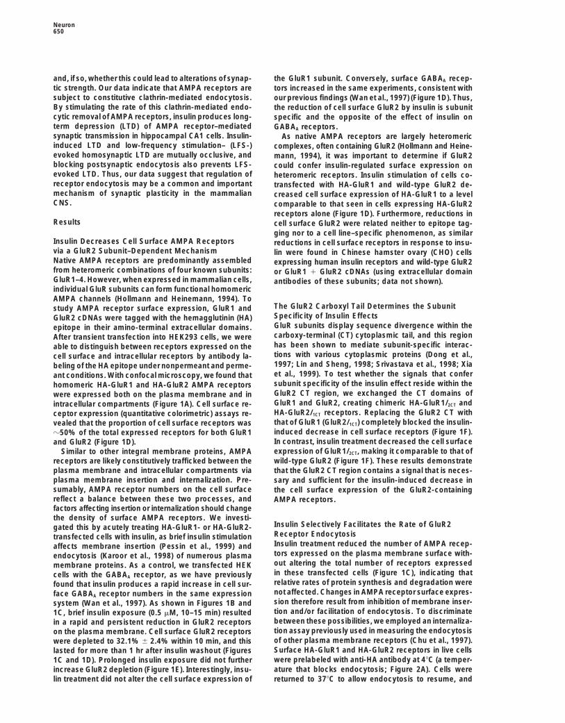
and, if so, whether this could lead to alterations of synap- the GluR1 subunit. Conversely, surface GABAA recep-tic strength. Our data indicate that AMPA receptors are tors increased in the same experiments, consistent withsubject to constitutive clathrin-mediated endocytosis. our previous findings (Wan et al., 1997) (Figure 1D). Thus,By stimulating the rate of this clathrin-mediated endo- the reduction of cell surface GluR2 by insulin is subunitcytic removal of AMPA receptors, insulin produces long- specific and the opposite of the effect of insulin onterm depression (LTD) of AMPA receptor–mediated GABAA receptors.synaptic transmission in hippocampal CA1 cells. Insulin- As native AMPA receptors are largely heteromericinduced LTD and low-frequency stimulation– (LFS-) complexes, often containing GluR2 (Hollmann and Heine-evoked homosynaptic LTD are mutually occlusive, and mann, 1994), it was important to determine if GluR2blocking postsynaptic endocytosis also prevents LFS- could confer insulin-regulated surface expression onevoked LTD. Thus, our data suggest that regulation of heteromeric receptors. Insulin stimulation of cells co-receptor endocytosis may be a common and important transfected with HA-GluR1 and wild-type GluR2 de-mechanism of synaptic plasticity in the mammalian creased cell surface expression of HA-GluR1 to a levelCNS. comparable to that seen in cells expressing HA-GluR2
receptors alone (Figure 1D). Furthermore, reductions inResults cell surface GluR2 were related neither to epitope tag-
ging nor to a cell line–specific phenomenon, as similarInsulin Decreases Cell Surface AMPA Receptors reductions in cell surface receptors in response to insu-via a GluR2 Subunit–Dependent Mechanism lin were found in Chinese hamster ovary (CHO) cellsNative AMPA receptors are predominantly assembled expressing human insulin receptors and wild-type GluR2from heteromeric combinations of four known subunits: or GluR1 1 GluR2 cDNAs (using extracellular domainGluR1–4. However, when expressed in mammalian cells, antibodies of these subunits; data not shown).individual GluR subunits can form functional homomericAMPA channels (Hollmann and Heinemann, 1994). To
The GluR2 Carboxyl Tail Determines the Subunitstudy AMPA receptor surface expression, GluR1 andSpecificity of Insulin EffectsGluR2 cDNAs were tagged with the hemagglutinin (HA)GluR subunits display sequence divergence within theepitope in their amino-terminal extracellular domains.carboxy-terminal (CT) cytoplasmic tail, and this regionAfter transient transfection into HEK293 cells, we werehas been shown to mediate subunit-specific interac-able to distinguish between receptors expressed on thetions with various cytoplasmic proteins (Dong et al.,cell surface and intracellular receptors by antibody la-1997; Lin and Sheng, 1998; Srivastava et al., 1998; Xiabeling of the HA epitope under nonpermeant and perme-et al., 1999). To test whether the signals that conferant conditions. With confocal microscopy, we found thatsubunit specificity of the insulin effect reside within thehomomeric HA-GluR1 and HA-GluR2 AMPA receptorsGluR2 CT region, we exchanged the CT domains ofwere expressed both on the plasma membrane and inGluR1 and GluR2, creating chimeric HA-GluR1/2CT andintracellular compartments (Figure 1A). Cell surface re-HA-GluR2/1CT receptors. Replacing the GluR2 CT withceptor expression (quantitative colorimetric) assays re-that of GluR1 (GluR2/1CT) completely blocked the insulin-vealed that the proportion of cell surface receptors wasinduced decrease in cell surface receptors (Figure 1F).z50% of the total expressed receptors for both GluR1In contrast, insulin treatment decreased the cell surfaceand GluR2 (Figure 1D).expression of GluR1/2CT, making it comparable to that ofSimilar to other integral membrane proteins, AMPAwild-type GluR2 (Figure 1F). These results demonstratereceptors are likely constitutively trafficked between the
plasma membrane and intracellular compartments via that the GluR2 CT region contains a signal that is neces-plasma membrane insertion and internalization. Pre- sary and sufficient for the insulin-induced decrease insumably, AMPA receptor numbers on the cell surface the cell surface expression of the GluR2-containingreflect a balance between these two processes, and AMPA receptors.factors affecting insertion or internalization should changethe density of surface AMPA receptors. We investi-
Insulin Selectively Facilitates the Rate of GluR2gated this by acutely treating HA-GluR1- or HA-GluR2-Receptor Endocytosistransfected cells with insulin, as brief insulin stimulationInsulin treatment reduced the number of AMPA recep-affects membrane insertion (Pessin et al., 1999) andtors expressed on the plasma membrane surface with-endocytosis (Karoor et al., 1998) of numerous plasmaout altering the total number of receptors expressedmembrane proteins. As a control, we transfected HEKin these transfected cells (Figure 1C), indicating thatcells with the GABAA receptor, as we have previouslyrelative rates of protein synthesis and degradation werefound that insulin produces a rapid increase in cell sur-not affected. Changes in AMPA receptor surface expres-face GABAA receptor numbers in the same expressionsion therefore result from inhibition of membrane inser-system (Wan et al., 1997). As shown in Figures 1B andtion and/or facilitation of endocytosis. To discriminate1C, brief insulin exposure (0.5 mM, 10–15 min) resultedbetween these possibilities, we employed an internaliza-in a rapid and persistent reduction in GluR2 receptorstion assay previously used in measuring the endocytosison the plasma membrane. Cell surface GluR2 receptorsof other plasma membrane receptors (Chu et al., 1997).were depleted to 32.1% 6 2.4% within 10 min, and thisSurface HA-GluR1 and HA-GluR2 receptors in live cellslasted for more than 1 hr after insulin washout (Figureswere prelabeled with anti-HA antibody at 48C (a temper-1C and 1D). Prolonged insulin exposure did not furtherature that blocks endocytosis; Figure 2A). Cells wereincrease GluR2 depletion (Figure 1E). Interestingly, insu-
lin treatment did not alter the cell surface expression of returned to 378C to allow endocytosis to resume, and

AMPA Receptor Endocytosis and Synaptic Plasticity651
Figure 1. Insulin-Induced Depletion of Cell Surface AMPA Receptors in HEK293 Cells
(A) Confocal sections show subcellular homomeric HA-GluR1 or HA-GluR2 AMPA receptors. Receptors on the cell surface were labeled withantibody under nonpermeant conditions (green), and the total receptor population was probed under permeant conditions (red).(B) Representative optical sections of cells stained under nonpermeant conditions show that insulin reduces cell surface GluR2, but not GluR1,AMPA receptor expression. Scale bars, 10 mm (A and B).(C) Time course of insulin-induced reduction in cell surface GluR2 receptors using quantitive colorimetric assays (Wang et al., 1998). Insulin(closed bar) induces a rapid, long-lasting decrease in cell surface GluR2 receptors (“Surface,” closed circles) without affecting the total numberof GluR2 receptors (“Total,” open circles), resulting in a lower proportion of surface receptors (“Surface/total,” closed triangles). Data areexpressed as the ratio between absorbance readings at the indicated time point (“Tt”) and the initial absorbance (“T0”) (378C, n 5 6 for eachtime point).(D) Insulin-induced reduction in cell surface AMPA receptors is GluR2 dependent. Cell surface expression of HA-GluR receptors in HEK cellswas determined using the ratio of absorbance readings obtained with HA antibody labeling under nonpermeant (surface) versus permeant(total) conditions. In cells transfected with GABAA receptor a1
FLAGb2g2 cDNAs, GABAA receptors translocated to the plasma membrane inresponse to insulin.(E) Reduction in HA-GluR2 receptor cell surface expression by insulin (“Ins 10 min”) plateaus within 10 min and prolonged stimulation (“Ins30 min”) fail to further reduce cell surface receptors.(F) Exchanging carboxyl tails between HA-GluR1 and HA-GluR2 subunits demonstrates that insulin-induced cell surface AMPA receptorreduction is dependent on the presence of the GluR2 CT. Colorimetric assays were performed using anti-HA antibodies. In (D) through (F),paired t tests were used to analyze differences between control and insulin-treated groups; asterisk and triple asterisk denote p , 0.05 andp , 0.001, respectively (mean 6 SE, n 5 6 in each group).

Neuron652
Figure 2. Insulin Stimulates Clathrin-Mediated Endocytosis of HA-GluR2 Receptors in HEK Cells
(A) Superimposed confocal sections of cell surface HA-GluR2 receptors (green) and internalized receptors (red) showing constitutive endocyto-sis of HA-GluR2. Live cells were prelabeled with anti-HA antibody at 48C, returned to 378C to allow constitutive internalization, and fixed atthe indicated time points. Cell surface and internalized receptors were sequentially stained with secondary antibodies under nonpermeant(green) and permeant (red) conditions.(B) Time course of constitutive endocytosis of GluR2 (left) and GluR1 (right) determined using colorimetric assays. Time-dependent loss ofsurface-bound antibody was observed at 378C nonperm. This reflects a constitutive endocytosis of the prelabeled cell surface receptors thatis due to neither a dissociation of antibody from the receptors, as there is no detectable antibody loss following incubation for various periods oftime at 48C (“48C nonperm”), nor to antibody instability, as no detectable change in antibody absorbance readings was observed following a 1 hrincubation at 378C under permeant conditions (“378C perm”). Brief insulin treatment (closed bar) enhanced both the rate and magnitude of GluR2receptor internalization but had no effect on those of GluR1 (“378C nonperm1insulin”). Green and red lines represent the best single exponentialfit to the mean data of GluR receptor endocytosis without and with insulin treatment, respectively (n 5 6 in each group, for each time point).

AMPA Receptor Endocytosis and Synaptic Plasticity653
internalization of the labeled surface receptors at differ-ent time points was then visualized using confocal mi-croscopy and quantified by colorimetric assays. GluR2receptors showed a constitutive time-dependent endo-cytosis from the cell surface in untreated cells (Figures2A and 2B, left). After 10 min, most internalized receptorswere localized to small punctae closely associated withthe plasma membrane that may represent early endo-somes. At later times, internalized receptors accumu-
Figure 3. Inhibiting Clathrin-Dependent Receptor Endocytosis Blockslated in larger punctae farther away from the plasma Insulin-Induced Reduction in Cell Surface AMPA Receptorsmembrane, which may indicate vesicle fusion or local-
Colorimetric assays show insulin-induced depletion of cell surfaceization in late endosomes. The time course for constitu- GluR2 receptors is blocked by treatment of cells with hypertonictive internalization (determined by quantitative colori- sucrose solution (A) or coexpression of mutant dynamin K44E (“Dyn-
mut”) (B). In control experiments, coexpression of wild-type dynaminmetric assays) may be described by a single exponential(“Dyn”) does not affect either the basal cell surface GluR2 levels orand a time constant of 42 min (Figure 2B, left). Fifteeninsulin-induced reduction of cell surface GluR2 (B). Neither sucroseminutes after the start of endocytosis, z15% 6 2.1%treatment nor Dyn-mut expressions alter basal levels of cell surfaceof the labeled cell surface GluR2 receptors were internal-AMPA receptors. Data are expressed as cell surface labeling versus
ized, and this increased to 48% 6 4.7% by 1 hr (Figure total cell labeling (“surface/total”). Paired t tests; double asterisk2B, left; n 5 6). In the presence of insulin (0.5 mM, 15 denotes p , 0.01 (mean 6 SE; n 5 6 in each group).min), the labeled GluR2 receptors were found to be inter-nalized at an accelerated rate following an exponential
Facilitated Endocytosis Is Fully Responsibletime course with a time constant of 15 min, nearly threefor Insulin-Induced Reduction in Celltimes faster than observed in non insulin-stimulatedSurface GluR2 Receptorscells (Figure 2B, left). Insulin stimulation also increasedAs mentioned previously, reduction of cell surfacethe degree of GluR2 internalization. At 15 min and at 1AMPA receptors by insulin (Figure 1) could result fromhr, 39% 6 1.2% and 58.9% 6 1.0% of receptors wereincreased endocytosis, decreased insertion, or a combi-internalized, respectively (Figure 2B, left). Prelabelednation of the two processes. As shown in Figure 3A,GluR1 receptors were also subject to constitutive endo-when AMPA receptor cell surface levels were quantified bycytosis at a rate and degree comparable to that of GluR2colorimetric assays, we found that the insulin-induced re-(Figure 2B, right). However, in contrast to its actions onduction was completely abolished by the blockade ofGluR2, insulin failed to alter either the rate or level ofclathrin-mediated endocytosis with hypertonic sucrose.GluR1 receptor endocytosis, supporting the depen-Moreover, internalization via the clathrin pathway isdence of insulin’s selective actions on the presenceknown to be dependent on the GTPase dynamin, andof GluR2 (Figures 1D and 1F). Thus, while constitutivedominant-negative mutants of dynamin (K44A or K44E)receptor endocytosis may be a common feature of dif-block clathrin-mediated endocytosis without affectingferent AMPA receptor subunits, the insulin-stimulatedvesicle exocytosis (Damke et al., 1994) or insulin signal-endocytosis is GluR2 specific.ing (Kao et al., 1998). In HA-GluR2 and dynamin-K44Ecotransfected HEK293 cells, the basal level of surfacereceptors was maintained, but insulin-induced de-GluR2 Endocytosis Is Mediated
by Clathrin-Coated Pits creases in cell surface GluR2 were blocked (Figure 3B).Overexpression of wild-type dynamin did not affect eitherMost plasma membrane proteins are internalized by
clathrin-mediated endocytosis. Consistent with GluR2 the basal cell surface GluR2 levels or insulin-induced re-ductions of cell surface GluR2. Together, these resultsendocytosis by clathrin-dependent mechanisms, we
found that with both constitutive and insulin-stimulated indicate that insulin can reduce the number of GluR2-containing AMPA plasma membrane receptors over aconditions, internalized GluR2 receptors extensively
colocalized with EPS15, an integral component of time course of minutes, primarily by stimulating clathrin-mediated endocytosis.clathrin-coated pits (van Delft et al., 1997) (Figure 2C).
Moreover, pretreatment of cells with hypertonic sucrose(0.45 M, 10–30 min), a blocker of clathrin-mediated en- Clathrin-Mediated Endocytosis of Native AMPA
Receptors in Cultured Neuronsdocytosis (Hansen et al., 1993), prevented both constitu-tive and insulin-enhanced internalization of GluR2 re- To determine if the results observed with recombinant
HA-tagged AMPA receptor subunits were consistent forceptors (Figure 2C). These results indicate that AMPAreceptors undergo clathrin-mediated constitutive endo- native neuronal AMPA receptors, we investigated AMPA
receptor endocytosis in cultured hippocampal neuronscytosis and that insulin accelerates this process.
(C) Clathrin-coated pits mediate constitutive and insulin-stimulated GluR2 endocytosis. Individual (GluR2, green; EPS15, red) and superimposed(“Overlay”) confocal images show colocalization of endocytosed GluR2 receptors with EPS15 in GluR2-transfected HEK cells. GluR2 receptorswere prelabeled with primary antibody at 48C, and following 10 min incubation at 378C cells were sequentially stained for GluR2 and EPS15under permeant conditions. Most internalized GluR2 in the presence (“Insulin”) and absence (“Control”) of 0.5 mM insulin are colocalized withEPS15. GluR2 endocytosis was blocked by incubation of the cells with hypertonic sucrose solution (0.45 M, 10 min) prior to insulin application(“Insulin1sucrose”).

Neuron654
Figure 4. Clathrin-Mediated Endocytosis ofAMPA Receptors in Cultured HippocampalNeurons
(A) Confocal images of internalization of cellsurface AMPA receptors at time 0 (A1) and10 min in the absence (A2) or presence (A3)of insulin treatment. The receptors were pre-labeled with an antibody raised against theamino-terminal extracellular epitope of GluR1in live neurons and allowed to endocytosefor varying lengths of time at 378C. AMPAreceptors remaining on the cell surface andthose internalized into the interior of the cellswere then sequentially stained in green andred under nonpermeant and permeant condi-tions. The red labeling of endocytosed recep-tors was specific and not artifactual as, fol-lowing identical treatment with insulin, no redstaining could be visualized without permea-bilization (A4).(B) Inhibition of clathrin-mediated endocyto-sis by hypertonic sucrose abolishes bothconstitutive and insulin-stimulated AMPA re-ceptor internalization. Following 10 min hy-pertonic sucrose (0.45 M) treatment, labelingof internalized AMPA receptors was nearlyabolished, even in the presence of insulinstimulation (B2), compared with nontreatedcontrol (B1).(C) Colorimetric quantitation of constitutiveand insulin-stimulated endocytosis of AMPAreceptors. Following prelabeling of cell sur-face AMPA receptors, there was a time-dependent decline of cell surface AMPA re-ceptors at 15 and 60 min (open bars). Insulinincreased the loss of the surface receptorswhen compared with control values at alltimes points (closed bars).(D) Insulin produces a rapid reduction ofcell surface AMPA receptors by facilitatingclathrin-mediated endocytosis. After fixationof neurons, AMPA receptors on the surfaceand in the cell interior were labeled withamino-terminal extracellular antibodies un-der nonpermeant and permeant conditions,respectively, and changes in cell surfaceAMPA receptors were quantified using colori-metric assays. Note that hypertonic sucroseblocked insulin-induced reductions of cellsurface AMPA receptor expression withoutaltering basal numbers of the cell surfaceAMPA receptors.
following the prelabeling of cell surface AMPA receptors AMPA receptor internalization when compared with thecontrol (Figures 4A1 and 4A3). Internalized receptorsin live cells and internalization at various lengths of time.
In Figure 4A, there was a modest but clear AMPA recep- under control and insulin-stimulated conditions werecharacterized by small red punctae in dendritic shafts,tor internalization 10 min after prelabeling under un-
treated control conditions (Figures 4A1 and 4A2), sug- typically lying within the boundaries of the plasma mem-brane delineated by surface-localized (green) receptors.gesting constitutive endocytosis of native neuronal
AMPA receptors. Insulin treatment greatly enhanced Internalized red staining was virtually abolished when

AMPA Receptor Endocytosis and Synaptic Plasticity655
Figure 5. Insulin Increases the Association ofNative AMPA Receptors and the AP2 AdaptorProtein Complex In Situ
(A) Antibody to adaptin b2 coimmunoprecipi-tates GluR2 (and GluR1; data not shown) withadaptin b2 from hippocampal slice homoge-nate, but control immunoprecipitation withnonspecific mouse IgG precipitates neither.Immunoprecipitates were first immunoblot-ted for GluR2 (“GluR2”) and then stripped andreprobed for adaptin b2 (“Adaptin b2”).(B) Insulin treatment increases the amountof native AMPA receptor associated with theAP2 complex. Coimmunoprecipitates usinganti–adaptin b2 antibody from control and in-sulin-treated (0.5 mM, 10 min) hippocampalslices were sequentially immunoblotted forGluR2 and adaptin b2. Densitometric quanti-tation from five separate experiments is sum-marized in the histogram in (C); asterisk, p ,
0.05.(D) NMDA receptor does not associate withthe AP2 adaptor complex. Protein samplesfrom control and insulin-treated slices werecoimmunoprecipitated with anti–adaptin b2antibody or control IgG and then sequentiallyprobed with anti-NMDAR1 and anti–adaptinb2 antibody.
the permeabilization between the FITC- and Cy3-conju- evidence that the native AMPA receptor, like its counter-part in transfected HEK cells, undergoes clathrin-depen-gated secondary antibody detection was eliminateddent constitutive and regulated endocytosis and that(Figure 4A4), indicating that red punctate labeling trulystimulating regulated endocytosis can rapidly reducerepresents the internalized AMPA receptors. Using col-the overall number of receptors expressed on the cellorimetric assays, we further quantified both constitutivesurface.and insulin-sensitive receptor endocytosis, and, as
shown in Figure 4C, following prelabeling there was adecrease in the number of prelabeled cell surface AMPA
Insulin Stimulates Association of Native AMPAreceptors to 76% of control values at 15 min that de-
Receptors with the AP2 Complexclined to 58% by 60 min, and insulin stimulation reduced
To examine AMPA receptor endocytosis in a more physi-these values to 43% and 37% respectively. Thus, native
ological context, we investigated whether clathrin-medi-AMPA receptors in cultured hippocampal neurons also ated constitutive and insulin-induced internalization oc-undergo both constitutive and regulated endocytosis at curs in mature neurons in situ using hippocampal slicesrates similar to those of recombinant GluR2-containing prepared from adult rats. Plasma membrane receptorAMPA receptors in HEK cells. Constitutive and insulin- endocytosis is initiated by the recruitment and concen-stimulated AMPA receptor internalization in neurons tration of receptors in clathrin-coated pits, presumablywere both found to be dependent on clathrin-mediated accomplished by binding of the receptors to a clathrinendocytosis, as they were completely inhibited by hy- adaptor protein complex such as AP2 (Schmid, 1997).pertonic sucrose treatment (Figure 4B). We examined the association of native AMPA receptors
We next sought to determine if insulin stimulation was with the AP2 complex in control or insulin-treated (0.5capable of reducing the overall number of AMPA recep- mM, 10 min) slices. In control conditions (Figure 5A), ators expressed on the cell surface by facilitating AMPA small number of AMPA receptor subunits were coimmu-receptor endocytosis. Using quantitative colorimetric noprecipitated with adaptin b2, a key component of theassays, we found surface levels of AMPA receptors in AP2 complex, but not with control immunoglobulin Gcultured hippocampal neurons to be z56% of the total (IgG) antibody, perhaps reflecting constitutive AMPA re-number of receptors (Figure 4D). As expected, brief insu- ceptor internalization. Insulin treatment of the slices in-lin stimulation produced a significant reduction in neu- creased the amount of AMPA receptor coimmunopreci-ronal cell surface expression of AMPA receptors that pitated with adaptin b2 (Figures 5B and 5C). In contrast,could be blocked by pretreatment with hypertonic su- under the same conditions, anti–adaptin b2 did not im-crose (Figure 4D). Interestingly, we found that, similar to munoprecipitate the N-methyl-D-aspartate- (NMDA-)the results in HEK cells (Figures 2C and 3A), hypertonic subtype glutamate receptors from either control or insu-sucrose treatment of hippocampal neurons, although lin-treated slice homogenates (Figure 5D). This suggestsblocking constitutive and insulin-stimulated AMPA re- that in mature neurons, AMPA receptors are constitu-ceptor endocytosis (Figure 4B2), did not alter the basal tively internalized via clathrin-mediated mechanismslevels of the receptors expressed on the cell surface and that insulin enhances this process by recruitment
of receptor subunits to the AP2 complex.(Figure 4D). Taken together, these data provide strong

Neuron656
Figure 6. Insulin Induces Long-Lasting Depression of AMPA Recep-tor–Mediated EPSCs in Hippocampal CA1 Neurons
(A) Bath application of insulin selectively inhibits the AMPA, but notthe NMDA, component of EPSCs. Representative EPSCs averagedfrom four individual recordings before (“Control”) or during insulinapplication (0.5 mM). The NMDA component was recorded at aholding membrane potential of 140 mV. Current–voltage curveswere constructed by plotting the AMPA component of EPSCs re-corded before (“Control”) and after 10 min application of insulin(“Insulin”) at a series of membrane holding potentials.(B) Insulin-induced depression of AMPA EPSCs is dependent onthe activation of postsynaptic insulin receptor tyrosine kinase. Nor-malized EPSCs (EPSCt/EPSC0) were plotted from neurons recordedwith regular intracellular solution without (“Control,” n 5 10) or withbath application of insulin (“Insulin,” n 5 10), or from neurons re-corded with intracellular solution supplemented with 60 mg/ml insu-lin receptor–neutralizing antibody (“Anti-IR,” n 5 6) or 100 mg/mlcontrol mouse IgG (“IgG,” n 5 5) with insulin in the bath. Time 0, inthis as well in all following figures, is defined as the time at whichEPSC amplitudes stabilized (typically 10 min after the start of whole-cell recording); at t 5 5 min, insulin was added (black bar for “Insu-
Figure 7. Insulin Selectively Inhibits Minimum Stimulus–Evokedlin,” “Anti-IR,” and “IgG” groups).AMPA EPSCs
(A) Plot of the minimum stimulus–evoked AMPA EPSC amplitudesInsulin Produces Long-Lasting Depression of the from hippocampal CA1 neurons (n 5 8). Individual traces before
(left) and after (right) insulin bath application (0.5 mM) are shownAMPA Component of Hippocampal CA1 EPSCsabove the EPSC plot.To determine the functional consequences of insulin-(B) Insulin has little effect on the NMDA component of minimuminduced AMPA receptor internalization, we examinedstimulus–induced EPSCs (n 5 6). NMDA EPSCs were recorded in
insulin’s effects on AMPA receptor–mediated synaptic the presence of CNQX (10 mM) at a holding membrane potential oftransmission in hippocampal slices. Electrical stimula- 140 mV.tion of Schaffer collateral–commissural fibers evoked (C) A bar graph showing insulin’s selective effects on both the ampli-
tude and failure rate of the AMPA (n 5 8), but not the NMDA (n 5excitatory postsynaptic currents (EPSCs) in CA1 cells,6), component of the minimum stimulus–evoked EPSCs. Open andwhich consist of two pharmacologically distinct compo-closed bars represent data obtained before and after insulin treat-nents mediated by AMPA and NMDA receptors (Figurement, respectively.
6A). Bath application of insulin (0.5 mM, 10 min) reducedthe AMPA component amplitude of EPSCs but had littleeffect on the NMDA component (Figure 6A). The reduc- The Induction of Insulin-Induced Depression
of AMPA EPSCs Is Postsynaptiction in AMPA EPSC amplitude was not associated withchanges in either the voltage–current relationship or the Insulin’s effects on AMPA receptor EPSCs may be ex-
erted either pre- or postsynaptically, as insulin receptorreversal potential (Figure 6A). Moreover, the time courseof EPSC inhibition by insulin (Figure 6B) was similar to tyrosine kinases are expressed at both presynaptic and
postsynaptic membranes (Jonas et al., 1997; Abbott etthat of receptor internalization observed in HEK cells(Figure 1C). al., 1999). The differential effects on AMPA and NMDA

AMPA Receptor Endocytosis and Synaptic Plasticity657
Figure 8. Inhibiting Clathrin-Mediated Endocytosis in Postsynaptic Neurons Blocks Insulin-Induced Depression of AMPA EPSCs
(A) Wild-type but not mutant amphiphysin-SH3 domain GST fusion proteins specifically interact with dynamin. Purified GST alone (“GST”),GST-amphiphysinSH3 (“amphiSH3”), and mutant GST-amphiphysinSH3 (“AmphiSH3m”) fusion proteins were quantified using a Coomassieblue–stained gel (top) and added to hippocampal homogenates in a pull-down assay. Precipitated proteins were then immunoblotted withanti-dynamin I antibody (bottom).(B) Normalized EPSCs are plotted in CA1 hippocampal neurons recorded with intracellular solution supplemented with 10 mM BAPTA (n 5
7), or with 100 mg/ml amphiSH3 (n 5 7) or 100 mg/ml amphiSH3m (n 5 6) GST fusion proteins. AmphiSH3, which interacts with and competesfor endogenous dynamin (A), thereby being capable of blocking clathrin-mediated endocytosis, reduced insulin’s ability to depress AMPAEPSCs. In contrast, the amphiSH3m, which cannot interact with endogenous dynamin (A) and is therefore incapable of inhibiting clathrin-dependent endocytosis, has little effect on insulin-induced depression of AMPA EPSCs.
component EPSCs are more consistent with a postsyn- (data not shown). This could be a result of the reductionof EPSC amplitude below the detection threshold, butaptic action of insulin. To examine insulin’s site of action,
we postsynaptically applied a membrane-impermeant it could also be due to the conversion of active synapsesinto silent ones. In marked contrast, insulin failed to altermonoclonal antibody against the tyrosine kinase domain
of the insulin receptor, which specifically inhibits the either the amplitude or the failure rate of the minimumstimulus–evoked NMDA component EPSCs (Figures 7Bactivity of insulin receptors in in vitro kinase assays and
in situ when injected directly into cells (Morgan and and 7C). These results strongly suggest that the expres-sion of insulin-induced depression of AMPA responsesRoth, 1987). The postsynaptic application of this anti-
insulin receptor antibody (but not of the control mouse is also postsynaptic.IgG) eliminated insulin’s ability to inhibit AMPA EPSCs(Figure 6B). These results demonstrate that insulin acts Insulin-Induced Depression of AMPA EPSCs Ispostsynaptically via its receptor tyrosine kinase, sup- Dependent on Clathrin-Mediated Endocytosisporting a postsynaptic locus for insulin-induced depres- in Postsynaptic Neuronssion of AMPA EPSCs. Insulin’s postsynaptic loci of induction and expression
are both consistent with its effect being mediated bythe clathrin-dependent endocytosis of AMPA receptors.
The Expression of Insulin-Induced Depression To address this more specifically, we chelated intracel-of AMPA EPSCs Is Also Postsynaptic lular Ca21 with the postsynaptic application of the mem-To determine the site(s) of expression of the insulin- brane-impermeant Ca21 chelator bis-(o-aminophenoxy)-induced depression of AMPA EPSCs, we evoked unitary N,N,N9,N9-tetraacetic acid (BAPTA) (10 mM), sinceEPSCs by lowering the stimulus intensity to a level that clathrin-mediated endocytosis is dependent on intra-produced a failure rate of .20%. If the expression of cellular Ca21 (Marks and McMahon, 1998). Followingdepression is presynaptic, insulin should affect only the BAPTA application, insulin treatment failed to depressfailure rate and not the amplitude of the weak, stimulus- the AMPA EPSC amplitude (Figure 8B). More direct evi-induced EPSCs. If, on the other hand, expression is dence for the involvement of postsynaptic clathrin-postsynaptic, resulting from the removal of postsynaptic mediated endocytosis was obtained by the blockade ofAMPA receptors, the altering of channel conductance, the insulin effect with a glutathione S-transferase (GST)or open probability, insulin should alter both the ampli- fusion protein of the amphiphysin SH3 domain (GST-tude and failure rate of EPSCs, with the latter due to amphiSH3). It is believed that amphiphysin SH3 domainpostsynaptic failure in released transmitter detection binding to the proline-rich region of dynamin recruits(Liao et al., 1995). Consistent with a postsynaptic site dynamin to clathrin-coated pits, initiating clathrin/of expression, insulin reduced the amplitude of AMPA dynamin-dependent endocytosis (Shupliakov et al.,EPSCs in 8 of 11 cells tested, and this reduction was 1997). When incubated with hippocampal slice homoge-associated with a dramatic increase in the failure rate nates, GST-amphiSH3 was able to pull down a protein(Figures 7A and 7C). In the 3 remaining cells, EPSCs of z100 kDa, which was recognized by dynamin anti-
bodies on Western blots (Figure 8A). GST alone, andwere no longer detected following insulin application

Neuron658
GST-amphiSH3 with two mutations in its dynamin bind-ing domain (GST-amphiSH3m) that render it incapableof binding to dynamin (Shupliakov et al., 1997), failedto precipitate endogenous dynamin (Figure 8A). Thus,GST-amphiSH3 specifically interacts with endogenousrat brain dynamin and was therefore used as a competi-tive inhibitor to disrupt binding between endogenousamphiphysin and dynamin in rat hippocampal neurons.GST-amphiSH3 reduced the ability of insulin to depressAMPA EPSCs (Figure 8B). In contrast, GST-amphiSH3mhad little effect on insulin-induced depression of AMPAEPSCs.
Collectively, our biochemical and electrophysiologicalresults from hippocampal slices and cultured hippocam-pal cells suggest that native AMPA receptors, similar totheir recombinant counterparts in HEK cells, are subjectto clathrin-mediated endocytosis. Insulin stimulates thisprocess and causes the rapid removal of postsynapticAMPA receptors, leading to LTD or even the silencingof AMPA receptor–mediated synaptic transmission.
Postsynaptic Clathrin-Mediated EndocytosisIs Required for the Expression of CA1 LTDHomosynaptic LTD of AMPA receptor–mediated synap-tic transmission in hippocampal CA1 neurons is a well-characterized in vitro model of synaptic plasticity (Bearand Malenka, 1994). While it is agreed that inductionof the most common form of LTD at this synapse ispostsynaptic, mechanisms underlying its expression re-main hotly debated (Bear and Malenka, 1994; Bolshakovand Siegelbaum, 1994). Our preceding experiments sug-gested that enhanced AMPA receptor endocytosiscould depress AMPA receptor–mediated transmission,so we investigated if postsynaptic AMPA receptor endo-cytosis could contribute to homosynaptic LTD andwhether LTD and insulin-induced reductions in AMPAEPSCs shared common pathways. Delivering 900 pulsesat 1 Hz to the Schaffer collaterals consistently producedLTD of AMPA EPSCs in CA1 neurons, recorded withstandard intracellular solution (EPSC amplitude: 62% 63% of baseline 60 min poststimulation, n 5 6; Figure9A). However, when 100 mM of GST-amphiSH3 was in-cluded in the recording pipette, LTD failed to be induced
Figure 9. Expression of Homosynaptic Hippocampal CA1 LTD Re- in every neuron tested (EPSC: 95% 6 3% of controlquires Clathrin-Dependent Endocytosis in Postsynaptic Neurons after 60 min post LTD induction, n 5 6; Figure 9A). In(A) Inhibition of clathrin-dependent endocytosis in postsynaptic contrast, inclusion of GST-amphiSH3m altered neitherneurons prevents the expression of LFS-induced LTD in CA1 neu- the time course nor the extent of LTD (64% 6 4%, n 5rons in a hippocampal slice preparation. Plot of averaged AMPA
6; Figure 9A). These results indicate that a dynamin-EPSC amplitudes in recordings with pipettes containing standarddependent, clathrin-mediated endocytic process is re-recording solution (“Control,” n 5 6), or solution supplemented withquired for the production of this particular form of LTD100 mg/ml GST-amphiSH3 (“AmphiSH3,” n 5 6) or 100 mg/ml GST-
amphiSH3m (“AmphiSH3m,” n 5 6). Top panels are superimposed and further suggest that LFS-induced LTD may use aindividual recordings before (“b”) and 60 min after (“a”) the LFS–LTD similar mechanism underlying insulin-induced LTD thatinduction. involves clathrin-mediated postsynaptic AMPA receptor(B) and (C) Plots of normalized EPSC amplitudes show that LFS- and
internalization. Indeed, as shown in Figure 9B, after LFS-insulin-induced LTDs mutually occlude each other. Representativeinduced LTD was fully established, application of insulintraces shown in the plots were taken at time points, as indicated.
Following induction of LFS LTD (“LFS”), the ability of insulin (“Insu- failed to further reduce EPSC amplitudes. Conversely,lin,” 0.5 mM) to depress AMPA EPSCs was dramatically reduced after insulin induced rapid and persistent decreases in([B], n 5 6), and, conversely, after the establishment of insulin- AMPA EPSCs (Figure 9C), LFS no longer produced LTD.induced depression of AMPA EPSCs, LFS protocol failed to induce
Thus, LFS and insulin-induced LTD mutually occludeLTD ([C], n 5 6).each other, suggesting that the two share a commonfinal pathway, namely the clathrin-mediated endocyto-sis of postsynaptic AMPA receptors.

AMPA Receptor Endocytosis and Synaptic Plasticity659
Discussion receptor subunits, insulin-stimulated endocytosis ap-pears to be GluR2 specific, contingent on unique se-quences in its intracellular carboxyl tail. Many intracellu-Constitutive versus Regulated AMPAlar proteins have recently been revealed to be AMPAReceptor Endocytosisreceptor–interacting proteins (e.g., NSF, PICK1, andIt is becoming clear that clathrin-mediated internaliza-GRIPs). It is noteworthy that the majority of these pro-tion of plasma membrane proteins is a tightly regulatedteins bind specifically to the GluR2 cytoplasmic tailprocess and that such regulation is an important means(Dong et al., 1997; Lin and Sheng, 1998; Srivastava etof controlling the cell surface expression, and henceal., 1998; Xia et al., 1999). Although the functions offunction, of these proteins (Schmid, 1997; Karoor et al.,these GluR2-interacting proteins remain elusive, they1998). In the present study, we examined the role ofmay regulate endocytosis or other aspects of AMPAclathrin-mediated endocytosis in the activity-dependentreceptor vesicle trafficking via their binding to the GluR2redistribution of AMPA receptors. Using antibodiescarboxyl tail, thereby playing an important role in con-against extracellular epitopes on AMPA receptors, wetrolling AMPA receptor density in the postsynaptic mem-selectively visualized the trafficking of AMPA receptorsbrane. Indeed, evidence is accumulating suggesting theexpressed on the surface of live cells and found thatimportant role that some of these proteins may play inboth GluR1 and GluR2 receptors undergo constitutiveregulating the level of cell surface AMPA receptors, andclathrin-dependent endocytosis. One would expect thathence the efficacy of the AMPA receptor–mediated syn-perturbation of constitutive endocytosis should lead toaptic transmission (Li et al., 1999; Luscher et al., 1999;altered AMPA receptor expression on the plasmaLuthi et al., 1999; Noel et al., 1999). For instance, inter-membrane. Surprisingly, we found that while hypertonicrupting NSF-GluR2 interaction by postsynaptic injectionsucrose treatment and overexpression of the dominant-of the pep2m peptide, like insulin stimulation, producesnegative dynamin mutant effectively blocked constitu-an LTD of AMPA EPSCs that is also mutually occlusivetive AMPA receptor endocytosis, both manipulationswith the LFS-induced CA1 LTD (Luscher et al., 1999;failed to alter AMPA receptor numbers on the cell sur-Luthi et al., 1999). As demonstrated in this work, theface. Among many possible explanations, the simplestregulated clathrin-dependent endocytosis of AMPA re-one is that there is an unknown mechanism tightly cou-ceptors is contingent upon the GluR2 carboxyl tail, andpling the constitutive endocytosis and insertion of AMPAenhanced endocytotic removal of AMPA receptorsreceptors. Thus, factors affecting one pathway wouldclearly contributes to the LTD expression. It would there-also affect the other through a feedback mechanism tofore be very interesting to know if NSF actually actsensure a balance between receptor insertion and re-to maintain the levels of AMPA receptor cell surfacemoval. The result would produce a constant numberexpression, primarily by preventing the GluR2-depen-of receptors on the cell membrane, and hence stabledent regulated AMPA receptor endocytosis. In this case,baseline synaptic transmission, even under conditionspep2m, by interrupting NSF-GluR2 interaction, pro-in which either constitutive exocytosis (Lledo et al., 1998)duces its rapid reduction of cell surface expression ofor endocytosis (the present work; Wang and Linden,AMPA receptors by stimulating the endocytotic arm of2000 [this issue of Neuron]) has been selectively altered.the AMPA receptor cycling but not by impairing theIt should be noted that Luscher et al. (1999) have recentlyexocytotic arm, as originally envisaged.reported rundown and runup of AMPA receptor–medi-
Thus, from the evidence presented here, we speculateated EPSCs in hippocampal slices following postsyn-that there are two distinct clathrin-mediated endocyticaptic injection of putative inhibitors of exocytosis andpathways involved in the removal of cell surface AMPAendocytosis, respectively. Thus, some discrepanciesreceptors. Constitutive endocytosis seems to be sharedbetween this and our results remain to be explained.by all GluRs, and its major function would be to counter-
In addition to constitutive endocytosis, we observedact constitutive receptor insertion, ensuring a constant
insulin-stimulated, clathrin-dependent endocytosis ofnumber of cell surface AMPA receptors under most con-
AMPA receptors in both transfected HEK cells and cul- ditions. In contrast, the regulated pathway is GluR2 spe-tured hippocampal neurons. Several features associ- cific and rapidly regulates AMPA receptor cell surfaceated with this insulin-regulated pathway distinguish it expression. Therefore, this pathway may be importantfrom constitutive endocytosis, the most prominent be- in certain forms of synaptic plasticity involving GluR2-ing its ability to rapidly reduce AMPA receptors on the containing AMPA receptors. It remains to be determinedcell surface, thereby suppressing AMPA receptor– how distinct constitutive and regulated endocytoticmediated responses. This indicates that insulin may se- pathways are in terms of their molecular mechanismslectively facilitate receptor endocytosis without affect- and how they converge on clathrin-mediated internaliza-ing receptor insertion, hence resetting the equilibrium tion as a common final step.between receptor removal and insertion. This featuremakes regulated endocytosis an attractive mechanism Clathrin-Mediated Endocytosis in Synaptic Plasticityfor regulating synaptic plasticity in models such as LTD. The “silent synapse” is a recent hypothesis that hasIndeed, a similar mechanism appears to be critical in been proposed to explain the postsynaptic locus of ex-various forms of LTD, as suggested by this study (see pression of LTP and LTD (Kullmann, 1994; Isaac et al.,below) and the companion paper (Wang and Linden, 1995; Liao et al., 1995; Durand et al., 1996; Malinow,2000). 1998; Malenka and Nicoll, 1999). A silent synapse con-
Another feature of regulated endocytosis is its subunit tains functional NMDA receptors but lacks functionalspecificity. Unlike the constitutive pathway, which seems AMPA receptors and becomes activated during the in-
duction of LTP by recruitment of AMPA receptors. Byto be common across at least the GluR1 and GluR2

Neuron660
extrapolation, an active synapse may be silenced during in mediating the expression of these forms of LTD butLTD by the loss of functional AMPA receptors. One key rather that insulin and LTD-inducing stimuli may con-question that remains is how synapses are switched verge to cause AMPA receptor endocytosis. It is likelybetween active and silent states. Since plasma mem- that multiple signal transduction pathways exist for thebrane insertion and internalization of proteins are highly regulation of AMPA receptor trafficking, and the elucida-controlled, regulated exocytosis and endocytosis of tion of these pathways will provide further insight intoAMPA receptors offer attractive mechanisms for activat- the molecular mechanisms of synaptic plasticity anding and silencing synapses, and hence for the expres- may ultimately provide mechanistic clues for the role ofsion of LTP and LTD. Indeed, evidence is emerging to insulin in learning and memory.support a critical role for postsynaptic membrane fusion
Experimental Procedures(Lledo et al., 1998) and AMPA receptor translocation(Shi et al., 1999) in LTP induction.
cDNA Plasmids, Cell Cultures, and Plasmid TransfectionWe report here several observations that suggest theRat GluR1 and GluR2 cDNAs were amplified by PCR and cloned intoinvolvement of clathrin-mediated endocytotic removalthe XbaI/EcoRI and HindIII/SalI sites, respectively, of the mammalian
of postsynaptic AMPA receptors in LTD. First, rapid en- expression vector GW1 (British Biotechnology). For extracellular HAdocytosis of AMPA receptors occurs in mature hippo- tagging of GluR1 and GluR2 subunits, site-directed mutagenesiscampal neurons, shown by the increased association of was performed to insert an AscI restriction site after A374 in GluR1
and after G384 in GluR2. An oligonucleotide cassette encoding theAMPA receptors with the AP2 complex following insulinHA epitope was inserted into these AscI restriction sites. For ex-stimulation. Second, facilitation of clathin-mediated en-changes of carboxyl tails between GluR1 and GluR2 subunits, EcoRIdocytosis of postsynaptic AMPA receptors by insulinsites were generated by silent mutation in the carboxyl termini of HA-
produces LTD and, in some cases, silencing of AMPA GluR1 and HA-GluR2 immediately after the fourth transmembranereceptor–mediated synaptic transmission. Third, insu- domain. After the removal of their cytoplasmic tails using EcoRIlin- and LFS-induced CA1 LTDs are mutually occlusive, digestion, the resulting backbones of HA-GluR1 and HA-GluR2 were
ligated, respectively, to the PCR products of the GluR2 and GluR1suggesting a common mechanism mediating the twocarboxy-terminal tails. Both chimeras were confirmed by sequenc-forms of LTD. Finally, LFS-induced LTD is blocked bying. Rat GABAA receptor subunit cDNAs have been described pre-postsynaptic microinjection of the amphiphysin SH3 do-viously (Wan et al., 1997). Wild-type GluR1 and GluR2 cDNAs weremain. Thus, together with work from Luscher et al. expressed in pCIS2 plasmid vectors. pcDNA3 expression vectors
(1999), our results suggest that rapid, clathrin-mediated containing wild-type and K44E mutant dynamin I cDNAs were giftsendocytotic removal of postsynaptic AMPA receptors of Dr. R. B. Vallee (Worcester Institute, Shrewsbury, MA). HEK293
(ATCC) and CHO cells overexpressing human insulin receptors (pro-may play an important role in LFS-induced hippocampalvided by Dr. C. C. Yip, Banting and Best Institute, Toronto) werehomosynaptic CA1 LTD production.transfected using the Ca21-phosphate precipitation method. Thirty-Endocytosis of postsynaptic AMPA receptors may notsix to forty-eight hours after transfection, cells were washed withbe limited to homosynaptic CA1 LTD, as in a relatedextracellular recording solution (ECS, in mM: NaCl, 140; CaCl2, 1.3;
study (Wang and Linden, 2000) we found that insulin/ KCl, 5.4; HEPES, 25; and glucose, 33 [pH 7.4], 320 mOsm) andIGF-I also produces a rapid and long-term depression incubated in ECS (serum starvation) for at least 1 hr. For insulinof AMPA responses mediated by postsynaptic clathrin- treatment, cells were incubated with ECS supplemented with 0.5
mM human recombinant insulin (Sigma) for 10–15 min. Cultureddependent endocytosis in cultured cerebellar neurons.hippocampal neurons were prepared from embryonic Wistar ratsThe insulin/IGF-I-induced depression of AMPA currents(embryonic day 18–19; Charles River) as described previously (Wanoccludes cerebellar LTD, which in turn can be blockedet al., 1997). Cells were used following 2–3 weeks growth in culture.by the inhibition of postsynaptic clathrin-dependent en-
docytosis (Wang and Linden, 2000). Additionally, Carroll GST Fusion Protein Productionet al. (1999a) have reported a rapid, activity-dependent pGEX2T plasmids encoding either wild-type or mutant SH3 domainreduction of postsynaptic AMPA receptors in a culture of human amphiphysin were obtained from Dr. P. De Camilli (Yale
University, New Haven). GST fusion proteins were expressed inmodel of LTD induced by field stimulation. Taken to-DH5a Escherichia coli and purified from bacterial lysates accordinggether, these data suggest that rapid, clathrin-depen-to the manufacturer’s protocol (Pharmacia). Products were dialyzeddent removal of postsynaptic AMPA receptors may bein phosphate-buffered saline (PBS) and concentrated using Micro-a common final step in the expression of certain forms con-10 (Amicon) tubes for intracellular application during whole-cell
of LTD. recordings.How is the clathrin-dependent endocytosis of AMPA
receptors stimulated by LTD-inducing protocols? As Immunofluorescent Confocal MicroscopyHEK293 cells were transfected with each plasmid described above.neurons contain and are able to release insulin in anFor cell surface receptor expression assays, cells at 48 hr post-activity-dependent manner, and as insulin receptors aretransfection were fixed with 4% paraformaldehyde in PBS for 10concentrated in the postsynaptic density (Wozniak etmin. Surface AMPA receptors were first labeled with monoclonal
al., 1993; Abbott et al., 1999), one mechanism may in- anti-HA antibody (1:5000, Babco, Berkeley, CA) and a FITC-conju-volve the release of insulin presynaptically in response gated anti-mouse antibody (1:500, Sigma). Total cellular AMPA re-to LFS during LTD induction. Insulin may in turn activate ceptors were then stained with polyclonal anti-GluR1 (Cedarlaneits postsynaptic neuronal receptors to facilitate clathrin- Laboratories) or GluR2 (Chemicon) antibody and Cy3-labeled anti-
rabbit antibodies following permeation of the cells with 0.25% Tritondependent endocytosis of AMPA receptors. However,X-100 in PBS for 10 min. For the AMPA receptor internalizationpostsynaptic injection of the insulin receptor–neutraliz-assay, HEK293 cells transfected with HA-GluR constructs were incu-ing antibody, while blocking insulin-induced depressionbated live at 48C with 10 mg/ml monoclonal anti-HA antibody for 1
of AMPA EPSCs, had little effect on either hippocampal hr to label surface receptors and then incubated at 378C for varioushomosynaptic LTD (L. D. L. and Y. T. W., unpublished time periods to allow for constitutive internalization of labeled recep-data) or cerebellar LTD (Wang and Linden, 2000). These tors. Following fixation without permeation, receptors remaining on
the plasma membrane surface were stained with FITC-conjugatedresults suggest that insulin is not itself directly involved

AMPA Receptor Endocytosis and Synaptic Plasticity661
anti-mouse antibodies. Cells were subsequently treated for 1 min Homosynaptic CA1 LTD was recorded in slices prepared fromrats aged 16–26 postnatal days old. Following 20 min recording ofwith 100% methanol and stained with Cy3-conjugated anti-mouse
antibodies. Colocalization of HA-tagged GluR2 receptors with baseline EPSCs evoked every 30 s, recording was switched to cur-rent-clamp mode, and 15 min of train stimulation at 1 Hz (900 pulsesEPS15 was studied by sequentially staining both monoclonal anti-
HA and FITC-conjugated anti-mouse antibody and polyclonal anti- in total) was delivered from the same stimulating electrode. Therecording was then switched back to voltage-clamp mode, andEPS15 and Cy3-conjugated anti-rabbit antibody under permeant
conditions. Subcellular localization of fluorescently labeled recep- EPSC recordings at the baseline stimulus rate were then recordedfor more than 1 hr thereafter.tors was examined with a Leica TCS-4D confocal microscope (Wan
et al., 1997).Acknowledgments
Colorimetric AssaysThis work was supported by research grants from the Heart andColorimetric assays were performed using a protocol modified fromStroke Foundation of Ontario (NA-3762), the Medical Researcha published method (Wang et al., 1998). Briefly, HEK293 cells wereCouncil of Canada, and the EJLB foundation (to Y. T. W.) and fromtransfected by calcium phosphate in 15 cm dishes. Cells were re-the National Institutes of Health (NS35050) (to M. S.). Y. T. W. is aplated 24 hr after transfection into 12-well plates. Treatment andResearch Scholar of the Heart and Stroke Foundation of Canada,control studies, as well as time course studies, were performed onand M. S. is Assistant Investigator of the Howard Hughes Medicalthe same sets of 12-well plates derived from the same populationInstitute. H. Y. M. is supported by a Clinician-Scientist Award fromof transfected cells so as to minimize transfection efficiency–relatedthe Research Training Centre at The Hospital for Sick Children andvariation. For the assay, cells were fixed in paraformaldehyde asa fellowship from the Ontario Neurotrauma Foundation. We thankdescribed above. Cells were, under either nonpermeant (PBS alone)Dr. R. B. Vallee, at the Worcester Institute, for dynamin cDNAs; Dr.or permeant (PBS containing 0.2% Triton X-100) conditions, blockedP. De Camilli, at Yale University, for the amphiphysin-SH3 con-for 1 hr at room temperature with 3% bovine serum albumin andstructs; Dr. R. A. Roth, at Stanford University, for the anti-insulinthen incubated overnight at 48C in anti-HA (1:2000) to detect HA-receptor kinase domain antibody; Dr. S. Egan, at the University oftagged receptors, polyclonal antibodies raised against the amino-Toronto, for the anti-EPS15 antibody; Dr. C. C. Yip, at the Universityterminal region of GluR1 (1:1000, Oncogene Science) to detectof Toronto, for CHO cells expressing human insulin receptors; andwild-type GluR1, or monoclonal antibody raised against an amino-Dr. D. Linden, at Johns Hopkins University, for helpful comments.terminal sequence of GluR2/4 for wild-type GluR2 (1:1000; Phar-
mingen). After extensive wash, cells were sequentially incubated forReceived October 28, 1999; revised February 7, 2000.1 hr at room temperature with the appropriate horseradish peroxi-
dase–conjugated secondary antibody (1:800, Amersham) and for 2Referencesmin with 1 volume of OPD substrate (Sigma). Reactions were
stopped with 0.2 volume of 3N HCl, and the optical density of 1 mlAbbott, M.A., Wells, D.G., and Fallon, J.R. (1999). The insulin recep-of supernatant was read on a spectrophotometer at 492 nm.tor tyrosine kinase substrate p58/53 and the insulin receptor arecomponents of CNS synapses. J. Neurosci. 19, 7300–7308.Coimmunoprecipitation and ElectrophysiologyBear, M.F., and Malenka, R.C. (1994). Synaptic plasticity: LTP andin Hippocampal SlicesLTD. Curr. Opin. Neurobiol. 4, 389–399.Hippocampal slices (300 mm thickness) were prepared from adult
male Sprague-Dawley rats (150–200 g, Charles River) as described Bliss, T.V.P., and Collingridge, G.L. (1993). A synaptic model ofmemory: long-term potentiation in the hippocampus. Nature 361,(Wan et al., 1997). For immunoprecipitation and immunoblotting,
homogenate (z500 mg protein) from control or slices treated with 31–39.0.5 mM insulin for 10 min were incubated with anti–adaptin b2 mono- Bolshakov, V.Y., and Siegelbaum, S.A. (1994). Postsynaptic induc-clonal antibody (Sigma) in 500 ml of 50 mM Tris-HCl, 150 mM NaCl, tion and presynaptic expression of hippocampal long-term depres-and 0.1% Triton X-100 for 4 hr at 48C. The antibody-protein com- sion. Science 264, 1148–1152.plexes were then pelleted with protein A–Sepharose beads. Proteins Carroll, R.C., Lissin, D.V., von Zastrow, M., Nicoll, R.A., and Malenka,eluted from the beads were subjected to SDS–PAGE and immu- R.C. (1999a). Rapid redistribution of glutamate receptors contributesnoblotting for anti–adaptin b2 (1:5000), GluR2/4 (Pharmingen, 1 mg/ to long-term depression in hippocampal cultures. Nat. Neurosci. 2,ml), GluR1 (1 mg/ml, Oncogene Science), or monoclonal anti- 454–460.NMDAR1 (1:800, Pharmingen), respectively. For sequential probing
Carroll, R.C., Beattie, E.C., Xia, H., Scher, C., Altschuler, Y., Nicoll,of the same membrane, the membranes were stripped of antibodyR.A., Malenka, R.C., and von Zastrow, M. (1999b). Dynamin-depen-and reprobed. The blot was developed using enhanced chemilumi-dent endocytosis of ionotropic glutamate receptors. Proc. Natl.nescence detection methods (Amersham). Band intensities wereAcad. Sci. USA 96, 14112–14117.quantified using Scion Image PC software.Chu, P., Murray, S., Lissin, D., and von Zastrow, M. (1997). Deltaand kappa opioid receptors are differentially regulated by dynamin-Whole-Cell Recordingsdependent endocytosis when activated by the same alkaloid ago-For electrophysiological recordings, slices were perfused at roomnist. J. Biol. Chem. 272, 27124–27130.temperature (22–248C) with artificial cerebrospinal fluid containingCraig, A.M. (1998). Activity and synaptic receptor targeting: the long126 mM NaCl, 3 mM KCl, 1 mM MgCl2, 1 mM CaCl2, 1.2 mM KH2PO4,view. Neuron 21, 459–462.26 mM NaHCO3, and 10 mM glucose and bubbled with 95%
O2/5% CO2. An incision was made between CA3 and CA1 to reduce Damke, H., Baba, T., Warnock, D.E., and Schmid, S.L. (1994). Induc-epileptiform activity. Whole-cell recordings of CA1 neurons were tion of mutant dynamin specifically blocks endocytic coated vesicleperformed using the “blind” method with an Axopatch-1D amplifier formation. J. Cell Biol. 127, 915–934.(Axon Instruments, Foster City, CA). Recording pipettes (4–5 MV) Dong, H., O’Brien, R.J., Fung, E.T., Lanahan, A.A., Worley, P.F.,were filled with intracellular solution containing (in mM): CsCl, 135; and Huganir, R.L. (1997). GRIP: a synaptic PDZ domain–containingEGTA, 0.05; HEPES, 10; Mg-ATP, 4; GTP, 0.2; and QX-314, 5 (pH protein that interacts with AMPA receptors. Nature 386, 279–284.7.4), 310 mOsm, or the same solution supplemented with one of
Durand, G.M., Kovalchuk, Y., and Konnerth, A. (1996). Long-termthe following: 10 mM BAPTA, anti-insulin receptor kinase domainpotentiation and functional synapse induction in developing hippo-antibody (60 mg/ml) (17A3; Morgan and Roth, 1987), mouse IgG (100campus. Nature 381, 71–75.
mg/ml), amphiphysin-SH3 domain (100 mg/ml), or mutant amphiphy-Hansen, S.H., Sandvig, K., and van Deurs, B. (1993). Clathrin andsin-SH3 domain (100 mg/ml). EPSCs were evoked by stimulation ofHA2 adaptors: effects of potassium depletion, hypertonic medium,the Schaffer collateral–commissural pathway with a bipolar tungstenand cytosol acidification. J. Cell Biol. 121, 61–72.electrode (0.05 ms duration at a rate of 0.07 Hz) in the presence of
bicuculline (20 mM). Minimum stimulus–evoked EPSCs were re- Hollmann, M., and Heinemann, S. (1994). Cloned glutamate recep-tors. Annu. Rev. Neurosci. 17, 31–108.corded following a modified protocol (Liao et al., 1995).

Neuron662
Isaac, J.T., Nicoll, R.A., and Malenka, R.C. (1995). Evidence for silent Shupliakov, O., Low, P., Grabs, D., Gad, H., Chen, H., David, C.,synapses: implications for the expression of LTP. Neuron 15, Takei, K., De Camilli, P., and Brodin, L. (1997). Synaptic vesicle427–434. endocytosis impaired by disruption of dynamin–SH3 domain inter-
actions. Science 276, 259–263.Jonas, E.A., Knox, R.J., Smith, T.C.M., Wayne, N.L., Connor, J.A.,and Kaczmarek, L.K. (1997). Regulation by insulin of a unique neu- Srivastava, S., Osten, P., Vilim, F.S., Khatri, L., Inman, G., States,ronal Ca21 pool and neuropeptide secretion. Nature 385, 343–346. B., Daly, C., DeSouza, S., Abagyan, R., Valtschanoff, J.G., et al.
(1998). Novel anchorage of GluR2/3 to the postsynaptic density byKanzaki, M., Zhang, Y.Q., Masjedi, H., Li, L., Shibata, H., and Kojima,the AMPA receptor–binding protein ABP. Neuron 21, 581–591.I. (1999). Translocation of a calcium-permeable cation channel in-
duced by insulin-like growth factor-I. Nat. Cell Biol. 1, 165–170. van Delft, S., Schumacher, C., Hage, W., Verkleij, A.J., and vanBergen en Henegouwen, P.M. (1997). Association and colocalizationKao, A.W., Ceresa, B.P., Santeler, S.R., and Pessin, J.E. (1998).of Eps15 with adaptor protein–2 and clathrin. J. Cell Biol. 136,Expression of a dominant interfering dynamin mutant in 3T3L1 adi-811–821.pocytes inhibits GLUT4 endocytosis without affecting insulin signal-
ing. J. Biol. Chem. 273, 25450–25457. Wan, Q., Xiong, Z.G., Man, H.Y., Ackerley, C.A., Braunton, J., Lu,W.Y., Becker, L.E., MacDonald, J.F., and Wang, Y.T. (1997). Recruit-Karoor, V., Wang, L., Wang, H.Y., and Malbon, C.C. (1998). Insulinment of functional GABAA receptors to postsynaptic domains bystimulates sequestration of beta-adrenergic receptors and en-insulin. Nature 388, 686–690.hanced association of beta-adrenergic receptors with Grb2 via tyro-
sine 350. J. Biol. Chem. 273, 33035–33041. Wang, Q., Khayat, Z., Kishi, K., Ebina, Y., and Klip, A. (1998). GLUT4translocation by insulin in intact muscle cells: detection by a fastKullmann, D.M. (1994). Amplitude fluctuations of dual-componentand quantitative assay. FEBS Lett. 427, 193–197.EPSCs in hippocampal pyramidal cells: implications for long-term
potentiation. Neuron 12, 1111–1120. Wang, Y.T., and Linden, D.J. (2000). Expression of cerebellar long-term depression requires postsynaptic clathrin-mediated endocyto-Li, P., Kerchner, G.A., Sala, C., Wei, F., Huettner, J.E., Sheng, M.,
and Zhuo, M. (1999). AMPA receptor–PDZ interactions in facilitation sis. Neuron 25, this issue, 635–647.of spinal sensory synapses. Nat. Neurosci. 2, 972–977. Wickelgren, I. (1998). Tracking insulin to the mind. Science 280,Liao, D., Hessler, N.A., and Malinow, R. (1995). Activation of postsyn- 517–519.aptically silent synapses during pairing-induced LTP in CA1 region Wozniak, M., Rydzewski, B., Baker, S.P., and Raizada, M.K. (1993).of hippocampal slice. Nature 375, 400–404. The cellular and physiological actions of insulin in the central ner-Lin, J.W., and Sheng, M. (1998). NSF and AMPA receptors get physi- vous system. Neurochem. Int. 22, 1–10.cal. Neuron 21, 267–270. Xia, J., Zhang, X., Staudinger, J., and Huganir, R.L. (1999). ClusteringLissin, D.V., Carroll, R.C., Nicoll, R.A., Malenka, R.C., and von Zas- of AMPA receptors by the synaptic PDZ domain–containing proteintrow, M. (1999). Rapid, activation-induced redistribution of iono- PICK1. Neuron 22, 179–187.tropic glutamate receptors in cultured hippocampal neurons. J. Neu-rosci. 19, 1263–1272.
Lledo, P.M., Zhang, X., Sudhof, T.C., Malenka, R.C., and Nicoll, R.A.(1998). Postsynaptic membrane fusion and long-term potentiation.Science 279, 399–403.
Luscher, C., Xia, H., Beattie, E.C., Carroll, R.C., von Zastrow, M.,Malenka, R.C., and Nicoll, R.C. (1999). Role of AMPA receptor cy-cling in synaptic transmission and plasticity. Neuron 24, 649–658.
Luthi, A., Chittajallu, R., Duprat, F., Palmer, M.J., Benke, T.A., Kidd,F.L., Henley, J.M., Isaac, J.T.R., and Collingridge, G.J. (1999). Hippo-campal LTD expression involves a pool of AMPARs regulated bythe NSF–GluR2 interaction. Neuron 24, 389–399.
Malenka, R.C., and Nicoll, R.A. (1999). Long-term potentiation—adecade of progress? Science 285, 1870–1874.
Malinow, R. (1998). Silencing the controversy in LTP? Neuron 21,1226–1227.
Marks, B., and McMahon, H.T. (1998). Calcium triggers calcineurin-dependent synaptic vesicle recycling in mammalian nerve terminals.Curr. Biol. 8, 740–749.
Morgan, D.O., and Roth, R.A. (1987). Acute insulin action requiresinsulin receptor kinase activity: introduction of an inhibitory mono-clonal antibody into mammalian cells blocks the rapid effects ofinsulin. Proc. Natl. Acad. Sci. USA 84, 41–45.
Noel, J., Ralph, G.S., Pickard, L., Williams, J., Molnar, E., Uney, J.B.,Collingridge, G.L., and Henley, J.M. (1999). Surface expression ofAMPA receptors in hippocampal neurons is regulated by an NSF-dependent mechanism. Neuron 23, 365–376.
O’Brien, R.J., Kamboj, S., Ehlers, M.D., Rosen, K.R., Fischbach,G.D., and Huganir, R.L. (1998). Activity-dependent modulation ofsynaptic AMPA receptor accumulation. Neuron 21, 1067–1078.
Pessin, J.E., Thurmond, D.C., Elmendorf, J.S., Coker, K.J., andOkada, S. (1999). Molecular basis of insulin-stimulated GLUT4 vesi-cle trafficking. Location! Location! Location! J. Biol. Chem. 274,2593–2596.
Schmid, S.L. (1997). Clathrin-coated vesicle formation and proteinsorting: an integrated process. Annu. Rev. Biochem. 66, 511–548.
Shi, S.H., Hayashi, Y., Petralia, R.S., Zaman, S.H., Wenthold, R.J.,Svoboda, K., and Malinow, R. (1999). Rapid spine delivery and redis-tribution of AMPA receptors after synaptic NMDA receptor activa-tion. Science 284, 1811–1816.