Endocannabinoids potently protect the newborn brain against AMPA-kainate receptor-mediated...
10
Endocannabinoids potently protect the newborn brain against AMPA-kainate receptor-mediated excitotoxic damage 1,2,7 Basma Shouman, 1,2 Romain H. Fontaine, 1,2,3 Olivier Baud, 1,2 Leslie Schwendimann, 4 Matthias Keller, 5 Michael Spedding, 1,2 Vincent Lelie`vre & * ,1,2,6 Pierre Gressens 1 Inserm, U676, Paris, Hoˆpital Robert Debre´, 48 Blvd Se´rurier, F-75019, Paris, France; 2 Universite´ Paris 7, Faculte´ de Me´decine Denis Diderot, IFR02, Paris, France; 3 AP HP, Hoˆpital Robert Debre´, Service de Re´animation Ne´onatale, Paris, France; 4 Department of Neonatology, Medical University Innsbruck, Innsbruck, Austria; 5 Institut de Recherches Internationales Servier (I.R.I.S.), Neuilly sur Seine, France and 6 AP HP, Hoˆpital Robert Debre´, Service de Neurologie Pe´diatrique, Paris, France 1 Brain lesions induced in newborn mice or rats by the glutamatergic agonists ibotenate (acting on NMDA and metabotropic receptors) or S-bromowillardiine (acting on AMPA-kainate receptors) mimic some aspects of white matter cysts and transcortical necrosis observed in human perinatal brain damage associated with cerebral palsy. Exogenous and endogenous cannabinoids have received increasing attention as potential neuroprotective agents in a number of neurodegenerative disorders of the adult. One recent study showed neuroprotection by the cannabinoid agonist WIN-55212 in a newborn rat model of acute severe asphyxia. 2 The present study was designed to assess the neuroprotective effects of the endogenous cannabinoid anandamide using a well-defined rodent model of neonatal excitotoxic brain lesions. 3 In this model, anandamide provided dose-dependent and long-lasting protection of developing white matter and cortical plate reducing the size of lesions induced by S-bromowillardiine. Anandamide had only marginal neuroprotective effect against ibotenate-induced cortical grey matter lesions. Anandamide-induced neuroprotection against AMPA-kainate receptor-mediated brain lesions were blocked by a CB1 antagonist but not by a CB2 antagonist. Furthermore, anandamide effects were mimicked by a CB1 agonist but not by a CB2 agonist. Real-time PCR confirmed the expression of CB1 receptors, but not CB2 receptors, in the untreated newborn neocortex. Finally, neuroprotective effects of anandamide in white matter involved increased survival of preoligoden- drocytes and better preservation of myelination. 4 The present study provides experimental support for the role of endocannabinoids as a candidate therapy for excitotoxic perinatal brain lesions. British Journal of Pharmacology (2006) 148, 442–451. doi:10.1038/sj.bjp.0706755; published online 8 May 2006 Keywords: Anandamide; CB1 receptor; cerebral palsy; neuroprotection; NMDA; oligodendrocyte; periventricular leukomalacia; willardiine Abbreviations: ACPA, arachidonylcyclopropylamide; CP, cerebral palsy Introduction The major brain lesions associated with cerebral palsy (CP) are periventricular leukomalacia (periventricular white matter lesion) in preterm infants and cortico-subcortical lesions in term infants (Volpe, 2001). Several preconception, prenatal and perinatal factors implicated in the pathophysiology of brain lesions associated with CP include hypoxia-ischemia, endocrine imbalances, genetic factors, growth factor defi- ciency, abnormal competition for growth factors, excess free reactive oxygen species production, maternal infection yielding excess cytokines, and other proinflammatory agents (Wil- loughby and Nelson, 2002; Mesples et al., 2005). Excess release of glutamate could represent a molecular mechanism common to some of these risk factors. Accord- ingly, injection of glutamate agonists into the striatum, the neocortex or the periventricular white matter of newborn rodents or rabbits produces histological lesions that mimic CP- associated brain damage characterized by cystic periventricu- lar leukomalacia and hypoxic-ischemic or ischemic-like cor- tical and striatal lesions (McDonald et al., 1988; Barks and Silverstein, 1992; Marret et al., 1995; Acarin et al., 1999; Follett et al., 2000; Sfaello et al., 2005a) . The use of specific agonists for the different classes of glutamatergic receptors has demonstrated that oligodendrocyte precursor cell death is a key event in periventricular white matter lesions induced by agonists acting on or alpha-3-amino-hydroxy-5-methyl-4- isoxazole propionic acid (AMPA) and kainate receptors (Follett et al., 2000), while microglial activation plays a major role in periventricular white matter lesions induced by agonists acting on N-methyl-D-aspartate (NMDA) receptors (Tahraoui et al., 2001). Despite major improvements in neonatal care, there is still no specific treatment for perinatal brain lesions (Gressens *Author for correspondence at: INSERM U 676, Hoˆpital Robert Debre´, 48 Blvd Se´rurier, F-75019 Paris, France. E-mail: [email protected] 7 Current address: Mansoura University Children’s Hospital, Man- soura, Egypt. British Journal of Pharmacology (2006) 148, 442–451 & 2006 Nature Publishing Group All rights reserved 0007 – 1188/06 $30.00 www.nature.com/bjp
Transcript of Endocannabinoids potently protect the newborn brain against AMPA-kainate receptor-mediated...

Endocannabinoids potently protect the newborn brain against
AMPA-kainate receptor-mediated excitotoxic damage
1,2,7Basma Shouman, 1,2Romain H. Fontaine, 1,2,3Olivier Baud, 1,2Leslie Schwendimann,4Matthias Keller, 5Michael Spedding, 1,2Vincent Lelievre & *,1,2,6Pierre Gressens
1Inserm, U676, Paris, Hopital Robert Debre, 48 Blvd Serurier, F-75019, Paris, France; 2Universite Paris 7, Faculte de MedecineDenis Diderot, IFR02, Paris, France; 3AP HP, Hopital Robert Debre, Service de Reanimation Neonatale, Paris, France;4Department of Neonatology, Medical University Innsbruck, Innsbruck, Austria; 5Institut de Recherches Internationales Servier(I.R.I.S.), Neuilly sur Seine, France and 6AP HP, Hopital Robert Debre, Service de Neurologie Pediatrique, Paris, France
1 Brain lesions induced in newborn mice or rats by the glutamatergic agonists ibotenate (acting onNMDA and metabotropic receptors) or S-bromowillardiine (acting on AMPA-kainate receptors)mimic some aspects of white matter cysts and transcortical necrosis observed in human perinatal braindamage associated with cerebral palsy. Exogenous and endogenous cannabinoids have receivedincreasing attention as potential neuroprotective agents in a number of neurodegenerative disordersof the adult. One recent study showed neuroprotection by the cannabinoid agonist WIN-55212in a newborn rat model of acute severe asphyxia.
2 The present study was designed to assess the neuroprotective effects of the endogenouscannabinoid anandamide using a well-defined rodent model of neonatal excitotoxic brain lesions.
3 In this model, anandamide provided dose-dependent and long-lasting protection of developingwhite matter and cortical plate reducing the size of lesions induced by S-bromowillardiine.Anandamide had only marginal neuroprotective effect against ibotenate-induced cortical grey matterlesions. Anandamide-induced neuroprotection against AMPA-kainate receptor-mediated brainlesions were blocked by a CB1 antagonist but not by a CB2 antagonist. Furthermore, anandamideeffects were mimicked by a CB1 agonist but not by a CB2 agonist. Real-time PCR confirmed theexpression of CB1 receptors, but not CB2 receptors, in the untreated newborn neocortex. Finally,neuroprotective effects of anandamide in white matter involved increased survival of preoligoden-drocytes and better preservation of myelination.
4 The present study provides experimental support for the role of endocannabinoids as a candidatetherapy for excitotoxic perinatal brain lesions.British Journal of Pharmacology (2006) 148, 442–451. doi:10.1038/sj.bjp.0706755;published online 8 May 2006
Keywords: Anandamide; CB1 receptor; cerebral palsy; neuroprotection; NMDA; oligodendrocyte; periventricularleukomalacia; willardiine
Abbreviations: ACPA, arachidonylcyclopropylamide; CP, cerebral palsy
Introduction
The major brain lesions associated with cerebral palsy (CP) are
periventricular leukomalacia (periventricular white matter
lesion) in preterm infants and cortico-subcortical lesions in
term infants (Volpe, 2001). Several preconception, prenatal
and perinatal factors implicated in the pathophysiology of
brain lesions associated with CP include hypoxia-ischemia,
endocrine imbalances, genetic factors, growth factor defi-
ciency, abnormal competition for growth factors, excess free
reactive oxygen species production, maternal infection yielding
excess cytokines, and other proinflammatory agents (Wil-
loughby and Nelson, 2002; Mesples et al., 2005).
Excess release of glutamate could represent a molecular
mechanism common to some of these risk factors. Accord-
ingly, injection of glutamate agonists into the striatum, the
neocortex or the periventricular white matter of newborn
rodents or rabbits produces histological lesions that mimic CP-
associated brain damage characterized by cystic periventricu-
lar leukomalacia and hypoxic-ischemic or ischemic-like cor-
tical and striatal lesions (McDonald et al., 1988; Barks and
Silverstein, 1992; Marret et al., 1995; Acarin et al., 1999;
Follett et al., 2000; Sfaello et al., 2005a) . The use of specific
agonists for the different classes of glutamatergic receptors has
demonstrated that oligodendrocyte precursor cell death is
a key event in periventricular white matter lesions induced
by agonists acting on or alpha-3-amino-hydroxy-5-methyl-4-
isoxazole propionic acid (AMPA) and kainate receptors
(Follett et al., 2000), while microglial activation plays a major
role in periventricular white matter lesions induced by agonists
acting on N-methyl-D-aspartate (NMDA) receptors (Tahraoui
et al., 2001).
Despite major improvements in neonatal care, there is still
no specific treatment for perinatal brain lesions (Gressens
*Author for correspondence at: INSERM U 676, Hopital RobertDebre, 48 Blvd Serurier, F-75019 Paris, France.E-mail: [email protected] address: Mansoura University Children’s Hospital, Man-
soura, Egypt.
British Journal of Pharmacology (2006) 148, 442–451 & 2006 Nature Publishing Group All rights reserved 0007–1188/06 $30.00
www.nature.com/bjp

& Spedding, 2004). The endocannabinoid system is an
emerging target for drug discovery, because it is involved in
the regulation of many cellular and physiological functions
(Fernandez-Ruiz et al., 2000; Fride, 2002). The endocannabi-
noid system constitutes the endogenous lipids anandamide,
2-arachidonoylglycerol and noladin ether, and the cannabi-
noid CB1 and CB2 receptors, as well as the proteins involved
in the synthesis and inactivation of the endocannabinoids (van
der Stelt et al., 2002).
Endogenous cannabinoids are clearly a major site for drug
action – and do not share the deleterious effects of cannabis
which swamps the endogenous system by stimulating all the
receptors. The distribution of cannabinoid receptors in the
brain shows how important they are in modulating the brain
schemes shown above with differential distribution in hippo-
campus and amygdala (Freund et al., 2003). The massive
endocannabinoid distribution in the basolateral amygdala is
responsible for the anxiolytic effects of cannabis.
There is a clear link between the cannabinoid system and
the GABAergic system. For example, endogenous anandamide
inhibits CA1 neurones via interneurones and this effect is
mediated by CB1 receptor stimulation. The inhibition is very
local feedback system, involving only a few interneurones.
Depolarization-induced suppression of inhibition (DSI) is
a calcium-dependent, retrograde-signalling process mediated
by endocannabinoids by a reduction of GABA release via
presynaptic cannabinoid receptor (CB1) activation. DSI lasts
around one minute while synaptic long term depression (LTD
– usually loss of AMPA receptors from the synapse) is long
lasting.
However, in early life the GABAergic system is depolarizing
rather than hyperpolarizing (up to BP8–P10 in mice) and this
obviously has major effects on the drugs acting on the GABA
system, particularly as the time for the GABA hyperpolariza-
tion/depolarization switch is not known in man, and may vary
with brain areas. Bernard et al. (2005) showed that retrograde
signaling of the cannabinoid system can substitute for the
GABA system in early development, controlling synaptic
transmission and preventing epileptic discharges. As there is
a continuing problem of CP in the newborn, and the use
of benzodiazepine activators of the GABAergic system is used
for controlling epileptic activity in these children (perhaps
inappropriately if the GABA hyperpolarization/depolarization
switch has not taken place in all brain areas), it seems essential
to determine if endogenous cannabinoid agonists can have
neuroprotective effects in newborn animals. The use of
synthetic agonists may be inappropriate as there will be no
local inactivation of these substances and they may, therefore,
swamp the local control systems, resulting in impaired
developmental synaptic plasticity (Bernard et al., 2005).
Exogenously administered endocannabinoids have been
shown to exert neuroprotection in a variety of in vitro and
in vivo models of adult neuronal injury (Baker et al., 2003;
Di Marzo et al., 2004) including 6-hydroxydopamine toxicity
(Lastres-Becker et al., 2005), beta-amyloid-induced toxicity
(Ramirez et al., 2005), and neurodegeneration in models of
multiple sclerosis (Ortega-Gutierrez et al., 2005) or excitotoxi-
city (Marsicano et al., 2003; Veldhuis et al., 2003). The
proposed mechanisms include, among others, blockade of
microglial activation (Ramirez et al., 2005), increase in brain-
derived neurotrophic factor (Khaspekov et al., 2004), reduc-
tion of calcium influx (Nadler et al., 1993), and antioxidant
activity (El-Remessy et al., 2003). Despite the rapid enzymatic
degradation of endocannabinoids, neuroprotection was ob-
served even if exogenous endocannabinoids were administered
systemically in the absence of inhibitor of the fatty acid amid
hydrolase (FAAH) which is involved in the hydrolysis of
endogenous cannabinoids such as anandamide (Lastres-Becker
et al., 2005).
To our knowledge, only a few studies have addressed the
potential neuroprotective effects of cannabinoids for the
neonatal brain. In one study (Hansen et al., 2001), it was
shown that anandamide accumulates in neonatal rat models
characterized by widespread neurodegeneration as a conse-
quence of altered glutamatergic neurotransmission. In another
study (Martinez-Orgado et al., 2003), the authors showed
neuroprotection by the cannabinoid agonist WIN-55212 in
an in vivo newborn rat model of acute severe asphyxia. This
neuroprotective effect was blocked by a specific CB1 receptor
antagonist. Conversely, it was shown that a specific CB1
receptor antagonist was neuroprotective against NMDA-
induced neuronal cell death in newborn rats (Hansen et al.,
2002).
The present study was designed to assess the neuroprotective
effects of the endocannabinoid anandamide using in a well-
defined mouse model of neonatal excitotoxic brain lesions
(Marret et al., 1995; Tahraoui et al., 2001; Husson et al., 2002;
Husson et al., 2005; Sfaello et al., 2005b), which mimics several
aspects of brain damage associated with human CP.
Methods
Animals and drugs
Swiss mice and Sprague Dawley rats of both sexes were used
for this study. Experimental protocols were approved by the
institutional review committee, meet the INSERM guidelines,
and were carried out in accordance with the Guide for the Care
and use of Laboratory Animals as adopted and promulgated
by the U.S. National Institutes of Health. All drugs were
purchased from Tocris (Bristol, U.K.) except for nabilone
which was obtained from Servier. Ibotenate was diluted in
phosphate-buffer saline (PBS) containing 0.01% acetic acid.
S-bromowillardiine, MK-801, and NBQX were diluted in PBS.
Anandamide, arachidonylcyclopropylamide (ACPA), AM251,
AM630, JWH133, nabilone, and URB597 were diluted in
TocrisolveTM.
Ibotenate activates NMDA and metabotropic glutamatergic
receptors while S-bromowillardiine activates both AMPA and
kainate receptors. MK801 is a NMDA receptor antagonist and
NBQX is an AMPA/kainate receptor antagonist. Anandamide
is an endogenous cannabinoid activating both CB1 and CB2
receptors, ACPA is a selective CB1 agonist, JWH133 is a
selective CB2 agonist, nabilone is a CB1 and CB2 agonist,
AM251 is a selective CB1 antagonist, and AM630 is a selective
CB2 antagonist (Howlett et al., 2002). URB597 is selective
inhibitor of the FAAH (Fegley et al., 2005).
Excitotoxic brain lesions
We induced excitotoxic brain lesions by injecting ibotenate
(10mg) or S-bromowillardiine (15 mg) into developing mouseand rat brains, as previously described (Marret et al., 1995;
B. Shouman et al Endocannabinoids protect the newborn brain 443
British Journal of Pharmacology vol 148 (4)

Gressens et al., 1997; Tahraoui et al., 2001; Husson et al.,
2002). Controls received intracerebral ibotenate (10 mg)þMK-801 (1 mg) or S-bromowillardiine (15 mg)þNBQX (20 mg).Briefly, on postnatal day (P) 5, pups anesthetized with
isoflurane were kept under a warming lamp to maintain body
temperature. They were injected intracerebrally (into the
neopallial parenchyma) with ibotenate or S-bromowillardiine
on the fifth postnatal day (P). Intracerebral injections were
performed with a 25-gauge needle on a 50-ml Hamilton syringemounted on a calibrated microdispenser. The needle was
inserted 2mm below the skin’s external surface. The needle tip
was placed in the frontoparietal area of the right hemisphere, 2
(mice) or 2.5mm (rats) from the midline in the lateral-medial
plane, and three (mice) or 4mm (rats) from the bregma in the
rostro-caudal plane. Histopathological observation confirmed
that the needle tip always reached the periventricular white
matter. Two 1-ml boluses of ibotenate or S-bromowillardiinewere injected at 20-s intervals. The needle was left in place for
an additional 20 s.
Experimental groups
Pups from at least two different litters were used in each
experimental group, and data were obtained from two or more
successive experiments.
In the first set of experiments, P5 mouse pups were
intracerebrally injected with 10mg ibotenate, 10 mg ibo-
tenateþ 20mg MK801, 15 mg S-bromowillardiine or 15mgS-bromowillardiine þ 20 mg NBQX.In the second set of experiments, 10mg ibotenate or 15mg
S-bromowillardiine were intracerebrally injected into P5
mouse pups and anandamide (0.01, 0.03, 0.1, 0.3, 1, 3, or
10mgkg�1), ACPA (1, 3, or 10mg kg�1), nabilone (1 or
10mgkg�1), JWH133 (1 or 10mgkg�1), anandamide
(10mgkg�1)þAM251 (20mgkg�1), anandamide (10mgkg�1)þAM630 (20mgkg�1), AM251 (20mgkg�1), AM630 (20mgkg�1),
anandamide (1mgkg�1)þURB597 (0.3mgkg�1), or vehicle
(controls) were administered intraperitoneally immediately
following the excitotoxin injection.
In the third set of experiments, 15mg S-bromowillardiinewere intracerebrally injected into P5 mouse pups and
intraperitoneal (i.p.) anandamide (10mgkg�1) was adminis-
tered at immediately, 4, 8, 12, or 24 h after the excitotoxin
injection.
In the last set of experiments, 15 mg S-bromowillardiine wereintracerebrally injected into P5 rat pups and i.p. anandamide
(10mg kg�1) was administered immediately after the excito-
toxin injection.
Determination of lesion size
Mouse and rat pups were kill by decapitation 5 (P10) or 25
(P30) days after the excitotoxic challenge. Brains were fixed
immediately in 4% formalin and remained in this solution for
5 days. Following paraffin embedding, 16-mm-thick coronal
sections were cut. Every third section was stained with cresyl-
violet. The size of neocortical and white matter lesions can be
defined by the length on three orthogonal axes: the lateral-
medial axis (in a coronal plane), the radial axis (also in a
coronal plane, from the pial surface to the lateral ventricle),
and the fronto-occipital axis (in a sagittal plane). In previous
studies (Marret et al., 1995; Gressens et al., 1997; Husson
et al., 2002), we found an excellent correlation among the
measurements from the three axes of the excitotoxic lesions.
Based on these findings, we cut serial sections of the entire
brain in the coronal plane for this study. This permitted an
accurate and reproducible determination of the sagittal fronto-
occipital diameter (which is equal to the number of sections
where the lesion was present multiplied by 16mm). Thismeasure was used as an index of the lesion volume.
Immunohistochemistry for oligodendrocytes and myelin
To study the neuroprotective effect of TPM on oligodendro-
cytes and myelin, P5 rat pups were injected with 15mgS-bromowillardiine and 10mg kg�1 i.p. anandamide, or 15mgS-bromowillardiine and i.p. PBS. Animals were killed at 1 (P6)
or 9 (P14) days after injections. Myelin basic protein (MBP,
a marker of myelin; 1/1000, Chemicon, Temecula, CA, U.S.A.)
immunostaining was performed on formalin-fixed, 15-mmthick, paraffin sections. O4 (a marker of preoligodendrocytes;
1/500, generous gift from Dr PA Rosenberg, Boston, MA,
U.S.A.) immunofluorescence staining was performed on 4%
paraformaldehyde-fixed, 10-mm thick, frozen sections. For 04
staining, detection of labeled antigens was performed with
secondary antibody conjugated to Texas red (Vector) and
nuclei were counter-stained with bis-benzimide.
Five animals were included in each group. For each animal,
several sections were immunoreacted in successive experi-
ments, and two different investigators blinded to the experi-
mental groups independently performed the analyses. MBP
immunostaining was evaluated in a qualitative manner in the
white matter at the site of the lesion. O4 labeled cells were
quantified at the level of neocortical lesion and/or underlying
white matter cystic lesion. For each animal, counts were
performed in 0.0625mm2 area of the most affected section.
Real time RT–PCR for CB1 and CB2 receptors mRNA
Mouse pups (n¼ 5 per group) were sacrificed on P5
immediately before or 3 h after intracerebral injection with
ibotenate, S-bromowillardiine or PBS. Brains were removed
and tissues immediately adjacent to the site of excitotoxin
injection (see above) were collected from the different animals
for RNA extraction. Total RNA was extracted as previously
described (Lelievre et al., 2002), followed by DNaseI treatment
(TURBO DNA-freet, Ambion, Austin, TX, U.S.A.). For
each sample, 600 ng were used in reverse transcription (iScript
kit from BioRad, Hercules, CA, U.S.A.). The following
oligonucleotides (Oligo6 software, Molecular Biology Insights,
Cascade, CO, U.S.A.) 50- TAATTGCTGTGTTGCCTCTCC-30 and 50-TCCGATCCAGAACATCAGGTA-30, 50- GGCAGTGTGACCATGACCTT-30 and 50-GGTAGGCGGGTAACACAGACA-30 were used as sense and antisense primers, forCB1 and CB2 receptors, respectively. PCR amplification
resulted in the generation of a single band at 107 and 90 bp
corresponding to the region 1288-1395 and 514–604 of the
previously published sequences (NCBI access numbers
NM007726 and NM009924) of mouse CB1 and CB2 receptors,
respectively. A primer set (50- TGGTGAAAAGGACCTCTCGAA-30 and 50-TCAAGGGCATATCCAACAACA-30, as
sense and antisense, respectively) for the mouse hypoxanthine
guanine phosphoribosyl transferase (HPRT) gene was used to
standardize the experiments. These primers amplified an 90 bp
444 B. Shouman et al Endocannabinoids protect the newborn brain
British Journal of Pharmacology vol 148 (4)

region encoding the nucleotides 578–668 of the published
sequence (access number NM013556) of the mouse mRNA.
Real-time PCR was set up using sybergreen-containing super-
mix from BioRad, for 45 cycles (20 s denaturation at 961C, 20 s
annealing at 621C, and 20 s extension at 721C). Amplification
specificity was assessed by melting curve and sequencing
analyses. Quantification of each PCR samples was made using
standard curves made from serial dilutions of control samples.
Relative expression levels of genes of interest were calculated
as the difference between the specific ratios (CB receptor/
HPRT) and were further adjusted on the basis of their
differences in CT values (number of cycles to reach threshold).
Experiments were independently run three times. In each
experiment, samples were performed in quadruplicates.
Statistical analysis
All variables were found normally distributed based on
Skewness and Kurtosis analyses. Data were analyzed with a
Student’s t-test or a univariate ANOVA (GraphPad Prism
version 3.03 for Windows, GraphPad Software, San Diego,
CA, U.S.A.). When ANOVA showed significant differences
among multiple experimental groups, multiple comparisons of
treated vs control groups were performed using Dunnett’s post
hoc test.
Results
Overall mortality was low in the present study (o3% of the
animals injected with ibotenate or S-bromowillardiine). No
significant difference was observed in a test of contingency
(exact Fisher test) when the different treatment groups were
compared to the animals injected with ibotenate plus vehicle or
with S-bromowillardiine plus vehicle. Epileptic manifestations
including clonic or tonic seizures and apneas were observed in
all ibotenate-treated animals. However, treatment with ana-
ndamide, ACPA, nabilone, AM251 or AM630 did not induce
any detectable difference in severity and frequency of seizures
(frequency of seizures was quantitatively assessed during a
10-min period, once every hour during the first 6 h following
excitotoxin injection; severity of seizures was qualitatively
assessed according to the same schedule) when compared to
controls (data non shown).
Mouse pups injected on P5 with intracerebral ibotenate or
S-bromowillardiine and i.p. vehicle developed cortical lesions
and periventricular white matter cysts (Figure 1a). The cortical
lesion was typically characterized by neuronal loss in all
neocortical layers and almost complete disappearance of
neuronal cell bodies along the axis of excitotoxin injection.
Ibotenate-induced lesions were totally abolished by cointra-
cerebral injection of MK-801, a specific NMDA receptor
antagonist (Figure 2). S-bromowillardiine-induced lesions
were totally abolished by co-intracerebral injection of NBQX,
a specific AMPA-kainate receptor antagonist (Figure 3a). P5
rats injected with intracerebral S-bromowillardiine and i.p.
PBS displayed cortical and white matter lesions similar to
those observed in P5 mice (data not shown).
In P5 mice, i.p. anandamide induced a moderate neuropro-
tection of the cortical plate against ibotenate-induced lesions
but had no detectable effect on ibotenate-induced white matter
lesions when observed on P10 (Figure 2). This moderate
protective effect was independent of the dose used. The
addition of URB597 (0.3mgkg�1), an inhibitor of FAAH, to
anandamide (1mgkg�1) did not enhance anandamide-induced
neuroprotection (Figure 2).
In P5 mice, i.p. anandamide induced a significant and
dose-dependant neuroprotection against S-bromowillardiine-
induced cortical plate and white matter lesions when observed
Figure 1 TPM protects against S-bromowillardiine-induced brainlesions. Cresyl violet-stained sections showing brain lesions inducedby S-bromowillardiine injected at P5 and studied at the age of P10.(a) Brain from a pup cotreated with intracerebral S-bromowillar-diine and i.p. vehicle, showing the typical neuronal loss in layersII–VI (arrow) and the white matter cystic lesion (*). (b) Brain frompup cotreated with intracerebral S-bromowillardiine and i.p.anandamide (10mgkg�1). LV, lateral ventricle. Bar¼ 40 mm.
B. Shouman et al Endocannabinoids protect the newborn brain 445
British Journal of Pharmacology vol 148 (4)

on P10 (Figures 1b and 3a) or P30 (Figure 3b). Similar effects
were observed in rat pups injected on P5 and killed on P10
(Figure 3c). In mice, these neuroprotective effects of ananda-
mide were mimicked by i.p. ACPA, a CB1 agonist but not by
i.p. nabilone, a CB1 and CB2 agonist, and not by i.p. JWH133,
a CB2 agonist (Figure 3d). In mice, anandamide neuroprotec-
tive effects against S-bromowillardiine were abolished by
AM251, a CB1 antagonist, but not by AM630, a CB2
antagonist (Figure 3e).
When i.p. injection of anandamide followed the excitotoxic
challenge, neuroprotection was a function of time. Protection
was observed in mice receiving anandamide within the first 4 h
after S-bromowillardiine administration (Figure 3f).
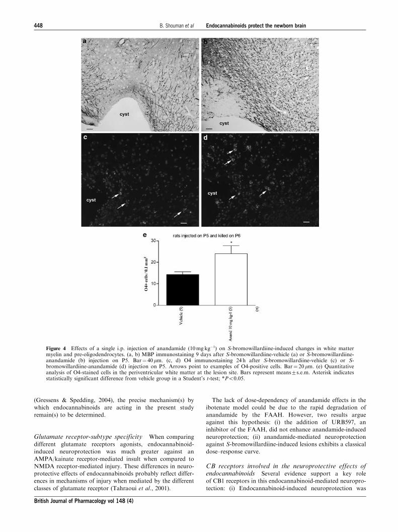
In rats, i.p. administration of anandamide (10mgkg�1, i.p.)
largely prevented S-bromowillardiine-induced reduction
of MBP staining when analyzed 9 days after the insult
(Figure 4a and b). Similarly, when compared to a control
PBS injection, i.p. injection of anandamide (10mg kg�1, i.p.)
significantly increased the density of cells labelled with O4, a
marker of preoligodendrocytes, when examined between 24 h
after S-bromowillardiine administration (Figure 4c and e).
Quantitative real time PCR showed high levels of CB1
receptor mRNA while CB2 receptor mRNA was barely
detectable in untreated P5 neocortex (Figure 5a). CB1 mRNA
expression was not modified by intracerebral injection with
ibotenate or S-bromowillardiine (Figure 5b). In contrast, CB2
mRNA expression was significantly enhanced 3 h after the
injection of ibotenate of S-bromowillardiine (Figure 5c).
However, the levels of CB2 mRNA were still much lower
than the levels of CB1 mRNA.
Discussion
The present study provides experimental support for the
consideration of endocannabinoids as a candidate therapy for
reducing the risk of excitotoxic perinatal brain lesions. In this
mouse model, endocannabinoids protected the developing
white matter and cortical plate in a dose-dependent and long-
lasting manner against an AMPA/kainate receptor-mediated
challenge. Endocannabinoids had only a marginal effect on
NMDA receptor-mediated cortical brain lesions. Endocanna-
binoid-induced neuroprotection of white matter involved
increased survival of preoligodendrocytes and increased
preservation of myelination.
Neuroprotection conferred by endocannabinoids onexcitotoxic injury in the developing brain: comparisonof our findings with those of others
As mentioned above, only a few studies have addressed the
potential neuroprotective effects of cannabinoids for the
neonatal brain. In one study (Hansen et al., 2001), it was
shown that anandamide accumulates in neonatal rat models of
neurodegeneration. In another study (Martinez-Orgado et al.,
2003), the authors showed neuroprotection by the cannabinoid
agonist WIN-55212 in a newborn rat model of acute asphyxia.
As it has been previously shown that, in this neonatal rat
model, hypoxic-ischemic brain lesions are largely mediated by
excess glutamate release (Ikonomidou et al., 1989; Hagberg
et al., 1994), limitation of excitotoxic damage as demonstrated
in the present study might underlie some of the neuroprotec-
tive effects afforded by WIN-55212 against hypoxic-ischemic
insult. In addition, exogenously administered endocannabi-
noids have been shown to exert neuroprotection against
neuronal excitotoxicity in cell culture and in adult rodents
(Marsicano et al., 2003; Veldhuis et al., 2003; Chen et al.,
2005).
Figure 2 Anandamide has marginal effects on the size of ibotenate-induced lesions. Mouse pups were injected with intracerebralibotenate on P5 and sacrificed on P10. Pups were injected witha single i.p. injection of vehicle, anandamide or anandami-deþURB597 (0.3mg kg�1) immediately after ibotenate injectionor with intracerebral MK-801 concomitantly with ibotenate. Barrepresents mean length of the brain lesions at P10þ s.e.m. Asterisksindicate statistically significant difference from black bars;**Po0.01; ***Po0.001 in ANOVA with Bonferroni’s multiplecomparison tests.
Figure 3 Anandamide significantly mitigated S-bromowillardiine-induced lesions. Mouse or rat pups were injected with intracerebralS-bromowillardiine on P5. (a) Mouse pups were injected with a single i.p. injection of vehicle or anandamide immediately afterS-bromowillardiine injection, or with intracerebral NBQX concomitantly with S-bromowillardiine. Pups were sacrificed on P10. (b) Mousepups were injected with a single i.p. injection of vehicle or anandamide (Anand) immediately after S-bromowillardiine injection. Pups werekilled on P30. (c) Rat pups were injected with a single i.p. injection of vehicle or anandamide (Anand) immediately after S-bromowillardiineinjection. Pups were killed on P10. (d) Mouse pups were injected with a single i.p. injection of vehicle, ACPA, nabilone (Nab), or JWH133(JWH) immediately after S-bromowillardiine injection. Pups were sacrificed on P10. (e) Mouse pups were injected with a single i.p. injection ofdrug of combination of drugs indicated on the X-axis immediately after S-bromowillardiine injection. Pups were killed on P10. (f) Mouse pupswere injected with a single i.p. injection of anandamide administered at immediately, 4, 8, 12, or 24 h after S-bromowillardiine injection. Pupswere sacrificed on P10. Bar represents mean length of the brain lesions at P10 or P30þ s.e.m. Asterisks indicate statistically significantdifference from black bars; *Po0.05; **Po0.01; ***Po0.001 in ANOVA with Bonferroni’s multiple comparison tests.
446 B. Shouman et al Endocannabinoids protect the newborn brain
British Journal of Pharmacology vol 148 (4)
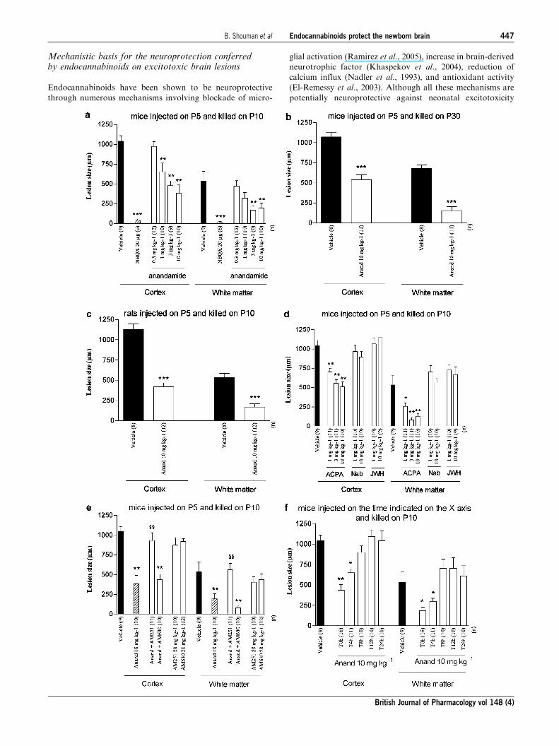
Mechanistic basis for the neuroprotection conferredby endocannabinoids on excitotoxic brain lesions
Endocannabinoids have been shown to be neuroprotective
through numerous mechanisms involving blockade of micro-
glial activation (Ramirez et al., 2005), increase in brain-derived
neurotrophic factor (Khaspekov et al., 2004), reduction of
calcium influx (Nadler et al., 1993), and antioxidant activity
(El-Remessy et al., 2003). Although all these mechanisms are
potentially neuroprotective against neonatal excitotoxicity
B. Shouman et al Endocannabinoids protect the newborn brain 447
British Journal of Pharmacology vol 148 (4)
(Gressens & Spedding, 2004), the precise mechanism(s) by
which endocannabinoids are acting in the present study
remain(s) to be determined.
Glutamate receptor-subtype specificity When comparing
different glutamate receptors agonists, endocannabinoid-
induced neuroprotection was much greater against an
AMPA/kainate receptor-mediated insult when compared to
NMDA receptor-mediated injury. These differences in neuro-
protective effects of endocannabinoids probably reflect differ-
ences in mechanisms of injury when mediated by the different
classes of glutamate receptor (Tahraoui et al., 2001).
The lack of dose-dependency of anandamide effects in the
ibotenate model could be due to the rapid degradation of
anandamide by the FAAH. However, two results argue
against this hypothesis: (i) the addition of URB597, an
inhibitor of the FAAH, did not enhance anandamide-induced
neuroprotection; (ii) anandamide-mediated neuroprotection
against S-bromowillardiine-induced lesions exhibits a classical
dose–response curve.
CB receptors involved in the neuroprotective effects ofendocannabinoids Several evidence support a key role
of CB1 receptors in this endocannabinoid-mediated neuropro-
tection: (i) Endocannabinoid-induced neuroprotection was
Figure 4 Effects of a single i.p. injection of anandamide (10mgkg�1) on S-bromowillardiine-induced changes in white mattermyelin and pre-oligodendrocytes. (a, b) MBP immunostaining 9 days after S-bromowillardiine-vehicle (a) or S-bromowillardiine-anandamide (b) injection on P5. Bar¼ 40 mm. (c, d) O4 immunostaining 24 h after S-bromowillardiine-vehicle (c) or S-bromowillardiine-anandamide (d) injection on P5. Arrows point to examples of O4-positive cells. Bar¼ 20 mm. (e) Quantitativeanalysis of O4-stained cells in the periventricular white matter at the lesion site. Bars represent means7s.e.m. Asterisk indicatesstatistically significant difference from vehicle group in a Student’s t-test; *Po0.05.
448 B. Shouman et al Endocannabinoids protect the newborn brain
British Journal of Pharmacology vol 148 (4)

mimicked by a CB1 agonist (ACPA) and not by a CB2 agonist
(JWH133). The lack of neuroprotective effect of nabilone,
which acts on both CB1 and CB2 receptors, might be due to
the fact that CB1 receptors activated by nabilone in the brain
might be different from CB1 receptors activated by other
cannabinoids (Diana et al., 2003). (ii) This neuroprotective
effect was blocked by a CB1 antagonist (AM251) and not by a
CB2 antagonist (AM630). (iii) Real-time PCR showed high
levels of CB1 receptor mRNA while CB2 receptor mRNA was
barely detectable in untreated P5 neocortex. We observed a
similar profile of CB1 and CB2 mRNA expression in adult
mouse brains (data not shown), in agreement with previous
studies showing a predominance of CB1 receptors in the brain
while CB2 receptors are largely expressed on the immune cells
(Howlett et al., 2002).
In the present excitotoxic mouse model, a postinsult
inflammatory response appears around 4 h following the
glutamatergic agent, peaks around 24 h and last for several
days after the insult (Tahraoui et al., 2001). This inflammatory
response plays a deleterious role in the lesion and strategies
targeting this immune response have been shown to be
neuroprotective provided drugs are administered over a
protracted period following the insult (Dommergues et al.,
2003). In the present study, we observed, within a few hours
after the excitotoxic insult, a significant increase in CB2
mRNA expression which could correspond to CB receptors
expressed by inflammatory cells. In this context, CB2 agonists,
which have been shown to limit inflammatory responses
(Croxford and Yamamura, 2005), could also have a neuro-
protective effect in the present model if they were given in
a repeated manner following the excitotoxic insult.
Potential implications for the neuroprotection of humanneonates
The mouse brain is lissencephalic, while its periventricular
white matter thickness is much smaller and brain maturation
is different than in humans, which limit the extrapolation of
observations in rodents to humans. However, excitotoxic white
matter lesions in the newborn mice mimic several key aspects
of human periventricular leukomalacia, including the periven-
tricular location, initial cystic appearance, secondary evolution
towards a glial scar, early death of preoligodendrocytes,
deleterious effect of inflammatory cytokines and the discrete
ontogenetic window of the periventricular white matter’s
sensitivity to damage (Marret et al., 1995; Gressens et al.,
1997; Dommergues et al., 2000; Follett et al., 2000; Tahraoui
et al., 2001; Sfaello et al., 2005a). Similarly, excitotoxic cortical
plate lesions mimic lesions observed in asphyxiated human
term neonates (Ikonomidou et al., 1989; Hagberg et al., 1994;
Marret et al., 1995).
The fact that endocannabinoids protected both periventri-
cular white matter and cortical plate against an AMPA/
kainate receptor-mediated challenge suggests that endocanna-
binoids could be a candidate therapy for preterm and term
human neonates at risk of perinatal brain lesions. However,
endogenous agents have specific inactivation systems and
therefore may run less risk of interfering with ongoing
developmental profiles than artificial ligands. This is an
important issue, as endocannabinoids have major roles in
embryonic implantation (associated with low anandamide
levels), in neural development, in suckling (Fride, 2004a, b)
and in developmental gene expression profiles (Fernandez-
Ruiz et al., 2004). Furthermore, schizophrenia is a develop-
mental disorder and cannabis use has been associated
with its onset (Zammit et al., 2002; Fergusson et al., 2005).
Prenatal exposure to the CB1 receptor agonist WIN 55,212-2
caused disruption of learning and decreased emotional
reactivity of offspring and changes in NMDA function
(Antonelli et al., 2005). Thus interventions targeted at the
cannabinoid system need to be minimal during development
(Bernard et al., 2005), and endogenous agonists may be less
deleterious.
Figure 5 CB1 receptors are highly expressed in the P5 neocortex.(a) Real time PCR quantification of CB1 and CB2 receptor mRNAsin neocortices of untreated P5 mice. (b, c) Real time PCRquantification of CB1 (b) and CB2 (c) receptor mRNAs inneocortices of P5 mice intracerebrally injected 3 h earlier withPBS, ibotenate, or S-bromowillardiine. Data are presented as meanCB receptor/HPRT ratiosþ s.e.m. Asterisks indicate statisticallysignificant difference from black bars; **Po0.01 in ANOVA withBonferroni’s multiple comparison tests.
B. Shouman et al Endocannabinoids protect the newborn brain 449
British Journal of Pharmacology vol 148 (4)
As previously shown (Follett et al., 2000; Volpe, 2001),
white matter preoligodendrocytes express high levels of
AMPA/kainate receptors and exhibit a selective vulnerability
to overactivation of these receptors. This pathway seems to
play a major role in the pathogenesis of white matter lesions
observed in preterm infants. Therefore, modulating this
pathway, as demonstrated in the present study with endocan-
nabinoids, could represent an efficient neuroprotective strat-
egy in this population at high risk to develop CP.
In conclusion, the present study demonstrates that endo-
cannabinoids are neuroprotective in a mouse model of
neonatal excitotoxic brain lesions mimicking brain damage
observed in human neonates.
Supported by the INSERM, the Universite Paris 7, the EuropeanSociety for Paediatric Research, the APETREIMC, the Fondationpour la Recherche Medicale, and the Fondation Grace de Monaco.
References
ACARIN, L., GONZALEZ, B., HIDALGO, J., CASTRO, A.J. &CASTELLANO, B. (1999). Primary cortical glial reaction versussecondary thalamic glial response in the excitotoxically injuredyoung brain: astroglial response and metallothionein expression.Neuroscience., 92, 827–839.
ANTONELLI, T., TOMASINI, M.C., TATTOLI, M., CASSANO, T.,TANGANELLI, S., FINETTI, S., MAZZONI, E., TRABACE, L.,STEARDO, L., CUOMO, V. & FERRARO, L. (2005). Prenatalexposure to the CB1 receptor agonist WIN 55,212-2 causes learningdisruption associated with impaired cortical NMDA receptorfunction and emotional reactivity changes in rat offspring. Cereb.Cortex., 15, 2013–2020.
BAKER, D., PRYCE, G., GIOVANNONI, G. & THOMPSON, A.J. (2003).The therapeutic potential of cannabis. Lancet. Neurol., 2, 291–298.
BARKS, J.D. & SILVERSTEIN, F.S. (1992). Excitatory amino acidscontribute to the pathogenesis of perinatal hypoxic-ischemic braininjury. Brain. Pathol., 2, 235–243.
BERNARD, C., MILH, M., MOROZOV, Y.M., BEN-ARI, Y., FREUND,T.F. & GOZLAN, H. (2005). Altering cannabinoid signaling duringdevelopment disrupts neuronal activity. Proc. Natl. Acad. Sci.U.S.A., 102, 9388–9393.
CHEN, J., LEE, C.T., ERRICO, S., DENG, X., CADET, J.L. & FREED,W.J. (2005). Protective effects of Delta(9)-tetrahydrocannabinolagainst N-methyl-d-aspartate-induced AF5 cell death. Brain. Res.Mol. Brain. Res., 134, 215–225.
CROXFORD, J.L. & YAMAMURA, T. (2005). Cannabinoids and theimmune system: potential for the treatment of inflammatorydiseases? J. Neuroimmunol., 166, 3–18.
DI MARZO, V., BIFULCO, M. & DE PETROCELLIS, L. (2004). Theendocannabinoid system and its therapeutic exploitation. Nat. Rev.Drug. Discov., 3, 771–784.
DIANA, G., MALLONI, M. & PIERI, M. (2003). Effects of the syntheticcannabinoid nabilone on spatial learning and hippocampal neuro-transmission. Pharmacol. Biochem. Behav., 75, 585–591.
DOMMERGUES, M.A., PATKAI, J., RENAULD, J.C., EVRARD, P. &GRESSENS, P. (2000). Proinflammatory cytokines and interleukin-9exacerbate excitotoxic lesions of the newborn murine neopallium.Ann. Neurol., 47, 54–63.
DOMMERGUES, M.A., PLAISANT, F., VERNEY, C. & GRESSENS, P.(2003). Early microglial activation following neonatal excitotoxicbrain damage in mice: a potential target for neuroprotection.Neuroscience., 121, 619–628.
EL-REMESSY, A.B., KHALIL, I.E., MATRAGOON, S., ABOU-
MOHAMED, G., TSAI, N.J., ROON, P., CALDWELL, R.B., CALDWELL,R.W., GREEN, K. & LIOU, G.I. (2003). Neuroprotective effect of(-)Delta9-tetrahydrocannabinol and cannabidiol in N-methyl-D-aspar-tate-induced retinal neurotoxicity: involvement of peroxynitrite. Am. J.Pathol., 163, 1997–2008.
FEGLEY, D., GAETANI, S., DURANTI, A., TONTINI, A., MOR, M.,TARZIA, G. & PIOMELLI, D. (2005). Characterization of thefatty acid amide hydrolase inhibitor cyclohexyl carbamic acid30-carbamoyl-biphenyl-3-yl ester (URB597): effects on anandamideand oleoylethanolamide deactivation. J. Pharmacol. Exp. Ther.,313, 352–358.
FERGUSSON, D.M., HORWOOD, L.J. & RIDDER, E.M. (2005). Tests ofcausal linkages between cannabis use and psychotic symptoms.Addiction., 100, 354–366.
FERNANDEZ-RUIZ, J., BERRENDERO, F., HERNANDEZ, M.L. &RAMOS, J.A. (2000). The endogenous cannabinoid system andbrain development. Trends. Neurosci., 23, 14–20.
FERNANDEZ-RUIZ, J., GOMEZ, M., HERNANDEZ, M., DE MIGUEL,R. & RAMOS, J.A. (2004). Cannabinoids and gene expression duringbrain development. Neurotox. Res., 6, 389–401.
FOLLETT, P.L., ROSENBERG, P.A., VOLPE, J.J. & JENSEN, F.E.(2000). NBQX attenuates excitotoxic injury in developing whitematter. J. Neurosci., 20, 9235–9241.
FREUND, T.F., KATONA, I. & PIOMELLI, D. (2003). Role of endogenouscannabinoids in synaptic signaling. Physiol. Rev., 83, 1017–1066.
FRIDE, E. (2002). Endocannabinoids in the central nervous system–an overview. Prostaglandins. Leuk. Essent. Fatty. Acids., 66, 221–233.
FRIDE, E. (2004a). The endocannabinoid-CB receptor system:Importance for development and in pediatric disease. Neuro.Endocrinol. Lett., 25, 24–30.
FRIDE, E. (2004b). The endocannabinoid-CB(1) receptor system in pre-and postnatal life. Eur. J. Pharmacol., 500, 289–297.
GRESSENS, P. & SPEDDING, M. (2004). Strategies for neuroprotectionin the newborn. Drug. Discovery. Today. Therapeut. Strategies., 1,77–82.
GRESSENS, P., MARRET, S., HILL, J.M., BRENNEMAN, D.E., GOZES,I., FRIDKIN, M. & EVRARD, P. (1997). Vasoactive intestinalpeptide prevents excitotoxic cell death in the murine developingbrain. J. Clin. Invest., 100, 390–397.
HAGBERG, H., GILLAND, E., DIEMER, N.H. & ANDINE, P. (1994).Hypoxia-ischemia in the neonatal rat brain: histopathology afterpost-treatment with NMDA and non-NMDA receptor antagonists.Biol. Neonate., 66, 205–213.
HANSEN, H.H., AZCOITIA, I., PONS, S., ROMERO, J., GARCIA-SEGURA, L.M., RAMOS, J.A., HANSEN, H.S. & FERNANDEZ-
RUIZ, J. (2002). Blockade of cannabinoid CB(1) receptorfunction protects against in vivo disseminating brain damagefollowing NMDA-induced excitotoxicity. J. Neurochem., 82,
154–158.HANSEN, H.H., SCHMID, P.C., BITTIGAU, P., LASTRES-BECKER, I.,
BERRENDERO, F., MANZANARES, J., IKONOMIDOU, C.,SCHMID, H.H., FERNANDEZ-RUIZ, J.J. & HANSEN, H.S. (2001).Anandamide, but not 2-arachidonoylglycerol, accumulates duringin vivo neurodegeneration. J. Neurochem., 78, 1415–1427.
HOWLETT, A.C., BARTH, F., BONNER, T.I., CABRAL, G., CASELLAS,P., DEVANE, W.A., FELDER, C.C., HERKENHAM, M., MACKIE,K., MARTIN, B.R., MECHOULAM, R. & PERTWEE, R.G. (2002).International Union of Pharmacology. XXVII. Classification ofcannabinoid receptors. Pharmacol. Rev., 54, 161–202.
HUSSON, I., MESPLES, B., BAC, P., VAMECQ, J., EVRARD, P. &GRESSENS, P. (2002). Melatoninergic neuroprotection of themurine periventricular white matter against neonatal excitotoxicchallenge. Ann. Neurol., 51, 82–92.
HUSSON, I., RANGON, C.M., LELIEVRE, V., BEMELMANS, A.P.,SACHS, P., MALLET, J., KOSOFSKY, B.E. & GRESSENS, P. (2005).BDNF-induced white matter neuroprotection and stage-dependentneuronal survival following a neonatal excitotoxic challenge. Cereb.Cortex., 15, 250–261.
IKONOMIDOU, C., MOSINGER, J.L., SALLES, K.S., LABRUYERE, J.& OLNEY, J.W. (1989). Sensitivity of the developing rat brain tohypobaric/ischemic damage parallels sensitivity to N-methyl-aspar-tate neurotoxicity. J. Neurosci., 9, 2809–2818.
KHASPEKOV, L.G., BRENZ VERCA, M.S., FRUMKINA, L.E.,HERMANN, H., MARSICANO, G. & LUTZ, B. (2004). Involvementof brain-derived neurotrophic factor in cannabinoid receptor-dependent protection against excitotoxicity. Eur. J. Neurosci., 19,1691–1698.
450 B. Shouman et al Endocannabinoids protect the newborn brain
British Journal of Pharmacology vol 148 (4)

LASTRES-BECKER, I., MOLINA-HOLGADO, F., RAMOS, J.A.,MECHOULAM, R. & FERNANDEZ-RUIZ, J. (2005). Cannabinoidsprovide neuroprotection against 6-hydroxydopamine toxicity in vivoand in vitro: relevance to Parkinson’s disease. Neurobiol. Dis., 19,96–107.
LELIEVRE, V., HU, Z., BYUN, J.Y., IOFFE, Y. & WASCHEK, J.A.(2002). Fibroblast growth factor-2 converts PACAP growth actionon embryonic hindbrain precursors from stimulation to inhibition.J. Neurosci. Res., 67, 566–573.
MARRET, S., MUKENDI, R., GADISSEUX, J.F., GRESSENS, P. &EVRARD, P. (1995). Effect of ibotenate on brain development: anexcitotoxic mouse model of microgyria and posthypoxic-likelesions. J. Neuropathol. Exp. Neurol., 54, 358–370.
MARSICANO, G., GOODENOUGH, S., MONORY, K., HERMANN, H.,EDER, M., CANNICH, A., AZAD, S.C., CASCIO, M.G., GUTIERREZ,S.O., VAN DER STELT, M., LOPEZ-RODRIGUEZ, M.L., CASANOVA,E., SCHUTZ, G., ZIEGLGANSBERGER, W., DI MARZO, V., BEHL, C.& LUTZ, B. (2003). CB1 cannabinoid receptors and on-demanddefense against excitotoxicity. Science., 302, 84–88.
MARTINEZ-ORGADO, J., FERNANDEZ-FRUTOS, B., GONZALEZ, R.,ROMERO, E., URIGUEN, L., ROMERO, J. & VIVEROS, M.P.
(2003). Neuroprotection by the cannabinoid agonist WIN-55212in an in vivo newborn rat model of acute severe asphyxia. Brain.Res. Mol. Brain. Res., 114, 132–139.
MCDONALD, J.W., SILVERSTEIN, F.S. & JOHNSTON, M.V.
(1988). Neurotoxicity of N-methyl-D-aspartate is markedlyenhanced in developing rat central nervous system. Brain. Res.,459, 200–203.
MESPLES, B., PLAISANT, F., FONTAINE, R.H. & GRESSENS, P.(2005). Pathophysiology of neonatal brain lesions: lessons fromanimal models of excitotoxicity. Acta. Paediatr., 94, 185–190.
NADLER, V., MECHOULAM, R. & SOKOLOVSKY, M. (1993).Blockade of 45Ca2+ influx through the N-methyl-D-aspartatereceptor ion channel by the non-psychoactive cannabinoid HU-211.Brain. Res., 622, 79–85.
ORTEGA-GUTIERREZ, S., MOLINA-HOLGADO, E., AREVALO-MARTIN,A., CORREA, F., VISO, A., LOPEZ-RODRIGUEZ, M.L., DI MARZO,V. & GUAZA, C. (2005). Activation of the endocannabinoid systemas therapeutic approach in a murine model of multiple sclerosis.FASEB. J., 19, 1338–1340.
RAMIREZ, B.G., BLAZQUEZ, C., GOMEZ DEL PULGAR, T.,GUZMAN, M. & DE CEBALLOS, M.L. (2005). Prevention ofAlzheimer’s disease pathology by cannabinoids: neuroprotectionmediated by blockade of microglial activation. J. Neurosci., 25,1904–1913.
SFAELLO, I., BAUD, O., ARZIMANOGLOU, A. & GRESSENS, P.(2005b). Topiramate prevents excitotoxic damage in the newbornrodent brain. Neurobiol. Dis., 20, 837–848.
SFAELLO, I., DAIRE, J.L., HUSSON, I., KOSOFSKY, B., SEBAG, G. &GRESSENS, P. (2005a). Patterns of excitotoxin-induced brainlesions in the newborn rabbit: a neuropathological and MRIcorrelation. Dev. Neurosci., 27, 160–168.
TAHRAOUI, S.L., MARRET, S., BODENANT, C., LEROUX, P.,DOMMERGUES, M.A., EVRARD, P. & GRESSENS, P. (2001).Central role of microglia in neonatal excitotoxic lesions of themurine periventricular white matter. Brain. Pathol., 11, 56–71.
VAN DER STELT, M., VELDHUIS, W.B., MACCARRONE, M., BAR,P.R., NICOLAY, K., VELDINK, G.A., DI MARZO, V. &VLIEGENTHART, J.F. (2002). Acute neuronal injury, excitotoxicity,and the endocannabinoid system. Mol. Neurobiol., 26, 317–346.
VELDHUIS, W.B., VAN DER STELT, M., WADMAN, M.W.,VAN ZADELHOFF, G., MACCARRONE, M., FEZZA, F.,VELDINK, G.A., VLIEGENTHART, J.F., BAR, P.R., NICOLAY, K.& DI MARZO, V. (2003). Neuroprotection by the endogenouscannabinoid anandamide and arvanil against in vivo excitotoxicityin the rat: role of vanilloid receptors and lipoxygenases.J. Neurosci., 23, 4127–4133.
VOLPE, J.J. (2001). Perinatal brain injury: from pathogenesis toneuroprotection. Ment. Retard. Dev. Disabil. Res. Rev., 7, 56–64.
WILLOUGHBY JR, R.E. & NELSON, K.B. (2002). Chorioamnionitisand brain injury. Clin. Perinatol., 29, 603–621.
ZAMMIT, S., ALLEBECK, P., ANDREASSON, S., LUNDBERG, I. &LEWIS, G. (2002). Self reported cannabis use as a risk factor forschizophrenia in Swedish conscripts of 1969: historical cohortstudy. BMJ., 325, 1199.
(Received January 3, 2006Revised March 8, 2006
Accepted March 17, 2006Published online 8 May 2006)
B. Shouman et al Endocannabinoids protect the newborn brain 451
British Journal of Pharmacology vol 148 (4)