The chemotherapeutic agent DMXAA potently and specifically activates the TBK1-IRF-3 signaling axis
11
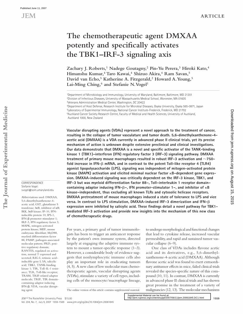
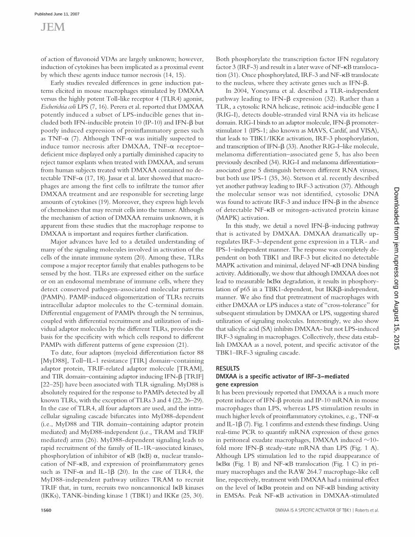
The Journal of Experimental Medicine ARTICLE JEM © The Rockefeller University Press $15.00 Vol. 204, No. 7, July 9, 2007 1559-1569 www.jem.org/cgi/doi/10.1084/jem.20061845 1559 For years, a primary goal of tumor immunolo- gists has been to trigger an anticancer response by the patient’s own immune system, directed largely at engaging the adaptive immune sys- tem to mount a tumor-specific response (1–3). However, a considerable body of evidence sug- gests that nonlymphocytic immune cells also play an important role in eradicating tumors (4, 5). A new class of low molecular mass chemo- therapeutic agents, vascular disrupting agents (VDAs), stimulate a variety of cell types, includ- ing cells of the monocyte/macrophage lineage, to undergo morphological and functional changes that lead to cytokine release, increased vascular permeability, and rapid and sustained tumor vas- cular collapse (6–9). One class of VDAs includes flavone acetic acid and its derivatives, e.g., 5,6-dimethyl- xanthenone-4-acetic acid (DMXAA). Although flavone acetic acid was found to exert extraordi- nary antitumor effects in mice, failed clinical trials revealed the species-specific nature of this com- pound (10, 11). In contrast, DMXAA is currently in advanced phase II clinical trials and has shown great promise in the treatment of a variety of malignancies (12, 13). The molecular mechanisms The chemotherapeutic agent DMXAA potently and specifically activates the TBK1–IRF-3 signaling axis Zachary J. Roberts, 1 Nadege Goutagny, 2 Pin-Yu Perera, 3 Hiroki Kato, 4 Himanshu Kumar, 4 Taro Kawai, 4 Shizuo Akira, 4 Ram Savan, 5 David van Echo, 5 Katherine A. Fitzgerald, 2 Howard A. Young, 5 Lai-Ming Ching, 6 and Stefanie N. Vogel 1 1 Department of Microbiology and Immunology, University of Maryland, Baltimore, Baltimore, MD 21201 2 Division of Infectious Diseases, University of Massachusetts Medical School, Worcester, MA 01605 3 Veterans Administration Medical Center, Washington, DC 20422 4 Department of Host Defense, Research Institute for Microbial Diseases, Osaka University, Osaka 565-0871, Japan 5 Laboratory of Experimental Immunology, National Cancer Institute-Frederick, Frederick, MD 21702 6 Auckland Cancer Society Research Centre, Faculty of Medical and Health Sciences, University of Auckland, Auckland 1000, New Zealand Vascular disrupting agents (VDAs) represent a novel approach to the treatment of cancer, resulting in the collapse of tumor vasculature and tumor death. 5,6-dimethylxanthenone-4- acetic acid (DMXAA) is a VDA currently in advanced phase II clinical trials, yet its precise mechanism of action is unknown despite extensive preclinical and clinical investigations. Our data demonstrate that DMXAA is a novel and specific activator of the TANK-binding kinase 1 (TBK1)–interferon (IFN) regulatory factor 3 (IRF-3) signaling pathway. DMXAA treatment of primary mouse macrophages resulted in robust IRF-3 activation and 750- fold increase in IFN-β mRNA, and in contrast to the potent Toll-like receptor 4 (TLR4) agonist lipopolysaccharide (LPS), signaling was independent of mitogen-activated protein kinase (MAPK) activation and elicited minimal nuclear factor κB–dependent gene expres- sion. DMXAA-induced signaling was critically dependent on the IRF-3 kinase, TBK1, and IRF-3 but was myeloid differentiation factor 88–, Toll–interleukin 1 receptor domain– containing adaptor inducing IFN-β–, IFN promoter-stimulator 1–, and inhibitor of κB kinase–independent, thus excluding all known TLRs and cytosolic helicase receptors. DMXAA pretreatment of mouse macrophages induced a state of tolerance to LPS and vice versa. In contrast to LPS stimulation, DMXAA-induced IRF-3 dimerization and IFN-β expression were inhibited by salicylic acid. These findings detail a novel pathway for TBK1- mediated IRF-3 activation and provide new insights into the mechanism of this new class of chemotherapeutic drugs. CORRESPONDENCE Stefanie Vogel: [email protected] Abbreviations used: DMXAA, 5,6-dimethylxanthenone-4- acetic acid; GST, glutathione S transferase; IκB, inhibitor of κB; IKK, IκB kinase; IP-10, IFN- inducible protein 10; IPS-1, IFN-β promoter-stimulator 1; IRF-3, IFN regulatory factor 3; MAPK, mitogen-activated protein kinase; MEF, mouse embryonic fibroblast; MyD88, myeloid differentiation factor 88; PAMP, pathogen-associated molecular pattern; PRD, posi- tive regulatory domain; RANTES, regulated on activa- tion, normal T expressed and secreted; RIG-I, retinoic acid– inducible gene I; SA, salicylic acid; TBK1, TANK-binding kinase 1; TIR, Toll–IL-1 resist- ance; TLR, Toll-like receptor; TRAM, TRIF-related adaptor molecule; TRIF, TIR domain– containing adaptor inducing IFN-β; VDA, vascular disrupt- ing agent. The online version of this article contains supplemental material. on August 15, 2015 jem.rupress.org Downloaded from Published June 11, 2007 http://jem.rupress.org/content/suppl/2007/06/11/jem.20061845.DC1.html Supplemental Material can be found at:
-
Upload
independent -
Category
Documents
-
view
0 -
download
0
Transcript of The chemotherapeutic agent DMXAA potently and specifically activates the TBK1-IRF-3 signaling axis
The
Journ
al o
f Exp
erim
enta
l M
edic
ine
ARTICLE
JEM © The Rockefeller University Press $15.00
Vol. 204, No. 7, July 9, 2007 1559-1569 www.jem.org/cgi/doi/10.1084/jem.20061845
1559
For years, a primary goal of tumor immunolo-gists has been to trigger an anticancer response by the patient’s own immune system, directed largely at engaging the adaptive immune sys-tem to mount a tumor-specifi c response (1–3). However, a considerable body of evidence sug-gests that nonlymphocytic immune cells also play an important role in eradicating tumors (4, 5). A new class of low molecular mass chemo-therapeutic agents, vascular disrupting agents (VDAs), stimulate a variety of cell types, includ-ing cells of the monocyte/macrophage lineage,
to undergo morphological and functional changes that lead to cytokine release, increased vascular permeability, and rapid and sustained tumor vas-cular collapse (6–9).
One class of VDAs includes fl avone acetic acid and its derivatives, e.g., 5,6-dimethyl-xanthenone-4-acetic acid (DMXAA). Although fl avone acetic acid was found to exert extraordi-nary antitumor eff ects in mice, failed clinical trials revealed the species-specifi c nature of this com-pound (10, 11). In contrast, DMXAA is currently in advanced phase II clinical trials and has shown great promise in the treatment of a variety of malignancies (12, 13). The molecular mechanisms
The chemotherapeutic agent DMXAA potently and specifi cally activates the TBK1–IRF-3 signaling axis
Zachary J. Roberts,1 Nadege Goutagny,2 Pin-Yu Perera,3 Hiroki Kato,4 Himanshu Kumar,4 Taro Kawai,4 Shizuo Akira,4 Ram Savan,5 David van Echo,5 Katherine A. Fitzgerald,2 Howard A. Young,5 Lai-Ming Ching,6 and Stefanie N. Vogel1
1Department of Microbiology and Immunology, University of Maryland, Baltimore, Baltimore, MD 212012Division of Infectious Diseases, University of Massachusetts Medical School, Worcester, MA 016053Veterans Administration Medical Center, Washington, DC 204224Department of Host Defense, Research Institute for Microbial Diseases, Osaka University, Osaka 565-0871, Japan5Laboratory of Experimental Immunology, National Cancer Institute-Frederick, Frederick, MD 217026Auckland Cancer Society Research Centre, Faculty of Medical and Health Sciences, University of Auckland,
Auckland 1000, New Zealand
Vascular disrupting agents (VDAs) represent a novel approach to the treatment of cancer,
resulting in the collapse of tumor vasculature and tumor death. 5,6-dimethylxanthenone-4-
acetic acid (DMXAA) is a VDA currently in advanced phase II clinical trials, yet its precise
mechanism of action is unknown despite extensive preclinical and clinical investigations.
Our data demonstrate that DMXAA is a novel and specifi c activator of the TANK-binding
kinase 1 (TBK1)–interferon (IFN) regulatory factor 3 (IRF-3) signaling pathway. DMXAA
treatment of primary mouse macrophages resulted in robust IRF-3 activation and �750-
fold increase in IFN-β mRNA, and in contrast to the potent Toll-like receptor 4 (TLR4)
agonist lipopolysaccharide (LPS), signaling was independent of mitogen-activated protein
kinase (MAPK) activation and elicited minimal nuclear factor κB–dependent gene expres-
sion. DMXAA-induced signaling was critically dependent on the IRF-3 kinase, TBK1, and
IRF-3 but was myeloid differentiation factor 88–, Toll–interleukin 1 receptor domain–
containing adaptor inducing IFN-β–, IFN promoter-stimulator 1–, and inhibitor of κB
kinase–independent, thus excluding all known TLRs and cytosolic helicase receptors.
DMXAA pretreatment of mouse macrophages induced a state of tolerance to LPS and vice
versa. In contrast to LPS stimulation, DMXAA-induced IRF-3 dimerization and IFN-β
expression were inhibited by salicylic acid. These fi ndings detail a novel pathway for TBK1-
mediated IRF-3 activation and provide new insights into the mechanism of this new class
of chemotherapeutic drugs.
CORRESPONDENCE
Stefanie Vogel:
Abbreviations used: DMXAA,
5,6-dimethylxanthenone-4-
acetic acid; GST, glutathione S
transferase; IκB, inhibitor of κB;
IKK, IκB kinase; IP-10, IFN-
inducible protein 10; IPS-1,
IFN-β promoter-stimulator 1;
IRF-3, IFN regulatory factor 3;
MAPK, mitogen-activated
protein kinase; MEF, mouse
embryonic fi broblast; MyD88,
myeloid diff erentiation factor
88; PAMP, pathogen-associated
molecular pattern; PRD, posi-
tive regulatory domain;
RANTES, regulated on activa-
tion, normal T expressed and
secreted; RIG-I, retinoic acid–
inducible gene I; SA, salicylic
acid; TBK1, TANK-binding
kinase 1; TIR, Toll–IL-1 resist-
ance; TLR, Toll-like receptor;
TRAM, TRIF-related adaptor
molecule; TRIF, TIR domain–
containing adaptor inducing
IFN-β; VDA, vascular disrupt-
ing agent. The online version of this article contains supplemental material.
on August 15, 2015
jem.rupress.org
Dow
nloaded from
Published June 11, 2007
http://jem.rupress.org/content/suppl/2007/06/11/jem.20061845.DC1.html Supplemental Material can be found at:
1560 DMXAA IS A SPECIFIC ACTIVATOR OF TBK1 | Roberts et al.
of action of fl avonoid VDAs are largely unknown; however, induction of cytokines has been implicated as a proximal event by which these agents induce tumor necrosis (14, 15).
Early studies revealed diff erences in gene induction pat-terns elicited in mouse macrophages stimulated by DMXAA versus the highly potent Toll-like receptor 4 (TLR4) agonist, Escherichia coli LPS (7, 16). Perera et al. reported that DMXAA potently induced a subset of LPS-inducible genes that in-cluded both IFN-inducible protein 10 (IP-10) and IFN-β but poorly induced expression of proinfl ammatory genes such as TNF-α (7). Although TNF-α was initially suspected to induce tumor necrosis after DMXAA, TNF-α receptor– defi cient mice displayed only a partially diminished capacity to reject tumor explants when treated with DMXAA, and serum from human subjects treated with DMXAA contained no de-tectable TNF-α (17, 18). Jassar et al. later showed that macro-phages are among the fi rst cells to infi ltrate the tumor after DMXAA treatment and are responsible for secreting large amounts of cytokines (19). Moreover, they express high levels of chemokines that may recruit cells into the tumor. Although the mechanism of action of DMXAA remains unknown, it is apparent from these studies that the macrophage response to DMXAA is important and requires further clarifi cation.
Major advances have led to a detailed understanding of many of the signaling molecules involved in activation of the cells of the innate immune system (20). Among these, TLRs compose a major receptor family that enables pathogens to be sensed by the host. TLRs are expressed either on the surface or on an endosomal membrane of immune cells, where they detect conserved pathogen-associated molecular patterns (PAMPs). PAMP-induced oligomerization of TLRs recruits intracellular adaptor molecules to the C-terminal domain. Diff erential engagement of PAMPs through the N terminus, coupled with diff erential recruitment and utilization of indi-vidual adaptor molecules by the diff erent TLRs, provides the basis for the specifi city with which cells respond to diff erent PAMPs with diff erent patterns of gene expression (21).
To date, four adaptors (myeloid diff erentiation factor 88 [MyD88], Toll–IL-1 resistance [TIR] domain–containing adaptor protein, TRIF-related adaptor molecule [TRAM], and TIR domain–containing adaptor inducing IFN-β [TRIF] [22–25]) have been associated with TLR signaling. MyD88 is absolutely required for the response to PAMPs detected by all known TLRs, with the exception of TLRs 3 and 4 (22, 26–29). In the case of TLR4, all four adaptors are used, and the intra-cellular signaling cascade bifurcates into MyD88-dependent (i.e., MyD88 and TIR domain–containing adaptor protein mediated) and MyD88-independent (i.e., TRAM and TRIF mediated) arms (26). MyD88-dependent signaling leads to rapid recruitment of the family of IL-1R–associated kinases, phosphorylation of inhibitor of κB (IκB) α, nuclear translo-cation of NF-κB, and expression of proinfl ammatory genes such as TNF-α and IL-1β (20). In the case of TLR4, the MyD88-independent pathway utilizes TRAM to recruit TRIF that, in turn, recruits two noncannonical IκB kinases (IKKs), TANK-binding kinase 1 (TBK1) and IKKε (25, 30).
Both phosphorylate the transcription factor IFN regulatory factor 3 (IRF-3) and result in a later wave of NF-κB transloca-tion (31). Once phosphorylated, IRF-3 and NF-κB translocate to the nucleus, where they activate genes such as IFN-β.
In 2004, Yoneyama et al. described a TLR-independent pathway leading to IFN-β expression (32). Rather than a TLR, a cytosolic RNA helicase, retinoic acid–inducible gene I (RIG-I), detects double-stranded viral RNA via its helicase domain. RIG-I binds to an adaptor molecule, IFN-β promoter-stimulator 1 (IPS-1; also known as MAVS, Cardif, and VISA), that leads to TBK1/IKKε activation, IRF-3 phosphorylation, and transcription of IFN-β (33). Another RIG-I–like molecule, melanoma diff erentiation–associated gene 5, has also been previously described (34). RIG-I and melanoma diff erentiation–associated gene 5 distinguish between diff erent RNA viruses, but both use IPS-1 (35, 36). Stetson et al. recently described yet another pathway leading to IRF-3 activation (37). Although the molecular sensor was not identifi ed, cytosolic DNA was found to activate IRF-3 and induce IFN-β in the absence of detectable NF-κB or mitogen-activated protein kinase (MAPK) activation.
In this study, we detail a novel IFN-β–inducing pathway that is activated by DMXAA. DMXAA dramatically up-regulates IRF-3–dependent gene expression in a TLR- and IPS-1–independent manner. The response was completely de-pendent on both TBK1 and IRF-3 but elicited no detectable MAPK activation and minimal, delayed NF-κB DNA binding activity. Additionally, we show that although DMXAA does not lead to measurable IκBα degradation, it results in phosphory-lation of p65 in a TBK1-dependent, but IKKβ-independent, manner. We also fi nd that pretreatment of macrophages with either DMXAA or LPS induces a state of “cross-tolerance” for subsequent stimulation by DMXAA or LPS, suggesting shared utilization of signaling molecules. Interestingly, we also show that salicylic acid (SA) inhibits DMXAA- but not LPS-induced IRF-3 signaling in macrophages. Collectively, these data estab-lish DMXAA as a novel, potent, and specifi c activator of the TBK1–IRF-3 signaling cascade.
RESULTS
DMXAA is a specifi c activator of IRF-3–mediated
gene expression
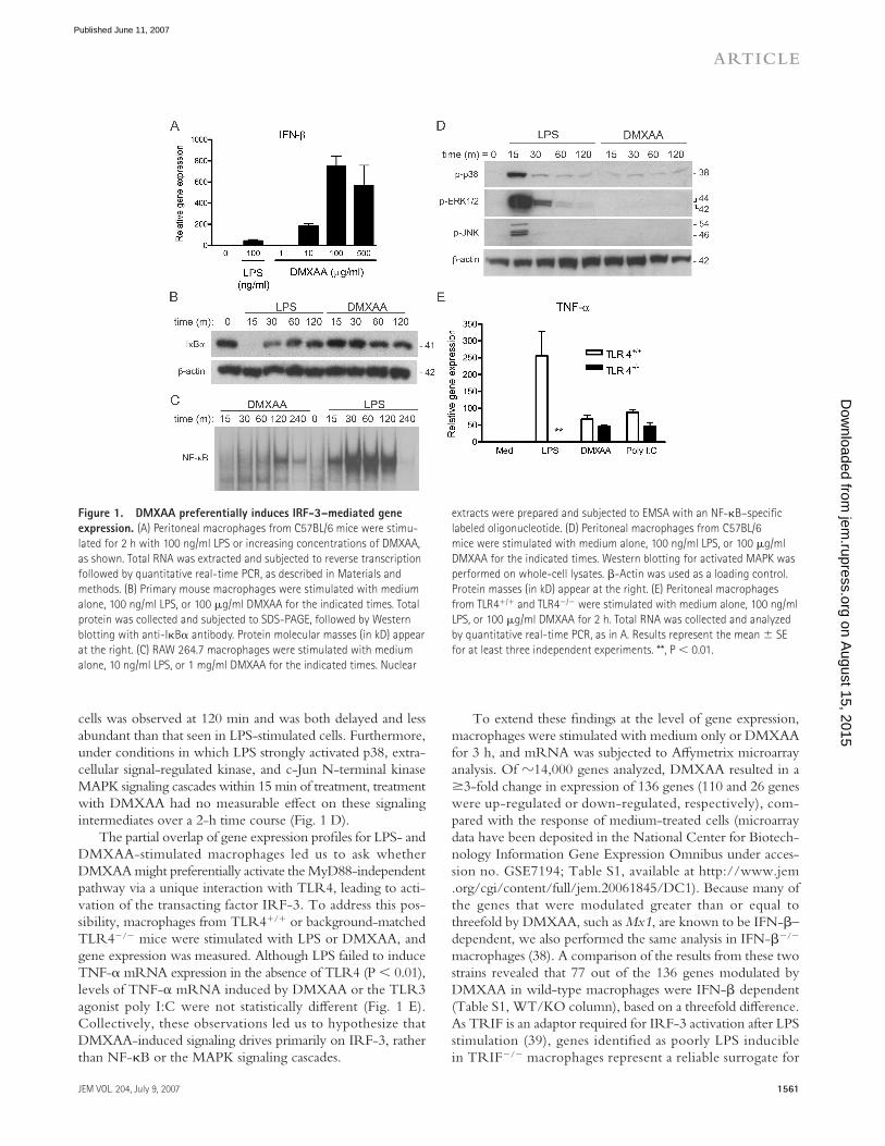
It has been previously reported that DMXAA is a much more potent inducer of IFN-β protein and IP-10 mRNA in mouse macrophages than LPS, whereas LPS stimulation results in much higher levels of proinfl ammatory cytokines, e.g., TNF-α and IL-1β (7). Fig. 1 confi rms and extends these fi ndings. Using real-time PCR to quantify mRNA expression of these genes in peritoneal exudate macrophages, DMXAA induced �10-fold more IFN-β steady-state mRNA than LPS (Fig. 1 A). Although LPS stimulation led to the rapid disappearance of IκBα (Fig. 1 B) and NF-κB translocation (Fig. 1 C) in pri-mary macrophages and the RAW 264.7 macrophage-like cell line, respectively, treatment with DMXAA had a minimal eff ect on the level of IκBα protein and on NF-κB binding activity in EMSAs. Peak NF-κB activation in DMXAA-stimulated
on August 15, 2015
jem.rupress.org
Dow
nloaded from
Published June 11, 2007
JEM VOL. 204, July 9, 2007 1561
ARTICLE
cells was observed at 120 min and was both delayed and less abundant than that seen in LPS-stimulated cells. Furthermore, under conditions in which LPS strongly activated p38, extra-cellular signal-regulated kinase, and c-Jun N-terminal kinase MAPK signaling cascades within 15 min of treatment, treatment with DMXAA had no measurable eff ect on these signaling intermediates over a 2-h time course (Fig. 1 D).
The partial overlap of gene expression profi les for LPS- and DMXAA-stimulated macrophages led us to ask whether DMXAA might preferentially activate the MyD88-independent pathway via a unique interaction with TLR4, leading to acti-vation of the transacting factor IRF-3. To address this pos-sibility, macrophages from TLR4+/+ or background-matched TLR4−/− mice were stimulated with LPS or DMXAA, and gene expression was measured. Although LPS failed to induce TNF-α mRNA expression in the absence of TLR4 (P < 0.01), levels of TNF-α mRNA induced by DMXAA or the TLR3 agonist poly I:C were not statistically diff erent (Fig. 1 E). Collectively, these observations led us to hypothesize that DMXAA-induced signaling drives primarily on IRF-3, rather than NF-κB or the MAPK signaling cascades.
To extend these fi ndings at the level of gene expression, macrophages were stimulated with medium only or DMXAA for 3 h, and mRNA was subjected to Aff ymetrix microarray analysis. Of �14,000 genes analyzed, DMXAA resulted in a ≥3-fold change in expression of 136 genes (110 and 26 genes were up-regulated or down-regulated, respectively), com-pared with the response of medium-treated cells (microarray data have been deposited in the National Center for Biotech-nology Information Gene Expression Omnibus under acces-sion no. GSE7194; Table S1, available at http://www.jem.org/cgi/content/full/jem.20061845/DC1). Because many of the genes that were modulated greater than or equal to threefold by DMXAA, such as Mx1, are known to be IFN-β– dependent, we also performed the same analysis in IFN-β−/− macrophages (38). A comparison of the results from these two strains revealed that 77 out of the 136 genes modulated by DMXAA in wild-type macrophages were IFN-β dependent (Table S1, WT/KO column), based on a threefold diff erence. As TRIF is an adaptor required for IRF-3 activation after LPS stimulation (39), genes identifi ed as poorly LPS inducible in TRIF−/− macrophages represent a reliable surrogate for
Figure 1. DMXAA preferentially induces IRF-3–mediated gene
expression. (A) Peritoneal macrophages from C57BL/6 mice were stimu-
lated for 2 h with 100 ng/ml LPS or increasing concentrations of DMXAA,
as shown. Total RNA was extracted and subjected to reverse transcription
followed by quantitative real-time PCR, as described in Materials and
methods. (B) Primary mouse macrophages were stimulated with medium
alone, 100 ng/ml LPS, or 100 μg/ml DMXAA for the indicated times. Total
protein was collected and subjected to SDS-PAGE, followed by Western
blotting with anti-IκBα antibody. Protein molecular masses (in kD) appear
at the right. (C) RAW 264.7 macrophages were stimulated with medium
alone, 10 ng/ml LPS, or 1 mg/ml DMXAA for the indicated times. Nuclear
extracts were prepared and subjected to EMSA with an NF-κB–specifi c
labeled oligonucleotide. (D) Peritoneal macrophages from C57BL/6
mice were stimulated with medium alone, 100 ng/ml LPS, or 100 μg/ml
DMXAA for the indicated times. Western blotting for activated MAPK was
performed on whole-cell lysates. β-Actin was used as a loading control.
Protein masses (in kD) appear at the right. (E) Peritoneal macrophages
from TLR4+/+ and TLR4−/− were stimulated with medium alone, 100 ng/ml
LPS, or 100 μg/ml DMXAA for 2 h. Total RNA was collected and analyzed
by quantitative real-time PCR, as in A. Results represent the mean ± SE
for at least three independent experiments. **, P < 0.01.
on August 15, 2015
jem.rupress.org
Dow
nloaded from
Published June 11, 2007
1562 DMXAA IS A SPECIFIC ACTIVATOR OF TBK1 | Roberts et al.
IRF-3–dependent gene induction. Many of the same genes induced by DMXAA in our microarray analysis were identi-fi ed as being poorly inducible by LPS in macrophages derived from TRIF-null mice (40); e.g., Rantes (Ccl5), Ifi t1, Ccl4, and Oasl were shown by Hirotani et al. to be highly TRIF depen-dent in LPS-treated macrophages. Thus, these data support the hypothesis that DMXAA preferentially induces IRF-3–dependent genes.
DMXAA is a potent and specifi c activator of TBK1
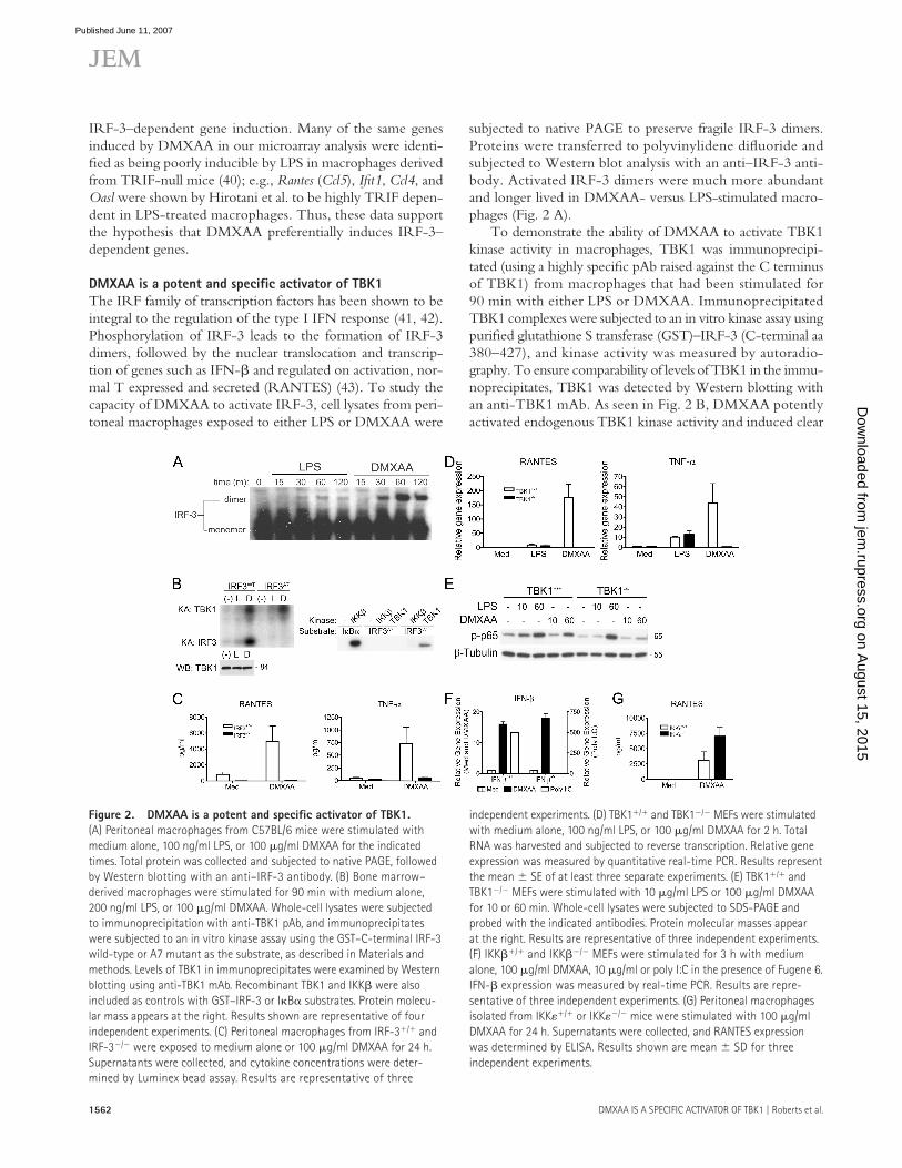
The IRF family of transcription factors has been shown to be integral to the regulation of the type I IFN response (41, 42). Phosphorylation of IRF-3 leads to the formation of IRF-3 dimers, followed by the nuclear translocation and transcrip-tion of genes such as IFN-β and regulated on activation, nor-mal T expressed and secreted (RANTES) (43). To study the capacity of DMXAA to activate IRF-3, cell lysates from peri-toneal macrophages exposed to either LPS or DMXAA were
subjected to native PAGE to preserve fragile IRF-3 dimers. Proteins were transferred to polyvinylidene difl uoride and subjected to Western blot analysis with an anti–IRF-3 anti-body. Activated IRF-3 dimers were much more abundant and longer lived in DMXAA- versus LPS-stimulated macro-phages (Fig. 2 A).
To demonstrate the ability of DMXAA to activate TBK1 kinase activity in macrophages, TBK1 was immunoprecipi-tated (using a highly specifi c pAb raised against the C terminus of TBK1) from macrophages that had been stimulated for 90 min with either LPS or DMXAA. Immunoprecipitated TBK1 complexes were subjected to an in vitro kinase assay using purifi ed glutathione S transferase (GST)–IRF-3 (C-terminal aa 380–427), and kinase activity was measured by autoradio-graphy. To ensure comparability of levels of TBK1 in the immu-noprecipitates, TBK1 was detected by Western blotting with an anti-TBK1 mAb. As seen in Fig. 2 B, DMXAA potently activated endogenous TBK1 kinase activity and induced clear
Figure 2. DMXAA is a potent and specifi c activator of TBK1.
(A) Peritoneal macrophages from C57BL/6 mice were stimulated with
medium alone, 100 ng/ml LPS, or 100 μg/ml DMXAA for the indicated
times. Total protein was collected and subjected to native PAGE, followed
by Western blotting with an anti–IRF-3 antibody. (B) Bone marrow–
derived macrophages were stimulated for 90 min with medium alone,
200 ng/ml LPS, or 100 μg/ml DMXAA. Whole-cell lysates were subjected
to immunoprecipitation with anti-TBK1 pAb, and immunoprecipitates
were subjected to an in vitro kinase assay using the GST–C-terminal IRF-3
wild-type or A7 mutant as the substrate, as described in Materials and
methods. Levels of TBK1 in immunoprecipitates were examined by Western
blotting using anti-TBK1 mAb. Recombinant TBK1 and IKKβ were also
included as controls with GST–IRF-3 or IκBα substrates. Protein molecu-
lar mass appears at the right. Results shown are representative of four
independent experiments. (C) Peritoneal macrophages from IRF-3+/+ and
IRF-3−/− were exposed to medium alone or 100 μg/ml DMXAA for 24 h.
Supernatants were collected, and cytokine concentrations were deter-
mined by Luminex bead assay. Results are representative of three
independent experiments. (D) TBK1+/+ and TBK1−/− MEFs were stimulated
with medium alone, 100 ng/ml LPS, or 100 μg/ml DMXAA for 2 h. Total
RNA was harvested and subjected to reverse transcription. Relative gene
expression was measured by quantitative real-time PCR. Results represent
the mean ± SE of at least three separate experiments. (E) TBK1+/+ and
TBK1−/− MEFs were stimulated with 10 μg/ml LPS or 100 μg/ml DMXAA
for 10 or 60 min. Whole-cell lysates were subjected to SDS-PAGE and
probed with the indicated antibodies. Protein molecular masses appear
at the right. Results are representative of three independent experiments.
(F) IKKβ+/+ and IKKβ−/− MEFs were stimulated for 3 h with medium
alone, 100 μg/ml DMXAA, 10 μg/ml or poly I:C in the presence of Fugene 6.
IFN-β expression was measured by real-time PCR. Results are repre-
sentative of three independent experiments. (G) Peritoneal macrophages
isolated from IKKε+/+ or IKKε−/− mice were stimulated with 100 μg/ml
DMXAA for 24 h. Supernatants were collected, and RANTES expression
was determined by ELISA. Results shown are mean ± SD for three
independent experiments.
on August 15, 2015
jem.rupress.org
Dow
nloaded from
Published June 11, 2007
JEM VOL. 204, July 9, 2007 1563
ARTICLE
phosphorylation of both TBK1 itself (left, top) and the wild-type GST–IRF-3 substrate (left, bottom). Consistent with the results of the IRF-3 dimerization assay, DMXAA-induced TBK1 kinase activity was considerably more potent than that observed after stimulation with LPS. Importantly, a mutant version of IRF-3, in which seven serine/threonine residues were mutated to alanine (S385, S386, S396, S398, S402, T404, and S405®A; GST–IRF-3–A7), was not phosphorylated by endogenous TBK1 under conditions in which TBK1 auto-phosphorylation was intact. In addition, an in vitro kinase assay revealed that recombinant TBK1 phosphorylated the wild-type GST-IRF-3, but not the A7 mutant, whereas recombinant IKKβ, which potently phosphorylated IκBα, failed to phos-phorylate GST–IRF-3 measurably, consistent with previously published data (44). Collectively, these results clearly demon-strate that DMXAA is a potent activator of the TBK1–IRF-3 signaling axis.
To address the possibility that IRF-3 was required for activation of cells by DMXAA, peritoneal macrophages from wild-type and IRF-3−/− mice were cultured in medium only or DMXAA. Supernatants collected at 24 h were analyzed for cytokine production. Consistent with the robust IRF-3 acti-vation observed in DMXAA-treated cells (Fig. 2, A and B), IRF-3−/− macrophages failed to produce RANTES, the product of a known IRF-3–dependent gene (45). Surprisingly, secretion of TNF-α was also reduced to background levels in IRF-3–defi cient macrophages (Fig. 2 C). To evaluate further the role of activated IRF-3 in DMXAA-induced signaling, we exposed wild-type or TBK1-defi cient mouse embryonic fi bro-blasts (MEFs) to medium only, LPS, or DMXAA and measured gene expression. Interestingly, we found that, in contrast to experiments with macrophages, DMXAA induced much more robust responses in MEFs than did LPS, an observation that is consistent with the diminished LPS sensitivity that has been observed in MEFs by others (46). In agreement with previous work (46), LPS-stimulated, TBK1−/− MEFs pro-duced wild-type levels of RANTES and TNF-α mRNA (Fig. 2 D). However, TBK1−/− MEFs failed to express either RANTES or TNF-α mRNA in response to DMXAA. These results suggest that, in addition to being a potent activator of TBK1, DMXAA is critically dependent on both TBK1 and its downstream target, IRF-3, for gene expression.
Although TBK1 seems to function primarily as an IRF-3 kinase, it has also been shown that, under certain circumstances, TBK1 may phosphorylate the NF-κB subunit p65 on serine 536 (47). This phosphorylation event is believed to play a role in p65 transactivation, because cells lacking TBK1 show a de-fect in NF-κB–dependent gene expression despite normal IκBα degradation and NF-κB binding activity (48). Because DMXAA is a relatively poor inducer of both IκBα degradation and NF-κB binding activity when compared with LPS (Fig. 1, B and C) but has previously been shown to induce NF-κB– dependent gene expression (49), we sought to examine the phos-phorylation status of p65 in LPS versus DMXAA-stimulated cells. In wild-type MEFs, LPS-induced phosphorylation of p65 on S536 was observed at 10 min and peaked at 60 min, whereas
DMXAA-induced p65 phosphorylation was undetectable at 10 min but measurable at 60 min. Surprisingly, in contrast to LPS-induced phospho-p65, DMXAA-induced p65 phosphor-ylation was ablated in TBK1-null MEFs at 60 min (Fig. 2 E). In further support of the assertion that DMXAA is a specifi c activator of the TBK1–IRF-3 signaling axis, we tested the abil-ity of DMXAA to induce IFN-β in MEFs defi cient in the NF-κB–activating kinase IKKβ. Remarkably, under conditions in which transfected poly I:C, a known inducer of NF-κB (32), failed to activate IFN-β expression in IKKβ-null MEFs, DMXAA-induced IFN-β was found to be independent of IKKβ (Fig. 2 F). Collectively, these fi ndings suggest that DMXAA activates NF-κB in a manner that is both indepen-dent of IKKβ but completely dependent on TBK1.
To address a possible role for IKKε, the only other IRF-3 kinase identifi ed thus far, in DMXAA-induced signaling, we compared the response of macrophages isolated from wild-type and IKKε-defi cient mice after treatment with DMXAA. Induction of RANTES protein was not inhibited in IKKε-null cells (Fig. 2 G). Collectively, these data support the con-clusion that DMXAA activates a pathway that is dependent on both IRF-3 and TBK1 but is independent of both IKKβ and IKKε.
DMXAA-induced gene expression is TLR-
and IPS-1–independent
Because all known TLRs, with the exception of TLRs 3 and 4, have an absolute requirement for MyD88 to induce gene expression, we tested the ability of DMXAA to induce signal-ing in MyD88−/− macrophages. Consistent with previous reports (22, 26), LPS-induced IFN-β mRNA and protein (Fig. 3 A) were not signifi cantly decreased by MyD88 defi ciency, whereas levels of TNF-α were dramatically inhibited in the MyD88−/− macrophages (Fig. 3 B). In contrast, DMXAA-induced IFN-β and TNF-α mRNA and protein were not signifi cantly diff erent in wild-type and MyD88−/− cells. Thus, DMXAA-induced gene expression is MyD88 independent.
TLRs 3 and 4 share the ability to activate IRF-3 and induce IFN-β via another adaptor, TRIF. To directly address the possibility that DMXAA utilizes the MyD88-independent pathway mediated by TRIF, background-matched, wild-type, and TRIF−/− MEFs were stimulated with DMXAA or the TLR3 agonist poly I:C. Fig. 3 C illustrates that compared with poly I:C, a known TRIF-dependent inducer of RANTES (25), DMXAA-induced RANTES was unaff ected by the absence of TRIF. In further support of the conclusion that DMXAA does not require any known TLR for activity, macro-phages defi cient in both MyD88 and TRIF responded to DMXAA by making RANTES protein at a level that was not statistically diff erent from that made by wild-type cells, whereas LPS-induced RANTES was reduced to baseline levels in TRIF−/−/MyD88−/−-defi cient macrophages (Fig. 3 D). Because DMXAA is, therefore, neither MyD88- nor TRIF-dependent, these data indicate that none of the known TLRs serve as a receptor for DMXAA, because all require MyD88 and/or TRIF to mediate signaling.
on August 15, 2015
jem.rupress.org
Dow
nloaded from
Published June 11, 2007
1564 DMXAA IS A SPECIFIC ACTIVATOR OF TBK1 | Roberts et al.
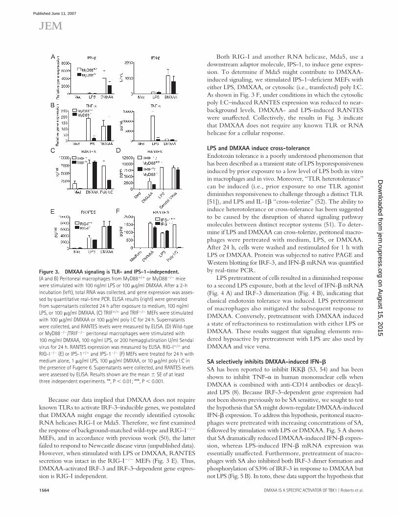
Because our data implied that DMXAA does not require known TLRs to activate IRF-3–inducible genes, we postulated that DMXAA might engage the recently identifi ed cytosolic RNA helicases RIG-I or Mda5. Therefore, we fi rst examined the response of background-matched wild-type and RIG-I−/− MEFs, and in accordance with previous work (50), the latter failed to respond to Newcastle disease virus (unpublished data). However, when stimulated with LPS or DMXAA, RANTES secretion was intact in the RIG-I−/− MEFs (Fig. 3 E). Thus, DMXAA-activated IRF-3 and IRF-3–dependent gene expres-sion is RIG-I independent.
Both RIG-I and another RNA helicase, Mda5, use a downstream adaptor molecule, IPS-1, to induce gene expres-sion. To determine if Mda5 might contribute to DMXAA-induced signaling, we stimulated IPS-1–defi cient MEFs with either LPS, DMXAA, or cytosolic (i.e., transfected) poly I:C. As shown in Fig. 3 F, under conditions in which the cytosolic poly I:C–induced RANTES expression was reduced to near-background levels, DMXAA- and LPS-induced RANTES were unaff ected. Collectively, the results in Fig. 3 indicate that DMXAA does not require any known TLR or RNA helicase for a cellular response.
LPS and DMXAA induce cross-tolerance
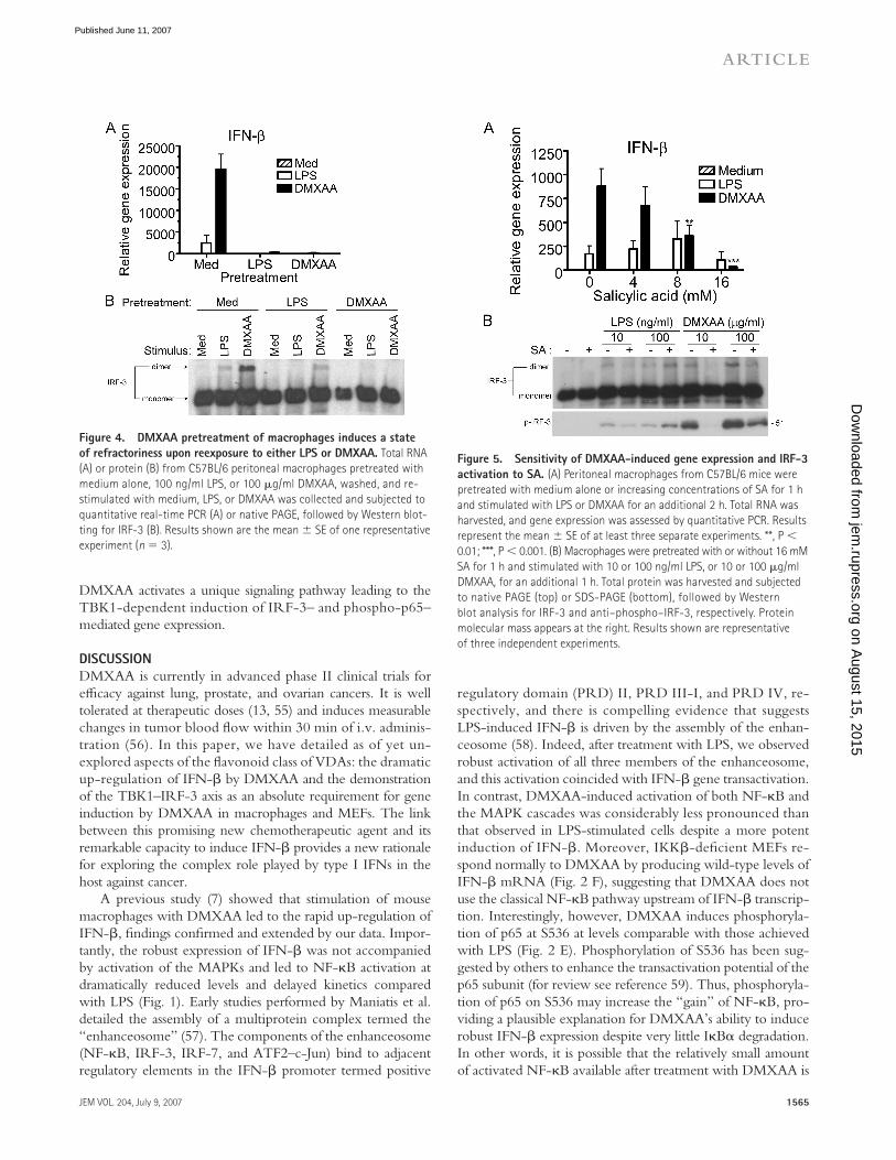
Endotoxin tolerance is a poorly understood phenomenon that has been described as a transient state of LPS hyporesponsiveness induced by prior exposure to a low level of LPS both in vitro in macrophages and in vivo. Moreover, “TLR heterotolerance” can be induced (i.e., prior exposure to one TLR agonist diminishes responsiveness to challenge through a distinct TLR [51]), and LPS and IL-1β “cross-tolerize” (52). The ability to induce heterotolerance or cross-tolerance has been suggested to be caused by the disruption of shared signaling pathway molecules between distinct receptor systems (51). To deter-mine if LPS and DMXAA can cross-tolerize, peritoneal macro-phages were pretreated with medium, LPS, or DMXAA. After 24 h, cells were washed and restimulated for 1 h with LPS or DMXAA. Protein was subjected to native PAGE and Western blotting for IRF-3, and IFN-β mRNA was quantifi ed by real-time PCR.
LPS pretreatment of cells resulted in a diminished response to a second LPS exposure, both at the level of IFN-β mRNA (Fig. 4 A) and IRF-3 dimerization (Fig. 4 B), indicating that classical endotoxin tolerance was induced. LPS pretreatment of macrophages also mitigated the subsequent response to DMXAA. Conversely, pretreatment with DMXAA induced a state of refractoriness to restimulation with either LPS or DMXAA. These results suggest that signaling elements ren-dered hypoactive by pretreatment with LPS are also used by DMXAA and vice versa.
SA selectively inhibits DMXAA-induced IFN-𝛃SA has been reported to inhibit IKKβ (53, 54) and has been shown to inhibit TNF-α in human mononuclear cells when DMXAA is combined with anti-CD14 antibodies or deacyl-ated LPS (8). Because IRF-3–dependent gene expression had not been shown previously to be SA sensitive, we sought to test the hypothesis that SA might down-regulate DMXAA-induced IFN-β expression. To address this hypothesis, peritoneal macro-phages were pretreated with increasing concentrations of SA, followed by stimulation with LPS or DMXAA. Fig. 5 A shows that SA dramatically reduced DMXAA-induced IFN-β expres-sion, whereas LPS-induced IFN-β mRNA expression was essentially unaff ected. Furthermore, pretreatment of macro-phages with SA also inhibited both IRF-3 dimer formation and phosphorylation of S396 of IRF-3 in response to DMXAA but not LPS (Fig. 5 B). In toto, these data support the hypothesis that
Figure 3. DMXAA signaling is TLR- and IPS-1–independent.
(A and B) Peritoneal macrophages from MyD88+/+ or MyD88−/− mice
were stimulated with 100 ng/ml LPS or 100 μg/ml DMXAA. After a 2-h
incubation (left), total RNA was collected, and gene expression was asses-
sed by quantitative real-time PCR. ELISA results (right) were generated
from supernatants collected 24 h after exposure to medium, 100 ng/ml
LPS, or 100 μg/ml DMXAA. (C) TRIF+/+ and TRIF−/− MEFs were stimulated
with 100 μg/ml DMXAA or 100 μg/ml poly I:C for 24 h. Supernatants
were collected, and RANTES levels were measured by ELISA. (D) Wild-type
or MyD88−/−/TRIF−/− peritoneal macrophages were stimulated with
100 mg/ml DMXAA, 100 ng/ml LPS, or 200 hemagglutination U/ml Sendai
virus for 24 h. RANTES expression was measured by ELISA. RIG-I+/+ and
RIG-I−/− (E) or IPS-1+/+ and IPS-1−/− (F) MEFs were treated for 24 h with
medium alone, 1 μg/ml LPS, 100 μg/ml DMXAA, or 10 μg/ml poly I:C in
the presence of Fugene 6. Supernatants were collected, and RANTES levels
were assessed by ELISA. Results shown are the mean ± SE of at least
three independent experiments. **, P < 0.01; ***, P < 0.001.
on August 15, 2015
jem.rupress.org
Dow
nloaded from
Published June 11, 2007
JEM VOL. 204, July 9, 2007 1565
ARTICLE
DMXAA activates a unique signaling pathway leading to the TBK1-dependent induction of IRF-3– and phospho-p65–mediated gene expression.
D I S C U S S I O N
DMXAA is currently in advanced phase II clinical trials for effi cacy against lung, prostate, and ovarian cancers. It is well tolerated at therapeutic doses (13, 55) and induces measurable changes in tumor blood fl ow within 30 min of i.v. adminis-tration (56). In this paper, we have detailed as of yet un-explored aspects of the fl avonoid class of VDAs: the dramatic up-regulation of IFN-β by DMXAA and the demonstration of the TBK1–IRF-3 axis as an absolute requirement for gene induction by DMXAA in macrophages and MEFs. The link between this promising new chemotherapeutic agent and its remarkable capacity to induce IFN-β provides a new rationale for exploring the complex role played by type I IFNs in the host against cancer.
A previous study (7) showed that stimulation of mouse macrophages with DMXAA led to the rapid up-regulation of IFN-β, fi ndings confi rmed and extended by our data. Impor-tantly, the robust expression of IFN-β was not accompanied by activation of the MAPKs and led to NF-κB activation at dramatically reduced levels and delayed kinetics compared with LPS (Fig. 1). Early studies performed by Maniatis et al. detailed the assembly of a multiprotein complex termed the “enhanceosome” (57). The components of the enhanceosome (NF-κB, IRF-3, IRF-7, and ATF2–c-Jun) bind to adjacent regulatory elements in the IFN-β promoter termed positive
regulatory domain (PRD) II, PRD III-I, and PRD IV, re-spectively, and there is compelling evidence that suggests LPS-induced IFN-β is driven by the assembly of the enhan-ceosome (58). Indeed, after treatment with LPS, we observed robust activation of all three members of the enhanceosome, and this activation coincided with IFN-β gene transactivation. In contrast, DMXAA-induced activation of both NF-κB and the MAPK cascades was considerably less pronounced than that observed in LPS-stimulated cells despite a more potent induction of IFN-β. Moreover, IKKβ-defi cient MEFs re-spond normally to DMXAA by producing wild-type levels of IFN-β mRNA (Fig. 2 F), suggesting that DMXAA does not use the classical NF-κB pathway upstream of IFN-β transcrip-tion. Interestingly, however, DMXAA induces phosphoryla-tion of p65 at S536 at levels comparable with those achieved with LPS (Fig. 2 E). Phosphorylation of S536 has been sug-gested by others to enhance the transactiva tion potential of the p65 subunit (for review see reference 59). Thus, phosphoryla-tion of p65 on S536 may increase the “gain” of NF-κB, pro-viding a plausible explanation for DMXAA’s ability to induce robust IFN-β expression despite very little IκBα degradation. In other words, it is possible that the relatively small amount of activated NF-κB available after treatment with DMXAA is
Figure 4. DMXAA pretreatment of macrophages induces a state
of refractoriness upon reexposure to either LPS or DMXAA. Total RNA
(A) or protein (B) from C57BL/6 peritoneal macrophages pretreated with
medium alone, 100 ng/ml LPS, or 100 μg/ml DMXAA, washed, and re-
stimulated with medium, LPS, or DMXAA was collected and subjected to
quantitative real-time PCR (A) or native PAGE, followed by Western blot-
ting for IRF-3 (B). Results shown are the mean ± SE of one representative
experiment (n = 3).
Figure 5. Sensitivity of DMXAA-induced gene expression and IRF-3
activation to SA. (A) Peritoneal macrophages from C57BL/6 mice were
pretreated with medium alone or increasing concentrations of SA for 1 h
and stimulated with LPS or DMXAA for an additional 2 h. Total RNA was
harvested, and gene expression was assessed by quantitative PCR. Results
represent the mean ± SE of at least three separate experiments. **, P <
0.01; ***, P < 0.001. (B) Macrophages were pretreated with or without 16 mM
SA for 1 h and stimulated with 10 or 100 ng/ml LPS, or 10 or 100 μg/ml
DMXAA, for an additional 1 h. Total protein was harvested and subjected
to native PAGE (top) or SDS-PAGE (bottom), followed by Western
blot analysis for IRF-3 and anti–phospho–IRF-3, respectively. Protein
molecular mass appears at the right. Results shown are representative
of three independent experiments.
on August 15, 2015
jem.rupress.org
Dow
nloaded from
Published June 11, 2007
1566 DMXAA IS A SPECIFIC ACTIVATOR OF TBK1 | Roberts et al.
suffi cient to complete the IFN-β enhanceosome or is compen-sated for by its increased transactivation potential. Finally, in contrast to LPS treatment, DMXAA-induced p65 phosphory-lation is abolished in TBK1-defi cient MEFs, providing further support for the conclusion that DMXAA is a novel and specifi c activator of the TBK1–IRF-3 signaling axis.
This claim is further supported by our results derived from TBK1- and IRF-3–defi cient mice. DMXAA-induced expression of RANTES, a heavily IRF-3–dependent gene (45), was observed to be completely dependent on the TBK1–IRF-3 axis (Fig. 2). Surprisingly, this dependence on TBK1 and IRF-3 extended to genes not normally considered to be dependent on IRF-3, such as TNF-α. Under conditions where LPS-induced TNF-α was unaff ected, IRF-3–defi cient cells failed to induce TNF-α mRNA in response to DMXAA. This suggests that DMXAA-induced TNF-α expression is strictly IRF-3–dependent. Although it is possible that the failure of DMXAA-treated TBK1-null MEFs to phosphory-late p65 contributes to reduced availability of NF-κB for induction of genes such as TNF-α, our DNA microarray data (Table S1) revealed that TNF-α expression in response to DMXAA is diminished in IFN-β–null macrophages. These results support the alternative possibility that TNF-α is part of an IFN-β–dependent “second wave” of gene expression after DMXAA treatment.
Although the role of type I IFN in both tumor immunity and the treatment of cancer has been studied for decades, the direct involvement of IRF-3 is considerably less well under-stood. However, it was recently shown that IRF-3 drives the up-regulation of TNF-related apoptosis-inducing ligand in virally infected cells, as well as directing cells into p53- dependent cell-cycle arrest and senescence (60, 61). Perhaps even more pertinent to the current work are recent studies by Duguay et al. with human IRF-3–expressing B16 melanoma tumors (62). In their study, tumors expressing IRF-3 grew more slowly than those that had been mock transduced. Furthermore, the IRF-3–positive tumors demonstrated a robust up-regulation of a variety of chemokines in vivo, including RANTES, macrophage infl ammatory protein 1β, and IP-10. Accordingly, IRF-3–expressing tumors recruited consider-ably more neutrophils and lymphocytes and showed signs of retarded tumor growth, including a larger capsule, fewer blood vessels, and areas of necrosis. Importantly, the results reported by Duguay et al. mirror those of Jassar et al. (19) in which implanted tumors showed dramatically increased levels of IP-10 and RANTES, as well as necrosis, after i.v. DMXAA administration. The results presented herein provide a plausible link between the direct antitumor results of IRF-3 overexpres-sion and those after treatment with DMXAA.
The ability of DMXAA to activate IRF-3 and induce IRF-3–mediated gene expression led us to address the involve-ment of established pattern recognition receptors in DMXAA signaling. DMXAA-induced signaling was found to be intact in both MyD88−/−/TRIF−/− and IPS-1−/− cells, thereby elim-inating the possibility of involvement of all known TLRs and RNA helicases. However, a third non–TLR-dependent pathway
leading to expression of IFN-β was recently described by Stetson et al. in which the presence of cytosolic, non–CpG-containing DNA stimulated high levels of type I IFN (37). In that study, however, the molecular sensor of the stimulus was not iden-tifi ed. Notably, the authors reported that the type I IFNs induced by cytosolic DNA were not accompanied by either MAPK activation or NF-κB translocation, consistent with our observations. With a molecular mass of 304 daltons, DMXAA is much smaller than the DNA used in that study. However, it remains possible that DMXAA may engage the molecular sensor or a downstream signaling component of this novel pathway to initiate signaling leading to IRF-3 activation. Studies to identify the nature of this sensor are currently in progress.
Previous studies have shown that pretreatment of macro-phages or mice with LPS results in a transient desensitization to subsequent stimulation by LPS, other TLR agonists, or IL-1β. The mechanisms that underlie tolerance are clearly multifactorial; however, interference with signal transduction appears to be a common mechanism (51, 63). DMXAA, like LPS, induced a state of tolerance in macrophages to sub-sequent stimulation with either DMXAA or LPS, as indicated by inhibition of not only IFN-β gene expression but also IRF-3 dimer formation (Fig. 4). This implies that disrupted signaling in LPS- or DMXAA-tolerized cells is a consequence of an event that occurs early in the signaling pathway, be-cause IRF-3 dimers are detected within 15 min after agonist stimulation. Although we have shown that LPS and DMXAA seem to engage distinct signaling pathways, both lead to IRF-3 activation via TBK1. Thus, it seems plausible that one signaling component tolerized by pretreatment with LPS or DMXAA is TBK1 itself. Studies are ongoing to address the role of TBK1 expression levels and enzymatic activity in the induction of cross-tolerance by LPS and DMXAA.
During the course of these studies, we extended previous fi ndings that demonstrated SA as an inhibitor of DMXAA. Although an inhibitory eff ect of SA on DMXAA-induced TNF-α expression had been previously reported (8), our re-sults identify a possible explanation for the role played by SA in DMXAA inhibition. Pretreatment of macrophages with SA blocked DMXAA-induced phosphorylation of IRF-3 at resi-due S396, IRF-3 dimerization, and IFN-β expression (Fig. 5). However, all three events were unaff ected by SA in LPS-stimulated cells. These results support our conclusion that the pathways leading to IFN-β gene expression by these two stimuli diff er.
In conclusion, we present data that fi rmly establishes the clinically important VDA DMXAA as a potent and specifi c activator of the TBK1–IRF-3 axis. The link between height-ened activity of this signaling pathway and a systemic anti-tumor response likely involves myriad and divergent events. However, by identifying a key signaling pathway with known antitumor potential as critical to the response to DMXAA, we hope to further our understanding of both the mechanism of action of this promising new chemotherapeutic agent as well as the role of the innate immune response in defending the host against cancer.
on August 15, 2015
jem.rupress.org
Dow
nloaded from
Published June 11, 2007
JEM VOL. 204, July 9, 2007 1567
ARTICLE
MATERIALS AND METHODSMice. 5–6-wk-old C57BL/6J females were purchased from the Jackson
Laboratory. IRF-3−/− mice were a gift of T. Taniguchi (University of Tokyo,
Tokyo, Japan). IFN-β−/− mice (>N8 on a C57BL/6 background) were a gift
of E. Fish (University of Toronto, Toronto, Canada). MyD88−/−/TRIF−/−
mice were bred from MyD88−/− (64) and TRIF−/− (39) mice. IKKε−/−
mice were generated at Millennium Pharmaceuticals. TBK1−/− mice were a
gift of W.-C. Yeh (University of Toronto, Toronto, Canada) and were bred
with TNFR1−/− mice at the University of Massachusetts Medical School.
All experiments were conducted with Institutional Animal Care and Use
Committee approval.
Reagents and virus. DMXAA (sodium salt; 304 daltons) was synthesized at
the Auckland Cancer Society Research Centre (65). Poly I:C (GE Healthcare)
was used exogenously as a TLR3 agonist. For triggering intracellular RNA
helicases, poly I:C was transfected as follows: 10 μg/ml poly I:C was mixed with
a transfection reagent (Fugene 6; Roche) at a ratio of 1:1 (volume/weight) in
OptiMEM (Invitrogen) and incubated for 15 min before stimulation. Sendai
virus was used at 200 hemagglutination U/ml. Protein-free E. coli K235 LPS
(66) was used as a TLR4 agonist. SA was obtained from Sigma-Aldrich. C-
terminal GST fusions of IRF-3 (wild-type aa 380–427 and IRF-3–A7 mutant
in which amino acids S385, S386, S396, S398, S402, T404, and S405 were
all mutated to alanine) were purifi ed according to standard protocols (GE
Healthcare). pAb to TBK1 (raised against the C terminus of human TBK1)
was provided by T. Maniatis (Millenium Pharmaceuticals, Cambridge, MA).
Anti-TBK1 mAb (IMG-139, raised against aa 563–577 of human TBK1) was
obtained from Imgenex.
Cell culture. Thioglycollate-elicited mouse peritoneal macrophages were
obtained and cultured as previously described (52). Bone marrow–derived macro-
phages were generated from bone marrow cells cultured in L929-conditioned
media for 10 d and were examined by FACS and found to be >99% F4/80
and CD11b double positive. Mouse macrophage-like RAW 264.7 cells were
purchased from the American Type Culture Collection. Embryonic fi broblasts
from TBK1+/+ and TBK1−/− mice were a gift of W.-C. Yeh. RIG-I (50) and
IPS-1 (36) knockout MEFs have been described elsewhere. Embryonic fi bro-
blasts from IKKβ+/+ and IKKβ−/− mice were a gift of J. DiDonato (Cleveland
Clinic, Cleveland, Ohio). RAW 264.7 macrophages and embryonic fi broblasts
were cultured in DMEM (BioWhittaker), supplemented with 10% (vol/vol)
FBS (HyClone Laboratories), 10,000 U/ml penicillin, and 10,000 μg/ml
streptomycin at 37°C in 5% CO2 in air. The endotoxin content in the medium
was <0.01 EU/ml, according to the manufacturer’s specifi cations. Only cells
passaged ≤20 times were used.
Quantitative real-time PCR. Primers for detection of IFN-β, RANTES,
TNF-α, and hypoxanthine phosphoribosyltransferase (HPRT) mRNAs were
designed using the Primer Express program (version 2.0; Applied Biosystems).
31.25 ng of total cDNA was used as starting material for real-time PCR quan-
titation with SYBR Green (Applied Biosystems) on a real-time PCR system
(7900HT; Applied Biosystems). Ct values were compared using the ∆∆-Ct
method using HPRT as a housekeeping gene (67).
Cytokine analysis. IFN-β protein in cell culture supernatants was measured
using a custom ELISA originally described elsewhere (68), with few modifi -
cations. In brief, 96-well polystyrene plates (Maxisorp; Nunc International)
were coated overnight with a 1:4,000 dilution of rat anti–mouse IFN-β mAb
(Yamasa) in 0.1 M sodium carbonate at 4°C. Plates were blocked with 10%
FCS in 1× PBS for 2 h at room temperature. Samples and a mouse IFN-β
standard (National Institutes of Health [NIH]) were added to wells and incu-
bated overnight at 4°C. Plates were washed 3 times with 1% FCS/PBS-T,
followed by incubation with a 1:2,000 dilution of rabbit anti–mouse IFN-β
pAb (PBL Biomedical Laboratories) in 10% FCS-PBS overnight at 4°C. Wells
were washed 3 times, followed by incubation with a 1:2,000 dilution of goat
anti–rabbit horseradish peroxidase (HRP; Cell Signaling Technologies) in
10% FCS-PBS for 1 h at room temperature. Plates were washed 3 times and
developed with TMB substrate (KPL). The reaction was stopped by addition of
1 N H2SO4, and plates were read at 450 nm. For quantifi cation of RANTES
and TNF-α, Luminex bead-based colorimetric assays were performed by the
Cytokine Core Laboratory (University of Maryland, Baltimore).
EMSA. Oligonucleotides containing the DNA sequence corresponding to
the prototypic NF-κB binding site in the mouse Igκ light chain gene en-
hancer (5′-A G C T C A G A G G G G A C T T T C C G A G A G -3′ and 3′-G T C T C C-
C C T G A A A G G C T C T C T C G A -5′; NF-κB binding sites are bolded) was
annealed in a buff er containing 10 mM Tris HCl (pH 8), 50 mM NaCl, 10 mM
MgCl2, and 1 mM dithiothreitol. 50 ng of the annealed oligonucleotide
was labeled with an oligolabeling kit, according to the manufacturer’s instruc-
tions (GE Healthcare). After the labeling, unincorporated nucleotides were
removed with a Bio-spin column (Bio-Rad Laboratories). For each DNA–
protein binding reaction, 5 μg of nuclear extract was used in the presence of
0.2 ng of labeled probe in a 25-μl reaction mixture. The DNA–protein bind-
ing buff er contained 1 μg of (poly dI:dC) per ml, 10% glycerol, 10 mM Tris
HCl (pH 8), 1 mM EDTA, 40 mM KCl, and 1 mM dithiothreitol. All
DNA–protein binding reactions were allowed to proceed at room temperature
for 30 min. Samples were loaded onto a nondenaturing 4% polyacrylamide
gel. After electrophoresis, the gel was transferred to chromatography paper
and dried at 80°C. The dried gel was exposed for signal development to fi lm
(XAR-5; Kodak) in the presence of an intensifying screen at −70°C.
Antibodies and native PAGE. Polyclonal rabbit anti–mouse IκBα was
purchased from Santa Cruz Biotechnology, Inc. Rabbit anti–mouse MAPK
pAbs and rabbit anti–human phospho–IRF-3 pAbs were purchased from
Cell Signaling Technologies. Rabbit anti–mouse IRF-3 pAb was purchased
from Zymed Laboratories. Anti-TBK1 was a gift of T. Maniatis. Native
PAGE for the detection of IRF-3 dimers was performed as previously de-
scribed (69). In brief, thioglycollate-elicited peritoneal macrophages were
lysed after stimulation with either LPS or DMXAA, as indicated in the fi g-
ures. Proteins were separated in the absence of SDS in 7.5% Tris-Glycine
gels (Bio-Rad Laboratories) and transferred to polyvinylidene difl uoride.
Membranes were probed with a 1:250 dilution of rabbit anti–mouse IRF-3
for 1 h at room temperature. Goat anti–rabbit IgG-HRP at a 1:2,000 dilution
was used as the secondary antibody. Blots were developed with ECL Plus
(GE Healthcare).
In vitro kinase assays. Bone marrow–derived macrophages were diff er-
entiated ex vivo, plated, allowed to rest overnight, and stimulated with
medium alone, 200 ng/ml LPS, or 100 μg/ml DMXAA for 90 min. Cells
were lysed, and 500 μg of whole-cell lysate was subjected to immunoprecipita-
tion with anti-TBK1 pAb together with protein G beads (Sigma-Aldrich).
Immunoprecipitates were washed three times and examined for TBK1 protein
levels by Western blotting with anti-TBK1 mAb and TBK1 kinase activity
by an in vitro kinase assay. For in vitro kinase assays, TBK1 immunoprecipi-
tates were incubated with a wild-type C-terminal (aa 380–427) GST–IRF-3
or GST–IRF-3–A7 mutant (1 μg). Recombinant TBK1 and IKKβ were
also examined for their ability to phosphorylate wild-type GST–IRF-3 and
the GST–IRF-3–A7 mutant (40 ng). GST-IκBα was used as a positive con-
trol for IKKβ kinase activity. Kinase reactions were performed in kinase
buff er (20 mM Hepes, 50 mM NaCl, 10 mM MgCl2, 20 mM β-glycerol
phosphate, 1 mM sodium orthovanadate, 1 mM dithiothreitol) for 30 min at
30°C in the presence of γ-[32P]ATP using methods previously outlined (44).
Proteins were separated by SDS-PAGE and visualized via autoradiography.
Online supplemental material. Table S1 shows the results of a micro array
analysis performed using Aff ymetrix mouse array 430A_2 exposed to total
RNA prepared from C57BL/6J or IFN-β−/− macrophages that had been
treated with medium alone or DMXAA for 3 h. Fold induction was calcu-
lated using GeneChip operating software (Aff ymetrix). A ≥3-fold increase or
decrease between inducible and basal mRNA levels was set as the criteria for
inclusion of a gene as modulated. Complete microarray data have been depos-
ited in the National Center for Biotechnology Information Gene Expression
on August 15, 2015
jem.rupress.org
Dow
nloaded from
Published June 11, 2007
1568 DMXAA IS A SPECIFIC ACTIVATOR OF TBK1 | Roberts et al.
Omnibus under accession no. GSE7194. Online supplemental material is
available at http://www.jem.org/cgi/content/full/jem.20061845/DC1.
This work was supported in part by NIH grants T32 AI007540 (to Z.J. Roberts),
AI067497 (to K.A. Fitzgerald), and AI18797 and AI44936 (to S.N. Vogel), the Center
for Cancer Research, the National Cancer Institute, the NIH (R. Savan, D. van Echo,
and H.A. Young), and the Health Research Council of New Zealand project no.
05-237 (L.-M. Ching).
The authors have no confl icting fi nancial interests.
Submitted: 29 August 2006
Accepted: 16 May 2007
R E F E R E N C E S 1. Kaplan, D.H., V. Shankaran, A.S. Dighe, E. Stockert, M. Aguet, L.J.
Old, and R.D. Schreiber. 1998. Demonstration of an interferon gamma-dependent tumor surveillance system in immunocompetent mice. Proc. Natl. Acad. Sci. USA. 95:7556–7561.
2. Shankaran, V., H. Ikeda, A.T. Bruce, J.M. White, P.E. Swanson, L.J. Old, and R.D. Schreiber. 2001. IFNgamma and lymphocytes prevent primary tumour development and shape tumour immunogenicity. Nature. 410:1107–1111.
3. van den Broek, M.E., D. Kagi, F. Ossendorp, R. Toes, S. Vamvakas, W.K. Lutz, C.J. Melief, R.M. Zinkernagel, and H. Hengartner. 1996. Decreased tumor surveillance in perforin-defi cient mice. J. Exp. Med. 184:1781–1790.
4. Pace, J.L., S.W. Russell, P.A. LeBlanc, and D.M. Murasko. 1985. Comparative eff ects of various classes of mouse interferons on macro-phage activation for tumor cell killing. J. Immunol. 134:977–981.
5. Tsung, K., J.P. Dolan, Y.L. Tsung, and J.A. Norton. 2002. Macrophages as eff ector cells in interleukin 12-induced T cell-dependent tumor re-jection. Cancer Res. 62:5069–5075.
6. Galbraith, S.M., D.J. Chaplin, F. Lee, M.R. Stratford, R.J. Locke, B. Vojnovic, and G.M. Tozer. 2001. Eff ects of combretastatin A4 phos-phate on endothelial cell morphology in vitro and relationship to tumour vascular targeting activity in vivo. Anticancer Res. 21:93–102.
7. Perera, P.Y., S.A. Barber, L.M. Ching, and S.N. Vogel. 1994. Acti-vation of LPS-inducible genes by the antitumor agent 5,6-dimethyl-xanthenone-4-acetic acid in primary murine macrophages. Dissection of signaling pathways leading to gene induction and tyrosine phosphor-ylation. J. Immunol. 153:4684–4693.
8. Philpott, M., L.M. Ching, and B.C. Baguley. 2001. The antitumour agent 5,6-dimethylxanthenone-4-acetic acid acts in vitro on human mononuclear cells as a co-stimulator with other inducers of tumour necrosis factor. Eur. J. Cancer. 37:1930–1937.
9. Zhao, L., L.M. Ching, P. Kestell, L.R. Kelland, and B.C. Baguley. 2005. Mechanisms of tumor vascular shutdown induced by 5,6-dimethyl-xanthenone-4-acetic acid (DMXAA): Increased tumor vascular per-meability. Int. J. Cancer. 116:322–326.
10. Havlin, K.A., J.G. Kuhn, J.B. Craig, D.H. Boldt, G.R. Weiss, J. Koeller, G. Harman, R. Schwartz, G.N. Clark, and D.D. Von Hoff . 1991. Phase I clinical and pharmacokinetic trial of fl avone acetic acid. J. Natl. Cancer Inst. 83:124–128.
11. Kerr, D.J., T. Maughan, E. Newlands, G. Rustin, N.M. Bleehen, C. Lewis, and S.B. Kaye. 1989. Phase II trials of fl avone acetic acid in advanced malignant melanoma and colorectal carcinoma. Br. J. Cancer. 60:104–106.
12. Tozer, G.M., C. Kanthou, and B.C. Baguley. 2005. Disrupting tumour blood vessels. Nat. Rev. Cancer. 5:423–435.
13. Jameson, M.B., P.I. Thompson, B.C. Baguley, B.D. Evans, V.J. Harvey, D.J. Porter, M.R. McCrystal, M. Small, K. Bellenger, L. Gumbrell, et al. 2003. Clinical aspects of a phase I trial of 5,6-dimethylxanthenone-4-acetic acid (DMXAA), a novel antivascular agent. Br. J. Cancer. 88:1844–1850.
14. Wang, L.C., C.B. Reddy, B.C. Baguley, P. Kestell, R. Sutherland, and L.M. Ching. 2004. Induction of tumour necrosis factor and inter-feron-gamma in cultured murine splenocytes by the antivascular agent DMXAA and its metabolites. Biochem. Pharmacol. 67:937–945.
15. Cao, Z., B.C. Baguley, and L.M. Ching. 2001. Interferon-inducible pro-tein 10 induction and inhibition of angiogenesis in vivo by the antitumor
agent 5,6-dimethylxanthenone-4-acetic acid (DMXAA). Cancer Res. 61:1517–1521.
16. Ching, L.M., H.A. Young, K. Eberly, and C.R. Yu. 1999. Induction of STAT and NFkappaB activation by the antitumor agents 5,6-dimethyl-xanthenone-4-acetic acid and fl avone acetic acid in a murine macro-phage cell line. Biochem. Pharmacol. 58:1173–1181.
17. Rustin, G.J., C. Bradley, S. Galbraith, M. Stratford, P. Loadman, S. Waller, K. Bellenger, L. Gumbrell, L. Folkes, and G. Halbert. 2003. 5,6-dimethylxanthenone-4-acetic acid (DMXAA), a novel antivascular agent: phase I clinical and pharmacokinetic study. Br. J. Cancer. 88:1160–1167.
18. Zhao, L., L.M. Ching, P. Kestell, and B.C. Baguley. 2002. The anti-tumour activity of 5,6-dimethylxanthenone-4-acetic acid (DMXAA) in TNF receptor-1 knockout mice. Br. J. Cancer. 87:465–470.
19. Jassar, A.S., E. Suzuki, V. Kapoor, J. Sun, M.B. Silverberg, L. Cheung, M.D. Burdick, R.M. Strieter, L.M. Ching, L.R. Kaiser, and S.M. Albelda. 2005. Activation of tumor-associated macrophages by the vascular disrupting agent 5,6-dimethylxanthenone-4-acetic acid in-duces an effective CD8+ T-cell-mediated antitumor immune re-sponse in murine models of lung cancer and mesothelioma. Cancer Res. 65:11752–11761.
20. Akira, S., S. Uematsu, and O. Takeuchi. 2006. Pathogen recognition and innate immunity. Cell. 124:783–801.
21. Vogel, S.N., K.A. Fitzgerald, and M.J. Fenton. 2003. TLRs: diff erential adapter utilization by toll-like receptors mediates TLR-specifi c patterns of gene expression. Mol. Interv. 3:466–477.
22. Kawai, T., O. Adachi, T. Ogawa, K. Takeda, and S. Akira. 1999. Unresponsiveness of MyD88-defi cient mice to endotoxin. Immunity. 11:115–122.
23. Fitzgerald, K.A., E.M. Palsson-McDermott, A.G. Bowie, C.A. Jeff eries, A.S. Mansell, G. Brady, E. Brint, A. Dunne, P. Gray, M.T. Harte, et al. 2001. Mal (MyD88-adapter-like) is required for Toll-like receptor-4 signal transduction. Nature. 413:78–83.
24. Fitzgerald, K.A., D.C. Rowe, B.J. Barnes, D.R. Caff rey, A. Visintin, E. Latz, B. Monks, P.M. Pitha, and D.T. Golenbock. 2003. LPS-TLR4 signaling to IRF-3/7 and NF-κB involves the Toll adapters TRAM and TRIF. J. Exp. Med. 198:1043–1055.
25. Yamamoto, M., S. Sato, K. Mori, K. Hoshino, O. Takeuchi, K. Takeda, and S. Akira. 2002. Cutting edge: a novel Toll/IL-1 receptor domain-containing adapter that preferentially activates the IFN-beta promoter in the Toll-like receptor signaling. J. Immunol. 169:6668–6672.
26. Toshchakov, V., B.W. Jones, P.Y. Perera, K. Thomas, M.J. Cody, S. Zhang, B.R. Williams, J. Major, T.A. Hamilton, M.J. Fenton, and S.N. Vogel. 2002. TLR4, but not TLR2, mediates IFN-beta-induced STAT1alpha/beta-dependent gene expression in macrophages. Nat. Immunol. 3:392–398.
27. Schnare, M., A.C. Holt, K. Takeda, S. Akira, and R. Medzhitov. 2000. Recognition of CpG DNA is mediated by signaling pathways dependent on the adaptor protein MyD88. Curr. Biol. 10:1139–1142.
28. Hayashi, F., K.D. Smith, A. Ozinsky, T.R. Hawn, E.C. Yi, D.R. Goodlett, J.K. Eng, S. Akira, D.M. Underhill, and A. Aderem. 2001. The innate immune response to bacterial fl agellin is mediated by Toll-like receptor 5. Nature. 410:1099–1103.
29. Hemmi, H., T. Kaisho, O. Takeuchi, S. Sato, H. Sanjo, K. Hoshino, T. Horiuchi, H. Tomizawa, K. Takeda, and S. Akira. 2002. Small anti-viral compounds activate immune cells via the TLR7 MyD88-dependent signaling pathway. Nat. Immunol. 3:196–200.
30. Fitzgerald, K.A., S.M. McWhirter, K.L. Faia, D.C. Rowe, E. Latz, D.T. Golenbock, A.J. Coyle, S.M. Liao, and T. Maniatis. 2003. IKKepsilon and TBK1 are essential components of the IRF3 signaling pathway. Nat. Immunol. 4:491–496.
31. Covert, M.W., T.H. Leung, J.E. Gaston, and D. Baltimore. 2005. Achieving stability of lipopolysaccharide-induced NF-kappaB activation. Science. 309:1854–1857.
32. Yoneyama, M., M. Kikuchi, T. Natsukawa, N. Shinobu, T. Imaizumi, M. Miyagishi, K. Taira, S. Akira, and T. Fujita. 2004. The RNA helicase RIG-I has an essential function in double-stranded RNA-induced innate antiviral responses. Nat. Immunol. 5:730–737.
33. Johnson, C.L., and M. Gale Jr. 2006. CARD games between virus and host get a new player. Trends Immunol. 27:1–4.
on August 15, 2015
jem.rupress.org
Dow
nloaded from
Published June 11, 2007
JEM VOL. 204, July 9, 2007 1569
ARTICLE
34. Kang, D.C., R.V. Gopalkrishnan, Q. Wu, E. Jankowsky, A.M. Pyle, and P.B. Fisher. 2002. mda-5: An interferon-inducible putative RNA helicase with double-stranded RNA-dependent ATPase activity and melanoma growth-suppressive properties. Proc. Natl. Acad. Sci. USA. 99:637–642.
35. Kato, H., O. Takeuchi, S. Sato, M. Yoneyama, M. Yamamoto, K. Matsui, S. Uematsu, A. Jung, T. Kawai, K.J. Ishii, et al. 2006. Diff erential roles of MDA5 and RIG-I helicases in the recognition of RNA viruses. Nature. 441:101–105.
36. Kawai, T., K. Takahashi, S. Sato, C. Coban, H. Kumar, H. Kato, K.J. Ishii, O. Takeuchi, and S. Akira. 2005. IPS-1, an adaptor triggering RIG-I- and Mda5-mediated type I interferon induction. Nat. Immunol. 6:981–988.
37. Stetson, D.B., and R. Medzhitov. 2006. Recognition of cytosolic DNA activates an IRF3-dependent innate immune response. Immunity. 24:93–103.
38. Deonarain, R., A. Alcami, M. Alexiou, M.J. Dallman, D.R. Gewert, and A.C. Porter. 2000. Impaired antiviral response and alpha/beta interferon induction in mice lacking beta interferon. J. Virol. 74:3404–3409.
39. Yamamoto, M., S. Sato, H. Hemmi, K. Hoshino, T. Kaisho, H. Sanjo, O. Takeuchi, M. Sugiyama, M. Okabe, K. Takeda, and S. Akira. 2003. Role of adaptor TRIF in the MyD88-independent toll-like receptor signaling pathway. Science. 301:640–643.
40. Hirotani, T., M. Yamamoto, Y. Kumagai, S. Uematsu, I. Kawase, O. Takeuchi, and S. Akira. 2005. Regulation of lipopolysaccharide-inducible genes by MyD88 and Toll/IL-1 domain containing adaptor inducing IFN-beta. Biochem. Biophys. Res. Commun. 328:383–392.
41. Schafer, S.L., R. Lin, P.A. Moore, J. Hiscott, and P.M. Pitha. 1998. Regulation of type I interferon gene expression by interferon regulatory factor-3. J. Biol. Chem. 273:2714–2720.
42. Honda, K., H. Yanai, H. Negishi, M. Asagiri, M. Sato, T. Mizutani, N. Shimada, Y. Ohba, A. Takaoka, N. Yoshida, and T. Taniguchi. 2005. IRF-7 is the master regulator of type-I interferon-dependent immune responses. Nature. 434:772–777.
43. Au, W.C., P.A. Moore, W. Lowther, Y.T. Juang, and P.M. Pitha. 1995. Identifi cation of a member of the interferon regulatory factor family that binds to the interferon-stimulated response element and acti-vates expression of interferon-induced genes. Proc. Natl. Acad. Sci. USA. 92:11657–11661.
44. McWhirter, S.M., K.A. Fitzgerald, J. Rosains, D.C. Rowe, D.T. Golenbock, and T. Maniatis. 2004. IFN-regulatory factor 3-dependent gene expression is defective in Tbk1-defi cient mouse embryonic fi bro-blasts. Proc. Natl. Acad. Sci. USA. 101:233–238.
45. Lin, R., C. Heylbroeck, P. Genin, P.M. Pitha, and J. Hiscott. 1999. Essential role of interferon regulatory factor 3 in direct activation of RANTES chemokine transcription. Mol. Cell. Biol. 19:959–966.
46. Hemmi, H., O. Takeuchi, S. Sato, M. Yamamoto, T. Kaisho, H. Sanjo, T. Kawai, K. Hoshino, K. Takeda, and S. Akira. 2004. The roles of two IκB kinase–related kinases in lipopolysaccharide and double-stranded RNA signaling and viral infection. J. Exp. Med. 199:1641–1650.
47. Buss, H., A. Dorrie, M.L. Schmitz, E. Hoff mann, K. Resch, and M. Kracht. 2004. Constitutive and interleukin-1-inducible phosphorylation of p65 NF-{kappa}B at serine 536 is mediated by multiple protein kinases including I{kappa}B kinase (IKK)-{alpha}, IKK{beta}, IKK{epsilon}, TRAF family member-associated (TANK)-binding kinase 1 (TBK1), and an unknown kinase and couples p65 to TATA-binding protein- associated factor II31-mediated interleukin-8 transcription. J. Biol. Chem. 279:55633–55643.
48. Bonnard, M., C. Mirtsos, S. Suzuki, K. Graham, J. Huang, M. Ng, A. Itie, A. Wakeham, A. Shahinian, W.J. Henzel, et al. 2000. Defi ciency of T2K leads to apoptotic liver degeneration and impaired NF-kappaB-dependent gene transcription. EMBO J. 19:4976–4985.
49. Woon, S.T., S. Zwain, M.A. Schooltink, A.L. Newth, B.C. Baguley, and L.M. Ching. 2003. NF-kappa B activation in vivo in both host and tumour cells by the antivascular agent 5,6-dimethylxanthenone-4-acetic acid (DMXAA). Eur. J. Cancer. 39:1176–1183.
50. Kato, H., S. Sato, M. Yoneyama, M. Yamamoto, S. Uematsu, K. Matsui, T. Tsujimura, K. Takeda, T. Fujita, O. Takeuchi, and S. Akira. 2005. Cell type-specifi c involvement of RIG-I in antiviral response. Immunity. 23:19–28.
51. Dobrovolskaia, M.A., A.E. Medvedev, K.E. Thomas, N. Cuesta, V. Toshchakov, T. Ren, M.J. Cody, S.M. Michalek, N.R. Rice, and S.N. Vogel. 2003. Induction of in vitro reprogramming by Toll-like recep-tor (TLR)2 and TLR4 agonists in murine macrophages: eff ects of TLR “homotolerance” versus “heterotolerance” on NF-kappa B signaling pathway components. J. Immunol. 170:508–519.
52. Medvedev, A.E., K.M. Kopydlowski, and S.N. Vogel. 2000. Inhibition of lipopolysaccharide-induced signal transduction in endotoxin-tolerized mouse macrophages: dysregulation of cytokine, chemokine, and toll-like receptor 2 and 4 gene expression. J. Immunol. 164:5564–5574.
53. Kopp, E., and S. Ghosh. 1994. Inhibition of NF-kappa B by sodium salicylate and aspirin. Science. 265:956–959.
54. Yin, M.J., Y. Yamamoto, and R.B. Gaynor. 1998. The anti-infl amma-tory agents aspirin and salicylate inhibit the activity of I(kappa)B kinase-beta. Nature. 396:77–80.
55. McKeage, M.J., P. Fong, M. Jeff ery, B.C. Baguley, P. Kestell, M. Ravic, and M.B. Jameson. 2006. 5,6-dimethylxanthenone-4-acetic acid in the treatment of refractory tumors: a phase I safety study of a vascular disrupting agent. Clin. Cancer Res. 12:1776–1784.
56. Zwi, L.J., B.C. Baguley, J.B. Gavin, and W.R. Wilson. 1994. The morphological eff ects of the anti-tumor agents fl avone acetic acid and 5,6-dimethyl xanthenone acetic acid on the colon 38 mouse tumor. Pathology. 26:161–169.
57. Maniatis, T., J.V. Falvo, T.H. Kim, T.K. Kim, C.H. Lin, B.S. Parekh, and M.G. Wathelet. 1998. Structure and function of the interferon-beta enhanceosome. Cold Spring Harb. Symp. Quant. Biol. 63:609–620.
58. Honda, K., H. Yanai, A. Takaoka, and T. Taniguchi. 2005. Regulation of the type I IFN induction: a current view. Int. Immunol. 17:1367–1378.
59. Viatour, P., M.P. Merville, V. Bours, and A. Chariot. 2005. Phos-phorylation of NF-kappaB and IkappaB proteins: implications in cancer and infl ammation. Trends Biochem. Sci. 30:43–52.
60. Kirshner, J.R., A.Y. Karpova, M. Kops, and P.M. Howley. 2005. Identifi cation of TRAIL as an interferon regulatory factor 3 transcrip-tional target. J. Virol. 79:9320–9324.
61. Kim, T.K., J.S. Lee, J.E. Jung, S.Y. Oh, S. Kwak, X. Jin, S.Y. Lee, J.B. Lee, Y.G. Chung, Y.K. Choi, et al. 2006. Interferon regulatory factor 3 activates p53-dependent cell growth inhibition. Cancer Lett. 242:215–221.
62. Duguay, D., F. Mercier, J. Stagg, D. Martineau, J. Bramson, M. Servant, R. Lin, J. Galipeau, and J. Hiscott. 2002. In vivo interferon regulatory factor 3 tumor suppressor activity in B16 melanoma tumors. Cancer Res. 62:5148–5152.
63. Medvedev, A.E., A. Lentschat, L.M. Wahl, D.T. Golenbock, and S.N. Vogel. 2002. Dysregulation of LPS-induced Toll-like receptor 4-MyD88 complex formation and IL-1 receptor-associated kinase 1 activation in endotoxin-tolerant cells. J. Immunol. 169:5209–5216.
64. Adachi, O., T. Kawai, K. Takeda, M. Matsumoto, H. Tsutsui, M. Sakagami, K. Nakanishi, and S. Akira. 1998. Targeted disruption of the MyD88 gene results in loss of IL-1- and IL-18-mediated function. Immunity. 9:143–150.
65. Rewcastle, G.W., G.J. Atwell, B.C. Baguley, S.B. Calveley, and W.A. Denny. 1989. Potential antitumor agents. 58. Synthesis and structure-activity relationships of substituted xanthenone-4-acetic acids active against the colon 38 tumor in vivo. J. Med. Chem. 32:793–799.
66. McIntire, F.C., M.P. Hargie, J.R. Schenck, R.A. Finley, H.W. Sievert, E.T. Rietschel, and D.L. Rosenstreich. 1976. Biologic properties of nontoxic derivatives of a lipopolysaccharide from Escherichia coli K235. J. Immunol. 117:674–678.
67. Livak, K.J., and T.D. Schmittgen. 2001. Analysis of relative gene expres-sion data using real-time quantitative PCR and the 2(-Delta Delta C(T)) Method. Methods. 25:402–408.
68. Weinstein, S.L., A.J. Finn, S.H. Dave, F. Meng, C.A. Lowell, J.S. Sanghera, and A.L. DeFranco. 2000. Phosphatidylinositol 3-kinase and mTOR mediate lipopolysaccharide-stimulated nitric oxide production in macrophages via interferon-beta. J. Leukoc. Biol. 67:405–414.
69. Iwamura, T., M. Yoneyama, K. Yamaguchi, W. Suhara, W. Mori, K. Shiota, Y. Okabe, H. Namiki, and T. Fujita. 2001. Induction of IRF-3/-7 kinase and NF-kappaB in response to double-stranded RNA and virus infection: common and unique pathways. Genes Cells. 6:375–388.
on August 15, 2015
jem.rupress.org
Dow
nloaded from
Published June 11, 2007