Efficacy of Antioxidants as a Complementary and Alternative Medicine (CAM) in Combination With the...
12
Integrative Cancer Therapies 1–12 © The Author(s) 2014 Reprints and permissions: sagepub.com/journalsPermissions.nav DOI: 10.1177/1534735414564425 ict.sagepub.com Research Articles Introduction Chemotherapy using antitumor drugs plays an important role in clinical cancer treatments. However, these remedies are accompanied by several problems, including severe adverse reactions and expression of drug-resistant cells, which may necessitate a discontinuation of chemotherapy and thus contribute to reducing the therapeutic index. 1 Doxorubicin (Dox) is an anthracyline antibiotic used in numerous chemotherapeutic regimens to treat hematological and solid tumors. Though effective as an anticancer drug, dose-dependent cardiotoxicity is a well-described side effect of Dox therapy and is a major limitation to its use. The mechanisms underlying the effects on cardiac tissues have been intensively investigated. 2 Free radical formation, lipid peroxidation, mitochondrial dysfunction, altered calcium handling, DNA damage, p53 accumulation, and activation of proapoptotic signaling cascades/inhibition of survival signaling were all implicated. Although these mechanisms are not fully elucidated and are multifactorial, there is wide acceptance that oxidative stress and the production of free radicals are involved in Dox-induced cardiotoxicity. Therefore, Dox was found to enhance the formation of reac- tive oxygen species (ROS) in heart tissues. 3,4 Pegylated liposomal doxorubicin (PLD) is a formulation of Dox that has been extensively used in a variety of cancer therapies, mainly against breast and ovarian cancers. 5 PLD was shown to be as effective as nonliposomal conventional Dox and to have a significantly better cardiac safety profile. However, there is a side effect called palmar-plantar eryth- rodysesthesia (also called hand-foot syndrome [HFS]) asso- ciated with PLD that is not seen with nonliposomal Dox. 6 HFS usually begins with painless erythema, desquamation, and swelling and progresses to stinging blisters, ulcers, and swelling. After a PLD injection, symptoms of different 564425ICT XX X 10.1177/1534735414564425Integrative Cancer TherapiesSheu et al research-article 2014 * Ming-Thau Sheu and Hua-Jing Jhan contributed equally to this work. 1 Taipei Medical University, Taipei, Taiwan, ROC 2 Providence University, Taichung City, Taiwan, ROC Corresponding Author: Hsiu-O Ho, School of Pharmacy, College of Pharmacy, Taipei Medical University, 250 Wu-Hsing Street, Taipei 110, Taiwan, ROC. Email: [email protected] Efficacy of Antioxidants as a Complementary and Alternative Medicine (CAM) in Combination With the Chemotherapeutic Agent Doxorubicin Ming-Thau Sheu, PhD 1,* , Hua-Jing Jhan, PhD 1,* , Chien-Ming Hsieh, PhD 2 , Chien-Ju Wang, MS 1 , and Hsiu-O Ho, PhD 1 Abstract Introduction. Although doxorubicin (Dox)-induced cardiac toxicity and pegylated liposomal doxorubicin (PLD)–induced hand-foot syndrome (HFS) were reported to be correlated with reactive oxygen species (ROS) generation, there is no effective preventive treatment at present. Therefore, the aim of this study was to investigate whether antioxidants— resveratrol (RSVL), tetrahydroxystilbene glucoside (THSG), curcumin, and the ethanolic extract of Antrodia cinnamomea (EEAC)—have the ability to reduce Dox-induced ROS and have a synergistic anticancer effect with Dox that could prevent those side effects and enhance the efficacy of cancer treatment. Methods. 3T3 normal cells were used as a model to evaluate the effects of these antioxidants in reducing ROS accumulation. Furthermore, the synergistic anticancer effect of antioxidants with Dox on the MCF-7 breast cancer model was also evaluated. Results. Pretreatment of cells with RSVL, curcumin, and EEAC increased the cell antioxidant ability by improving the activity of superoxide dismutase (SOD), prevented or limited intracellular damage, and ameliorated the harmful effects of ROS. Additionally, RSVL, curcumin, and EEAC had synergistic effects with Dox against MCF-7 breast cancer cells. Conclusion. RSVL, curcumin, and EEAC have the potential to be clinically applied to prevent cardiac toxicity and HFS and enhance the anticancer efficiency of Dox. Keywords doxorubicin, reactive oxygen species, tetrahydroxystilbene glucoside, resveratrol, curcumin, Antrodia cinnamomea by guest on June 11, 2016 ict.sagepub.com Downloaded from
-
Upload
independent -
Category
Documents
-
view
1 -
download
0
Transcript of Efficacy of Antioxidants as a Complementary and Alternative Medicine (CAM) in Combination With the...

Integrative Cancer Therapies 1 –12© The Author(s) 2014Reprints and permissions: sagepub.com/journalsPermissions.navDOI: 10.1177/1534735414564425ict.sagepub.com
Research Articles
Introduction
Chemotherapy using antitumor drugs plays an important role in clinical cancer treatments. However, these remedies are accompanied by several problems, including severe adverse reactions and expression of drug-resistant cells, which may necessitate a discontinuation of chemotherapy and thus contribute to reducing the therapeutic index.1 Doxorubicin (Dox) is an anthracyline antibiotic used in numerous chemotherapeutic regimens to treat hematological and solid tumors. Though effective as an anticancer drug, dose-dependent cardiotoxicity is a well-described side effect of Dox therapy and is a major limitation to its use. The mechanisms underlying the effects on cardiac tissues have been intensively investigated.2 Free radical formation, lipid peroxidation, mitochondrial dysfunction, altered calcium handling, DNA damage, p53 accumulation, and activation of proapoptotic signaling cascades/inhibition of survival signaling were all implicated. Although these mechanisms are not fully elucidated and are multifactorial, there is wide acceptance that oxidative stress and the production of free radicals are involved in Dox-induced cardiotoxicity.
Therefore, Dox was found to enhance the formation of reac-tive oxygen species (ROS) in heart tissues.3,4
Pegylated liposomal doxorubicin (PLD) is a formulation of Dox that has been extensively used in a variety of cancer therapies, mainly against breast and ovarian cancers.5 PLD was shown to be as effective as nonliposomal conventional Dox and to have a significantly better cardiac safety profile. However, there is a side effect called palmar-plantar eryth-rodysesthesia (also called hand-foot syndrome [HFS]) asso-ciated with PLD that is not seen with nonliposomal Dox.6 HFS usually begins with painless erythema, desquamation, and swelling and progresses to stinging blisters, ulcers, and swelling. After a PLD injection, symptoms of different
564425 ICTXXX10.1177/1534735414564425Integrative Cancer TherapiesSheu et alresearch-article2014
*Ming-Thau Sheu and Hua-Jing Jhan contributed equally to this work.
1Taipei Medical University, Taipei, Taiwan, ROC2Providence University, Taichung City, Taiwan, ROC
Corresponding Author:Hsiu-O Ho, School of Pharmacy, College of Pharmacy, Taipei Medical University, 250 Wu-Hsing Street, Taipei 110, Taiwan, ROC. Email: [email protected]
Efficacy of Antioxidants as a Complementary and Alternative Medicine (CAM) in Combination With the Chemotherapeutic Agent Doxorubicin
Ming-Thau Sheu, PhD1,*, Hua-Jing Jhan, PhD1,*, Chien-Ming Hsieh, PhD2, Chien-Ju Wang, MS1, and Hsiu-O Ho, PhD1
AbstractIntroduction. Although doxorubicin (Dox)-induced cardiac toxicity and pegylated liposomal doxorubicin (PLD)–induced hand-foot syndrome (HFS) were reported to be correlated with reactive oxygen species (ROS) generation, there is no effective preventive treatment at present. Therefore, the aim of this study was to investigate whether antioxidants—resveratrol (RSVL), tetrahydroxystilbene glucoside (THSG), curcumin, and the ethanolic extract of Antrodia cinnamomea (EEAC)—have the ability to reduce Dox-induced ROS and have a synergistic anticancer effect with Dox that could prevent those side effects and enhance the efficacy of cancer treatment. Methods. 3T3 normal cells were used as a model to evaluate the effects of these antioxidants in reducing ROS accumulation. Furthermore, the synergistic anticancer effect of antioxidants with Dox on the MCF-7 breast cancer model was also evaluated. Results. Pretreatment of cells with RSVL, curcumin, and EEAC increased the cell antioxidant ability by improving the activity of superoxide dismutase (SOD), prevented or limited intracellular damage, and ameliorated the harmful effects of ROS. Additionally, RSVL, curcumin, and EEAC had synergistic effects with Dox against MCF-7 breast cancer cells. Conclusion. RSVL, curcumin, and EEAC have the potential to be clinically applied to prevent cardiac toxicity and HFS and enhance the anticancer efficiency of Dox.
Keywordsdoxorubicin, reactive oxygen species, tetrahydroxystilbene glucoside, resveratrol, curcumin, Antrodia cinnamomea
by guest on June 11, 2016ict.sagepub.comDownloaded from

2 Integrative Cancer Therapies
grades of HFS can disrupt patients’ quality of life and daily activities. Depending on the severity of HFS, dose modifi-cation of PLD or even discontinuation of treatment may be required. Several treatments to prevent HFS in the clinic include ice-cooling of the extremities; application of a topi-cal cream containing urea or vitamins A, D, and E on the palms and feet; and administration of vitamin B6 or steroid every day. However, these are supportive treatments.7,8
The etiology of HFS associated with PLD is not well under-stood. The drug may accumulate in microcapillaries of the hands and feet. PLD was detected in eccrine sweat glands, which are more numerous in the palms of the hands and soles of the feet. Yokomichi et al9 prepared an animal model to com-pare the effects of Dox versus PLD and found that only treat-ment with PLD resulted in HFS, which led to the conclusion that extravasation caused by long-term circulation is one of the causes of HFS. The primary factor leading to the skin-specific outbreaks in the extremities is the appearance of ROS caused by interactions between Dox and metallic Cu(II) ions abundant in skin tissues.9,10 Massive apoptosis of keratinocytes was implicated in the pathogenesis of chemotherapy-induced skin toxicities. Because there are no reports of effective treatments for PLD-induced HFS, to ensure completion of medical treat-ment, it is extremely important to establish precautionary mea-surements and reduce adverse reactions associated with PLD.
Nowadays, complementary and alternative medicine (CAM) is popular worldwide. Common reasons for using CAM include believing in complementary and/or holistic care, taking charge of a disease, dealing with cancer symp-toms, dealing with side effects of conventional treatments, improving the quality of life/well-being, strengthening the immune system, increasing energy, working with a support-ive practitioner, and supplementing conventional cancer treatment.11-13 Clinically, there have been hundreds of trials looking at the effects of CAM on asthma, allergic rhinitis, and atopic dermatitis.14 However, the use of CAM is com-mon among cancer patients. Use of herbal medicines, including extracts and dietary supplements, among patients diagnosed with cancer is the most commonly used CAM therapy.15 Curcumin, a natural compound extracted from Curcuma longa, is known for its antioxidant properties.16 In combination with Dox, it was found to increase apoptosis in several cancer cell lines.17 A number of dietary phytochemi-cals exhibit synergistic effects with conventional chemo-therapy and radiotherapy. Thus, naturally derived phytochemicals could play important roles in cancer ther-apy owing to multitargeted mechanistic actions and a lack of substantial toxicity. Among such compounds, resveratrol (RSVL) was identified as an effective candidate for over-coming chemoresistance by tumor cells. Combined treat-ment with RSVL and clofarabine may have therapeutic value in treating human MSTO-211H malignant mesothe-lioma cells.18 Many studies were conducted using adjunc-tive therapy to counteract the adverse cardiac effects of
Dox. Gu et al19 investigated the protective effect of RSVL on a nude mouse model of Dox-treated lymphomas and found that RSVL reduced Dox-induced cardiac damage and apoptosis. In recent years, the medical mushroom, Antrodia cinnamomea, which is known as niu-chang chih in Chinese, has been referred to as a “national treasure of Taiwan.” The scientific literature also showed that this mushroom pos-sesses a number of bioactive properties such as anticancer, anti-inflammatory, antioxidant, antihypertensive, anti–hep-atitis B virus replication, hepatoprotective, and neuropro-tective functions.20,21 Pretreatment with anthroquinonol, a ubiquinone derivate isolated from the fruiting body and mycelium of A cinnamomea, protected hepatic cells against ethanol-induced oxidative stress.22 Radix Polygoni multi-fori has been recognized as an important health-protective and nutritional supplement for thousands of years. As the bioactive and marker ingredient of Radix Polygoni multifori, 2,3,5,4′-tetrahydroxystilbene-2-β-O-d-glucoside (THSG) was proven to possess a variety of bioactivities. Recent pharmacological studies indicated that THSG has strong antioxidant and free radical–scavenging properties. Researchers also showed that THSG can diminish peroxi-dation levels in the brain in a mouse model of Alzheimer’s disease and cerebral ischemia reperfusion.23 Based on those previous reports, the goals of the present study were to eval-uate the efficacy of various antioxidants (RSVL, THSG, curcumin, and the ethanolic extract of A cinnamomea [EEAC]) as CAM in combination with the chemotherapeu-tic agent, Dox, in ameliorating the side effects associated with Dox and its liposomal dosage form (PLD) and deter-mining whether they have synergistic antitumor effects.
Materials and Methods
Materials
Dox was purchased from Zhejiang Hinsun Pharmaceutical (Zhejiang, China). RSVL, dimethyl sulfoxide (DMSO), and N-acetyl-l-cysteine (NAC) were obtained from Sigma-Aldrich (St Louis, MO). Curcumin and 3-(4,5-dime-thylthiazol-2-yl)2,5-diphenyltetrazolium bromide (MTT) were bought from Alfa Aesar (Ward Hill, MA). 2′,7′-Dichlorodihydrofluorescein diacetate (DCF-DA), Hoechst 33342, MitoSOX Red mitochondrial superoxide indicator, Hank’s balanced salt solution (HBSS), Dulbecco’s modified Eagle medium, and fetal bovine serum were pro-vided from Life Technologies (Grand Island, NY). Dulbecco’s phosphate-buffered saline (PBS), 100× penicil-lin-streptomycin solution, and 10× trypsin-EDTA (0.5%) were obtained from Biowest (Nuaillé, France). Ethanol was purchased from ECHO Chemical (Taoyuan, Taiwan). THSG was obtained from Prof Ching-Chiung Wang of Taipei Medical University, Taipei, Taiwan, ROC. A cinna-momea was provided by Well Shine Biotechnology
by guest on June 11, 2016ict.sagepub.comDownloaded from

Sheu et al 3
Development (Taipei, Taiwan). The EEAC was prepared as previously described with minor modifications.24 Briefly, fresh air-dried A cinnamomea was immersed in ethanol at continuously stirred for 24 hour. Residues were removed by filtration (Whatman No. 1), and then the ethanol was removed in a rotary evaporator to obtain the dried EEAC. Finally, the EEAC was dissolved in DMSO, and a concen-tration of 40 mg/mL was stored at −20°C. Other reagents were of analytical grade.
Cell Culture Conditions
3T3 normal cells were used as a model to evaluate the effects of antioxidants in reducing ROS accumulation. MCF-7 breast cancer cells were used as a model to evaluate the synergistic anticancer effects of antioxidants and Dox. 3T3 cells and MCF-7 cells were obtained from the Bioresource Collection and Research Center (Hsinchu, Taiwan) and maintained in Dulbecco’s modified Eagle medium supplemented with 10% fetal bovine serum, and 1% penicillin-streptomycin incubated in a humidified atmo-sphere containing 5% CO
2 in air at 37°C.
Cell Viability Assay
Cell viability was measured using an MTT assay, which is based on the conversion of MTT to formazan crystals by mitochondrial dehydrogenase. 3T3 cells were seeded in 24-well culture plates at a concentration of 5 × 10⁵ cells/well and incubated for 24 hours prior to treatment. Stock solutions of curcumin, RSVL, and EEAC were, respec-tively, prepared in DMSO. THSG was prepared in sterile water (ddH
2O). Then, 4 antioxidants were added to 3T3 cell
cultures and incubated for differential time intervals (5 and 24 hours) at the desired concentrations. Subsequently, an MTT solution (5 mg/mL in PBS) was added to the 24-well plates for 2 hours. Finally, DMSO (100 µL) was added to solubilize the formazan salts formed, and the amount of formazan salts was determined by measuring the optical density at 570 nm with a microplate reader (Bio-Tek, Synergy HT, USA). Results of cell viability are expressed as a percentage, based on the ratio of the absorbance of treated cells to that of the controls (100%).
Measurement of the Content of Intracellular ROS
ROS accumulation in 3T3 cells was monitored using the fluorescent marker, DCF-DA, as described previously21 with minor modifications. DCF-DA is a nonfluorescent com-pound that is deacetylated by viable cells to dichlorofluores-cein (DCF) by ROS. 3T3 cells (105 cells/well) were seeded in 12-well plates and treated with a combination of various concentrations of antioxidants (RSVL, THSG, curcumin,
and EEAC) and Dox (1.5 µM) for desired time intervals (1, 3, 5, and 24 hours), or they were pretreated with various con-centrations of antioxidants for 5 hours and then treated with Dox only for various times (1, 3, and 24 hours). At the end of the incubation period, the culture supernatant was removed, and cells were washed twice with PBS. DCF-DA (25 µM) was mixed with HBSS and added to the culture plate. After incubation for 30 minutes, DCF fluorescence was quantified at 485/528 nm in a microplate reader (Bio-Tek, Synergy HT). Results of the relative levels of fluores-cence are expressed as a percentage, based on the ratio of the absorbance of treated cells to that of the controls (100%).
Nuclear Apoptosis Detection
Apoptotic cells were determined on the basis of characteris-tic changes in the nuclear morphology after staining with the DNA-binding fluorochrome, Hoechst 33342.25 Briefly, 3T3 cells were pretreated with various antioxidants for 5 hours and then treated with Dox for 1 hour or treated with Dox only (the control group). Finally, 10 mg/mL of Hoechst 33342 was added to the medium, which was incubated for 30 minutes at 37°C and analyzed for apoptosis by looking for cells with intensely condensed chromatin and/or frag-mented nuclei by fluorescence microscopy (Leica Microsystems, Allendale, NJ).
Mitochondrial Superoxide Detection
Mitochondrial superoxide formation was detected by fluores-cence microscopy using MitoSOX Red as a specific fluores-cent probe. Briefly, 3T3 cells were pretreated with antioxidants for 5 hours and then treated with Dox for 1 hour or were treated with Dox only for 5 hours. Finally, treated cells were incubated with 5 µM of the MitoSOX Red probe for 10 minutes at 37°C in the dark. They were then thor-oughly washed with warm HBSS buffer and mounted for imaging. MitoSOX Red was visualized using an excitation wavelength of 510 nm and an emitter band pass of 580 nm.
Intracellular Superoxide Dismutase (SOD) Activities
First, 3T3 cells were treated with various concentrations of antioxidants (150 µM RSVL, 240 µM THSG, 20 µM cur-cumin, and 120 µM EEAC) and Dox (1.5 µM) for 24 hours, respectively. The cell-free condition served as a blank. Then, intracellular SOD activities24 were determined using commercially available kits. All the procedures were per-formed in complete compliance with the manufacturer’s instructions. The assay for measuring SOD activities was based on the ability of SOD to inhibit the oxidation of hydroxylamine by O
2
− produced from the xanthine-xan-thine oxidase system. The optical density at 490 nm was
by guest on June 11, 2016ict.sagepub.comDownloaded from

4 Integrative Cancer Therapies
measured with a microplate reader. Increased percentages of SOD activities of samples, including antioxidants and Dox were defined by the following equation:
Increased SOD activity (%) = (OD [Blank − Sample]/OD [Blank]) × 100%.
(1)
Synergistic Anticancer Effects of Antioxidants and the Chemotherapeutic Agent, Dox, Against MCF-7 Breast Cancer Cells
MCF-7 breast cancer cells were seeded in 12-well plates overnight and then treated with different concentrations of Dox, RSVL, curcumin, THSG, and EEAC for 48 hours. To evaluate the synergistic anticancer effects of the antioxi-dants and Dox, MCF-7 breast cancer cells were also treated with combinations of various antioxidants at the indicated concentration (200 µM RSVL, 30 µM curcumin, 240 µM THSG, and 120 µM EEAC) and 1.5 µM Dox. After 48 hours of incubation at 37°C in a humidified atmosphere containing 5% CO
2 in air, they were treated with MTT and
incubated for 2 hours. The light absorbance was measured at 570 nm in a microplate reader (Bio-Tek, Synergy HT). Results of the viability of MCF-7 breast cancer cells are expressed as a percentage, based on the ratio of the absor-bance of treated cells to that of the controls (100%).
Combined Effect (CE)
The CE of antioxidants and Dox was evaluated as follows: CE = V/V
a, where V is the cell viability of MCF-7 cells
treated with antioxidants at the indicated concentration in combination with 1.5 µM Dox and V
a is the cell viability of
MCF-7 cells treated with Dox alone at the same concentra-tion. The CE is defined as follows: a CE of <1 indicates a synergistic effect, whereas a CE of >1 indicates an antago-nistic effect.18
Results
In this study, the effects of antioxidants (RSVL, THSG, cur-cumin, and EEAC) on reducing Dox-induced ROS and syner-gistic anticancer effects with Dox were determined in vitro by 3T3 normal fibroblast cells and MCF-7 breast cancer cells.
Effects of Antioxidants on Normal 3T3 Cells
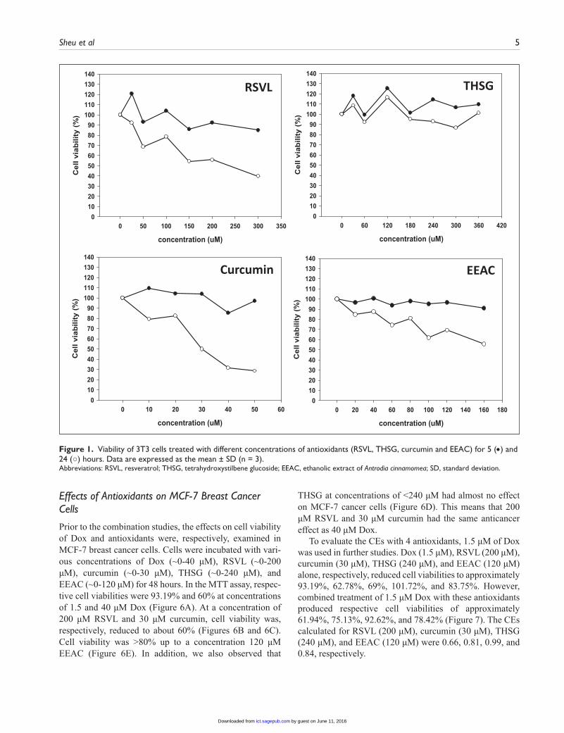
To characterize the viability response of normal cells to antioxidant treatments, 3T3 cells were treated with various concentrations of antioxidants, and their viability was deter-mined after 5 and 24 hours. All antioxidant treatment caused dose- and time-dependent decreases in cell viability (Figure 1). However, results showed that various concentrations of
RSVL, THSG, curcumin, and EEAC themselves were non-toxic (cell viabilities of >80%) to normal 3T3 cells after 5 hours of treatment. The cell viability in normal 3T3 cells decreased to 50% after incubation in the presence of about 200 µM RSVL, 30 µM curcumin, or 160 µM EEAC for 24 hours. Treatment with THSG in the indicated concentration range revealed no cytotoxic effects against 3T3 cells after 24 hours. Therefore, different concentrations of RSVL (~0-200 µM), THSG (~0-360 µM), curcumin (~0-30 µM), and EEAC (~0-160 µM) were used to evaluate the effects on reducing Dox-induced ROS and synergistic anticancer effects with Dox.
To evaluate the effects of antioxidants on oxidative stress in 3T3 cells induced by Dox, the fluorescent dye, DCF-DA, was used to measure ROS production. 3T3 cells were treated with a combination of antioxidants and Dox or were pretreated with antioxidants for 5 hours and then treated with Dox. Results showed that Dox cotreated with RSVL (150 and 200 µM) for 24 hours and EEAC (40, 60, and 80 µM) for 1, 3, and 5 hours significantly decreased intracel-lular ROS levels compared with control cells. In contrast, THSG and curcumin cotreated with Dox showed no signifi-cant effect of decreasing ROS levels (Figure 2A). However, increases in ROS production induced by Dox with 1, 3, and 24 hours of treatment were significantly reduced by, respec-tively, pretreating 3T3 cells with RSVL (150 and 200 µM), curcumin (30 µM), and EEAC (80 µM). In contrast, THSG failed to prevent ROS generation in 3T3 cells treated with Dox (Figure 2B).
To characterize the effect of pretreatment with antioxi-dants on the apoptotic response by Dox treatment, 3T3 cells were pretreated at the indicated concentration of antioxi-dant (150 µM RSVL, 240 µM THSG, 30 µM curcumin, and 120 µM EEAC) for 5 hours then treated with 1.5 µM Dox. Apoptosis was determined by a Hoechst 33342 assay. Apoptotic cells treated with 1.5 µM Dox only exhibited condensed and/or fragmented nuclei with bright nuclear fluorescence (Figure 3, upper row). However, Dox treat-ment caused apoptosis that was reduced or inhibited by pre-treating cells with these antioxidants for 5 hours (Figure 3). These results are consistent with pretreatment with RSVL, curcumin, and EEAC for 5 hours, reducing the ROS pro-duction induced by Dox (Figure 2B). We also used mito-chondrion-targeted hydroethidium (MitoSox Red) to investigate mitochondria as the potential source of superox-ide generation. Results showed that Dox increased the cel-lular dihydroethidium fluorescence intensity, which was reduced or inhibited by pretreatment with antioxidants for 5 hours (Figure 4). Therefore, the generation of mitochon-drial superoxide induced by Dox was inhibited by pretreat-ment with antioxidants. Figure 5 shows that treating 3T3 cells with various antioxidants for 24 hours significantly increased SOD levels. Curcumin and RSVL showed greater potencies in increasing SOD activity.
by guest on June 11, 2016ict.sagepub.comDownloaded from
Sheu et al 5
Effects of Antioxidants on MCF-7 Breast Cancer Cells
Prior to the combination studies, the effects on cell viability of Dox and antioxidants were, respectively, examined in MCF-7 breast cancer cells. Cells were incubated with vari-ous concentrations of Dox (~0-40 µM), RSVL (~0-200 µM), curcumin (~0-30 µM), THSG (~0-240 µM), and EEAC (~0-120 µM) for 48 hours. In the MTT assay, respec-tive cell viabilities were 93.19% and 60% at concentrations of 1.5 and 40 µM Dox (Figure 6A). At a concentration of 200 µM RSVL and 30 µM curcumin, cell viability was, respectively, reduced to about 60% (Figures 6B and 6C). Cell viability was >80% up to a concentration 120 µM EEAC (Figure 6E). In addition, we also observed that
THSG at concentrations of <240 µM had almost no effect on MCF-7 cancer cells (Figure 6D). This means that 200 µM RSVL and 30 µM curcumin had the same anticancer effect as 40 µM Dox.
To evaluate the CEs with 4 antioxidants, 1.5 µM of Dox was used in further studies. Dox (1.5 µM), RSVL (200 µM), curcumin (30 µM), THSG (240 µM), and EEAC (120 µM) alone, respectively, reduced cell viabilities to approximately 93.19%, 62.78%, 69%, 101.72%, and 83.75%. However, combined treatment of 1.5 µM Dox with these antioxidants produced respective cell viabilities of approximately 61.94%, 75.13%, 92.62%, and 78.42% (Figure 7). The CEs calculated for RSVL (200 µM), curcumin (30 µM), THSG (240 µM), and EEAC (120 µM) were 0.66, 0.81, 0.99, and 0.84, respectively.
concentration (uM)
0 50 100 150 200 250 300 350
Cel
l via
bilit
y (%
)
0102030405060708090
100110120130140
concentration (uM)
0 10 20 30 40 50 60
Cel
l via
bilit
y (%
)
0102030405060708090
100110120130140
concentration (uM)
0 60 120 180 240 300 360 420
Cel
l via
bilit
y (%
)
0102030405060708090
100110120130140
concentration (uM)
0 20 40 60 80 100 120 140 160 180
Cel
l via
bilit
y (%
)
0102030405060708090
100110120130140
RSVL THSG
Curcumin EEAC
Figure 1. Viability of 3T3 cells treated with different concentrations of antioxidants (RSVL, THSG, curcumin and EEAC) for 5 (•) and 24 (○) hours. Data are expressed as the mean ± SD (n = 3).Abbreviations: RSVL, resveratrol; THSG, tetrahydroxystilbene glucoside; EEAC, ethanolic extract of Antrodia cinnamomea; SD, standard deviation.
by guest on June 11, 2016ict.sagepub.comDownloaded from

6 Integrative Cancer Therapies
Cotreated time1 hr 3 hr 5 hr 24 hr
Rela
�ve
DCF
fluor
esce
nce
(%)
0102030405060708090
100110120130140150160170
UntreatedRSVL 150 uMRSVL 200 uM
**
Cotreated time1 hr 3 hr 5 hr 24 hr
Rela
�ve
DCF
fluor
esce
nce
(%)
0102030405060708090
100110120130140150160170
UntreatedTHSG 60 uMTHSG 120 uM
Cotreated time1 hr 3 hr 5 hr 24 hr
Rela
�ve
DCF
fluor
esce
nce
(%)
0102030405060708090
100110120130140150160170
Untreatedcurcumin 10 uMcurcumin 20 uMcurcumin 30 uM
Cotreated time1 hr 3 hr 5 hr 24 hr
Rela
�ve
DCF
fluor
esce
nce
(%)
0102030405060708090
100110120130140150160170
UntreatedEEAC 40 uMEEAC 60 uMEEAC 80 uM
**
* *
**
* **
Treated time1 hr 3 hr 24 hr
Rela
�ve
DCF
fluor
esce
nce
(%)
0102030405060708090
100110120130140150160
UntreatedRSVL 150 uMRSVL 200 uM
* * **
*
*
Treated time 1 hr 3 hr 24 hr
Rela
�ve
DCF
fluor
esce
nce
(%)
0102030405060708090
100110120130140150160
UntreatedTHSG 60 uMTHSG 120 uM
*
Treated time 1 hr 3 hr 24 hr
Rela
�ve
DCF
fluor
esce
nce
(%)
0102030405060708090
100110120130140150160
UntreatedCurcumin 10 uMCurcumin 20 uMCurcumin 30 uM
**
* * *
Treated time1 hr 3 hr 24 hr
Rela
�ve
DCF
fluor
esce
nce
(%)
0102030405060708090
100110120130140150160
UntreatedEEAC 40 uMEEAC 60 uMEEAC 80 uM
* ** * *
**
A
B
Figure 2. Effects of various concentrations of antioxidants (RSVL, THSG, curcumin, and EEAC) on Dox-mediated ROS generation in 3T3 cells. (A) Cells were cotreated with 1.5 µM Dox in the absence or presence of antioxidants and (B) cells were pretreated with antioxidants then received 1.5 µM Dox for different times, after which they were analyzed for ROS generation using DCF-DA as a fluorescent probe. Data are expressed as the mean ± SD (n = 3). *P < .05 by a t test.Abbreviations: RSVL, resveratrol; THSG, tetrahydroxystilbene glucoside; EEAC, ethanolic extract of Antrodia cinnamomea; ROS, reactive oxygen species; DCF-DA, 2′,7′-dichlorodihydrofluorescein diacetate; SD, standard deviation.
by guest on June 11, 2016ict.sagepub.comDownloaded from

Sheu et al 7
Discussion
Cardiac toxicity and HFS are common toxicities experi-enced by patients receiving Dox and PLD for cancer treatment and are dose-limiting toxicities. However, ROS were proposed as being responsible for Dox-induced cardiac toxicity and PLD-induced HFS.26 Although cardiac and skin toxicity induced by chemo-therapeutic agents were reported to be correlated with ROS generation, there is no effective preventive treat-ment at present. In this study, we observed that Dox-induced intracellular ROS accumulation in 3T3 cells was attenuated by cotreatment with EEAC and pretreatment with RSVL (150 and 200 µM), curcumin (30 µM), and EEAC (80 µM) for 5 hours, as shown by a reduced
distribution of the DCF fluorescent dye in cells (Figures 2). Interestingly, pretreatment of cells with different con-centrations of antioxidants for 5 hours showed more potent decreases in ROS levels than that seen in the cotreatment groups. So these antioxidants may serve as potential protective agents against Dox-induced cardiac toxicity and PLD-induced HFS through a mechanism that involves their ROS-scavenging properties.27,28
Using the Hoechst assay, we demonstrated that Dox induced apoptosis of 3T3 cells, and it was inhibited by pretreating cells with RSVL, curcumin, THSG, and EEAC (Figure 3). We also found that mitochondria are the primary source of superoxide generation based on observations that the generated superoxide induced by Dox was detected using the mitochondrial ROS probe,
1.5 uM Dox only
150 uM RSVL 30 uM Curcumin
240 uM THSG 120 uM EEAC
Figure 3. Fluorescence micrographs of treated cells stained with Hoechst dye. Control included cells exposed to Dox alone (upper row) or cells preincubated with various antioxidants (RSVL, THSG, curcumin, and EEAC) for 5 hours, then incubated with Dox for 1 hours. Apoptotic cells exhibited condensed and/or fragmented nuclei with bright nuclear fluorescence (original magnification, 400×).Abbreviations: Dox, doxorubicin; RSVL, resveratrol; THSG, tetrahydroxystilbene glucoside; EEAC, ethanolic extract of Antrodia cinnamomea.
by guest on June 11, 2016ict.sagepub.comDownloaded from

8 Integrative Cancer Therapies
1.5 uM Dox only
150 uM RSVL 30 uM Curcumin
240 uM THSG 120 uM EEAC
Figure 4. Cells were treated with Dox only for 5 hours (control group; upper row) or were pretreated with various antioxidants (RSVL, THSG, curcumin, and EEAC) for 5 hours and then received Dox for 1 hour. They were then analyzed for mitochondrial superoxide generation by fluorescence microscopy using MitoSox Red as a probe.Abbreviations: Dox, doxorubicin; RSVL, resveratrol; THSG, tetrahydroxystilbene glucoside; EEAC, ethanolic extract of Antrodia cinnamomea.
MitoSOX (Figure 4). This finding is consistent with the established role of mitochondria as a key source of ROS production through the electron transport chain.29 The Dox-induced MitoSox fluorescence intensity was also inhibited by pretreating cells with RSVL, curcumin, THSG, and EEAC for 5 hours (Figure 4). These results are in good agreement with the ROS generation data and indicate that superoxide is the primary oxidative species and key mediator of Dox-induced apoptosis in 3T3 cells. In the process of apoptosis, mitochondria serve as a source of ROS, which is generated by a reduction in the mitochondrial membrane potential, and the enhanced ROS production is related to the apoptotic response induced by Dox. It was reported that cells are often
equipped with several antioxidants to prevent free-radical damage. SOD and glutathione peroxidase, along with other enzymatic and nonenzymatic antioxidants, play piv-otal roles in preventing cellular damage caused by ROS. Therefore, intracellular ROS can be effectively elimi-nated by the combined action of SOD, glutathione peroxi-dase, and other endogenous antioxidants, which provide a repair mechanism for oxidized membrane components.25 In the present study, significant increases in SOD were observed in 3T3 cells after exposure to RSVL, curcumin, THSG, and EEAC (Figure 5), indicating the ability of antioxidants to decrease ROS contents. The present find-ings corroborate similar findings by Zhang et al30 who reported that Dox was able to enhance the formation of
by guest on June 11, 2016ict.sagepub.comDownloaded from

Sheu et al 9
ROS in heart tissues. Excessive production of ROS may induce cell damage via apoptosis in any cell type, and such effects can be blocked by a wide variety of antioxi-dants.30,31 Therefore, free radicals are responsible for the dangerous side effects of cardiotoxicity elicited by the drug’s use, although these same mechanisms make Dox a potent anticancer drug, allowing it to be efficacious against various forms of cancer.32 Together, our results indicate that pretreatment with RSVL (150 and 200 µM), curcumin (30 µM), and EEAC (80 µM) could ameliorate Dox-induced cardiotoxicity and PLD-induced HFS.
The antitumor activity of DOX was shown to be a result of topoisomerase II inhibition and DNA damage through radical reactions and apoptosis. The mechanism of apoptosis involving DOX was determined to be a result of oxidative DNA damage by DOX-induced ROS genera-tion, although DOX-induced apoptosis may involve topoisomerase II inhibition.1 The viability of MCF-7 cells was decreased by Dox in a dose-dependent manner. Antioxidative properties of RSVL, curcumin, and EEAC have the ability to inhibit ROS generation. The viability of MCF-7 cells treated with RSVL (200 µM), curcumin (30 µM), and EEAC (120 µM) was also significantly lower than that of cells treated with 1.5 µM Dox alone (Figure 6). The ability of RSVL to inhibit cell prolifera-tion and induce cell death is well established.18 It was also reported that curcumin caused apoptosis of tumor cells through various pathways.1 Viability of cells cotreated
with 1.5 µM Dox and RSVL (200 µM), curcumin (30 µM), and EEAC (120 µM) was significantly lower than that with Dox alone (Figure 7). A greater synergistic effect was achieved with 200 µM RSVL and 1.5 µM Dox in MCF-7 cells. Recent in vitro and in vivo studies showed promising results regarding antioxidants in different can-cer models, in addition to their chemopreventive proper-ties. Combined treatment with the chemotherapeutic agent, clofarabine, and RSVL was expected to exert a more significant inhibitory effect on molecules related to cell growth. As one of the novel molecular effects of com-bined treatment, combined treatment with RSVL and clo-farabine exhibited inhibition of Akt, Sp1, and sp1-regulated gene products, including c-Met, cyclin D1, and p21.18 Curcumin, a polyphenol compound, is able to modulate several important signaling pathways such as nuclear factor-κB and increase apoptosis in several can-cer cell lines in combination with Dox.33 The present study supports these notions, showing that antioxidants (RSVL, curcumin, and EEAC) alone or combined with Dox can inhibit the proliferation of MCF-7 cells, even at low treatment doses of Dox, and resulted in at least 20% inhibition of cell survival.
We found that antioxidants (RSVL, curcumin, and EEAC) apart from their antioxidant effect also have anti-cancer activity owing to multitargeted chemopreventive properties. The antioxidant effect can reduce side effects, including Dox-induced cardiotoxicity and PLD-induced HFS. The anticancer activity can enhance chemotherapeu-tic potential. Cancer chemotherapy commonly involves a cocktail strategy in which multiple drugs are used. Dox is commonly used in combination regimens with other cyto-toxic drugs such as methotrexate, cisplatin, ifosfamide, and vincristine in the clinic. To be considered successful, a drug combination should have advantages such as increased therapeutic efficacy and reduced toxicity to the host. Therefore, the combination of Dox and antioxidants (RSVL, curcumin, and EEAC) may have therapeutic value in treat-ing cancer.
In conclusion, our study is the first to demonstrate the effects of combined treatment of antioxidants and Dox on ameliorating Dox-induced cardiotoxicity and PLD-induced HFS and causing synergistic anticancer effects. RSVL, cur-cumin, and EEAC are useful modulators to enhance the antitumor activity of Dox while reducing the adverse effects of cardiac toxicity and HFS by decreasing ROS production in normal tissues. We expect that the addition of RSVL, cur-cumin, and EEAC may enable a reduction in the effective dose of Dox and enhance the quality of life of patients in clinical cancer therapy. In the future, we hope that these antioxidants (RSVL, curcumin, and EEAC) can serve as potential CAM and integrative cancer therapies to treat patients suffering from cancer.
BlankDox
RSVLTHSG
curcuminEEAC
Incr
ease
d SO
D a
ctiv
ity (%
)
0
1
2
3
4
5
6
7
8
9
10
Figure 5. The effect of Dox and various antioxidants (RSVL, THSG, curcumin, and EEAC) on increasing SOD activities in 3T3 cells for 24 hours. The cell-free condition served as a blank.Abbreviations: Dox, doxorubicin; RSVL, resveratrol; THSG, tetrahydroxystilbene glucoside; EEAC, ethanolic extract of Antrodia cinnamomea; SOD, superoxide dismutase.
by guest on June 11, 2016ict.sagepub.comDownloaded from

10 Integrative Cancer Therapies
0 10 20 30 40 50
Cel
l via
bilit
y (%
)
0
10
20
30
40
50
60
70
80
90
100
110
120
0 50 100 150 200 250
Cel
l via
bilit
y (%
)
0
10
20
30
40
50
60
70
80
90
100
110
120
Concentration (uM)
(A) Dox
Concentration (uM)
(B) RSVL
0 5 10 15 20 25 30 35
Cel
l via
bilit
y (%
)
0
10
20
30
40
50
60
70
80
90
100
110
120
Concentration (uM)0 60 120 180 240 300
Cel
l via
bilit
y (%
)
0
10
20
30
40
50
60
70
80
90
100
110
120
0 20 40 60 80 100 120 140
Cel
l via
bilit
y (%
)
0
10
20
30
40
50
60
70
80
90
100
110
120Concentration (uM)
Concentration (uM)
(C) Curcumin (D) THSG
(E) EEAC
Figure 6. Viability of MCF-7 breast cancer cells treated with different concentrations of Dox, RSVL, curcumin, THSG, and EEAC for 48 hours. Data are expressed as the mean ± SD (n = 3).Abbreviations: Dox, doxorubicin; RSVL, resveratrol; THSG, tetrahydroxystilbene glucoside; EEAC, ethanolic extract of Antrodia cinnamomea; SD, standard deviation.
by guest on June 11, 2016ict.sagepub.comDownloaded from

Sheu et al 11
Declaration of Conflicting Interests
The author(s) declared no potential conflicts of interest with respect to the research, authorship, and/or publication of this article.
Funding
The author(s) received no financial support for the research, authorship, and/or publication of this article.
References
1. Sadzuka Y, Nagamine M, Toyooka T, Ibuki Y, Sonobe T. Beneficial effects of curcumin on antitumor activity and adverse reactions of doxorubicin. Int J Pharm. 2012;432: 42-49.
2. Menna P, Recalcati S, Cairo G, Minotti G. An introduction to the metabolic determinants of antracycline cardiotoxicity. Cardiovasc Toxicol. 2007;7:80-85.
3. Šimunek T, Šterba M, Popelová O, Adamcová M, Hrdina R, Gerši V. Anthracycline-induced cardiotoxicity: overview of studies examining the roles of oxidative stress and free cel-lular iron. Pharmacol Rep. 2009;611:154-171.
4. Deavall DG, Martin EA, Horner MJ, Roberts R. Drug-induced oxidative stress and toxicity. J Toxicol. 2012;64:54-60.
5. Gabizon AA. Pegylated liposomal doxorubicin: metamor-phosis of an old drug into a new form of chemotherapy. Cancer Invest. 2001;19:424-436.
6. Jiang W, Lionberger R, Yu LX. In vitro and in vivo charac-terizations of PEGylated liposomal doxorubicin. Bioanalysis 2011;3:333-344.
7. Swenson KK, Bell EM. Hand-foot syndrome related to lipo-somal doxorubicin. Oncol Nurs Forum. 2010;37:137-139.
8. Farr KP, Safwat A. Palmer-plantar erythrodysesthesia associ-ated with chemotherapy and its treatment. Case Rep Oncol. 2011;4:229-235.
9. Yokomichi N, Nagasawa T, Coler-Reilly A, et al. Pathogenesis of hand-foot syndrome induced by PEG-modified liposomal-doxorubicin. Hum Cell. 2013;26:8-18.
10. O’Brien MER, Wigler N, Inbar M, et al. Reduced cardiotox-icity and comparable efficacy in a phase III trial of pegylated liposomal doxorubicin HCl (Caelyx™/Doxil®) versus con-ventional doxorubicin for first-line treatment of metastatic breast cancer. Ann Oncol. 2004;15:440-449.
11. Richardson MA, Sanders T, Palmer JL, Greisinger A, Singletary SE. Complementary/alternative medicine use in a comprehensive cancer center and the implications for oncol-ogy. J Clin Oncol. 2000;18:2505-2514.
12. Verhoef MJ, Balneaves LG, Boon HS, Vroegingewey A. Reasons for and characteristics associated with complemen-tary and alternative medicine use among adult cancer patients: a systematic review. Integr Cancer Ther. 2005;4:274-286.
13. Barnes PM, Bloom B, Nahin LR. Complementary and alter-native medicine use among adults and children: United States, 2007. Natl Health Stat Report. 2008;12:1-24.
14. Mainardi T, Kapoor S, Bielory L. Complementary and alter-native medicine: herbs, phytochemicals and vitamins and their immunologic effects. J Allergy Clin Immunol. 2009;123: 283-294.
15. Can G, Erol O, Aydiner A, Topuz E. Quality of life and complementary and alternative medicine use among cancer patients in Turkey. Eur J Oncol Nurs. 2009;13:287-294.
16. Yang D, Li T, Liu Z, et al. LRRK2 kinase activity medi-ates toxic interactions between genetic mutation and oxida-tive stress in a Drosophila model: suppression by curcumin. Neurobiol Dis. 2012;47:385-392.
17. Hosseinzadeh L, Behravan J, Mosaffa F, et al. Curcumin potentiates doxorubicin-induced apoptosis in H9c2 cardiac muscle cells through generation of reactive oxygen species. Food Chem Toxicol. 2011;49:1102-1109.
18. Lee YJ, Im JH, Won SY, et al. Synergistic anti-cancer effects of resveratrol and chemotherapeutic agent clofarabine against human a malignant mesothelioma MSTO-211H cells. Food Chem Toxicol. 2013;52:61-68.
19. Gu J, Song ZP, Gui DM, Hu W, Chen YG, Zhang DD. Resveratrol attenuates doxorubicin-induced cardiomyocyte apoptosis in lymphoma nude mice by heme oxygenase-1 induction. Cardiovasc Toxicol. 2012;12:341-349.
20. Su CH. Health Guardian Angel: Antrodia camphorate. 1st ed. Taipei, Taiwan: EKS Book Publishing Co; 2002.
21. Geethangili M, Tzeng YM. Review of pharmacologi-cal effects of Antrodia camphorata and its bioactive compounds. Evid Based Complement Alternat Med. 2011;2011:212641.
22. Kumar KJS, Chu FH, Hsieh HW, et al. Antroquinonol from ethanolic extract of mycelium of Antrodia cinnamomea protects hepatic cells from ethanol-induced oxidative stress through Nrf-2 activation. J Ethnopharmacol. 2011;136: 168-177.
23. Liu L, Zhao L, Li YL, Zhang L, Ye CF, Li L. Protective effect of 2,3,5,4’-tetrahydroxystilbene-2-β-O-d-glucoside on
RSVL Curcumin THSG EEAC
Cel
l via
bilit
y (%
of c
ontr
ol)
0
20
40
60
80
100
120
Control1.5 uM Dox onlyantioxidants onlyantioxidants plus 1.5 uM Dox
Figure 7. Viability of MCF-7 breast cancer cells treated with 1.5 µM Dox and various antioxidants (200 µM RSVL, 30 µM curcumin, 240 µM THSG, and 120 µM EEAC), alone and in combination, for 48 hours.Abbreviations: Dox, doxorubicin; RSVL, resveratrol; THSG, tetrahydroxystilbene glucoside; EEAC, ethanolic extract of Antrodia cinnamomea.
by guest on June 11, 2016ict.sagepub.comDownloaded from

12 Integrative Cancer Therapies
hippocampus neurons in dementia rats induced by chronic cerebral ischemia. Chin J Pharm. 2006;41:354-357.
24. Chen LY, Sheu MT, Liu DZ, et al. Pretreatment with an etha-nolic extract of Taiwan fungus camphoratus (Antrodia cam-phorata) enhances the cytotoxic effects of amphotericin B. J Agric Food Chem. 2011;59:11255-11263.
25. Yang Y, Duan W, Liang Z, et al. Curcumin attenuates endo-thelial cell oxidative stress injury through Notch signaling inhibition. Cell Signal. 2013;25:615-629.
26. Luanpitpong S, Chanvorachote P, Nimmannit U, et al. Mitochondrial superoxide mediates doxorubicin-induced kera-tinocyte apoptosis through oxidative modification of ERK and Bcl-2 ubiquitination. Biochem Pharmacol. 2012;83:1643-1654.
27. Force T, Kolaja KL. Cardiotoxicity of kinase inhibitors: the prediction and translation of preclinical models to clinical outcomes. Nat Rev Drug Discov. 2011;10:111-126.
28. Gianni L, Herman EH, Lipshultz SE, et al. Anthracycline cardiotoxicity: from bench to bedside. J Clin Oncol. 2008;26:3777-3784.
29. Moungjaroen J, Nimmannit U, Callery PS, et al. Reactive oxygen species mediate caspase activation and apoptosis induced by lipoic acid in human lung epithelial cancer cells through Bcl-2 down regulation. J Pharmacol Exp Ther. 2006;319:1062-1069.
30. Zhang AH, Wang WQ, Wang JL. Protective effect of tet-rahydroxystilbene glucoside on cardiotoxicity induced by doxorubicin in vitro and in vivo. Acta Pharmacol Sin. 2009;30:1479-1487.
31. Nordberg J, Arnér ESJ. Reactive oxygen species, antioxi-dants, and the mammalian thioredoxin system. Free Radic Biol Med. 2001;31:1287-1312.
32. Tacar O, Sriamornsak P, Dass CR. Doxorubicin: an update on anticancer molecular action, toxicity and novel delivery systems. J Pharm Pharmacol. 2012;65:157-170.
33. Choi BH, Kim CG, Lim Y, Shin SY, Lee YH. Curcumin down-regulates the multidrug-resistance mdr1b gene by inhibiting the PI3K/Akt/NF[kappa]B pathway. Cancer Lett. 2008;259:111-118.
by guest on June 11, 2016ict.sagepub.comDownloaded from