
omics based methods for health claims of dietary antioxidants
50
Use of conventional and -omics based methods for health claims of dietary antioxidants: a critical overview Siegfried Knasmu ¨ller 1 *†, Armen Nersesyan 1 †, Miroslav Mis ˇı ´k 1 , Christopher Gerner 1 , Wolfgang Mikulits 1 , Veronika Ehrlich 1 , Christine Hoelzl 1 , Akos Szakmary 1 and Karl-Heinz Wagner 2 1 Institute of Cancer Research, Inner Medicine I, Medical University of Vienna, Borschkegasse 8a, A-1090 Vienna, Austria 2 Department of Nutritional Sciences, University of Vienna, Althanstrasse 14, 1090 Vienna, Austria This article describes the principles and limitations of methods used to investigate reactive oxygen species (ROS) protective properties of dietary constituents and is aimed at providing a better understanding of the requirements for science based health claims of antioxidant (AO) effects of foods. A number of currently used biochemical measurements aimed of determining the total antioxidant capacity and oxidised lipids and pro- teins are carried out under unphysiologcial conditions and are prone to artefact formation. Probably the most reliable approaches are measure- ments of isoprostanes as a parameter of lipid peroxidation and determination of oxidative DNA damage. Also the design of the experimental models has a strong impact on the reliability of AO studies: the common strategy is the identification of AO by in vitro screening with cell lines. This approach is based on the assumption that protection towards ROS is due to scavenging, but recent findings indicate that activation of tran- scription factors which regulate genes involved in antioxidant defence plays a key role in the mode of action of AO. These processes are not adequately represented in cell lines. Another shortcoming of in vitro experiments is that AO are metabolised in vivo and that most cell lines are lacking enzymes which catalyse these reactions. Compounds with large molecular configurations (chlorophylls, anthocyans and polyphenolics) are potent AO in vitro, but weak or no effects were observed in animal/human studies with realistic doses as they are poorly absorbed. The development of -omics approaches will improve the scientific basis for health claims. The evaluation of results from microarray and proteomics studies shows that it is not possible to establish a general signature of alterations of transcription and protein patterns by AO. However, it was shown that alterations of gene expression and protein levels caused by experimentally induced oxidative stress and ROS related diseases can be normalised by dietary AO. Dietary antioxidants: -Omics methods: Health claims It is well documented that oxidative stress, defined by Blomh- off (1) as a “condition that is characterised by the accumulation of non-enzymatic oxidative damage to molecules that threaten the normal functions of a cell or the organism” is involved in the aetiology of a large number of human diseases. Typical examples are various forms of cancer (breast, colon and liver), neuropathological disorders such as Parkinson’s and Altzheimer’s disease, inflammations including hepatitis and inflammatory bowel diseases, different types of dermatitis as well as bacterial and viral infections (HBV, sepsis), diabetes, and rheumatoid arthritis (for review see (2) ). Also coronary heart diseases which are the major cause of death in industrialised countries (2,3) and idiopathic infertility (4,5) , which has increased over the last decades in Western countries seem to be causally related to reactive oxygen species (ROS) meditated damage and it has been stressed that oxidative damage is also involved in several diseases of ageing, e.g. Werner’s syndrome (6) , progeria (7) , amyotrophic lateral scler- osis, cataract formation and decreased immune functions (8 – 10) . Already half a century ago it was found that the acute toxic, DNA-damaging and carcinogenic effects of ionising radiation which are predominantly caused by the formation of ROS can be reduced by antioxidant vitamins such as C, E, and A (11 – 13) . In the following decades, it became apparent that plant derived foods as well as beverages contain a large number of compounds which protect against oxidative damage and its consequences. Typical examples for such antioxidants which have been defined as “redox-active com- pounds that reduce pro-oxidative stress by reacting non-enzy- matically with a reactive oxidant” (1) are flavonoids and phenolic acids contained in fruits and vegetables (14 – 17) , ally- slulfides in Allium species (18) , hydroxycinnamic acids in coffee (19,20) , phenolic compounds in wines (21) and veg- etable oils (22) , catechins in teas (23) , specific ingredients of common spices such as capsaicin in chillies (24) , gingerol (25) and curcumin (26) , chlorophylls (27) , anthocyanins in berries (28) as well as carotenoids (29) to name only a few. The increasing evidence of the strong impact of the redox status on human health has stimulated intense research activi- ties in this field. It has been estimated that around 10 papers dealing with oxidative stress and/or antioxidants are published daily (1) and many of them concern the identification of dietary compounds in the diet and investigations concerning their mode of action. The results of these efforts have a strong * Corresponding author: Professor Siegfried Knasmu ¨ller, fax þ431 4277 9651, email [email protected] † Contributed equally British Journal of Nutrition (2008), 99, E-Suppl. 1, ES3–ES52 doi:10.1017/S0007114508965752 q The Authors 2008 British Journal of Nutrition Downloaded from https://www.cambridge.org/core. IP address: 65.21.229.84, on 15 Feb 2022 at 11:56:28, subject to the Cambridge Core terms of use, available at https://www.cambridge.org/core/terms. https://doi.org/10.1017/S0007114508965752
-
Upload
khangminh22 -
Category
Documents
-
view
1 -
download
0
Transcript of omics based methods for health claims of dietary antioxidants

Use of conventional and -omics based methods for health claims of dietary
antioxidants: a critical overview
Siegfried Knasmuller1*†, Armen Nersesyan1†, Miroslav Misık1, Christopher Gerner1, Wolfgang Mikulits1,
Veronika Ehrlich1, Christine Hoelzl1, Akos Szakmary1 and Karl-Heinz Wagner 2
1Institute of Cancer Research, Inner Medicine I, Medical University of Vienna, Borschkegasse 8a, A-1090 Vienna, Austria2Department of Nutritional Sciences, University of Vienna, Althanstrasse 14, 1090 Vienna, Austria
This article describes the principles and limitations of methods used to investigate reactive oxygen species (ROS) protective properties of dietary
constituents and is aimed at providing a better understanding of the requirements for science based health claims of antioxidant (AO) effects of
foods. A number of currently used biochemical measurements aimed of determining the total antioxidant capacity and oxidised lipids and pro-
teins are carried out under unphysiologcial conditions and are prone to artefact formation. Probably the most reliable approaches are measure-
ments of isoprostanes as a parameter of lipid peroxidation and determination of oxidative DNA damage. Also the design of the experimental
models has a strong impact on the reliability of AO studies: the common strategy is the identification of AO by in vitro screening with cell lines.
This approach is based on the assumption that protection towards ROS is due to scavenging, but recent findings indicate that activation of tran-
scription factors which regulate genes involved in antioxidant defence plays a key role in the mode of action of AO. These processes are not
adequately represented in cell lines. Another shortcoming of in vitro experiments is that AO are metabolised in vivo and that most cell lines are
lacking enzymes which catalyse these reactions. Compounds with large molecular configurations (chlorophylls, anthocyans and polyphenolics)
are potent AO in vitro, but weak or no effects were observed in animal/human studies with realistic doses as they are poorly absorbed. The
development of -omics approaches will improve the scientific basis for health claims. The evaluation of results from microarray and proteomics
studies shows that it is not possible to establish a general signature of alterations of transcription and protein patterns by AO. However, it was
shown that alterations of gene expression and protein levels caused by experimentally induced oxidative stress and ROS related diseases can be
normalised by dietary AO.
Dietary antioxidants: -Omics methods: Health claims
It is well documented that oxidative stress, defined by Blomh-off(1) as a “condition that is characterised by the accumulationof non-enzymatic oxidative damage to molecules that threatenthe normal functions of a cell or the organism” is involvedin the aetiology of a large number of human diseases. Typicalexamples are various forms of cancer (breast, colon and liver),neuropathological disorders such as Parkinson’s andAltzheimer’s disease, inflammations including hepatitisand inflammatory bowel diseases, different types of dermatitisas well as bacterial and viral infections (HBV, sepsis),diabetes, and rheumatoid arthritis (for review see(2)). Alsocoronary heart diseases which are the major cause of deathin industrialised countries(2,3) and idiopathic infertility(4,5),which has increased over the last decades in Western countriesseem to be causally related to reactive oxygen species (ROS)meditated damage and it has been stressed that oxidativedamage is also involved in several diseases of ageing, e.g.Werner’s syndrome(6), progeria(7), amyotrophic lateral scler-osis, cataract formation and decreased immune functions(8 – 10).
Already half a century ago it was found that the acutetoxic, DNA-damaging and carcinogenic effects of ionisingradiation which are predominantly caused by the formation
of ROS can be reduced by antioxidant vitamins such as C,E, and A(11 – 13). In the following decades, it became apparentthat plant derived foods as well as beverages contain a largenumber of compounds which protect against oxidativedamage and its consequences. Typical examples for suchantioxidants which have been defined as “redox-active com-pounds that reduce pro-oxidative stress by reacting non-enzy-matically with a reactive oxidant”(1) are flavonoids andphenolic acids contained in fruits and vegetables(14 – 17), ally-slulfides in Allium species(18), hydroxycinnamic acidsin coffee(19,20), phenolic compounds in wines(21) and veg-etable oils(22), catechins in teas(23), specific ingredients ofcommon spices such as capsaicin in chillies(24), gingerol(25)
and curcumin(26), chlorophylls(27), anthocyanins in berries(28)
as well as carotenoids(29) to name only a few.The increasing evidence of the strong impact of the redox
status on human health has stimulated intense research activi-ties in this field. It has been estimated that around 10 papersdealing with oxidative stress and/or antioxidants are publisheddaily(1) and many of them concern the identification of dietarycompounds in the diet and investigations concerning theirmode of action. The results of these efforts have a strong
*Corresponding author: Professor Siegfried Knasmuller, fax þ431 4277 9651, email [email protected]
† Contributed equally
British Journal of Nutrition (2008), 99, E-Suppl. 1, ES3–ES52 doi:10.1017/S0007114508965752q The Authors 2008
British
Journal
ofNutrition
Dow
nloaded from https://w
ww
.cambridge.org/core . IP address: 65.21.229.84 , on 15 Feb 2022 at 11:56:28 , subject to the Cam
bridge Core terms of use, available at https://w
ww
.cambridge.org/core/term
s . https://doi.org/10.1017/S0007114508965752

impact on the development of nutritional recommendationsand led to the development of supplements which containhigh levels of food derived antioxidants(30) and to the pro-duction of functional foods. A broad variety of differentmethods are currently used to study antioxidants in humanfoods and to identify and characterise their active principles.The models include chemical–analytical and physicalmeasurements, experiments with subcellular fractions andintact cells, animal studies as well as human interventiontrails. In the last decade, new biomarkers have been developedand validated which can be used in human studies and therapid development of -omics techniques (in particular theuse of microarrays and two dimensional gel electrophoresis)offers the possibility to explore the effects of antioxidantson gene expression and protein levels and to study alterationsof disease related patterns(31 – 33).
The aim of the present article is it to give a critical overviewon the advantages and limitations of the different approacheswhich are currently used with particular emphasis on thenewly developed methods. We anticipate that it will help inthe interpretation of existing data and lead to the develop-ment of improved strategies concerning the detection ofantioxidants.
The formation of ROS as well as their physical and chemi-cal properties, their reactions with organic molecules andtheir inactivation by antioxidants have been extensivelydescribed in the scientific literature(34 – 36). Therefore, thesetopics are confined in the present article to short descriptionswhich are essential to understand the subsequent chapters.
Formation of reactive oxygen species (ROS)
Pro-oxidants (often termed as reactive oxygen species) can beclassified in two groups, namely radicals and non-radicals.Radicals (O2, O2
2 , OHz, ROOz, ROz, and NOz) contain unpairedelectrons in the shells around the nucleus which causes thehigh reactivity of these species (except O2), due to their abilityto donate or receive other atoms to obtain stability. Importantnon-radicals comprise hyperchlorous acid (HOCl), hydrogenperoxide (H2O2), organic peroxides, aldehydes and ozone(O3). The most relevant ROS as well as some of their mainreactions are shown in Fig. 1.
Superoxide ðO22 Þ, which is formed for example during
respiration in mitochondria (as a consequence of reductionof oxygen required for APT production) forms at low pHhydroperoxyl ðHO2Þ which penetrates the cell membranesmore easily than the charged form(37,38). Enzymatic as wellas non-enzymatic dismutation leads to formation of hydrogenperoxide (H2O2) which can be detoxified enzymatically (cata-lase, glutathion peroxidase). H2O2 molecules can damage cellsat low concentrations and degrade haem proteins and oxidiseDNA, enzymes, –SH groups and keto acids and are also thesource of more deleterious species such as HOCl and OHz.The latter radical is short lived and reacts at a high ratewith most organic molecules (DNA, amino acids, sugars, pro-teins, lipids). Transition metals (first row of the D block of theperiodic table) contain unpaired electrons (except Zn) and cantherefore be considered as radicals. In particular copper andiron are contained at relatively high concentrations in manyorganisms. At physiological pH, most of the metals arepresent in oxidised forms (Fe3þz, Fe2þ), but after reduction
(e.g. by ascorbic acid or via the Haber-Weiss reactionO2
2 þ Fe3þ ! O2 þ Fe2þ), they can undergo “Fenton type”reactions (e.g. Fe2þ þ H2O2 ! Fe3þ þ OH þ OH2). Thesetwo processes explain the formation of OHz in vivo. However,it is notable that in organisms, metals are always bound to pro-teins and membranes and it has been shown that they canundergo in this state the aforementioned reactions and produceOHz at a single site and convert non-reactive radicals to highlyreactive species.
Nitric oxide (NOz) is produced by oxydation of the terminalguanidine-nitrogen atoms of arginine(38 – 40). This reaction iscatalysed by nitric oxide synthetases (NOS, i.e. neuronalNOS, endothelial NOS and inducible NOS). NOz can reactwith different radicals, the most important reaction underphysiological conditions is the formation of peroxynitirite(ONOO2) in which O2
2 is involved(38,40). ONOO2 cancause damage similar to that induced by OHz(39).
The biological significance of the different ROS species hasbeen discussed quite controversially. Some authors suggestedthat O2
2 and NOz are the most relevant ones, while otherstressed that peroxyl radicals may be even more important(8).
Several exogenous factors contribute to oxidative stress.Ionising radiation causes toxic effects in organisms primarilyvia ionisation of intracellular water(41). Also non-ionising radi-ation (UV light) can indirectly produce a variety of ROSspecies including O3
(42). Other major sources of exposure areair pollutants such as cigarette smoke(43) and car exhausts(44),drugs (bleomycin, doxorubicine)(45) as well as pesticides andherbicides(46) and industrial chemicals(47). Also pathogenicmicroorganisms may produce oxygen species, but the mostrelevant external source is nutrition as most of the foods we
Dietary antioxidants
NO·nitric oxide radical
O2· (HO2
·)superoxide ion radical
OH·hydroxylradical
ROO·peroxyl radicalsRO·alkoxy radical
reduction ofthe metals -
Fe+3/Cu+2
Fe+2/Cu+1
O2
–SOD
Cl–
Endogenous inactivation
H2O2
CIO–(HCIO)
altered signalling pathways
altered gene expression
altered protein synthesis
ONOO–
hydrogen peroxide
hypochlorous acid
peroxynitrite
DNA(base oxidation, gene
mutations, chromosomebreaks, etc.)
PROTEINS(protein oxidation
products)
LIPIDS(lipid peroxidation, LDL-
oxidation, aldehydes,breath hydrocarbons)
Fig. 1. Different form of reactive oxygen species and their interaction with
organic molecules.
S. Knasmuller et al.ES4
British
Journal
ofNutrition
Dow
nloaded from https://w
ww
.cambridge.org/core . IP address: 65.21.229.84 , on 15 Feb 2022 at 11:56:28 , subject to the Cam
bridge Core terms of use, available at https://w
ww
.cambridge.org/core/term
s . https://doi.org/10.1017/S0007114508965752
consume are oxidised and contain oxidants such as peroxides,aldehydes, fatty acids and transition metals(48,49).
The most important endogenous processes of ROS pro-duction are respiration processes in the mitochondria and themassive continuous production of radicals is even increasedin ageing cells(50,51). Another source are white blood cellsinvolved in immune responses which can undergo a respira-tory bust that is characterised by an up to 20-fold increaseof oxygen production(52,53). During this reaction, NAPDHserves as a donor of electrons which results in the productionof O2
2 from oxygen. The enzyme myeloperoxidase catalysesthe production of HOCl by interaction between H2O2
peroxides and chlorides(54,55).
Targets of oxidative damage
The continuous exposure to ROS from exogenous and endoge-nous sources results in oxidative damage of many cell com-ponents and alterations of cellular functions; some of thesechanges can be used as markers of oxidative stress and to inves-tigate putative protective effects of phytochemicals.
Proteins. Radicals react in particular with nucleophilicamino acids for example with tryptophane, histidine andcysteine(56,57). Apart from direct oxidation of SH-groups byH2O2 and O2
2 , organic radicals may bind covalently to cellularproteins which are part of cell membranes or have enzymaticfunctions. One of the major adducts which can be easilydetected is 3-nitrotyrosine which is produced by interactionsbetween ONOOZ and other nitrogen reactive radicals withthe amino acid tyrosine(58). Also relatively resistant aminoacids such as lysine and proline can be hydroxylated non-enzymatically by OHz(59).
It is also known that ROS can destroy peptide bonds andcause drastic alterations of their structures resulting in changesof their cellular functions (for review see(36)). NOz reacts inparticular with Fe–S centres of proteins which transportelectrons and this affects the functions of mitochondria(60).Another important feature is their reaction with thiol groupsof proteins; a typical example is the S-nitrosylation of caspaseswhich are part of cell signalling processes(61).
Lipids. All cellular membranes are vulnerable to oxidationdue to their high concentrations of unsaturated fatty acids.Damage of lipids by ROS (lipid peroxidation, LP) occurs inthree stages. In the first (“initiation phase”), double bonds offatty acids are attacked by radicals which leads to formationof fatty acids radicals. During the “propagation”, a chain reac-tion takes place which leads to continuous formation of theseradicals. The last stage (chain determination) occurs followinginteractions ROOz or with other radical types and/or antioxi-dants (for a detailed description of LP see(38,62,63)). Importantmarker molecules formed during LP are aldeydes and ketones,for example malonedialdehyde (MDA) and 4-hydroxynonenal(4-HNE) as well as the family of isoprostanes which areexcreted in the urine. Oxidation of fatty acids can be measuredwith the relative change in the fatty acid pattern and theformation of conjugated dienes.
Blood cholesterols can be oxidised by ROS to formoxidised low density lipoproteins (LDL). The oxidativemodification hypothesis of atherosclerosis assumes that circu-lating LDL particles are modified by oxidation and that these
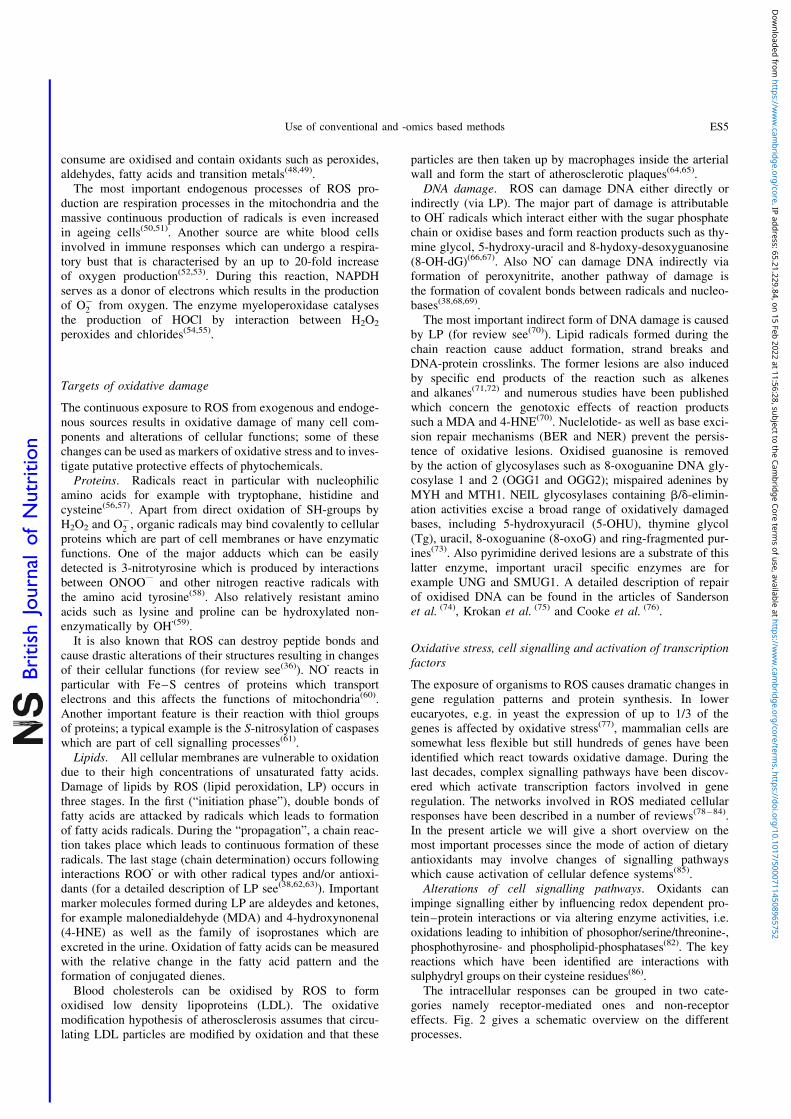
particles are then taken up by macrophages inside the arterialwall and form the start of atherosclerotic plaques(64,65).DNA damage. ROS can damage DNA either directly or
indirectly (via LP). The major part of damage is attributableto OHz radicals which interact either with the sugar phosphatechain or oxidise bases and form reaction products such as thy-mine glycol, 5-hydroxy-uracil and 8-hydoxy-desoxyguanosine(8-OH-dG)(66,67). Also NOz can damage DNA indirectly viaformation of peroxynitrite, another pathway of damage isthe formation of covalent bonds between radicals and nucleo-bases(38,68,69).
The most important indirect form of DNA damage is causedby LP (for review see(70)). Lipid radicals formed during thechain reaction cause adduct formation, strand breaks andDNA-protein crosslinks. The former lesions are also inducedby specific end products of the reaction such as alkenesand alkanes(71,72) and numerous studies have been publishedwhich concern the genotoxic effects of reaction productssuch a MDA and 4-HNE(70). Nuclelotide- as well as base exci-sion repair mechanisms (BER and NER) prevent the persis-tence of oxidative lesions. Oxidised guanosine is removedby the action of glycosylases such as 8-oxoguanine DNA gly-cosylase 1 and 2 (OGG1 and OGG2); mispaired adenines byMYH and MTH1. NEIL glycosylases containing b/d-elimin-ation activities excise a broad range of oxidatively damagedbases, including 5-hydroxyuracil (5-OHU), thymine glycol(Tg), uracil, 8-oxoguanine (8-oxoG) and ring-fragmented pur-ines(73). Also pyrimidine derived lesions are a substrate of thislatter enzyme, important uracil specific enzymes are forexample UNG and SMUG1. A detailed description of repairof oxidised DNA can be found in the articles of Sandersonet al. (74), Krokan et al. (75) and Cooke et al. (76).
Oxidative stress, cell signalling and activation of transcriptionfactors
The exposure of organisms to ROS causes dramatic changes ingene regulation patterns and protein synthesis. In lowereucaryotes, e.g. in yeast the expression of up to 1/3 of thegenes is affected by oxidative stress(77), mammalian cells aresomewhat less flexible but still hundreds of genes have beenidentified which react towards oxidative damage. During thelast decades, complex signalling pathways have been discov-ered which activate transcription factors involved in generegulation. The networks involved in ROS mediated cellularresponses have been described in a number of reviews(78 – 84).In the present article we will give a short overview on themost important processes since the mode of action of dietaryantioxidants may involve changes of signalling pathwayswhich cause activation of cellular defence systems(85).Alterations of cell signalling pathways. Oxidants can
impinge signalling either by influencing redox dependent pro-tein–protein interactions or via altering enzyme activities, i.e.oxidations leading to inhibition of phosophor/serine/threonine-,phosphothyrosine- and phospholipid-phosphatases(82). The keyreactions which have been identified are interactions withsulphydryl groups on their cysteine residues(86).
The intracellular responses can be grouped in two cate-gories namely receptor-mediated ones and non-receptoreffects. Fig. 2 gives a schematic overview on the differentprocesses.
Use of conventional and -omics based methods ES5
British
Journal
ofNutrition
Dow
nloaded from https://w
ww
.cambridge.org/core . IP address: 65.21.229.84 , on 15 Feb 2022 at 11:56:28 , subject to the Cam
bridge Core terms of use, available at https://w
ww
.cambridge.org/core/term
s . https://doi.org/10.1017/S0007114508965752

Growth factors and cytokines (e.g. TNF-a, IL-1) cause ROSproduction in non-pathogenic cells and activate intracellularreceptor mediated signalling which affect mitogen activatedprotein kinases (MAPKs). Growth factor receptors are tyrosinekinases (RTLs), apart from these, also non-receptor proteinkinases have been identified which are also activated byROS and belong for example to the Src family(87,88). ActivatedSrc binds to membranes and initiates MAPKs, NfkB and PI3Ksignalling pathways(78). Other important targets involved insignalling are Ras (membrane bound G proteins involvedin the regulation of cell growth), protein tyrosine phosopha-tases (PTP) and seronine/threonine kinases. The most import-ant representative of the latter group is protein kinase C (PKC),its catalytic site is a zinc finger domain containingseveral cysteine rich regions which can be modified by variousoxidants(89).
MAPKs (for a detailed description see(90)) relay signalsgenerated by exogenous or endogenous stimuli to intracellularspace via phosphorylation of proteins. During this process, thekinases interact also with downstream mediators includingtranscription factors(91). Studies on the upregulation ofMAPKs have shown that these processes are type and stimulispecific. For example, it was found that endogenous H2O2
production by respiratory burst induces ERK but not p38kinase(92) while exogenous peroxide treatment activates thelatter enzyme(93).Activation of transcription factors by ROS. The most sig-
nificant effects of ROS on MAPKs concern the activation oftranscription factors which control the expression of protectivegenes, arrest division of damaged cells and induce apoptosis(programmed cell death).
AP-1 is a collection of dimeric basic region-leucine zipperproteins which are for example induced by metals andH2O2
(94,95) and regulate cell growth, differentiation andapoptosis.
NFkB is a DNA binding protein which is sequestered in thecytoplasm because of an interaction with a member of theinhibitory IkB family. Activation via ROS causes dissociationand allows NFkB to enter the nucleus and activate genesinvolved in inflammatory responses, transformation and angio-genesis(96). A number of investigations showed that that acti-vation by different stimuli can be blocked by antioxidantsincluding N-acetlylcysteine, cysteine, vitamin E, thiols andgreen tea polyphenolics(78).
Another important factor which plays a key role in protect-ing cells from malignant transformation is p53, also termed a“tumour suppressor” since it arrests cell cycle and inducesapoptosis(97). p53 is directly activated by oxidants and itsoverexpression leads to increase of intracellular ROS levels.One of the important functions of p53 is the up regulationof proteins that play a role in ROS mediated apoptosisnamely ferrodoxin reductase (FDXR) and a novel stress-response gene Redd1/HIF-1 originally isolated as an HIF-1-response gene(98). It was shown in a number of in vitro studiesthat antioxidants reduce apoptosis rates due to interaction withp53(99,100). In a recent human intervention study we observeda drastic reduction of the apoptosis frequencies in lympho-cytes after consumption of wheat sprouts which is probablydue to antioxidant effects(101).
Two other transcription factors affected by ROS are nuclearfactor of activated T cells (NFAT) and HIF. The formerfamily regulates muscle growth and differentiation as well
ROS
ROS
Cytokine/
Growth factor
receptorsSrc Ras
PI3k
Keap1
Keap1 Nrf2
Nrf2
Nrf2
Gene transcription
p53NFATAP-1HIF NFκB
Maf
ARC
Antioxidant enzymes
Phase I/ Phase II enzymes
Stress proteins
Chaperones
MAPKs
Akt
PI3k
PKCSignalling
Other factorse.g. PERK
cell membrane
Akt
NADPHoxidase
MAPKs
Inactive forms oftranscription factors
PKC
mitochondria
Cytosol
Nucleus
DNA
nuclear membrane
Fig. 2. Impact of ROS on cell signalling and activation of transcription factors.
S. Knasmuller et al.ES6
British
Journal
ofNutrition
Dow
nloaded from https://w
ww
.cambridge.org/core . IP address: 65.21.229.84 , on 15 Feb 2022 at 11:56:28 , subject to the Cam
bridge Core terms of use, available at https://w
ww
.cambridge.org/core/term
s . https://doi.org/10.1017/S0007114508965752

as cytokine formation and angiogenesis(94,102); while the latteris a heterodimer controlling genes encoding for vascular endo-thelial growth factor (VEGF), aldolase, enolase and lactatedehydrogenase(103).
Probably the most important contribution to cell defenceagainst oxidative stress is mediated through transcriptionalactivation of genes via a cis-acting enhancer known as antioxi-dant responsive element (ARE) which was discovered byPickett and co-workers(104 – 106) and identified in the 50flankingflanking regions of many genes. A number of studies showedthat the transcription factor Nrf2 which belongs to the CNC(cap‘N’collar) basic leucine zipper family and is representedin many tissues is the key mediator of ARE dependent acti-vation(107,108). Comparative investigations with geneticallyaltered rodents (Nrf2 þ /þ and Nrf2 2 /2 ) showed thatnumerous genes are regulated by the element includingthose which encode for protection against ROS such as gluta-mate cystein ligase (GCL) which catalyses the rate limitingstep of glutathione synthesis, NADPH quinone oxidoreductase(NQO), glutathione S-transferase (GST), aldehyde dehydro-genase (ADH), glutathione peroxidase, glutathione reductase,peroxiredoxin I (PrxI), superoxide dismutase (SOD), catalase,and thioredoxin(109,110). Also enzymes which are involved inthe supply of reducing equivalents (e.g. glucose-6-phosphatededydrogenase) as well as xenobiotic drug metabolisingenzymes (e.g. CYPs), chaperones, and stress response proteinsare regulated by Nrf2(111,112).
Recent investigations showed that the actin binding proteinKelch-like Ech-associated protein (Keap1) regulates transcrip-tion factor Nrf2 by controlling its stability and subcellularlocalisation(113 – 115). The disruption of the Keap-Nrf2 complexby oxidative stress leads to Nrf2 accumulation in the nucleuswhere it is associated with small MAF transcription factorsand mediates ARE dependent gene expression (see Fig. 2).
It is well documented that chemicals which release ROSsuch as metals(116,117) and vascular diseases (arteriosclerosis,diabetes, chronic renal failure, preeclampsia) both causeinduction of the different transcription factors describedabove(118).
The interaction of phytochemicals with these processes hasbeen reviewed in several articles(118 – 121). It was shown thatphenolics such as EGCG and resveratrol and spice ingredients(e.g. capsaicin, curcumin) inhibit the transcription factorsNFkB, AP-1 and b-catenin-TcF signalling via interactionwith upstream signalling pathways (IKK phosphorylation,MAPK phosphorylation and PI3K/Akt phosphorylation), inparallel proinflammatory mediators (TNF-a, IL, PGE2 andNO) and the activities of proinflammatory enzymes (COX 2,iNOS) were reduced(119). In the case of COX 2, it is knownthat inhibition by synthetic compounds such as non-steroidalanti-inflammatory drugs (NSAIDS) is paralleled by decreasedrates of colon and colorectal cancers in humans(122,123). On thecontrary, the induction of detoxifying enzymes (includingthose which inactivate ROS) is due to activation of the trans-cription factor Nrf2. Typical example for dietary antioxidantswhich cause an induction of this transcription factor aresynthetic and tea specific phenolics, isothicoyanates andsulforaphane(124), curcumin(125) and flavonoids(126,127).
The molecular mechanisms by which phytochemicals inter-act with signal transmission cascades are not precisely known.It is supposed that the downregulation of transcription factors
may be due to the direct scavenging of ROS. In the case ofNrf2 it was shown that the activation by sulforaphane and syn-thetic alkylating compounds is due to modifications ofcysteine residues of Keap1, a sensor protein which regulatesNrf2(128 – 131). However it cannot be excluded that Nrf2 acti-vation seen with certain phytochemicals may be due to releaseof ROS; it is known that antioxidants (in particular phenolics)can act under certain conditions as pro-oxidants.
Conventional and new methods for the detection of dietaryantioxidants
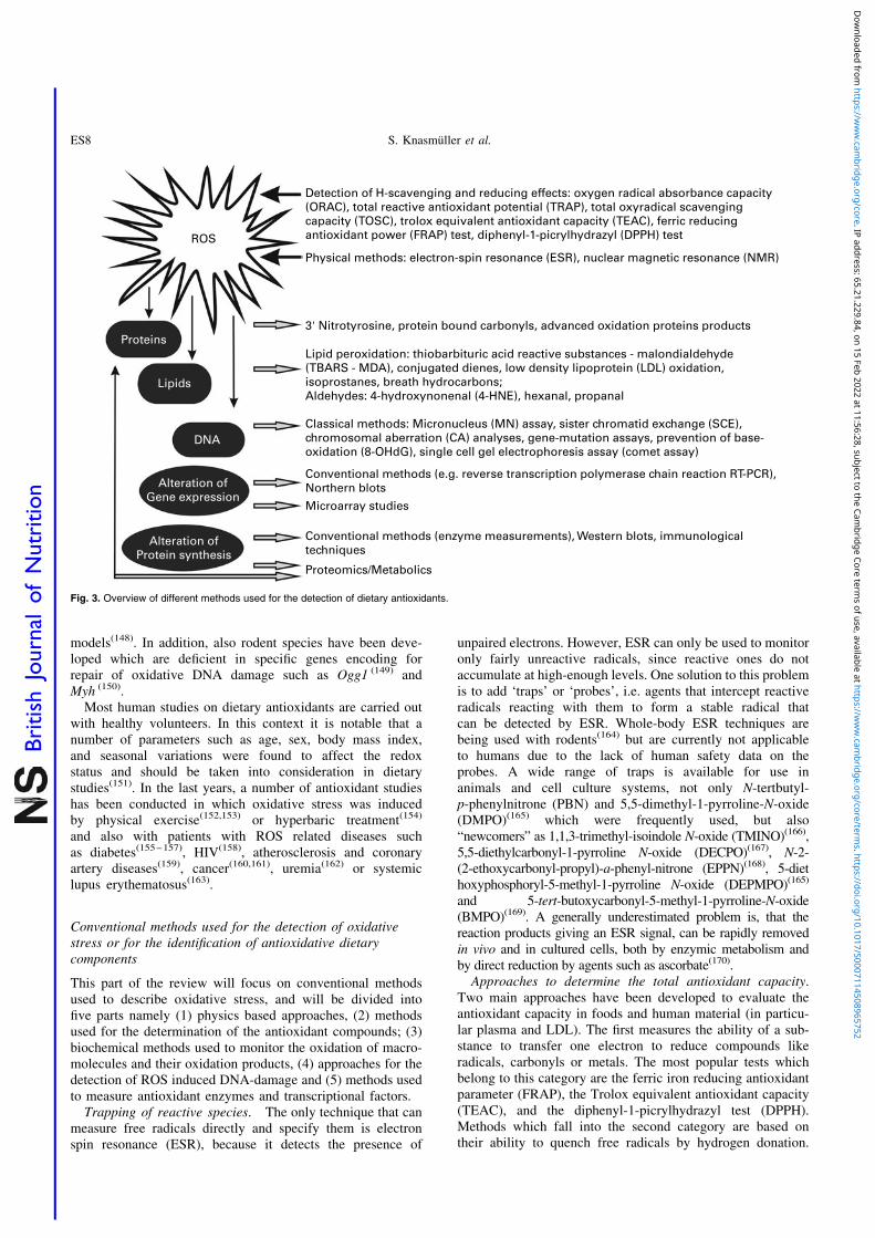
A large number of different techniques have been developedto monitor oxidative damage and its consequences; theseapproaches can be also used to identify dietary antioxidantsand their mode of action. These methods are often appliedin experimental systems in which oxidative stress is inducedby specific treatments or diseases. The most frequentlyemployed models are described in the next chapter; the fol-lowing sections describe physics–based and biochemicalmethods, techniques for the detection of oxidative DNA-damage, approaches used to investigate alterations of signal-ling pathways as well as the advantages and disadvantagesof -omics techniques (Fig. 3).
Induction of oxidative stress in biological systems
In experiments with subcellular fractions and in in vitroexperiments with cells, ROS are in most cases generated bychemical reactions, for example with the xanthine/xanthineoxidase system by hydroquinone oxidation(32,132,133) whichgenerate O2
2 . Transition metals such as copper and iron playa major role in Fenton type reactions thereby formingmainly OHz radicals. Another frequently used approach isthe use of chemicals such as H2O2, t-butyl-hydrogenperoxideor bleomycin which release O2
2 and OHz or of compoundssuch as menadione(134), paraquat(135), and plumbagin(136)
which form O2.Activated phagocytic cells produce oxygen radicals as part
of their defence system and a burst of ROS can be induced byexposing such cells to bacteria, particles or certain chemicals;one of the most powerful responses can be evoked with thetumour promotor phorbol myristate acetate(137).
Chemicals which generate ROS are rarely used in animalstudies due to their high reactivity. A more convenient wayto cause oxidative damage which has been also used in numer-ous in vitro experiments is ionising radiation. Indirectapproaches are feeding of vitamin E deficient diets(138), ironoverload(139) or inhalation of oxygen(140).
Organ specific inflammations can be induced with certainchemicals; for example liver cirrhosis with CCl4
(141) or thioa-cetamide(142). 2,4,6-Trinitrobenzene sulfonic acid, oxazoloneand dextran sodium sulfate are used in models for inflamma-tory bowel diseases(143); diabetes can be caused by the anti-biotic streptozotocin.
In the last years, a variety of genetically altered mice and ratstrains have been developed as models for ROS-related dis-eases for example animals which are deficient in specificSOD forms(144) and GST isozymes(144).
Disease related models include those of ataxia-telangiecta-sia(145), Alzheimer’s(146) and Parkinson’s(147) disease, ageing
Use of conventional and -omics based methods ES7
British
Journal
ofNutrition
Dow
nloaded from https://w
ww
.cambridge.org/core . IP address: 65.21.229.84 , on 15 Feb 2022 at 11:56:28 , subject to the Cam
bridge Core terms of use, available at https://w
ww
.cambridge.org/core/term
s . https://doi.org/10.1017/S0007114508965752
models(148). In addition, also rodent species have been deve-loped which are deficient in specific genes encoding forrepair of oxidative DNA damage such as Ogg1 (149) andMyh (150).
Most human studies on dietary antioxidants are carried outwith healthy volunteers. In this context it is notable that anumber of parameters such as age, sex, body mass index,and seasonal variations were found to affect the redoxstatus and should be taken into consideration in dietarystudies(151). In the last years, a number of antioxidant studieshas been conducted in which oxidative stress was inducedby physical exercise(152,153) or hyperbaric treatment(154)
and also with patients with ROS related diseases suchas diabetes(155 – 157), HIV(158), atherosclerosis and coronaryartery diseases(159), cancer(160,161), uremia(162) or systemiclupus erythematosus(163).
Conventional methods used for the detection of oxidativestress or for the identification of antioxidative dietarycomponents
This part of the review will focus on conventional methodsused to describe oxidative stress, and will be divided intofive parts namely (1) physics based approaches, (2) methodsused for the determination of the antioxidant compounds; (3)biochemical methods used to monitor the oxidation of macro-molecules and their oxidation products, (4) approaches for thedetection of ROS induced DNA-damage and (5) methods usedto measure antioxidant enzymes and transcriptional factors.Trapping of reactive species. The only technique that can
measure free radicals directly and specify them is electronspin resonance (ESR), because it detects the presence of
unpaired electrons. However, ESR can only be used to monitoronly fairly unreactive radicals, since reactive ones do notaccumulate at high-enough levels. One solution to this problemis to add ‘traps’ or ‘probes’, i.e. agents that intercept reactiveradicals reacting with them to form a stable radical thatcan be detected by ESR. Whole-body ESR techniques arebeing used with rodents(164) but are currently not applicableto humans due to the lack of human safety data on theprobes. A wide range of traps is available for use inanimals and cell culture systems, not only N-tertbutyl-p-phenylnitrone (PBN) and 5,5-dimethyl-1-pyrroline-N-oxide(DMPO)(165) which were frequently used, but also“newcomers” as 1,1,3-trimethyl-isoindole N-oxide (TMINO)(166),5,5-diethylcarbonyl-1-pyrroline N-oxide (DECPO)(167), N-2-(2-ethoxycarbonyl-propyl)-a-phenyl-nitrone (EPPN)(168), 5-diethoxyphosphoryl-5-methyl-1-pyrroline N-oxide (DEPMPO)(165)
and 5-tert-butoxycarbonyl-5-methyl-1-pyrroline-N-oxide(BMPO)(169). A generally underestimated problem is, that thereaction products giving an ESR signal, can be rapidly removedin vivo and in cultured cells, both by enzymic metabolism andby direct reduction by agents such as ascorbate(170).
Approaches to determine the total antioxidant capacity.Two main approaches have been developed to evaluate theantioxidant capacity in foods and human material (in particu-lar plasma and LDL). The first measures the ability of a sub-stance to transfer one electron to reduce compounds likeradicals, carbonyls or metals. The most popular tests whichbelong to this category are the ferric iron reducing antioxidantparameter (FRAP), the Trolox equivalent antioxidant capacity(TEAC), and the diphenyl-1-picrylhydrazyl test (DPPH).Methods which fall into the second category are based ontheir ability to quench free radicals by hydrogen donation.
ROS
Detection of H-scavenging and reducing effects: oxygen radical absorbance capacity(ORAC), total reactive antioxidant potential (TRAP), total oxyradical scavengingcapacity (TOSC), trolox equivalent antioxidant capacity (TEAC), ferric reducingantioxidant power (FRAP) test, diphenyl-1-picrylhydrazyl (DPPH) test
Proteins
Lipids
DNA
Alteration ofGene expression
Alteration ofProtein synthesis
Physical methods: electron-spin resonance (ESR), nuclear magnetic resonance (NMR)
3' Nitrotyrosine, protein bound carbonyls, advanced oxidation proteins products
Lipid peroxidation: thiobarbituric acid reactive substances - malondialdehyde(TBARS - MDA), conjugated dienes, low density lipoprotein (LDL) oxidation,isoprostanes, breath hydrocarbons;Aldehydes: 4-hydroxynonenal (4-HNE), hexanal, propanal
Classical methods: Micronucleus (MN) assay, sister chromatid exchange (SCE),chromosomal aberration (CA) analyses, gene-mutation assays, prevention of base-oxidation (8-OHdG), single cell gel electrophoresis assay (comet assay)
Conventional methods (e.g. reverse transcription polymerase chain reaction RT-PCR),Northern blots
Microarray studies
Conventional methods (enzyme measurements), Western blots, immunologicaltechniques
Proteomics/Metabolics
Fig. 3. Overview of different methods used for the detection of dietary antioxidants.
S. Knasmuller et al.ES8
British
Journal
ofNutrition
Dow
nloaded from https://w
ww
.cambridge.org/core . IP address: 65.21.229.84 , on 15 Feb 2022 at 11:56:28 , subject to the Cam
bridge Core terms of use, available at https://w
ww
.cambridge.org/core/term
s . https://doi.org/10.1017/S0007114508965752
Some scientists believe that these reactions are similar to thereaction mechanisms of antioxidants(171). The most popularmethods are the oxygen radical absorbance capacity test(ORAC), the total radical trapping antioxidant parameter(TRAP), the total oxidant scavenging capacity (TOSC)method (all measuring effects in the hydrophilic compartmentof the plasma) and the inhibition of linoleic acid and LDLoxidation.
Free radical quenching methods. The ORAC assay, whichis based on the work of Ghiselli et al. (172), Glazer(173) and Caoet al. (174) measures the antioxidant inhibition of ROOz inducedoxidations. Therefore, it reflects the classical H donatingability of antioxidants in the hydrophilic compartment. Theperoxyl-radical reacts with a fluorescent probe therebyforming a non-fluorescent product which can be quantitatedby following the fluorescence over time. In earlier studies,b-phycoerythrin was used as the fluorescent agent emittingin the visible region (Exc 495 nm, Em 595 nm), but due toshortcomings and inconsistencies of the results, fluorescein ordichlorofluorescein are currently used, since they are lessreactive and more stable(175). The antioxidative activity can beexpressed as the lag time or the net integrated area underthe fluorescence curve (AUC). ORAC values are reported asTrolox equivalents. Originally, the ORAC assay was limitedto the measurement of hydrophilic chain breaking anti-oxidant capacity. A newer protocol, in which lipophilic andhydrophilic compounds are selectively separated by extraction,allows now also the quantification of lipophilic antioxidantsusing a mixture of acetone and water(176). The advantage ofthe ORAC assay is that it can be automated. Convincing resultshave been obtained with 48 or 96 well plates coupled witha microplate reader(177). One important parameter is the tem-perature control (378C), as small temperature differencesdecrease the reproducibility of the test(178). A principal draw-back of the test is, that the effect of oxidation of the photo-receptor of the protein used does not necessarily reflectprotection against oxidative damage of the protein itself(179).
The TRAP assay, proposed by Wayner et al. (180) is basedon the use of 2,20-azobis(2,4-amidinopropane)dihydrochloride(AAPH), a hydrophilic azo-compound which generatesperoxyl-radicals. AAPH decomposes at 378C spontaneouslywith a known rate. Various substances like 2,2-azinobis(3-ethylbenzothiazoline-6-sulfonic acid) (ABTS), R-phycoery-thrin or 20-70-dichlorofluorescin (DCFH)(181 – 183) have beenused as oxidizable agents. A comprehensive review on thedifferent modifications has been published by Ghiselliet al. (184). The basic reactions of the procedure are similarto those of the ORAC assay. The probe reacts with ROOz radi-cals at low concentrations with a significant spectroscopicchange in between the native and the oxidized sample andno radical chain reaction beyond sample oxidation shouldoccur. The antioxidant capacity is determined as the timerequired to consume all antioxidants by extension of the lagtime for appearance of the oxidized probe when antioxidantsare present. TRAP values are usually expressed as the lagtime of the sample compared to the corresponding times forTrolox. The test is relatively complex to perform, requiresexperience and is time-consuming. The use of the lag phaseis based on the assumption that all antioxidants show a lagphase and that the lag phase corresponds to the antioxidativecapacity.
One drawback of TRAP and ORAC is the interference ofproteins which contribute by > 80 % to the total antioxidantcapacity(175,184). Therefore, Trolox can be used as an internalstandard or the samples must be deproteinized prior to themeasurements.
The TOSC assay was initially used for environmentrelated studies on marine organisms(185,186). It is based onthe inhibition of the radical-dependent formation of ethylenefrom ketomethiolbutyric acid by antioxidants. This procedurepermits testing against three different ROS species (i.e.peroxyl-, hydroxyl-radicals as well as peroxynitrite) withphysiological relevance and different reactivities. It can beconducted at physiological temperature and pH; non-linearconcentration-dependent activity variations can be examinedeasily and different types of antioxidant reactions (retardantor fast-acting) can be distinguished. However, high throughputanalyses are not possible and multiple injections of eachsample are required in order to observe ethylene formation.Further limitations are the multiple endpoints of calculated20, 50 and 80 % TOSC and the DT50 (first derivative ofTOSC of 50 %) since it was shown that there is no linearrelationship between the different multiple endpoints(187).
The chemiluminescence assay (CL) is a modification of theTRAP assay. Radical formation is followed by CL or photo-CL (PCL). CL is characterized by low emission intensityand by the fact that reactions with oxidants emit CL. Themost widely used marker is luminol(188,189), but also biumines-cent proteins like pholasin are becoming popular(190 – 192). Theantioxidant capacity is the time of depressed light emission,which is measured at 10 % recovery of light output.
Recently Popov et al. (193) have described the PCL, a com-mercial test system termed PHOTOCHEM for the determi-nation of the integral antioxidative capacity towards O2
(193).In a strict sense, the method measures antiradical capacity.In contrast to many other assays used to determine AOC,this procedure requires no standardisation of the pH and ofthe temperature. However, to date, the system is only mar-keted by one company (Analytic Jena, Germany) and reagentsfor the hydrophilic and lipophilic assays are only availablefrom the manufacturer; furthermore, a high throughput is notpossible. Ascorbic acid is normally used for the determinationof hydrophilic and Trolox for the lipophilic antioxidativecapacity.
Low-density lipoprotein (LDL) oxidation is based on theautoxidation of linoleic acid or LDL which is inducedin vitro mainly by Cu2þ or some other azo-initiators(194 – 196).LDL-oxidation is of higher physiological relevance whentested under in vivo conditions and not ex vivo. The oxidationis monitored at 234 nm for conjugated dienes or by peroxidevalues for lipid hydroperoxides. LDL has to be freshly isolatedfrom blood which is a time- and material-consuming pro-cedure which requires ultracentrifugation. During the prep-aration, low temperature and light protection areessential(197). Further, conjugated dienes can be formed in pre-sence of polyunsaturated fatty acids.
Recently, fluorescence and UV based ELISA assays withplasma became available, for which no complicated andtime consuming LDL isolation is needed. This methods canalso be used for larger human trials. Also the procedure deve-loped by Holvoet et al. (198) who measured oxidized LDLlevels by a competitive ELISA utilizing a specific murine
Use of conventional and -omics based methods ES9
British
Journal
ofNutrition
Dow
nloaded from https://w
ww
.cambridge.org/core . IP address: 65.21.229.84 , on 15 Feb 2022 at 11:56:28 , subject to the Cam
bridge Core terms of use, available at https://w
ww
.cambridge.org/core/term
s . https://doi.org/10.1017/S0007114508965752

monoclonal antibody (mAb-4E6) based on UV is employedquite often at present(198). The AOC is determined in allthese experiments either as AUC or as the lag time until theantioxidants are consumed. An important modification wasdeveloped by Frankel et al. (199) who determined the second-ary oxidation product hexanal from LDL. Hexanal waschosen, since it is the major oxidation product of n-6 fattyacids and is monitored with head space gas chromatography,the percentage inhibition of hexanal formation is used as aparameter for AOC. In many ex vivo studies LDL was iso-lated, subsequently the substances were added and tested ontheir ability to delay oxidation. This scenario does not reflectin vivo conditions. Furthermore, not all oxidation inducerswhich are used ex vivo can be used for in vivo testing(179).
The Crocin bleaching assay monitors the protection ofAAPH-induced crocin bleaching, by antioxidants(200). Crocinis a mix of natural pigments and absorbs, similar to carote-noids, at 450 nm. Therefore, the interpretation of the resultscan be complicated in experiments with food samples.Initially, the test was used for the analysis of plasmasamples(201). One of its limitations is that crocin is notcommercially available, but high sample throughput withmicroplates is possible.Single electron transfer methods. In these assays, the
sample itself is an oxidant that abstracts an electron fromthe antioxidant, thereby causing colour changes which are pro-portional to the AOC. When the change of absorbance isplotted against the antioxidant concentration, the slope ofthe curve reflects the total reducing capacity. In contrast tothe methods described in the last chapter, no oxygen radicalsare present in the system; therefore, it can be assumed that thereducing capacity is equal to the antioxidant capacity.
The TEAC-assay is a spectrophotometric test which wasdeveloped by Miller et al. (202). 2,2-Azinobis(3-ethylben-zothiazoline-6-sulfonic acid) (ABTS) is oxidized by ROOz toa green-blue radical cation. The ability of antioxidants todelay colour formation is expressed relative to Trolox. Orig-inally, the test used metmyoglobin and H2O2, and the ABTSradical was measured at 734 nm. Meanwhile, various modifi-cations have been developed(203,204). After generation of theABTS radical, the sample to be tested is added, subsequentlyother chemicals like manganese dioxide, ABAP, potassiumpersulfate or enzymes are used to generate the ABTS rad-ical(175,202,203,205). Temperatures higher than 378C, which arenot physiological and different absorption maxima (415, 645734 or 815 nm) are frequently used. Depending on the proto-col, the decrease or increase in ABTS radical absorbance inpresence of the test sample or Trolox at a fixed time point ismeasured and the antioxidant capacity is calculated asTrolox equivalents. ABTS is not a physiological substance.It reacts fast with aqueous and organic solvents and substanceswith a low redox potential show a good response. Therefore,phenolic compounds or ascorbic acid react quite well withABTS whereas lipophilic compounds respond more weakly.The test can be adapted to microplates and is not restrictedto a narrow pH range, but high haemoglobin concentrationin the plasma may interfere with the measurements.
The FRAP assay determines the reduction of 2,4,6-tripyri-dyl-s-triazine (TPTZ) in plasma to a coloured product(206)
and has also been adapted for food samples(207,208). Similarto the TEAC assay, compounds with a redox potential
,0·7 V are detected. FRAP not enable to monitor compoundsthat quench radicals like proteins or thiol compounds such asglutathione (similar to all the “reducing assays”) and cantherefore underestimate the AOC. In order to maintain thesolubility of Fe, the assay is conducted at acidic conditions(pH of 3·6). Most redox reactions take place within a few min-utes, therefore both tests consider most of the substanceeffects (like polyphenols) but not all, since some substanceshave longer reaction times(175). This was recently shown forpolyphenols like caffeic-, tannic-, ferulic- or ascorbic acids,where the absorption steadily increased for hours(209). Sincethe test reflects only the reducing potential and does not con-sider H transfer, it should only be considered in combinationwith other methods to give a more complete picture. Similarto the TEAC, it is a relatively easy test procedure, can bedone manually or fully automated and requires no expensiveequipment.
The copper reducing assay (CUPRAC, AOP-90) is a modi-fication of the FRAP in which iron is replaced by Cu; Cu2þ isreduced to Cu1þ(210). The assay is conducted at 490 nm withbathocuprine, or at 450 nm with neocuproine. Results areexpressed as uric acid equivalents. Since copper has a lowerredox potential than iron, but enhances the redox cyclingpotential, its pro-oxidative potential can be more sensitive(175).The limitations regarding the underestimation of slower reac-tive molecules is similar to the other assays but one of itadvantages is that almost every antioxidant including thiolscan be detected(175).
The DPPH (2,2-diphenyl-1-picrylhydrazyl) radical used inthe DPPH-assay is stable, deep purple, commercially avai-lable and has not to be generated. The test was developedby Brand-Williams et al. (211). The loss of DPPH colour at515 nm after reaction with test compounds is measuredeither by decrease of absorbance or by electron spin reson-ance(212). The concentration of a 50 % decrease of the DPPHradical is defined as EC50, the time to reach it as TEC50.Since the DPPH assay uses a wavelength of 515 nm, it caninterfere with substances with a similar spectrum like carote-noids(213). The test considers both, electron transfer as wellas hydrogen (H) transfer reactions with the focus on theprior. Again, smaller molecules have better access to the rad-ical site and contribute to a higher extent to the AOC(175). Thetest procedure itself is quite simple and fast and requires onlya spectrophotometric device.
For all the electron transfer tests it can be assumed that they“overestimate” smaller molecules and hydrophilic substanceslike ascorbic acid, uric acid or polyphenols and reflects notalways the situation in the organism(214).
From the evaluation of the different assays it becomes clearthat no single test reflects the overall antioxidative capacity ofantioxidants. Both hydrophilic and lipophilic activities mustbe considered, as well as H transfer and single electron trans-fer mechanisms and additional tests which reflects the inacti-vation of various reactive oxygen/nitrogen species areneeded to fully estimate the AOC (Table 1).
Oxidation of macromoleculesBiomarkers of lipid oxidation. Cell membranes are highly
susceptible to LP due to their specific composition which ischaractesized by high amounts of polyunsaturated fatty acids(PUFAs)(215). LP oxidation is a chain reaction (see above)and leads to structural and functional damage of membranes
S. Knasmuller et al.ES10
British
Journal
ofNutrition
Dow
nloaded from https://w
ww
.cambridge.org/core . IP address: 65.21.229.84 , on 15 Feb 2022 at 11:56:28 , subject to the Cam
bridge Core terms of use, available at https://w
ww
.cambridge.org/core/term
s . https://doi.org/10.1017/S0007114508965752

as well as to the formation of lipid hydroperoxides which areunstable and degrade to various secondary oxidation products.
The formation of malondialdehyde (MDA) is the mostwidely used parameter of PUFA peroxidation in the thiobarbi-turic acid-reacting substances (TBARS) assay. One of theoldest and still most widely used methods is based on the pre-cipitation of protein nearly at boiling conditions(216). Thesamples are heated for 1 h with TBA at low pH and thepink chromogen formed absorbs at 532 nm. The sample prep-aration has been criticized since it is far from physiologicalconditions and not the free MDA in the original sample ismeasured but the amount generated by decomposition oflipid peroxides during heating(217). Furthermore, it is knownthat also other compounds like sugars, amino acids or bilirubinare able to react with TBA(218). The sensitivity of the test canbe increased by combining it with HPLC to separate suchcompounds before acidic heating(219).
In the last few years, several innovations have been intro-duced to improve the specificity of the test and to reduceknown bias. In particular the temperature at the deproteniza-tion step has been reduced to physiological conditions. Inaddition, several methods have been developed which do notrequire derivatization(220,221) or new derivatization agentslike 2,4-dinitrophenylhydrazine(222) or diaminonaphtha-lene(223). Very recently, GC/MS based methods have beendeveloped which possess high sensitivity showing an overesti-mation of MDA levels(224,225). Although it is questionablewhether MDA measurements are a reliable method for LP,it is well documented by a large number of studies thatincreased levels are found in patients with ROS relateddiseases(226).
The first step of PUFA oxidation is the conjugation ofdouble bonds leading to formation of conjugated dienes(CD) which absorb at around 234 nm. They can either beabsorbed by lipids but also in plasma samples. The plasmapreparation is more physiological and requires no heat treat-ment. In plasma samples, CD are usually analysed withHPLC–UV detection(227). Determination of the diene levelscannot be used alone to describe oxidative stress, but whenmeasured with HPLC–CD, the findings can support resultsobtained with other more reliable parameters of oxidative
stress. Although MDA and CD are both primary oxidationproducts, they can develop differently in the same sampledue to different mechanisms of formation(228).
Isoprostanes are stable oxidation products from arachidonicacid, initially formed from phospholipids and released intocirculation before the hydrolyzed form is excreted inurine(229). A large number of endproducts can theoreticallybe generated but interest has focused mainly on F2a-isopros-tanes(230); the must promising marker for oxidative stress/injury being 8-prostaglandin F2a
(231). At present it is regardedas one of the most reliable markers of oxidative stress,although the presence of detectable concentrations ofisoprostanes in biological fluids requires continuous lipid per-oxidation(232). Several favourable characteristics make iso-prostanes attractive as reliable markers for oxidative stress,i.e. they are specific oxidation products, stable, present indetectable quantities, increased strongly at in vivo oxidativestress, and their formation is modulated by antioxidants(231).Various approaches such as gas chromatography–mass spec-trometry (GC–MS), GC–tandem MS, liquid chromato-graphy–tandem MS and immunoassays are available for thedetection of F2a-isoprostanes(233). The first results were pro-duced by use of the MS technique, and various isoprostanescan be separated with this method(233). Recently, immunoas-says have been developed which correlate apparently quitewell with the results obtained with GC–MS measurementsin urine but some discrepancies might occur with plasmasamples when they were not tested on cross reactivity withother prostaglandin metabolites(232). Nevertheless, their usemight be appropriate in intervention studies with variousblood samplings from the same subject, thereby focusing noton absolute levels but on relative changes.
The measurement of breath hydrocarbons is a non-invasivemethod which allows to determine LP through exhaled breathby measuring trace volatile hydrocarbons(234). Ethane formationresults from n-3 oxidation, pentane formation is caused by n-6oxidation. Although the data reported on their consistencyto describe LP are quite convincing, the limiting factor istheir detection. They are mainly employed with GC–FID, butone concern is the background level in the breath sincebacteria were shown to produce significant amounts of theses
Table 1. Comparison of methods used to determine the total antioxidant capacity (TAC)
Test systems Principle Endpoint Biological relevance Simplicity/throughput* Number of studies with dietary antioxidants†
ORAC H-transfer Lag time AUC þþþ þþ /þþþ 156TRAP H-transfer Lag time þþþ 2 2 2 /2 2 270 TRAPTOSC H-transfer AUC DT50 þþ 2 /2 2 2 23 TOSCCL/PCL Not known Lag time AUC þþ þ /2 2 CL 2445
PHOTOCHEM 11LDL Oxidation H-transfer Lag time þþþ UC: 2 2 /2 2 LDL-oxidation 2357
2 (in vitro) ELISA: þþ /þþ
TEAC Electron-transfer D Optical density 2 þþ /þþ TEAC 217TAC 133
FRAP Electron-transfer D Optical density 2 2 þþþ /þþþ FRAP 173CUPRAC Electron-transfer D Optical density 2 þþþ /þþþ 6DPPH Electron-transfer D Optical density 2 þ /þ DPPH 1061
CL, chemiluminescence assay; CUPRAC, cupric reducing antioxidant capacity; DPPH, 2,2-diphenyl-2-picrylhydrazyl assay; ELISA, enzyme-linked immunosorbent assay;FRAP, ferric reducing ability of plasma; LDL, low density lipoprotein; ORAC, oxygen radical absorbance capacity test; PCL, photo-chemiluminescence assay; TEAC, troloxequivalent antioxidative capacity; TOSC, total oxidant scavenging capacity; TRAP, total radical trapping antioxidant parameter.
* þ ,þþ ,þþþ , positive; 2 , 2 2 , 2 2 2 , negative; UC, ultracentrifugation.† Number of antioxidant studies conducted with the different methods identified by use of a computer aided search (Scopus database).
Use of conventional and -omics based methods ES11
British
Journal
ofNutrition
Dow
nloaded from https://w
ww
.cambridge.org/core . IP address: 65.21.229.84 , on 15 Feb 2022 at 11:56:28 , subject to the Cam
bridge Core terms of use, available at https://w
ww
.cambridge.org/core/term
s . https://doi.org/10.1017/S0007114508965752

hydrocarbons in vivo (235). Furthermore, the separation of differ-ent hydrocarbons is not easy due to similar boiling points(236).Aldehydes represent stable products of PUFA oxidation.
4-Hydroxynonenal (4-HNE) and hexanal are mainly formedby n-6 fatty acid oxidation, while propanol and 4-hydroxyhexe-nal result from n-3 fatty acid oxidation(199,237,238). High concen-trations of 4-HNE have been shown to trigger well-known toxicpathways such as induction of caspases, the laddering of geno-mic DNA, and release of cytochrome c from mitochondria,which may lead to cell death(239). The most frequently usedmethods for determination of the aldehydes are GC–MS,GC–head space or HPLC(240). Also polyclonal or monoclonalantibodies directed against 4-HNE-protein conjugates are nowfrequently used for 4-HNE measurements(241).Biomarkers of protein oxidation. Markers of protein oxi-
dation are less frequently used than lipid oxidation parameters(Table 2). They are mainly applied in combination with LPmarkers, although their formation has been associated withseveral diseases(242).
Formation of protein bound carbonyls is most abundantendpoint used to monitor protein oxidation(243,244) by a con-ventional colorimetric assay using 2,4-dinitrophenylhdra-zine(245). The test is easy to perform, but large quantities ofsolvents are required. Recently, an ELISA method has beendeveloped and the results correlated well with the spectropho-tometric method(246).Advanced oxidation protein products (AOPP) are predomi-
nantly albumin and its aggregates damaged by oxidativestress(247). They contain abundantly dityrosines which causecrosslinking, disulfide bridges and carbonyl groups and areformed mainly by chlorinated oxidants such as hypochloricacid and chloramines resulting from myeloperoxidaseactivity(248). AOPP have several similar characteristics asadvanced glycation endproducts-modified proteins. Inductionof proinflammatory activities, adhesive molecules and cytokinesis even more intensive than that caused by advancedglycation end products (AGEs). They are referred to as markersof oxidative stress as well as markers of neutrophil acti-vation(249). Protein oxidation products mediated by chlorinatedspecies (HOCl) generated by the enzyme myeloperoxidasewere found in the extracellular matrix of human atheroscleroticplaques and increased levels of advanced oxidation proteinproducts were postulated to be an independent risk factor forcoronary artery disease(250,251). AOPPs are expressed as chlora-mine-T equivalents by measuring absorbance in acidicconditions at 340 nm in presence of potassium iodide. The testis easy to perform and can be carried out with microprobes.Markers of oxidative DNA damage used in studies with diet-
ary antioxidants. During the last fifty years, a broad varietyof genotoxicity test procedures have been developed which areused for routine testing of chemicals, in environmentalresearch and also in human studies concerning the impact ofoccupational exposure, lifestyle factors and nutrition onDNA-integrity. Due to the conservative structure of the gen-etic material, mutagenicity experiments can be carried outwith a broad variety of indicator organisms including bacteria,yeasts, plants, invertebrates including Drosophila, laboratoryrodents and also with cultured mammalian cells(252 – 254).The advantages and limitations of the different methods forthe detection of DNA-protective dietary factors have beendescribed by Knasmuller and coworkers(255 – 257). One of the T
able
2.
Main
bio
mark
ers
for
lipid
and
pro
tein
oxid
ation
Test
Mark
er
Meth
ods
Bio
logic
alsyste
mS
implic
ity/t
hro
ughput/
import
an
ce
Num
ber
of
stu
die
sw
ith
die
tary
antioxid
ants
*
TB
AR
S/M
DA
Lip
idoxid
ation
HP
LC
Pla
sm
a/s
eru
m,
cells
HP
LC
:þ
/þ/2
TB
AR
S-3
570;
16
%iv
;53
%an;
31
%hu
GC
–M
SG
C–
MS
:2
/þ/þ
MD
A-1
6255;
14
%iv
;53
%an;
33
%hu
Conju
gate
ddie
nes
Lip
idoxid
ation
HP
LC
Foods,
pla
sm
a,
cells
þ/þ
/2C
onju
gate
ddie
nes
with
HP
LC
-68;
29
%iv
;23
%an;
48
%hu
F2a-I
so-p
rosta
nes
Lip
idoxid
ation
GC
–M
SP
lasm
a/s
eru
m,
urine
GC
–M
S:2
/2/þ
F2-I
sopro
sta
ne-2
60;
12
%iv
;34
%an;
54
%hu
ELIS
AE
LIS
A:2
/þ/þ
Bre
ath
hydro
carb
ons
Lip
idoxid
ation
GC
Exhale
dair
þ/2
/þ299;
24
%an;
76
%hu
Ald
ehydes
Lip
idoxid
ation
HP
LC
Pla
sm
a,
LD
L,
urine
HP
LC
:þ
/þ/þ
4-H
NE
-389;
12
%iv
;52
%an;
36
%hu
GC
–M
SG
C–
MS
:2
/2/þ
Pro
tein
bound
carb
onyls
Pro
tein
oxid
ation
Photo
mP
lasm
aP
hoto
m:þ
/þ/2
Pro
tein
carb
onyls
-355;
26
%iv
;31
%an;
43
%hu
ELIS
AE
LIS
A:þ
/þ/2
AO
PP
Pro
tein
oxid
ation
ELIS
AP
lasm
a,
urine
þ/þ
/þA
OP
P-1
96;
19
%iv
;12
%an;
69
%hu
AO
PP
,advanced
oxid
ation
pro
tein
pro
ducts
;C
D,
conju
gate
ddie
nes;
GC
/MS
,gas
chro
mato
gra
phy
–m
ass
spectr
om
etr
y;
ELIS
A,
enzym
e-lin
ked
imm
unosorb
ent
assay;
4-H
NE
,4-h
ydro
xynonenal;
HP
LC
,hig
h-p
erf
orm
ance
liquid
chro
ma-
togra
phy;
MD
A,
malo
ndia
ldehyde;
TB
AR
S,
thio
barb
ituric
acid
reactive
substa
nces.
Dis
trib
ution
of
the
tota
lstu
die
sto
invitro
(iv),
anim
al
(an)
and
hum
an
(hu)
stu
die
s:
iv%
,perc
enta
gein
vitro
;an%
,perc
enta
ge
anim
al
stu
die
s;
%hu,
perc
enta
ge
hum
an
stu
die
s.
*T
he
num
ber
of
antioxid
ant
stu
die
sconducte
dw
ith
the
diffe
rent
meth
ods
was
assessed
by
use
of
acom
pute
raid
ed
searc
h(S
copus
data
base).
S. Knasmuller et al.ES12
British
Journal
ofNutrition
Dow
nloaded from https://w
ww
.cambridge.org/core . IP address: 65.21.229.84 , on 15 Feb 2022 at 11:56:28 , subject to the Cam
bridge Core terms of use, available at https://w
ww
.cambridge.org/core/term
s . https://doi.org/10.1017/S0007114508965752

main problems (which is encountered also relevant for studiesof the effects of dietary antioxidants) encountered in experi-ments with mammalian cells and lower organisms concernsthe inadequate representation of the metabolism of the testcompounds which may lead to results which cannot beextrapolated to humans. Nevertheless, all these methodshave been used to study oxidative DNA-damage and toinvestigate putative protective effects of phytochemicals.At present, the most widely used endpoints are gene muta-tion assays with bacteria and mammalian cells (Salmonellatyphimurium/microsome test, HPRT gene mutation assay),chromosome analyses in metaphase cells which can be con-ducted with stable cell lines (e.g. CHO, V79) and lymphocytesin vitro but can be also scored in in vivo and ex vivo exper-iments with blood cells. Another important endpoint aremicronuclei which are formed as a consequence of chromo-some breakage (clastogenicity) and aneuploidy and are lesstime consuming to evaluate as chromosomal aberra-tions(256,258). The most frequently used approaches to monitorantioxidant effects of dietary factors are described in the sub-sequent chapters.
The most widely used bacterial mutagenicity test procedureis the Salmonella/microsome assay, which has been developedby B. Ames in the 1970s(259). The test is based on the detectionof back mutations in specific genes which encode for histidinebiosynthesis. One of the disadvantages of the initial set of testerstrains was, that none of them was highly sensitive towards oxi-dative effects, therefore new polyplasmid strains (TA102 andTA104) were constructed which are in particular suitable forthe detection of mutagenic effects caused by ROS(260). Sincethe target gene is located in these strains (in contrast to the clas-sical tester strains) on plasmids, it can be easily lost and due tothe high spontaneous reversion rates many groups encountereddifficulties with these derivatives. Nevertheless, the Salmonellastrains are at present widely used in antioxidant experiments;mutations are induced either by radiation or chemically andputative protective compounds or complex mixtures areplated on histidine free selective media plates together withthe indicator bacteria. After incubation, the differences in thenumbers of hisþ revertants serves as an indication of protectiveeffects. The test procedure has been standardised for routinetesting of chemicals (see for example OECD guideline471(261)) and the criteria which have been defined (sufficientnumber of plates, inclusion of positive and negative controls,testing of different doses) can also be applied for the detectionof antioxidants. One of the problems which affects thereliability of the test results, concerns the fact that false positiveeffects may be obtained with compounds which cause bacteri-cidal or bacteriostatic effects since these parameters are notmonitored under standard conditions. A typical example arethe protective effects seen with cinnamaldehyde(262). Withcomplex dietary mixtures, difficulties may be encountereddue to their histidine contents(263).
Apart from the Salmonella/microsome assay, also a numberof other bacterial genotoxicity tests have been developedwhich are less frequently used, e.g. assays, based on thescoring of backward or forward mutations with E. coli strains,tests based on the induction of repair processes, such as umuand SOS chromotest or differential DNA-repair assays basedon the comparison of the survival of strains differing intheir DNA-repair capacity(256).
Single cell gel electrophoresis (SCGE) assays are based onthe determination of DNA migration in an electric field whichleads to formation of COMET shaped images. In the initialversion, the experiments were carried out under neutralconditions which allowed only the detection of doublestrand breaks(264). Subsequently, Tice et al. (265) and Singhet al. (266) developed protocols for experiments in which thecells are lysed under alkaline conditions (pH . 13), whichenables the additional detection of single strand breaks andapurinic sites. One of the main advantages of this testprocedure is, that it does not require cell divisions (whichare a prerequisite for gene mutation and micronucleus exper-iments), therefore only short incubation periods are requiredso that not only stable cell lines can be used for in vitrostudies but additionally also primary cells from differentorgans. Furthermore, it is also possible to carry out in vivoexperiments with rodents and to study effects in a broad var-iety of tissues(267). In most human studies, peripheral lympho-cytes have been used as target cells, few experiments wereconducted with exfoliated epithelial cells(268 – 271) which areproblematic due to their low viability. Very recently, alsoresults from a human intervention trail with antioxidantswere reported in which bioptic material from the colon wasanalysed(272).
Collins et al. developed in the 1990s(273) a protocol inwhich isolated nuclei are treated with lesion specific enzymes(endonculease III, formadidopyrimidine glycosylase, FPG).This approach has been used intensely to study the preventionof endogenous formation of oxidised purines and pyrimidines.However, it is crucial in these experiments to determine theoptimal amounts of the enzymes due to their instability.More recently, Collins and coworkers published an additionalmodified version of the SCGE test which enables to monitorthe impact of compounds on the repair capacity of thecells(274,275) and it was shown in a few model studies that anti-oxidants may alter DNA-repair processes(276,277). In order toobtain information concerning alterations of the sensitivitytowards exogenous ROS mediated damage, it is possible totreat the cells with H2O2 other radical generating chemicalsor radiation (ROS-challenge).
In the guidelines published by Tice et al. (265) and Hartmannet al. (278), a number of criteria are defined which are essentialto obtain reliable results. They concern for example thenumber of parallel cultures and cells which are required inin vitro studies, the number of animals and treatment periodsand also adequate statistical methods. It was generally agreed,that different parameters such as tail lengths of the comets, aswell as percentage DNA in tail and tail moment are acceptableand that apart from automated image analysis systems alsomanual scoring methods are acceptable(279). An importantpoint which is also relevant for antioxidant studies concernsthe fact that multiple doses should be tested to substantiateeffects and that it is necessary to monitor acute toxic effects.It is well documented that cell death leads to degradation ofDNA, therefore it is essential to determine the viability ofthe cells after treatment with appropriate methods in in vitroexperiments and to monitor toxic effects in inner organs byhistopathology(279) and exclude conditions under whichstrong acute effects are observed in animal experiments.These criteria are fulfilled in most recent studies, but not ina number of older investigations.
Use of conventional and -omics based methods ES13
British
Journal
ofNutrition
Dow
nloaded from https://w
ww
.cambridge.org/core . IP address: 65.21.229.84 , on 15 Feb 2022 at 11:56:28 , subject to the Cam
bridge Core terms of use, available at https://w
ww
.cambridge.org/core/term
s . https://doi.org/10.1017/S0007114508965752

The SGCE technique has been used in numerous in vitroinvestigations to study antioxidant effects of individual com-pounds and complex mixtures in stable cell lines and inhuman lymphocytes. Typical examples are investigations offlavonoids by Anderson and coworkers with blood andsperm cells(280,281), experiments concerning antioxidanteffects of coffee and its constituents(282), studies on the protec-tive effects of tea catechins(283,284) to name only a few. Alsoquite a substantial number of in vivo studies with labora-tory animals have been published, e.g. with carotenoids(285),quercetin(286), vitamins E and C(287) and garlic oil(288).
The results of human studies are described in the reviews ofMoller & Loft(289 – 291). Most of them were conducted as inter-vention trails which have the advantage that inter-individualvariations can be reduced. In total, 76 investigations havebeen published since the first trial was conducted by Pool-Zobel et al. in 1997(292). Evidence for antioxidant effectswas observed in approximately 64 % of the studies (25/39);the highest number of protective effects concerned the preven-tion of endogenous formation of oxidised pyrimidines, (39 %of the trials, 7/19), while in few studies evidence for reducedsensitivity towards ROS and protection against formation ofoxidised purines was found. The lowest rate of protectiveeffects was seen when the SCGE analyses were carried outunder standard conditions (i.e. without ROS challenge andenzymes). Clear evidence for protective effects was observedfor example in experiments with lycopene and tomatoes(293),cruciferous and leguminous sprouts(294) and carotenoid sup-plementation(295). In our own experiments we found strongprotective properties of sumach (a common spice) and itsmain active principle gallic acid(296), wheat sprouts(101), afterconsumption of coffee(282) and in experiments with Brusselssprouts(297). The latter observation is of particular interest;since no effects were seen in a large intervention trial inwhich the participants consumed 600 g of fruits and veg-etables/d(298) our findings may be taken as an indication thatcoffee intake may contribute to a higher extent to ROS protec-tion than plant derived foods.
Moller & Loft (2004) discuss in their papers also the possi-ble shortcomings of intervention trails and emphasize theimportance of controlled study designs which should includea sufficient number of participants and additionally alsoeither placebo groups or washout periods(290). These latter cri-teria are not fulfilled in some of the older studies.Micronucleus (MN) experiments have nowadays largely
replaced conventional chromosomal aberration (CA) analyses(which have been used extensively in the past to studyprotective effects of antioxidants towards radiation andchemically induced DNA-damage) as they are less time con-suming and laborious. Both endpoints can be used in in vitroexperiments with cell lines and lymphocytes; one of the mostpromising newer developments is the use of human derivedliver cell lines (HepG2, HepG3 etc). HepG2 cells haveretained the activities of drug metabolising enzymes includ-ing those which metabolise plant specific antioxidants andthus may reflect the effects in humans more adequatelythan cell lines which are commonly used in genetic toxi-cology (for reviews see(299 – 301)).
The most widely procedure used in animal experiments isthe bone marrow MN assay. Since it is problematic toinduce MN with chemical oxidants in vivo, most experiments
were conducted with radiation. Standardised guidelines havebeen published for this test system, e.g. by OECD(302), andone of the most important parameters relevant for AO studiesis the treatment period. It is also notable, that one of the mainshortcomings of this test is due to the fact that reactivemolecules may not reach the target cells; this explains whynegative results were obtained with a number of potentgenotoxic carcinogens, also antioxidant effects may not bedetected due to this limitation. Typical examples for AOstudies in which this assay was used are experiments withepigallocatechin gallate, vitamin C, lipoic acid, ubidecarenoneor coenzyme Q(10)(303 – 308).
The development of the cytokinesis block micronucleus(CBMN) method by Fenech et al. (309,310) allows to monitorMN formation in peripheral blood cells in vitro and can bealso used in dietary human studies with lymphocytes. It isbased on the use of the mitogen phytohaemagglutinin (whichstimulates nuclear division), in combination with cytochalasinB (which stops cellular division), so that MN can be scoredin binucleated cells. Typical examples for the use of thisapproach are supplementation studies with vitamin C andintervention trials with red wine(311 – 313), in vitro experimentswith wine specific phenolic compounds(314) as well as testswith the antioxidant drugs amifostine and melatonin(315).
More than 100 different oxidative modifications of DNAhave been described in the literature(316) and it is notablethat oxidative adducts occur at a frequency of 1 or moreorders of magnitude higher than non-oxidative adducts(317).The dominant oxidative modification of DNA is 8-hydroxy-lation of guanine, which leads to formation of 8-oxo-7,8-dihy-dro-2-deoxyguanosine (8-oxodG). In most studies, thisoxidation product was measured in urine and in leukocytes,but it is also possible to monitor it in various inner organs,e.g. in liver, lung and kidneys(318 – 321). The first investigationswere conducted in the late 1980s(322) and a number of differ-ent methods have been developed namely gas chromatographycoupled with mass spectrometry(323), liquid chromatographyprepurification followed by gas chromatography coupledwith mass spectrometry(324), liquid chromatography coupledwith tandem mass spectrometry(325), high-performance liquidchromatography with electrochemical detection(326 – 328),high-performance liquid chromatography with mass spec-trometry(329) and enzyme-linked immunosorbent assay basedmethods(330).
It is well known that guanine is easily prone to oxidation ifstringent precautions are not taken during preparation ofsamples for analysis(331), therefore much of what is measuredcould be artefacts. In order to prepare compounds for analysisby gas chromatography coupled with mass spectrometrywhich enables the simultaneous analysis of a number of diffe-rent adducts(332), DNA is chemically hydrolysed and con-verted into a volatile form by derivatization with a suitablereagent. This reaction is normally carried out at high tempera-ture. During the derivatization reaction, oxidation occurs to anextent that overwhelms the amount originally present(333 – 335).Generally speaking, gas chromatography coupled withmass spectrometry estimates of DNA oxidation have beenhigher than high-performance liquid chromatography withelectrochemical detection estimates by about a factor of10(333). The cause of this difference (an overestimate due toartificial oxidation with gas chromatography coupled with
S. Knasmuller et al.ES14
British
Journal
ofNutrition
Dow
nloaded from https://w
ww
.cambridge.org/core . IP address: 65.21.229.84 , on 15 Feb 2022 at 11:56:28 , subject to the Cam
bridge Core terms of use, available at https://w
ww
.cambridge.org/core/term
s . https://doi.org/10.1017/S0007114508965752

mass spectrometry vs an underestimate due to inefficientenzymatic digestion with high-performance liquid chromato-graphy with electrochemical detection) has now beensettled(335). In addition, GC/MS analyses are difficult toperform, labour-intensive and exhibit poor sensitivity orinadequate specificity when they are used to test urinarysamples(336). After enzymatic hydrolysis of DNA to nucleo-sides it is possible to analyse the probes with chromatographyby high-performance liquid chromatography(328). 8-Oxo-dGand its corresponding base 8-oxo-Gua are electrochemicallyactive, lending themselves to sensitive electrochemicaldetection. The relative simplicity and sensitivity of high-performance liquid chromatography with electrochemicaldetection of 8-oxo-dG have made it the most popularmethod for monitoring of DNA-oxidation(337). The high-performance liquid chromatography with electrochemicaldetection method itself has been criticized for its highvariability(338,339). Estimates of the ratio of 8-oxodG to dGhave ranged from approximately 0·25 £ 1025 to > 1024,possibly due to artificial oxidation(337,338). Dreher &Junod(340) described in detail how the formation of artefactsthat occurs with high-performance liquid chromatographywith electrochemical detection can be avoided. Also thismethod is prone to oxidation of samples during preparationfor analysis but by use of antioxidants and anaerobic con-ditions during isolation and hydrolysis of DNA, these effectscan be minimised.
Immunoassays have, by far, demonstrated the greatestversatility in terms of the matrices to which they can be applied(urine, serum, plasma, cell culture medium, DNA hydrolysates),and simplicity of use(330), but comparisons with urinary8-oxodG levels by chromatographic techniques revealedstrong discrepancies of the results(341 – 343). The precise reasonfor the higher enzyme-linked immunosorbent assay basedmethods values remains unknown although there is a correlationbetween the data obtained by this and HPLC basedmethods(341 – 343). Nevertheless, this method can be applied tostudies comparing relative urinary 8-oxo-dG values amongseveral groups if the determination of the exact concentratinsof 8-oxo-dG in urine is not required(342,343).
The European Standards Committee on Oxidative DNADamage(261,344 – 347) studied systematically the reasons forthe discrepancies of the results obtained with differentmethods. Comparative analyses of baseline 8-oxo-dG levelsin mammalian cell DNA, by different methods in differentlaboratories showed that estimates of 8-oxo-dG in pig liver,using chromatographic techniques, ranged between 2.23 and441 per 106 guanines, and in HeLa cells DNA between 1.84and 214. In the case of chromatographic methods, it wasargued that spurious oxidation during work-up has been a pro-blem and the trustworthiest results are the lowest. Mostlaboratories employing HPLC–ECD were able to measurechemically induced damage with similar efficiency and doseresponse gradients for seven of the eight sets of results werealmost identical. GC–MS and HPLC–MS/MS, employed inthree laboratories, did not convincingly detect dose responseeffects. In another study, Gedik & Collins(347) evaluated dataobtained in various laboratories in studies on 8-oxodG incalf thymus DNA, pig liver, oligonucleotides, HeLa cellsand lymphocytes. The authors conclude that HPLC–ECD iscapable of measuring 8-oxo-dG induced experimentally in
the different types of samples. On the contrary, GC–MSfailed to detect a dose response of physically (photosensitizerRo 19-8022 and visible light) induced 8-oxodG formation andwas not regarded as a reliable method for measuring lowlevels of damage; also HPLC–MS/MS was not found capableof detecting low levels of oxidative DNA damage.
8-Oxo-dG studies were used in a number of animal studieswith laboratory rodents for example with cyanidin-3-glyco-side(348), curcumin(349), grape juice(350), green tea extract,catechins(351,352) and coffee(353) to name only a few.
The formation of oxidised guanine was also monitoredextensively in human studies. It was shown that certain dis-eases such as cancer, Parkinson’s and Alzheimer’s diseases,multiple sclerosis, HIV, cystic fibrosis of lung, muscular dys-trophy, diabetes mellitus, rheumatoid arthritis(325) as well asoccupational exposure to coal dust(354), polyaromatic hydro-carbons and aromatic amines(355,356), asbestos(357), arsenic(336)
and smoking(336,358) lead to increased urinary excretion.The results of dietary intervention trials are summarised in
the reviews of Moller & Loft(289,290). At present results of 23studies are available, and in 12, protective effects were found.Evidence for reduced formation was seen for example in inter-vention trials with individual vitamins (C and E) and with sup-plements containing different vitamins, red ginseng, green teaand red wine and in newer investigations with specific veg-etables(359,360). Machowetz et al. (361) published recently aninteresting study in which they found a significant (13 %)reduction of the formation of oxidised bases after consumptionof olive oil which could be not explained by the phenolic con-tents of the different oils. Examples for studies in which noeffects were detected are intervention trials with b-carotene,low vitamin E levels in combination with polyunsaturatedfatty acids and cranberry juice(348,362).Correlations between different markers of oxidative stress.
The questions if and to which extent relationships existbetween the sensitivity of commonly used biomarkers of oxi-dative stress and endpoints of oxidative DNA damage, isaddressed in the review of Dotan et al. (363). The authors ana-lysed the results of studies in which two or more methodswere used under identical experimental conditions and con-clude that good correlations exist between measurements ofperoxidation products such as MDA, lipid hydroperoxides,F-2 isoprostanes, conjugated dienes, glutathione and proteincarbonyls but not with other criteria of the oxidative statussuch as the concentrations of antioxidants (TAC) and DNAmigration (SCGE assay).
The evaluation of recent studies with dietary antioxidantsshows that in general good correlations are observed betweendifferent biomarkers in in vitro experiments while this is notthe case for animal and human studies. Results obtainedwith curcurmin, lycopene and ellagic acid in X-radiationexperiments in vitro showed that the antioxidants increasedthe levels of GSH and reduced lipid peroxidation (MDA for-mation), in parallel a decrease of MN formation wasobserved(364 – 366). Also in a study with colon derived celllines (HT29, CaCo2), and phenolic apple juice extracts,improvement of the intracellular redox status (measuredwith dichloroflurecein assay) was paralleled by decreasedDNA-damage in SCGE experiments(367).
In a recent Indian study(368) with mice, a clear correlationwas observed between protection against radiation induced
Use of conventional and -omics based methods ES15
British
Journal
ofNutrition
Dow
nloaded from https://w
ww
.cambridge.org/core . IP address: 65.21.229.84 , on 15 Feb 2022 at 11:56:28 , subject to the Cam
bridge Core terms of use, available at https://w
ww
.cambridge.org/core/term
s . https://doi.org/10.1017/S0007114508965752

DNA-migration in lymphocytes and TBARS formation bygallic acid, while no such relations could be seen betweenthese parameters in experiments with vitamin E deficientrats. Although the TBARS levels were significantly increased,formation of 8-oxodG and comet formation were notaffected(369).
Also in human studies controversial results were obtained inmultiple endpoint studies. Consumption of anthocyanin/poly-phenolic rich fruit juices reduced in an intervention trial(n ¼ 18) DNA-migration in lymphocytes and increasedthe levels of reduced GSH, while other parameters (MDAin plasma, excretion of isoprostanes in urine) were notaffected(370). Also in an earlier study (n ¼ 27) with twojuices containing either cyanidin glycoside or EGCG, a signifi-cant decrease of ENDO III lesions was observed in COMETassays, while other markers of the redox status (FRAP,TBARS, and FOX2) were not altered(371). In another largerstudy (n ¼ 36), significant decreases of plasma antioxidantcapacity and reduced ratio of GSH:GGSG were found whilethe concentration of reduced GSH and 8-oxo-dG in lympho-cytes were not altered(372).
Also changes of markers of oxidative DNA-damage didnot correlate in all studies. In workers exposed to cokeovens and of graphite electrode producing plants significantincreases of both 8-oxo-dG concentrations and FPGlesions were observed in white blood cells(356). Also Gediket al. (373) found a good overall correlation between8-oxo-dG excretion in urine and FPG lesions in lymphocytesin unexposed individuals (n ¼ 8), while no associations weredetected at the individual level. On the contrary, no corre-lations between these two endpoints were found in a muchlarger study (n ¼ 99)(374). Only few investigations havebeen conducted in which MN and comet formation werestudied in parallel. In in vitro experiments with radiated lym-phocytes both parameters were reduced by several dietaryantioxidants(364 – 366), but in a recent trial with wheat sprouts(n ¼ 13), a significant decrease of FPG lesions was detectedin lymphocytes whereas no differences in the comets weredetected under standard conditions with Endo III and the fre-quencies of MN were also not altered in the same targetcells(101). It is notable that the differences between formationof 8-oxo-dG and comet formation may be due to the fact thatthe latter parameter detects a broader spectrum of lesions.The discrepancies between results of MN assays and cometmeasurements may be due to the fact that DNA oxidationis efficiently repaired and does not necessarily lead todouble strand breaks which cause formation of chromosomalaberrations and MN.
The discrepancies seen in comparative experiments raisethe question which of the parameters is the most reliable. Interms of prevention of diseases the most reliable endpointsare probably those which are directly related to specific patho-logic conditions. For example, oxidized LDL is known to berelated to coronary heart disease(375) while for chromosomalaberrations and micronuclei it is well known that they are pre-dictive for increased cancer risks(376,377). The associationbetween comet formation in lymphocytes and human diseasesis not fully understood at present but it is known that occupa-tionally exposed individuals as well as individuals with dis-eases which are associated with elevated cancer risks showincreased comet formation(257,378,379).
Measurement of antioxidant enzymes and of proteinsinvolved in cell signalling and transcription
In many AO studies, the activities of enzymes were measured,which inactivate ROS and are part of the endogenous defencesystem. Table 3 lists up examples of such studies, a detaileddescription of currently used methods for the measurementof SOD, GPx and CAT can be found in the paper of Vives-Bauza et al. (380).
In many investigations it was found, that dietary factors causean induction of SOD. For example, significant effects were seenin human trials with coffee(282), Brussels sprouts(297), gallicacid(381), plant extract supplements(382) and phenolics richdiets(383). However, in some studies e.g. in an animal study byAkturk and coworkers(384), a decrease of its activity was foundwith antioxidants. Also in the case of heme oxygenase-1 (HO-1) induction effects were observed in most investigations (seeTable 3), while glutathione peroxidase (GPx) was not alteredin the majority of human and animal studies(282,381,384 – 386),one exception being experiments with Ginkgo biloba extractsadministered to ethanol fed rats, in which an increase wasdetected(387). It can be seen in Table 3 that the results ofcatalase (CAT) measurements with different dietary the anti-oxidants are inconsistent. Antioxidant diets caused an inductionof CAT in a human study(382) while garlic feeding to rats causeda decrease of its activity(386).
NADPH-quinone reductase (NADPH-QR) was used as amarker enzyme for the general induction of phase I andphase II enzymes. Prochaska & Talalay(388) developed a pro-tocol for high throughput measurements of food derivedenzyme inducers with a murine hepatoma cell line (Hepa1c1c7), subsequently, an improved protocol was developed,which enables the discrimination between monofunctionaland bifunctional inducers(389) i.e. between compounds whichcause an induction of both, phase I and phase II enzymesand those which only affect the latter group. In a largenumber of studies, induction of NADPH-QR was observedwith a variety of different dietary compounds in this expe-rimental system; one of the most potent inducers identifiedwas sulforaphane, which was used as a model compound innumerous subsequent experiments(390). However, comparativemeasurements in human derived cells (Hep G2) showed, thatthe measurement of NADPH-QR does not correlate quantita-tively with the induction of GST, a key enzyme involved inthe detoxification of many carcinogens(391).
Gamma glutamylcysteine synthetase (GCS) can be regardedas an “indirect” AO enzyme. It catalyses the rate limiting stepof glutathione (GSH) synthesis(392) and its activity can bemeasured with the protocol of Nardi and coworkers(393).This mehtod is based on the formation of c-glutamylcysteinefrom cysteine and glutamic acid which is quantified by deriva-tization with bromobimane and fluorimetric detection afterseparation by high performance liquid chromatography. Itwas shown that coffee diterpenoids and also other antioxidantssuch as the phenolic food additive butylated hydroxyanisoleand vitamin E induce GCS(394 – 396). GSH itself is a potentantioxidant and scavenges predominantly O2
2 , zOH, ROz
and ROOz(397). In a number of dietary studies the ratiobetween oxidised and reduced GSH was monitored as aparameter of antioxidant effects. The method is based onspectrophotometric measurements with Ellmans reagent(398)
S. Knasmuller et al.ES16
British
Journal
ofNutrition
Dow
nloaded from https://w
ww
.cambridge.org/core . IP address: 65.21.229.84 , on 15 Feb 2022 at 11:56:28 , subject to the Cam
bridge Core terms of use, available at https://w
ww
.cambridge.org/core/term
s . https://doi.org/10.1017/S0007114508965752

Table 3. Overview on currently used methods for the detection of antioxidant enzymes and examples of their induction by food constituents
Detection method* Example/results† Reference
Superoxide dismutase (SOD)SM of SOD activites: Bioxytech SOD-525e kit (OXIS
International): tetrahydro 3,9,10-trihydroxy-benzofluorene isconverted to a chromophore, l ¼ 525 nm
Untreated hypertensive (SHR) rats (n ¼ 6/group): significant "
of immunodetectable SOD in the renal cortex and # in themedulla
(585)
WB analysis of protein levels with immunodetection (using aprimary antibody for Cu Zn SOD and Mn SOD)
A diet enriched with a-tocopherol, ascorbic acid, selenium, zincreversed: the effects on SOD on the protein level
t.o. kidney In control rats no effect of the AO-diet was seen $ SOD activity(same in all groups)
SM (McCord & Fridovich(586)) ferricytochrome c as indicatingscavenger (SOD activities were assayed by their capacity tocompete with native or partially succinylated ferricytochrome cfor superoxide radicals generated by the xanthine/xanthineoxidase system); l ¼ 550 nm
Plant extracts in capsules (675 mg/d for 120 days Protandim: B.monniera, S. marianum, W. somnifera, green tea polyphenols,EGCG, tumeric) were administered to healthy volunteers(n ¼ 20) and caused a significant " in erythrocyte SOD after120 days
(382,586)
t.o. bloodSM of SOD-activity(587) in the supernatant of tissue homoge-
nates: a red formazan dye formed by superoxide radicals(generated by the xanthine/xanthine oxidase system) reactingwith iodophenyl-nitrophenol phenyltetrazolium
Diazinone (an insecticide causing ROS and lipid peroxidation)treated Wistar albino rats (n ¼ 6–8/group): " levels of SOD
Diazinone þ Vit C þ Vit E: # SOD activity and # lipidperoxida-tion
(384,587)
t.o. heartSM (588); modified from(589) of erythrocyte Cu Zn SOD activity by
monitoring the inhibition of autooxidation of pyrogallol;l ¼ 320 nm
t.o. blood
Soy isoflavone-rich capsules (138 mg isoflavones/day for 24days or placebo, cross-over design) were given to postmeno-pausal breast cancer survivors (n ¼ 7); a significant " oferythrocyte Cu Zn SOD levels was found
(588,590)
Catalase (CAT)SM (591): variation of absorbance due to dismutation of H2O2 by
CAT, l ¼ 240 nmWB: quantification of post translational protein levels
(immunodetection with primary antibodies)t.o. kidney
Untreated hypertensive (SHR) rats: " levels of immunodetect-able CAT compared to control rats, but no " in enzymeactivity
Antioxidant therapy (see above): " CAT activity
(585)
SM of erythrocyte CAT(592) following breakdown of H2O2 basedon UV-absorption of peroxide, l ¼ 240 nm
t.o. blood
Supplementation with plant extract (675 mg/d for 120 days; seeabove): " erythrocyte CAT activity in healthy volunteers(n ¼ 20)
(382,592)
SM of the rate constant of decomposition of H2O2(593);
l ¼ 240 nmDiazinone (see above) treatment in Wistar rats: " CAT activity (384,593)
t.o. heart Diazinone þ Vit C þ Vit E: $ CAT activitySM of H2O2
(593)
WB analysis of protein levels (immunodetection with rabbitanti-human CAT antibody)
Northern blot analysis of m-RNA levelsCAT synthesis and degradation determined by kinetic of
reappearance of CAT activity after aminotriazole injectionaccording to Price et al. (594)
t.o. kidney and liver
Effect of a 2 % garlic diet (2 weeks) on renal and hepatic CATexpression of Wistar rats (n ¼ 10/group)
CAT activity # and CAT protein level # (Western blot) in garlicfed rats in kidney and liver
CAT m-RNA level $ (Northern blot)Rate of CAT synthesis # in kidney and liverRate of CAT degradation $
(386)
(593,594)
Glutathione peroxidase (GPx)SM of GPx activity(595): GSH oxidation by cumene hydroperoxide
is catalysed by GPx; GSH-reductase recovers GSSG withconcomitant oxidation of NADPH to NADPþ; decrease ofabsorbance by NADPH is measured at l ¼ 340 nm
Diazinone treatment in Wistar rats: $ GPx activity; diazinone þ
Vit C þ Vit E: $ GPx activity; both groups had the sameGPx activity as the control group
(384)
t.o. heart (595)SM of GPx activitiy(596) using the same substrates as Paglia &
Valentine(595), l ¼ 340 nm2 % garlic diet had no effect on GPx activity in kidneys and livers
of Wistar rats (n ¼ 10/group)(386)
t.o. kidney and liverSM of GPx activity: Bioxytech GPX-340e kit (OXIS Inter-
national): the assay is based on the assay of Paglia &Valentine(595) but t-BOH is used instead of cumene hydro-peroxide
WB analysis of protein levels (immunodetection with primaryantibodies)
t.o. kidney
Untreated hypertensive (SHR) rats (n ¼ 10): " of immunodetect-able GPx, $ GPx activity compared to controls (WKY)
AO treated (diet enriched with a-tocopherol, ascorbic acid, sel-enium, zinc) SHR rats: # of immunodetectable GPx to thelevels seen in controls, slight # GPx activity
(585)
SM of GPx activity(597) with slight medications: incubation withH2O2 and GSH, addition of trichlor-acetic acid; after centrifu-gation disodium hydrogen phosphate and DTNB are added tothe supernatant and absorbance measured, l ¼ 412 nm
Ethanol fed rats (2·4 g/kg, n ¼ 8/group): # GPx activityAfter administration of a Ginkgo biloba extract (48 or 96 mg/kg)
for 90 days before ethanol treatment: " GPx activityNo effect on GPx without ethanol treatment
(387,597)
t.o. liver
Use of conventional and -omics based methods ES17
British
Journal
ofNutrition
Dow
nloaded from https://w
ww
.cambridge.org/core . IP address: 65.21.229.84 , on 15 Feb 2022 at 11:56:28 , subject to the Cam
bridge Core terms of use, available at https://w
ww
.cambridge.org/core/term
s . https://doi.org/10.1017/S0007114508965752

Table 3. Continued
Detection method* Example/results† Reference
Heme oxygenase-1 (HO-1)RT-PCR for m-RNA level determination Chronic ethanol administration to rats (see above): (599)Flow cytometry for determining HO-1 protein levels according to
Chow et al. (598): fluorescent emission caused by binding ofHO-1 antibodies was measured with a flow cytometer
# HO-1 expression on m-RNA and protein levelGinkgo biloba extract without ethanol treatment: " of HO-1m-RNA and protein levels v. controls
(598)
t.o. liverSM of HO-1 activity(600): determination of the conversion rate of
heme to bilirubin (D absorbance between l ¼ 464 and 530 nm)Cells: human hepatocytes
Coincubation of human hepatocytes with ethanol (100 mM) andquercetin (10–200mM) for 24 h in vitro: " of HO-1 activity andprotection from ethanol derived oxidative stress
(599,600)
SM of HO-1 activity (see above)WB analysis of protein levels (immunodetection with a polyclonal
rabbit anti-HO-1 antibody)Northern blot analysis of m-RNA levelsCells: bovine aortic endothelial cells
Exposure of cells to curcumin (5–15mM, for 18 h): " of HO-1m-RNA, " of HO-1 protein level, " of HO-1 activityHypoxic culture conditions (for 18 h): " of HO-1 m-RNA, " ofHO-1 activity, effect potentiated by curcumin (5mM)
(601)
NADPH-quinone reductase (NAPHD-QR)Luciferase assay (Dual-Luciferase Assay System, Promega): rat
QR luciferase promoter construct transfected into humanhepatoma cells; luciferase activity determined as ratio of fireflyluminescence to renilla luminescene by use of a luminometer
Sulphoraphane treated Hep G2 cells transfected with QRreporter constructs: " of NADPH-QR transcription
(391)
Cells: human hepatoma cell line (HepG2)SM of NADPH-QR activity(602): dicumorol sensitive reduction of
DCPP, l ¼ 600 nmt.o. liver
CDF 344 (crl/BR) male rats (n ¼ 4/group) received crambene(50 mg/kg bw) or I3C (56 mg/kg bw) by gavage for 7 days:" of NADPH-QR and GST activity
(602,603)
Co-treatment of crambene and I3C potentiated activation ofNADPH-QR and GST
Quantitative bioassay of inducer potency (adapted from De Longet al. (604) as described in Prochaska et al. (388)) in Hepa 1c1c7cells grown in 96-well microtiter plates; SM of the reduction of2,6-dichloroindophenol by NADH; l ¼ 610 nm
Hepa 1c1c7 murine hepatoma cells treatment with extract ofmature fresh broccoli or of broccoli sprouts: " of NADPH-QRactivity; see also Talalay et al. (605) for results with differentcompounds
(390,606)
Cells: Hepa 1c1c7 murine hepatoma cell line
Inducible nitric oxygen synthetase (iNOS)RT-PCR for m-RNA level determinationWB analysis of protein levelsCells: mouse macrophage (RAW 264.7)
In vitro treatment of LPS-stimulated mouse macrophage cells(RAW 264.7) with lutein: # LPS-induced NO-production;# iNOS m-RNA levels; # iNOS protein levels
(607)
RT-PCR for m-RNA level determinationWB analysis of protein levelsImmunohistochemistry (using a mouse monoclonal anti-iNOS
antibody)
Transgenic mice (Alb c-Myc/TGF-a) show " iNOS levels; dietarysupplementaion with Vit E (2000 units/kg diet for 9 or 26weeks, n ¼ 8–15/group): # iNOS expression
(608)
t.o. liverRT-PCR for m-RNA level determinationCitrulline assay (according to Hevel & Marletta(609)) enzyme
Curcumin (1–20mM) treatment caused of ex vivo culturedmouse macrophages: # iNOS m-RNA level
(609,610)
activity measured by the conversion rate of [3H]arginine to[3H]citrulline, the level of radioacitivity correlates to enzymeactivity
Oral administration (2 £ 92 ng curcumin/g bw) to LPS pretreatedmice: # iNOS m-RNA
$ in ad libitum fed micet.o. liver, cells: BALB/c mouse peritoneal macrophages
NAD(P)H: quinone oxidoreductase 1 (NQO1)RT-PCR for m-RNA level determinationWB analysis of protein levelsCells: primary cultured rat and human hepatocytes
SM of NQO1 activity(388,389): based on the coupling of theoxidation of menadione to the reduction of a tetrazolium saltand measuring the formation of the resulting blue formazandye with a microplate reader; l ¼ 595 nm
Treatment of cells with the phenolic antioxidant butylated hydro-xyanisole (5–200mM for 24 h) and its metabolite tert-butylhy-droquinone (200mM for 24 h): " of NQO1 protein level and" of NQO1 transcription
6-methylsulfinyl hexyl isothiocyanate (an active principle ofwasabi) treatment (0·1–5mM for 24 h) of cells
" of NQO1 protein level and " of NQO1 activity
(611)
(612)
RT-PCR for m-RNA level determinationCells: Hepa 1c1c7 murine hepatoma cellsSM of NQO1 activity(613) using 2,6 dichlorophenol indophenol as
a substrateWB analysis of protein levels (due to low pulmonary expression
of NQO1, WB analysis could not be performed in the lung)t.o. forestomach and lung
Oral treatment of A/J mice (n ¼ 4–5/group) with garlic organosu-lifdes (2 £ 25mM): " of forestomach NQO1 activity, " offorestomach NQO1 protein levelsDATS and DAS treatment: " of lung NQO1 activity
(614)
* SM, spectrophotometric measurement; t.o., target organ; GSH, glutathione; GSSG, oxidised form of glutathione; NADPH, nicotinamide adenine dinucleotide phosphate; n.i.,not indicated; RT-PCR, real time polymerase chain reaction; WB, Western blot.
† " , increase; # , decrease; $ , unchanged; bw, body weight; h, hours; AP-1, activating protein-1; CAT, catalase; DAS, diallyl sulphide; DATS, diallyl trisulfide; EGCG, epigal-locatechin-3-gallate; GPx, glutathione peroxidase; HO-1, heme oxigenase-1 protein; LPS, lipopolysaccharide; iNOS, inducible nitric oxide synthase; NAD(P)H, nicotinamideadenine dinucleotide phosphate; QR, quinone reductase; SOD, superoxide dismutase; TGF-a, transforming growth factor-a.
S. Knasmuller et al.ES18
British
Journal
ofNutrition
Dow
nloaded from https://w
ww
.cambridge.org/core . IP address: 65.21.229.84 , on 15 Feb 2022 at 11:56:28 , subject to the Cam
bridge Core terms of use, available at https://w
ww
.cambridge.org/core/term
s . https://doi.org/10.1017/S0007114508965752

and changes were observed in newer in vivo studies forexample with kawheol/cafestol(394), polyphenol rich cere-als(399) antioxidant (vitamins C and E, L-carnitine, and lipoicacid or additional bioflavonoids, polyphenols, and caroten-oids) enriched diets(400) and vitamin E(401). GSH is also thesubstrate of glutathione-S-transferases (GSTs), which areregulated by Nrf2 and catalyse the detoxification of a broadvariety of xenobiotics including food specific carcinogens(for reviews see(402,403)) and it has been postulated thatGSTs are also involved in the inactivation of radicals(397,402).
Increased levels of the antioxidant enzymes listed in Table 3are also seen under oxidative stress conditions and in inflam-matory and ROS-related diseases and in tumour tis-sues(404,405). This holds also true for GCS which is increasedfor example by NO and in meta- and dysplastic cells(404).Therefore the induction of these enzymes cannot be taken asa firm evidence for protective properties of dietary constitu-ents and results of such measurements should be supportedby additional data.
A plethora of methods has been developed to monitor theimpact of antioxidants on proteins involved in cell signallingand on the activation of transcription factors. A detaileddescription of the currently used techniques is beyond thescope of the present article. In general, biochemical standardmethods, such as Western blots, Northern blots, real timePCR and ELISA-based techniques are employed. Forthe measurement of kinases, e.g. mitogen activated proteinkinases (MAPKs) and others, like protein kinase C (PKC)as well as phosphoinositide 3 kinase (PI3K), which are alsoimportant targets of phytochemicals(121,406) mainly Westernblots with phosphorylation specific antibodies(407,408) andmore recently also more expensive ELISA techniques are used(for commercially available test kits and description of methodssee also www.biocompare.com or www.cellsignal.com).Transcription factors like the nuclear factor kappa B (NFkB)can be determined by the luciferin/luciferase system, which isa sensitive reporter assay for gene expression. D-Luciferin isthe substrate for firefly luciferase catalyzing the oxidation ofluciferin to oxyluciferin in presence of ATP and magnesium,resulting in bioluminescence(409). By use of this assay,expression of NFkB can be measured in vivo simultaneouslyin different organs(410) or in stable cell cultures(391,411).
Two other enzymes, which are often monitored in anti-oxidant studies are cyclooxygenase 2 (COX 2) and ornithinedecarboxylase (ODC). They are regarded as importantmakers of inflammation and tumour development.
COX 2 is an inducible enzyme which can be activated byNFkB and catalyses the cyclooxygenase pathway of prosta-glandin synthesis from arachidonic acid. It is abundant in acti-vated macrophages and other cells at sites of inflammation.COX 2 overexpression has been found in various types ofhuman cancers(412). Protein and m-RNA levels of COX 2are measured with Western blots, SDS-PAGE, and Northernblots. COX 2 activity can be determined in various ways; a)oxygen consumption can be monitored using an oxygen elec-trode(413), b) by measurement of COX 2 produced prostaglan-dines via enzyme immunoassays (EIA)(414), c) In addition,several methods for the determination of peroxidase activityof COX 2 with luminescent detection have been developed(for a detailed description of methods and commerciallyavailable test kits see e.g. www.caymanchem.com/app/
template/cur-rents,010.vm/a/z; ELISA kits also can be foundon www.calbiochem.com). The results obtained in exper-iments with dietary antioxidants on COX 2 inhibition arereviewed in the article of Surh and coworkers(415).
ODC catalyses the decarboxylation of ornithine intoputrescine and is involved in cell proliferation. Its activity israpidly modified in response to various chemical or physicalstimuli(416). ODC is a marker enzyme for tumour promotionand is upregulated in transformed cells(417). The reduction ofits activity as well as reversal of the malignant state of cellsby antioxidants has been shown in many studies(418,419). Vari-ous methods for ODC activity measurements were developed,including radiometric assays based on trapping the [14C]O2
generated from the decarboxylation of [1214C]ornithine(420),the retention of 3H-putrescine from tritiated ornithine on astrong cation-exchange paper(421) and the determination ofthe radiolabelled form of the enzyme-activated suicide inhibi-tor of ODC, difluoro-methylornithine (DFMO)(422). A radio-immunoassay using purified polyclonal antibodies has alsobeen employed in some experiments(423). More recently,chemiluminescence and spectrophotometry based methodshave became available(416,424).
Use of -omics based approaches for the investigation ofoxidant and antioxidant effects
During the last decade, a number of new techniques has beendeveloped and optimised which enable the simultaneousdetection of a large number of alterations of biologicalfunctions. The use of these approaches in nutritional scienceshas led to the formation of a new discipline termed “Nutrige-nomics”(425). The overall aim of this new field of research is tofind out how dietary factors alter gene transcription, proteinexpression and metabolism(426) in regard to health effects.The area covers three interrelated main areas namely trans-criptomics (gene expression analyses), proteomics (global pro-tein analysis) and metabolomics (metabolite profiling). Detailson the basic concepts can be found in the article of Kussmannet al. (427).
In the following chapters we will focus on the descriptionof results which have been obtained in studies concerningthe effects of oxidative stress and with dietary antioxidantsin transcriptomics, proteomics and metabolomics. The latterapproach are based on the chemical characterisation ofmetabolites by use of nuclear magnetic resonance and/ormass spectrometry(428,429). The use of these techniques fordietary studies is described in recent reviews, e.g.(430,431). Ofparticular interest for the detection of oxidant/antioxidanteffects are analyses of the lipidome (i.e. of the lipids inbiological liquids).
Only few dietary studies have been carried out in which theeffects of plant foods, beverages and phytochemicals wereinvestigated. Urinary patterns of three types of diets – vege-tarian, low meat and high meat consumption in humanswere compared, and a specific vegetarian metabolic signatures(i.e. a metabolite of microbial metabolism) were found(432). Inother dietary studies with humans, e.g. with camomile tea(433)
and also with black and green teas(434) changes were regis-tered; e.g. in the latter trial a change of the excretion ofdihydroxyphenylsulphates (end products of flavonoid degra-dation by colonic bacteria) was seen.
Use of conventional and -omics based methods ES19
British
Journal
ofNutrition
Dow
nloaded from https://w
ww
.cambridge.org/core . IP address: 65.21.229.84 , on 15 Feb 2022 at 11:56:28 , subject to the Cam
bridge Core terms of use, available at https://w
ww
.cambridge.org/core/term
s . https://doi.org/10.1017/S0007114508965752

Other studies which are notable are the one of Solankyet al. (435) who investigated the impact of isoflavones onplasma profiles, and the investigations of Ito et al. (436,437) whostudied metabolites of dietary polyphenolics. Fardet et al.studied the excretion profiles after consumption of whole-grain and refined wheat flours(438). Overall, none of these inves-tigations is clearly indicative for antioxidant properties of thefood factors studies and the changes detected concern alterationsof the composition of the gut microflora and of energy metab-olism, or the identification of compound specific metabolites.TranscriptomicsBasic principles. Subtractive and differential hybridization
of messenger RNAs (mRNAs) have been the most frequentlyemployed techniques to determine disparities in the steadystate expression levels of transcripts in cells at a given time,the latter in general referred to as the transcriptome. Startingin the early eighties, cDNA (complementary DNA to mRNA)libraries have been used to identify clones representative ofgenes whose changes in transcription and/or mRNA stabilityresult from variations in the proliferation state(439), the stage ofdevelopment and disease(440,441), or the consequences of anagent(442). Although these techniques allow to reveal thewhole transcriptome and to select for both, known and unknowndifferentially expressed mRNAs characteristic to open systems,these methods showed limitations due to the preferential identi-fication of highly to moderately abundant mRNAs(443). Sub-sequently, several more efficient procedures emerged, amongthem the reverse transcription-polymerase chain reaction (RT-PCR)-based representational difference analysis (RDA)(444),the differential display (DD) technique and the serial analysisof gene expression (SAGE)(445,446), which both represent openscreening techniques even allowing to detect and quantifydifferentially expressed mRNA species of low abundance.Yet, these methods are subject to the criticism that they requireconsiderable sequencing efforts and may yield false positivesignals which demand additional experimental arrangementsto get rid of them. In the mid to late nineties, DNA microarrayshave been developed for the high throughput expressionprofiling of the transcriptome which facilitates to dissectthe genetic flow of information upon various (patho)physiol-ogical stages and to identify suitable drug targets(447,448).
DNA microarrays are currently widely used and providethe unique opportunity to detect hybridization signals tosequenced clones and collections of partially sequencedcDNAs known as expressed sequence tags (ESTs)(449,450).A variety of different cDNA microarrays with high densityhybridization targets immobilized on nitrocellulose or nylonsurfaces are available and frequently applied in basic researchand clinical settings(451). Advances in the microarray technologyare associated with the development of different techniques tosynthesize DNA probes and to deliver them by immobilizationon solid surfaces(452). Beside mechanical microspotting andink jetting of non-synthetic cDNA probes with various lengthson coated glass slides which were prepared by biochemicalmethods such as PCR, the photolithography technologiesemploys in situ synthesis of oligonucleotides.
The isolation of total RNA or mRNA by standard bioche-mical methods requires quality control by analyzing therespective integrity. Solid techniques are available to amplifyeven low amounts of the extracted RNA for subsequent label-ing and hybridization on microarrays. Particular efforts have
been made to separate RNA in polysome-associated specieswhich are involved in protein synthesis and to profile trans-lated mRNA in order to more closely depict the proteomeand to identify both transciptionally and translationally con-troled mRNAs(453 – 455). In all experimental settings, standardoperating procedures are recommended for the RNA extrac-tion and labeling, the hybridization procedure and the sub-sequent monitoring of hybridization signals to obtainreproducible and comparable microarray data. Cross-analysisof hybridization signals from independent experiments per-formed in triplicate ensures a high reliability of results. Forroutine applications, e.g. GeneSpringe provides a user-friendly solution for the computational analysis of raw dataallowing hierarchical clustering of data sets. However, evalu-ation of whole-genome microarrays requires particular effortswith regard to the management of data. Changes in the tran-scriptome indicated by microarray assays depend finally onthe confirmation of data by quantitative RT-PCR which isthe most sensitive method for the detection of rarely expressedtranscripts(456). The incorporation of SYBR Green dye or thedisruption of the Taqman probe exhibit fluorescence duringRT-PCR, and are frequently using techniques for the quantita-tive analysis of mRNA expression. Together, cDNA or oligo-nucleotide microarrays are regarded as powerful tools forthe profiling of the transcriptome, and being greatly versatilein gene throughput by customization and applicability invarious experimental approaches.
Results of microarray studies concerning the transcriptionof genes by oxidative stress. Several reviews on the impactof oxidative stress on gene regulation have been pub-lished(80,457 – 459), probably the most comprehensive overviewis the one by Allen & Tressin(80). However, most of the datacontained in these surveys are derived from experimentswith conventional methods (including RT-PCR). The presentpaper is confined to newer findings obtained with arrayswhich are increasingly used in the last years.
The investigations can be grouped into three categoriesnamely in vitro experiments with stable cell lines or primarycells; animal studies and gene expression analyses withhumans. Table 4 gives an overview of groups of geneswhich were measured in a number of studies.
In vitro studies: An interesting and promising approach hasbeen published by Scherf and coworkers(460) who assessedgene expression profiles in 60 human derived cancer celllines for a drug discovery screen. They used relatively largearrays (8000 genes) and tested approximately 70 000 com-pounds; subsequently the results were clustered according todifferent parameters including known mechanisms of actionof the different compounds. The database which emergedfrom this large study is available from NCI (http//dtp.nci.nih.gov) and provides a valuable source of information.It was employed for example in a study by Efferth &Oesch(461) to characterize the molecular mechanisms of thetoxicity of two antimalarial drugs; the authors selected fromthe database 170 genes involved in oxidative defence andmetabolism which they used to design a custom made arrayand tested the acute toxic effects of the compounds in theindividual cell lines; on the basis of the patterns they founddistinct differences in the mode of action of the two drugs.
All other studies with human derived cells comprised a lowernumber of cell lines and chemicals. Murray et al. (462) compared
S. Knasmuller et al.ES20
British
Journal
ofNutrition
Dow
nloaded from https://w
ww
.cambridge.org/core . IP address: 65.21.229.84 , on 15 Feb 2022 at 11:56:28 , subject to the Cam
bridge Core terms of use, available at https://w
ww
.cambridge.org/core/term
s . https://doi.org/10.1017/S0007114508965752
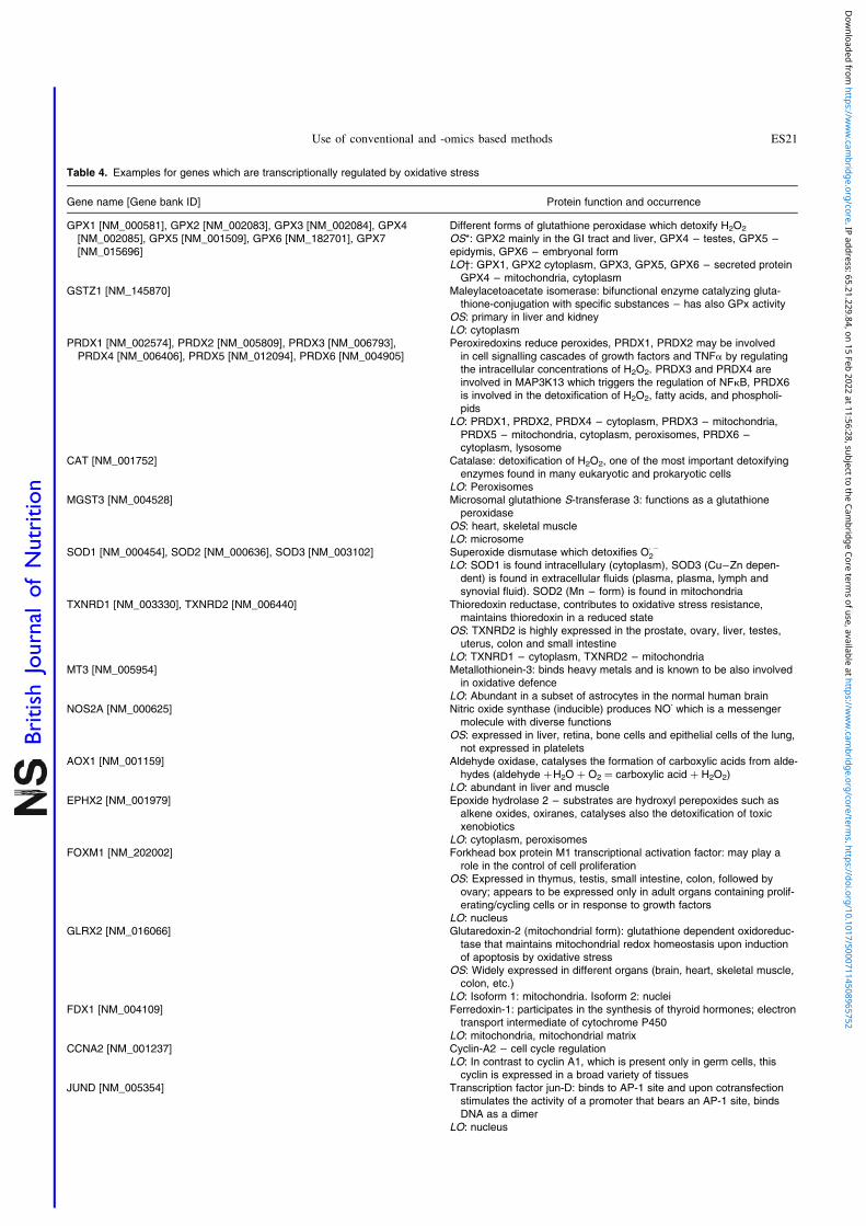
Table 4. Examples for genes which are transcriptionally regulated by oxidative stress
Gene name [Gene bank ID] Protein function and occurrence
GPX1 [NM_000581], GPX2 [NM_002083], GPX3 [NM_002084], GPX4[NM_002085], GPX5 [NM_001509], GPX6 [NM_182701], GPX7[NM_015696]
Different forms of glutathione peroxidase which detoxify H2O2
OS*: GPX2 mainly in the GI tract and liver, GPX4 – testes, GPX5 –epidymis, GPX6 – embryonal formLO†: GPX1, GPX2 cytoplasm, GPX3, GPX5, GPX6 – secreted protein
GPX4 – mitochondria, cytoplasmGSTZ1 [NM_145870] Maleylacetoacetate isomerase: bifunctional enzyme catalyzing gluta-
thione-conjugation with specific substances – has also GPx activityOS: primary in liver and kidneyLO: cytoplasm
PRDX1 [NM_002574], PRDX2 [NM_005809], PRDX3 [NM_006793],PRDX4 [NM_006406], PRDX5 [NM_012094], PRDX6 [NM_004905]
Peroxiredoxins reduce peroxides, PRDX1, PRDX2 may be involvedin cell signalling cascades of growth factors and TNFa by regulatingthe intracellular concentrations of H2O2. PRDX3 and PRDX4 areinvolved in MAP3K13 which triggers the regulation of NFkB, PRDX6is involved in the detoxification of H2O2, fatty acids, and phospholi-pids
LO: PRDX1, PRDX2, PRDX4 – cytoplasm, PRDX3 – mitochondria,PRDX5 – mitochondria, cytoplasm, peroxisomes, PRDX6 –cytoplasm, lysosome
CAT [NM_001752] Catalase: detoxification of H2O2, one of the most important detoxifyingenzymes found in many eukaryotic and prokaryotic cells
LO: PeroxisomesMGST3 [NM_004528] Microsomal glutathione S-transferase 3: functions as a glutathione
peroxidaseOS: heart, skeletal muscleLO: microsome
SOD1 [NM_000454], SOD2 [NM_000636], SOD3 [NM_003102] Superoxide dismutase which detoxifies O·22
LO: SOD1 is found intracellulary (cytoplasm), SOD3 (Cu–Zn depen-dent) is found in extracellular fluids (plasma, plasma, lymph andsynovial fluid). SOD2 (Mn – form) is found in mitochondria
TXNRD1 [NM_003330], TXNRD2 [NM_006440] Thioredoxin reductase, contributes to oxidative stress resistance,maintains thioredoxin in a reduced state
OS: TXNRD2 is highly expressed in the prostate, ovary, liver, testes,uterus, colon and small intestine
LO: TXNRD1 – cytoplasm, TXNRD2 – mitochondriaMT3 [NM_005954] Metallothionein-3: binds heavy metals and is known to be also involved
in oxidative defenceLO: Abundant in a subset of astrocytes in the normal human brain
NOS2A [NM_000625] Nitric oxide synthase (inducible) produces NOz which is a messengermolecule with diverse functions
OS: expressed in liver, retina, bone cells and epithelial cells of the lung,not expressed in platelets
AOX1 [NM_001159] Aldehyde oxidase, catalyses the formation of carboxylic acids from alde-hydes (aldehyde þH2O þ O2 ¼ carboxylic acid þ H2O2)
LO: abundant in liver and muscleEPHX2 [NM_001979] Epoxide hydrolase 2 – substrates are hydroxyl perepoxides such as
alkene oxides, oxiranes, catalyses also the detoxification of toxicxenobiotics
LO: cytoplasm, peroxisomesFOXM1 [NM_202002] Forkhead box protein M1 transcriptional activation factor: may play a
role in the control of cell proliferationOS: Expressed in thymus, testis, small intestine, colon, followed by
ovary; appears to be expressed only in adult organs containing prolif-erating/cycling cells or in response to growth factors
LO: nucleusGLRX2 [NM_016066] Glutaredoxin-2 (mitochondrial form): glutathione dependent oxidoreduc-
tase that maintains mitochondrial redox homeostasis upon inductionof apoptosis by oxidative stress
OS: Widely expressed in different organs (brain, heart, skeletal muscle,colon, etc.)
LO: Isoform 1: mitochondria. Isoform 2: nucleiFDX1 [NM_004109] Ferredoxin-1: participates in the synthesis of thyroid hormones; electron
transport intermediate of cytochrome P450LO: mitochondria, mitochondrial matrix
CCNA2 [NM_001237] Cyclin-A2 – cell cycle regulationLO: In contrast to cyclin A1, which is present only in germ cells, this
cyclin is expressed in a broad variety of tissuesJUND [NM_005354] Transcription factor jun-D: binds to AP-1 site and upon cotransfection
stimulates the activity of a promoter that bears an AP-1 site, bindsDNA as a dimer
LO: nucleus
Use of conventional and -omics based methods ES21
British
Journal
ofNutrition
Dow
nloaded from https://w
ww
.cambridge.org/core . IP address: 65.21.229.84 , on 15 Feb 2022 at 11:56:28 , subject to the Cam
bridge Core terms of use, available at https://w
ww
.cambridge.org/core/term
s . https://doi.org/10.1017/S0007114508965752

the effects of different types of stress (heat shock, ESR stress,oxidative stress and crowding) in human fibroblasts and HeLacells and found clear differences in the responses. On the con-trary, similar transcription patterns were identified by Chuanget al. (463) who analysed the effects of three ROS generatingchemicals namely H2O2, 4-HNE and tBOH in human retinalcells. Other papers with human derived cells were publishedby Yoneda et al. (464), who studied the effects of H2O2 andtobacco smoke in bronchial epithelial cells and Morgan andcoworkers(465) who used the human derived liver cell lineHepG2 to investigate the effects of a variety of ROS generatingchemicals. Except in the later study, relative large arrays(.1000 genes) were used in these investigations. In someexperiments, ionising radiation was used to induce stressresponses; for example Amundson et al. (466) analysed theexpression of genes caused by g-radiation in twelve differenthuman cell lines.
Another topic which was addressed in in vitro geneexpression studies concerns the impact of specific cellfunctions (transcription factors, enzymes involved in signal-ling pathways) which are regulated by ROS. Investigationswhich fall into this category are for example analyses of theimpact of PI3K in tBOH induced gene expression in IMR3cells(467), experiments on the role of the BRCA1 gene inbreast cancer cells(468), as well as comparative studies withcell lines differing in their p53 status, Nrf2 functions andSOD activity(469 – 471).Animal experiments: Only a few studies have been carried
out in which the effects of ROS were investigated in labora-tory rodents have been published so far. Examples for exper-iments with different oxidants are described in the articles ofMcMillian et al. (472,473) who analysed gene expression patternsof oxidants in rat livers and attempted to establish compoundspecific expression signatures. Examples for the use of knock-out animals are the investigations of Yoshihara et al. (474) and
Thimmulappa and coworkers(475) who studied comparativelygene expression patterns in normal, SOD and Nrf2 deficientanimals.
Another topic which has been addressed in a number ofin vivo array studies concerns the impact of ageing on genetranscription and oxidant responses. As mentioned above,it is assumed that several age related diseases are dueto increased oxidative damage and it was shown by Leeet al. (476 – 478) that transcription patterns in muscles, brainand heart of mice are age dependent. Edwards et al. (479)
found subsequently, that paraquat induced expression patternsdiffer significantly depending on the age of the animals.
Human studies: Only few articles have appeared in the lastyears. An example for an occupational study is the one pub-lished by Wang et al. (480) who investigated the responses ofblood cells in metal fume exposed workers. It is assumedthat ROS are at least partly involved in the toxic effects ofmetals and indeed the authors found that oxidative damageresponsive genes were altered in their expression. Microarrayswere also used in a number of studies with patients who suf-fered from ROS related diseases such as rheumatoid arthritis,inflammatory bowel disease, arterial fibrillation and sickle cellanaemia(481 – 483).
The results of all these investigations aimed at identifyinggenes which are transcriptionally regulated by ROS dependlargely on the experimental model used and the most impor-tant parameters are the origin of the cells, the time scheduleas well as the mechanisms by which oxidative damage isinduced; also the design and size of array technique playsan important role.
The results of the in vitro experiments described aboveshow that the patterns of gene alterations caused by oxidantsdepends primarily on the type of indicator cells used and toa lesser extent on the chemicals(462,484). On the basis of thecurrent state of knowledge one may expect that genes which
Table 4. Continued
Gene name [Gene bank ID] Protein function and occurrence
HMOX1 [NM_002133] Heme oxygenase 1 – cleaves the heme ring at the alpha methenebridge to form biliverdin, which is subsequently converted to bilirubinby biliverdin reductase. Under physiological conditions, the activity ofheme oxygenase is highest in the spleen, where senescent erythro-cytes are sequestrated and destroyed
LO: microsomesHSPA1A [NM_005345], HSPA1B [NM_005346] In cooperation with other chaperones, Hsp70s stabilize preexistent pro-
teins against aggregation and mediates the folding of newly translatedpolypeptides in the cytosol as well as within organelles. These cha-perones participate in these processes through their ability to recog-nize nonnative conformations of other proteins
GCLM [NM_002061] 8-Glutamyl-cysteinyl ligase 2 ATP þ L-glutamate þ L-cysteine ¼ ADP þ
phosphate þ gamma-L-glutamyl-L-cysteineLO: in all tissues examined; highest levels in skeletal muscles
TOP2A [NM_001067], TOP2B [NM_001068] DNA topoisiomerase: control of the topological states of DNA by transi-ent breakage and subsequent rejoining of DNA strands; topoisome-rase II causes double-strand breaks
LO: TOP2A: generally located in the nucleoplasm, TOP2B: in thecytoplasm
DDIT3 [NM_004083] DNA damage-inducible transcript 3: inhibits the DNA-binding activity ofC/EBP and LAP by forming heterodimers that can not bind DNA
LO: nucleus
* OS, Organ specificity.† LO, Location.
S. Knasmuller et al.ES22
British
Journal
ofNutrition
Dow
nloaded from https://w
ww
.cambridge.org/core . IP address: 65.21.229.84 , on 15 Feb 2022 at 11:56:28 , subject to the Cam
bridge Core terms of use, available at https://w
ww
.cambridge.org/core/term
s . https://doi.org/10.1017/S0007114508965752

are predominately altered in their transcription are those whichare regulated by ROS dependent transcription factors. Indeed,it was found in some studies that Nrf2/ARE, apoptosis andp53 regulated genes are affected(467,468,472,473). However, insome investigations only little evidence for the up regulationof such genes was detected(468,485,486) and often genes wereidentified which are involved in cell cycle regulation, cellularcommunication, biosynthesis and metabolism. Some of themare controlled by ROS dependent signalling pathways andtranscription factors but the patterns seen in the differentstudies are highly divergent and it is not possible to identifyspecific marker genes(467,468,484,487).
What are the reasons for the strong inconsistencies? As men-tioned above, one of the important parameters is the origin ofthe cells. For example, Murray et al. (462) compared the effectsof ROS generating chemicals in human fibroblasts and HeLacells under identical experimental conditions and found theformer cells by far more responsive. The reasons for thesediscrepancies may be due to the fact that signalling pathwaysand transcription factors are partly tissue specific and may beimpaired in cancer cells lines which are frequently used inarray studies. Nrf2 and p53 are found in a broad variety ofdifferent organs(488), while NFkB is lacking in a number oftissues(80). It is known that p53 is frequently mutated in tumoursin many organs(489 – 491), and it was shown in a comparativestudy with p53 þ and p53 2 cell lines that loss of this functionhas a substantial impact on oxidative stress induced genetranscription(469). In addition, it is so that xenobiotic drugmetabolising as well as antioxidant enzymes, which arecontrolled by the ARE element are highly organ specific(see Table 3). It is also well documented that stable cell linesused routinely in toxicological studies lack the activities ofphase I, phase II and antioxidant enzymes which are regulatedby ARE via Nrf2(299), therefore strong efforts have been madeto establish cell lines which have retained the activities ofthese enzymes in an inducible form. Also other factors mayaccount for the inconsistency of the results(462) found strongdifferences between the results of array studies conductedin vitro and in rodents, and the authors hypothesized that thefact that substantially more genes were transcriptionallyregulated in vivo may be due to the inadequate representationof cell communication in cultures.
Another important parameter which has been often neg-lected is the time dependency of AO responses(462,464,487).For example, three distinct phases could be discriminated inexperiments with human bronchial epithelial cells. The firstwas characterised by upregulation of apoptosis related genesand MAPKs, the second by activation of proteins involvedin the turnover of damaged proteins and only in the third(10 h after the challenge) activation of genes was observedwhich are involved in the detoxification of ROS(464,487).
Despite these inconsistencies, it is possible to define anumber of genes which are typical markers of oxidativestress under experimental conditions (Table 3) and it waspossible to establish distinct signatures of gene expressionfor different types of oxidative tress in specific experimentalmodels such as macrophage activation, peroxisomal prolifer-ation and ROS releasing chemicals in rat hepatocytesin vivo (472,473). Also in in vitro studies with a human derivedliver cell line a clear difference was observed betweenoxidative and non-oxidative stress responses(465).
It is known for other areas of genomic research that studiesinterrogating gene transcription in similar contexts oftenresulted in poor overlapping lists of regulated genes (for ageneral review see(492)); are a typical example are the stronglydivergent results obtained in investigations aimed at identify-ing retinoic acid responsive genes(493). These inconsistencieshave lead to strong efforts to elucidate the methodologicalreasons for these variations and it became clear that corre-lations are improved when low abundant genes areexcluded(494). Different platforms were established whichattempted to standardise the techniques of array basedapproaches. The outcome of these efforts have been publishedin a number of recent articles(495 – 498).Results of microarray studies with dietary antioxidants. In
total, results from approximately 60 studies are available at pre-sent in which the impact of dietary antioxidants on geneexpression patterns was investigated. The number of publicationhas almost doubled in the last two years and it is also interestingthat many of the newer studies were conducted with laboratoryrodents; nevertheless in vitro experiments with stable cell linesare still dominating. It is also notable, that research focusedpredominantly on specific types of compounds such as resvera-trol, a phenolic compound from red wine, epigallocatechingalle-ate (EGCG), the main catechin in green tea, cucurmin containedin the spice tumeric, sulphoraphane the breakdown product ofglucobrassicin (a glucosinolate found in cruciferous vegetables)and the phytoestrogen genistein which is contained in soybeans. Several reviews have been published which containresults of microarray studies with phytochemicals. The mostcomprehensive one by Narayanan(31) describes data obtainedin twenty studies (3 in vivo, 17 in vitro) with various dietarycomponents, the findings with EGCG are summarised in thepaper of Mariappan et al. (499), results with carotenoids can befound in the overview of Elliott(500).
Table 5 summarises the results of newer studies which havenot been evaluated in the aforementioned articles. The firstpart describes results of in vitro experiments with cells lines,the second contains in vivo experiments with laboratoryrodents.
Apart from the in vitro and animal studies, also a fewhuman intervention trials have been carried out with antioxi-dants. For example Majewitz et al. (501) conducted a studywith normal and apoE smokers (which carry a specificmutation in apolipoprotein E and are at increased risk for cor-onary heart disease). Before and after vitamin C supplemen-tation (60 mg/P/4 weeks) they analysed monocytes by use ofa small array (225 genes). After the intervention, theexpression of 22 of the genes was altered in normal smokers,71 were altered in apoE individuals. The most interestingobservation was the down regulation of TNF-b and its recep-tor, also genes encoding for the neutrophil growth factorreceptor and the monocyte chemoattractant protein receptorwere affected. These genes are involved in inflammatoryresponses and it is known that TNF-b is regulated by NFkB.Another human study was published by Hoffmann and cowor-kers(502) who analysed the effects of consumption of a poly-phenolics mix in lymphocytes of healthy individuals by useof a small targeted array (containing 96 transcripts related todrug metabolism). Fifty six genes were found expressed inthe target cells and seven of them were altered after the inter-vention, three of them encoded for cytochrome P450 isozymes
Use of conventional and -omics based methods ES23
British
Journal
ofNutrition
Dow
nloaded from https://w
ww
.cambridge.org/core . IP address: 65.21.229.84 , on 15 Feb 2022 at 11:56:28 , subject to the Cam
bridge Core terms of use, available at https://w
ww
.cambridge.org/core/term
s . https://doi.org/10.1017/S0007114508965752

Table 5. Examples for results obtained with dietary antioxidants in microarray experiments
Treatment/aim Cells/AS* Results† References
In vitro investigationsQuercetinAim: study the impact on
growth inhibition of pros-tate cancer cells
PK-3, NLCap, DU-145 human prostate cancercells lines; BG-9 normal human fibroblastcells line
AS: number of genes not specified
In prostate cancer cell lines # of theexpression of cell cycle related genes (e.g.E- and D-type cycline genes) was observedas well as # of tumour suppressor genes(e.g. CBP, PTEN, MSH2, TGFbR1, ALK-5)and # of oncogenes (p53, CDK2)
(615)
QuercetinAim: study the antitumour
activity of Q
Human CO-115 colon adenocarcinoma cellsAS: whole genome microarray
5000–7000 genes were affected, specificeffects on genes related to cell cycle arrest(CDKN-group) and modulation of theexpression of apoptosis related genes(e.g. p53-related genes), only one antioxi-dant gene (LOXL3) was altered
(616)
GenisteinAim: study the impact on
prostate cancer develop-ment
Human prostate cancer cell lines LNCaP andPC-3
AS: 557 genes related to cancer
11 genes were strongly altered; " glutathioneperoxidase 1, aldolase A, quiescin Q6, rashomolog gene family, member D; # ofapoptosis inhibitor (survivin), MAPK6,fibronectin 1, topoisomerase IIa etc.
(617)
Epigallocatechin gallateAim: study effects on cell
division
SH-SY5Y human neuroblastoma cell linesAS: 25 genes
# of expression of apoptotic genes (Bax andBcl-2) and of the cell cycle inhibitor Gadd45as well as caspase 6
(618,619)
Epigallocatechin gallateAim: investigation of the
protective role of EGCGon breast cancer develop-ment
Dimethyl benzanthracene transformed breastcancer cell line D3-1
AS: 7500 genes
EGCG treatment caused changes in trans-formed cells that promote a more “normal”phenotype. # of AhR (a transcription factorinvolved in the biological responses to poly-cyclic aromatic hydrocarbons, e.g. CYP1A1,CYP1A2 and CYP1B1). Also changesinvolved in nucleo-cytoplasmatic transport(SCS-1) were affected
(620)
" of dehydrocarbonases (AKR)Epigallocatechin gallateAim: study impact of gene
regulation of glucosemetabolism
Rat H4IIE hepatoma cellsAS: size not specified
# of number of genes involved in fatty acidsynthesis, oxidation and activation as wellas in triacethylglycerol synthesis
" of genes involved in glycolysis and glucosetransport
(621)
Diallyl disulfideAim: investigation on cell
division and overalltumour behaviour
HCT-15 human colon tumour cell lineAS: size of the array not specified
# of cell cycle regulated genes (e.g. Cdk6) aswell as oncogenes, tumour suppressors andextracellular matrix and communicationgenes
(622)
" cell cycle proteins (cdk2, 3, 4, etc.) as wellas different growth factors, microfilaments,protein turnover related genes
" DNA damage related genes (e.g. XP-relatedgenes)
Diallyl trisulfideAim: investigation of lipid-
lowering properties
Human derived hepatoma cells (HepG2)AS: 452 genes
Only three genes were altered: " PPAR-a(peroxisome activated receptor alfa) andHNF-4a (hepatocyte nuclear factor 4 alfa),# CYP7A1 (involved in the oxidation ofxenobiotics)
(623)
Gallic acidAim: investigation of antioxi-
dant effects
Human chronic myogenous leukaemia cellK562
AS: 82 genes (antioxidant enzymes and DNArepair)
Several antioxidant genes # , i.e. GPX, thio-redoxin (TXN), thioredoxin peroxidase(AOE372), several DNA polymerase genes(POLD1 and 2), X-ray repair genes(XRCC5), DNA-3-methyladenineglycosidase
(32)
Vanillin and cinnamaldehydeAim: investigation of preven-
tion of formation of spon-taneous mutations
Mismatch-repair deficient (MMR2) humancolon cancer cell line HCT116
AS: 14 500 genes
Eight genes affected which play a role in DNA-damage and oxidative and stress response,e.g. " HMOX1 (heme oxygenase), HSPA1B(heat shock protein)
(624)
# of 14 genes involved in cell growth anddifferentiation: (MAPK2, FGFR2, TGFB111,etc.)
ResveratrolAim: general chemo-
protective properties
Human ovarian cancer cell line PA-1AS: 7448 genes
118 genes were altered " of antioxidantenzymes (NQO 1, NAD(P)H quinone oxire-ductase 1, thioredoxin reductase 1), apopto-sis related genes (p21), investigation of timecourse of gene regulation
(625)
S. Knasmuller et al.ES24
British
Journal
ofNutrition
Dow
nloaded from https://w
ww
.cambridge.org/core . IP address: 65.21.229.84 , on 15 Feb 2022 at 11:56:28 , subject to the Cam
bridge Core terms of use, available at https://w
ww
.cambridge.org/core/term
s . https://doi.org/10.1017/S0007114508965752

Table 5. Continued
Treatment/aim Cells/AS* Results† References
LycopeneAim: investigation of the
effects on breast cancergenes
Human breast cancer cells lines (MCF7,MDA-MB-231) and the fibroblastic cell lineMCF-10a
AS: 202 genes
Changes in genes related to apoptosis, cellcycle and signalling. Apoptosis relatedgenes: p53, caspase 8, TNF (all # ), cellcycle genes: cyclin E, etc. " . Also variousDNA-repair genes "
(626)
Vitamin EAim: investigations on gene
expression related to pre-vention of DNA-adductformation by lipid peroxi-dation products
Differentiated and undifferentiated humancolonocytes (with/without oxidative stress)
AS: global gene expression
In total 118 genes were affected which con-cern the cell cycle (cyclin D1, p27, p21,MAPKs, CDK-2); DNA-repair (p21, RAD54homologous recombination) and also con-nective tissue related genes
(627)
Apple flavonoidsAim: study patterns of gene
expression (not specified)
HT29 human colon cancer cellsAS: 96 genes of drug metabolism
" of glutathione tranferases (GSTP-1, GSTP-2, MGST2) and cytochromes CYCP4F3,CHST5 genes
(628)
# of EPHX1Resveratrol CD18 and S2-013 human pancreatic cell lines " of expression of MIC-1 in both cell lines (629)Aim: investigations on the
role of macrophage inhibi-tory cytokine-1 (MIC-1) inresveratrol-induced growthinhibition of human pan-creatic cancer cell lines
AS: global gene expression " of expression of growth differentiation factor15 (GDF15), sensescence-associatedepithelial membrane protein (SEMP),major histocompatibility complex, class I, F(HLA-F) and 17 other genes in S2-013cell line
Genistein (G)Aim: investigations on the
effect of G on global geneexpression patterns inandrogen-responsivehuman prostate cancercell line
LNCaP androgen-responsive human prostatecancer cell line
AS: global gene expression
28 genes were affected19 androgen up-regulated genes were # by G
(TNF-induced protein, prostatic kallikrein 2,prostate specific antigen kallikrein 3 andothers)
4 androgen down-regulated genes " by G(dopa decarboxylase, BRCA-1-associatedRING domain 1, butyrylcholinesterase,phosphoinositide-3-kinase)
5 genes " by both androgen and G (stearoyl-CoA desaturase, UDP-glucose dehydro-genase)
(630)
Phenethylisothiocyanate(PEITC)
HCT-116 human colon adenocarcinoma cellline
" of expression of GADD45 (631)
Aim: to further clarify themolecular effects ofPEITC in causing death ofhuman colon adenocarci-noma cells
AS: small cluster of apoptosis-related genes
QuercetinAim: to elucidate possible
mechanisms involved ininhibition of proliferation oftumour cells
Caco-2 human colon cancer cell lineAS: expression of 4000 human genes
# expression of cell cycle genes (for exampleCDC6, CDK4 and cyclin D1), # cell prolifer-ation and induced cell cycle arrest
(632)
" of expression of several tumour suppressorgenes. In addition, genes involved in signaltransduction pathways like beta catenin/TCFsignalling and MAPK signal transductionwere influenced by quercetin
Vitamin EAim: effects on global
gene expression – timedependency
F Fisher 344 rats fed with vitamin E deficientdiet or Vit E supplemented diet (60 mg/kg)
AS: 7000 genes t.o. testes and liverSeveral time points were monitored
Five genes and seven sequence taqs werealtered at least at three time points (gammaglutamylcysteinyl synthetase, GSH synthe-tase, 5-a-steroid reductase, factor IX, sca-venger receptor CD36), furthermore, subunitof 8-GCS, coagulation factor IX, steroidreductase type 1, etc.
(633,634)
Turmeric supplementsAim: mechanism of action of
turmeric in the treatmentof arthritis
C Lewis rats were treated i.p. with 25mgrhamnose/g bw and with various doses ofturmeric extract i.p. (0·5–1·0ml/g)
AS: 31 000 genest.o. arthritic joints
" of a variety of chemokines and cytokines(GRO/KC, MCP-1, MIP-1a, MIP3a, CXCchemokine LIX) and adhesion factors thatfacilitate inflammatory cell recruitment to thejoint for another group of target genes.COX2 was # significantly, gene expressioncontrolled by transcriptional factors TIL-2,TNFa was "
(505)
Use of conventional and -omics based methods ES25
British
Journal
ofNutrition
Dow
nloaded from https://w
ww
.cambridge.org/core . IP address: 65.21.229.84 , on 15 Feb 2022 at 11:56:28 , subject to the Cam
bridge Core terms of use, available at https://w
ww
.cambridge.org/core/term
s . https://doi.org/10.1017/S0007114508965752

(2F1, 3A4 and 3A5), two for them for sulfotransferases (SULT2A1 and 1C2) the others were associated with microsomalGST, nicotinamide–N-methyltransferase and the ATP bindingcassette.
All of the compounds listed in the table are potent antioxi-dants and it is apparent that many of the transcriptionalchanges which were detected concern genes which encodefor AO defence, inflammatory responses, apoptosis and cellsignalling and division. Nevertheless, it is not possible, todefine a general signature of AO effects. The lack of acommon pattern may be due to the fact that the chemicalstructures of the antioxidants as well as their mode of actionand tissue specificity differs substantially. For example
vitamins C and E differ strongly in their organ distributionand it their ability to inactive ROS species(503). Furthermore,also the experimental design of the studies plays an importantrole. As described above, the expression patterns seen withresveratrol were highly sex specific, in female mice thenumber of genes which were transcriptionally altered in hepa-tic tissue was approximately 3 times higher in females as com-pared to males(504). Also organ differences have an impact onthe results of array studies, for example EGCG affected in theliver of mice three times more genes as compared to thecolon(505). Another important parameter affecting the outcomeis the time dependency of the expression patterns and also thedose dependency of the responses (see Table 5).
Table 5. Continued
Treatment/aim Cells/AS* Results† References
Epigallocatechin gallateAim: comparison of the
impact of Nrf2 onexpression profiles
F C57BL/6J (Nrft/t) and F C57BL/6J/Nrf(2 /2 ) knockout mice
EGCG: 200 mg/kg bw oral single doseAS: 34 000 genest.o. liver and colon
EGCG regulated 671 Nrf2 dependent genesand 256-independent genes in the liver; inthe colon 228 Nrf2 regulated genes and 98independent genes were identified. Genesfell into the functional categories: proteol-ysis, detoxification, transport, cell growthand apoptosis, cell adhesion and transcrip-tion factors
(635)
Red wine polyphenolics(RWP)
Aim: investigate the effecton the prevention of car-cinogenesis in the intesti-nal mucosa
F Fisher 344 rats fed with high fat dietRWP were fed in the diet (50 mg/kg)AS: 5677 genest.o. colon mucosa
20 genes " , 366 # , among the # genes 41were related to immune- and anti-inflamma-tory response. In addition also genes wereaffected which are involved in steroid meta-bolism, # of genes involved in energymetabolism pathways (COX6-8, etc.). " ofcholesterol 7a-hydroxylase (CYP7A1)
(636)
Resveratrol (R)Aim: investigation on stress
response in rat liver
F and C CD ratst.o. liverTreatment: different doses of R
(0·3–3·0 gm/kg/day), oral administration,28 days
AS: 140 stress related genes
Dose dependent effects " of CYP-450 isoen-zymes as well as CYP-reductase inductionof AO genes such as SOD2 and thisulfatesulfurtransferase (TST) at the highest dose,but decrease of these genes at lower doses.Other genes affected were MAPK, p38,CAT, HO-1, as well as UGT-encodinggenes. In C the number of genes alteredwas ca. 2-fold higher than in F
(504)
Genistein (G)Aim: study impact of the iso-
flavone on fatty acidmetabolism
F C57BL/6 mice fed low fat diet, a high fatdiet or high fat diet supplemented withgenistein (2 g/kg), 12 weeks
AS: 6531 transcriptst.o. liver
97 genes were altered by high fat diet, 80 ofthem were normalized by G supplemen-tation. Many of them were associated withcholesterol biosynthesis, but additionallyalso genes involved in detoxification andinflammatory processes were affected,e.g. metallothionein 1, GST, kallikrein B(plasma1), serine proteinase inhibitor (cladeA, member 3G), serine protease inhibitor1–5. Also apoptosis related genes wereaffected
(637)
Vitamin C (Vc)Aim: impact on lifespan and
antioxidant defencemechanisms
C57BL/6 mice, sex not specified, maintainedeither at normal temperature (þ228C) orþ78C (increase of metabolism) and sup-plemented diet (180 mg/kg), lifespan feeding
AS: 163 genest.o. liver
After 6 months no gene alterations werefound, after 22 months: 3 genes " (COX2,p21, UDP–glucuronosyltransferase)
8 genes # : e.g. SOD, quinone NAD(P)H-dehy-drogenase and different other genes notinvolved in AO defence
(506)
Caffeic acid phenetyl esterAim: impact on atheros-
clerosis
Apolipoprotein E-deficient mice (Apoe 2 /2 )and normal C57/B6, 30 mg/kg bw 12 weeksAS: 3758 genes expression t.o. aortaAnalyses were conducted in untreated miceand mice which were under oxidative stress
Altered genes are not clearly specified; overallexpression induced by oxidative stress wasreverted by CAPE-treatment
Authors mention that that basic transcriptionfactors, growth factors and cytokines as wellas cell adhesion genes are # in apoe 2 /2mice after CAPE treatment
(638)
* AS, Arrays size (genes number); SD, Sprague–Dawley† # , downregulation; " , upregulation; $ , no changes; CDKN, cyclin-dependent kinase inhibitor; COX, cyclooxygenase; FGFR2, fibroblast growth factor receptor 2; GAPDH,
glyceraldehyde-3-phosphatase dehydrogenase; LOXL, lysyl oxidase-like genes; MAPK2, mitogen-activated kinase 2; MCP-1, monocyte chemoattractant protein 1; MPK6,mitogen-activated protein kinase 6; NAD(P)H, nicotinamide adenine dinucleotide phosphate; NQO1, NAD(P)H dehydrogenase, quinone 1; TNF, tumour necrosis factor;TGFB, transforming growth factor beta; TF, transcription factor; SOD, superoxide dismutase; n.s., not statistically significant.
S. Knasmuller et al.ES26
British
Journal
ofNutrition
Dow
nloaded from https://w
ww
.cambridge.org/core . IP address: 65.21.229.84 , on 15 Feb 2022 at 11:56:28 , subject to the Cam
bridge Core terms of use, available at https://w
ww
.cambridge.org/core/term
s . https://doi.org/10.1017/S0007114508965752

The results of microarray analyses do at present not allow todraw firm conclusions if a compound elicits ROS protectiveeffects. For example, findings obtained with moderate dosesof vitamin C in mice(506) indicate that it rather induces inflam-mation (up regulation of COX 2) and reduces ROS protection(down regulation of SOD). Also the results obtained withdiallyldisulfide in HepG2 cells were unspecific and not indica-tive for antioxidant properties which are well documented forthis compound(507). However, it may be possible that generalantioxidant gene expression patterns can be defined on thebasis of comparative standardised experiments with a varietyof compounds which are lacking at present. The currentlyavailable results show that microarray studies neverthelessprovide highly useful information concerning the molecularmechanisms of prevention of ROS related diseases. Typicalexamples are the results of studies with prostate cancer celllines and compounds such as resveratrol and sulforaphanewhich showed that these compounds interact with thetranscription of genes involved in cell cycle regulation andapoptosis; these findings provide a possible explanation forprotective effects of these dietary components towardsprostate cancer cell line LNCaP (for review see(31)); also theassumption of cancer protective effects of EGCG and retinoidscould be enhardened by results of array studies(499,500).
ProteomicsBasic principles. Proteomics is a screening technology
based on the separation, quantification and identification ofproteins from biological samples and allows to directly ident-ify proteins bearing oxidative modifications. Adaptive cellresponses in order to cope with the cell damage imposed byoxidative stress may as well be investigated by transcrip-tomics. As proteins are translated by ribosomes based on theinformation provided by mRNA molecules, no protein canbe synthesised without the appropriate RNA. On the otherhand, the mere existence of mRNA does not mean that thecorresponding protein gets translated. It only means that theprotein might get translated upon demand. This defines onemain difference between proteomics and transcriptomics.Transcriptomics shows which genes are expressed, but doesnot tell which proteins actually get translated. The picturegets even more complicated, when time is considered. Someproteins such as histones are almost exclusively translatedduring the S-phase of the cell cycle, but not in G0(508).A poor correlation of proteomics to transcriptomics datamay be the consequence, because these proteins may be abun-dant in G0, but no corresponding mRNA may be detectable atthis time. On the other hand, several regulatory proteins suchas p53 may be subject to continuous protein degradation byproteasomes. Here, protein amounts may accumulate tominute and undetectable amounts only, while the mRNAmay be easily detectable. This explains why proteomics maygive very different results when compared to transcriptomics.
In order to assign the identity of the modified molecules,separation of proteins may be applied before the detectionof oxidative modification. 2D-PAGE is still the most import-ant technique to separate proteins according to electriccharge and molecular weight(509). Protein spots on 2D gelscan be identified by Western analysis or mass spectrometricanalysis of tryptic digests and quantified by a variety oftechniques including silver staining, fluorescence detectionor autoradiography(510). This approach is used to perform
comparative analysis of protein fractions isolated from e.g.treated and untreated cells. Spot patterns can be compared inorder to detect alterations in relative amounts or, quite relevantto oxidative stress, protein modifications accompanied bychanges of the molecular charge of the protein. Applicationof immunological detection of carbonyl groups or nitro-tyro-sine by Western analysis may subsequently identify the modi-fied proteins. Thus, application of 2D-PAGE may enable thedetection of upregulated and/or modified proteins consequentto oxidative stress. The main draw-back of 2D-PAGE is itslimited resolution and sometimes poor reproducibility con-cealing many important effects(511). The number of proteinsaccessible via 2D-PAGE is usually in the order of several hun-dreds and hardly exceeds thousand different proteins(509).
Another essential technique for proteome analysis ismass spectrometry. Upon tryptic digestion, peptides can beobtained from isolated proteins which can be forwarded tomass analysis. Determination of the molecular weight ofthe peptides by e.g. MALDI-TOF provides a fingerprint ofpeptide masses for each protein(512). As tryptic peptides maybe predicted from databases for all known proteins, a best-fitsearch can be performed resulting in candidates which showtheoretical peptide masses comparable to those identified inthe experiment. This technique is called fingerprint analysisand can be applied only to well-purified proteins as potentiallyobtained from 2D-PAGE. Minimal contamination of peptidepreparations may render fingerprint data not accessible formeaningful interpretation and are a main drawback of thismethod.
Much more robust and powerful is the identification of pep-tide sequences obtained from peptide fragmentation analysis.Peptides can be fragmented specifically in a mass spec-trometer resulting in the breakage of a single peptide bondper molecule(513). Ordering of the resulting peptide fragmentsmay generate mass differences according to single aminoacids in the peptide. As a result, not only total masses of pep-tides are determined as in case of fingerprint analysis, butcomplete amino acid sequences are obtained, which allowidentification of proteins with much higher confidence.Furthermore, amino acid modifications may be determinedby fragmentation analysis. When applied to isolated spotsfrom 2D-gels, proteins can be identified with very highconfidence. Furthermore, specific modifications such as oxi-dation of methionine, cysteine, tryptophan or others can bedetermined(514). On the other hand, the determination ofamino acid sequences renders separation of proteins beforetryptic digest unnecessary. Protein mixtures may be digestedand then peptide mixtures separated by e.g. nano-flow chro-matography(515). Peptide sequences may be determined suc-cessively and peptides derived from one protein sorted afterthe experiment. This powerful technique is called shotgunanalysis and enables the identification of more proteins as2D-PAGE/MS(511). As long as peptide modifications aredetected, protein modification can also be assigned. Themajor drawback of this approach is the limited sequence cove-rage rendering a lot of information missing and the inappli-cability of immunological detection methods(516).
As a consequence, the experimental design studies has tobe carefully prepared for proteome. First of all, it shouldbe clear what to look at. In case of assessment of theextent of damage in response to a defined challenge, a bulk
Use of conventional and -omics based methods ES27
British
Journal
ofNutrition
Dow
nloaded from https://w
ww
.cambridge.org/core . IP address: 65.21.229.84 , on 15 Feb 2022 at 11:56:28 , subject to the Cam
bridge Core terms of use, available at https://w
ww
.cambridge.org/core/term
s . https://doi.org/10.1017/S0007114508965752

analysis summarizing the extent of e.g. carbonyl formationwith chemical or immunological methods may be appropri-ate. When the identity of target proteins appears desirable,2D-PAGE can be used to assign the identity to the modifiedproteins. Here, the poor access of e.g. membrane proteinsby most proteome analysis techniques poses(517) a severelimitation. Mass analysis of the tryptic digest of an isolated2D-spot may easily identify the corresponding proteinbecause a few peptide fragmentation data may be sufficientfor unequivocal protein identification. When the targetamino acids of a modified protein are desired, full sequencecoverage of the identified peptides is desired, but hard torealise. Peptides have to be ionised before they can be ana-lysed by mass spectrometry. Ionisation is an unpredictableevent and may be strongly dependent on chemical propertiesof the peptide. Consequently, only a subset of the peptidesgenerated by proteolytic digests can usually be detected bymass spectrometry(518).Results of proteomic studies concerning oxidative stress. In
order to identify proteins related to oxidative stress, several strat-egies were employed. A direct consequence of ROS-mediateddamage may be amino acid modifications such as oxidation ornitration. Such modifications are accessible via peptide frag-mentation analysis by mass spectrometry. While this methodis very exact providing direct proof of oxidation together withthe identification of the target protein and target amino acid, ithas several draw-backs. First, it is almost impossible to surveythe total amino acid sequence of proteins. Consequently, a nega-tive result is hardly a proof for the absence of modifications.Second, proteins are very sensitive to artifacts introduced bysample handling procedures. For the proteome analysis ofcells, they have to be lysed to extract the proteins for furtheranalyses. Upon cell lysis, organelles get ruptured, calcium isset free, enzymes may get activated and many biochemicalreactions including oxidations may get out of control. Alsoseparation of proteins by 2D-PAGE exposes proteins tooxygen. This can be directly observed by the detection ofoxidized methionine in samples from the same cell lysis,which were processed by 2D-PAGE and shotgun analysis in par-allel. The proteins separated by 2D-PAGE generally show moremethionine oxidation compared to the same proteins analysedby shotgun proteomics(519). Thus, it is essential to most accu-rately control all experimental conditions and run comparativeexperiments strictly in parallel. Actually, several amino acidmodifications consequent to oxidative stress are detectable bymass spectrometry, including oxidation of cysteine(520,521),methionine and tryptophan(519), and nitration of tyrosine(522).Beside direct amino acid modification, the formation of disulfidebridges by oxidation may form protein adducts(523) or proteinglutathione adducts(524). Amino-acid adducts can also bedetected with antibodies specific for e.g. nitrotyrosine andcarbonyl adducts(525,526). As mentioned above, the assignmentof the target protein identity might be a daunting task andthe assignment of the modified amino acid within the sequenceis impossible. On the other hand, modified peptides whichmay be missed by MS-based analysis might be easily detectedwith antibodies.
In contrast to the identification of proteins directly targetedby oxidative stress, an alternative approach is the investi-gation of the response of cells via up-regulation and down-regulation of regulatory proteins(82). Here, rather indirect
effects are observed, which may be consequent to transcrip-tional alterations of genes helping the cell to manage thestress situation and, on the other hand, proteasomal degra-dation of damaged proteins. As a consequence, the readoutof such an experiment may become very complex, becausewe can see a lot of protein alterations, but we do not knowwhere they are coming from. To reduce the complexity ofthe biological system, prokaryotes and yeast were used toinvestigate the basic responses to oxidative stress(527,528).These works indicate that the expected up-regulation of anti-oxidant proteins and chaperones may be accompanied bysubstantial alterations in the metabolic state of the cells.Again, several approaches are feasible to investigate cellularresponses. First, cells can be treated with sub-lethal concen-trations of agents mediating oxidative stress such as H2O2.After a defined period of time, treated and untreated samplesare forwarded to proteome analysis and the results comparedin a quantitative fashion. Several studies with human mono-cytes and epithelial cells demonstrated that again, besides theobvious target proteins such as redox regulators and chaper-ones, a broad variety of other protein families seems to beinvolved in regulatory processes consequent to oxidativestress including cytoskeletal proteins and glycolytic pro-teins(529,530).
Second, cells which are very sensitive to oxidative stressmay be compared to others which were made resistant e.g.by chronic exposure with increasing H2O2 concentrations(531).Here, differentially expressed proteins may include thosewhich actually mediate resistance. Obviously, proteins differ-entially expressed due to different differentiation or activationstates of the investigated cells may repress the identification ofthe true players related to oxidative stress control. Rathermultiple comparisons of various cell systems or additionalfunctional assays may allow to identify the relevant proteins.For example, it was demonstrated that enzyme aldosereductase identified by comparative analysis indeed mediatesprotection in Chinese hamster fibroblasts(531).
Finally, the involvement of oxidative stress in human dise-ases has been investigated by analysis of clinical material andit was shown that proteomics may help to understand patho-physiological processes relevant to the disease state. Theinvolvement and relevance of protein oxidation in braintissue in Alzheimer’s disease has been clearly demons-trated(532,533). Specific oxidative modifications may also beinvolved in obesity(534), asthma(535), and various forms ofinflammatory diseases(536).
Results of proteomic studies with dietary antioxidants.Although numerous position papers emphasized the impor-tance of proteomics techniques in the investigations ofhealth effects of dietary factors(537 – 546), the number of studieswhich contain data on the effects of food related compoundson protein patterns is strikingly low. One of the reasons maybe that proteomics requires, unlike other techniques describedin this article, a sound financial background to establish a pro-teomics facility. Most of the existing labs are specialised inthe analysis of specific cells and tissues. Table 6 summarizesexamples for results obtained in recent studies.
It is quite clear that in most in vitro studies more drasticeffects were observed than in human trials. This discrepancymay be not only due to the differences in the effects of thecompounds tested, but also to the use of high doses in the
S. Knasmuller et al.ES28
British
Journal
ofNutrition
Dow
nloaded from https://w
ww
.cambridge.org/core . IP address: 65.21.229.84 , on 15 Feb 2022 at 11:56:28 , subject to the Cam
bridge Core terms of use, available at https://w
ww
.cambridge.org/core/term
s . https://doi.org/10.1017/S0007114508965752

Table 6. Examples for results obtained with dietary antioxidants in recent proteomics studies
Treatment/methods/aim* Cells/species† Results‡ Reference
In vitroDiallyl trisulfide (DATS)25, 50mM for 24, 48, 72 and 96 h2DE, MALDI-TOF MS and
LC–MS/MSAim: responses of protein
expression to DATS treatment ofcells
BGC823, human gastric cancercell line
41 spots identified
17 proteins # and 27 proteins " after DATS treatment,50 % of these sensitive proteins are associated withapoptotic pathways, e.g. GST pi # , cytokeratin 8 # ,VDAC-1 # , VDAC-2 # , annexin I " , enolase 1 " ,ATP-synthase C " , etc.
(639)
Quercetin150mM for 72 h2DE, MALDI-TOF MSAim: identify cellular targets of puta-
tive colorectal cancer protectiveeffect of flavonoids
HT-29, human colon cancer cells28 spots identified
Alterations of apoptosis involved proteins: anti-apoptotic heatshock proteins hsp 70-1 and HSPBP1 # ; GrpE #
(substrate of hsp-70), caspases " (various cytoskeletalproteins: lamins, ninein), annexin I, II " , human DEAD boxprotein # , stathmin # , triosephosphate isomerase #
(all over-expressed in human cancers)
(405)
Alterations of metabolism: Krebs-cycle enzymesResveratrol (RSV)50, 100, 200mM for 12 and 24 h2DE, MALDI-TOF MSAim: identify mechanisms of RSV
triggered apoptosis
4 HCT116, human colon cancercell lines (Bax þ /2 ; 2 /2 ,p53 þ /þ ; 2 /2 )
4 spots identified
RSV responsive events: fragmentation of lamin A/C protein;caspase 6 activation (caspase protease " , accumulationof the active form); ribosomal protein p0 " , dUTPase # ,stathmin 1 # ; data suggest that caspase 6 and itscleavage of lamin A plays an important role in apoptosissignalling triggered by RSV
(640)
Resveratrol (RSV)50mM for 12, 24 and 48 hRT PCR2DE-SDS-PAGE, MALDI MSAim: study anti-carcinogenic
mechanism of RSV againstprostate cancer
LNCap, DU145, PC-3, humanprostate cancer cell lines
1 spot identified
24 polypeptide spots were markedly " or # 1 of themwas identified by MS as phosphoglycerate mutase B #
(essential for glucose metabolism, functional significanceunclear); apoptosis " by RSV (determined by DAPI andAnnexin V staining); microarray analysis of cDNA showeddifferential expression of 48 genes (involved in cell cycleregulation, apoptosis and glycolysis)
(641)
Cocoa-derived pentamericprocyanidin (pentamer)
100mg/ml for 48 or 72 hp53 and pRb: PAGEMultiplayer dot blot, immunoblottingAim: elucidate which mechanism
the pentamer causes cell cyclearrest in breast cancer cells
Human breast cancer cells (MDAMB-231; -436; -438; SKBR-3,MCF-7) and B(a)p immortalizedcells (184-A1N4, -B5)
45 spots
G1-modulatory proteins: specific dephosphorylation, $
protein expression (Cdc2, forkhead TF) $ p53 depho-sphorylation, $ in protein expression
Retinoblastoma protein: # of expression; specific cytotoxiceffects of the pentamer in breast cancer cells due to sitespecific dephosphorylation and # of cell cycle regulatoryproteins
(642)
L-Ascorbic acid (LAA)0·5mM for 30 min2DE, MALDI-TOF MSAim: to identify the early protein
targets of LAA induced toxicity inleukaemia cells
All-trans retinoic acid (vitamin Arelated compound)
1mM for up to 7 days,2DE, MALDI-TOF MS; MS peptide
sequencingAim: to analyse the molecular
mechanism of Vit A inducedgrowth inhibition in breastcancer cells
NB4 human leukemia cells9 spots identified
MCF-7, human breast cancercells
18 spots identified
9 proteins were sensitive to LAA treatment: e.g. subunit ofprotein disulfide isomerase protein # , Ig heavy chainbinding protein (BiP ¼ Hsp70 chaperone) # , tropomyosin# , etc.
Change in intracellular thiol/disulfide conditions (GSSG " ,modulation of disulfide bond formation); activation ofoxidative stress-inducible apoptosis (measured by FACSanalysis)
Alterations in clusters of proteins involved in the TGFbsignalling pathway and apoptosis (e.g. post-transcriptionaland splicing factors: hnRNP A/B # , H1 " , H2 " , H3 # )
Energy production: GTP ATP phopspho-transferase # ;glutamate dehydrogenase #
Cell proliferation/differentiation: Rho GDP dissociationinhibitor " ; nuclear protein Hcc-1 #
(643)
(644)
Flavone (a potent apoptosisinducer)
150mM, 24 h2DE, MALDI-TOF MSAim: search for the molecular
targets of flavone induced apop-tosis in human colon cancer cells
HT-29, a human coloncancer cell line
20 spots identified
Apoptosis associated proteins (annexins # , cytoskeletalcaspase substrates and heat shock proteins only detectedin control)
Metabolic changes: enzymes of the citric acid cycle #
(succinate dehydrogenase, isocitrate dehydrogenase,NADPH dehydrogenase, etc.)
AO-enzymes: protein levels of catalase and thioredoxinperoxidase #
Marker of colon cancer: protein kinase C-b not detectable
(645)
Genistein (co-treatment with oxi-dised LDL) v. ox-LDL treatmentalone
2·5mM, 24 h (ox-LDL 5mM)2DE, MALDI-TOF MSAim: mechanism of atherosclerosis
preventive effects of genistein
EA. hy926, human endothelialcells
47 spots identified
47 spots were found to differ after ox-LDL treatment; com-bined treatment (ox-LDL þ genistein) could reverse in thecase of 29 proteins the alterations induced by the stressore.g. cyto-skeletal proteins, annexins, S-adenosylhomocys-teine hydrolase ( # by ox-LDL, " by genistein), methionineadenosyl-transferase subunit ( # by ox-LDL, " bygenistein)
(646)
Use of conventional and -omics based methods ES29
British
Journal
ofNutrition
Dow
nloaded from https://w
ww
.cambridge.org/core . IP address: 65.21.229.84 , on 15 Feb 2022 at 11:56:28 , subject to the Cam
bridge Core terms of use, available at https://w
ww
.cambridge.org/core/term
s . https://doi.org/10.1017/S0007114508965752

Table 6. Continued
Treatment/methods/aim* Cells/species† Results‡ Reference
Genistein (in combination withhomocysteine or ox-LDL)
2·5 or 25mM
2DE, MALDI-TOF MSAim: search of molecular targets of
anti-atherogenic effects of genis-tein
HUVEC, primary humanendothelial cell line
19 spots identified
Genistein reversed stressor induced alterations (8 of 10proteins altered by homocysteine and 2 of 9 altered byox-LDL), e.g. homocysteine reduced proteins werereversed by genistein: annexin V (anti-thrombotic protein)" , lamin A (role in prevention of hypertension) " , apoptosislinked gene 2-protein " ; ox-LDL induced proteins werereversed by genistein: ubiquitin conjugating enzyme 12(foam cell formation) # , aldehyde dehydrogenase #
(544)
In vivo studiesControl food vs AO rich diet: Vit C,
Vit E, L-carnithine, a-lipoic acid(combined with a program ofbehavioural enrichment) for 2,8years
2DE, MALDI-TOF MSAim: effect of an AO-fortified diet on
cognitive dysfunction and oxi-dative stress in a canine model ofhuman ageing
Beagle dogsn ¼ 23, sex n.i.4 treatment groups6 spots identifiedt.o. brain (parietal cortex)
# in expression levels of oxidative stress biomarkers: proteincarbonyl levels of glutamate dehydrogenase # , GAPDH# , neurofilament triplet protein # , a-enolase # , GIT- andfascin actin bundling protein # , Cu/Zn SOD " , fructosebisphosphate aldolase C " , creatine kinase " , glutamatedehydrogenase " , GAPDH " (increased enzyme activi-ties: SOD, GST, hemeoxygenase)
(546)
Grape seed extract (rich inproanthocyanidins) v. controlfood
5 % in the diet for 6 weeks2DE, MALDI-TOFAim: analysis of molecular basis of
AO activities of polyphenols,search for alterations in specific(protective) brain proteins
C SD ratsn ¼ 10, 2 groups18 spots identified7 of these by LC–MS/MSt.o. brain
18 altered proteins identified: e.g. heat shock proteins associ-ated with protein folding and apoptosis " (HSP-60, HSC70 and 71), creatine kinase brain b chain " (ATP-turn-over), neurofilament triplet protein light and medium chain" (NF-L, NF-M: cytoskeletal components important forneuronal maintenance)
Glial fibrillary acidic protein # , vimentin # ( ¼ cytoskeletalproteins, " in Alzheimer’s disease), enolase # (glycolyticpathway)
(647)
Basal diet and 3 differentvegetable diets: 10, 20 and 40 %vegetable mixtures (lyophilisedcauliflower, carrots, peas andonions mixed with basal diet),2DE, MALDI-TOF MS
Aim: effect of increased vegetableintake on protein expression
C C57Bl6 micen ¼ 28, 4 groupsAnalysis of 6 spots identifiedt.o. colon (mucosa cells)
39 proteins displayed differential expression; 6 could beidentified (with a role in the protection against colon can-cer): myosin regulatory light chain 2 " but n.s., carbonicanhydrase I " , high mobility group protein 1 # but n.s.,pancreatitis associated protein 3 " , GAPDH # , ATPsynthase oligomycin sensitivity conferral protein #
(648)
Genistein (isoflavone in soy)500mg/g bw, s.c., day 16, 18 and
20 postpartum (prepubertal)2DE, MALDI-TOF MS and immuno-
blotingAim: to elucidate mechanisms of
suppression of chemicallyinduced breast cancer and effecton mammary gland differentiation
C SD ratsn ¼ 5 rats/groupImmunoblots: n ¼ 8–10
rats/groupt.o. breast (mammary gland)6 spots identified
Proteins important for cell differentiation and gland matu-ration were found altered: GTP-cycohydrolase " , tyrosinehydroxylase " , vascular endothelial growth factor receptor2 # , fertility protein SP22 " , g-synuclein " , peroxiredoxin1 # , ABC-transporter "
(649)
Diet supplemented with cruciferous(radish, sprouts, broccoli,cabbage. . . 436 g/d), allium(chives, garlic, onion, 190 g/d) orapiaceous vegetables (dill, celery,carrots, 270 g/d) v. control diet;
Healthy volunteersn ¼ 38, 2 groups1 spot identifiedt.o. blood (analysis of plasma
proteins)
The cruciferous diet yielded the largest impact on serum pro-teins, further classification showed alterations of 2 polypep-tides in the broccoli-group: one could be identified as B-chain of a 2-HS glycoprotein # (involved in insulin resist-ance and immune functions)
(650)
cross-over study design, 7 daysMALDI-TOF MS
Aim: monitoring the impact of diffe-rent vegetable diets on proteinexpression in plasma
Human studiesBrussels sprouts Healthy non-smoking volunteers 511 spots were detected, 48 of them identified (385)300 g/P/d for 5 days; dietary inter
vention study 2D-PAGE, MS/MS;protein amount: fluorescencedetection; protein synthesis:autoradiography quantification(metabolic labelling with35S-methionine/cysteine)
Aim: monitoring the impact ofsprout consumption on proteinexpression
n ¼ 5, C ¼ 3, F ¼ 248 spots identifiedt.o. blood (primary white blood
cells)
2 alterations after sprout consumption were found: " of syn-thesis of Mn-SOD (1·56-fold); # of synthesis of heat shockprotein 70 kDa protein (2·27-fold)
S. Knasmuller et al.ES30
British
Journal
ofNutrition
Dow
nloaded from https://w
ww
.cambridge.org/core . IP address: 65.21.229.84 , on 15 Feb 2022 at 11:56:28 , subject to the Cam
bridge Core terms of use, available at https://w
ww
.cambridge.org/core/term
s . https://doi.org/10.1017/S0007114508965752

experiments with isolated cells. Unphysiological test con-ditions my lead to secondary effects which may be due tocell death and not caused by the test compound itself.Furthermore, in some of the studies high abundant proteins(a-2 HS glycoprotein) with high inter-individual variationswere postulated to be biomarkers for dietary effects. Manyof the compounds/diets altered the expression of proteins,which play a role in apoptosis, cytoskeleton structure andenergy metabolism, while no substantial alterations ofenzymes were seen which are involved in ROS defence,except SOD, which was found upregulated in some of thestudies.
MetabolomicsImpact of the experimental design on the outcome of antiox-
idant studies. In the last chapters we described extensivelythe technical difficulties of AO studies and the limitations ofthe reliability of different methods. Also the experimentaldesign has a strong impact on the outcome and on the predic-tive value of investigations concerning antioxidant propertiesof dietary compounds which are conducted to find out ifbeneficial effects can be expected in humans.
General problems of the experimental designs of AOstudies. Fig. 4 summarises the advantages and disadvantagesof different experimental models. At present around 90 % ofAO experiments are conducted in vitro with stable cell linesor with subcelluar fractions.
The classical strategy used to identify dietary antioxidants isthe screening of a large number of potential candidates in highthroughput systems and in vitro by use of stable cell lines andsubsequently investigate the most promising compounds inmore reliable models (e.g. in animals studies and in humanintervention trials). This approach is justified on the basis ofthe assumption that the key mode of action of dietary antiox-idants is the direct (non-enzymatic) inactivation of ROS whichcan be easily detected in this simple systems. However, it
became clear over the last years that the mechanisms bywhich dietary compounds protect against ROS are by farmore complex and that indirect modes of action such as induc-tion of enzymes and other defence mechanisms play animportant role. For example, the coffee diterpeonids cawheoland kafestol do not protect human lymphocytes under invitro conditions against ROS mediated DNA damage(282)
while Huber et al. (394) found in experiments with rats thatthey are potent inducers of the enzyme gamma-glutamylcys-teine synthetase (GCS) which catalyses the rate limiting stepof the synthesis of glutathione which is a potent antioxi-dant(547). It is conceivable that the increased GSH levelsseen in the plasma of coffee drinkers(548) and the protectionagainst oxidative DNA damage observed in a human interven-tion trial and in animal experiments(282,549) with coffee are dueto this mechanism.
AO enzymes and also transcription factors which regulatecellular defence mechanisms may be not represented ade-quately in stable cancer cell lines which are used in AOstudies. For example, it is known that that p53 controls thetranscription of a variety of genes which encode for responsestowards antioxidants(550). Since .50 % of human tumours arep53 deficient due to mutations(551), cell lines derived fromthem will not adequately reflect AO responses. Also theorigin of the cells is important, while p53 and NrF2 arefound in many organs, NFkB is not or only weakly expressedin certain tissues(80); also the activities of AO enzymes showstrong organ specificities (see Table 4). As mentionedabove, the differences in the transcription pattern of genes instudies with oxidants and antioxidants support the assumptionthat the cellular responses depend largely on the type of indi-cator cells used.
Another important issue concerns the representation ofphase I and phase II enzymes which metabolise dietary antiox-idants and therefore may alter their protective properties;
Table 6. Continued
Treatment/methods/aim* Cells/species† Results‡ Reference
Vitamin C (Vc) supplementationprospective randomised openlabel trial
250 mg, 3 £ /week, oral for 2months;
CE and ESI-TOF-MS; relevant poly-peptides: MALDI TOF/TOF
Aim: investigate the effects of Vcon biomarkers of oxidative stressand inflammation in haemodialy-sis (HD) patients
Haemodialysis ptsn ¼ 40, C þ F; 2 groups þ /2Vit C and control group, n ¼ 13,
C þ F1 CE fraction identifiedt.o. blood (plasma proteins)
30 polypeptides showed different levels in HD patients incomparison to individuals with normal renal function
Levels of 15 polypeptides were altered after Vc supplemen-tation in, only one of them could be identified and mostlikely represents b-2-microglobulin # (predictor of renalinsufficiency)
(651)
a-Tocopherol (AT) v. placebo,double blind, parallel design
Healthy volunteersn ¼ 32 (11 F, 21 C)3 groups (n ¼ 10)1 spot identifiedt.o. blood (plasma proteins)
12 proteins " after supplementation; one of them wasidentified: " proapolipoprotein A1 (result confirmedby ELISA)
(652)
134 or 268 mg (200 or 400 IU)/dayfor 14 or 28 days; 2DE,MALDI-MS
Aim: examine effects of AT onprotein expression
SwissProt Accession numbers: VDAC-1, P21796; VDAC-2, P45880; HSPBP1, Q9NZL4; GrpE, Q9HAV7; DEAD, Q92499; TGFb, PO1137; Rho GDP, P52565; forkhead TF,Q08050.
* AO, antioxidant; bw, body weight; CE, capillary electrophoresis; 2 DE, two-dimensional electrophoresis; 2DE-SDS-PAGE, two-dimensional electrophoresis-sodium dodecylsul-fate polyacrylamide gel electrophoresis; ESI, electro spray ionisation; h, hour; LC–MS/MS, liquid chromatography ion trap tandem mass spectrometry; MALDI-TOF MS,matrix assisted laser desorption ionization time-of-flight mass spectrometry; n.i., not indicated; ox-LDL, oxidised low density lipoproteins; s.c., subcutaneous.
† SD, Sprague–Dawley, pts, patients, t.o., target organ.‡ # , downregulation; " , upregulation; $ , no changes; TF, transcription factor; SOD, superoxide dismutase; GAPDH, glyceraldehyde-3-phosphatase dehydrogenase; n.s., not
significant.
Use of conventional and -omics based methods ES31
British
Journal
ofNutrition
Dow
nloaded from https://w
ww
.cambridge.org/core . IP address: 65.21.229.84 , on 15 Feb 2022 at 11:56:28 , subject to the Cam
bridge Core terms of use, available at https://w
ww
.cambridge.org/core/term
s . https://doi.org/10.1017/S0007114508965752

furthermore also reactions catalysed by the intestinal micro-flora are not reflected under in vitro conditions. Polyphenolicsare extensively metabolised in the gut and in the body andnon-conjugated metabolites most often account for a minorfraction of the circulating metabolites(552). In the case of iso-quercitrin it was shown that completely different conjugatesare formed under in vitro conditions (i.e. after exposure tocolon cell lines) as in the serum of rats(553,554). Hydroxycin-namic acids which are potent oxidants in coffee are glucuro-nated in the body(555,556), but stable cell lines and peripherallymphocytes which are used in in vitro experiments lack glu-curonosyltransferase (UGT) activity which catalyses this reac-tion(557,558). In most cases, it is not known if and to whichextent the AO properties of conjugates differ from that ofthe parent compounds, but for glucuronides of isoflavonesand epicatechin it has been shown that they provide no
protection against oxidative stress(559,560) while the parentcompounds are potent antioxidants. The inadequate represen-tation of drug metabolising enzymes is a general problemwhich is also encountered in acute toxicity and genotoxicityexperiments with stable cells lined and various attemptswere made to solve it. For example, the cultivation con-ditions of primary hepatocytes which possess a broad spec-trum of xenobiotic drug metabolising enzymes have beenimproved(561,562), other solutions may be the establishmentof hepatoma cell lines which have retained the activities ofseveral enzymes in an inducible form, (for review see(299))and the construction of genetically engineered lines whichexpress human phase I and phase II enzymes(563,564).
The cultivation conditions of the cells may have a strongimpact on the outcome of antioxidant studies. It is knownthat the composition of the medium affects the sensitivity
Predictive value, costs, time requirement
System Advantages/Disadvantages
♦no firm information if effects take place intracellulary
♦no information if effects take place intracellulary♦no information concerning the metabolism and bio- availability of the antioxidants screening
♦no information about biological effects
♦inexpensive and fast (high throughput)
♦high throughput screening
♦only partial information on intracellular effects♦metabolism of antioxidant only partly reflected
♦signalling pathways are only partly reflected
♦enable to monitor organ specific effects♦reflect absorption and metabolism
♦extrapolation of the results to humans possible
♦provide information concerning absorption and metabolism in humans
♦most experiments are conducted with lymphocytes, plasma or urine and provide less information on effects in inner organs
♦requirement to establish "controlled" conditions in intervention trials
♦strong intra-individual variations
♦some mammalian/ROS-models reflect the situation in humans inadequately
♦no information on absorbtion and on organ specific effects
♦no information about bioavailability and on impact of the microflora
♦provide partial information about radical specificity♦"Artificial"redox reactions
Subcellularfractions
Experimentswith intact cells
Animal studies
Human studies
Fig. 4. Advantages and disadvantages of different experimental used to investigate ROS – protective effects of phytochemicals.
S. Knasmuller et al.ES32
British
Journal
ofNutrition
Dow
nloaded from https://w
ww
.cambridge.org/core . IP address: 65.21.229.84 , on 15 Feb 2022 at 11:56:28 , subject to the Cam
bridge Core terms of use, available at https://w
ww
.cambridge.org/core/term
s . https://doi.org/10.1017/S0007114508965752

of cells towards ROS. For example when lymphocytes aretreated with ROS-generating chemicals in serum, they areup to five times less sensitive as compared to treatment inregular medium (L. Elbling, personal communication).When coffee or coffee specific compounds (chlorogenic andcaffeic acid) were tested at high concentrations in genotoxi-city tests with bacteria or human lymphocytes in vitro,strong effects were observed which were attributed to for-mation of H2O2, while with lower doses clear antioxidanteffects were detectable and it was postulated by Aeschbacheret al. (565) that adverse effects do not take place underrealistic in vivo conditions.
On the basis of the results of in vitro experiments it waspossible to identify a number of highly potent antioxidantssuch as chlorophylls(566), curcumin(567), EGCG(568) andanthocyanins(569) which are currently sold as food sup-plements and/or used for the production of functionalfoods(570). However, the results of experiments in whichprotection of oxidative DNA damage was monitored withfoods containing these compounds are not promising, i.e.negative results were for example obtained with greenvegetables, EGCG supplemented products(289,298,571) andanthocyan rich blueberries(348). The reasons for these disap-pointing results are probably due to the fact that the activecompounds which have a large molecular configuration areonly poorly absorbed, therefore protective effects can beexpected in the digestive tract but not in inner organs. Inrecent investigations, we found in a human interventiontrial (SCGE experiments) with lymphocytes that gallic acid(GA), a small phenolic molecule, contained in specificplant foods reduces the formation of oxidised bases inperipheral lymphocytes with 30–40 fold higher efficiencyas vitamins E and C while under in vitro conditions similarprotective effects were observed(296,572). One of the reasonsfor the strong antioxidant properties of GA may be its highabsorption rate(573).
Overall, the predictive value of results obtained in in vivoexperiments with rodents are higher as those of in vitro fin-dings but one of the problems encountered in the interpretationof animal derived results is due to the fact that putativeantioxidants were often administered at high doses whichare irrelevant for humans.
The major problem of human studies concerns the fact thatmost experiments are carried out with peripheral blood cellsand plasma and it remains unclear if and to which extentprotection against ROS mediated damage can be expected ininner organs. In the case of GA we observed significantreduction of radical induced damage in a variety of innerorgans in animal experiments(381,572), also for a number ofother compounds such as lycopene and vitamins and E it isknown that the reduction of damage seen in blood cells inhumans is paralleled by protective effects in a variety oftissues(574,575).
An important question which has been neglected in humanintervention trials concerns the impact of the overall antioxi-dant status of the participants on the outcome of protectionstudies. It can be expected that individuals which consumeAO rich diets respond less sensitive. In the case of folicacid it has been shown that supplementation is only effectivein regard to improvement of the DNA stability in individualswith low intake levels(576). On the basis of these findings
Fenech(577) developed the “genome health clinic and genomehealth nutrigenomics“ concept which postulates individualisedsupplementation strategies on the basis of biochemical andDNA-stability measurements and can be also applied toantioxidants(578,579).
Justification of heath claims for antioxidant propertiesof dietary factors?
Growing consumer concerns about the health attributes offood products and supplements and the increased productionof health foods have led to extensive discussions of the scien-tific evidence required for health claims(580). Already in 2001,the global sales of functional foods were estimated USD 47,6billion worldwide(581). As a consequence, authorities such asthe US Food and Drug Administration and the EuropeanCommission have issued new rules directed at inaccuracies,confusion and false information related to functional anddose related risk reduction claims(582 – 584). The EU regulationon nutrition and health claims made on foods (No. 1924/2006)entered into force on July 1st, 2007 and it is clearly stated inarticle 6 that claims have to be “based on and substantiated bygenerally accepted scientific evidence”.
The evaluation of the methods which are currently usedto investigate the antioxidant properties of foods and foodconstituents show that not all approaches provide reliableresults. Data from in vitro experiments cannot be extrapolatedto the human situation in general, while the value of resultsobtained in animal and human studies depends largely onthe experimental design and on the techniques employed. Asmentioned above, frequently used methods such as TBARSmeasurements and the quantification of protein bound carbo-nyls and breath hydrocarbons do not provide firm evidencefor AO effects, also some TAC measurements, which areconducted under unphyiological conditions (e.g. TOSC,CUPRAC), may not be reliable(171,257). Despite the uncertaini-ties concerning the reliability of TAC measurements, results ofsuch experiments can support the overall evidence for ROSprotective effects but claims should be not solely based onsuch studies. Probably more relevant information can beexpected from HPLC based isoprostane measurements as amarker for lipid peroxidation and from results of experimentsconcerning prevention of oxidative DNA-damage (HPLCbased determination of oxidised bases, SCGE experimentswith restriction enzymes). In this context it is notable thatthere is strong evidence that a number of ROS related diseases(including cancer) are causally directly related to damage ofthe genetic material (see introduction section).
As mentioned above, measurements of the induction ofantioxidant enzymes do not provide reliable informationfor protective effects as they can also be induced by ROSthemselves and their up regulation may reflect prooxidanteffects.
Although the results obtained with -omics techniques nonot allow to establish antioxidant specific patterns of genetranscription and protein alterations they can provide evidencefor protective effects of antioxidants in specific experimentalsettings.
For example, it was found in a few studies that the trans-cription of genes caused by oxidative stress or by ROS related
Use of conventional and -omics based methods ES33
British
Journal
ofNutrition
Dow
nloaded from https://w
ww
.cambridge.org/core . IP address: 65.21.229.84 , on 15 Feb 2022 at 11:56:28 , subject to the Cam
bridge Core terms of use, available at https://w
ww
.cambridge.org/core/term
s . https://doi.org/10.1017/S0007114508965752

diseases can be normalised by dietary antioxidants whichstrongly supports the assumption of protective properties.
A promising strategy for the justification of health claimsmay be the combination of different methods to a test battery,but the selection of the individual approaches and techniquesto be included will be a matter of intense debate.
Acknowledgements
The publication of this paper was made possible by the finan-cial support of the European Co-operation in the field ofScientific and Technical (COST) Research Action 926“Impact of new technologies on the health benefits andsafety of bioactive plant compounds” (2004–2008). Theauthors had no conflicts of interest to disclose.
References
1. Blomhoff R (2005) Dietary antioxidants and cardiovascular
disease. Curr Opin Lipidol 16, 47–54.
2. Elahi MM & Matata BM (2006) Free radicals in blood: evol-
ving concepts in the mechanism of ischemic heart disease.
Arch Biochem Biophys 450, 78–88.
3. Lefer DJ & Granger DN (2000) Oxidative stress and cardiac
disease. Am J Med 109, 315–323.
4. Agarwal A, Sharma RK, Nallella KP, Thomas AJ Jr, Alvarez
JG & Sikka SC (2006) Reactive oxygen species as an indepen-
dent marker of male factor infertility. Fertil Steril 86,
878–885.
5. Pasqualotto FF, Sharma RK, Kobayashi H, Nelson DR,
Thomas AJ Jr & Agarwal A (2001) Oxidative stress in normo
spermic men undergoing infertility evaluation. J Androl 22,
316–322.
6. Von Kobbe C, May A, Grandori C & Bohr VA (2004) Werner
syndrome cells escape hydrogen peroxide-induced cell pro-
liferation arrest. FASEB J 18, 1970–1972.
7. Yan T, Li S, Jiang X & Oberley LW (1999) Altered levels of
primary antioxidant enzymes in progeria skin fibroblasts. Bio-
chem Biophys Res Commun 257, 163–167.
8. Kohen R & Nyska A (2002) Oxidation of biological systems:
oxidative stress phenomena, antioxidants, redox reactions,
and methods for their quantification. Toxicol Pathol 30,
620–650.
9. Gracy RW, Talent JM, Kong Y & Conrad CC (1999) Reactive
oxygen species: the unavoidable environmental insult? Mutat
Res 428, 17–22.
10. Calabrese V, Guagliano E, Sapienza M, Mancuso C, Butter-
field DA & Stella AM (2006) Redox regulation of cellular
stress response in neurodegenerative disorders. Ital J Biochem
55, 263–282.
11. Zimmermann JS & Kimmig B (1998) Pharmacological
management of acute radiation morbidity. Strahlenther Onkol
174, Suppl. 3, 62–65.
12. Sarma L & Kesavan PC (1993) Protective effects of vitamins
C and E against gamma-ray-induced chromosomal damage in
mouse. Int J Radiat Biol 63, 759–764.
13. Gebhart E (1974) Antimutagens. Data and problems. Human-
genetik 24, 1–32.
14. Li L, Tsao R, Yang R, Liu C, Zhu H & Young JC (2006)
Polyphenolic profiles and antioxidant activities of heartnut
(Juglans ailanthifolia Var. cordiformis) and Persian walnut
(Juglans regia L.). J Agric Food Chem 54, 8033–8040.
15. Anagnostopoulou MA, Kefalas P, Kokkalou E, Assimopoulou
AN & Papageorgiou VP (2005) Analysis of antioxidant
compounds in sweet orange peel by HPLC–diode array
detection–electrospray ionization mass spectrometry. Biomed
Chromatogr 19, 138–148.
16. Minoggio M, Bramati L, Simonetti P, Gardana C, Iemoli L,
Santangelo E, Mauri PL, Spigno P, Soressi GP & Pietta PG
(2003) Polyphenol pattern and antioxidant activity of different
tomato lines and cultivars. Ann Nutr Metab 47, 64–69.
17. Erdman JW Jr, Balentine D, Arab L, Beecher G, Dwyer JT,
Folts J, Harnly J, Hollman P, Keen CL, Mazza G, Messina
M, Scalbert A, Vita J & Williamson G & Burrowes J (2007)
Flavonoids and heart health: proceedings of the ILSI North
America Flavonoids Workshop, May 31–June 1, 2005,
Washington, DC. J Nutr 137, 718S–737S.
18. Kim JM, Chang HJ, Kim WK, Chang N & Chun HS (2006)
Structure–activity relationship of neuroprotective and reactive
oxygen species scavenging activities for allium organosulfur
compounds. J Agric Food Chem 54, 6547–6553.
19. Iwai K, Kishimoto N, Kakino Y, Mochida K & Fujita T (2004)
In vitro antioxidative effects and tyrosinase inhibitory activi-
ties of seven hydroxycinnamoyl derivatives in green coffee
beans. J Agric Food Chem 52, 4893–4898.
20. Fujioka K & Shibamoto T (2006) Quantitation of volatiles and
nonvolatile acids in an extract from coffee beverages: correlation
with antioxidant activity. J Agric Food Chem 54, 6054–6058.
21. Lopez-Velez M, Martinez-Martinez F & Del Valle-Ribes C
(2003) The study of phenolic compounds as natural antioxi-
dants in wine. Crit Rev Food Sci Nutr 43, 233–244.
22. Hidalgo FJ, Leon MM & Zamora R (2006) Antioxidative
activity of amino phospholipids and phospholipid/amino acid
mixtures in edible oils as determined by the Rancimat
method. J Agric Food Chem 54, 5461–5467.
23. Luximon-Ramma A, Neergheen VS, Bahorun T, Crozier A,
Zbarsky V, Datla KP, Dexter DT & Aruoma OI (2006) Assess-
ment of the polyphenolic composition of the organic extracts
of Mauritian black teas: a potential contributor to their antioxi-
dant functions. Biofactors 27, 79–91.
24. Antonious GF, Kochhar TS, Jarret RL & Snyder JC (2006)
Antioxidants in hot pepper: variation among accessions. J
Environ Sci Health B 41, 1237–1243.
25. Masuda Y, Kikuzaki H, Hisamoto M & Nakatani N (2004)
Antioxidant properties of gingerol related compounds from
ginger. Biofactors 21, 293–296.
26. Somparn P, Phisalaphong C, Nakornchai S, Unchern S &
Morales NP (2007) Comparative antioxidant activities of cur-
cumin and its demethoxy and hydrogenated derivatives. Biol
Pharm Bull 30, 74–78.
27. Hsu CY, Yang CM, Chen CM, Chao PY & Hu SP (2005)
Effects of chlorophyll-related compounds on hydrogen per-
oxide induced DNA damage within human lymphocytes.
J Agric Food Chem 53, 2746–2750.
28. Xiong S, Melton LD, Easteal AJ & Siew D (2006) Stability and
antioxidant activity of black currant anthocyanins in solution and
encapsulated in glucan gel. J Agric Food Chem 54, 6201–6208.
29. Semba RD, Lauretani F & Ferrucci L (2007) Carotenoids as
protection against sarcopenia in older adults. Arch Biochem
Biophys 458, 141–145.
30. Aruoma OI, Sun B, Fujii H, Neergheen VS, Bahorun T,
Kang KS & Sung MK (2006) Low molecular proanthocyanidin
dietary biofactor Oligonol: its modulation of oxidative stress,
bioefficacy, neuroprotection, food application and chemo-
prevention potentials. Biofactors 27, 245–265.
31. Narayanan BA (2006) Chemopreventive agents alters global
gene expression pattern: predicting their mode of action and
targets. Curr Cancer Drug Targets 6, 711–727.
32. Abdelwahed A, Bouhlel I, Skandrani I, Valenti K, Kadri M,
Guiraud P, Steiman R, Mariotte AM, Ghedira K, Laporte F,
Dijoux-Franca MG & Chekir-Ghedira L (2007) Study of antimu-
tagenic and antioxidant activities of gallic acid and 1,2,3,4,
S. Knasmuller et al.ES34
British
Journal
ofNutrition
Dow
nloaded from https://w
ww
.cambridge.org/core . IP address: 65.21.229.84 , on 15 Feb 2022 at 11:56:28 , subject to the Cam
bridge Core terms of use, available at https://w
ww
.cambridge.org/core/term
s . https://doi.org/10.1017/S0007114508965752

6-pentagalloylglucose from Pistacia lentiscus. Confirmation by
microarray expression profiling. Chem Biol Interact 165, 1–13.
33. Scandalios JG (2002) Oxidative stress responses – what have
genome-scale studies taught us? Genome Biol 3, 1019.
34. Choe E & Min DB (2006) Chemistry and reactions of reactive
oxygen species in foods. Crit Rev Food Sci Nutr 46, 1–22.
35. Foksinski M, Gackowski D, Rozalski R, Siomek A, Guz J,
Szpila A, Dziaman T & Olinski R (2007) Effects of basal
level of antioxidants on oxidative DNA damage in humans.
Eur J Nutr 46, 174–180.
36. Karihtala P & Soini Y (2007) Reactive oxygen species and
antioxidant mechanisms in human tissues and their relation
to malignancies. Apmis 115, 81–103.
37. Fridovich I (1986) Biological effects of the superoxide radical.
Arch Biochem Biophys 247, 1–11.
38. Halliwell B & Gutteridge JM (1999) Free Radicals in Biology
and Medicine, 3rd ed. Avon, England: Oxford University Press.
39. Beckman JS & Koppenol WH (1996) Nitric oxide, superoxide,
and peroxynitrite: the good, the bad, and the ugly. Am J Phy-
siol 271, C1424–C1437.
40. Czapski G & Goldstein S (1995) The role of the reactions
of NO with superoxide and oxygen in biological systems: a
kinetic approach. Free Radic Biol Med 19, 785–794.
41. Shadyro OI, Yurkova IL & Kisel MA (2002) Radiation-
induced peroxidation and fragmentation of lipids in a model
membrane. Int J Radiat Biol 78, 211–217.
42. Pentland AP (1994) Active oxygen mechanisms of UV inflam-
mation. Adv Exp Med Biol 366, 87–97.
43. Koren HS (1995) Associations between criteria air pollutants
and asthma. Environ Health Perspect 103, Suppl. 6, 235–242.
44. Victorin K (1994) Review of the genotoxicity of nitrogen
oxides. Mutat Res 317, 43–55.
45. Yu TW & Anderson D (1997) Reactive oxygen species-
induced DNA damage and its modification: a chemical inves-
tigation. Mutat Res 379, 201–210.
46. Wiseman A, Ridgway T, Goldfarb PS & Woods L (2002) Are
food and environmental toxicants ‘overdetected’ by bioassays?
Trends Biotechnol 20, 13–15.
47. Parke DV & Sapota A (1996) Chemical toxicity and reactive
oxygen species. Int J Occup Med Environ Health 9, 331–340.
48. Ames BN (1986) Food constituents as a source of mutagens,
carcinogens, and anticarcinogens. Prog Clin Biol Res 206,
3–32.
49. Kanner J & Lapidot T (2001) The stomach as a bioreactor:
dietary lipid peroxidation in the gastric fluid and the effects
of plant-derived antioxidants. Free Radic Biol Med 31,
1388–1395.
50. Richter C, Gogvadze V, Laffranchi R, Schlapbach R, Schwei-
zer M, Suter M, Walter P & Yaffee M (1995) Oxidants in
mitochondria: from physiology to diseases. Biochim Biophys
Acta 1271, 67–74.
51. Fleury C, Mignotte B & Vayssiere JL (2002) Mitochondrial
reactive oxygen species in cell death signaling. Biochimie
84, 131–141.
52. Dhalla NS, Temsah RM & Netticadan T (2000) Role of oxi-
dative stress in cardiovascular diseases. J Hypertens 18,
655–673.
53. Forman HJ & Torres M (2001) Signaling by the respiratory
burst in macrophages. IUBMB Life 51, 365–371.
54. Hawkins CL, Brown BE & Davies MJ (2001) Hypochlorite-
and hypobromite-mediated radical formation and its role in
cell lysis. Arch Biochem Biophys 395, 137–145.
55. Rodrigues MR, Rodriguez D, Henrique Catalani L, Russo M &
Campa A (2003) Interferon-gamma independent oxidation of
melatonin by macrophages. J Pineal Res 34, 69–74.
56. Davies KJ (1987) Protein damage and degradation by oxygen
radicals. I. General aspects. J Biol Chem 262, 9895–9901.
57. Grune T, Reinheckel T & Davies KJ (1997) Degradation of
oxidized proteins in mammalian cells. FASEB J 11, 526–534.
58. Levine RL & Stadtman ER (2001) Oxidative modification of
proteins during aging. Exp Gerontol 36, 1495–1502.
59. Stadtman ER (2004) Role of oxidant species in aging. Curr
Med Chem 11, 1105–1112.
60. Kushnareva Y, Murphy AN & Andreyev A (2002) Complex
I-mediated reactive oxygen species generation: modulation
by cytochrome c and NAD(P)þ oxidation–reduction state.
Biochem J 368, 545–553.
61. Squadrito GL & Pryor WA (1998) Oxidative chemistry of
nitric oxide: the roles of superoxide, peroxynitrite, and
carbon dioxide. Free Radic Biol Med 25, 392–403.
62. Younes M & Siegers CP (1984) Interrelation between lipid
peroxidation and other hepatotoxic events. Biochem Pharma-
col 33, 2001–2003.
63. Horton AA & Fairhurst S (1987) Lipid peroxidation and
mechanisms of toxicity. Crit Rev Toxicol 18, 27–79.
64. Zock PL & Katan MB (1998) Diet, LDL oxidation, and coro-
nary artery disease. Am J Clin Nutr 68, 759–760.
65. Forrester JS & Shah PK (2006) Emerging strategies for increas-
ing high-density lipoprotein. Am J Cardiol 98, 1542–1549.
66. Moller P & Wallin H (1998) Adduct formation, mutagenesis
and nucleotide excision repair of DNA damage produced by
reactive oxygen species and lipid peroxidation product.
Mutat Res 410, 271–290.
67. Barzilai A & Yamamoto K (2004) DNA damage responses to
oxidative stress. DNA Repair (Amst) 3, 1109–1115.
68. Niles JC, Wishnok JS & Tannenbaum SR (2006) Peroxynitrite-
induced oxidation and nitration products of guanine and 8-oxo-
guanine: structures and mechanisms of product formation.
Nitric Oxide 14, 109–121.
69. Tsutsui H, Ide T & Kinugawa S (2006) Mitochondrial oxi-
dative stress, DNA damage, and heart failure. Antioxid
Redox Signal 8, 1737–1744.
70. Vaca CE, Wilhelm J & Harms-Ringdahl M (1988) Interaction
of lipid peroxidation products with DNA. A review. Mutat Res
195, 137–149.
71. Ehrenberg L, Osterman-Golkar S, Segerbck D, Svensson K &
Calleman CJ (1977) Evaluation of genetic risks of alkylating
agents. III. Alkylation of haemoglobin after metabolic conver-
sion of ethene to ethene oxide in vivo. Mutat Res 45, 175–184.
72. Segerback D (1983) Alkylation of DNA and hemoglobin in the
mouse following exposure to ethene and ethene oxide. Chem
Biol Interact 45, 139–151.
73. Doublie S, Bandaru V, Bond JP & Wallace SS (2004) The
crystal structure of human endonuclease VIII-like 1 (NEIL1)
reveals a zincless finger motif required for glycosylase activity.
Proc Natl Acad Sci U S A 101, 10284–10289.
74. Sanderson RJ, Bennett SE, Sung JS & Mosbaugh DW (2001)
Uracil-initiated base excision DNA repair synthesis fidelity in
human colon adenocarcinoma LoVo and Escherichia coli cell
extracts. Prog Nucleic Acid Res Mol Biol 68, 165–188.
75. Krokan HE, Drablos F & Slupphaug G (2002) Uracil in DNA –
occurrence, consequences and repair.Oncogene 21, 8935–8948.
76. Cooke MS, Evans MD, Dizdaroglu M & Lunec J (2003) Oxi-
dative DNA damage: mechanisms, mutation, and disease.
FASEB J 17, 1195–1214.
77. Kim KS, Chang YJ, Chung YJ, Park CU & Seo HY (2007)
Enhanced expression of high-affinity iron transporters via
H-ferritin production in yeast. J Biochem Mol Biol 40, 82–87.
78. Valko M, Rhodes CJ, Moncol J, Izakovic M & Mazur M
(2006) Free radicals, metals and antioxidants in oxidative
stress-induced cancer. Chem Biol Interact 160, 1–40.
79. Dalton TP, Shertzer HG & Puga A (1999) Regulation of gene
expression by reactive oxygen. Annu Rev Pharmacol Toxicol
39, 67–101.
Use of conventional and -omics based methods ES35
British
Journal
ofNutrition
Dow
nloaded from https://w
ww
.cambridge.org/core . IP address: 65.21.229.84 , on 15 Feb 2022 at 11:56:28 , subject to the Cam
bridge Core terms of use, available at https://w
ww
.cambridge.org/core/term
s . https://doi.org/10.1017/S0007114508965752

80. Allen RG & Tresini M (2000) Oxidative stress and gene regu-
lation. Free Radic Biol Med 28, 463–499.
81. Martindale JL & Holbrook NJ (2002) Cellular response to oxi-
dative stress: signaling for suicide and survival. J Cell Physiol
192, 1–15.
82. Mathers J, Fraser JA, McMahon M, Saunders RD, Hayes JD &
McLellan LI (2004) Antioxidant and cytoprotective responses
to redox stress. Biochem Soc Symp, 157–176.
83. Owuor ED & Kong AN (2002) Antioxidants and oxidants
regulated signal transduction pathways. Biochem Pharmacol
64, 765–770.
84. Valko M, Leibfritz D, Moncol J, Cronin MT, Mazur M &
Telser J (2007) Free radicals and antioxidants in normal
physiological functions and human disease. Int J Biochem
Cell Biol 39, 44–84.
85. Ma Q, Battelli L & Hubbs AF (2006) Multiorgan autoimmune
inflammation, enhanced lymphoproliferation, and impaired
homeostasis of reactive oxygen species in mice lacking the
antioxidant-activated transcription factor Nrf2. Am J Pathol
168, 1960–1974.
86. Poli G, Leonarduzzi G, Biasi F & Chiarpotto E (2004) Oxidative
stress and cell signalling. Curr Med Chem 11, 1163–1182.
87. Abe J & Berk BC (1999) Fyn and JAK2 mediate Ras activation
by reactive oxygen species. J Biol Chem 274, 21003–21010.
88. Esposito F, Chirico G, Montesano Gesualdi N, Posadas I,
Ammendola R, Russo T, Cirino G & Cimino F (2003) Protein
kinase B activation by reactive oxygen species is independent
of tyrosine kinase receptor phosphorylation and requires SRC
activity. J Biol Chem 278, 20828–20834.
89. Gopalakrishna R & Jaken S (2000) Protein kinase C signaling
and oxidative stress. Free Radic Biol Med 28, 1349–1361.
90. Kyriakis JM & Avruch J (2001) Mammalian mitogen-activated
protein kinase signal transduction pathways activated by stress
and inflammation. Physiol Rev 81, 807–869.
91. Lopez-Ilasaca M, Crespo P, Pellici PG, Gutkind JS & Wetzker
R (1997) Linkage of G protein-coupled receptors to the MAPK
signaling pathway through PI 3-kinase gamma. Science 275,
394–397.
92. Iles KE & Forman HJ (2002) Macrophage signaling and respir-
atory burst. Immunol Res 26, 95–105.
93. Torres M & Forman HJ (1999) Activation of several MAP
kinases upon stimulation of rat alveolar macrophages: role of
the NADPH oxidase. Arch Biochem Biophys 366, 231–239.
94. Rao A, Luo C & Hogan PG (1997) Transcription factors of the
NFAT family: regulation and function. Annu Rev Immunol 15,
707–747.
95. Pinkus R, Weiner LM & Daniel V (1996) Role of oxidants
and antioxidants in the induction of AP-1, NF-kappaB, and
glutathione S-transferase gene expression. J Biol Chem 271,
13422–13429.
96. Amiri KI & Richmond A (2005) Role of nuclear factor-kappa
B in melanoma. Cancer Metastasis Rev 24, 301–313.
97. Hofseth LJ, Hussain SP & Harris CC (2004) P53: 25 years
after its discovery. Trends Pharmacol Sci 25, 177–181.
98. Jin HO, An S, Lee HC, Woo SH, Seo SK, Choe TB, Yoo DH,
Lee SB, Um HD, Lee SJ, Park MJ, Kim JI, Hong SI, Rhee CH
& Park IC (2007) Hypoxic condition- and high cell
density-induced expression of Redd1 is regulated by activation
of hypoxia-inducible factor-1alpha and Sp1 through the
phosphatidylinositol 3-kinase/Akt signalling pathway. Cell
Signal 19, 1393–1403.
99. Li MH, Jang JH & Surh YJ (2005) Nitric oxide induces apop-
tosis via AP-1-driven upregulation of COX-2 in rat pheochro-
mocytoma cells. Free Radic Biol Med 39, 890–899.
100. Sablina AA, Budanov AV, Ilyinskaya GV, Agapova LS, Krav-
chenko JE & Chumakov PM (2005) The antioxidant function
of the p53 tumor suppressor. Nat Med 11, 1306–1313.
101. Ehrlich V, Hoelzl C, Nersesyan A, Winter H, Koller V, Ferk F,
Fenech M, Dusinska M & Knasmuller S (2007) Wheat sprout
consumption and DNA stability: results from an controlled inter-
vention trial. In Synthetic and Natural Compounds in Cancer
Therapy and Prevention, p. 15 [Z Durackova, D Slamenova
and M Micksche et al., editors]. Bratislava, Slovakia.
102. Jauliac S, Lopez-Rodriguez C, Shaw LM, Brown LF, Rao A
& Toker A (2002) The role of NFAT transcription factors
in integrin-mediated carcinoma invasion. Nat Cell Biol 4,
540–544.
103. Semenza GL (2000) HIF-1: mediator of physiological and
pathophysiological responses to hypoxia. J Appl Physiol 88,
1474–1480.
104. Huang HC, Nguyen T & Pickett CB (2000) Regulation of the
antioxidant response element by protein kinase C-mediated
phosphorylation of NF-E2-related factor 2. Proc Natl Acad
Sci USA 97, 12475–12480.
105. Nguyen T, Yang CS & Pickett CB (2004) The pathways and
molecular mechanisms regulating Nrf2 activation in response
to chemical stress. Free Radic Biol Med 37, 433–441.
106. Favreau LV & Pickett CB (1995) The rat quinone reductase
antioxidant response element. Identification of the nucleotide
sequence required for basal and inducible activity and detec-
tion of antioxidant response element-binding proteins in
hepatoma and non-hepatoma cell lines. J Biol Chem 270,
24468–24474.
107. Gupta M, Dobashi K, Greene EL, Orak JK & Singh I (1997)
Studies on hepatic injury and antioxidant enzyme activities
in rat subcellular organelles following in vivo ischemia and
reperfusion. Mol Cell Biochem 176, 337–347.
108. Klaunig JE & Kamendulis LM (2004) The role of oxidative
stress in carcinogenesis. Annu Rev Pharmacol Toxicol 44,
239–267.
109. Michiels C, Raes M, Toussaint O & Remacle J (1994) Impor-
tance of Se-glutathione peroxidase, catalase, and Cu/Zn-SOD
for cell survival against oxidative stress. Free Radic Biol
Med 17, 235–248.
110. Fridovich I (1986) Superoxide dismutases. Adv Enzymol Relat
Areas Mol Biol 58, 61–97.
111. Kwak MK, Wakabayashi N, Itoh K, Motohashi H, Yamamoto
M & Kensler TW (2003) Modulation of gene expression by
cancer chemopreventive dithiolethiones through the Keap1-
Nrf2 pathway. Identification of novel gene clusters for cell
survival. J Biol Chem 278, 8135–8145.
112. Thimmulappa RK, Mai KH, Srisuma S, Kensler TW, Yama-
moto M & Biswal S (2002) Identification of Nrf2-regulated
genes induced by the chemopreventive agent sulforaphane by
oligonucleotide microarray. Cancer Res 62, 5196–5203.
113. McMahon M, Itoh K, Yamamoto M & Hayes JD (2003) Keap1-
dependent proteasomal degradation of transcription factor Nrf2
contributes to the negative regulation of antioxidant res-
ponse element-driven gene expression. J Biol Chem 278,
21592–21600.
114. Itoh K, Wakabayashi N, Katoh Y, Ishii T, Igarashi K, Engel JD
& Yamamoto M (1999) Keap1 represses nuclear activation of
antioxidant responsive elements by Nrf2 through binding to the
amino-terminal Neh2 domain. Genes Dev 13, 76–86.
115. Kwak MK & Kensler TW (2006) Induction of 26S proteasome
subunit PSMB5 by the bifunctional inducer 3-methylcholan-
threne through the Nrf2-ARE, but not the AhR/Arnt-XRE,
pathway. Biochem Biophys Res Commun 345, 1350–1357.
116. He X, Lin GX, Chen MG, Zhang JX & Ma Q (2007) Protection
against chromium (VI)-induced oxidative stress and apoptosis
by Nrf2. Recruiting Nrf2 into the nucleus and disrupting the
nuclear Nrf2/Keap1 association. Toxicol Sci 98, 298–309.
117. He X, Chen MG, Lin GX & Ma Q (2006) Arsenic induces
NAD(P)H-quinone oxidoreductase I by disrupting the Nrf2 £
S. Knasmuller et al.ES36
British
Journal
ofNutrition
Dow
nloaded from https://w
ww
.cambridge.org/core . IP address: 65.21.229.84 , on 15 Feb 2022 at 11:56:28 , subject to the Cam
bridge Core terms of use, available at https://w
ww
.cambridge.org/core/term
s . https://doi.org/10.1017/S0007114508965752

Keap1 £ Cul3 complex and recruiting Nrf2 £ Maf to the
antioxidant response element enhancer. J Biol Chem 281,
23620–23631.
118. Mann GE, Niehueser-Saran J, Watson A, Gao L, Ishii T, de
Winter P & Siow RC (2007) Nrf2/ARE regulated antioxidant
gene expression in endothelial and smooth muscle cells in
oxidative stress: implications for atherosclerosis and pree-
clampsia. Sheng Li Xue Bao 59, 117–127.
119. Kundu JK & Surh YJ (2005) Breaking the relay in deregulated
cellular signal transduction as a rationale for chemoprevention
with anti-inflammatory phytochemicals. Mutat Res 591,
123–146.
120. Chen C & Kong AN (2004) Dietary chemopreventive com-
pounds and ARE/EpRE signaling. Free Radic Biol Med 36,
1505–1516.
121. Surh YJ, Kundu JK, Na HK & Lee JS (2005) Redox-sensitive
transcription factors as prime targets for chemoprevention with
anti-inflammatory and antioxidative phytochemicals. J Nutr
135, 2993S–3001S.
122. Stange EF (2006) Review Article: the effect of aminosalicy-
lates and immunomodulation on cancer risk in inflammatory
bowel disease. Aliment Pharmacol Ther 24, Suppl. 3, 64–67.
123. Chia VM, Newcomb PA, Bigler J, Morimoto LM, Thibodeau
SN & Potter JD (2006) Risk of microsatellite-unstable colorec-
tal cancer is associated jointly with smoking and nonsteroidal
anti-inflammatory drug use. Cancer Res 66, 6877–6883.
124. Kong AN, Owuor E, Yu R, Hebbar V, Chen C, Hu R &
Mandlekar S (2001) Induction of xenobiotic enzymes by the
MAP kinase pathway and the antioxidant or electrophile
response element (ARE/EpRE). Drug Metab Rev 33, 255–271.
125. Nishinaka T, Ichijo Y, Ito M, Kimura M, Katsuyama M, Iwata K,
Miura T, Terada T & Yabe-Nishimura C (2007) Curcumin acti-
vates human glutathione S-transferase P1 expression through
antioxidant response element. Toxicol Lett 170, 238–247.
126. Tanigawa S, Fujii M & Hou DX (2007) Action of Nrf2 and
Keap1 in ARE-mediated NQO1 expression by quercetin.
Free Radic Biol Med 42, 1690–1703.
127. Gonzalez-Gallego J, Sanchez-Campos S & Tunon MJ (2007)
Anti-inflammatory properties of dietary flavonoids. Nutr
Hosp 22, 287–293.
128. Hong F, Freeman ML & Liebler DC (2005) Identification of
sensor cysteines in human Keap1 modified by the cancer
chemopreventive agent sulforaphane. Chem Res Toxicol 18,
1917–1926.
129. Hong F, Sekhar KR, Freeman ML & Liebler DC (2005)
Specific patterns of electrophile adduction trigger Keap1
ubiquitination and Nrf2 activation. J Biol Chem 280,
31768–31775.
130. Wakabayashi N, Dinkova-Kostova AT, Holtzclaw WD, Kang
MI, Kobayashi A, Yamamoto M, Kensler TW & Talalay P
(2004) Protection against electrophile and oxidant stress by
induction of the phase 2 response: fate of cysteines of the
Keap1 sensor modified by inducers. Proc Natl Acad Sci U S A
101, 2040–2045.
131. Eggler AL, Liu G, Pezzuto JM, van Breemen RB & Mesecar
AD (2005) Modifying specific cysteines of the electrophile-
sensing human Keap1 protein is insufficient to disrupt binding
to the Nrf2 domain Neh2. Proc Natl Acad Sci U S A 102,
10070–10075.
132. Le SB, Hailer MK, Buhrow S, Wang Q, Flatten K, Pediaditakis
P, Bible KC, Lewis LD, Sausville EA, Pang YP, Ames MM,
Lemasters JJ, Holmuhamedov EL & Kaufmann SH (2007)
Inhibition of mitochondrial respiration as a source of adaphos-
tin-induced reactive oxygen species and cytotoxicity. J Biol
Chem aufmann 282, 8860–8872.
133. Wiseman A (2006) Oxygen-induced reperfusion-injury is caused
by ROS: amelioration is possible by recombinant-DNA
antioxidant enzymes and mimics in selected tissues. Med
Hypotheses 66, 329–331.
134. Criddle DN, Gillies S, Baumgartner-Wilson HK, Jaffar M,
Chinje EC, Passmore S, Chvanov M, Barrow S, Gerasimenko
OV, Tepikin AV, Sutton R & Petersen OH (2006) Menadione-
induced reactive oxygen species generation via redox cycling
promotes apoptosis of murine pancreatic acinar cells. J Biol
Chem 281, 40485–40492.
135. McCarthy S, Somayajulu M, Sikorska M, Borowy-Borowski H
& Pandey S (2004) Paraquat induces oxidative stress and neur-
onal cell death; neuroprotection by water-soluble coenzyme
Q10. Toxicol Appl Pharmacol 201, 21–31.
136. Srinivas P, Gopinath G, Banerji A, Dinakar A & Srinivas G
(2004) Plumbagin induces reactive oxygen species, which
mediate apoptosis in human cervical cancer cells. Mol Carci-
nog 40, 201–211.
137. Phillips BJ, Anderson D & Gangolli SD (1986) Influence of
phagocyte-derived active oxygen species in tissue responses
to tumour promoters and irritants. Food Chem Toxicol 24,
681–683.
138. Minamiyama Y, Takemura S, Hai S, Suehiro S & Okada S
(2006) Vitamin E deficiency accelerates nitrate tolerance via
a decrease in cardiac P450 expression and increased oxidative
stress. Free Radic Biol Med 40, 808–816.
139. Isomura H, Fujie K, Shibata K, Inoue N, Iizuka T, Takebe G,
Takahashi K, Nishihira J, Izumi H & Sakamoto W (2004)
Bone metabolism and oxidative stress in postmenopausal rats
with iron overload. Toxicology 197, 93–100.
140. Gregorevic P, Lynch GS & Williams DA (2001) Hyperbaric
oxygen modulates antioxidant enzyme activity in rat skeletal
muscles. Eur J Appl Physiol 86, 24–27.
141. He SX, Luo JY, Wang YP, Wang YL, Fu H, Xu JL, Zhao G &
Liu EQ (2006) Effects of extract from Ginkgo biloba on
carbon tetrachloride-induced liver injury in rats. World J
Gastroenterol 12, 3924–3928.
142. Bruck R, Ashkenazi M, Weiss S, Goldiner I, Shapiro H, Aeed
H, Genina O, Helpern Z & Pines M (2007) Prevention of liver
cirrhosis in rats by curcumin. Liver Int 27, 373–383.
143. Wirtz S, Neufert C, Weigmann B & Neurath MF (2007)
Chemically induced mouse models of intestinal inflammation.
Nat Protoc 2, 541–546.
144. Fujimoto K, Arakawa S, Shibaya Y, Miida H, Ando Y, Yasumo
H, Hara A, Uchiyama M, Iwabuchi H, Takasaki W, Manabe S &
Yamoto T (2006) Characterization of phenotypes in Gstm1-null
mice by cytosolic and in vivo metabolic studies using 1,2-
dichloro-4-nitrobenzene. Drug Metab Dispos 34, 1495–1501.
145. Erker L, Schubert R, Elchuri S, Huang TT, Tarin D, Mueller
K, Zielen S, Epstein CJ & Wynshaw-Boris A (2006) Effect
of the reduction of superoxide dismutase 1 and 2 or treatment
with alpha-tocopherol on tumorigenesis in Atm-deficient mice.
Free Radic Biol Med 41, 590–600.
146. Ashe KH (2006) Molecular basis of memory loss in the
Tg2576 mouse model of Alzheimer’s disease. J Alzheimers
Dis 9, 123–126.
147. Hamaue N, Ogata A, Terado M, Ohno K, Kikuchi S, Sasaki H,
Tashiro K, Hirafuji M & Minami M (2006) Brain catechol-
amine alterations and pathological features with aging in Par-
kinson disease model rat induced by Japanese encephalitis
virus. Neurochem Res 31, 1451–1455.
148. Zha Y, Le VT, Higami Y, Shimokawa I, Taguchi T &
Razzaque MS (2006) Life-long suppression of growth hor-
mone-insulin-like growth factor I activity in genetically altered
rats could prevent age-related renal damage. Endocrinology
147, 5690–5698.
149. Larsen E, Kwon K, Coin F, Egly JM & Klungland A (2004)
Transcription activities at 8-oxoG lesions in DNA. DNA
Repair (Amst) 3, 1457–1468.
Use of conventional and -omics based methods ES37
British
Journal
ofNutrition
Dow
nloaded from https://w
ww
.cambridge.org/core . IP address: 65.21.229.84 , on 15 Feb 2022 at 11:56:28 , subject to the Cam
bridge Core terms of use, available at https://w
ww
.cambridge.org/core/term
s . https://doi.org/10.1017/S0007114508965752

150. Xie Y, Yang H, Cunanan C, Okamoto K, Shibata D, Pan J,
Barnes DE, Lindahl T, McIlhatton M, Fishel R & Miller JH
(2004) Deficiencies in mouse Myh and Ogg1 result in tumor
predisposition and G to T mutations in codon 12 of the
K-ras oncogene in lung tumors. Cancer Res 64, 3096–3102.
151. Glasziou PP & Sanders SL (2002) Investigating causes of
heterogeneity in systematic reviews. Stat Med 21, 1503–1511.
152. Hartmann A, Niess AM, Grunert-Fuchs M, Poch B & Speit G
(1995) Vitamin E prevents exercise-induced DNA damage.
Mutat Res 346, 195–202.
153. Sacheck JM, Milbury PE, Cannon JG, Roubenoff R &
Blumberg JB (2003) Effect of vitamin E and eccentric exercise
on selected biomarkers of oxidative stress in young and elderly
men. Free Radic Biol Med 34, 1575–1588.
154. Dennog C, Radermacher P, Barnett YA & Speit G (1999)
Antioxidant status in humans after exposure to hyperbaric
oxygen. Mutat Res 428, 83–89.
155. Astley S, Langrish-Smith A, Southon S & Sampson M (1999)
Vitamin E supplementation and oxidative damage to DNA
and plasma LDL in type 1 diabetes. Diabetes Care 22,
1626–1631.
156. Lean ME, Noroozi M, Kelly I, Burns J, Talwar D, Sattar N &
Crozier A (1999) Dietary flavonols protect diabetic human
lymphocytes against oxidative damage to DNA. Diabetes 48,
176–181.
157. Sampson MJ, Astley S, Richardson T, Willis G, Davies IR,
Hughes DA & Southon S (2001) Increased DNA oxidative
susceptibility without increased plasma LDL oxidizability in
Type II diabetes: effects of alpha-tocopherol supplementation.
Clin Sci (Lond) 101, 235–241.
158. Jaruga P, Jaruga B, Gackowski D, Olczak A, Halota W,
Pawlowska M & Olinski R (2002) Supplementation with anti-
oxidant vitamins prevents oxidative modification of DNA in
lymphocytes of HIV-infected patients. Free Radic Biol Med
32, 414–420.
159. Soccio M, Toniato E, Evangelista V, Carluccio M & De Cater-
ina R (2005) Oxidative stress and cardiovascular risk: the role
of vascular NAD(P)H oxidase and its genetic variants. Eur J
Clin Invest 35, 305–314.
160. Hercberg S, Czernichow S & Galan P (2006) Antioxidant
vitamins and minerals in prevention of cancers: lessons from
the SU.VI.MAX study. Br J Nutr 96, Suppl. 1, S28–S30.
161. Flora SJ (2007) Role of free radicals and antioxidants in health
and disease. Cell Mol Biol (Noisy-le-grand) 53, 1–2.
162. Kan E, Undeger U, Bali M & Basaran N (2002) Assessment of
DNA strand breakage by the alkaline COMET assay in dialysis
patients and the role of vitamin E supplementation. Mutat Res
520, 151–159.
163. Evans MD, Cooke MS, Akil M, Samanta A & Lunec J (2000)
Aberrant processing of oxidative DNA damage in systemic
lupus erythematosus. Biochem Biophys Res Commun 273,
894–898.
164. Berliner LJ, Khramtsov V, Fujii H & Clanton TL (2001)
Unique in vivo applications of spin traps. Free Radic Biol
Med 30, 489–499.
165. Khan N, Wilmot CM, Rosen GM, Demidenko E, Sun J, Joseph
J, O’Hara J, Kalyanaraman B & Swartz HM (2003) Spin traps:
in vitro toxicity and stability of radical adducts. Free Radic
Biol Med 34, 1473–1481.
166. Bottle SE, Hanson GR & Micallef AS (2003) Application of
the new EPR spin trap 1,1,3-trimethylisoindole N-oxide
(TMINO) in trapping HO. and related biologically important
radicals. Org Biomol Chem 1, 2585–2589.
167. Karoui H, Clement J-L, Rockenbauer A, Siri D & Tordo P
(2004) Synthesis and structure of 5,5-diethoxycarbonyl-1-pyr-
roline N-oxide (DECPO). Application to superoxide radical
trapping. Tetrahedron Lett 45, 149–152.
168. Stolze K, Udilova N, Rosenau T, Hofinger A & Nohl H (2003)
Spin trapping of superoxide, alkyl- and lipid-derived radicals
with derivatives of the spin trap EPPN. Biochem Pharmacol
66, 1717–1726.
169. Zhao H, Joseph J, Zhang H, Karoui H & Kalyanaraman B
(2001) Synthesis and biochemical applications of a solid
cyclic nitrone spin trap: a relatively superior trap for detecting
superoxide anions and glutathiyl radicals. Free Radic Biol Med
31, 599–606.
170. Halliwell B & Whiteman M (2004) Measuring reactive species
and oxidative damage in vivo and in cell culture: how should
you do it and what do the results mean? Br J Pharmacol
142, 231–255.
171. Aruoma OI (2003) Methodological considerations for charac-
terizing potential antioxidant actions of bioactive components
in plant foods. Mutat Res 523–524, 9–20.
172. Ghiselli A, Serafini M, Maiani G, Azzini E & Ferro-Luzzi A
(1995) A fluorescence-based method for measuring
total plasma antioxidant capability. Free Radic Biol Med 18,
29–36.
173. Glazer AN (1990) Phycoerythrin fluorescence-based assay for
reactive oxygen species. Methods Enzymol 186, 161–168.
174. Cao G, Alessio HM & Cutler RG (1993) Oxygen-radical
absorbance capacity assay for antioxidants. Free Radic Biol
Med 14, 303–311.
175. Prior RL, Wu X & Schaich K (2005) Standardized methods
for the determination of antioxidant capacity and phenolics
in foods and dietary supplements. J Agric Food Chem 53,
4290–4302.
176. Huang D, Ou B, Hampsch-Woodill M, Flanagan JA & Deemer
EK (2002) Development and validation of oxygen radical
absorbance capacity assay for lipophilic antioxidants using
randomly methylated b-cyclodextrin as the solubility enhan-
cer. J Agric Food Chem 50, 1815–1821.
177. Huang D, Ou B, Hampsch-Woodill M, Flanagan JA & Prior RL
(2002) High-throughput assay of oxygen radical absorbance
capacity (ORAC) using a multichannel liquid handling
system coupled with a microplate fluorescence reader in
96-well format. J Agric Food Chem 50, 4437–4444.
178. Lussignoli S, Fraccaroli M, Andrioli G, Brocco G & Bellavite
P (1999) A microplate-based colorimetric assay of the total
peroxyl radical trapping capability of human plasma. Anal
Biochem 269, 38–44.
179. Frankel EN & Meyer AS (2000) The problems of using
one-dimensional methods to evaluate multifunctional
food and biological antioxidants. J Sci Food Agric 80,
1925–1941.
180. Wayner DDM, Burton GW, Ingold KU & Locke S (1985)
Quantitative measurement of the total, peroxyl radical-trapping
antioxidant capability of human blood plasma by controlled
peroxidation. The important contribution made by plasma
proteins. FEBS Lett 187, 33–37.
181. DeLange RJ & Glazer AN (1989) Phycoerythrin fluorescence-
based assay for peroxy radicals: a screen for biologically
relevant protective agents. Anal Biochem 177, 300–306.
182. Bartosz G, Janaszewska A, Ertel D & Bartosz M (1998)
Simple determination of peroxyl radical-trapping capacity.
Biochem Mol Biol Int 46, 519–528.
183. Rota C, Chignell CF & Mason RP (1999) Evidence for free
radical formation during the oxidation of 20-70-dichlorofluore-
scin to the fluorescent dye 20-70-dichlorofluorescein by horse-
radish peroxidase: possible implications for oxidative stress
measurements. Free Radic Biol Med 27, 873–881.
184. Ghiselli A, Serafini M, Natella F & Scaccini C (2000) Total
antioxidant capacity as a tool to assess redox status: critical
view and experimental data. Free Radic Biol Med 29,
1106–1114.
S. Knasmuller et al.ES38
British
Journal
ofNutrition
Dow
nloaded from https://w
ww
.cambridge.org/core . IP address: 65.21.229.84 , on 15 Feb 2022 at 11:56:28 , subject to the Cam
bridge Core terms of use, available at https://w
ww
.cambridge.org/core/term
s . https://doi.org/10.1017/S0007114508965752

185. Winston GW, Regoli F, Dugas AJ Jr, Fong JH & Blanchard
KA (1998) A rapid gas chromatographic assay for determining
oxyradical scavenging capacity of antioxidants and biological
fluids. Free Radic Biol Med 24, 480–493.
186. Regoli F & Winston GW (1999) Quantification of total oxidant
scavenging capacity of antioxidants for peroxynitrite, peroxyl
radicals, and hydroxyl radicals. Toxicol Appl Pharmacol 156,
96–105.
187. Lichtenthaler R & Marx F (2005) Total oxidant scavenging
capacities of common European fruit and vegetable juices.
J Agric Food Chem 53, 103–110.
188. Lundqvist H, Kricka LJ, Stott RA, Thorpe GH & Dahlgren C
(1995) Influence of different luminols on the characteristics of
the chemiluminescence reaction in human neutrophils. J Bio-
lumin Chemilumin 10, 353–359.
189. Whitehead TP, Thorpe GHG & Maxwell SRJ (1992) Enhanced
chemiluminescent assay for antioxidant capacity in biological
fluids. Analytica Chimica Acta 266, 265–277.
190. Witko-Sarsat V, Nguyen AT, Knight J & Descamps-Latscha B
(1992) Pholasinw: a new chemiluminescent probe for the
detection of chloramines derived from human phagocytes.
Free Radic Biol Med 13, 83–88.
191. Swindle EJ, Hunt JA & Coleman JW (2002) A comparison of
reactive oxygen species generation by rat peritoneal macro-
phages and mast cells using the highly sensitive real-time
chemiluminescent probe pholasin: inhibition of antigen-
induced mast cell degranulation by macrophage-derived hydro-
gen peroxide. J Immunol 169, 5866–5873.
192. Glebska J & Koppenol WH (2005) Chemiluminescence of
Pholasin caused by peroxynitrite. Free Radic Biol Med 38,
1014–1022.
193. Popov I, Volker H & Lewin G (2001) Photochemiluminescent
detection of antiradical activity. V. Application in combination
with the hydrogen peroxide-initiated chemiluminescence of
blood plasma proteins to evaluate antioxidant homeostasis in
humans. Redox Report 6, 43–48.
194. Esterbauer H, Jurgens G, Quehenberger O & Koller E (1987)
Autoxidation of human low density lipoprotein: loss of poly-
unsaturated fatty acids and vitamin E and generation of
aldehydes. J Lipid Res 28, 495–509.
195. Quehenberger O, Jurgens G, Zadravec S & Esterbauer H
(1988) Oxidation of human low density lipoprotein initiated
by copper (II) chloride. Basic Life Sci 49, 387–390.
196. Rice-Evans C, Leake D, Bruckdorfer R & Diplock AT
(1996) Practical approaches to low density lipoprotein oxi-
dation: whys, wherefores and pitfalls. Free Radic Res 25,
285–311.
197. Wagner K-H, Tomasch R & Elmadfa I (2001) Impact of diets
containing corn oil or olive/sunflower oil mixture on the
human plasma and lipoprotein lipid metabolism. Eur J Nutr
40, 161–167.
198. Holvoet P, Donck J, Landeloos M, Brouwers E, Luijtens K,
Arnout J, Lesaffre E, Vanrenterghem Y & Collen D (1996)
Correlation between oxidized low density lipoproteins and
von Willebrand factor renal failure. Thromb Haemost 76,
663–669.
199. Frankel EN, German JB & Davis PA (1992) Headspace gas
chromatography to determine human low density lipoprotein
oxidation. Lipids 27, 1047–1051.
200. Bors W, Michel C & Saran M (1984) Inhibition of the bleach-
ing of the carotenoid crocin. A rapid test for quantifying anti-
oxidant activity. Biochim Biophys Acta 796, 312–319.
201. Tubaro F, Ghiselli A, Rapuzzi P, Maiorino M & Ursini F
(1998) Analysis of plasma antioxidant capacity by competition
kinetics. Free Radic Biol Med 24, 1228–1234.
202. Miller NJ, Rice-Evans C, Davies MJ, Gopinathan V & Milner
A (1993) A novel method for measuring antioxidant capacity
and its application to monitoring the antioxidant status in
premature neonates. Clin Sci 84, 407–412.
203. Re R, Pellegrini N, Proteggente A, Pannala A, Yang M &
Rice-Evans C (1999) Antioxidant activity applying an
improved ABTS radical cation decolorization assay. Free
Radic Biol Med 26, 1231–1237.
204. Van Den Berg R, Haenen G, Van Den Berg H & Bast A (1999)
Applicability of an improved Trolox equivalent antioxidant
capacity (TEAC) assay for evaluation of antioxidant capacity
measurements of mixtures. Food Chem 66, 511–517.
205. Miller NJ, Sampson J, Candeias LP, Bramley PM & Rice-
Evans CA (1996) Antioxidant activities of carotenes and
xanthophylls. FEBS Lett 384, 240–242.
206. Benzie IFF & Strain JJ (1996) The ferric reducing ability of
plasma (FRAP) as a measure of ‘antioxidant power’: the
FRAP assay. Anal Biochem 239, 70–76.
207. Benzie IFF & Szeto YT (1999) Total antioxidant capacity of
teas by the ferric reducing/antioxidant power assay. J Agric
Food Chem 47, 633–636.
208. Ou B, Huang D, Hampsch-Woodill M, Flanagan JA &
Deemer EK (2002) Analysis of antioxidant activities of
common vegetables employing oxygen radical absorbance
capacity (ORAC) and ferric reducing antioxidant power
(FRAP) assays: a comparative study. J Agric Food Chem 50,
3122–3128.
209. Pulido R, Bravo L & Saura-Calixto F (2000) Antioxidant
activity of dietary polyphenols as determined by a modified
ferric reducing/antioxidant power assay. J Agric Food Chem
48, 3396–3402.
210. Apak R, Guclu K, Ozyurek M & Karademir SE (2004) Novel
total antioxidant capacity index for dietary polyphenols and
vitamins C and E, using their cupric ion reducing capability
in the presence of neocuproine: CUPRAC method. J Agric
Food Chem 52, 7970–7981.
211. Brand-Williams W, Cuvelier ME & Berset C (1995) Use of a
free radical method to evaluate antioxidant activity. Lebensm
Wiss Technol 28, 25–30.
212. Bondet V, Brand-Williams W & Berset C (1997) Kinetics and
mechanisms of antioxidant activity using the DPPH† free
radical method. Food Science and Technology 30, 609–615.
213. Nomura T, Kikuchi M, Kubodera A & Kawakami Y (1997)
Proton-donative antioxidant activity of fucoxanthin with
1,1-diphenyl-2-picrylhydrazyl (DPPH). Biochem Mol Biol Int
42, 361–370.
214. Huang D, Ou B & Prior RL (2005) The chemistry behind anti-
oxidant capacity assays. J Agric Food Chem 53, 1841–1856.
215. Spiteller G (2006) Peroxyl radicals: inductors of neurodegene-
rative and other inflammatory diseases. Their origin and how
they transform cholesterol, phospholipids, plasmalogens, poly-
unsaturated fatty acids, sugars, and proteins into deleterious
products. Free Radic Biol Med 41, 362–387.
216. Yagi K (1976) A simple fluorometric assay for lipoperoxide in
blood plasma. Biochem Med 15, 212–216.
217. Gutteridge JM (1986) Aspects to consider when detecting and
measuring lipid peroxidation. Free Radic Res Commun 1,
173–184.
218. Meagher EA & Fitzgerald GA (2000) Indices of lipid peroxi-
dation in vivo: strengths and limitations. Free Radic Biol
Med 28, 1745–1750.
219. Wong SHY, Knight JA, Hopfer SM, Zaharia O, Leach CN Jr
& Sunderman FW Jr (1987) Lipoperoxides in plasma
as measured by liquid-chromatographic separation of malon-
dialdehyde-thiobarbituric acid adduct. Clin Chem 33,
214–220.
220. Karatas F, Karatepe M & Baysar A (2002) Determination of
free malondialdehyde in human serum by high-performance
liquid chroma tography. Anal Biochem 311, 76–79.
Use of conventional and -omics based methods ES39
British
Journal
ofNutrition
Dow
nloaded from https://w
ww
.cambridge.org/core . IP address: 65.21.229.84 , on 15 Feb 2022 at 11:56:28 , subject to the Cam
bridge Core terms of use, available at https://w
ww
.cambridge.org/core/term
s . https://doi.org/10.1017/S0007114508965752

221. Wilson DW, Metz HN, Graver LM & Rao PS (1997) Direct
method for quantification of free malondialdehyde with high-
performance capillary electrophoresis in biological samples.
Clin Chem 43, 1982–1984.
222. Sim AS, Salonikas C, Naidoo D & Wilcken DEL (2003)
Improved method for plasma malondialdehyde measurement
by high-performance liquid chromatography using methyl
malondialdehyde as an internal standard. J Chromatogr B
Analyt Technol Biomed Life Sci 785, 337–344.
223. Steghens J-P, Van Kappel AL, Denis I & Collombel C
(2001) Diaminonaphtalene, a new highly specific regent
for HPLC–UV measurement of total and free malondialde-
hyde in human plasma or serum. Free Radic Biol Med 31,
242–249.
224. Cighetti G, Allevi P, Anastasia L, Bortone L & Paroni R
(2002) Use of methyl malondialdehyde as an internal
standard for malondialdehyde detection: validation by iso-
tope-dilution gas chromatography–mass spectrometry. Clin
Chem 48, 2266–2269.
225. Stalikas CD & Konidari CN (2001) Analysis of malondialde-
hyde in biological matrices by capillary gas chromatography
with electron-capture detection and mass spectrometry. Anal
Biochem 290, 108–115.
226. Del Rio D, Stewart AJ & Pellegrini N (2005) A review of
recent studies on malondialdehyde as toxic molecule and
biological marker of oxidative stress. Nutr Metab Cardiovasc
Dis 15, 316–328.
227. Ramel A, Wagner K-H & Elmadfa I (2004) Plasma antioxi-
dants and lipid oxidation after submaximal resistance exercise
in men. Eur J Nutr 43, 2–6.
228. Konig D, Wagner KH, Elmadfa I & Berg A (2001) Exercise
and oxidative stress: significance of antioxidants with refe-
rence to inflammatory, muscular, and systemic stress. Exerc
Immunol Rev 7, 108–133.
229. Morrow JD, Hill KE, Burk RF, Nammour TM, Badr KF &
Roberts LJ II (1990) A series of prostaglandin F2-like
compounds are produced in vivo in humans by a non-cyclo-
oxygenase, free radical-catalyzed mechanism. Proc Natl
Acad Sci U S A 87, 9383–9387.
230. Milne GL, Musiek ES & Morrow JD (2005) F2-isoprostanes as
markers of oxidative stress in vivo: an overview. Biomarkers
10, Suppl. 1, S10–S23.
231. Roberts LJ II & Morrow JD (2000) Measurement of F2-
isoprostanes as an index of oxidative stress in vivo. Free
Radic Biol Med 28, 505–513.
232. Young IS (2005) Oxidative stress and vascular disease:
insights from isoprostane measurement. Clin Chem 51, 14–15.
233. Schwedhelm E & Boger RH (2003) Application of gas chro-
matography–mass spectrometry for analysis of isoprostanes:
their role in cardiovascular disease. Clin Chem Lab Med 41,
1552–1561.
234. Frank H, Hintze T, Bimboes D & Remmer H (1980) Monito-
ring lipid peroxidation by breath analysis: endogenous hydro-
carbons and their metabolic elimination. Toxicol Appl
Pharmacol 56, 337–344.
235. Romero FJ, Bosch-Morell F, Romero MJ, Jareno EJ, Romero
B, Marin N & Roma J (1998) Lipid peroxidation products
and antioxidants in human disease. Environ Health Perspect
106, Suppl. 5, 1229–1234.
236. Risby TH & Sehnert SS (1999) Clinical application of breath
biomarkers of oxidative stress status. Free Radic Biol Med
27, 1182–1192.
237. Esterbauer H, Schaur RJ & Zollner H (1991) Chemistry and
Biochemistry of 4-hydroxynonenal, malonaldehyde and related
aldehydes. Free Radic Biol Med 11, 81–128.
238. Benedetti A, Comporti M & Esterbauer H (1980) Identification
of 4-hydroxynonenal as a cytotoxic product originating from
the peroxidation of liver microsomal lipids. Biochim Biophys
Acta 620, 281–296.
239. Cahuana GM, Tejedo JR, Jimenez J, Ramirez R, Sobrino F &
Bedoya FJ (2003) Involvement of advanced lipooxidation end
products (ALEs) and protein oxidation in the apoptotic actions
of nitric oxide in insulin secreting RINm5F cells. Biochem
Pharmacol 66, 1963–1971.
240. Holley AE, Walker MK, Cheeseman KH & Slater TF (1993)
Measurement of n-alkanals and hydroxyalkenals in biological
samples. Free Radic Biol Med 15, 281–289.
241. Sharma R, Brown D, Awasthi S, Yang Y, Sharma A, Patrick B,
Saini MK, Singh SP, Zimniak P, Singh SV & Awasthi YC
(2004) Transfection with 4-hydroxynonenal-metabolizing
glutathione S-transferase isozymes leads to phenotypic
transformation and immortalization of adherent cells. Eur
J Biochem 271, 1690–1701.
242. Agarwal R (2004) Chronic kidney disease is associated with
oxidative stress independent of hypertension. Clin Nephrol
61, 377–383.
243. Winterbourn CC & Buss IH (1999) Protein carbonyl measure-
ment by enzyme-linked immunosorbent assay. Methods
Enzymol 300, 106–111.
244. Levine RL, Wehr N, Williams JA, Stadtman ER & Shacter E
(2000) Determination of carbonyl groups in oxidized proteins.
Methods Mol Biol 99, 15–24.
245. Levine RL, Garland D, Oliver CN, Amici A, Climent I, Lenz
A-G, Ahn B-W, Shaltiel S & Stadtman ER (1990) Determi-
nation of carbonyl content in oxidatively modified proteins.
Methods Enzymol 186, 464–478.
246. Alamdari DH, Kostidou E, Paletas K, Sarigianni M, Konstas
AGP, Karapiperidou A & Koliakos G (2005) High sensitivity
enzyme-linked immunosorbent assay (ELISA) method for
measuring protein carbonyl in samples with low amounts of
protein. Free Radic Biol Med 39, 1362–1367.
247. Witko-Sarsat V, Friedlander M, Capeillere-Blandin C,
Nguyen-Khoa T, Nguyen AT, Zingraff J, Jungers P &
Descamps-Latscha B (1996) Advanced oxidation protein
products as a novel marker of oxidative stress in uremia.
Kidney Int 49, 1304–1313.
248. Capeillere-Blandin C, Gausson V, Descamps-Latscha B &
Witko-Sarsat V (2004) Biochemical and spectrophotometric
significance of advanced oxidized protein products. Biochim
Biophys Acta 1689, 91–102.
249. Witko-Sarsat V, Friedlander M, Khoa TN, Capeillere-Blandin
C, Nguyen AT, Canteloup S, Dayer J-M, Jungers P, Drueke T
& Descamps-Latscha B (1998) Advanced oxidation protein
products as novel mediators of inflammation and monocyte
activation in chronic renal failure. J Immunol 161, 2524–2532.
250. Kaneda H, Taguchi J, Ogasawara K, Aizawa T & Ohno M
(2002) Increased level of advanced oxidation protein products
in patients with coronary artery disease. Atherosclerosis 162,
221–225.
251. Kalousova M, Zima T, Tesar V, Dusilova-Sulkova S & Skrha J
(2005) Advanced glycoxidation end products in chronic
diseases – clinical chemistry and genetic background. Mutat
Res 579, 37–46.
252. Kilbey BJ, Legator M, Nichols W & Ramel C (1984) Hand-
book of Mutagenicity Test Procedures. Amsterdam: Elsevier
Science Publisher.
253. Venitt SP & Parry JM (1984) Mutagenicity Testing: A Practi-
cal Approach. Oxford: IRL-Press.
254. Fahrig R (1993) Mutationsforschung und genetische Toxikolo-
gie. Darmstadt: Wiss Buchges.
255. Knasmuller S, Steinkellner H, Majer BJ, Nobis EC, Scharf G
& Kassie F (2002) Search for dietary antimutagens and antic-
arcinogens: methodological aspects and extrapolation pro-
blems. Food Chem Toxicol 40, 1051–1062.
S. Knasmuller et al.ES40
British
Journal
ofNutrition
Dow
nloaded from https://w
ww
.cambridge.org/core . IP address: 65.21.229.84 , on 15 Feb 2022 at 11:56:28 , subject to the Cam
bridge Core terms of use, available at https://w
ww
.cambridge.org/core/term
s . https://doi.org/10.1017/S0007114508965752

256. Knasmuller S, Majer BJ & Buchmann C (2004) Identifying
antimutagenic constituents of food. In Functional Foods,
Ageing and Degenerative Disease, pp. 581–614 [C Remacle
and B Reusens, editors]. Cambridge, England/Boca Raton,
Boston, New York, Washington, DC: CRC Press.
257. Hoelzl C, Bichler J, Ferk F, Simic T, Nersesyan A, Elbling L,
Ehrlich V, Chakraborty A & Knasmuller S (2005) Methods for
the detection of antioxidants which prevent age related dise-
ases: a critical review with particular emphasis on human inter-
vention studies. J Physiol Pharmacol 56, Suppl. 2, 49–64.
258. Fenech M (1990) The cytokinesis-block micronucleus assay in
nucleated cells. Prog Clin Biol Res 340B, 195–206.
259. Ames BN (1972) A bacterial system for detecting mutagens
and carcinogens. In Mutagenic Effects of Environmental
Contaminants, pp. 57–66 [HE Sutton and MI Harris, editors].
New York: Academic Press.
260. Levin DE, Hollstein M, Christman MF, Schwiers EA & Ames
BN (1982) A new Salmonella tester strain (TA102) with A X T
base pairs at the site of mutation detects oxidative mutagens.
Proc Natl Acad Sci U S A 79, 7445–7449.
261. OECD (1997) Bacterial Reverse Mutation Test. OECD Guide-
lines for the Testing of Chemicals. Test Guideline No. 471
262. Rutten B & Gocke E (1988) The ‘antimutagenic’ effect of cin-
namaldehyde is due to a transient growth inhibition. Mutat Res
201, 97–105.
263. Wurgler FE, Friederich U, Furer E & Ganss M (1990)
Salmonella/mammalian microsome assay with tetranitro-
methane and 3-nitro-L-tyrosine. Mutat Res 244, 7–14.
264. Ostling O & Johanson KJ (1984) Microelectrophoretic study of
radiation-induced DNA damages in individual mammalian
cells. Biochem Biophys Res Commun 123, 291–298.
265. Tice RR, Agurell E, Anderson D, Burlinson B, Hartmann A,
Kobayashi H, Miyamae Y, Rojas E, Ryu JC & Sasaki YF
(2000) Single cell gel/comet assay: guidelines for in vitro
and in vivo genetic toxicology testing. Environ Mol Mutagen
35, 206–221.
266. Singh NP, McCoy MT, Tice RR & Schneider EL (1988) A
simple technique for quantitation of low levels of DNA
damage in individual cells. Exp Cell Res 175, 184–191.
267. Sasaki YF, Sekihashi K, Izumiyama F, Nishidate E, Saga A,
Ishida K & Tsuda S (2000) The comet assay with multiple
mouse organs: comparison of comet assay results and carcino-
genicity with 208 chemicals selected from the IARC mono-
graphs and U.S. NTP Carcinogenicity Database. Crit Rev
Toxicol 30, 629–799.
268. Dhillon VS, Thomas P & Fenech M (2004) Comparison of
DNA damage and repair following radiation challenge in
buccal cells and lymphocytes using single-cell gel electropho-
resis. Int J Radiat Biol 80, 517–528.
269. Martino-Roth MG, Viegas J & Roth DM (2003) Occupational
genotoxicity risk evaluation through the comet assay and the
micronucleus test. Genet Mol Res 2, 410–417.
270. Rojas E, Valverde M, Sordo M & Ostrosky-Wegman P (1996)
DNA damage in exfoliated buccal cells of smokers assessed by
the single cell gel electrophoresis assay. Mutat Res 370,
115–120.
271. Szeto YT, Benzie IF, Collins AR, Choi SW, Cheng CY, Yow
CM & Tse MM (2005) A buccal cell model comet assay:
development and evaluation for human biomonitoring and
nutritional studies. Mutat Res 578, 371–381.
272. DeMarini DM (2006) Inhibition of fried meat-induced
DNA-damage: use of cruciferous vegetables, yogurt and chlor-
ophyllin in a dietary intervention study in humans. In 36th
Annual Meeting of the European Environmental Mutagen
Society. From Genes to Molecular Epidemiology. Prague,
Czech Republic.
273. Collins AR, Duthie SJ & Dobson VL (1993) Direct enzymic
detection of endogenous oxidative base damage in human
lymphocyte DNA. Carcinogenesis 14, 1733–1735.
274. Collins AR & Horvathova E (2001) Oxidative DNA damage,
antioxidants and DNA repair: applications of the comet
assay. Biochem Soc Trans 29, 337–341.
275. Collins AR (2004) The comet assay for DNA damage and
repair: principles, applications, and limitations. Mol Biotechnol
26, 249–261.
276. Chakraborty S, Roy M & Bhattacharya RK (2004) Prevention
and repair of DNA damage by selected phytochemicals as
measured by single cell gel electrophoresis. J Environ Pathol
Toxicol Oncol 23, 215–226.
277. Collins BH, Horska A, Hotten PM, Riddoch C & Collins AR
(2001) Kiwifruit protects against oxidative DNA damage in
human cells and in vitro. Nutr Cancer 39, 148–153.
278. Hartmann A, Agurell E, Beevers C, Brendler-Schwaab S,
Burlinson B, Clay P, Collins A, Smith A, Speit G, Thybaud
V & Tice RR (2003) Recommendations for conducting the
in vivo alkaline comet assay. Mutagenesis 18, 45–51.
279. Burlinson B, Tice RR, Speit G, Agurell E, Brendler-Schwaab
SY, Collins AR, Escobar P, Honma M, Kumaravel TS,
Nakajima M, Sasaki YF, Thybaud V, Uno Y, Vasquez M &
Hartmann A (2007) Fourth International Workgroup on
Genotoxicity testing: results of the in vivo comet assay work-
group. Mutat Res 627, 31–35.
280. Anderson D, Yu TW, Phillips BJ & Schmezer P (1994) The
effect of various antioxidants and other modifying agents on
oxygen-radical-generated DNA damage in human lymphocytes
in the COMET assay. Mutat Res 307, 261–271.
281. Anderson D, Basaran N, Dobrzynska MM, Basaran AA &
Yu TW (1997) Modulating effects of flavonoids on food muta-
gens in human blood and sperm samples in the comet assay.
Teratog Carcinog Mutagen 17, 45–58.
282. Bichler J, Cavin C, Simic T, Chakraborty A, Ferk F, Hoelzl C,
Schulte-Hermann R, Kundi M, Haidinger G, Angelis K &
Knasmuller S (2007) Coffee consumption protects human lym-
phocytes against oxidative and 3-amino-1-methyl-5H-
pyrido[4,3-b]indole acetate (Trp-P-2) induced DNA-damage:
results of an experimental study with human volunteers.
Food Chem Toxicol 45, 1428–1436.
283. Elbling L, Weiss RM, Teufelhofer O, Uhl M, Knasmueller S,
Schulte-Hermann R, Berger W & Micksche M (2005) Green
tea extract and (2)-epigallocatechin-3-gallate, the major tea
catechin, exert oxidant but lack antioxidant activities. FASEB
J 19, 807–809.
284. Kundu T, Bhattacharya RK, Siddiqi M & Roy M (2005)
Correlation of apoptosis with comet formation induced by
tea polyphenols in human leukemia cells. J Environ Pathol
Toxicol Oncol 24, 115–128.
285. Vanitha A, Murthy KN, Kumar V, Sakthivelu G, Veigas JM,
Saibaba P & Ravishankar GA (2007) Effect of the carote-
noid-producing alga, Dunaliella bardawil, on CCl4-induced
toxicity in rats. Int J Toxicol 26, 159–167.
286. Kapiszewska M, Cierniak A, Papiez MA, Pietrzycka A,
Stepniewski M & Lomnicki A (2007) Prolonged quercetin
administration diminishes the etoposide-induced DNA
damage in bone marrow cells of rats. Drug Chem Toxicol
30, 67–81.
287. Gabbianelli R, Nasuti C, Falcioni G & Cantalamessa F (2004)
Lymphocyte DNA damage in rats exposed to pyrethroids:
effect of supplementation with vitamins E and C. Toxicology
203, 17–26.
288. Liu HG & Xu LH (2007) Garlic oil prevents tributyltin-
induced oxidative damage in vivo and in vitro. J Food Prot
70, 716–721.
Use of conventional and -omics based methods ES41
British
Journal
ofNutrition
Dow
nloaded from https://w
ww
.cambridge.org/core . IP address: 65.21.229.84 , on 15 Feb 2022 at 11:56:28 , subject to the Cam
bridge Core terms of use, available at https://w
ww
.cambridge.org/core/term
s . https://doi.org/10.1017/S0007114508965752

289. Moller P & Loft S (2002) Oxidative DNA damage in human
white blood cells in dietary antioxidant intervention studies.
Am J Clin Nutr 76, 303–310.
290. Moller P & Loft S (2004) Interventions with antioxidants and
nutrients in relation to oxidative DNA damage and repair.
Mutat Res 551, 79–89.
291. Moller P & Loft S (2006) Dietary antioxidants and beneficial
effect on oxidatively damaged DNA. Free Radic Biol Med
41, 388–415.
292. Pool-Zobel BL, Bub A, Muller H, Wollowski I & Rechkemmer G
(1997) Consumption of vegetables reduces genetic damage in
humans: first results of a human intervention trial with carote-
noid-rich foods. Carcinogenesis 18, 1847–1850.
293. Riso P, Visioli F, Erba D, Testolin G & Porrini M (2004)
Lycopene and vitamin C concentrations increase in plasma
and lymphocytes after tomato intake. Effects on cellular
antioxidant protection. Eur J Clin Nutr 58, 1350–1358.
294. Gill CI, Haldar S, Porter S, Matthews S, Sullivan S, Coulter J,
McGlynn H & Rowland I (2004) The effect of cruciferous and
leguminous sprouts on genotoxicity, in vitro and in vivo.
Cancer Epidemiol Biomarkers Prev 13, 1199–1205.
295. Zhao X, Aldini G, Johnson EJ, Rasmussen H, Kraemer K,
Woolf H, Musaeus N, Krinsky NI, Russell RM & Yeum KJ
(2006) Modification of lymphocyte DNA damage by carote-
noid supplementation in postmenopausal women. Am J Clin
Nutr 83, 163–169.
296. Ferk F, Knasmuller S, Dusinska M, Brantner A, Uhl M &
Chakraborty A (2003) DNA-protective effects of sumach
(Rhus coriaria) in human and human derived cells in vitro
and identifications of its active principles. In Eighth Inter-
national Conference on Mechanisms of Antimutagenesis and
Anticarcinogenesis, p. 47 [G Bronzetti, LR Ferguson and
S De Flora, editors]. Italy: Pisa.
297. Hoelzl C, Glatt H-R, Meinl W, Sontag G, Heidinger G,
Kundi M, Simic T, Chakraborty A, Bichler J, Ferk F, Angelis
K, Nersesyan A & Knasmuller S (2008) Consumption of
Brussels sprouts protects peripheral human lymphocytes against
2-amino-1-methyl-6-phenylimidazo[4,5-b]pyridine (PhIP) and
oxidative. Mol Nutr Food Res 53, 330–341.
298. Moller P, Vogel U, Pedersen A, Dragsted LO, Sandstrom B & Loft
S (2003) No effect of 600 grams fruit and vegetables per day on
oxidative DNA damage and repair in healthy nonsmokers.
Cancer Epidemiol Biomarkers Prev 12, 1016–1022.
299. Knasmuller S, Mersch-Sundermann V, Kevekordes S, Dar-
roudi F, Huber WW, Hoelzl C, Bichler J & Majer BJ (2004)
Use of human-derived liver cell lines for the detection of
environmental and dietary genotoxicants; current state of
knowledge. Toxicology 198, 315–328.
300. Knasmuller S, Parzefall W, Sanyal R, Ecker S, Schwab C, Uhl
M, Mersch-Sundermann V, Williamson G, Hietsch G, Langer
T, Darroudi F & Natarajan AT (1998) Use of metabolically
competent human hepatoma cells for the detection of mutagens
and antimutagens. Mutat Res 402, 185–202.
301. Mersch-Sundermann V, Knasmuller S, Wu XJ, Darroudi F &
Kassie F (2004) Use of a human-derived liver cell line for
the detection of cytoprotective, antigenotoxic and cogenotoxic
agents. Toxicology 198, 329–340.
302. OECD (1997) Mammalian Erythrocyte Micronucleus Test.
OECD Guidelines for the Testing of Chemicals. Test Guideline
No. 474.
303. Hosseinimehr SJ, Azadbakht M, Mousavi SM, Mahmoudzadeh
A & Akhlaghpoor S (2007) Radioprotective effects of haw-
thorn fruit extract against gamma irradiation in mouse bone
marrow cells. J Radiat Res (Tokyo) 48, 63–68.
304. Isbrucker RA, Bausch J, Edwards JA & Wolz E (2006) Safety
studies on epigallocatechin gallate (EGCG) preparations. Part
1: genotoxicity. Food Chem Toxicol 44, 626–635.
305. Misra S & Choudhury RC (2006) Vitamin C modulation of
cisplatin-induced cytogenotoxicity in bone marrow, spermato-
gonia and its transmission in the male germline of Swiss mice.
J Chemother 18, 182–187.
306. Kitano M, Hosoe K, Fukutomi N, Hidaka T, Ohta R,
Yamakage K & Hara T (2006) Evaluation of the mutagenic
potential of ubidecarenone using three short-term assays.
Food Chem Toxicol 44, 364–370.
307. Subapriya R, Kumaraguruparan R, Abraham SK & Nagini S
(2005) Protective effects of ethanolic neem leaf extract on
DMBA-induced genotoxicity and oxidative stress in mice.
J Herb Pharmacother 5, 39–50.
308. Prahalathan C, Selvakumar E, Varalakshmi P, Kumarasamy P
& Saravanan R (2006) Salubrious effects of lipoic acid against
adriamycin-induced clastogenesis and apoptosis in Wistar rat
bone marrow cells. Toxicology 222, 225–232.
309. Fenech M & Morley AA (1985) Measurement of micronuclei
in lymphocytes. Mutat Res 147, 29–36.
310. Fenech M (2000) The in vitro micronucleus technique. Mutat
Res 455, 81–95.
311. Fenech M, Stockley C & Aitken C (1997) Moderate wine
consumption protects against hydrogen peroxide-induced
DNA damage. Mutagenesis 12, 289–296.
312. Crott JW & Fenech M (1999) Effect of vitamin C supplemen-
tation on chromosome damage, apoptosis and necrosis ex vivo.
Carcinogenesis 20, 1035–1041.
313. Greenrod W, Stockley CS, Burcham P, Abbey M & Fenech M
(2005) Moderate acute intake of de-alcoholized red wine, but
not alcohol, is protective against radiation-induced DNA
damage ex vivo – results of a comparative in vivo intervention
study in younger men. Mutat Res 591, 290–301.
314. Greenrod W & Fenech M (2003) The principal phenolic and
alcoholic components of wine protect human lymphocytes
against hydrogen peroxide- and ionizing radiation-induced
DNA damage in vitro. Mutagenesis 18, 119–126.
315. Kopjar N, Miocic S, Ramic S, Milic M & Viculin T (2006)
Assessment of the radioprotective effects of amifostine and
melatonin on human lymphocytes irradiated with gamma-
rays in vitro. Arh Hig Rada Toksikol 57, 155–163.
316. Dizdaroglu M (1994) Chemical determination of oxidative
DNA damage by gas chromatography–mass spectrometry.
Methods Enzymol 234, 3–16.
317. Ames BN, Shigenaga MK & Hagen TM (1993) Oxidants, anti-
oxidants, and the degenerative diseases of aging. Proc Natl
Acad Sci U S A 90, 7915–7922.
318. Chater S, Abdelmelek H, Douki T, Garrel C, Favier A, Sakly
M & Ben Rhouma K (2006) Exposure to static magnetic field
of pregnant rats induces hepatic GSH elevation but not
oxidative DNA damage in liver and kidney. Arch Med Res
37, 941–946.
319. Hays AM, Srinivasan D, Witten ML, Carter DE & Lantz RC
(2006) Arsenic and cigarette smoke synergistically increase
DNA oxidation in the lung. Toxicol Pathol 34, 396–404.
320. Hofer T, Seo AY, Prudencio M & Leeuwenburgh C (2006) A
method to determine RNA and DNA oxidation simultaneously
by HPLC–ECD: greater RNA than DNA oxidation in rat liver
after doxorubicin administration. Biol Chem 387, 103–111.
321. Kim SY, Suzuki N, Laxmi YR, Umemoto A, Matsuda T &
Shibutani S (2006) Antiestrogens and the formation of DNA
damage in rats: a comparison. Chem Res Toxicol 19, 852–858.
322. Shigenaga MK, Gimeno CJ & Ames BN (1989) Urinary
8-hydroxy-20-deoxyguanosine as a biological marker of in
vivo oxidative DNA damage. Proc Natl Acad Sci USA 86,
9697–9701.
323. Dizdaroglu M (1990) Gas chromatography–mass spectrometry
of free radical-induced products of pyrimidines and purines in
DNA. Methods Enzymol 193, 842–857.
S. Knasmuller et al.ES42
British
Journal
ofNutrition
Dow
nloaded from https://w
ww
.cambridge.org/core . IP address: 65.21.229.84 , on 15 Feb 2022 at 11:56:28 , subject to the Cam
bridge Core terms of use, available at https://w
ww
.cambridge.org/core/term
s . https://doi.org/10.1017/S0007114508965752

324. Gackowski D, Rozalski R, Roszkowski K, Jawien A, Foksinski
M & Olinski R (2001) 8-Oxo-7,8-dihydroguanine and 8-oxo-
7,8-dihydro-20-deoxyguanosine levels in human urine do not
depend on diet. Free Radic Res 35, 825–832.
325. Cooke MS, Olinski R & Evans MD (2006) Does measurement
of oxidative damage to DNA have clinical significance? Clin
Chim Acta 365, 30–49.
326. Kasai H (2003) A new automated method to analyze urinary
8-hydroxydeoxyguanosine by a high-performance liquid chro-
matography–electrochemical detector system. J Radiat Res
(Tokyo) 44, 185–189.
327. Loft S, Deng XS, Tuo J, Wellejus A, Sorensen M & Poulsen
HE (1998) Experimental study of oxidative DNA damage.
Free Radic Res 29, 525–539.
328. Shigenaga MK, Aboujaoude EN, Chen Q & Ames BN (1994)
Assays of oxidative DNA damage biomarkers 8-oxo-20-deoxy-
guanosine and 8-oxoguanine in nuclear DNA and biological
fluids by high-performance liquid chromatography with elec-
trochemical detection. Methods Enzymol 234, 16–33.
329. Harman SM, Liang L, Tsitouras PD, Gucciardo F, Heward CB,
Reaven PD, Ping W, Ahmed A & Cutler RG (2003) Urinary
excretion of three nucleic acid oxidation adducts and isopros-
tane F(2)alpha measured by liquid chromatography–mass
spectrometry in smokers, ex-smokers, and nonsmokers. Free
Radic Biol Med 35, 1301–1309.
330. Chiou CC, Chang PY, Chan EC, Wu TL, Tsao KC & Wu JT
(2003) Urinary 8-hydroxydeoxyguanosine and its analogs as
DNA marker of oxidative stress: development of an ELISA
and measurement in both bladder and prostate cancers. Clin
Chim Acta 334, 87–94.
331. Hofer T & Moller L (1998) Reduction of oxidation during the
preparation of DNA and analysis of 8-hydroxy-20-deoxyguano-
sine. Chem Res Toxicol 11, 882–887.
332. Wiseman H, Kaur H & Halliwell B (1995) DNA damage
and cancer: measurement and mechanism. Cancer Lett 93,
113–120.
333. Douki T, Delatour T, Bianchini F & Cadet J (1996) Obser-
vation and prevention of an artefactual formation of oxidized
DNA bases and nucleosides in the GC–EIMS method.
Carcinogenesis 17, 347–353.
334. Poulsen HE, Weimann A & Loft S (1999) Methods to detect
DNA damage by free radicals: relation to exercise. Proc
Nutr Soc 58, 1007–1014.
335. Ravanat JL, Turesky RJ, Gremaud E, Trudel LJ & Stadler RH
(1995) Determination of 8-oxoguanine in DNA by gas chroma-
tography–mass spectrometry and HPLC–electrochemical
detection: overestimation of the background level of the oxi-
dized base by the gas chromatography–mass spectrometry
assay. Chem Res Toxicol 8, 1039–1045.
336. Hu CW, Wang CJ, Chang LW & Chao MR (2006)
Clinical-scale high-throughput analysis of urinary 8-oxo-7,8-
dihydro-20-deoxyguanosine by isotope-dilution liquid chroma-
tography–tandem mass spectrometry with on-line solid-phase
extraction. Clin Chem 52, 1381–1388.
337. Beckman KB & Ames BN (1997) Oxidative decay of DNA.
J Biol Chem 272, 19633–19636.
338. Adachi S, Zeisig M & Moller L (1995) Improvements in the
analytical method for 8-hydroxydeoxyguanosine in nuclear
DNA. Carcinogenesis 16, 253–258.
339. Finnegan MT, Herbert KE, Evans MD, Griffiths HR & Lunec J
(1996) Evidence for sensitisation of DNA to oxidative damage
during isolation. Free Radic Biol Med 20, 93–98.
340. Dreher D & Junod AF (1996) Role of oxygen free radicals in
cancer development. Eur J Cancer 32A, 30–38.
341. Cooke MS, Evans MD, Herbert KE & Lunec J (2000) Urinary
8-oxo-20-deoxyguanosine – source, significance and sup-
plements. Free Radic Res 32, 381–397.
342. Shimoi K, Kasai H, Yokota N, Toyokuni S & Kinae N (2002)
Comparison between high-performance liquid chromatography
and enzyme-linked immunosorbent assay for the determination
of 8-hydroxy-20-deoxyguanosine in human urine. Cancer
Epidemiol Biomarkers Prev 11, 767–770.
343. Yoshida R, Ogawa Y & Kasai H (2002) Urinary 8-oxo-7,
8-dihydro-20-deoxyguanosine values measured by an ELISA
correlated well with measurements by high-performance
liquid chromatography with electrochemical detection.
Cancer Epidemiol Biomarkers Prev 11, 1076–1081.
344. OECD (2003) Measurement of DNA oxidation in human cells
by chromatographic and enzymic methods. Free Radic Biol
Med 34, 1089–1099.
345. OECD (2002) Comparative analysis of baseline 8-oxo-7,8-
dihydroguanine in mammalian cell DNA, by different methods
in different laboratories: an approach to consensus. Carcino-
genesis 23, 2129–2133.
346. Collins AR, Cadet J, Moller L, Poulsen HE & Vina J (2004)
Are we sure we know how to measure 8-oxo-7,8-dihydrogua-
nine in DNA from human cells? Arch Biochem Biophys 423,
57–65.
347. Gedik CM & Collins A (2005) Establishing the background
level of base oxidation in human lymphocyte DNA: results
of an interlaboratory validation study. FASEB J 19, 82–84.
348. Duthie SJ, Jenkinson AM, Crozier A, Mullen W, Pirie L, Kyle
J, Yap LS, Christen P & Duthie GG (2006) The effects of cran-
berry juice consumption on antioxidant status and biomarkers
relating to heart disease and cancer in healthy human volun-
teers. Eur J Nutr 45, 113–122.
349. Tunstall RG, Sharma RA, Perkins S, Sale S, Singh R, Farmer
PB, Steward WP & Gescher AJ (2006) Cyclooxygenase-2
expression and oxidative DNA adducts in murine intestinal
adenomas: modification by dietary curcumin and implications
for clinical trials. Eur J Cancer 42, 415–421.
350. Jung KJ, Wallig MA & Singletary KW (2006) Purple grape
juice inhibits 7,12-dimethylbenz[a ]anthracene (DMBA)-
induced rat mammary tumorigenesis and in vivo DMBA–
DNA adduct formation. Cancer Lett 233, 279–288.
351. Hong JT, Ryu SR, Kim HJ, Lee JK, Lee SH, Kim DB, Yun
YP, Ryu JH, Lee BM & Kim PY (2000) Neuroprotective
effect of green tea extract in experimental ischemia-reperfu-
sion brain injury. Brain Res Bull 53, 743–749.
352. Kaneko T, Tahara S & Takabayashi F (2003) Protective effect
of fluvastatin, an HMG-CoA reductase inhibitor, on the for-
mation of 8-oxo-20-deoxyguanosine in the nuclear DNA of
hamster pancreas after a single administration of N-nitroso-
bis(2-oxopropyl)amine. Biol Pharm Bull 26, 1245–1248.
353. van Zeeland AA, de Groot AJ, Hall J & Donato F (1999)
8-Hydroxydeoxyguanosine in DNA from leukocytes of healthy
adults: relationship with cigarette smoking, environmental
tobacco smoke, alcohol and coffee consumption. Mutat Res
439, 249–257.
354. Schins RP, Schilderman PA & Borm PJ (1995) Oxidative
DNA damage in peripheral blood lymphocytes of coal
workers. Int Arch Occup Environ Health 67, 153–157.
355. Loft S, Poulsen HE, Vistisen K & Knudsen LE (1999)
Increased urinary excretion of 8-oxo-20-deoxyguanosine, a bio-
marker of oxidative DNA damage, in urban bus drivers. Mutat
Res 441, 11–19.
356. Marczynski B, Rihs HP, Rossbach B, Holzer J, Angerer J, Scher-
enberg M, Hoffmann G, Bruning T & Wilhelm M (2002) Anal-
ysis of 8-oxo-7,8-dihydro-20-deoxyguanosine and DNA strand
breaks in white blood cells of occupationally exposed workers:
comparison with ambient monitoring, urinary metabolites and
enzyme polymorphisms. Carcinogenesis 23, 273–281.
357. Yoshida R, Ogawa Y, Shioji I, Yu X, Shibata E, Mori I,
Kubota H, Kishida A & Hisanaga N (2001) Urinary 8-oxo-7,
Use of conventional and -omics based methods ES43
British
Journal
ofNutrition
Dow
nloaded from https://w
ww
.cambridge.org/core . IP address: 65.21.229.84 , on 15 Feb 2022 at 11:56:28 , subject to the Cam
bridge Core terms of use, available at https://w
ww
.cambridge.org/core/term
s . https://doi.org/10.1017/S0007114508965752

8-dihydro-20-deoxyguanosine and biopyrrins levels among
construction workers with asbestos exposure history. Ind
Health 39, 186–188.
358. Loft S, Astrup A, Buemann B & Poulsen HE (1994) Oxidative
DNA damage correlates with oxygen consumption in humans.
FASEB J 8, 534–537.
359. Thompson HJ, Heimendinger J, Diker A, O’Neill C, Haegele
A, Meinecke B, Wolfe P, Sedlacek S, Zhu Z & Jiang W
(2006) Dietary botanical diversity affects the reduction of oxi-
dative biomarkers in women due to high vegetable and fruit
intake. J Nutr 136, 2207–2212.
360. Thompson HJ, Heimendinger J, Gillette C, Sedlacek SM, Hae-
gele A, O’Neill C & Wolfe P (2005) In vivo investigation of
changes in biomarkers of oxidative stress induced by plant
food rich diets. J Agric Food Chem 53, 6126–6132.
361. Machowetz A, Poulsen HE, Gruendel S, Weimann A, Fito M,
Marrugat J, de la Torre R, Salonen JT, Nyyssonen K, Mursu J,
Nascetti S, Gaddi A, Kiesewetter H, Baumler H, Selmi H,
Kaikkonen J, Zunft HJ, Covas MI & Koebnick C (2007)
Effect of olive oils on biomarkers of oxidative DNA stress
in Northern and Southern Europeans. FASEB J 21, 45–52.
362. Jenkinson AM, Collins AR, Duthie SJ, Wahle KW & Duthie
GG (1999) The effect of increased intakes of polyunsaturated
fatty acids and vitamin E on DNA damage in human lympho-
cytes. FASEB J 13, 2138–2142.
363. Dotan Y, Lichtenberg D & Pinchuk I (2004) Lipid peroxi-
dation cannot be used as a universal criterion of oxidative
stress. Prog Lipid Res 43, 200–227.
364. Srinivasan M, Sudheer AR, Pillai KR, Kumar PR, Sudhakaran
PR & Menon VP (2007) Lycopene as a natural protector
against gamma-radiation induced DNA damage, lipid
peroxidation and antioxidant status in primary culture of iso-
lated rat hepatocytes in vitro. Biochim Biophys Acta 1770,
659–665.
365. Srinivasan M, Rajendra Prasad N & Menon VP (2006) Protec-
tive effect of curcumin on gamma-radiation induced DNA
damage and lipid peroxidation in cultured human lymphocytes.
Mutat Res 611, 96–103.
366. Sudheer AR, Muthukumaran S, Devipriya N & Menon VP
(2007) Ellagic acid, a natural polyphenol protects rat
peripheral blood lymphocytes against nicotine-induced cellular
and DNA damage in vitro: with the comparison of N-acetyl-
cysteine. Toxicology 230, 11–21.
367. Schaefer S, Baum M, Eisenbrand G & Janzowski C (2006)
Modulation of oxidative cell damage by reconstituted mixtures
of phenolic apple juice extracts in human colon cell lines. Mol
Nutr Food Res 50, 413–417.
368. Gandhi NM & Nair CK (2005) Protection of DNA and mem-
brane from gamma radiation induced damage by gallic acid.
Mol Cell Biochem 278, 111–117.
369. Duthie SJ, Gardner PT, Morrice PC, Wood SG, Pirie L, Best-
wick CC, Milne L & Duthie GG (2005) DNA stability and
lipid peroxidation in vitamin E-deficient rats in vivo and
colon cells in vitro – modulation by the dietary anthocyanin,
cyanidin-3-glycoside. Eur J Nutr 44, 195–203.
370. Weisel T, Baum M, Eisenbrand G, Dietrich H, Will F, Stockis
JP, Kulling S, Rufer C, Johannes C & Janzowski C (2006) An
anthocyanin/polyphenolic-rich fruit juice reduces oxidative
DNA damage and increases glutathione level in healthy
probands. Biotechnol J 1, 388–397.
371. Bub A, Watzl B, Blockhaus M, Briviba K, Liegibel U, Muller
H, Pool-Zobel BL & Rechkemmer G (2003) Fruit juice
consumption modulates antioxidative status, immune status
and DNA damage. J Nutr Biochem 14, 90–98.
372. Janse van Rensburg C, Erasmus E, Loots DT, Oosthuizen W,
Jerling JC, Kruger HS, Louw R, Brits M & van der Westhuizen
FH (2005) Rosa roxburghii supplementation in a controlled
feeding study increases plasma antioxidant capacity and gluta-
thione redox state. Eur J Nutr 44, 452–457.
373. Gedik CM, Boyle SP, Wood SG, Vaughan NJ & Collins AR
(2002) Oxidative stress in humans: validation of biomarkers
of DNA damage. Carcinogenesis 23, 1441–1446.
374. Hofer T, Karlsson HL & Moller L (2006) DNA oxidative
damage and strand breaks in young healthy individuals: a
gender difference and the role of life style factors. Free
Radic Res 40, 707–714.
375. Domanski MJ (2007) Primary prevention of coronary artery
disease. N Engl J Med 357, 1543–1545.
376. Bonassi S, Znaor A, Ceppi M, Lando C, Chang WP, Holland N,
Kirsch-Volders M, Zeiger E, Ban S, Barale R, Bigatti MP,
Bolognesi C, Cebulska-Wasilewska A, Fabianova E, Fucic A,
Hagmar L, Joksic G, Martelli A, Migliore L, Mirkova E,
Scarfi MR, Zijno A, Norppa H & Fenech M (2007) An increased
micronucleus frequency in peripheral blood lymphocytes pre-
dicts the risk of cancer in humans. Carcinogenesis 28, 625–631.
377. Norppa H, Bonassi S, Hansteen IL, Hagmar L, Stromberg U,
Rossner P, Boffetta P, Lindholm C, Gundy S, Lazutka J,
Cebulska-Wasilewska A, Fabianova E, Sram RJ, Knudsen
LE, Barale R & Fucic A (2006) Chromosomal aberrations
and SCEs as biomarkers of cancer risk. Mutat Res 600, 37–45.
378. Knudsen LE & Hansen AM (2007) Biomarkers of intermediate
endpoints in environmental and occupational health. Int J Hyg
Environ Health 210, 461–470.
379. Maluf SW & Erdtmann B (2001) Genomic instability in Down
syndrome and Fanconi anemia assessed by micronucleus
analysis and single-cell gel electrophoresis. Cancer Genet
Cytogenet 124, 71–75.
380. Vives-Bauza C, Starkov A & Garcia-Arumi E (2007) Measure-
ments of the antioxidant enzyme activities of superoxide
dismutase, catalase, and glutathione peroxidase. Methods
Cell Biol 80, 379–393.
381. Ferk F, Chakraborty A, Simic T, Kundi M & Knasmuller S
(2006) Detection of a “super antioxidant” in sumac (Rhus cor-
iaria), a common spice and identification of gallic acid as its
active principle. In COST 925/927 “Molecular and Physiologi-
cal Effects of Bioactive Food Components”, p. 114 [J Gee and
V Fogliano, editors]. Vienna, Austria.
382. Nelson SK, Bose SK, Grunwald GK, Myhill P & McCord JM
(2006) The induction of human superoxide dismutase and cata-
lase in vivo: a fundamentally new approach to antioxidant
therapy. Free Radic Biol Med 40, 341–347.
383. Kim HY, Kim OH & Sung MK (2003) Effects of phenol-
depleted and phenol-rich diets on blood markers of oxidative
stress, and urinary excretion of quercetin and kaempferol in
healthy volunteers. J Am Coll Nutr 22, 217–223.
384. Akturk O, Demirin H, Sutcu R, Yilmaz N, Koylu H & Altuntas
I (2006) The effects of diazinon on lipid peroxidation and
antioxidant enzymes in rat heart and ameliorating role of vita-
min E and vitamin C. Cell Biol Toxicol 22, 455–461.
385. Hoelzl C, Lorenz O, Haudek V, Gundacker N, Knasmuller S &
Gerner C (2008) Proteome alterations induced in human white
blood cells by consumption of Brussels sprouts: results of a
pilot intervention study. Proteomics Clin Appl 2, 108–117.
386. Pedraza-Chaverri J, Granados-Silvestre MD, Medina-Campos
ON, Maldonado PD, Olivares-Corichi IM & Ibarra-Rubio
ME (2001) Post-transcriptional control of catalase expression
in garlic-treated rats. Mol Cell Biochem 216, 9–19.
387. Yao P, Li K, Song F, Zhou S, Sun X, Zhang X, Nussler AK &
Liu L (2007) Heme oxygenase-1 upregulated by Ginkgo biloba
extract: potential protection against ethanol-induced oxidative
liver damage. Food Chem Toxicol 45, 1333–1342.
388. Prochaska HJ, Santamaria AB & Talalay P (1992) Rapid
detection of inducers of enzymes that protect against carcino-
gens. Proc Natl Acad Sci USA 89, 2394–2398.
S. Knasmuller et al.ES44
British
Journal
ofNutrition
Dow
nloaded from https://w
ww
.cambridge.org/core . IP address: 65.21.229.84 , on 15 Feb 2022 at 11:56:28 , subject to the Cam
bridge Core terms of use, available at https://w
ww
.cambridge.org/core/term
s . https://doi.org/10.1017/S0007114508965752

389. Prochaska HJ & Talalay P (1988) Regulatory mechanisms of
monofunctional and bifunctional anticarcinogenic enzyme
inducers in murine liver. Cancer Res 48, 4776–4782.
390. Talalay P & Fahey JW (2001) Phytochemicals from crucife-
rous plants protect against cancer by modulating carcinogen
metabolism. J Nutr 131, 3027S–3033S.
391. Hintze KJ, Wald KA, Zeng H, Jeffery EH & Finley JW (2003)
Thioredoxin reductase in human hepatoma cells is transcrip-
tionally regulated by sulforaphane and other electrophiles via
an antioxidant response element. J Nutr 133, 2721–2727.
392. Wild AC & Mulcahy RT (2000) Regulation of gamma-gluta-
mylcysteine synthetase subunit gene expression: insights into
transcriptional control of antioxidant defenses. Free Radic
Res 32, 281–301.
393. Nardi G, Cipollaro M & Loguercio C (1990) Assay of gamma-
glutamylcysteine synthetase and glutathione synthetase in
erythrocytes by high-performance liquid chromatography
with fluorimetric detection. J Chromatogr 530, 122–128.
394. Huber WW, Scharf G, Rossmanith W, Prustomersky S, Grasl-
Kraupp B, Peter B, Turesky RJ & Schulte-Hermann R (2002)
The coffee components kahweol and cafestol induce gamma-
glutamylcysteine synthetase, the rate limiting enzyme of che-
moprotective glutathione synthesis, in several organs of the
rat. Arch Toxicol 75, 685–694.
395. Ip C (1984) Comparative effects of antioxidants on enzymes
involved in glutathione metabolism. Life Sci 34, 2501–2506.
396. Eaton DL & Hamel DM (1994) Increase in gamma-glutamyl-
cysteine synthetase activity as a mechanism for butylated
hydroxyanisole-mediated elevation of hepatic glutathione.
Toxicol Appl Pharmacol 126, 145–149.
397. Younes M (2004) Freie Radikale und reaktive Sauerstoffspe-
zies. In Lehrbuch der Toxikologie, pp. 117–130 [H Marquart
and S Schafer, editors]. Stuttgart: Wissenschaftliche Verlags-
gesellschaft mbH Stuttgart.
398. Boyne AF & Ellman GL (1972) A methodology for analysis of
tissue sulfhydryl components. Anal Biochem 46, 639–653.
399. Alvarez P, Alvarado C, Mathieu F, Jimenez L & De la Fuente
M (2006) Diet supplementation for 5 weeks with polyphenol-
rich cereals improves several functions and the redox state of
mouse leucocytes. Eur J Nutr 45, 428–438.
400. Rebrin I, Zicker S, Wedekind KJ, Paetau-Robinson I, Packer L
& Sohal RS (2005) Effect of antioxidant-enriched diets on glu-
tathione redox status in tissue homogenates and mitochondria
of the senescence-accelerated mouse. Free Radic Biol Med
39, 549–557.
401. Lii CK, Ko YJ, Chiang MT, Sung WC & Chen HW (1998)
Effect of dietary vitamin E on antioxidant status and antioxidant
enzyme activities in Sprague–Dawley rats. Nutr Cancer 32,
95–100.
402. Landi S (2000) Mammalian class theta GST and differential sus-
ceptibility to carcinogens: a review. Mutat Res 463, 247–283.
403. Tew KD & Ronai Z (1999) GST function in drug and stress
response. Drug Resist Updat 2, 143–147.
404. Soini Y, Kaarteenaho-Wiik R, Paakko P & Kinnula V (2003)
Expression of antioxidant enzymes in bronchial metaplastic
and dysplastic epithelium. Lung Cancer 39, 15–22.
405. Wenzel U, Herzog A, Kuntz S & Daniel H (2004) Protein
expression profiling identifies molecular targets of quercetin
as a major dietary flavonoid in human colon cancer cells.
Proteomics 4, 2160–2174.
406. Surh YJ (2003) Cancer chemoprevention with dietary phyto-
chemicals. Nat Rev Cancer 3, 768–780.
407. Favata MF, Horiuchi KY, Manos EJ, Daulerio AJ, Stradley
DA, Feeser WS, Van Dyk DE, Pitts WJ, Earl RA, Hobbs F,
Copeland RA, Magolda RL, Scherle PA & Trzaskos JM
(1998) Identification of a novel inhibitor of mitogen-activated
protein kinase kinase. J Biol Chem 273, 18623–18632.
408. Fukazawa H & Uehara Y (2000) U0126 reverses Ki-ras-
mediated transformation by blocking both mitogen-activated
protein kinase and p70 S6 kinase pathways. Cancer Res 60,
2104–2107.
409. Bronstein I, Fortin J, Stanley PE, Stewart GS & Kricka LJ
(1994) Chemiluminescent and bioluminescent reporter gene
assays. Anal Biochem 219, 169–181.
410. Mann DA (2002) The NFkappaB luciferase mouse: a new tool
for real time measurement of NFkappaB activation in the
whole animal. Gut 51, 769–770.
411. Lai C, Jiang X & Li X (2006) Development of luciferase reporter-
based cell assays. Assay Drug Dev Technol 4, 307–315.
412. Sarkar FH, Adsule S, Li Y & Padhye S (2007) Back to the
future: COX-2 inhibitors for chemoprevention and cancer
therapy. Mini Rev Med Chem 7, 599–608.
413. Kulmacz RJ & Lands WEM (1987). Cyclo-oxygenase:
measurement, purification and properties. In Prostaglandins
and Related Substances: A Practical Approach, pp. 209–227
[C Benedetto, RG McDonald-Gibson and S Nigam et al.,
editors]. Washington, DC: IRL Press.
414. Pradelles P, Grassi J & Maclouf J (1990) Enzyme immuno-
assays of eicosanoids using acetylcholinesterase. Methods
Enzymol 187, 24–34.
415. Surh YJ, Chun KS, Cha HH, Han SS, Keum YS, Park KK &
Lee SS (2001) Molecular mechanisms underlying chemo-
preventive activities of anti-inflammatory phytochemicals:
down-regulation of COX-2 and iNOS through suppression of
NF-kappa B activation. Mutat Res 480–481, 243–268.
416. Gaboriau F, Havouis R, Groussard K, Moulinoux JP & Lescoat
G (2005) Measurement of ornithine decarboxylase activity in
cell extracts using mass spectrometry detection of dansylated
putrescine. Anal Biochem 341, 385–387.
417. Pegg AE (2006) Regulation of ornithine decarboxylase. J Biol
Chem 281, 14529–14532.
418. Hunt NH & Fragonas JC (1992) Effects of anti-oxidants on
ornithine decarboxylase in mitogenically-activated T lympho-
cytes. Biochim Biophys Acta 1133, 261–267.
419. Friedman J & Cerutti P (1983) The induction of ornithine
decarboxylase by phorbol 12-myristate 13-acetate or by serum
is inhibited by antioxidants. Carcinogenesis 4, 1425–1427.
420. Russell D & Snyder SH (1968) Amine synthesis in rapidly
growing tissues: ornithine decarboxylase activity in regenera-
ting rat liver, chick embryo, and various tumors. Proc Natl
Acad Sci U S A 60, 1420–1427.
421. Djurhuus R (1981) Ornithine decarboxylase (EC 4.1.1.1.17)
assay based upon the retention of putrescine by a strong
cation-exchange paper. Anal Biochem 113, 352–355.
422. Seely JE, Poso H & Pegg AE (1983) Labeling and
quantitation of ornithine decarboxylase protein by reaction
with alpha-[5-14C]difluoromethylornithine. Methods Enzymol
94, 206–209.
423. Seely JE & Pegg AE (1983) Changes in mouse kidney
ornithine decarboxylase activity are brought about by changes
in the amount of enzyme protein as measured by radio-
immunoassay. J Biol Chem 258, 2496–2500.
424. Wang Y & Bachrach U (2000) A luminescence-based test for
determining ornithine decarboxylase activity. Anal Biochem
287, 299–302.
425. Ordovas JM & Mooser V (2004) Nutrigenomics and nutrige-
netics. Curr Opin Lipidol 15, 101–108.
426. Ordovas JM & Corella D (2004) Nutritional genomics. Annu
Rev Genomics Hum Genet 5, 71–118.
427. Kussmann M, Affolter M & Fay LB (2005) Proteomics in
nutrition and health. Comb Chem High Throughput Screen 8,
679–696.
428. Gavaghan CL, Holmes E, Lenz E, Wilson ID & Nicholson JK
(2000) An NMR-based metabonomic approach to investigate
Use of conventional and -omics based methods ES45
British
Journal
ofNutrition
Dow
nloaded from https://w
ww
.cambridge.org/core . IP address: 65.21.229.84 , on 15 Feb 2022 at 11:56:28 , subject to the Cam
bridge Core terms of use, available at https://w
ww
.cambridge.org/core/term
s . https://doi.org/10.1017/S0007114508965752

the biochemical consequences of genetic strain differences:
application to the C57BL10J and Alpk:ApfCD mouse. FEBS
Lett 484, 169–174.
429. Glassbrook N & Ryals J (2001) A systematic approach to
biochemical profiling. Curr Opin Plant Biol 4, 186–190.
430. Scalbert A, Milenkovic D, Llorach R, Manach C & Leroux C
(2007) Nutrigenomics: techniques and applications. In Energy
and protein metabolism and nutrition. EAAP publication No.
124, pp. 259–276 [I Ortigues-Marty, editor]. Wageningen:
Wageningen Academic Publishers.
431. Ovesna J, Slaby O, Toussaint O, Kodıcek M, Marsık P,
Pouchova V & Vanek T (2008) High throughput ‘omics’
approaches to assess the effects of phytochemicals in human
health studies. Br J Nutr. 99, E-Suppl.1, ES127–ES134.
432. Stella C, Beckwith-Hall B, Cloarec O, Holmes E, Lindon JC,
Powell J, van der Ouderaa F, Bingham S, Cross AJ & Nicholson
JK (2006) Susceptibility of human metabolic phenotypes to diet-
ary modulation. J Proteome Res 5, 2780–2788.
433. Wang Y, Tang H, Nicholson JK, Hylands PJ, Sampson J &
Holmes E (2005) A metabonomic strategy for the detection
of the metabolic effects of chamomile (Matricaria recutita
L.) ingestion. J Agric Food Chem 53, 191–196.
434. Van Dorsten FA, Daykin CA, Mulder TP & Van Duynhoven
JP (2006) Metabonomics approach to determine metabolic
differences between green tea and black tea consumption.
J Agric Food Chem 54, 6929–6938.
435. Solanky KS, Bailey NJ, Beckwith-Hall BM, Davis A, Bingham
S, Holmes E, Nicholson JK & Cassidy A (2003) Application of
biofluid 1H nuclear magnetic resonance-based metabonomic
techniques for the analysis of the biochemical effects of dietary
isoflavones on human plasma profile. Anal Biochem 323,
197–204.
436. Ito H, Gonthiera MP, Manach C, Morand C, Mennen L,
Remesy C & Scalbert A (2004) High-throughput profiling of
dietary polyphenols and their metabolites by HPLC–ESI–
MS–MS in human urine. Biofactors 22, 241–243.
437. Ito H, Gonthier MP, Manach C, Morand C, Mennen L, Remesy
C & Scalbert A (2005) Polyphenol levels in human urine after
intake of six different polyphenol-rich beverages. Br J Nutr 94,
500–509.
438. Fardet A, Canlet C, Gottardi G, Lyan B, Llorach R, Remesy C,
Mazur A, Paris A & Scalbert A (2007) Whole-grain and
refined wheat flours show distinct metabolic profiles in rats
as assessed by a 1H NMR-based metabonomic approach.
J Nutr 137, 923–929.
439. Mohn KL, Melby AE, Tewari DS, Laz TM & Taub R (1991)
The gene encoding rat insulinlike growth factor-binding
protein 1 is rapidly and highly induced in regenerating liver.
Mol Cell Biol 11, 1393–1401.
440. DeRisi J, Penland L, Brown PO, Bittner ML, Meltzer PS, Ray
M, Chen Y, Su YA & Trent JM (1996) Use of a cDNA micro-
array to analyse gene expression patterns in human cancer. Nat
Genet 14, 457–460.
441. Rothstein JL, Johnson D, DeLoia JA, Skowronski J, Solter D
& Knowles B (1992) Gene expression during preimplantation
mouse development. Genes Dev 6, 1190–1201.
442. Friedman J & Weissman I (1991) Two cytoplasmic candidates
for immunophilin action are revealed by affinity for a new
cyclophilin: one in the presence and one in the absence of
CsA. Cell 66, 799–806.
443. Liang P & Pardee AB (2003) Analysing differential gene
expression in cancer. Nat Rev Cancer 3, 869–876.
444. Lisitsyn N & Wigler M (1993) Cloning the differences
between two complex genomes. Science 259, 946–951.
445. Liang P & Pardee AB (1992) Differential display of eukaryotic
messenger RNA by means of the polymerase chain reaction.
Science 257, 967–971.
446. Velculescu VE, Zhang L, Vogelstein B & Kinzler KW (1995)
Serial analysis of gene expression. Science 270, 484–487.
447. Brown PO & Botstein D (1999) Exploring the new world of
the genome with DNA microarrays. Nat Genet 21, 33–37.
448. Duggan DJ, Bittner M, Chen Y, Meltzer P & Trent JM (1999)
Expression profiling using cDNA microarrays. Nat Genet 21,
10–14.
449. Adams MD, Kelley JM, Gocayne JD, Dubnick M, Polymeropou-
los MH, Xiao H, Merril CR, Wu A, Olde B, Moreno RF, et al.
(1991) Complementary DNA sequencing: expressed sequence
tags and human genome project. Science 252, 1651–1656.
450. Greely HT (2001) Human genome diversity: what about the
other human genome project? Nat Rev Genet 2, 222–227.
451. Mikulits W, Pradet-Balade B, Habermann B, Beug H,
Garcia-Sanz JA & Mullner EW (2000) Isolation of translation-
ally controlled mRNAs by differential screening. FASEB J
14, 1641–1652.
452. Schena M, Heller RA, Theriault TP, Konrad K, Lachenmeier E
& Davis RW (1998) Microarrays: biotechnology’s discovery
platform for functional genomics. Trends Biotechnol 16,
301–306.
453. Pradet-Balade B, Boulme F, Beug H, Mullner EW & Garcia-
Sanz JA (2001) Translation control: bridging the gap between
genomics and proteomics? Trends Biochem Sci 26, 225–229.
454. Waerner T, Alacakaptan M, Tamir I, Oberauer R, Gal A, Bra-
bletz T, Schreiber M, Jechlinger M & Beug H (2006) ILEI: a
cytokine essential for EMT, tumor formation, and late events
in metastasis in epithelial cells. Cancer Cell 10, 227–239.
455. Petz M, Kozina D, Huber H, Siwiec T, Seipelt J, Sommer-
gruber W & Mikulits W (2007) The leader region of laminin
B1 mRNA confers cap-independent translation. Nucleic
Acids Res 35, 2473–2482.456. Bustin SA (2000) Absolute quantification of mRNA using
real-time reverse transcription polymerase chain reaction
assays. J Mol Endocrinol 25, 169–193.
457. Kunsch C & Medford RM (1999) Oxidative stress as a regulator
of gene expression in the vasculature. Circ Res 85, 753–766.
458. MacDonald J, Galley HF & Webster NR (2003) Oxidative
stress and gene expression in sepsis. Br J Anaesth 90,
221–232.
459. Finkel T & Holbrook NJ (2000) Oxidants, oxidative stress and
the biology of ageing. Nature 408, 239–247.
460. Scherf U, Ross DT, Waltham M, Smith LH, Lee JK, Tanabe L,
Kohn KW, Reinhold WC, Myers TG, Andrews DT, Scudiero
DA, Eisen MB, Sausville EA, Pommier Y, Botstein D,
Brown PO & Weinstein JN (2000) A gene expression database
for the molecular pharmacology of cancer. Nat Genet 24,
236–244.
461. Efferth T & Oesch F (2004) Oxidative stress response of tumor
cells: microarray-based comparison between artemisinins and
anthracyclines. Biochem Pharmacol 68, 3–10.
462. Murray JI, Whitfield ML, Trinklein ND, Myers RM, Brown
PO & Botstein D (2004) Diverse and specific gene expression
responses to stresses in cultured human cells. Mol Biol Cell 15,
2361–2374.
463. Chuang YY, Chen Y, Gadisetti V, Chandramouli VR, Cook JA,
Coffin D, Tsai MH, DeGraff W, Yan H, Zhao S, Russo A, Liu ET
& Mitchell JB (2002) Gene expression after treatment with
hydrogen peroxide, menadione, or t-butyl hydroperoxide in
breast cancer cells. Cancer Res 62, 6246–6254.
464. Yoneda K, Chang MM, Chmiel K, Chen Y & Wu R (2003)
Application of high-density DNA microarray to study
smoke- and hydrogen peroxide-induced injury and repair in
human bronchial epithelial cells. J Am Soc Nephrol 14,
S284–S289.
465. Morgan KT, Ni H, Brown HR, Yoon L, Qualls CW Jr, Crosby
LM, Reynolds R, Gaskill B, Anderson SP, Kepler TB,
S. Knasmuller et al.ES46
British
Journal
ofNutrition
Dow
nloaded from https://w
ww
.cambridge.org/core . IP address: 65.21.229.84 , on 15 Feb 2022 at 11:56:28 , subject to the Cam
bridge Core terms of use, available at https://w
ww
.cambridge.org/core/term
s . https://doi.org/10.1017/S0007114508965752

Brainard T, Liv N, Easton M, Merrill C, Creech D, Sprenger
D, Conner G, Johnson PR, Fox T, Sartor M, Richard E,
Kuruvilla S, Casey W & Benavides G (2002) Application of
cDNA microarray technology to in vitro toxicology and the
selection of genes for a real-time RT-PCR-based screen
for oxidative stress in Hep-G2 cells. Toxicol Pathol 30,
435–451.
466. Amundson SA, Bittner M, Chen Y, Trent J, Meltzer P &
Fornace AJ Jr (1999) Fluorescent cDNA microarray hybridiz-
ation reveals complexity and heterogeneity of cellular geno-
toxic stress responses. Oncogene 18, 3666–3672.
467. Li J, Lee JM & Johnson JA (2002) Microarray analysis reveals
an antioxidant responsive element-driven gene set involved
in conferring protection from an oxidative stress-induced
apoptosis in IMR-32 cells. J Biol Chem 277, 388–394.
468. Bae I, Fan S, Meng Q, Rih JK, Kim HJ, Kang HJ, Xu J,
Goldberg ID, Jaiswal AK & Rosen EM (2004) BRCA1 induces
antioxidant gene expression and resistance to oxidative stress.
Cancer Res 64, 7893–7909.
469. Amundson SA, Do KT, Vinikoor L, Koch-Paiz CA, Bittner
ML, Trent JM, Meltzer P & Fornace AJ Jr (2005) Stress-
specific signatures: expression profiling of p53 wild-type and
-null human cells. Oncogene 24, 4572–4579.
470. Li Z, Khaletskiy A, Wang J, Wong JY, Oberley LW & Li JJ
(2001) Genes regulated in human breast cancer cells overex-
pressing manganese-containing superoxide dismutase. Free
Radic Biol Med 30, 260–267.
471. Kirby J, Menzies FM, Cookson MR, Bushby K & Shaw PJ
(2002) Differential gene expression in a cell culture model
of SOD1-related familial motor neurone disease. Hum Mol
Genet 11, 2061–2075.
472. McMillian M, Nie A, Parker JB, Leone A, Kemmerer M,
Bryant S, Herlich J, Yieh L, Bittner A, Liu X, Wan J, Johnson
MD & Lord P (2005) Drug-induced oxidative stress in rat liver
from a toxicogenomics perspective. Toxicol Appl Pharmacol
207, 171–178.
473. McMillian M, Nie AY, Parker JB, Leone A, Bryant S,
Kemmerer M, Herlich J, Liu Y, Yieh L, Bittner A, Liu X,
Wan J & Johnson MD (2004) A gene expression signature
for oxidant stress/reactive metabolites in rat liver. Biochem
Pharmacol 68, 2249–2261.
474. Yoshihara T, Ishigaki S, Yamamoto M, Liang Y, Niwa J,
Takeuchi H, Doyu M & Sobue G (2002) Differential
expression of inflammation- and apoptosis-related genes in
spinal cords of a mutant SOD1 transgenic mouse model of
familial amyotrophic lateral sclerosis. J Neurochem 80,
158–167.
475. Thimmulappa RK, Scollick C, Traore K, Yates M, Trush MA,
Liby KT, Sporn MB, Yamamoto M, Kensler TW & Biswal S
(2006) Nrf2-dependent protection from LPS induced inflamma-
tory response and mortality by CDDO-imidazolide. Biochem
Biophys Res Commun 351, 883–889.
476. Lee CK, Allison DB, Brand J, Weindruch R & Prolla TA
(2002) Transcriptional profiles associated with aging and
middle age-onset caloric restriction in mouse hearts. Proc
Natl Acad Sci U S A 99, 14988–14993.
477. Lee CK, Weindruch R & Prolla TA (2000) Gene-expression
profile of the ageing brain in mice. Nat Genet 25, 294–297.
478. Lee CK, Klopp RG, Weindruch R & Prolla TA (1999) Gene
expression profile of aging and its retardation by caloric
restriction. Science 285, 1390–1393.
479. Edwards MG, Sarkar D, Klopp R, Morrow JD, Weindruch R &
Prolla TA (2003) Age-related impairment of the transcriptional
responses to oxidative stress in the mouse heart. Physiol
Genomics 13, 119–127.
480. Wang Z, Neuburg D, Li C, Su L, Kim JY, Chen JC &
Christiani DC (2005) Global gene expression profiling in
whole-blood samples from individuals exposed to metal
fumes. Environ Health Perspect 113, 233–241.
481. Kim YH, Lim DS, Lee JH, Shim WJ, Ro YM, Park GH,
Becker KG, Cho-Chung YS & Kim MK (2003) Gene
expression profiling of oxidative stress on atrial fibrillation in
humans. Exp Mol Med 35, 336–349.
482. Heller RA, Schena M, Chai A, Shalon D, Bedilion T, Gilmore
J, Woolley DE & Davis RW (1997) Discovery and analysis of
inflammatory disease-related genes using cDNA microarrays.
Proc Natl Acad Sci U S A 94, 2150–2155.
483. Jison ML, Munson PJ, Barb JJ, Suffredini AF, Talwar S,
Logun C, Raghavachari N, Beigel JH, Shelhamer JH, Danner
RL & Gladwin MT (2004) Blood mononuclear cell gene
expression profiles characterize the oxidant, hemolytic, and
inflammatory stress of sickle cell disease. Blood 104,
270–280.
484. Weigel AL, Handa JT & Hjelmeland LM (2002) Microarray
analysis of H2O2-, HNE-, or tBH-treated ARPE-19 cells.
Free Radic Biol Med 33, 1419–1432.
485. Suzuki T, Spitz DR, Gandhi P, Lin HY & Crawford DR (2002)
Mammalian resistance to oxidative stress: a comparative ana-
lysis. Gene Expr 10, 179–191.
486. Weindruch R, Kayo T, Lee CK & Prolla TA (2002) Effects of
caloric restriction on gene expression. In Nutrition and Ageing:
Nestle Nutr Workshop Ser Clin Perform Programme, pp.
17–32 [I Rosenberg and A Sastre, editors]. Basel/Vevey:
Nestec LTD/S. Karger AG.
487. Yoneda K, Peck K, Chang MM, Chmiel K, Sher YP, Chen J,
Yang PC, Chen Y & Wu R (2001) Development of high-den-
sity DNA microarray membrane for profiling smoke- and
hydrogen peroxide-induced genes in a human bronchial epi-
thelial cell line. Am J Respir Crit Care Med 164, S85–S89.
488. Lee JM, Li J, Johnson DA, Stein TD, Kraft AD, Calkins MJ,
Jakel RJ & Johnson JA (2005) Nrf2, a multi-organ protector?
FASEB J 19, 1061–1066.
489. Ivyna Bong PN, Patricia L, Pauline B, Edmund SU &
Zubaidah Z (2006) Quantitative analysis of the expression of
p53 gene in colorectal carcinoma by using real-time PCR.
Trop Biomed 23, 53–59.
490. De Flora S, Izzotti A, D’Agostini F, Bennicelli C, You M,
Lubet RA & Balansky RM (2005) Induction and modulation
of lung tumors: genomic and transcriptional alterations in
cigarette smoke-exposed mice. Exp Lung Res 31, 19–35.
491. Stewart SL, Querec TD, Ochman AR, Gruver BN, Bao R,
Babb JS, Wong TS, Koutroukides T, Pinnola AD, Klein-
Szanto A, Hamilton TC & Patriotis C (2004) Characterization
of a carcinogenesis rat model of ovarian preneoplasia and
neoplasia. Cancer Res 64, 8177–8183.
492. Jarvinen AK, Hautaniemi S, Edgren H, Auvinen P, Saarela J,
Kallioniemi OP & Monni O (2004) Are data from different
gene expression microarray platforms comparable? Genomics
83, 1164–1168.
493. van der Spek PJ, Kremer A, Murry L & Walker MG (2003) Are
gene expression microarray analyses reliable? A review of
studies of retinoic acid responsive genes. Genomics Proteomics
Bioinformatics 1, 9–14.
494. Shippy R, Sendera TJ, Lockner R, Palaniappan C, Kaysser-
Kranich T, Watts G & Alsobrook J (2004) Performance evalu-
ation of commercial short-oligonucleotide microarrays and the
impact of noise in making cross-platform correlations. BMC
Genomics 5, 61.
495. Brazma A, Hingamp P, Quackenbush J, Sherlock G, Spellman P,
Stoeckert C, Aach J, Ansorge W, Ball CA, Causton HC,
Gaasterland T, Glenisson P, Holstege FC, Kim IF, Markowitz
V, Matese JC, Parkinson H, Robinson A, Sarkans U, Schulze-
Kremer S, Stewart J, Taylor R, Vilo J & Vingron M (2001)
Minimum information about a microarray experiment
Use of conventional and -omics based methods ES47
British
Journal
ofNutrition
Dow
nloaded from https://w
ww
.cambridge.org/core . IP address: 65.21.229.84 , on 15 Feb 2022 at 11:56:28 , subject to the Cam
bridge Core terms of use, available at https://w
ww
.cambridge.org/core/term
s . https://doi.org/10.1017/S0007114508965752

(MIAME)-toward standards for microarray data. Nat Genet 29,
365–371.
496. Irizarry RA, Warren D, Spencer F, Kim IF, Biswal S,
Frank BC, Gabrielson E, Garcia JG, Geoghegan J, Germino
G, Griffin C, Hilmer SC, Hoffman E, Jedlicka AE, Kawasaki
E, Martinez-Murillo F, Morsberger L, Lee H, Petersen D,
Quackenbush J, Scott A, Wilson M, Yang Y, Ye SQ &
Yu W (2005) Multiple-laboratory comparison of microarray
platforms. Nat Methods 2, 345–350.
497. Larkin JE, Frank BC, Gavras H, Sultana R & Quackenbush J
(2005) Independence and reproducibility across microarray
platforms. Nat Methods 2, 337–344.
498. Bammler T, Beyer RP, Bhattacharya S, Boorman GA, Boyles A,
Bradford BU, Bumgarner RE, Bushel PR, Chaturvedi K, Choi D,
Cunningham ML, Deng S, Dressman HK, Fannin RD, Farin FM,
Freedman JH, Fry RC, Harper A, Humble MC, Hurban P,
Kavanagh TJ, Kaufmann WK, Kerr KF, Jing L, Lapidus JA,
Lasarev MR, Li J, Li YJ, Lobenhofer EK, Lu X, Malek RL,
Milton S, Nagalla SR, O’Malley JP, Palmer VS, Pattee P,
Paules RS, Perou CM, Phillips K, Qin LX, Qiu Y, Quigley SD,
Rodland M, Rusyn I, Samson LD, Schwartz DA, Shi Y,
Shin JL, Sieber SO, Slifer S, Speer MC, Spencer PS,
Sproles DI, Swenberg JA, Suk WA, Sullivan RC, Tian R,
Tennant RW, Todd SA, Tucker CJ, Van Houten B, Weis BK,
Xuan S & Zarbl H (2005) Standardizing global gene exp-
ression analysis between laboratories and across platforms.
Nat Methods 2, 351–356.
499. Mariappan D, Winkler J, Parthiban V, Doss MX, Hescheler J
& Sachinidis A (2006) Dietary small molecules and large-
scale gene expression studies: an experimental approach for
understanding their beneficial effects on the development of
malignant and non-malignant proliferative diseases. Curr
Med Chem 13, 1481–1489.
500. Elliott R (2005) Mechanisms of genomic and non-genomic
actions of carotenoids. Biochim Biophys Acta 1740, 147–154.
501. Majewicz J, Rimbach G, Proteggente AR, Lodge JK, Kraemer
K & Minihane AM (2005) Dietary vitamin C down-regulates
inflammatory gene expression in apoE4 smokers. Biochem
Biophys Res Commun 338, 951–955.
502. Hofmann T, Liegibel U, Winterhalter P, Bub A, Rechkemmer
G & Pool-Zobel BL (2006) Intervention with polyphenol-rich
fruit juices results in an elevation of glutathione S-transferase
P1 (hGSTP1) protein expression in human leucocytes of
healthy volunteers. Mol Nutr Food Res 50, 1191–1200.
503. Cotgreave IA, Moldeus P & Orrenius S (1988) Host biochemi-
cal defense mechanisms against prooxidants. Annu Rev Phar-
macol Toxicol 28, 189–212.
504. Hebbar V, Shen G, Hu R, Kim BR, Chen C, Korytko PJ,
Crowell JA, Levine BS & Kong AN (2005) Toxicogenomics
of resveratrol in rat liver. Life Sci 76, 2299–2314.
505. Funk JL, Frye JB, Oyarzo JN, Kuscuoglu N, Wilson J,
McCaffrey G, Stafford G, Chen G, Lantz RC, Jolad SD,
Solyom AM, Kiela PR & Timmermann BN (2006) Efficacy
and mechanism of action of turmeric supplements in the
treatment of experimental arthritis. Arthritis Rheum 54,
3452–3464.
506. Selman C, McLaren JS, Meyer C, Duncan JS, Redman P, Col-
lins AR, Duthie GG & Speakman JR (2006) Life-long vitamin
C supplementation in combination with cold exposure does not
affect oxidative damage or lifespan in mice, but decreases
expression of antioxidant protection genes. Mech Ageing Dev
127, 897–904.
507. Belloir C, Singh V, Daurat C, Siess MH & Le Bon AM (2006)
Protective effects of garlic sulfur compounds against DNA
damage induced by direct- and indirect-acting genotoxic
agents in HepG2 cells. Food Chem Toxicol 44, 827–834.
508. Schumperli D (1988) Multilevel regulation of replication-
dependent histone genes. Trends Genet 4, 187–191.
509. Nordhoff E, Egelhofer V, Giavalisco P, Eickhoff H, Horn M,
Przewieslik T, Theiss D, Schneider U, Lehrach H & Gobom
J (2001) Large-gel two-dimensional electrophoresis-matrix
assisted laser desorption/ionization-time of flight-mass spec-
trometry: an analytical challenge for studying complex protein
mixtures. Electrophoresis 22, 2844–2855.
510. Gerner C, Vejda S, Gelbmann D, Bayer E, Gotzmann J &
Schulte-Hermann R & Mikulits W (2002) Concomitant deter-
mination of absolute values of cellular protein amounts, syn-
thesis rates, and turnover rates by quantitative proteome
profiling. Mol Cell Proteomics 1, 528–537.
511. Rabilloud T (2002) Two-dimensional gel electrophoresis in
proteomics: old, old fashioned, but it still climbs up the moun-
tains. Proteomics 2, 3–10.
512. Gras R, Muller M, Gasteiger E, Gay S, Binz PA, Bienvenut W,
Hoogland C, Sanchez JC, Bairoch A, Hochstrasser DF &
Appel RD (1999) Improving protein identification from pep-
tide mass fingerprinting through a parameterized multi-level
scoring algorithm and an optimized peak detection. Electro-
phoresis 20, 3535–3550.
513. Fountain ST, Lee H & Lubman DM (1994) Ion fragmentation
activated by matrix-assisted laser desorption/ionization in an
ion-trap/reflectron time-of-flight device. Rapid Commun Mass
Spectrom 8, 407–416.
514. Spickett CM, Pitt AR, Morrice N & Kolch W (2006) Proteomic
analysis of phosphorylation, oxidation and nitrosylation in
signal transduction. Biochim Biophys Acta 1764, 1823–1841.
515. Wolters DA, Washburn MP & Yates JR 3rd (2001) An auto-
mated multidimensional protein identification technology for
shotgun proteomics. Anal Chem 73, 5683–5690.
516. Bodnar WM, Blackburn RK, Krise JM & Moseley MA (2003)
Exploiting the complementary nature of LC/MALDI/MS/MS
and LC/ESI/MS/MS for increased proteome coverage. J Am
Soc Mass Spectrom 14, 971–979.
517. Watarai H, Inagaki Y, Kubota N, Fuju K, Nagafune J,
Yamaguchi Y & Kadoya T (2000) Proteomic approach to
the identification of cell membrane proteins. Electrophoresis
21, 460–464.
518. Zhu K, Kim J, Yoo C, Miller FR & Lubman DM (2003) High
sequence coverage of proteins isolated from liquid separations
of breast cancer cells using capillary electrophoresis-time-of-
flight MS and MALDI-TOF MS mapping. Anal Chem 75,
6209–6217.
519. Manzanares D, Rodriguez-Capote K, Liu S, Haines T, Ramos
Y, Zhao L, Doherty-Kirby A, Lajoie G & Possmayer F (2007)
Modification of tryptophan and methionine residues is impli-
cated in the oxidative inactivation of surfactant protein B.
Biochemistry 46, 5604–5615.
520. Paron I, D’Elia A, D’Ambrosio C, Scaloni A, D’Aurizio F,
Prescott A, Damante G & Tell G (2004) A proteomic approach
to identify early molecular targets of oxidative stress in human
epithelial lens cells. Biochem J 378, 929–937.
521. Rabilloud T, Heller M, Gasnier F, Luche S, Rey C, Aebersold
R, Benahmed M, Louisot P & Lunardi J (2002) Proteomics
analysis of cellular response to oxidative stress. Evidence for
in vivo overoxidation of peroxiredoxins at their active site.
J Biol Chem 277, 19396–19401.
522. Koeck T, Fu X, Hazen SL, Crabb JW, Stuehr DJ & Aulak KS
(2004) Rapid and selective oxygen-regulated protein tyrosine
denitration and nitration in mitochondria. J Biol Chem 279,
27257–27262.
523. Brennan JP, Wait R, Begum S, Bell JR, Dunn MJ & Eaton P
(2004) Detection and mapping of widespread intermolecular
protein disulfide formation during cardiac oxidative stress
S. Knasmuller et al.ES48
British
Journal
ofNutrition
Dow
nloaded from https://w
ww
.cambridge.org/core . IP address: 65.21.229.84 , on 15 Feb 2022 at 11:56:28 , subject to the Cam
bridge Core terms of use, available at https://w
ww
.cambridge.org/core/term
s . https://doi.org/10.1017/S0007114508965752

using proteomics with diagonal electrophoresis. J Biol Chem
279, 41352–41360.
524. Fratelli M, Demol H, Puype M, Casagrande S, Eberini I,
Salmona M, Bonetto V, Mengozzi M, Duffieux F, Miclet E,
Bachi A, Vandekerckhove J, Gianazza E & Ghezzi P (2002)
Identification by redox proteomics of glutathionylated proteins
in oxidatively stressed human T lymphocytes. Proc Natl Acad
Sci U S A 99, 3505–3510.
525. Pocernich CB, Poon HF, Boyd-Kimball D, Lynn BC, Nath A,
Klein JB & Butterfield DA (2005) Proteomic analysis of
oxidatively modified proteins induced by the mitochondrial toxin
3-nitropropionic acid in human astrocytes expressing the HIV
protein tat. Brain Res Mol Brain Res 133, 299–306.
526. Stagsted J, Bendixen E & Andersen HJ (2004) Identification of
specific oxidatively modified proteins in chicken muscles using
a combined immunologic and proteomic approach. J Agric
Food Chem 52, 3967–3974.
527. Godon C, Lagniel G, Lee J, Buhler JM, Kieffer S, Perrot M,
Boucherie H, Toledano MB & Labarre J (1998) The H2O2
stimulon in Saccharomyces cerevisiae. J Biol Chem 273,
22480–22489.
528. Mostertz J, Scharf C, Hecker M & Homuth G (2004)
Transcriptome and proteome analysis of Bacillus subtilis
gene expression in response to superoxide and peroxide
stress. Microbiology 150, 497–512.
529. Chan HL, Gharbi S, Gaffney PR, Cramer R, Waterfield MD &
Timms JF (2005) Proteomic analysis of redox- and ErbB2-
dependent changes in mammary luminal epithelial cells
using cysteine- and lysine-labelling two-dimensional differ-
ence gel electrophoresis. Proteomics 5, 2908–2926.
530. Seong JK, Kim DK, Choi KH, Oh SH, Kim KS, Lee SS & Um
HD (2002) Proteomic analysis of the cellular proteins induced
by adaptive concentrations of hydrogen peroxide in human
U937 cells. Exp Mol Med 34, 374–378.
531. Keightley JA, Shang L & Kinter M (2004) Proteomic analysis
of oxidative stress-resistant cells: a specific role for aldose
reductase overexpression in cytoprotection. Mol Cell Proteo-
mics 3, 167–175.
532. Ding Q, Dimayuga E & Keller JN (2007) Oxidative damage,
protein synthesis, and protein degradation in Alzheimer’s dis-
ease. Curr Alzheimer Res 4, 73–79.
533. Sultana R, Perluigi M & Butterfield DA (2006) Protein
oxidation and lipid peroxidation in brain of subjects with
Alzheimer’s disease: insights into mechanism of neurodegene-
ration from redox proteomics. Antioxid Redox Signal 8,
2021–2037.
534. Grimsrud PA, Picklo MJ Sr, Griffin TJ & Bernlohr DA (2007)
Carbonylation of adipose proteins in obesity and insulin resis-
tance: identification of adipocyte fatty acid-binding protein as
a cellular target of 4-hydroxynonenal. Mol Cell Proteomics 6,
624–637.
535. Ghosh S, Janocha AJ, Aronica MA, Swaidani S, Comhair SA,
Xu W, Zheng L, Kaveti S, Kinter M, Hazen SL & Erzurum SC
(2006) Nitrotyrosine proteome survey in asthma identifies
oxidative mechanism of catalase inactivation. J Immunol
176, 5587–5597.
536. Dalle-Donne I, Scaloni A, Giustarini D, Cavarra E, Tell G,
Lungarella G, Colombo R, Rossi R & Milzani A (2005)
Proteins as biomarkers of oxidative/nitrosative stress in
diseases: the contribution of redox proteomics. Mass Spectrom
Rev 24, 55–99.
537. Go VL, Butrum RR & Wong DA (2003) Diet, nutrition, and
cancer prevention: the postgenomic era. J Nutr 133,
3830S–3836S.
538. Davis CD & Milner J (2004) Frontiers in nutrigenomics, pro-
teomics, metabolomics and cancer prevention. Mutat Res 551,
51–64.
539. Guengerich FP (2001) Functional genomics and proteomics
applied to the study of nutritional metabolism. Nutr Rev 59,
259–263.
540. Wang J, Li D, Dangott LJ & Wu G (2006) Proteomics and its
role in nutrition research. J Nutr 136, 1759–1762.
541. Trujillo E, Davis C & Milner J (2006) Nutrigenomics, proteo-
mics, metabolomics, and the practice of dietetics. J Am Diet
Assoc 106, 403–413.
542. Keusch GT (2006) What do -omics mean for the science and
policy of the nutritional sciences? Am J Clin Nutr 83,
520S–522S.
543. Kim H (2005) New nutrition, proteomics, and how both can
enhance studies in cancer prevention and therapy. J Nutr
135, 2715–2718.
544. Fuchs D, Winkelmann I, Johnson IT, Mariman E, Wenzel U &
Daniel H (2005) Proteomics in nutrition research: principles,
technologies and applications. Br J Nutr 94, 302–314.
545. Kussmann M & Affolter M (2006) Proteomic methods in
nutrition. Curr Opin Clin Nutr Metab Care 9, 575–583.
546. Opii WO, Joshi G, Head E, Milgram NW, Muggenburg BA,
Klein JB, Pierce WM, Cotman CW & Butterfield DA (2008)
Proteomic identification of brain proteins in the canine
model of human aging following a long-term treatment with
antioxidants and a program of behavioral enrichment: rele-
vance to Alzheimer’s disease. Neurobiol Aging 29, 51–70.
547. Seifried HE, Anderson DE, Fisher EI & Milner JA (2007)
A review of the interaction among dietary antioxidants and
reactive oxygen species. J Nutr Biochem 18, 567–579.
548. Grubben MJ, Nagengast FM, Katan MB & Peters WH (2001)
The glutathione biotransformation system and colorectal
cancer risk in humans. Scand J Gastroenterol Suppl, 68–76.
549. Hoelzl C, Bichler J, Ferk F, Ehrlich V, Cavin C, Simic T,
Edelbauer L, Grasl-Kraupp B & Knasmuller S (2007) Anti-
mutagenic and anticarcinogenic properties of coffee. In
The Activity of Natural Compounds in Diseses and Therapy,
pp. 127–138 [Z Durackova and S Knasmuller, editors].
Bratislava: Slovak Academic Press.
550. Li Y & Prives C (2007) Are interactions with p63 and p73
involved in mutant p53 gain of oncogenic function? Oncogene
26, 2220–2225.
551. Levesque AA & Eastman A (2007) p53-based cancer thera-
pies: is defective p53 the Achilles heel of the tumor? Carcino-
genesis 28, 13–20.
552. Manach C, Williamson G, Morand C, Scalbert A & Remesy C
(2005) Bioavailability and bioefficacy of polyphenols in
humans I. Review of 97 bioavailability studies. Am J Clin
Nutr 81, 230S–242S.
553. Depeint F, Gee JM, Williamson G & Johnson IT (2002)
Evidence for consistent patterns between flavonoid structures
and cellular activities. Proc Nutr Soc 61, 97–103.
554. Depeint F (2003) Dietary phytochemicals and their effect on
colon cell proliferation. PhD thesis, Institute of Food Research.
555. Nardini M, Cirillo E, Natella F & Scaccini C (2002) Absorp-
tion of phenolic acids in humans after coffee consumption.
J Agric Food Chem 50, 5735–5741.
556. Clifford MN (2004) Diet-derived phenols in plasma and
tissues and their implications for health. Planta Med 70,
1103–1114.
557. Mitoma J & Fukuda M (2006) Expression of specific
carbohydrates by transfection with carbohydrate modifying
enzymes. Methods Enzymol 416, 293–304.
558. Hu Z & Wells PG (2004) Human interindividual variation in
lymphocyte UDP-glucuronosyltransferases as a determinant
of in vitro benzo[a ]pyrene covalent binding and cytotoxicity.
Toxicol Sci 78, 32–40.
559. Zhang Y, Song TT, Cunnick JE, Murphy PA & Hendrich S
(1999) Daidzein and genistein glucuronides in vitro are
Use of conventional and -omics based methods ES49
British
Journal
ofNutrition
Dow
nloaded from https://w
ww
.cambridge.org/core . IP address: 65.21.229.84 , on 15 Feb 2022 at 11:56:28 , subject to the Cam
bridge Core terms of use, available at https://w
ww
.cambridge.org/core/term
s . https://doi.org/10.1017/S0007114508965752
weakly estrogenic and activate human natural killer cells at
nutritionally relevant concentrations. J Nutr 129, 399–405.
560. Spencer JP, Schroeter H, Crossthwaithe AJ, Kuhnle G,
Williams RJ & Rice-Evans C (2001) Contrasting influences
of glucuronidation and O-methylation of epicatechin on hydro-
gen peroxide-induced cell death in neurons and fibroblasts.
Free Radic Biol Med 31, 1139–1146.
561. Eckl PM & Raffelsberger I (1997) The primary rat hepatocyte
micronucleus assay: general features. Mutat Res 392, 117–124.
562. Fahrig R, Rupp M, Steinkamp-Zucht A & Bader A (1998) Use
of primary rat and human hepatocyte sandwich cultures for
activation of indirect carcinogens: monitoring of DNA strand
breaks and gene mutations in co-cultured cells. Toxicol In
Vitro 12, 431–444.
563. Dogra S, Doehmer J, Glatt H, Molders H, Siegert P, Friedberg
T, Seidel A & Oesch F (1990) Stable expression of rat cyto-
chrome P-450IA1 cDNA in V79 Chinese hamster cells and
their use in mutagenicity testing. Mol Pharmacol 37, 608–613.
564. Muckel E, Frandsen H & Glatt HR (2002) Heterologous
expression of humanN-acetyltransferases 1 and 2 and sulfotrans-
ferase 1A1 in Salmonella typhimurium for mutagenicity testing
of heterocyclic amines. Food Chem Toxicol 40, 1063–1068.
565. Aeschbacher HU, Wolleb U, Loliger J, Spadone JC & Liardon
R (1989) Contribution of coffee aroma constituents to the
mutagenicity of coffee. Food Chem Toxicol 27, 227–232.
566. Kumar SS, Shankar B & Sainis KB (2004) Effect of chloro-
phyllin against oxidative stress in splenic lymphocytes in
vitro and in vivo. Biochim Biophys Acta 1672, 100–111.
567. Su CC, Chen GW, Lin JG, Wu LT & Chung JG (2006) Curcu-
min inhibits cell migration of human colon cancer colo 205
cells through the inhibition of nuclear factor kappa B/p65
and down-regulates cyclooxygenase-2 and matrix metallopro-
teinase-2 expressions. Anticancer Res 26, 1281–1288.
568. Ogasawara M, Matsunaga T & Suzuki H (2007) Differential
effects of antioxidants on the in vitro invasion, growth and
lung metastasis of murine colon cancer cells. Biol Pharm
Bull 30, 200–204.
569. Heinonen M (2007) Antioxidant activity and antimicrobial
effect of berry phenolics – a Finnish perspective. Mol Nutr
Food Res 51, 684–691.
570. Shahidi F (2004) Food phenolics and cancer chmoprevention.
In Funktional Foods, Aging and Degenerative Disesase,
pp. 669–680 [C Remacle and B Reusens, editors]. Cambridge,
England: Woodhead Publishing Limited.
571. Watzl B & Leitzmann C (2005) Bioaktive Substanzen in
Lebensmitteln. Stuttgart: Hippokrates Verlag.
572. Ferk F, Chakraborty A, Simic T, Kundi M & Knasmuller S
(2006) Antioxidant and free radical scavenging activities of
sumac (Rhus coriaria) and identification of gallic acid as its
active principle. In 12th Scientific Symposium of the Austrian
Pharmacological Society (APHAR). Joint Meeting with the
Austrian Society for Toxicology (ASTOX), p. 11 [R Schulte-
Herman, editor]. Vienna, Austria.
573. Shahrzad S, Aoyagi K, Winter A, Koyama A & Bitsch I (2001)
Pharmacokinetics of gallic acid and its relative bioavailability
from tea in healthy humans. J Nutr 131, 1207–1210.
574. Ellinger S, Ellinger J & Stehle P (2006) Tomatoes, tomato pro-
ducts and lycopene in the prevention and treatment of prostate
cancer: do we have the evidence from intervention studies?
Curr Opin Clin Nutr Metab Care 9, 722–727.
575. Domenici FA, Vannucchi MT, Jordao AA Jr, Meirelles MS &
Vannucchi H (2005) DNA oxidative damage in patients with
dialysis treatment. Ren Fail 27, 689–694.
576. Beetstra S, Thomas P, Salisbury C, Turner J & Fenech M
(2005) Folic acid deficiency increases chromosomal instability,
chromosome 21 aneuploidy and sensitivity to radiation-
induced micronuclei. Mutat Res 578, 317–326.
577. Fenech M (2005) The Genome Health Clinic and Genome
Health Nutrigenomics concepts: diagnosis and nutritional treat-
ment of genome and epigenome damage on an individual
basis. Mutagenesis 20, 255–269.
578. Kazimirova A, Barancokova M, Krajcovicova-Kudlackova M,
Volkovova K, Staruchova M, Valachovicova M, Paukova V,
Blazicek P, Wsolova L & Dusinska M (2006) The relationship
between micronuclei in human lymphocytes and selected
micronutrients in vegetarians and non-vegetarians. Mutat Res
611, 64–70.
579. Fenech M (2001) Recommended dietary allowances (RDAs)
for genomic stability. Mutat Res 480-481, 51–54.
580. Katan MB & de Roos NM (2003) Public health. Toward evi-
dence-based health claims for foods. Science 299, 206–207.
581. Sloan AE (2002) The natural and organic foods marketplace.
Food Technol 56, 32.
582. Derby BM & Levy AS (2005) Working Paper: Effects of
Strength of Science Disclaimers on the Communication
Impact of Health Claims. DoSS US Food and Drug Adminis-
tration, editor
583. Regulation (2006) (EC) No. 1924/2006 of the European Parlia-
ment and the Council of 20 December 2006 on Nutrition and
Health Claims made on Foods. Official J Eur Union 404,
L12/13–L12/17.
584. Nickelson L (2005) Legal context of consumer research on
health claims and other food. Public Meeting: Assessing Con-
sumer Perceptions of Health Claims, Colleage Park, Maryland
http://www.cfsan.fda.gov , dms/qhctran.html#legal
585. Zhan CD, Sindhu RK, Pang J, Ehdaie A & Vaziri ND (2004)
Superoxide dismutase, catalase and glutathione peroxidase in
the spontaneously hypertensive rat kidney: effect of antioxi-
dant-rich diet. J Hypertens 22, 2025–2033.
586. McCord JM & Fridovich I (1969) Superoxide dismutase.
An enzymic function for erythrocuprein (hemocuprein). J Biol
Chem 244, 6049–6055.
587. Woolliams JA, Wiener G, Anderson PH & McMurray CH
(1983) Variation in the activities of glutathione peroxidase
and superoxide dismutase and in the concentration of copper
in the blood in various breed crosses of sheep. Res Vet Sci
34, 253–256.
588. Prohaska JR (1983) Changes in tissue growth, concentrations
of copper, iron, cytochrome oxidase and superoxide dismutase
subsequent to dietary or genetic copper deficiency in mice.
J Nutr 113, 2048–2058.
589. Marklund S & Marklund G (1974) Involvement of the super-
oxide anion radical in the autoxidation of pyrogallol and a
convenient assay for superoxide dismutase. Eur J Biochem
47, 469–474.
590. DiSilvestro RA, Goodman J, Dy E & Lavalle G (2005)
Soy isoflavone supplementation elevates erythrocyte superox-
ide dismutase, but not plasma ceruloplasmin in postmenopau-
sal breast cancer survivors. Breast Cancer Res Treat 89,
251–255.
591. Claiborne A (1985) Catalase activity. In Handbook of Methods
for Oxygen Free Radical Research, pp. 283–284 [RA Green-
wald, editor]. Boca Raton: CRC Press.
592. Beers RF Jr & Sizer IW (1952) A spectrophotometric method
for measuring the breakdown of hydrogen peroxide by cata-
lase. J Biol Chem 195, 133–140.
593. Aebi H (1984) Catalase in vitro. Methods Enzymol 105,
121–126.
594. Price VE, Sterling WR, Tarantola VA, Hartley RW Jr &
Rechcigl M Jr (1962) The kinetics of catalase synthesis and
destruction in vivo. J Biol Chem 237, 3468–3475.
595. Paglia DE & Valentine WN (1967) Studies on the quantitative
and qualitative characterization of erythrocyte glutathione
peroxidase. J Lab Clin Med 70, 158–169.
S. Knasmuller et al.ES50
British
Journal
ofNutrition
Dow
nloaded from https://w
ww
.cambridge.org/core . IP address: 65.21.229.84 , on 15 Feb 2022 at 11:56:28 , subject to the Cam
bridge Core terms of use, available at https://w
ww
.cambridge.org/core/term
s . https://doi.org/10.1017/S0007114508965752

596. Lawrence RA & Burk RF (1976) Glutathione peroxidase
activity in selenium-deficient rat liver. Biochem Biophys Res
Commun 71, 952–958.
597. Sazuka Y, Tanizawa H & Takino Y (1989) Effect of adriamy-
cin on the activities of superoxide dismutase, glutathione
peroxidase and catalase in tissues of mice. Jpn J Cancer Res
80, 89–94.
598. Chow S, Patel H & Hedley DW (2001) Measurement of MAP
kinase activation by flow cytometry using phospho-specific anti-
bodies to MEK and ERK: potential for pharmacodynamic moni-
toring of signal transduction inhibitors. Cytometry 46, 72–78.
599. Yao P, Nussler A, Liu L, Hao L, Song F, Schirmeier A & Nussler
N (2007) Quercetin protects human hepatocytes from ethanol-
derived oxidative stress by inducing heme oxygenase-1 via the
MAPK/Nrf2 pathways. J Hepatol 47, 253–261.
600. Woo J, Iyer S, Cornejo MC, Mori N, Gao L, Sipos I, Maines M
& Buelow R (1998) Stress protein-induced immunosuppres-
sion: inhibition of cellular immune effector functions follow-
ing overexpression of haem oxygenase (HSP 32). Transpl
Immunol 6, 84–93.
601. Motterlini R, Foresti R, Bassi R & Green CJ (2000) Curcumin,
an antioxidant and anti-inflammatory agent, induces heme
oxygenase-1 and protects endothelial cells against oxidative
stress. Free Radic Biol Med 28, 1303–1312.
602. Benson AM, Hunkeler MJ & Talalay P (1980) Increase of
NAD(P)H:quinone reductase by dietary antioxidants: possible
role in protection against carcinogenesis and toxicity. Proc
Natl Acad Sci USA 77, 5216–5220.
603. Nho CW & Jeffery E (2001) The synergistic upregulation of
phase II detoxification enzymes by glucosinolate breakdown
products in cruciferous vegetables. Toxicol Appl Pharmacol
174, 146–152.
604. De Long MJ, Prochaska HJ & Talalay P (1986) Induction of
NAD(P)H:quinone reductase in murine hepatoma cells by
phenolic antioxidants, azo dyes, and other chemoprotectors: a
model system for the study of anticarcinogens. Proc Natl
Acad Sci U S A 83, 787–791.
605. Talalay P, Dinkova-Kostova AT & Holtzclaw WD (2003)
Importance of phase 2 gene regulation in protection against
electrophile and reactive oxygen toxicity and carcinogenesis.
Adv Enzyme Regul 43, 121–134.
606. Fahey JW, Zhang Y & Talalay P (1997) Broccoli sprouts: an
exceptionally rich source of inducers of enzymes that protect
against chemical carcinogens. Proc Natl Acad Sci U S A 94,
10367–10372.
607. Rafi MM & Shafaie Y (2007) Dietary lutein modulates induci-
ble nitric oxide synthase (iNOS) gene and protein expression in
mouse macrophage cells (RAW 264.7). Mol Nutr Food Res 51,
333–340.
608. Calvisi DF, Ladu S, Hironaka K, Factor VM & Thorgeirsson
SS (2004) Vitamin E down-modulates iNOS and NADPH oxi-
dase in c-Myc/TGF-alpha transgenic mouse model of liver
cancer. J Hepatol 41, 815–822.
609. Hevel JM & Marletta MA (1994) Nitric-oxide synthase assays.
Methods Enzymol 233, 250–258.
610. Chan MM, Huang HI, Fenton MR & Fong D (1998) In vivo
inhibition of nitric oxide synthase gene expression by curcu-
min, a cancer preventive natural product with anti-inflamma-
tory properties. Biochem Pharmacol 55, 1955–1962.
611. Keum YS, Han YH, Liew C, Kim JH, Xu C, Yuan X, Shakarjian
MP, Chong S & Kong AN (2006) Induction of heme oxygenase-
1 (HO-1) and NAD[P]H: quinone oxidoreductase 1 (NQO1) by
a phenolic antioxidant, butylated hydroxyanisole (BHA) and its
metabolite, tert-butylhydroquinone (tBHQ) in primary-cultured
human and rat hepatocytes. Pharm Res 23, 2586–2594.
612. Hou DX, Fukuda M, Fujii M & Fuke Y (2000) Transcriptional
regulation of nicotinamide adenine dinucleotide phosphate:
quinone oxidoreductase in murine hepatoma cells by
6-(methylsufinyl)hexyl isothiocyanate, an active principle of
wasabi (Eutrema wasabi Maxim). Cancer Lett 161, 195–200.
613. Hollander PM & Ernster L (1975) Studies on the reaction
mechanism of DT diaphorase. Action of dead-end inhibitors
and effects of phospholipids. Arch Biochem Biophys 169,
560–567.
614. Singh SV, Pan SS, Srivastava SK, Xia H, Hu X, Zaren HA &
Orchard JL (1998) Differential induction of NAD(P)H:quinone
oxidoreductase by anti-carcinogenic organosulfides from
garlic. Biochem Biophys Res Commun 244, 917–920.
615. Nair HK, Rao KV, Aalinkeel R, Mahajan S, Chawda R &
Schwartz SA (2004) Inhibition of prostate cancer cell colony for-
mation by the flavonoid quercetin correlates with modulation of
specific regulatory genes. Clin Diagn Lab Immunol 11, 63–69.
616. Murtaza I, Marra G, Schlapbach R, Patrignani A, Kunzli M,
Wagner U, Sabates J & Dutt A (2006) A preliminary investi-
gation demonstrating the effect of quercetin on the expression
of genes related to cell-cycle arrest, apoptosis and xenobiotic
metabolism in human CO115 colon-adenocarcinoma
cells using DNA microarray. Biotechnol Appl Biochem 45,
29–36.
617. Suzuki K, Koike H, Matsui H, Ono Y, Hasumi M, Nakazato H,
Okugi H, Sekine Y, Oki K, Ito K, Yamamoto T, Fukabori Y,
Kurokawa K & Yamanaka H (2002) Genistein, a soy isofla-
vone, induces glutathione peroxidase in the human prostate
cancer cell lines LNCaP and PC-3. Int J Cancer 99, 846–852.
618. Weinreb O, Mandel S & Youdim MB (2003) cDNA gene
expression profile homology of antioxidants and their anti-
apoptotic and proapoptotic activities in human neuroblastoma
cells. FASEB J 17, 935–937.
619. Weinreb O, Mandel S & Youdim MB (2003) Gene and protein
expression profiles of anti- and pro-apoptotic actions of dopa-
mine, R-apomorphine, green tea polyphenol (2)-epigallocate-
chine-3-gallate, and melatonin, Ann N Y Acad Sci 993,
351–361; discussion 387–393.
620. Guo S, Yang S, Taylor C & Sonenshein GE (2005) Green tea
polyphenol epigallocatechin-3 gallate (EGCG) affects gene
expression of breast cancer cells transformed by the carcinogen
7, 12-dimethylbenz[a ]anthracene. J Nutr 135, 2978S–2986S.
621. Wolfram S, Raederstorff D, Preller M, Wang Y, Teixeira SR,
Riegger C & Weber P (2006) Epigallocatechin gallate sup-
plementation alleviates diabetes in rodents. J Nutr 136,
2512–2518.
622. Knowles LM & Milner JA (2003) Diallyl disulfide induces
ERK phosphorylation and alters gene expression profiles in
human colon tumor cells. J Nutr 133, 2901–2906.
623. Zhou Z, Tan HL, Xu BX, Ma ZC, Gao Y & Wang SQ (2005)
Microarray analysis of altered gene expression in diallyl trisul-
fide-treated HepG2 cells. Pharmacol Rep 57, 818–823.
624. King AA, Shaughnessy DT, Mure K, Leszczynska J, Ward
WO, Umbach DM, Xu Z, Ducharme D, Taylor JA, DeMarini
DM & Klein CB (2007) Antimutagenicity of cinnamaldehyde
and vanillin in human cells: global gene expression
and possible role of DNA damage and repair. Mutat Res
616, 60–69.
625. Yang SH, Kim JS, Oh TJ, Kim MS, Lee SW, Woo SK, Cho
HS, Choi YH, Kim YH, Rha SY, Chung HC & An SW
(2003) Genome-scale analysis of resveratrol – induced gene
expression profile in human ovarian cancer cells using a
cDNA microarray. Int J Oncol 22, 741–750.
626. Chalabi N, Delort L, Le Corre L, Satih S, Bignon YJ &
Bernard-Gallon D (2006) Gene signature of breast cancer cell
lines treated with lycopene. Pharmacogenomics 7, 663–672.
627. Lunec J, Halligan E, Mistry N & Karakoula K (2004) Effect of
vitamin E on gene expression changes in diet-related carcino-
genesis. Ann N Y Acad Sci 1031, 169–183.
Use of conventional and -omics based methods ES51
British
Journal
ofNutrition
Dow
nloaded from https://w
ww
.cambridge.org/core . IP address: 65.21.229.84 , on 15 Feb 2022 at 11:56:28 , subject to the Cam
bridge Core terms of use, available at https://w
ww
.cambridge.org/core/term
s . https://doi.org/10.1017/S0007114508965752

628. Veeriah S, Kautenburger T, Habermann N, Sauer J, Dietrich H,
Will F & Pool-Zobel BL (2006) Apple flavonoids inhibit
growth of HT29 human colon cancer cells and modulate
expression of genes involved in the biotransformation of
xenobiotics. Mol Carcinog 45, 164–174.
629. Golkar L, Ding XZ, Ujiki MB, Salabat MR, Kelly DL,
Scholtens D, Fought AJ, Bentrem DJ, Talamonti MS, Bell
RH & Adrian TE (2007) Resveratrol inhibits pancreatic
cancer cell proliferation through transcriptional induction of
macrophage inhibitory cytokine-1. J Surg Res 138, 163–169.
630. Takahashi Y, Lavigne JA, Hursting SD, Chandramouli GV,
Perkins SN, Barrett JC & Wang TT (2004) Using DNA
microarray analyses to elucidate the effects of genistein in
androgen-responsive prostate cancer cells: identification of
novel targets. Mol Carcinog 41, 108–119.
631. Powolny A, Takahashi K, Hopkins RG & Loo G (2003) Induc-
tion of GADD gene expression by phenethylisothiocyanate in
human colon adenocarcinoma cells. J Cell Biochem 90,
1128–1139.
632. van Erk MJ, Roepman P, van der Lende TR, Stierum RH,
Aarts JM, van Bladeren PJ & van Ommen B (2005) Integrated
assessment by multiple gene expression analysis of quercetin
bioactivity on anticancer-related mechanisms in colon cancer
cells in vitro. Eur J Nutr 44, 143–156.
633. Barella L, Muller PY, Schlachter M, Hunziker W, Stocklin E,
Spitzer V, Meier N, de Pascual-Teresa S, Minihane AM &
Rimbach G (2004) Identification of hepatic molecular
mechanisms of action of alpha-tocopherol using global gene
expression profile analysis in rats. Biochim Biophys Acta 1689,
66–74.
634. Rota C, Barella L, Minihane AM, Stocklin E & Rimbach G
(2004) Dietary alpha-tocopherol affects differential gene
expression in rat testes. IUBMB Life 56, 277–280.
635. Shen G, Xu C, Hu R, Jain MR, Nair S, Lin W, Yang CS, Chan
JY & Kong AN (2005) Comparison of (2)-epigallocatechin-3-
gallate elicited liver and small intestine gene expression pro-
files between C57BL/6J mice and C57BL/6J/Nrf2 (2 /2)
mice. Pharm Res 22, 1805–1820.
636. Dolara P, Luceri C, Filippo CD, Femia AP, Giovannelli L,
Caderni G, Cecchini C, Silvi S, Orpianesi C & Cresci A
(2005) Red wine polyphenols influence carcinogenesis, intesti-
nal microflora, oxidative damage and gene expression profiles
of colonic mucosa in F344 rats. Mutat Res 591, 237–246.
637. Kim S, Sohn I, Lee YS & Lee YS (2005) Hepatic gene
expression profiles are altered by genistein supplementation
in mice with diet-induced obesity. J Nutr 135, 33–41.
638. Hishikawa K, Nakaki T & Fujita T (2005) Oral flavonoid
supplementation attenuates atherosclerosis development in
apolipoprotein E-deficient mice. Arterioscler Thromb Vasc
Biol 25, 442–446.
639. Li N, Guo R, Li W, Shao J, Li S, Zhao K, Chen X, Xu N, Liu S
& Lu Y (2006) A proteomic investigation into a human gastric
cancer cell line BGC823 treated with diallyl trisulfide. Car-
cinogenesis 27, 1222–1231.
640. Lee SC, Chan J, Clement MV & Pervaiz S (2006) Functional
proteomics of resveratrol-induced colon cancer cell apoptosis:
caspase-6-mediated cleavage of lamin A is a major signaling
loop. Proteomics 6, 2386–2394.
641. Narayanan NK, Narayanan BA & Nixon DW (2004) Resvera-
trol-induced cell growth inhibition and apoptosis is associated
with modulation of phosphoglycerate mutase B in human pros-
tate cancer cells: two-dimensional sodium dodecyl sulfate-
polyacrylamide gel electrophoresis and mass spectrometry
evaluation. Cancer Detect Prev 28, 443–452.
642. Ramljak D, Romanczyk LJ, Metheny-Barlow LJ, Thompson
N, Knezevic V, Galperin M, Ramesh A & Dickson RB
(2005) Pentameric procyanidin from Theobroma cacao selec-
tively inhibits growth of human breast cancer cells. Mol
Cancer Ther 4, 537–546.
643. Park S, Lee J & Yeom CH (2006) A proteomic approach to the
identification of early molecular targets changed by L-ascorbic
acid in NB4 human leukemia cells. J Cell Biochem 99,
1628–1641.
644. Wang Y, He QY, Chen H & Chiu JF (2007) Synergistic effects
of retinoic acid and tamoxifen on human breast cancer cells:
proteomic characterization. Exp Cell Res 313, 357–368.
645. Herzog A, Kindermann B, Doring F, Daniel H & Wenzel U
(2004) Pleiotropic molecular effects of the pro-apoptotic die-
tary constituent flavone in human colon cancer cells identified
by protein and mRNA expression profiling. Proteomics 4,
2455–2464.
646. Fuchs D, de Pascual-Teresa S, Rimbach G, Virgili F, Ambra R,
Turner R, Daniel H & Wenzel U (2005) Proteome analysis for
identification of target proteins of genistein in primary human
endothelial cells stressed with oxidized LDL or homocysteine.
Eur J Nutr 44, 95–104.
647. Deshane J, Chaves L, Sarikonda KV, Isbell S, Wilson L, Kirk
M, Grubbs C, Barnes S, Meleth S & Kim H (2004) Proteomics
analysis of rat brain protein modulations by grape seed extract.
J Agric Food Chem 52, 7872–7883.
648. Breikers G, van Breda SG, Bouwman FG, van Herwijnen MH,
Renes J, Mariman EC, Kleinjans JC & van Delft JH (2006)
Potential protein markers for nutritional health effects on colo-
rectal cancer in the mouse as revealed by proteomics analysis.
Proteomics 6, 2844–2852.
649. Rowell C, Carpenter DM & Lamartiniere CA (2005) Chemopre-
vention of breast cancer, proteomic discovery of genistein action
in the rat mammary gland. J Nutr 135, 2953S–2959S.
650. Mitchell BL, Yasui Y, Lampe JW, Gafken PR & Lampe PD
(2005) Evaluation of matrix-assisted laser desorption/ioni-
zation-time of flight mass spectrometry proteomic profiling:
identification of alpha 2-HS glycoprotein B-chain as a bio-
marker of diet. Proteomics 5, 2238–2246.
651. Weissinger EM, Nguyen-Khoa T, Fumeron C, Saltiel C,
Walden M, Kaiser T, Mischak H, Drueke TB, Lacour B &
Massy ZA (2006) Effects of oral vitamin C supplementation
in hemodialysis patients: a proteomic assessment. Proteomics
6, 993–1000.
652. Aldred S, Sozzi T, Mudway I, Grant MM, Neubert H, Kelly FJ
& Griffiths HR (2006) Alpha tocopherol supplementation
elevates plasma apolipoprotein A1 isoforms in normal healthy
subjects. Proteomics 6, 1695–1703.
S. Knasmuller et al.ES52
British
Journal
ofNutrition
Dow
nloaded from https://w
ww
.cambridge.org/core . IP address: 65.21.229.84 , on 15 Feb 2022 at 11:56:28 , subject to the Cam
bridge Core terms of use, available at https://w
ww
.cambridge.org/core/term
s . https://doi.org/10.1017/S0007114508965752