Natural Antioxidants from Plant Extracts in Skincare Cosmetics
Upload
khangminh22Category
view
1download
0

R n t ioxi da n t I in Diaktu
l Ianag4mmt e d i t e d b y
L E S T E R P R C K E R Uriiversity of Califoriiia
Berkeley? Cdiforiiin
P E T E R R O S E 1 Diabetes Research Itistitiite Heiririch Heirie University
Dii.ssekIor$ Gerriiariy
H R n S J . T R I T S C H L E R ASTA Medicii AWD GiiibH
Frmkfiirt. Gerniutiy
G E O R G E L . K I n G Ha rwi rd Merlica 1 Scli ool Josliri Diabetes Canter Boston. Massachiisetts
R I G E L 0 R Z Z I Utiiversity of Bern Bern, Switzerland
UNESCO-MCBN Global Network of' Molecular & Cell Biology
Oxygen Club of California, '98 World Congrcss Sponsored Workshop
. ...
MARCEL DEKKER, INC.
D E K K E R
N E W YORK - BASEL

ISBN: 0-8247-8844-3
This book is printed on acid-free paper.
Headquarters Marcel Dekker, Inc. 270 Madison Avenue, New York, NY 10016 tel: 212-696-9000; fax: 212-685-4540
Eastern Hemisphere Distribution Marcel Dekker AG Hutgasse 4, Postfach 812, CH-4001 Basel, Switzerland tel: 41-61 -261-8482; fax: 41-61 -261-8896
World Wide Web http://www.dekker.com
The publisher offers discounts on this book when ordered in bulk quantities. For more information, write to Special SalesProfessional Marketing at the headquarters address above.
Copyright 0 2000 by Marcel Dekker, Inc. All Rights Reserved.
Neither this book nor any part may be reproduced or transmitted in any form or by any means, electronic or mechanical, including photocopying, microfilming, and re- cording, or by any information storage and retrieval system, without permission in writing from the publisher.
Current printing (last digit): 10 9 8 7 6 5 4 3 2 I
PRINTED IN THE UNITED STATES OF AMERICA

Series Introduction
Oxygen is a dangerous friend. Overwhelming evidence indicates that oxidative stress can lead to cell and tissue injury. However, the same free radicals that are generated during oxidative stress are produced during normal metabolism and thus are involved in both human health and disease.
Free radicals are molecules with an odd number of electrons. The odd, or unpaired, electron is highly reactive as it seeks to pair with another free electron.
Free radicals are generated during oxidative metabolism and energy pro- duction in the body.
Free radicals are involved in: Enzyme-catalyzed reactions Electron transport in mitochondria Signal transduction and gene expression Activation of nuclear transcription factors Oxidative damage to molecules, cells, and tissues Antimicrobial action of neutrophils and macrophages Aging and disease
Normal metabolism is dependent upon oxgyen, a free radical. Through evolution, oxygen was chosen as the terminal electron acceptor for respiration. The two unpaired electrons of oxygen spin in the same direction; thus, oxygen is a biradical, but is not a very dangerous free radical. Other oxygen-derived free radical species, such as superoxide or hydroxyl radicals, formed during metabolism or by ionizing radiation are stronger oxidants and are therefore more dangerous.
In addition to research on the biological effects of these reactive oxygen species, research on reactive nitrogen species has been gathering momentum.
iii

iv Series Introduction
NO, or nitrogen monoxide (nitric oxide), is a free radical generated by NO synthase (NOS). This enzyme modulates physiological responses such as vasodilation or signaling in the brain. However, during inflammation, synthe- sis of NOS (INOS) is induced. This iNOS can result in the overproduction of NO, causing damage. More worrisome. however, is the fact that excess NO can react with superoxide to produce the very toxic product peroxynitrite. Oxidation of lipids, proteins, and DNA can result, thereby increasing the likeli- hood of tissue injury.
Both reactive oxygen and nitrogen species are involved in normal cell regulation in which oxidants and redox status are important in signal transduc- tion. Oxidative stress is increasingly seen as a major upstream component in the signaling cascade involved in inflammatory responses, stimulating ad- hesion molecule and chemoattractant production. Hydrogen peroxide, which breaks down to produce hydroxyl radicals, can also activate NF-KB, a transcrip- tion factor involved in stimulating inflammatory responses. Excess production of these reactive species is toxic, exerting cytostatic effects, causing membrane damage, and activating pathways of cell death (apoptosis and/or necrosis).
Virtually all diseases thus far examined involve free radicals. In most cases, free radicals are secondary to the disease process, but in some instances free radicals are causal. Thus, there is a delicate balance between oxidants and antioxidants in health and disease. Their proper balance is essential for ensuring healthy aging.
The term oxidative stress indicates that the antioxidant status of cells and tissues is altered by exposure to oxidants. The redox status is thus depen- dent upon the degree to which a cell’s components are in the oxidized state. In general, the reducing environment inside cells helps to prevent oxidative damage. In this reducing environment, disulfide bonds (S-S) do not sponta- neously form because sulfhydryl groups kept in the reduced state (SH) prevent protein misfolding or aggregation. This reducing environment is maintained by oxidative metabolism and by the action of antioxidant enzymes and sub- stances, such as glutathione, thioredoxin, vitamins E and C, and enzymes such as superoxide dismutase (SOD), catalase, and the selenium-dependent gluta- thione and thioredoxin hydroperoxidases, which serve to remove reactive oxy- gen species.
Changes in the redox status and depletion of antioxidants occur during oxidative stress. The thiol redox status is a useful index of oxidative stress mainly because metabolism and NADPH-dependent enzymes maintain cell glutathione (GSH) almost completely in its reduced state. Oxidized glutathi- one (glutathione disulfide, GSSG) accumulates under conditions of oxidant exposure, and this changes the ratio of oxidized to reduced glutathione; and
Series Introduction V
increased ratio indicates oxidative stress. Many tissues contain large amounts of glutathione, 2-4 mM in erythrocytes or neural tissues and up to 8 mM in hepatic tissues. Reactive oxygen and nitrogen species can directly react with glutathione to lower the levels of this substance, the cell’s primary preventa- tive antioxidant.
Current hypotheses favor the idea that lowering oxidative stress can have a clinical benefit. Free radicals can be overproduced or the natural antioxidant system defenses weakened, first resulting in oxidative stress, and then leading to oxidative injury and disease. Examples of this process include heart disease and cancer. Oxidation of human low-density lipoproteins is considered the first step in the progression and eventual development of atherosclerosis, leading to cardiovascular disease. Oxidative DNA damage initiates carcinogenesis.
Compelling support for the involvement of free radicals in disease devel- opment comes from epidemiological studies showing that an enhanced anti- oxidant status is associated with reduced risk of several diseases. Vitamin E and prevention of cardiovascular disease is a notable example. Elevated antioxidant status is also associated with decreased incidence of cataracts and cancer, and some recent reports have suggested an inverse correlation between antioxidant status and occurrence of rheumatoid arthritis and diabetes mellitus. Indeed, the number of indications in which antioxidants may be useful in the prevention andor the treatment of disease is increasing.
Oxidative stress, rather than being the primary cause of disease, is more often a secondary complication in many disorders. Oxidative stress diseases include inflammatory bowel disease, retinal ischemia, cardiovascular disease and restenosis, AIDS, ARDS, and neurodegenerative diseases such as stroke. Parkinson’s disease, and Alzheimer’s disease. Such indications may prove amenable to antioxidant treatment because there is a clear involvement of oxidative injury in these disorders.
In this new series of books, the importance of oxidative stress in diseases associated with organ systems of the body will be highlighted by exploring the scientific evidence and the medical applications of this knowledge. The series will also highlight the major natural antioxidant enzymes and antioxi- dant substances such as vitamins E, A, and C, flavonoids, polyphenols, carot- enoids, lipoic acid, and other nutrients present in food and beverages.
Oxidative stress is an underlying factor in health and disease. More and more evidence is accumulating that a proper balance between oxidants and antioxidants is involved in maintaining health and longevity, and that altering this balance in favor of oxidants may result in pathological responses causing functional disorders and disease. This series is intended for researchers in the basic biomedical sciences and clinicians. The potential for healthy aging and

vi Series Introduction
disease prevention necessitates gaining further knowledge about how oxidants and antioxidants affect biological systems.
Diabetes (both type I and type 11) has been found to be associated with indices of oxidative damage. This suggests that oxidative stress is a contribut- ing factor in these disorders. Aging itself is known to involve the deleterious effects of oxygen and glucose, which, albeit essential for energy, may also lead to oxidative stress. Indeed, insulin insufficiency is thought to be one of the underlying factors in accelerating aging. Thus, it is logical that natural antioxidants may have beneficial implications for diabetes management and healthy aging. Vitamin E and lipoic acid are two such substances receiving attention in this regard. Therefore, Antioxidants in Diabetes Management is an appropriate addition to the entire series, Oxidant Stress and Disease, since this volume provides a comprehensive and up-to-date evaluation of the role of oxidants and antioxidants in diabetes. Chapters focusing on biochemistry, molecular biology, cell and organ physiology, as well as human clinical studies of diabetes management, are included.
Lester Packer Enrique Cadenas

Preface
Diabetes and its complications present a serious medical and socioeconomic problem. Diabetes niellitus is a chronic derangement of insulin action and carbohydrate metabolism. Its major distinguishing diagnostic feature is hyper- glycemia, in which blood glucose rises above 200 mg/dL within 2 h of in- gestion of 75 g oral glucose. There are two major classifications: insulin- dependent diabetes (IDDM, also known as type I or juvenile-onset diabetes), which accounts for about 20% of cases, and non-insulin-dependent diabetes (NIDDM, also known as type I1 or adult-onset diabetes), which accounts for the rest. IDDM is characterized by lack of insulin secretion, whereas NIDDM patients generally exhibit normal or elevated insulin levels but their peripheral tissues lack insulin sensitivity. NIDDM presently affects 30 to 50% of the elderly and is also characterized by the loss of insulin-triggered glucose uptake from the bloodstream to the insulin-sensitive tissues, thus leading to elevated blood glucose levels.
The management of diabetes has changed significantly in the past half century. Diabetic coma due to uncontrolled hyperglycemia was the medical challenge 50 years ago, but with greater availability of increasingly effective insulin (e.g., recombinant human insulin), the focus of medical care has shifted toward the management of diabetic complications. Polyneuropathy, one of the most common of these, occurs in 50% of those patients who have had diabetes for 25 or more years, and leads to pain and decreased mobility and function. Nephropathy and retinopathy are also common complications, which can re- sult in kidney dysfunction and blindness, respectively. The major cause of death among diabetic patients, cardiovascular disease, is two to four times more common in diabetic than in nondiabetic populations. In addition, periph- eral vascular disease and neuropathy can cause ischemia and, in some cases, gangrene of the lower limbs leading to amputation.
The medical and socioeconomic impact of these complications is enor-
vii

viii Preface
mous: diabetes is the leading cause of adult blindness, dialysis, kidney trans- plantation, and foot amputations. Diabetes afflicts approximately 6% of the population and is expected to rise to over 10%. Globally, there will be about 150 million diabetic patients by the year 2000, and 215 million by the year 2015. In the United States alone, the cost of diabetes mellitus and its complica- tions increased from $20 billion in 1987 to $90 billion in 1997.
The current strategy to combat diabetes focuses on increasingly stringent control of hyperglycemia to prevent or modify the onset and progression of the disease and it complications. The Diabetes Control and Complication Trial (DCCT) demonstrated that intensified insulin treatment with an improvement in blood sugar control in type 1 diabetic patients reduced the rate of develop- ment and progression of some diabetic micro- and neurovascular complica- tions. This clinical study supported the hypothesis that hyperglycemia is a major risk factor in the development of diabetic complications. Research over the last 10 years has shown that there are direct, potentially damaging, effects of diabetic hyperglycemia, including glucose-induced vascular abnormalities that are relevant to many diabetic complications. Cardiovascular risk, how- ever, was not diminished by the intensified insulin treatment used in the study.
Only a minority of diabetic patients can achieve this kind of strict blood sugar control over several years in order to reach the preventive effects of hypoglycemia therapy on late diabetic complications. This is especially true for type I1 diabetic patients. The UKPDS study has shown that only 30% of type I1 diabetic patients can achieve the required glycemic control levels necessary to prevent late diabetic complications after 3 years and only 10% can do so after 9 years. Because of the therapeutic limitations of hypoglycemic therapy in practice, further interventional strategies must be developed.
Recent research indicates another promising area for inquiry and ther- apy-glucose (AGE)-induced vascular abnormalities, which are of relevance for all or most diabetic complications. Impaired microcirculation, capillary hypoxia, and ischemia syndrome are present in most diabetic complications. In addition to elevated blood glucose levels, increased production of reactive oxygen species (free radicals), which are known to exhibit direct tissue- damaging properties, may contribute to a number of diabetic complications and to the development of insulin resistance itself. These deleterious species can be neutralized by endogenous and exogenous antioxidants such as vitamin E, vitamin C, and thioctic (lipoic) acid. Compared to control subjects, NIDDM patients have lower plasma antioxidant vitamins (E and C) and double the lipid hydroperoxides (a measure of oxidative damage). IDDM patients also have low serum total antioxidant activity.
Increased oxidative stress in diabetic patients appears to be related to

Preface ix
the underlying metabolic abnormalities, and is also an early stage in the disease pathology that may contribute to the development of complications. The im- paired microcirculation, capillary hypoxia, and ischemia syndrome present in most diabetic complications are associated with the production of reactive oxygen species. In addition to control of blood sugar, control of oxidative stress offers another avenue for the treatment of the disease.
This volume summarizes the current knowledge of the pathogenic role of oxidative stress in the onset and progression of diabetes and its complica- tions, and presents results of studies aimed at modulating oxidatively induced complications through the use of antioxidants. Chapters in this volume focus on (1) basic research on oxidative stress in the development of diabetes and diabetic complications; ( 2 ) studies aimed at specific complications such as cardiovascular disease and polyneuropathy; and (3) clinical trials of antioxi- dants in diabetic subjects.
An overall understanding of free radical pathology and its modulation by antioxidants is central to basic research into the relationships between diabetes, oxidative stress, and antioxidants. The concept of oxidative stress and antioxi- dant protection is explored with emphasis on the potential synergistic effects of an interlinked antioxidant network. The significance of oxidative stress markers in diabetes, and the evidence for and against an oxidative component in the genesis of diabetes is an ongoing controversy. Oxidative stress appears to play a role not only in complications arising from the disease, but in the development of insulin resistance in NIDDM. The possibility of its modulation by the antioxidant a-lipoic acid is explored. A related report focuses on oxida- tive stress and antioxidant treatment in animal models of both IDDM and NIDDM. The nonobese diabetic mouse model is often used in diabetic re- search, providing the basis for respective inquiries on basic research into dia- betic mechanisms and prevention of NIDDM by antioxidant therapy.
Recently, much interest has been expressed in the possible interactions of oxidative stress induced by diabetes and cell signaling molecules in the development of diabetic complications. Studies concentrate on the tissue- damaging effects of free radicals, and also on oxidative-stress-sensitive molec- ular factors such as IRS-I, PI-3, PKC, and NF-KB, which are known to contrib- ute to insulin resistance and diabetic complications. All of these molecular factors are redox-sensitive, which means that an imbalance between oxidative stress and antioxidant dysfunction can convert them to pathogenic factors. If one accepts the concept of oxidative stress described here as an important risk factor for diabetes and its complications, one has to consider the possible therapeutic value of antioxidants in treating this disorder. Research is pre- sented in this volume that examines, in particular, the relationship between

X Preface
oxidative stress, NF-KB activation, and late diabetic complications, as well as the effects of a-tocopherol (vitamin E) on protein kinase C and its implications for diabetes.
Polyneuropathy, one of the most common, painful, and disabling com- plications of diabetes, also comes into focus. The hypothesis that ischemic reperfusion, a mechanism that induces oxidative stress, is the primary cause of diabetic polyneuropathy is explored, as is the potential for treatment with antioxidants in synergistic combination with essential fatty acids. This concept is further examined in an experimental model of polyneuropathy, using a thi- octic acid (a-lipoic acid)-gamma-linolenic acid conjugate for protection.
As mentioned, cardiovascular complications represent the most common cause of death among diabetics, as well as the most frequent reason for limb amputation. Potential mechanisms for antioxidant intervention are discussed. Myocardial infarction is a leading cause of death among diabetics, and oxida- tion of low-density lipoprotein (LDL) is now a well-established causal factor in this pathology. One chapter links protein kinase C activation with the devel- opment of diabetic vascular complications and also suggests a role for vitamin E in their prevention. Another way in which oxidative stress may be involved in vascular complications, especially microangiopathy, is explored by one group through effects on cell adhesion molecules and the related potentially protective effect of antioxidants. Antioxidant vitamins, especially vitamin E, may reduce LDL oxidation in diabetes.
The ultimate goal of research on oxidative stress and diabetes is to intro- duce new therapeutic possibilities and to establish the efficacy of various thera- peutic regimens. Therefore, several contributions to this volume relate to clini- cal trials of therapeutic effects of antioxidants such as vitamin E and lipoic acid. These include an overview of clinical trials of antioxidants to reduce isulin resistance, an evaluation of the clinical evidence on antioxidants in the treatment of diabetic polyneuropathy, and a report on the clinical status of antioxidants in the treatment of diabetic vascular abnormalities. Thus, research in oxidative stress, antioxidants, and diabetes may be achieving progress in attacking the basic mechanisms of the disease and in ameliorating some of its most common complications.
The medical and socioeconomic burden of diabetes and its complica- tions requires a successful therapeutic concept. We hope that this selection of preclinical and clinical studies will stimulate scientific discussion of the possi- ble pathogenic role of oxidative stress in diabetes and its complications, and will help to illustrate the therapeutic potential of antioxidants for treatment of the disease.
This volume is the result of two recent workshops, “Oxidative Stress

Preface xi
in Diabetes and Its Complications: Implications of Antioxidant Treatment,” held in Leipzig, as a satellite of the German Diabetes Association annual meet- ing and in Santa Barbara at the Oxygen Club of California World Congress. We would like to acknowledge the support of the following sponsors: UN- ESCO-MCBN (Global Network for Molecular and Cell Biology), the Ameri- can Diabetes Association, the Henkel Nutrition and Health Group, and ASTA Medica AWD GmbH.
Lester Packer Peter Rosen
Hans J. Tritschler George L. King
Angelo Azzi

This Page Intentionally Left Blank

Contents
Series Introduction Prejace Contributors
(Lester Packer and Enrique Cadenas)
1. Oxidative Stress and Antioxidants: The Antioxidant Net- work, a-Lipoic Acid, and Diabetes Lester Packer
2. Oxidative Stress in Diabetes: Why Does Hyperglycemia Induce the Formation of Reactive Oxygen Species? Peter Riisen, Xueliang Dii, and Guatzg-Zhi Si4i
3. Oxidative Stress Markers in Human Disease: Application to Diabetes and to Evaluation of the Effects of Antioxidants Barry Halliwell
4. Plasma Lipid Hydroperoxide and Vitamin E Profiles in Patients with Diabetes Mellitus Jaffcir Nourooi-Zadeh
5. Concentrations of Antioxidative Vitamins in Plasma and Low-Density Lipoprotein of Diabetic Patients Wo&przg Leonhardt
6. Oxidative Stress in Diabetes John W. Briynes and Suzanne R. Thorpe
... 1 1 1
vii xvii
1
17
33
53
65
77
xiii

xiv
7.
8.
9.
10.
11.
12.
13.
14.
Antioxidative Defense in Diabetic Peripheral Nerve: Effects of DL-a-Lipoic Acid, Aldose Reductase Inhibitor, and Sorbi- to1 Dehydrogenase Inhibitor Irina G. Obrosova, Douglas A. Greene, and Hans-Jochen brig
Pathways of Glucose-Mediated Oxidative Stress in Diabetic Neuropath y Douglas A. Greene, Iritia G. Obrosova, Martin J . Stevens, and Eva L. Feldman
Experimental Diabetic Neuropathy: Oxidative Stress and Antioxidant Therapy Hans J. Tritschler, Jatnes D. Schmelzer, Yutaka Kishi, Yoshi-yuki Mitsui, Masaaki Nagantatsu, Kitit K. Nickander, Paulu J. Zollman, and Phillip A. Low
Antioxidants in the Treatment of Diabetic Polyneuropathy: Synergy with Essential Fatty Acids Norman E. Cameron and Mary A. Cotter
A Thioctic Acid-Gamma-Linolenic Acid Conjugate Protects Neurotrophic Support in Experimental Diabetic Neuropathy Luke Hounsotn and David R. Totnlinson
Clinical Trials of a-Lipoic Acid in Diabetic Polyneuropathy and Cardiac Autonomic Neuropathy Dan Ziegler
Oxidative Stress, NF-KB Activation, and Late Diabetic Complications Peter P. Nawroth, Valentitt Borcea, Angelika Bierhaus, Murtina Joswig, Stephari Schiekofer, and Hans J. Tritschler
Role of Oxidative Stress and Antioxidants on Adhesion Molecules and Diabetic Microangiopathy Klaus Kusterer, Jiirg Bojunga, Gerald Bayer, Thomas Konrad, Eva Haak, Thornus Haak, Klaus H. Usadel and Hans J . Tritschler
Contents
93
111
121
129
155
173
I85
205

Contents
15.
16.
17.
18.
19.
20.
Molecular Basis of a-Tocopherol Action and Its Protective Role Against Diabetic Complications Angelo Azzi, Roberta Ricciarelli, Sophie Clhrnent, and Nesriri i izer
Protein Kinase C Activation, Development of Diabetic Vascular Complications, and Role of Vitamin E in Pre- venting These Abnormalities Sven-Erik Bursell and George L. King
Oxidative Stress and Pancreatic P-Cell Destruction in Insulin-Dependent Diabetes Mellitus Mizuo Hotta, Eiji Ymnato, arid Jun-ichi Miyuzaki
Interrelationship Between Oxidative Stress and Insulin Resistance Karen Ymvorsky, Rornel Somwir, arid Anzira Klip
Oxidative Stress and Antioxidant Treatment: Effects on Muscle Glucose Transport in Animal Models of Type 1 and Type 2 Diabetes Erik J. Henriksen
Oxidative Stress and Insulin Action: A Role for Antioxidants? Stephan Jacob, Ruiner Lelztnann, Kristian Rett, and Hans- Ulricli Hiiring
Index
xv
219
24 I
265
275
303
319
339

This Page Intentionally Left Blank

Contributors
Angelo Azzi, M.D. versity of Bern, Bern, Switzerland
Institute of Biochemistry and Molecular Biology, Uni-
Gerald Bayer University of Frankfurt, Frankfurt, Germany
John W. Baynes, Ph.D. versity of South Carolina, Columbia, South Carolina
Department of Chemistry and Biochemistry, Uni-
Angelika Bierhaus University of Heidelberg, Heidelberg, Germany
Jorg Bojunga University of Frankfurt, Frankfurt, Germany
Valentin Borcea University of Heidelberg, Heidelberg, Germany
Sven-Erik Bursell Harvard Medical School, Beetham Eye Institute Eye Research, Boston, Massachusetts
Norman E. Cameron, D.Phi1. Aberdeen, Aberdeen, Scotland
Institute of Medical Sciences, University of
Sophie CKment, Ph.D. University of Bern, Bern, Switzerland
Institute of Biochemistry and Molecular Biology,
Mary A. Cotter, Ph.D. Aberdeen, Aberdeen, Scotland
Department of Biomedical Sciences, University of
xvii

xviii Contributors
Xueliang Du, Ph.D. search Institute, Heinrich-Heine-University, Dusseldorf, Germany
Department of Clinical Biochemistry, Diabetes Re-
Eva L. Feldman University of Michigan Medical Center, Ann Arbor, Michigan
Douglas A. Greene of Michigan Medical Center, Ann Arbor, Michigan
Division of Endocrinology and Metabolism, University
Eva Haak University of Frankfurt, Frankfurt, Germany
Thomas Haak University of Frankfurt, Frankfurt, Germany
Barry Halliwell Department of Biochemistry, National University of Singa- pore, Singapore
Hans-Ulrich Haring, M.D. Department of Endocrinology, Metabolism, and Pathobiochemistry, University of Tiibingen, Tubingen, Germany
Erik J. Henriksen, Ph.D. Department of Physiology, University of Ari- zona, Tucson, Arizona
Mizuo Hotta, M.D., Ph.D. Department of Nutrition and Physiological Chemistry, Osaka University Medical School, Osaka, Japan
Luke Hounsom, B.Sc. (Hons) and Westfield College, London, England
Department of Pharmacology, Queen Mary
Stephan Jacob, M.D. Pathobiochemistry, University of Tubingen, Tubingen, Germany
Department of Endocrinology, Metabolism, and
Martina Joswig University of Heidelberg, Heidelberg, Germany
George L. King Massachusetts
J o s h Diabetes Center, Harvard Medical School, Boston,
Yutaka Kishi Minnesota
Department of Neurology, Mayo Foundation, Rochester,
Amira Klip, Ph.D. dren and University of Toronto, Toronto, Ontario, Canada
Cell Biology Programme, The Hospital for Sick Chil-

Contributors xix
Thomas Konrad University of Frankfurt, Frankfurt, Germany
Klaus Kusterer University of Frankfurt, Frankfurt, Germany
Hans- Jochen Lang Hoechst Marion Roussel, Frankfurt, Germany
Rainer Lehmann, Ph.D. Department of Endocrinology, Metabolism, and Pathobiochemistry, University of Tubingen, Tubingen, Germany
Wolfgang Leonhardt Technical University, Dresden, Germany
Phillip A. Low Minnesota
Department of Neurology, Mayo Foundation, Rochester,
Yoshiyuki Mitsui Minnesota
Department of Neurology, Mayo Foundation, Rochester,
Jun-ichi Miyazaki, M.D., Ph.D. Professor, Department of Nutrition and Physiological Chemistry, Osaka University Medical School, Osaka, Japan
Masaaki Nagamatsu Department of Neurology, Mayo Foundation, Roch- ester, Minnesota
Peter P. Nawroth berg, Heidelberg, Germany
Department of Internal Medicine I, University of Heidel-
Kim K. Nickander ter, Minnesota
Department of Neurology, Mayo Foundation, Roches-
Jaffar Nourooz-Zadeh, Ph.D. Department of Medicine, University College London, London, England
Irina G. Obrosova Division of Endocrinology and Metabolism, University of Michigan Medical Center, Ann Arbor, Michigan
Nesrin Ozer, Ph.D. Department of Biochemistry, Marmara University, Is- tanbul, Turkey
Lester Packer, Ph.D. Department of Molecular and Cell Biology, Univer- sity of California, Berkeley, California

xx Contributors
Kristian Rett, M.D. Department of Endocrinology, Metabolism, and Pathobiochemistry, University of Tiibingen, Tiibingen, Germany
Roberta Ricciarelli, Ph.D. ogy, University of Bern, Bern, Switzerland
Institute of Biochemistry and Molecular Biol-
Peter Rosen, Prof.Dr. search Institute, Heinrich-Heine-University, Dusseldorf, Germany
Department of Clinical Biochemistry, Diabetes Re-
Stephan Schiekofer University of Heidelberg, Heidelberg, Germany
James D. Schmelzer Department of Neurology, Mayo Foundation, Roches- ter, Minnesota
Rome1 Somwar The Hospital for Sick Children and University of Toronto, Toronto, Ontario, Canada
Martin J. Stevens Michigan
University of Michigan Medical Center, Ann Arbor,
Guang-Zhi Sui, Ph.D. search Institute, Heinrich-Heine-University, Dusseldorf, Germany
Department of Clinical Biochemistry, Diabetes Re-
Suzanne R. Thorpe of South Carolina, Columbia, South Carolina
Department of Chemistry and Biochemistry, University
David R. Tomlinson, Ph.D., D.Sc. England
University of Manchester, Manchester,
Hans J. Tritschler ASTA Medica Aktiengesellschaft, Frankfurt, Germany
Klaus H. Usadel University of Frankfurt, Frankfurt, Germany
Eiji Yamato, M.D., Ph.D. Physiological Chemistry, Osaka University Medical School, Osaka, Japan
Associate Professor, Department of Nutrition and
Karen Yaworsky The Hospital for Sick Children and University of To- ronto, Toronto, Ontario, Canada

Contributors xxi
Dan Ziegler, M.D. University, Dusseldorf, Germany
Professor, Diabetes Research Institute, Heinrich Heine
Paula J. Zollman Minnesota
Department of Neurology, Mayo Foundation, Rochester,

Oxidative Stress and Antioxidants: The Antioxidant Network, a-Lipoic Acid, and Diabetes
Lester Packer University of California, Berkeley, California
In this introductory chapter, oxidative stress in diabetes and implications of antioxidant treatment are considered. It is thought that free radicals may play a major role in aging and disease. Free radicals arise from radiation, environ- mental chemicals, cigarette smoke, and various other environmental sources. In addition, all through our life, we have a fire burning inside of us-our own body metabolism, which generates free radicals. Finally, many environmental substances (as well as drugs and alcohol) are metabolized in our body, generat- ing free radicals through cytochrome P450-mediated oxidations. Many free radicals can be cytotoxic.
However, free radical reactions are also essential. They are essential for enzymes and for host defense mechanisms such as neutrophils, macrophages, and other cells of the immune system. Free radicals are important in the activa- tion of transcription factors and in cell signal transduction and gene expres- sion. But if free radicals are overproduced, they also can create oxidative stress and damage to molecules, cells, and tissues.
So what, then, is oxidative stress? Oxidative stress is an upset in the balance between oxidants and antioxidants. It was defined by Helmut Sies (1) in the following way: “Oxidative stress is a change in the pro-oxidant/ antioxidant balance in the favor of the former, potentially leading to biological damage.” The result is molecular damage products, which are markers of oxidative stress.
1

2
I. DEFINITION OF AN ANTIOXIDANT
What is an antioxidant? To find a definition,
Packer
we went to the dictionary. Dor- land’s Medical Dictionary reports (2 ) , “An antioxidant is one of many widely used synthetic or natural substances added to a product to prevent or delay deterioration by action of oxygen in the air.” Examples of such products to which antioxidants may be added are rubber, paint, vegetable oils, and so on. But there are many other definitions of an antioxidant. For example, Halliwell and Gutteridge ( 3 ) defined an antioxidant as “any substance that, when present at low concentrations compared to those of an oxidizable substrate, signifi- cantly delays or inhibits oxidation of that substrate.” Another definition of an antioxidant (and the one I favor) is that of a inetribolic antioxidant (43): “An antioxidant is a substance which protects biological tissues from free radical damage, which is able to be recycled or regenerated by biological re- ductants.” Thus, metabolic antioxidants have something similar to a catalytic activity, as long as they are connected, directly or indirectly, to biological reductants.
So what, then, is the antioxidant network? If antioxidants can be recycled and regenerated, then there must be some sort of coordinated network connect- ing them to one another and to cellular metabolic processes. The antioxidant network consists of a series of proteins and substances that provide these con- nections.
Among the proteins that are most important in antioxidant defense are superoxide dismutases to remove superoxide; enzymes that catalyze the re- moval of hydroperoxides; reduced thioredoxin; and a number of proteins, like transferrin and ceruloplasmin, that bind transition metals like iron and copper in such a way that they are not able to catalyze free radical reactions. Another vital antioxidant enzyme is methionine sulfoxide reductase, which repairs sulf- hydryl groups of methionine residues, thus protecting cysteine residues (which are critical for biological protection against oxidation). The group of antioxi- dant substances, all of which are phytonutrients, is rather small: vitamin C, also known as ascorbate; vitamin E, a family of eight compounds-four to- copherols and four tocotrienols; carotenes, of which 500 different varieties may exist in nature; and flavonoids and polyphenols, of which there may be 4000-5000 different varieties. Of course, few of the carotenoids and flavo- noids are corninon in the diet. a-Lipoic acid is another antioxidant compound naturally occurring in foods and also produced by the body. Others are metals that are covalently bound to the antioxidant defense proteins to assist with the proteins’ catalytic functions.

Oxidative Stress and Antioxidants 3
Lipoic acid is a good example of a metabolic antioxidant. It is an ana- logue of octanoic acid and has a dithiolane ring in its oxidized form, but the ring can be broken by reduction to form dihydrolipoic acid. Both lipoic acid and dihydrolipoic acid have unique antioxidant profiles (6). The reduced form of lipoate has somewhat more antioxidant properties than the oxidized forni in that the reduced form can scavenge superoxide and peroxyl radicals. Lipoic acid was found some years ago, in work from Helmut Sies’s laboratory, to be taken up by the fatty acid carrier in isolated mammalian hepatocytes (7). Thus, lipoic acid can readily be taken up by cells. Three different enzymes have been identified as contributing to the reduction of lipoic acid so far: glutathione reductase (8,9) and thioredoxin reductase (lo), which are NADPH- dependent enzymes, and the more abundant lipoaniide dehydrogenase, an NADH-dependent enzyme. Lipoamide dehydrogenase is the E-3 component common to the a-keto acid dehydrogenase complexes that exist only in the mitochondria of animal cells. After reduction, lipoic acid can be released to the extracellular compartment, so there can be a cycle of lipoic acid reduction inside the cell, its release to the outside where it is oxidized, its reuptake into cells as the oxidized form, and its reduction again, as the cycle continues ( 1 1).
II. THE ANTIOXIDANT NETWORK
The antioxidant network is composed of redox-sensitive antioxidant sub- stances. I like to say that the hub of the antioxidant network is vitamin C. The antioxidant network usually gets activated by vitamin E (12). After vitamin E is oxidized by oxidants or lipid free radicals, then the vitamin E free radical is formed, which in turn activates vitamin C to regenerate vitamin E nonenzy- matically. Vitamin C itself becomes a radical, the vitamin C radical, in this process. Glutathione, with the aid of enzymes, can reduce the vitamin C radical (or dehydroascorbate, the completely reduced form of vitamin C). The oxi- dized glutathione thus produced can be reduced through enzymatic reactions that draw on cellular reducing power. There are also substances that we can obtain in our diet or that we can supplement-like flavonoids, polyphenols, and lipoic acid-that can also act in the antioxidant network ( I 3,14). An ex- ample of how the antioxidant network works with respect to vitamin C, vita- min E, and thiol antioxidants is shown in Figure l .
If vitamin E is made into a radical by reacting with a lipid peroxyl radical, the chromanol of vitamin E becomes a chromanoxyl radical and a lipid hydroperoxide forms. If this process is induced in human low-density

4 Packer
Vitamin E Chromanoxyl
NAD(P)H $iFg,: Radical E l
Vitamin E Vitamin C ROH (Tocotrienols) Cycle Cycle ROOH, (Tocopherols)
Glutathione, lipoic acid
Ascorbate ROO., ROW
\ Radical PUFA
\ O2p & Other Radicals
UVA, UVB, Ozone @ X z Q Cigarette Smoke
Figure 1 Oxidative stress activates network antioxidants.
lipoproteins, there is enough vitamin E present in these lipoproteins to follow these reactions by detecting the electron spin resonance (ESR) signal of the vitamin E radical. Once vitamin E is made into a radical, it is more reactive, of course. It can react with itself or other radicals in a chain-breaking reaction. It is a slowly reacting radical because the free electron is delocalized around the chromanol ring of vitamin E. Thus, i t exists for a sufficient time for ascorbic acid to react with most of the vitamin E radicals and convert them back to vitamin E, thus sparing vitamin E (15,16). When ascorbic acid does that, it becomes an ascorbyl radical (Fig. 2). The vitamin C radical can be regenerated by glutathione with the aid of enzymes or nonenzymatically by lipoic acid or certain flavonoids.
Lipoic acid is unique in this regard because it has a redox potential (-320 mV) that is even lower ( 1 7) than the glutathione system (-280 mV); thus, in its reduced form, it can nonenzymatically regenerate vitamin C, which in turn can regenerate vitamin E.
A dramatic example of this effect was recently observed by Podda et al. (18). When animals were placed on a vitamin E-deficient diet, they lost weight and eventually died. But when 1.65 g lipoic acidlkg of diet was fed to these animals, they did not develop the symptoms of vitamin E deficiency- weight loss and motor discoordination. Hence, lipoic acid was obviously able to take over some functions of vitamin E in these animals, presumably either

Oxidative Stress and Antioxidants 5
ROO* + Chr-OH - ROOH + Chr-O-,.., Formation of Chromanoxvl Radical
Decay to Non-Radical Products
. . Chr-0- + Chr-O* - products
Reaeneration-Recvclina bv Addina Vitamin C ..v - ...
........... +
* Acid (AscH-) Semiascorbyl Radical (AscHo-)
+ ...........
* \*" Acid (AscH-) Semiascorbyl Radical
Y (AscHo-)
..................... When added vitamin C becomes oxidized (AscZ-) The vitamin E radical reappears
Figure 2 Vitamin E radical reactions during lipid peroxidation.
through regenerating vitamin E as described above or through directly substi- tuting for vitamin E as an antioxidant, or some combination of both.
The vitamin E radical slowly decomposes as a result of reacting with itself or with other radicals. When ascorbic acid is added to a system in which vitamin E radical is being generated, the vitamin E radical disappears as it is reduced back to vitamin E by reaction with ascorbate and the semiascorbyl radical appears. But the vitamin E radical eventually returns as the the semi- ascorbyl radical disappears. From this experiment, the time that it takes for the vitamin E radical to reappear, or the lag period, can be determined. The time of the lag period is directly related to the vitamin C concentration. How- ever, if dihydrolipoic acid is added to the reaction mixture, as well as vitamin C, and the same experiment is performed, one now observes that it takes a much longer time for the vitamin E radical to return. In this experiment, the lag period time is directly related to the concentration of the reduced lipoic acid. Reduced lipoic acid recycles the vitamin C radical (13). There are other ways in which lipoic acid can react with, and thus recycle, other antioxidants. After lipoic acid is reduced, it can regenerate oxidized thioredoxin, glutathione disulfide, or dehydroascorbate. Also, reduced lipoic acid has been reported to regenerate the semiquinone of ubiquinone (coenzyme QlO) in membranes (H.

Packer 6
Noh1 Laboratory, Vienna). Thus, the entire antioxidant defense system can be affected by the presence of reduced lipoic acid.
111. OXIDATIVE STRESS, THE ANTIOXIDANT NETWORK, AND DIABETES
Why are oxidative stress, antioxidants, and the antioxidant network important in diabetes? Hyperglycemia causes, as a result of stimulation of the sorbitol dehydrogenase pathway, accumulation of NADH and an increase in the lactate/pyruvate ratio. This is accompanied by decreased glycolysis, increased reactive oxygen species formation, increased protein kinase C activity, and decreased Na.'/K+ ATPase, among other effects, as shown in Figure 3. It is reasoned that the reductive imbalance that occurs in hyperglycemia, and which may also occur in ischemdhypoxia injury (20-22), might be reversed by
scavenges tree radicals
Lactate Lactate ; I NAD+ 4 1 Dihydrolipoate NAD+>.
t - Lipoate NADH
Pyruvate Pyruvate
Glycolysis 0 Respiration 0.2- 0 Fatty Acid Oxidation 0 PKC Activity 0 Na+K+-ATPase 0 I + R-a-lipoate I
Figure 3 ance in hyperglycemia.
Proposed mechanism of lipoate-mediated reversal of the reductive imbal-

Oxidative Stress and Antioxidants 7
% change
20 -
lipoic acid. Roy et al. ( I 9) performed experiments to study if lipoic acid could affect the metabolic situation in hyperglycemia.
If R-lipoic acid is added to cells, it should, as a result of its reduction by mitochondria1 dihydrolipoamide dehydrogenase activity, reverse the reductive imbalance in hyperglycemia and perhaps normalize the imbalance of overpro- duction of NADH and change the NADH/NAD+ ratio toward normal. Using human T lymphocytes as a model system, we performed experiments to deter- mine if this was the case (19). Indeed, treating these human T cells with R- lipoic acid (but not S-lipoic acid) normalized the redox status (Fig. 4).
Lipoate treatment caused the NADH/NAD+ ratio to be reversed in hy- perglycemia; the ATP/ADP ratio, which had fallen, was increased, and the imbalance of the pyruvatellactate ratio was also reversed. Furthermore, the uptake of glucose by these cells was stimulated. With lipoic acid treatment, even at 100-pM concentrations, significant increases in the uptake of glucose by these cells were observed (19).
Of course, one may wish to know how relevant these observations from a cellular system are to diabetes. We have proposed two models to link the ideas presented above and the possible therapeutic effects of lipoic acid in diabetes:
* 40
I * * - 4 0 '
NADH I NAD ATPlADP pyruvatellactate
pc0.05 0.5 mM R-lipoate treatment for 24 hours
Figure 4 Effect of R-lipoate treatment on redox status and ATP/ADP ratio in Wurz- burg T cells.

8 Packer
wLA human leukocytes, erythrocytes
transporter
GI IJT GSH, TR reductase
Reduced Lipoic Acid increases the Ascorbate:Dehydroascorbafe ratio.
This relieves inhibition of glucose uptake by overcoming competitive inhibition by dehydroascorbate which enters the cell by the glucose transpotter.
. . Dehydroascorbate Dihydrolipoate This may COntribute to the
hypoglycemic effect of Lipoate
Ascorbate K Lipoate
Figure 5 Proposed mechanism whereby a-lipoic acid stimulates glucose uptake.
1. Lipoic acid, when it became reduced, would be exported from the cells as dihydrolipoic acid; this in turn could regenerate vitamin C radicals or dehydroascorbate, because vitamin C in the plasma becomes oxidized after reacting with radicals that are produced by neutrophils or other cells of the immune system. This maintains plasma ascorbate in its reduced form. This is important because dehydroascorbate competes with glucose for uptake by the “fast track” glucose (GLUT) transport system present in most cells. Hence, the inhibition of glucose uptake by dehydroascorbate would be overcome (23), as shown schematically in Figure 5. There also may be a direct effect of lipoate on the uptake of glucose by the glucose (GLUT) transport system and thus an effect on the insulin-dependent stimulation of glucose uptake. It was of interest to investigate some of those parameters using the skeletal muscle- derived L6 myotube cell culture system. When L6 myoblasts differ- entiate into myotubes, they gain the ability to take up glucose. Amira Hip’s laboratory has reported extensively on this system
2.

Oxidative Stress and Antioxidants 9
75
2-Deoxy-Glucose Uptake
(% change compared with basal rate in non-
treated cells)
50
25
0
f - -r
f
- IOOBM 250flM 500flM IOOOpM
R-LA ,30 minutes (in DMSO)
Figure 6 Dose-dependent effect of lipoate on glucose uptake by skeletal muscle L6 niyotubes. (From Sen CK, Khanna S, Loukianoff S, Roy S, Packer L, unpublished data.)
(24,25). Using similar conditions, my colleagues Chandan Sen, Sav- ita Khanna, Sonia Loukianoff, and Sashwati Roy have measured the cellular uptake of deoxy-d-glucose from a buffer system by following the uptake of the radiolabeled deoxy-glucose. A dose- dependent effect of lipoate on glucose uptake by L6 inyotubes showed that under our conditions, even 100 pM lipoic acid was suf- ficient to markedly stimulate glucose uptake. At higher concentra- tions, it continuously increased glucose uptake in a dose-dependent manner (Fig. 6).
In further experiments, pretreatment with 250 pM lipoic acid for 30 min was used; after such treatment, a 30-40% stimulation of glucose uptake was usually observed. This is about the same extent of stimulation that has been observed under the same conditions with insulin treatment. If insulin and lipoic acid are added together, an additive, not a synergistic effect, is observed as has been reported previously (24,25).
It was of interest to determine whether the effect of lipoic acid on stimu- lating glucose uptake was due to the fatty acid molecular structure of lipoic

10 Packer
acid. Octanoic acid, an analogue of lipoic acid, had no effect on glucose uptake stimulated by 250 pM lipoic acid, indicating that the fatty acid structure is not the cause of the stimulation.
Lipoic acid is a thiol antioxidant. Hence, it was of interest to know whether other thiol antioxidants or thiol reagents can mimic the effect of lipoic acid, one of which is the ability to increase glutathione levels or maintain levels under oxidative stress conditions (26,27). Therefore, we tested pyrroli- dine dithiocxbamate (PDTC) (28), a thiol reagent, which upregulates gluta- thione levels in cells; diamide, which oxidizes thiol residues; and thioredoxin, which can reduce thiol residues. None of these reagents or treatments had any stimulatory effects on the lipoate-induced uptake of glucose by L6 myotubes. Next we wanted to determine if the intracellular glutathione level, which is the cell's primary preventive antioxidant, was important for glucose uptake. To find out whether modulations in the internal glutathione level was responsi- ble for promoting glucose uptake, we treated the L6 myotubes with the inhibi- tor of a glutathione synthesis, butamine sulfoxamine (BSO) (29). This reagent inhibits cell glutathione synthesis. After treating cells for 24 h with BSO, glutathione levels in cells fall to very low levels. The effect of lipoic acid in stimulating glucose uptake in the presence of BSO was unchanged. So modu- lation of the internal glutathione level is not responsible for the stimulation of glucose uptake by lipoic acid. Confirming this, PDTC, the thiol reagent known to upregulate glutathione, also does not prevent lipoic acid from stimu- lating glucose uptake.
What was regulating the lipoate-dependent glucose uptake? Because cal- cium is an important factor in cell regulation (30), we investigated the effect of calcium-binding reagents. Two types of calcium chelators were used: EGTA, which is membrane impermeable, and the esterified form of EGTA, which is known to permeate to the inside of cells. Both reagents, when added to the L6 myotubes, inhibited the lipoic acid-stimulated glucose uptake. Fur- ther evidence was obtained from the effects of calcium channel blockers like verapamil and nifedipine; both of these reagents inhibited lipoic acid- stimulated glucose uptake. To prove that one of the effects of lipoic acid was stimulating calcium uptake, we directly followed the uptake of radiolabeled 45C by L6 myotubes. After 30 min, 250 pM lipoic acid markedly stimulated (-30%) 4sCa2' uptake.
It is known that a ryanodine-sensitive receptor is involved in calcium entry. Indeed, 25 pM ryanodine inhibited the lipoic acid-stimulated uptake of glucose. Moreover, by adding 250 pM 4-chloro-~n-cresol, i t was possible to mimic the effect of lipoic acid. 4-Chloro-m-cresol is known to stimulate the

Oxidative Stress and Antioxidants 11
ryanodine receptor (3 1,32). Stimulating the ryanodine gives almost the same effect as the lipoic acid, suggesting that this receptor is involved.
IV. LIPOATE- AND INSULIN-DEPENDENT CELL SIGNALING PATHWAYS
Insulin activates numerous metabolic and mitogenic effects by first binding to its specific transmembrane glycoprotein receptor, which has intrinsic tyrosine kinase activity. Tyrosine phosphorylation of various other substrates, particu- larly the insulin receptor substrate (IRS) proteins, then induces formation of a network of docking proteins that mediate insulin action of gene expression involved in its anabolic and catabolic effects (33).
From the results of the present study, it would appear that the action of lipoate in stimulating glucose uptake may also be through protein kinase activation, likely mediated by transient increases in cytosolic calcium, which is essential for the Wortmanin-sensitive, PI-3 kinase-dependent, lipoate- stimulated glucose uptake in the L6 myotube system (24,25). After upregula- tion of glucose transport after mobilizing GLUT transporters from the cyto- solic to the plasma membrane domain, lipoate, like insulin, may recruit a broad array of kinases in target cells to activate its numerous metabolic actions.
V. LlPOlC ACID, DIABETIC POLYNEUROPATHY, AND DIABETES
Lipoic acid has been used successfully as a therapeutic agent in the treatment of diabetic polyneuropathy both in animal models and in human clinical trials (34-36). Diabetes is considered as an oxidative stress disease; evidence indi- cates that both insulin-dependent and noninsulin-dependent diabetes exhibit molecular markers indicative of oxidative stress. Thus, it could be anticipated that one of the most potent metabolic antioxidants known in biological sys- tems, free a-lipoic acid, should be effective in treating diabetic complications. In particular, R-lipoic acid is recognized by the mitochondria1 lipoamide dehy- drogenase that reduces it to dihydrolipoate, a powerful reductant that is capa- ble of direct scavenging of radicals, regenerating vitamins E and C , increasing the potency of the entire redox antioxidant network, upregulating cellular lev- els of glutathione, affecting important cell regulatory activities such as nuclear factor-Kl3 transcriptional activation, regulating free cytosolic calcium, and re-

12
dihydrolipoamide
Packer
dehydrogenase
reductive stress: high NADH/NAD+
, NF-KB activation / CAM expression
/ Apoptosis
antioxidants
Direct scavenging of free radicals
e.g. *OH, 02e LOO*, NO*
Low negative redox potential (Ascorbate, Glutathione, Thioredoxin)
Figure 7 Redox regulation of cell functions by a-lipoate: biochemical and molecular aspects. (From Ref. 37.)
versing the reductive imbalance in diabetes resulting from hyperglycemic con- ditions. These various effects of lipoic acid have been described previously (37) and are shown schematically in Figure 7. These properties may have therapeutic effects in oxidative stress diseases and aging.
Importantly, lipoic acid has also been demonstrated, at higher concentra- tions, to have hypoglycemic effects. It exhibits effects on glucose disposal, an important function of skeletal muscle, as demonstrated in animal models by Henriksen et al. (38) and in human clinical studies. It is therefore to be expected that lipoic acid somehow has a profound effect on the mechanism of glucose uptake and disposal and in regulating the glucose-dependent metabolic changes that ensue. The evidence, summarized in this chapter, provides new and interesting findings relevant to these questions.
The many molecular effects of lipoate and dihydrolipoate on receptor- mediated activity, cell signaling, transcriptional activation, and gene expres- sion remain to be elucidated. Important among these considerations for diabe- tes is how it modulates insulin-dependent cell signaling system pathways.
Because the effects of insulin and lipoate in the L6 niyotube experiments are additive, it is reasonable to suggest that the pathways of insulin-stimulated glucose uptake and utilization and that of lipoate-stimulated glucose uptake differ from one another. Lipoate may affect protein kinases and phosphatases,

Oxidative Stress and Antioxidants 13
which will modulate phosphorylation systems, and at some point may have common actions with the insulin-signaling pathways. These pathways re- main to be elucidated, particularly the mechanism whereby lipoate stimulates calcium-dependent signaling pathways related to glucose transport.
VI. SUMMARY
Oxidative stress, antioxidants, and the antioxidant network can be relevant to diabetes because diabetes appears to involve oxidative stress. One antioxidant that may have particular relevance to diabetes is lipoic acid. Reduced lipoic acid powers the antioxidant network after being taken up by cells. Lipoic acid reverses the reductive imbalance that occurs in hyperglycemia. Plausible mechanisms for this effect are as follows. First, cell reduction of lipoic acid is released into the extracellular space and maintains reduced plasma ascor- bate. It can thus relieve the competitive inhibition of glucose uptake by dehy- droascorbate. Second, lipoic acid stimulates glucose uptake in skeletal muscle (the main tissue responsible for glucose disposal). In L6 myotubes, used as a model system for glucose disposal, this stimulation apparently occurred by a calcium-dependent mechanism.
REFERENCES
I .
2.
3.
4.
5.
6.
I .
8.
Sies H, ed. Oxidative Stress: Oxidants and Antioxidants. London: Academic Press, 1991. Dorland’s Illustrated Medical Dictionary. 25th ed. Philadelphia: W.B. Saunders, 1974: 1 11. Halliwell B, Gutteridge JMC. Free Radicals in Biology and Medicine. Oxford: Clarendon Press, 1985. Packer L, Tritschler HJ. Alpha-lipoic acid-the metabolic antioxidant. Free Rad Biol Med 1996; 20:625-626. Packer L, Roy S, Sen CK. Alpha-lipoic acid: metabolic antioxidant and potential redox modulator of transcription. Adv Pharmacol 1996; 38:79-101. Packer L, Witt EH, Tritschler HJ. Alpha-lipoic acid as a biological antioxidant. Free Rad Biol Med 1995; 19:227-250. Peinado J, Sies H, Akerboom TP. Hepatic lipoate uptake. Arch Biochem Biophys
Haramaki N, Han D, Handelman GJ, Tritschler H-J, Packer L. Cytosolic and mitochondria1 systems for NADH and NADPH dependent reduction of a-lipoic acid. Free Rad Biol Med 1997; 22535-542.
1989; 273:389-395.

14 Packer
9.
10.
11.
12.
13.
14.
15.
16.
17.
18.
19.
20.
21.
22.
23.
Pick U, Haramaki N, Constantinescu A, Handelman GJ, Tritschler H-J, Packer L. Glutathione reductase and lipoamide dehydrogenase have opposite stereospec- ificities for a-lipoic acid enantiomers. Biochem Biophys Res Commun 1995;
Marcocci L, FlohC L, Packer L. Evidence for a functional relevance of the seleno- cysteine residue in mammalian thioredoxin reductase. BioFactors 1997; 6:35 1-358. Handelman GJ, Han D, Tritschler H-J, Packer L. Alpha-lipoic acid reduction by mammalian cells to the dithiol form, and release into the culture medium. Bio- chern Pharniacol 1994; 47: 1725- 1730. Packer L. Vitamin E is nature’s master antioxidant. Sci Am Sci Med 1994; 1:
Packer L. Antioxidant defenses in biological systems: an overview. In: Packer L, Traber M, Xin W, eds. Proceedings of the International Symposium on Natural Antioxidants: Molecular Mechanisms and Health Effects. Champaign: AOCS Press, 1996:9-23. Packer L, Witt EH, Tritschler HJ. Antioxidant properties and clinical iniplica- tions of alpha-lipoic acid and dihydrolipoic acid. In: Cadenas E, Packer L, eds. Handbook of Antioxidants. Vol. 3. New York: Marcel Dekker, 1996545-591. Kagan VE, Serbinova EA, Forte T, Scita G, Packer L. Recycling of vitamin E in human low density lipoproteins. J Lipid Res 1992; 33:385-397. Kagan VE, Shvedova A, Serbinova E, Khan S, Swanson C, Powell R, Packer L. Dihydrolipoic acid-a universal antioxidant both in the membrane and in the aqueous phase. Reduction of peroxyl, ascorbyl and chromanoxyl radicals. Biochem Pharmacol 1992; 44: 1637-1649. Jocelyn PC. The standard redox potential of cysteine-cystine from the thiol- disulphide exchange reaction with gluatathione and lipoic acid. Eur J Biochem
Podda M, Tritschler, H-J, Ulrich H, and Packer L. Alpha-lipoic acid supplemen- tation prevents symptoms of vitamin E deficiency. Biochem Biophys Res Com- mun 1994; 204:98-104. Roy S, Sen CK, Tritschler H-J, Packer L. Modulation of cellular reducing equiva- lent homeostasis by a-lipoic acid: mechanisms and implications for diabetes and ischemic injury. Biochem Pharmacol 1997; 53:393-399. Williamson JR, Chang K, Frangos M, Hasan KS, Ido Y, Kawamura T, Nyen- gaard JR, van den Enden M, Kilo C, Tilton RG. Hypoglycemic pseudohypoxia and diabetic complications. Diabetes 1993; 42:801-8 13. Dawson TL, Gotes GJ, Nieminen AL, Herman B, Lemasters JJ. Mitochondria as a source of reactive oxygen species during reactive stress in rat hepatocytes. Am J Physiol 1993; 264:C961-C967. Jaeschke H, Kleinwaechter C, Wendel A. NADH-dependent reductive stress and territin-bound iron in ally1 alcohol induced lipid peroxidation in vivo: the protec- tive effect of vitamin E. Chem Biol Interact 1992; 8157-68. Packer L. The role of anti-oxidative treatment in diabetes mellitus. Diabetologia 1993; 36: 1212-1 213.
206:724-730.
54-63.
1967; 2:327-331.

Oxidative Stress and Antioxidants 15
24.
2.5.
26.
27.
28.
29.
30.
31.
32.
33.
34.
3.5.
36.
37.
38.
Estrada E, Ewart HS, Tsakindis T, Volchuk A, Ramlal T, Tritschler H, Klip A. Stimulation of glucose uptake by the natural coenzyme a-lipoic acid/thioctic acid: participation of element of the insulin signaling pathway. Diabetes 1996;
Han D, Handelinan G, Marcocci L, Sen CK, Roy S, Kobuchi H, Tritschler H-J, Floh6 L, Packer L. Lipoic acid increases de r i o w synthesis of cellular gluta- thione by improving cystine utilization. BioFactors 1997; 6:321-338. Panigrahi M, Sadguna Y, Shivakumar BR, Kolluri VR, Roy S, Packer L, Ravin- dranath V. Alpha-lipoic acid protects against reperfusion injury following cere- bral ischemia in rats. Brain Res 1996; 7 17: 184- 188. Packer L, Tritschler JJ, Wessel K. Neuroprotection by the metabolic antioxidant a-lipoic acid. Free Rad Biol Med 1997; 22:359-378. Sen CK, Khanna S, Reznick A, Roy S, Packer L. Glutathione regulation of tumor necrosis factor-a-induced NF-KB activation in skeletal muscle-derived L6 cells. Biochem Biophys Res Cominun 1997; 237:64.5-649. Maitra I, Serbinova E, Trischler H, Packer L. Alpha-lipoic acid prevents buthio- nine sulfoximine-induced cataract formation in newborn rats. Frec Rad Biol Med
Sen CK, Packer L. Antioxidant regulation of gene transcription. FASEB J 1996;
Herrmannfrank A, Richter M, Sarkozi S, Mohr U, Lehmann-Horn F. 4-Chloro- rri-cresol, a potent and specific activator of the skeletal muscle ryanodine recep- tor. Biochini Biophys Acta 1996: 1289:31-40. Zorzato F, Scutari E, Tegazzin V, Clementi E, Treves S. Chlorocresol: an activa- tor of ryanodine receptor-mediated Ca" release. Mol Pharinacol 1993; 44: 1 192- 1201. Avruch J. Insulin signal transduction through protein kinase cascades. Mol Cell Biochem 1998; 18231-48. Ziegler D, Hanefeid M, Ruhnau KJ, MeiBner HP, Lobisch M, Schutte K, Gries FA. Treatment of symptomatic diabetic peripheral neuropathy with the antioxi- dant a-lipoic acid. Diabetologia 1995; 38: 142.5- 1433. Strodter D, Lehmann E, Lehmann U. Tritschler H-J, Bretzel RG, Rederlin K. The influence of thioctic acid on metabolism and function of diabetic heart. Dia- betes Res Clin Pract 199.5; 29:19-26. Ziegler D. Gries FA. Alpha-lipoic acid in the treatment of diabetic peripheral and cardiac autonomic neuropathy. Diabetes 1997; 46(suppl 2):62-66. Roy S, Packer L. Redox regulation of cell functions by a-lipoate: biochemical and molecular aspects. BioFactors 1998; 7:263-267. Henriksen E, Jacob S, Streeper R, Fogt D, Hokania J, Tritschler H. Stimulation by alpha-lipoic acid of glucose transport activity in skeletal muscle of lean and obese Zucker rats. Life Sci 1997: 61:805-812.
45: 1798- 1804.
1995; 185323-829.
10:709-720.

This Page Intentionally Left Blank

Oxidative Stress in Diabetes: Why Does Hyperglycemia Induce the Formation of Reactive Oxygen Species?
Peter Rosen, Xueliang Du, and Cuang-Zhi Sui Diabetes Research Institute, Heinrich-Heine-University, Diisseldot$ Germany
There is much evidence that the formation of various markers of oxidative stress are increased in diabetes: In the plasma of diabetic patients the concen- trations of lipid hydroperoxides, isoprostanes, inalonic dialdehyde, and oxi- dized lipoproteins are elevated ( 1 -6). The intracellular levels of antioxidants such as tocopherol and glutathione are reduced, whereas the enzymatic activity of antioxidative acting enzymes is at least partly increased (7-12). Similarly, there are inany reports about the consequences of an imbalance between pro- and antioxidant actions in the cells (“oxidative stress”) and the importance of disturbances in the intracellular antioxidant network for the development of vascular complications in hypertensive or hypercholesterolemic patients. Such a pathophysiological link between oxidative stress and vascular compli- cations is in line with inany experimental observations, with large epidemio- logical studies, and to a lesser extent with recent clinical investigations (13- 22) . There is increasing evidence that the generation of reactive oxygen inter- mediates is also of major importance for the development of vascular compli- cations in diabetes (23-29). However, neither the mechanisms that specifically lead to the generation of reactive oxygen intermediates (ROI) in hyperglyce-
17

18 Rosen et al.
mic states nor the cascade of reactions linking the formation of ROI with the pathophysiological event are well understood.
Here we present evidence that the vasculature is an important source for the formation of reactive oxygen species; that high glucose activates an endo- thelial NADPH-oxidase and thereby causes the release of superoxide anions; that the superoxide anions are able to react with nitric oxide, leading to the for- mation of peroxynitrite; and that the formation of peroxynitrite is responsible for the impaired endothelium-dependent vasodilatation and variety of cytotoxic effects on the vasculature observed in hyperglycemia, such as activation of the nuclear transcription factor kappa-B (NF-I&) and induction of a apoptosis.
In addition, peroxynitrite has been shown to accelerate the oxidation of low-density lipoproteins and to activate metalloproteinases. Thus, i t is intri- guing to suggest that the generation of ROI induced by hyperglyceniia is one of the major causes for the transformation of endothelium into a proinflamma- tory and thrombogenic state as observed in diabetes. We assume that this endo- thelial activation or dysfunction is the basis for the enhancement of atheroscle- rosis and the development of other vascular complications in diabetes and may contribute to a destabilization of established plaques that has been shown to be one of the most decisive events for induction of myocardial infarction, angina pectoris, and cardiac death (30,31).
1. IS THE VASCULATURE A SOURCE FOR ROls?
We have already shown (29) that the endothelium-dependent increase in coro- nary flow is disturbed in isolated perfused hearts of diabetic rats. The dose- response curve for the increased coronary flow in response to 5-hydroxytryp- tamin is shifted to higher concentrations in diabetes, whereas the maximum coronary flow is not altered under these conditions. This defect could be pre- vented in vivo by treatment of the animals with high concentrations of vitamin E (1000 U/kg/day) and, more interestingly, under the aspect of mechanisms, by the addition of superoxide dismutase to the perfusion medium (Fig. 1). This observation suggests that the vasculature of diabetic rats releases superox- ide anions spontaneously and continuously into the perfusion medium and that the generated superoxide anions are the cause for the disturbed endothelium- dependent flow regulation. We assume that nitric oxide (NO) as the main mediator of endothelium-dependent vasodilatation becomes inactivated by the simultaneously released superoxide anions.
SOD
2 ONOO- t- 2 0 2 - _j H202 + 0 2 2 NO Z R '

Oxidative Stress in Diabetes 19
150
*
h
$ 100
Y E aJ 0 -
0 9 2 50
0 C DB +SOD + W E
Figure 1 Impairment of the endothelium-dependent increase in coronary flow and its prevention by superoxide dismutase (SOD) and vitamin E. Diabetes was induced in rats by streptozotocin. After a diabetes duration of 16 weeks, the stimulation of coronary flow by 5-hydroxytryptamin was measured in the isolated heart preparation as described (29). The half-maximal concentration (EC-,,) was determined and repre- sents a measure for the sensitivity of endothelium to dilate the coronary vasculature. As can be seen, in diabetes (DB), the sensitivity of endothelium is impaired as com- pared with healthy controls (C), but perfusion with SOD (50 yU/mL) or pretreatment of the animals with a-tocophcrol (1000 U/kg body weight) were able to improve or to restore the endothelium-dependent vasodilatation in diabetes. (From Ref. 23.)
Such an interaction between superoxide anions with NO has already been described. In a diffusion controlled reaction, both compounds react with each other under the formation of peroxynitrite (32-34).
Direct evidence for this conclusion is derived from experiments using isolated aortas from streptozotocin diabetic rats. This model enables us to directly measure the formation of superoxide anions by standard techniques as the reduction of cytochroine c (35). When aortas from diabetic and control animals were perfused under nornioglycemic conditions, vessels from diabetic rats released significantly more superoxide anions than those from controls. In addition, the generation of superoxide anions was stimulated in both types of aortas by hyperglycemic buffers ( 10-30 mM glucose). The increased gener- ation of superoxide anions could be totally reduced to control values when the endothelium was removed from the intact aortas by mechanical disruption (Rosen 1998, unpublished data). It is interesting to note that an endothelial production of ROI has also been reported for vessels isolated from hypercho-

20 Rosen et al.
lesterolemic and hypertonic animals (13- 15). Thus, the stimulus for activation of endothelium is different in these various pathophysiological conditions, but the consequences seem to be comparable.
These experimental observations lead to the conclusion that endothelium is an important source of ROI and identify hyperglycemia as a stimulus for the formation of superoxide anions. Furthermore, the disturbed endothelium- dependent vasomotion in diabetes is an immediate pathophysiological conse- quence of the release of superoxide anions by the vasculature.
II. WHICH MECHANISMS CONTRIBUTE TO THE ENDOTHELIAL FORMATION OF SUPEROXIDE ANIONS IN DIABETES?
To study the mechanisms of ROI generation induced by hyperglycemia in more detail, we used human umbilical vein endothelial cells (HUVECs). To identify the generation of ROI, HUVECs were loaded with dichlorodihy- drofluorescin ester (DCF) (36), which is taken up by the cells and then rapidly hydrolyzed. DCF reacts with superoxide anions but presumably also other ROI under the emission of fluorescence light so that the formation of ROI can be determined in a time- and concentration-dependent manner.
Incubation of DCF-loaded cells with increasing concentrations of glu- cose (5-30 mM) leads to a time- and glucose-dependent increase in fluores- cence (Fig. 2). A comparable increase in fluorescence was also observed if the cells were incubated with 3-O-methyl-~-glucose (30 niM), a glucose deriv- ative, which is taken by the cells but not metabolized by glycolysis. These data indicate that the formation of ROI is dependent on high glucose in the culture medium but not on the synthesis of diacyl-glycerol and a glucose- dependent activation of protein kinase C. In line with this conclusion, we did not observe an alteration in DCF fluorescence by treating the cells with an inhibitor bisindolylmaleimide (BIM) or activator phorbol 12-myristate 13-ace- tate (PhA) of protein kinase C (23).
The formation of ROI by endothelial cells incubated with high glucose was completely inhibited by antioxidants (a-tocopherol 10 pg/mL and thioctic acid 0.5 pM) and by diphenyliodoniuni (DPI, 1 pM), a selective inhibitor of flavoprotein containing NAD(P)H oxidases (37). The inhibitory effect of DPI is consistent with the assumption that NAD(P)H oxidases are the major source of ROI in HUVECs cultivated in hyperglycemic glucose. DPI was also re- ported to inhibit the NADH-dependent production of superoxide anions in bovine coronary endothelial (38).

Oxidative Stress in Diabetes 21
0 0 5 10 15 20 25 30 35
D-Glucose (mM)
Figure 2 Increase in the formation of ROIs by human endothelial cells in depen- dence of glucose. HUVECs were preloaded with the DCF ( 1 pM) and dichlorodihy- drofluorescin (10 pM) for 45 min. After washing, the cells were incubated with D-
glucose (5-30 mM). For control, cells were incubated with mannitol and L-glucose (25 + 5 mM). After a 15-min incubation (37”C), the fluorescence intensity as a parame- ter of the ROI generation was analyzed by fluorescent microscopy and quantified.
Although cyclooxygenases and lipoxygenases may also be sources of superoxide anion generation in endothelium (35,38), our data do not link these enzymes to the production of superoxide anions induced by hyperglycemia, because indomethacin and nordihydroguaretic acid did not inhibit the release of superoxide anions. Similar observations have already been reported for por- cine endothelial cells (35).
Surprisingly, the DCF fluorescence was also prevented by inhibitors of NO synthase (t-nitroarginine, 100 pM) and a chelator of intracellular calcium 1 ,2-bis (2-Aminoprenoxy)ethane-N,N,N’,N’-tetraacetic acid (BAPTA). These observations indicate that the mobilization of intracellular calcium and an acti- vation of NO synthesis are necessary steps for the formation of DCF fluores- cence by hypoglycemia. In line with this conclusion, the release of nitrite (as parameter of NO synthesis) by HUVECs was stimulated by glucose (Fig. 3) .
Thus, under hyperglycemic conditions, both NAD(P)H oxidase and NO synthase become activated, and both steps are a precondition for the formation of DCF fluorescence by HUVECs in hyperglycemia. This synergistic actions of NAD(P)H oxidase and NO synthase suggest that DCF fluorescence does not

22
1200
1000
h g aoo - 2 6 600 a
2 400
U
L c .-
200
0
5mM 30 mM 30 mM D-Glucose L-Glucose
Rosen et al.
..
Figure 3 Glucose stimulates the formation of NO by human endothelial cells. HUVECs were incubated with glucose (5-30 mM) for 24 h. The formation of nitrite in the supernatant was analyzed by the Cries reaction. For control, cells were incubated with i>-glucose (25 + 5 mM).
specifically reflect the generation of superoxide anions but rather the reaction product of both NO and superoxide anions, presumably peroxynitrite.
Because peroxynitrite has been reported to react with tyrosine residues in proteins, leading to the formation of o-nitrotyrosylated proteins, o-nitroty- rosylation has been suggested as a long-term parameter for an enhanced forma- tion of peroxynitrite and oxidative stress (32-34). Demonstration of nitroty- rosylated proteins in the vasculature would represent direct evidence of a preceding formation of peroxynitrite and oxidative stress. Endothelial cells were therefore incubated with high glucose and the proteins were extracted, separated by gel electrophoresis, and stained by an antibody specifically recog- nizing o-nitrotyrosylated proteins. As expected, hyperglycernia results in a dose-dependent formation of o-nitrotyrosylated proteins (data not shown).
There are at least two other pathways that might contribute to the genera- tion of ROI. Giardino et al. (39) showed that the intracellular formation of advanced glycation endproducts (AGE) products is closely associated with the generation of ROI determined by DCF fluorescence and lipid peroxidation. Inhibition of lipid peroxidation also prevented the formation of AGE products,

Oxidative Stress in Diabetes 23
TNFa, IL-1 t endothelial cells smooth muscle cells (NF-KB-activation)
M-CSF, GM-CSF, c-myc (proliferation)
MCP-1 (Chemotaxis)
VCAM-1, ICAM-1 (Adhesion)
TF t (Thrombogenesis)
02-
OONO-
Ir ox-LDL macrophages
monocytes T cells
Figure 4 the vessel wall.
NF-KB-mediated pathways leading to a thrombogenic transformation of
suggesting that the ROI generation is necessary for the synthesis of AGE prod- ucts. On the other hand, there is some evidence that endothelium starts to produce ROI as soon as the receptor for AGE products becomes occupied (40,41). Although the exact intracellular signaling is not yet known, there is some evidence that binding of AGE products to its receptors causes an activa- tion of NADPH oxidases (41). This AGE-mediated production of ROI is pre- vented by antioxidants and inhibitors of NADPH oxidases but not inhibitors of NO synthases, cyclo- and lipoxygenases, or xanthin oxidase.
Thus, the available evidence suggests that activation of NADPH oxidase by glucose or AGE is a key step for the generation of ROI by endothelium. Whether the generated ROI are transformed to peroxynitrite depends on the type of cell and the concomitant reactions. If, as in HUVECs, NO-synthase becomes activated simultaneously, peroxynitrite may be formed and represent the key mediator for the subsequent transformation of endothelium. In the absence of NO synthase activation or insufficient amounts of NO, superoxide anions may directly act as signal mediators and exert the deleterious cytotoxic effects of hyperglycemia and AGE on endothelium and other vascular cells.
There are two open questions. What are the mechanisms for the hyper- glycemia-mediated increase in intracellular calcium? The formation of vascu- lar endothelial growth factor (VEGF) as a consequence of the generation of superoxide anions is one interesting mechanism, especially because it has been shown that AGE are able to induce the expression of VEGF (42). Do changes

24 Rosen et al.
in the cytosolic NADH/NAD ratio contribute to the formation of ROI in addi- tion to the activation of NADPH oxidases? It has been suggested that the NADH/NADPH ratio is elevated in diabetes because more glucose is metabo- lized by the sorbitol pathway and that the increased NADHINAD ratio causes the formation of superoxide anions by various mechanisms (43). We do not believe that this mechanism is working in endothelium, because the formation of ROI was not inhibited by inhibitors of the sorbitol dehydrogenase, either in HUVEC or in porcine endothelial cells (35).
In summary, there is good evidence that the vasculature and more spe- cifically the endothelium is one important source for the generation of ROIs. The formation of ROIs is specifically related to the diabetic state because it is stimulated by glucose and advanced glycation end products in a dose- dependent manner. It is interesting to note that similar observations have been reported for the vasculature in hypertension and hypercholesterolemia, sug- gesting that the initial processes and stimuli might be different, but that the three pathophysiological conditions finally result in an enhanced oxidative stress.
111. WHAT ARE THE CONSEQUENCES OF OXIDANT STRESS IN DIABETES?
There is a lot of evidence that R01 and especially peroxynitrite are involved in activation of transcription factors such as NF-KB. NF-KB is responsible for a variety of reactions contributing to the thrombogenic transformation of endothelium (Fig. 4), (44-48): release of tumor necrosis factor a and interleu- kin 1 p (proinflammatory), release of growth factors (M-CSF [monocyte col- ony stimulating factor], GM-CSF [granulocyte-monocyte colony stimulating factor], c-myc), activation of the monocyte chemoattractant protein MCP- 1 (chemotaxis); expression of adhesion proteins (VCAM- I [vascular cell adhe- sion molecule- 11, ICAM- 1 [intercellular adhesion molecule- I]); and expres- sion of tissue factor (thrombogenesis).
We used two different approaches to test whether hyperglycemia causes an activation of NF-KB: the electromobility shift assay (EMSA), measuring the DNA-binding activity of nuclear proteins to an NF-KB-specific oligonucle- otide (48), and a histochemical approach using a fluorescence-labeled antibody coupled to the NF-KB-specific oligonucleotide (49). Using both methods we can show that hyperglycemia causes a dose- and time-dependent activation of NF-KB. The maximum of activation by high glucose is achieved after 4 h; after 10- I2 h NF-KB is again completely inactivated (Fig. 5). This activation is inhibited by antioxidants (tocopherol 10 pg/mL, thioctic acid 0.5 pM) and

Oxidative Stress in Diabetes 25
T
, 0 4 8 12
Time (hrs)
Figure 5 Time dependence of NF-KB activation by hyperglycemia in human cndo- thelial cells. HUVECs were incubated with low glucose ( 5 mM), high glucose (30 mM), and 3-O-methyl-~-glucose (3-OMG, 25 + 5 mM glucose). After a 2-, 4-, 6-, and 12-h incubation (37 "C), the cells were fixed with paraformaldehyde and stained by the specific fluorescein isothiocyanate (F1TC)-labeled consensus sequence for NF- KB as describcd. Osmotic controls (25 mM niannitol + 5 mM glucose) did not show staining above the background. 0, Controls; a, high glucose; A, 3-OMG.
by the NO synthase inhibitor L-nitroarginine (100 pM), whereas the modula- tion of protein kinase C was without any influence. These data suggest that the short-term activation of NF-KB by hyperglycemia is caused by peroxynitrite. It is an open question whether the recently reported long-term activation of NF- KB by AGE products (48) was caused by a similar mechanism. In any case, hyperglycemia seems to cause an activation of NF-KJ~ by different signaling cascades: High glucose leads to a short-term activation, which might be impor- tant for the transformation of immediate and short-term variations in blood glucose into vascular reactions. Such a mechanism would also explain why not only the long-term elevation of blood glucose is cytotoxic for the vessel wall but also the spikes in blood glucose that are often observed even in pa- tients with an overall near normoglycemic metabolic control. Activation of NF-KB by AGE products, on the other hand, would induce a long-term modu- lation of vascular functions and might be especially of impoi-tance for angio- genesis.

26 Rosen et al.
In addition to activation of NF-KB, the formation of peroxynitrite may have several other consequences that may contribute to the development of vascular complications in diabetes. First, the induction of apoptosis. We have recently reported (50) that high glucose induces the programmed cell death in HUVECs. This process was inhibited by antioxidants (thioctic acid and tocopherol), but also by inhibitors of NO synthases. The underlying mecha- nism is not yet fully understood at this time, but there are several lines of evidence that the induction of apoptosis is independent of the activation of NF- KB. The induction of apoptosis can be understood as an indicator of damage of endothelium by high glucose, as an attempt of the vasculature to get rid of damaged endothelial cells. Such a loss of endothelium would be associated with induction of angiogenesis, a process typically observed in the eye and the kidney of many diabetic patients (5 1,52). On the other hand, loss of endo- thelium would lead to an exposition of thronibogenic structures (subendothel- ial matrix) to the bloodstream and thereby cause an increased thrombotic risk.
Another consequence is the oxidation of low-density lipoproteins. It has been reported that peroxynitrite is a strong prooxidant and accelerates the oxi- dation of low-density lipoproteins (53). Because oxidized low-density lipopro- teins are themselves cytotoxic for endothelium, the formation of peroxynitrite would reinforce the oxidant stress by constituting a deleterious vicious cycle. Finally, the activation of metalloproteinases has been reported to become acti- vated by peroxynitrite in vivo and in vitro (54). Such an activation of metallo- proteinases at the edge of an atherosclerotic plaque is assumed to cause a destabilization of the plaque, enhance plaque rupture, and finally a thrombotic event that is the most common cause for myocardial infarction, angina pecto- ris, and cardiac death (30,31).
IV. CONCLUSIONS
Taken together, there is good evidence that short- and long-term hyperglyce- mia cause an activation of NADPH oxidase and the formation of ROI. The endothelium has been demonstrated as one of the major sources of ROI gen- eration. Experimental data from in vitro and in vivo studies clearly show that these ROIs are able to induce a thrombogenic transformation of the vessel wall and to be the cause for the endothelial dysfunction observed in diabetes but also in hypertension and hypercholesterolemia. Whether these cytotoxic effects are exerted by superoxide anions directly or are mediated by peroxynitrite depends on the local environment and the type of vascula- ture regarded. Our current knowledge is summarized in the hypothesis shown

Oxidative Stress in Diabetes 27
Glucose I AGE
NADH-Oxidase
Apoptosis NF-KB ”
Figure 6 Current hypothesis: formation of R01 by hyperglycemia and the effects of R01 on the vessel wall as cause for the development vascular complications in diabetes.
in Fig. 6. It is important to recognize that the consequences of these ROI- induced vascular dysfunctions might be different depending on the size of the vasculature affected: In small vessels the oxidative stress induced by hypergly- cemia might be one important factor for the stimulation of proliferation of endothelium and the formation of new partially malfunctional vessels and thereby contribute to the development of small vessel disease (retinopathy, nephropathy). In coronary vasculature, the generation of R01 might be more important for the destabilization of atherosclerotic plaques, causing an in- creased cardiac risk in diabetic patients. The reestablishment of the antioxida- tive network might therefore be a useful approach to protect the vasculature in diabetes.
ACKNOWLEDGMENTS
Supported by the Ministerium fiir Frauen, Familie und Gesundheit der Bundes- republik Deutschland, and the Wissenschaftsministerium des Landes NRW; the Deutsche Forschungsgemeinschaft, Bonn; and the “Klinische Zellbiologie und Biophysik” e.V., Diisseldorf.

28
REFERENCES
Rosen et al.
1.
2.
3.
4.
5 .
6.
7.
8.
9.
10.
I I .
12.
13.
14.
Nourooz-Zadeh J, Tajaddini-Sarmadi J, McCarthy S, Bettendge DJ, Wolff SP. Elevated levels of authentic plasma hydroperoxides in NIDDM. Diabetes 1995;
Nishigaka 1, Hagihara M, Tsunekawa H, Maseki M, Yagi K. Lipid peroxide levels of seruni lipoprotein fractions of diabetic patients. Biochem Med 198 I ;
Gopaul NK, Anggard EE, Mallet AI, Betteridge DJ, Wolff SP, Nourooz-Zadeh J. Plasma S-epi-PGF?, levels are elevated in individuals with NIDDM. FEBS Lett 1995; 368:225-229. Davi G, Mezzetti A, Vitacolonna E, Costantini F, Pennese E, Falco A, Ciabattoni G, Patron0 C, Consoli A. In vivo formation of 8-epiprostaglandin FZa in diabetes mellitus. Effects of tight control and vitamin E supplementation. Diabetes 1997; 46(suppl 1): 13A. Jain SK, McVie R, Duett J, Herbst JJ. Erythrocyte membrane lipid peroxidation and glycosylated hemoglobin in diabetes. Diabetes 1989; 38: 1539-1543. Belloino G, Maggi E, Polli M, Agosta FG, Bollati P, Finardi G. Autoantibodies against oxidatively modified low density lipoproteins in NIDDM. Diabetes 199 1 ;
Nourooz-Zadeh J, Halliwell B, Tritschler HJ, Betteridge DJ. Decreased lipid standardised plasma a-tocopherol in non-insulin-dependent diabetes mellitus. Diabetes 1997; 6(suppl 2):20-23. Leonhardt W, Hanefeld M, Lattke P, Jarob W. Vitamin E Mangel und Oxidier- barkeit der Low-Density-Lipoproteine bei Typ I und Typ I1 Diabetes: EinfluP der Qualilat der Stoffwechselkontrolle. Diabetes 1997; 6(suppl 2):24-28. Simon-Schnass I, Rosak C, Tritschler HJ, Roesen P. Alpha-Tocopherolaufnahrne und -zufuhr bei Typ I1 Diabetikern. Diabetes 1997; 6(suppl 2):16-19. Maxwell SR, Thomason H, Sandler D, Leguen C, Baxter MA, Thorpe GH, Jones AF, Barnett AH. Antioxidant status in patients with uncomplicated insulin- dependent and non-insulin-dependent diabetes mellitus. Eur J Clin Invest 1997;
Kashiwagi A, Asahina T, Ikebuchi M, Tanaka Y, Takagi Y, Nishio Y, Kikkawa R, Shigeta Y. Abnormal glutathione metabolism and increased cytotoxicity caused by H?02 in human umbilical vein endothelial cells cultured in high glu- cose. Diabetologia 1994; 37:264-269. Belloino G, Maggi E, Palladini G, Pelugini, Seccia M. Oxidation of low density lipoproteins and vitamin E status in non insulin dependent diabetes mellitus (NIDDM). Diabetes 1997; 6(suppl 2): 29-33. Minor RL, Myers PR, Guerra R, Bates JN, Harrison DG. Diet-induced athero- sclerosis increases the release of vascular relaxing factor. J Clin Invest 1990;
Ohara Y, Peterson TE, Harrison DG. Hypercholesterolemia increases endothelial superoxide anion production. J Clin Invest 1993; 91:2546-255 1.
44: 1054- 1058.
25:373-378.
44160-66.
27~484-490.
86:2109-2116.

Oxidative Stress in Diabetes 29
15.
16.
17.
18.
19.
20.
21.
22.
23.
24.
25.
26.
27.
28.
29.
30.
Harrison DG. Endothelial function and oxidant stress. Clin Cardiol 1997;
Riemersma RA, Wood DA, Macintyre CC, Elton RA, Gey KF, Oliver MF. Risk of angina pectoris and plasma concentrations of vitamins A, C and E and caro- tene. Lancet 1991; 337:l-5. Gey KF, Puska P, Jordan P, Moser UK. Inverse correlation between plasma vita- min E and mortality from ischaemic heart disease in crosscultural epidemiology. Am J Clin Nutr 1991; 53:3268-3348. Stampfer MJ, Hennekens CH, Marrison JE, Colditz GA, Rosner B, Willet WC. Vitamin E consumption and the risk of coronary disease in woman. N Engl J Med 1993; 328:1444-1449. Rimm EB, Stampfer MJ, Ascherio A, Giovannuci E, Colditz GA, Willet WC. Vitamin E consumption and the risk of coronary heart disease in men. N Engl J Med 1993; 328:1450-1456. Stephens NG, Parsons A, Schofield PM, Kelly F, Cheeseman K, Mitchinson M, Brown MI. Randomised controlled trial of vitamin E in patients with coronary disease: Cambridge Heart Antioxidant Study (CHAOS). Lancet 1996; 347:78 1- 786. Hodis HN, Mack WJ, LaBree L, Cashin-Hemphill L, Sevanian A, Johnson R, Azen SP. Serial coronary angiographic evidence that antioxidant vitamin intake reduced progression of coronary artery atherosclerosis. JAMA 1995; 273: 1849- 1854. DeMaio SJ, King SB, Lembo NJ, Roubin GS, H e m JA, Bliagavan HN, Sgoutas DS. Vitamin E supplementation, plasma lipids and incidence of restenosis after percutaneous transluminal coronary angioplasty (PTCA). J Am Coll Nutr 1992;
Rosen P, Du XL, Tschope D. Role of oxygen derived radicals for vascular dys- function in the diabetic heart: prevention by a-tocopherol? Mol Cell Biol 1998; 188: 103- 11 1. Roesen P, Tchoepe D. Vitamin E and diabetes. Fat Sci Techno1 1991; I 1 :425- 431. Lyons TJ. Oxidised low density lipoproteins: a role in the pathogenesis of athero- sclerosis in diabetes? Diabetes Med 1991; 8:411-419. Pieper GM, Gross GJ. Oxygen free radicals abolish endothelium dependent re- laxation in diabetic rat aorta. Am J Phyiol 1988; 255:H825-H833. Tesfamariam B. Free radicals in diabetic endothelial cell dysfunction. Free Rad Biol Med 1994; 16:383-391. Cohen RA. Dysfunction of vascular endothelium in diabetes mellitus. Circulation 1993; 87(suppl V):V67-V76. Rosen P, Ballhausen Th, Bloch W, Addicks K. Endothelial relaxation is disturbed in the diabetic rat by oxidative stress: the influence of tocopherol as antioxidant. Diabetologia 1995; 38: 1157-1 168. Falk E, Shah PK, Fuster V. Coronary plaque disruption. Circulation 1995; 92:
~O(SUPP~ 2):11-11-11-17.
1 1168-73.
657-67 I .

30 Rosen et al.
31.
32.
33.
34.
35.
36.
37.
38.
39.
40.
41.
42.
43.
44.
45.
Constantinides P. Plaque hemorrhages. their genesis and therir role in supra- plaque thrombosis and atherogenesis. In: Glagov S, Newman WPI, Schaffer SA, eds. Pathobiology of the Human Atherosclerotic Plaque. New York: Springer- Verlag, 1989:392-412. Huie RE, Padmaja S. The reaction of NO with superoxide. Free Rad Res Corn- mun 1993; 18:195-199. Beckman JS, Koppenol WH. Nitric oxide, superoxide, and peroxynitrite: the good, the bad, and the ugly. Am J Physiol 1996; 271:C1424-C1437. Beckman JS, Ye YZ, Anderson PG, Chen J, Accavitti MA, Tarpey MM, White CR. Extensive nitration of protein tyrosines in human atherosclerosis detected by ininiunohistochemistry. Biol Chem Hoppe-Seyler 1994: 375:Sl-88. Graier WF, Siinecek S, Kukovetz WR, Kostner DM. High 11-glucose-induced changes in endothelial Ca’+/EDRF signalling are due to generation of superoxide anions. Diabetes 1996; 45: 1386- 1396. Posse1 H, Noack H, Augustin W, Keilhoff G, Wolf G. 2,7-Dihydrodichloro-fluo- rescein diacetate as a Huorescent marker for peroxinitrite formation. FEBS Lett
Cross AR, Jones OTG. Enzymatic mechanisms of superoxide production. Bio- chiin Biophys Acta 1991; 1057:291-298. Mohazzab-H KM, Kaniinski PM, Wolin MS. NADH oxidoreductase is a major source of superoxide anion in bovine coronary artery endothelium Am J Physiol
Giardino 1, Edelstein D. Brownlee M. BCL-2 expression or antioxidants prevent hyperglycemia induced formation of intracellular advanced glycation endpro- ducts in bovine endothelial cells. J Clin Invest 1996; 97: 1422- 1428. Yan SI, Schmidt AM. Anderson GM, Zhang J, Brett J, Zou YS. Pinsky D, Stern D. Enhanced cellular oxidant stress by the interaction of advanced glycation end products with their receptodbinding proteins. J Biol Cheni 1994; 269:9889- 9897. Chappey 0, Dosquet C, Wautier MP, Wautier JL. Advanced glycation end products, oxidant stress and vascular lesions. Eur J Clin Invest 1997: 27:97- 108. Tilton RG, Kawaniura T, Chang KC. Ido Y, Bjercke RJ, Stephan CC, Brock TA, Williamson JR. Vascular dysfunction induced by elevated glucose levels in rats is mediated by vascular endothelial growth actor. J Clin Invest 1997; 99:
Williamson JR, Chang K, Frangos M. Hasan KS, Ido Y, Kawainuea T. Nyen- gaaard JR, van den Enden M, Kilo C, Tilton RG. Hypcrglycemic pseudohypoxia and diabetic complications. Diabetes 1993; 42: 1497- 1505. Brand K, Page S, Rogler G, Bartch A, Brandl R, Knuechel R. Page M, Kaltsch- inidt C, Baeuerle PA, Neumeier D. Activated transcription factor-kappa B is present in atherosclerosis lesion. J Clin Invest 1996; 97: 17151722. Lenardo MJ, Baltimore D. NF-KB: a pleiotropic mediator of inducible and tissue- specific gene control. Cell 1989; 58:227-229.
1997; 416: 175- 178.
1994; 266:H2568-H2572.
2 192-2202.

Oxidative Stress in Diabetes 31
46.
47.
48.
49.
50.
51.
52.
53.
54.
Bacuerle PA. The inducible transcription factor NF-KB: regulation by distinct protein subunits. Biochini Biophys Acta 1991 ; 1972:63-80. Powis G, Gasdaska JR, Baker A. Redox signaling and the control of cell growth and death. In: Sies H, ed. Antioxidants in Disease: Mechanisms and Therapy. New York: Academic Press, 1997:329-359. Bierhaus A, Chevion S, Chevion M, Hofmann M, Quehenberger P, Illmer T, Luther T, Berentshtein E, Tritschler H, Muller M, Wahl P, Ziegler R, Nawroth P. Advanced glycation end product-induced activation of NF-KB is suppressed by a-lipoic acid in cultured endothelial cells. Diabetes 1997; 46: 148 1 - 1490. Du XL, Stockklauser-Firber K, Rosen P. Generation of reactive oxygen interme- diates, activation of NF-KB. and induction of apoptosis in human endothelial cells by glucose: role of nitric oxide? Free Rad Res Med Biol, in press. Du X, Guang-Zhi Sui, Stockklauser K, We$ J, Zink S, Schwippert B, Qi-Xia Wu, Tschope D, Rosen P. Induction of apoptosis by high proinsulin and glucose in cultured human umbilical vein endothelial cells is mediated by reactive oxygen species. Diabetologia 1998; 41:249-256. Klein R. Retinopathy and other ocular complications in diabetes. In: Porte D, Sherwin RS, eds. Ellenberg & Rifkin’s Diabetes Mellitus. Stamford: Appleton & Lange, l997:93 1-970. DeFronzo RA. Diabetic renal disease. In: Porte D, Sherwin RS, eds. Ellenberg & Rifkin’s Diabetes Mellitus. Stamford: Appleton & Lange, 1997:971-1008. Hogg N, Darley-Usmar VM, Wilson MT, Moncada S. The oxidation of a - tocopherol in human low density lipoproteins by the simultaneous generation of superoxide amd nitric oxide. FEBS Lett 1993; 326:199-203. Rajagopalan S, Meng XP, Ramasaniy S, Harrison DG, Galis ZS. Reactive oxy- gen species produced by macrophage-derived foam cells regulate the activity of vascular matrix metalloproteinases in vitro. J Clin Invest 1996; 98:2572-2579.

This Page Intentionally Left Blank

Oxidative Stress Markers in Human Disease: Application to Diabetes and to Evaluation of the Effects of Antioxidants
Barry Halliwell National University of Singapore, Singapore
The biomedical literature contains multiple claims that “free radicals” and other “reactive species” are involved in different human diseases. They have been implicated in over 100 disorders, ranging from rheumatoid arthritis, hem- orrhagic shock, and ulcerative colitis to gastrointestinal damage by Helico- lmcter pylori and acquired immunodeficiency syndrome (reviewed in Refs. 1-4). Indeed, their importance in diabetes is widely proposed (5,6). This wide range of disorders implies that free radicals are not something esoteric but that their increased formation accompanies tissue injury in most or all human diseases ( 1 4, for the reasons summarized in Figure 1. Sometimes they make a significant contribution to the disease pathology; at other times they may not (7,8). Reasons for such differences are summarized in Figure 2 . Establish- ing the real importance of reactive oxygen, nitrogen, and chlorine species (ROSIRNSIRCS) requires specific assays for their formation and the damage that they do in vivo. The lack of such assays in the past has impeded progress in our understanding of the role played by reactive species in normal physiol- ogy and in human disease. Thus, as summarized in Table 1, demonstrating that reactive species are important in diabetes (or indeed any other disease) involves much more than a mere demonstration of their formation. Table 2 lists some of the reactive species to be considered.
33

34 Halliwell
IN.IIIKY
Infection
Radiation
Ilsogenous toxins
Elevated levels of
endogenous toxins.
e g ,glycoxidation
Exercise to excess
I;rceziiig
rrauma
Heat
Ischeiiiiaireperfiisioii
I Phagocyte recruitment and activation (makes 0: -, 1I2O1. NO . HOCI)
t Aracliidonic acid release. enzymic peroxide formation (by activation of
lipoxygcnase, cycloovygenase enzymes) Decomposition o f peroxides to
peroxylialkoxyl radicals can spread damage to otlicr lipidsfproteins
B Metal ion release horn storage sites (Fez-. Cu). stimulating conversioii of
N?O: (and perhaps tIOC1) to 011 and lipid peroxlde breakdown to
R02 IRO
I Henic protein release (myoglobin, liemoglohin. cytocliromcs). heme
proteins react with peroxides to stiniiilatc free-radical dninage and ( i f
peroxide IS in exccss) to release I;e and Iiacni. wli icl i can decompose
peroxides to KO. and KO
Interference wit11 antioxidant dcfencc systems. ( e g GSkl loss hii cells)
w Conversion of saiirliiiie dehydrogenase to oxidase in cenaiii tIsstic's.
possible release of ohidase froin damaged cells to ciiiise systemlc damage,
increased hypoxanthine Icvcls due to disrupted energy nierabolisiii
\litochondrial daiiiage. increased leakage of'electroiis to fwi i 0: -, H20?
Raised iiitracellular ('a' , stimulating colpains. ( a ' -dependent nticleases
and Ca' icnliiiodulin-dependent nitric oxide svi~tliasc. gibing more NO
and increased risk of 0x00. forniation
-i I SI'KESS
Figure 1 Some reasons why tissue injury causes oxidative stress.
1. OXIDATIVE STRESS IN DISEASE: DOES IT MAlTER?
The term oxidative stress is widely used in the literature but rarely defined. In essence, it refers to the situation of a serious iinhalance between production of ROS/RNS/RCS and antioxidant defense. Sies, who introduced the term from the title of the book he edited in 1985, introduced a somewhat vague definition in 1991 in the introduction to the second edition (9) as a disturbance in the pro-oxidant-antioxidant balance in favor of the former, leading to poten- tial damage.
In principle, oxidative stress can result from two mechanisms: 1. Diminished antioxidants, for example, mutations affecting antioxi-
dant defense enzymes (such as CuZnSOD, MnSOD, and glutathione peroxi- dase) or toxins that deplete such defenses. For example, many xenobiotics are metabolized by conjugation with GSH; high doses can deplete GSH and cause

Oxidative Stress Markers in Diabetes
Induction of
antioxidant and
other defense
systems
oxidative stress
injury, accompanying
cell death
Early stage in tissue
injury
Necrotic death of
sonic cells spreads
injury to others, e.g.
by releasing
FelCuIherne proteins
No significant contribution to
discase pathology
I
antioxidant
defense
induction
inadequate or
nonexistent
Aggravates disease
Amcnable to
antioxidant
intervention
35
Figure 2 Why reactive species may be important or not important in human disease.

36 Halliwell
Table 1 Criteria for Implicating Reactive Oxygen/Nitrogen/Chlorine Species or Any Other Agent as a Significant Mechanism of Tissue Injury in Human Disease
~ ~~ ~ ~~~
1. The agent should always be present at the site of injury. 2. Its time course of formation should be consistent with the time course of tissue
injury. 3. Direct application of the agent to the tissue at concentrations within the range
found in vivo should reproduce most or all of the damage observed. 4. Removing the agent or inhibiting its formation should diminish the injury to an
extent related to the degree of removal of the agent or inhibition of its formation.
oxidative stress even if the xenobiotic is not itself a generator of ROS or RNS. Depletions of dietary antioxidants and other essential dietary constituents can also lead to oxidative stress.
Increased production of ROS/RNS/RCS, for example, by exposure to elevated 02, the presence of toxins that are themselves reactive species (e.g., NO;) or are metabolized to generate ROS/RNS/RCS, or excessive activation of “natural” ROS/RNS/RCS-producing systems (e.g., inappropriate activa- tion of phagocytic cells in chronic inflammatory diseases, such as rheumatoid arthritis and ulcerative colitis).
Mechanism 2 is usually thought to be more relevant to human diseases and is frequently the target of attempted therapeutic intervention but rarely is much attention paid to the antioxidant nutritional status of sick patients (e.g., Ref. 10). For example, calculations show that diabetic patients on fat-restricted diets may sometimes have a suboptimal intake of vitamin E ( 1 I ) . Prolonged oxidative stress can lead to depletion of essential antioxidants. For example, subnormal plasma ascorbate levels are well known in diabetics (12,13). There are conflicting views on whether diabetic patients show depleted plasma vita- min E levels, but data of Nourooz-Zadeh et al. (14) show a clear decrease in lipid standardized a-tocopherol levels in diabetic patients (Table 3), although there is considerable variability between patients, as indicated by the ranges in Table 3. It is possible that variations in dietaiy intake can account for some of the different results reported in the literature.
In principle, the onset of oxidative stress can result in adaptation, tissue injury, or cell death. Adaptdon, most often by upregulation of defense sys- tems, may completely protect against damage; protect against damage, but not completely; or “overprotect,” for example, the cell is then resistant to higher levels of oxidative stress imposed subsequently. As an example of not com- pletely protecting against damage, if adult rats are gradually acclimatized to
2.

Oxidative Stress Markers in Diabetes 37
Table 2 Reactive Species
Radicals Nonradicals
ROS Superoxide, 0;- Hydroxyl, O H Peroxyl, R 0 i Alkoxyl, R O Hydroperoxyl, H0;
Nitric oxide (nitrogen monoxide), N O Nitrogen dioxide, NOI
RNS
RCS Atomic chlorine, C1’
Hydrogen peroxide, HzOz Hypochlorous acid, HOCl Hypobromous acid, HOBr Ozone, O3 Singlet oxygen, ‘Ag
Nitrous acid, HN02 Nitrosyl cation, NOt Nitroxyl anion, NO- Dinitrogen tetroxide, NzOJ Dinitrogen trioxide, N203 Peroxynitrite, ONOO- Peroxynitrous acid, ONOOH Nitronium (nitryl) cation, NO2’
Alkyl peroxynitrites, ROONO (e.g., as nitryl chloride, N02CI)
Hypochlorous acid, HOCl Chlorine CI2 Nitronium (nitryl) chloride, NO2C1
ROS is a collective term that includes both oxygen radicals and certain nonradicals that are oxidiz- ing agents andlor are easily converted into radicals (HOCI, 0,. ONOO-, lo2, H20z). RNS is also a collective term including nitric oxide and nitrogen dioxide radicals and such nonradicals as HNOz and NzOI. ONOO- is often classified as both an RNS and an ROS. “Reactive” is not always an appropriate term: H?02, NO, and 02.- react quickly with few molecules, whereas O H reacts quickly with almost everything. RO;, RO, HOCI, NO;, ONOO-, and O3 have intermediate reactivities. HOCl and NOzCl can also be classified as “reactive chlorine species.” HOBr as a “reactive bromine species.”
elevated O,, they can tolerate pure 0, for much longer than control rats, appar- ently due to increased synthesis of antioxidant defense enzymes and of GSH in the lung. However, the damage is merely slowed, not prevented (15). As an example of overprotection, treatment of Escherichia coli with low levels of HzOz increases transcription of genes regulated by the oxyR protein and renders the bacteria resistant to higher H202 levels (16). However, few exam- ples of this type of “overadaptation” have been reported in animals.
Oxidative stress can cause tissue injury to all molecular targets: DNA, proteins, and lipids (lipid peroxidation). Often, it is not clear which is the first

38 Halliwell
Table 3 Parameters of Oxidative Stress in Healthy and Diabetic (NIDDM) Subjects
Variables Healthy NIDDM I.’
Total cholesterol 5.0 2 1.1
Fasting glucose 4.9 -+ 0.4 (mmol/L)
(mniol/L) HbA, (%) - ROOH” (pmol/L) 4.1 2 2.2 a-Tocopherol 23.8 t 8.3 (10.6-47.0)
(pniol/L)
cholesterol pniol/L/nimol/L
tocopherol/ cholesterol
a-Tocopherol/ 5.1 t 2.3 (1.9-13.0)
ROOHla- 0.9 -+ 0.6 (0.1-2.7)
~
6.0 t 1.3 <0.002
12.1 t 5.1 <0.0005
11.0 If: 2.4 -
9.4 t 3.3 <0.0005 19.6 -+ 7.5 (8.6-44.3) C0.05
3.3 2 1.0 (1.5-6.2) <0.0005
3.2 ? 1.6 (0.7-8.3) <0.0005
Values are means t SD, with ranges in parentheses. *Measured by FOX assay. Source: Ref. 14.
point of attack, because injury mechanisms are interrelated in a complex way (1,17). Indeed, depending on the tissue under study and the type of reactive species causing the insult, the primary cellular target of oxidative stress can vary (18). For example, DNA is an important early target of damage when H202 is added to many mammalian cells; increased DNA damage occurs be- fore detectable lipid peroxidation or oxidative protein damage (19).
Cell death can result from multiple different insults (20). Excessive acti- vation of poly (ADP ribose) polymerase can so deplete intracellular NAD+/ NADH levels that the cell cannot make ATP and dies. This effect has some- times been called a suicide response; because DNA repair is not completely efficient, a cell with extensively damaged DNA may “commit suicide” to avoid the risk of becoming an initiated cell. Cells can die from rupture of membrane blebs occurring as a result of uncontrolled increases in intracellular “free” Ca2+ concentrations (20).
Cell death can be described by essentially two mechanisms, necrosis and apoptosis; both can result from oxidative stress (20). It is increasingly realized, however, that cell death can sometimes have features of both pro- cesses. In essence, during necrotic cell death, the cell swells and ruptures,

Oxidative Stress Markers in Diabetes 39
releasing its contents into the surrounding area and affecting adjacent cells. Contents can include antioxidants such as catalase or GSH and pro-oxidants such as copper and iron ions (Fig. I ) . Hence, even if a cell dies by mechanisms not involving oxidative stress, necrotic cell death can impose oxidative stress on the surrounding tissues (Figs. 1 and 2). In apoptosis, the cell’s own intrinsic “suicide mechanism” is activated; apoptosing cells do not release their con- tents and so apoptosis does not, in general, cause disruption to surrounding cells. Apoptotic cell death may be accelerated by oxidative stress, and added antioxidants often delay or prevent apoptosis induced by a range of insults (20-22). The caspases essential to apoptosis have active site cysteine residues, and so they can be inhibited by ROS/RNS/RCS at high levels (23,24). Hence, oxidative stress can induce apoptosis, but high levels of such stress can halt the apoptotic process and cause cells to die by necrosis (23).
II. NEED FOR “BIOMARKERS”
To assess the importance of oxidative damage in human disease, accurate methods to measure it are essential. Before clinical trials of putative “antioxi- dant agents” take place, it is important to establish whether or not the planned treatment really can decrease oxidative damage in vivo. In principle, one can measure markers of oxidative damage in humans and examine how they are affected by intake of putative therapeutic antioxidants. The optimal dosage could then be determined. The same approach can be used to show not only that an antioxidant drug really is acting as an antioxidant in vivo but also to study nutritional antioxidants (25). It is important to assess all major molecular targets of damage by ROS/RNS/RCS (DNA, proteins, lipids) because an anti- oxidant that protects one target may fail to protect (or even exacerbate injury to) another (25). Hence, measurements of oxidative damage must accompany clinical tests of the effects of antioxidants (Fig. 3) to show that they actually did or did not affect the ROS/RNS/RCS.
111. WHAT BIOMARKERS ARE AVAILABLE?
A. DNA
Reactive species-mediated DNA damage can lead to cell death and, even worse, might produce initiated cells and thus facilitate cancer development (26-28). The chemistry of DNA damage by several reactive species has been well characterized in vitro (29-38), although further studies are needed with

40 Halliwell
LIPID r SPECIFIC MARKERS
show that oxidative damage has occurred
peroxides LOOH
chlorinatedlnitrated lipids other aldehydes oxysterols MDA-HPLC
isoprostanes
oxidized bases in cells and urine nitratedldeaminated bases in cells and urine aldehyde-base adducts in cells and urine 1 DNA 1
PROTEIN
-SH oxidation carbonyls aldehyde adducts oxidized tyr, trp, his, met, lys, leu, ileu, val nitratedkhlorinated tyrosine, trp protein peroxideslhydroxides
ANTIOXIDANT DEPLETION Does not prove oxidative damage, only
Total AOX potential (multiple methods, e.g. TRAP, FRAP, hBTS", ORAC)
Depletion of specific AOX, Measurement of AOX-derived species (e.g. ascorbate radical, urate oxidation products)
INDUCTION OF AOX ENZYMES Does not prove oxidative damage, only that
the defence system is working
ROSRNSlRCS formation does not imply their importance. if they are important and the traps are efficient, the traps should be protective. For example, L-phe, used as a trap for OH', protected against myocardial stunning at the same time as it detected OH'
PBNlother spin traps Aromatic probes (e.g. salicylate,
phenylalanine) I other detectors
Figure 3 on Ref. 25.)
Some biomarkers of oxidative stress used to study human disease. (Based

Oxidative Stress Markers in Diabetes 41
RO;, RO’, and 03. Nitric oxide (NO), probably via products derived from i t (NO;, HNO?, ONOO-, N201, etc.) can cause nitrosation and deamination of amino groups on DNA bases (34,39,40).
Whereas 0;- and HzOz appear not to react with DNA bases, OH’ gener- ates a multiplicity of products from all four DNA bases (31). By contrast, ‘ 0 2
appears selective for attack upon guanine (32,33). The most common base lesion, and the one most often measured as an index of oxidative DNA dam- age, is the nucleoside 8-hydroxy-2‘-deoxyguanosine (80HdG) (38). Measure- ment of 80HdG alone can give misleading results under certain circumstances (reviewed in Ref. 40), although it is still widely used. In principle, there are two types of measurement of oxidative DNA damage. Stendy-stnte cfuiiznge can be measured when DNA is isolated from human cells and tissues and analyzed for base damage products; it presumably reflects the balance between damage and DNA repair. Hence, a rise in steady-state oxidative DNA damage could be due to increased damage and/or decreased repair. It is worth men- tioning that the measurement of baseline levels of oxidatively modified DNA bases does not provide information as to whether this damage is in active genes or quiescent DNA.
However, it is important also to have an index of total DNA damage in the human body (i.e., the “input” side of the steady-state equation). The most common approach has been to assess the “output” side (i.e., trying to estimate the rate of repair of oxidized DNA). This is usually achieved by measuring urinary excretion of DNA base damage products. Several DNA base damage products are excreted in human urine, including SOHdG, 8-hydroxyguanine, 8-hydroxyadenine, and 7-methyl-8-hydroxy-guanine (39,4 1,42), but the one most exploited is SOHdG, usually measured by a method involving high- performance liquid chromatography (HPLC) with electrochemical detection (38). For example, in one study of 169 humans, the average 8OHdG excretion was 200-300 pmol/kg per 24 h, corresponding to 140-200 oxidative modifica- tions of guanine per cell per day (42,43). Furthermore, smokers excrete 50% more 80HdG than nonsmokers on average, suggestive of a mean 50% in- creased rate of oxidative DNA damage from smoking (42). Gas chromatogra- phy-mass spectroscopy (GC-MS) has also been used to measure 80HdG in urine, and the limit of detection was 1.8 pmol, corresponding to a level of 80HdG in urine of 35 nM (44).
The validity of these urinary measurements of oxidative DNA damage must be considered. The level of 80HdG in urine is presumably unaffected by the diet because nucleosides are thought not to be absorbed from the gut. However, this question, and the question as to whether any 80HdG is metabo- lized to other products in humans, has not been rigorously addressed in the

42 Halliwell
literature. In addition, it is possible that some or all of the 80HdG excreted in urine may arise not from DNA but from dGTP in the DNA precursor pool of nucleotides. An enzyme has been described that hydrolyzes dGTP containing oxidized guanine, presumably to prevent its incorporation into DNA (45).
I . Relevarice to Diabetes
Both steady-state levels of 80HdG in blood monocytes (46) and urinary excre- tion of 80HdG (47) have been reported as elevated in diabetic patients. Gas chromatography/mass spectrometry has recently shown elevated levels of a wide range of base oxidation products in DNA from white blood cells of diabetic patients. The pattern of base damage was diagnostic of increased OH’ formation in vivo (48).
B. Lipid Peroxidation
Lipid peroxidation is important in vivo for several reasons, in particular be- cause it contributes to the development of atherosclerosis (49-5 l), a process known to be accelerated in diabetic patients (6). Lipid peroxides and other end products of the peroxidation process may be toxic to vascular endothelium in diabetics (6,52). Many assays are available to measure lipid peroxidation, but the simpler ones, such as the TBA test and diene conjugation, are notori- ously unreliable when applied to human tissues and body fluids (reviewed in Ref. 53), although the TBA test can be improved by linking it to HPLC and adding antioxidants with the TBA reagents to prevent peroxidation during the assay procedure (54). Levels of lipid peroxides in human plasma, as measured by reliable analytical methods (54-59), seem to be low, usually <0.1 pM. Human body fluids also contain low levels of F2-isoprostdnes, compounds isomeric to prostaglandins that are thought to arise by free radical oxidation of phospholipids containing arachidonic acid (60). It has been suggested that one of the F,-isoprostanes, S-epiPGF,a, can be generated by cyclooxygenase in human platelets (6 I ) , although this does not appear to be a significant con- tributor to total body production of 8-epiPGF2a (62). Isoprostanes appear to exist in human plasma largely esterified to phospholipids rather than “free,” and sensitive assays to measure them have been described (60-65). Families of F3- and F,,-isoprostanes, derived from eicosapentaenoic and docosahexae- noic acids, respectively, have recently been described (66).
1. Relevance to Diabetes
There seems to be general agreement that lipid peroxidation is elevated in diabetes, although its relationship to disease progression is uncertain. Some

Oxidative Stress Markers in Diabetes 43
investigators have reported an association between plasma or serum TBA- reactive substances (TBARS) or diene conjugates and diabetic complications, whereas others have not (67-70). However, TBARS and diene conjugation assays should be interpreted with caution (53). MacRury et al. (70) compared different methods (conjugated dienes, TBARS, and chemiluminescence) of assessing free radical activities in diabetic subjects. In each case, diabetes was associated with elevated levels of different indirect measurements of lipid peroxidation. However, they did not find a relationship between diabetic com- plications and plasma measures of oxidative stress. More convincingly, ele- vated levels of plasma 8-epi PGFza have been reported in diabetics, although its association with disease progression was not discussed (7 1). Another study, using the ferrous oxidation with xylenol orange (FOX) assay to measure lipid peroxides, found higher lipid-standardized peroxides in plasma from diabetic patients (Table 3) . This elevated level was not influenced by sex, age, smoking habit, or diabetic complications and was taken to suggest that the elevated levels of plasma hydroperoxide in patients are associated with the diabetes itself rather than consequent tissue injury (e.g., nephropathy, neuropathy). The reliability of the FOX assay as a measure of lipid peroxides in human plasma remains to be established, however. As Table 3 indicates, “basal” peroxide levels in plasma from healthy subjects seen1 higher than the 0.1 pM or less measured by more chemically robust assays (see above).
2.
Peroxide levels in body fluids or tissues represent a balance between peroxide formation and peroxide metabolism or decomposition (i.e., they are essentially a “steady-state” measurement). Can some measure of total body lipid peroxi- dation be obtained?
This has most often been attempted by measuring hydrocarbon gases (ethane, pentane) in exhaled air (72) and urinary excretion of MDA (more properly called TBA-reactive material) (73). The latter assay is probably con- founded by diet: Most lipid-related TBARS appearing in urine seems to arise from lipid peroxides or aldehydes in ingested food, which are presumably largely generated during cooking (74,75). For example, Brown et al. (75) showed that a diet rich in cooked meat promoted urinary TBARS excretion, to an extent depending on the temperature at which the meat was cooked. Hence, urinary TBARS is not a suitable assay to assess whole body lipid peroxidation, although it could theoretically be used to look at effects of antioxidant supple- mentation of people on a controlled diet (74). In any case, HPLC must be used to separate the real (TBA)2MDA adduct; much TBARS in urine is not even lipid derived (76) or arises from aldehydes other than MDA (77).
Measuring “Total Lipid Peroxidutiori in the Hurnuri Body

44 Halliwell
Breath excretion of ethane and pentane, minor end products of lipid peroxidation, is difficult [but not impossible (78)] to measure in humans be- cause of the problem of contamination of the atmosphere by these gases, lead- ing to their partitioning into body fat stores (79). Particular problems with pentane include the fact that it is metabolized by cytochromes P450 (78,SO) and that GC columns frequently used to separate “pentane” for measurement can fail to separate it from isoprene, a hydrocarbon also excreted in exhaled air (79,81,82). Indeed, the levels of excreted real pentane seem close to zero in most humans (79,81,82). Perhaps further evaluation of the technique of hydrocarbon gas exhalation should focus on ethane (72), but in general the technique would seem difficult to use reliably in human studies except where subjects are confined to controlled environments, breathing air of minimal hydrocarbon content. The possible effect of dietary changes on hydrocarbon gas production by gut flora is another potential confounding factor.
Isoprostanes and their metabolites can be measured in human urine (60,62,64), and this may prove to be a valuable assay of whole body lipid peroxidation if a confounding effect of diet can be ruled out.
C. Protein Damage
Damage to proteins may be important in vivo both in its own right [affecting the function of receptors, enzymes, transport proteins, etc., and perhaps generating new antigens that provoke immune responses (83)] and also because it can con- tribute to secondary damage to other biomolecules (e.g., by inactivation of anti- oxidant defense enzymes or repair enzymes). Attack of various RNS (ONOO-, NOz‘, N02CI, and possibly some other species) upon tyrosine (both free and within proteins) leads to production of 3-nitrotyrosine, which can be measured immunologically or by HPLC or GC-MS techniques (reviewed in Refs. 84 and 85). RCS can produce chlorinated products (e.g., 3-chlorotyrosine), and these have been detected in human atherosclerotic lesions (50).
The chemical reactions resulting from attack of ROS/RNS/RCS on pro- teins are complex. Free radical attack can generate protein peroxides, which may decompose to generate free radicals (reviewed in Ref. 86). Several assays to measure damage to specific amino acid residues in proteins by ROS/RNS/ RCS have been developed. They include assays of L-DOPA (produced by tyrosine hydroxylation), valine hydroxides (produced from valine hydroperox- ides), tryptophan hydroxylation and ring-opening products, 8-oxohistidine, di- tyrosine, and ortho- and rnera-tyrosines, products of attack of O H upon phe- nylalanine (86). The levels of any one (or, preferably, of more than one) of these products in proteins could in principle be used to assess the balance

Oxidative Stress Markers in Diabetes 45
between oxidative protein damage and the removal of damaged proteins. The only products exploited to date have been the hydroxylated phenylalanines. For example, levels of ortho- tyrosine and dityrosine in human lens proteins have been reported in relation to age (87). These products were also measured in hair from “Alpine Man,” Homo tirolensis (88).
1. Carbonyl Assay
More use has been made of the carbonyl assay, a general assay of oxidative protein damage (89), to assess steady-state levels of such damage in human tissues and body fluids. The carbonyl assay is based on the ability of several ROS to attack amino acid residues in proteins (particularly histidine, arginine, lysine, and proline) to produce carbonyl functions that can be measured after reaction with 2,4-dinitrophenylhydrazine (89,90). The carbonyl assay has be- come widely used, and many laboratories have developed individual protocols for it. Sometimes the assay procedures used in a particular laboratory are not specified precisely in published papers and often differ from those used origi- nally by the group of Stadtman et al. This point is important because there is a considerable variation in the “baseline” levels of protein carbonyls in certain human tissues, depending on how the assay is performed (reviewed in Ref. 91). By contrast, most groups seem to obtain broadly comparable values for protein carbonyls in human plasma, of < 1 nmol/mg protein, so plasma protein carbonyls should be a useful assay of oxidative protein damage. However, protein glycation and covalent binding of certain aldehyde end products of lipid peroxidation to proteins can also generate carbonyls (89).
2. Relevance to Diabetes
Glycoxidation seems to play an important role in the vascular endothelial dys- function detected in diabetic patients (92,93) and perhaps in the nephropathy (94). Elevated glucose may cause increased generation of 0;- by endothelium, antagonizing the action of N O and perhaps forming peroxynitrite, ONOO- (92,93). All these effects should be amenable to treatment by appropriate anti- oxidants (92).
IV. CONCLUSION
Despite the arguments that can be raised about the validity of some individual biomarkers, the sum of evidence from biomarkers reporting oxidative damage shows that such damage is increased in diabetes, affecting DNA, lipids, and

46 Halliwell
proteins (glycoxidation), supporting the concept of increased oxidative stress in diabetes. Indeed, the newly introduced drug troglitazone may exert some of its protective effects by its antioxidant capacity (95). Further work is re- quired using modern bioinarkers to evaluate the extent to which agents bene- ficial in the treatment of diabetes, including lipoic acid (see other chapters in this volume), act by suppressing oxidative stress. Another exciting area is the prevention of the teratogenic effects of hyperglycemia: Studies in rats have shown that vitamin E (96) and overexpression of CuZnSOD (97) can be bene- ficial.
REFERENCES
1.
2.
3.
4.
5.
6.
7.
8.
9.
10.
11.
12.
13.
Halliwell B, Gutteridge JMC. Free Radicals in Biology and Medicine. 3rd ed. Oxford, England: Clarendon Press, 1999. Halliwell B, Cross CE. Reactive oxygen species, antioxidants and acquired im- munodeficiency syndrome. Arch Intern Med 1991 ; 157:29-31. Parks DA. Oxygen radicals: mediators of gastrointestinal pathophysiology. Gut 1989; 30:293-298. Southorn PA. Free radicals in medicine. 11. Involvement in human disease. Mayo Clin Proc 1988; 63:390-408. Jones AF, Winkles JW, Jennings PE, et al. Serum antioxidant activity in diabetes mellitus. Diabetes Res 1988; 7539-92. Tesfamariam B. Free radicals in diabetic endothelial cell dysfunction. Free Rad Biol Med 1994; 16:383-391. Halliwell B, Gutteridge JMC. Lipid peroxidation, oxygen radicals, cell damage and antioxidant therapy. Lancet 1984; 1: 1396- 1398. Halliwell B, Cross CE, Gutteridge JMC. Free radicals, antioxidants and human disease: where are we now? J Lab Clin Med 1992; 119598-620. Sies H. ed. Oxidative Stress: Oxidants and Antioxidants. New York: Academic Press, 1991. Scorah CJ, Downing C, Piripitsi A, Gallivan L, Al-Hazaa AH, Sanderson MJ, Bodenham A. Total vitamin C, ascorbic acid, and dehydroascorbic acid con- centrations in plasma of critically ill patients. Am J Clin Nutr 1996; 63:760- 765. Rosak C, Simon-Schnass I, Rosen P, Tritscher HJ, Halliwell B. Possible dietary induced a-tocopherol deficiency in type I1 diabetic patients. In preparation. Sinclair AJ, Lunec J. Free radicals, oxidative stress and diabetes mellitus. In: Winyard P, Blake DR, eds. Immunopharmacology of Free Radical Species. Lon- don: Academic Press, 1995:183-198. Maxwell SRJ, Thomason H, Sandler D, Leguen C, Baxter MA, Thorpe GHG, Jones AF, Bamett AH. Antioxidant status in patients with uncomplicated insulin-

Oxidative Stress Markers in Diabetes 47
14.
15.
16.
17.
18.
19.
20.
21.
22.
23.
24.
3-5.
26.
27.
28.
29
dependent and non-insulin-dependent diabetes inellitus. Eur J Clin Invest 1997;
Nourooz-Zadeh J, Rahimi A, Tajaddini-Sarmadi J, Tritschler H, Rosen P, Halli- well B, Betteridge DJ. Relationship between plasma measures of oxidative stress and metabolic control in NIDDM. Diabetologia 1997; 40:647-653. Kinnula VL, Crapo JD, Raivio KO. Generation and disposal of reactive oxygen metabolites in the lung. Lab Invest 1995; 73:3-19. Kullik I, Stevens J, Teledano MB, Storz G. Mutational analysis of the redox- sensitive transcriptional regulator oxyR: regions important for DNA binding and multimerization. J Bacteriol 1995; 177:1285-1291. Hyslop PA, Hinshaw DB, Halsey WA Jr, et al. Mechanisms of oxidant-mediated cell injury. The glycolytic and mitochondria1 pathways of ADP phosphorylation are major intracellular targets inactivated by hydrogen peroxide. J Biol Chem
Halliwell B. Antioxidant characterization: methodology and mechanisms. Bio- chem Pharinacol 1995; 49: 1341 - 1348. Spencer JPE, Jenner A, Chiinel K, Aruoma 01. Cross CE, Wu R, Halliwell B. DNA strand breakage and base modification induced by hydrogen peroxide treat- ment of human respiratory tract epithelial cells. FEBS Lett 1995; 374:233-236. Nicotcra P. Orrenius S. Molecular mechanisms of toxic cell death: an overview. Methods Toxicol 1994; IB:23-28. Kohno T, Yamada Y, Hata T. Mori H, Yaniamura M, Tonionaga M, Urata Y, Goto S, Kondo T. Relation of oxidative stress and glutathione synthesis to 095 (FaslAPO- I ) mediated apoptosis of adult T cell leukemia cells. J Imniunol 1996;
Johnson TM, Yu ZX, Ferrans VJ, Lowenstein RA, Finkel T. Reactive oxygen species are downstream mediators of pS3-dependent apoptosis. Proc Natl Acad Sci USA 1996; 93: I 1848- I 1852. Hainpton MB, Orrenius S. Dual regulation of caspase activity by hydrogen per- oxide: implications for apotosis. FEBS Lett 1997; 414552-556. Mohr S, Zech B, Lapetina EG, Brune B. Inhibition of caspase-3 by S-nitrosation and oxidation caused by nitric oxide. Biochem Biophys Res Commun 1997; 238: 387-39 1 . Halliwell B. Oxidative stress. nutiition and health. Expcriinental strategies for optimization of nutritional antioxidant intake in humans. Free Rad Res 1996;
Wisernan H, Halliwcll B. Carcinogenic antioxidants: diethylstilboestrol, hexoes- trol and 17a-ethynyl-oestradiol. FEBS Lett 1993; 322: 159- 163. Totter JR. Spontaneous cancer and its possible relationship to oxygen nietabo- lism. Proc Natl Acad Sci USA 1980: 77: 1763- 1767. Aines BN, Shigenaga MK, Hagen TM. Oxidants, antioxidants and the degenera- tive diseases of aging. Proc Natl Acad Sci USA 1993; 90:7915-7922. von Sonntag C. The Chemical Basis of Radiation Biology. London: Taylor and Francis, 1987.
271484-490.
1988; 263:1665-1675.
15614722-472s.
2557-74.

48
30.
31.
32.
33.
34.
35.
36.
37.
38.
39.
40.
41.
42.
43,
44
Halliwell
Steenken S. Purine bases, nucleosides and nucleotides: aqueous solution redox chemistry and transformation reactions of their radical cations and e- and O H adducts. Chem Rev 1989; 89503-520. Dizdaroglu M. Chemistry of free radical damage to DNA and nucleoproteins. In: Halliwell B, Aruoma 01, eds. DNA and Free Radicals. Chichester: Ellis Hor-
Epe B. DNA damage induced by photosensitization. In: Halliwell B, Aruoma 01, eds. DNA and Free Radicals. Chichester: Ellis Honvood UK, 1993:41-65. Cadet J, Ravanat JL, Buchko GW, et al. Singlet oxygen DNA damage: chromato- graphic and mass spectrometric analysis of damage products. Methods Enzymol
Spencer JPE, Wong J, Jenner A, Aruoma 01, Cross CE, Halliwell B. Base modi- fication and strand breakage in isolated calf thymus DNA and in DNA from human skin epidermal keratinocytes exposed to peroxynitrite or 3-morpholino- sydnonimine. Chem Res Tox 1996; 9:1152-1158. Whiteman M, Jenner A, Halliwell B. Hypochlorous acid-induced base modifica- tion in isolated calf thymus DNA. Chem Res Tox 1997; 10:1240-1246. de Rojas-Walker T, Tamir S, Ji H, Wishnock JS, Tannenbaum SR. Nitric oxide induces oxidative damage in addition to deamination in macrophage DNA. Chem Res Toxicol 1995; 8:473-477. Yermilov V, Rubio J, Oshima H. Formation of 8-nitroguanine in DNA treated with peroxynitrite in vitro and its rapid removal by depurination. FEBS Lett
Floyd RA, Watson JJ, Wong PK, et al. Hydroxy-free radical adduct of deoxygua- nosine: sensitive detection and mechanisms of formation. Free Rad Res Commun
Stillwell WG, Xu HX, Adkins JA, et al. Analysis of methylated and oxidized purines in urine by capillary gas chromatography-mass spectrometry. Chem Res Toxicol 1989; 2:94-99. Halliwell B. Oxygen and nitrogen are pro-carcinogens. Damage to DNA by reac- tive oxygen, chlorine and nitrogen species: measurement, mechanism and the effects of nutrition. Mutat Res 1999; 443:37-52. Ames BN. Endogenous oxidative DNA damage, aging, and cancer. Free Rad Res Commun 1989; 7: 12 1 - 128. Loft S, Vistisen K, Ewertz M, et al. Oxidative DNA-damage estimated by 8- hydroxydeoxyguanosine excretion in man: influence of smoking, gender and body mass index. Carcinogenesis 1992; 13:2241-2247. Loft S, Fischer-Nielsen A, Jeding IB, et al. 8-Hydroxydeoxyguanosine as a uri- nary marker of oxidative DNA damage. J Toxicol Environ Health 1993: 40:391- 404. Teixeira AJR, Gommers-Ampt JH, van de Werken G, et al. Method for the analy- sis of oxidized nucleosides by gas chromatography/mass spectrometry. Anal
wood UK, 1993~19-39.
1994; 234:79-88.
1995; 3761207-210.
1986; 1:163-172.
Biochem 1993; 214:474-483. Sakumi K, Furuichi M, Tsuzuki T, et al. Cloning and expression of cDNA for 45.

Oxidative Stress Markers in Diabetes 49
46.
47.
48.
49.
50.
51.
52.
53.
54.
55.
56.
57.
58.
59.
a human enzyme that hydrolyzes 8-0x0-dGTP, a mutagenic substrate for DNA synthesis J Biol Chem 1993; 268:23524-23530. Dandona P, Thusu K, Cook S, Snyder B, Makowski J, Armstrong D, Nicotera T. Oxidative damage to DNA in diabetes mellitus. Lancet 1996; 347:&-445. Leinonen J, Lehtimiiki T, Toyokuni S, Okada K, Tanaka T, Hiai H, Ochi H, Laippala P, Rantalaiho V, Wirka 0, Pasternack A, Alho H. New bioniarker evi- dence of oxidative DNA damage in patients with non-insulin-dependent diabetes niellitus. FEBS Lett 1997; 417:150-152. Rehman A, Nourooz-Zadeh J, Moller W et al. Increased oxidative damage to all DNA bases in patients with type I1 diabetes mellitus. FEBS Lett 1999; 448: 120- 122. Steinberg D, Parthasarathy S, Carew TE, et al. Beyond cholesterol. Modifications of low-density lipoprotein that increase its atherogenicity. N Engl J Med 1989;
Heinecke JW. Mechanisms of oxidative damage of low density lipoprotein in atherosclerosis. Curr Opin Lipidol 1997; 8:268-274. Esterbauer H, Gebicki J, Puhl H, Jurgens G. The role of lipid peroxidation and antioxidants in oxidative modification of LDL. Free Rad Biol Med 1992; 13:
Pieper GM, Siebeneich W. Diabetes-induced endothelial dysfunction is pre- vented by long-term treatment with the modified iron chelator, hydroxyethyl starch conjugated-deferoxainine. J Cardiovasc Pharni 1997; 30:734-738. Halliwell B, Chirico S. Lipid peroxidation: its mechanism, measurement and significance. Am J Clin Nutr 1993; 57:715S-725S. Chirico S, Smith C, Marchant C, et al. Lipid peroxidation in hyperlipidaeniic patients. A study of plasma using an HPLC-based thiobarbituric acid test. Free Rad Res Coinmun 1993; 1951-57. Akasaka K, Ohata A, Ohrui H, Meguro H. Automatic determination of hydroper- oxides of phosphatidylcholine and phosphatidylethanolamine in human plasma. J Chromatogr 1995; B665:37-43. Frei B. Yamainoto Y, Niclas D, Aines BN. Evaluation of an isoluminol chemilu- minescence assay for the detection of hydroperoxides in human blood plasma. Anal Biochein 1988; 175:120-130. Holley AE, Slater TF. Measurement of lipid hydroperoxides in normal human blood plasma using HPLC-chemiluminescence linked to a diode array detector for measuring conjugated dienes. Free Rad Res Commun 1991; 1551-63. Wilson R, Smith R, Wilson P, Shepherd MJ. Riemersma RA. Quantitative gas chromatography-mass spectrometry isomer-specific measurement of hydroxy fatty acids in biological samples and food as a marker of lipid peroxidation. Anal Biochem 1997; 248:76-85. Yasuda M, Narita S. Simultaneous determination of phospholipid hydroperox- ides and cholesteryl ester hydroperoxides in human plasma by high-performance liquid chromatography with chemiluminescence detection. J Chromatogr 1997;
32019 15-924.
341 -390.
693:211-217.

50 Halliwell
60.
61.
62.
63.
64.
65.
66.
67.
68.
69.
70.
71.
12.
73.
74.
75.
Morrow JD, Roberts LJ 11. Mass spectrometry of prostanoids: F,-isoprostanes produced by non-cyclooxygenase free radical-catalyzed mechanism. Methods Enzyniol 1994; 233: 163- 174. Pratico D, Lawson JA, Fitzgerald GA. Cyclooxygenase-dependent formation of the isoprostane, 8-epiprostaglandin F 2 a J Biol Chem 1995; 270:9800-9808. Wang Z, Ciabattoni G, Creminon C, et al. Immunological characterization of urinary 8-epi-prostaglandin F2a excretion in man. J Pharinacol Exp Ther 1995; 275:94- 100. Nourooz-Zadeh J, Gopaul NK, Barrow S, et al. Analysis of F?-isoprostanes as indicators of non-enzymatic lipid peroxidation in vivo by gas chroniatography- inass spectrometry: development of a solid-phase extraction procedure. J Chro- rnatogr 1995; B667: 199-208. Morrow JD, Frei B, Longinire AW, et al. Increase in circulating products of lipid peroxidation (F&oprostanes) in smokers. N Engl J Med 1995; 332:1198-1203. Gopaul NK, Nourooz-Zadeh J, Mallet AI, Anggard EE. Formation of F2-isopros- t a m during aortic endothelial cell-mediated oxidation of low density lipopro- tein. FEBS Lett 1994; 348:297-300. Nourooz-Zadeh J, Liu EHC, Anggkd EE, Halliwell B. F,-isoprostanes: a novel class of prostanoids formed during peroxidation of docosahexaenoic acid (DHA). Biochein Biophys Res Commun 1998; 242:338-344. Griesmacher A, KinderHauser M, Andert S, et al. Enhanced serum levels of TBARS in diabetes inellitus. Am J Med 1995; 98:469-475. Sundarani RK, Bhaskar A, Vijayalingani S, Viswanatthan M, Mohan R, Shaninu- gasundarain KR. Antioxidant status and lipid peroxidation in type I1 diabetes with and without complications. Clin Sci 1996; 90:255-260. Velazques E, Winocour PH, Kesteven P. Alberti KGMM, Laker MF. Relation of lipid peroxides to macrovascular disease in type 2 diabetes. Diabet Med 1991;
MacRury SM, Gordon D, Wilson R, et al. A comparison of different methods of assessing free radical activity in type 2 diabetes and peripheral vascular dis- ease. Diabet Med 1993; 10:331-335. Gopaul NK, Anggard EE, Mallet AI, Betteridge DJ, Wolff SP, Nourooz-Zadeh J. Plasma S-epi-PGF?, levels are elevated in individuals with non-insulin dependent diabetes mellitus. FEBS Lett 1995; 368:225-229. Burk RF, Ludden TM. Exhaled alkanes as indices of in vivo lipid peroxidation. Biochein Pharmacol 1989; 38:1029-1032. McGi1-r LG, Hadley M, Draper HH. Identification of Nr1-acetyl-&-(2-propenal) lysine as a urinary nietabolite of nialondialdehyde. J Biol Chein 1985; 260: 15427-15431. Dhanakoti SN, Draper HH. Response of urinary malondialdehyde to factors that stimulate lipid peroxidation in vivo. Lipids 1987; 22:643-646. Brown ED, Morris VC, Rhodes DG, et al. Urinary excretion of malondialdehyde in subjects fed meat cooked at high or low temperatures. Lipids 1995; 30:1053- 1056.
81752-758.

Oxidative Stress Markers in Diabetes 51
76.
77.
78.
79.
80.
81.
82.
83.
84.
85.
86.
87.
88.
89.
90.
91.
92.
93.
Gutteridge JMC, Tickner TR. The characterisation of thiobarbituric acid reactiv- ity in human plasma and urine. Anal Biochein 1978; 91:250-257. Kosugi H, Kojima T, Kikugawa K. Characteristics of the thiobarbituric acid reac- tivity of human urine as a possible consequence of lipid peroxidation. Lipids
l ieepkens CMF, Lepage G, Roy CC. The potential of the hydrocarbon breath test as a measure of lipid peroxidation. Free Rad Biol Med 1994; 17:127- 160. Springfield JR, Levitt MD. Pitfalls in the use of breath pentane measurements to assess lipid peroxidation. J Lipid Res 1994; 35:1497-1504. Wade CR, van Rij AM. In vivo lipid peroxidation in Man as measured by thc respiratory excretion of ethane, pentane and other low-molecular-weight hydro- carbons. Anal Biochem 1985; 150: 1-7. Kohliniiller D. Kochen W. Is n-pentane really an index of lipid peroxidation in humans and animals? A methodological reevaluation. Anal Biochem 1993; 2 10:
Phillips M, Greenberg J, Sabas M. Alveolar gradient of pentane in normal human breath. Free Rad Res 1994; 2033-337. Aruoina 01, Halliwell B, Butler J , Hoey BM. Apparent inactivation of a,-antipro- teinase by sulphur-containing radicals derived from penicillaniine. Biochein Pharniacol 1989; 38:4353-4357. Becknian JS, Chen J. Ischiropoulos H, Crow JP. Oxidative chemistry of peroxy- nitrite. Methods Enzyniol 1994: 233229-240. Halliwell B. What nitrates tyrosine? Is nitrotyrosine specific as a bioniarker of peroxynitrite formation in vivo'? FEBS Lett 1997; 41 1:157-160. Davies MJ, Dean RT. Radical-Mediated Protein Oxidation. From Chemistry to Medicine. Oxford, England: Oxford University Press, 1997. Wells-Knecht MC, Huggins TG, Dyer DG, et al. Oxidized amino acids in lens proteins with age. Measurement of o-tyrosine and dityrosine in the aging human lens. J Biol Chem. 1993; 268:12348-12352. Lubec G, Weninger M, Anderson SR. Racemization and oxidation studies of hair protein in the Homo tirolerzsis. FASEB J 1994; 8.1 166-1 169. Levine RL, Garland D, Oliver CN, Amici A, Climent I, Lenz AG. Ahn BW. Shaltiel S, Stadtinan ER. Determination of carbonyl content in oxidatively niodi- fied protein. Methods Enzyniol 1990; 186:464-487. Ainici A, Levine RL, Tsai L, Stadtman ER. Conversion of amino acid residues in proteins and amino acid homopolymers to carbonyl derivatives by metal- catalyzed oxidation reactions. J Biol Chem 1989; 264:3341-3346. Lyras L, Shaw PJ, Evans PJ, Halliwell B. Oxidative damage and motor neurone disease. Difficulties in the measurement of protein carbonyls in human brain tissue. Free Rad Res 1996; 24:397-406. Mayhan WG. Superoxide dismutase partially restores impaired dilatation of the basilar artery during diabetes mellitus. Brain Res 1997; 760:204-209. Cosentino F, Hishikawa K, Katusic ZS, Liischer TF. High glucose increases ni-
1993; 281337-343.
268-276.

52 Halliwell
tric oxide synthase expression and superoxide anion generation in human aortic endothelial cells. Circulation 1997; 96:25-28. Hone K, Miyata T, Maeda K, Miyata S, Sugiyama S, Sakai H, van Ypsersele de Strihou C, Monnier VM, Witztum JL, Kurokawa K. Irnmunohistochemical colocalization of glycoxidation products and lipid peroxidation products in dia- betic renal glomerular lesions. Implications for glycoxidative stress in the patho- genesis of diabetic nephropathy . J Clin Invest 1997; 100:2995-3004. Coininacini L, Young MMR, Capriati A, Garbin U, Fratta Pasini A, Cainpagnola M, Davoli, A, Rigoni A, Contessi GB, Lo Cascio V. Troglitazone increases the resistance of low density lipoprotein to oxidation in healthy volunteers. Diabeto- logia 1997; 40:1211-1218. Viana M, Hei-rera E, Bonet B. Teratogenic effects of diabetes mellitus in the rat. Prevention by vitamin E. Diabetologia 1996; 39: 1041 - 1046. Hagay ZJ, Weiss Y, Zusman I, Peled-Kamar M, Reece EA, Eriksson UJ, Groner Y. Prevention of diabetes-associated embryopathy by overexpression of the free radical scavenger copper zinc superoxide dismutase in transgenic mouse ern- bryos. Am J Obstet Gynecol 1995; 173:1036-1041.
94.
95.
96.
97.

Plasma Lipid and Vitamin
Hydroperoxide E Profiles in Patients
with Diabetes Mellitus
Jaffar Nourooz-Zadeh University College London, London, England
Patients with non-insulin-dependent diabetes mellitus (NIDDM) are at in- creased risk of developing vascular and other complications. This excess risk is only partially explained by the traditional risk factors, including smoking, hypertension, and dyslipidemia ( 1-3). Therefore, oxidative stress has been proposed as a possible explanation for the accelerated complications in NIDDM (4-7). A major hypothesis is that low-density lipoprotein (LDL) modification by oxidation or glycosylation contributes to tissue damage through cytotoxic reactions with endothelial cells or through further reactions to generate “modified” LDL that is selectively accumulated by “scavenger” receptors (8).
Despite the biochemical importance of oxidative stress, its measurement in vivo has been difficult (9). Common approaches to assess oxidative stress in biological fluids are measurement of lipid peroxidation products, oxidatively modified DNA, or protein damage and measurement of the depletion of anti- oxidants.
Enhanced lipid peroxidation in diabetics has been reported using thiobar- bituric acid reactive substances (TBARS) as an assay (10-14). The simple TBARS assay in fact measures many substances in addition to products of lipid peroxidation and is affected by the lipid content of the sample ( 1 5). Therefore,
53

Nourooz-Zadeh 54
0.18
0.15 h
E g 0.12
; 0.09
Ln v
m a $ 0.06
0.03
0.00
9
Plasma +(5-HPETE) +(PC-OOH)
Figure 1 peroxides; 5-HPETE, 5-hydroperoxyeicotetraenoic acid.
Detection of authentic ROOHs in plasma. PC-OOH, phospholipid hydro-
it is unclear to what extent plasma lipoprotein peroxidation assessed by this method accounts for biological changes associated with oxidative stress.
We have used the ferrous oxidation with xylenol orange (FOX) assay coupled with triphenylphosphine (TPP) to determine plasma lipid hydroper- oxide (ROOH) levels in health and disease (16,17). TPP is used to reduce ROOHs. This maneuver is necessary to generate a proper control for each individual plasina sample because plasma contains interfering components, mainly ferric ions that are detected by xylenol orange (Fig. 1). Other advan- tages of the FOX2 assay over existing techniques are kinetics of the reaction are independent of the chemical structure of the ROOHs and no extraction step is normally needed because the use of the 90% methanol-25 mM HzSOj denatures proteins sufficiently allow access of the ferrous ions to available ROOHs. The coefficient of variation for individual plasma samples using this method is typically less than 5%, whereas that for the interassay coefficient of variation is <lo% (16-19).
1. DIURNAL VARIATION OF PLASMA ROOHs
No information is available on the effect of diurnal variation on plasma lipid peroxidation products. Using the FOX assay, we examined this issue in two subjects (one woman, one man, aged 30 and 36, respectively) under fasting

ROOH and Vitamin E in Diabetes 55
h x Y U
1OT 0-0 Subject 1
x 0
a 0 L a h .s: U
I 0-0 Subject2
oi 18 19 20 21 22 23 24 48 52 192
Sampling time (hours)
Figure 2 Diurnal change in plasma ROOHs. (From Ref. 18.)
and nonfasting conditions (17). As shown in Figure 2, little fluctuation around the mean ROOH value was associated with either the fasted or fed state. These data suggest that dietary input of ROOHs appears to be small by comparison with metabolic production of ROOH.
II. PLASMA ROOH LEVELS IN HEALTHY AND DIABETIC SUBJECTS
Mean levels of ROOH of 9.04 ? 4.3 and 9.4 2 3.3 pmol/L were detected in freshly prepared plasma from type I1 diabetics from two different studies (17 = 22 and 87, respectively) (17,18). The corresponding levels for healthy volunteers from three different studies were 3.02 2 1.85, 3.76 -f. 2.48, and 4.1 rt 2.2 pmol/L (n = 23, 2 1 . and 41, respectively) (1 6- 18). Data spread for plasma ROOHs and ratio of ROOHs/cholesterol standardized a-tocopherol in NIDDM and control subjects are shown in Figure 3. Clinical characteristics of NIDDM and control subjects are summarized in Table 1 . Similar plasma ROOH concentrations have also been reported by other investigators using the FOX2 assay (20-22). These data together provide the evidence for the reliability of the FOX2 assay for the measurement of plasma ROOHs.

20
i s 43
030 0033 W W 0333 cccam 00333 03 W W m Q3ca) 00333 030 03 W 030 030 4: cco
-
-
0 . T w
Control NlDDM
Figure 3 Data spread for ROOHs (top) and ROOHs/cholesterol-standardized a- tocopherol (bottom) in control and type I1 diabetic subjects.
56

ROOH and Vitamin E in Diabetes 57
Table 1 Clinical Characteristics of Healthy and NIDDM Individuals
Variables Healthy NIDDM I.'
Numbers Sex (F/M) Age (yr)
Diabetes duration (yr)
Total cholesterol (mmol/L)
Triglycerides (mmol/L)
Fasting glucose (mmollL)
HbAlc (%)
ROOH (pM)
a-Tocopherol (pmol/L)
a-Tocopherollcholesterol (pniol/L)/ (nimol/L)
ROOH/a-tocopherol/cholesterol
41 24/ 17
38.2 t 12.3 (2 1-69)" -
5.0 +. 1.1 ( 1.4-6.9) 0.9 2 0.5 (0.3-2.5) 4.9 t 0.4 (4.2-5.8) -
(5-8)** 4.1 2 2.2
23.8 t 8.3
5.1 ? 2.3
(0.4- 10.3)
( 10.6-47.0)
(1.9-1 3.0) 0.9 +- 0.6 (0.1-2.7)
87 40147
58.4 2 14.7
12.0 +. 8.3 (0.0-44) 6.0 2 1.3 (3.3-9.9) 2.8 i 1.8 (0.6-3.5)
12.1 t 5.1
11.0 i 2.4 (5.9- 17.8) 9.4 i 3.3 (2.7-16.8)
19.6 +- 7.5 (8.6-44.3) 3.3 i 1.0
(1.5-6.2)
(17-86)
(1.9-28.9)
3.2 +. 1.6 (0.7-8.3)
- -
<0.0005
-
a . 0 0 2
<0.0005
<0.0005
-
<0.0005
a . 0 5
<0.0005
<0.0005
*Data in brackets are ranges. **Data in parentheses are normal ranges. Source: Ref. 18.
111. PLASMA ROOH IN DIABETICS WITH AND WITHOUT COMPLICATIONS
The association between diabetic complications and plasma lipid peroxidation as measured by nonspecific techniques (e.g., thiobarbituric acid- or ultraviolet- absorbing diene conjugates) has been examined by a number of investigators but has yielded contradictory results (,lo- 12). We have shown that the elevated level of plasma ROOHs in diabetic subjects was not influenced by diabetic complications (Table 2). These data suggest that oxidative stress is an early stage in the disease pathology and not simply a consequence of the complica- tions.

58 Nou rooz-Zadeh
Table 2 Clinical Characterization for NIDDM Subjects With and Without Complications
Variables No complications Complications 17
Numbers Age (yr)
Total cholesterol (mmol/L)
Triglycerides (mniol/L)
Fasting glucose (mmol/L)
HbAlc (%)
ROOH (pmol/L)
a-Tocopherol (pmol/L)
a-Tocopherollcholesterol (pmol/L)/(mmol/L)
ROOHla-tocopherollcholesterol
38 53.3 ? 13.7
(l7-82)* 5.6 2 1.2 (3.3-9.9) 2.5 2 1.5 (0.7-6.2)
11.4 2 5.4
10.9 ? 2.5 (5.9- 16.8) 9.5 +- 3.3 (2.7-15.5)
18.6 ? 5.6 (9.2-30.9) 3.4 +- 0.9
3.0 ? 1.4
(1.9-28.9)
(1.9-5.0)
(0.6-6.1)
49 62.3 +- 14.1
(3 1-86) 6.4 +- 1 . 1
3.1 ? 1.9 (0.5-9.5)
12.7 +- 5.1
1 1 . 1 ? 2.3 (6.9-17.8) 9.4 ? 3.4 (2.7-16.8)
20.4 t 8.7 (8.6-44.3) 3.2 2 1.0 (1.5-6.2) 3.3 ? 1.6 (0.9-8.3)
(4.2-9.9)
(2.3-28.5)
-
-4.005
<0.05
NS
NS
NS
NS
NS
NS
NS
*Data in hrackets are ranges. NS, not significant. Soirrc~,: Ref. 18.
IV. PLASMA VITAMIN E STATUS IN HEALTHY AND DIABETIC SUBJECTS
Data are conflicting on the a-tocopherol status in diabetic subjects. Some stud- ies report no changes, others a decrease, and still others an increase (23-25). One problem with the previous studies was a failure to standardize a-tocoph- erol for lipid concentration, which would produce misleading results in hyper- lipideniic patients. We have found that absolute plasma a-tocopherol levels in diabetic subjects were slightly, but significantly, lower than those of the control subjects (Table 1). Plasma a-tocopherol levels between the two groups differed markedly when a-tocopherol levels were expressed per unit of choles- terol. No difference was found in absolute plasma or cholesterol-standardized a-tocopherol in the diabetic patients with and without complications (Table 2). These findings have recently been confirmed by Borcea et al. (22). Further

ROOH and Vitamin E in Diabetes 59
studies are needed to address the question whether the low plasma a-tocoph- erol in the diabetic patients is related to increased oxidative stress or chronic low dietary intake of vitamin E.
V. RELATIONSHIP BETWEEN ROOH AND GLYCEMIC CONTROL
Two independent studies from this laboratory have shown no correlation be- tween plasma ROOHs and HbAlc (l7,18). On the other hand, there was a scatter association between ROOH and fasting blood glucose ( r = 0.2, p < 0.05) in the diabetic subjects but not in the control group (l7,18). ROOH/ cholesterol-standardized a-tocopherol ratio also showed a weak association with fasting blood glucose in the diabetic subjects but not in the control group ( r = 0.23, I-, < 0.05) (18).
VI. INFLUENCE OF INSULIN THERAPY ON PLASMA ROOHs
Little information is available on the effect of glycemic control on plasma markers of oxidative stress. Berg et al. (26) compared the effect of continuous intensified insulin treatment (CIIT) and conventional insulin treatment (CIT) on plasma lipid peroxides as measured by the FOX assay. Plasma ROOHs in patients receiving CIIT fell by 3 1 % as compared with baseline over a period of 24 months. HbAlc fell by 15% during the same period (Fig. 4). By contrast, no difference was seen in patients receiving CIT over the same period. Faure et al. (27) also examined the effect of CIIT on plasma lipid peroxides using the TBA assay. They too reported a marked reduction in TBARs after CIIT as compared with the baseline level (2.42 ? 0.25 vs. 3.03 2 0.27 pmol/L: I? = 16) over a period of 7 years (27). These observations provide further sup- port for the hypothesis of a beneficial effect of insulin therapy on lipid peroxi- dation brought about by decreasing circulating HbAl c levels
VII. EFFECT OF ANTIOXIDANT TREATMENT ON PLASMA ROOHs
To the best of our knowledge, there is one study addressing the effect of antioxidant therapy on plasma markers of oxidative stress in diabetic patients.

60
12 h
10 d c) P 5 8 2 e6 Y
u r(
2 4 s
2
0
s
5
3 4
5 - 0 K
2
I
0
Nourooz-Zadeh
1 +Clrr ----CIT
i
0
-
12 24
0 12
months
24
Figure 4 Change in plasma ROOHs and HbAlc during continuous intensified insu- lin therapy (CIIT) and conventional insulin therapy (CIT).
Borcea et al. (22) studied the effect of the antioxidant a-lipoic acid on plasma ROOHs in diabetic patients (n = 33) receiving a-lipoic acid for 12 weeks (600 mg/day). Diabetics treated with a-lipoic acid had markedly lower levels of plasma ROOHs than the control group. A trend toward higher a-tocopherol concentration was seen in the a-lipoic acid-treated subjects as compared with control subjects. a-Lipoic acid exists naturally in physiological systems as a cofactor for enzymatically catalyzed acyl transfer reactions (28). a-Lipoic acid and its intracellularly reduced form, dehydrolipoate, have been shown to scav-

ROOH and Vitamin E in Diabetes 61
enge a variety of reactive species such as HO, ROO', HOCl, and peroxynitrite; to regenerate both a-tocopherol and ascorbate; and to raise intracellular glu- tathione levels. Thus, these data provide the first direct evidence for the hy- pothesis that treatment with the antioxidant a-lipoic acid reduces accumulation of ROOHs in the circulation.
VIII. CONCLUSION
Diabetes mellitus has been proposed to be associated with a high risk of athero- sclerosis and kidney and nerve damage. Preliminary work from this laboratory has shown that plasma from individuals with diabetes mellitus contains ele- vated levels of ROOHs. The diabetic subjects also had lower levels of plasma a-tocopherol as compared with control subjects, which was unrelated to dys- lipidemia. Another important point from our data is that ROOH levels were similar in diabetics with and without complications, suggesting that oxidative stress occurs at an early stage in the disease pathology. It predates the compli- cations, not simply a consequence of the complications. In addition, we have shown that insulin therapy and antioxidant therapy have a beneficial effect on oxidative stress.
ACKNOWLEDGMENT
Supported by the British Heart Foundation.
REFERENCES
I .
2.
3.
4. 5.
Pyorala K, Laakso M, Uusitupa M. Diabetes and atherosclerosis: an epidernio- logical view. Diabetes 1987; 3:463-524. Kanell WB, Hjorland M, Castelli WP. Role of diabetes in cardiac disease: con- clusion from population studies. Am J Cardiol 1974; 34:29-34. Uusittupa M, Niskanen LK, Siitonen 0, Voutilainen E, Pyorala K. Five-years incidence of atherosclerotic vascular disease in relation to general risk factors, insulin level, and abnormalities in lipoprotein composition in NIDDM and non- diabetic subjects. Circulation 1990; 82:27-36. Oberley LW. Free radicals and diabetes. Free Rad Biol Med 1988; 5:113-124. Baynes JW. Perspectives in diabetes: role of oxidative stress in development of complications in diabetes. Diabetes 1991; 40:405-412.

62 Nou rooz-Zadeh
6. 7.
8.
9.
10.
11.
12.
13.
14.
15.
16.
17.
18.
19.
20.
Wolff SP. Diabetes and free radicals. Br Med Bull 1993; 49542-652, Giugliano D, Ceriello A. Paolisso G. Diabetes mellitus, hypertension, and cardio- vascular disease: which role for oxidative stress? Metabolism 1995; 44:363-368. Steinberg D, Parthasarathy S, Carew TE, Khoo JC, Witztum JL. Beyond choles- terol: modification of LDL that increase atherogenicity. N Engl J Med 1989:
Halliwell B. Oxidative stress, nutrition and health: experimental strategies for optimization of nutritional antioxidants intakes in humans. Free Rad Res Comm
Velazques E, Winocour PH, Kesteven P, Alberti KGMM, Laker MF. Relation of lipid peroxides to macrovascular disease in type 2 diabetes. Diabetic Med
MacRury SM, Gordon D, Wilson R, Bradley H, Gemmell CG, Paterson JR, Rumley AG, MacCuish AC. A comparison of different methods of assessing free radical activity in type 2 diabetes and peripheral vascular disease. Diabetic Med 1993; 10:331-335. Griesniacher A, Kinder-Hauser M, Andert S, Schreiner W, Toma C, Knoebl P, Pietschmann P, Prager R. Enhanced serum levels of TBARS in diabetes mellitus. Am J Med 1995; 98:469-475. Niskanen LK, Salonen JT, Nyssonen K, Uusitupa MIJ. Plasma lipid peroxidation and hyperglycaemia: a coiinection through hyperinsulinaemia. Diabetic Med
Sundaram RK, Bhaskar A, Vijayalingam S, Viswaiiatthan M, Mohan R, Shanmu- gasundaram KR. Antioxidant status and lipid peroxidation in type I1 diabetes with and without complications. Clin Sci 1996; 90:255-260. Chirico S. Smith C, Marchant C, Mitchinson MJ, Halliwell B. Lipid peroxidation in hyperlipidaemic patients. A study of plasma using an HPLC-based thiobar- bituric acid test. Free Rad Res Commun 1993; 19:51-57. Nourooz-Zadeh J. Tajaddini-Sarmadi J, Wolff SP. Measurement of plasma hy- droperoxide concentrations by the ferrous-oxidation in xylenol (FOX) assay in conjunction with triphenylphosphine. Anal Biochem 1994; 220:403-409. Nourooz-Zadeh J, Tajaddini-Sarmadi J, McCarthy S, Betteridge DJ, Wolff SP. Elevated levels of authentic plasma hydroperoxides in NIDDM. Diabetes 1995; 44: 1054-1058. Nourooz-Zadeh J, Rahimi A, Tajaddini-Sxniadi J, Tritschler H, Rosen P, Halli- well B, Betteridge DJ. Relationship between plasma measures of oxidative stress and metabolic control in NIDDM. Diabetologia 1997; 40:647-653. Siidergren E, Nourooz-Zadeh J, Berglund L, Vessby B. Re-evaluation of the ferrous oxidation in xylenol orange assay for the measurement of lipid hydroper- oxides. J Biochem Biophys Methods 1998; 37: 137-146. Santini SA, Mama G. Giardina B. Cotroneo P, Mordente A, Martorana GE, Manto A, Ghirlanda G. Detective plasma antioxidant defenses and enhanced SUS-
ceptibility to lipid peroxidation in uncomplicated IDDM. Diabetes 1997; 46:
3201915-924.
1996; 25157-74.
1991: 81752-758.
1995; 12:802-808.
1 853- 1 858.

ROOH and Vitamin E in Diabetes 63
21.
22.
23.
24.
25.
26.
27.
28.
Nuttall SL, Martin U. Sinclair AJ, Kendall. Glutathione: in sickness and in health. Lancet 1998; 35 1545-646. Borcea V, Nourooz-Zadeh J, Wolff SP, Zunibach M, Hofmann M, Urich H, Wahl P, Ziegler R, Tritschler HJ, Nawroth PP. Oxidative stress and antioxidant depletion in patients with diabetes niellitus. Diabetes Care (submitted). Thompson KH, Godin DV. Micronutrients and antioxidants in the progression of diabetes. Nutr Res 1995; 15:1377-1410. Tsai EC, Hirsch IB, Brunzell JD. Chait A. Reduced plasma peroxyl radical trap- ping capacity and increased susceptibility of LDL to oxidation in poorly con- trolled IDDM. Diabetes 1994; 43: 1010-1014. Asayama K, Uchida N, Nakane T, Hayashibe H, Dobashi K, Amemiya S, Kato K, Nakazawa S. Antioxidants in the serum of children with IDDM. Free Rad Biol Med 1993; 15597-602. Berg TJ, Nourooz-Zadeh J , Wolff SP, Tritschler HJ, Bangstad HJ, Hanssen KE. Hydroperoxides in plasma are reduced by intensified insulin treatment: a ran- domised, controlled study of IDDM patients with niicroalbuniinuria. Diabetes Care (in press). Faure P, Corticelli P, Richard MJ, Arnaud J, Coudray C, Halirni S, Favier A, Roussel AM. Lipid peroxidation and trace element status in diabetic ketonic pa- tients: Influence of insulin therapy. Clin Chem 1993; 39:789-793. Packer L, Witt EH, Tritschler HJ. Alpha-lipoic acid as a biological antioxidant. Free Rad Biol Med 1995; 19:227-250.

This Page Intentionally Left Blank

Concentrations of Antioxidative Vitamins in Plasma and Low-Density Lipoprotein of Diabetic Patients
Wolfgang Leonhardt Technical University, Dresden, Germany
Several recent studies indicate that oxidative stress is increased in diabetic patients. Oxidative stress is a major component in the development of late complications in diabetic patients (1-4). It is mainly based on hyperglyce- mia. During the cellular metabolization of glucose, superoxide anions can be formed that shift the pro/antioxidative balance in blood (5,6). Intracellular activation of the polyol pathway produces an imbalance in the ratio of NADH/ NAD+. Elevated blood glucose concentrations also cause increased glycation of lipoproteins. Because of these factors, reactive oxygen species and lipo- peroxides are formed in the blood of diabetic patients (7-12) and their lipo- proteins are more prone to in vitro oxidation (13,14). Glucose can act pro- oxidatively on low-density lipoprotein (LDL) in vitro (1,6,15,16). Impaired protection of lipid membranes against damage by free radicals is important in insulin-dependent diabetes mellitus (IDDM) because islet cell destruction by leukocytes may be mediated through the generation of toxic oxygen radi- cals (17).
There are many defenses to protect the organism from free radical pro- cesses ( 18). Antioxidant enzymes such as superoxide dismutase, catalase, and glutathione peroxidase are preventive antioxidants because they eliminate species involved in the initiation of free radical chain reactions. Some small molecule antioxidants, such as ascorbate, the tocopherols, ubiquinol, urate,
65

66 Leonhardt
and glutathione, are able to repair oxidizing radicals directly and therefore are chain-breaking antioxidants.
1. VITAMIN E IN DIABETES
Vitamin E (RRR-a-tocopherol) is the most important lipid-soluble antioxi- dant, which protects lipoproteins and cell membrane lipids from oxidative damage. This ability is coupled to other antioxidant systems (vitamin C, gluta- thione, lipoic acid) that can recycle the vitamin E radical (19). In the absence of such systems, vitamin E can behave as an oxidant (20). Dietary vitamin E is transported to the liver and secreted from the liver within very-low-density lipoproteins (VLDL). It is distributed among VLDL and LDL during the trans- fer and metabolism of the lipoprotein lipids (21). Thermodynamic partitioning also permits some transfer into high-density lipoproteins (22). An important part of vitamin E is constituent of cell membranes where it protects the lipid moiety against peroxidation (23).
About one-half of the total plasma vitamin E is a constituent of circulat- ing LDL. Interindividual variations in plasma vitamin E are closely related to those in LDL (24,25). The concentration of vitamin E per LDL particle is rather low (i.e., in the order of 5-9 molecules compared with 2200 molecules of cholesterol and 170 molecules of triglycerides) (26). Nevertheless, the level of vitamin E in LDL is an independent factor that influences susceptibility of LDL to oxidation. The lagtime of in vitro LDL oxidation was found to be related to the level of vitamin E in LDL when diabetic patients were supple- mented with vitamin E (27,28). For persons with usual nutritional habits, the corresponding relationship was observed in two studies (29,30) but not in others (3,3 1,32).
There are several publications on vitamin E concentrations in the plasma of diabetic patients. The assumption that the oxidative stress in diabetes is due to deficient vitamin E in plasma could not be confirmed. In most stud- ies, no statistically significant differences in the concentrations between dia- betic and control persons were observed. Two studies exhibited even signifi- cantly higher levels in diabetes (Table I ) . The weighted mean values (control subjects 26.4 ? 1.9 pmol/L, IDDM 24.4 f 1.6 pmol/L, NIDDM 22.4 -+ 2.0 pmol/L) are only marginally different for the plasma of control persons, IDDM, and non-IDDM (NIDDM).
Published data on vitamin E concentrations in LDL (Table 2) show very similar mean values for diabetic persons and control subjects (control subjects 7.5 mol/mol, IDDM 7.0 mol/mol, NIDDM 6.8 mol/mol). One study demon-

Antioxidant Vitamins in Diabetics 67
Table 1 Plasma (Serum) Vitamin E Concentrations in Diabetes
Vitamin E Group (pn1011L) 11 Sign. Ref.
Control subjects IDDM Control subjects IDDM Control subjects IDDM NIDDM Control subjects NIDDM Control subjects IDDM Control subjects NIDDM Control subjects (vs. NIDDM) Control subjects (vs. IDDM) NIDDM IDDM Control subjects NIDDM Control subjects NIDDM Control subjects NIDDM Control subjects (M/F) IDDM (MIF) Control subjects NIDDM Control subjects (vs. NIDDM) Control subjects (vs. IDDM) NIDDM IDDM
21.4 -+ 0.6 23.7 -+ 0.8 24.9 t 1.4 24.6 t 1.6
26.5 22.7 21.4
24.8 t 2.3 21.1 t 1.5 23.9 -+ 1.4 28.3 t 1.5 17.0 t- 2.1 16.2 t 2.9 20.1 t 2.7 18.0 t 1.9 22.6 t 6.5 21.0 t 4.1 27.8 ? 2.4 25.8 t- 2.1 20.6 ? 0.7 24.2 t 1.0
27.9 ( 1 6.3-39.5) 27.5 ( 8.7-46.3)
28.6127.9* 23.6126.2
23.8 t- 1.3 19.5 t 0.8 33.3 ? 3.3 27.5 ? 1.4 32.0 T 1.8 25.2 t 1.4
29 27 20 15 62 77 81 180 164 20 20 12 9
15 10 53 10 47 59 40 40 28 21
2 101240 60163
41 87 20 24 24 28
* 33
3
34
*. 35
36
37
30
38
* 39
40
4: 41
42
43
-
*Median.

68 Leonhardt
Table 2 LDL Vitamin E Concentrations in Diabetes
Vitamin E Group (mol/mol) I? Sign. Ref.
Control subjects (vs. NIDDM) 7.5 ?. 1.0 20 31 Control subjects (vs. IDDM) 7.1 2 1.3 20 NIDDM 8.5 2 3.5 20 IDDM 7.7 2 2.1 20 Control subjects (vs. NIDDM) 8.1 (1.20)” 15 30 Control subjects (vs. IDDM) 7.4 (1.33) 10 NIDDM 6.1 (1.33) 53 1;
IDDM 5.7 (1.32) 10
*Geometric mean and geoinctric standard deviation (in parentheses).
strated significantly lowered levels in NIDDM; vitamin E in LDL was in- versely related to HbAlc and positively related to the lagtime of ex vivo oxi- dation of LDL. This means that vitamin E in LDL is a better marker of antioxidant deficiency in diabetes than vitamin E in plasma (30).
II. PROBLEM WITH LIPID STANDARDIZATION OF VITAMIN E
There are close correlations between the concentrations of vitamin E and lipids (triglycerides, cholesterol, and phospholipids) in plasma. They are due to the lipophilic properties of vitamin E, and moreover they reflect that the antioxida- tive capacity of lipids is regulated in progression with the lipid mass.
Epidemiological studies revealed that the mathematical relationship be- tween vitamin E and lipid concentrations (44) has the feature that it does not pass the origin. Rather it has a large ordinate section. This means that low lipid concentrations are associated with relatively more vitamin E and high lipid concentrations with relatively less vitamin E. Lipid standardization has the purpose to make vitamin E concentrations comparable irrespective of the corresponding lipid concentrations. The ratio of vitamin E to lipid concentra- tion does not fulfill this condition. It can be shown that this ratio is inversely related to the lipid concentrations. Therefore, the widely used division of vi- tamin E by cholesterol (3,37,39,41,42,45) or cholesterol plus triglyceride (34, 40,45) concentrations is inadequate. By this procedure, most persons with above-normal lipid concentrations appear vitamin deficient. An alternative is

Antioxidant Vitamins in Diabetics 69
a correction formula derived from the multiple regression of vitamin E on cholesterol and triglyceride concentrations. The constants in this formula should be derived from the population under study. For this purpose, 15-20 complete data sets may be sufficient (44). Recently, the formula was published with constants as follows (vitamin E in pmol/L) (46):
Standardized vitamin E = measured vitamin E -2.9 (cholesterol -5.2)
- 1.5 (triglycerides - 1.3)
The multiple regression on cholesterol and triglyceride concentrations was used for vitamin E standardization by several authors (44,46-48). No statisti- cally significant difference of standardized values between diabetic patients and control persons was observed.
111. VITAMIN A IN DIABETES
Vitamin A (trans-retinol) is required for normal growth, vision, and resistance to radical-mediated processes during infections. The fat-soluble vitamin is taken up with animal food and partly formed from other carotenoids. After absorption, vitamin A is transported in chylomicrons from the gut via the lymph duct and blood to the liver. Although the liver secretes a specific retinol- binding protein (38,49) into the bloodstream, plasma vitamin A correlates with plasma lipids almost as strongly as vitamin E does (50).
The level of vitamin A in diabetes has found much attention in recent literature. Vitamin A deficiency may cause blindness. Poorly controlled diabe- tes mellitus is attributed to a decreased availability of retinol carrier protein and subsequently to depressed vitamin A levels in blood. The impaired vitamin A status may not be improved by vitamin A supplementation but by insulin administration (5 1). In persons with well-controlled NIDDM without insulin deficiency, the metabolism of vitamin A appears not to be impaired. Interest- ingly, persons with impaired glucose tolerance show increased (2.5 ymol/L) vitamin A versus persons with normal glucose tolerance (2.1 ymol/L) (46). Most studies demonstrated lowered levels in IDDM, the difference being sig- nificant in six studies. Significantly lowered levels in NIDDM were found in two studies (Table 3). The weighed mean values are similar for control subjects and NIDDM and lower for IDDM (control subjects 1.95 2 0.23 ymol/L, IDDM 1.52 ? 0.46 pmol/L, NIDDM 2.29 & 0.23 ymol/L).

70 Leonhardt
Table 3 Plasma (Serum) Vitamin A (Retinol) Concentrations in Diabetes
Vitamin A Group (pnol/L) 12 Sign. Ref.
Control subjects IDDM Control subjects IDDM Control subjects IDDM Control subjects IDDM Control subjects IDDM Control subjects NIDDM Control subjects NIDDM Control subjects NIDDM Control subjects NIDDM Control subjects (M/F) IDDM (M/F) Control subjects (vs. NIDDM) Control subjects (vs. IDDM) NIDDM IDDM Control subjects IDDM NIDDM
2.32 2 0.36 1.68 2 0.83 1.86 2 0.35 1.63 2 0.33 1.82 2 0.45 1.45 2 0.36 1.95 2 0.11 1.55 t 0.07 1.89 t 0.56 1.71 t 0.99 2.14 2 0.10 2.23 2 0.10 2.3 2 0.18 2.2 2 0.11 1.7 2 0.1 1.5 2 0.1
2.7 (0.8-4.4) 2.6 (1.6-3.6)
1.86/1.54* 1.46/ 1.28
2.23 2 0.18 1.94 t 0.10 2.23 Z 0.14 1.30 t 0.05 2.9 2 0.6 3.3 2 1.0 3.2 t 0.9
20 15 20 20 47 59 180 164 47 46 28 21
210/240 60/63
20 24 24 28 35 10 53
52
53
54
3
36
38
35
39 40
41
43
51
*Median.
IV. VITAMIN C IN DIABETES
Vitamin C (ascorbic acid) is a powerful antioxidant and a cofactor in collagen biosynthesis, which affects platelet activation, prostaglandin biosynthesis, and the polyol pathway. Vitamin C acts as an antioxidant both in vitro and in vivo and protects plasma lipids and lipid membranes. It has the power to spare and to increase plasma-reduced glutathione (56). The antioxidative ability of vitamin E can be continuously restored through its recycling by other antioxi- dants, mainly vitamin C (18,19).

Antioxidant Vitamins in Diabetics 71
Table 4 Plasnia Vitamin C Concentrations in Diabetes
Group Vitamin C (p mol /L) I 1 Sign. Ref.
Control subjects 82.9 t 30.9 NIDDM 55.6 ? 20.0 NIDDM + Cornplic. 42.1 ? 19.3 Control subjects 60.0 ? 5.5 NIDDM 48.8 t 2.3 NIDDM + Cornplic. Normal persons 61.3 -t 9.7 Borderline FBG 47.7 2 7.4 NlDDM 28.4 ? 5.7 Control subjects 71.6 (37.5-105.6) NIDDM 57.9 (5 1.7-64.2)
29.9 - 41.2
ND, PVD 47. I (7.4-86.9) NIDDM, PVD 48.3 (15.3-80.1) Control subjects A = 28 yr 87.5 -t- 4.9 IDDM 63.6 ? 6.0
58.5 ? 5.3 NIDDM 38.6 5 5.7 Control A = 63 yr
22 21 20
180 164 163 20 10 30 28 21 21 I I 24 28 20 24
57
* * 35
I:
I: 58
40 * *
1: 43
*
Coniplic., diabetic complications; FBG, fasting blood glucose; PVD, peripheral vascular disease.
Abnormalities of vitamin C metabolism have been reported in experi- mentally induced diabetes and in diabetic patients. Hyperglycemia may be directly responsible for a vitamin C deficit. Exposure to glucose may inactivate antioxidant enzymes and impair the intracellular regeneration of vitamin C by removing reducing equivalents in the form of NADPH for the polyol path- way (59).
Nearly all studies demonstrated significantly diminished levels of vita- min C in the plasma of NIDDM. The reduction was also observed in IDDM and in borderline fasting blood glucose, and i t was more pronounced when diabetic complications occurred in addition (Table 4). The weighed mean values are very different for control subjects and NIDDM (control subjects 65.0 ? 7.8 pnol/L, NIDDM 46.8 ? 4.6 pmol/L).
V. CONCLUSION
The epidemiological data of this chapter refer to diabetic patients and control persons who are not supplemented with antioxidant vitamins and who do not

72 Leonhardt
take antioxidative medications. There are many reports showing that increased dietary intake or supplementation with vitamins E, A, and C can largely in- crease the plasma levels of the antioxidant vitamins, with positive conse- quences concerning metabolic control and late complications of diabetes.
REFERENCES
1.
2.
3.
4.
5.
6.
7.
8.
9.
10.
11 .
13.
14.
15.
Hunt JV, Smith CC, Wolff SP. Autoxidative glycosylation and possible involve- ment of peroxides and free radicals in LDL modification by glucose. Diabetes 1990; 39: 1420-1424. Ceriello A, Giugliano D, Quatraro A, Donzella C, Dipalo G, Lefebvre PJ. Vita- min E reduction of protein glycosylation in diabetes: new prospect for prevention of diabetic complications? Diabetes Care 1991; 14:68-72. Tsai EC, Hirsch IB, Brunzell JD, Chait A. Reduced plasma peroxyl radical trap- ping capacity and increased susceptibility of LDL to oxidation in poorly con- trolled IDDM. Diabetes 1994; 43:lOlO-1014. Giugliano D, Ceriello A, Paolisso G. Diabetes mellitus, hypertension, and cardio- vascular disease: which role for oxidative stress? Metabolism 1995; 44:363-368. Thornalley P. Monosaccharide auto-oxidation in health and disease. Environ Health Perspect 1985; 64:297-307. Rifici VA, Schneider SH, Khachadurian AK. Stimulation of low density lipopro- tein oxidation by insulin and insulin like growth factor I. Atherosclerosis 1994;
Altomare F, Vendemiale G, et al. Increased lipid peroxidation in type 2 poorly controlled diabetic patients. Diabetes Metab 1992; 18:264-27 1. Chittar HS, Nihalani KD, Varthakavi PK, Udipi SA. Lipid peroxide levels in diabetics with micro- and macroangiopathies. J Nutr Biochem 1994; 5442-445. Evans RW, Orchard TJ. Oxidized lipids in insulin-dependent diabetes mellitus: a sex-diabetes interaction? Metabolism 1994; 43: 1196-1200. Gallou G, Ruelland A, Campion L, Maugendre D, Lemoullec N, Legras B, Allan- nic H, Cloarec L. Increase in thiobarbituric acid-reactive substances and vascular complications in type 2 diabetes mellitus. Diabetes Metab 1994;20:258-264. Haffner SM, Agil A, Mykkanen L, Stem MP, Jialal I. Plasma oxidizability in subjects with normal glucose tolerance, impaired glucose tolerance, and NIDDM. Diabetes Care 1995; 18:646-653. Babiy AV, Gebicki JM, Sullivan DR, Willey K. Increased oxidizability of plasma lipoproteins in diabetic patients can be decreased by probucol therapy and is not due to glycation. Biochem Pharmacol 1992; 43:995- 1000. Cestaro B, Gandini R, Viani P, Maraffi F, Cervato G, Montalto C, Gatti P, Megali R. Fluorescence-determined kinetics of plasma high oxidizability in diabetic pa- tients. Biochem Mol Biol Int 1994; 32:983-994. Hicks M, Delbridge L, Yue DK, Reeve TS. Catalysis of lipid peroxidation by
107:99- 108.

Antioxidant Vitamins in Diabetics 73
16.
17. 18.
19.
20.
21.
22.
23.
24.
25.
26.
27.
28.
29.
30.
3 I .
glucose and glycosylated collagen. Biochem Biophys Res Commun 1988: 15 1:
Kawaniura M, Heinecke JW. Chait A. Pathophysiological concentrations of glu- cose promote oxidative modification of low density lipoprotein by a superoxide- dependent pathway. J Clin Invest 1994; 94:77 1-778. Oberley LW. Free radicals and diabetes. Free Rad Biol Med 1988; 5 : I 13- 124. Frei B, Stocker R, Ames BN. Antioxidant defenses and lipid peroxidation in human blood plasma. Proc Natl Acad Sci USA 1988: 85:9748-9752. Kagan VE, Serbinova EA. Forte T, Scita G, Packer L. Recycling of vitamin E in human low density lipoproteins. J Lipid Res 1992:33:385-397. Stocker R, Bowry VW, Frei B. Ubiquinol-I0 protects human low density lipopro- tein more efficiently against lipid peroxidation than does alpha-tocopherol. Proc Natl Acad Sci USA 199 1 : 88: 1646- 1650. Kostner GM, Oettl K, Jauhiainen M, Ehnholm C, Esterbauer H, Dieplinger H. Human plasma phospholipid transfer protein accelerates exchangeltransfer of ci-
tocopherol between lipoproteins and cells. Biochem J 1995: 305:659-667. Behrens WA, Thompson JN, Madere R. Distribution of or-tocopherol in human plasma lipoproteins. Am J Clin Nutr 1982: 35:691-696. Buettner GR. The pecking order of free radicals and antioxidants: lipid peroxida- tion, alpha-tocopherol, and ascorbate. Arch Biochein Biophys 1993; 300535-543. Davies T, Kelleher J, Losowsky MS. Interrelation of serum lipoprotein and to- copherol levels. Clin Chiin Acta 1969: 24:43 1-436. Rubba P. Mancini M, Fidanza F, Leccia G, Rieniersma RA, Gey KF. Plasma vitamin E, apolipoprotein B, and HDL-cholesterol in middle-aged men from southern Italy. Atherosclerosis 1989;77:25-29. Esterbauer H, Gebicky J, Puhl H, Jiirgens G. The role of lipid peroxidation and antioxidants in oxidative modification of LDL. Free Rad Biol Med 1992: 13:
Reaven PD, Herold DA, Barnett J, Edelman S. Effects of vitamin E on suscepti- bility of low-density lipoprotein and low-density lipoprotoin subfractions to oxi- dation and on protein glycation i n NIDDM. Diabetes Care 1995; 185307-816. Fuller CJ, Chandalia M, Garg A, Grundy SM, Jialal I. RRR-alpha-tocopheryl acetate supplementation at pharmacologic doses decreases low density lipopro- tein oxidative susceptibility but not protein glycation in patients with diabetes mellitus. Am J Clin Nutr 1996: 63:753-759. Frei B, Gaziano JM. Content of antioxidants, preformed lipid hydroperoxides, and cholesterol as predictors of the susceptibility of human LDL to metal ion- dependent and ion-independent oxidation. J Lipid Res 1993: 34:2135-2145. Leonhardt W, Hanefeld M, Muller G, Hora C, Meissner D, Lattke P, Paetzold A, Jaross W, Schroeder HE. Impact of concentrations of glycated hemoglobin, alpha-tocopherol, copper. and manganese on oxidation of low density lipopro- teins in patients with type I diabetes, type I1 diabetes and control subjects. Clin Chini Acta 1996: 254:173-186. Beaudeux JL, Guillausseau PJ, Peynet J, Flourie F, Assayng M, Tielmans D, Warnet A, Rousselet F. Enhanced susceptibility of low density lipoprotein to in
649-655.
341 -390.

74 Leonhardt
32.
33.
34.
35.
36.
37.
38.
39.
40.
41.
42.
43.
44.
45.
vitro oxidation in type I and type 2 diabetic patients. Clin Chim Acta 1995; 239: 131-141. Dimitriadis E, Griffin M, Owens D. Johnson A. Colliiis P, Toinkin GH. Oxida- tion of low density lipoprotein in NIDDM: its relationship to fatty acid composi- tion. Diabetologia 1995; 38: 1300- 1306. Asayama K, Uchida N, Nakane T, Hayashibe H, Dobashi K, Ainemiya S, Kato K, Nakazawa S. Antioxidants in the serum of children with insulin-dependent diabetes mellitus. Free Rad Biol Med 1993; 15597-602. Griesmacher A, Kindhauser M, Andert SE, Schreiner W, Toina C. Knoebl P, Pietschmann P, Prager R, Schnack C. Schernthaner G, Mueller MM. Enhanced serum levels of thiobarbituric-acid-reactive substances in diabetes niellitus. Am J Med 1995; 98:469-475. Sundaram RK, Bhaskar A, Vijayalingain S, Viswanthan M, Mohaii R, Shanmu- gasundarnin KR. Antioxidant status and lipid peroxidation in type I1 diabetes niellitus with and without complications. Clin Sci 1996: 90:255-260. Osterode W, Holler C, Ulberth F. Nutritional antioxidants, red cell membrane fluidity, and blood viscosity in type I (insulin dependent) diabetes niellitus. Dia- betic Med 1996; 13: 1044- 1050. Galvan AQ, Muscelli E, Catalan0 C, Natali A, Sanna G, Masoni A, Beriiardini B, Barsacchi R, Ferrannini E. Insulin decreases circulating vitamin E levels in humans. Metabolism 1996; 45998- 1003. Basualdo CG, Wein EE, Basu TK. Vitamin A (retinol) status of first nation adults with non-insulin-dependent diabetes mellitus. J Am Coll Nutr 1997; 1639-45. Ceriello A, Bortolotti N, Falleti E, Taboga C, Tonutti L. Crescentini A, Motz E, Lizzio S, Russo A, Bartoli E. Total radical-trapping antioxidant parameter i n NIDDM patients. Diabetes Care 1997: 20: 194-197. Dyer RG, Stewart MW. Mitcheson J, George K, Albei-ti MM, Laker MF. 7- Ketocholesterol, a specific indicator of lipoprotein oxidation, and malondialde- hyde in non-insulin dependent diabetes and peripheral vascular disease. Clin Chiin Acta 1997: 260:l-13. Olniedilla B, Granado F, Gilmartinez E, Blanco I. Rojashidalgo E. Reference values for retinol, tocopherol, and main carotenoids in seruiii of control and insu- lin dependent diabetic Spanish subjects. Clin Cheni 1997; 43: 1066-1071. Nourooz-Zadeh J, Rahinii A, Tajaddinisarniadi J, Tritschler H, Rosen P, Halli- well B, Betteridge DJ. Relationships between plasma nieasures of oxidative stress and metabolic control in NIDDM. Diabetologia 1997; 40:647-653. Maxwell SRJ, Thomason H, Saiidler D, Leguen C. Baxter MA, Thorpe GHG, Jones AF, Barnett AH. Antioxidant status in patients with uncomplicated insulin- clepcndent and non-insulin-dependent diabetes inellitus. Eur J Clin Invest 1997;
Jordan P, Brubacher D, Moser U, Staheliii HB, Gey KF. Vitamin E and vitamin A concentrations in plasma adjusted for cholesterol and triglycerides by iiiultiple regression. Clin Chein 1995; 41 :924-927. Thurnham DI, Davies JA, Crump BJ, Situnayake RD, Davis M. The use of differ-
271484-490.

Antioxidant Vitamins in Diabetics 75
46.
47.
48.
49.
50.
51.
52.
53.
54.
55.
56.
57.
58.
59.
ent lipids to express serum tocopherol: lipid ratios for the measurement of vita- min E status. Ann Clin Biochem 1986; 23:514-520. Tavridou A, Unwin NG, Laker MF, White M, Alberti KGMM. Serum concentra- tions of vitamins A and E in impaired glucose tolerance. C h i Chiin Acta 1997;
Salonen JT, Nyyssiinen K, Tuoinainen T-P, Miienpiia TH, Korpela H, Kaplan GA, Lynch J, Helnirich SP, Salonen R. Increased risk of non-insulin-dependent diabetes mellitus at low plasma vitamin E concentrations: a 4-year follow-up study in men. Br Med J 1995; 3 1 I : 1 124- I 127. Nyyssonen K, Porkkalasarataho E, Kaikkonen J, Salonen JT. Ascorbatc and urate arc the strongest determinants of plasma antioxidative capacity and serum lipid resistance to oxidation in Finnish men. Atherosclerosis 1997; 130223-233. Krill D, Oleary L, Koehler AN, Kramer MK, Warty V, Wagner MA, Dorman JS. Association of retinol-binding protein in multiple-case families with insulin- dependent diabetes. Hum Biol 1997: 69:89-96. Gey KF, Puska P, Jordan P, Moser UK. Inverse correlation between plasma vita- min E and mortality from ischemic heart diseasc i n cross-cultural epidcmiology. Am J Clin Nutr I99 I ; 533263-334s. Basu TK. Basualdo C. Vitamin A homeo and diabetes niellitus. Nutrition
Wako Y, Suzuki K, Goto Y, Kiniura S. Vitamin A transport in plasma of diabetic patients. Tohoku J Exp Med 1986; 49:133. Basu TK, Tze WJ, Lcichter J. Serum vitamin A and retinol-binding protein in patients with insulin-dependent diabetes niellitus. Am J Clin Nutr 1989; 50:329. Martinoli L, Dc Felice M, Seghieri G, et al. Plasma retinol and alpha-tocopherol concentrations in insulin-dcpendent diabetes niellitus: the relationship to micro- vascular complications. Int J Vitam Nutr Res 1993; 63:87. Leonhardt W, Hanefeld M, Lattke P, JaroB W. Vitamin E-Mangel und Oxidier- barkeit dcr Low-Density-Lipoproteine bei Typ-I-und Typ-11-Diabetes: EinfluR der Qualitiit der Stoffwechsclkontrolle. Diabetes 1997; 6(suppl 2):24-28. Paolisso G. D’Amore A, Balbi V, Volpe C, Galzerano D, Giugliano D, Sgainbato S, Varricchio M, D’Onofrio F. Plasma vitamin C affects glucose homeostasis in healthy subjects and in non-insulin-dependent diabetics. Am J Physiol 1994; 266:
Sinclair AJ, Girling AJ, Gray L, Le Guen C, Lunec J, Barnett AH. Disturbed handling of ascorbic acid in diabetic patients with and without microangiopathy during high-dose ascorbate supplementation. Diabetologia 1997; 34: I7 I - 175. Srinivasan KN, Pugalendi KV, Sainbandam G, Rao MR, Menon PV. Diabetes inellitus, lipid peroxidation, and antioxidant status in rural patients. Clin Chim Acta 1997; 259: 183- 186. Maxwell SRJ, Thoinason H, Sandler D, Leguen C, Baxter AM, Thorpe GHG, Jones AF, Barnett AH. Poor glycaemic control is associated with reduced serum free radical scavenging (antioxidant) activity in non-insulin-dependent diabetes niellitus. Ann Clin Biochein 1997; 34538-644.
266:129-140.
1997; 13:804-806.
E261 -E268.

This Page Intentionally Left Blank

Oxidative Stress in Diabetes
John W. Baynes and Suzanne R. Thorpe University of South Carolina, Columbia, South Carolina
1. DEFINING OXIDATIVE STRESS
The first step in addressing the role of oxidative stress (OxS) in diabetic com- plications is to define OxS. It is often defined as a shift in the pro-oxidant- antioxidant balance in the pro-oxidant direction. This definition of OxS is more descriptive than quantitative and chemical in nature. Philosophically, it implies a null point, a balance point at which there is no OxS-OxS occurs only when the balance is shifted toward the pro-oxidant direction. There is a conceptual flaw in this definition because it fails to recognize that OxS is a constant feature of biological systems. Peroxides, superoxide, hydroxyl radicals, and other re- active oxygen species (ROS), the mediators of OxS, are being formed continu- ously in the body and always exist at some steady-state concentration. The resulting oxidative damage to protein, DNA, and other biomolecules is a ubiq- uitous and universal consequence of life under aerobic conditions.
OxS might be better defined as “a measure of the prevailing level of ROS in a biological system.” This definition acknowledges the continuous presence of ROS in biological systems, at some level determined by the rela- tive rates of their formation and consumption. It accepts OxS as a normal feature of cellular metabolism rather than a disturbance in an equilibrium- OxS waxes and wanes but never disappears from the biological scene. Like metabolites, levels of ROS may differ at different stages in the feeding-fasting and diurnal cycles, among different subcellular compartments, among differ- ent cell types, in a cell at different stages in its growth and development, and
77

78 Baynes and Thorpe
even among cells of the same type but in different regions of a tissue. ROS are mediators of hormone action and growth factor and cytokine activity, and variations in ROS concentrations and OxS in intracellular and extracellular environments appear to be a central feature of regulatory biology (1).
From a quantitative viewpoint, OxS may be considered the sum of the products of the concentration and reactivity of numerous ROS in the cell. Should cells in which redox coenzyme systems are off-balance or in which reduced glutathione (GSH) is depleted also be considered oxidatively stressed? This is not a trivial question because alterations in ascorbate or GSH homeo- stasis are often cited as evidence of OxS. However, even a poor defense may be adequate in the absence of an oxidative challenge. Persons with glucose 6-phosphate dehydrogenase deficiency, for example, are asymptomatic until they are challenged by drugs or infection, leading to hemolytic anemia. Thus, a shift in the set point or concentration of redox coenzymes in a cell may predispose to oxidative stress, but the perturbation per se does not necessarily indicate that the cell is oxidatively stressed.
At this time it is not possible to quantify OxS, but this may eventually be achievable. The total radical antioxidant potential (TRAP) of plasma can now be estimated, for example, as the sum of a variety of antioxidant concen- trations in plasma, including ascorbate, tocopherols, uric acid, and protein ( 2 ) . Plasma TRAP is commonly expressed relative to that of a concentration of an antioxidant standard, such as Trolox (3). It may eventually be possible to develop a standard, such as “H202 equivalents” for assessing OxS in cells and tissues or actually to measure the concentration of specific oxyradicals by electron paramagnetic resonance spectroscopy (4).
Because of the many components and factors affecting OxS (Fig. I ) , it is difficult, if not impossible, to assess the overall status of OxS in a biological system by measurement of the status of an individual or even several enzymes or antioxidant systems. Indeed, the interpretation of these data is often gratu- itous. A low level of antioxidant enzyme is often interpreted as evidence of OxS, but high levels of superoxide dismutase are associated with OxS in the lungs in response to hyperbaric oxygen. Similarly, high plasma levels of uric acid are associated with inflammation in gout, whereas high levels of ascorbic may be pro-oxidant in the presence of free or heme iron.
In the absence of unambiguous assays or standards for measuring OxS, measurement of the consequences of OxS has been used as a surrogate. One approach for assessing the status of OxS is to measure the rate of excretion of products of oxidation of DNA, such as thymi(di)ne glycol or 8-oxodeoxy- guan(os)ine (5,6). Another is to measure the level of activation or expression of protein kinases, activator protein-1 or nuclear factor kappa B, growth factors (transforming growth factor-P, insulin-like growth factor- 1, and vascular en-

Oxidative Stress in Diabetes 79
a measure of the steady state level of reactive oxygen species
Pro-Oxidant Anti-Oxidant
Hyperbaric oxygen Metals
overload decompartmentalization
Metabolic hyperglycemia
glycation &AGES polyol pathway activity
Complement activation autoimmune disease
NADPH oxidase myeloperoxidase
Drugs 8 Xenobiotics
Immunological inflammation
phagocytosis
smoking alcohol
Antioxidant enzymes SOD, CAT, GPx
Antioxidant vitamins A, C, E
Other antioxidants bilirubin glutathione taurine ubiquinol urate
albumin transferrin ferritin hemopexin
Dietary factors flavonoids micronutrients
selenium
Metal sequestration
Figure 1 Some factors determining the status of OxS in biological systems.
dothelial growth factor) or heme oxygenase, all of which are involved i n the response to oxidative stress (1,7) but also respond to other stresses such as reductive, thermal (heat/cold), and osmotic shock. A third approach, the focus of our research and of this article, is the measurement of the extent of oxidative damage to long-lived proteins, such as collagen, which integrates the time- averaged ambient level of OxS.
II. NATURE OF OXIDATIVE DAMAGE
Oxidative damage to protein may be divided chemically into priinaiy and sec- ondary damage (Table 1 ). Primary damage results from direct reaction of pro-

80 Baynes and Thorpe
Table 1 Biomarkers of Oxidative Stress
Precursors: Reactive Oxygen Species HZ02, 02-’, OOH, OH, HOCI, ONOOH, metal:oxo complexes
Primary Products of Oxidation of Protein Class Example
Aromatic o-Tyrosine, dityrosine, chlorotyrosine nitrotyro-
Sulfhydryl Protein disulfides, methionine sulfoxide Amino acid hydroperoxides Protein carbonyls Oxohistidine, adipic semialdehyde
sine, ditytrosine, dihydroxyphenylalanine
Valine, leucine, isoleucine
~ ~~~~
Secondary Products of Oxidation of Protein Lipid Carbohydrate Mixed
Precursor MDA-LYS Pentosidine CML, CEL Products HNE-(Lys, His, Cys) Crosslines GOLD, MOLD
Pyrroles Vesperly sines Argpyrimidine
teins with ROS. Products of these reactions include stable compounds, isolable by acid hydrolysis of proteins, such as o-tyrosine and methionine sulfoxide (MetSO). Some products, such as nitrotyrosine and chlorotyrosine, provide insight into the source of the damage, peroxynitrite and HOCI, respectively. Other products, such as protein carbonyls, may be derived from several sources, are unstable to acid hydrolysis, and may be transient but important intermediates in the cell, e.g., in marking proteins for turnover (8).
Secondary oxidative damage results from reaction of proteins with prod- ucts of oxidation of small molecules, including lipids, carbohydrates, and amino acids. The intermediates in this process are reactive carbonyl and dicar- bony1 compounds, such as malondialdehyde, a, 0-unsaturated and hydroxy- aldehydes, glyoxal, and methylglyoxal (MGO), which react with nucleophilic groups on protein to form lipoxidation (9) and glycoxidation (10,ll) products (Table 1). Lipoxidation products require oxidation (peroxidation) for their for- mation from lipids, whereas glycoxidation products are a subclass of advanced glycation end products (AGEs), requiring autoxidation chemistry (oxidation by molecular oxygen) for their formation from reducing sugars or ascorbate. Some AGEs (e.g., pyrraline and imidazolones formed by reaction of 3-deoxy- glucosone [3DG] with lysine and arginine residues in protein) do not require oxidation for their formation from reducing sugars. These AGEs are useful indicators of nonoxidative chemical modification of proteins.

Oxidative Stress in Diabetes 81
Some secondary oxidation products, such as carboxymethyllysine (CML), carboxyethyllysine (CEL), pentosidine (12,13), and vesperlysines (14), are stable to acid hydrolysis, whereas others, such as crosslines (15) and the malondialdehyde and 4-hydroxynonenal adducts to lysine (MDA-lysine, HNE-lysine) (16), are labile. Some are characteristic of lipid peroxidation, such as MDA-Lys and HNE adducts to Lys, His, and Cys, whereas pentosi- dine, vesperlysines and crosslines are derived exclusively from carbohydrates. CML and CEL are more general markers that may be formed after oxidation of carbohydrates, lipids, or amino acids (17,18). Some of these compounds, such as CML, CEL, and the imidazoliiim salts, glyoxal- and methylglyoxal- lysine dimer (GOLD, MOLD) (19), are end products, whereas others, such as MDA and HNE adducts to lysine, may progress to form crosslinks and pyrrole adducts in protein.
Both priinary and secondary oxidation products are measured as indica- tors of OxS. In the several cases in which stable end products are formed, such as o-tyrosine, MetSO, CML, CEL, pentosidine, GOLD, and MOLD, the products accumulate with age in long-lived proteins, such as collagens and crystallines ( 1 2,13,17,19,20). Because they are oxidation products, these com- pounds are biomarkers of oxidative damage to protein and should provide insight into levels of oxidative stress. Primary oxidation products are formed directly by reaction of ROS with protein, and their concentration in tissue proteins should provide a direct index of OxS. In contrast, secondary oxidation products should be second-order products of OxS (Fig. 2), that is, their levels in tissues are determined by both the prevailing level of OxS and the ambient concentration of oxidizable substrates.
[Substrate] (lipid, carbohydrate,
amino acid) Protein
[Reactive ( ~ [Reactive ( = Modified carbonyl oxygen
species] species] protein
Figure 2 Reaction pathway illustrating the role of both prevailing oxidative stress (ROS concentration) and ambient substrate concentration in formation of secondary oxidation products. According to this scheme, the rate of production of reactive car- bony1 species, intermediates in the formation of secondary oxidation products, is first order in ROS, first order in substrate. and second order overall. Increases in either ROS or substrate may increase the rate of formation of reactive carbonyl species and secondaiy oxidation products.

82 Baynes and Thorpe
111. IS OxS INCREASED IN DIABETES?
OxS is a feature of all chronic diseases. Free Radicals in Biology and Medicine (21), one of the early monographs in this field, lists a full page of diseases in which OxS is implicated as a pathogenic agent. OxS is a sign of cellular stress, injury, and apoptosis. At sites of overt pathology, such as in the kidney in diabetes and in plaque formed in atherosclerosis, Alzheimer’s disease, and dialysis-related amyloidosis, both primary and secondary biomarkers of OxS are detected together by irnmunohistochemical techniques (22-28). Although these observations indicate a broad spectrum of oxidative damage to tissue proteins in chronic disease, it is unlikely that OxS is a primary pathogenic mechanism in most chronic diseases. This is most obvious in autoimmune diseases where an errant immunological response underlies the development of pathology. Even in the case of disease induced by environmental agents, such as chronic pulmonary disease, OxS associated with phagocytosis and complement activation is probably the major source of tissue damage. The question about the role of OxS in diabetes is, therefore, not whether OxS in increased but whether OxS is a primary pathogenic mechanism in diabetes.
IV. WHAT DO CHEMICAL BIOMARKERS OF OxS TELL US ABOUT THE STATUS OF OxS IN DIABETES?
As suininarized below and presented in greater detail elsewhere (1 1 ), there are several lines of evidence that OxS is not a primary pathogenic mechanism underlying diabetic complications.
1. Age-adjusted levels of the primary oxidation products, o-Tyr and MetSO, in skin collagen are not increased in diabetes (Fig. 3) (20). Increases in secondaiy oxidation (lipoxidation and glycoxidation) products in plasma and tissue proteins and in urine can be explained by increases in substrate concentrations alone, without invoking an increase in oxidative stress (1 1 , I 3). Increases in lipoxidation and glycoxidation products in plasma are more closely associated with the presence of vascular (29,30) and renal (3 1) complications rather than diabetes itself, suggesting that OxS is apparent only when advanced tissue damage has oc- curred. Increased levels of 3DG, a nonoxidative AGE precursor, are ob-
2.
3.
4.

Oxidative Stress in Diabetes 83
B
0
.1. 0 20 40 60 0 20 40 60
AGE (years)
C 0
/ 0
0
0 5 10 15 20 25
o-Tyr (prnollmol Phe)
200
160 5 E
0
Figure 3 Age-dependent increase in (A) MetSO and (B) o-tyrosine in human skin collagen from diabetic and nondiabetic subjects. Despite significant differences in the absolute concentrations of these biomarkers, there is a strong correlation (C) between levels of these primary oxidation products in skin collagen. (From Ref. 20.)

04 Baynes and Thorpe
served in diabetic serum (32,33), suggesting a more generalized in- crease in carbonyl stress (1 1) in diabetes.
V. CARBONYL STRESS IN DIABETES
The increase in lipoxidation and glycoxidation products in diabetes is the di- rect result of an increase in carbonyl precursors; however, not all of these intermediates are derived from oxidative reactions. 3DG, for example, is formed nonoxidatively from Amadori compounds (34) or fructose-3-phos- phate (35) and is also increased in diabetes, along with increases in both pyrra- line and 3DG-arginine imidazolone (36) adducts that are derived from 3DG by nonoxidative mechanisms. MGO adducts to protein, including CEL and MOLD, are also increased in diabetes (37,38). Like 3DG, MGO is formed by anaerobic mechanisms, either enzymatically as an intermediate in amino acid catabolism or by p-elimination reactions of triose phosphates (39,40). How- ever, MGO may also be produced during the oxidative chemistry of both lipids and carbohydrates, so that its precise origin in vivo is unknown. In any case, the increase in 3DG, and possibly MGO, and their adducts to proteins suggests that limitations in the detoxification of reactive carbonyl compounds, produced by both oxidative and nonoxidative mechanisms, underlie the increased chemi- cal modification of proteins in diabetes (Fig. 4).
There are three major routes for detoxification of reactive carbonyl compounds: NAD'-dependent oxidation of aldehydes to carboxylic acids, NADPH-dependent reduction of aldehydes to alcohols, and rearrangement of ketoaldehydes to hydroxyacids. The first pathway is illustrated by the oxida- tion of HNE to hydroxynonenoic acid (41,42) or of 3DG to 2-keto-3-deoxyglu- conic acid (43), the second by reduction of 3DG to 3-deoxyfructose (44,45), and the third by the GSH-dependent glyoxalase pathway (40). Conjugation of reactive aldehydes to GSH thiohemiacetals by GSH S-transferases is also important in presenting substrates for oxidation by dehydrogenases (42,46), for metabolism of MGO in the glyoxalase pathway (38), or for export of the GSH conjugates from cells (42,47). There is widespread evidence that these pathways are compromised in diabetes as a result of shifts in redox coenzyme systems or that they are overwhelmed by an excess of carbonyl substrates. The shift in the redox potential of the NAD' 11 NADH couple during pseudo- hypoxia (48,49) may limit NAD'-dependent oxidation reactions, contributing to the increase in diacylglycerol concentration and activation of protein kinase C. Shifts in the NADP' 11 NADPH system as a result of polyol pathway activity

Oxidative Stress in Diabetes 85
Secondary 2 Ros Reactive Protein oxidation
Substrates - carbonyl - Products intermediates 8 nonoxidative
AGEs I Lipid
Carbohydrate Amino acids
Detoxification
Oxidation (NAD+) Reduction (NADPH)
Rearrangement (GSH) Export (GSH conjugates)
Figure 4 Reaction scheme illustrating the role of impaired detoxification pathways in increased formation of lipoxidation and glycoxidation products and nonoxidative AGEs in tissues in diabetes. The increase in substrate concentrations and impairment of detoxification systems, rather than increased oxidative stress, is considered the most significant factor contributing to the increased chemical modification of proteins in diabetes.
may limit the efficiency of NADPH-dependent reduction reactions (50), ex- plaining the increase in both 3DG (30,31) and 3-deoxyfructose (51) in plasma of diabetic patients, and decreases in GSH (52). Similarly, the parallel in- creases in MGO, D-lactate and S-lactoylglutathione (40), in diabetic blood suggests an overload on the glyoxalase pathway in diabetes resulting from a combination of increased substrate flux, rate-limiting GSH production, and excessive GSH-conjugate efflux from tissues.
The combination of increased concentration of oxidizable substrates and decreased efficiency of detoxification pathways can cause an increase in the concentration of reactive carbonyl intermediates, even in the absence of an increase in OxS. The resultant increase in secondary oxidation products (lip- oxidation and glycoxidation products) in tissues may appear, at first glance, to be the result of an increase in OxS. However, the increase in secondary oxidation products (lipoxidation and glycoxidation products) (1 3) without a corresponding increase in primary oxidation products (MetSO and a-tyrosine) (20) suggests that this is not the case in diabetes. At least in the early stages of the disease, before the appearance of overt complications, the increase in

86 Baynes and Thorpe
secondary oxidation products can be attributed to an increase in oxidizable substrates, coupled with insufficient detoxification activity, without invoking an increase in oxidative stress.
VI. IS THERE A RATIONALE FOR ANTIOXIDANT THERAPY IN DIABETES?
In principle, all chronic disease should respond to antioxidant therapy. In fact, however, antioxidant therapy has had limited impact on the progress of chronic diseases, such as chronic pulmonary disease, or autoimmune diseases, such as lupus erythematosus or rheumatoid arthritis. There is epidemiological evi- dence that antioxidants may limit atherogenesis, but alternative therapies, such as antihypertensive and lipid-lowering drugs, are substantially more effective than antioxidant therapy. Antioxidant therapy in autoimmune diseases is also symptomatic therapy, directed at limiting the damage rather than suppressing the pathogenic mechanism. Likewise, antioxidant therapy to address the fulmi- nating OxS characteristic of advanced complications in diabetes may have limited effect in retarding the development of early diabetic complications. However, “antioxidant” therapy with agents such as N-acetylcysteine and lipoic (thioctic) acid, which yield an increase in cellular GSH, may provide protection against tissue damage by enhancing detoxification systems for reac- tive carbonyl compounds. GSH is a bifunctional coenzyme-it supports both antioxidant and other detoxification activities in the cell. It is oxidized (to GSSG) when it functions as a coenzyme for antioxidant reactions (e.g., in reduction of protein disulfides or in detoxification of hydrogen or organic per- oxides) catalyzed by GSH peroxidase. However, GSH is regenerated intact when i t participates in the glyoxalase pathway or as a substrate for GSH S- transferases. In these instances, it acts as a coenzyme, not as an antioxidant. The therapeutic benefits of N-acetylcysteine and lipoic acid therapy may there- fore be attributable to their role in enhancing GSH-dependent detoxification rather than antioxidant pathways. At the same time, other antioxidants, such as ascorbate and vitamin E, may spare GSH for use in detoxification functions, providing some protective advantage to cells challenged by increase rates of reactive carbonyl formation in diabetes. The interplay between the antioxidant and carbonyl detoxification roles of GSH is critical for protecting the cell against both oxidative and carbonyl stre

Oxidative Stress in Diabetes 87
VII. SUMMARY AND CONCLUSION
In the foregoing discussion we have tried to define the nature of OxS and then, using that definition, to assess the status of OxS in diabetes. Based on analysis of various biomarkers of OxS in long-lived proteins, we conclude that OxS is not overtly or systemically increased in diabetes, except at later stages in the development of complications. Metabolic derangements in diabe- tes lead to an increase in concentration of oxidizable substrates and compro- mised detoxification pathways. The resulting increase in reactive carbonyls in tissues, known as carbonyl stress, leads directly to increased chemical niodifi- cation of proteins in diabetes. Efforts directed at decreasing substrate concen- tration (maintenance of euglyceniia and normolipidemia), bolstering detoxi- fication pathways (GSH precursors or enhancers), and trapping reactive carbonyl species (AGE inhibitors, carbonyl traps) represent reasonable thera- peutic approaches for limiting the chemical modification and crosslinking of proteins in diabetes and inhibiting the development of diabetic complications. Antioxidant vitamins and drugs may spare coenzymes for detoxification path- ways during early stages of diabetes and may be useful as supportive therapy at later stages of the disease.
ACKNOWLEDGMENT
Supported by research grant DK-19971 from the National Institutes of Diabe- tes and Digestive and Kidney Diseases.
REFERENCES
Suzuki YJ. Fornian HJ, Sevanian A. Oxidants as stimulators of signal transduc- tion. Free Rad Biol Med 1997; 22:269-285. Ghiselli A, Serafini M, Maiani G, Azziiii E, Ferro-Luzzi A. A fluorescence-based method for measuring total plasma antioxidant capability. Free Rad Biol Med 1995; 18:29-36. Tubaro F, Ghiselli A, Rapuzzi P, Maiorino M, Ursini F. Analysis of plasma antioxidant capacity by competition kinetics. Free Rad Biol Med 1998; 24: 1228- 1234. Delmas-Beauvieux MC, Peuchant E, Thomas MJ, Dugourg L, Pinto AP, Clerc M, Gin H. The place of electron spin resonance methods in the detection of

88 Baynes and Thorpe
5 .
6.
10.
11.
12.
13.
14.
15.
16.
17.
18.
oxidative stress in type 2 diabetes with poor glycemic control. Clin Biochem
Beckman KB, Ames BN. Detection and quantification of oxidative adducts of mitochondria1 DNA. Methods Enzymol 1996;264:442-453. Cadet J, Berger M, Douki T, Ravanat JL. Oxidative damage to DNA: formation, measurement and biological significance. Rev Physiol Biochem Pharmacol 1997;
Camhi SL, Lee P, Choi AM. The oxidative stress response. New Horizons 199.5;
Stadtman ER. Covalent modification reactions are marking steps in protein turn- over. Biochemistry 1990; 29:6323-6331. Requena JR, Fu MX, Ahmed MU, Jenkins AJ, Lyons TJ, Thorpe SR. Lipoxida- tion products as biomarkers of oxidative damage to proteins during lipid peroxi- dation reactions. Nephrol Dial Transplant 1996; 1 l(suppl 1):48-53. Wells-Knecht KJ, Brinkmann E, Wells-Knecht MC, Ahmed MU, Zyzak DV, Thorpe SR, Baynes JW. New biomarkers of Maillard reaction damage to pro- teins. Nephrol Dial Transplant 1996; 1 I(suppl 5):41-47. Baynes JW, Thorpe SR. The role of oxidative stress in diabetic complications: a new perspective on an old paradigm. Diabetes 1999; 48: 1-9. Ahnied MU, Brinkmann-Frye E, Degenhardt TP, Thorpe SR, Baynes JW. N'-(car- boxyethyl)lysine, a product of chemical modification of protein by methylglyoxal, increases with age in human lens proteins. Biochem J 1997; 324565-570. Dyer DG, Dunn JA, Thorpe SR, Baillie KE, Lyons TJ, McCance DR, Baynes JW. Accumulation of Maillard reaction products in skin collagen in diabetes and aging. J CIin Invest 1993; 91:2463-2469. Nakamura K, Nakazawa Y, Ienaga K. Acid-stable fluorescent advanced glycation end products: vesperlysines A, B, and C are formed as crosslinked products in the Maillard reaction between lysine or proteins with glucose. Biocheni Biophys Res Commun 1997; 232:227-230. Ienaga K, Nakamura K, Hochi T, Nakazawa Y, Fukunaga Y, Kakita H, Nakano K. Crosslines, fluorophores in AGE-related cross-linked proteins. Contrib Nephrol 1995; 1 12:42-5 I . Requena JR, Fu M-X, Ahmed MU, Jenkins AJ. Lyons TJ, Baynes JW, Thorpe SR. Quantitation of malondialdehyde and 4-hydroxynonenal adducts to lysine residues in native and oxidized human low density lipoprotein. Biocheni J 1997;
Fu M-X, Requena JR, Jenkins AJ, Lyons TJ, Baynes JW, Thorpe SR. The ad- vanced glycation end-product, N'-(carboxyniethyl)lysine, is a product of both lipid peroxidation and glycoxidation reactions. J Biol Chem 1996; 27 1 :9982- 9986. Hazen SL, Hsu FF, d'Avignon A, Heinecke JW. Human neutrophils employ niyeloperoxidase to convert alpha-amino acids to a battery of reactive aldehydes: a pathway for aldehyde generation at sites of inflammation. Biochemistry 1998;
1998; 31:221-228.
13 1 1-87,
3: 170- 182.
322:317-325.
37 :6864-6873.

Oxidative Stress in Diabetes a9
19.
20.
21.
22.
23.
24.
2s.
26.
27.
28.
29.
30.
31.
32.
Brinkniann Frye E, Degenhardt TP, Thorpe SR, Baynes JW. Role of the Maillard reaction in aging of tissue protein ge-dependent increase in imidazolium cross- links in human lens protein. J Biol Chem 1998; 273:18714-18719. Wells-Knecht MC, Lyons TJ. McCance DR, Thorpc SR, Baynes JW. Age- dependent accumulation of orrho-tyrosine and inethionine sulfoxide in human skin collagen is not increased in diabetes: evidence against a generalized increase in oxidative stress in diabetes. J Clin Invest 1997; 100:839-846. Halliwell B, Gutteridge JMC. Free Radicals in Biology and Medicine. 2d ed. Oxford: Clarendon Press, 1989. Niwa T, Katsuzaki T, Miyazaki S, Miyazaki T, Ishizaki Y, Hayase F, Tatemichi N, Takei Y. Iinmunoliistocheniical detection of iniidazolone, a novel advanced glycation end product, in kidneys and aortas of diabetic patients. J Clin Invest
Horie K, Miyata T. Maeda K, Miyata S, Sugiyama S, Sakai H, van Ypersele C, Monnier VM, Witztum JL, Kurokawa K. Immunohistocheinical colocalization of glycoxidation and lipid peroxidation products in diabetic renal disease: impli- cation of glycoxidative stress in the pathogenesis of diabetic nephropathy. J Clin Invest 1997; 100:2995-3004. Beckniann JS, Yao ZY, Anderson PG, Chen J, Accavitti MA, Tarpey MM, Whitc CR. Extensive nitration of protein tyrosines in human atherosclerosis detected by imniunohistochemistry. Biol Chem Hoppe-Seyler 1994; 37531 -86. Hazen SL, Heinecke JW. 3-Chlorotyrosine, a specific marker of myeloperoxi- dase-catalyzed oxidation, is markedly elevated in low density lipoprotein isolated from human atherosclerotic plaque. J Clin Invest 1997; 99:2075-208 I. Haberland ME, Fong D, Cheng L. Malondialdehyde-altered protein occurs in atheronia of Watanabe heritable hyperlipidemic rabbits. Science 1988; 241 :2 1 S - 218. Kume S, Takeya M, Mori T, Araki N, Suzuki H, Horiuchi S, Kodama T, Miyau- chi Y, Takahashi K. Imniunohistochemical and ultrastructural detection of ad- vanced glycation end products in atherosclerotic lesions of human aorta with a novel specific monoclonal antibody. Am J Pathol 1995; 147:654-667. Sayre LM, Zelasko DA, Harris PL. Perry G, Salomon RG, Smith MA. 4-Hy- droxynonenal-derived advanced lipid peroxidation end products are increased in Alzheimer’s disease. J Neurochem 1997; 68:2092-2097. Stringer MD, Gorog PG, Freeman A, kikkar VV. Lipid peroxides and athero- sclerosis. Br Med J 1989; 298:281-284. Willems D, Dorchy H, Dufrasne D. Serum antioxidant status and oxidized LDL in well-controlled young type 1 diabetic patients and without subclinical compli- cations. Atherosclerosis 1998: 137(suppl):S6 1-64, Miyata T, Fu MX, Kurokawa K, van Ypersele C, Thorpe SR, Baynes JW. Autoxidation products of both carbohydrates and lipids are increased in uremic plasma: is the oxidative stress in uremia? Kidney Int 1998; 54: 1290- 1295. Niwa T, Takeda H, Yoshizunii H, Tatematsu A, Ohara M. Timiyania S. Niiniura
1997; 99: 1272- 1280.

90 Baynes and Thorpe
33.
34.
35.
36.
37.
38.
39.
40.
41.
42.
43.
44.
45.
46.
K. Presence of 3-deoxyglucosone, a potent protein crosslinking intermediate of the Maillud reaction. Biochem Biophys Res Conimun 1993; 1962337-843. Haniada Y, Nakamura J, Fujisawa H, Yago H, Nakashiina E, Koh N, Hotta N. Effects of glyceniic control on plasma 3-deoxyglucosone levels in NlDDM pa- tients. Diabetes Care 1997; 20: 1466- 1469. Hayashi T, Namiki M. Role of sugar fragmentation i n the Maillard reaction. In: Fujiinaki M, Nainiki FM, Kato H, eds, Amino-carbonyl Reactions in Food and Biological Systems. Amsterdam: Elsevier, 1986:29-38. La1 S, Szwergold BS, Taylor AH, Randall WC, Kappler F, Wells-Knecht KJ, Baynes JW, Brown TR. Metabolism of fructose 3-phosphate in the diabetic rat lens. Arch Biocheni Biophys 1995; 318:191-199. Niwa T, Katsuzaki T, Miyazaki S, Miyazaki T, Ishizaki Y, Hayase F. Tatemichi N, Takei Y. Iminunoliistocheinical detection of iniidazolone, a novel advanced glycation end product, in kidneys and aortas of diabetic patients. J Clin Invest 1997; 991272-1280. Degenhardt TP, Thorpe SR, Baynes JW. Chemical modification of proteins by methylglyoxal. Cell Mol Biol 1998; 44:1139-1145. Nagaraj RH, Shipanova IN. Faust FM. Protein crosslinking by the Maillard reac- tion. Isolation, characterization, and in vivo detection of a lysine-lysine cross- link derived from methylglyoxal. J Biol Cheni 1996; 271: 19339-19345. Richard JP. Kinetic parameters for elimination reaction catalyzed by triosephos- phate isomerase and an estimation of the reaction’s physiological significance. Biochemistry 1991; 30:4581-4585. Thornalley PJ. The glyoxalase system in health and disease. Mol Aspects Med
Sierns W, Grune T, Beierl B, Zollner H, Esterbauer H. The metabolism of 4- hydroxynonenal, a lipid peroxidation product, is dependent on tumor age in Ehr- lich mouse ascites cells. In: Emerit I, Chance B, eds. Free Radicals and Aging. Basel: Birkhauser, 1992: 124-135. Srivastava S, Chandra A, Wang LF, Seifert WE, DaGue BB, Ansari NH, Srivas- tava SK, Bhatnagar A. Metabolism of the lipid peroxidation product, 4-hydroxy- rr.nns-2-nonenal, in isolated perfused rat heart. J Biol Chem 1998; 273: 10893- 10900. Fujii E, Iwase H, Ishii-Karakasa I, Yajiina Y, Hotta K. The presence of 2-keto- 3-deoxygluconic acid and oxoaldehyde dehydrogeiiase activity in human eiyth- rocytes. Biochem Biophys Res Commun 1995; 2 10:852-857. Kato H, van Chuyen N, Shinoda T, Sekiya F, Hayase F. Metabolisin of 3-deoxy- glucosone, an intermediate compound in the Maillard reaction, administered orally or intravenously to rats. Biochiin Biophys Acta 1990; 1035:71-76. Kanazu T, Shinoda M, Nakayama T, Deyashiki Y. Hara A. Sawada H. Aldose reductase is a major protein associated with 3-deoxyglucosone reductase activity in rat, pig and human livers. Biochem J 1991; 279303-906. Srivastava S, Chandra A, Bhatnagar A, Srivastava S, Ansari NH. Lipid peroxida- tion product, 4-hydroxynonenal and its conjugate with GSH are excellent sub-
1993; 14:287-371.

Oxidative Stress in Diabetes 91
47.
48.
49.
50.
51.
52.
strates of bovine lcns aldose reductase. Biochem Biophys Res Cornmun 1995;
Ishikawa T, Esterbauer H, Sies H. Role of cardiac glutathione transferase and of the glutathione S-conjugate export system in biotransformation of 4-hydroxy- nonenal in the heart. J Biol Chein 1986; 261:1576-1581. Williamson JR, Chang K. F~iiigos M, Hasan KS, Ido Y. Kawamura T, Nyen- gaard JR, Van den Enden M, Kilo C, Tilton RG. Hyperglyceniic pseudohypoxia and diabetic complications. Diabetes 1903; 42:801-813. Ido Y, Kilo C. Williainson JR. Cytosolic NADH/NAD', free radicals, and V;ISCLI- lar dysfunction in early diabetes niellitus. Diabetologia 1997; 40:S115-S117. Bravi MC, Pietrangeli P, Laurenti 0, Basili S , Cassone-Faldetta M, Ferri C, De Mattia G. Polyol pathway activation and glutathione redox status in non-insulin- dependent diabetic patients. Metabolism 1997; 46: 1 194- I 198. Wells-Knecht KJ, Lyons TJ, McCance DR, Thorpe SR, Feather MS. Baynes JW. 3-Deoxyfructosc concentrations are increased i n hunian plasma and urine in diabetes. Diabetes 1994; 43:1152-1156. Saniiec PS, Drews-Botsch C. Flagg EW, Kurtz JC. Sternberg P Jr. Reed RL. Jones DP. Glutathione in hunian plasma: decline in association with aging, age- related niacular degeneration, and diabetes. Free Rad Biol Med 1998; 24:699- 704.
2 17:74 1-746.

This Page Intentionally Left Blank

7 Antioxidative Defense in Diabetic Peripheral Nerve: Effects of DL-CX-
Lipoic Acid, Aldose Reductase Inhibitor, and Sorbitol Dehydrogenase In hi bitor
lrina C. Obrosova and Douglas A. Creene University of Michigan Medical Center, Ann Arbor, Michigan
Hans-Jochen Lang Hoechst Marion Roussel, Frankfurt, Germany
Diabetes-induced oxidative stress in target tissues for diabetic complications, including peripheral nerve, results froin at least three mechanisms (Fig. l), including glucose autooxidation, formation of advanced glycation end prod- ucts, and increased aldose reductase (AR) activity (1). The contribution of oxidative stress to peripheral diabetic neuropathy has been well established ( 1-6). Diabetes-induced oxidative stress leads to decreased endoneurial blood flow with resulting endoneurial hypoxia ( 1 -4,6,7). Increased formation of re- active oxygen species (ROS) impairs neurotrophic support (8) and causes re- dox imbalances (9), energy deficiency (9), and perhaps defects in ion-transport mechanisms, which theoretically can be both mediated by and be independent of the corresponding changes in nerve blood flow.
Although the role of oxidative stress in diabetes-induced nerve vascular dysfunction and nerve conduction deficits is no longer a subject for debate, there is still a number of questions that need to be addressed. It is still unclear
93

94 Obrosova et
-i t
Mechanisms and pathogenetic consequences oxidative in diabetic peripheral nerve.
(and no consensus has been reached so far) what the relative contribution of oxidative stress-linked defects in endoneuria1 blood flow, metabolism, and neurotrophic support to nerve conduction deficits is. It remains to be estab- lished whether ROS-induced neurovascular dysfunction and resulting endo- neuria] hypoxia mediate the diabetes-induced decrease in neurotrophic support as they, at least partially, mediate metabolic defects (lo), considering that the levels of substance P (a product of nerve growth factor [NGFI-influenced gene in primary afferents) in the nerve are decreased under hypoxic conditions (1 Mechanisms leading from hyperglycemia to increased ROS formation require further studies as well. In particular, the relative contribution of the three afore- mentioned ROS-generating mechanisms to diabetes-induced nerve free radical damage remains to be identified. Little information is available on the changes in antioxidative defense enzymes (1) and mechanisms of their downregulation in diabetic peripheral nerve and a possibility of modulation of their activity with antioxidants and other pharmacological interventions. In addition, the role for AR in diabetes-induced changes in nerve antioxidant status needs further studies because reports (2,12) indicate an inconsistency between marked depletion of nerve total glutathione (TG) versus very minor increase in oxidized glutathione (GSSG) in diabetes and restoration of TG levels with

Antioxidative Defense in Diabetic Peripheral Nerve 95
AR inhibitor (ARI) treatment, which points to the contribution of other AR- dependent mechanism(s) in addition to or instead of AR-mediated NADPH deficiency to nerve antioxidant deficit. Also, i t remains to be established whether there is any role for the second enzyme of the sorbitol pathway, sorbi- to1 dehydrogenase, in diabetes-induced nerve oxidative injury.
Some of the aforementioned questions were addressed in the present study, which was designed to identify diabetes-related deficits of peripheral nerve antioxidative defense enzymes; to evaluate a role of oxidative stress in impairment of protective mechanisms against superoxide, hydrogen peroxide, and semiquinone radical-induced oxidative injury by assessing a possibility of preventing downregulation of superoxide dismutase, catalase, and total quinone reductase by antioxidant DL-a-lipOiC acid; and to compare the ef- fects of ARI and sorbitol dehydrogenase inhibitor (SDI) on parameters of oxi- dative stress and antioxidative defense in diabetes and thus to identify the role for two enzymes of the sorbitol pathway in diabetes-induced nerve oxidative injury.
1. MATERIALS AND METHODS
Experiments were performed in accordance with regulations specified by the National Institutes of Health 1985 revised version of the principles of labora- tory animal care and University of Michigan Protocol for Animal Studies.
A. Animals
Ban-ier-sustained cesarean-delivered male Wistar rats (Charles River, Wilming- ton, MA), body weight 250-300 g, were fed a standard rat chow diet (ICN Biomedicals, Cleveland, OH) and had ad libitum access to water. Diabetes was induced by a single intraperitoneal injection of streptozotocin (Upjohn, Kalamazoo, MI, 55 mg/kg body weight, IP, in 0.2 niL of 10 mM citrate buffer, pH 5.5) to animals that were fasted overnight. Blood samples for measure- ments of glucose were taken from the tail vein -48 h after streptozotocin injection and the day before they were killed. The rats with blood glucose 2250 mg/dL were considered diabetics, and the treatments were started -48 h after streptozotocin injection. Three experiments were performed. In experi- ment 1, the experimental groups included control, 6-week diabetic, and 6- week diabetic rats treated with DL-a-lipoic acid (Sigma, 100 mg/kg body weight/day, IP, 5 days a week). Experiment 2 was performed in control, 3- week diabetic, and 3-week diabetic rats treated with the SDI-157 (Hoechst

96 Obrosova et al.
Marion Roussel, 100 mg/kg body weight/day, in the drinking water). In exper- iment 3 , the groups included control, 6-week diabetic, and 6-week diabetic rats treated with the ARI sorbinil (Pfizer, 65 mg/kg body weight/day, in the diet, for 2 weeks, after 4 weeks of untreated diabetes).
6. Reagents
Unless otherwise stated, all chemicals were of reagent-grade quality and were purchased from Sigma Chemical (St. Louis, MO). Methanol (high-perfor- mance liquid chromatography grade), perchloric acid, hydrochloric acid, and sodium hydroxide were purchased from Fisher Scientific (Pittsburgh, PA). Ethyl alcohol (200 proof dehydrated alcohol, U.S.P. punctilious) was pur- chased from Quantum Chemical Company (Tiscola, IL).
C. Experimental Procedure
Rats from each group were sedated with carbon dioxide and subsequently killed by cervical dislocation. Both nerves were rapidly dissected, carefully blotted with fine filter paper to remove any accompanying blood, and frozen in liquid nitrogen for subsequent biochemical analyses. In experiment 1, we measured levels of sorbitol pathway intermediates (glucose, sorbitol, fructose) and activities of antioxidative defense enzymes, including superoxide dismu- tase (SOD), catalase, glutathione reductase (GSSGRed), glutathione trans- ferase (GSHTrans), total quinone reductase (TQR), and DT-diaphorase. In experiment 2 and 3 , in addition to the above-mentioned sorbitol pathway inter- mediates and antioxidative defense enzyme activities (except DT-diaphorase), measurements of total MDA and 4-hydroxyalkenal and reduced glutathione (GSH) levels and glutathione peroxidase (GSH-Px) activities were performed.
D. Biochemical Measurements
1.
Nerve segments (-20 mg) were weighed and homogenized in 0.8 mL 0.9% NaC1. A 100-pL volume of 0.3 M zinc sulfate, followed by an equivalent of barium hydroxide, was then added to homogenate for protein precipitation. The samples were centrifuged at 4000 X g for 10 min (Sorvall MC 12V), and aliquots of the supernatant were taken for spectrofluorometric measure- ments of glucose, sorbitol, and fructose by enzymatic procedures, using hexokinase/glucose 6-phosphate dehydrogenase (13), sorbitol dehydrogenase
Measurements of Sorbitol Pathway Intermediates

Antioxidative Defense in Diabetic Peripheral Nerve 97
(14), and fructose dehydrogenase (15). In brief, the analytical mixture for glu- cose contained 0.9 mL of 1 mnM MgCI2, 0.5 mM dithiothreitol, 300 pM ATP, 5 pM NADP, and 0.02 U/mL glucose 6-phosphate dehydrogenase in 25 mM Tris-HCI buffer, pH 8.1, and deproteinized extract (0.1 mL for control and 0.02 plus 0.08 mL H20 for diabetic nerves). The reaction was initiated by addition of -0.14 U of hexokinase. Initial and final readings were taken at h excitation 340 nm, h emission 460 mn, slits 20 and 15 (spectrofluorometer Perkin-Elmer LS-SB), and were compared with the corresponding glucose standards (0.5-10 X 10P M) processed in the same run. The analytical mix- ture for sorbitol contained 0.8 mL 0.5 mM NAD in 0.1 M glycine-NaOH buffer (pH 9.5) and deproteinized extract (0.2 mL for control and 0.05 mL plus 0.15 mL H20 for diabetic nerves). The reaction was started by addition of -0.8 U of sorbitol dehydrogenase. Initial and final readings were taken at h excitation 340 nm, h emission 460 nm, slits 20 and 15. The analytical mix- ture for fructose contained 0.9 mL of rezasurin-containing 150 mM citrate buffer, pH 4.5 ( 1 aliquot of rezasurin [5 mg- 10 mL H20] was mixed with 100 aliquots of citrate buffer), and deproteinized extract (0.1 mL for control and 0.05 mL plus 0.05 mL H20 for diabetic nerves). The reaction was started by addition of -0.5 U of fructose dehydrogenase. Initial and final readings were taken at h excitation 572 nm, h emission 585, slits 5 and 5. The differ- ences in initial and final readings for tested samples were compared with those with corresponding sets of standards for glucose, sorbitol, and fructose (0.1 - 10 X M) processed in the same run.
2. Measurernuits of GSH
Nerve segments (- 15-20 mg) were weighed, homogenized in 1 mL of ice- cold 6% HCI04, and centrifuged at 4000 X g for 10 min. After centrifugation, the samples were immediately neutralized with 5M K2C0, to pH 6-7 and were centrifuged again at 4000 X g for 5 min to precipitate insoluble KC104. A total of 0.1 mL of extract was mixed with 0.89 mL of 20 mM EDTA in I .O M Tris-HCI buffer (pH 8.1), and the reaction was initiated by addition of 0.01 niL of o-phthaldialdehyde (10 mg-1 mL methanol). Initial and final read- ings were taken at h excitation 345 nm, h emission 425 nm, slits 5 and 5. The differences in initial and final readings were compared with those i n corre- sponding GSH standards (1-10 X M) processed in the same run.
3.
Measurements of total malondialdehyde (MDA) and 4-hydroxyalkenal levels were performed using kits from Oxis International, Inc. (LPO-586 assay). The
Measiuements of Total MDA and 4-Hydroxyalkerial

98 Obrosova et al.
method is based on the reaction of a chromogenic reagent, N-methyl-2-phcnyl- indole, with MDA and 4-hydroxyalkenals at 45 "C. The samples were prepared by homogehization of preweighed nerve segments (-40 mg) in 1 mL of 20 mM Tris buffer, pH 7.4, containing 5 mM butylated hydroxytoluene a total of 200 pL of homogenate was used for measurements of total MDA and 4- hydroxyalkenal levels according to the procedure described in detail in the kit. The absorbance of chroniogenic product was measured at 586 nm (speclro- photometer Beckman DU 640) and was compared with the absorbance in cor- responding 4-hydroxyalkenal standards.
4.
For measurements of antioxidative defense enzyme activities, -30 mg of fro- zen sciatic nerves were homogenized in 2 mL of ice-cold 0.1 M sodium-phos- phate buffer, pH 6.5 (16). Homogenates were centrifuged at 20,000 X g, and supernatant fraction was used for assays of enzymatic activities and protein content. Protein levels were quantified with the Pierce BCA protein assay kits (Rockford, IL). SOD activity was measured by following spectrophotometri- cally (at 480 nm) the autooxidation of (-)-epinephrine at pH 10.4 (17). The reaction mixture contained 0.8 mL 50 mM glycine buffer, pH 10.4, and 0.2 tnL supernatant. The reaction was started by addition of 0.02 ml of (-)-epi- nephrine (due to poor solubility of (-)-epinephrine in water, the solution was prepared by suspending 40 mg of the compound in 2 mL water and then by adding two to three drops of 2 N HCI). SOD activity was expressed as nmol of (-)-epinephrine protected from oxidation after addition of the sample com- pared with the corresponding readings i n the blank cuvette. The molar extinc- tion coefficient of 4.02 mM-' cm-' was used for calculations. Catalase activity was measured by following spectrophotometrically (at 240 nm) for 5 min the decrease in absorbance of hydrogen peroxide after addition of 0.1 mL of super- natant to 0.9 mL of H202-containing 50 niM phosphate buffer, pH 6.8. The enzyme activity was calculated using 2.04 mMrl cnir ' as molar extinction coefficient. GSHTrans activity toward 1 -chloro-2,4-dinitrobenzene (CDNB) was measured according to Habig ct al. (18). A total of 0.8 mL of the reaction mixture contained 0.1 M sodium-phosphate buffer, pH 6.5, 1 mM GSH, 1 niM CDNB (preliminary dissolved in ethanol), and 1 mM EDTA. The reaction was staitcd by addition of 0.2 mL supernatant and was monitored spectropho- tomctrically at 340 nm for 5 min. Calculations were performed using extinc- tion coefficient of 9.6 mM-' cn-I. GSSGRed activity was measured spectro- photometrically at 340 nm by monitoring NADPH oxidation coupled to reduction of GSSG to GSH: 0.8 mL of the reaction mixture contained 0.1 M
Mensurerneiits of Antioxidative Defense E q w e Activities

Antioxidative Defense in Diabetic Peripheral Nerve 99
potassium-phosphate buffer, pH 7.0, 2.5 mM GSSG, and 125 pM NADPH. The reaction was started by addition of 0.2 mL of the sample and was moni- tored for 5 min. The calculations were performed by using a molar extinction coefficient of 6.22 mM-' cm-I. Total quinone reductase and DT-diaphorase activities were measured with p-benzoquinone as a substrate and Twin-20 as an activator as described in detail by Ernster et al. (19).
E. Statistical Analysis
The results are expressed as means 5 SD. In experiment 1, differences among experimental groups were determined by ANOVA, and the significance of differences between these groups assessed by the Student-Newman-Keuls multiple range test. Significance was defined at p = 0.05. In experiments 2 and 3, analysis of the parameters was performed (on natural logarithm trans- formed data) with the SAS general linear models procedure. Overall differ- ences among experimental groups for each parameter were first assessed by the Van der Waerden test; individual pairwise comparisons were evaluated by least-square means analysis only if the Van der Waerden test was significant at p < 0.05 for a given parameter. A nonparainetric Blom transformation of all data was perfonned before assessment of individual painvise group differ- ences. Uncorrected 11 values based on two-tailed tests of significance are shown for the relevant comparisons.
II. RESULTS
The body weights were lower i n diabetic rats compared with those in controls (experiment 1, 327.3 t 50.9 vs. 420.4 t 27.0 g, p < 0.01; experiment 2, 3 13.6 1- 25.7 vs. 372.8 5 20.8 g, p < 0.05; and experiment 3, 300.1 t 44. I vs. 428.3 t 17.9 g, p < 0.01). The initial body weights were similar in control and diabetic groups in all three experiments (not shown). No statistically sig- nificant difference was found between body weights in diabetic rats treated with DL-a-lipoic acid (305.5 t 32.0 g), SDI (308.5 5 32.6 g), or ARI (307.8 -+ 65.2 g) and the corresponding untreated groups.
Blood glucose levels were markedly increased in diabetic rats compared with those in controls (experiment I , 340.0 5 60.3 vs. 66.8 -+ 9.3 mg/dL; experiment 2, 334.1 t 62.3 vs. 7 1.4 t 10.5 mg/dL; and experiment 3, 326.3 ? 42.7 vs. 58.2 2 5.0; p < 0.001 for all three comparisons). Blood glucose levels in diabetic rats were not affected by DL-a-lipOiC acid (322.6 f. 43.5 mg/dL), SDI (348.5 t 87.0 mg/dL), or ARI (332.7 t 38.5 mg/dL).

100 Obrosova et al.
Table 1 the Sciatic Nerve of Control and Diabetic Rats Treated With/Without DL-CX-LipOiC Acid (LA) (ti = 7-8)
The Levels of Glucose, Sorbitol, and Fructose (pinollg wet weight) in
Control Diabetic Diabetic + LA
Glucose 3.54 2 0.69 10.46 t 2.32'' 19.44 +- 3.791. Sorbitol 0.260 +- 0.091 1.34 t 0.51* 2.14 t 0.883; Fructose 2.00 t 0.42 7.47 +- 1.23* 10.12 +- 1.203;
'b Significantly diffcrent compared with those in controls ( p < 0.0001). t$ Significantly different compared with those i n untreated diabetics ( p < 0.01 and 0.001, respec-
tively).
Nerve glucose, sorbitol, and fructose levels in control and diabetic rats treated with/without DL-a-lipoic acid are presented in Table I . Glucose, sorbi- tol, and fructose levels in diabetic rats were increased 3.9-, 19.6-, and 6.4- fold, respectively, compared with those in controls. DL-a-Lipoic acid treat- ment further increased levels of glucose, sorbitol, and fructose (1.9-, 1.6-, and 1.4-fold, respectively, compared with those in untreated diabetics).
Nerve glucose, sorbitol, and fructose levels in control and diabetic rats treated with/without SDI are presented in Table 2 . Glucose, sorbitol, and fruc- tose levels in diabetic rats were increased 3.6-, 11.1-, and 5.5-fold, respec- tively, compared with those in controls. Glucose levels were indistinguishable in diabetic rats treated with/without SDI. Sorbitol levels in the SDI-treated diabetic rats were increased 4.4-fold compared with those in untreated diabet- ics, whereas fructose levels were markedly reduced but not completely nor- malized.
Table 2 The Levels of Glucose, Sorbitol, and Fructose (pmol/g wet weight) in the Sciatic Nerve of Control and 3-Week Diabetic Rats Treated With/Without SDI ( 1 1 = 7-8)
Control Diabetic Diabetic + SDI
Glucose 3.24 L 0.94 11.78 +- 4.17* 11.32 +- 2.78t Sorbitol 0.149 -C 0.034 1.65 +- 0.36" 7.34 -C 1.773; Fructose 1.22 +- 0.15 6.66 +- 2.10': 1.73 2 0.77$
* Significantly different compared with those in controls ( p < 0.0001). t$ Significantly different compared with those i n untreated diabetics ( p < 0.01 and 0.001, respec-
tively).

Antioxidative Defense in Diabetic Peripheral Nerve 101
Table 3 The Levels of Glucose, Sorbitol, and Fructose (pmol/g wet weight) in the Sciatic Nerve of Control and 6-Week Diabetic Rats Treated With/Without ARI (n = 7-8)
Control Diabetic Diabetic + ARI
Glucose 3.58 ? 0.44 13.43 2 2.44* 13.54 ? 1.78t Sorbitol 0.128 2 0.045 1.36 2 0.37* 0.062 2 0.042$ Fructose 1.29 -C 0.40 6.54 5 0.19* 1.21 -C 0.33$
* Significantly different compared with those in controls ( p < 0.OOOl). $ Significantly different compared with those in untreated diabetics ( p < 0.01 and 0.001, respec-
tively).
Nerve glucose, sorbitol, and fructose levels in control and diabetic rats treated with/without ARI are presented in Table 3. Glucose, sorbitol, and fruc- tose levels in diabetic rats were increased 3.8-, 10.6-, and 5.1-fold over those in controls. Glucose levels in diabetic rats were not affected by the ARI treat- ment, whereas sorbitol levels were decreased below those in nondiabetic con- trols and fructose levels were normalized.
Nerve total MDA and 4-hydroxyalkenal levels in control and diabetic rats treated with/without ARI and SDI are presented in Fig. 2, A and B. Total MDA and 4-hydroxyalkenal levels were increased in both 3-week diabetic rats and 6-week diabetic rats vs. controls ( p < 0.02 and <0.001, respectively). The increase in 6-week diabetic rats was completely corrected by the ARI treatment ( p < 0.001 vs. untreated diabetics), whereas the increase in 3-week
A 0.0 1.2
L
o 0.4
Control Diabetes Diabetes + ARI
B
Control Diabetes Dlabetes + SDI
Figure 2 Total MDA and 4-hydroxyalkenal levels in sciatic nerve of control and diabetic rats treated with or without ARI (A) or SDI (B) (mean 2 SD, n = 8-12).

102
A B
Obrosova et al.
0.24
0.1 8
0.1 2
0.06
Control DiabetesDlabetes Control Diabetes Diabetes + ARI + SDI
Figure 3 GSH levels in sciatic nerve of control and diabetic rats treated with or without ARI (A) or SDI (B) (mean t SD, n = 8-12).
diabetic rats further progressed with the SDI treatment ( p < 0.03 vs. untreated diabetics)
Nerve GSH levels in control and diabetic rats treated with/without ARI and SDI are presented in Fig. 3, A and B. GSH levels were decreased by 23.4% and 37.6% in 3-week and 6-week diabetic rats vs. conesponding controls ( p < 0.002 and <0.001, respectively). The decrease in 6-week diabetic rats was completely corrected by the ARI treatment ( p < 0.001 vs. untreated diabetics), whereas the decrease in 3-week diabetic rats further progressed with the SDI treatment ( p < 0.02 vs. untreated diabetics).
Antioxidative defense enzyme activities in control and diabetic rats treated with/without DL-a-lipOic acid are presented in Table 4. SOD, catalase,
Table 4 Antioxidative Enzyme Activities in the Sciatic Nerve of Control and 6- Week Diabetic Rats Treated WithlWithout DL-a-Lipoic Acid (LA) (nniol/nig protein per min, I I = 5-8).
Control Diabetic Diabetic + LA
SOD 95.9 t 18.9 70.5 2 9.8" 136.8 t 55.81. Catalase 109.7 t 28.6 77.4 2 19.8" 122.9 t 31.5$ GSHTrans 39.6 t 15.9 57.7 2 12.9 56.3 t 19.8 GSSGRed 9.0 2 2.6 10.5 ? 3.0 12.0 2 3.6 TQRed 174.9 t 29.7 131.2 % 32.8" 170.4 2 44.21. DT-diaphorase 119.7 t 25.8 123.8 +- 30.8 119.7 2 30.9
* Significantly different compared with those in controls ( p < 0.05). t$ Significantly different compared with those in untreated diabetics ( p < 0.05 and <0.01).

Antioxidative Defense in Diabetic Peripheral Nerve 103
Table 5 3-Week Diabetic Rats Treated With/Without SDI (nmol/mg protein per min,
Antioxidative Enzyme Activities in the Sciatic Nerve of Control and
tI = 8-10)
Control
SOD 50.8 f 17.8 Catalase 194.6 t 56.6 GSHTrans 39.3 t 4.9 GSSGRed 21.6 ? 3.5 GSH-PX 13.7 t 3.6 TQRed 313.9 t 52.3
Diabetic Diabetic + SDI
73.0 ? 26.4 195.2 t 50.2 31.5 ? 2.83 18.3 t 2.6 9.8 t 4.6
236.8 5 22.511
93.0 f 31.8* 300.1 ? 82.3*t 30.4 t 3.6s 16.5 t 2.53 17.1 ? 8.67
193.5 ? 39.2fl ~ ~ ~ ~~ ~~
*$$/I Significantly different compared with those i n controls ( p < 0.05, <0.01. <0.001, and
'(7 Significantly different conipared with those i n untreated diabetics ( p < 0.05 and <0.01, respec- <0.0001, respectively).
tively).
and TQRed activities were decreased in 6-week diabetic rats vs. controls, whereas GSHTrans, GSSGRed, and DT-diaphorase activities were indistin- guishable between the two groups. DL-a-Lipoic acid treatment prevented dia- betes-induced downregulation of SOD, catalase, and TQRed and did not affect GSHTrans, GSSGRed, and DT-diaphorase.
Antioxidative defense enzyme activities in control and 3-week diabetic rats treated withlwithout SDI are presented in Table 5. SOD, catalase, GSH-
Table 6 Antioxidative Enzyme Activities in the Sciatic Nerve of Control and 6-Week Diabetic Rats Treated With/Without ARI (nmol/mg protein per min, n = 8-10)
Control Diabetic Diabetic + ARI
76.9 t 11.67s SOD 89.7 5 22.5 64.7 t 8.5* Catalase 102.0 t 37.1 71.6 k 13.4* 76.8 t 17.5* GSHTrans 33.8 t- 4.5 44.5 t 10.8 36.2 t 4.4 GSSGRed 13.1 t- 1.9 12.2 t- 2.5 12.1 t- 2.5 GSH-PX 11.2 ? 2.2 13.0 t 4.8 11.3 t 1.7 TQRed 197.8 5.64.4 134.6 t 38.2* 160.4 f 25.3311
*t§ Significantly different compared with those in controls ( p < 0.05, <0.01, and <0.001, re-
$11 Significantly different compared with those in untreated diabetics ( p < 0.05 and <0.01, respec- spectively).
tively).

104 Obrosova et al.
Px, and GSSGRed activities were similar in control and 3-week diabetic rats. SOD, catalase, and GSH-Px were increased in SDI-treated diabetic rats com- pared with untreated diabetic group, whereas GSSGRed activity was slightly decreased. TQRed and GSHTrans were decreased in 3-week diabetic rats vs. controls, and TQRed but not GSHTrans activity was further decreased by the SDI treatment.
Antioxidative defense enzyme activities in control and 6-week diabetic rats treated with/without ARI are presented in Table 6. Similar to experiment 1, SOD, catalase, and TQRed activities were decreased in the diabetic group vs. controls, whereas GSH-Px, GSSGRed, and GSHTrans remained unaf- fected. The ARI treatment partially corrected SOD and TQRed but not catalase activity. GSH-Px, GSSGRed, and GSHTrans activities were indistinguishable between the SDI-treated and untreated diabetic groups.
111. DISCUSSION
The findings of the present study are indicative of increased vulnerability of diabetic peripheral nerve to free radical-induced oxidative damage and are consistent with the studies of Low and colleagues (1,20,21), demonstrating accumulation of conjugated dienes and lipid peroxide, reduction of GSH levels and Cu,Zn-SOD activity, and changes of other markers of oxidative stress in nerve in the model of streptozotocin-induced diabetes. Comparison of Tables 5 and 6 suggests that antioxidative defense enzyme activities, especially those of SOD, catalase, GSSGRed, and TQRed, are age-dependent [consistent with observations of age dependence for other parameters related to oxidative stress (20)]. Also, it is important to point out that diabetes-induced changes in most parameters in the present study revealed strong dependence on the duration of diabetes. For example, depletion of GSH, the major biological antioxidant, progressed with the duration of diabetes, being more advanced in 6-week than in 3-week diabetic model. SOD, catalase, and TQRed activities remained within the normal range in 3-week diabetic rats but were markedly reduced in 6-week diabetic rats, whereas, on the contrary, GSHTrans deficit appeared to be transient and was present in the 3-week but not in the 6-week model of streptozotocin diabetes. Another important characteristic of diabetes-induced impairment of antioxidative defense is its selectivity, manifested by deficits in SOD, catalase, and TQRed coexisting (in 6-week diabetes) with normal GSSGRed, GSHTrans, and diaphorase activities. Although the significance of these changes for diabetes-induced increased ROS production and antioxida- tive defense against certain free radical species still remains to be established,

Antioxidative Defense in Diabetic Peripheral Nerve 105
the finding of the diabetes-induced deficit in TQRed together with normal DT- diaphorase activity [DT-diaphorase represents a cytoplasmic component of TQRed [ 19)] is probably indicative of increased attack of semiquinone free radicals (16) (very strong reducing agents that can also rapidly react with molecular oxygen generating the superoxide anion radical, 0,) in mitochondria and is consistent with the concept that mitochondria are a primary target for oxidative damage (1). It is also interesting that only those enzymes that are affected by diabetes respond to antioxidant (DL-a-lipoic acid) treatment.
The latter observation is consistent with the report of Maitra et al. (22) that demonstrated restoration of buthionine sulfoximine-induced catalase and GSH-Px deficits with a-lipoic acid treatment and with studies in other models of oxidative stress (23,24), suggesting a possibility of posttranslational down- regulation of some antioxidative defense enzymes by oxidative modification of their proteins. The concept of posttranslational regulation of antioxidative defense enzymes in diabetic peripheral nerve is supported by recent unpub- lished findings of Phillip Low's laboratory demonstrating the lack of any effect of streptozotocin-diabetes of different duration on Cu,Zn-SOD, Mn-SOD, GSSGRed, and GSH-Px niRNAs. The finding of a marked upregulation of SOD by a-lipoic acid in diabetic peripheral nerve in the present study is differ- ent from reports for other tissues in other models of oxidative stress (22,25), demonstrating the lack of correcting effect of a-lipoic acid on SOD activity. Based on this apparent discrepancy, it is possible to suggest the indirect modu- lation of SOD by a-lipoic acid in diabetic peripheral nerve, probably via NGF, which is known to both upregulate and replace SOD in some types of oxidative injury (26,27). This assumption is supported by the findings of decreased NGF levels in diabetic peripheral nerve and of partial prevention of diabetes-in- duced deficit in neurotrophic support with DL-a-lipoic acid treatment (28).
The elevated levels of total MDA and 4-hydroxyalkenals (products of lipid peroxidation) are consistent with the presence of peripheral nerve oxida- tive injury in both 3-week and 6-week streptozotocin-diabetic rat models. As MDA levels were reported (20) to be similar in control and diabetic rats (re- gardless of duration of diabetes), the increase in lipid aldehyde level in the present study probably reflects the accumulation of 4-hydroxy-2,3-transnon- enal (HNE), a major toxic product of lipid peroxidation (29). Interestingly and surprisingly, the lipid aldehyde level is normalized by ARI despite the fact that AR has been reported to be involved in HNE metabolism (29). A possible explanation can be derived from a comparison of the results of the present study with the recent report of Srivastava et al. (30) for isolated perfused hearts from nondiabetic rats, indicating that the major metabolic transformations of HNE involve conjugation with glutathione and oxidation to 4-hydroxy-2-non-

106 Obrosova et al.
enoic acid and providing evidence that sorbinil does not affect formation of glutathione-HNE conjugates (at the same time, preventing their further AR- mediated reduction to glutathione-1,4-dihydroxy-2-nonene) and stimulates HNE oxidation to 4-hydroxy-tmns-2-nonenoic acid. Based on these findings and assuming that the pathways of HNE metabolism are similar in heart and peripheral nerve, one would expect to find an accumulation of HNE in periph- eral nerve under diabetic conditions [characterized by depletion of GSH (2, and in the present study) and by a decrease in the free cytosolic NAD'/NADH ratio (9,3 1) that may affect NAD-dependent oxidation of HNE to 4-hydroxy- tmns-2-nonenoic acid by aldehyde dehydrogenase], which is consistent with the results of the present study. Also, taking into consideration that ARI treat- ment corrects both GSH levels (12, and in the present study) and the redox state of free cytosolic NAD-couple (3 I ) , the metabolic basis for prevention of total MDA and 4-hydroxyalkenal accumulation by sorbinil treatment becomes understandable.
The importance of GSH for neutralization of toxic products of lipid peroxidation is confirmed by the experiments with SDI, which effectively (by -90.8%) inhibited increased flux through sorbitol dehydrogenase in the 3- week streptozotocin-diabetic rat model. GSH depletion in 3-week streptozo- tocin-diabetic rats was further exacerbated by the SDI treatment, which is consistent with a further accumulation of HNE over the level in the untreated diabetic group. A more advanced GSH depletion in the SDI-treated diabetic rats compared with the corresponding untreated group is consistent with the findings of Geisen et al. (32) for diabetic lens. These findings implicate sorbitol accumulation-linked osmotic stress in the mechanisms underlying increased vulnerability of diabetic peripheral nerve to oxidative injury and are in accor- dance with other reports demonstrating the inconsistency between a substantial (-40%) depletion of GSH versus a very minor (2 ) or an absent (12) depletion of GSSG, which calls into question the importance of glutathione redox cy- cling mechanism in diabetes-induced GSH depletion. In addition, total gluta- thione depletion in the diabetic nerve and correction of both total and reduced glutathione levels by ARls (12, and in the present study) points to the involve- ment of other AR mediated mechanisms in addition to (or instead of) NADPH deficiency to nerve GSH deficit. It is important to point out that involvement of the osmotic factor in GSH depletion in the diabetic nerve does not necessar- ily mean increased GSH leakage from the endoneurium (although some stud- ies for the lens (33) implicated decreased amino acid uptake and increased GSH efflux under hypersmotic conditions in the GSH deficit). We suggest that osmotic stress can be involved in diabetes-induced energy deficiency

Antioxidative Defense in Diabetic Peripheral Nerve 1 07
(9,lO) and, considering that ATP is required for both y-glutamyl cysteine syn- thetase and glutathione synthetase reactions, contributes to diabetes-related GSH deficit through the impairment of cofactor supply of GSH biosynthesis. The assumption regarding the link between sorbitol accumulation-linked os- motic stress and energy deficiency in diabetic peripheral nerve is supported by observations in rx-a-lipoic acid-treated diabetic rats, demonstrating an in- creased sorbitol accumulation and the lack of prevention of nerve energy defi- ciency despite correction of both mitochondria1 and cytosolic redox state of NAD couple (9,34) and by the findings of exacerbated decrease of nerve phosphocreatine/creatine ratio (a marker of free cytosolic ATP/ADP ratio) in diabetic rats treated with SDI (CP- 166,572, Pfizer, 200 mg/kg/day in the drinking water), which had nerve sorbitol levels exceeding those in untreated diabetics -6.3-fold (Obrosova, 1996, unpublished observations).
It has been suggested (35) that diabetes-induced increase in flux through sorbitol dehydrogenase causes increase in free cytosolic NADH/NAD’ ratio (so called “pseudohypoxia”), which in turn contributes to increased ROS formation due to activation of NADH oxidase, a superoxide generating en- zyme [studies of NADH oxidase in diabetic tissues reported contradictory re- sults (36-38)]. If this was true, one would expect the SDI treatment to be beneficial on parameters of oxidative injury and antioxidative defense. How- ever, the present study demonstrates that the SDI treatment resulted in an opposite effect, because total MDA and 4-hydroxyalkenal accumulation, GSH depletion, and total quinone reductase deficit in the diabetic nerve were exacer- bated with the SDI treatment. Interestingly, nerve SOD, catalase, and GSH- Px activities (which were similar in control and 3-week streptozotocin-diabetic rats) were increased in 3-week streptozotocin-diabetic rats treated with SDI, a phenomenon probably indicative of a compensatory response to the increased attack of ROS of superoxide and hydrogen peroxide due to exacerbated GSH and total quinone reductase deficits.
In conclusion, the increased vulnerability of diabetic peripheral nerve to oxidative injury results from impairment of antioxidative defense mechanisms manifested by decreased GSH levels and SOD, catalase, and total quinone reductase activities. Downregulation of these enzymes under diabetic condi- tions is caused by ROS (probably posttranslational regulation) and is pre- vented by antioxidant m-a-lipoic acid treatment. AR is an important, although not the only, mechanism of diabetes-induced oxidative injury, whereas sorbitol dehydrogenase has a protective role, and its inhibition exacerbates oxidative damage. These findings implicate sorbitol accumulation-linked osmotic stress in nerve antioxidant deficit in diabetes.

108
REFERENCES
Obrosova et al.
1.
2.
3.
4.
5.
6.
7.
8.
9.
10.
11.
12.
13.
14.
Low PA, Nickander KK, Tritschler HJ. The roles of oxidative stress and antioxi- dant treatment in experimental diabetic neuropathy. Diabetes 1997; 46(2S):38S- 42s. Nagamatsu M, Nickander KK, Schmelzer JD, Raya A, Wittrock DA, Tritschler H, Low PA. Lipoic acid improves nerve blood flow, reduces oxidative stress, and improves distal nerve conduction in experimental diabetic neuropathy. Dia- betes Care 1995; 18:1160-1 167. Cameron NE, Cotter MA, Archibald V, et al. Anti-oxidant and pro-oxidant ef- fects on nerve conduction velocity, endoneurial blood flow and oxygen tension in non-diabetic and streptozotocin-diabetic rats. Diabetologia 1994; 37:449- 459. Cotter MA, Love A, Watt MJ, et al. Effects of natural free radical scavengers on peripheral nerve and neurovascular function in diabetic rats. Diabetologia
Karaso C, Dewhurst M, Stevens EJ, Tornlinson DR. Effects of anti-oxidant trcat- ment on sciatic nerve dysfunction in streptozotocin-diabetic rats; comparison with essential fatty acids. Diabetologia 1995; 38: 129-134. Cameron NE, Cotter MA, Horrobin DH, Tritschler HJ. Effects of alpha-lipoic acid on neuro-vascular function in diabetic rats: interaction with essential fatty acids. Diabetologia 1998; 41:390-399. Love A, Cotter MA, Cameron NE. Nerve function and regeneration in diabetic and galactoseinic rat ntioxidant and metal chelator effects. Eur J Pharmacol
Hounsoin L, Horrobin DR, Tritschler H, et al. A lipoic acid-gamma linolenic acid conjugate is effective against inultiple indices of experimental diabetic neu- ropathy. Diabetologia 1998; 4 12339-843. Greene DA, Cao X, Van Huysen C, Obrosova I. Effects of m-or-lipoic acid on diabetic nerve function, bioenergetics, and antioxidative defense (abstr). Diabetes 1998; 47(suppl I ) : A136. Obrosova I, VanHeyningen D, Cao X, et al. Metabolic compensation for diabe- tes-induced endoneurial hypoxia (abstr). J Periph Nerv Syst 1997; 2:290. Smith WJ, Diemel LT, Leach RM, Tomlinson DR. Central hypoxaemia in rats provokes neurological defects similar to those seen in experimental diabetes mel- litus: evidence for a partial role of endoneurial hypoxia in diabetic neuropathy. Neuroscience 199 1 ; 45255-259. Hohman TC, Banis D, Basso M. et al. Resistance to increased oxidative stress is decreased in experiniental diabetic neuropathy (abstr). J Periph Nerv Syst 1997; 2:272. Lowry OH, Passonneau JV. A Flexible Systeni of Enzymatic Analysis. Orlando: Academic Press, 1972. Bergmeyer HU. Methods of Enzymatic Analysis. Weinheim: Verlag Chemie, 1974.
1995; 38: 1285- 1294.
1996; 3 1 :433-439.

Antioxidative Defense in Diabetic Peripheral Nerve 109
15.
16.
17.
18.
19.
20.
21.
22.
23.
24.
25.
26.
27.
28.
29.
30.
Holmes EW. Coupled enzymatic assay for the determination of sucrose. Anal Biochem 1997; 244:103-109. Romero FJ, Monsalve E, Hermenegildo C, et aI. Oxygen toxicity in the nervous tissue: comparison of the antioxidant defense of rat brain and sciatic nerve. Neu- rochem Res 1991; 16: 157-161. Misra HP, Fridovich I. The role of superoxide anion in the autooxidation of epinephrine and a simple assay for superoxide dismutase. J Biol Chem 1972;
Habig WH, Pabst MJ, Jacoby WB. Glutathione S-transferases. The first enzy- matic step in mercapturic acid formation. J Biol Chem 1974; 249:7130-7139. Ernster L, Danielson L, Ljunggren M. DT diaphorase. I. Purification from the soluble fraction of rat liver cytoplasm, and properties. Biochim Biophys Acta
Low PA, Nickander KK. Oxygen free radical effects in sciatic nerve in expefi- mental diabetes. Diabetes 199 1 ; 40373-877. Nickander KK, Schmelzer JD, Rohwer DA, Low PA. Effect of alpha-tocopherol deficiency on indices of oxidative stress in normal and diabetic peripheral nerve. J Neurol Sci 1994; 126:6-14. Maitra I, Serbinova E, Trischler H, Packer L. Alpha-lipoic acid prevents buthio- nine sulfo-ximine-induced cataract formation in newborn rats. Free Rad Biol Med 1995; 18:823-829. Sumathi R, Jayanthi S, Kalpanadevi V, Varalakshmi P. Effect of DL alpha-lipoic acid on tissue lipid peroxidation and antioxidant systems in normal and glycollate treated rats. Pharm Res 1993; 27:309-318. Sandhya P, Varalakshmi P. Effect of lipoic acid administration on gentamicin- induced lipid peroxidation in rats. J Appl Toxicol 1997; 17:405-408. Seaton TA, Jenner P, Marsden CD. Mitochondria1 respiratory enzyme function and superoxide dismutase activity following brain glutathione depletion in the rat. Biochem Pharmacol 1996; 52:1657-1663. Nistico G, Ciriolo MR, Fiskin K, et al. NGF restores decrease in catalase activity and increases superoxide dismutase and glutathione peroxidase activity in the brain of aged rats. Free Rad Biol Med 1992; 12:177-181. Santos FX, Escudero M, Perez L, et al. Comparison of the effects of nerve growth factor and superoxide dismutase on vascular extravasation in experimental burns.
Garrett NE, Malcangio M, Dewhurst M, Tomlinson DR. a-Lipoic acid corrects neuropeptide deficits in diabetic rats via induction of trophic support. Neurosci Lett 1997; 222:191-194. Spycher SE, Tabataba-Vakili S, O’Donnell VB, et al. Aldose reductase induc- tion: a novel response to oxidative stress of smooth muscle cells. FASEB 1997; 11: 18 1 - 188. Srivastava S. Chandra A, Wang LF, et al. Metabolism of the lipid peroxidation product, 4-hydroxy-rmns-2-nonenal, in isolated perfused rat heart. J Biol Chem
247: 3 170-3 175.
1962; 58:171-188.
Burns 1995; 21:445-448.
1998; 273:10893-10900.

110 Obrosova et al.
31.
32.
33.
34.
3s.
36.
37.
38.
Obrosova I, Marvel J, Faller A, Williamson JR. Reductive stress is a very early metabolic imbalance in sciatic nerve in diabetic and galactose-fed rats (abstr). Diabetologia 1995; 38(suppl I):A8. Geisen K. Utz R, Grotsch H, Lang J, Ninimesgern H. Sorbitol-accumulating pyrimidine derivatives. Arzneiin Forsch Drug Res 1994; 44: 1032- 1043. Lou MF, Dickerson JE Jr, Garadi R, York BM Jr. Glutathione depletion in the lens of galactoseinic and diabetic rats. Exp Eye Res 1988; 46517-530. Low PA, Yao JK, Kishi Y, et al. Peripheral nerve energy metabolism in experi- mental diabetic neuropathy. Neurosci Res Comrn 1997; 2 1 :49-S6. Williamson JR, Chang K, Frangos M, et al. Hyperglyceniic pseudohypoxia and diabetic complications. Diabetes 1993; 422301-8 13. Kuo TH, Moore KH, Giacomelli F, Wiener J. Defective oxidative nietabolism of heart mitochondria from genetically diabetic mice. Diabetes 1983; 32:78 1 - 787. Askar MA, Baquer NZ. Changes in the activity of NADH oxidase in rat tissues during experimental diabetes. Biochem Mol Biol Int 1994; 34:909-914. Ellis EA, Grant MB, Murray FT, et al. Increased NADH oxidase activity in the retina of the BBZ/Wor diabetic rat. Free Rad Biol Med 1998; 24:lll-120.

Pathways of Glucose-Mediated Oxidative Stress in Diabetic Neuropathy
Douglas A. Creene, lrina C. Obrosova, Martin J. Stevens, and Eva 1. Feldman University of Michigan Medical Center, Ann Arbor, Michigan
Diabetic distal symmetric sensorimotor polyneuropathy (DPN), the most com- mon peripheral neuropathy in developed countries (1-3), affects up to 60- 70% of diabetic patients (4) and is the leading cause of foot amputation (5). The typical slowing of nerve conduction and the advancing distal symmetrical sensorimotor deficits are thought to reflect an underlying slowly progressive distal axonopathy of the dying-back type primarily affecting sensory nerve fibers (6). Improved blood glucose control substantially reduces the risk of developing DPN in insulin-dependent (type I ) diabetes (7,8), thereby strongly implicating hyperglycemia as a causative factor.
Animal and in vitro experiments have implicated a variety of enzymatic and nonenzymatic metabolic mechanisms in the initiation of glucose-induced neurotoxicity. These “metabolic initiators” include nonenzymatic glycation of proteins with subsequent chemical reawangements, yielding complex pro- tein adducts known as advanced glycation end products (AGES) (9,lO); auto- oxidation of glucose (1 I ) ; increased aldose reductase (AR) activity leading to sorbitol and fructose accumulation, NADP-redox imbalances, and alterations in signal transduction (12-14); and activation of protein kinase C (PKC), per- haps due to increased de novo synthesis of diacylglycerol (DAG) from glucose
111

112 Greene et al.
and inhibition of DAG kinase (1 5- 18). These metabolic initiators are compart- mentalized within the rich anatomical complexity and cellular heterogeneity of the peripheral nervous system (PNS) and its supporting vasculature and connective tissue elements. This compartmentalization channels and shapes the physiological response to metabolic initiators into the specific nerve fiber damage and loss underlying DPN. The intervening physiological mediators include interruption of nerve blood flow (NBF) (19-22), mitochondria1 dys- function ( 19,23), impaired neurotropic support (24), osmolyte derangements (14), and induction of neuronal and/or Schwann cell apoptosis (25). Combina- tions and permutations of metabolic initiators, cellular and subcellular com- partmentalization, and physiological mediators give rise to the current spec- trum of pathogenetic hypotheses for DPN (Table 1).
The intellectual challenge to basic and clinical scientists exploring the pathogenesis of DPN is the identification and characterization of the important initiators, compartments, and mediators common to various pathogenetic hypotheses. These common elements may serve as a linchpins around which to array and perhaps unify otherwise competing pathogenetic mechanisms. Glucose-induced generation of reactive oxygen species (ROS) may subserve this purpose (Fig. 1). Autooxidation of glucose, catalyzed by trace amounts of free transition metals such as iron and copper (26), generate ROS in vitro (27), and metal chelating agents to preserve normal nerve conduction velocity
Table 1 Toxicity in Experimental Diabetic Neuropathy
Putative Metabolic Initiators and Physiological Mediators of Glucose
~
Metabolic initiators Tissue Physiological
compartments mediators
Nonenzymatic gly- Endoneurial microcircu- Interruption of nerve
Sorbitol pathway Perineurial/epineurial Mitochondria1 dysfunc-
Glucose autooxidation Dorsal root/anterior Reduced neurotrophic
Protein kinase C acti- M yelinated/ Osniolyte derangements
cation lation blood flow
vessels tion
horn neurons support
vation unmyelinated axons Schwann cells Perineurial cells Distal motor/sensory
projections

Oxidative Stress in Diabetic Neuropathy 113
Figure 1 Generation of ROS elevated levels of glucose may occur by multiple putative metabolic pathways: nonenzymatic glucose autooxidation and nonenzymatic formation of AGEs (yellow) and activation of the enzymes of the sorbitol pathway by mass action (red). Sorbitol pathway activation produces osmolyte and NADPH deple- tion that diminish antiooxidative defense and produce osmotic stress. Osmotic stress may trigger cellular stress-response mechanisms such as PKC that have been linked to endothelial dysfunction, vasoconstriction, and ischemia. Ischemia and osmotic stress impair mitochondrial function and integrity, which can limit mitochondrial contribution to oxidative defense. Ischemiaheperfusion and impaired oxidative defense further magnify ROS accumulation, vascular function, and mitochondrial integrity. Neuro- trophic support that upregulates oxidative defense mechanisms may be impaired by ROS, further compromising oxidative defense.
and NBF in diabetic rats (28) [transition metal handling may be impaired in experimental diabetes (Fig. 1, yellow). Furthermore, ROS may intercon- nect autooxidation and AGE formation: ROS accelerate AGE formation and AGEs in turn supply ROS (“autooxidative glycosylation”) (30) (Fig. 1, yel- low). AGEs generate ROS through a series of complex biochemical and mo- lecular pathways (31-33). Binding of AGEs to their cell surface receptor (RAGE) is associated with activation and nuclear translocation of the tran- scription factor NF-KB (34), possibly contributing to endothelial dysfunction (35,36) impaired NBF and ischemia (Fig. 1, black).
Activation of the AR pathway has also been linked to ROS generation (Fig. 1, red). Reduction of glucose to sorbitol by AR oxidizes NADPH, di- rectly impairing antioxidative defense (37). Sorbitol accumulation produces osmotic stress, which may promote oxidative stress through depletion of gluta-

114 Greene et al.
thione and other putative antioxidants such as taurine (38). The novel hypothe- sis that AR pathway activation produces mitochondria1 dysfunction through osmotic stress is discussed in this volume. Mitochondria1 dysfunction could impair antioxidative defense by diminishing ATP for the de novo synthesis of glutathione (39). AR pathway activation may also contribute to the activa- tion of PKC reported in some (15,16) but not all tissues prone to diabetic complications (total PKC activity is reduced rather than increased by diabetes in rat sciatic nerve (40,41), but selective activation of specific isoforins in some tissue components has been described in diabetic kidney (42) and has not been excluded in diabetic PNS). Increased AR pathway activity could promote de novo DAG synthesis by diverting dihydroxyacetone phosphate toward formation of a-glycerophosphate or PKC activation through osmotic stimulation of the JNK-kinase cascade. PKC activation would further exacer- bate reciprocal osmolyte depletion promoted by sorbitol accumulation by in- hibiting the transport activity of the Na+-myoinositol(43) and the Nat-taurine (44) cotransporters. If activation of endoneurial or perineurial vascular PKC promoted vasoconstriction and nerve ischemia, then this would further exacer- bate mitochondrial dysfunction through oxygen deprivation (ischemia). Mito- chondrial dysfunction, impaired antioxidative defense, and ischemia would all contribute further to the generation of ROS (which would further exacerbate vasoconstriction (Fig. 1) (19).
ROS may interact with diminished neurotropic support, impaired energy metabolism, and ischemia in experimental DPN (neurotrophism). Oxidative stress induced by diabetes (45) would be particularly injurious to the PNS, which is particularly vulnerable to oxidative stress (46). Impaired neurotrophic support in diabetes (47) may be mediated by ROS (48). ROS contribute to inschemia-reperfusion injury (49). ROS-induced apoptosis may share similar cell death pathways with neurotrophic withdrawal (50,5 I ) , and neurotrophins may protect against ROS damage by inducing antioxidative defense mecha- nisms (52-54). Recent data suggest that antioxidant therapy may ameliorate some aspects of reduced neurotrophic support in experimental DPN (55).
Thus, oxidative stress and ROS link all of the potential intiators, encom- pass most of the cellular compartments in the PNS, and relate to virtually all physiological mediators implicated in the progressive nerve fiber dysfunction, damage, and loss in DPN (Table 1). In each of these pathogenetic elements, generation of ROS may initiate a feed-forward cycle, because oxidative stress itself impairs antioxidative defense mechanisms (Fig. 1, double-headed black arrow) (56), resulting in a “viscous” cycle of metabolic damage. The role of ROS and oxidative stress in DPN has only recently begun to emerge, and its

Oxidative Stress in Diabetic Neuropathy 115
potential as a therapeutic target for DPN holds great but as yet unfulfilled promise.
REFERENCES
I .
2.
3.
4.
5.
6.
7.
8.
9.
10.
11.
12.
The DCCT Research Group. Factors in the development of diabetic neuropathy: baseline analysis of the neuropathy in the feasibility phase of the Diabetes Con- trol and Complication Trial (DCCT). Diabetes 1988: 37:476-481. Pirart J. Diabetes niellitus and its degenerative complications: a prospective study of 4,400 patients observed between 1947 and 1973. Diabetes Care 1978; 1: 168- 188. Young MJ, Boulton AJM, Macleod AF, Williams DRR, Sonksen PH. A multi- centre study of the prevalence of diabetic peripheral neuropathy in the United Kingdom hospital clinic populations. Diabetologia 1993; 36: 150- 154. Division of Diabetes Translation Center for Disease Control, American Diabetes Association, National Center for Health Statistics, The Bureau of the Census. Diabetes in America. 2nd ed. NIH Publication 95-1468. Bethesda, MD: National Institutes of Diabetes and Digestivc and Kidney Diseases, 1995. Brand PW. The diabetic foot. In: Ellenberg M, Rifkin H, eds. Diabetes Mellitus. New York: Medical Examination Publishing Co., Inc., 1982:829-849. Thomas PK, Brown MJ. Diabetic polyneuropathy. In: Dyck PJ, Asbury AK, Winegrad AI, Porte D, eds. Diabetic Neuropathy. Philadelphia: W.B. Saunders,
The DCCT Research Group. The effect of intensive treatment of diabetes on the development and progression of long-term coinplications in insulin-dependent diabetes Inellitus. N Engl J Med 1993; 329:977-986. The DCCT Research Group. The effect of intensive diabetes therapy on the de- velopment and progression of neuropathy. Ann Intern Med 1995; 122:561-568. Schmidt AM, Hasu M, Popov D, Zhang JH, Chen J, Yan SD, Brett J, Cao R, Kuwabara K, Costache G, Simionescu N, Simionescu M, Stern D. Receptor for advanced glycation end products (AGE’S) has a central role in vessel wall inter- actions and gene activation in response to circulating AGE proteins. Proc Natl Acad Sci USA. 1994; 9153807-8811. Varnia SD, Devarnanoharan PS, Ali AH. Formation of advanced glycation end (AGE) products in diabetes: prevention by pyruvate and a-ketoglutarate. Mol Cell Biochem 1997; 171:23-28. Love A, Cotter MA, Cameron NE. Nerve function and regeneration in diabetic and galactosemic rats: antioxidant and metal chelator effects. Eur J Pharinacol 1996; 3 1:433-439. Greene DA, Sima AA, Stevens MJ, Feldman EL, Killen PD, Henry DN, Thomas
198756-65.

116 Greene et al.
13.
14.
15.
16.
17.
18.
19.
20.
21
22.
23.
24.
T, Dananberg J, L a t h e r SA. Aldose reductase inhibitors: an approach to the treatment of diabetic nerve damage. Diabetes Metab Rev 1993; 9: 189-217. Stevens MJ, Dananberg J, Feldman EL, Lattimer SA, Kaniijo M, Thomas TP, Shindo H, Sima AA, Greene DA. The linked roles of nitric oxide, aldose reduc- tase and, (Na', K')-ATPase in the slowing of nerve conduction in the streptozo- tocin diabetic rat. J Clin Invest 1994; 94:853-859. Cameron NE, Cotter MA, Basso M, Hohman TC. Comparison of the effects of inhibitors of aldose reductase and sorbitol dehydrogenase on neurovascular function, nerve conduction and tissue polyol pathway metabolites in skeptozo- tocin-diabetic rats. Diabetologia 1997; 40:27 1-281. Inoguchi T, Battan R, Handler E, Sportsman JR, Heath W, King GL. Preferential elevation of protein kjnase C isoform beta I1 and diacylglycerol levels in the aorta and heart of diabetic rats: differential reversibility to glyceinic control by islet cell transplantation. Proc Natl Acad Sci USA 1992; 89:l 1059-1 1063. Bursell SE, Takagi C, Clerniont AC, Takagi H. Mori F, Ishii H, King GL. Spe- cific retinal diacylglycerol and protein kinase C beta isoform modulation mimics abnormal retinal hemodynainics in diabetic rat. Invest Ophthalinol Vis Sci 1997;
Ishii H, Koya D, King GL. Protein kinase C activation and its role in the develop- ment of vascular complication in diabetes niellitus. J Mol Med 1998; 78:21-3 I . Cameron NE, Cotter MA, Lai K, Hohman TC. Effects of protein kinase C inhibi- tion on nerve function, blood flow and Na', K'-ATP-ase defects i n diabetic rats (abst). Diabetes 1997; 46(suppl 1):3 IA. Low PA, Nickander KK, Tritschler HJ. The roles of oxidative stress and antioxi- dant treatment in experimental diabetic neuropathy. Diabetes 1997; 46(2S):38S- 42s. Nagamatsu M, Nickander KK. Schmelzer JD, Raya A, Wittrock DA, Tritschler H, Low PA. Lipoic acid improves nerve blood flow, reduces oxidative stress, and improves distal nerve conduction in experimental diabetic neuropathy. Dia- betes Care 1995; 18:1I6O-ll67. Cameron NE, Cotter MA, Archibald V, Dines KC, Maxfield EK. Anti-oxidant and pro-oxidant effects on nerve conduction velocity, endoneurial blood flow and oxygen tension in non-diabetic and streptozotocin-diabetic rats. Diabetologia 1994; 37:449-459. Cotter MA, Love A, Watt MJ. Cameron NE, Dines KC. Effects of natural free radical scavengers on peripheral nerve and neurovascular function in diabetic rats. Diabetologia 1995; 38: 1285-1294. Obrosova I, VanHeyningen D, Cao X, Stevens M, Greene D. Metabolic compeii- sation for diabetes-induced endoneurial hypoxia. J Periph Nerv Syst 1997; 2: 290. Fernyhough P, Diemel LT, Toinlinson DR. Target tissue production and axonal transport of neurotrophin-3 are reduced in streptozotocin-diabetic rats. Diabeto- logia 1998; 41:300-306.
381271 1-2720.

Oxidative Stress in Diabetic Neuropathy 117
25.
26.
27.
28.
29.
30.
31.
32.
33.
34.
35.
36.
37.
38.
39.
Ekstrom PA. Neurones and glial cells of the mouse sciatic nerve undergo apoptosis after injury in vivo and in vitro. Neuroreport 1995; 6:1029-1032. Wolf SP. Transition metals and oxidative stress in the complications of diabetes. In: Gries FA, Wessels K, eds. The Role of Anti-oxidants in Diabetes Mellitus. Frankfurt am Main: pmi Verlagsgmpe, 199332- 101. Jiang, Z-Y, Woollard ACS, Wolf SP. Hydrogen peroxide production during ex- perimental protein glycation. FEBS Lett 1990; 268:69-7 1. Cameron NE, Cotter MA. Neurovascular dysfunction in diabetic rats. Potential contribution of autoxidation ‘and free radicals examined using transition metal chelating agents. J Clin Invest 1995; 96: 1159-1 163. Cutler P. Deferoxamine therapy in high-ferritin diabetes. Diabetes 1989; 38:
Baynes JW. Role of oxidative stress in the development of complications of diabetes. Diabetes 1991; 40:405-412. Van SD, Schmidt AM, Anderson GM, Zhang J, Brett J, Zou YS, Pinsky D, Stem D. Enhanced cellular oxidant stress by the interaction of advanced glycation endproducts with their receptodbinding proteins. J Biol Cheni 1994; 269:9889- 9897. Mullarkey CJ, Edelstein D, Brownlee M. Free radical generation by early glyca- tion products: a mechanism for accelerated atherogenesis in diabetes. Biochem Biophys Res Commun 1990; 173:932-939. Giardino I, Fard AK, Hatchell DL, Brownlee M. Aminoguanidine inhibits reac- tive oxygen species formation, lipid peroxidation, and oxidant-induced apoptosis. Diabetes 1998, 47: I 1 14-1 120. Bierhaus A, Illmer T, Kasper M, Luther T, Quehenberger P, Tritschler H, Wahl P, Ziegler R, Muller M, Nawroth PP. Advanced glycation end product (AGE)- mediated induction of tissue factor in cultured endothelial cells is dependent on RAGE. Circulation, in press. Amore A, Cirina P, Mitola S, Peruzzi L, Gianoglio B, Rabbone I, Sacchetti C, Cerutti F, Grillo C, Coppo R. Nonenzymatically glycated albumin (Amadori ad- ducts) enhances nitric oxide synthase activity and gene expression in endothelial cells. Kidney Int 1997; 51:27-35. Pieper GM, Riaz-ul-Haq. Activation of nuclear factor-kappaB in cultured endo- thelial cells by increased glucose concentration: prevention by calphostin C. J Cardiovasc Pharmacol 1997; 30528-532. Hohman TC, Banis D, Basso M, Cotter MA, Cameron NE. Resistance to in- ceased oxidative stress is decreased in experimental diabetic neuropathy. J Periph Nerv Syst 1997; 2:272. Aruoma 01, Halliwell B, Hoey BM, Butler J. The antioxidant action of taurine, hypotaurine and their metabolic precursors. Biochem J 1988; 256:25 1-25, Hothersall JS, Taylaur CE, McLean P. Antioxidant status in an in vitro model for hyperglycemic lens Cataract formation: effect of aldose reductase inhibitor statil. Biochem Med Metab Biol 1988; 40:109-1 17.
1207-1210.

118 Greene et al.
40.
41.
42.
43.
44.
45.
46.
47.
48.
49.
SO.
51.
52.
53.
54.
Kim J, Rushovich EH, Thomas TP, Ueda T, Agranoff BW. Greene DA. Dimin- ished specific activity of cytosolic protein kinase C in sciatic nerve of strepto- zocin-induced diabetic rats and its correction by dietary myo-inositol. Diabetes
Kowluru RA, Jirousek M, Stramm L, Farid N, Engernian RL, Kern TS. Abnor- malities of retinal metabolism in diabetes or experimental galactosemia. V. Rela- tionship between protein kinase C and ATPases. Diabetes 1998; 47:464-469. Koya D, Lee IK, Ishii H, Kanoh H, King GL. Prevention of glomerular dysfunc- tion in diabetic rats by treatment with d-alpha-tocopherol. J Am SOC Nephro
Karihaloo A, Kato K, Greene DA. Thomas TP. Protein kinase and Ca” niodula- tion of myo-inositol transport in cultured retinal pigment epithelial cells. Am J Physiol 1997; 273:C67 1 -C678. Brandsch M, Miyanioto Y, Ganapathy V. Leibach FH. Regulation of taurine transport in human colon carcinoma cell lines (HT-29 and Caco-2) by protein kinase C. Am J Physiol 1993; 264:G939-G946. Low PA, Nickander KK. Oxygen free radical effects in sciatic nerve in experi- mental diabetes. Diabetes 199 I ; 40373-877. Romero FJ, Monsalve E, Hernienegildo C, Puertas FJ, Higueras V, Nies E, Seg- ura-Aguilar J, Roma J. Oxygen toxicity in the nervous tissue: Comparison of the antioxidant defense of rat brain and sciatic nerve. Neurochem Res 1991; 16:
Fernyhough P, Diemel LT, Brewster WJ, Tonilinson DR. Altered neurotrophin mRNA levels in peripheral nerve and skeletal muscle of experimentally diabetic rats. J Neurochem 1995; 64:1231-1237. Hounsom L, Horrobin DR, Tritschler H, Corder R, Tonilinson DR. A lipoic acid- gamma linolenic acid conjugate is effective against multiple indices of experi- mental diabetic neuropathy. Diabetologia (in press). Chan PH, Epstein CJ, Li Y, Huang TT, Carlson E, Kinouchi H, Yang G, Kamii H, Mikawa S, Kondo T. Transgenic mice and knockout mutants in the study of oxidative stress in brain injury. J Neurotrauma 1995; 12:815-824. Park DS, Morris EJ, Stefanis L, Troy CM, Shelanski ML. Geller HM, Greene LA. Multiple pathways of neuronal death induced by DNA-damaging agents, NGF deprivation, and oxidative stress. J Neurosci 1998; 18:830-840. Luo Y, Uniegaki H, Wang X, Abe R, Roth GS. Dopamine induces apoptosis through an oxidation-involved SAPKlJNK activation pathway. J Biol Chem
Jackson GR, Apffel L, Werrbach-Perez K, Perez-Polo JR. Role of nerve growth factor in oxidant-antioxidant balance and neuronal injury. I. Stimulation of hy- drogen peroxide resistance. J Neurosci Res 1990; 25:360-368. Jackson GR, Werrbach-Perez K, Perez-Polo JR. Role of nerve growth factor in oxidant-antioxidant balance and neuronal injury. 11. A conditioning lesion para- digm. J Neurosci Res 1990; 25:369-374. Nistico G, Ciriolo MR, Fiskin K, Iannone M. de Martino A, Rotilio G. NGF
199 1 ; 40: 1545- 1554.
1997; 8:426-435.
157- 161.
1998; 273:3756-3764.

Oxidative Stress in Diabetic Neuropathy 119
restores decrease in catalase activity and increases superoxide dismutase and glutathione peroxidase activity in the brain of aged rats. Free Rad Biol Med
Garrett NE, Malcangio M, Dewhurst M, Toinlinson DR. Alpha-lipoic acid cor- rects neuropeptide deficits in diabetic rats via induction of trophic support. Neu- rosci Lett 1997; 222:191-194. Magwere T, Naik YS, Hasler JA. Primaquine alters antioxidant enzyme profiles in rat liver and kidney. Free Rad Res 1997; 27:173-179.
1992; 12: 177-18 1. 55.
56.

This Page Intentionally Left Blank

Experimental Diabetic Neuropathy: Oxidative Stress and Antioxidant Therapy
Hans J. Tritschler ASTA Medica A WD GniDH, Frankfurt, Germany
James D. Schmelzer, Yutaka Kishi, Yoshiyuki Mitsui, Masaaki Nagamatsu, Kim K. Nickander, Paula J. Zollman, and Phillip A. l ow Mayo Foundation, Rochester, Minnesota
There is ample evidence of oxidative stress in both experimental (EDN) and human diabetic neuropathy. Most studies have been done on plasma, with limited study on neural tissues. We briefly review and update our studies.
1. MECHANISMS OF OXIDATIVE STRESS
A. Endoneurial Ischemia/Hypoxia
There is a perfusion deficit of approximately 50% that affects peripheral nerve endoneurium (1) and the parent cell bodies in relevant dorsal root and sympa- thetic ganglia ( 2 ) . The onset of ischemia occurs within the first week (3) and is due to a reduction in nutritive rather than arteriovenous flow. There is atten- dant hypoxia, seen in both experimental (4) and human diabetes (5).
121

122 Tritschler et al.
B. Hyperglycemic Glycation and Autooxidation
Glucose, by a process of autooxidation in the presence of decompartmenta- lized trace transitional metals, can cause lipid peroxidation (6). We have evalu- ated the role of hyperglycemia in lipid peroxidation in vitro, using an in vitro lipid peroxidation model, with an ascorbate-iron-EDTA system. The addition of 20 mM glucose to the incubation medium increased lipid peroxidation four- fold, confirming rapid and marked glucose-mediated autooxidative lipid per- oxidation (7). Glucose autooxidation results in the production of protein reactive ketoaldehydes, hydrogen peroxide highly reactive oxidants, and the fragmentation of proteins (free radical mechanisms). Glycation and oxidation are simultaneous and inextricably linked (8).
11. ANTIOXIDANT ENZYMES
Free radical defenses of peripheral nerve are reduced relative to brain and liver, especially involving glutathione (GS1-I)-containing enzymes (9). Cuprozinc superoxide dismutase (SOD) is reduced in sciatic nerve of experimental dia- betic neuropathy, and this reduction is improved by insulin treatment (10). Glutathione peroxidase (GSH-Px) is reported to be further reduced in experi- mental diabetic neuropathy in alloxan diabetic mice 7-21 days after induction of diabetes, and enzyme activity inversely regresses with glucose level ( 1 I ) . We recently evaluated the gene expression of the antioxidant enzymes, GSH- Px, SOD (cuprozinc [czSOD] and manganese [mnSOD] separately), and cata- lase (CAT) in L4-L6 dorsal root ganglia (DRG) and superior cervical ganglion (SCG) of rats that had been diabetic for 3 and 12 months (Kishi et al., unpub- lished data). cDNA fragments for rat GSH-Px, czSOD, mnSOD, CAT, and cyclophilin was obtained by reverse transcriptase polyrnerese chain reaction of rat DRG RNA using specific primers for each probe and evaluated by North- em blot analysis. We also evaluated GSH-Px activity in sciatic nerve, DRG, and superior cervical ganglion, of these animals. GSH-Px, CAT, czSOD, and mnSOD were not reduced in EDN at either 3 or 12 months. CAT mRNA was significantly increased in EDN more than 12 months. GSH-Px enzyme activity was normal in sciatic nerve. We conclude that gene expression is not reduced in peripheral nerve tissues in EDN. Changes in enzyme activity may be due to posttranslational modifications.

Experimental Diabetic Neuropathy 123
111. ANTIOXIDANT THERAPY WITH CY-LIPOIC ACID IN EDN
Hyperglycemia causes lipid peroxidation of brain and sciatic nerve in vitro. We evaluated the effectiveness of the R( +)-, S( -)-enantiomers, and racemate of a-lipoic acid in reducing thiobarbituric acid reactive substances generation in rat brain and sciatic nerve. Studies were also done in an incubation medium containing 20 mM glucose, which increased lipid peroxidation up to fourfold. A dose-dependent and statistically significant reduction in lipid peroxidation was seen in both tissues with similar potencies for both enantiomers (7).
We also evaluated if lipoic acid will reduce oxidative stress in diabetic peripheral nerve and improve neuropathy in vivo, using the model of strepto- zotocin diabetic neuropathy. End points were nerve blood flow (NBF), electro-
16
Nerve 12 blood flow
(mU100 glmin) 8
4 Con Con50 STZ STZ20 STZ50 STZlOO
I ,-' P c 0.05
25 c T T 20
vascular - 15
-
N e N e
resistance (mm HglmU100 glmin)
Con Con50 STZ STZ20 STZdO STZiOO c* . imwxai*
Figure 1 NBF and nerve vascular resistance of control (Con; I I = 8), streptozotocin- diabetic neuropathy (STZ; I I = 5) , and animals given lipoic acid supplements at doscs of 20 mg/kg (STZ2O; I I = 61, 50 mg/kg (COil50; STZSO; IZ = 5) , and 100 mg/kg (STZIOO; I I 8). Lipoic acid supplementation results in normal flow and nerve vascular resistance. Significance of difference, STZ-supplemented vs. STZ. Bars, SE. (From Ref. 12.)

124 Tritschler et al.
9
._ YT----l c , “ 8
F 7 If* r a -2
E
0 a L
1
c 6
0 5
I 0
4
Figure 2 Sciatic nerve GSH concentrations in controls (Con) and on restricted calo- ric intake (Con[R]), streptozotocin diabetic (STZ), a-tocopherol-depleted (-), and sup- plemented with lipoic acid at 20, 50, and 100 mg/kg. Lipoic acid supplementation resulted in a dose-dependent prevention of GSH depletion. Significance of difference, vs. control, * p < 0.05; *** p < 0.001; vs. STZ: a, p < 0.05: z, p < 0.001. (From Low et al., Diabetes 1997; 46 (suppl 2): S38-S42.
physiology, and indices of oxidative stress in peripheral nerve at 1 month after onset of diabetes and in age-matched control rats (12). Lipoic acid, in doses of 20, 50, and 100 mg/kg, was administered intraperitoneally five times per week after onset of diabetes. NBF in EDN was reduced by 50%; lipoic acid did not affect NBF of normal nerves but improved that of EDN in a dose- dependent fashion. After 1 month of treatment, lipoic acid-supplemented rats (100 mg/kg) had normal NBF (Fig. 1). The most sensitive and reliable indica- tor of oxidative stress was a reduction in GSH, which was significantly reduced in EDN; it was improved in a dose-dependent manner in lipoic acid-supple- mented rats (Fig. 2). The conduction velocity of digital nerve was reduced in EDN and was significantly improved by lipoic acid.
IV. ANTIOXIDANT THERAPY WITH CX-LIPOIC ACID IN ISCHEMIA-REPERFUSION INJURY
Reperfusion after peripheral nerve ischemia results in reduced reperfusion and a breakdown of the blood-nerve barrier (13). After 1 h of ischemia, the perme- ability-surface area product (PA) is unaltered but becomes significantly greater with reperfusion. After 3 h of ischemia, PA is increased and becomes further increased with reperfusion (1 3). Reperfusion results in a significant increase

Experimental Diabetic Neuropathy 125
in endoneurial lipid hydroperoxides (14). Because a-lipoic acid is a powerful lipophilic antioxidant, we evaluated its efficacy in protecting peripheral nerve from reperfusion injury, using our established model of ischemia-reperfusion injury. We used male Sprague-Dawley rats, 300 ? 5 g. Surgical ligation of the supplying arteries to the sciatic-tibia1 nerve of the right hindlimb was per- formed for predetermined periods of ischemia (either 3 or 5 h), followed by the release of the ligatures. Lipoic acid (100 mg/kg/day) was given by intra- peritoneal injection daily for 3 days pre- and postsurgery. The same dose of saline was given intraperitoneally to the control rats. A behavioral score of clinical neurological deficits and electrophysiology of motor and sensory nerves was analyzed at 1 week after the surgery. After the electrophysiological examination, the sciatic-tibia1 nerve was fixed in situ and embedded in epon. One-micron sections with toluidine blue staining were evaluated for ischemic fiber degeneration (IFD) and edema, using previously described methodology (15).
Distal sensory conduction (amplitude of sensory action potential and sensory conduction velocity of digital nerve) was significantly improved in 3-h ischemia treated with lipoic acid ( p < 0.05). Lipoic acid also improved IFD and edema. The changes after a longer duration of ischemia (5 h) were fewer. These results suggest that the therapeutic window of a-lipoic acid might be relatively narrow but still has some protective effect on peripheral nerve against mild ischemia and reperfusion insults, especially on distal sensory nerves.
V. NEUROPATHOLOGY
Neuropathological alterations in sciatic nerve have been modest in EDN. How- ever, with more recent focus on nerve root, marked alterations in the spinal roots have been reported in long-standing streptozotocin-diabetic rats ( 16). We recently undertook a study addressing the status of vascular perfusion and neuropathology of DRG (17). Vascular perfusion and neuropathologic evaluation of the lumbar spinal roots and DRG were studied in long-standing (duration of 12- 18 months) streptozotocin-induced diabetic rats and age- and sex-matched control rats. We also undertook nerve conduction studies, includ- ing F wave recordings.
We have undertaken both perfusion-fixed and immersion-fixed ganglia. Light microscopically, changes of the myelin sheath in the dorsal and ventral roots and vacuolated cells in the DRG were the major findings, being signifi- cantly higher in diabetic rats than in control rats. The effects of the diabetic state on myelin splitting was greater in the dorsal than ventral roots. Electron

126 Tritschler et al.
microscopic studies revealed the consecutive changes of myelin from mild separation to severe ballooning of myelin with relative axonal sparing (Fig.
DRG cells showed vacuoles of all sizes with cristae-like residua, suggestive of mitochondria. These findings suggest that diabetes mellitus has a dual ef- fect: It accelerates the normal age-related degenerative changes in the spinal roots and DRG and it also has a selective effect on the sensory neuron.
Nerve conduction studies showed markedly reduced conduction veloci- ties in the distal nerve segments and prolonged F wave latency and proximal conduction time despite the shorter conduction pathway in diabetic rats. We suggest that the combination of hyperglycemia and ischemia results in oxida- tive stress and a predominantly sensory neuropathy.
Figure 3 Electron micrographs of representative ventral root fibers showing a pro- gression myelinopathy in experimental diabetes. (A) Myelin decompaction. @) A rim of intact myelin surrounds degenerating myelin with early myelin balls and a de- nuted atrophic axon. (C) An atrophic axon is surrounded by myelin, showing residual rims separated by myelin degeneration and assuming a prominent honeycombed ap- pearance. (D) A completely demyelinated axon is seen. (From Ref. 17.)

Experimental Diabetic Neuropathy 127
VI. GLUCOSE UPTAKE AND ENERGY METABOLISM
a-Lipoic acid has a number of actions in addition to its antioxidant properties. These include its effect on glucose uptake. We therefore evaluated glucose uptake, nerve energy metabolism, and the polyol pathway in EDN induced by streptozotocin. Control and diabetic rats received lipoic acid at various doses (0, 10, 25, 50, and 100 mg/kg). Duration of diabetes was 1 month, and a-lipoic acid was administered intraperitoneally 5 times during the final week of the experiment. Nerve glucose uptake was reduced to 60%, 37% and 30% of control values in the sciatic nerve, L5 DRG, and superior cervical ganglion, respectively. in EDN. a-Lipoic acid supplementation had no effect on glucose uptake in normal nerves at any dose but reversed the deficit in EDN, with a threshold between 10 and 25 mg/kg.
Endoneurial glucose, fructose, sorbitol, and myo-inositol were measured in sciatic nerve and L5 DRG. ATP, creatine phosphate, and lactate were mea- sured in sciatic nerve and superior cervical ganglion. a-Lipoic acid had no significant effect on either energy metabolism or polyol pathway of normal nerves. In contrast, it significantly increased glucose, fructose, and sorbitol but paradoxically increased, rather than reduced, endoneurial myo-inositol. a-Lipoic acid prevented the reduction in superior cervical ganglion creatine phosphate. We conclude that glucose uptake is reduced in EDN and that this deficit is dose-dependently reversed by a-lipoic acid, a change associated with an improvement in peripheral nerve function, possibly by improving energy metabolism in ischemic nerve and by increasing endoneurial myo-inositol.
VII. CONCLUSION
Oxidative stress occurs in EDN due to ischemic and autooxidative lipid peroxi- dation, with resultant neuropathy. Antioxidant therapy with lipoic acid will improve perfusion, electrophysiology, and indices of oxidative stress. The drug has additional effects, improving glucose uptake and energy metabolism.
REFERENCES
1. Low PA, Lagerlund TD, McManis PG. Nerve blood flow and oxygen delivery in normal, diabetic, and ischemic neuropathy. Int Rev Neurobiol 1989; 31:355- 438.

128 Tritschler et al.
2.
3.
4.
Brook WH. Postural hypotension and the anti-gravity suit. Aust Fam Physician 1994; 23:1948, 1948-1949. Cameron NE, Cotter MA, Low PA. Nerve blood flow in early experimental dia- betes in rats: relation to conduction deficits. Am J Physiol 1991; 261:EI-ES. Tuck RR, Schmelzer JD, Low PA. Endoneurial blood flow and oxygen tension in the sciatic nerves of rats with experimental diabetic neuropathy. Brain 1984;
Newrick PG, Wilson AJ, Jakubowski J, Boulton AJ, Ward JD. Sural nerve oxy- gen tension in diabetes. Br Med J 1986; 293:1053-1054. Wolff SP, Jiang ZY, Hunt JV. Protein glycation and oxidative stress in diabetes mellitus and ageing. Free Rad Biol Med 1991; 10:339-352. Nickander KK, McPhee BR, Low PA, Tritschler H-J. a-Lipoic acid: antioxidant potency against lipid peroxidation of neural tissue in vitro and implications for diabetic neuropathy. Free Rad Biol Med 1996; 21:63 1-639. Hunt JV, Wolff SP. Oxidative glycation and free radical production: a causal mechanism of diabetic complications. Free Rad Res Commun 1991; 1:I15-123. Romero FJ, Monsalve E, Hermenegildo C, Puertas FJ, Higueras V, Nies E, Seg- ura-Aguilar J, Roma J. Oxygen toxicity in the nervous tissue: comparison of the antioxidant defense of rat brain and sciatic nerve. Neurochem Res 1991; 16: 157- 161. Low PA, Nickander KK. Oxygen free radical effects in sciatic nerve in experi- mental diabetes. Diabetes 1991; 40:873-877. Hermenegildo C, Raya A, Roma J, Romero FJ. Decreased glutathione peroxidase activity in sciatic nerve of alloxan-induced diabetic mice and its correlation with blood glucose levels. Neurochem Res 1993; 18:893-896. Nagamatsu M, Nickander KK, Schmelzer JD, Raya A, Wittrock DA, Tritschler H, Low PA. Lipoic acid improves nerve blood flow, reduces oxidative stress, and improves distal nerve conduction in experimental diabetic neuropathy. Dia- betes Care 1995; 18:1160-1167. Schmelzer JD, Zochodne DW, Low PA. Ischemic and reperfusion injury of rat peripheral nerve. Proc Natl Acad Sci USA 1989; 86:1639-1642. Nagamatsu M, Schmelzer JD, Zollman PJ, Smithson IL, Nickander KK, Low PA. Ischemic reperfusion causes lipid peroxidation and fiber degeneration. Mus- cle Nerve 1996; 19:37-47. Kihara M, Zollman PJ, Schmelzer JD, Low PA. The influence of dose of micro- spheres on nerve blood flow, electrophysiology, and fiber degeneration of rat peripheral nerve. Muscle Nerve 1993; 16: 1383-1389. Taniura E, P m y GJ. Severe radicular pathology in rats with longstanding diabe- tes. J Neurol Sci 1994; 127:29-35. Sasaki H, Schmelzer JD, Zollman PJ, Low PA. Neuropathology and blood flow of nerve, spinal roots and dorsal root ganglia in longstanding diabetic rats. Acta Neuropathol 1997; 93:118-128.
107~935-950. 5.
6.
7.
8.
9.
10.
11.
12.
13.
14.
15.
16.
17.

10 Antioxidants in the Treatment of Diabetic Polyneuropathy: Synergy with Essential Fatty Acids
Norman E. Cameron and Mary A. Cotter University of Aberdeen, Aberdeen, Scotland
1. INTRODUCTION
A. Neuropathy and Nerve Blood Flow
Neuropathy is a common complication of diabetes mellitus. Studies in patients and animal models have shown that endoneurial hypoxia, caused by impaired nerve blood flow, is a major factor in the etiology of diabetic neuropathy ( I - 4). Changes in vascular function, particularly of the endothelium, occur early after diabetes induction in experimental models, and in some preparations, this may even be partially mimicked by acute exposure to hyperglycemia (5,6). In streptozotocin-induced diabetic rats, sciatic nerve blood flow is reduced by approximately 50% within a week of diabetes induction (7,8), and this pre- cedes changes in nerve conduction velocity (NCV). Large diameter sensory and motor fibers are particularly susceptible to endoneurial hypoxia in experi- mental diabetes (9,lO).
Several treatment strategies have been used to prevent or correct the blood flow deficit in diabetic rats, and, when achieved, this results in improve- ments of nerve function measures, such as sensory and motor NCV, and the increased resistance to hypoxic conduction failure (3). Powerful evidence for
129

130 Cameron and Cotter
a direct link between impaired blood flow and nerve dysfunction in diabetes comes from studies using peripheral vasodilators. These do not change the hyperglycemic state or consequent alterations in nerve metabolism such as increased polyol pathway activity; however, they can completely correct re- duced NCV and attenuate the development of resistance to hypoxic conduction failure (1 1 - 15). Vasodilator treatment has also been used to improve nerve function in diabetic patients (16). Other approaches, such as chronic electrical nerve stimulation (17), and drugs that correct metabolic changes in diabetes, including L-carnitine analogues, n-6 essential fatty acids, aldose reductase in- hibitors (ARIs), protein kinase C (PKC) inhibitors, antiadvanced glycation agents, and antioxidants, ameliorate NCV defects via their effects on nerve blood flow (3,18). For several of these agents, the vascular endothelium nitric oxide (NO) system appears to be a primary target because their effects on NCV and blood flow are abolished by NO synthase inhibitor cotreatment, whereas many of their other direct biochemical effects on nerve remain un- changed, for example, ARI-mediated suppression of pol yo1 pathway metabo- lite levels (19-22). Several studies have identified a diminished vasa nervorum NO system in experimental diabetes (23,24). Thus, impaired nerve perfusion lies at the heart of the etiology of diabetic neuropathy. The relationship be- tween sciatic motor NCV and nutritive (capillary) endoneurial blood flow is shown in Figure 1 for groups of diabetic rats pooled from a large number of experiments in which various doses of these drugs were used (reviewed in Refs. 3 and 4). This includes antioxidants, which are the main subject of this review. It is clear that the results of these diverse treatments all fit the same relationship: NCV increases with increasing perfusion and reaches asymptote at blood flow levels within the normal range.
B. Sources of Reactive Oxygen Species in Diabetes, NO, and Vasorelaxation
Reactive oxygen species (ROS) are increased by diabetes. NO is an important vascular target for ROS; superoxide neutralizes NO (25), and the peroxynitrite formed is a source of hydroxyl radicals that can cause endothelial damage (26). Glucose-induced oxidative stress therefore diminishes vessel endothe- lium-dependent relaxation (27), which contributes to impaired vasa nervorum function (3,4). There are several sources of ROS in diabetes, including those derived from altered metabolism such as autoxidation of glucose and its me- tabolites, the advanced glycation/glycoxidation process, altered prostanoid production, inefficient mitochondria1 function, and upregulation of the vascu- lar NAD(P)H oxidase system (28-30). ROS are also produced as a result of

Antioxidants in the Treatment of Diabetic Polyneuropathy 131
r - - - - - - - - - - I I I
00
5 10 15 20 25
FIOW (ml min” 100 g-’)
Figure 1 Relationship between sciatic nutritive endoneurial blood flow and motor conduction velocity in groups of streptozotocin-diabetic rats (12 = 6- 16) given different drug treatments in our laboratory. Diabetes duration was 1-3 months, and treatment was preventive or corrective. Groups treated with various vasodilators (*); groups treated with essential fatty acids, miscellaneous metabolically active compounds such as L-carnitine derivatives, aminoguanidine, sorbitol dehydrogenase inhibitors, PKC in- hibitors, and myo-inositol (0): groups treated with different antioxidants (0); and data from aldose reductase inhibitor studies (r). The solid curve is the best-fitting Boltz- mann sigmoid curve (r’ = 0.95 for d’ = 97). The dashed rectangle denotes the non- diabetic range ( 2 1 SD; I I = 40). All treatment effects appear to follow a similar relationship; conduction velocity is low at low flow rates and reaches an asymptote that approximates the nondiaberic level as perfusion increases.
the blood flow problems they cause during episodes of ischemia-reperfusion by the xanthine oxidase mechanism (3 1). Another potential source that may be relevant during infection and inflammatory disease is the macrophage respi- ratory burst. The degree of oxidative stress seen in diabetic patients is inversely proportional to the degree of metabolic regulation (32), and very tight meta- bolic control is necessary to slow the development of the major diabetic com- plications, including neuropathy (33). Because strict glycemic control is diffi- cult to achieve and carries with it the risk of hypoglycemic episodes, there is
132 Cameron and Cotter
a strong case for supplementary treatment with antioxidants to further reduce ROS activity.
II. OXIDATIVE STRESS AND ANTIOXIDANT TREATMENT EFFECTS ON NEUROVASCULAR FUNCTION IN EXPERIMENTAL DIABETES
Antioxidant protection mechanisms are compromised in nerves of diabetic rats; lipid peroxidation is increased, and the levels of superoxide dismutase and reduced glutathione (GSH) are decreased, although glutathione peroxidase and reductase remain unchanged (34-37). Long-term exposure to elevated ROS, coupled with diminished endogenous antioxidant protection, could lead to cumulative neurodegenerative changes involving axonopathy and deniye- lination, and damage to dorsal root ganglion cell bodies and their mitochondria has been observed (37,38). However, in the short term, ROS effects on vasa nervorum are more important, being responsible for the earliest defects in nerve function in diabetic rats.
A. Antioxidant Treatment, Vascular Endothelium,
Defective endothelium-dependent relaxation has been found in diabetic ani- mals and in type 1 and type 2 patients (39-47) and is an important target for antioxidant treatment. An example is shown in Figure 2, where the lipophilic ROS scavenger, a-tocopherol, protected rat aorta against a diabetic deficit in NO-mediated endothelium-dependent relaxation to acetylcholine (39). A simi- lar protective effect of ROS scavengers has been noted for vasa nervorum blood flow and NCV. The magnitude of effects possible by this approach is illustrated in Figure 3, where a high dose of the probucol analogue, BM 150639, completely corrected motor NCV and blood flow deficits in diabetic rats. These effects were attenuated by cotreatment with a NO synthase inhibi- tor, emphasizing the importance of the vasa nervorum NO system (19). The effectiveness of a variety of scavengers has been assessed over the last 5 years, including lipophilic drugs like butylated hydroxytoluene, a-tocopherol (vita- min E), p-carotene, and probucol, and the hydrophilic scavengers, N-acetyl- L-cysteine and ascorbic acid (vitamin C) (48-54).
a-Lipoic acid is the subject of considerable current interest and is both lipid and water soluble (55). In experiments on diabetic rats, a-lipoic acid has been shown to improve nerve antioxidant protection by increasing GSH con-
and Nerve Function

Antioxidants in the Treatment of Diabetic Polyneuropathy 133
loo 1
Concentration (log M)
Figure 2 Effects of diabetes and antioxidant treatment with a-tocopherol on endo- thelium-dependent relaxation of phenylephrine-precontracted aortas to acetylcholine in vitro. Groups (n = 14- 17); nondiabetic control (0); 8-week streptozotocin-diabetic (0); a-tocopherol (1 g / kg/day) treated from diabetes induction (4). (From Ref. 39.)
tent and to correct blood flow and motor and sensory NCV deficits (36,56,57). R and S enantiomers of a-lipoic acid had similar efficacy on impaired nerve function and perfusion (57) and for the inhibition of lipid peroxidation of neural tissues in vitro (56). a-Lipoic acid was found to be approximately 10 times more potent than a-tocopherol in correcting motor NCV deficits in dia- betic rats (50,57). Clinical trials of symptomatic and cardiac autonomic neu- ropathy revealed beneficial effects of a-lipoic acid treatment (58,59).
In rats, the diabetes-induced decrease in sciatic nutritive blood flow was accompanied by a reduction in mean endoneurial oxygen tension, which was prevented by probucol treatment (49). In nondiabetic rats, prooxidant treat- ment with the antimalarial drug primaquine mimicked the reductions in blood flow, endoneurial oxygen tension, and NCV found in experimental diabetes while having no effect on plasma glucose levels. These effects were blocked by probucol, which stresses the importance of ROS to neurovascular dysfunc- tion. Furthermore, both diabetes and primaquine treatment caused an increase in plasma angiotensin-converting enzyme activity, a marker of endothelial damage, which was attenuated by probucol treatment (49). Effects on plasma

134 Cameron and Cotter
251A T -- 20 b, 0 0 .- 15 t E E 10
I K
.- - v
0
5
0 C 1D 2D DA DAN
60
.. in E 55 v
A
0 c ._ $ 50 >
45
40 C 1D 2D DA DAN
Figure 3 Reversal of sciatic nutritive endoneurial blood flow (A) and motor conduc- tion velocity (B) deficits in diabetic rats by treatment with the probucol analogue BM 150639. Groups (n = 9-10); C, nondiabetic control; lD, 2D, 1-month, and 2-month streptozotocin-diabetic; DA, 1-month untreated diabetes followed by 1 -month BM 150639 (400 mg/kg/day) treatment; DAN, as for DA but cotreated with the nitric oxide synthase inhibitor, NG-nitro-L-arginine (10 mg/kg/day) during the second month. Data are mean 2 SEM. The diabetic deficits in blood flow and motor conduction velocity were well developed after 1 month. BM 150639 treatment was highly effective in reversing these defects, blood flow being approximately 33% supranormal and conduc- tion velocity in the nondiabetic range. The effects of BM 150639 on blood flow and conduction velocity were almost completely blocked by fl-nitro-L-arginine, which suggests that blood flow modulates conduction velocity and that antioxidant treatment corrects a diabetic deficit in the vasa nervorum nitric oxide mechanism. (See Ref. 24 for further details.)

Antioxidants in the Treatment of Diabetic Polyneuropathy 135
angiotensin-converting enzyme activity suggest increased activation of the va- soconstrictor renin-angiotensin system in diabetes, and studies with an,' uioten- sin-converting enzyme inhibitors and angiotensin AT, receptor antagonists have shown that this is important for vasa nervorum; these inhibitors correct blood flow, endoneurial hypoxia, and NCV defects in diabetic rats (60). ln- creased local angiotensin 11 also causes upregulation of endothelial NADH oxidase, which may exacerbate dysfunction by increasing ROS production (30). Furthermore, oxidative stress also stimulates endothelial endothelin- 1 synthesis, and this interacts with the angiotensin 11 system to increase vasa nervorum vasoconstriction (6 1). Thus, ROS cause a self-reinforcing cycle that compromises vasodilation by NO and increases local vasoconstrictor mecha- nisms. This cycle may be interrupted by antioxidant treatment.
B. Antioxidant Dose Considerations
The norinal dietary intake of natural antioxidants could in theory influence NCV and blood flow in diabetic rats. However, high doses that far exceed normal availability are required in practice; for example, 0.62 g/kg/day of a- tocopherol was necessary to give -50% protection of sciatic motor NCV in diabetic rats (50). For ascorbic acid, 150 mg/kg/day gave an optimal level of protection that was relatively modest ( -35%). At high doses (500 mg/kg/ day), protection was less, probably because ascorbic acid is susceptible to autoxidation, acting as a prooxidant (50,62). Under physiological conditions, ascorbic acid may aid the recycling of a-tocopherol from its tocopheroxyl radical form (63); however, with the pharmacological doses used in diabetic rats, there was no evidence of a synergy between ascorbic acid and a-toco- pherol cotreatment on NCV. Instead, their effects were simply additive, as if they were acting independently in lipid and aqueous phases (50).
C. Studies Using Transition Metal Chelators
As an alternative to scavenging ROS, it may be therapeutically preferable to prevent their formation by autoxidation, the Fenton reaction, and the advanced glycation process, all of which are catalyzed by free transition metal ions. This can be accomplished using transition metal chelators. Low doses of defer- oxamine (relatively specific for iron) and trientine (relatively specific for cop- per) completely corrected sciatic nerve blood flow and motor and sensory NCV deficits in diabetic rats (64). In vessels such as aorta, chronic trientine and deferoxamine treatment prevented the development of defective NO-me- diated, endothelium-dependent relaxation (65,66). For diabetic rat vasa nerv-

136 Cameron and Cotter
Concentration (log M)
Figure 4 Cumulative dose-response curves for changes in sciatic vasa nervorum vascular conductance after suffusion of saline containing increasing concentrations of norepinephrine. Blood flow in epi- and perineurial vessels was monitored by laser- Doppler flowmetry, and the results are expressed as a percentage of the maximum vascular conductance. Groups (n = 8- 12): nondiabetic control (0); 8-week duration diabetic control (0); 8-week diabetic rats treated with deferoxamine (8 mg/kg/day) for the last 2 weeks (W). Data are mean 2 SEM. Diabetes caused a leftward shift of the dose-response curve, indicating greatly enhanced sensitivity to vasoconstriction by norepinephrine. In nondiabetic rats, cosuffusion with the nitric oxide synthase inhibitor fl-nitro-L-arginine (100 pM) caused a similar leftward shift (0). Treatment of diabetic rats with deferoxamine completely restored norepinephrine sensitivity, the interpreta- tion being that it corrected a vasa nervorum NO deficit. (See Refs. 52 and 67 for further details.)
orum, the NO deficit markedly increases reactivity to norepinephrine (Fig. 4), and this was corrected by deferoxamine treatment (24,67). The relatively high potency of a-lipoic acid compared with other natural scavengers such as a- tocopherol could be due to the additional property of transition metal chelation (55). Thus, transition metal-catalyzed ROS production makes an important contribution to nerve and vascular dysfunction in experimental diabetes.
D. Nerve Growth, Regeneration, and Small Fiber Function
In addition to effects on NCV and blood flow, antioxidant treatment improves other aspects of nerve function, including growth and regenerative responses

Antioxidants in the Treatment of Diabetic Polyneuropathy 137
40- E E - c m v) c
30-
and the performance of small fibers (which do not normally contribute to NCV measurements). Thus, the hydrophilic scavenger N-acetyl-L-cysteine allowed normal nerve maturation in young diabetic rats, preventing a reduction in mean nerve fiber size caused by impaired growth. N-acetyl-L-cysteine improved the regenerative response to nerve trauma, which is blunted by diabetes (Fig. 5), inhibited an increase in plasma tumor necrosis factor activity, and prevented red cell lipid peroxidation (5 132). The lipophilic scavengers a-tocopherol and butylated hydroxytoluene and the metal chelator trientine also prevent blunted nerve growth and regeneration in young diabetic rats (53,54). Recently, im- proved regeneration, remy elination, and muscle reinnervation have been noted for a-lipoic acid treatment (Flint H, Cotter MA, and Cameron NE, unpublished observations, 1998). Interestingly, butylated hydroxytoluene and trientine also prevented nerve regeneration and growth deficits in the galactosemic rat model of enhanced polyol pathway and PKC activity (54). Vasodilator treatment had similar effects on these nerve growth parameters in diabetic rats (60); there- fore, it is likely that antioxidant-mediated improvements in perfusion and their consequences for the supply of energy and nutrients to nerve fibers and cell
20 4 I
8 9 10 11 12 13 14 15 Postlesion time (days)
Figure 5 Effects of 4 weeks of diabetes and N-acetyl-L-cysteine treatment on sciatic nerve niyelinated fiber regeneration distance 9 and 14 days after a freeze lesion. Groups (n = 7-10): nondiabetic control (0); diabetic control (0); diabetic rats treated with 250 mg/kg/day N-acetyl-L-cysteine from diabetes induction (W). Data are mean t SEM. At both time points there was a significant regeneration deficit (11 < 0.01) with untreated diabetes, which was completely prevented by N-acetyl-L-cysteine treatment. (See Ref. 52 for further details.)

138 Cameron and Cotter
bodies are primarily responsible for their growth and regeneration-promoting actions.
The effects of antioxidant treatment on small fiber function has not been examined in as much detail as large fibers. However, in the isolated corpus cavernosum preparation from diabetic rats, a deficit in vasorelaxation to ni- trergic nerve fiber stimulation was completely prevented by chronic a-lipoic acid treatment and partially prevented by trientine (68).
E. Polyol Pathway, Oxidative Stress, and Neurovascular Dysfunction
Polyol pathway activity also contributes to oxidative stress; therefore, ARIs have an indirect antioxidant action. The reductions in nerve GSH content found in experimental diabetes and galactoseniia were rapidly corrected by ARI treatment (69), which also prevented the elevation of nerve malondialde- hyde, a marker of lipid peroxidation, in diabetic rats (70). ARIs correct nerve blood flow and NCV defects in diabetic rats (7 1,72). They improve defective NO-mediated endothelium-dependent relaxation in vessels from diabetic ani- mals and similar dysfunction resulting from acute hyperglycemic exposure of vessels from nondiabetic animals (3,6). The first half of the polyol pathway, catalyzed by aldose reductase, is much more important than the second half, catalyzed by sorbitol dehydrogenase, because sorbitol dehydrogenase inhibi- tors did not correct blood flow or NCV in diabetic rats (73) and did not rectify the nerve GSH deficit (Hohman TC, personal communication, 1997).
Aldose reductase requires NADPH as a cofactor, and NADPH is also used by glutathione reductase for maintaining GSH concentrations. Therefore, competition for NADPH in diabetes probably contributes to diminished GSH levels. A further potential polyol pathway action is to increase the formation of advanced glycation end products (AGES). ARI treatment reduces tissue AGE accumulation, perhaps by inhibiting the synthesis of fructose or by de- creasing elevated flux through the pentose phosphate pathway, processes that produce sugars that are considerably more potent glycating agents than glucose (74). Alternatively, the ARI-induced increases in tissue GSH and antioxidant capacity may be sufficient to oppose AGE formation by glycoxidation (75). AGE reactions are an important source of ROS; therefore, their reduction would decrease oxidative stress. Aminoguanidine, although not [directly] an antioxidant, irreversibly binds to reactive carbonyl inteimediates, thus blocking AGE formation. Aminoguanidine treatment of diabetic rats has simi- lar functional effects to ARIs and antioxidants in preventing and correcting NCV, nerve blood flow, and NO-mediated endothelium-dependent vasorelax-

Antioxidants in the Treatment of Diabetic Polyneuropathy 139
DYSFUNCTION
GLUCOSE A GALACTOSE
\
\
\
\
NADPH \ aldose
reductase
Sorbitol Galactitol
ADP'U
Glutathione redoicycle
Increased polyol pathway flux - Impaired antioxidant protection
Figure 6 Relation of oxidative stress and nerve dysfunction to elevated polyol path- way activity and transition metal-catalyzed reactions in diabetes or galactosemia. The increased autoxidation and advanced glycation reactions produce ROS, and flux through the first half of the polyol pathway consumes NADPH. This impairs the gluta- thione redox cycle so that endogenous antioxidant protection is reduced.
ation deficits (3,4,40,76). The putative interrelations between the polyol path- way, advanced glycation, and autoxidation processes in the production of oxi- dative stress under hyperglycemic conditions are suminarized in Figure 6.
F. Antioxidants, NF-KB, and PKC
One of the cellular events stimulated by oxidative stress-related biochemical changes in hyperglycemia, cither directly or via AGE receptors, oxidized low- density lipoprotein, or cytokine receptors, is the activation of NF-KB, an effect that may be prevented by antioxidant treatment with a-lipoic acid (77). This transcription factor is responsible for changes in gene expression that have important effects on vascular function relevant to diabetes, including elevated endothelin-1 synthesis and upregulation of intercellular and vascular cell adhe- sion molecules. It has also been linked to increased NADH oxidase activity (30,78).
PKC is another cell-signaling mechanism activated by diabetes, paiticu- larly in vascular tissue (79) although not in the nerve itself (80). In the retina

140 Cameron and Cotter
of diabetic rats, there is an early reduction in blood flow paralleling that for nerve. In both retina and nerve, flow was restored by PKC inhibitor treatment (18,81), and nerve NCV deficits were corrected. Diabetes activates PKC via increased de novo synthesis of diacylglycerol from glucose; however, even in the absence of hyperglycemia, PKC is also stimulated by oxidative stress (82). Antioxidants such as a-tocopherol inhibit PKC both directly and via stimulation of diacylglycerol kinase, which breaks down diacylglycerol (79,82). When activated, PKC can modulate several important vascular sys- tems. For example, it is involved in cell signaling mechanisms for endothelin- 1 action and can also stimulate NF-I&, which increases endothelin-1 gene expression in endothelial cells (83). Phosphorylation by PKC controls endothelial constitutive NO synthase, reducing its activity (84). Furthermore, phosphorylation of vascular smooth muscle contractile proteins promotes vasoconstriction (85). Thus, PKC is at the heart of altered vascular responses in experimental diabetes and forms a major component of the dysfunctional mechanisms targeted by antioxidant treatment.
G. Therapeutic Implications
From this brief literature review, it is clear that antioxidant strategies based on the use of ROS scavengers and transition metal chelators can be very effective against experimental models of diabetic neuropathy and vasculopathy. The drawback with the scavenger approach is that very large doses of drug are required, one-two orders of magnitude greater than necessary if using transi- tion metal chelators. However, it is possible that with lower blood glucose concentrations than normally found in the experimental models, more physio- logical doses, for example, of a-tocopherol, could be effective as an adjunct to tight metabolic control in patients. The use of agents with both scavenger and chelator properties, such as a-lipoic acid, or combined therapy with drugs that improve endogenous antioxidant protection mechanisms, such as ARIs, or drugs that target key cell signaling events, such as PKC inhibitors, could provide exciting future strategic approaches to the therapy of diabetic compli- cations including neuropathy.
111. INTERACTIONS BETWEEN ANTIOXIDANTS AND ESSENTIAL FATTY ACIDS
Most of the preceding discussion has dealt with effects of ROS on the NO system and cell-signaling pathways. Another major ROS target is the polyun-

Antioxidants in the Treatment of Diabetic Polyneuropathy 141
saturated fatty acids, including the essential fatty acids necessary for eicosa- noid production and normal membrane structure and fluidity. ROS subject these important molecules to self-propagating destructive chain reactions initi- ated by lipid peroxidation.
A. Essential Fatty Acids, Diabetes, and Neurovascular Dysfunction
The most common dietary essential fatty acids are n-6 linoleic acid and n-3 a-linolenic acid. They are metabolized by an alternating series of desaturation and elongation steps to produce n-6 arachidonic acid and n-3 eicosapentaenoic acid, which are important precursors of prostanoids, leukotrienes, and other mediators. In addition to being subjected to destruction by the elevated ROS in diabetes, metabolism of these essential fatty acids is rate limited by the desaturation steps that are inhibited by diabetes. Depressed hepatic A-6 desatu- ration results in lower plasma levels of the n-6 metabolites y-linolenic acid (GLA) and arachidonic acid. This reduces synthesis of the vasodilator, prosta- cyclin (PGI2), by vasa nervorum (86). The desaturation deficit may be by- passed by GLA or arachidonic acid treatments, which improve vasa nervorum prostacyclin production, nerve blood flow, and NCV (3,4,87). In contrast to the n-6 essential fatty acids, n-3 metabolites, such as eicosapentaenoic and docosahexaenoic acids, have relatively little effect on nerve function in experi- mental diabetes (88).
B. Multiple Endothelial Dysfunction in Diabetes and the Potential for Synergistic Therapies
There are multiple defects of vasa nervorum endothelium and possibly smooth muscle in diabetes that cause reduced nerve blood flow and function. The NO deficit is compounded by diminished PGIl synthesis and increases in endo- thelin-1 and angiotensin 11. Recently, it was shown that relaxation mediated by endothelium-derived hyperpolarizing factor (EDHF) is also affected by diabetes. In the rat mesenteric vascular bed, EDHF was 76% reduced after 8 weeks of diabetes. In common with the NO defect, the EDHF deficit was attenuated by antioxidant treatment with a-lipoic acid (89). Vasodilator prostanoid synthesis is also deleteriously affected by oxidative stress; high levels of lipid peroxides inhibit cyclooxygenase (90), and a-tocopherol treat- ment corrected the lowering of the PG12/thromboxane A2 ratio found in dia- betic rats (91).

142 Cameron and Cotter
Normally, these different mechanism provide an integrated local sys- tem for nerve blood flow control; however, by disrupting several mechanisnis simultaneously, diabetes disintegrates control, and markedly shifts the balance toward vasoconstriction. These individual mechanisms do not exist in isolation but are mutually interactive (92); therefore, changes in one will affect the others. In experiments on nondiabetic rats designed to mimic some of the vasa nervorum changes in diabetes and assess the consequences for nerve function, chronic treatment with a low dose of a cyclooxygenase inhibitor to reduce PGlz synthesis or a NO synthase inhibitor caused modest NCV reductions. However, with combined treatment, there was a fivefold amplification of drug effects on NCV compared with that expected for simple summation (93). This demonstrates a marked synergism between blockade of the prostanoid and the NO systems, suggesting that they norinally act in a mutually compensatory manner to limit nerve perfusion changes. The converse effect, joint treatment of diabetes with drugs that target the NO and prostanoid mechanisms, as sche- matized in Figure 7, could potentially offer a marked therapeutic advan- tage.
C. Synergy Between Antioxidant and n-6 Essential Fatty Acid Treatments
Low doses of an ARI or evening primrose oil (which contains GLA) had modest effects on NCV and blood flow in diabetic rats; however, in combina- tion, improvements matching those obtained by an eightfold increase in the dose of either drug alone were noted (20). A similar magnitude of synergistic interaction was seen for joint treatment with low-dose GLA and the lipophilic scavenger, BM 150639 (94). The notion of antioxidant-n-6 essential fatty acid combination therapy has led to the synthesis of hybrid drugs, such as ascorbyl- 6-GLA. Although ascorbic acid alone is not particularly effective against NCV deficits in diabetic rats, its combination with GLA increases lipid solubility and therefore the ability to enter cell membranes. It also places the ascorbate moiety in a good physical position to protect the GLA component from ROS damage. Compared with GLA alone, ascorbyl-6-GLA was 4.4-fold more effi- cacious in correcting NCV and nerve blood flow deficits in diabetic rats (95).
Recent attention has focused on GLA-a-lipoic acid combinations be- cause both drugs have similar dose-response relationships for correcting NCV deficits and because of the greater effective antioxidant power of a-lipoic acid than ascorbate in experimental diabetic neuropathy. Effects on motor NCV and nerve blood flow are shown in Figure 8. Low doses of GLA or a-lipoic

Antioxidants in the Treatment of Diabetic Polyneuropathy 143
DIABETES L-Arginine Linoleic acid
Impaired Superoxide desaturation
gamma- linolenic acid
INTERACTION
Anfioxidanfs
NITRIC OXIDE - PROSTACYCLIN
VASODllATlON
NERVE BLOOD FLOW
J. NERVE FUNCTION
Figure 7 Schematic for the syncrgistic interaction between NO and prostacyclin pathways. Increascd oxidativc strcss in diabetes and the generation of superoxide neu- tralizes endothelial NO production, which impairs vasa nervorum vasodilation. Diabe- tes also reduces hepatic ti-6 esscntial fatty acid metabolism, particularly the rate-lini- iting desaturation steps in the conversion of dietary linoleic acid to y-linolenic acid. In turn this reduces vasa nervorum prostacyclin production and vasodilation. Normally, at the level of the endothelial cell, and possibly vascular smooth muscle, there is a mutual facilitatory interaction between NO and prostacyclin systems. This may give a therapeutic advantage for the use of joint treatment with antioxidant, which corrects the NO defect, and y-linolenic acid, which bypasses the desaturation block to boost prostacyclin production.
acid alone had modest effects on NCV and no statistically significant effects on blood flow. However, when the drugs were combined, NCV and blood flow were in the nondiabetic range, showing a greater than additive effect (57). Synergy was found for GLA-a-lipoic acid mixtures in ratios between 1 : 3 and 3 : I , the greatest amplification of drug action being for ratios near 1 : I or with GLA slightly in excess. Similar interactive effects were noted for the novel orally active drug, SOC0150, which contains equimolar amounts of GLA and a-lipoic acid (Fig. 9). The dose-response curve for correction of sciatic motor NCV showed that SOCO150 had an EDSo of 9.3 mg/kg/day (giv- ing 3.5 mglkglday a-lipoic acid and 4.5 mg/kg/day GLA). This compares very favorably with the EDSO of a-lipoic acid alone (38 mg/kg/day). In con- trast to SOCOl50, a-lipoic acid-containing compounds in which the GLA was substituted by the n-3 essential fatty acids docosahexaenoic acid (57) or eico-

144 Cameron and Cotter
60 - - in E 55 v
2,
0 - ._ 8 50 >
45
40 C D G L L G
20, T T
- C D G L L G
Figure 8 Effects of low-dose y-linolenic acid and a-lipoic acid treatment, alone and in combination, on (A) sciatic motor conduction velocity and (B) sciatic endoneurial blood flow in streptozotocin-diabetic rats. Groups (t i = 8- 12): C, nondiabetic control; D, 8-week diabetic control; G or L, 8-week diabetic treated for the final 2 weeks with y-linolenic acid (20 mg/kg/day) as the monoester or a-lipoic acid (20 mg/kg/day); LG, 8-week diabetic given combined y-linolenic acid and a-lipoic acid treatment for the final 2 weeks. Data are mean 2 SEM. The horizontal lines show the predicted conduction velocity and blood flow values for additive drug effects. The LG group greatly exceeded this level for both measures, indicating a marked synergistic drug interaction. (See Ref. 57 for further details.)

Antioxidants in the Treatment of Diabetic Polyneuropathy 145
s--s
SOCOI 50
65
50
1 -(z,z,z-octadeca-6,9,12-trienoic acid)d-(DL-1,2-dithiolane- 3-pentanoic acid)-propyl diester
I I I
I
/ ,
. _ _ _ _ - - - - diabetic range
I I I I I
0.5 1 .o 1.5 2.0 2.5
a-Lipoic acid dose (log [mg kg-’I)
Figure 9 Dose-response relationship for the correction of sciatic motor conduction velocity by the novel drug SOC0 150 containing equitnolar amounts of y-linolenic acid and a-lipoic acid. The chemical name and structure of SOC0150 are shown at the top of the figure. Diabetes duration was 8 weeks, and oral treatment was given for the final 2 weeks. The graph shows data points (? SEM; 11 = 7-10) for SOC0150 (0). plotted in terms of a-lipoic acid content, and the best-fitting signioid dose-response relationship (solid curve) for comparison with the dose-response relationship for a- lipoic acid (dashed curve). The SOC0150 curve is displaced approximately 1 log unit to the left of that for a-lipoic acid, indicating an approximately 10-fold increase in efficacy. Also shown are data points for similar drugs in which the y-linolenic acid moiety was replaced with the 11-3 components, eicosapeiitaenoic acid (B, 11 = 6), or docosahexaenoic acid (0, H = 8). Conduction velocity was not significantly different from that predicted from their a-lipoic acid content alone, showing that synergistic interactions with antioxidants are specific to it-6 essential fatty acids. (From Ref. 57 and Cameron NE, Cotter MA, unpublished observations, 1998).

146 Cameron and Cotter
sapentaenoic acid (Cameron NE, Cotter MA, unpublished observations, 1998) were no more effective than their a-lipoic acid component alone (Fig. 9). This suggests that synergistic actions are relatively specific to the n-6 series.
IV. SUMMARY AND CONCLUSIONS
Studies on antioxidant treatment have shown that ROS makes a marked contri- bution to the etiology of nerve dysfunction in experimental diabetes. Effects on vasa nervorum predominate in the short term; ROS cause dysfunction of vascular endothelium which at the very least reduces NO-mediated vasodila- tion and increases local vasoconstrictor production and reactivity. The effects of oxidative stress are crucial, complex, and far reaching, causing basic changes in cell signaling such as PKC and NF-KB that affect a plethora of systems involved in the maintenance of vascular control and integrity. ROS effects also impinge on prostanoid and EDHF systems, further exacerbating a diabetic deficit of substrate availability in the former. The result is reduced nerve perfusion, causing endoneurial hypoxia, which in turn is responsible for NCV and other functional deficits. Autooxidation of glucose and its metabo- lites and other transition metal-catalyzed reactions such as advanced glycation are important sources of ROS. Polyol pathway activity contributes to oxidative stress by compromising the glutathione redox cycle. Antioxidant treatment strategies, in combination with good metabolic control, offer a potential way forward in the prevention or control of diabetic neuropathy and other vascular complications. The powerful synergistic interactions between ROS-NO and n-6 essential fatty acid-prostanoid mechanisms on nerve perfusion offer a potential therapeutic advantage for the use of antioxidant-GLA mixtures and novel compounds.
REFERENCES
1. Low PA, Lagerlund TD, McManis PG. Nerve blood flow and oxygen delivery in normal, diabetic and ischenlic neuropathy. Int Rev Neurobiol 1989; 31:355- 438. Tesfaye S, Malik R, Ward JD. Vascular factors in diabetic neuropathy. Diabeto- logia 1994; 37:847-854. Cameron NE, Cotter MA. The relationship of vascular changes lo metabolic fac- tors in diabetes inellitus and their role in the development of peripheral nerve complications. Diabetes Melab Rev 1994; 10: 189-224.
2.
3.

Antioxidants in the Treatment of Diabetic Polyneuropathy 147
4.
5.
6.
7.
8.
9.
10.
1 1 .
12.
13.
14.
15.
16.
17.
18.
19.
Cameron NE, Cotter MA. Metabolic and vascular factors in the pathogenesis of diabetic neuropathy. Diabetes 1997; 46(suppl 2):S3 1 -S37. Maylian WG, Patel KP. Acute effects of glucose on reactivity of cerebral micro- circulation: role of activation of protein kinase C. Am J Physiol 1995; 269: H1297-Hl302. Cohen RA. Dysfunction of vascular endothelium in diabetes inellitus. Circulation
Cameron NE, Cotter MA, Low PA. Nerve blood flow in early experimental dia- betes in rats: relation to conduction deficits. Am J Physiol 1991; 26l:El-E8. Wright RA, Nukada H. Vascular and metabolic factors in the pathogenesis of experimental diabetic neuropathy in mature rats. Brain 1994: 117: 1395-1407. Cameron NE, Cotter MA, Harrison J. The effects of diabetes on motor nerve conduction velocity in different branches of the rat sciatic nerve. Exp Neurol
Cameron NE, Cotter MA, Robertson S. The effect of aldose reductase inhibition on the pattern of nerve conduction deficits in diabetic rdts. Q J Exp Physiol 1989;
Cameron NE, Cotter MA, Ferguson K, Robertson S, Radcliffe MA. Effects of chronic a-adrenergic receptor blockade on peripheral nerve conduction, hypoxic resistance, polyols, Na’ -K ‘-ATPase activity, and vascular supply in STZ-D rats. Diabetes 1991; 40:1652-1658. Cotter MA, Cameron NE. Correction of neurovascular deficits in diabetic rats by p2 agonist and a, adrenoceptor antagonist treatment: interactions with the nitric oxide system. Eur J Pharmacol 1988; 343:217-223. Kihara M, Schinelzer JD, Low PA. Effect of cilostazol on experimental diabetic neuropathy in the rat. Diabetologia 1995; 38:914-918. Hotta N, Koh N, Sakakibara F, Nakamura J, Kakuta H, Fukasawa H, Sakamoto N. Effects of beraprost sodium and insulin on the electroretinogram, nerve con- duction and nerve blood flow in rats with streptozotocin-induced diabetes. Diabe- tes 1996; 45:361-366. Cameron NE, Cotter MA, Robertson S. Angiotensin converting enzyme inhibi- tion prevents the development of muscle and nerve dysfunction and stimulates angiogenesis in streptozotocin-diabetic rats. Diabetologia 1992; 35: 12- 18. Reja A, Tesfaye S, Harris ND, Ward JD. Is ACE inhibition with lisinopril helpful in diabetic neuropathy? Diabet Med 1995; 12:307-309. Cameron NE, Cotter MA, Robertson S, Maxfield EK. Nerve function in experi- mental diabetes in rats: effects of electrical stimulation. Am J Physiol 1993; 264:
Cameron NE, Cotter MA, Jack A, Hohman TC. Inhibition of protein kinase C corrects nerve conduction and blood flow deficits in diabetic rats (abstr). Diabeto- logia 1997: 40(suppl l):A31. Cameron NE, Cotter MA. Reversal of peripheral nerve conduction and perfusion deficits by the free radical scavenger, BM15.0639, in diabetic rats. Naunyn Schmiedeberg’s Arch Phaniiacol 1995; 32 1 :685-690.
1993; 87(~uppl V):V67-V76.
1986; 92:757-761.
741917-926.
El61 -El66.

148 Cameron and Cotter
20.
21.
22.
23.
24.
2s.
26.
27.
28.
29.
30.
31.
32.
33.
34.
35.
36.
Cameron NE, Cotter MA, Hohnian TC. Interactions between essential fatty acid. prostanoid, polyol pathway and nitric oxide mechanisms in the neurovascular deficit of diabetic rats. Diabetologia 1996; 39: 172-182. Cameron NE, Cotter MA. Rapid reversal by aminoguanidine of the neurovascu- lar effects of diabetes in rats: modulation by nitric oxide synthase inhibition. Metabolism 1996; 45:1147-1152. Cameron NE, Cotter MA. Neurovascular effects of L-carnitine treatment i n dia- betic rats. Eur J Pharmacol 1997; 319:239-244. Kihara M, Low PA. Impaired vasoreactivity to nitric oxide in experimental dia- betic neuropathy. Exp Neurol 1995; 132: 180- 185. Maxfield EK, Cameron NE, Cotter MA. Effects of diabetes on reactivity of sci- atic vasa nervoruni in rats. J Diabet Complications 1997; 11:47-5S. Gryglewski RJ, Palmer RMJ, Moncado S. Superoxide anion is involved in the breakdown of endotheliuni-derived vascular relaxing factor. Nature 1986; 320:
Beckman JS, Becknian TW, Chen J, Marshall PA, Freeman BA. Apparent hy- droxyl radical production by peroxynitrite: implications for endothelial injury froni nitric oxide and superoxide. Proc Natl Acad Sci USA 1990; 87:1620- 1624. Tesfamariain B. Cohen RA. Free radicals mediate endothelial cell dysfunction caused by elevated glucose. Am J Physiol 1992; 263:H321-H326. Baynes JW. Role of oxidative stress in the development of complications in diabetes. Diabetes 1991; 40:405-412. Wolff SP. Diabetes niellitus and free radicals. Br Med Bull 1993; 49:642- 652. Harrison DG. Endothelial function and oxidant stress. Clin Cardiol 1997;
McCord JM. Oxygen-derived free radicals in postischemic tissue injury. N Engl J Med 198.5; 312:159-163. Wieruszwysocka B, Wysocki H, Byks H, Zozulinska D, Wykretowicz A, Kazier- czak M. Metabolic control quality and free radical activity in diabetic patients. Diabetes Res Clin Pract 1995; 27:193-197. Diabetes Control and Complications Trial Research Group. The effect of inten- sive diabetes therapy on the development and progression of neuropathy. Ann Intern Med 1995; 122561-568. Low PA, Nickander KK. Oxygen free radical effects in sciatic nerve i n experi- mental diabetes. Diabetes 199 1; 40:873-877. Nickander KK, Schmelzer JD, Rohwer D, Low PA. Effects of a-tocopherol- deficiency on indices of oxidative stress in normal and diabetic peripheral nerve. J Neurol Sci 1994; 126:6-14. Nagamatsu M, Nickander KK, Schmelzer JD, Raya A, Wittrock A, Tritschler H, Low PA. Lipoic acid improves nerve blood flow, reduces oxidative stress and improves distal nerve conduction in experimental diabetic neuropathy. Diabetes Care 199.5; l8:1l60-l167.
454-456.
~ O ( S U P P ~ 2):11-17.

Antioxidants in the Treatment of Diabetic Polyneuropathy 149
37.
38.
39.
40.
41.
42.
43.
44.
45.
46.
47.
48.
49.
50.
Low PA, Nickander KK, Tritschler HJ. The roles of oxidative stress and antioxi- dant treatment in experimental diabetic neuropathy. Diabetes 1997; 46(suppl 2):
Sasaki H, Schmelzer JD, Zollman PJ, Low PA. Neuropathology and blood flow of nerve, spinal roots and dorsal root ganglia in longstanding diabetic rats. Acta Neuropathol 1997; 93: 1 18-128. Keegan A, Walbank H, Cotter MA, Cameron NE. Chronic vitamin E treatment prevents defective endothelium-dependent relaxation in diabetic rat aorta. Diabe- tologia 1995; 38: 1475-1478. Archibald V, Cotter MA, Keegan A, Cameron NE. Contraction and relaxation of aortas from diabetic rats: effects of chronic anti-oxidant and aminoguanidine treatments. Naunyn Schmiedeberg’s Arch Pharmacol 1996; 353:584-591. Rosen P, Ballhausen T, Bloch W, Addicks K. Endothelial relaxation is disturbed by oxidative stress in the diabetic rat heart: influence of tocopherol as antioxidant. Diabetologia 1995; 38:1157-1168. Pieper GM, Gross GJ. Oxygen free radicals abolish endothelium-dependent re- laxation in diabetic rat aorta. Am J Physiol 1988; 255:H825-H833. Kamata K, Miyata N, Kasuya Y. Impairment of endotheliuni-dependent relax- ation and changes in levels of cyclic GMP in aorta from streptozotocin-induced diabetic rats. Br J Pharmacol 1989; 97:614-618. McVeigh GE, Brennan GM, Johnston GD, McDermott BJ, McGrath LT, Henry WR, Andrews JW, Hayes JR. Impaired endothelium -dependent and independent vasodilation in patients with type 2 (non-insulin-dependent) diabetes mellitus. Diabetologia 1992; 35:77 1-776. Johnstone MT, Creager SJ, Scales KM, Cusco JA, Lee BK, Creager MA. Im- paired endothelium-dependent vasodilation in patients with insulin-dependent di- abetes mellitus. Circulation 1993; 88:25 10-25 16. Nitenberg A, Valensi P, Sachs R, Dali M, Aptecar E, Attali JR. Impairment of coronary vascular reserve and ACh-induced coronary vasodilation in diabetic patients with angiographically normal coronary arteries and normal left ventricu- lar systolic function. Diabetes 1993; 42: 1017- 1025. Morris SJ, Shore AC, Tooke JE. Responses of the skin microcirculation to acetyl- choline and sodium nitroprusside in patients with NIDDM. Diabetologia 1995;
Cameron NE, Cotter MA, Maxfield EK. Antioxidant treatment prevents the de- velopment of peripheral nerve dysfunction in streptozotocin-diabetic rats. Diabe- tologia 1993; 36:299-304. Cameron NE, Cotter MA, Archibald V, Dines KC, Maxfield EK. Anti-oxidant and pro-oxidant effects on nerve conduction velocity, endoneurial blood flow and oxygen tension in non-diabetic and streptozotocin-diabetic rats. Diabetologia 1994; 37:449-459. Cotter MA, Love A, Watt MJ, Cameron NE, Dines KC. Effects of natural free radical scavengers on peripheral nerve and neurovascular function in diabetic rats. Diabetologia 1995; 38: 1285- 1294.
S38-S42.
38: 1337- 1344.

150 Cameron and Cotter
51.
52.
53.
54.
55.
56.
57
58.
59.
60.
61.
62.
63.
64.
65.
Sagara M, Satoh J, Wada R, Yagihashi S, Takahashi K, Fukuzawa M, Muto G, Muto Y, Toyota T. Inhibition of development of peripheral neuropathy in streptozotocin-induced diabetic rats with N-acetylcysteine. Diabetologia 1996;
Love A, Cotter MA, Cameron NE. Effects of the sulphydryl donor, N-acetyl-L- cysteine, on nerve conduction, perfusion, maturation, and regeneration following freeze-damage in diabetic rats. Eur J Clin Invest 1996; 26:698-706. Love A, Cotter MA, Cameron NE. Effects of a-tocopherol on nerve conduction velocity and regeneration following a freeze lesion in immature diabetic rats. Naunyn Schmeideberg’s Arch Phariiiacol 1997; 355: 126- 130. Love A, Cotter MA, Cameron NE. Nerve function and regeneration i n diabetic and galactosaeinic rats: antioxidant and metal chelator effects. Eur J Pharmacol 1996; 31433-39. Packer L, Tritschler HJ, Wessel K. Neuroprotection by the metabolic antioxidant a-lipoic acid. Free Rad Biol Med 1996: 22:359-378. Nickander KK, McPhee BR, Low PA, Tritschler H. Alpha-lipoic acid: antioxi- dant potency against lipid peroxidation of neural tissues in vitro and implications for diabetic neuropathy. Free Rad Biol Med 1996; 21:631-639. Cameron NE, Cotter MA, Horrobin DH, Tritschler HJ. Effects of a-lipoic acid on neurovascular function in diabetic rats: interaction with essential fatty acids. Diabetologia 1998; 41 390-399. Ziegler D. Hanefeld M, Ruhnau KJ, MeiBner HP, Lobisch M, Schiitte K, Gries FA, The ALADIN Study Group. Treatment of symptomatic diabetic peripheral neuropathy with the anti-oxidant a-lipoic acid. A 3-week niulticentre randomized controlled trial (ALADIN Study). Diabetologia 1995: 38: 1425- 1433. Ziegler D, Schatz H, Gries FA, Ulrich H, Reichel G. Effects of treatment with the antioxidant a-lipoic acid on cardiac autonomic neuropathy in NIDDM pa- tients. A 4-month randomized controlled multicenter trial (DEKAN Study). Dia- betes Care 1997; 20369-373. Maxfield EK, Love A, Cotter MA, Cameron NE. Nerve function and regenera- tion in diabetic rats: effects of ZD-7 155, an AT, receptor antagonist. Am J Phys- iol 1995; 269:E530-E537. Cameron NE, Cotter MA. Effects of a nonpeptide endothelin- 1 ET,, antagonist on neurovascular function in diabetic rats: interaction with the renin-angiotensin system. J Pharinacol Exp Ther 1996; 278: 1262- 1268. Hunt JV, Bottoms MA, Mitchison MJ. Ascorbic acid oxidation: a potential cause of the elevated severity of atherosclerosis in diabetes inellitus. FEBS Lett 1992;
Packer LE, Slater TF, Wilson RL. Direct observation of a free radical interaction between vitamin E and vitamin C. Nature 1979; 278:737-738. Cameron NE, Cotter MA. Neurovascular dysfunction i n diabetic rats: potential contribution of autoxidation and free radicals examined using transition metal chelating agents. J Clin Invest 1995: 96:l 159-1 163. Pieper GM, Siebeneich W. Diabetes-induced endothelial dysfunction is pre-
39:263-269.
31 1:161-164.

Antioxidants in the Treatment of Diabetic Polyneuropathy 151
66.
67.
68.
69.
70.
71.
72.
73.
74.
75.
76.
77.
vented by long-term treatment with the modified iron chelator. hydroxyethyl starch conjugated-deferoxamine. J Cardiovasc Pharinacol 1997; 30:734-738 Keegan A, Cotter MA, Archibald V, Cameron NE. Metal chelator and free radi- cal scavenger treatments prevent chronic aorta relaxation defects in diabetic rats (abstr). Diabetologia 1996: 39(suppI 1 ):A240. Cameron NE, Cotter MA, Maxfield EK. Correction of impaired neurovasciilar function by desferrioxamine in anaesthetized diabetic rats (abstr). J Physiol 1995:
Keegaii A, Cotter MA, Cameron NE. Impaired endothelial and autonomic relax- ation of the corpus cavernosuni in diabetic rats: effects of antioxidants (abstr). Diabetic Med 1998; 15(suppl I):S7. Hohniati TC, Banas D, Busso M, Cotter MA, Cameron NE. Increased oxidative stress in experimental diabetic neuropathy (abstr). Diabetologia 1997: 4O(suppl 1):A540. Lowitt S, Malone JI, Salem AF, Korthals J, Benford S. Acetyl-L-carnitine cor- rects altered peripheral nerve function in experimental diabetes. Metabolism
Cameron NE, Cotter MA, Dines KC, Maxfield EK, Carey F, Mirrlees DJ. Aldose reductase inhibition, nerve perfusion, oxygenation and function in streptozo- tocin-diabetic rats: dose-response considerations and independence from a myo- inositol mechanism. Diabetologia 1994: 37:65 1-663. Hotta N, Kaskuta H, Fukasawa H, Koh N, Sakakibara F, Nakamura J, Hamada Y, Wakao T, Hara T. Mori K, Naruse K, Nakashiriia E, Inukai S, Sakanioto N. Effect of a potent new aldose reductase inhibitor (5-(3-thienyl)tetrazol- I-yl)- acetic acid (TAT), on diabetic neuropathy in rats. Diabetes Res Clin Pract 1995;
Cnnieron NE, Cotter MA, Basso M, Hohman TC. Comparison of the effects of inhibitors of aldose reductase and sorbitol dehydrogenase on neurovascular function, nerve conduction and tissue polyol pathway metabolites in streptozo- tocin-diabetic rats. Diabetologia 1907; 40:27 1-281. Hamada Y, Araki N. Horiuchi S, Hotta N. Role of polyol pathway in nonenzy- matic glycation. Nephrol Dial Transplant 1996: 1 1 :95-08. Fu MX, Wells-Knecht KJ, Blackledge JA, Lyons TJ, Thorpe SR, Baynes JW. Glycation, glycoxidation and cross-linking of collagen by glucose: kinetics, mechanisms, and inhibition of late stages of the Maillard reaction. Diabetes 1994;
Cameron NE, Cotter MA. Rapid reversal by aminoguanidine of the neurovascu- lar effects of diabetes in rats: modulation by nitric oxide synthase inhibition. Metabolism 1996; 45: I 147-1 152. Bierhaus A. Chevion S, Chevion M, Hofniann M, Quehenberger P, Illmer T, Luther Y, Berentshtein E, Tritschler H, Miiller M, Wahl P, Ziegler R. Nawroth PP. Advanced glycation end product-induced activation of NF-KB is sup- pressed by a-lipoic acid in cultured endothelial cells. Diabetes 1907; 46: 1481- 1400.
487:84P-SSP.
1995; 441677-680.
27:107-117.
431676-683.

152 Cameron and Cotter
78.
79.
80.
81.
82.
83.
84.
85.
86.
87.
88.
89.
90.
91.
Collins T. Endothelial nuclear factor-KB and the initiation of the atherosclerotic lesion. Lab Invest 1993; 68:499-508. Kunisaki M, Bursell S, Umeda F, Nawata H, King GL. Normalization of diacyl- glycerol-protein kinase C activation by vitamin E in aorta of diabetic rats and cultured rat smooth muscle cells exposed to elevated glucose levels. Diabetes
Borghini I, Ania-Lahuerta A, Regazzi R. Ferrari G, Gjinovci A, Wollheim CB, Pralong W-F. a, 01, 011, 6, and E protein kinase C isoforms and compound activ- ity in the sciatic nerve of normal and diabetic rats. J Neurochem 1994; 62:686- 696. Ishii H, Jirousek MR, Koya D, Takagi C, Xia P, Clermont A, Bursell S-E, Kern TS, Ballas LM, Heath WF, Stramm LE, Feener EP, King GL. Amelioration of vascular dysfunctions in diabetic rats by an oral PKC 0 inhibitor. Science 1996;
Keaney JF Jr, Guo Y, Cunningham D, Shwaery GT, Xu A, Vita JA. Vascular incorporation of alpha-tocopherol prevents endothelial dysfunction due to oxi- dized LDL by inhibiting protein kinase C stimulation. J Clin Invest 1996; 98: 386-394. Rubanyi GM, Polokoff MA. Endothelins: molecular biology, biochemistry, phar- inacology, physiology and pathophysiology. Pharmacol Rev 1994; 46:325- 415. Davda RK, Chandler LJ, Guzinan NJ. Protein kinase C modulates receptor-inde- pendent activation of endothelial nitric oxide synthase. Eur J Pharmacol 1994;
Shimanioto Y, Shimamoto H, Kwan C, Daniel EE. Differential effects of putative protein kinase C inhibitors on contraction of rat aortic sniooth muscle. Am J Physiol 1993; 264:H1300-H1306. Ward KK, Low PA, Schrnelzer JD, Zochodne DW. Prostacyclin and noradrena- line in peripheral nerve of chronic experimental diabetes in rats. Brain 1989;
Cotter MA, Cameron NE. Effects of dietary supplementation with arachidonic acid rich oils on nerve conduction and blood flow in streptozotocin-diabetic rats. Prostaglandins Leukot Essent Fatty Acids 1997; 56:337-343. Dines KC, Cotter MA, Cameron NE. Contrasting effects of treatment with 0-3 and 0-6 essential fatty acids on peripheral nerve function and capillaiization in streptozotocin-diabetic rats. Diabetologia 1993; 36: I 132-1 138. Jack A, Cotter MA, Cameron NE. Impaired endothelium-derived hyperpolaiiz- ing factor in the inesenteric vasciilature of diabetic rats: effects of antioxidant treatment (abstr). Diabetic Med 1998; 15(suppl 1):S31. Moncada S, Gryglewski RJ, Bunting S, Vane JR. A lipid peroxide inhibits the enzyme in blood vessel microsonies that generates from prostaglandin endoper- oxides the substance (prostaglandin X) which prevents platelet aggregation. Pros- taglandins 1976; 12:7 15-737. Karpen CW, Pritchard KA, Arnold JH, Cornwell DG, Pangonamala RV. Restora-
1994; 43: 1372-1377.
272:728-73 1.
266:237-244.
1 12: 197-208.

Antioxidants in the Treatment of Diabetic Polyneuropathy 153
92.
93.
94.
95.
tion of the prostacyclin/tlironiboxane A? balance in the diabetic rat: influence of vitamin E. Diabetes 1982; 3 1 :947-95 I . Dc Nucci G, Gryglewski RJ, Warner TD, Vane JR. Receptor mediated release of endotheliuni-derived relaxing factor and prostacyclin from bovine aortic endo- thelial cclls is coupled. Proc Natl Acad Sci USA 1988: 852334-2338. Cameron NE, Cotter MA, Dines KC, Maxtield EK. Pharmacological manipula- tion of vascular cndothclium in non-diabetic and streptozotocin-diabetic rats: ef- fects on nervc conduction, hypoxic resistance and endoneurial capillarization. Diabetologia 1993; 36:s 16-522. Cameron NE, Cotter MA. Interaction between oxidative stress and y-linolenic acid in the impaired neurovascular function of diabctic rats. Am J Physiol 1996:
Cameron NE, Cotter MA. Comparison of the effects of ascorbyl y-linolenic acid and y-linolenic acid in the correction of neurovascular deficits in diabetic rats. Diabetologia 1996: 39: 1047-1054.
27 1 :E47 1-E476.

This Page Intentionally Left Blank

A Thioctic Acid-Gamma-Linolenic Acid Conjugate Protects Neurotrophic Support in Experimental Diabetic Neuropathy
Luke Hounsom Queen Mary and Westfield College, London, England
David R. Tomlinson University of Manchester, Manchester, England
Defects of the peripheral nervous system are common in patients with diabetes mellitus, and a large fraction of patients will develop a form of diabetic neu- ropathy within 25 years after diagnosis (1). Although a rigid classification of diabetic neuropathy is difficult, at least three major syndromes are recognized: symmetrical distal polyneuropathy, symmetrical proximal motor neuropathies, and focal asymmetrical neuropathies (2-4). As in most polyneuropathies, sen- sory, motor, and autonomic nerves are concomitantly involved although sen- sory dysfunction usually predominates.
Recent attempts to rationalize the etiology of diabetic neuropathy have focused initially on the identification of the biochemical defects that follow directly from hyperglycemia, in this case, the polyol pathway, protein glyca- tion, and oxidative stress (for recent reviews, see Refs. 5 and 6). Second, there is the issue of the importance of reduced nerve blood flow and possible consequent ischemic damage (7-9). Related to this is the proposition that defective metabolism of essential fatty acids, resulting in reduced levels of y- linolenic acid and subsequently arachidonic acid and vasoactive prostanoids, also has a large part to play in the pathogenesis of diabetic neuropathy (see
155

156 Hounsom and Tomlinson
Ref. 10 for review). A chain of consequences comprising hyperglycemia, ex- aggerated polyol pathway flux, oxidative stress, impaired endoneurial nitric oxide production, reduced nerve blood flow, endoneurial hypoxia, and im- paired nerve conduction has been suggested (1 1- 13). Impaired neurotrophic support from nerve growth factor (NGF) and neurotrophin 3 (NT-3) (14-17) is also important, because a failure of neurotrophic factors to regulate neuronal phenotype might be expected to result in such a clinical picture as presents in symptomatic diabetic neuropathy.
Recently, the antioxidant thioctic (a-lipoic) acid has been found to pro- tect against a broad range of defects in diabetic rats, (18-20) including both those related to nerve blood flow and those related to neurotrophins.
1. NEUROTROPHIC FACTORS
Neurotrophic factors were discovered as agents with the capacity to stimulate neurite outgrowth in culture systems of embryonic sensory or sympathetic neurons. Some of these factors, such as ciliary neurotrophic factor (a member of the interleukin-6 cytokine family) and basic fibroblast growth factor (a member of the fibroblast growth factor family), are quite distinct from NGF, the archetypal neurotrophic factor. It has now been established that NGF be- longs to a family of neurotrophic factors known as the neurotrophins that now includes NT-3, brain-derived neurotrophic factor (BDNF), and neurotrophin- 4/5 (see Ref. 21 for review).
NGF undergoes retrograde transport in sensory and sympathetic neurons in adult rats (22,23), and about 50% of adult lumbar sensory neurons can bind NGF with high affinity (24). BDNF and NT-3 also undergo retrograde trans- port from an injection site in the sciatic nerve to the dorsal root ganglia (DRG) and motor neurons of adult rats (25).
The neurotrophins bind to two distinct sites on responsive neurons. Low- affinity binding is associated with the p7.5 neurotrophin receptor (p75NTR) and shows no selectivity between the neurotrophins. High-affinity binding is provided by a family of trk proto-oncogene receptors that have intrinsic tyro- sine kinase activity. trkA is specific for NGF (26,27), trkB for BDNF and NT- 4/5 (28,29), and trkC for NT-3 (30). However, NT-3 is promiscuous and will also bind to trkB and trkA at increasing concentrations.
There is a large body of evidence suggesting that NGF and the other neurotrophins are involved, not just in the survival of neurons during embry- onic development but also in the regulation of neuronal phenotype in the adult. Deprivation of trophic support by nerve transection provokes a pattern of

Neurotrophic Support in Experimental Diabetic Neuropathy 157
change in the nerve cell body that is prevented by the administration of exoge- nous neurotrophin. Direct evidence for modulation of adult phenotype was provided when it was demonstrated that NGF can regulate the expression of substance P (SP) and calcitonin gene-related peptide (CGRP) in primary cul- tures of adult rat DRG neurons (31). Additionally, deprivation of NGF has been found to cause a proportional reduction in the expression of these neuro- peptides (32,33).
II. NEUROTROPHINS IN DIABETIC NEUROPATHY
Decreased capture and retrograde transport of iodinated NGF in the sciatic nerve was observed in diabetic rats many years ago (34). Reduced retrograde transport of iodinated NGF in ileal mesenteric nerves has also been demon- strated (35). These observations imply that even in the absence of any deficit in production of NGF in diabetes, a deficit in the amount delivered to the cell body might be expected. In diabetic rats, there are reduced levels of NGF in the submandibular gland, superior cervical ganglion, and sciatic nerve (36- 38). NGF levels have also been shown to be decreased in the serum of diabetic patients with symptomatic peripheral neuropathy (39).
Work in our laboratory has shown that with increasing duration of diabe- tes, progressive reductions in NGF mRNA appear in leg muscle and sciatic nerve followed by reductions in skin. There is a profound reduction in the retrograde transport of NGF in the sciatic nerve, which can be reversed by intensive insulin treatment, and dose-related increases in sciatic nerve NGF retrograde transport were seen with recombinant human NGF (rhNGF) treat- ment (33). Additionally, it is clear that there are also deficits in the production of NGF target genes, and deficits in expression of SP and CGRP are easily demonstrable in experimental diabetic neuropathy (32).
It is quite apparent that there are deficits in NGF expiation in experimen- tal diabetes, but this does not explain the earlier observation of reduced capture and transport of exogenous NGF. Recent work in our laboratory has revealed a marked decrease in the retrograde transport of the p75NTR, which closely follows the changes seen with NGF transport in diabetes (40). No changes in transport of trkA were observed, but it is not yet possible to suggest which are the precedent changes because NGF availability is known to affect the expression of the p75NTR (41).
NT-3 mRNA levels are reduced in leg muscle from diabetic rats, but an assessment of NT-3 neurotrophic support is difficult because gene targets of NT-3 have yet to be identified. Work in our laboratory showed that treat-

158 Hounsom and Tomlinson
ment of diabetic rats with rhNT-3 for the last 4 weeks of a 12-week period of diabetes could completely normalize the reduced sensory nerve conduction velocity, which is characteristic of diabetic rats (16). This implies that NT-3 may be even more instrumental than NGF in the development of important functional deficits in diabetic neuropathy. However, although treatment of ani- mals with NGF or NT-3 prevents some of the deficits characteristic of experi- mental neuropathy (14,16), these agents do not influence reduced nerve blood flow or motor nerve conduction velocity (42), other classic hallmarks of exper- imental neuropathy.
111. NEUROTROPHINS AND OXIDATIVE STRESS
Shifts in cellular redox balance due to increased levels of free radicals may cause or result from neuronal injury (43,44). Oxygen free radicals are gener- ated as a consequence of ischemia-reperfusion, inflammation, traumatic, and oxidative iiijury and are associated with neuronal cell death (45,46).
Peripheral nerves, including the sciatic nerve, have inherent low antioxi- dant defenses compared with the central nervous system, because total reduced glutathione (GSH) content and activities of GSH utilizing enzymes like gluta- thione peroxidase (GSH-Px) are about 10-fold lower than they are in brain (47).
NGF stimulates cellular resistance to oxidative stress in PC 12 cells (48). In particular, NGF protects from oxidative injury induced by hydrogen perox- ide and 6-hydroxydopamine, (48-50) both of which generate hydroxyl radi- cals. From these observations, it might be expected that NGF regulates cellular oxidant-antioxidant equilibrium.
NGF has been found to regulate the expression of the antioxidant en- zymes. catalase (Cat), and GSH-Px. Application of NGF to PC12 cells in culture results in an increase in the transcription of the niRNA for Cat and GSH-Px and, in addition, appears to stabilize the transcript for Cat ( 5 1). This is associated with an increase in the activities of both enzymes (52). Furthermore, newborn rat astrocytes in culture synthesise NGF in a dose-dependent fashion in response to superoxide anion, as generated by xanthinelxanthine oxidase and to hydrogen peroxide (53).
IV. OXIDATIVE STRESS IN DIABETES
Diabetes is associated with increases in oxidative stress in humans and in experimental animal models. Chronic hyperglycemia pet- se results in autoxi-

Neurotrophic Support in Experimental Diabetic Neuropathy 159
dative glycation/oxidation and lipid peroxidation (54-56), and hyperglycemia alone will cause lipid peroxidation of peripheral nerve in vitro (57).
Diabetic peripheral nerve has increased levels of conjugated dienes (end products from peroxidation of polyunsaturated fatty acids) (57,58), decreased levels of GSH (19). and reduced activity of copper/zinc-superoxide dismutase that is reversible with reinstatement of moderate glycemic control (58).
Further support for the role of oxidative stress in the pathogenesis of diabetic neuropathy come from the effectiveness of antioxidant treatment in reversing some of the functional neurological deficits observed in experimen- tal animal models.
Probucol, a powerful free radical scavenger, normalizes both decreases in endoneurial nerve blood flow and motor nerve conduction velocity (59,60). Dietary treatment with 1 % butylated hydroxytoluene or a-tocopherol also has similar effects (61,62). Intravenous administration of GSH can also partially prevent motor nerve conduction velocity slowing in diabetic rats (63). Benefi- cial effects on conduction velocity have also been reported using the metal chelator deferoxamine (64).
V. THlOCTlC ACID
Thioctic acid (TA) was first isolated in 1951 (65) and is now known by a variety of different names, including a-lipoic acid, 1,2-dithiolane-3-pentanoic acid, 1,2-dithiolane valenic acid, and 6,8-thioctic acid. As lipoamide, it func- tions as a cofactor in the multienzyme complexes that catalyze the oxidative decarboxylations of a-keto acids such as pyruvate, a-ketoglutarate, and branched chain a-keto acids (66). In addition, TA is a powerful antioxidant (67) and a potent free radical scavenger in peripheral nerve (57,68) that would appear to act by substituting for a-tocopherol (57). Interestingly, endogenous TA is depleted in diabetes (69,70).
A study carried out by Kahler et al. (71) confirnied the association of long-term diabetic complications with increased lipid peroxidation and en- hanced lipid peroxidation. Treatment with TA decreased peroxidation rate, improved thermal and vibration sensibilities, and improved patellar and Achil- les tendon reflexes. Furthermore, results from the a-Lipoic Acid in Diabetic Neuropathy study (72) revealed that TA at a dose of 600 or 1200 mg adminis- tered intravenously significantly reduced the incidence of burning sensation, parasthesiae, and numbness in patients with symptomatic diabetic neuropathy.
TA has effects beyond that of a simple antioxidant. TA stimulates glu- cose utilization by tissues (73,741 and may also act as a chelator of transition

160 Hounsom and Tomlinson
metal ions, which are thought to contribute to oxidative stress (75-77). Fur- thermore, TA also inhibits nonenzymatic glycation/glycosylation, which is now thought to play a role in the pathogenesis of diabetic late complications (see Ref. 78 for review).
Unrelated to this, TA is also able to induce neurite outgrowth (79) and promote nerve regeneration (80). TA can also stimulate NGF synthesis and secretion (81).
VI. IN VlVO STUDIES WITH THIOCTYL-y-LINOLENIC ACID
y-Linolenic acid (GLA) is an Q-6 essential fatty acid that is thought to be the active ingredient of evening primrose oil (EPO). Previous studies using EPO as a dietary supplement have shown complete reversal of motor nerve conduc- tion deficits in diabetic rats (82,83). EPO has also been found to increase prostacyclin release in diabetic sciatic nerve (84), increase nerve blood flow, reduce subsequent nerve trunk ischemia (85), and reduce resistance to isch- emic conduction failure (86,87).
Given the known effects of TA in experimental diabetes (19,20) and the ability of EPO to reverse some of the deficits seen in experimental diabetes, the rationale for use of a TA-y-linolenic acid conjugate (thioctyl y-linolenic acid; GLAA*TATA) are not difficult to realize. However, in the search for compounds that are active against a broad spectrum of defects encountered in diabetes, no such compound has fit that bill. Therefore, in vivo studies with the conjugate were designed to reflect two classes of defect, with motor and sensory nerve conduction velocities indicating the polyol-nerve blood flow pathway and NGF and neuropeptides as markers of neurotrophic support.
In vivo studies evaluated the efficacy of dietary supplementation with 2.5% GLA'TA (see Fig. 1 for structural representation), 100 mg/kg TA IP five times per week, and dietary supplementation with 1.5% butylated hydroxytoluene (BHT) in 8-week streptozotocin-induced diabetic rats.
Untreated diabetic animals showed both motor and sensory nerve con- duction velocity deficits (Fig. 2A) and reduced levels of NGF, neuropeptide Y (NPY), and SP (Fig. 2B) in their sciatic nerves. Treatment with BHT was without effect on any of these variables. Treatment with TA increased the NGF content of sciatic nerve and produced small arithmetic increases in both SP and NPY, which did not attain significance. There was no effect on either sensory or motor nerve conduction velocity. Treatment with the GLAh'TA conjugate increased sciatic nerve levels of both SP and NPY, so that they were significantly higher than those measured in nerves from untreated diabetic rats ( p < 0.05), although they remained significantly lower than those of controls

Neurotrophic Support in Experimental Diabetic Neuropathy 161
CH2-O-GLA
CHz
CH2-O-TA
Figure 1 Structure of thioctyl-y-linolenic acid (GLA'."'TA). The molecule is a 1,2,3- propanediol diester with one molecule of GLA and one molecule of TA joined to the propanediol backbone by ester linkages.
(11 < 0.05 and 0.01 respectively) (Fig. 2B). GLAIL'TA also increased NGF levels (Fig. 2B) and both motor and sensuous nerve conduction velocities (Fig. 2A), so that these values were not significantly different from those of control rats. The potential dependence of the neuropeptide changes on NGF levels was examined by regression analyses. For the dependence of SP on NGF, 9 was 0.257 ( p < 0.005), but the levels of NPY were less closely related to those of NGF, where r' was 0.145 and the regression was barely significant ( p < 0.05).
NPY in the sciatic nerve may derive from mixed fiber populations- some must be in sympathetic postganglionic fibers (88,89), but there may also be some in somatic sensory fibers. However, the level of expression in somatic afferents is low unless they are damaged, when it increases (90); after axo- tomy, this increased expression may be reduced by either NT-3 or NGF (91,92). Expression of NPY by the sympathetic phenotype is clearly stimu- lated by NGF (93), and NGF-responsive elements have been identified on the NPY promoter (94). Thus, the findings reported here might be most easily explained by the proposition that the NPY deficit in sciatic nerves of diabetic rats is also derived from reduced NGF neurotrophic support. However, our previous study showed that treatment of diabetic rats with NGF, although normalizing the SP levels in sciatic nerve, did not affect the NPY deficit (20). Thus, there may be control of NPY expression in these fibers by another neuro- trophin, and the NGF response elements become functional only when other influences are removed. This might explain the NGF effects on NPY in vitro (93,95) and increases in NPY after axotomy in vivo (90,96). Thus, the evolu- tion of the decrease in NPY expression in our diabetic rats cannot be explained as yet, though the deficit clearly responds to treatment with the GLA""TA conjugate and, in our previous study, also responded to TA (20).

162 Hounsom and Tomlinson
A h . u)
6 0 1 T
.- MNCV
5 50
9 40 0 SNCV 2 30
u 20
0 10
0 - c
0 J
c 0
d)
z Control Diabetic Diabetic $ 0
B
Control Diabetic Diabetic + GLAAATA
Figure 2 (A) Bar chart showing nerve conduction velocities in control, diabetic, and 2.5% GLA"*TA-treated diabetic rats. Control versus diabetic p < 0.01 for both motor and sensory nerve conduction velocity; 2.5% GLA"TA treated animals were not sta- tistically different from controls or untreated diabetic rats by ANOVA with Duncan's multiple range tests. (B) Bar chart showing sciatic nerve levels of SP, NPY, and NGF. Control versus diabetic p < 0.01 for SP, NPY, and NGF; 2.5% GLA""TA increased sciatic nerve levels of SP and NPY (11 < 0.05), although they remained significantly different from controls, 11 < 0.05 and p < 0.01, respectively. GLA"TA 2.5% also increased NGF levels, so that these values were not different from controls.

Neurotrophic Support in Experimental Diabetic Neuropathy 163
EFFECT OF THIOCTYL-y-LINOLENIC ACID ON THE ACTIVATION OF STRESS-ACTIVATED PROTEIN KINASES
Mitogen-activated protein (MAP) kinases are proline-directed serinelthreo- nine kinases that are activated by dual phosphorylation in response to a wide variety of extracellular stimuli. Three distinct groups of MAP kinases have been identified in mammalian cells; extracellular signal-regulated kinase (ERK), c-jun N-terminal kinase (JNK), and p38MAP kinase (p38) (see Refs. 97 and 98 for review). MAP kinase activation is achieved through kinase cas- cades, which serve as information relays connecting cell surface receptors to specific transcription factors and other regulatory proteins, thus allowing extracellular signals to regulate the expression of specific genes (99).
Recently, among this large family of MAP kinases, a family of stress- activated protein kinases (SAPKs), including JNK and p38, have been deline- ated and characterized (see Ref. 100 for review). JNK exits in three forms in mammalian cells: JNK1, JNK2, and JNK3 of 46, 54, and 56 kDa molecular weight, respectively. JNK has been found to be activated by a variety of differ- ent stimuli, including inflammatory cytokines such as interleukin- 1 and tumor necrosis factor a, y-irradiation, ultraviolet irradiation, and oxidative stress (10 1). Once activated, JNK phosphorylates a number of different substrates, including c-jun and ATF2. Both of these transcription factors can form part of the AP-1 transcription factor complex, which is known to regulate the tran- scription of many different genes (reviewed in Ref. 98). Because oxidative stress, as part of the dysmetabolism of diabetes mellitus, seemed an excellent candidate for activation of SAPKs, we included measurement of activation of these molecules in the present study.
The expression of the subtypes of JNK was investigated using antibodies raised against either the native protein (JNK-FL) of the phosphorylated (acti- vated) form (pJNK) in the DRG from 8-week diabetic rats and diabetic rats treated with 2.5% GLA"/'TA. Western blots were analyzed by densitometry and levels of protein compared by normalizing to controls.
Eight weeks of diabetes resulted in a significant increase in the level of the p54 form of activated JNK ( p < 0.05 compared with controls). This in- crease in activation of p54 JNK as measured by Western blotting with phospho- specific antibodies was reversed by 2.5% GLA'"'TA (Fig. 3) Total levels of the full-length JNK protein were unchanged by diabetes or GLA"TA treatment.
Levels of the phosphorylated transcription factors, ATF2, and c-jun were investigated using antibodies specific to the phosphorylated forms of these transcription factors and were also unchanged by diabetes or GLA,\*TA treat- ment.
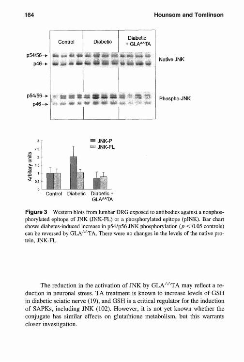
164 Hounsom Tomlinson
p54l56-c
P46+
p54/56+
P46 "b
I I
I I + Diabetic
T
JNK-P JNK-FL
Native JNK
Phospho-JNK
+ GLAAATA
Figure 3 Western blots from lumbar DRG exposed to antibodies against a nonphos- phorylated epitope of JNK (JNK-F%) or a phosphorylated epitope (pJNK). Bar chart shows diabetes-induced increase in p54/p56 JNK phosphorylation ( p < 0.05 controls) can be reversed GLA'TA. There were no changes in the levels the native pro- tein, JNK-F%.
The reduction in the activation of JNK by GLAMTA may reflect a re- duction in neuronal stress. TA treatment is known to increase levels of GSH in diabetic sciatic nerve and GSH is a critical regulator for the induction of SAPKs, including JNK However, it is not yet known whether the conjugate has similar effects glutathione metabolism, but this warrants closer investigation.

Neurotrophic Support in Experimental Diabetic Neuropathy 165
VIII. ROLE FOR GLAAATA IN THE TREATMENT OF DIABETIC NEUROPATHY
This is the first instance of a treatment that is capable of attenuating both electrophysiological and neurochemical deficits in the nerves of diabetic rats. The effect of the GLAMTA conjugate is also remarkable when related to the current effects of TA and previously reported effects of EPO. Approximate calculations suggest that the dose of GLAMTA used here could deliver about 14 mg/kg/day TA and 18 mg/kg/day GLA. TA must be given at doses of at least 50 mg/kg/day to influence nerve conduction (19), and in the present and in previous (20) studies had little effect on sciatic nerve NGF levels at 100 mg/kg/day. Assuming an approximate content of 10% GLA in EPO (83), a daily consumption of about 180 mg oil would be required to match the current dose of the conjugate. A previous study demonstrated no effect of EPO at about 3.5 g/day per rat (equivalent to about 10 g/kg/day) on the deficit in sciatic nerve SP in diabetic rats (82). It is therefore clear that the properties of the conjugate are significantly greater than those of its constituent mole- cules. We suspected that a membrane localization might be important and that was why BHT, a lipid-soluble antioxidant, was included in this study, but it showed no efficacy, indicating that simple membrane sequestration of TA does not provide an explanation for the efficacy of the conjugate. Furthermore, although NGF treatment of diabetic rats does not affect nerve conduction deficits, the conjugate had multiple protective effects on different fiber groups-viz, increasing nerve NGF levels, stimulating NPY expression by a different mechanism, and boosting deficient conduction velocities. Clearly, this molecule shows clinical potential.
REFERENCES
1. Diabetes Control and Complications Trial Research Group. The effect of inten- sive treatment of diabetes on the development and progression of long-term complications in insulin-dependent diabetes mellitus. N Engl J Med 1993; 329:
Brown MJ, Asbury AK. Diabetic neuropathy. Ann Neurol 1984; 15:2-12. Dyck PJ, Bushek W, Spring EM, Karnes JL, Litchy WJ, O'Brien PC, Service FJ. Vibratory and cooling detection thresholds compared with other tests in diagnosing and staging diabetic neuropathy. Diabetes Care 1987; 10:432-440.
977-986. 2. 3.

166 Hounsom and Tomlinson
4.
5.
6.
7.
8.
9.
10.
11.
12.
13.
14.
15.
16.
17.
18.
19.
Thomas PK, Tonilinson DR. Diabetic and hypoglycaemic neuropathy. In: Dyck PJ, Thomas PK, Griffin JW, Low PA, Poduslo JF, ed. Peripheral Neuropathy. 3rd ed. Philadelphia: W.B. Saunders, 1992: 1219- 1250. Biessels GJ. Van Dam PS. Diabetic neuropathy pathogenesis and current treat- ment perspectives. Neurosci Res Coniniun 1997; 20: 1 - 10. Yagihashi S. Pathology and pathogenetic mechanisms of diabetic neuropathy. Diabetes Metab Rev 1996; 11: 193-225. Dyck PJ, Giannini C. Pathologic alterations in the diabetic neuropathies of hu- mans: a review. J Neuropathol Exp Neurol 1996; 55: 1 18 1 - 1 193. Tesfaye S, Malik R, Ward JD. Vascular factors in diabetic neuropathy. Diabeto- logia 1994; 37:847-854. Williamson JR, Chang K, Frangos M, Hasan KS, Ido Y, Kawamura T, et al. Hyperglycemic pseudohypoxia and diabetic complications. Diabetes 1993; 42:
Horrobin DF. The roles of essential fatty acids in the development of diabetic neuropathy and other complications of diabetes mellitus. Prostaglandins Leukot Essent Fatty Acids 1988; 31:181-197. Cameron NE, Cotter MA, Basso M, Hohman TC. Comparison of the effects of inhibitors of aldose reductase and sorbitol dehydrogenase on neurovascular function, nerve conduction and tissue polyol pathway metabolites in streptozo- tocin-diabetic rats. Diabetologia 1997; 40:27 1-28 I . Sasaki H, Schmelzer JD, Zollman PJ, Low PA. Neuropathology and blood flow of nerve, spinal roots and dorsal root ganglia in longstanding diabetic rats. Acta Neuropathol (Berl) 1997; 93: 118-128. Kihara M, Low PA. Impaired vasoreactivity to nitric oxide in experimental diabetic neuropathy. Exp Neurol 1995; 132:180-185. Tonilinson DR, Fernyhough P, Diemel LT. Role of neurotrophins in diabetic neuropathy and treatment with nerve growth factors. Diabetes 1997; 46:S43- s49. Dyck PJ. Nerve growth factor and diabetic neuropathy. Lancet 1996; 348: 1044- 1045. Tomlinson DR, Fernyhough P, Diemel LT. Neurotrophins and peripheral neu- ropathy. Philos Trans R Soc Lond B 1996; 351:455-462. Thomas PK. Growth factors and diabetic neuropathy. Diabet Med 1994; 11:
Packer L. Antioxidant properties of lipoic acid and its therapeutic effects in prevention of diabetes complications and cataracts. Ann NY Acad Sci 1994;
Nagamatsu M, Nickander KK, Schnielzer JD, Raya A, Wittrock DA, Tritschler H, et al. Lipoic acid improves nerve blood flow, reduces oxidative stress, and improves distal nerve conduction in experimental diabetic neuropathy. Diabetes Care 1995; 18:1160-1167.
801-81 3.
732-739.
738:257-264.

Neurotrophic Support in Experimental Diabetic Neuropathy 167
20.
21.
22.
23.
24.
25.
26.
27.
28.
29.
30.
31.
32.
33.
34.
Garrett NE, Malcangio M, Dewhurst M, Tonilinson DR. a-Lipoic acid corrects neuropeptide deficits in diabetic rats via induction of trophic support. Neurosci Lett 1997; 222:191-194. Ebendal T. Function and evolution in the NGF family and its receptors. J Neu- rosci Res 1992: 323461-470. Hcndry IA, Stoeckel K. Thoenen H, Iversen LL. The retrograde axonal trans- port of nerve growth factor. Brain Res 1974; 68:103-121. Schmidt RE, Plurad SB, Saffitz JE, Grabau GG, Yip HK. Retrograde axonal transport of ['I51]-nerve growth factor in rat Ileal inesentric nerves-effect of streptozocin diabetes. Diabetes 1985; 34: 1230-1240. Richardson PM, Verge Issa VMK, Riopelle RJ. Distribution of neuronal recep- tors for nerve growth factor in the rat. J Neurosci 1986; 6:2312-2321. DiStefano PS, Friedman B, Radziejewski C, Alexander C, Boland P, Schick CM, et al. The neurotrophins BDNF, NT-3, and NGF display distinct patterns of retrograde axonal transport in peripheral and central neurons. Neuron 1992; 8:983-993. Meakin SO, Suter U, Drinkwater CC, Welcher AA, Shooter EM. The rat trk protooncogene product exhibits properties characteristic of the slow nerve growth factor receptor. Proc Natl Acad Sci USA 1992; 89:2374-2378. Martin-Zanca D, Oskam R. Mitra G, Copeland T, Barbacid M. Molecular and biochemical characterization of the human trk proto-oncogene. Mol Cell Biol
Klein R, Nanduri V, Jing SA, Lamballe F, Tapley P, Bryant S, et al. The trkB tyrosine protein kinase is a receptor for brain-derived neurotrophic factor and neurotrophin-3. Cell 1991; 66395-403. Klein R, Lamballe F, Bryant S, Barbacid M. The trkB tyrosine protein kinase is a receptor for neurotrophin-4. Neuron 1992; 8:947-956. Lamballe F, Tapley P, Barbacid M. rrkC encodes multiple neurotrophin-3 re- ceptors with distinct biological properties and substrate specificities. EMBO J
Lindsay RM, Warmar AJ. Nerve growth factor regulates expression of neuro- peptide genes in adult sensory neurons. Nature 1989;337:362-364. Fernyhough P, Diemel LT, Brewster WJ, Tomlinson DR. Deficits in sciatic nerve neuropeptide content coincide with a reduction in target tissue nerve growth factor mRNA in streptozotocin-diabetic rats; effects of insulin treat- ment. Neuroscience 1994; 62:337-344. Fernyhough P, Diemel LT, Hardy J, Brewster WJ, Mohiuddin L, Tomlinson DR. Human recombinant nerve growth factor replaces deficient neurotrophic support in the diabetic rat. Eur J Neurosci 1995; 7: 1 107-1 110. Jakobsen J, Brimijoin S, Skau K, Sidenius P, Wells D. Retrograde axonal trans- port of transmitter enzymes, fucose-labeled protein, and nerve growth factor in streptozotocin-diabetic rats. Diabetes 1981 ; 30:797-803,
1989; 9:24-33.
1993; 12:3083-3094.

168 Hounsom and Tomlinson
3s.
36.
37.
38.
39.
40.
41.
42.
43
44.
4s.
46.
47.
48.
49.
Schmidt RE, Grabau GG, Yip HK. Retrograde axonal transport of ['251]nerve growth factor in ileal mesenteric nerves in vitro: effect of streptozotocin diabe- tes. Brain Res 1986; 378:325-336. Hellweg R, Hartung H-D. Endogenous levels of nerve growth factor (NGF) are altered in experimental diabetes mellitus: a possible role for NGF in the pathogenesis of diabetic neuropathy. J Neurosci Res 1990; 26:258-267. Hellweg R, Wohrle M, Hartung H-D, Stracke H, Hock C, Federlin K. Diabetes mellitus-associated decrease in nerve growth factor levels is reversed by alloge- neic pancreatic islet transplantation. Neurosci Lett 1991 ; 125: 1-4. Hanaoka Y, Ohi T, Furukawa S, Furukawa Y, Hayashi K, Matsukura S. Effect of 4-methylcatechol on sciatic nerve growth factor level and motor nerve con- duction velocity in experimental diabetic neuropathic process in rats. Exp Neu- rol 1992; 115:292-296. Faradji V, Sotelo J. Low serum levels of nerve growth factor in diabetic neurop- athy. Acta Neurol Scand 1990; 81:402-406. Delcroix J, Tomlinson DR, Fernyhough P. Diabetes and axotomy-induced deficits in retrograde axonal transport of nerve growth factor correlate with decreased levels of p7sLm protein in lumbar dorsal root ganglia. Mol Brain Res
Doherty P, Seaton P, Flanigan TP, Walsh FS. Factors controlling the expression of the NGF receptor in PC12 cells. Neurosci Lett 1988; 92:222-227. Maeda K, Fernyhough P, Tomlinson DR. Effects of treatment of diabetic rats with human recombinant nerve growth factor on sciatic nerve conduction, Doppler flux and substance P levels. Diabet Nutr Metab 1997; 10:3-8. Perez-Polo JR. The nerve growth factor receptor. In: Horrocks LA, Neff NH, Yates AJ, Handjiconstantinou M, eds. Trophic Factors and the Nervous System. New York: Raven Press, 1990:107-118. Gaetani P, Lombardi D. Brain damage following subarachnoid hemorrhage: the imbalance between anti-oxidant systems and lipid peroxidative processes. J Neurosurg Sci 1992; 36:l-10. Kitagawa K, Matsumoto M, Oda T, Niinobe M, Hata R, Handa N, et al. Free radical generation during brief period of cerebral ischemia may trigger delayed neuronal death. Neuroscience 1990; 3555 1-558. Jenner P, Dexter DT, Sian J, Schapira AH, Marsden CD. Oxidative stress as a cause of nigral cell death in Parkinson's disease and incidental Lewy body disease. Ann Neurol 1992; 32(suppl):S82-S87. Romero FJ, Monsalve E, Hermenegildo C, Puertas FJ, Higueras V, Nies E, et al. Oxygen toxicity in the nervous tissue: comparison of the antioxidant defense of rat brain and sciatic nerve. Neurochem Res 1991; 16:157-161. Jackson GR, Apffel L, Werrbach-Perez K, Perez-Polo JK. Role of nerve growth factor in oxidant-antioxidant balance and neuronal injury. I. Stimulation of hy- drogen peroxide resistance. J Neurosci Res 1990; 25:360-368. Tiffany-Castiglioni E, Perez-Polo JR. Stimulation of resistance to 6-hydroxy-
1997; 51 :82-90.

Neurotrophic Support in Experimental Diabetic Neuropathy 169
50.
51.
52.
53.
54. 55.
56.
57.
58.
59.
60.
61.
62.
63.
64.
dopamine in a human neuroblastonia cell line by nerve growth factor. Neuro- sci Lett 1981; 26:157-161. Tiffany-Castiglioni E, Perez-Polo JR. The role of nerve growth factor in vitro in cell resistance to 6-hydroxydopaniine toxicity. Exp Cell Res 1979; 12 I : 179- 189. Sampath D, Perez-Polo R. Regulation of antioxidant enzyme expression by NGF. Neurochem Res 1997; 2235 1-362. Sainpath D, Jackson GR, Werrbach-Perez K, Perez-Polo JR. Effects of nerve growth factor on glutathione peroxidase and catalase in PC12 cells. J Neuro- chein 1994; 622476-2479. Naveilhan P, Neveu I, Jehan F, Baudet C, Wion D, Brachet P. Reactive oxygen species influence nerve growth factor synthesis in primary rat astrocytes. J Neu- rochem 1994; 62:2178-2186. Wolff SP. Diabetes mellitus and free radicals. Br Med Bull 1993; 49:642-652. Wolff SP, Jiang ZY, Hunt JV. Protein glycation and oxidative stress in diabetes mellitus and ageing. Free Rad Biol Med 1991; 10:339-352. Stith BJ, Proctor WR. Microinjection of inositol 1,2-(cyclic)-4,5-trisphosphate. inositol I ,3,4,5,-tetrkisphosphate, and inositol 1,4,5-trisphosphate into intact Xenoprs oocytes can induce membrane currents independent of extracellular calcium. J Cell Biochem 1989: 40:321-330. Nickander KK, McPhee BR, Low PA, Tritschler H. Alpha-lipoic acid: antioxi- dant potency against lipid peroxidation of neural tissues in vitro and implica- tions for diabetic neuropathy. Free Rad Biol Med 1996; 21:631-639. Low PA, Nickander KK. Oxygen free radical effects in sciatic nerve in experi- mental diabetes. Diabetes 1991: 40:873-877. Cameron NE, Cotter MA, Archibald V, Dines KC, Maxfield EK. Anti-oxidant and pro-oxidant effects on nerve conduction velocity, endoneurial blood flow and oxygen tension in non-diabetic and streptozotocin-diabetic rats. Diabeto- logia 1994; 37:449-459. Karasu c, Dewhurst M, Stevens EJ, Toinlinson DR. Effects of anti-oxidant treatment on sciatic nerve dysfunction in streptozotocin-diabetic rats; compari- son with essential fatty acids. Diabetologia 1995; 38: 129- 134. Cameron NE, Cotter MA, Maxfield EK. Anti-oxidant treatment prevents the development of peripheral nerve dysfunction in streptozotocin-diabetic rats. Diabetologia 1993; 36:299-304. Love A, Cotter MA, Cameron NE. Effects of a-tocopherol on nerve conduction velocity and regeneration following a freeze lesion in immature diabetic rats. Naunyn Schniiedebergs Arch Pharmacol 1997; 355: 126- 130. Bravenboer B, Kappelle AC, Homers FPT, Van Buren T, Erkelens DW, Gispen WH. Potential use of glutathione for the prevention and treatment of diabetic neuropathy in the streptozotocin-induced diabetic rat. Diabetologia 1992; 35:
Cameron NE. Cotter MA. Neurovascular dysfunction in diabetic rats. Potential 8 13-8 17.

170 Hounsom and Tomlinson
65.
66. 67.
68.
69.
70.
71.
72.
73.
74.
75.
76.
77.
78.
79.
80.
81.
contribution of autoxidation and free radicals examined using transition metal chelating agents. J Clin Invest 1995; 96: 1159- 1163. Reed LJ, DeBusk BG, Gunsalius IC, Hornberger CSJ. Crystalline alpha-lipoic acid: a catalytic agent associated with pyruvate dehydrogenase. Science 195 I ;
Reed LJ. Multienzynie complex. Acc Chem Res 1974; 7:40-46. Packer L, Witt EH, Tritschler HJ. a-Lipoic acid as a biological antioxidant. Free Rad Biol Med 1995; 19227-250. Busse E, Zimnier G, Schopohl B, Kornhuber B. Influence of alpha-lipoic acid on intracellular glutathione in vitro and in vivo. Arzneiniittelforschung 1992;
Natraj CV, Gandhi VM, Menon KKG. Lipoic acid and diabetes: effects of dihy- drolipoic acid administration in diabetic rats and rabbits. J Biosci 1984; 6:37- 46. Wagh SS, Natraj CV, Menon KKG. Mode of action of lipoic acid in diabetes. J Biosci 1987; I1:59-74. KHhler W, Kuklinski B, Ruhlinann C, Plotz C. Diabetes mcllitus-a free radi- cal-associated disease. Results of adjuvant antioxidant supplementation. Z Gesamte Inn Med 1993; 48:223-232. In German. Ziegler D, Hanefeld M, Ruhnau KJ, Meissner HP, Lobisch M, Schiitte K, et al. Treatment of symptomatic diabetic peripheral neuropathy with the anti- oxidant a-lipoic acid-a .?-week inulticentre randomized controlled trial (ALADIN study). Diabetologia 1995; 38:1425-1433. Haugaard N, Haugaard ES. Stimulation of glucose utilization by thioctic acid in rat diaphragm incubated in vitro. Biochim Biophys Acta 1970; 222583- 586. Bashan N, Burdett E, Klip A. Effect of thioctic acid on glucose transport. 3rd Int Thoctic Acid Workshop 1993; 3:218-223. Grunert R. The effect of D L-alpha-lipoic acid on heavy metal intoxication in mice and dogs. Arch Biochem Biophys 1960; 86: 190- 195. Matsugo S, Yan LJ, Han D, Trischler HJ, Packer L. Elucidation of antioxidant activity of alpha-lipoic acid toward hydroxyl radical. Biochem Biophys Res Cominun 1995; 208:161-167. Ou P, Tritschler HJ, Wolff SP. Thioctic (lipoic) acid: a therapeutic metal-chelat- ing antioxidant? Biochern Pharmacol 1995: SO: 123- 126. Brownlee M. Glycation products and the pathogenesis of diabetic complica- tions. Diabetes Care 1992; 15: 1835- 1843. Dimpfel W, Spuler M, Pierau FK, Ulrich H. Thioctic acid induces dose-depen- dent sprouting of neurites in cultured rat neuroblastoma cells. Dev Pharniacol Thcr 1990; 14: 193- 199. Kemplay S, Martin P, Wilson S. The effects of thioctic acid on motor nerve terminals in acrylamide-poisoned rats. Neuropathol Appl Neurobiol 1988; 14:
Mulase K, Hattori A, Kohno M, Hayashi K. Stimulation of iiervc growth factor
114:93-94.
421829-83 1.
275-288.

Neurotrophic Support in Experimental Diabetic Neuropathy 171
82.
83.
84.
85.
86.
87.
88.
89.
90.
91.
92.
93.
94.
synthesis/secretion in mouse astroglial cells by coenzymes. Biochem Mol Biol Int 1993; 30:615-621. Tonilinson DR, Robinson JP, Conipton AM, Keen P. Essential fatty acid treat- ment-effects on nerve conduction, polyol pathway and axonal transport in streptozotocin diabetic rats. Diabetologia 1989; 32:655-659. Lockett MJ, Tomlinson DR. The effects of dietary treatment with essential fatty acids on sciatic nerve conduction and activity of the Nat/K' pump in strcptozo- tocin-diabetic rats. Br J Pharmacol 1992; 105:355-360. Stevens EJ, Carrington AL. Tomlinson DR. Prostacyclin release in experimen- tal diabetes: effects of evening primrose oil. Prostaglandins Leukot Essent Fatty Acids 1993; 49:699-706. Stevens EJ, Lockett MJ, Carrington AL, Tomlinson DR. Essential fatty acid treatment prevents nerve ischaemia and associated conduction anomalies in rats with experimental diabetes mellitus. Diabetologia 1993; 36397-401. Carrington AL, Lockett MJ, Tomlinson DR. The effects of dietary supplernen- tation with evening primrose oil on resistance to anoxic conduction block in nerves from control and streptozotocin-diabetic rats (abstr). Diabet Med 1992; 9(suppl 1):35A. Stevens EJ, Lockett MJ, Carrington AL, Tomlinson DR. Essential fatty acid treatment prevents nerve ischaemia and associated conduction anomalies in ex- perimental diabetes (abstj. Br J Phaiinacol 1993; 108:38P. Lundberg JM. Terenius L, Hokfelt T, Martling CR, Tatenioto K. Mutt V, et al. Neuropeptide Y (NPY)-like irnrnunorcactivity in peripheral noradrenergic ncurons and effects of NPY on syinpathctic function. Acta Physiol Scand 1982;
Tatemoto K, Carlquist M, Mutt V. Neuropeptide Y-a novel brain peptide with structural similarities to peptide YY and pancreatic polypeptide. Nature 1982;
Wakisaka S, Kajander KC, Bennett GJ. Increased neuropeptide Y (NPYj-like immunoreactivity in rat sensoiy neurons following peripheral axotomy. Neu- rosci Lett 199 I ; 124:200-203. Ohara S , Tantuwaya V, DiStefano PS, Schmidt RE. Exogenous NT-3 mitigates the transganglionic neuropeptide Y response to sciatic nerve injury. Brain Res
Verge VMK, Richardson PM, Wiesenfeld-Hallin Z, Hokfelt T. Differential in- fluence of nerve growth factor on neuropeptide expression in vivo: a novel role in peptide suppression in adult sensory neurons. J Neurosci 1995: 15:2081- 2096. Allen JM, Tischler AS, Lee YC, Bloom SR. Neuropeptide Y (NPY) in PC12 phaeochrornocytoma cultures: responses to dexamethasone and nerve growth factor. Neurosci Lett 1984; 46:29 1-296. Higuchi H, Nakano K, Miki N. Identification of NGF-response element in the rat neuropeptide Y gene and induction of the binding proteins. Biochem Bio- phys Res Commun 1992; 189: 1553-1560.
116:477-480.
2961659-660.
1995: 699:143-148.

172 Hounsom and Tomlinson
9s.
96.
97.
98
99.
100.
101.
102.
Balbi D, Allen JM. Role of protein kinase C in mediating NGF effect on neuro- peptide Y expression in PC12 cells. Brain Res Mol Brain Res 1994; 23:310- 3 16. Zhang X, Wiesenfeld-Hallin Z, Hokfelt T. Effect of peripheral axotomy on expression of neuropeptide Y receptor mRNA in rat lumbar dorsal root ganglia. Eur J Neurosci 1994; 6:43-57. Kiefer F, Tibbles LA, Lassam N, Zanke B, lscove N, Woodgett JR. Novcl components of mammalian stress-activated protein kinase cascades. Biocheni SOC Trans 1997; 25:491-498. Whitmarsh AJ, Davis RJ. Transcription factor AP-I regulation by niitogen- activated protein kinase signal transduction pathways. J Mol Med 1996; 74:
Su B, Karin M. Mitogen-activated protein kinase cascades and regulation of gene expression. Curr Opin Inimunol 1996; 8:402-41 I . Woodgett JR, Avruch J, Kyriakis J. The stress activated protein kinase pathway. Cancer Surv 1996; 27:127-138. Minden A, Karin M. Regulation and function of the JNK subgroup of MAP kinases. Biochim Biophys Acta Rev Cancer 1997; 1333:F8S-F104. Wilhelm D, Bender K, Knebel A, Angel P. The level of intracellular glutathione is a key regulator for the induction of stress-activated signal transduction path- ways including Jun N-terminal protein kinases and p38 kinase by alkylating agents. Mol Cell Biol 1997; 17:4792-4800.
589-607.

12 Clinical Trials of a-Lipoic Acid in Diabetic Polyneuropathy and Cardiac Autonomic Neuropathy
Dan Ziegler Diabetes Research Institute, Heinrich-Heine-University, Diisseldorf, Germany
Polyneuropathy involving the somatic and autonomic nervous system is re- sponsible for substantial morbidity and increased mortality among diabetic patients, Near normoglycemia is now generally accepted as the primary ap- proach to prevention of diabetic neuropathy (1,2). However, in diabetic pa- tients with advanced stages of peripheral neuropathy, relatively long periods of near-normal glycemic control for several months or even years may be needed to retard the progression of nerve dysfunction (3). Because normogly- cemia is not achievable in most diabetic patients, the effects of several medical treatments derived from the pathogenetic concepts of diabetic neuropathy have been evaluated in numerous randomized clinical trials during the past two decades. However, due to various reasons, none of these compounds has been marketed as yet in the major European countries or in the United States. None- theless, in symptomatic diabetic neuropathy, additional pharmacological treat- ment of painful neuropathic symptoms is frequently required to maintain the patients’ quality of life. Although treatment of pain with antidepressants is effective, it may be of limited value because of frequent adverse reactions (4). Other symptomatic approaches including anticonvulsants, mexiletine, and topical capsaicin either have not been unequivocally effective, have shown only partial effects, or caution has been expressed as to potential neurotoxic
173

174 Ziegler
side effects in view of longer term treatment (4). Furthermore, these medica- tions are designed to modulate symptoms without influencing the underlying neuropathy.
Cardiovascular autonomic neuropathy (CAN) is a serious complication of diabetes that is associated with a poor prognosis and m y result in severe clinical symptoms (5 ) . Although CAN is appreciated as a clinical entity since 1945, in the past it has received less attention than peripheral sensorimotor neuropathy, one reason being that noninvasive quantitative and reliable meth- ods for assessment of cardiovascular autonomic function are available since only the last two decades. After the introduction of cardiovascular reflex tests based on changes in heart rate variability (HRV) and blood pressure regulation into clinical routine, i t became evident that CAN may be frequently detected at early stages in asymptomatic diabetic patients (5) .
The question of long-term primary prevention of CAN has been previ- ously addressed only in trials that studied the role of near norinoglycemia in Type 1 diabetic patients. These studies have shown that the development of abnormalities in HRV can be prevented or retarded by intensive insulin ther- apy (1,6). Secondary intervention trials in patients with advanced CAN have demonstrated that its progression can be delayed during long-term near nor- moglycemia, but periods of more than 2 years are needed ( 3 5 ) .
1. ROLE OF OXIDATIVE STRESS IN DIABETIC NEUROPATHY: CLINICAL AND EXPERIMENTAL EVIDENCE
A growing body of evidence suggests that oxidative stress resulting from en- hanced free radical formation and/or defects in antioxidant defense is impli- cated in the development of various disorders, including neurodegenerative diseases (7) and diabetic complications (8,9). Increased free radical formation and changes in hemostatic variables related to endothelial damage have been found in Type 2 diabetic patients with microalbuminuria (10). Impaired endo- thelium-dependent vasodilation is improved by administration of vitamin C, suggesting that nitric oxide inactivation by increased oxygen free radical activ- ity contributes to abnormal vascular reactivity in diabetes (1 1). Furthermore, impaired cellular scavenging activity against oxidative stress ( 12) and elevated levels of plasma hydroperoxides in conjunction with a trend to lower vitamin E levels ( I 3) have recently been demonstrated in patients with Type 2 diabetes.
In experimental diabetic neuropathy, oxygen free radical activity in the sciatic nerve is increased (9). Treatment with a-lipoic acid, a potent lipophilic

a-Lipoic Acid in Diabetic Neuropathy 175
free radical scavenger (14), results in prevention of neurovascular abnormali- ties associated with experimental diabetic neuropathy (15). It has been demon- strated that reduced digital nerve conduction velocity (NCV), nerve blood flow, and glutathione levels in diabetic rats are normalized and in vitro lipid peroxidation of neural tissue reduced by a-lipoic acid in a dose-dependent manner ( 15,16), suggesting that the improvement in neurovascular changes were induced by improving oxygen free radical scavenging activity. One mechanism of reduced nerve blood flow is the inhibitoiy effect of superoxide anion on nitric oxide synthase. Because nitric oxide synthase is reduced in experimental diabetic neuropathy ( I7), a-lipoic acid might prevent this inhibi- tion by reducing oxidative stress (15). A recent study has also demonstrated that treatment with a-lipoic acid may correct neuropeptide deficits in diabetic rats, indicating that the compound may boost neurotrophic support (IS). Ad- ministration of low doses of an a-lipoic acid-gamma-linolenic acid conjugate corrects the nerve conduction and nerve blood flow deficits (19) and sciatic nerve contents of nerve growth factor, substance P, and neuropeptide Y (20) in diabetic rats, suggesting a marked synergistic action of these compounds. These experimental findings provide the rationale for a potential therapeutic value of a-lipoic acid in diabetic patients with neuropathy.
11. MULTICENTER CONTROLLED CLINICAL TRIALS
Randomized clinical trials (RCTs) are a widely accepted means of applying experimental methods to a clinical setting and have been advocated as the gold standard for comparing and evaluating different treatments (2 I ) . However, the quality of the RCTs that evaluated the effects of medical treatment in diabetic polyneuropathy was poor. Cavaliere et al. (22) assessed the quality of scientific evidence for the efficacy of various pathogenetically oriented treatment ap- proaches for diabetic polyneuropathy examined in RCTs published between 1981 and 1992. They used a quality system covering the internal (scientific) validity (the ability to demonstrate a treatment effect if i t really exists) and external validity (the possibility of generalizing the study results to patients seen in clinical practice). The analysis based on 38 RCTs in total revealed a devastating picture: The methods of randomization were unspecified and a detailed a priori estimate of the sample size needed to detect a treatment differ- ence was not reported in 95% of the RCTs, respectively. Only 11% of the RCTs had sufficient statistical power to detect a clinically meaningful differ- ence (22). More generally spoken. a recent analysis by Freiman et al. (23) revealed that only 4 of 71 controlled clinical trials (5.6%) that reported nega-

176 Ziegler
Table 1 a-Lipoic Acid in Diabetic Peripheral and Cardiac Autonomic Neuropathy
Multicenter, Randomized, Double-Blind, Placebo-Controlled Trials Using
ALADIN Study8 DEKAN Studyh ALADIN 3 Study'
Number 328 13 SO9 Design Four parallel groups Two parallel groups Three parallel groups Dose 1,200/600/ I 0 0 nig a/p 800 mg d P PO 600 lV/l800 mg
Duration of 3 wk 4 mo 3 wk + 6 mo
Effect Symptoms+, NDS+ HRV+, TSS TSS. NIS+
a, a-Lipoic acid; P, placebo; +, improvement; IV, intravenous; PO, orally 'Diabetologia, 38, 1995. hDiabetes Care. 20, 1997. 'Diabetes Care, 22, 1999.
I.V. PO( a-da-P/P-P)
treatment
tive results ( p > 0.1) in major medical journals between 1960 and 1977 in- cluded a sample size large enough to have a 90% chance of detecting a 25% difference in treatment effects. In another analysis, the situation did not im- prove as the number was 4 of 65 trials (6.2%) (23). Thus, as recently stated by Altman and Bland (24), absence of evidence is not evidence of absence. In other words, to interpret most published RCTs as providing evidence of an ineffectiveness of the respective treatment in diabetic neuropathy would be clearly misleading, because the likelihood to detect differences in the paranie- ters of nerve function between the treatment groups was too low in view of the small sample sizes.
Adequate designs for RCTs in diabetic neuropathy have to consider the following aspects: type and stage of neuropathy, homogeneity of the study population, outcome measures (neurophysiological markers, intermediate clin- ical end points, ultimate clinical outcomes, quality of life), natural history, sample size, study duration, reproducibility of neurophysiological and inter- mediate end points, regression to the mean, true and perceived placebo effects, measures of treatment effect, and the generalizability of the overall trial result to individual patients (25). The results of three multicenter RCTs that evalu- ated the effects of a-lipoic acid in Type 2 diabetic patients with polyneuropa- thy are summarized and discussed below (Table 1).
A. ALADIN Study
The improvement in neuropathic symptoms during a 3-week period of intrave- nous treatment with 600 mg a-lipoic acid/day as compared with vitamin Bl

a-Lipoic Acid in Diabetic Neuropathy 177
(26) in conjunction with the recent experimental evidence (15) formed the rationale for a large-scale, 3-week, multicenter, randomized, double-blind, pla- cebo-controlled trial (Alpha-Lipoic Acid in Diabetic Neuropathy: ALADIN) (27). Assuming from previous studies a placebo response of about 30% and a dnig response of about 60%, the a priori estimate of the required sample size yielded I I = 67 patients per group, with a = 0.05 and p = 0.1 for the two-tailed log-rank test.
The efficacy and safety of intravenous infusion of a-lipoic acid were evaluated in 328 NIDDM outpatients with symptomatic peripheral neuropathy who were randomly assigned to treatment using three doses (a-lipoic acid I200 mg/day, 600 mg/day, 100 mg/day) or placebo. Neuropathic symptoms (pain, burning, paresthesias, and numbness) were assessed by the total symp- tom score (TSS) at baseline and each visit (days 2-5, 8- 12, and 15- 19) before infusion. In addition, the Hamburg Pain Adjective List (HPAL), a multidimen- sional specific pain questionnaire, and the Neuropathy Symptom and Disabil- ity Scores (NDS) were assessed at baseline and at day 19 (27).
According to the protocol, 260 patients (n = 65, 1200 mg/day; I I = 63, 600 mg/day; ti = 66, 100 mg/day; i i = 66, placebo) completed the study. No significant differences were noted for the mean changes in HbA, and blood glucose levels between the four groups studied. TSS in the feet decreased from baseline to day 19 (mean 5 SD) by -4.5 t 3.7 (-58.6%) points in a-lipoic acid 1200, -5.0 t 4.1 (-63.5%) points in a-lipoic acid 600, -3.3 5 2.8 (-43.2%) points in a-lipoic acid 100, and -2.6 ? 3.2 (-38.4%) points in placebo (a-lipoic acid 1200 vs. placebo, p = 0.003; a-lipoic acid 600 vs. placebo, p < 0.001). The response rates after 19 days, defined as an improve- ment in the TSS of at least 30%, were 70.8% in a-lipoic acid 1200, 82.5% in a-lipoic acid 600, 65.2% in a-lipoic acid 100, and 57.6% in placebo (a - lipoic acid 600 vs. placebo, p = 0.002). The total scale of the HPAL was significantly reduced in a-lipoic acid 1200 and a-lipoic acid 600 as compared with placebo after 19 days (both p < 0.01). Detailed analysis of pain revealed that both the affective and sensory components of pain, representing pain ex- perience and pain perception, could be improved. NDS decreased by - 1.8 +. 0.3 points in a-lipoic acid 1200, by -1.5 ? 0.3 points in a-lipoic acid 600, by -0.9 t 0.3 in a-lipoic acid 100, and by -1.0 5 0.2 in placebo after 19 days ( p = 0.03 for a-lipoic acid 1200 vs. placebo). The rates of adverse events were 32.6% in a-lipoic acid 1200, 18.2% in a-lipoic acid 600, 13.6% in a- lipoic acid 100, and 20.7% in placebo. These findings demonstrate that paren- teral treatment with a-lipoic acid over 3 weeks using a dose of 600 mg/day in NIDDM patients is associated with a significant reduction of various symp- toms of peripheral neuropathy including pain, paresthesias, and numbness as

178 Ziegler
compared with placebo. Furthermore, it is evident that a dose of 100 mg/day does not exert an effect superior to that seen with placebo. An increase in the dosage to 1200 mglday is associated with an enhanced rate of adverse events rather than with maximized efficacy. The increased risk of gastrointestinal side effects associated with 1200 mg/day precludes from using this dose (27).
It may be argued that nerve conduction studies have not been used in this study as objective measures of neuropathy. However, electrophysiological changes are not relevant in patients with pain. In addition, in a short-term study of this kind, a significant difference between the groups treated with a- lipoic acid and the placebo group regarding NCV would not appear likely to occur. Previous studies using drugs such as the aldose reductase inhibitors have shown that NCV was either unchanged (28) or only a minimal increase was seen within several weeks of treatment (29) that was subject to substantial criticism as to whether i t represented a clinically meaningful degree of change or merely a physiological variation (30). By contrast, neuropathic symptoms have been shown susceptible to intervention within a few weeks. Painful symptoms but not motor and sensory NCV were improved after 4 weeks of treatment with sorbinil as compared with placebo (28), and withdrawal of tolrestat resulted in a rapid worsening of pain scores (31).
It may also be argued that the exclusion of 51 patients due to failures to adhere to the protocol potentially could have introduced bias. However, an additional analysis of the results of the ALADIN Study based on the intention to treat revealed no appreciable differences in the outcome of the parameters studied when compared with the per-protocol analysis, indicting that the ad- herence to the study protocol did not introduce bias. In summary, it has been demonstrated that in diabetic subjects a relief of neuropathic symptoms can be achieved by short-term intravenous with a-lipoic acid.
B. DEKAN Study
An effective treatment aimed at improving or retarding the progression of reduced HRV, the hallmark and earliest sign of CAN, might potentially favor- ably influence the poor prognosis among these patients.
The efficacy and safety of a-lipoic acid were studied in a randomized, double-blind, placebo-controlled, multicenter trial (Deutsche Kardiale Auto- noine Neuropathie: DEKAN Study) in NIDDM patients with CAN (32). Inclu- sion criteria were age 2 18 and a 7 0 years, NIDDM treated with diet, oral antidiabetic agents and/or insulin, spectral power of HRV in the low-fre- quency band (0.05-0.15 Hz) and/or high-frequency band (0.15-0.5 Hz) below

a-Lipoic Acid in Diabetic Neuropathy 179
the 2.5 centile of age-related normal ranges (33). Eligible patients were ran- domly assigned to treatment with an oral dose of 800 mg/day (200 mg four times daily) a-lipoic acid ( n = 39) or placebo (w = 34) for 4 months. Parame- ters of HRV at rest, including the coefficient of variation (CV), root mean squared successive difference (RMSSD), and spectral power in the low-fre- quency (0.05-0.15 Hz) and high-frequency (0.15-0.5 Hz) bands, and the QTc interval were assessed at baseline, 2 weeks, and at the end of each month of study using a validated computer system (33). Seventeen patients (a-lipoic acid, 11 = 10; placebo, ti = 7) dropped out of the study, but only 3 (a-lipoic acid, 11 = I ; placebo, n = 2) of these dropouts were due to adverse reactions.
Mean blood pressure, heart rate, and HbA, levels did not differ between the groups during the study. RMSSD increased from baseline to 4 months by 1.5 (-37.6-77.1) ins (median [min-max]) in the group treated with a-lipoic acid and decreased by -0.1 (- 19.2-32.8) ms in the placebo group ( p < 0.05 for a-lipoic acid vs. placebo). Power spectrum in the low-frequency band increased by 0.06 (-0.09-0.62) bpm’ (beats per minute squared) in a-lipoic acid, whereas it declined by -0.01 (-0.48-1.86) bpm’ in placebo ( p < 0.05 for a-lipoic acid vs. placebo). Furthermore, there was a trend toward a fa- vorable effect of a-lipoic acid versus placebo for the CV and for the high- frequency band power spectrum. QTc interval was shortened insignificantly by -8.8 (-87.9-46.5) ins in a-lipoic acid and remained unchanged in placebo (0.0 I- 118-77.91). No differences between the groups were noted regarding the rates of adverse events. These findings suggest that oral treatment with a- lipoic acid for 4 months using a well-tolerated dose of 800 nig/day in NIDDM patients is associated with a significant (two indices) or borderline (two indi- ces) improvement in HRV but not QTc interval as compared with placebo (32).
Apart from the DEKAN study, the effects of antioxidants on CAN have not been previously examined in a controlled clinical trial. However, a recent study has demonstrated that glutathione, a physiological antioxidant, nornial- izes reduced HRV during the squatting test (baroreflex changes) induced by acute hyperglycemia in healthy nondiabetic subjects (34). Presumably, such an effect is mediated by a reduction in free radical activity. In the isolated perfused diabetic rat heart, treatment with vitamin E completely prevented a progressive loss of histofluorescent nerve fibers in the myocardium and intra- neural catecholamines (35). There are no studies using a-lipoic acid in experi- mental diabetic cardiac neuropathy, but the compound normalizes reduced glucose uptake and glucose utilization and consequently oxygen uptake, myo- cardial ATP levels, and cardiac output in the isolated diabetic rat heart model

180 Ziegler
(36). However, it is not known whether and to which degree these experimen- tal findings relate to the chronic process that characterizes human diabetic autonomic neuropathy.
C. ALADIN 3 Study
This was a randomized, double-blind, placebo-controlled, multicenter trial in- cluding type 2 diabetic outpatients with symptomatic distal symmetric poly- neuropathy. Patients were allocated to three parallel groups receiving a-lipoic acid or placebo: group 1 ( 1 1 = 167), a-lipoic acid 600 mg intravenously for 3 weeks, followed by a-lipoic acid 600 mg three times a day orally for 6 months; group 2 (n = 174), a-lipoic acid 600 mg intravenously for 3 weeks, followed by placebo orally for 6 months; group 3 ( 1 1 = 168), placebo intrave- nously for 3 weeks, followed by placebo orally for 6 months. At baseline, there were no significant differences between the groups regarding the demographic variables such as age, sex, body inass index, duration of diabetes, and HbA,,. After 3 weeks, the Neuropathy Impail-ment Score (NIS) decreased signifi- cantly from baseline by -4.34 +- 0.35 ( 5 SEM) in the patients treated with a-lipoic acid and by -3.49 ? 0.58 points in the placebo group ( p = 0.016 for a-lipoic acid vs. placebo). The NIS of the lower limbs (NIS[LL]) declined by -3.32 2 0.26 on a-lipoic acid and -2.79 +- 0.42 points on placebo ( p =
0.055). After completion of the 6-month oral phase, the reduction in the NIS was -5.82 ? 0.73 in group 1, -5.76 ? 0.69 in group 2, and -4.31 +- 0.83 points in group 3 ( p = 0.095 for group 1 vs. 3), whereas the reduction in NIS[LL] was -4.39 f 0.51 in group I , -4.20 t 0.52 in group 2, and -3.37 +- 0.54 points in group 3 ( p = 0.086 for group I vs. 3). Regarding the TSS, no significant differences were noted between the groups at baseline and after 6 months. These results indicate a trend toward an improvement of neuropathic deficits but not symptoms after 6 months of oral treatment with a-lipoic acid (600 mg three times a day) after a 3-week intravenous phase in type 2 diabetic patients with polyneuropathy. The percentages of adverse events were compa- rable i n the three groups throughout the study.
111. FUTURE ASPECTS
Dyck et al. (37) recently calculated the sample size needed in a clinical trial based on the 2-year follow-up of the Rochester Diabetic Neuropathy Study. The estimates were derived from the changes of a composite score including NIS of the lower limbs plus seven tests (NIS[LL]+7: VPT great toe; R-R

a-Lipoic Acid in Diabetic Neuropathy 181
variation to DB; peroneal CMAP, MNCV, and MNDL; tibial MNDL; and sural SNAP) that among other measures performed best at showing monotone worsening over time. Assuming that a treatment effect of two NIS points is clinically meaningful, a 2-year study would need 68 patients in each treatment arm to have a power of 0.90 at the two-sided 0.05 level. If the effect of treat- ment is to halt the progression of neuropathy without improving it, a study of 3.7 years would be required. A 4-year study would require 45 patients per arm to achieve power of 0.90 to detect a treatment effect that inhibits progres- sion of neuropathy, and a clinically meaningful effect could be expected after approximately 2.4 years. Thus, a conservative estimate would yield 70- 100 patients per arm for a period of at least 3 years to achieve a high probability of detecting a clinically meaningful effect (37). A 4-year RCT evaluating the effects of a-lipoic acid in diabetic polyneuropathy (Neurological Assessment of Thioctic Acid in Diabetic Neuropathy 1 Study) has been designed on the basis of these estimates. The design of this trial is summarized in Table 2.
In conclusion, it is conceivable that the initial diabetes-related changes in the nerve are mediated by oxidative stress that, on a long-term basis, could result in progressive neuronal damage and therefore would be of pathogenetic relevance. Studies in proof of the promising results reported herein are needed. An ongoing pivotal long-term trial of oral treatment with a-lipoic acid de- signed along the recent guidelines of the Peripheral Nerve Society (38) aimed at slowing the progression of clinical neuropathy using a reliable clinical end
Table 2 Outline of the NATHAN 1 (Neurological Assessment of Thioctic Acid in Diabetic Neuropathy) Study
Design: Randomized, double-blind, placebo-controlled, multicenter trial Subjects: Two parallel groups of type 1 or type 2 diabetic patients (n = 500
Medication: Thioctic acid 600 m g or placebo tablets once daily orally Duration: Screening, 2 wk; placebo run-in, 6 wk; treatment, 192 wk; follow-up, 4
Inclusion criteria: Stage 1 or 2a polyneuropathy [NIS(LL) + 7 2 97.5 centile;
Primary outcome measure: NIS(LL) + 7 tests score (VDT; HBDB; peroneal
Secondary outcome measures: NSC, TSS, CDT, HP, other NIS and NC
VDT: vibration detection threshold; HBDB: heart beat to deep breathing; CMAP: compound muscle action potential; MNCV: motor nerve conduction velocity: MNDL: motor nerve distal latency; SNAP: sensory nerve action potential; NSC: neuropathy symptoms and changes; CDT: coding detection threshold; HP: heat as pain; NC: nerve conduction.
enrolled)
wk (interim analysis, at 96 wk)
TSS 5 51
CMAP, MNCV, and MNDL; tibial MNDL; sural SNAP)

182 Ziegler
point addresses the question as to whether the observed improvement in neuro- pathic symptoms and autonomic dysfunction can be translated into long-term effects on objective neurophysiological parameters and neuropathic deficits.
ACKNOWLEDGMENTS
The ALADIN and DEKAN studies were supported by ASTA Medica AG, Frankfurt am Main, Germany.
REFERENCES
1.
2.
3.
4.
5.
6.
7.
8.
9.
10.
I I .
The Diabetes Control and Complications Trial Research Group. The effect of intensive diabetes therapy on the development and progression of neuropathy. Ann Intern Med 1995; 122:561-568. Diabetes Control and Complications Trial (DCCT) Research Group. Effect of intensive diabetes treatment on nerve conduction in the Diabetes Control and Complications Trial. Ann Neurol 1995; 38369-880. Kennedy WR, Navarro X, Goetz FC, Sutherland DER, Najarian JS. Effects of pancreatic transplantation on diabetic neuropathy. N Engl J Med 1990; 322:
Ziegler D. Diagnosis and management of diabetic peripheral neuropathy. Dia- betic Med 1996; 13:S34-S38. Ziegler D. Diabetic cardiovascular autonomic neuropathy: prognosis, diagnosis, and treatment. Diabetes Metab Rev 1994; 1039-383. Ziegler D, Piolot R, Gries FA. The natural history of diabetic neuropathy is gov- erned by the degree of glycaemic control. A 10-year prospective study in IDDM. Diabetologia 1996; 39(suppl I ):A35. Jenner P. Oxidative damage in neurodegenerative discase. Lancet 1994: 344:
Baynes JW. Role of oxidative stress in development of complications in diabetes. Diabetes 199 I ; 4W405-412. Cameron NE. Cotter MA. The relationship of vascular changes to metabolic fac- tors in diabetes nlellitus arid their role in the development of peripheral nerve complications. Diabetes Metab Rev 1994; 10: 189-224. Collier A, Rumley A, Rumley AG, Paterson JR, Leach JP, Lowe GDO, Small M. Free radical activity and hemostatic factors in NIDDM patients with and without microalbuminuria. Diabetes 1992: 41:909-913. Ting HH, Timimi FK, Boles KS, Creager SJ. Ganz P, Creager MA. Vitamin C improves endothelium-dependent vasodilation in patients with non-insulin- dependent diabetes mellitus. J Clin Invest 1996; 97:22-28.
I03 1 - 1037.
796-798.

a-Lipoic Acid in Diabetic Neuropathy 183
12.
13.
14.
15.
16.
17.
18.
19.
20.
21.
22.
23.
24.
25.
26.
Yoshida K, Hirokawa J, Tagaini S, Kawakami Y, Urata Y, Kondo T. Wcakened cellular scavenging activity against oxidative stress in diabetes mellitus: regula- tion of glutathione synthesis and efflux. Diabetologia 1995; 38:201-210. Nourooz-Zadeh J, Tajaddini-Sarniadi J, McCru-thy S, Betteridge DJ, Wolff SP. Elevated levels of authentic plasma hydroperoxides in NIDDM. Diabetes 1995;
Suzuki YJ, Tsuchiya M, Packer L. Thioctic acid and dihydrolipoic acid are novel antioxidants which interact with reactive oxygen species. Free Rad Res Comniun
Nagamatsu M, Nickander KK, Schmelzer JD, Raya A, Wittrock DA, Tritschler H, Low PA. Lipoic acid improves nerve blood flow, reduces oxidative strcss and improves distal nerve conduction in experimental diabetic neuropathy. Diabetes Care 1995; 18:1160-1167. Nickander KK, McPhee BR. Low PA, Tritschler H. Alpha-lipoic acid: antioxi- dant potency against lipid peroxidation of neural tissues in vitro and implications for diabetic neuropathy. Free Rad Biol Med 1996; 21:631-639. Kihara M, Low PA. Impaired vasoreactivity to nitric oxide in experimental dia- betic neuropathy. Exp Neurol 1994; 132: l SO- 185. Garrett NE, Malcangio M, Dewhurst M, Toinlinson DR. a-Lipoic acid corrects neuropeptide deficits in diabetic rats via induction of trophic support. Neurosci Lett 1997; 222: I9 I - 194. Cameron NE, Cotter MA, Horrobin DH, Tritschler HJ. Effects of a-lipoic acid on neurovascular function in diabetic rats: interaction with essential fatty acids. Diabetologia 1998; 41 390-399. Hounsom L, Horrobin DF, Tritschler H, Corder R, Tonilinson DR. A lipoic acid- gamma linolenic acid conjugate is effective against multiple indices of experi- mental diabetic neuropathy. Diabetologia 1998; 415339-843. Pringle M, Churchill R. Randomized controlled trials in general practice. Br Med
Cavaliere D, Scorpiglione N, Belfiglio M, Carinci F, Cubasso D, Labbrozzi D, Mari E. Massi Benedetti M, Pontano C, Tognoni G, Nicolucci A. Quality assess- ment of randomised clinical trials on medical treatment of diabetic neuropathy. Diab Nutr Metab 1994; 7287-294. Freiinan JA. Chaliners TC, Smith H, Kuebler RR. The importance of beta, the type I1 error, and sample size in the design and interpretation of the rmdomized controlled trial: survcy of two sets of “negative” trials. In: Bailar JC, Mosteller F, cds. Medical Uses of Statistics. Boston: NEJM Books, 1992:357-373. Altman DG, Bland JM. Absence of evidence is not evidence of absence. Br Med J 1995: 31 1:485. Ziegler D. The design of clinical trials for treatment of diabetic neuropathy. Neurosci Res Coinmun 1997; 2 1 :83-9 1. Ziegler D, Mayer P, Muhlen H, Gries FA. Effekte einer Therapie niit a-Lipon- siiure gegenuber Vitamin B, bei der diabetischen Neuropathie. Diab Stoffw 1993;
44: 1054-1058.
1991; 15:255-263.
J 1995; 31 1:1382-1383.
21443-448.

184 Ziegler
27.
28.
29.
30.
31.
32.
33.
34.
35.
36.
37.
38.
Ziegler D, Hanefeld M, Ruhnau KJ, Meiher HP, Lobisch M, Schutte K, Gries FA, The ALADIN Study Group. Treatment of symptomatic diabetic peripheral neuropathy with the antioxidant a-lipoic acid. A 3-week randomized controlled trial (ALADIN Study). Diabetologia 1995; 38: 1425- 1433. Young RJ, Ewing DJ, Clarke BF. A controlled trial of sorbinil, an aldose reduc- tase inhibitor, in chronic painful diabetic neuropathy. Diabetes 1983; 32:938- 942. Judzewitsch RG, Jaspan JB, Polonsky KS, Weinberg CR, Halter JB, Halar E, Pfeifer MA, Vukadinovic C, Bernstein L, Schneider M, Liang K-Y, Gabbay KH, Rubenstein AH, Porte D. Aldose reductase inhibition improves nerve conduction velocity in diabetic patients. N Engl J Med 1983; 308:119-125. Young RR, Shahani BT. Nerve conduction velocity in diabetes. N Engl J Med
Santiago JV, Sonksen PH, Boulton AJM, Macleod A, Beg M, Bochenek W, Graepel GJ, Gonen B, The Tolrestat Study Group. Withdrawal of the aldose reductase inhibitor tolrestat in patients with diabetic neuropathy: effect on nerve function. J Diab Comp 1993; 7:170-178. Ziegler D, Conrad F, Ulrich H, Reichel G, Schatz H, Gries FA, the DEKAN Study Group. Effects of treatment with the antioxidant a-lipoic acid on cardiac autonomic neuropathy in NIDDM patients. A 4-month randomized controlled multicenter trial (DEKAN Study). Diabetes Care 1997; 20:369-373. Ziegler D, Laux G, Dannehl K, Spuler M, Muhlen H, Mayer P, Gries FA. Assess- ment of cardiovascular autonomic function: age-related normal ranges and repro- ducibility of spectral analysis, vector analysis, and standard tests of heart rate variation and blood pressure responses. Diabetic Med 1992; 9:166-175. Marfella R, Verrazzo G, Acampora R, La Marca C, Giunta R, Lucarelli C, Pao- lisso G, Ceriello A, Giugliano D. Glutathione reverses systemic hemodynamic changes induced by acute hyperglycemia in healthy subjects. Am J Physiol 1995;
Rosen P, Ballhausen T, Bloch W, Addicks K. Endothelial relaxation is disturbed by oxidative stress in the diabetic rat heart: influence of tocopherol as antioxidant. Diabetologia 1995; 38: 1 157- 1 168. Strodter D, Lehmann E, Lehmann U, Tritschler H-J, Bretzel RG, Federlin K. The influence of thioctic acid on metabolism and function of the diabetic heart. Diabetes Res Clin Pract 1995; 29:19-26. Dyck PJ, Davies JL, Litchy WJ, O’Brien PC. Longitudinal assessment of dillbetic polyneuropathy using a composite score in the Rochester Diabetic Neuropathy Study cohort. Neurology 1997; 49:229-239. Peripheral Nerve Society. Diabetic polyneuropathy in controlled clinical trials: consensus report of the Peripheral Nerve Society. Ann Neurol 1995; 38:478- 482.
1983; 308: 190-19 I .
268:E1167-E1173.

13 Oxidative Stress, NF-KB Activation, and Late Diabetic Complications
Peter P. Nawroth, Valentin Borcea, Angelika Bierhaus, Martha Joswig, and Stephan Schiekofer University of Heidelberg, Heidelberg, GernJany
Hans J. Tritschler ASTA Medica A WD GmbH, Frankfurt, GerllJany
Oxidative stress is widely believed to play a central role in the pathogenesis of late diabetic complications. Recently, the understanding of oxidative stress in diabetes has been improved by the availability of assays exactly determining defined products of reactive oxygen species. These studies have revealed oxi- dative stress to occur before diabetic complications are present, further sup- porting the concept that oxidative stress is pivotal for the development of diabetic complications. Studies looking at the oxidative stress-activated tran- scription factor NF-KB help to understand the cellular consequences of oxida- tive stress at the molecular level. However, the occurrence of oxidative stress and oxygen species-mediated secondary end products are not sufficient proof for the hypothesis of oxidative stress-dependent diabetic complications. It has to be demonstrated that antioxidant therapy does not only reduce plasmatic markers of oxidative stress and subsequent NF-KB activation but also late diabetic complications.
185

186 Nawroth et al.
1. OXIDATIVE STRESS IN PATIENTS WITH DIABETES ME LLlTU S
Several studies have shown that increased production of reactive oxygen spe- cies and antioxidant depletion occurs in patients with diabetes niellitus (1-1 1). Oxidative stress may lead to endothelial cell damage and vascular dysfunction through various mechanisms (1 2-32).
Lately, considerable effort has been devoted to gain insights into the role of oxidative stress in the development and progression of late micro- and macrovascular complications in diabetes (17,23,27,28,3 1-34). Although hyperglycemia is an acknowledged pathogenic factor in diabetic complica- tions, i t is not known through which mechanism an excess of glucose results in tissue damage. Accumulating data support the hypothesis that oxidative stress might play an important role in the pathogenesis of late diabetic compli- cations. Several pathways are leading to oxidative stress associated with acute or chronic hyperglycemia, such as the polyol pathway, prostanoid synthesis, glucose autoxidation, and protein glycation by increasing the production of free radicals (35-39). A close relationship of oxidative stress with glycemic control has been described, showing a significant positive correlation between nialondialdehyde (MDA) and both fasting blood sugar and glycosilated hemo- globin (40). Moreover, many injurious effects of hyperglycemia on endothelial functions, such as delayed cell replication, impaired endothelial cell-dependent relaxation, and the activation of NF-KB, are reversed by antioxidants (38,41).
Several studies indicate that not only increased production of free radi- cals but also the depletion of antioxidative capacities may play an important role in the pathogenesis of late diabetic complications (42,43). In this regard, a prospective study has described an association between low lipid-standard- ized a-tocopherol levels and the incidence of type 2 diabetes, suggesting a link between hyperglycemia-induced depletion of antioxidants and the pathol- ogy of diabetes (44). Recently, it has been shown that oxidative stress appears to be primarily related to the underlying metabolic disorder, occurring before manifestation of late diabetic complications, consistent with the idea that oxi- dative stress is an early event in the pathology of diabetes and its complications (45). The imbalance between lipid peroxidation products and antioxidant ca- pacities in diabetic patients has been demonstrated using a precise technique for measurement of plasma lipid hydroperoxides. A significantly higher ratio of hydroperoxides to cholesterol-standardized a-tocopherol has been found in diabetics compared with control subjects (45). Further, it has been described that endothelial dysfunction is associated with oxidant injury and tubular dam-

Late Diabetic Complications 187
age and may precede inicroalbuminuria in development of diabetic nephropa- thy (46). Free radicals produced by the system myeloperoxidase/hydrogen peroxide/halogen derivatives activate proteinases, which break down collagen and other components of the extracellular matrix present in the basal mem- brane of the glomeruli and in the mesangium. It has been shown that hydroxyl radicals may depolarize glomerular heparan sulfate in vitro and in experimen- tal nephrotic syndrome, leading to loss of glomerular basement membrane integrity and albuminuria (47). Thus, oxygen radicals and proteinases can cause and amplify glomerular damage.
II.
A. Advanced Glycation End Products
Advanced glycation end products (AGEs) are a heterogeneous group of irre- versible adducts resulting from nonenzymatic glycation and oxidation of pro- teins, lipids, and nucleic acids. Glucose and other reducing sugars react in a nonenzymatic reaction (Maillard reaction) with the N-terminal residues and/ or €-amino groups of proteins initially forming a Schiff base. Rearrangement of this aldiinine leads after a short time to the formation of more stable but still reversible Aniadori adducts. The open chain of the resulting ketoamin can react with other amino groups. Oxidation, dehydration, and condensation reactions finally lead to the production of irreversible crosslinks, which are proteinase resistant.
The formation of AGEs in vitro and in vivo depends on the turnover rate of the modified substrate, sugar concentration, and time. Recent studies have shown that AGEs can be formed not only at long-living proteins but occur also on short-living proteins (48), peptides (48), lipids (49), and nucleic acids (50-52).
AGE formation and protein crosslinking alter the structural and func- tional properties of proteins, lipid components, and nucleic acids. AGEs have also been shown to induce cellular signaling, activation of transcription fac- tors, and consequently gene expression in vitro and in vivo (32). They have been suggested to represent general markers of oxidative stress and long-term damage to proteins and to induce pathogenic changes in endothelial cells. Thus, AGEs are not only markers but also mediators of chronic vasuclar dis- eases and late diabetic complications.
HYPOTHESIS OF ADVANCED GLYCATION END PRODUCTS AND ITS RECEPTOR

188 Nawroth et al.
B. Formation of AGEs in Diabetes
AGE formation proceeds slowly under normal glycemic conditions but is en- hanced in the presence of hyperglycemia, oxidative stress, and/or conditions in which protein and lipid turnover are prolonged. For example, N-epsilon- (carboxymethy1)lysine (CML), one of the various AGE structures postulated to date, has been found to be a product of both glycoxidation (combined non- enzymatic glycation and oxidation) and lipid peroxidation reactions (53). CML and pentosidine have been shown to accumulate in diabetic kidneys in colocal- ization with a marker of lipid peroxidation (MDA), suggesting an association of local oxidative stress with the etiology of diabetic glonierular lesions (54). Evidence for an age-dependent increase in CML accumulation in distinct lo- calizations and acceleration of this process in diabetes has been provided by immunolocalization of CML in skin, lung, heart, kidney, intestine, interverte- bra1 discs, and particularly in arteries (55) . In diabetic kidneys, AGEs were preferentially localized in vascular lesions (56), renal cortex (57), expanded tnesangial areas (58), and the glomerular basement membrane (56-59). An increased CML content in serum proteins of diabetic patients (55 ) and a corre- lation of serum AGE levels with the progressive loss of kidney function was found (60). The increased formation of tissue AGEs has been described to precede and to correlate with early manifestations of renal and retinal compli- cations in patients with diabetes (61).
Increased levels of AGE-modified low-density lipoprotein (LDL) with a markedly impaired clearance have been found in the plasma of diabetic patients, suggesting a pathway for pathogenic modification of LDL (62). The mediating role of AGEs in development of late diabetic complications (Table 1) (49,63-70) has been studied in animal models by short- and long-term administration of AGEs. Short-term adiiiinistratioti of AGEs led to increased vascular permeability and leakage, impaired endothelial relaxation, subendo- thelial mononuclex recruitment, activation of NF-KB, and subsequent VCAM-1 gene expression (7 1-73). Long-term administration of AGEs re- sulted in arteriolar basement thickening and complex vascular dysfunction (74) and in glomerular basement thickening, mesangial expansion, glomenilo- sclerosis, and proteinuria (68).
C. AGE-RAGE Interactions
The principal means through which AGEs exert their cellular effects is via specific cellular receptors (Table 2). One of them, the receptor for AGE (RAGE), a 35-kDa protein, is also expressed by endothelial cells (23,75,76).

Late Diabetic Complications 189
Table 1
Formation of HbA,, as a marker of production of AGEs: poor glycemic control increases formation of AGEs and AGE-dependent cell activation (37,4 1,63)
Toxic effects of AGEs on retinal endothelial cells (64) and positive correlation between accumulation of AGEs, expression of vascular endothelial growth factor, and nonproliferative and proliferative diabetic retinopathy (65)
treatment (49).
nerve (66)
The Role of AGEs in Diabetic Complications
Inhibition of development of experimental diabetic retinopathy by aminoguanidine
Excessive deposition of intra- and extracellular AGEs in human diabetic peripheral
Inhibition of AGE formation prevents diabetic peripheral nerve dysfunction (67) Accumulation of AGEs in the kidney of diabetic patients (56,60) Injection of AGE-albumin in normal rats induces symptoms of diabetic nephropathy
Blocking of AGE binding to RAGE reduces albuminuria (69) Inhibitor AGEs reduces urinary ablumin excretion, mesangial expansion, and
(68)
glonierular basement membrane thickening (70)
Table 2 AGE Binding Proteins and Their Localization
AGE binding proteins Localization
AGE-R, (OST-48)
AGE-R2 (80KH)
AGE-R3 (Galectin-3 or GBP-35)
RAGE
Lactoferrin, lysozyme Fructosylline-specific binding protein Macrophage scavenger receptor
~~~~
Monocytedmacrophages, endothelial cells, T lymphocytes, mesangial cells, neurons
Monocytes/macrophages, endothelial cells, T lymphocytes, fibroblasts, mesangial cells, neurons
Monocyteslmacrophages, endothelial cells, T lymphocytes,
Endothelial cells, monocytes/ macrophages, smooth muscle cells, mesangial cells, neu- rons, T lymphocytes, erythrocytes
Endothelial cells Monoc ytes Macrophages

190 Nawroth et al.
An induction of endothelial RAGE expression has been shown on vessels from patients with arteriosclerosis, diabetes, uremia, and vasculitis (77-79). Binding of AGES to their cellular binding sites results in generation of oxygen free radicals and depletion of antioxidants such as glutathione and ascorbate (32,SO). The consequently enhanced cellular oxidative stress leads to acti- vation of the redox-sensitive transcription factor NF-KB in endothelial cells, smooth muscle cells, inesangial cells, and nionocytes/macrophages (23,25,32,78-8 I ) .
111. ACTIVATION OF NF-KB
The niultiprotein complex NF-KB resides as an inactive form in the cytoplasm associated with its inhibitor, IKB (Table 3). NF-KB translocates to the nucleus after phosphorylation and proteolitic degradation of IKB. NF-KB activation is modulated by redox reactions that increase the cytosolic phosphorylation, and degradation of IKB and rcquires a thioredoxin-dependent status in the nucleus (82-85). NF-KB-dependent genes and their products [include] TKB~, RAGE, cytokines (tumor necrosis factor-a, interleukin-6 and -8), adhesion molecules (VCAM- I , ICAM- 1 , ELAM), receptors for coagulation factors such as the procoagulant tissue factor endothelin- I , inducible nitric oxide synthase, induc- ible cyclooxygenase, heme oxygenase type I , and 5-lipoxygenase (23,77,86). Because transcription of lKBa is autoregulated by NF-KB (87), activation of NF-KB terminates itself (86,88), leading to a short-living acute cellular re- sponse. Recent studies showed that IKBP mediates a more sustained activation of NF-KB that lasts up to 48 h (89.90).
Activation of NF-KB and induction of increased binding activity of NF- KB are believed to have a pivotal role in the pathogenesis and progression of chronic diseases, such as diabetes and atherosclerosis (39,86,9 1,92). Accumu-
Table 3 Protcins of the NF-KB and IKB Families
Proteins of the NF-KB family Proteins of the IKB family
P50 (p150) P52/p49 (p100) P65 (relA) c-re1 relB
IKl3cBa IKBP I K N ~ IKBE IKB-R

Late Diabetic Complications 191
lating data indicate a close link between hyperglycemia, oxidative stress, for- mation of AGEs, and induction of NF-KB to the etiology of late diabetic com- plications. Increased glucose concentration has been shown to induce NF-KB activation in endothelial cells (38) and to increase NF-KB binding activity in peripheral blood niononuclear cells isolated from diabetic patients with poor glycemic control (37), suggesting that NF-KB activation is an early event in response to elevations in glucose contributing to diabetes-induced endothelial cell injury.
AGEs interacting with endothelial cell RAGE have been identified as relevant mediators of NF-KB activation by generating intracellular oxidative stress (39,41,76,80). Recently, it has been shown that the binding of AGEs or amyloid-P peptides to RAGE leads to perpetuated NF-KB activation in vitro and in vivo resulting i n a 1-week translocation of NF-KB (p50/p65) from the cytoplasm into the nucleus (39). The AGE-RAGE-mediated NF-KB activation was initiated by the degradation of both I K B ~ and IKBP. The key event i n maintaining the activation of NF-KB is the induction of de novo synthesis of p65-mRNA, leading to a constantly growing pool of free N F - K B ~ ~ ~ . Thus, AGEs are capable of activating NF-KB in vitro and in vivo, pointing to a central role of AGE-mediated NF-KB activation in late diabetic complications.
IV. INHIBITION OF DIABETIC COMPLICATIONS BY ANTIOXIDANT TREATMENT
In patients with diabetes mellitus, oxidative stress is increased by enhanced production of free radicals and by antioxidant depletion, resulting in an in- creased susceptibility to oxidative damage and possibly development of late diabetic complications. Endogenous antioxidant proteins such as superoxide dismutase, glutathione peroxidase, and metal-binding proteins may protect the body against the effect of prooxidant reactions. Multiple antioxidants, includ- ing a-lipoic acid, vitamins C and E, urate, carotenoids, flavonoids, the amino acid methionine, and protein-bound zinc and selenium, are interacting addi- tively in these biological systems. In vitro and in vivo studies using antioxi- dants support the concept of radical-mediated diabetic complications.
A. Vitamin E
Vitamin E is the most abundant antioxidant in LDL. In vitro it scavenges peroxyl radicals 10,000-fold avidly, and then these react with fatty acids. But

192 Nawroth et al.
in LDL, vitamin E is located in the more rigid outer layer of the particle, whereas the free radicals seem to accumulate in the more fluid core and might therefore not be scavenged effectively by the antioxidant. However, vitamin E has been shown to inhibit lipid peroxidation and to reduce protein glycation in diabetic patients (93-95).
Intervention studies have shown that vitamin E treatment could prevent early changes of diabetic glomerular dysfunction in diabetic rats through acti- vation of diacylglyceral kinase, decreasing diacylglycerol and protein kinase C levels (96,97). Further, vitamin E has been demonstrated to improve sig- nificantly diabetes-induced abnormal contractility and endothelial dysfunction (98) and to exhibit cardiovascular protection in diabetes (99). Thus, these stud- ies support the concept of oxidative stress-mediated diabetic complications.
B. Vitamin C
Vitamin C is the most effective water-soluble antioxidant in the organism. It acts as a potent electron donor, which is then reduced back to ascorbic acid primarily by glutathione. Ascorbate is the first antioxidant consumed in plasma exposed in vitro to aqueous peroxyl radicals, followed by sulfhydryl groups, urate, and vitamin E. Vitamin C supplementation has been shown to regenerate vitamin E and to increase glutathione levels (94).
Treatment with vitamin C improved endothelium-dependent vasodilata- tion in patients with insulin-dependent diabetes mellitus (100) and decreased albuminuria, glomerular transforming growth factor-P, and glomerular size in diabetic rats (101). These data are in support for a role of water-soluble antiox- idants in diabetic complications.
C. a-Lipoic Acid
a-Lipoic acid occurs naturally in physiological systems as a cofactor for enzy- matically catalyzed acyl transfer reactions. It has powerful antioxidant actions in vitro and in vivo (102,103). a-Lipoic acid exists in oxidized and reduced forms and regenerates NAD+ from NADH (104-106). It acts as a universal antioxidant both in the membrane and the aqueous phase by reducing peroxyl, ascorbyl, and chromanoxyl radicals (107) and by decreasing microsomal lipid peroxidation ( 108). a-Lipoic acid participates in establishing a cellular antiox- idant network, raising intracellular glutathione levels, reducing the oxidized amino acid cysteine to cystine and regenerating other important antioxidants such as the vitamins C and E. On the cellular level, a-lipoic acid has also been shown to prevent single oxygen-induced DNA damage (104,109). Also

Late Diabetic Complications 193
of interest, and related to diabetic complications, are the findings regarding the effects of a-lipoic acid with respect to the glucose homeostasis and the production of AGES ( 1 lo). a-Lipoic acid has been found to reduce glycemia and to stimulate glucose uptake and transport activity in skeletal muscle both human and experimental diabetes (1 1 1,112).
One of the earliest events in atherogenesis is the adhesion of monocytes to the endothelium and its migration into the arterial intima. Endothelin- I , which is increased in diabetes and is believed to be relevant for the progression of nephropathy (1 13), has been shown to increase monocyte chemotaxis in a dose-dependent manner (97). a-Lipoic acid inhibits migration ( I 14). a-Lipoic acid has also been shown to be an effective inhibitor of aldose reductase ( 1 15). Aldose reductase inhibitors have been suggested to prevent or reduce the dif- ferent components of vascular dysfunction, cataract, neuropathy, and nephrop- athy in animal models of diabetes.
Several intervention studies have been performed to establish the role of a-lipoic acid as a powerful antioxidant in diabetes. Therapeutic effects of a-lipoic acid in the prevention of diabetic retinopathy and cataract have been described (1 16). The neuroprotective effect of a-lipoic acid in the treatment of symptomatic diabetic peripheral neuropathy by reducing oxidative stress and improving nerve blood flow and distal nerve conduction is well docu- mented ( 1 17- 1 19). The increased blood flow is consistent with the vascular protective effect of a-lipoic acid.
Recently, i t has been reported that a-lipoic acid completely prevented the AGE-dependent depletion of glutathione and ascorbate in vitro and re- duced in a time- and dose-dependent manner the AGE albumin-mediated acti- vation of NF-K€3 in endothelial cells as long as a-lipoic acid was added at least 30 min before AGE albumin stimulation (Fig. 1) (41). It was shown that the inhibition of NF-KB activation was not due to physical interactions with protein DNA binding, because a-lipoic acid did not prevent binding activity of recombinant NF-KB when it was included directly into the binding reaction. Furthermore, it was demonstrated by Western blots that a-lipoic acid inhibited the release and translocation of NF-KB from the cytoplasm into the nucleus. In addition, a-lipoic acid reduced the NF-KB-mediated transcription of tissue factor and endothelin- 1, both of them being relevant for endothelial cell dys- function in diabetes (4 1). Ongoing studies demonstrated that a-lipoic acid- dependent downregulation of N F - m is also evident in monocytes of diabetic patients under a-lipoic acid therapy (Fig. 1) (37,41). Thus, a-lipoic acid re- duces oxidative stress-dependent NF-K€3 activation in vitro and in vivo. It is of interest that these effects are present even in patients with poor glycelllic control.

194 Nawroth et al.
In vRro
ell. wlth
n-llpole
In VIVO
.. . . . : / . , . _
Figure 1 Inhibition of NF-KB DNA binding activity a-lipoic acid in vitro (left) and in vivo (right).
The effects a-lipoic acid on the development late diabetic compli- cations have been studied with respect to the progressive endothelial cell dam- age and albuminuria in patients with diabetes mellitus (120). The progression
endothelial cell damage has been evaluated in a pilot study over 18 months that assessed the course plasma thrombomodulin as a marker endothelial

Late Diabetic Complications
120 -
110 -
l o o - -
195
- Control group - - alpha. Lipoic acid group
I
1 - - - - -
90 1 I-
a0
I I-
0 6 12 Months
Figure 2 Progression of thrombomodulin in 205 patients with diabetes niellitus studied over 18 months according to the use of a-lipoic acid expressed as rclative values (% of study entry values). -, I I = 151 patients without a-lipoic acid treat- ment; ---, I I = 54 patients treated with 600 mg/day a-lipoic acid.
injury. It has been shown that a-lipoic acid significantly reduced the time- dependent increase of plasma thrombomodulin that was seen in the control patients (Fig. 2). Treatment with a-lipoic acid was found to be the only factor significantly predicting a decrease of the urinary albumin concentration and a decrease of plasma thrombomodulin in multiple regression analysis (120).
V. SUMMARY
There is increasing evidence that oxidative stess plays a major role in the development of late diabetic complications. Oxidative stress generated by hy- perglycemia, AGES, or other factors of cellular activation results in activation of NF-KB activation correlates in diabetic patients with glucose control and can be reduced by treatment with a-lipoic acid. The vasculoprotective action of a-lipoic acid supports the hypothesis shown in Figure 3.
18

196 Nawroth et al.
Antioxldant depletion
Prostanold Polyol Glucose Protein AGE-RAGE Production of synthesis pathway autoxldatlon glycation Interactions free radlcals
O x i d a t i v e s t r e s s
1 N F - K B a c t i v a t i o n
Nephropathy Retinopathy Neuropathy Vasculopathy
Figure 3 Possible linkage between hyperglyceniia-induced oxidative stress, subse- quent activation of NF-KB, and development of secondary complications in patients with diabetes niellitus.
REFERENCES
I .
2.
3.
4.
5 .
Nourooz Zadeh J, Tajaddini Sarmadi J, McCarthy S, Betteridge DJ, Wolff SP. Elevated levels of authentic plasma hydroperoxides in NIDDM. Diabetes 1995;
Rabini RA, Fumelli P, Galassi R, et al. Increased susceptibility to lipid oxida- tion of low-density lipoproteins and erythrocyte membranes from diabetic pa- tients. Metabolism 1994; 43: 1470- 1474. Ghiselli A, Laurenti 0, De-Mattia G, Maiani G, Ferro-Luzzi A. Salicylate hy- droxylation as an early marker of in vivo oxidative strcss in diabetic patients. Free Radic Biol Med 1992; 13:621-626. Karpen CW, Cataland S, O’Dorisio TM, Panganamala RV. Production of 12- hydroxyeicosatetraenoic acid and vitamin E status in platelets from type I hu- man diabetic subjects. Diabetes 1985; 34526-531. Griesniacher A, Kinderhauser M, Andert S, Schreiner W, Toma C, Knoebl P,
44: 1054- 1058.

Late Diabetic Complications 197
6.
7.
8.
9.
10.
11.
12.
13.
14.
15.
16.
17.
18.
19.
20.
21.
Pietschmann P, Prager R. Enhanced serum levels of TBARS in diabetes melli- tus. Am J Med 1995; 98:469-475. Srivastava S, Joshi CS, Sethi PP, Agrawal AK, Srivastava SK, Seth PK. Altered platelet functions in non-insulin-dependent diabetes mellitus (NIDDM). Thromb Res 1994; 76:45 1-46 1. Gopaul NK, Anggard EE, Mallet AI, Betteridge DJ, Wolff SP, Nourooz-Zadeh J. Plasma 8-epi-PGF, alpha levels are elevated in individuals with non-insulin- dependent diabetes mellitus. FEBS Lett 1995; 368:225-229. Asayama K, Uchida N, Nakane T, et al. Antioxidants in the serum of children with insulin-dependent diabetes mellitus. Free Radic Biol Med 1993; 15:597- 602. De-Mattia G, Laurenti 0, Bravi C, Ghiselli A, Iuliano L, Balsano F. Effect of aldose reductase inhibition on glutathione redox status in erythrocytes of dia- betic patients. Metabolism 1994; 43:965-968. Sinclair AJ, Taylor PB, Lunec J, Girling AJ, Barnett AH. Low plasma ascorbate levels in patients with type 2 diabetes mellitus consuming adequate dietary vita- min C. Diabet Med 1994; 11:893-898. Nourooz-Zadeh J, Halliwell B, Tritschler H, Betteridge DJ. Decreased lipid standardised a-tocopherol in non-insulin dependent diabetes mellitus. Diab
Zhao B, Zhang Y, Liu B, Nawroth P, Dierichs R. Endothelial cells injured by oxidized low density lipoprotein. Am J Hematol 1995; 49:250-252. Zhang W, Khanna P, Chan LL, Campbell G, Ansari NH. Diabetes-induced apoptosis in rat kidney. Biochem Mol Med 1997; 61:58-62. Messent M, Sinclair DG, Quinlan GJ, Mumby SE, Gutteridge JM, Evans TW. Pulmonary vascular permeability after cardiopulmonary bypass and its relation- ship to oxidative stress. Crit Care Med 1997; 25:425-429. Holman RG, Maier RV. Oxidant-induced endothelial leak correlates with de- creased cellular energy levels. Am Rev Respir Dis 1990; 141:134-140. Rosen P, Ballhausen T, Bloch W, Addicks K. Endothelial relaxation i s dis- turbed by oxidative stress in the diabetic rat heart: influence of tocopherol as antioxidant. Diabetologia 1995; 38: 1157-1 168. Giugliano D, Ceriello A, Paolisso G. Diabetes mellitus, hypertension, and car- diovascular disease: which role for oxidative stress? Metabolism 1995; 44:363- 368. Mertsch K, Grune T, Siems WG, Ladhoff A, Saupe N, Blasig IE. Hypoxia and reoxygenation of brain endothelial cells in vitro: a comparison of biochemical and morphological response. Cell Mol Biol (Noisy-le-grand) 1995; 41 :243- 253. Ciolino HP, Levine RL. Modification of proteins in endothelial cell death dur- ing oxidative stress. Free Radic Biol Med 1997; 22:1277-1282. Halliwell B. Free radicals, proteins and DNA: oxidative damage versus redox regulation. Biochem SOC Trans 1996; 24: 1023-1027. Pieper GM, Siebeneich W, Roza AM, Jordan M, Adams MB. Chronic treatment
Stoffw 1997; ~(SUPPI 2):20-23.

198 Nawroth et al.
22.
23.
24.
25.
26.
27.
28.
29.
30.
31.
32.
33.
34.
35.
36.
37.
in vivo with dimethylthiourea, a hydroxyl radical scavenger, prevents diabetes- induced endothelial dysfunction. J Cardiovasc Pharrnacol 1996; 28:741-74.5. Shi MM, Godleski JJ, Paulauskis JD. Regulation of inacrophage inflaniniatory protein- lalpha inRNA by oxidative stress. J Biol Chem 1996; 27 1:5878-5883. Hori 0, Yan SD, Ogawa S, et al. The receptor for advanced glycation end- products has a central role in mediating the effects of advanced glycation end- products on the development of vascular disease in diabetes mellitus. Nephrol Dial Transplant 1996; 1 I(suppl 5):13-16. Lewis MS, Whatley RE, Cain P, McIntyre TM, Prescott SM, Ziinnierinan GA. Hydrogen peroxide stiinulatcs the synthesis of platelet-activating factor by en- dothelium and induccs endothelial cell-dependent neutrophil adhesion. J Clin Invest 1988; 82:2045-2055. Aghajanian AA. Oguogho A, Sinzinger H. Isoprostanes, a new substance group in angiology-of future significance? Vasa 1997; 26:65-69. Rattan V, Shen Y, Sultana C. Kuniar D, Kalra VK. Diabetic RBC-induced oxidant stress leads to transendothelial migration of nionocyte-like HL-60 cells. Am J Physiol 1997; 273(2 Pt l):E369-375. Chappey 0, Dosquet C, Wautier MP, Wautier JL. Advanced glycation end products, oxidant stress and vascular lesions. Eur J Clin Invest 1997; 27:97- 108. Tesfaniariam 8 . Free radicals in diabetic endothelial cell dysfunction. Free Radic Biol Med 1994; 16:383-391. Cei-iello A, Giaconiello R, Stel G. et al. Hyperglycemia-induced thrombin for- mation in diabetes. The possible role of oxidative stress. Diabetes 1995; 44:
Ceriello A. Curcio F, dello-Russo P, et al. The defence against free radicals protects endothelial cells from hyperglycaemia-induced plasniinogen activator inhibitor 1 over-production. Blood Coagul Fibrinolysis 1995; 6: 133-137. Brownlee M. Advanced glycation end products in diabetic complications. Curr Opin Endocrinol Diabetes 1996; 3:291-297. Schmidt AM, Hori 0, Brett J, Yan SD, Wautier JL, Stern D. Cellular receptors for advanced glycation end products. Implications for induction of oxidant stress and cellular dysfunction in the pathogenesis of vascular lesions. Arte- iioscleros Thrombus 1994; 14: 1521-1528. Baynes JW, Thorpe SR. The role of oxidative stress in diabetic complications. Curr Opin Endocrinol 1996; 3:277-284. Giugliano D, Ceriello A, Paolisso G. Oxidative stress and diabetic vascular complications. Diabetes Care 1996; 19:257-267. Ceriello A. Acute hyperglycaemia and oxidative stress generation. Diabet Med
Reddi AS, Bollineni JS. Renal cortical expression of mRNAs for antioxidant enzymes in normal and diabetic rats. Biochem Biophys Res Commun 1997; 235598-601. Hofmann AM, Schiekofer S, Kanitz M, et al. Insufficient glycemic control in-
924-928.
1997; 14(~uppl 3):S45-49.

Late Diabetic Complications 199
38.
39.
40.
41.
42.
43.
44.
45.
46.
47.
48.
49.
50.
51.
52.
creases NF-B binding activity in peripheral blood mononuclear cells isolated from patients with diabetes mellitus. Diabetes Care (in press). Pieper GM, Riaz-ul-Haq. Activation of nuclear factor-kappaB in cultured endo- thelial cells by increased glucose concentration: prevention by calphostin C. J Cardiovasc Pharmacol 1997; 30528-532. Bierhaus A, Klevesath M, Schwanninger M. et al. Perpetuated NF-KB activa- tion: A receptor mediated, transcription dependent pathway. Exp Med (in press). Noberasco G, Odetti P, Boeri D, Maiello M, Adezati L. Malondialdehyde (MDA) level in diabetic subjects. Relationship with blood glucose and glyco- sylated hemoglobin. Biomed Pharmacother 1991; 45: 193-196. Bierhaus A, Chevion S, Chevion M, et al. Advanced glycation end product- induced activation of NF-KB is suppressed by a-lipoic acid in cultured endothe- lial cells. Diabetes 1997; 461481-1490. Sundarani RK, Bhaskar A, Vijayalingani S, Viswanattham M, Mohan R, Shan- mugasundaram KR. Antioxidant status and lipid peroxidation in type I1 diabetes with and without complications. Clin Sci (Colch) 1996; 90:255-260. Maxwell SR, Thomason H, Sandler D, et al. Antioxidant status in patients with uncomplicated insulin-dependent and non-insulin-dependent diabetes mellitus. Eur J Clin lnvcst 1997; 27:484-490. Salonen JT, Nyyssonen K, Tuomainen TP, et al. Increased risk of non-insulin dependent diabetes mellitus at low plasma vitamin E concentrations: a four year follow up study in men. BMJ 1995; 31 1:1124-1127. Nourooz-Zadeh J, Rahimi A, Tajaddini-Sarmadi J. et al. Relationships between plasma measures of oxidative stress and metabolic control in NIDDM. Diabeto- logia 1997; 40:647-653. Yaqoob M, Patrick AW, McClelland P, et al. Relationship between markers of endothelial dysfunction, oxidant injury and tubular damage in patients with insulin-dependent diabetes mellitus. Clin Sci (Colch) 1993; 8537-562. Raats CJI, Bakker MAH, van den Born J, Berden JHM. Hydroxyl radicals de- polimerize heparan sulfate in vitro and in experimental nephrotic syndrome. J Biol Chem 1997; 4226734-26741. Vlassara H. Advanced glycation end-products and atherosclerosis. Ann Med 1996; 28:419-426. Hammes HP, Martin S, Federlin K, Geisen K, Brownlee M. Aminoguanidine treatment inhibits the development of experimental diabetic retinopathy. Proc Natl Acad Sci USA 1991; 88:11555-11558. Bucala R, Model P, Ceranii A. Modification of DNA by reducing sugars: a possible mechanism for nucleic acid aging and age-related dysfunction in gene expression. Proc Natl Acad Sci USA 1984; 81:105-109. Gugliucci A, Bendayan M. Histones from diabetic rats contain increased levels of advanced glycation end products. Biochem Biophys Res Commun 1995;
Papoulis A, Youssef A, Bucala R. Identification of N2-( 1-carboxyethyl) gua- 212:56-62.

200 Nawroth et al.
53.
54.
55.
56.
57.
58.
59.
60.
61.
62.
63.
64.
65.
nine (CEG) as a guanine advanced glycosylation end product. Biochemistry 1995; 34:684-655. Fu MX, Requena JR, Jenkins AJ, Lyons TJ, Baynes JW, Thorpe SR. The ad- vanced glycation end product, N epsilon-(carboxymethyl)lysine, is a product of both lipid peroxidation and glycoxidation reactions. J Biol Chem 1996; 271:
Horie K, Miyata T, Maeda K, et al. Immunohistochemical colocalization of glycoxidation products and lipid peroxidation products in diabetic renal glomer- ular lesions. Implication for glycoxidative stress in the pathogenesis of diabetic nephropathy. J Clin Invest 1997; 100:2995-3004. Schleicher ED, Wagner E, Nerlich AG. Increased accumulation of the glycoxi- dation product N(epsilon)-(carboxymethy1)lysine in human tissues in diabetes and aging. J Clin Invest 1997; 99:457-468. Nishino T, Horii Y, Shiiki H, et al. Immunohistochemical detection of advanced glycosylation end products within the vascular lesions and glomeruli in diabetic nephropathy. Hum Pathol 1995; 26:308-313. Mitsuhashi T, Nakayama H, Itoh T, et al. Immunochemical detection of ad- vanced glycation end products in renal cortex from STZ-induced diabetic rat. Diabetes 1993; 42:826-832. Shikata K, Makino H, Sugimoto H, et al. Localization of advanced glycation endproducts in the kidney of experimental diabetic rats. J Diabetes Complica- tions 1995; 9:269-271. Gugliucci A, Bendayan M. Reaction of advanced glycation endproducts with renal tissue from normal and streptozotocin-induced diabetic rats: an ultrastruc- tural study using colloidal gold cytochemistry. J Histochem Cytochem 1995;
Makita Z, Radoff S, Rayfield EJ, et al. Advanced glycosylation end products in patients with diabetic nephropathy N Engl J Med 1991; 325:836-842. Beisswenger PJ, Makita Z, Curphey TJ, et al. Formation of immunochemical advanced glycosylation end products precedes and correlates with early mani- festations of renal and retinal disease in diabetes. Diabetes 1995; 44:824- 829. Bucala R, Makita Z, Vega G, et al. Modification of low density lipoprotein by advanced glycation end products contributes to the dyslipidemia of diabetes and renal insufficiency. Proc Natl Acad Sci USA 1994; 91:9441-9445. Makita Z, Vlassara H, Rayfield E. Hemoglobin-AGE: a circulating marker of advanced glycosylation. Science 1992; 258:65 1-653. Chibber R, Molinatti PA, Rosatto N, Lambourne B, Kohner EM. Toxic action of advanced glycation end products on cultured retinal capillary pericytes and endothelial cells: relevance to diabetic retinopathy. Diabetologia 1997; 40: 156- 164. Murata T, Nagai R, Ishibashi T, Inomuta H, lkeda K, Horiuchi S. The relation- ship between accumulation of advanced glycation end products and expression of vascular endothelial growth factor in human diabetic retinas. Diabetologia
9982-9986.
43:59 1-600.
1997; 40:764-769.

Late Diabetic Complications 201
66.
67.
68.
69.
70.
71.
72.
73.
74.
75.
76.
77.
78.
Sugiiiioto K, Nishizawa Y, Horiuchi S, Yagihashi S. Localization in human diabetic peripheral nerve of N(epsilon)-carboxymethyllysine-protein adducts, an advanced glycation endproduct. Diabetologia 1997; 40: 1380- 1387. Cameron NE, Cotter MA, Dines K, Love A. Effects of aniinoguanidine on peripheral nerve function and polyol pathway metabolites in streptozotocin- diabetic rats. Diabetologia 1992; 35:946-950. Vlassara H, Striker LJ, Teichberg S , Fuh H, Li YM, Steffes M. Advanced glyca- tion end products induce glomerular sclerosis and albuminuria in normal rats. Proc Natl Acad Sci USA 1994: 9 I : 1 1704- 1 1708. Wautier JL, Zoukourian C, Chappey 0. et al. Receptor-mediated endothelial cell dysfunction in diabetic vasculopathy. Soluble receptor for advanced glyca- tion end products blocks hyperpermeability in diabetic rats. J Clin Invest 1996;
Yamauchi A, Takei 1, Makita Z, et al. Effects of aniinoguanidine on serum advanced glycation endproducts, urinary albumin excretion, mesangial expan- sion, and glonierular basement membrane thickening in Otsuka Long-Evans Tokushinia fatty rats. Diabetes Res Clin Pract 1997; 34: 127-133. Esposito C, Gerlach H, Brett J, Stern D, Vlassara H. Endothelial receptor-medi- ated binding of glucose-modified albumin is associated with increased niono- layer permeability and modulation of cell surface coagulant properties. J Exp Med 1989; 170: 1387- 1407. Schmidt AM, Hori 0. Chen JX, et al. Advanced glycation endproducts inter- acting with their endothelial receptor induce expression of vascular cell adhe- sion molecule-1 (VCAM-I) in cultured human endothelial cells and in mice. A potential mechanism for the accelerated vasculopathy of diabetes. J Clin In- vest 1995; 96:1395-1403. Vlassara H, Fuh H, Donnelly T, Cybulsky M. Advanced glycittion endproducts promote adhesion molecule (VCAM- I , ICAM- I ) expression and atherotna for- mation in normal rabbits. Mol Med 1995; 1:447-456. Vlassara H, Fuh H, Makita Z, Krungkrai S, Ceranii A, Bucala R. Exogenous advanced glycosylation end products induce complex vascular dysfunction i n normal animals: a model for diabetic and aging complications. Proc Natl Acad Sci USA 1992: 89:12043- 12047. Yan SD, Stern D, Schmidt AM. What's the RAGE? The receptor for advanced glycation end products (RAGE) and the dark side of glucose. Eur J Clin Invest 1997; 27: 179-18 1. Bierhaus A, Hofmann MA, Ziegler R, Nawroth PP. AGE'S and their interaction with AGE-receptors in vasculu disease and diabetes mellitus. Cardiovasc Res 1998; 37586-600. Bierhaus A, Illmer T, Kasper M. Advanced glycation end product (AGE)-medi- ated induction of tissue factor in cultured endothelial cells is dependent on RAGE. Circulation 1997; 96:2262-2271. Bierhaus A, Ritz E, Nawroth PP. Expression of receptors for advanced glyca- tion end-products in occlusive vascular and renal disease. Nephrol Dial Trans- plant 1996; I l(suppl 5):87-90.
97 1238-243.

202 Nawroth et al.
79.
80.
81
82.
83.
84.
85.
86. 87.
88.
89.
YO.
91.
92.
93.
94.
Abel M, Ritthaler U, Zhang Y. Expression of receptors for advanced glycosyl- ated end-products in renal disease. Nephrol Dial Transplant 1995; 10: 1662- 1667. Yan SD, Schmidt AM, Anderson GM. Enhanced cellular oxidant stress by the interaction of advanced glycation end products with their receptors/binding proteins. J Biol Chein 1994; 269:9889-9897. Yan SD, Yan SF, Chen X, et al. Non-enzymatically glycated tau in Alzheimer's disease induces neuronal oxidant stress resulting in cytokine gene expression and release of amyloid beta-peptide. Nat Med 1995; 1:693-699. Flohe L. Brigelius-Flohe R, Saliou C, Traber MG, Packer L. Redox regulation of NF-kappa B activation. Free Radic Biol Med 1997; 22:1115-1126. Jin DY, Chae HZ, Rhee SG, Jeang KT. Regulatory role for a novel human thioredoxin peroxidase in NF-kappa B activation. J Biol Chem 1997; 272:
Piette J, Piret B, Bonizzi G. Multiple redox regulation in NF-kappa B transcrip- tion factor activation. Biol Cheni 1997; 378: 1237-1245. Alkalay I , Yaron A, Hatzubai A, Orian A, Ciechanover A, Ben-Beriah Y. Stini- ulation-dependent I kappa B alpha phosphorylation marks the NF-kappa B in- hibitor for degradation via the ubiquitin-proteasome pathway. Proc Natl Acad Sci USA 1995: 92:10599-10603. Baeuerle PA, Baltimore D. NF-KB: ten years after. Cell 1996; 87:13-20. de-Martin R, Vanhove B, Cheng Q, Hofer E, Csizmadia V, Winkler H, Bach FH. Cytokine-inducible expression in endothelial cells of an I kappa B alpha- like gene is regulated by NF kappa B. EMBO J 1993; 12:2773-2779. Read MA, Whitley MZ, Williams AJ, Collins T. NF-kippa-B and I-kappa-B- alpha: an inducible regulatory system in endothelial activation. J Exp Med
Thompson JE, Phillips RJ. Erdjunient-Bromage H, Tenipst P, Ghosh S. I kappa B-beta regulates the persistent response in a biphasic activation of NF-kappa B. Cell 1995; 80573-582. Johnson DR, Douglas 1, Jahnke A, Ghosh S, Pober JS. A sustained reduction in IkappaB-beta may contribute to pe tent NF-kappa B activation in human endothelial cells. J Biol Cheni 1996; 27 I : 163 17- 16322. Collins T. Endothelial nuclear factor-kappa B and the initiation of the athero- sclerotic lesion. Lab Invest 1993; 68:499-508. Hofmann AM, Schiekofer S, Klevesath MS, et al. Peripheral blood niononu- clew cells isolated from patients with diabetic nephropathy demonstrate in- creased activation of the oxidative-stress sensitive transcription factor NF-KB Diabetologia (in press). Jain SK, McVie R, Jarainillo JJ. et al. The effect of modest vitamin E supple- mentation on lipid peroxidation products and other cardiovascular risk factors in diabetic patients. Lipids 1996; 3 I (suppl):S87-90. Stall W, Sies H. Antioxidant defcnse: vitamins E and C and carotenoids. Diabe- tes. 1997: 46(suppl 2):S14-18.
30952-30961.
1994; 179503-512.

Late Diabetic Complications 203
95.
96.
97.
98.
99.
100.
101.
102.
103.
104.
105.
106.
107.
108.
109.
Ceriello A, Giugliano D, Quatraro A, Donzella C, Dipalo G, Lefebvre PJ. Vita- min E reduction of protein glycosylation in diabetes. New prospect for preven- tion of diabetic complications? Diabetes Care 199 1; 14:68-72. lshii H, Koya D, King GL. Protein kinase C activation and its role in the devel- opment of vascular complications in diabetes inellitus. J Mol Med 1998; 76:
Koya D, Lee IK, Ishii H, Kanoh H, King GL. Prevention of glomenilar dysfunc- tion in diabetic rats by treatment with D-alpha-tocopherol. J Am Soc Nephrol
Karasu C, Ozansoy G, Bozkurt 0, Erdogan D. Omeroglu S. Antioxidant and triglyceride-lowering effects of vitamin E associated with the prevention of abnormalities in the reactivity and morphology of aorta from streptozotocin- diabetic rats. Antioxidants in Diabetes-Induced Complications (ADIC) study group. Metabolism 1997; 46:872-879. Gazis A, Page S, Cockcroft J. Vitamin E and cardiovascular protection in diabe- tes. BMJ 1997; 3 14: 1845- 1846. Timiini FK, Ting HH, Haley EA, Roddy MA, Gnnz P, Creager MA. Vitamin C improves endothelium-dependent vasodilation in patients with insulin-dcpen- dent diabetes mellitus. J Am Coll Cardiol 1998; 31552-557. Craven PA, DeRubertis FR, Kagan VE, Melheni M, Studer RK. Effects of supplementation with vitamin C or E on albuniinuria, glomerular TGF-beta. and glomerular size in diabctes. J Am Soc Nephrol 1997; 8:1405-1414. Chevion S, Hofman M, Ziegler R. Chevion M, Nawroth PP. The antioxidant properties of thioctic acid: characterization by cyclic voltammetry. Biochcin Mol Biol Int 1997; 41317-327. Packer L, Witt EH, Tritschler HJ. Alpha-lipoic acid as a biological antioxidant. Free Radic Biol Med 1995; 19:227-250. Scott BC, Aroma EI, Evans PJ, et al. Lipoic and dihydrolipoic acids as antioxi- dants. A critical evaluation. Free Radic Res 1994; 20:119-133. Roy S, Sen CK, Tritschler HJ, Packer L. Modulation of cellular 1-educing equiv-
by alpha-lipoic acid. Mechanisms and implications for diabe- tes and ischemic injury. Biochem Pharmacol 1997; 53:393-399. Suzuki YJ, Tsuchiya M. Packer L. Thioctic acid and dihydrolipoic acid are novel antioxidants which interact with reactive oxygen species. Free Radic Res Commun I99 1; 15:225-236. Kagan VE, Shvedova A, Serbinova E, et al. Dihydrolipoic acid-a universal antioxidant in the membrane and the aqueous phase. Reduction of peroxyl, ascorbyl and chromanoxyl radicals. Biochem Pharmacol 1992; 44: 1637- 1649. Scholich H, Murphy ME, Sies H. Antioxidant activity of dihydrolipoate against microsoinal lipid peroxidation and its dependence on alpha-tocopherol. Bio- chim Biophys Acta 1989; 1001:256-261. Devasagayam TP, Subramanian M, Pradhan DS, Sies H. Prevention of singlet oxygen-induced DNA damage by lipoate. Cheni Biol Interact 1993; 86: 79- 92.
21-31.
1997; 81426-435.

204 Nawroth et al.
110.
111.
112.
113.
114.
115.
116.
117.
118.
119.
120.
Suzuki YJ, Tsuchiya M, Packer L. Lipoate prevents glucose-induced protein modifications. Free Radic Res Coiiiniun 1992; 1721 1-2 17. Kliamaisi M, Potashnik R, Tirosh A, et al. Lipoic acid reduces glycemia and increases muscle GLUT4 content in streptozotocin-diabetic rats. Metabolism
Estrada DE, Ewart HS, Tsakiridis T. et al. Stimulation of glucose uptake by the natural coenzyme alpha-lipoic acidkhioctic acid: participation of elements of the insulin signaling pathway. Diabetes 1996; 45: 1798-1 804. Bruzzi I, Reinuzzi G, Benigni A. Endothelin: a mediator of renal disease pro- gression. J Nephrol 1997; 10: 179- 183. Achmad TH, Rao GS. Cheniotaxis of human blood monocytes toward endo- thelin-l and the influence of calcium channel blockers. Biochem Biophys Res Commun 1992; 189:994- 1000. Ou P, Nourooz-Zadeh J, Tritschler HJ, Wolff S. Activation of aldose reductase in rat lens and metal-ion chelation by aldose reductase inhibitors and lipoic acid. Free Radic Res 1996; 25337-346. Packer L. Antioxidant properties of lipoic acid and its therapeutic effects in prevention of diabetes complications and cataracts. Ann NY Acad Sci 1994;
Ziegler D, Hanefeld M, Ruhnau KJ, et al. Treatment of symptomatic diabetic peripheral neuropathy with the anti-oxidant a-lipoic acid. A 3-week multicentre randomized controlled trial (ALADIN Study). Diabetologia 1995; 38: 1425- 1433. Nagamatsu M, Nickander KK, Schnielzer JD, et al. Lipoic acid improves blood flow, reduces oxidative stress, and improves distal nerve conduction in experi- mental diabetic neuropathy. Diabetes Care 1995; 18:1160-1167. Packer L, Tritschler HJ, Wessel K. Neuroprotection by the metabolic antioxi- dant alpha-lipoic acid. Free Radic Biol Med 1997; 22:359-378. Borcea V, Isermann B, Henkels M, et al. Effect of the antioxidant alpha-lipoic acid on the progression of endothelial cell damage and albuminuria in patients with diabetes mellitus. Submitted.
1997; 461763-768.
7381257-264.

14 Role of Oxidative Stress and Antioxidants on Adhesion Molecules and Diabetic Microangiopathy
Klaus Kusterer, Jorg Bojunga, Gerald Bayer, Thomas Konrad, Eva Haak, Thomas Haak, and Klaus H. Usadel University of Frankfurt, Frankfurt, Germany
Hans J. Tritschler ASTA Medica A WD GmbH, Frankfurt, Germany
Each cell can mobilize an armory of antioxidant defense systems. Under nor- mal metabolic conditions, the production of free radicals and the antioxidant capacity are balanced. Hyperglycemia in diabetes mellitus is associated with an increased production of free radicals. Furthermore, observational studies indicate lower levels of antioxidants like vitamin E, vitamin C, carotene, ascor- bate, and thiols in patients with diabetes mellitus (1,2). Imbalance between free radical production and the antioxidant defense system leads to oxidative stress. In diabetic patients, oxidative stress can be demonstrated by increased levels of lipid peroxidation products (3-8). There is a body of evidence that vascular and neurological complications in patients with diabetes mellitus are a consequence of oxidative stress (9-12).
1. FREE RADICALS AND DIABETES MELLITUS
Various sources of free radicals are considered in patients with diabetes melli- tus. Free radicals are produced during autooxidation (10). Furthermore, glu-
205

206
Glucose + Protein
1 Schiff Base
Kusterer et al.
Amadori-type Advanced Glycosylation End Products (AGE)
Free Radicals I Specific Receptors (RAGE)
Figure 1 ceptors (RAGES) generates free radicals.
The production of AGEs. The interaction of AGEs with their specitic re-
cose is known to form glycosylation products with protein (Fig. 1). These Schiff bases rearrange and form more stable Amadori-type glycosylation end products (13). Some of these early glycosylation end products on collagen or proteins of the vessel wall undergo a complex series of chemical rearrange- ments to form irreversible advanced glycosylation end products (AGE). Recep- tors specific for AGEs (RAGE) have been identified on endothelial cells, monocytes, neurons, and smooth muscle cells ( 1 4-20). AGE-RAGE inter- action induces free radicals (18).
The sorbitol pathway is another mechanism involved in glucotoxicity (21) (Fig. 2). Glucose is reduced to sorbitol by aldose reductase. Then sorbitol is oxidized by sorbitol dehydrogenase to fructose. The second reaction is cou- pled with the reduction of NAD.' to NADH (Fig. 1). The pathophysiological consequences are similar to changes during ischemia, and the increased NADH/NAD+ ratio has been termed pseudohypoxia ( I 1). During hyperglyce- mia, the sorbitol pathway activity is increased. The increased ratio of NADH/

Stress and Antioxidants 207
Alrloseredrrclase Sorbitolrleliydro~enase
b D-Fructose n b Sorbitol
NADPH NADP+ N A D NADH - D-Glucose
Figure 2 Sorbitol oxidation increases cytosolic NADH/NAD+, which is linked to hyperglycemic pseudohypoxia ( 1 I ) .
NAD' enhances the synthesis of prostaglandins, which leads to free radical production. Further, the oxidation of NADH to NAD'. in the electron transport chain in the mitochondria involves increased production of superoxide radical (Fig. 3).
The superoxide radical is norinally catalyzed by superoxide dismutase to hydrogen peroxide. The iron-dependent Fenton reaction leads to the aggres- sive hydrogen peroxide radical that further attacks the side chains of lipids,
H ypergl ycemia
Sorbitol pathway activity 0
NADH/NAD+
Prostaglandin synthesis * NADH NAD' - oxidation in mitochondria
Figure 3 Hyperglyceinia activates the sorbitol pathway. Sorbitol oxidation increases cytosolic NADH/NAD'. Increased NADH/NAD + ratios increase prostaglandin syn- thesis, leading to free radical production. In the mitochondria, the superoxide radical is generated by oxidation of NADH in the electron transport chain.

208 Kusterer et al.
Fe2+
Fe3+
OH-
Lipid peroxidation
Figure 4 The superoxide radical is rapidly disinutized to H 2 0 z by superoxide dismu- tase. The iron-dependent Fenton reaction produces a hydrogen peroxide radical, which induces lipid peroxidation.
resulting in lipid peroxidation products (22) (Fig. 4). Patients with diabetes mellitus have increased lipid peroxidation products (3-8).
II. ENDOTHELIUM-DEPENDENT VASODILATION
In physiological terms, hyperglycemia increases blood pressure and leads to endothelial dysfunction with impaired vascular reactivity (23). Hypoxia is ac- companied with an influx of calcium, which might activate nitric oxide (NO) synthase followed by vasodilation and hyperemia (1 1) (Fig. 5). In diabetes, increased free radicals might quench NO, leading to ischemia (1 1,24). In vitro acetylcholine-induced vasodilation of vasculature from diabetic animals is im- paired (25,26). The rate of NO synthesis in vivo compared with the rate of NO quenching is unclear. We measured the blood flow of the arteria iliaca in diabetic and nondiabetic rats. The NO-mediated stimulation by acetylcholine was impaired in diabetic rats, but in contrast to in vitro experiments, treatment

Stress and Antioxidants 209 - - B l o o d f l o w
Endo- thelial Cell
Acetylcholine Shear stress
Ca*+ Bradykinin
4
4 NO-Synthase L-Arginin 7 L-Citrullin
\ NO
Figure 5 The increase cytosolic calcium activates nitric oxide synthase.
with antioxidants did not restore the impaired endothelium-dependent vasodi- lation (unpublished results).
ISCHEMIA-REPERFUSION IN DIABETES MELLITUS
Microvascular dysfunction has been studied extensively in animal models. One of the most widely used models is streptozotocin-induced diabetes in the rat. In this model, rats are treated with a single toxic dose of streptozotocin, which destroys the islets of Langerhans (27). The effect of hyperglycemia on the microvasculature is then evaluated after 4-12 weeks Gly- cemic control by insulin treatment can prevent the microvascular dysfunction. In diabetic humans, metabolic status is not stable, and episodes of normogly- cemia are followed by phases of hyper- and hypoglycemia. Thus, blood sugar levels vary considerably, and in consequence to the pathophysiological changes presented above, the status of oxidative stress changes and blood flow will go up and down. Therefore, it is reasonable to assume that under realistic conditions, a diabetic patient undergoes episodes of ischemia and reperfusion

21 0 Kusterer et al.
during the time until manifestation of microangiopathy. The time intervals of the increased blood sugar levels are sufficient to induce leukocyte adherence to the vascular endothelium.
We therefore propose an additional pathophysiological approach to the understanding of microangiopathy (Fig. 6). A patient with diabetes mellitus has frequent episodes of ischemia followed by reperfusion. During ischemia, there is a loss of energy-rich phosphates leading to an accumulation of hypo- xanthine (38). In the endothelial cell, xanthine dehydrogenase is converted to xanthine oxidase. During reperfusion, superoxide radical and hydrogen perox-
Changing blood sugar level
Changing blood flow
Ischemia
Degrudulion
ATP -+ Hypoxanthine
Xanthine L Xanthine oxidase
I dehydrogenase
Reperfusion
.Xmkme orrdose
Hypoxanthine j F 0, + H,O, + Urate
Figure 6 Changes in blood sugar are associated with changes in blood flow. Frequent episodes of hyperglycemia in a diabetic patient will induce frequent episodes of ischemia-reperfusion. Ischemia-reperfusion generates free radicals and induces leuko- cyte adherence to vascular endothelium followed by tissue injury.

Stress and Antioxidants 21 1
ide are produced by the oxidation of hypoxanthine. In addition, ischemia- reperfusion induces the adherence of leukocytes to the endothelium. The ad- herent leukocytes migrate to the tissue of the vessel wall and release their inflammatory mediators, including free radicals. Free radicals are produced by leukocytes by the following reaction:
202 + NADPH+ 201 + NADP' + H i
To test this hypothesis, we first evaluated the effect of lipoic acid on leukocyte adherence induced by ischemia-reperfusion. Lipoic acid was cho- sen because its beneficial effect in diabetic neuropathy has been demonstrated in animals and multicenter trials of clinically manifested polyneuropathy. Li- poic acid is an effective radical scavenger (39). Nerve conductance and blood flow of the nerves improved by the treatment with lipoic acid (37). In multicen- ter clinical trials, symptoms in diabetic patients with polyneuropathy improved (40). Ischemia-reperfusion experiments were performed in groups of rats re- ceiving either solvent propylenglycol or 25, 50, or 100 mg/kg intravenous (IV) lipoic acid, respectively, 30 min before the beginning of the ischemia. Ischemia of 30 min was produced by means of a plastic ring in mesentery vessels. With in vivo microscopy, leukocyte adherence was measured during 10, 20, and 30 min of reperfusion. Leukocyte adherence was reduced dose dependently by lipoic acid. Diabetic animals were treated either with propylen- glycol or lipoic acid at 100 mg/kg intraperitoneally (IP) 5 days a week for 1 month. After 1 month of treatment with propylenglycol IP, the acute pretreat- ment with lipoic acid 100 mg/kg IV did not prevent the increased leukocyte adhesion in diabetic rats. Treatment with lipoic acid (100 mg/kg/day IP) from the induction of diabetes combined with an IV bolus 10 min before the ex- periment reduced ischemia-reperfusion-induced leukocyte adhesion from 7550 +- 1073/mm2 vein cross-section to 1774 5 840 ( p < 0.001).
In conclusion, the therapeutic agent for polyneuropathy, lipoic acid, reduces reperfusion injury. Thus, ischemia-reperfusion caused by changing blood sugar levels might contribute to the pathogenesis of diabetic polyneu- ropathy, and the benefical effects of lipoic acid in diabetic polyneuropathy might be partially explained by the inhibition of leukocyte adherence.
uxidmc
IV. ADHESION MOLECULES
The migration of leukocytes from the bloodstream into inflamed tissue re- quires a cascade of events in the microcirculation (41). The sequence of bind-

212 Kusterer et al.
adhesion migration rolling cell-activation
leukocvte
MAC-I vLA4 receptors L-Selectin cytokine- receptors
bloodflow LFA-l a,p,-lntegrin LFA-1
endothelial ligand PNAd cytokines ICAM-1 VCAM-I C A M - I
CD34 chemotactic ICAM-2 MAdCAM-1 ICAM-2 GlyCAM factors MAdCAM
Figure 7 The migrotion of leukocytes froni the bloodstrcain to the tissue starts with rolling of the leukocytes, which is mediated by sclectins. Chenioattractants or cytokincs increase thc exprcssioii of adhesion inolecules on the endothelium or increase the avid- ity of the integrins on the leukocytes. leading to adhesion of leukocytes to the endothe- lium and finally to emigration from the bloodstream.
ing events starts with leukocytes rolling along the endothelium (Fig. 7). This first step is mediated by selectins (42-47). Upon activation by inflammatory signals such as chetnoattractants, rolling progresses to firmer adhesion, due to interaction of the integrins on leukocytes with adhesion molecules of the immunoglobulin family, such as ICAM-1 or VCAM- I , on the endothelium. Monoclonal antibodies that interfere with different steps of the cell-binding cascade have shown a beneficial effect on ischemia-reperfusion injury (48).
Enhanced expression of ICAM- 1 and P-selectin has been demonstrated in the diabetic human retina and choroid (49). This increased expression of cell adhesion molecules may contribute to the retinal and choroidal microangi- opathy in diabetic patients. A potential mechanism for the accelerated vasculo- pathy has been proposed recently (14). The interaction of AGES with their endothelial receptor induced the expression of VCAM- 1 in cultured human endothelial cells and in mice (14). AGE-RAGE interaction generates free radicals that activate NF-KB. NF-KB regulates in addition to other genes the expression of VCAM- 1 . The incubation of human umbilical vein endothelial cells with AGE increased the endothelial expression of VCAM- 1. Preincuba- tion of the cells with lipoic acid suppressed VCAM-I expression to baseline

Stress and Antioxidants 21 3
levels (50). The AGE-induced endothelial binding of monocytes was also re- duced by lipoic acid (50).
Soluble adhesion molecules are detectable only in small quantities in the serum of healthy individuals (51,52). However, increased serum levels of soluble adhesion molecules have been described in different pathologic situations, including ischemia-reperfusion injuries (53,54), insulin-dependent (IDDM) ( 5 3 , and non-insulin-dependent diabetes mellitis (NIDDM) (56). In IDDM, circulating ICAM- 1 and VCAM- 1 but not ELAM- 1 was elevated. The increased levels were found in IDDM patients with and without microangiopa- thy. In first-degree relatives of NIDDM, the levels of E-selectin relates to vascular risk in contrast to soluble VCAM-1, which showed no difference compared with control subjects (56). Thus, elevated levels of circulating adhe- sion molecules can be used as a risk marker for developing microangiopathy. The pathophysiological role of the circulating adhesion molecules is unclear. It might be that they just indicate endothelial cell stimulation by oxidative stress or that they are a reaction to protect the endothelium from further leuko- cyte attack. The second hypothesis might be reasonable because we could recently demonstrate that leukocyte adhesion during ischemia-reperfusion can be reduced by the application of naturally occurring soluble recombinat ICAM-I (57).
V. CONCLUSION
In line with many other investigators, oxidative stress is an important patho- genic factor for the development of microangiopathy in diabetic patients. In addition to the well-characterized soruces of free radicals, we propose a new concept for free radical production: Changes of metabolic control are coupled to episodes of ischemia-reperfusion with subsequent free radical production and leukocytes adherence. Lipoic acid prevents increased leukocyte adherence in diabetic and nondiabetic rats, which might contribute to its beneficial effect in diabetic neuropathy.
REFERENCES
1. Godin TV, Wohaieb SA, Garnett ME, Gouineniouk AD. Antioxidant enzyme alterations in experimental and clinical diabetes. Mol Cell Biocheni 1988; 84: 223-23 1.

21 4
2.
3.
4.
5 .
6.
7.
8.
9.
10.
I I .
12.
13.
14.
15.
16.
Kusterer et al.
Packer L, Witt EH. Tritschler HJ. a-Lipoic acid a s a biological antioxidant. Free Radic Biol Med 1995; 19:227-250. Jennings PE, Jones AF, Florkowski CM, Lunec J, Barnett AH. Increased diene conjugates in diabetic subjects with microangiopathy. Diabetic Med 1987; 4:
Valazques E, Winocour HP, Kesteven P, Alberti KGMM, Laker MF. Relation of lipid peroxides to macrovascular disease in type 2 diabetes. Diabetic Med 199 I ; 8:752-758. MacRury SM, Gordon D, Wilson R, Bradley H, Geminell CG, Paterson JR, et al. A comparison of different mcthods of assessing free radical activity in type 2 diabetes and peripheral vascular disease. Diabetic Med 1993; 10:331- 335. Griesmacher A, Kinder-Hauser M, Andert S, Schreiner W. Toma C, Knoebel P, ct al. Enhanced serum level of TBARS in diabetes inellitus. Am J Med 1995;
Sundarani RK, Bhaskar A, Vijayalingam S, Viswanatthan M, Mohan R, Shanmu- gasundarani KR. Antioxidant status and lipid peroxidation in type I1 diabetes with and without complications. Clin Sci 1996; 90:255-260. Nourooz-Zadeh J, Tajaddin-Saniadi J. McCauthy S. Betteridge DJ. Elevated lev- els of authentic plasma hydroperoxides in NIDDM. Diabetes 1995; 44: 1054- 1058. Giugliano D, Ceriello A, Paolisso G. Oxidative stress in diabetic vascular conipli- cations. Diabetes Care 1996; 19:257-267. Baynes JW. Perspectivcs in diabetes. Role of oxidative stress i n development of complications in diabetes. Diabetes I99 1 ; 40:405-4 12. Williamson JR, Chang K, Frangos M, Hasan KS, Ido Y, Kawamura T, et al. Perspectives in diabetes hyperglycemic pseudohypoxia and diabetic complica- tions. Diabetes 1993; 42:801-813. Nourooz-Zadeh J, Rahinii A, Tajaddin-Sarmadi J. Tritschler H, Rosen P, Halli- well B, et al. Relationships between plasma measures of oxidative stress and metabolic control in NIDDM. Diabetologia 1997; 40647-653. Brownlee M, Cerami A, Vlassala H. Advanced glycosylation end products in tissue and the biochemical basis of diabetic complications. N Engl J Med 1988; 3 IS: I3 15-1321. Schmidt AM, Hori 0, Chen JX. Li JF, Crandall J, Zhang J, et al. Advanced glycation endproducts interacting with their endothelial receptor induce expres- sion of vascular cell adhesion molecule- I (VCAM-I) in cultured human endothe- lial cells and in mice. A potential mechanism for the accelerated vasculopathy of diabetes. J Clin Invest 1995; 96:1395-1403. Ritthaler U, Deng Y, Zhang Y, Greten J, Abel M, Sido B, et al. Expression of receptors for advanced glycation end products in peripheral occlusive vascular disease. Am J Pathol 1995; 146:688-694. Schmidt AM, Mora R, Cao R, Yan SD, Brett J, Ramakrishnan R, et al. The endothelial cell binding site for advanced glycation end products consist of a
452-456.
981469-475.

Stress and Antioxidants 21 5
17.
18.
19.
20.
21.
22.
23.
24.
25.
26.
27.
28.
29.
30.
complex: an integral membrane protein and a lactoferrin-like polypeptide. J Biol Chem 1994: 269:9882-9888. Wu VY, Cohen MP. Identification of aortic endothelial cell binding proteins for Amadon adducts in glycated albumin. Biochein Biophys Res Commun 1993;
Stitt AW, Li YM, Gardiner TA, Bucala R, Archer DB. Vlassara H. Advanced glycation end products (AGES) co-localize with AGE receptors in the retinal vasculature of diabetic and of AGE-infused rats. Am J Pathol 1997; 150:523- 531. Yang Z, Makita Z , Hori Y, Brunelle S, Cerami A, Sehajpal P, et al. Two novel rat liver membrane proteins that bind advanced glycosylation end products: rela- tion to macrophage receptor for glucose-modified proteins. J Exp Med 1991:
Yan SD, Stern D, Schmidt AM. What's the RAGE? The receptor for advanced glycation end products (RAGE) and the dark side of glucose. Eur J Clin Invest 1997; 27: 179- 18 I . Tilton RG, Chang K, Neyengaard JR, Van den Enden M. [do Y, Williamson JR. Inhibition of sorbitol dehydrogenase. Effects on vascular and neural dysfunction in streptozotocin-induced diabetic rats. Diabetes 1995: 44234-242. Farber JL, Kyle ME, Coleman JB. Biology of disease: mechanisms of cell injury by activated oxygen species. Lab Invest 1990; 62:670-679. Tooke JE. Perspectives in Diabetes Microvascular function in human diabetes. A physiological perspective. Diabetes 1995; 44:721-726. Stevens MJ, Dananberg J, Feldman EL, Lattimer SA, Kamijo M, Thomas TP, ct al. The linked roles of nitric oxide, aldose reductase and (Na ' , K ')-ATPase in the slowing of nerve conduction in the streptozotocin diabetic rat. J Clin Invest
Rosen P, Ballhausen T, Bloch W, Addicks K. Endothelial relaxation is disturbed by oxidative stress in the diabetic rat heart: influence of tocopherol as antioxidant. Diabetologia 1995; 38: 1 157- I 168. Cameron NE, Cotter MA. Impaired contraction and relaxation in aorta from streptozotocin-diabetic rats: role of pol yo1 pathway. Diabetologia 1992; 35: 101 1-1019. Brosky G, Logothetopoulos J. Streptozotocin diabetes in the mouse and guinea pig. Diabetes 1969: 18:606-6 1 1. Cameron NE, Cotter MA, Dines KC, Maxfield EK. Pharmacological manipula- tion of vascular endothelium function in non-diabetic and streptozotocin-diabetic rats: effects on nerve conduction, hypoxic resistance and endoneurial capillariza- tion. Diabetologia 1993: 36516-522. LOW PA, Nickander KK. Oxygen free radical effects in sciatic nerve in experi- mental diabetes. Diabetes 199 I ; 402373-877. Low PA, Ward K, Schmelzer JD, Brimijoin S. Ischemic conduction failure and energy metabolism in experimental diabetic neuropathy. Am J Physiol 1985:
193:1131-1136.
17415 15-524.
1994; 94:853-859.
248 :E457-E462.

21 6 Kusterer et al.
31.
32.
33.
34.
35.
36.
37.
38.
39.
40.
41.
42.
43.
44.
45.
Cotter MA, Love A, Watt MJ, Cameron NE, Dines KC. Effects of natural free radical scavangers on peripheral nerve and neurovascular function in diabetic rats. Diabetologia 1995; 38: 1285-1294. Cameron NE, Cotter MA, Dines KC, Maxfield EK. Anti-oxidant and pro-oxidant effects on nerve conduction velocity, endoneurial blood flow and oxygen tension in non-diabetic and streptozotocin-diabetic rats. Diabetologia 1994; 37449-459. Cameron NE, Cotter MA. Neurovascular dysfunction in diabetic rats. Potential contribution of autoxidation and free radicals examined using transition metal chelating agents. J Clin Invest 1995; 96:1159-1163. Cameron NE, Cotter MA, Maxfield EK. Anti-oxidant treatment prevents the de- velopment of peripheral nerve dysfunction in streptozotocin-diabetic rats. Diabe- tologia 1993; 36:299-304. Cameron NE, Cotter MA, Hohman TC. Interactions between essential fatty acid, prostanoid, polyol pathway and nitric oxide mechanisms in the neurovascular deficit of diabetic rats. Diabetologia 1996; 39: 172-182. Bravenboer B, Kappelle AC, Hamers FPT, van Buren T, Erkelens DW, Gispen WH. Potential use of glutathione for the prevention and treatment of diabetic neuropathy in the streptozotocin-induced diabetic rat. Diabetologia 1992; 35:
Naganiatsu M, Nickander KK, Schmelzer JD, Raya A, Wittrock DA, Tritschler H, et al. Lipoic acid improves nerve blood flow, reduces oxidative stress, and improves distal nerve conduction in experimental diabetic neuropathy. Diabetes Care 1995; 18:1160-1167. McCord JM. Oxygen-derived free radicals in postischemic tissue injury. N Engl J Med 1985; 312:159-163. Matsugo S, Yan L-J, Han D, Tritschler HJ, Packer L. Elucidation of antioxidant activity of a-lipoic acid toward hydroxyl radical. Biocheni Biophys Res Com- niun 1995; 208:161-167. Ziegler D, Hanefeld M, Ruhnau KJ, Meiner HP, Lobisch M. Schiitte K, et al. Treatment of symptoniatic diabetic perpheral neuropathy with the anti-oxidant a - lipoic acid. A 3-week niulticentre randomized controlled trial (ALADIN study). Diabetologia 1995; 38: 1425-1433. Springer TA. Traffic signals for lymphocyte recirculation and leukocyte emigra- tion: the multistep paradigm. Cell 1994; 76:301-3 14. Lawrence MB, Springer TA. Leukocytes roll on a selectin at physiologic flow rates: distinction from and prerequisite for adhesion through integrins. Cell 199 1;
Ley K, Tedder TF. Leukocyte interactions with vascular endothelium New in- sights into selectin-mediated attachment and rolling. J Imniunol 1995; 155525- 528. Ley K, Bullard DC, Arbones ML, Bosse R, Vestweber D, Tedder TF, et al. Sequential contribution of L- and P-selectin to leukocyte rolling in vivo. J Exp Med 1995; I81 :669-675. Morita Y, Clemens MG, Miller LS, Rangan U, Kondo S, Miyasaka M, et al.
813-817.
651859-873.

Stress and Antioxidants 21 7
46.
47.
48.
49.
50.
51.
52.
53.
54.
55.
56.
57.
Reactive oxidants mediate TNF-a-induced leukocyte adhesion to rat mesenteric venular endotheliuin. Ain J Physiol 1995; 269:H 1833-HI 842. Tedder TF, Steeber DA, Chen A, Engel P. The selectins: vascuhr adhesion mole- cules. FASEB J 1995; 9:866-873. McEver RP, Moore KL, Cuiiimings RD. Leukocyte trafficking mediated by selectin-carbohydrate interactions. J Biol Cheiii 1995: 270: 1 1025- I 1028. Kubes P, Jutila M, Payne D. Therapeutic potential of inhibiting lcukocyte rolling in ischcmialreperfusion. J Clin Invest 1995; 95:25 10-25 19. McLeod. Lcfer DJ, Merges C, Lutty GA. Enhanced expression of intracellular atlhcsion moleculc- 1 and P-selectin i l l the diabetic human retina and choroid.
Kunt T, Forst T, Willielm A, Tritschler H, Pfuetzner A, Harzer 0, et al. Alphn id reduccs expression of vascular cell adhesion molccule- 1 and endothe-
lial adhesion of human nionocytes after stimulation with ntlvanced glycation endprotlucts. Clin Sci 1999: 96:75-82. Rothlcin R. Mainolti EA, Czajkowski M, Marlin SD. A form of circulating ICAM-I in human seruiii. J Inirnunol 199 1: 1473788-3793. Sctli R. Rayinond FD. Makgoba MW. Circulating ICAM-1 isoforms: diagnostic prospects for intlammatoiy and iinmune disorders. Lancet 199 I : 338:83-84. Haught WH, Mansour M, Rothlein R, Kishiinoto TK, Mainolli EA. Hendricks JB. ct al. Alterations in circulating intcrcellulnr adhesion molcculc- 1 and L-selec- tin: further evidence for chronic inltaminntion in ischemic lieart disease. Am Heart J 1996: 132:l-8. Gearing AJH, Newman W. Circulating adhcsion molecules in disease. Immunol Today 1993; 14:506-512. Fasching P, Vcitl M. Rohac M, Streli C. Schneider B, Wnldhiiusl W, et al. Ele- vntcd concentriltions of circulating adhesion molecules and their association with microvascular coiiiplications in iiisulin-dependent diabetes niellitus. J C h i Endo- crinol Metab 1996: 8 I :43 13-43 17. Bannan S, Mansfield MW, Grant PJ. Soluble vascular cell adhesion molecule- I and E-selectin lcvels in relation to vascular risk factors and to E-selectin geno- type in the first degree relatives of NIDDM patients and in NIDDM patients. Diabetologia 1998; 41 :460-466. Kusterer K, Bojunga J. Engliofer M, Heidetitlial E. Usadel KH, Kolb H, et al. Soluble intercellular adhesion molecule 1 reduces leukocyte adhesion to vascular endothelium in ischeini3-reper~iasioii injury i n inice. Am J Physiol 1998; 275: G377-G380
Am J Path01 1995: 1471642453.

This Page Intentionally Left Blank

15 Molecular Basis of a-Tocopherol Action and Its Protective Role Against Diabetic Complications
Angelo Azzi, Roberta Ricciarelli, and Sophie Clkment University of Bern, Bern, Switzerland
Nesrin Ozer Marmara University, Istanbul, Turkey
Atherosclerosis is a pathology frequently associated with diabetes ( 1 -4). Mi- gration and proliferation of smooth muscle cells (snic) from the media to the intima of the arterial wall take place early at the beginning of the atheroscle- rotic process (5 ) .
Proliferation of smc is accompanied by an increased expression of extra- cellular matrix protein, which contributes to the development of diabetic vas- cular complications (6-8). Several studies have shown beneficial effects of antioxidant vitamins, in particular vitamin E, in protection against the vascular complications seen in diabetic patients (9- 12).
In recent years, the mechanism of action of vitamin E has been discussed (13,14) because its simple role as an antioxidant and protective component of the cell membrane (15) is becoming insufficient to explain the action of this compound.
Vitamin E deficiency leads to premature cell aging and to the binding of immunoglobulin G to erythrocytes (16), and it has been shown to play an important role in porphyrin metabolism (17). Moreover, reports suggest an effect of vitamin E on the arachidonic acid cascade, related to the biosynthesis of prostaglandins (18). A specific neuropathology ataxia with vitamin E defi-
21 9

220 A u i et al.
ciency (AVED) has also been found to be due to familiar vitamin E deficiency
In our laboratory we found that a-tocopherol (the most active form of vitamin E) inhibits vascular smooth muscle cell proliferation (22-25). smc are under the control of several growth factors that activate a cascade of ki- nases and phosphatases ending with the activation of transcription factors (such as AP-I and NF-KB) and those proteins involved in the cell cycle pro- gression, in particular protein kinase C (PKC) (26).
First described 20 years ago as a proteolytically activated serine/threo- nine kinase, PKC still justifies the attention of researchers, providing new surprises. The enormous number of weekly publications requires a continuous effort to summarize and simplify the knowledge on the field. Since its original discovery, PKC has expanded into a family of closely related proteins, which can be subdivided on the basis of certain structural and biochemical properties. The original members of the PKC family are the Ca’ -dependent or conven- tional (c) a, PI. PI,, and y isoforms. Later, with the discovery of Ca”-indepen- dent PKC isoenzymes, the group of a new or novel (n) isoforms has been classified, including E, q, and 8 PKCs. Finally, there are the so-called atypical (a) 6, T, h, and p isoforms, which take an intermediate position between the nPKC and aPKC isoforms. For a complete description of the structures, there are several recent reviews (27-29).
PKC has played an important role in the field of cancer research since the discovery that the tumor-promoting phorbol ester class of compounds caused PKC activation. For the first time, a connection between the process of signal transduction and tumor promotion could be made. Since then, PKC isoforms have been implicated in several pathways not always related to carci- nogenesis. Today, the observation that individual isoenzymes are located in different subcellular compartments and undergo different regulation strongly suggests that each isoform has a unique individual role. For example, PKCp has been found to be associated with the B-cell antigen receptor complex (30), PKCq is thought to mediate transcriptional activation of the human transglu- taniinase 1 gene (3 l), and PKCB has been described to stimulate the transcrip- tion factor complex AP-1 in T lymphocytes (32). Implication of specific PKC isoenzymes has also been shown in pathological conditions such as Alzhei- mer’s (33-36), atherosclerosis (37-39), and diabetes (40-42).
In our laboratory we observed the inhibition of PKC activity and smc proliferation by physiological concentrations of a-tocopherol. By using isoform-specific inhibitors and activators, we found that PKCa is the selec- tive target of a-tocopherol action. P-Tocopherol, an antioxidant almost as po- tent as a-tocopherol, did not show any effect on cell proliferation of PKC
(19-21).

Molecular Basis of a-Tocopherol Action 221
activity, suggesting that the mechanism of action of a-tocopherol is not related to its antioxidant properties. Our results could offer a model to explain the beneficial effects of vitamin E on diabetic vascular complication.
1. METHODS
Rat A7r5 aortic vascular smc were obtained from American Type Culture Collection. Purified PKCa and protein phosphatase type 2A were from UBI (New York). Tissue culture media and polyclonal antibodies to PKCa, 6, and E isoforms were from Life Technologies, Inc. (Grand Island, NY). Fetal calf serum (FCS) was from PAA (Linz, Austria). Anti-rat PKC 6 polyclonal anti- body, purified PKCa, and trimeric PP2A were from Upstate Biotechnology (Lake Placid, NY). Polyclonal anti-PKCp antibody, caliculin A, okadaic acid, phorbol 12-myristate 13-acetate, and GO 6976 were from LC Laboratories (Woburn, MA). Ly379196 was a gift of Eli Lilly (Indianapolis, IN). (‘y-’’P) ATP (3000 Ci/mmol), 32PI, monoclonal anti-PKCa (clone M C 3 , ECL detec- tion system, and ECL Hyperfilm were from Amersham International (Buck- inghamshire, UK). Anti-protein kinase C a rabbit polyclonal used for the ki- nase reactions of the immunoprecipitated protein was from Oxford Biomedical Research, Inc. (Oxford, MI). Phosphorylase-P, phosphorylase kinase, strepto- lysin-0, Histone 111-SS, and phorbol dibutyrate were from Sigma (St. Louis, MO). Gcerarnide was from BioMol (Hamburg, Germany). Phenylmethylsul- fonylfluoride (PMSF), leupeptin, pepstatin, and aprotinin were from Bohringer (Mannheim, Germany). Protein A-Sepharose 4B was from Pharmacia Biotech Inc. PKCS antisense oligodeoxynucleotide was from MWG Biotech (Ebers- berg, Germany). The peptide PLSRTLSVAAKK used as substrate for assay PKC activity was synthesized by Dr. Semis (Epalinges, Switzerland). Myelin basic protein (4-14 fragment) was produced by Bachem (Switzerland). RRR- a-tocopherol and RRR-P-tocopherol are obtained from Henkel (LaGrange, IL). Tocopherols are adsorbed to FCS before the addition to the cells, as described (43). Protein concentration was determined using a Pierce kit ac- cording to the manufacturer’s procedures.
A. Cell Cultures
Rat A7r5 cells are maintained in Dulbecco’s modified Eagle’s medium (DMEM) containing I .O g/L glucose, 60 U/mL penicillin, 60 mg/mL strepto- mycin, and supplemented with 10% (v/v) FCS. Cells in a subconfluent state were made quiescent by incubation in DMEM containing 0.2% FCS for 48

222 A u i et al.
h. Cells were then washed with phosphate-buffered saline (PBS) and treated as indicated in the figure legends. Cell viability, determined by Trypan blue exclusion method, was 90-95% in all experiments. Cells are used between passages 7 and 15.
B. Animals
Thirty inale albino rabbits aged 2-4 months were assigned randomly to one of the following four groups. All rabbits were fed 100 g/day vitamin E-free diet as described (44,45). One group of rabbits was only fed the diet without addition of treatments. The second group received daily injections of 50 mg/ kg vitamin E. The diet of the third group contained 2% cholesterol, and the fourth received the same diet with daily injections of 50 mg/kg vitamin E. After 4 weeks, the thoracic aortas were removed, and media strips were minced, homogenized, and cytosolic and membrane extracts were prepared for analysis of PKC activity (46).
C. Cell Proliferation
Quiescent A715 cells were restimulated to grow by addition of 10% FCS. a- Tocopherol was added to cells at the indicated concentrations. Cell number was determined 30 h later by using a hemocytometer. To measure DNA syn- thesis, cells were pulsed for I h with [.'H]thymidine ( I pCi/well) during the S phase ( I 1 h after entry into the cycle). After labeling, cells were processed as described (22), and radioactivity was determined.
D. PKC Activity in Permeabilized Cells
Quiescent A7r5 cells were subjected to different treatments as indicated in the figure legends. During the last hour of the preincubation period, cells were treated with 100 nM phorbol 12-myristate 13-acetate. Aliquots of cells were resuspended in a reaction buffer containing 5.2 mM MgCI?, 94 mM KC1, 12.5 mM HEPES pH 7.4, 12.5 mM EGTA, and 8.2 mM CaCl?, and assays were started by adding [y-32P]ATP (9 cpidpinol, final concentration 250 pM), peptide substrate (final concentration 70 pM), and streptolysin-0 (0.3 IU). Samples were incubated at 37 "C for 10 min, quenched, and analyzed as de- scribed previously (22,47).

Molecular Basis of a-Tocopherol Action 223
E. lmmunoprecipitation of PKC lsoforms
After treatment, cells were harvested in 1 mL lysis buffer (50 mM Tris pH 7.5, 150 mM NaCl, 1% [v/v] Triton X-100, 1 mM EGTA, 2 mM EDTA, chymostatin, leupeptin, antipain, pepstatin [5 mg/L each], 1 mg/L E64, and I mM PMSF. Cell lysates were forced through a 25-gauge syringe (15 times) and cleared by centrifugation at 15,800 X g for 10 min. Immunoprecipitation was carried out on equal amounts of protein with the indicated anti-PKC anti- body (3 pg) incubated for 1-3 h at 4 "C followed by adsorption to protein A-Sepharose beads ( 1 0 mg) for 1 h at 4 "C. Precipitated samples were recov- ered by centrifugation and proteins were either resolved by SDS-PAGE or used in autophosphorylation and kinase reactions.
F. Western Blot Analyses
Immunocomplexes were dissolved in Laemmli's sample buffer and separated by electrophoresis on a 10% polyacrylamide gel followed by electrotransfer to poly (vinylidene difluoride) (PVDF) membranes (DuPont, NEN Research Products). Membranes were incubated at room temperature with 0.1 mg/mL anti-PKC isoforms. Proteins were detected with the ECL system (Amershani).
G. Autophosphorylation of PKC lsoforms
Immunoprecipitated PKCs bound to protein A-Sepharose beads were washed three times with lysis buffer and once with the same buffer containing 0.4 M NaCl and without EDTA/EGTA. Samples were incubated in 40 mL of a mix- ture containing 5 pCi of [y-3'P]ATP, 10 mM ATP, 400 pM MgCI?, 5 mM CaC12, 400 pM phospliatidylserine, 100 nM phorbol 12,1-dibutyrate, 1 mM sodium orthovanadate, and 20 mM Tris, pH 7.4 at 37 "C for 10 min. The reactions were terminated by addition of 10 pL of boiling SDS sample buffer and electrophoresed on a 10% polyacrylamide gel. Gels were stained using the SYPRO protein gel stain kit (Molecular Probes), blotted, and radioactivity in the membranes was detected by using a BioRad GS-250 Molecular Imager. Alternatively, gels were dried down for autoradiography on Kodak X-Omat S films. Quantification was done by using a BioRad GS-700 imaging densi- tometer.
H. Activity of lmmunoprecipitated PKCa
Confluent A7r5 cells, after the treatment described in the figure legends, were lysed in a buffer containing 150 mM NaCl, 50 mM Tris-HC1 pH 8.0, 1%

224 Aui et al.
Nonidet P-40,0.5% deoxycholate, 0.1% SDS, 10 mM NaF, protease inhibitor cocktail (Bohringer), 1 mM Na3V04, and 1 mM PMSF. Extracts were pre- pared by passing the lysates through a 25-gauge needle (15 times) and cleared by centrifugation at 15,800 X g for 10 min. Anti-protein kinase C a antibody (3 pg) was added to the supernatants for 1 h at 4 "C, and afterward protein A-Sepharose was added for an additional hour. The resulting immunocom- plexes were collected by centrifugation, washed in lysis buffer, and finally in kinase buffer (50 mM Tris-HC1 pH 7.4, 10 mM NaF, 0.5 mM EDTA, 0.5 mM EGTA, 2 mM MgCl?, 1 mM PMSF, and protease inhibitor cocktail). Kinase reactions with the immunocomplexes were carried out in a 40-pL final volume of an activation buffer containing 20 mM Tris-HC1 pH 7.4, 10 mM MgC12. 10 mM ATP, 2.5 yCi (Y-~~P) ATP (600 Ci/mmol), 0.4 mg/mL histone 111-S, 1.2 mM CaC12, 40 mg/mL phosphatidylserine, and 3.3 mM dioleylglycerol. Reactions were terminated by adding 20 pL boiling SDS sample buffer and frozen until use. Samples were loaded in a 10% SDS-PAGE and blotted on a PVDF membrane for 1 h at 100 mA. Histone phosphorylation was detected by using a phosphorimager, and the signals were quantified by densitometric scanning and normalized in respect to the amount of immunoprecipitated PKCa, which was detected by immunoblots using the MC5 anti-PKC anti- body.
1. In Vivo Labeling of Cells
Quiescent A7r5 cells (6 X 10') were incubated in phosphate-free DMEM (Amimed) with 0.25 mCi/mL 32Pj during 14 h at 37 "C. Then cells were stimu- lated with 10% dialyzed FCS (Sigma) in the presence of the indicated agents and further incubated for 7 h. During the last hour, cells received 100 nM phorbol 12-myristate 13-acetate (PMA). Cells were then washed exhaustively with PBS, lysed in SDS buffer, and subjected to immunoprecipitation for PKCa. PKCa was resolved on SDS-PAGE, and its phosphorylation was ana- lyzed on a BioRad Molecular Imager GS-250. Staining with the SYPRO kit has been used to control protein loading of the gels.
J. Protein Phosphatase Activity Assay
Purified PP2A (25 ng) resuspended in 10 pL assay buffer (Tris 50 mM, EDTA 1 mM pH 7.6) was preincubated with a solution of either a-tocopherol (50 yM) or 0-tocopherol (50 pM) for 10 min at 30 "C. Control reactions contained vehicle (ethanol 0.1%) alone. Assays were started by addition to the mixtures of [32P]phosphorylase-a solution (8.5 mg protein, 9 X lo4 dpm) and further incubated for 10 min at 30 "C. Reactions were stopped by adding 120 pL ice- cold trichloroacetic acid 10% and 150 mg albumin in 20 pL H20. Samples

Molecular Basis of a-Tocopherol Action 225
were left on ice and centrifuged 2 niin at 12,000 X g. The clear supernatant was counted in a liquid scintillation counter.
K. PKCdProtein Phosphatase Assay
PKCa (1 6 nM) was incubated for 10 min at 30°C with PP2A or PP, at the indicated concentrations, in 40 pL activation buffer containing 10 mM MOPS (3-[N-1norpholino]propanesulfonic acid), pH 7.2, 0.5 mM DTT (1,4-dithio- DL-threitol), 100 pM MBPJ.,., (myelin basic protein peptide fragment 4-14), 0.25 mM ATP, 20 mM MgCI?, 5 pg phosphatidylserine, 5 pg diacylglicerides, and 5 pCi ["PI-ATP. Reaction was stopped with 20 pL 25% TCA (trichloro- acetic acid). Aliquots of 50 pL were spotted onto 3 X 3 cni P81 Whatman filters, washed twice with 0.75% phosphoric acid, and then washed once with acetone. Radioactivity was counted in a liquid scintillation analyzer.
II. a-TOCOPHEROL IS A SPECIFIC INHIBITOR OF CELL PRO LI FER ATlON
a-Tocopherol, at concentrations between 10 and 50 pM, was shown to inhibit rat A7r5 smc proliferation (Fig. l), whereas P-tocopherol appeared ineffective (48). When a-tocopherol and P-tocopherol were added together, no inhibition of cell proliferation was seen. Both compounds were transported equally in
~ .-
\.. ----I-
,\..
40 1~ 60 4\..
- 2 ..A
10 20 30 40
d-a-Tocopherol concentration, p M
'T Figure 1 Inhibition of SMC proliferation. Quiescent cells were restimulated with FCS in the presence of the indicated concentrations of a-tocopherol. For DNA synthe- sis determination, [3H]thimydine was given to the cells in the S-phase. The control represents 84,332 2 5150 cpm.

226 A u i et al.
cells and did not compete with each other for uptake (48). The oxidized prod- uct of a-tocopherol, a-tocopheryl quinone, and several other water- and lipid- soluble antioxidants were not inhibitory, indicating that the effects of a- tocopherol were not related to its antioxidant properties (48).
Inhibitory effects of a-tocopherol were also observed in primary human aortic snic, Balb/3T3 mouse fibroblasts, and NB-2a mouse neuroblastoma but not in Chinese hamster ovary (CHO), Saos-2 human osteosarcoma cells, or P388 mouse macrophages.
111. a-TOCOPHEROL IS A SPECIFIC INDIRECT INHIBITOR OF PKC
A. a-Tocopherol Inhibits PKC Activity
During the transition Go -+ G I phase of the cell cycle, an a-tocopherol-sensi- tive increase in PKC activity not paralleled by changes in the mRNA levels of the PKCa, P, 6, E, and 5 isoforms was observed (Fig. 2) (Riccixelli R., et al., 1997, unpublished results). Similarly, no changes in the protein levels of the major isoform (PKCa), expressed during the transition were observed in the presence or absence of a-tocopherol (Fig. 2).
Maximal PKC inhibition by a-tocopherol was found 6-7 h after the entry of cells into the G I phase (Fig. 2, inset), and this inhibition was only observed when a-tocopherol was added at the time of restimulation (22).
PKC activity has been also measured in homogenates of aortas from rabbits fed different dietary supplements. As shown in Table 1, an approximate 50% reduction in PKC activity is observed in vitamin E-treated animals com- pared with controls (vitamin E-poor diet). With cholesterol supplementation, PKC activity increases to 10.2 A-absorbency units/min/mg protein, which is significantly reduced by vitamin E treatment.
B. PKC lsoforms in A7r5 smc
The finding that PKC is involved in the a-tocopherol inhibition of smc prolif- eration (22) prompted the question of which isoform(s) is(are) affected by a- tocopherol. In A7r5 cells the presence of PKCa, 6, &, <, and p was documented (Fig. 3, lane A). To determine the specificity of the reaction, the corresponding competitor peptides were used (lane B). The PKCP isoform was determined in a separate experiment as well and shown to be present in snic. It was also

Molecular Basis of a-Tocopherol Action 227
1.41
i 1.2 I
1,) ---
0.81- ,
0.4 1
,'
/
( 1- A-- - ' ' PKCa protein (Western blot)
\ \
\ / \
'\\ \
--- .-
/--
'\ (I~ --- '\ ?/i -----.-L>'-- /
activity +a-tocopherol
Figure 2 Inhibition of PKC activity by a-tocopherol is a function of the cell cycle. Quiescent A7rS cells were stimulated for different times with FCS in the absence or presence of SO pM a-tocopherol. At the indicated points, PMA-stimulated PKC activity in pernieabilized cells was measured. PKCa levels were analyzed by Western analysis and the signals scanned by densitolnetry. Data are expressed as arbitrary units of ab- sorbency. The inset shows the percentage of PKC inhibition by a-tocopherol at differ- ent incubation times. Data are representative of four independent experiments.
found that long PMA treatment downregulated the PKCa, 6, E (lane C) and P and p isoforms (not shown).
C. a-Tocopherol Selectively Inhibits PKCa
Nanoniolar concentrations of GO 6976 have been shown to inhibit PKCa and P, whereas even micromolar concentrations have no effect on the activity of PKCF, E, or 6 (49).
As can be seen in Figure 4, when PKCa and p isoforms were inhibited by GO 6976, the residual activity was not sensitive to a-tocopherol, indicating that the PKCF, E, 6, and p isoforms were not involved in the a-tocopherol- induced PKC inhibition. Differently, after PKCP activity was inhibited by the specific inhibitor Ly379 196 (42), the effect of a-tocopherol was still present,

228 A u i et al.
Table 1 Obtained from Differently Treated Rabbits
PKC Activity from smc Homogenntes
Treatment PKC activity
Control 8.4 2 1.1 Vitamin E 4.5 2 2.54:
Cholesterol 10.2 2 2.4 Cholesterol + vitaniiii E 4.5 2 1.0;
11 < 0.01 comparcd with control group. t 11 < 0.02 with respect to cholesterol group. Aortic nicdin are minced iand homogenized. and nuclei are scdiinented by centrifugation. Supernatants arc ccntri- luged again at 100.000 X g to obtain cytosolic fractions. Pellets are used for preparation of niernbranc fractions. Protcin kinasc C activity is nieasured in both frac- tions. Since the homogenates did not show significant membrane/cytosol distribution changes, only the values of total PKC activity are reported. Results arc expressed as mean -+ SD (11 = 5). Statistical analysis was pcrforrncd by one-way analysis of variance (ANOVA).
indicating that this isoforin of PKC is not involved in a-tocopherol inhibition (Table 2).
Further evidence was obtained by the experiments reported in Figures 5 and 6. Figure 5 shows that after the inhibition of PKCa and p by GO 6976 and C by PKCC specific antisense oligonucleotide, the remaining PKC iso- forms (6, E, and p) were not affected by a-tocopherol. Finally, when PKCa, p, 6, E, and p isoforms were downregulated, the residual PKCC was not sensi- tive to a-tocopherol either in the absence or presence of the activator Cz- ceramide (50) (Fig. 6).
Taken together, the above experiments suggested that PKCa is the spe- cific target for a-tocopherol. To further substantiate this finding, inimunopre- cipitation of the different PKC isoforins and the determination of the kinase and autophosphorylating activity were carried out.
D. Effect of a-Tocopherol on PKC lsoforms Autophosphorylation
PKC autophosphorylation in imniunoprecipitates has been found to correlate with its enzymatic activity and has been taken as a reliable indication of PKC activity (28).

Molecular Basis of wTocopherol Action 229
a-PKC 6-PKC
- 84 kD
- 63 kD - 52 kD
A B C E-PKC
A B C
C-PKC
A B C A B C
p-PKC n
- W k D
- 6 3 k D
- 52kD
PO-PKC
-112kD
- 84 kD
- 63 kD
- 52kD
Figure 3 Characterization of PKC isoforms in A7r5 cells. Cells ( 1 X lo6) at 90% confluence were harvested. PKC isoforms were immunoprecipitated with the corre- sponding antibodies and subjected to SDS-PAGE and immunoblotting as described in Methods. A, control; B, plus competitor peptide; C, cells treated for 24 h with 1 pM PMA.
Cells were incubated in the absence or presence of a-tocopherol or p- tocopherol for 7 h during the G , phase. Then extracts were prepared, and immunoprecipitation of the individual PKC isoforms was performed. Auto- phosphorylation activity and protein amounts were determined for each iso- form. In Figure 7, the effects on PKCa and PKCC are shown. The bar graphs correspond to the PKC activity, and values are normalized with respect to the protein content. As can be seen, only PKCa from a-tocopherol-treated cells was less active relative to its control. The activity of all other PKC isoforms was not affected by the treatment of cells with a-tocopherol (data not shown).

230 al.
Protein kinase C activity, pmol/min/lO6 cells
0 50 00 150 200 I
PiAiG0 976
1
opherol
4 Selective inhibition PKCa by a-tocopherol. Quiescent A7r5 cells were restimulated 7 h with FCS in the absence presence 50 a-tocopherol. GO 6976 (20 nM) was added to the permeabilized cells where indicated, and PMA-stimu- lated PKC activity was measured as described in Methods.
E. a-Tocopherol Selectively Inhibits PKCa Activity and Phosphorylation State
To establish whether the incubation cells with a-tocopherol resulted in a change in the phosphorylation state PKCa, an in vivo labeling reaction was carried out. Cells were labeled with 32Pi overnight, and after stimulation with
Table 2 Effect the PKC P-Specific Inhibitor Ly379196 on the a-Tocopherol-Induced Inhibition PKC
Treatment CPm % Change Inhibition
PMA 13,428 100 0 a-tocopherol 8,928 67 33% Ly379 196 15,280 113 -13 Ly379196 + a-tocopherol 9,228 69 31%
Quiescent A715 cells were restimulated for 7 with FCS in the absence or presence of 50 a-tocopherol. Ly379196 (20 nM) was added to the permeabilized cells where indicated, and PMA-stimulated PKC activity was measured as described in Method. PMA, phorbol IZmyristate 13-acetate.

Molecular Basis of a-Tocopherol Action 231
Protein kinase C activity, pmol/min/lO cells
PMA
. . -~ ~
~. . t Figure 5 Selective inhibition PKCa by a-tocopherol. Quiescent cells were treated for 24 h with 1 phosphorothioate antisense oligodeoxynucleotide designed to hy- bridize PKC6 mRNA (anti-6). Then cells were restimulated with FCS for 7 h in the absence presence pM a-tocopherol. GO 6976 was added to PKC assay as described above.
6 cells
0 10 20 40 50 60 70
a-tocopherol
"
F"
C
-
di-tocopkerot ' .'.
Figure 6 Effect of a-tocopherol on PKCS. Quiescent cells were treated 24 h with 1 pM PMA (downregulation, DR) and afterward treated 7 h with a- tocopherol. C2-ceramide (20 was added to the PKC assay in permeabilized cells as described.

232
Autophosphorylation a-PKC
et al.
Autophosphorylation G-PKC
Autophosphorylation activity of different PKC isoforms. Quiescent cells were restimulated h with FCS in the absence (C) or presence of either ph4 a-tocopherol (a) or p-tocopherol (p). PMA nM) was added for the last hour of the preincubation period. Then, cell extracts were prepared, and PKC isoforms were immunoprecipitated with the indicated antibodies. Autophosphorylation reaction of the individual isoforms was performed as described in Methods. Samples were electropho- resed and blotted, and the calculated ratio between incorporated radioactivity ['*P] and the protein levels (Western blot) is represented in the bar graphs for each condition. Data are representative of three independent experiments.
FCS for h in the presence or absence of a-tocopherol, PKCa was immuno- precipitated, blotted, and the 32P incorporation measured. Figure 8 shows a bar graph presentation the radioactivity intensities integrated and normal- ized with respect to the protein levels each sample. Relative to the control (N), the PMA-treated cells (P) showed a significant increase in 32P incorpora- tion into PKCa. Cells pretreated with a-tocopherol (column a) showed a large inhibition PKCa phosphorylation, whereas cells preincubated with p- tocopherol (column p) showed much less inhibition. The inhibitory effect of a-tocopherol was reversed by two potent protein phosphatase inhibitors, oka- daic acid 2 (not shown) or calyculin A 2 (column a + C). Figure 9 shows the PKCa activity measured after immunoprecipitation of the enzyme

Molecular Basis of a-Tocopherol Action
~.
'4 0 ' -
233
N P U P u + c
Figure 8 Effect of u-tocopherol and P-tocopherol on PKCa phosphoiylation state. Quiescent A7r5 cells were incubated in phosphate-free DMEM medium (ICN) for 48 h. They received 0.25 niCilml P, for the last 14 h. Cells were restimulated for 7 h with FCS in the absence (P) or presence of either 50 pM a-tocopherol (a ) or P-tocopherol (p). PMA (100 nM) was added for the last hour to all samples except N. Caliculin A ( 2 nM) was addcd to cells for I h where indicated (a + C). Cell extracts were prepared and immunoprecipitated with anti-PKCa. Proteins were resolved by SDS-PAGE and radioactivity ["PI and protein levels were quantified as described in Methods. The bar graph represents thc ratio between cpm incorporated to PKCa and the protein levels. Data are representative of three independent experiments.
from cells preincubated with a-tocopherol or P-tocopherol at the late G , phase of the cell cycle. As can be seen, a-tocopherol inhibited PKCa activity more strongly compared with P-tocopherol.
In the case of PKCP, no significant PKC activity changes were observed if the cells were preincubated with either a-tocopherol or P-tocopherol.
F. Protein Phosphatase PPPA Is Activated by a-Tocopherol and Can Dephosphorylate PKCa
The role of a phosphatase PP2A on the deactivation of PKCa has been postu- lated on the basis of previous experiments (22). To establish if a direct effect of PP2A on PKC took place, the following experiment was carried out (Fig. 10). The two enzymes were preincubated together and then the activity of PKCa was measured. It was observed that PP2A produced a deactivation of PKCa. The inhibition of approximately 50% in PKCa activity obtained at a ratio 1 PP2A/16 PKC molecules indicates a catalytic role of PP2A on PKC inactivation.

A u i et al.
60
50
40
30
20
10
1 - 1
-
-
7
-
-
-
I I
Figure 9 Determination of PKCa activity after its irnniunoprecipitation from cells treated with a-tocopherol or 0-tocopherol. Cells were stimulated for 7 h with FCS in the absence (Control) or presence of either 50 pM a-tocopherol or 0-tocopherol as indicated. During the last hour of preincubation, they received 100 nM PMA. Then extracts were prepared, PKCa was immunoprecipitated, and a kinase reaction using Histonc 111-SS was performed as described in Methods. Proteins were resolved by electrophoresis. and radioactive bands were quantified with a BioRad Molecular Ana- lyst software. Protein levels were estimated by staining the gel with the SYPRO kit or by immunoblots with the MC5 monoclonal antibody. The ratio between radioactivity incorporated into the substrate and the amount of PKCa precipitated was expressed as arbitrary units of the densitometric scanning of the bands. Data are representative of three independent experiments.
The inhibition by a-tocopherol of PKCa at a cellular level may thus be related to a possible activation of PP2A by a-tocopherol. This hypothesis was investigated in the experiment outlined in Figure 1 1. Purified PP2A was incu- bated with ['2P]phosphorylase-a as a substrate; its activation by a-tocopherol, P-tocopherol, and a mixture of the two was analyzed. a-Tocopherol produced almost a twofold activation. P-Tocopherol was slightly inhibitory, and the mixture of both tocopherols was without significant effect. It thus appears that one of the cellular targets of a-tocopherol may be PP2A.

601 -~
0.16 mU
.~
0.13 rnU . i-- -
0.06 rnU
PP2A concentration
Figure 10 Inhibition of PKCa activity by PP2A. Purified PKCa(16 nM) was incu- bated for 10 min at 30°C with PP2A in 40 pL activation buffer as described in Methods. PKCa activity was detelmined using a peptide substrate as described previously. Data are representative of three independent experiments.
30
20
10
Figure 11 Effect of a-tocopherol and P-tocopherol on PP2A activity. Phosphatase activity, using pure PP2A, was assayed in the presence of different a-tocopherol or p- tocopherol concentrations as indicated. PP2A activity stimulation was calculated with respect to control samples. The background in the absence of the enzyme represented less than 3.5% of the initial total counts. Data are representative of three separate experiments.

236 A u i et al.
IV. CONCLUSIONS
Cell proliferation, especially of smc and mesangial cells, plays an important role in the pathogenesis of diabetic complications. Increase in smc prolifera- tion rate is of primary importance in the progression of atherosclerosis. In- creased growth of mesangial cells, a cell type similar to smc, takes place during nephrosclerosis. PKC has been shown to be involved in both diseases. Smooth muscle cells from aortas of rabbits fed a high cholesterol diet show increased PKC activity. a-Tocopherol treatment of the animals has been shown to dimin- ish such activity.
In vitro smooth muscle cells are subject to a fine control by a-tocoph- erol. Their proliferation is strongly diminished by a-tocopherol i n a specific way. Many other cell types are, in fact, insensitive to a-tocopherol. a-Tocoph- erol is also unique in its action in smooth muscle cells, because other tocopher- ols, tocotrienols, and general antioxidants are not as potent as a-tocopherol. Some of them, such as probucol, are not effective at all. It is thus conceivable that the action of a-tocopherol is not mediated by its radical scavenging func- tion.
Data on the competition of a-tocopherol and P-tocopherol suggest the existence of a receptor with the capacity of recognizing a-tocopherol as an agonist and P-tocopherol as an antagonist. The finding that a phosphatase PP2A is modulated in an opposite way to proliferation by several tocopherols may indicate a role of this enzyme in the cascade of events at the basis of a - tocopherol inhibition of cell proliferation. In the studies reported above, it has also been clarified that PKC, in smooth muscle cells, is inhibited by a-tocoph- erol. Also, such an event is a-tocopherol specific, with a specificity pattern similar to that of cell proliferation. PKC inhibition takes place only at cellular level and is associated with a diminution of PKC phosphorylation. This finding parallels the observed activation pattern of a phosphatase. Consequently, the entire picture relative to the understanding of smc inhibition may be under- stood in the following way. In the hypothesis that a receptor for a-tocopherol exists, this may produce PP2A increase expression. Alternatively, the latter enzyme could be by itself the receptor, distinguishing between a-tocopherol and P-tocopherol. In either case, the increased PP2A activity results in a de- phosphorylation of PKC and in a reversible diminution of its activity. The role of a-tocopherol has also been described i n terms of inhibition, in mesan- gial cells, of PKC activity via diminution of diacylglycerol, the physiological activator of PKC. Such a decrease would be the consequence of the specific activation by a-tocopherol of the enzyme diacylglycerol kinase.

Molecular Basis of a-Tocopherol Action 237
The two described mechanisms of cell proliferation control by a-tocoph- erol are both centered on PKC regulation. In one (22,48), emphasis is given to the postranslational modifications of PKC mediated by an a-tocopherol- sensitive protein phosphatase. In the other (42), the role of a-tocopherol would be to subtract PKC major activator by biochemical conversion. Both mecha- nisms may coexist or acquire a major role in one or the other cell type.
ACKNOWLEDGMENTS
Supported by the Swiss National Science Foundation and by F. Hoffmann- La Roche, AG. We thank Mrs. Maria Feher for her expert help with cellular cultures.
REFERENCES
1 .
2.
3.
4.
5.
6.
7.
8.
Ybarra J, Lopez-Talavera JC. Non insulin dependent diabetes inellitus, lipid me- tabolism and atherosclerosis [editorial]. Med Clin (Barc) 1998; 110: 19-21. Valek J. Prevention of atherosclerosis in diabetics. Cas Lek Cesk 1997; 136:
Steiner G. Diabetes mellitus, dyslipoproteinaeinias and atherosclerosis. Diabeto- logia 1997; 40:S147-S148. Visseren FL, Bouter KP, Pon MJ, Hoekstra JB, Erkelens DW, Diepersloot RJ. Patients with diabetes inellitus and atherosclerosis: a role for cytoniegalovirus? Diabetes Res Clin Pract 1997; 36:49-55. Bornfeldt KE, Raines EW, Nakano T, Graves LM, Krebs EG, Ross R. Insulin- like growth factor-1 and platelet-derived growth factor-BB induce directed mi- gration of human arterial smooth muscle cells via signaling pathways that are distinct from those of proliferation. J Clin Invest 1994; 921266-1274, Gilbert RE, Cox A, Wu LL, Allen TJ, Hulthen UL, Jerurns G, Cooper ME. Ex- pression of transforming growth factor-beta1 and type IV collagen in the renal tubulointerstitium in experimental diabetes: effects of ACE inhibition. Diabetes
Park IS, Kiyornoto H, Abboud SL, Abboud HE. Expression of transforming growth factor-beta and type IV collagen in early streptozotocin-induced diabetes. Diabetes 1997; 46:473-480. Sumida Y, Ura H, Yano Y, Misaki M, Shirna T. Abnormal metabolism of type- 1V collagen in normotensive non-insulin-dependent diabetes rnellitus patients. Horm Res 1997; 48:23-28.
523-526.
1998; 47~414-422.
238 Azzi et al.
9.
10.
11.
12.
13.
14.
15.
16.
17.
18.
19.
20.
21.
22.
23.
Karasu C, Ozansoy G, Bozkurt 0, Erdogan D, Omcroglu S. Antioxidant and triglyceride-lowering effects of vitamin E associated with the prevention of abnormalities in the reactivity and morphology of aorta from streptozotocin- diabetic rats. Antioxidants in Diabetes-Induced Complications (ADIC) Study Group. Metabolism 1997; 462372-879. Gazis A, Page S, Cockcroft J. Vitamin E and cardiovascular protection in diabe- tes [editorial]. Br Med J 1997; 314:1845-1846. Siman CM, Eriksson UJ. Vitamin E decreases the occurrence of malformations in the offspring of diabetic rats. Diabetes 1997; 46:1054-1061. Viana M. Herrera E, Bonet B. Teratogenic effects of diabetes inellitus in the rat. Prevention by vitamin E. Diabetologia 1996; 39: 1041- 1046. Shklar G, Schwnrtz JL. Vitamin E inhibits experimental carcinogenesis and tu- mour angiogenesis. Eur J Cancer B Oral Oncol 1996; 32B: 114-1 19. Pentland AP, Morrison AR, Jacobs SC, Hruza LL, Hebert JS, Packer L. Tocoph- erol analogs suppress arachidonic acid metabolism via phospholipase inhibition. J Biol Chem 1992; 267:15578-15584. Lucy JA. Functional and structural aspects of biological membranes: a suggcsted structural role for vitamin E in the control of membrane permeability and stabil- ity. Ann NY Acad Sci 1972; 203:4-11. Gillilan RE, Mondell B, Warbasse JR. Quantitative evaluation of vitamin E in the treatment of angina pectoris. Am Heart J 1977; 93:M-449. Briggs M. Are vitamin E supplements beneficial? Med J Aust 1974: 1:434- 437. Diplock AT, Xu GL, Yeow CL. Okikiola M. Relationship of tocopherol structure to biological activity, tissue uptake, and prostaglandin biosynthesis. Ann NY Acad Sci 1989; 570:72-84. Hentati A, Deng HX, Hung WY, Nayer M, Ahmed MS, He X, Tim R, Stumpf DA, Siddique T, Ahmed. Human alpha-tocopherol transfer protein: gene structure and mutations in familial vitamin E deficiency. Ann Neurol 1996; 39995-300. Doerflinger N, Linder C, Ouahchi K, Gyapay G, Weissenbach J, Le Paslier D, Rigault P, Belal S, Ben Hamida C, Hentati F, et al. Ataxia with vitamin E defi- ciency: refinement of genetic localization and analysis of linkage disequilibrium by using new markers in 14 families. Am J Hum Genet 1995; 56:1116- 1124. Ouahchi K, Arita M, Kayden H, Hentati F, Ben Hamida M, Sokol R, Arai H, h o v e K, Mandel JL, Koenig M. Ataxia with isolated vitamin E deficiency is caused by mutations in the alpha-tocopherol transfer protein. Nat Genet 1995;
Tasinato A, Boscoboinik D, Bartoli GM, Maroni P, Azzi A. d-Alpha-tocopherol inhibition of vascular smooth muscle cell proliferation occurs at physiological concentrations, correlates with protein kinase C inhibition, and is independent of its antioxidant properties. Proc Natl Acad Sci USA 1995; 92:12190- 12194. Boscoboinik D, Ozer NK, Moser U, Azzi A. Tocopherols and 6-hydroxy-chro-
9: 14 1 - 145.

Molecular Basis of a-Tocopherol Action 239
24.
25.
26.
27.
28.
29.
30.
31.
32.
33.
34.
35.
36.
37.
man-’-carbonitrile derivatives inhibit vascula smooth muscle cell proliferation by a nonantioxidant mechanism. Arch Biochein Biophys 1995; 3 18:241-246. Boscoboinik DO, Chatelain E, Bartoli GM, Stauble B, Azzi A. Inhibition of protein kinase C activity and vascular smooth muscle cell growth by d-alpha- tocopherol. Biochim Biophys Acta 1994; 1224:418-426. Chatelain E, Boscoboinik DO, Bartoli GM, Stauble BI Azzi A. Inhibition of smooth musclc cell proliferation and protein kinase C activity by tocopherols and tocotrienols. Biochim Biophys Acta 1993; 1 17633-89. Muller R, Mumberg D, Lucibello FC. Signals and genes in the control of cell- cycle progression. Biochim Biophys Acta 1993; 1 155: I5 I - 179. Azzi A, Boscoboinik D, Hensey C. The protein kinase C family. Eur J Biocheni
Newton AC. Protein kinase C: structure, function, and regulation. J Biol Cheni
Mellor H, Parker PJ. The extended protein kinase C superfamily [In Proccss Citation]. Biocheni J 1998: 332:281-292. Korinek V, Barker N, Morin PJ, van Wichen D, dc Weger R, Kinzler KW. Vo- gelstein B, Clevers H. Constitutive transcriptional activation by a beta- catenin-Tcf complex in APC-/- colon carcinoma. Science 1997; 275: 1784- 1787. Morin PJ, Sparks AB, Korinek V, Barker N, Clevers H. Vogelstein B. Kinzler KW. Activation of beta-catenin-Tcf signaling in colon cancer by mutations in beta-catcnin or APC. Science 1997; 275: 1787- 1790. Rubinfeld B, Robbins P, El-Gainil M, Albert I, Porfiri E, Polakis P. Stabilization of beta-catenin by genetic defects in melanoma cell lines. Science 1997; 275:
Matsushinia H, Shiniohama S, Tanaka S, Taniguchi T, Hagiwara M, Hidaka H, Kiniura J. Platelet protein kinase C levels in Alzheimer’s disease. Neurobiol Aging 1994; 15:671-674. Kinouchi T, Soiimachi H, Maruyama K, Mizunio K. Ohno S, Ishiura S, Suzuki K. Conventional protein kinase C (PKC)-alpha and novel PKC epsilon, but not delta, increase thc secretion of an N-terminal fragment of Alzheimer’s disease aniyloid precursor protein from PKC cDNA transfected 3Y I fibroblasts. FEBS Lett 1995; 364:203-206. Boyce JJ, Shea TB. Phosphorylation events mediated by protein kinase C alpha and epsilon participate in regulation of tau steady-state levels and generation of certain “Alzheinier-like” phospho-epitopes. Int J Dev Neurosci 1997; 15:295- 307. Govoni S. Racchi M, Bergainaschi S, Trabucchi M, Battaini F, Bianchetti A, Binetti AG. Defective protein kinase C alpha leads to impaired secretion of solu- ble beta-amyloid precursor protein from Alzheimer’s disease fibroblasts. Ann NY Acad Sci 1996: 777:332-337. Haller H, Lindschau C, Quass P, Distler A, Luft FC. Differentiation of vascular smooth muscle cells and the regulation of protein kinase C-alpha. Circ Res 199.5:
1992; 208:547-557.
1995; 270128495-28498.
1790- 1792.
7 6 ~ 2 1-29,

240 Aui et al.
38.
39.
40.
41.
42.
43.
44.
45.
46.
47.
48.
49.
50.
Wang S, Desai D, Wright G, Niles RM, Wright GL. Effects of protein kinase C alpha overexpression on A7r5 smooth muscle cell proliferation and differentia- tion. Exp Cell Res 1997; 236: l 17- 126. Leng L, Du B, Consigli S, McCaffrey TA. Translocation of protein kinase C- delta by PDGF in cultured vascular smooth muscle cells: inhibition by TGF-beta 1. Artery 1996; 22: 140- 154. Schaffer SW, Ballard C, Mozaffari MS. Is there a link between impaired glucose metabolism and protein kinase C activity in the diabetic heart? Mol Cell Biochem
Malhotra A, Reich D, Reich D, Nakouzi A, Sanghi V, Geenen DL, Buttrick PM. Experimental diabetes is associated with functional activation of protein kinase C epsilon and phosphorylation of troponin I in the heart, which are prevented by angiotensin 11 receptor blockade. Circ Res 1997; 81: 1027- 1033. Ishii H, Jirousek MR, Koya D, et al. Amelioration of vascular dysfunctions in diabetic rats by an oral PKC beta inhibitor. Science 1996: 272728-731. Chan AC, Tran K. The uptake of (R, R, R)alpha-tocopherol by human endothelial cells in culture. Lipids 1990; 2517-21. Konneh MK, Rutherford C. Li SR, Anggard EE, Ferns GA. Vitamin E inhibits the intimal response to balloon catheter injury in the carotid artery of the choles- terol-fed rat. Atherosclerosis 1995; 1 13:29-39. Sirikci 0, Ozer NK, Azzi A. Dietary cholesterol-induced changes of protein kinase C and the effect of vitamin E in rabbit aortic smooth muscle cells. Athero- sclerosis 1996; 126:253-263. Porreca E, Ciccarelli R, Di Febbo C, Cuccurullo F. Protein kinase C pathway and proliferative responses of aged and young rat vascular smooth muscle cells. Atherosclerosis 1993; 104: 137- 145. Alexander DR, Graves JD, Lucas SC, Cantrell DA, Crunipton MJ. A method for measuring protein kinase C activity in pemieabilized T lymphocytes by using peptide substrates. Evidence for multiple pathways of kinase activation. Biocheni
Azzi A, Boscoboinik D, Marilley D, Ozer NK, Stauble B, Tasinato A. Vitamin E: a sensor and an information transducer of the cell oxidation state. Am J Clin Nutr 1995; 62:3337S-13468. Martiny-Baron G, Kazanietz MG, Mischak H, Blutnberg PM, Kochs G, Hug H, Marme D, Schachtele C. Selective inhibition of protein kinase C isozymes by the indolocxbazole Go 6976. J Biol Chem 1993; 268:9194-9197. Muller G, Ayoub M, Storz P, Rennecke J, Fabbro D, Pfizenmaier K. PKC zeta is a molecular switch in signal transduction of TNF-alpha, bifunctionally regu- lated by ceramide and arachidonic acid. EMBO J 1995: 14:1961-1969.
1997; 176:219-225.
J 1990; 268~303-308.

16 Protein Kinase C Activation, Development of Diabetic Vascular Complications, and Role of Vitamin E in Preventing These Abnormalities
Sven-Erik Bursell Harvard Medical School, 6eet/Jam Eye lnstitute Eye Research, Boston, Massachusetts
George L. King lodin Diabetes Center, Harvard Medical School, 6oston, Massachusetts
Hyperglyceniia induces multiple changes in vasculature or in neuronal cells in animal models of diabetes or in diabetic patients. The multifactorial nature of the. changes is not surprising because the flux of glucose and its metabolites are known to affect many cellular pathways. The main challenge in this area has been to identify hyperglycemia-induced biochemical changes that can have a significant impact on vascular dysfunction and subsequent development of pathologies. Multiple theories have been proposed to explain the pathogene- sis of the various complications involving retina, glomeruli, peripheral nerves, cardiovascular tissues, wound healing, and pregnancy. No one single theory, however, has emerged to account for all these changes.
Extracellularly, glucose can react nonenzymatically with the primary amines of proteins, forming glycated compounds or oxidants (1). These prod-
241

242 Bursell and King
ucts can secondarily act on inflammatory cells or vascular cells directly via receptor- or nonreceptor-mediated processes to cause vascular dysfunction (2,3). Excessive glucose can also be transported intracellularly, mainly by the glucose transporter GLUT- I , and the resulting metabolism can cause changes in the redox potential, increase sorbitol production via aldose reductase, or alter signal transduction pathways such as the activation of diacylglycerol (DAG) and protein kinase C (PKC) pathway activation (4-10). It is probable that all hyperglycemia-induced intra- and extracellular changes and their ad- verse effects are being mediated through the alteration of some of these signal transduction pathways.
The effect of hyperglycemia on signal transduction pathways has not been extensively studied except for the activation of the DAG-PKC pathway. This pathway is known to be important in vascular cells because i t is associated with the regulation of vascular permeability, contractility, extrcellular matrix, cell growth, angiogenesis, cytokine actions, and leukocyte adhesions, all of which are abnormal in diabetes (1 l , l2).
1. PROTEIN KINASE C
PKC includes at least 1 1 isoforms (a, pl , p2, y, 6, E, <. q, 8, h, p), representing the major downstream targets for lipid second messengers or phorbol esters (1 1-13). The conventional PKC isoforms (a, P I , p2, y) are Ca?+-dependent, containing two cysteine-rich, zinc finger-like motifs (C1 region), which are the binding sites of DAG or phorbol ester, and a Ca?'/phospholipid (C2 re- gion). New PKCs (6, &, q, 8, p) are DAG sensitive but Ca?' independent due to the absence of the C2 region. The two atypical PKCs, c, and h, are insensi- tive to DAG and lack one of the cysteine-rich motifs in the C1 region, but they can be activated by phosphatidylserine.
The source of DAG resulting in PKC activation can be derived from the hydrolysis of phosphatidylinositides (PI) or from the metabolism of phos- phatidylcholine (PC) by phospholipase C (PLC) or D (PLD). Recent data, however, have shown that each isoform can be regulated by more than one lipid second messenger (9) such as the activation of PKC 5 by PIP3 (12-14). Multiple isoforms can be expressed in different cell types, but despite exten- sive study, the attribution of a specific function to a specific isoform cannot be consistently established, suggesting that several isofomis can possibly me- diate a similar range of functions and that their actions may be cell specific (12,15).

PKC Activation and the Effect of Vitamin E 243
II. MECHANISMS OF HYPERGLYCEMIA-INDUCED PKC ACTIVATION
In association with the diabetic vascular complications, increased total DAG content has been demonstrated in a variety of tissues, including retina (16), aorta, heart (17), and renal glomeruli (18,19), in both diabetic animal models and patients (Table 1). This has also been observed in classically termed “in- sulin-sensitive” tissues such as the liver and skeletal muscle (20,21). In all vascular cells studied, increasing glucose levels from 5 to 22 mM in the media elevated the cellular DAG contents ( I 7). Increased DAG levels in response to elevated glucose may not occur immediately and can take as long as 3-5 days to reach a maximum after elevating glucose levels ( 1 8,22). Xia et al. (22) also showed that increased DAG content was chronically maintained in the aorta of diabetic dogs even after 5 years of disease. In fact, Inoguchi et al. (17) reported that euglyceinic control by islet cell transplant after 3 weeks was not able to completely reverse the increases in DAG level or PKC activa- tion in the aorta of diabetic rats. These results clearly indicate that the activa- tion of the DAG-PKC pathway can be chronically sustained.
Cellular DAG content can also be increased by agonist-stimulated hy- drolysis of PI or PC such as PLC or PLD (1 1 - 13). Because inositol phosphate products are not increased by hyperglycemia in aortic cells and glonienilar
Table 1 Exposed to High Glucose and Tissues Isolated from Diabetic Animals
Suniniaiy of DAG Level and PKC Activity in Cultured Cells
Diacylglycerol Protein kinase C
Cultured cells Retinal endothelial cells Aortic endothelial cells Aortic smooth muscle cells Renal mesangial cells Pericy tes
Retina (diabetic rats and dogs) Heart (diabetic rats) Aorta (diabetic rats and dogs) Renal glomeruli (diabetic rats) Brain (diabetic rats) Peripheral nerve
Tissues
T t T t +
1‘ T t t
+T ND
T 1‘ 1‘ t
+1 Not changed
ND, not determined.

244 Bursell and King
mesangial cells, increases in PI hydrolysis are most likely not involved in diabetes (22,23). Increases in DAG content could also arise from PC metabo- lism since Yasunare et al. (24) reported that PLD activity was increased by elevated glucose levels in aortic smooth muscle cells. They did not, however, quantitate the amount of total DAG. Most studies, however, have shown that the source of glucose-induced DAG increases were through the de novo syn- thesis pathway. In this case, labeling studies using [6-3H]- or [U-'JC]-glucose demonstrated that elevated glucose levels increased the incorporation of la- beled glucose into the glycerol backbone of DAG in aortic endothelial cells (25), aortic smooth muscle cells (22), and glomeruli (1 8). These studies clearly established that the increased DAG content was partially derived from glyco- lytic intermediates (26-28). Palmitic acid and oleic acid are the predominate fatty acids incorporated into DAG through the de novo pathway and from the metabolism of PC, which is consistent with the findings in vascular tissues from diabetic animals (25). In contrast, DAG derived from PLC activation consists mainly of 1-stearolyl, 2-arachidonyl fatty acid, which was not altered by glucose (25,29).
The activation of PKC by hyperglyceniia may be tissue specific because it was noted in the retina (16), aorta, heart (17), and glomeruli (8,18) but not clearly demonstrated in the brain (16) and peripheral nerves (30) (Table 1). Similar increases in DAG levels and PKC activation have also been shown in multiple types of cultured vascular cells in response to increased glucose levels (Table 1) (8,16,22,31). Thus, it is likely that the DAG-PKC pathway is activated by the hyperglycernic-diabetic state in all vascular cells. Among the various PKC isoforms in vascular cells, PKC p and 6 isoforins appear to be preferentially activated as shown by immunoblotting studies in aorta and heart of diabetic rats (17) and in cultured aortic smooth muscle cells exposed to high levels of glucose (32). However, increases in other isoforms such as PKC a, p2, and E in the retina (1 8) and PKC a, p 1, and 6 in the glomerular cells from diabetic rats have also been noted (33,34). These results demon- strate that diabetes and hyperglyceniia will activate the DAG-PKC pathways in many tissue types, including vascular tissues, and thus glucose and its me- tabolites can cause many cellular abnormalities. However, for a hyperglyce- mia-induced change to be credible as a causal factor of diabetic complications, it has to be shown to be able to be chronically altered, difficult to reverse, to cause similar vascular changes when activated without diabetes, and to be able to prevent complications when it is inhibited. Thus far, based on the evidence presented, DAG-PKC pathway activation appears to fulfill the first two crite- ria. In the following sections, data are presented to support a fulfillment of the final two criteria with respect to the DAG-PKC pathway.

PKC Activation and the Effect of Vitamin E 245
111. FUNCTIONAL ALTERATIONS IN VASCULAR CELLS MEDIATED BY DAG-PKC ACTIVATION
Multiple cellular and functional abnormalities in the diabetic vascular tissues have been attributed to the activation of DAG-PKC pathways, including vas- cular blood flow, vascular permeability, Na +-K.' ATPase, and extracellular matrix components.
A. Vascular Blood Flow
Abnormalities in vascular blood flow and contractility have been found in many organs of diabetic animals or patients, including the kidney, retina, pe- ripheral arteries, and microvessels of peripheral nerves. In the retina of diabetic patients without clinical retinopathy (35-38) and animals with short durations of disease (39-43), retinal blood flows have been shown to be decreased. However, retinal blood flow may be normal or increased with longer duration of retinopathy (37,38,44). Multiple lines of evidence have supported the hy- pothesis that the decreases in retinal blood flow are due to PKC activation. For example, introduction of a PKC agonist such as a phorbol ester into the retina will decrease retinal blood flow (16). On the other hand, decreases in retinal blood flow in diabetic rats have been reported to be normalized by PKC inhibitors (16,19). In nondiabetic animals, the intravitreal injection of a DAG kinase inhibitor resulted in increased retinal DAG levels, activation of PKC, and a concomitant reduction in retinal blood flow (1,3 1). DAG kinase metabolizes DAG to phosphatidic acid, so DAG kinase inhibition will result in an increase in the available DAG pool. The results from this study showed that increased retinal DAG levels resulted in retinal blood flow decreases com- parable with those measured in the diabetic rats. In addition to the retina, decreases in blood flow have also been reported in the peripheral nerves of diabetic animals, which were normalized by PKC inhibition (45,46), although some reports have noted increases in neuronal blood flow in diabetic rats (6).
One of the potential mechanisms by which PKC activation could be causing vasoconstriction in the retina is by increasing the expression of endo- thelin-l (ET-I) (47). We reported that the expression of ET-1, a potent vaso- constrictor, plays a primary role in the regulation of retinal hemodynamics (48) and is increased in the retina of diabetic rats. Additionally, intravitreous injection of an ET-A receptor antagonist BQI 23 prevented the decrease in retinal blood flow in these diabetic rats (47). The decrease in blood flow to the retina could lead to local hypoxia. Hypoxia is known to be a potent inducer

246 Bursell and King
of vascular endothelial growth factor (VEGF), causing increases in permeabil- ity and microaneurysm formation (49,50).
Abnormalities in hernodynamics have been clearly documented to pre- cede diabetic nephropathy (5 1,52). Elevated renal glomerular filtration rate (GFR) and modest increases in renal blood flow are characteristic findings in insulin-dependent diabetes mellitus (IDDM) patients (5 1,52) and experimental diabetic animals (53). Diabetic glomerular hyperfiltration is likely to be the result of hyperglycemia-induced decreases in arteriolar resistance, especially at the level of afferent arterioles ( 5 4 5 9 , resulting in an elevation of glomeru- lar filtration pressure. Multiple mechanisms have been proposed to explain the increases in GFR and glomerular filtration pressure, including an enhanced activity of angiotensin (56) and culturation i n prostinoid productions (57-59). It is possible that the activation of DAG-PKC may also play a role in the enhancement of angiotensin actions because angiotensin mediates some of its activity by the activation of the DAG-PKC pathway (57). In addition, in- creases in vasodilatory prostanoids such as prostaglandin E2 (PGE?) and pras- tafandin l2 could also be involved in causing glomerular hyperfiltration in dia- betes (58,59). The enhanced production of PGE2 induced by diabetes and hyperglycemia could be the result of sequential activation of PKC and cyto- solic phospholipase A2 (cPLA?), a key regulator of arachidonic acid synthesis
In the microvessels, increases in nitric oxide (NO) activities, a potent vasodilator, may also enhance glomerular filtration (64). Urinary excretions of N02/N03, stable metabolites of NO, have been reported to be increased in diabetes of short duration (64-66), possibly due to enhanced expression of inducible NO synthase (iNOS) gene and increased production of NO in niesan- gial cells (67). In addition, both increases in iNOS gene expression and NO production can be mimicked by PKC agonist and inhibited by PKC inhibitors when induced by hyperglycemia (67), suggesting that NO production might be increased in diabetes through PKC-induced iNOS overexpression. In addition, Graier et al. (68) suggested that NO production was enhanced by the elevation of glucose levels, possibly by the increased flux of Ca?+ and its activation of eNOS. However, Craven et al. (69) reported that the production of glomerular NO and its second messenger, cGMP, in diabetic rats in response to choliner- gic agents were decreased and that PKC inhibitors restored the glomerular cGMP production. Several authors also reported that elevated levels of glucose decreased NOS expression in vascular smooth muscle cells and that these effects of glucose were reversed by PKCP inhibitors (70,7 1). Thus, PKC can regulate renal hemodynamics by increasing or decreasing NO production dependent on the cell type and tissue location.
(60-63).

PKC Activation and the Effect of Vitamin E 247
In the macrovessels, increases in contractility found in diabetes are due to a delay in the relaxation response after contraction induced by cholinergic agents (72-75). These abnormal responses can also be prevented by PKC inhibitors (76), suggesting that PKC activation plays a general role in causing abnormal peripheral heniodynarnics in diabetes.
B. Vascular Permeability and Neovascularization
Increased vascular permeability is another characteristic systemic vascular ab- normality in diabetic animals, in which increased permeability to albumin can occur as early as after 4-6 weeks of diabetes (77), suggesting endothelial cell dysfunctions. PKC activation can directly increase the permeability of albumin and other macromolecules through barriers formed by endothelial cells (78,79) and skin chamber granulation tissues (SO), probably by phosphorylating cytoskeletal proteins forming the intracellular junctions (8 1,82). Interestingly, phorbol ester-induced increases in endothelial Permeability may be regulated by PKC D l activation (83), which is consistent with the preferential activation of PKC P isoforms in diabetes.
PKC activation could also regulate vascular permeability and neovascu- larization via the expression of growth factors, such as the VEGF/vascular Permeability factor (VPF), which is increased in ocular fluids from diabetic patients and has been implicated in the neovascularization process of prolifera- tive retinopathy (49). We reported that both the initogenic and the perineabil- ity-inducing actions of VEGF/VPF are due in part to the activation of the PKC p isoforni via tyrosine phosphorylation of PLC (84). Further, in vivo studies in the rat have shown that VEGF-associated increases in retinal vascu- lar permeability are mediated through the PKC pathway (85). Inhibition by the PKC P isoform selective inhibitor LY33353 I resulted in the decrease of both endothelial cell proliferation and angiogenesis (84) and permeability in- creases induced by VEGF (85). In addition, Williams et al. (50) showed that the expression of VEGF was increased in aortic smooth muscle cells by elevat- ing glucose concentration and was inhibited by PKC inhibitors.
In the kidney, the expression of transforming growth factor-P (TGF- 0) has been shown to be increased in the glomeruli of diabetic patients and experimental animals. Similar increases of TGF-P have also been reported in cultured inesangial cells exposed to high glucose levels (9). Because TGF-P can directly cause the overexpression of extracellular matrix, PKC inhibitors have been shown both to inhibit TGF-P expression by hyperglycemia and to prevent the mesangial expansion observed in diabetic nephropathy (7,9,11).

248 Bursell and King
C. Na+-K+ ATPase
Na-'-K' ATPase, an integral component of the sodium pump, is involved in the maintenance of cellular integrity and functions such as contractility, growth, and differentiation (5). It is well established that Na'-K+ ATPase activity is generally decreased in the vascular and neuronal tissues of diabetic patients and experimental animals (5,86-88). However, studies on the mecha- nisms by which hyperglycemia inhibited Na ' -K+ ATPase activity have pro- vided some conflicting results regarding the role of PKC.
Phorbol esters, activators of PKC, have been shown to prevent the inhib- itory effect of hyperglycemia on Na'-K-' ATPase (5 ) , which suggested that PKC activity might be decreased in diabetic conditions. Recently, however, we showed that elevated glucose level (20 mM) will increase PKC and cPLA2 activities, leading to increases of arachadonic acid release and PGEz produc- tion resulting in decreases in Na'-K+ ATPase activity. Inhibitors of PKC or PLA? prevented glucose-induced reduction in Na '-K+ ATPase activities in aortic smooth muscle cells and mesangial cells (6 I ) . The apparent paradoxical effects of phorbol ester and hyperglycemia are probably due to both the quanti- tative and the qualitative differences of PKC stimulation induced by these stimuli. Phorbol ester, which is not a physiological activator, can increase the activity of many PKC isoforms and overall PKC activity by 5-10 times, whereas hyperglycemia only increases PKC by twofold, a physiological rele- vant change (61) that appears to affect only a few isoforms. Thus, the results derived from the studies using phorbol esters are difficult to interpret with respect to their physiological significance.
D. Extracellular Matrix Components
The thickening of the capillary basement membrane is one of the early struc- tural abnormalities observed in almost all the tissues, including the vascular system, in diabetes (89). Because the basement membrane can affect numerous functions such as structural support, vascular permeability, cell adhesion, pro- liferation, differentiation, and gene expressions, alterations in its components may cause vascular dysfunction (90).
Histologically, increases in type IV and VI collagen, fibronectin, and laminin and decreases in proteoglycans are observed in the inesangium of diabetic patients (91 $2). These observations can be replicated in mesangid cells incubated in media of increasing glucose levels (5-20 mM). These mes- angium changes could be prevented by general PKC inhibitors (93-98). AS described above, the increased expression of TGF-P has been implicated in the development of mesangid expansion and basement membrane thickening in diabetes (99-104). Ziyadeh et al. (105,106) reported that neutralizing TGF-

PKC Activation and the Effect of Vitamin E 249
P antibodies significantly reduced collagen synthesis and gene expression of type (IV) collagen and fibronectin in the renal cortex of diabetic rats and in cultured mesangial cells exposed to high glucose levels. Because PKC activa- tion can increase the production of extracellular matrix and TGF-P expression, it is not surprising that several reports have shown that PKC inhibitors can also prevent hyperglycemia- or diabetes-induced increases in extracellular ma- trix and TGF-P in mesangial cells or renal glomeruli (32).
E. Selective PKCp lsoform Inhibition
Numerous studies have used PKC inhibitors, such as staurosporine, H-7, and GF109203X, to characterize the role of PKC activation in diabetic vascular complications. Long-term studies involving PKC inhibitors, however, have not been possible due to their toxicity, which is associated with their nonspe- cificity with respect to inhibition of other kinases (19,107). Because analyses of retina, kidney, and cardiovascular tissues from diabetic rats showed that the PKCP isoforms were preferentially activated (17,19,32), a specific inhibi- tor for the PKCP isoforms could potentially be more effective and less toxic than the general isoform nonspecific PKC inhibitors.
Recently, we reported that increases in albuminuria and abnormal retinal and renal heniodynamics in diabetic rats can be ameliorated by an orally avail- able PKCP isofonn selective inhibitor, LY33353 1. These physiological changes are concomitant with the inhibition of diabetes-induced PKC activa- tion in retina and renal glomeruli (19). LY33353 I prevented the overexpres- sion of TGF-P, a l ( IV) collagen, and fibronectin in renal glomeruli of diabetic rats (33). These results suggested that activation of PKCP isoforms are in- volved in the development of some of the early abnormalities of diabetic vas- cular complications. PKC inhibitors could also mediate their effect by the inhibition of angiotensin actions. Angiotensin action appears to be increased because angiotensin-converting enzyme inhibitors have been shown to delay the progression of nephropathy ( 107). However, long-term studies are needed to clarify the usefulness of LY33353 1 to prevent the chronic pathological changes of diabetic vascular complications.
IV. VITAMIN E AND PKC INHIBITION
Oxidative stress has been postulated as an underlying cause of diabetic vascu- lar complications (108- 112). Antioxidants such as vitamin E have been the subject of considerable interest with respect to their potential ability to amelio- rate diabetic complications. There has also been considerable interest in the

250 Bursell and King
use of vitamin E as an antioxidant agent for potential beneficial effects in coronary disease and cancers. Results from large multicenter clinical trials are now becoming available. A study on coronary heart disease in women (1 13) and in men ( 1 14) showed that increased vitamin E intake was associated with a significant risk reduction for coronary heart disease. Additionally, a recently published study involving male smokers in Finland (1 15) showed a 32% de- crease in the incidence of prostate cancer in subjects taking 50 nig vitamin E/day. Interestingly, in this study there was a 23% increase in prostate cancer in those subjects randomized to p-carotene. Clinical studies aimed at charac- terizing the effect of vitamin E in the eye have focused primarily on the poten- tial benefit of vitamin E in age-related macular degeneration ( 1 16,117), retini- tis pigmentosa ( I 18), and retinopathy of prematurity (1 19). The results from these studies are suggestive that vitamin E specifically and other antioxidants in general may be beneficial in treating macular degeneration and retinopathy of prematurity. There have been, however, no clinical studies aimed at investi- gating the effect of vitamin E in diabetes.
In the rat retina, vitamin E levels were fivefold higher than in other tissues such as the aorta (120). Vitamin E supplementation further increased these retinal vitamin E levels. Other investigators have shown that vitamin E is present in primate and human retinas (121-123), that the regional retinal distribution of vitamin E suggests an antioxidant protective effect against age- related niacula degeneration, and that the level of vitamin E in the retina corre- lates with serum vitamin E levels (121).
Vitamin E, in addition to its antioxidant potential, has the other interest- ing property of being effective in inhibiting the activation of the DAG-PKC pathway in vascular tissues and cultured vascular cells exposed to high glucose levels (32,120). When retinal vascular endothelial cells exposed to high glu- cose were treated with vitamin E (d-a-tocopherol), DAG decreased and PKC activation was normalized (32,120). We reported that vitamin E can inhibit PKC activation, probably by decreasing DAG levels (32,120), because the direct addition of vitamin E to purified PKC a or p isoforms in vitro did not have any inhibitory effect (120). These results are consistent with other studies demonstrating that d-a-tocopherol will inhibit PKC activation (124- 126). Boscoboinik et al. (124) first demonstrated in 1991 that PKC activation was inhibited by d-a-tocopherol in a manner unrelated to d-a-tocopherol’s antioxi- dant action (124-127). They also showed that the magnitude of the inhibition was related to the level of PKC activation (128) with little effect of &a- tocopherol if cellular PKC was not activated.
Recently, the activation of DAG kinase has been suggested to be one potential site of action for vitamin E to inhibit PKC. Results indicate an indi- rect effect through activation of DAG kinase and increased metabolic break-

PKC Activation and the Effect of Vitamin E 251
down of DAG to phosphatidic acid that resulted in decreased DAG levels and decreased PKC activation (129). Koya et al. (130) confirmed these results in the kidney and showed that glomerular dysfunction in diabetic rats could be prevented by d-a-tocopherol treatment through PKC inhibition, most likely mediated through increased DAG kinase activity.
In vivo studies in the diabetic rat have shown that the decreased retinal blood flow is related to elevation of retinal DAG levels, inhibition of DAG kinase (1 31), and the activation of PKC (16,32), particularly the p isoform of PKC (19,131). The results from these studies showed that the effects of in- creased DAG levels and PKC activation on retinal hemodynamics in nondia- betic rats can mimic the heinodynamic changes measured in untreated diabetic rats.
In diabetic rats, vitamin E treatment through regular intraperitoneal in- jections prevented the increases in both DAG levels and the activation of PKC in the retina, aorta, heart, and renal glomeruli (120,13 1). Functionally, vitamin E treatment prevented the abnormal hemodynamics in retina and kidney of diabetic rats in parallel with the inhibition of DAG-PKC activation (120, I3 1 ). In addition, increased albuniinuria was prevented by vitamin E treatment in diabetic rats (1 3 1). Normalization of the physiological parameters studied in these diabetic rats was achieved despite chronically maintained elevated blood glucose levels. Thus, it is possible that some of the PKC activation induced by diabetes could also be the result of excessive oxidants, which are known to activate PKC and can be produced by hyperglycemia, leading to the devel- opment of vascular dysfunction in the early stages of diabetes (132).
In diabetic patients with no or minimal diabetic retinopathy, retinal blood flow was reduced to an extent comparable with that measured in diabetic rats (35,37). Studies have shown that the reduction in retinal blood flow in these patients is associated with the level of glycemic control (35). These clinical results combined with prior animal studies provide the support for perfomiing clinical studies aimed at evaluating whether vitamin E treatment is also effective in normalizing retinal blood flow and renal function in patients with IDDM. Additionally, multicenter clinical trials will need to be initiated to answer the question of whether high doses of vitamin E can prevent the development of microvascular complications in diabetes.
V. SUMMARY
The results presented above are consistent with the activation of the DAG- PKC signal transduction pathway in diabetes. The initiating factors are chiefly metabolic with hyperglycemia as the main triggering element. Other metabolic

252 Bursell and King
changes such as those associated with free fatty acids are also potentially in- volved. The finding that the seconday metabolic products of glucose such as glycation products and oxidants can also increase DAG-PKC suggest that the activation of DAG-PKC could be a common downstream mechanism by which multiple byproducts of glucose are exerting their adverse effects. It is not surprising that changes in the DAG-PKC pathway can play a role in dia- betic microvascular complications as this signal transduction pathway is known to regulate many vascular actions and functions as described above ( I 1,121. It is also likely that hyperglyceniia and diabetes may affect other signal transduction pathways besides the DAG-PKC pathway because a nuni- ber of these other pathways can also regulate vascular functions.
Hyperglycemia or diabetes has also been associated with the activation of more than one PKC isoforin. Again, this is not surprising because inany isoforms are DAG sensitive and each cell usually contains several PKC iso- forms ( 1 1,12). However, it is surprising that the results of immunoblotting and the use of PKCP isoform inhibitor appear to suggest that PKCP isoforms are predominantly activated in all vascular tissues and may be responsible for many of the vascular dysfunctions.
The correlation between the activation of DAG-PKC and diabetic vas- cular and neurological complications are substantial in rodent models of diabe- tes (7,8,17-19,3 I ) ; however, limited data are available to indicate that DAG- PKC levels are increased in the vasculature of diabetic patients. This is primar- ily due to the difficulty of obtaining fresh human vascular or neurological tissues for the measurement of DAG-PKC levels. Thus, further studies are needed to confirm whether DAG-PKC activation plays a role in the develop- ment of diabetic complications. First, the activation of the DAG-PKC path- way needs to be chronically inhibited in a long-term animal model of diabetes to demonstrate which of the various retinal and renal pathologies can be pre- vented. Long-term experiments to chronically inhibit PKC can be accom- plished through the use of specific PKC isoform inhibitors or by characterizing the pathologic changes in transgenic mice strains lacking a specific PKC iso- form. These experimental approaches are now possible because a specific and relatively nontoxic oral inhibitor of PKCP isoforrns is now available and can be used to test which of the vascular dysfunctions are due to PKCP isoform activation (19).
Second, most of the reported findings to date have been performed in animal tissues and not in human vascular tissues. Thus, there may be differ- ences between human and animal vascular tissue responses in relation to glu- cose metabolism and PKC isoforin expression. A PKCP isoform inhibitor will only be useful in diabetic patients if the same profile of PKC isoforms are

PKC Activation and the Effect of Vitamin E 253
activated or expressed in diabetic patients as in the diabetic rodent models. In addition, the secondary markers of PKC activation need to be identified because they can be used to monitor the effectiveness of PKC inhibition when treated with intensive glycemic control or with PKC inhibitors. Progress has been made to identify some of these potential secondary parameters of vascu- lar pathologies, such as the levels of VEGF, changes in retinal hemodynamics, and endothelial cell function (40,49,50,61).
The most important requirement for determining the role of activation of the DAG-PKC pathway in the vascular complication of diabetic patients has to be clinical trials using specific PKC isoform inhibitors. These trials are now in progress, specifically with the orally available PKCP inhibitor. The need for clinical trials is vital as multiple agents have been shown to be capable of reversing vascular abnormalities induced by hyperglycemia in rodent mod- els of diabetes. None of these agents, however, has been shown to be effective in clinical trials ( 1 33,134), clearly indicating the difficulties in extrapolating results to humans from those obtained using rodent models for diabetic com- plications. An additional potential problem with any therapeutic PKC inhibitor used clinically is the issue of toxicity because PKC activation is involved in so many vital functions of the cell. This is especially true in the clinical arena as these agents can be used by patients over long periods of time.
Thus, a large body of evidence has suggested that the activation of the DAG-PKC pathway by hyperglycemia and diabetes plays a role in the devel- opment of some vascular dysfunctions and neurovascular changes noted in diabetes. However, definitive studies as described above are ongoing and should determine clearly the role of DAG-PKC in the development of the various complications of diabetic patients.
REFERENCES
1. Brownlee M, Ceranii A, Vlassara H. Advanced glycosylation end products in tissue and the biochemical basis of diabetic complications. N Engl J Med 1988: 3 18: 13 15-1321. Vlassara H. Advanced glycation end-products and atherosclerosis. Ann Med 1996; 28:419-426. Schmidt AM, Hori 0, Cao R, Yan SD, Brett J, Wautier JL, Ogawa S, Kuwabara K, Matsumoto M, Stern D. RAGE: a novel cellular receptor for advanced glyca- tion end products. Diabetes 1996; 45:S77-S80. Kaiser N, Sasson S, Feener EP, Boukobza-Vardi N, Higashi S, Moller DE, Davidheiser S, Przybylski RJ, King GL. Differential regulation of glucose
2.
3.
4.

254 Bursell and King
5.
6.
7.
8.
9.
10.
I I
12.
13. 14.
15.
16.
17.
18.
19.
20.
transport and transporters by glucose in vascular endothelial and smooth muscle cells. Diabetes 1993; 42:80-89. Greene D, Lattirner SA, Sinia AAF. Sorbitol, phosphoinositides and sodium-
iurn-ATPase in the pathogenesis of diabetic complications. N Engl J Med 1987; 3 16599-606. Williamson JR, Chang K, Frangos M, Hasari KS, Ido Y, Kawamura T, Nyen- gaard JR, Van den Enden M, Kilo C, Tilton RG. Hyperglycernic pseudohypoxia and diabctic complications. Diabetes 1993; 42:801-8 13. King GL, Ishii H, Koya D. Diabetic vascular dysfunctions: a model of excessive activation of protein kinase C. Kidney Int 1997: 52:S77-S85. Derubertis FR. Craven PA. Activation of protein kinase C in gloinerular cells in diabetes. Mechanisms and potential link to the pathogenesis of diabetic glo- mendopathy. Diabetes 1994: 43: 1-8. Sharma K, Ziyadeh FN. Hyperglyceinia and diabetic kidney disease. The case for transforming growth factor-P as a key modulator. Diabetes 1995; 44: 1139- 1 146. Baynes JW, Thorpe SR. The role of oxidative stress in diabetic complications. Cui? Opin Endocrinol 1996: 3:277-284. Nishizuka Y. lntracellular signaling by hydrolysis of phospholipids and activa- tion of protein kinase C. Science 1992; 258:607-614. Nishizuka Y. Protein kinase C and lipid signaling for sustained cellular re- sponses. FASEB J 1995; 9484-496. Liscovitch M, Cantley LC. Lipid second messengers. Cell 1994: 77:329-334. Nakanishi H, Brewer KA, Exton JH. Activation of the zeta isozynie of protein kinase C by phosphatidylinositol 3,4,5-trisphosphate. J Biol Chern 1993; 268:
Chang EY, Szallasi Z. Acs P, Raizada V, Wolfe PC. Fewtrell C, Blumberg PM, Rivera J. Functional effects of overexpression of protein kinase C-alpha, - beta, -delta, -epsilon, and -eta i n the mast cell line RBL-2H3. J Imiiiunol 1997;
Shiba T, Inoguchi T, Sportsinan JR, Heath W. Burscll S, King GL. Correlation of diacylglycerol and protein kinase C activity i n rat retina to retinal circulation. Am J Physiol 1993; 265:E783-E793. Inoguchi T, Battan R, Handler E. Sportsman JR, Heath W, King GL. Preferen- tial elevation of protein kinase C isoforni PI1 and diacylglycerol levels in thc aorta and heart of diabetic rats: differential reversibility of glyceniic control by islet cell transplantation. Proc Natl Acad Sci USA 1992; 89: 11059-1 1063. Craven PA, Davidson CM, DeRubertis FR. Increase in diacylglycerol mass in isolated glomeruli by glucose from de novo synthesis of glycerolipids. Diabetes
Ishii H, Jirousek MR. Koya D. Takagi C, Xia P, Clermont A. Bursell S-E, Kern TS, Ballas LM, Heath WF, Stramrn LE, Feener EP, King GL. Amelioration of vascular dysfunctions in diabetic rats by an oral PKC P inhibitor. Science 1996:
Considine RV. Nyce MR, Allen LE, Morales LM, Triester S, Serrano J, Colberg
13-16.
159:2624-2632.
1990: 391667-674.
272:728-73 I .

PKC Activation and the Effect of Vitamin E 255
21.
22.
23.
24.
25.
26.
27.
28.
29.
30.
31.
32.
33.
34.
J, Lanza-Jacoby S. Caro JF. Protein kinase C is increased in the liver of humans and rats with non-insulin-dependent diabetes mellitus: an alteration not due to hyperglycemia. J Clin Invest 1995; 95:2938-2944. Saha AK, Kurowski TG, Colca JR, Ruderman NB. Lipid abnormalities in tis- sues of the KKAy mouse: effects of pioglitazone on malonyl-CoA and diacyl- glycerol. Am J Physiol 1994; 267:E95-E101. Xia P, Inoguchi T, Kern TS, Engerinan RL, Oates PJ, King GL. Characteriza- tion of the mechanism for the chronic activation of diacylglycerol-protein ki- nase C pathway in diabetes and hypergalactoseniia. Diabetes 1994; 43: 1 122- I 129. Craven PA, DeRubertis FR. Protein kinase C is activated in glomeruli from streptozotocin diabetic rats. J Clin invest 1989; 83: 1667-1675. Yasanare K, Kahno M, Kano H, Yokokawa K, Horio T, Yoshikawa J. Possible involvement of phospholipase and protein kinase C in vascular growth induced by elevated glucose concentration. Hypertension 1996; 28: 159- 168. Inoguchi T, Pu X, Kunisaki M, Higashi S, Feeiier EP, King GL. Insulin’s effect on protein kinase C and diacylglycerol induced by diabetes and glucose in vas- cular tissues. Am J Physiol 1994; 267:E369-379. Berne C. The metabolism of lipids in mouse pancreatic islets. The biosynthesis of triacylglycerol and phospholipids. Biochem J 1975; 152667-673. Dunlop ME, Larkins RG. Pancreatic islets synthesize phospholipids de novo from glucose via acyl-dihydroxyacetone phosphate. Biochein Biophys Res Comniun 1985; 132:467-473. Brindley DN, Sturton RG. Phosphatidate metabolism and its relation to triacyl- glycerol biosynthesis. In: Hawthorne JN, Ansel GB, eds. Phospholipids. New York: Elsevier, 1982: 179-207. Holub BJ, Kuksis A. Metabolism of molecular species of diacylglycerol phos- pholipids. Adv Lipid Res 1986; 16: 1 - 125. Ido Y, McHowat J, Chang KC, Arrogori-Martelli E, Orfaliam Z, Kilo C, Corr PB, Williamson JR. Neural dysfunction and metabolic imbalances in diabetic rats. Prevention by acetyl-L carritine. Diabetes 1994; 43: 1469-1477. Ayo SH. Radnik R, Garoni JA, Troyer DA, Kreisberg JI. High glucose increases
nd activates protein kinase C in mesangial cell cultures. Am J Physiol 199 1 ; 26 1 :F57 1-577. Kunisaki M, Bursell S-E, Umeda F, Nawata H, King GL. Normalization of diacylglycerol-protein kinase C activation by vitamin E in aorta of diabetic rats and cultured rat smooth muscle cells exposed to elevated glucose levels. Diabe- tes 1994; 43:1372-1377. Koya D, Jirousek MR, Lin Y-W, lshii H, Kuboki K, King GL. Characteristics of protein kinase C p isoforni activation on the gene expression of transforming growth factor b, extracellular matrix components and prostanoids in the glomer- uli of diabetic rats. J Clin Invest 1997; 100:115-126. Kikkawa R, Haneda M, Uzu T, Koya D, Sugimoto T, Shigeta Y. Translocation of protein kinase o! and ( in rat glomerular mesangial cells cultured under high glucose conditions. Diabetologia 1994; 37:838-841.

256 Bursell and King
35.
36.
37.
38.
39.
40.
41.
42.
43.
44.
45.
46.
47.
48.
Bursell S-E, Clermont AC, Kinsley BT, Sinionson DC, Aiello LM, Wolpert HA. Retinal blood flow changes in patients with insulin-dependent diabetes mellitus and no diabetic retinopathy. Invest Ophthalmol Vis Sci 1996; 37386- 897. Feke GT, Buzney SM, Ogasawara H, Fujio N, Goger DG, Spack NP, Gabbay KH. Retinal circulatory abnormalities in type 1 diabetes. Invest Ophthalmol Vis Sci 1994; 35:2968-2975. Clermont AC, Aiello LP, Mori F, Aiello LM, Bursell SE. Vascular endothelial growth factor and severity of nonproliferative diabetic retinopathy mediate reti- nal hemodynamics in vivo: a potential role for vascular endothelial growth fac- tor in the progression of nonproliferative diabetic retinopathy. Ani J Ophthal- mol 1997; 124:433-446. Konno S, Feke GT, Yoshida A, Fujio N, Goger DG, Buzney SM. Retinal blood flow changes in type I diabetes: a long term follow-up study. Invest Ophthamol Vis Sci 1996; 37: 1140-1 148. Small KW, Stefinsson E, Hatchell D. Retinal blood flow in normal and diabetic dogs. Invest Ophthalmol Vis Sci 1987; 28:672-675. Clermont AC, Brittis M, Shiba T, McGovern T, King GL, Bursell S-E. Normal- ization of retinal blood flow in diabetic rats with primary intervention using insulin pumps. Invest Ophthalmol Vis Sci 1994; 35:981-990. Miyamoto K, Ogura Y, Nishiwaki H, Matsuda N, Honda Y, Kato S, Ishida H, Seino Y. Evaluation of retinal microcirculatory alterations in the Goto-Kakizllki rat. Invest Ophthalmol Vis Sci 1996; 37:898-905. Takagi C, King GL, Clermont AC, Cuniniins DR, Takagi H, Bursell SE. Rever- sal of abnormal retinal hernodynamics in diabetic rats by acarbose, an alpha- glucosidase inhibitor. Curr Eye Res 1995; 14:741-749. Higashi S, Clermont AC, Dhir V, Bursell SE. Reversibility of retinal flow ab- normalities is disease-duration dependent in diabetic rats. Diabetes 1998; 47:
Kohner EM. Role of blood flow and impaired autoregulation in the pathogenesis of diabetic retinopathy. Diabetes 1995; 44:603-607. Tesfayes S, Malik R, Ward JD. Vascular factors i n diabetic neuropathy. Diabe- tologia 1994; 37:847-85 1. Cameron NE, Cotter MA, Lai K, Hohman TC. Effect of protein kinase C inhibi- tion on nerve function, blood flow and Na'K+ ATPase defects in diabetic rats. Diabetes 1997; 46(suppl 1):31A-0121. Takagi C, Bursell S-E, Lin Y-W, Takagi H, Duh E, Jiang Z. Clermont AC, King GL. Regulation of retinal hemodynamics in diabetic rats by increased expression and action of endothelin-1. Invest Ophthalmol Vis Sci 1996; 37: 2504-25 18. Takagi C, King GL, Takagi H, Lin Y-W, Clermont AC, Bursell SE. Endothe- lin-1 action via endothelin receptors is a primary mechanism modulating retinal circulatory response to hyperoxia. Invest Ophthalmol Vis Sci 1996; 37: 2099-2 109.
653-659.

PKC Activation and the Effect of Vitamin E 257
49.
50.
51.
52.
53.
54.
55.
56.
57.
58.
59.
60.
61.
62.
Aiello LP, Avery RL, Arrigg PG, Keyt BA, Jampel HD, Shah ST, Pasquale LR, Thieme H, Iwamoto MA, Park JE, Nguyen HV, Aiello LM, Ferrara N, King GL. Vascular endothelial growth factor in ocular fluids of patients with diabetic retinopathy and other retinal disorders. N Engl J Med 1994; 33 1 : 1480- 1487. Williams B, Gallachen B, Patel H, Orme C. Glucose-induced protein kinase C activation regulates vascular permeability factor mRNA expression and peptide production vascular sniooth muscle cells in vitro. Diabetes 1997; 46: 1497- 1503. Ditzel J , Schwartz M. Abnormally increased glomerular filtration rates in short- tern1 insulin treated diabetic subjects. Diabetes 1967; 16:264-267. Christiansen JS, Gammelgaard J, Frandsen M, Parving HH. Increased kidney size, gloinerular filtration rate and renal plasma flow in short-term insulin- dependent diabetes. Diabetologia 198 I ; 20:45 1-456. Hostetter TH, Troy JL, Brenner BM. Glomerulm. hemodynamics in experirnen- tal diabetes inellitus. Kidney In1 198 1 ; 19:4 10-4 15. Viberti GC. Early functional and morphological changes in diabetic nephropa- thy. Clin Nephrol 1979; 1247-53. O’Donnell MP, Kasiske BL, Keane WF. Glomerular hemodynainic structural alterations in experimental diabeties inellitus. FASEB J 1988; 22339- 2347. Anderson S, Jung FF. Ingelfinger JR. Renal renin-angiotensin system in diabe- tes: functional immunohistochemical and molecular biologica correlations. Am J Physiol 1993; 265:F477-F486. R u m X, Arendshorst WJ. Role of protein kinase C in angiotensin 11-induced renal vasoconstriction in genetically hypertensive rats. Am J Physiol 1996; 270:
Craven PA, Caines MA, DeRubertis FR. Sequential alterations in glomerular prostaglandin and thromboxane synthesis in diabetic rats: rclationship to the hyperfiltration of early diabetes. Metabolism 1987; 36:95- 103. Perico N, Benigni A, Gabanelli M, Piccinelli A, R6g M, Riva CD, Remuzzi G. Atrial natriuretic peptide and prostacyclin synergistically mediate hyperlil- tration and hyperperfusion of diabetic rats. Diabetes 1992; 4 1533-538. Williams B, Schrier RW. Glucose-induced protein kinase C activily regulates arachidonic acid release and eicosanoid production by cultured glomerular mcs- angial cells. J Clin Invest 1993; 92:2889-2896. Xia P, Kramer RM, King GL. Identification of the mechanism for the inhibition of Na’ , K’-adenosine triphosphatiise by hyperglycemia involving activation of protein kinase C and cytosolic phospholipase A?. J Clin Invest 1995; 96:733- 740. Haneda M, Arakis I, Togaro M. Sugimoto T, Isono M, Kikkawa R. Mitogen activated protein kinase C is activated in glomeruli of diabetic rats and glomcru- lar inesangial cells cultured under high glucose conditions. Diabetes 1997: 46:
F945-F952.
847-853.

258 Bursell and King
63.
64.
65.
66.
67.
68.
69.
70.
71.
72.
73.
74.
75.
76.
77.
Lin L-L, Wartmmn M, Lin AY, Knopf JL, Seth A, Davis RJ. cPLAz is phos- phorylated and activated by MAP kinase. Cell 1993; 72:269-278. Bank N, Aynedjian HS. Role of EDRF (nitric oxide) in diabetic renal hyperfil- tration. Kidney Int 1993; 43:1306-1312. Tolins JP, Shultz PJ, Raij L, Brown DM, Mauer SM. Abnormal renal hemody- namic response to reduced renal perfusion pressure in diabetic rats: role of NO. Am J Physiol 1993; 265:F886-F895. Komers R, Allen TJ, Cooper ME. Role of endothelium-derived nitric oxide in the pathogenesis of the renal heniodynaniic changes of experimental diabetes. Diabetes 1994; 43:1190-1197. Sharma K, Danoff TM, DePiero A, Ziyadeh FN. Enhanced expression of induc- ible nitric oxide synthase in murine macrophages and glomerular mesangial cells by elevated glucose levels: possible mediation via protein kinase C. Bio- chem Biophys Res Conimun 1995; 207:80-88. Graier WF. Siniecek S, Kukovetz WR, Kostner GM. High D-glucose-induced changes in endothelial Ca” /EDRF signaling are due to generation of superox- ide anions. Diabetes 1996; 45: 1386-1395. Craven PA, Studer RK, DeRubertis FR. Impaired nitric oxide-dependent cyclic guanosine monophosphate generation in glomeruli from diabetic rats. J Clin Invest 1994; 93:311-320. Muniyappa R, Srinivas PR, Rani JL, Walsh MF, Sowers JR. Calcium and pro- tein kinase C mediate high-glucose-induced inhibition of inducible nitric oxide synthase in vascular smooth muscle cells. Hypertension 1998; 3 1 ( 1 Pt 2):289- 295. Nishio E, Watanabe Y. Glucose-induced down-regulation of NO production and inducible NOS expression in cultured rat aortic vascular smooth muscle cells: role of protein kinasc C. Biochem Biophys Res Cotnmun 1996; 2292357- 863. Kaniata K, Miyata N, Kasuya Y. Involvement of endothelial cells in relaxation and contraction responses of the aorta to isoproterenol in native and streptozo- tocin-induced diabetic rats. J Phaimacol Exp Ther 1989; 249:890-894. Mayhan WG. linpairnient of endothelium-dependent dilatation cerebral arteri- oles during diabetes mellitus. Am J Physiol 1989; 256:H621-H625. Tesfaniariam B, Jakubowski JA, Cohen RA. Contraction of diabetic rabbit aorta caused by endothelium-derived PGH2-TxA2. Am J Physiol 1989; 257:H1327- H1333. McVeigh GE, Brennan GM, Johnston GD, McDermott BJ, McGrath LT, Henry WR, Andrews JW. Impaired endothelium-dependent and independent vasodila- tion in patients with Type 2 (non-insulin-dependent) diabetes mellitus. Diabeto- logia 1992; 35:77 1-776. Tesfamaiiani B, Brown ML, Cohen RA. Elevated glucose impairs endothelium- dependent relaxation by activating protein kinase C. J Clin Invest 1991; 87:
Williamson JR, Chang K, Tilton RG, Prater C, Jeffrey JR, Weigel C, Sherman 1643- 1648.

PKC Activation and the Effect of Vitamin E 259
78.
79.
80.
81.
82.
83.
84.
85.
86.
87.
88.
89.
90.
91.
WR, Eades DM, Kilo C. Increased vascular permeability in spontaneously dia- betic BB/W rats and in rats with mild versus severe streptozotocin-induced diabetes. Diabetes 1987; 36:813-821. Oliver JA. Adenylate cyclase and protein kinase C mediate opposite actions on endothelial junctions. J Cell Physiol 1990; 145536-542. Lynch JJ, Ferro TJ, Blumenstock FA, Brockenauer AM, Malik AM. Increased endothelial albumin permeability mediated by protein kinase C activation. J Clin Invest 1990; 85: I99 I - 1998. Wolf BA, Williamson JR, Easom RA, Chang K, Sherman WR, Turk J. Diacyl- glycerol accumulation and niicrovascular abnormalities induced by elevated glucose levels. J Clin Invest 1991; 87:31-38. Werth DK, Niedel JE, Pastan I. Vinculin, a cytoskeletal substrate of protein kinase C. J Biol Chem 1983; 258:11423-1 1426. Stasek JE, Patterson CE, Garcia JGN. Protein kinase C phosphorylates caldes- mon,, and vimentin and enhances albumin permeability across cultured bovine pulmonary artery endothelial cell monolayers. J Cell Physiol 1992; I53:62- 75. Nagpala PG, Malik AB, Vuong FT, Luin H. Protein kinase C 0, overexpression augments phorbol ester-induced increase in endothelial permeability. J Cell Physiol 1996; I66:249-255. Xia P, Aiello LP, Ishii H, Jiang Z, Park DJ, Robinson GS, Takagi H, Newsoine WP, Jirousek MR, King GL. Characterization of vascular endothelial growth factor’s effect on thc activation of protein kinase C, its isoforms, and endothelial cell growth. J Clin Invest 1996; 98:2018-2026. Aiello LP, Bursell SE, Clermont A, Duh E, Ishii H, Takagi C, Mori F, Ciulla TA, Ways K, Jirousek M, Smith LE, King GL. Vascular endothelial growth factor-induced retinal permeability is mediated by protein kinase C in vivo and suppressed by an orally effective p-isoform-selective inhibitor. Diabetes 1997;
King GL, Shiba T, Oliver J. Inogiichi T, Bursell S-E. Cellular and molecular abnormalities in the vascular endotheliuni of diabetcs mellitus. Annu Rev Med
Winegrad AI. Does a common mechanism induce the diverse complications of diabetes? Diabetes 1987; 36:396-406. MacGregor LC, Matschinsky FM. Altered retinal metabolism in diabetes 11: measurement of sodium-potassium ATPase and total sodium and potassium in individual retinal layers. J Biol Chem 1986; 261:4052-4058. Williamson JR, Kilo C. Extracellular matrix changes in diabetes mellitus. In: Scarpelli DG, Migahi G, eds. Comparative pathobiology of major age-related diseases. New York: Liss, 1984:269-288. Lu TT, Yan LG, Madri JA. Integrin engagement mediates tyrosine dephosphor- ylation on platelet-endothelial cell adhesion molecule 1, Proc Natl Acad Sci USA 1996; 93:11808-11813. Scheinman JL, Fish AJ, Malas AJ, Michael AF. The immunohistopathology of
46: 1473- 1480.
1994; 45:179-188.

260 Bursell and King
92.
93.
94.
95.
96.
97.
98.
99.
100.
101.
102.
103.
104.
105.
glonierulru antigens. 11. The glonierular basement membrane, actomyosin, and fibroblast surface antigens in norinal, diseased and transplanted hunian kidneys. Am J Pathol 1978; 90:71-88. Bruneval P. Foidart JM, Nochy D, Camilleri JP, Bariety J. Glomerular matrix proteins in nodular glorneiulosclerosis in association with light chain deposition disease and diabetes mellitus. Hum Pathol 1985; 16:477-484. Studer RK, Craven PA, DeRubertis FR. Role for protein kinase C in the niedia- tion of increased fibronectin accumulation by mesangial cells grown in high- glucose medium. Diabetes 1993; 42: I 18-126. Ayo SH, Radnik RA, Garoni J, Glass I1 WF, Kreisberg JI. High glucose causes an increase in extracellular matrix proteins in cultured mesangial cells. Ani J
Ayo SH, Radnik RA, Glass I1 WF, Garoni JA, Ranipt ER, Appling DR, Kreis- berg JI. Increased extracellular matrix synthesis and mRNA in inesangial cells grown in high-glucose medium. Am J Physiol 1991; 260:F185-F191. Haneda M, Kikkawa R, Horide N, Togawa M. Koya D, Kajiwara N, Ooshiina A, Shigeta Y. Glucose enhances type IV collagen production in cultured rat glonierular niesangial cells. Diabetologia 199 I ; 34: 198-200. Pugliese G. Pricci F, Pugliese F, Mene P, Lenti L, Andreani D, Galli G, Casini A. Bianchi S, Rotella CM, Mario UD. Mechanisms of glucose-enhanced extra- cellular matrix accumulation in rat gloinerular mesangial cells. Diabetes 1994;
Fumo P, Kuncio GS, Ziyadeh FN. PKC and high glucose stiinulate collagen a, (IV) transcriptional activity i n a reporter niesangial cell line. Am J Physiol
MacKay K, Striker LJ, Stauffer JW. Doi T, Agodoa LY, Striker GE. Trans- forming growth factor P. Murine gloinerular receptors and responses of isolated glonierular cells. J Clin Invest 1989; 83: 1 160- I 167. Suzuki S, Ebihara I, Toniino Y. Koide H. Transcriptional activation of matrix genes by transforming growth factor beta 1 in niesangial cells. Exp Nephrol
Nakamuro T, Miller D, Ruoslahti E, Border WA. Production of extracellular matrix by glonierular epithelial cells regulated by transfornling growth factor- P I . Kidney Int 1992; 41:1213-1221. Yainainoto T. Nakaniura T, Noble NA, Ruoslahti E, Border WA. Expression of transforming growth factor P is clcvated in human and experimental diabetic nephropathy. Proc Natl Acad Sci USA 1993; 90: 18 14- 18 18. Shartna K, Ziyadeh FN. Renal hypertrophy is associated with upregulation of TGF-PI gene expression in diabetic BB rat and NOD mouse. Am J Physiol
Pankewycz OG, Gum J-X. Bolton WK, Gomez A, Benedict JF. Renal TGF- P regulation in spontaneously diabetic NOD iiiice with corrclations i n nicsan- gial cells. Kidney Int 1994: 46:748-758. Ziyadeh FN, Sharma K, Ericksen M, Wolf G. Stiniulation of collagen gene
Path01 1990; 136:1339-1348.
43:478-490.
1994; 267:F632-F638.
1993: 1 :229-237.
1994; 267:F1094-F1101.

PKC Activation and the Effect of Vitamin E 261
106.
107.
108.
109.
110.
111.
112.
113.
114.
115.
116.
117.
118.
119.
expression and protein synthesis in murine inesangial cells by high glucose is mediated by autocrine activation of transforming growth factor-p. J Clin Invest
Sharnia K, Jin Y, Guo J, Ziyadeh FN. Neutralization of TGF-p by anti-TGF- p antibody attenuates kidney hypertrophy and the enhanced extracellular matrix gene expression in STZ-induced diabetic mice. Diabetes 1996; 45522- 530. Lewis EJ, Hunsicker LG, Bain RP, Rohde RD. The effect of angiotensin con- verting enzyme inhibitor on diabetic nephropathy. N Engl J Med 1993; 329:
Baynes JW. Thorpe SR. The role of oxidative stress in diabetic complications. Curr Opin Endocrinol 1996; 3277-284. Giugliano D, Cerellio A, Paolisso G. Diabetes mellitus, hypertension and car- diovascular disease: which role for oxidative stress. Metabolism 1995; 44:363- 368. Giugliano D, Cerellio A, Paolisso G. Oxidative stress and diabetic vascular coniplications. Diabetes Care 1996; 19:257-265. Asahina T, Kashiwagi A, Yoshihiko N, et al. Impaired activation of glucose oxidation and NADH supply in human endothelial cells exposed to H20z in high glucose medium. Diabetes 1995; 44520-526. Chappey 0, Dosquet C, Wautier MP, Wautier JL. Advanced glycation end products, oxidant stress and vascular lesions. Eur J Clin Invest 1997; 27:97- 108. Stampfer MJ, Hennekens CH, Manson JE, Colditz GA, Rosner B, Willett WC. Vitamin E consumption and the risk of coronary disease in women. N Engl J Med 1993; 328:1444-1449. Riinm EB, Stampfer MJ, Ascherio A, Giovannucci E, Colditz GA, Willett WC. Vitamin E consumption and the risk of coronary heart disease in men. N Engl J Med 1993; 328:1450-1456. Heinonen OP, Albanes D, Virtamo J et al. Prostate cancer and supplementation with a-tocopherol and P-carotene: incidence and mortality in a controlled trial. J Natl Cancer Inst 1998; 90:440-446. Mares-Perlman JA, Brady WE, Klein R et al. Serum antioxidants and age- related macular degeneration in a population-based case-control study. Arch Ophthalmol 1995; 113:1518-1523. Snodderly DM. Evidence for protection against age-related macular degenera- tion by carotenoids and antioxidant vitamins. Am J C h i Nutr 1995; 62: 14488- 1461s. Berson EL, Rosner B, Sandberg MA et al. A randomized trial of vitamin A and vitamin E supplementation for retinitis pigmentosa. Arch Ophthalniol 1993;
Johnson L, Quinn GE, Abbasi S, Gerdes J, Bowen FW, Bhutani V. Severe retinopathy of prematurity in infants with birth weights less than 1250 grams: incidence and outcome of treatment with pharmacologic serum levels of vita-
1994; 93~536-542.
1456-1462.
1 1 I1761 -772.

262 Bursell and King
120.
121.
122.
123.
124.
125.
126.
127.
128.
129.
130.
131.
132.
min E in addition to cryotherapy from 1985 to 1991. J Pediatr 1995; 127:632- 639. Kunisaki M, Bursell S-E, Clerinont AC, lshii H, Ballas LM, Jirousek MR, Unieda F, Nawata H, King GL. Vitaniin E prevents diabetes-induced abnormal retinal blood flow via the diacylglycerol-protein kinase C pathway. Am J Phys- iol 1995; 269:E239-E246. Crabtree DV, Snodderly DM, Adler AJ. Retinyl palmitate in macaque retina- retinal pigment epithelium-choroid: distribution and correlation with age and vitamin E. Exp Eye Res 1997; 64:455-463. Crabtree DV, Adler AJ, Snodderly DM. Radial distribution of tocopherols in rhesus inonkey retina and retinal pigment epithelium-choroid. Invest Ophthal- iiiol Vis Sci 1996; 37:61-76. Friedrichson T, Kalbach HL, Buck P, van Kuijk FJ. Vitamin E in niacular and peripheral tissues of the human eye. Cum Eye Res 1995; 14:693-701. Boscoboinik D, Szewczyk A, Hensey C, Azzi A. Inhibition of cell proliferation by alpha-tocopherol. Role of protein kinase C. J Biol Cheni 1991; 266:6188- 6194. Boscoboinik DO, Chatelain E, Bartoli G-M, Stauble B. Azzi A. Inhibition of protein kinase C activity and vascular smooth muscle cell growth by d-a- tocopherol. Biochiin Biophys Acta 1994; 1224:418-426. Ozer NK, Palozza P, Boscoboinik D, Azzi A. d-a-Tocopherol inhibits low den- sity lipoprotein induced proliferation and protein kinase C activity in vascular smooth muscle cells. FEBS Lett 1993; 322:307-310. Boscoboinik D, Ozer NK, Moser U, Azzi A. Tocopherols and 6-hydroxy-chro- mdn-2carbonitrik derivatives inhibit vascular smooth muscle cell proliferation by a nonantioxidant mechanism. Arch Biochein Biophys 1995; 318:241- 246. Tasinato A, Boscoboinik D, Bartoli GM, Maroni P, Azzi A. cl-a-Tocopherol inhibition of vascular smooth muscle cell proliferation occurs at physiological concentrations, correlates with protein kinase C inhibition and is independent of its antioxidant properties. Proc Natl Acad Sci USA 1995; 92:12190- 12194. Tran K, Proulx PR, Chan AC. Vitamin E suppresses diacylglycerol (DAG) level in thrombin-stimulated endothelial cells through an increase of DAG kinase activity. Biochim Biophys Acta 1994; 1212:193-202. Koya D, Lee I-K, Ishii H. Kanoh H, King GL. Prevention of glomerular dys- functions in diabetic rats by treatment of d-a-tocopherol. J Am Soc Nephrol
Bursell S-E, Takagi C, Clerniont AC et al. Specific retinal DAG and PKC-p isoform modulation mimics abnormal retinal hemodynamics in diabetic rats. Invest Ophthalmol Vis Sci 1997; 38:2711-2720. Konishi H, Tanaka M, Takemura Y, Matsuzaki H, Ono Y, Kikkawa U, Nishi- zuka Y. Activation of protein kinase C by tyrosine phosphorylation in response to H202. Proc Natl Acad Sci USA 1997; 94:11233-11237.
1997; 8:426-35.

PKC Activation and the Effect of Vitamin E 263
133.
134.
Poite D Jr, Schwartz MW. Diabetes complications: why is glucose potentially toxic? Science 1996: 272:699-700. Nicolucci A, Carinci F, Cavaliere D, Scorpiglione N, Beltiglio M, Labbrozzi D, Mari E, Benedetti MM, Tognoni G, Liberati A. A meta-analysis of trials on aldose reductase inhibitors in diabetic peripheral neuropathy. The Italian Study Group. The St. Vincent Declaration. Diabetes Med 1996; 13:1017- 1026.

This Page Intentionally Left Blank

17 Oxidative Stress and Pancreatic P-Cell Destruction in Insulin- Dependent Diabetes Mellitus
Mizuo Hotta, Eiji Yamato, and Jun-ichi Miyazaki Osaka University Medical School, Osaka, Japan
Insulin-dependent diabetes mellitus (IDDM) is considered to be an autoim- mune disease (1,2). Recent reports suggest that reactive oxygen species (ROS) participate in the development of IDDM (3,4). Thioredoxin (TRX) is a sinall (12 kDa) reduction/oxidation (redox) protein (5,6) and has protective effects on cells against oxidative stress by scavenging ROS (5-7), by repairing DNA and proteins damaged by ROS (5-S) , and by blocking apoptosis induced by ROS (6,9).
Nonobese diabetic (NOD) mice are well known as an excellent ani- mal model for human IDDM (10-12). To elucidate the roles of oxidative stress in autoimmune diabetes, we generated NOD transgenic mice overex- pressing TRX exclusively in pancreatic p-cells. Spontaneous diabetes was prevented or delayed in the NOD transgenic mice. The results indicate that ROS in pancreatic p-cells may play an essential role in the development of IDDM.
Here we describe the proposed mechanisms of the p-cell destruction by ROS in IDDM and the protective effects of the TRX system against p-cell destruction.
265

266 Hotta et al.
1. PATHOGENESIS OF IDDM PATIENTS AND NOD MICE
IDDM is caused by autoimmune destruction of pancreatic p-cells (1,2). Be- cause p-cells are almost completely destroyed at clinical onset of this disease, the affected individuals require daily injection of insulin to prevent diabetic ketoacidosis and to sustain their lives.
NOD mice spontaneously develop autoimmune diabetes (1 0- 12) after the infiltration of inflammatory cells into pancreatic islets, termed ‘‘insulitis,” and are known as an excellent animal model for studying human IDDM (10- 12). Infiltrating cells are composed of T and B lymphocytes, macrophages (MQ), and natural killer (NK) cells. The p cells are thought to be destroyed by these immunocytes directly or via cytotoxic cytokines such as interleukin- 1 (1L-l), tumor necrosis factor-a(TNF-a), and interferon-y (3,4,13).
I I . OXIDATIVE STRESS AND IDDM
Recent studies have indicated that ROS such as nitric oxide (NO), superoxide anion radical (OF), and hydrogen peroxide (H202) are generated by MQ in islets or induced in p-cells by cytotoxic cytokines secreted from immunocytes (3,4) (Fig. 1). It has been shown that the expression of inducible nitric oxide
( l m r n u n o c y t e s w Cytokines 1 Streptozotocin, A Il-xan T cells. B cells. IL-1, IFN-r, TNF-a
L J
Pancreatic I / p cell
NO, Oi, H202, etc. a Figure 1 in autoimmune diabetes and drug-induced diabetes.
A proposed mechanism of pancreatic P-cell destruction by oxidative stress

Oxidative Stress and Pancreatic p-Cell Destruction 267
synthase (iNOS) is augmented i n p-cells of NOD mice (14). It has also been suggested that NO was induced by iNOS in the p-cells of human IDDM pa- tients ( 15). These data indicate that oxidative stress may be one of the effector mechanisms of p-cell destruction by infiltrating inflammatory cells in autoini- mune diabetes.
It has previously been suggested that pancreatic p-cells are especially vulnerable to oxidative stress. In fact, 0-cells are selectively destroyed by ROS-generating agents, streptozotocin (STZ) and alloxan (16- 18). Such sus- ceptibility to ROS is probably due to low levels of key enzymes scavenging ROS, such as superoxide dismutase (SOD), catalase, and glutathione peroxi- dase ( 19-22). Overexpression of D~-osophila Cu/Zn SOD in p cells has becii shown to confer resistance to alloxan-induced diabetes (23). Antioxidative agents such as nicotinamide (24,25) and vitamin E (26) have been shown to have protective effects against diabetes of NOD mice. Recently, i t was re- ported that transgenic mice overexpressing iNOS in p cells develop insulin- dependent diabetes without insulitis (27). These results suggest that ROS and the antioxidative systems in p-cells play pivotal roles in the destruction of p- cells in the development of autoin~n~une diabetes.
111. OXIDATIVE STESS AND APOPTOSIS
Recent histochemical analysis suggested that apoptosis is the mechanism of p-cell destruction in autoimmune diabetes (28,29). The Fas/Fas ligand (FasL) system is profoundly related to apoptosis (30) (Fig. 2). Transgenic mice over- expressing FasL in p-cells were reported to be highly susceptible to autoim- mune diabetes (3 l), whereas Fas deficient (lpr/lpr) mice wese resistant to auto- immune diabetes (3 1,32). Fas/FasL system was also suggested to mediate p-cell destruction in human IDDM patients (15).
NF-KB has been implicated in apoptosis, although the functions of NF- KB are complicated and there is controversy about the functions of NF-KB in apoptosis (33,34). It has recently been reported that NF-KB is induced by IL-1 in rat islets and RINm5F cells (35). Because NF-KB is known to activate iNOS (33,34), these results indicate that excessive NO may be generated by IL-1 (36) via activation of NF-KB (33,34), resulting in p-cell destruction, possibly due to apoptosis (37).
IV. TRX AND ANTIOXIDATIVE EFFECTS
TRX is a small reduction/oxidation (redox) protein present in both prokaryotic and eukaryotic cells (5,6). In human, this protein was found as an adult T-

268
IL-1 IFN-v TNF-a FasL
Hotta et al.
NFKB 7 ( System iNOS 7
NO, 02-, H,O,, etc. F & D cell death/apoptosis
Figure 2 A proposed mechanism of pancreatic p-cell death/apoptosis by cytotoxic cytokines in autoiinniune diabetes.
cell leukemia-derived factor (6). TRX is induced by various types of stress, such as viral infection, ischemic insult, ultraviolet light, x-ray irradiation, and H2O2 (5,6). Recently, TRX has been shown to have protective effects on cells against oxidative stress. TRX itself can function as an ROS scavenger (6,7). TRX, originally studied as a cofactor of ribonucleotide reductase in DNA synthesis, has been revealed to be a substrate of the antioxidative and antiapop- totic enzyme, TRX peroxidase (38). Recent studies have indicated that TRX plays essential roles in the repair of DNA and proteins damaged by ROS (5-
Augmented expression of TRX is often seen in neoplastic cells (5,6,44- 46) and appears to protect neoplastic cells against the cytotoxicity of ROS- generating antineoplastic agents such as cis-diaminedichloroplatinum (11), adrianiycin, etoposide, and mitomycin C (6,9,45,46). Recombinant TRX pre- vents cell death induced by ROS (6). TRX also protects cells against apoptosis induced by TNF and Fas-agonistic antibody (6,47). A number of in vitro stud-
8,39-43).

Oxidative Stress and Pancreatic p-Cell Destruction 269
ies have suggested that the TRX system plays an important role in the protec- tion of cells against the cytotoxicity of oxidative stress (6,9,44-48).
V. TRX AS A REGULATOR OF INTRACELLULAR SIGNALING AGAINST OXIDATIVE STRESS
Recent reports revealed that TRX also functions as a regulator of intracellular signalings (6). NF-KB (6,49,50) and Ref-I (6,8,43), which have TRX binding sites, are regulated by TRX. Ref-I regulates activator factor (AP)-I (6,8,43). NF-KB and AP-I have been implicated in apoptosis (33,51), although the pre- cise functions and mechanisms are complicated and still unknown. It has been reported that overexpression of TRX inactivates NF-KB and activates AP- I (49). Inactivation of NF-KB by TRX overexpression may reduce excessive NO production under oxidative stress.
Very recently, mammalian TRX has been shown to function as a direct inhibitor of apoptosis signal-regulating kinase (ASK)-I (52). Das et al. (53) suggested that TRX can induce antioxidative proteins such as MnSOD. These results indicate that TRX acts as a regulator of intracellular signalings against oxidative stress.
VI. ENDOGENOUS TRX EXPRESSION IN PANCREATIC (3-CELLS
In our study using immunoblot analysis, we found the expression of endoge- nous TRX to be much lower in islet cells than in pancreatic exocrine cells. Based on immunohistochemical analysis, Hansson et al. (54) also suggested that TRX expression of islet cells was lower compared with pancreatic exo- crine cells. Their result was consistent with our study. This attenuation of TRX expression in 0-cells may be one of the reasons why p-cells are vulnerable to oxidative stress.
VII. PREVENTIVE EFFECTS OF TRX OVEREXPRESSION AGAINST AUTOIMMUNE DIABETES IN VlVO
To directly assess the roles of oxidative stress in p-cell destruction in autoim- mune diabetes, we generated NOD transgenic mice overexpressing TRX ex- clusively in p-cells. The incidence of diabetes in NOD transgenic mice was

270 Hotta et at.
Thioredoxin transgenic (NOD) mice '7 C57BU6J mice
(NOD x B6) F1 transgenic mice
\1 (250 mg/kg)
Measurements
a. Blood glucose levels b. Insulin contents of pancreas
Figure 3 pancreatic P-cell damage after streptozotocin treatment.
The generation of (NOD X B6) F1 transgenic mice and the evaluation of
remarkably reduced compared with their negative littermates. Although the incidence of diabetes was reduced, the severity of insulitis before overt diabe- tes was not significantly different between NOD transgenic mice and their negative littermates. To study the protective effects of TRX against ROS- generating agents, we produced (NOD X C57BL/6J (B6)) F1 transgenic mice and injected the ROS-generating agent STZ into them (Fig. 3). The elevation of blood glucose levels in (NOD X B6) F1 transgenic mice was reduced and the reduction of insulin contents was suppressed compared with their negative litterinates. These results suggest that ROS play pivotal roles in p-cell destruc- tion, in autoimmune diabetes and in STZ-induced diabetes.
VIII. SUMMARY
TRX overexpression has preventive effects on both autoimmune diabetes and ROS-generating agent-induced diabetes as described. Our data suggest that the pancreatic P-cell-targeted control of redox systems utilizing TRX and/or the antioxidative therapies targeted to p-cells are anticipated to prevent or delay the overt diabetes even after the development of insulitis.
REFERENCES
I , Gepts W. Pathologic anatomy of the pancreas in juvenile diabetes niellitus. Dia- betes 1965; 14:619-633.

Oxidative Stress and Pancreatic p-Cell Destruction 271
2.
3.
4.
5. 6.
7.
8.
9.
10.
11.
12.
13.
14.
15.
16.
17.
18.
Foulis AK, Clark A. Pathology of the pancreas in diabetes mellitus. In: Kahn CR. Weir GC, eds. Josh ’s diabetes mellitus, 13th ed. Philadelphia: Lea & Feb- iger, 1994:265-28 1. Mandrup-Poulsen T. The role of interleukin-I in the pathogenesis of IDDM. Diabetologia 1996: 39: 1005-1029. Eizirik DL, Flodstrom M, Karlsen AE, et al. The harmony of the spheres: induc- ible nitric oxide synthase and related genes i n pancreatic beta cells. Diabetologia
Holmgren A. Thioredoxin. Annu Rev Biochem 1985; 54:237-271. Nakarnura H, Nakamura K, Yodoi J. Redox regulation of cellular activation. Annu Rev Imniunol 1997; 15:351-369. Mitsui A, Hirakawa T, Yodoi J. Reactive oxygen-reducing and protein-refolding activities of adult T cell leukemia-derived factor/hunian thioredoxin. Biocheni Biophys Res Commun 1992; 186: 1220- 1225. Hirota K, Matsui M, lwata S, et al. AP-I transcriptional activity is regulated by a direct association between thioredoxin and Ref-1. Proc Natl Acad Sci USA
Baker A, Payne CM, Briehl MM, et al. Thioredoxin, a gene found overexpressed in human cancer, inhibits apoptosis in vitro and in vivo. Cancer Res 1997; 15:
Makino S. Kunirnoto K, Muraoka Y, et al. Breeding of a non-obese, diabetic strain of mice. Jikken Dobutsu 1980; 29:l-13. Kikutani H, Makino S. The murine autoimmune diabetes model: NOD and re- lated strains. Adv Imrnunol 1992; 51:285-322. Andre I, Gonzalez A, Wang B, et al. Checkpoints in the progression of autoini- mune disease: lessons from diabetes models. Proc Natl Acad Sci USA 1996; 93:
Pujol-Bomell R, Todd I, Doshi M, et al. HLA class 11 induction in human islet cells by interferon gamma plus tumour necrosis factor or lymphotoxin. Nature 1987; 326:304-306. Rabinovitch A, Suarez-Pinzon WL. Sorensen 0, et al. Inducible nitric oxide syn- thase (iNOS) in pancreatic islets of nonobese diabetic mice: identification of iNOS-expressing cells and relationships to cytokines expressed in the islets. En- docrinology 1996; 137:2093-2099. Stassi G , Maria RD, Trucco G , et al. Nitric oxide primes pancreatic beta cells for Fas-mediated destruction in insulin-dependent diabetes mellitus. J Exp Med
Grankvist K, Marklund S, Sehlin J, et al. Superoxide disrnutase, catalase and scavengers of hydroxyl radical protect against the toxic action of alloxan on pancreatic islet cells in vitro. Biochem J 1979; 182:17-25. Malaisse WJ, Malaisse-Lagae F, Sener A, et al. Determinants of the selective toxicity of alloxan to the pancreatic beta cell. Proc Natl Acad Sci USA 1982;
Takasu N, Komiya I, Asawa T, et al. Streptozocin- and alloxan-induced H202
1996; 39:875-890.
1997; 94~3633-3638.
5 162-5 167.
2260-2263.
1997; l86:1193-1200.
79:927-930.

272 Hotta et al.
19.
20.
21.
22.
23.
24.
25.
26.
27.
28.
29.
30. 31.
32.
33.
34.
35.
36.
generation and DNA fragmentation in pancreatic islets. Hz02 as mediator for DNA fragmentation. Diabetes 1991; 40:1141-1145. Grankvist K, Marklund SL, Taljedal IB. CuZn-superoxide dismutase, Mn-super- oxide dismutase, catalase and glutathione peroxidase in pancreatic islets and other tissues in the mouse. Biocheni J 1981; 199:393-398. Gandy SE 3d, Galbraith RA, Crouch RK, et al. Superoxide disinutase in human islets of Langerhans. N Engl J Med 1981; 304:1547-1548. Cornelius JG, Luttge BG, Peck AB. Antioxidant enzyme activities in IDD-prone and 1DD-resistant mice: a comparative study. Free Radic Biol Med 1993; 14:
Lenzen S, Drinkgern J. Tiedge M. Low antioxidant enzyme gene expression in pancreatic islets compared with various other mouse tissues. Free Radic Biol Med 1996; 20:463-466. Kubisch HM, Wang J, Bray TM, et al. Targeted overexpression of Cu/Zn super- oxide disinutase protects pancreatic beta-cells against oxidative stress. Diabetes
Yamada K, Nonaka K, Hanafusa T. et al. Preventive and therapeutic effects of large-dose nicotinamide injections on diabetes associated with insulitis. Diabetes
Elliott RB, Pilcher CC, Stewart A, et al. Thc use of nicotinamide in the preven- tion of type 1 diabetes. Ann NY Acad Sci 1993; 696:333-341. Beales PE, Williams AJ, Albertini MC, et al. Vitamin E delays diabetes onset in the non-obese diabetic mouse. Horm Metab Res 1994; 26:450-452. Tdkamura T, Kato 1, Kimura N, et al. Transgenic mice overexpressing type 2 nitric-oxide synthase in pancreatic beta cells develop insulin-dependent diabetes without insulitis. J Biol Chem 1998: 273:2493-2496. Kurrer MO, Pakala SV. Hanson HL, et al. Beta cell apoptosis in T cell-mediated autoimmune diabetes. Proc Natl Acad Sci USA 1997; 94:213-218. O'Brien BA, Harmon BV, Cameron DP, et al. Apoptosis is the mode of beta cell death responsible for the development of IDDM in the nonobese diabetic (NOD) mouse. Diabetes 1997; 46:750-757. Nagata S, Golstein P. The Fas death factor. Science 1995; 267:1449-1456. Chervonsky AV, Wang Y, Wong FS, et al. The role of Fas in autoimmune diabe- tes. Cell 1997; 89:17-24. Itoh N, Imagawa A, Hanafusa T, et al. Requirement of Fas for the development of autoimmune diabetes in nonobese diabetic mice. J Exp Med 1997: 186:613-618. Sonenshein GE. Rel/NF-kappa B transcription factors and the control of apoptosis. Seinin Cancer Biol 1997; 8:113-119. Sibenlist U, Franzoso G, Brown K. Structure, regulation and function of NFKB. Annu Rev Cell Biol 1994; 10:405-455. Kwon G, Corbett JA, Hauser S, et al. Evidence for involvement of the proteo- some complex (26s) and NFKB in 1L-1 P-induced nitric oxide and prostaglandin production by rat islets and RINm5F cells. Diabetes 1998; 47:583-591. Corbett JA, Wang JL, Sweetland MA, et al. Interleukin-I beta induces the forina-
409-420.
1997; 46: 1563- 1566.
1982; 311749-753.

Oxidative Stress and Pancreatic p-Cell Destruction 273
37.
38.
39.
40.
41. 42.
43.
44.
45.
46.
47.
48.
49.
50.
tion of nitric oxide by beta-cells purified from rodent islets of Langerhans. Evi- dence for the beta-cell as a source and site of action of nitric oxide. J Clin Invest
Kaneto H, Fujii J, Seo HG, et al. Apoptotic cell death triggered by nitric oxide in pancreatic beta-cells. Diabetes 1995; 44:733-738. Zhang P, Liu B, Kang SW, et al. Thioredoxin peroxidase is a novel inhibitor of apoptosis with a mechanism distinct from that of Bcl-2. J Biol Chem 1997; 272:
Laurent TC, Moore EC, Reichard P. Enzyiiiatic synthesis of deoxyribonucleo- tides. IV. Isolation and chru-acterzation of thioredoxin, the hydrogen donor from Escherichia coli B. J Biol Cheni 1964; 2393436-3444. Huber HE, Russel M, Model P, et al. Interaction of mutant thioredoxins of Esche- richia coli with the gene 5 protein of phage T7: the redox capacity of thioredoxin is not required for stimulation of DNA polymerase activity. J Biol Cheni 1986;
Steitz TA. A mechanism for all polymerases. Nature 1998; 391:231-232. Ramana CV, Boldogh I , Izuini T, et al. Activation of apuriniclapyrimidinic endo- nuclease in human cells by reactive oxygen species and its correlation with their adaptive response to genotoxicity of free radicals. Proc Natl Acad Sci USA 1998;
Qin J, Clore GM, Kennedy WP, et al. The solution structure of human thiore- doxin coinplexed with its target from Ref-I reveals peptide chain reversal. Struc- ture 1996; 4:613-620. Berggren M, Gallegos A, Gasdaska JR, et al. Thioredoxin and thioredoxin reduc- tase gene expression in human tumors and cell lines, and the effects of serum stimulation and hypoxia. Anticancer Res 1996; 163459-3466, Sasada T, Iwata S, Sato N, et al. Redox control of resistance to cis diamniine- dichloroplatinuin (11) (CDDP): protective effect of human thioredoxin against CDDP-induced cytotoxicity. J Clin Invest 1996; 97:2268-2276. Yokomizo A, Ono M, Nanri H, et al. Cellular levels of thioredoxin associated with drug sensitivity to cisplatin, niitoinycin C, doxorubicin, and etoposide. Can- cer Res 1995; 55:4293-4296. Matsuda M, Masutani H, Nakamura H, et al. Protective activity of adult T cell leukemia-derived factor (ADF) against tumor necrosis factor-dependent cytotox- icity on U937 cells. J Immunol 1991: 147:3837-3841. Gallegos A, Berggren M, Gasdaska JR, et al. Mechanisms of the regulation of thioredoxin reductase activity in cancer cells by the cheinopreventive agent sele- nium. Cancer Res 1997; 57:4965-4970. Schenk H, Klein M, Erdbrugger W, et al. Distinct effects of thioredoxin and antioxidants on the activation of transcription factors NF-kappa B and AP-I . Proc Natl Acad Sci USA 1994: 91:1672-1676. Qin J, Clore GM, Kennedy WM, et al . Solution structure of human thioredoxin in a mixed disulfide intermediate complex with its target peptide from the tran- scription factor NF kappa B. Structure 1995; 3:289-297.
1992; 9012384-239 1.
306lS-3O6 18.
261: 15006-1 5012.
95:5061-5066.

274 Hotta et al.
51.
52.
53.
Karin M, Liu Zg, Zandi E. AP-1 function and regulation. Curr Opin Cell Biol
Saitoh M, Nishitoh H, Fujii M, et al. Mammalian thioredoxin is a direct inhibitor of apoptosis signal-regulating kinase (ASK- I ) . EMBO J 1998; 17:2596-2606. Das KC, Lewis-Molock Y, White CW. Elevation of manganese superoxide dis- niutase gene expression by thioredoxin. Am J Respir Cell Mol Biol 1997; 17:
Hansson HA, Holrngren A, Rozell B, et al. Imrnunohistocheniical localization of thioredoxin and thioredoxin reductase in mouse exocrine and endocrine pan- creas. Cell Tissue Res 1986; 245:189-195.
1997; 91240-246.
7 13-726. 54.

18 Interrelationship Between Oxidative Stress and Insulin Resistance
Karen Yaworsky, Rome1 Somwar, and Amira Klip The Hospital for Sick Children and University of Toronto, Toronto, Ontario, Canada
1. INSULIN RESISTANCE: A KEY FACTOR IN TYPE 2 DIABETES
Insulin is the predominant hormone responsible for the maintenance of glucose homeostasis through its regulation of metabolic activites in muscle, liver, and adipose tissue. Insulin causes an increase in glucose uptake into peripheral tissues, specifically muscle and fat cells; conversely, in the liver, the hormone decreases gluconeogenesis, thereby reducing hepatic glucose output. Insulin is also responsible for the promotion of protein synthesis. These effects result from both rapid and long-term metabolic actions of the hormone (1).
One hundred million people worldwide suffer from type 2 diabetes (2), yet despite intense research, the primary lesion(s) responsible for type 2 diabe- tes remains unknown. The genetic susceptibility of this disease fails to follow simple Mendelian inheritance but is of a polygenic nature with superimposed environmental influences (3). An aggregation of small genetic effects rather than the effect of one single gene, gene-to-gene, and gene-to-environment in- teractions are thought to contribute to the development of this disease, yet most genes involved in the origin of type 2 diabetes remain unknown (R. Heggele, personal communication). The environmental factors that favor the development of this disease in genetically predispositioned individuals include high-fat diets, low levels of physical activity, and increasing age (3).
275

276 Yaworsky et al.
Type 2 diabetes is characterized by resistance to the insulin stimulation of glucose uptake in skeletal muscle and adipose tissue, by impaired insulin- dependent inhibition of hepatic glucose production, and by dysregulated insu- l in secretion (3). Both the insulin resistance and insulin secretory defects ap- pear to be the result of genetic and environmental factors associated with the disease.
It is largely acknowledged that insulin resistance is a primary factor responsible for glucose intolerance in the prediabetic state. Initially, to com- pensate for the insulin resistance, insulin secretion increases to maintain nor- mal glyceinic levels. However, when the insulin secretory capacity fails to adequately compensate for the impaired insulin action, hyperglycemia ensues. This in turn further exacerbates the primary insulin resistance through the effects of high glucose (collectively known as glucose toxicity) and through the increased circulation of fatty acids and triglycerides (4). Hence, insulin resistance has primary and secondary causes in type 2 diabetes (Fig. 1).
A. Molecular Basis of Insulin Action and Insulin Resistance in Peripheral Tissues
To understand the molecular basis of insulin resistance (whether primary or secondary) in muscle and fat cells, it is imperative to gain knowledge of the normal mechanisms of insulin action. In these tissues, insulin stimulates glu- cose uptake by rapidly mobilizing preexisting glucose transporters (primarily the GLUT4 isoform) from an intracellular storage organelle (or vesicle) to the plasma membrane (5,6). This is achieved by a series of signals elicited from the receptor, which are detected in an unknown fashion by the intracellular organelle. The latter is then free to find and interact with docking sites on the plasma membrane (7) that will ultimately enable fusion of the two membranes to provide functional glucose transporters. Detailed knowledge has emerged on the signals emanating from the receptor that are essential for GLUT4 trans- location and the nature of the proteins engaged in vesicle docking and fusion with the plasma membrane.
The signaling events involved in the insulin-mediated GLUT4 transloca- tion include autophosphorylation of the insulin receptor, tyrosine phosphoryla- tion of docking proteins known as insulin receptor substrates (IRS 1 -4), their subsequent binding to the enzyme phosphatidylinositol3-kinase (PI 3-kinase), and the resultant activation of PI 3-kinase to produce phosphorylated phospho- inositides. How these signals then translate into translocation of the GLUT4 vesicle is the subject of vigorous study. A serinehhreonine kinase identified

Oxidative Stress and Insulin Resistance 277
Figure 1 Proposed model for the development of insulin resistance and type 2 diabe- tes. It is proposed that elevations in insulin levels, as a result of genetic defects and excess ccarbohydrate and fat intake, lead to increased levels of nonesterified fatty acids (NEFA) and hyperlipidemia. These elevated levels of lipids result in impaired P-cell function and induce insulin resistance through changes in glucose metabolism in skele- tal muscle and in the liver. These changes in metabolism in insulin responsive tissues lead to hyperglyceinia and the diabetic state ensues.
as protein kinase B, also referred to as Akt, has been shown to be activated by these phosphoinositides in vitro (8,9) and in response to insulin in vivo ( 10) and has been proposed to be a mediator of GLUT4 translocation (1 1,12) (Fig. 2).
Among the proteins involved in vesicle docking and fusion with the target plasma membrane are the vesicular proteins of the synaptobrevin family and the plasma membrane proteins syntaxin4 and SNAP23 (1 3- 17). These proteins are isoforins of functional equivalents that participate in synaptic vesi- cle docking and fusion with the presynaptic plasma membrane of neurons. In contrast to this knowledge, little is known about how the glucose transporter- containing vesicles sense the insulin signal or travel to the plasma membrane.
It follows that the diminished response to insulin of glucose uptake into muscle and fat could result from any of the following possibilities: defects in

278 Yaworsky et al.
Genetic Defect Excess carbohydrate Initial Hyperinsulinemia I Nand fat intake
v Increased lipogenesis and growth of adipose tissue
Genetic Defect .c
Genetic Defect
XGenetlc
Impaired glucose metabolism Defect Impaired glucose-induced High hepatic glucose production insulin release
(Insulin Resistance) Dysfunction)
H yperglycemia
Figure 2 Schematic diagram of the glucose transporter translocation hypothesis. Insulin-responsive tissues. specifically adipose tissue and skeletal muscle, contain intra- cellular stores of glucose transporter proteins (GLUT). The binding of insulin and the subsequent increase in tyrosine kinase activity of the insulin receptor initiates a signal- ing cascade, which results in the tyrosine phosphorylation of insulin receptor substrates (IRS 1 -n), their binding to the enzyme phosphatidylinositol 3-kinase (PI 3-kinase), and the resultant activation of PI 3-kinase to produce phosphorylated phosphoinositides. This signaling cascade leads to the mobilization and insertion of stored glucose trans- porters into the plasma membrane, allowing for increased glucose influx into the cell i n response to insulin.
insulin signals, defects in detecting/transducing the signal, a reduction in the total amount of glucose transporters, and/or inability of the transporters to properly dock with and incorporate into the plasma membrane. Of these, de- fects in insulin signaling and glucose transporter levels have been amply ex- plored, and emerging evidence is being provided for defects in glucose trans- porter translocation. No studies have examined the mechanism of glucose transporter interaction with the plasma membrane in either humans or animals with diabetes. Finally, because the mechanism whereby the intracellular or- ganelle detects the insulin signal is largely unknown, the possibility that this step is defective remains unexplored. A brief account of the status of glucose transporter levels, GLUT4 translocation, and defects in the insulin signaling pathway in diabetes is provided below. It is important to realize that, for the

Oxidative Stress and Insulin Resistance 279
most part, these studies do not distinguish whether the defects found relate to the primary or secondary insulin resistance.
1. GLUT4 Expressiott
The levels of expression of the GLUT4 glucose transporter have been analyzed in a variety of animal models of type 2 diabetes and in tissue from individuals with type 2 diabetes (for review see 18,19). GLUT4 protein content is mark- edly diminished in adipose tissues of human and most animal models. It has been found that GLUT4 expression in adipocytes decreases as diabetes devel- ops in older Zucker rats (18,19), and adipose cells taken from humans with type 2 diabetes also show a reduction in GLUT4 content (20). However, this change in GLUT4 levels is restricted to adipose tissue and is not seen in skele- tal muscle of these animal models of type 2 diabetes as normal expression of GLUT4 is observed in muscle of Cll,/clD mice and Zucker rats (21-24). Muscle biopsies taken from individuals with type 2 diabetes also show normal skeletal muscle GLUT4 content (25). However, a small number of studies have exam- ined the amount of GLUT4 protein on the plasma membrane of muscle from diabetic animals, and it was found to be abnormal (1 8,19). This may suggest that sorting of the transporter is a key factor in muscle, whereas net synthesis of the transporter is more pertinent in fat. Whether these changes cause the diabetic state or ensue from hyperglycemia, hyperinsulinemia, and/or hypertri- glyceridemia remains to be established. In a study attempting to shed light on this question, brown adipose tissue was ablated in transgenic mice, resulting in a decrease in the total GLUT4 protein in adipocytes. This led to the develop- ment of diabetes, suggesting that a reduction in the level of GLUT4 could be causative in this disease (26). However, genetic knockout of the GLUT4 gene in muscle and fat did not create a phenotype of diabetes, although glucose intolerance was generated (27).
2. GLUT4 T,nnslocution
In skeletal muscle of humans with type 2 diabetes, the plasma membrane does not show a reduction and in fact may show a small increase in the amount of GLUT4 transporters. Yet the stimulation of glucose uptake by insulin is totally blunted. Zierath et al. (28) showed that the membranes of these muscles dis- play a diminished gain in glucose transporters in response to an insulin clamp. A similar observation was also made in two animal models of diabetes (24,29). Defective GLUT4 translocation is also seen in fat cells from these animals (30) and in fat cells from humans with type 2 diabetes (20). As a result, several explanations have been put forward to account for this reduced translocation

280 Yaworsky et al.
of GLUT4 to the cell surface in skeletal muscle and adipocytes. These include topics that have been briefly mentioned, such as impaired translocation ma- chinery and an inability of the transporters to functionally incorporate into the plasma membrane, in addition to the next topic to be discussed, an alteration in the signaling emerging from the insulin receptor.
3. Insulin Signaling
In animal models of type 2 diabetes and in humans with type 2 diabetes, there is considerable evidence for defects in the early stages of insulin action (3 1 - 33). There is an approximately 50% decrease in insulin receptor phosphoryla- tion and an 80% decrease in IRS- 1 phosphorylation in liver and skeletal mus- cle of ob/oh mice (34). This was associated with a more than 90% decrease in insulin-stimulated PI 3-kinase activity associated with IRS- 1 and no detectable stimulation of total PI 3-kinase activity. In addition, insulin-stimulated Akt kinase activity in skeletal muscle of the lean diabetic Goto-Kakizaki rat was reduced by 68% (35). Skeletal muscle isolated from individuals with type 2 diabetes also show defects at the level of the insulin receptor tyrosine kinase activity, IRS- 1 expression and phosphorylation, and IRS- I-associated PI 3- kinase activity (36). A reduction in IRS-1 expression (by 70%) and IRS-1- associated PI 3-kinase activity has also been reported in adipose cells isolated from individuals with type 2 diabetes (20). Thus, in type 2 diabetes, there are defects at four early steps of insulin action. Whether there are also defects distal to the initial signaling events that contribute to impaired translocation of GLUT4 remains to be determined.
In addition to alterations in the level of expression or activation of the signaling molecules in type 2 diabetes, the isoform selectivity of signaling also changes. In adipose cells isolated from humans with type 2 diabetes, IRS- 2 becomes the main docking protein for PI 3-kinase and Grb2 in response to insulin (20). This is not surprising because expression of IRS-2 increases, and this protein predominates as the main insulin receptor substrate in mice lacking IRS-1 (37,38). Importantly, mice genetically manipulated to lack IRS-1 do not develop diabetes (38), whereas mice lacking IRS-2 do (39).
In sumtnary, changes in the levels of glucose transporter expression, defects in the insulin signaling pathway, and alterations in pattern of signaling molecules may all contribute to either or both primary and secondary insulin resistance in type 2 diabetes.
B. Factors That May Trigger Insulin Resistance
It has been suggested that circulating and metabolic factors could play an important role in the etiology of insulin resistance. This is supported by the

Oxidative Stress and Insulin Resistance 281
observation that insulin resistance of in vitro muscle preparations can be re- versed by incubation in solutions of normal insulin and glucose levels (40). Circulating factors, such as tumor necrosis factor-a (TNF-a) and free fatty acids (FFA), and intracellular metabolites, such as glucosamine, can induce an insulin-resistant state in vitro and may contribute to the development of insulin resistance in vivo.
I . Tumor Necrosis Factor-a
The level of expression of adipose tissue TNF-a, a multifunctional cytokine, rises as a consequence of obesity. It is also closely correlated with circulating insulin levels, which serve as an index of insulin resistance, (41) and is ex- pressed in increased levels in skeletal muscle of individuals with insulin resis- tance (42). Mice homozygous for a targeted null mutation in the TNF-a gene were significantly less insulin resistant compared with normal obese mice (43), suggestive of a causative role of TNF-a in obesity-related insulin resistance. The mechanism by which TNF-a exerts an impairment of insulin action re- mains incompletely understood. In adipocytes, chronic TNF-a exposure in- duced serine phosphorylation of IRS- 1, inhibiting the insulin receptor tyrosine kinase (44). However, acute exposure of TNF-a in rat hepatoma cells induced serine phosphorylation of IRS-I without any inhibitory effect on the insulin receptor tyrosine kinase, yet IRS- I tyrosine phosphorylation and association with the p85 regulatory subunit of PI 3-kinase was blunted (45). Greater con- fusion on the TNF-a mechanism of action was seen in 3T3-Ll adipocytes, whereby TNF-a increased IRS- 1 tyrosine phosphorylation and association with p85 (46). Further studies are required to elucidate the mechanism by which TNF-a acts as a mediator, directly or indirectly, of obesity-related insu- lin resistance.
2. Free Fatty Acids
It has been demonstrated that increased levels of circulating FFA correlate with peripheral insulin resistance in humans (47,48). Animal models of hyper- lipidemia lend support to a correlation between increased FFA and insulin resistance (4930). In addition, infusion of intralipid in vivo was shown to inhibit insulin-stimulated glucose uptake (48,5 l), and numerous studies also demonstrate that elevated plasma FFA levels decrease insulin-stimulated glu- cose uptake in skeletal muscle (483 1,52). However, the underlying mecha- nism of how FFA induce insulin resistance remains unknown. Elevated plasma FFA levels have been suggested to have an inhibitory effect on glucose oxida- tion, via the classic glucose-fatty acid cycle (53) and have been linked to impaired glycogen synthesis (54). It has also been suggested that the decrease

282 Yaworsky et al.
in insulin-stimulated glucose uptake in skeletal muscle mediated by elevated FFA occurs at early steps in glucose utilization, specifically at the level of glucose transport and/or glucose phosphorylation (5 1.55). In support of a mechanism whereby elevated FFA induces insulin resistance through inhibi- tion of glucose transport/phosphorylation, it was recently reported that eleva- tions in FFA induce insulin resistance in vivo via inhibition of components of the insulin signaling cascade, specifically inhibition of IRS- 1-associated PI 3-kinase activity (56). Overall, these results suggest that elevated FFA may contribute to insulin resistance by action at several cellular processes that con- trol glucose uptake.
3. Glircosatiiine
An intracellular mechanism that has received much attention lately as a possi- ble generator of insulin resistance is increased flux of glucose through the hexosamine pathway (57). This pathway utilizes intracellular glucose and the amino acid glutamine to produce glucosamine, which is a precursor of UDP- N-acetylglucosamine (UDP-GlcNAc) and UDP-N-acetylgalactosamine (UDP- GalNAc) used in protein glycosylation. How glycosylation leads to insulin resistance is not known, but increasing evidence supports the concept that a rise in N-acetylglucosamine is linked to insulin resistance. The first observa- tions were made in primary cultures of rat adipocytes, in which it was observed that increased glucose flux through the hexosamine biosynthetic pathway is the mechanism by which prolonged exposure to high levels of glucose and insulin resulted in impaired glucose transport (58,59). Induction of insulin resistance via increased glucosamine flux can be reproduced by administration of glucosamine in vitro and in vivo and is associated with increased accumula- tions of UDP-GlcNAc and UDP-GalNAc (60,61). These serve as an index of the amount of carbon flux through the pathway. Impaired insulin-mediated glucose disposal induced by elevated glucosamine levels is associated with decreased muscle GLUT4 translocation to the sarcolemnial fraction in re- sponse to insulin (62). Furthermore, overexpression of glucosamine fructose amido transferase, the rate-limiting enzyme for the hexosamine biosynthetic pathway, resulted in decreased GLUT4 translocation to the plasma membrane in response to insulin (63). Increased glucose flux though the hexosamine biosynthetic pathway could therefore be responsible for inducing insulin resis- tance through the downregulation of the glucose transport system.
In contrast to the evidence presented whereby circulating factors such as TNF-a, FFA, and the metabolite glucosamine may be involved in the induc- tion of insulin resistance, there is no formal evidence to support or discard a

Oxidative Stress and Insulin Resistance 283
role of oxidative stress in the origin of insulin resistance. The folllowing sec- tion examines the possibility that a link might exist between them.
II. OXIDATIVE STRESS IN RELATION TO DIABETES AND INSULIN RESISTANCE
A. Methods of Detection of Oxidative Stress
Oxidative stress is defined as the oxidative damage inflicted by an excess of reactive oxygen species (ROS) on a cell or organ (64). As the natural balance between toxic oxidants and protective antioxidant defenses is altered, oxida- tive stress results. Such conditions include an increase in free radical concen- tration or a decrease in the antioxidant capacity-or oxidant scavenging abil- ity-of the cell (65).
Free radicals are highly reactive atoms or molecules containing one or more unpaired electrons (64). These toxic metabolites, such as peroxides, su- peroxides, and hydroxyl radicals, can be generated through both essential and nonessential oxidation-reduction reactions of the cytosol and mitochondria (66). Free radical-mediated oxidative damage has been observed in a variety of pathological conditions, including type 2 diabetes. However, difficulties arise in measuring oxygen radicals due to the highly reactive nature of these species, because they rapidly react with various substrates, including them- selves, and have short lifespans ( s) in aqueous systems (65). None- theless, measurements of free radicals via direct and indirect measures, al- though complicated, allow for an approximate assessment of oxidative stress. These measurements include spin trapping to directly measure the levels of free radicals (65), direct measurement of superoxide radicals in plasma (64), and identification of products of the oxidation of polyunsaturated fatty acids (PUFA) by lipid hydroperoxide assays (65). In addition, the plasma GSH/ GSSG ratio (reduced glutathione/oxidized glutathione) serves as an indirect measurement of free radical reactions and as an indicator of the oxidative stress that may occur under physiological and pathological conditions (65).
Several of these methods have been used to measure oxidative stress in diabetes (67), as discussed elsewhere in this book. A few examples follow.
Animal models have been used to address the possible in vivo relation- ship between oxidative stress and diabetes. Accelerated accumulation of ad- vanced glycation end products (AGES) was measured as the amount of fluo- rescent protein adducts with lipoperoxidative aldehydes (malondialdehyde [MDA] and 4-hydroxynonenal [HNE]) in rat skin collagen of diabetic BB rats

284 Yaworsky et al.
(68). The diabetic rats had significantly higher levels of autoantibody against albumin modified by the lipoperoxidative aldehydes, MDA, and HNE, in addi- tion to reactive oxygen species ROS (69). This suggests an increased genera- tion of oxygen free radicals and lipoperoxidative aldehydes in these diabetic rats. Also, the duration of diabetes correlated significantly with the develop- ment of antibodies directed against MDA and HNE. The presence of antibod- ies against oxidatively modified proteins in spontaneously diabetic rats pro- vides indirect evidence of the occurrence of oxidative modification of proteins in vivo (69). In addition, decreased plasma concentrations of ascorbic acid (AAj have been reported in individuals with diabetes (70). In a recent study, lens and renal AA levels were partially restored when normal glyceniia was approached by insulin treatment in streptozotocin-diabetic BB rats (7 1 j. These in vivo studies provide evidence for the coexistence of oxidative stress associ- ated with the increased level of oxidative damage in diabetes.
Studies of humans with type 2 diabetes also reveal the coexistence of oxidative stress with the disease. Serum has antioxidant activity against transi- tion metal ion-catalyzed reactions, a result of the ferrous iron oxidizing (fer- roxidase) activity of caeruloplasmin and of the iron-free fraction of transferrin (72). Increased levels of transferrin, ferritin, ferroxidase activity of caeru- loplasmin, and iron-binding capacity were described in the serum of 67 sub- jects with type 2 diabetes (73). This observation has been interpreted to repre- sent a protective response to the increased level of oxidative stress in subjects with diabetes.
Free radical-mediated oxidative stress has been implicated in the patho- genesis of complications associated with type 2 diabetes. This wide field is not discussed here because it is treated amply elsewhere in this book. The following paragraphs address the origin of oxidative stress in diabetes and its possible link to insulin resistance.
B. Origin of Oxidative Stress in Type 2 Diabetes
Suspected causative agents of the increased level of oxidative stress associated with type 2 diabetes are hyperglycemia, hyperinsulinemia, and an alteration of serum antioxidant activity (Fig. 3).
I . Hyperglycemia
Hyperglycemia has been strongly implicated in the development of diabetic complications, an effect also known as glucose toxicity. The mechanisms of glucose toxicity are the subject of extensive investigation and include glucose

Oxidative Stress and Insulin Resistance 285
Increased oxygen
GIYcation + Protein Oxidation
HIGH GLUCOSE Glucose Oxidation + radical production
\ Increased activity + Depletion of NaDPH of Polyol Pathway stores
HIGH INSULIN Sympathetic Nervous + Catecholamine-induced
System Overdrive free radical production
Elevated NEFA + Decreased GSH levels concentrations Increased free
radical production
Figure 3 How oxidative stress may arise in diabetes. The characteristic high levels of glucose and insulin observed in type 2 diabetes may contribute to oxidative stress through the production of free radicals, protein oxidation, and by depletion of intracel- Mar reductants and antioxidants. This increased level of oxidative stress may be attrib- uted to the increased levels of glucose oxidation, glycation, increased activity of the polyol pathway, sympathetic nervous system overdrive, and elevated nonesterified free fatty acid (NEFA) concentrations resulting from high levels of glucose and insulin.
oxidation, glycation, increased levels of polyols, and elevation of the hexos- amine biosynthetic pathway. Most of these actions can contribute to oxidative stress.
Glucose enolizes in vitro, reducing molecular oxygen catalyzed by transition metals, to yield oxidizing intermediates and a-ketoaldehydes (74) (Fig. 4). Hydroxyl radicals, superoxide anions, and hy- drogen peroxide are the reduced oxygen products formed in this glucose oxida- tion reaction, all of which are capable of protein damage through cross-linking, fragmentation, and lipid oxidation (64,75).
The protein reactive a-ketoaldehydes, which are formed via protein glycation, are in turn protein reactive and are thus responsible for the formation of AGEs and protein cross-linking (74) (Fig. 4). AGEs have been implicated in the generation of ROS, activation of the transcription factor NF-id3, and in modulation of endothelial physiology (66).
Intracellularly, elevated glucose levels can promote glucose reduction into sorbitol and fructose due to increased free glucose and an elevation in aldose-reductase and sorbitol-dehydrogenase ac- tivities (76). The rise in aldose-reductase activity diminishes NADPH cellular
Glucose Enolization.
Glycation and AGEs.
Sorbitol and Fructose Levels.

Hydroxyaldehyde Enediol H O H H OH OH
Protein -N='+H+CH~OH Metal"+
H O H H H
Schiff's Base
Enediol Radical Anion
I 1 0 2 0 H OH OH
H*N-cH2+ c H 2 0 H
1 J J - H
Protein' O H H H
Amadori ProducUAdduct aKetoaldehydes +
Protein -HZ H202
J t Me&l-l)+
I H H
Metal"+ I: Protein ,Np 1 t
Protein- N H ~ + aKetoaldehydes
Ketoaminomethylol
*OH t
'OH +
Protein Crosslinking AGE formation
Protein -MI
Hydroxyl Radical-Mediated Protein Damage
Figure 4 Glucose oxidation and glycation: glucose-induced protein damage and in- creased oxidative stress. Glucose, via glycative and oxidative processes, can result in protein damage and increased levels of oxidative stress as a result of the increased production of advanced glycation end products (AGES) and hydroxyl radicals. These processes can therefore be attributed to type 2 diabetes due to the increased glucose levels (hyperglycemia) associated with this disease. (From Hunt JV, Dean RT, Wolff SP. Hydroxyl radical production and autoxidative glycosylation: glucose autoxidation as the cause of protein damage in the experimental glycation model of diabetes rnellitus and ageing. Biochem J 1998; 256:205-212. Copyright 1988 by The Biochemical Soci- ety and Portland Press.)

Oxidative Stress and Insulin Resistance 287
levels, thereby lowering the reducing power of the cell. It has been proposed that this leads to inhibition of the activity of NADPH-requiring enzymes in- cluding nitric oxide synthase and glutathione reductase (64). Vasoconstriction and tissue injury can result from the diminished nitric oxide synthase activity (77), and the low glutathione reductase activity could result in an increased cellular susceptibility to free radical damage (78) and oxidative stress.
2. Hyperirisirlinernia
Hyperinsulinemia, a hallmark of insulin resistance, has recently been invoked as a plausible causative agent in the development of oxidative stress-induced diabetic complications. Experimental evidence suggests a relationship be- tween hyperinsulinemia and increased free radical production (79). In human fat cells, increased accumulation of hydrogen peroxidase occurs upon expo- sure to elevated insulin levels (79). It has been hypothesized that the increase in free radical production associated with elevated levels of insulin may result from heightened sympathetic nervous system activity and from elevated FFA concentrations (64). High insulin leads to an overdrive of the sympathetic nervous system that results in increased catecholamine release (80,8 1). Cate- cholamines have been associated with an augmented production of free radi- cals in diabetic animals through a rise in metabolic rate and autooxidation (82).
In vitro and in vivo studies suggest that the hyperinsulinemia-induced rise in fasting FFA concentrations may be associated with oxidative stress. An in vitro study with cultured endothelial cells showed an association between increased fatty acid levels in the medium with both an increase in oxidative stress and a decrease in initial glutathione levels (83). Further studies in hu- mans have supported a relationship between increased FFA levels and free radical-associated oxidative stress; the latter was inferred from the lower GSH/ GSSG ratio in the plasma of subjects with diabetes compared with controls (64).
3. Natural Antioxidant Activities
A brief summary of certain properties of selected antioxidants is addressed in the following section. Antioxidants are scavengers of free radicals that consti- tute an important component of the cellular defense mechanism against oxida- tive stress. As a result, reduction or changes in the activity of these compounds result in deleterious effects to the cell due to the development of free radical- mediated oxidative stress. In previous and in following sections, evidence is provided to discuss the status of antioxidants in diabetes.

288 Yaworsky et al.
Vitamin E (tocopherol) is the predominant lipophilic antioxidant and is localized to the plasma membrane (66). It is one of the most important chain- breaking antioxidants responsible for the prevention of the propagation of free radical-induced reactions (84). Interestingly, vitamin E has been shown to decrease the covalent linking of glucose to serum proteins in vitro and to inhibit the glycation of serum proteins in vivo (85).
Glutathione is one of the most important water-soluble antioxidants. It is a tripeptide, and the reduced form is the major hydrophilic intracellular reductant responsible for protection and repair against oxidant damage (66).
Ascorbate (vitamin C), a key aqueous phase antioxidant, is involved in vitamin E reduction (86), thereby promoting many of the antioxidant prop- erties of vitamin E, including the ability to prevent the propagation of free radical-induced chain reactions.
a-Lipoic acid is the most potent endogenous antioxidant and is also a natural cofactor of mitochondria1 dehydrogenase complexes. The redox poten- tial of the dihydrolipoate (DHLA)la-lipoic acid couple is -0.32 mV (87). This strong reductant is responsible for the regeneration of reduced glutathione and vitamin E. It can also prevent lipid peroxidation, possibly through its ability to scavenge superoxide and hydroxyl radicals, and is involved in the inhibition of the activation of NF-~ l3 by directly preventing its translocation from the cytoplasm to the nucleus (66). The ability of a-lipoic acid to protect against oxidative stress is an important feature that might be applied to coun- teract the oxidative stress-mediated diabetic complications. This approach is discussed elsewhere in this book.
C. Can Oxidative Stress Cause Insulin Resistance?
The question of precedence between hyperinsulinemia and oxidative stress into the subsequent development of diabetes remains unanswered (64). The studies mentioned above suggest that at least the secondary hyperinsulinemia could plausibly precede or cause increased free radical production and the resulting oxidative stress. Further studies are necessary to address whether oxidative stress, as a result of hyperinsulinemia-mediated increased free radi- cal production, could precede insulin resistance and lead to the onset of diabe- tes. To date there is no evidence that primary insulin resistance is linked to oxidative effects. The following sections analyze two emerging lines of study in this direction.
I .
There is little evidence for a role of antioxidant therapy in the prevention of insulin resistance and type 2 diabetes. Serum of individuals with type 2 diabe-
Oxidutive Stress, Antioxidants, arid Insulin Resistance I n Viiw

Oxidative Stress and insulin Resistance 289
tes have a lower vitamin E level and a higher GSH/GSSG ratio compared with control subjects (88). Individuals with type 2 diabetes who received vitamin E supplementation had improved metabolic control, as indicated by the signifi- cant drop in circulating levels of glycosylated hemoglobin (89). In a small randomized trial with humans with or without type 2 diabetes, vitamin E sup- plementation reduced oxidative stress and improved insulin action (64,88), verifying an important role for antioxidant therapy in diabetes. This study involved the use of a euglycemic hyperinsulinemic clamp to study the effects of vitamin E supplementation on insulin sensitivity (90). The results showed a significant gain in insulin-mediated nonoxidative glucose disposal after sup- plementation of 900 mg vitamin E (90). In addition, the vitamin E supplemen- tation significantly increased plasma vitamin E levels and significantly reduced GSH/GSSG ratios in all subjects (90). It was proposed that the mechanism by which vitamin E improves insulin responsiveness in individuals with and without diabetes was related to its role as an antioxidant (88). The hypothesis was put forward that increased lipid peroxidation, as a result of increased free radical-induced oxidative stress or a reduction in the antioxidant capacity of the cell, could cause changes in the fluidity of the membrane; this in turn would purportedly lower glucose uptake (91). The ability of an antioxidant to quench free radicals and reduce lipid peroxidation could presumably provide protection from changes to membrane fluidity and restore normal glucose transporter function.
The first prospective population study undertaken to address the role of free radical stress and antioxidants in relation to the incidence of diabetes examined whether low vitamin E concentrations are a risk factor for the inci- dence of type 2 diabetes (92). The authors computed the levels of plasma vitamin E and the incidence of developing diabetes over a 4-year period in 944 men aged 42-60 who were determined not to have diabetes at baseline examination (92). Type 2 diabetes was defined by either a fasting blood glu- cose concentration of 26.7 mM, a blood glucose concentration 2 10.0 mM 2 h after a glucose load, or by a clinical diagnosis of diabetes with either dietary, oral, or insulin treatments. Forty-five men developed diabetes over the 4-year follow-up period (92). However, these 45 men also had a raised baseline body mass index, elevated blood glucose and serum fructosamine concentrations, a higher ratio of saturated fatty acids to the sum of monounsa- turated and polyunsaturated fatty acids, and a higher serum triglyceride con- centration (92). From the multivariate logistic regression model used in this study, it was found that the baseline body mass index was the strongest pre- dictor of diabetes. Other factors with significant associations to an excess risk of diabetes included low plasma vitamin E concentrations, a high ratio of saturated to other fatty acids in serum, and a high socioeconomic status (92).

290 Yaworsky et al.
From this multivariate logistic regression model, a 3.9-fold risk of developing diabetes was associated with a low lipid standardized plasma vitamin E con- centration (below median) (92). In addition, another model was used whereby lipid standardized vitamin E concentrations were replaced by unstandardized vitamin E concentrations when other risk factors such as serum low-density- lipoprotein cholesterol and triglyceride concentrations were taken as the strongest predictors of diabetes. From this model, a decrease of 1 ymol/L of uncategorized vitamin E concentration was associated with an increment of 22% in the risk of developing diabetes (92). Hence, a significant relationship was proposed to exist between low vitamin E concentrations and an increased risk of diabetes. Clinical trials are needed to support the effect of antioxidants in the prevention of diabetes. It has been suggested that these trials should address the need for a “free radical initiative” (93) to understand how free radicals could affect the intrinsic mechanisms of diabetes (65). Also, trials are necessary to confirm a role of vitamin E, or other antioxidants, in the preven- tion of type 2 diabetes (92).
2.
In vitro, insulin resistance can be induced by prolonged insulin treatment, exposure of cells to high glucose concentrations, or by the preexposure of cells to glucosamine (Fig. 5) . Insulin resistance at the level of glucose transport can result from various signaling defects as previously mentioned, including alterations in insulin receptor function, depletion of the GLUT4 transporter pool, and alterations in the postreceptor signaling pathway (94).
Prolonged insulin treatment of 3T3-L 1 adipocytes induced an insulin- resistant state as a result of changes in the insulin signal transduction cascade (95). Treatment of 3T3-Ll adipocytes with 500 nM insulin for 24 h increased basal glucose transport and led to an insulin-resistant state for this transport (5,96,97). This treatment did not modify the insulin receptor or levels of GLUT4; however, it prevented GLUT4 translocation in response to acute insu- lin stimulation (95). Prolonged insulin treatment also induced a decrease in the level of IRS-1 expression and phosphorylation and a reduced ability of insulin to stimulate PI 3-kinase and MAP kinase (98). Thus, insulin resistance induced by prolonged insulin treatment of 3T3-Ll adipocytes is associated with multiple signaling defects, including defects at the level of IRS-1, PI 3- kinase, MAP kinase, and impaired GLUT4 translocation (98). These defects are similar to those observed in the hyperinsulinemic states of obese humans and rodents (98), validating this insulin-resistant model as an effective ap- proach to study alterations of the insulin signaling pathway and insulin resis- tance.
Oxidntive Srress ~ i i d Insulin Resisturice h i V i m

Oxidative Stress and Insulin Resistance 291
In Vitro Models of Insulin Resistance
On cells in culture or isolated tissue
Measure insulin action on glucose uptake
Figure 5 In vitro models of insulin resistance provide an effective tool to study alterations in thc insulin signaling pathway and insulin resistance. Treatment of cells in culture or tissue with high levels of glucose, insulin, glucosamine, or glucose oxidase gnerate systems of insulin resistance, thus enabling further studies of insulin action in the insulin-resistant state.
Hyperglycemia has been linked to a worsening of insulin resistance (99- 101) attributed to a disruption of normal cellular metabolism and of insulin- induced glucose disposal (102). In vitro, adipocytes exposed to high concentra- tions of glucose develop impaired insulin signaling, reduced insulin respon- siveness, and diminished recruitment of glucose transporters to the plasma membranes in response to insulin (58,103). These alterations may be a result of the adverse metabolic consequences of the hyperglyceniia.
Furthermore, it was discovered that an additional factor, glucosamine, was necessary for the glucose-induced desensitization of the insulin-stimulated glucose transport system (104). Glucosamine was also found to be more potent than glucose i n the ability to induce insulin resistance and to decrease insulin responsiveness (104). It has been hypothesized from these findings that hexo- samine metabolism may be the pathway by which cells sense and respond to ambient glucose levels and, when glucose flux is excessive, downregulation of glucose transport occurs and insulin resistance results (102,104). Thus, these models may lead to a better understanding of the mechanisms involved in the alterations of glucose metabolism seen in the insulin-resistant state.
To assess the role of oxidant stress and insulin resistance in vitro, Rudich et al. (105) used 3T3-LI adipocytes preexposed to an enzymatic system capa- ble of generating ROS. Exposure of the 3T3-Ll adipocytes to 25 mU/mL

292 Yaworsky et al.
glucose oxidase for 18 h resulted in steady production of H20?. with a concom- itant threefold increase in basal 2-deoxyglucose uptake activity and a reduction in insulin-dependent 2-deoxyglucose uptake ( 105). The increase in basal trans- port as a result of increased oxidative stress was associated with an increase in GLUT1 mRNA and protein level (105). A reduction in GLUT4 protein and mRNA content was also observed, and this may account for a portion of the reduced insulin-stimulated glucose transport in the glucose oxidase-treated ad- ipocytes ( I 05). Basal lipogenesis was also enhanced by this treatment, whereas acute insulin stimulation of glucose oxidase-treated adipocytes significantly reduced lipogenesis activity (105). A further alteration of insulin-stimulated metabolism was also observed in these adipocytes as exposure to glucose oxi- dase lowered both the basal and insulin-stimulated glycogen synthase (I activ- ity (105). Recently, Rudich et al. (106) observed that GLUT4 translocation was selectively impaired in glucose oxidase-treated 3T3-L I adipocytes as there was impaired redistribution of PI 3-kinase to the LDM fraction of cells upon insulin stimulation. This suggests that oxidative stress could impair the insulin-mediated PI 3-kinase cellular redistribution, which results in the im- paired GLUT4 translocation.
D. a-Lipoic Acid: Stimulation of Glucose Uptake via Components of the Insulin Signaling Pathway
As previously mentioned, a-lipoic acid is a naturally occurring cofactor of oxidative metabolism, which is found as lipoamide covalently bound to a lysyl residue in five eukaryotic proteins, including mitochondria1 deliydrogenase complexes (107). A natural antioxidant, lipoic acid has been used for the treat- ment of diabetic neuropathy (108) and ischemia-reperfusion injury (109) and has been indicated to improve glucose metabolism ( I 10). In vitro and in vivo studies have demonstrated that exogenously supplied a-lipoic acid is taken up and reduced to DHLA by NADH- or NADPH-dependent enzymes in a variety of cells and tissues ( 1 1 I , 1 12). Furthermore, a-lipoic acid has the ability to decrease the NADH/NAD' ratios elevated as a result of sorbitol oxidation to fructose under hyperglycemic conditions by the consumption of NADH (1 13). Further antioxidant properties attributed to a-lipoic acid include its ability to directly scavenge ROS and to recycle thioredoxin, glutathione, vitamin E, and vitamin C (1 14). However, it is not known whether these potent antioxidant prop- erties of a-lipoic acid contribute to its ability to improve glucose utilization.
a-Lipoic acid has been shown in vitro to stimulate glucose utilization in isolated rat diaphragms ( I 1 3 , to enhance insulin-stimulated glucose metab- olism in insulin-resistant skeletal muscle of obese Zucker rats ( I 16), and to

Oxidative Stress and insulin Resistance 293
stimulate glucose transport activity in skeletal muscle isolated from both lean and obese Zucker rats (1 17). In streptozotocin-diabetic rats, chronic a-lipoic acid treatment reduced blood glucose concentrations by enhancement of mus- cle GLUT4 content and increased muscle glucose utilization (1 18). In addi- tion, acute and repeated parenteral administration of a-lipoic acid improved insulin-stimulated glucose disposal in individuals with type 2 diabetes (1 10,119), strengthening its therapeutic value as an antidiabetic agent. Estrada et al. (120) established the ability of a-lipoic acid to stimulate glucose uptake into the insulin-responsive L6 skeletal muscle cells and 3T3-LI adipocytes in culture. The naturally occurring (R)+ isoforni of lipoic acid was shown to have a significantly greater effect on the stimulation of glucose uptake in L6 cells in comparison with the (S)- isoform or the racemic mixture (120). In addition, (R)+ lipoic acid had a positive effect on both basal and insulin- stimulated glucose uptake but did not improve the sensitivity of glucose uptake to submaximal concentrations of insulin (120). It was suggested that this in- crease in glucose uptake could not be entirely attributed to the antioxidant abilities of this agent alone. The increase in glucose uptake was mediated by a rapid translocation of the GLUT1 and GLUT4 glucose transporter isoforms from the internal membrane fraction to the plasma membrane of L6 myotubes (120).
We have recently shown that a-lipoic acid similarly stimulates the trans- location of GLUT1 and GLUT4 from the internal membrane fractions to the plasma membrane in 3T3-LI adipocytes (K. Yaworsky, unpublished data). To account for the mechanism by which a-lipoic acid could stimulate this in- crease in glucose uptake via rapid glucose transporter translocation, an inhibi- tor of PI 3-kinase, wortmannin, was used. As stated earlier, PI 3-kinase activity is essential for the propagation of the insulin signal responsible for the media- tion of insulin-stimulated glucose uptake as a result of the translocation of glucose transporters (1 21). Wortmannin significantly lowered the a-lipoic acid stimulated increase in glucose uptake in L6 myotubes, suggesting the involve- ment of PI 3-kinase in lipoic acid’s mechanism of action (1 20). More recent evidence has shown that a-lipoic acid directly stimulates IRS- 1 immunopre- cipitated PI 3-kinase activity in L6 myotubes, and this increase in PI 3-kinase activity is wortmannin sensitive (R. Somwar, K. Yaworsky, and A. Klip, un- published data). These data suggest that a-lipoic acid engages components of the insulin signaling pathway in its ability to stimulate glucose uptake in L6 myotubes. This differs from other stimuli of glucose uptake such as the exer- cise andlor hypoxia pathway that do not use PI 3-kinase in their ability to stimulate glucose uptake, although they require glucose transporter transloca- tion (122,133). The unique action of a-lipoic acid to increase glucose uptake

294 Yaworsky et al.
Figure 6 Schematic diagram of the proposed a-lipoic acid signaling mechanism. a- Lipoic acid uses components of the insulin signaling pathway in its ability to stimulate glucose uptake via the rapid translocation of GLUT1 and GLUT4 to the plasma mem- brane. In L6 myotubes, it has been shown that lipoic acid activates phosphatidylinositol 3-kinase (PI3K) and Akt in a wortmannin-sensitive fashion. These results indicate that lipoic acid uses components of the insulin signal transduction cascade in its ability to increase glucose uptake into insulin-responsive cells in culture.
via an insulin-sensitive pathway was further exemplified by studies whereby a-lipoic acid increased Akt activity in L6 rnyotubes (R. Somwar, K. Yawor- sky, and A. Klip, unpublished data) (Fig. 6). These actions of a-lipoic acid are distinctive from other currently used antidiabetic agents and highlight a-lipoic acid as an attractive therapeutic strategy for the treatment of insulin resistance in type 2 diabetes.
REFERENCES
1.
2.
Heesom K, Harbeck M, Kahn C, Denton R. Insulin action on metabolism. Dia- betologia 1997; 40:B3-B9. Groop LC, Tuonii T. Non-insulin dependent diabetes mellitus-a collision be- tween thrifty genes and an affluent society. Ann Med 1997; 29:37-53.

Oxidative Stress and insulin Resistance 295
3.
4.
5.
6.
7.
8.
9.
10.
11.
12.
13.
14.
15.
16.
17.
18.
Kahn BB. Type 2 diabetes: when insulin secretion fails to compensate for insu- lin resistance. Cell 1998; 921593-596. Day C, Grove J, Daly A, Stewart M, Avery P. Walker M. Tuniour necrosis factor-alpha gene promoter polymorphism and decreased insulin resistance. Di- abetologia 1998; 41 :430-434. Cushman S, Wardzala L. Potential niechanism of insulin action on glucose transport in the isolated rat adipose cell. Apparent translocation of intracellular transport systems to the plasma membrane. J Biol Chein 1980; 25514758-4762, Suzuki K, Kono T. Evidence that insulin causes translocation of glucose trans- port activity to the plasma membrane from an intracellular storage site. Proc Natl Acad Sci USA 1980; 77:2542-2545. Rea S, James D. Moving GLUT4 the biogenesis and trafficking of GLUT4 storage vesicles. Diabetes 1997; 46: 1667- 1677. Franke TF, Yang SI, Chan TO, et al. The protein kinase encoded by the Akt proto-oncogene is a target of the PDGF-activated phosphatidylinositol3-kinase. Cell 1995; 81 :727-736. Franke TF, Kaplan DR, Cantley LC, Toker A. Direct regulation of the Akt proto-oncogene product by phosphatidylinositol-3,4-bisphosphate [see com- ments]. Science 1997; 275:665-668. Kohn AD, Kovacina KS, Roth RA. Insulin stimulates the kinase activity of RAC-PK, a pleckstrin homology domain containing ser/thr kinase. EMBO
Kohn AD, Summers SA, Bimbauin MJ, Roth RA. Expression of a constitu- tively active Akt Ser/Thr kinase in 3T3-LI adipocytes stimulates glucose up- take and glucose transportcr 4 translocation. J Biol Chem 1996: 271:1372- 1378. Cong L, Chen H, Li Y, et al. Physiological role of Akt i n insulin-stimulated translocation of GLUT4 in transfected rat adipose cells. Mol Endocrinol 1997;
Sollner T, Whiteheart S, Brunner M, et al. SNAP receptors implicated in vesicle targeting and fusion. Nature 1993; 362:3 18-324. Volchuk A, Ewart HS, et al. Syntaxin 4 in 3T3-LI adipocytes: regulation by insulin and participation in insulin-dependent glucose transport. Mol Biol Cell
Sudhof T, De Camilli P. Nieniann H, Jahn R. Membrane fusion machinery: insights from synaptic proteins. Cell 1993; 75: 1-4. Rothman J. Mechanisms of intracellular protein transport. Nature 1994; 372:
Wong P, Daneman N, Volchuk A, et al. Tissue distribution of SNAP-23 and its subcellular localization in 3T3-LI cells. Biochem Biophys Res Coinmun 1997; 230:64-68. Klip A, Tsakiridis T, Marette A, Ortiz PA. Regulation of expression of glu- cose transporters by glucose-a review of studies in vivo and in cell cultures. FASEB J 1994; 8:43-53.
1995; 1414288-4295.
I1:1881-1890.
1996; 7:1075-1082.
55-63.

296 Yaworsky et al.
19.
20.
21.
22.
23.
24.
25.
26.
27.
28.
29.
30.
31.
32.
33.
Tsakiridis T, Marette A, Klip A. Glucose transporters in skeletal muscle of animal models of diabetes. In: Shafrir E, ed. Lessons from Animal Models of Diabetes 1994:141-159. Rondinone CM, Wang L-M, Lonnroth P, Wesslau C, Pierce JH, Smith U. Insu- lin receptor substrate (IRS) I is reduced and IRS-2 is the main docking protein for phosphatidylinositol 3-kinase in adipocytes from subjects with non-insulin- dependent diabetes mellitus. Proc Natl Acad Sci USA 1997; 94:4171-4175. Friedman JE, Sherman WM, Reed MJ, Elton CW, Dohm GL. Exercise training increases glucose transporter protein GLUT-4 in skeletal muscle of obese Zucker (falfa) rats. FEBS Lett 1990; 268:13-16. Koranyi L, James D. Mueckler M, Perinutt MA. Glucose transporter levels in spontaneously obese (dbldb) insulin-resistant mice. J Clin Invest 1990; 85:962- 967. Kahn BB, Rossetti L, Lodish HF, Charron MJ. Decreased in vivo glucose up- take but normal expression of GLUT1 and GLUT4 in skeletal muscle of dia- betic rats. J Clin Invest 1991; 87:2197-2206. King PA, Horton ED, Hirshrnan MF, Horton ES. Insulin resistance in obese Zucker rat (falfa) skeletal muscle is associated with a failure of glucose trans- porter translocation. J Clin Invest 1992; 90: 1568- 1575. Pedersen 0, Bak JF, Andersen PH, et al. Evidence against altered expression of GLUT1 or GLUT4 in skeletal muscle of patients with obesity or NIDDM. Diabetes 1990; 39:865-870. Hamann A, Benecke H, et al. Characterization of insulin resistancc and NIDDM in transgenic mice with reduced brown fat. Diabetes 1995; 44: 1266- 1273. Katz E, Stenbit A, Hatton K, DePinho R, Charron M. Cardiac and adipose tissue abnormalities but not diabetes in mice deficient in GLUT4. Nature 1995; 377: 151-155. Zierath JR, He L, Guma A, Odegoard Wahlstroin E, Klip A, Wallberg-Henriks- son H. Insulin action on glucose transport and plasma membrane GLUT4 con- tent in skeletal muscle from patients with NIDDM. Diabetologia 1996; 39:
Gibbs EM, Stock JL, McCoid SC, et al. Glycemic improvement in diabetic r /b/ (10 mice by overexpression of the human insulin-regulatable glucose transporter (GLUT4). J Clin Invest 1995; 95:1512-1518. Kahn B. Glucose transport: pivotal step in insulin action. Diabetes 1996; 45:
Kahn CR. Insulin action, diabetogenes, and the cause of type I1 diabetes. Diabe- tes 1994; 43:1066-1084. Saad MJA, Araki E, Miralpeix M, Rothenberg PL, White MF, Kahn CR. Regu- lation of insulin receptor substrate-I in liver and muscle of animal models of insulin resistance. J Clin Invest 1992; 90: 1839-1849. Le Marchand-Brustel Y, Gremeaux T, Ballotti R, Van Obberghen E. Insulin receptor tyrosine kinase is defective in skeletal muscle of insulin-resistant obese mice. Nature 1985; 315:676-678.
1 180- 1 189.
1644-1654.

Oxidative Stress and Insulin Resistance 297
34.
35.
36.
37.
38.
39.
40.
41.
42.
43.
44.
45.
46.
47.
48.
49,
Folli F, Snad MJA, Backer JM, Kahn CR. Regulation of phosphatidylinositol 3-kinase activity in liver and muscle of animal models of insulin-re insulin-deficient diabetes-mellitus. J Clin lnvest 1993: 92: 1787-1794. Krook A, Kawano Y, Song XM, et al. Improved glucose tolerance restores insulin-stimulated Akt kinase activity and glucose transport in skeletal muscle from diabetic Goto-Kakizaki rats. Diabetes 1997; 46:2110-2114. Bjornholm M, Kawano Y, Lehtihet M, Zierath JR. Insulin receptor substrate- 1 phosphorylation and phosphatidylinositol 3-kinase activity in skeletal muscle from NIDDM subjects after in vivo insulin stimulation. Diabetes 1997; 46:524- 527. Tamelnoto H, Kudowaki T, Tobe K, et al. Insulin resistance and growth retarda- tion in mice lacking insulin receptor substrate-I. Nature 1994; 372: 182- 186. Araki E, Lipes MA, Patti M-E, et al. Alternative pathway of insulin signaling in mice with targeted disruption of the IRS-1 gene. Nature 1994; 372:186-190. Withers DJ, Gutierrez JS, Towery H, et al. Disruption of IRS-2 causes type 2 diabetes in mice. Nature 1998: 391 :900-904. Zierath J, Galuska D, Nolte A, Thorne A, Kristensen J. Wallberg-Henriksson H. Effects of glycaemia on glucose transport in isolated skeletal muscle from patients with NIDDM-in vitro reversal of muscular insulin resistance. Diabe- tologia 1994; 37:270-277. Hotamisligil G, Arner P, Car0 J, Atkinson R, Spiegelnian B. Increased adipose tissue expression of tumour necrosis factor alpha in human obesity and insulin resistance. J Clin Invest 1995; 952409-2415. Saghizadeh M, Ong J, Garvey W, Henry R, Kern P. The expression of TNF alpha by human muscle. J Clin Invest 1996; 97: 1 1 I 1-1 116. Uysal K, Weisbrock S, Marino M, Hotamisligil G. Protection from obesity- induced insulin resistance in mice lacking TNF-alpha function. Nature 1997;
Hotamisligil G, Peraldi P, Budavari A, Ellis R, White M, Spiegelman B. IRS- 1 -mediated inhibition of insulin receptor tyrosine kinase activity in TNF-alpha and obesity-induced insulin resistance. Science 1996; 27 1:665-668. Kanety H, Hemi R, Papa M, Karasik A. Sphingomyelinase and ceramide sup- press insulin-induced tyrosine phosphorylation of the insulin receptor substrate- 1. J Biol Chem 1996; 27 1 :9895-9897. Guo D, Donner D. Tuniour necrosis factor promotes phosphorylation and bind- ing of insulin receptor substrate-I to phosphatidylinositol 3-kinase i n 3T3-LI adipocytes. J Biol Cheni 1996; 271:615-618. Falholt K, Jensen I, Lindkaer Jensen S, et al. Carbohydrate and lipid metabolism of skeletal muscle in type 2 diabetic patients. Diabetic Med 1988; 527-31. Kelley D, Mokan M, Siinoneau J-A, Mandarin0 L. Interaction between glucose and free fatty acid metabolism in human skeletal muscle. J Clin Invest 1993;
Shillabeer G, Chamoun C, Hatch G, Lau DCW. Exogenous triacylglycerol in-
389~610-614.
92:91-98.

298 Yaworsky et al.
50.
51.
52.
53.
54.
55.
56.
57.
58.
59.
60.
61.
62.
63.
hibits insulin-stimulated glucose transport in L6 muscle cells in vitro. Biocheni Biophys Res Connnun 1995; 207:768-774. Zarjevski N, Doyle P, Jeanrenaud B. Muscle insulin resistance may not be a primary etiological factor in the genetically obese fa/f:i rat. Endocrinology
Boden G, Jadali F, White J, et al. Effects of fat on insulin-stimulated carbohy- drate metabolism in normal men. J Clin Invest 1991: 88960-966. Vaag A, Handberg A, Skott P, Richter E, Beck-Nielson H. Glucose-fatty acid cycle operates in humans at the levels of both whole body and skeletal muscle during low and high physiological plasma insulin concentrations. Eur J Endo- crinol 1994; 130:70-79. Randle P, Garland P, Hales C, Newsholnie E. The glucose-fatty acid cycle: its role in insulin sensitivity and the metabolic disturbances of diabetes niellitus. Lancet 1963: 785-789. Shagro E, Woldegiorgis G, Ruoho A, DiRusso C. Fatty acyl-CoA esters as regulators of cell metabolism. Prostaglandins Leukot Essent Fatty Acids 1995;
Hargreaves M, Kiens B, Richter E. Effect of increased plasma free fatty acid concentrations on muscle metabolism in exercising men. J Appl Physiol I99 I :
Marcucci M, Griftin M, Estrada P, Barucci N, Cline G, Shulman G. Elevations in Tree fatty acids induce insulin resistance via inhibition of IRS- 1 -associated PI 3-kinase activity in vivo. Diabetes 1998; 47(suppl I):A284. O’Doherty R, Stein D, Foley J. Insulin resistance. Diabetologia 1997; 40:BIO- B15. Garvey W, Olefsky J, Matthaei S. Marshall S. Glucose and insulin coregulated the glucose transport system in primary cultured adipocytes. J Biol Cheni 1987;
Marshall S, Bacote V, Traxinger R. Discovery of a metabolic pathway niediat- ing glucose-induced desensitization of the glucose transport system. Role of hexosamine biosynthesis in the induction of insulin resistance. J Biol Chem
Rossetti L, Hawkins M, Chen W, Gindi J, Barzilai N. In vivo glucosamine infusion induces insulin resistance in normoglycemic but not in hyperglycemic conscious rats. J Clin Invest 1995; 96:132-140. Hawkins M, Angelov I. Liu R, Barzilai N, Rossetti L. The tissue concentration of UDP-N-acetylglucosatnine modulates the stiniulatory effect of insulin on skeletal muscle glucose uptake. J Biol Cheni 1997; 272:4889-4895. Baron A, Zhu J-S, Zhu J-H, Weldon H, Maianu L, Garvey W. Glucosaniine induces insulin resistance in vivo by affecting GLUT 4 translocation in skeletal muscle. Implications for glucose toxicity. J Clin Invest 1995; 96:2792- 2801. Chen H, Ing B, Robinson K, Feagin A, Buse M, Quon M. Effects of overexpres- sion of glutaniine: fructose-6-phosphate amidotransferase (GFAT) and glucos-
1992; 130:1564-1570.
52: 163- 166.
70:194-201.
262: 189-197.
I99 1 ; 266:4706-47 12.

Oxidative Stress and Insulin Resistance 299
64.
65.
66.
67.
68.
69.
70.
71.
72.
73.
74.
75.
76.
77.
78.
79.
80.
aniine treatment on translocation of GLUT4 in rat adipose cells. Mol Cell Endo- crinol 1997; 135:67-77. Paolisso G, Giugliano D. Oxidative stress and insulin action: is there a relation- ship? Diabetologia 1996; 3937-363. Cominacini L, Garbin U, Cascio V. The need for a “free radical initiative.” Diabetologia 1996; 39:364-366. Hofinann M. Bierhaus A, Ziegler R, Wahl P, Nawroth P, Tritschler H. Lipoate effects on atherogenesis. In: Fuchs J, Packer L, Zimmer G, eds. Lipoic Acid in Health and Disease. New York: Marcel Dekker, 1997. Cross C, Halliwell B, Borish E. Oxygen radicals and human disease. Ann Intern Med 1987; 107:526-545. Odetti P, Traverso N, Cosso L, Noberasco G, Pronzato M, Marinari U. Good glycemic control reduces oxidation and glycation end-products in collagen of diabetic rats. Diabetologia 1996; 39:1440-1447. Traverso N, Menini S, Cosso L, et al. Immunological evidence for increased oxidative stress in diabetic rats. Diabetologia 1998; 41:365-370. Paolisso G, D’Amore A, Balbi V, et al. Plasma vitamin C affects glucose ho- meostasis in healthy subjects and in non-insulin-dependent diabetics. Am J Physiol 1994; 266:E261-E268. Lindsay R, Jamieson N, Walker S, McGuigan C1 Smith W, Baird J. Tissue ascorbic acid and pol yo1 pathway metabolism in experimental diabetes. Diabe- tologia 1998; 41516-523. Stocks J, Gutteridge J, Sharp R, Dormandy T. The inhibition of lipid autoxida- tion by human serum and its relationship to serum proteins and tocopherol. Clin Sci Mol Med 1974; 47:223-233. Jones A, Winkles J, Jennings P, Florkowski C, Lunec J, Barnett A. Serum antioxidant activity in diabetes mellitus. Diabetes Res 1988; 739-92. Wolff S, Jiang Z, Hunt J. Protein glycation and oxidative stress in diabetes niellitus and ageing. Free Radic Biol Med 1991; 10:339-352. Brownlee M, Ceranii A, Vlassara H. Advanced glycosylation end products in tissue and biochemical basis of diabetic complication. N Engl J Med 1988; 318: 1315-1322. Hohinan T, Beg M. Diabetic complication: progress in the development of treat- ments. Exp Opin Invest Drugs 1994; 3:1041-1049. Lowenstein C, Dinerman J, Snyder S. Nitric oxide: a physiologic messenger. Ann Intern Med 1994; 120:227-237. Chari S, Noth N, Rothi A. Glutathione and its redox system in diabetic polymor- phonuclear leukocytes. Am J Med Sci 1984; 297:14-15. Krieger-Brauer H, Kather K. Human fat cells possess a plasma membrane bound H202 generating system that is activated by insulin via a mechanism bypassing the receptor kinase. J Clin Invest 1992; 89:1006-1013. DeFronzo R, Ferrannini E. Insulin resistance: a multifaceted syndrome respon- sible for NIDDM, obesity, hypertension and atherosclerotic cardiovascular dis- ease. Diabetes Care 1991; 14:173-194.

300 Yaworsky et al.
81.
82.
83.
84.
85.
86. 87.
88.
89.
90.
91.
92.
93. 94.
95.
96.
Rowe J, Young J, Minaker K, Stevens A, Pallotta J, Landsberg L. Effect of insulin and glucose infusion on sympathetic nervous system activity in normal men. Diabetes 1981; 30:219-225. Singal P, Beamish R, Dhalla N. Potential oxidative pathways of catecholainines in the formation of lipid peroxides and genesis of heart disease. Adv Exp Med Biol 1983; 161:391-401. Henning B, Enoch C, Chow C. Protection by vitamin E against endothelial cell injury by linoleic acid hydroperoxides. Nutr Res 1983: 7:1253-1259. Young I, Torney J, Triinble E. The effect of ascorbate supplementation on oxi- dative stress in the streptozotocin diabetic rat. Free Radic Biol Med 1992; 13:
Ceriello A, Quatraro A, Giugliano D. New insights on non-enzymatic glycosyl- ation may lead to therapeutic approaches for the prevention of diabetic conipli- cations. Diabet Med 1992; 9:297-299. Sies H. Strategies of antioxidant defense. Eur J Biocheni 1993; 215213-219. Jocelyn P. The standard redox potential of cysteine-cystine from the thioldisul- phide exchange reaction with glutathione and lipoic acid. Eur J Biochein 1967;
Caballero B. Vitamin E improves the action of insulin. Nutr Rev 1993: 51:
Cerierro A, Giugliano D, Quatraro A, Donzella C, Dipalo G, Lefebvre P. Vita- min E reduction of protein glycosylation in diabetics: ncw prospect for preven- tion of diabetic complications. Diabetes Care 1991 ; 14:68-72. Paolisso G. D’Amore A, Giugliano D, Ceriello A. Vericchio M, D’Onofrio F. Pharniacologic doses of vitamin E improve insulin action in healthy subjects and noninsulin-dependent diabetic patients. Am J Ch i Nutr 1993; 57:650- 656. Whitesell R. Reyen D, Meth A, Pelletier D, Abunirad N. Activation energy of slowest step in the glucose carrier cycle: coil-elation with membrane lipid fluid- ity. Biochemistry 1989; 28:5818-5825. Salonen I, Nyyssonen K, Tuoniainen T, et al. Increased risk of non-insulin dependent diabetes niellitus at low plasma vitamin E concentrations: a four year follow up study in men. Br Med J 1995; 31 1 : I 124-1 127. Meshnick S. Oxidant stress distressed. Redox Rep 1995; 1:77-78. Kahn B. Facilitative glucose transporters: regulatory mechanisms and dysregu- lation in diabetes. J Clin Invest 1992; 89: 1367- 1374. Kozka I, Clark A, Holman G. Chronic treatment with insulin selectively down regulates cell-surface GLUT4 glucose transporters in 3T3-L 1 adipocytes. J Biol Chem 1991; 266:11726-11731. Tordjman K, Leingang K, James D, Mueckler M. Differential regulation of two distinct glucose transporter species expressed in 3T3-L 1 adipocytes: effect of chronic insulin and tolbutaniide treatment. Proc Natl Acad Sci USA 1989; 86:
41-46.
21327-33 1.
339-340.
7761-7765.

Oxidative Stress and Insulin Resistance 301
97.
98.
99.
100.
101.
102.
103.
104.
105.
106.
107.
108.
109.
I 10.
1 1 1 .
Rosen 0, Siinith C, Fung C, Rubins C. Development of hormone receptors and hormone responsiveness in vitro. Effect of prolonged insulin treatment on hex- ose uptake in 3T3-LI ndipocytes. 3 Biol Chem 1978: 253:7579-7583. Ricort J-M, Tanti J-F, Van Obberghen E, Le Marchand-Brustel Y. Alterations in insulin signaling pathway induced by prolonged insulin treatment of 3T3- LI adipocytes. Diabetologia 1995; 38: 1148-1 156. Yki-Jarvinen H, Helvc E, Koivisto V. Hyperglycemia decreases glucose uptake in type I diabetes. Diabetes 1987; 36:892-896. Unger R, Grundy S. Hyperglycemia as an inducer as well as a consequence of impaired islet cell function and insulin resistance. Diabetologia 1985; 28: 1 19- 121. DelPrato S, Sheehan P, Leonetti F, Simonson D. Effect of chronic physiologic hyperglycemia on insulin secretion and glucose metabolism (Abstr). Diabetes 1986; 35(suppI I ): 196A. McClain DA, Crook ED. Hexosamines and insulin resistance. Diabetes 1996:
Garvey W, Huecksteadt T, Bimbauin M. Pretranslational suppression of an insulin-responsive glucose transporter in rats with diabetes mellitus. Science
Marshall S, Bacote V, Traxinger R. Discovery of a metabolic pathway mediat- ing glucose-induced desensitization of the glucose transport system. J Biol Chein 199 1 ; 266:4706-47 12. Rudich A, Kozlovsky N. Potashnik R, Bashan N. Oxidant stress reduces insulin responsiveness in 3T3-L I adipocytes. Am J Physiol 1997; 272:E935-E940. Rudich A, Kozlovsky N, Potashnik R. Tirosh A, Pessler D, Bashan N. Regula- tion of glucose transporters gene expression and protein function in oxidative stress induced insulin resistance. VII International Symposium on Insulin Re- ceptor & Insulin Action: Molecular and Clinical Aspects, Jerusalem, Israel, 1998. Packer L, Roy S, Sen C. Alpha-lipoic acid: a metabolic antioxidant and poten- tial redox modulator of transcription. Adv Pharmacol 1996; 38:79-101. Ziegler D, Hanefeid M, Ruhnau K, et al. Treatment of symptomatic diabetic peripheral neuropathy with the anti-oxidant alpha-lipoic acid. Diabetologia
Panigrahi M, Sadguna Y, Shivakumar B, et al. Alpha lipoic acid protects against reperfusion injury following cerebral ischemia in rats. Brain Res 1996; 7 17: 79-101. Jacob S, Henriksen E, Schieniann A, et al. Enhancement of glucose disposal in patients with type 2 diabetes by alpha-lipoic acid. Drug Res 1995; 45:872- 874. Handleman G, Han D, Tritschler H, Packer L. Alpha-lipoic acid reduction by mammalian cells to the dithiol form, and release into the culture medium. Bio- cheni Pharniacol 1994; 47: 1725-1730.
45:1003-1009.
1989; 245160-63.
1996; 38: 1425- 1433.

302 Yaworsky et al.
112.
113.
114.
115.
116.
117.
118.
1 19.
120.
121.
122.
123.
Haramaki N, Han D, Handelman G, Tritschler H, Packer L. Cytosolic and mito- chondrial systems for NADH- and NADPH-dependent reduction of alpha-lipoic acid. Free Radic Biol Med 1997: 22535-542. Roy S, Sen C, Tritschler H, Packer L. Modulation of cellular reducing equiva- lent homeostasis by alpha lipoic acid. Biochem Pharinacol 1997; 53:393-399. Packer L, Witt E, Tritschler H. Alpha-lipoic acid as a biological antioxidant. Free Radic Biol Med 1995; 19:227-250. Haugaard N, Haugaard E. Stimulation of glucose utilization by thioctic acid in rat diaphragm incubated in vitro. Biochiin Biophys Acta 1970; 222583-586. Jacob S, Streeper R, Fogt D, et al. The antioxidant alpha lipoic acid enhances insulin-stimulated glucose metabolism in insulin-resistant rat skeletal muscle. Diabetes 1996; 45:1024-1029. Henriksen E, Jacob S, Streeper R, Fogt D, Hokama J, Tritschler H. Stimulation by alpha lipoic acid of glucose transport activity in skeletal muscle of lean and obese Zucker rats. Life Sci 1997: 61:805-812. Khamaisi M, Potashnik R, Tirosh A, et al. Lipoic acid reduces glycaemia and increases muscle GLUT4 content in streptozotocin-diabetic rats. Metabolism
Jacob S, Henriksen E, Tritschler H, Augustin H, Dietze G. Improvement of insulin-stimulated glucose disposal in type 2 diabetes after repeated parented administration of thioctic acid. Exp Clin Endocrinol Diabetes 1996; I04:284- 288. Estrada D, Ewart H, Tsakiridis T, et al. Stimulation of glucose uptake by the natural coenzyme alpha-lipoic acidhhioctic acid. Diabetes 1996: 45: 1798- 1804. Cheatham B, Vlahos C, Cheathain L, Wang L, Blenis J, Kahn C. Phosphatidyl- inositol 3-kinase activation is required for insulin stimulation of pp70 S6 kinase, SNA synthesis and glucose transporter translocation. Mol Cell Biol 1994: 14:
Lee A, Hansen P, Holloszy J. Wortinannin inhibits insulin-stimulated but not contraction-stimulated glucose transport activity in skeletal muscle. FEBS Lett
Yeh J, Gulve E, Rameh L. Biriibaum M. The effects of wortmannin on rat skeletal muscle: dissociation of signaling pathways for insulin- and contraction- activated hexose transport. J Biol Chem 1995; 270:2107-2111.
1997; 46:763-768.
4902-49 1 1.
1995; 361:51-54.

Oxidative Stress and Antioxidant Treatment: Effects on Muscle Glucose Transport in Animal Models of Type 1 and Type 2 Diabetes
Erik j. Henriksen University of Arizona, Tucson, Arizona
By definition, diabetes mellitus is a group of pathophysiological conditions of varying etiologies that has as a common denominator-the derangement of blood glucose regulation (i.e., hyperglycemia). Two major forms of diabetes mellitus exist: type 1, a less common form in which there is an absolute defi- ciency of circulating insulin due to destruction of the p-cells of the pancreas, and type 2, the most common form, characterized primarily by a decreased ability of insulin to stimulate skeletal muscle glucose transport and metabo- lism. Although there is increasing information that oxidative stress, character- ized by the localized production of free radicals and other reactive oxygen species, may be associated with metabolic abnormalities present in both type 1 and type 2 diabetes and several studies have been published recently support- ing the effectiveness of antioxidant interventions in improving the defective metabolic state characteristic of diabetes, the relationship between oxidative stress and insulin resistance remains controversial (1,2).
The purpose of this chapter is to briefly review the regulation of skeletal muscle glucose transport by insulin under normal conditions and the underly- ing defects in this regulation present in type 1 and type 2 diabetes and the available information regarding the role of oxidative stress in diabetes and the
303

304 Henriksen
utility of antioxidant interventions, with a focus on the water-soluble antioxi- dant lipoic acid ( 3 ) , in ameliorating these metabolic abnormalities associated with type 1 and type 2 diabetes. In this context, I discuss primarily evidence from animal model studies because Chapter 20 specifically addresses clinical investigations involving diabetes and antioxidant interventions.
1. REGULATION OF MUSCLE GLUCOSE TRANSPORT
Skeletal muscle is the major tissue responsible for the peripheral disposal of glucose in the face of a glucose or insulin challenge or during exercise (4,5). Skeletal muscle glucose transport activity is acutely regulated by insulin through the activation of a series of intracellular proteins, including insulin receptor autophosphorylation and tyrosine kinase activation, tyrosine phos- phorylation of insulin receptor substrate- I (IRS-l), and activation of phos- photidylinositol 3-kinase (PI 3-kinase), ultimately resulting in the translo- cation of a glucose transporter protein isoform, the GLUT4 protein, to the sarcoleininal membrane where glucose transport takes place via a facilitative diffusion process (for a review, see 6). Recent evidence from rodent studies indicates that the amount of GLUT4 protein incorporated into the sarcolemmal membrane correlates closely with the degree of insulin-stimulated glucose transport (7,s) and supports the idea that GLUT4 translocation represents the major mechanism for insulin stimulation of glucose transport in skeletal muscle.
Skeletal muscle glucose transport is also stimulated by an insulin-inde- pendent process that is activated by contractions (9- 1 1) via a GLUT4 translo- cation mechanism (7,12). Evidence that these insulin-dependent and insulin- independent pathways for stimulation of glucose transport are mediated by different mechanisms came initially from studies demonstrating that the maxi- mal effects of the two pathways are completely additive (11,13-15). More- over, the additive effect of insulin and contractions in combination is due to an additivity of the effects of these stimuli on GLUT4 translocation into the sarcolemma (7). More recently, studies by Goodyear et al. (16) have provided a molecular basis for these two distinct pathways for activation of glucose transport in skeletal muscle. Although insulin increases tyrosine phosphoryla- tion of the insulin receptor and IRS-I and activates IRS-1-associated PI 3- kinase, muscle contraction alone has no effect on these factors, indicating that distinct intracellular pathways exist in muscle for activation of GLUT4 translocation and glucose transport by insulin and contractions (16).

Oxidative Stress and Antioxidant Treatment 305
11. INSULIN RESISTANCE IN DIABETES
Insulin resistance is defined as a reduced ability of insu’;n to activate specific insulin-dependent biological processes in cells of target organs. In poorly con- trolled type 1 diabetes, insulin resistance is thought to be a secondary effect of the dyslipidernic state (elevated free fatty acids) and the prolonged hyper- glycemic state. In type 2 diabetes, insulin resistance of skeletal muscle glucose disposal is generally considered to be a primary factor in the etiology of this disease. In this latter state, the skeletal muscle insulin resistance is often ac- companied by a variety of other metabolic abnormalities, including obesity, dyslipidemia, hypertension, and atherosclerosis ( 17- 19), a condition referred to variously as “syndrome X” (18,19) or the “insulin resistance syndrome” ( 17). The link among these disorders has been attributed to liyperinsulineinia, a consequence of the insulin resistance (17). Indeed, the increased cardiovas- cular mortality associated with this condition has been directly attributed by some leading investigators to the hyperinsulinemia itself (20-22).
Interventions that improve insulin action on skeletal muscle glucose me- tabolism in insulin-resistant individuals are therefore expected to decrease conversion rates to overt diabetes and to reduce cardiovascular mortality in diabetic populations. Therefore, an understanding of the pathophysiology un- derlying this insulin resistance and the search for optimal interventions for improving insulin action on skeletal muscle are of substantial interest.
111. ANIMAL MODELS OF TYPE 1 AND TYPE 2 DIABETES
A. Type 1 Diabetes
The most widely used animal model of type 1 diabetes is the streptozotocin- induced diabetic rat. Streptozotocin is a compound that causes hypersecretion of insulin from the pancreatic p-cells, resulting in their eventual dysfunction and leading to a hypoinsulinemic state (23). The streptozotocin-diabetic rat is characterized by marked postprandial hyperglyceinia and by an elevation in free fatty acids without ketoacidosis (23). Skeletal muscle from the streptozo- tocin-diabetic rat is markedly insulin resistant for stimulation of glucose trans- port (24,25) and expresses a significantly reduced protein expression of the GLUT4 glucose transporter isoforni (24,25).
B. Type 2 Diabetes
Although numerous rodent models of type 2 diabetes exist, the focus here is on the obese Zucker (fcr/fcr) rat. The obese Zucker rat is an animal model of

306 Henriksen
severe skeletal inuscle insulin resistance also characterized by marked hyper- insulinemia (26), glucose intolerance (27,28), dyslipideinia (26), moderate hypertension (29), and central adiposity (30). It is therefore an excellent inodel with which to study the underlying pathophysiology and potential interven- tions in the insulin-resistance syndrome. Studies have identified at least one cellular locus for the insulin resistance of glucose transport in this animal model. Insulin-stimulated GLUT4 protein translocation (8,3 1 ) and glucose transport activity (8,32,33) are substantially impaired in isolated skeletal mus- cle from these obese animals.
Anai et al. (34) have very recently shown that in skeletal muscle from obese Zucker rats, there are significant defects in crucial aspects of the insulin signaling cascade. Compared with age-matched lean Zucker rats, in hindliinb muscle from the obese Zucker rats, there is a 60% smaller IRS-I protein level, and insulin-stimulated IRS- 1 phosphorylation is only 72% of control values, despite elevated basal levels. The amount of the regulatory subunit of PI 3- kinase (detected using a p85a antibody) associated with the tyrosine-phos- phorylated IRS-I in the insulin-stimulated state is only 29% of control. Finally, IRS- I -associated PI 3-kinase activity in muscle immunoprecipitates from the obese animals is 54% of the level observed in lean animals (34). These findings likely represent the molecular basis for the skeletal muscle insulin resistance present in the obese Zucker rat.
IV. OXIDATIVE STESS, INSULIN RESISTANCE,
A. Streptozotocin-Diabetic Rat
There is ample evidence that markers of oxidative stress are increased in the most widely accepted rodent model of type I diabetes, the streptozotocin- diabetic rat. For example, plasma and liver lipid peroxides, as measured by the thiobarbituric acid reactive substances assay, are elevated in the streptozo- tocin-diabetic rat (35). In addition, recent evidence indicates that in this model of type 1 diabetes, sciatic nerve levels of reduced glutathione (GSH) are lower and the ratio of oxidized to reduced glutathione (GSSG/GSH) is elevated com- pared with tissue from normoglycemic control animals (36). Chronic treatment with the antioxidant lipoic acid brings about a nearly complete normalization of the GSH and GSSG/GSH profiles in sciatic nerve from the streptozotocin- diabetic rats and also significantly improves nerve blood flow and conduction velocity (36).
AND ANTIOXIDANT TREATMENT IN DIABETES

Oxidative Stress and Antioxidant Treatment 307
Plasma glucose is markedly elevated and insulin action on skeletal mus- cle glucose transport activity is substantially reduced in the streptozotocin- diabetic rat, possibly as a result of reduced muscle GLUT4 protein levels (25). Acutely, lipoic acid can cause a marked lowering of plasma glucose in these diabetic animals (25). Chronically, a 10-day treatment period of these diabetic animals with lipoic acid also results in a significant lowering of plasma glucose levels and causes profound increases in both skeletal muscle GLUT4 protein levels and insulin-stimulated glucose transport activity (25). Collectively, these results provide evidence that the beneficial metabolic effects of lipoic acid in this severely hyperglycemic diabetic animal model may be associated with an improvement in the oxidant/antioxidant status of the animal.
B. Obese Zucker Rat
Much less information regarding the oxidant/antioxidant status is presently available for the obese Zucker rat. It should be stressed that this animal model displays only mild fasting hyperglyceniia, with more severe abnormalities ob- served when the animal is presented with a glucose load (27,37,38). Neverthe- less, Nourooz-Zadeh (39) reported that the isoprostane 8-epi-PGF2,x, a marker of oxidative stress, is elevated in the plasma of the diabetic Zucker rat com- pared with lean controls. Interestingly, these elevated levels of oxidative stress are significantly reduced with antioxidant treatment, such as a-tocopherol (39). These results concerning oxidative stress in the diabetic Zucker rat are consistent with observations of human type 2 diabetes. During a euglycemic hyperinsulinemic clamp, a significant inverse relationship has been observed between insulin action on nonoxidative glucose disposal and plasma superox- ide ion, and a significant positive relationship has been seen between insulin action on nonoxidative glucose disposal and plasma GSH/GSSG ratio in type 2 diabetic patients (40). Patients with impaired glucose tolerance (a prediabetic state) or overt type 2 diabetes have significantly reduced erythrocyte levels of the antioxidant enzymes catalase and superoxide dismutase and diminished plasma GSH (41). Decreased serum vitamin E content, a marker of impaired oxidant/antioxidant status, was recently reported to be associated with in- creased risk of developing type 2 diabetes in a Finnish population (42), and type 2 diabetic patients themselves display significantly reduced plasma vita- min E levels (43). Finally, plasma hydroperoxides, another marker of oxidative stress, are higher in subjects with type 2 diabetes compared with healthy con- trol subjects and are significantly inversely correlated with the degree of meta- bolic control (43).

308 Henriksen
The effectiveness of antioxidant interventions, particularly chronic treat- ment with lipoic acid, in ameliorating the metabolic abnormalities present in the obese Zucker rat has been demonstrated in a series of studies from our laboratory. The results of these studies are summarized below. In these studies, the obese Zucker rats were treated intraperitoneally with a racemic mixture (50% R- and 50% S-enantiomers) of lipoic acid for 10-12 days and were investigated after an overnight fast (food restricted to 4 g at 5 P.M. of the previous evening). As shown in Figure 1, the obese Zucker rat displays only mild hyperglycemia, and this slight elevation in plasma glucose is completely reversed with chronic lipoic acid treatment (30 mg/kg) (44,45). More striking is the marked hyperinsulinemia and dyslipidemia of the obese Zucker rat com- pared with the lean Zucker rat (44,45). Chronic lipoic acid treatment leads to significant reductions in both plasma insulin (-20%) and free fatty acids (-15%) (Fig. 1). It should be noted that these alterations due to the racemic mixture of lipoic acid are entirely due to the R-enantiomer, as treatment with the S-enantiomer actually exacerbates the hyperinsulinemia and has no sig- nificant effect in lowering plasma free fatty acids (45).
More recently, we have shown that glucose tolerance after a 1 -g/kg oral glucose feeding is improved by lipoic acid in a dose-dependent fashion (Fig.
1 I I
Plasma Glucose Plasma Insulin Plasma FFA I (mM) 1 (nM) I (mM)
8.0
6.0
4.0
2.0
0
*
Lean Control 0 Obese Control Obese Lipoic Acid
Figure 1 Effect of chronic treatment of obese Zucker rats with lipoate on plasma glucose, insulin, and free fatty acids. Values are nieans t- SE. *p < 0.05 vs. obese vehicle-treated control; #I, < 0.05 vs. lean control. (From Ref. 44.)

Oxidative Stress and Antioxidant Treatment 309
Glucose Response
3 =a 250 t v
Q) cn
Obese 10 mgkg Lipoic Acid
Obese 90 mgkg Lipoic Acid
B 100 ?--- Insulin Response
- 300 t
I I I I
0 15 30 45 60
Time (min)
Figure 2 Effect of chronic treatment of obese Zucker rats with lipoate on glucose and insulin responses to il 1 -g/kg oral glucose tolerance test. Values are means 2 SE. *I, < 0.05 vs. obese vehicle-treated control.

31 0 Henriksen
2), with a significantly smaller area under the curve (AUC) of the glucose response in a group of obese animals treated for 10 days with 30 mg/kg lipoic acid compared with control (Fig. 3, left). Moreover, this improved glucose response was seen in the face of a reduced insulin response during the test (Fig. 2) and a smaller insulin AUC (Fig. 3, middle). The glucose-insulin index, the product of the glucose and insulin AUCs and an indirect index of in vivo insulin action, was significantly lower in the 30 mg/kg lipoic acid-treated obese group compared with the obese control group, implying that peripheral insulin action was enhanced by lipoic acid. Consistent with this finding was our observation that insulin-mediated glucose transport activity in both fast glycolytic muscle (m. epitrochlearis, Fig. 4) and slow oxidative muscle (in. soleus, Fig. 5) was improved in the 30 mg/kg lipoic acid-treated obese group compared with the obese control group.
To determine the functional relevance of this improvement of insulin- mediated glucose transport, we assessed the correlation between insulin-medi- ated glucose transport activity in either the epitrochlearis or the soleus and the glucose-insulin index in obese animals treated with either vehicle, 10 mg/ kg lipoic acid, or 30 nig/kg lipoic acid (Fig. 6). The correlation coefficients between the glucose-insulin index and insulin action on glucose transport in the epitrochlearis ( r = -0.598, p < 0.05) and in the soleus ( r = -0.654, p < 0.05) were statistically significant, indicating that the improved insulin action on muscle glucose transport was, at least in part, responsible for the improvement in whole-body glucose tolerance observed after lipoic acid treat- ment.
Because the whole homogenate level of GLUT4 protein in skeletal mus- cle from lipoic acid-treated obese Zucker rats is not significantly elevated com- pared with obese controls (44), this would imply that lipoic acid enhances the ability of insulin to activate translocation of intracellular GLUT4 protein into the sarcolemnial membrane, a process that is defective in obese Zucker rats (8,3 1). This hypothesis, however, remains to be tested experimentally.
V. PERSPECTIVES: ANTIOXIDANTS AND INSULIN RESISTANCE
A growing body of knowledge supports a role of oxidative stress in the compli- cations associated with the hyperglycemic state of diabetes. Moreover, in- creasing evidence, though still fairly limited at this point, indicates that oxida- tive stress may be associated with the skeletal muscle insulin resistance inherent to both type 1 and type 2 diabetes. Therefore, interventions that can

15000
12000
9000
6000
3000
0
F] mddl X min)
16,000
Obese Obese Obese Vehicle 10- 30mgflrg conhol ALA ALA
1 -I-
*
Obese Obese Obese Vehicle lomgkg W-mgkg Control ALA
7- I
Obese Obese Obese Vehicle 10mmgkg SOmgkg Control ALA
Figure 3 Areas under the curve (AUC) for the glucose and insulin responses to an oral glucose tolerance test in control and chronic lipoate-treated obese Zucker rats. The glucose-insulin index represents the product of the glucose AUC and the insulin AUC. Values are means ? SE. *p < 0.05 vs. obese vehicle-treated control.

31 2 Henriksen
Obese Zucker Epitrochlearis
* 300
2ooh 100 II 111 "
0 10 30
a-Lipoic Acid (mg/kg/day)
Figure 4 Effect of chronic treatment of obese Zucker rats with lipoate on in vitro insulin-stimulated glucose transport activity in the isolated epitrochlearis muscle. 0, Basal 2-deoxyglucose uptake; M, increase in 2-deoxyglucose uptake due to insulin (2 mU/mL). This increase is shown in the box for each bar. Values are means 2 SE. *p < 0.05 vs. 0 mg/kg lipoate.
ameliorate the oxidant/antioxidant imbalance in this condition will be helpful in improving peripheral insulin action on glucose transport and metabolism in skeletal muscle. Indeed, several animal model and clinical investigations support the beneficial effects of antioxidants, particularly lipoic acid, in the diabetic state.
Ample evidence now exists in the literature indicating that one locus of action of lipoic acid in improving metabolic control in animal models of insu- lin resistance is at the level of the skeletal muscle itself. Chronic treatment of the streptozotocin-diabetic rat, a model of type 1 diabetes, leads to a reduction in blood glucose that is associated with an increase in muscle GLUT4 protein expression and insulin-stimulated muscle glucose transport. Likewise, chronic treatment of obese Zucker rats, an animal model of the insulin resistance syn- drome, with lipoic acid enhances whole body glucose tolerance and is associ- ated with significant improvements in insulin action on skeletal muscle glu-

Oxidative Stress and Antioxidant Treatment 31 3
800 - 1 Obese Zucker Soleus I
0 ' I I I I
0 10 30
a-Lipoic Acid (mgkglday)
Figure 5 Effect of chronic treatment of obese Zucker rats with lipoate on in vitro insulin-stimulated glucose transport activity in the isolated soleus muscle strips. 0, basal 2-deoxyglucose uptake; ., increase in 2-deoxyglucose uptake due to insulin (2 mU/mL). This increase is shown in the box for each bar. Values are means -+ SE.
0.05 vs. 0 mg/kg lipoate.
cose transport and metabolism, and with reductions in plasma insulin and free fatty acids.
There are, however, areas where our knowledge of lipoic acid action on metabolism is incomplete. For example, we still need more information on the underlying molecular mechanism(s) responsible for the lipoic acid-induced improvement in insulin action. Some limited evidence indicates that there may be some interaction between lipoic acid and the insulin signaling cascade in the L6 muscle cell line (46) and in isolated muscle from the Zucker rat (47); however, a more complete characterization of this interaction in skeletal mus- cle is necessary. In addition, the relationship between lipoic acid action on skeletal muscle metabolism and its effects on cell oxidant/antioxidant status need to be more thoroughly investigated. It is clear that although investiga- tions of the metabolic actions of lipoic acid have yielded much important information over the last few years, there is still much more work to be done

31 4 Henriksen
200
150
100
IEpitrochlearisl r = -0.598, P<0.05
0 Obese Vehicle U
Obese SO mg/kgALA
0 100 200 900
Glucose-Insulin Index (units x 106)
600
600
400
300 I - 200
q-0 0 100 200 300
Glucose-Insulin Index (units x 106)
Figure 6 Correlations between the glucose-insulin index and skeletal muscle insulin- mediated glucose transport activity in epitrochlearis (left) or soleus (right) muscles from obese Zucker rats treated chronically with lipoate.
in the future to further our understanding of this important antioxidant com- pound.
ACKNOWLEDGMENTS
I would like to thank Dr. Stephan Jacob and Dr. Hans Tritschler for their intellectual contributions to the lipoic acid studies conducted in my laboratory and ASTA Medica AWD GmbH for continued financial support of these studies.
REFERENCES
I .
2.
3.
Paolisso G, Giugliano D. Oxidative stress and insulin action: is there a relation- ship? Diabetologia 1996; 39:357-364. Coniinacini L, Garbin U, Cascio L. The need for a free radical initiative. Diabeto- logia 1996; 39:364-366. Packer L, Witt EH, Tritschler HJ. Alpha-lipoic acid as a biological antioxidant. Free Radic Biol Med 1995; 19:227-250.

Oxidative Stress and Antioxidant Treatment 31 5
4.
5.
6.
7.
8.
9.
10.
11.
12.
13.
14.
15.
16.
17.
18.
19.
DeFronzo RA, Ferrannini E, Hendler R, Felig P, Wahren J. Regulation of splanchnic and peripheral glucose uptake by insulin and hyperglycen~ia in man. Diabetes 1983; 32:32-45. Baron AD, Brechtel G, Wallace P, Edelman SV. Rates and tissue sites of non- insulin- and insulin-mediated glucose uptake in humans. Am J Physiol 1988;
Cheatham B, Kahn CR. Insulin action and the insulin signaling network. Endo- crine Rev 199.5; 16:117-142. Gao J, Ren J, Gulve EA, Holloszy JO. Additive effect of contractions and insulin on GLUT-4 translocation into the sarcolemma. J Appl Physiol 1994; 77:1587- 1601. Etgen GJ, Wilson CM, Jensen J, Cushman SW, Ivy JL. Glucose transport and cell surface GLUT-4 protein in skeletal muscle of the obese Zucker rat. Am J Physiol 1996; 27 1 :E294-E301. Holloszy JO, Nxahara HT. Studies of tissue permeability. X. Changes in perme- ability to 3-methylglucose associated with contraction of frog muscle. J Biol Cheiii 1965; 240:3493-3500. Garetto LP, Richter EA, Goodman MN, Ruderman NB. Enhanced muscle glu- cose metabolism after exercise in the rat: the two phases. Am J Physiol 1984;
Nesher R, Karl IE, Kipnis DM. Dissociation of effects of insulin and contraction on glucose tra~isport in rat epitrochlearis muscle. Am J Physiol 1985; 249:C226- c232. Goodyear LJ, Hirshman MF, Horton ES. Exercise-induced translocation of skele- tal muscle glucose transporters. Am J Physiol 199 I ; 261 :E795-E799. Richter EA, Garetto LP, Goodman MN, Ruderman NB. Enhanced muscle glu- cose metabolism after exercise: modulation by local factors. Am J Physiol 1984; 246:E476-E482. Wallberg-Henriksson H, Constable SH, Young DA, Holloszy JO. Glucose trans- port into rat skeletal muscle: interaction between exercise and insulin. J Appl Physiol 1988; 65:909-913. Henriksen EJ, Bourey RE, Rodnick KJ, Koranyi L, Permutt MA, Holloszy JO. Glucose transporter protein content and glucose transport capacity in rat skeletal muscles. Am J Physiol 1990; 259:E593-E598. Goodyear LJ, Giorgino F, Balon TW, Condorelli G, Smith RJ. Effects ofcontrac- tile activity on tyrosine phosphoproteins and PI 3-kinase activity in rat skeletal muscle. Am J Physiol 1995; 268:E987-E995. DeFronzo RA. Ferrannini E. Insulin resistance. A multifaceted syndrome respon- sible for NIDDM, obesity, hypertension, dyslipidemia, and atherosclerotic car- diovascular disease. Diabetes Care 1991; 14:173-194. Reaven GM. Role of insulin resistance in human disease. Diabetes 1988; 37:
Reaven GM. Role of insulin resistance in human disease (syndrome X): a11 ex- panded definition. Annu Rev Med 1993; 44: I2 1 - 13 1.
255:E769-E774.
2461E47 I -E475.
1595- 1607.

31 6 Henri ksen
20.
21.
22.
23.
24.
25.
26. 27.
28.
29.
30.
31.
32.
33.
34.
35.
Ferrannini E. Haffner SM, Mitchell BD, Stern MP. Hyperinsulinaemia: the key feature of a cardiovascular and metabolic syndrome. Diabetologia 199 I : 34:416- 422. Reaven GM. The fourth Musketeer-from Alexander Dumas to Claude Betnard. Diabetologia 1995; 38:3- 13. King GL. The role of hyperglycaetnia and hyperinsuliiiaemia in causing vascular dysfunction in diabetes. Ann Med 1996; 28:427-432. Asayarna K, Nakane T. Uchida N. Hayashibe H, Dobashi K, Nakazawa S. Serum antioxidant status in streptozotocin induced diabetic rats. Hortii Metab Res 1994:
Kainulainen H, Breiner M, Schurrnann A, Marttinen A, Virjo A, Joost HG. I n vivo glucose uptake and glucose transporter proteins GLUT1 and GLUT4 in heart and various types of skeletal muscle from strcptozotocin-diabetic rats. Bio- cheni Biophys Acta 1994; 1225:275-282. Khaniaisi M., Potashnik R, Tirosh A. Denishchak E, Rudich A, Tritschler H, Wessel K, Bashan N. Lipoic acid reduces glycemia and increases muscle GLUT4 content in streptozotocin-diabetic rats. Metabolism 1997; 46:763-768. Bray GA. The Zucker-fatty rat: a review. Federation Proc 1977; 36:148-153. Becker-Zimmertnan K, Berger M, Berchtold P, Cries FA, Herberg L, Schwenen M. Treadmill training improves intravenous glucose tolerance and insulin sensi- tivity i n kitty Zucker rats. Diabetologia 1982; 22468-474. lonescu E, Sauter JF, Jeanrenaud B. Abnormal oral glucose tolerance in geneti- cally obese (fa/fa) rats. Am J Physiol 1985; 248:E500-E506. Turner NC, Gudgeon C. Toseland N. Effects of genetic hyperinsulinetnia on vascular reactivity. blood pressure, and renal structure i n the Zucker rat. J Cardio-
Mathe D. Dyslipidemia and diabetes: animal models. Diab Metabol 1995; 2 1 :
King PA, Horton ED. Hirshmiin MF, Horton ES. Insulin resistance in obese Zucker rat (fa/fa) is associated with a failure of glucose transporter translocation. J Clin Invest 1993; 90:1568-1575. Crettaz M, Prentki M, Zaninetti D, Jeatirenaud B. Insulin re muscle from obese Zucker rats: involvement of several defective sites. Biocheni
Henriksen EJ, Jacob S. Effects of captopril on glucose transport activity in skele- tal tiiusclc of obese Zucker rats. Metabolism 1995; 44967-272. Atiai M, Funaki M, Ogihara T. Terosaki J, Inukai K, Katagiri H, Fukushitna Y. Yazaki Y, Kikuchi M, Oka Y, Asano T. Altered expression levels and impaired steps in pathways to phosphotidylinositol-3-kinase activation via insulin receptor substrates I and 2 in Zucker Fatty rats. Diabetes 1998; 47:13-23. Pritchard KA Jr, Patel ST. Katpen CW. Newinan HA, Panganmala RV. Triglyc- eride-lowering effect of dietary vitamin E in stroptozotocin-induced diabetic rats: increased lipoprotein lipase activity in livers of diabetic rats fed high dictary vitamin E. Diabetes 1986; 35:278-281.
2613 1 3-3 1 5.
V ~ S C Pharlllacol 1995: 2617 14-720.
106-1 1 I .
J 1980; 186525-534.

Oxidative Stress and Antioxidant Treatment 31 7
36.
37.
38.
39.
40.
41.
42.
43.
44.
45.
46.
47.
Nagaiiiatsu M, Nickander KK, Schnielzer JD, Raya A, Wittrock DA. Tritschler H, Low PA. Lipoic acid improves nerve blood flow, reduces oxidative stress, and improves distal nerve conduction in experimental diabetic neuropathy. Dia- betes Care 1995; 18:1160-1167. Cortez MY, Torgan CE, Brozinick JT, Ivy JL. Insulin resistance of obese Zucker rats exercise trained at two different intensities. Am J Physiol 1991; 261:E613- E619. Henriksen EJ, Jacob S, Fogt DL, Youngblood EB, Godicke J. Antihypertensive agent nioxonidine enhances muscle glucose transport in insulin-resistant rats. Hypertension 1997; 30: 1560- 1565. Nourooz-Zadeh J. Antioxidant and prooxidant profile in diabetes mellitus and its relevance to the onset of the syndrome. Proceedings of the 1998 Oxygen Club of California World Congress. Santa Barbara, California, February 6-8, 1998. Paolisso G. D'Amore A, Volpe C, Balbi V, Saccomanno F, Galzerano D, Giugli- ano D, Varricchio M, D'Onofrio F. Evidence for a relationship between oxidative stress and insulin action in NIDDM patients. Metabolism 1994; 43: 1426-1429. Vijayalingam S, Parthiban A, Shanniugasundarani KR, Mohan V. Abnornial an- tioxidant status in impaired glucose tolerance and non-insulin-dependent diabetes mellitus. Diabet Med 1996; 13:715-719. Salonen JT, Nyyssonen K, Tuomainen TP, Maenpaa PH, Korpela H, Kaplan G, Lynch J, Helmrich SP, Salonen R. Increased risk of non-insulin-dependent diabe- tes mellitus at low plasma vitamin E concentrations: a four-year follow-up study in men. Br Med J 1995; 311:1124-I 127. Nourooz-Zadeh J, Rahinii A, Tajaddini-Sarmadi J, Tritschler H, Rosen P, Halli- well B, Betteridge DJ. Relationships between plasma measures of oxidative stress and metabolic control in NIDDM. Diabetologia 1997; 40:647-653. Jacob S , Streeper RS, Fogt DL, Hokama JY, Tritschler HJ, Dietze GJ. Henriksen EJ. The antioxidant alpha-lipoic acid enhances insulin-stimulated glucose metab- olism in insulin-resistant skeletal muscle. Diabetes 1996; 45: 1024- 1029. Streeper RS, Henriksen EJ, Jacob S, Hokaina JY, Fogt DL, Tritschler HJ, Dietze GJ. Differential effects of stereoisomers of alpha-lipoic acid on glucose metabo- lism in insulin-resistant rat skeletal muscle. Am J Physiol 1997; 273:EI 85-E 191. Tsakiridis T, Estrada DE, Tritschler H, Klip A. Thioctic (lipoic acid) induces protein tyrosine phosphorylation and phosphotidylinositol 3-kinase activation in muscle cells (abstr). Diabetologia 1995; 38:A132. Henriksen EJ, Jacob S, Streeper RS, Fogt DL, Hokama JY, Tritschler HJ. Stiniu- lation by alpha-lipoic of glucose transport activity in skeletal muscle of lean and obese Zucker rats. Life Sci 1997; 61:805-812.

This Page Intentionally Left Blank

20 Oxidative Stress and Insulin Action: A Role for Antioxidants?
Stephan Jacob, Rainer Lehmann, Kristian Rett, and Hans-Ulrich Haring University of Tubingen, Tubingen, Germany
There is increasing evidence that alterations in the capacity to reduce oxidants, like superoxide anion radical (O;), hydrogen peroxide (H,O,), hydroxyl radi- cal (OH-), nitric oxide (NO), and alkyl or peroxyl radicals, could play an important role in the pathogenesis of various diseases. The imbalance between oxidants and antioxidants in favor of the oxidants, so-called oxidative stress, results in a nonenzymatic free radical-mediated oxidation of biological mole- cules, membranes, and tissues associated with a variety of pathological events.
Although it is generally acknowledged that oxidative stress plays a role in the development of angiopathy in diabetes mellitus and its vascular compli- cations (1-4), there is little information available about the impact of radical oxygen species in the pathogenesis of insulin resistance and the development of diabetes mellitus (5). Several groups have shown that the levels of free radicals are increased when metabolic control is poor. This is found in both type 1 (6) and type 2 diabetes (7,8).
Indices of this augmented oxidative stress are reduced or even reversed to normal when glycemia is well controlled, and this can be shown even after a very short period of time of improved glycemic control (9). Better metabolic control was also clearly shown to be associated with a drastic reduction of diabetic complications (1 0-1 2). These observations could suggest that oxida- tive stress does not play a role in the nonhyperglycemic/euglycemic state.
31 9

320 Jacob et al.
However, oxidative stress also seems to be present in uncomplicated type 2 (8,13) or type 1 (6 ) diabetes.
Furthermore, few reports describe increased radical formation in sev- eral conditions without clinical diabetes mellitus, such as dyslipidemia, im- paired glucose tolerance, hypertension, coronary artery disease, aging, and smoking (1,2,14-20). These conditions have also been found to be asso- ciated with a decrease in insulin sensitivity (21-24), but contrary to the dia- betic state, effects mediated by hyperglycemia can be excluded. Therefore, it seems that the origin of oxidative stress cannot be solely explained by hyper- glycemia.
1. OXIDATIVE STRESS AND INSULIN SENSITIVITY- CLINICAL OBSERVATIONS
Paolisso et al. (25) demonstrated close correlations between the presence of 0; and insulin sensitivity in an elderly nondiabetic population. Epidemiologi- cal studies found a close correlation between low levels of antioxidants, such as vitamin E or vitamin C, and a high risk of developing frank type 2 diabetes (26,27). Several groups report a higher prevalence of radical oxygen species in prediabetic individuals who had an impaired oral glucose tolerance test
An increase in plasma thiobarbituric acid reactive substance (TBARS) was found in healthy subjects when free fatty acids (FFA) were experimentally kept elevated by an infusion of intralipid and heparin (28); under these experi- mental conditions, insulin sensitivity was markedly reduced (28). It could thus be speculated that the elevation of FFA seen in patients with type 2 diabetes or with insulin resistance (29) could be a source for such an augmented oxida- tive stress.
(1 7- 19).
II. ROLE OF IMPAIRED INSULIN ACTION IN THE PATHOGENESIS OF TYPE 2 DIABETES MELLITUS
In type 2 diabetes mellitus, plasma glucose levels are elevated as a result of an impairment of several metabolic pathways (29-31) (Table I). Skeletal mus- cle is the principal organ for postprandial glucose uptake (29,30). In the patho- genesis of diabetes mellitus type 2, reduced insulin-stimulated glucose dis- posal (insulin resistance) plays a key role (21,29). When clearance of plasma glucose is impaired, blood glucose after a meal will remain slightly elevated

Oxidative Stress and Insulin Action 321
Table 1
Diminished insulin-mediated peripheral glucose disposal and metabolism (insulin
Impaired insulin secretion (reduced first-phase response, prolonged second phase) Decreased insulin-mediated inhibition of lipolysis Increased gluconeogenesis
Metabolic Alterations in Type 2 Diabetes
resistance)
and will thus induce hyperinsulinemia to overcome resistance (Fig. I). Hyper- insulinemia, however, will evoke an alteration of the insulin-signaling cas- cade, which will further augment insulin resistance, thus leading to a vicious cycle (29,30).
There is an ongoing scientific discussion as to whether insulin resistance of skeletal muscle or an impairment of insulin secretion is the first and princi- pal disorder. However, epidemiological data indicate that reduced insulin sen- sitivity can already be demonstrated when insulin secretion is still adequate
Genes Physlcal inactivity Obesity Saturated fatlSmokingl TSNS
R-cell secr ion
I T
Insulin resistance-Glucose ?
/ Hyperinsulinemia Negative modulation of the 4
insulin signaling chain
Figure 1 Vicious cycle of insulin resistance and hyperinsulinemia. Due 10 the re- duced insulin sensitivity, peripheral glucose uptake is diminished. Therefore, plasma glucose clearance is reduced, and postprandial blood glucose will remain slightly higher. This will consequently induce hyperinsulinemia to overcome the insulin resis- tance. Hyperinsulinemia, however, will evoke a negative modulation of the insulin signaling chain, which will further exacerbate the insulin rcsistance, hence leading to n vicious cycle. SNS, sympathetic nervous system.

322 Jacob et al.
radicalox ens ecies F .1 EDNO- formation - hasodilation -$peripheral blood flow
hemodynamic / alterations to metabolic
insulin resistance
Hyperinsulinemia / I Negative modulation of the insulin signaling chain +
Figure 2 Model of the potential pathoinechanisni induced by oxidative stress. Inter- play between hemodynamic and metabolic alterations. EDNO, endothelium-derived nitric oxide.
(29). Thus, it is currently believed that as long as hyperinsulinemia can com- pensate for insulin resistance, glucose tolerance will be normal; with progres- sive impairment of p-cell function, impaired glucose tolerance (iGT) or frank type 2 diabetes will be the consequence (29) (Fig. 2).
111. NITRIC OXIDE AND INSULIN SENSITIVITY
A. Nitric Oxide and Endothelial Dysfunction
Several groups have shown that insulin-resistant subjects have a reduced insulin-stimulated increase in leg blood flow; the changes in peripheral blood flow were closely associated with the degree of insulin resistance (22,32-35). This endothelid dysfunction is demonstrated not only in patients with type 2 dia- betes but also in nondiabetic subjects, such as obese adolescents or first-degree relatives of patients with type 2 diabetes (33,35,36).
It seems that peripheral blood flow is augmented via an insulin-mediated NO-dependent process (Fig. 3), and inhibition of NO formation prevents the

Oxidative Stress and Insulin Action 323
impaired glucose tolerance
INSULIN RESISTANCE Figure 3 Progress from insulin resistance to type 2 diabetes mellitus.
insulin-induced increase in blood flow (37). Thus, NO could indirectly affect insulin sensitivity as i t increases peripheral blood flow and subsequent sub- strate delivery to the skeletal muscle. However, there is still a debate about the clinical relevance of these findings.
B. lntracellular NO-A Direct Modulator
Very recently i t was shown that NO synthase is also present within the skeletal muscle (38,39), and NO was found to increase glucose uptake by an insulin- independent mechanism (40-43). Furthennore, Roberts et al. (40) reported that exercise-stimulated glucose transport in the skeletal muscle of rats is NO dependent; this group provides evidence that NO is markedly involved in the regulation of exercise-induced glucose uptake. Exercise-induced skeletal mus- cle glucose uptake is normal, even in insulin-resistant animals (44). Thus, the exercised-induced elevation of NO availability (40,42) seems to provide an important alternative pathway. Animal studies indicate an improvement of endothelial function after exercise training (45).
of Glucose Uptake?
C. NO-A Radical Scavenger
NO is also known to act as a radical scavenger itself (46,47). An increased availability of NO was associated with a decrease in the levels of superoxide

324 Jacob et al.
(47). Furthermore, exercise training was not only found to improve metabo- lism and endothelial function (40,42,45) but also antioxidant defense (48); therefore, one of the beneficial effects of exercise could be mediated by the increase of antioxidant defense mechanism. Indirect support for the associa- tion between NO availability and insulin sensitivity emerges from a clinical study, in which L-arginine, the substrate required for NO synthesis, was in- fused and insulin sensitivity and endothelial function was improved (49); the increase of insulin sensitivity, however, remained significant after adjustment for changes in blood flow (49).
It is thus conceivable that an increase in radical oxygen species may reduce the availability of NO, and this consequently would contribute to the endothelial dysfunction and possibly to the development of insulin resistance (Fig. 3).
D. Homocysteine, NO, and Insulin Sensitivity
Homocysteine levels were found to be significantly higher in patients with coronary artery disease and in those with diabetes mellitus (50-54). Elevated homocysteine levels induce oxidative stress and reduce NO availability (55); this can contribute to endothelial dysfunction. It seems possible that hyperho- inocysteineinia could also alter insulin sensitivity by this mechanism. There- fore, it remains to be clarified whether there are any interactions between ele- vated homocysteine levels and the development of insulin resistance in nondiabetic subjects.
The interesting observation of an augmented oxidative stress, insulin resistance, and elevated homocysteine levels in smokers ( 14,23,24,50) sug- gests some interactions. However, this still remains to be evaluated.
IV.
A. Lifestyle
A diet high in saturated fat and low in fiber and a low level of physical activity are associated with an increased risk of developing type 2 diabetes mellitus (26,27). Modifications of these factors are known to improve metabolic control and insulin sensitivity (56) but should be also expected to reduce oxidative stress. Experimental data suggest that exercise training can improve insulin resistance in parallel with a better antioxidant defense (45). One explanation
INTERVENTIONS KNOWN TO ALTER RADICAL OXYGEN SPECIES

Oxidative Stress and insulin Action 325
for this could be the improvement of NO availability in the skeletal muscle (see above).
Epidemiological data support the protective role of regular exercise: Greater physical activity (57-60) reduces the risk of developing type 2 diabe- tes mellitus, even in those with a family history of type 2 diabetes. Because smoking is known to be associated with an increased oxidative stress (14) and insulin resistance (23,24), this association could be one mechanism by which smoking cessation improves insulin sensitivity (61).
B. Pharmacological Intervention
Several compounds with an antioxidant potential were found to modify insulin sensitivity.
1. Troglitazone
Troglitazone, a thiazolidinedione which is the first compound of the new class of insulin sensitizers (62,63), improves glycemia and dyslipidemia by reducing insulin resistance and hyperinsulinemia in type 2 diabetes mellitus (64) and also in normoglycemic subjects with insulin resistance (65,66). Experimental data indicate that troglitazone improves insulin action by various mechanisms (67-69) (Table 2).
Troglitazone has a similar structure to vitamin E and is also known to be a potent radical scavenger (70-72). At present, it is not known whether the radical scavenging ability of troglitazone is relevant for its beneficial effect on insulin resistance. Pioglitazone, another thiazolidinedione, is also a potent insulin sensitizer, but an experimental study recently reported that it had no radical scavenging property (72).
2. Glutathione
Administration of glutathione was found to be advantageous in type 2 diabetics and those with impaired glucose tolerance (Table 2). Glutathione improved insulin secretion in patients with iGT (73). Because insulin secretion is im- paired in type 2 diabetes (29) and because recent data suggest that lipotoxicity might play a role in decreasing P-cell function (74,75), glutathione could im- prove p-cell function by protecting the p-cell. However, this hypothesis still needs to be tested.
Glutathione also improves insulin sensitivity in patients with type 2 dia- betes after acute (76) and chronic administration (49). In type 2 diabetes, a

326 Jacob et al.
Table 2 and Insulin Sensitivity
Synopsis of the Effects of Interventions Improving Antioxidant Capacity
Intervention Direct Indirect Other effects Ref.
Diet yes Exercise yes Smoking ?
Troglitazone yes cessation
Glutathione ?yes
Vitamin E ?
Vitamin C ?
Thioctic acid ? yes
Others ACE inhibi- yes
tors
Vasodilating ? beta- blockers
endothelial function endothelial function
?endothelial function
endothelid function
endothelial function
endothelial function
insulin-independent glucose uptake
endothelial function 1 of adhesion mole-
cules NF-K B J
endothelial function
endothelial function
t NO-availability
PPARy 1 glucose toxicity ? LDL-oxidation t insulin secretion in
t microviscosity 1 ‘*lipotoxicity” t vasodilation no effect on insulin
secretion T insulin-stimulated
glucose uptake vasodilation
no effect on insulin secretion insulin-stimulated glucose uptake
? vasodilation 1‘ microcirculation
iGT
insulin-stimulated
uptake is kinin- glucose
mediated
1 2-9 10,11
]%-I9
20-24
25-32
31,33-35
36-45
46-48
49,50

Oxidative Stress and insulin Action 327
reduced plasma GSH/GSSG ratio was found; this was negatively associated with the levels of fasting FFA ( r = -0.53; p < 0.05) (49). In an experimental study with healthy volunteers, “metabolic oxidative stress” was induced by infusion of intralipid and heparin, resulting in a marked rise of FFA levels. This was associated with an increase of indicators of oxidative stress, as re- flected by increased TBARS and a reduced GSH/GSSG ratio (28). In contrast, the infusion of glutathione diminished the negative effect of the sustained elevation of FFA; although FFA were elevated, the alteration of both oxidative stress and insulin-stimulated glucose uptake were markedly attenuated when glutathione was coinfused with intralipid. In addition, glutathione even im- proved insulin sensitivity and oxidative stress in the control experiment in which no intralipid was given (28).
Ammon et al. (77) showed that the administration of acetyl-cysteine, a compound that could increase endogenous formation of glutathione, improved glucose disposal in healthy volunteers; this was associated with an improved GSH/GSSG ratio. Furthermore, experimental studies indicate a protective role of glutathione on endothelial function (78).
3. Vitamin E
A large epidemiological study indicates that a low level of vitamin E confers a marked risk for the development of a type 2 diabetes mellitus (26). Low levels of vitamin E are also documented in patients with coronary artery dis- ease (20). A regular intake of higher doses of vitamin E was associated with a marked decline in vascular events in coronary artery disease patients (79). These observations suggest a role for vitamin E and/or oxidative stress in these chronic diseases.
In experimental studies, it was shown that vitamin E has beneficial ef- fects on insulin sensitivity. Fructose feeding induces insulin resistance and hypertension in rats; this is also associated with an increase in radical oxygen species formation (80). Vitamin E administration prevented not only the diet- induced alterations in insulin sensitivity but also reduced oxidative stress. Fi- nally, several clinical studies by Paolisso’s group suggest that vitamin E intake improves insulin sensitivity as measured by the glucose clamp technique in healthy and diabetic subjects (8 1-84).
Vitamin E could also modulate insulin sensitivity by indirect effects because it can improve endothelial function. It was shown that in diabetes niellitus endothelial dysfunction is present (1,4), and it is suggested that oxida- tive stress reduces vasodilatation (3,85,86). Vitamin E administration restores this defect (3,85,86).

328 Jacob et al.
If endothelial dysfunction is involved in modulating insulin’s action, a restoration of endothelial function should also augment insulin sensitivity (see 1II.A and Table 2, Fig. 4). Furthermore, vitamin E improves insulin secretion in experimentally induced (type diabetes mellitus (87). At present, however, there are no data concerning the effect of vitamin E on P-cell function in type 2 diabetes mellitus.
4. Vitamin C
There are a few studies indicating a role for vitamin C in modulating insulin sensitivity. Epidemiological data identify low serum levels of the vitamin as a risk factor for the development of type 2 diabetes mellitus (27). Experimental data describe a protective role of vitamin C on the age-associated deterioration of insulin sensitivity (88). Clinical studies describe an enhanced insulin-stimu- lated glucose uptake in a glucose clamp study after acute vitamin C treatment in healthy subjects and in those with type 2 diabetes (89,90); the beneficial effects on insulin sensitivity were closely associated with the increases in the plasma levels of the vitamin C (90). Experimental studies indicate also a bene- ficial effect on endothelial function in experimental polyneuropathy (3). Clini- cal studies also describe an improvement endothelial dysfunction (91).
Oxidative stress
of the insulin signalling chain
Hyperinsuiinemla
Figure 4 Decrease of insulin resistance by antioxidants (hypothetical mechanisms).

Oxidative Stress and Insulin Action 329
5. a-Lipoic Acid Thioctic acid, also known as a-lipoic acid, was found to improve insulin action in various experimental models (92-95). Its action seems to involve interac- tion with the insulin receptor signaling cascade and potentially also insulin- independent steps (see Chaps. 18 and 19). In vivo experimental studies found an improvement of insulin sensitivity and glucose tolerance after the adminis- tration of the racemic mixture (95,96).
Clinical pilot trials suggest that this compound might also have benefi- cial effects in humans, because insulin resistance was improved after acute or chronic intravenous administration of a-lipoic acid (97-99). Recently, a small placebo-controlled pilot trial found an improvement of insulin sensitivity in patients with type 2 diabetes mellitus after oral administration (100). The changes in insulin sensitivity seen after the active treatment was significantly different from that seen in placebo: whereas insulin sensitivity decreased in the control group, it improved after a-lipoic acid. Furthermore, thioctic acid improves endothelial function (1 0 1) and microcirculation by reducing adhe- sion molecules (102) and preserves endothelial structure (103).
6. Other Cornpoitiids
There are several other compounds with antioxidant activity also shown to have a beneficial effect on insulin sensitivity. Two groups of antihypertensive agents are known to improve insulin sensitivity and oxidative stress.
Angiotensin-converting enzyme inhibitors increase the availability of kinins by inhibition of kininase I1 (104) and consequently NO (47). They were shown to augment insulin-stimulated glucose uptake in clinical and experi- mental studies (104,105). Recent data suggest that treatment with Ramipril has pronounced effects on NO synthase expression and NO formation and a concomitant decrease in superoxide accumulation, which was associated with an extended lifespan in the angiotensin-converting enzyme treated rats (47).
The vasodilating beta-blockers carvedilol and celiprolol have a marked antioxidant capacity (46,106,107) and were found to improve endothelial func- tion (108). In clinical studies they increased insulin sensitivity as documented by the glucose clamp ( 1 09- 1 1 I ) .
V. CONCLUSION AND OUTLOOK
At present, experimental and clinical data suggest but do not prove an associa- tion between insulin sensitivity and oxidative stress. Furthermore, experimen-

330 Jacob et at.
tal and some clinical studies suggest a beneficial effect on insulin secretion or insulin action after treatment with certain antioxidants. Currently, it is still unknown whether the effects on insulin sensitivity are modulated by direct mechanisms, for instance on the insulin receptor-signal transduction cascade, or whether metabolism improves indirectly such by an improvement of endo- thelial function.
It is necessary and seems to be promising to analyze further the associa- tion between oxidative stress and insulin action (1 12); this involves both the quantitative assessment of insulin sensitivity and the radical oxygen species respective of the oxidant defense system.
If an antioxidant is supposed to improve insulin sensitivity by decreasing oxidative stress, this should be associated with a decrease in radical oxygen species. Furthermore, it would be important to show a dose-response relation- ship, that is, the more the oxidative stress is reduced, the better the insulin resistance is improved.
To date, only small clinical trials with a short duration of treatment have been conducted (Table 2). It is absolutely necessary in the near future to con- duct larger trials involving intensive assessment of the oxidative stress and antioxidant defense and the exact analysis of insulin sensitivity and metabolic control. Therefore, there is a need for a “radical initiative” (1 13).
REFERENCES
1.
2.
3.
4.
5.
6.
7.
Giugliano D, Ceriello A, Paolisso G. Oxidative stress and diabetic vascular complications. Diabetes Care 1996; 19:257-267. Giugliano D, Ceriello A, Paolisso G. Diabetes mellitus, hypertension, and car- diovascular disease: which role for oxidative stress? Metabolism 1995; 44:363- 368. Cotter MA, Love A, Watt MJ, Cameron NE, Dines KC. Effects of natural free radical scavengers on peripheral nerve and neurovascular function in diabetic rats. Diabetologia 1995; 38: 1285- 1294. Brownlee M, Cerami A, Vlassara H. Advanced glycosylation end products in tissue and the biochemical basis of diabctic complications. N Engl J Med 1988; 3 18: 13 15-132 I . Paolisso G, Giugliano D. Oxidative stress and insulin action: is there a relation- ship’? Diabetologia 1996; 39:357-363. Santini SA, Marra G, Giardina B, Cotroneo P, Mordente A, Martorana GE, et al. Defective plasma antioxidant defenses and enhanced susceptibility to lipid peroxidation in uncomplicated IDDM. Diabetes 1997; 46: 18.53- 1858. Nourooz Zadeh J, Tajaddini Sarmadi J, McCarthy S, Betteridge DJ, Wolff SP.

Oxidative Stress and Insulin Action 331
8.
9.
10.
11.
12.
13.
14.
15.
16.
17.
18.
19.
20.
21.
Elevated levels of authentic plasma hydroperoxides in NIDDM. Diabetes 1995;
Ceriello A, Bortolotti N, Falleti E, Taboga C, Tonutti L, Crescentini A, et al. Total radical-trapping antioxidant parameter in NIDDM patients. Diabetes Care
Peuchant E, Delmas Beauvieux MC, Couchouron A, Dubourg L, Thomas MJ, Perrornat A, et al. Short-term insulin therapy and nonnoglycemia. Effects on eiythrocyte lipid peroxidation in NIDDM patients. Diabetes Care 1997; 20:
Lasker RD. The diabetes control and complications trial. Implications for policy and practice. N Engl J Med 1993; 329:1035-1036. The effect of intensive treatment of diabetes on the development and progres- sion of long-term complications in insulin-dependent diabetes mellitus. The Diabetes Control and Coniplications Trial Research Group. N Engl J Med 1993;
Implications of the Diabetes Control and Complications Trial. American Diabe- tes Association. Diabetes 1993; 42: 1555- 1558. Freitas JP, Filipe PM, Rodrigo FG. Lipid peroxidation in type 2 norniolipideinic diabetic patients. Diabetes Res Clin Pract 1997; 36:71-75. Marangon K, Herbeth B, Lecomte E, PadDauphin A, Grolier P, Chancerelle Y, et al. Diet, antioxidant status, and smoking habits in French men. Am J Clin Nutr 1998; 67:23 1-239. Paolisso G, Di Maro G, Pizza G, D’Amore A, Sgambato S, Tesauro P, et al. Plasma GSH/GSSG affects glucose homeostasis in healthy subjects and non- insulin-dependent diabetics. Am J Physiol 1992; 263:E435-E440. Paolisso G, D’Aniore A, Volpe C, Balbi V, Saccomanno F, Galzerano D, et al. Evidence for a relationship between oxidative stress and insulin action i n non-insulin-dependent (type 11) diabetic patients. Metabolism 1994; 43: 1426- 1429. Vijayalingani S, Parthiban A, Shanniugasundarani KR, Mohan V. Abnormal antioxidant status in impaired glucose tolerance and non-insulin-dependent dia- betes mellitus. Diabet Med 1996; 13:715-719. Sadasivudu B, Sasikala M. Sailaja V, Reddy SS. Serum malondialdehyde, insu- lin, glucose and lipid profile in hypertension. Med Sci Res 1997; 25:631- 633. Niskanen LK, Salonen JT, Nyyssonen K, Uusitupa MI. Plasma lipid peroxida- tion and hyperglycaemia: a connection through hyperinsulinaemia? Diabet Med 1995; 12:802-808. Regnstrom J, Nilsson J, Moldeus P, Stroni K, Bavenholm P, Tomvall P, et al. Inverse relation between the concentration of low-density-lipoprotein vitamin E and severity of coronary artery disease. Am J Clin Nutr 1996; 63:377-385. DeFronzo RA, Ferrannini E. Insulin resistance. A multifaceted syndrome re- sponsible for NIDDM, obesity, hypertension, dyslipidemia, and atherosclerotic cardiovascular disease. Diabetes Care 1991; 14:173-194.
44: 1054-1058.
1997; 20:194-197.
202-207.
3291977-956.

332 Jacob et al.
22.
23.
24.
25.
26.
27.
28.
29.
30.
31.
32.
33.
34.
35.
36.
Lind L, Lithell H. Decreased peripheral blood flow in the pathogenesis of the metabolic syndrome comprising hypertension, hyperlipidemia, and hyperinsuli- nemia. Am Heart J 1993; 125:1494-1497. Eliasson B, Attvall S, Taskinen MR, Smith U. The insulin resistance syndrome in smokers is related to smoking habits. Arterioscler Thromb 1994; 14:1946- 1950. Eliasson B, Mero N, Taskinen MR, Smith U. The insulin resistance syndrome and postprandial lipid intolerance in smokers. Atherosclerosis 1997; 129:79- 88. Paolisso G, D’Amore A, Di Maro G, Galzerano D, Tesauro P, Vmicchio M. ct al. Evidence for a relationship between free radicals and insulin action in the elderly. Metabolism 1993; 42:659-663. Salonen JT, Nyyssonen K, Tuoinainen TP, Maenpaa PH, Korpela H. Kaplan GA, et al. Increased risk of non-insulin dependent diabetes mellitus at low plasma vitamin E concentrations: a four year follow up study in men. BMJ
Feskens EJ, Viitanen SM, Rasanen L, Tuomilehto J, Stengard J, Pekkanen J, et al. Dietary factors determining diabetes and impaired glucose tolerance. A 20-year follow-up of the Finnish and Dutch cohorts of the Seven Countries Study. Diabetes Care 1995; 18:1104-1112. Paolisso G, Gambardella A, Taglianionte MR, Sacconianno F, Salvatore T, Gu- aldiero P, et al. Does free fatty acid infusion impair insulin action also through an increase in oxidative stress? J Clin Endocrinol Metab 1996; 81:4244- 4248. DeFronzo RA, Bonadonna RC, Femnnini E. Pathogenesis of NIDDM. A bal- anced overview. Diabetes Care 1992; 15:3 18-368. Haling H-U. Mehnert H. Pathogenesis of type 2 (non-insulin-dependent) diabe- tes mellitus: candidates for a signal transmitter defect causing insulin resistance of the skeletal muscle. Diabetologia 1993; 36: 176- 182. Kellerer M, Haring HU. Pathogenesis of insulin resistance: modulation of the insulin signal at receptor level. Diabetes Res Clin Pract 1995; 28(suppl):S173- S177. Baron AD. Hemodynaniic actions of insulin. Am J Physiol 1994; 267:E187- E202. Baron AD, Brechtel Hook G, Johnson A, Hardin D. Skeletal muscle blood flow. A possible link between insulin resistance and blood pressure. Hypertension
Petrie JR, Ueda S, Webb DJ, Elliott HL, Connell JM. Endothelial nitric oxide production and insulin sensitivity. A physiological link with implications for pathogenesis of cardiovascular disease. Circulation 1996; 93: 133 1 - 1333. Rocchini AP, Moorehead C, Katch V, Key J, Finta KM. Forearm resistance vessel abnormalities and insulin resistance in obese adolescents. Hypertension 1992; 19:615-620. Rittig K, Balletshofer B, Enderle M, Volk A, Maerker E, Pfohl M, et al. Endo-
1995; 31 1: 1124-1 127.
1993; 21: 129-135.

Oxidative Stress and Insulin Action 333
37.
38.
39.
40.
41.
42.
43.
44.
45.
46.
47.
48.
49.
50.
51.
thelial dysfunction and intima-media thickness in healthy but insulin-resistant first degree realtives of type 2 diabetics (abstr). Diabetes 1998; 47:A120. Steinberg HO, Brechtel G, Johnson A, Fineberg N, Baron AD. Insulin-mediated skeletal muscle vasodilation is nitric oxide dependent. A novel action of insulin to increase nitric oxide release. J Clin Invest 1994; 94:l 172-1 179. Kapur S, Bedard S, Marcotte B, Cote CH, Marette A. Expression of nitric oxide synthase in skeletal muscle: a novel role for nitric oxide as a modulator of insulin action. Diabetes 1997; 46: 1691 - 1700. Frandsen U, Lopez Figueroa M, Hellsten Y. Localization of nitric oxide syn- thase in human skeletal muscle. Biochem Biophys Res Commun 1996; 227:
Roberts CK, Barnard RJ, Scheck SH, Balon TW. Exercise-stimulated glucose transport in skeletal muscle is nitric oxide dependent. Am J Physiol 1997; 273:
Young ME, Leighton B. Evidence for altered sensitivity of the nitric oxide/ cGMP signalling cascade i n insulin-resistant skeletal muscle. Biocheni J 1998;
Balon TW, Nadler JL. Evidence that nitric oxide increases glucose transport in skeletal muscle. J Appl Physiol 1997; 82359-363. Etgen GJ, Jr., Fryburg DA, Gibbs EM. Nitric oxide stimulates skeletal muscle glucose transport through a calcium/contraction- and phosphatidylinositol-3- kinase-independent pathway. Diabetes 1997; 46:1915-1919. Kusunoki M, Storlien LH, MacDessi J, Oakes ND, Kennedy C, Chisholm DJ, et al. Muscle glucose uptake during and after exercise is normal in insulin- resistant rats. Am J Physiol 1993; 264:E167-E172. Sakamoto S, Minami K, Niwa Y, Ohnaka M, Nakaya Y. Mizuno A, et al. Effect of exercise training and food restriction on endothelium-dependent relaxation in the Otsuka Long-Evans Tokushima fatty rat, a model of spontaneous NIDDM. Diabetes 1998; 47:82-86. Mehta JL, Lopez LM, Chen L, Cox OE. Alterations in nitric oxide synthase activity, superoxide anion generation, and platelet aggregation in systemic hy- pertension, and effects of celiprolol. Am J Cardiol 1994; 74:901-905. Wiemer G, Linz W, Hatrik S, Scholkens BA, Malinski T. Angiotensin- converting enzyme inhibition alters nitric oxide and superoxide release in nor- motensive and hypertensive rats. Hypertension 1997; 30: 1183-1 190. De Angelis KL, Oliveira AR, Werner A. Bock P, Bello Klein A, Fernandes TG, et al. Exercise training in aging: hernodynamic, metabolic, and oxidative stress evaluations. Hypertension 1997; 30:767-77 I . Paolisso G, Tagliamonte MR, Marfella R, Verrazzo G, D’Onofrio F, Giugliano D. L-Arginine but not D-arginine stimulates insulin-mediated glucose uptake. Metabolism 1997; 46: 1068- 1073. Welch GN, Upchurch GR, Jr., Loscalzo J. Homocysteine, oxidative stress, and vascular disease. Hosp Pract Off Ed 1997; 3291-2,85,88-92. Hoogeveen EK, Kostense PJ, Beks PJ, Mackaay AJ, Jakobs C, Bouter LM, et al.
88-93.
E220-E225.
329:73-79.

334 Jacob et al.
52.
53.
54.
5 5 .
56.
57.
58.
59.
60.
61.
62.
63.
64.
65.
Hyperhomocysteinemia is associated with an increased risk of cardiovascular disease, especially in non-insulin-dependent diabetes mellitus: a population- based study. Arterioscler Thromb Vasc Biol 1998: 18:133-138. Hofmann MA, Kohl B, Zumbach MS, Borcea V, Bierhaus A, Henkels M, et al. Hyperhomocyst(e)inemia and endothelial dysfunction in IDDM. Diabetes Care 1997; 20: 1880- 1886. Araki A, Sako Y, Ito H. Plasma homocysteine concentrations in Japanese pa- tients with non-insulin-dependent diabetes mellitus: effect of parented niethyl- cobalamin treatment. Atherosclerosis 1993: 103: 149- 157. Stainpfer MJ, Malinow MR, Willett WC, Newcomer LM, Upson B, Ullinann D, et al. A prospective study of plasma homocyst(e)ine and risk of myocardial infarction in US physicians. JAMA 1992; 268377-881. Upchurch GR, Jr., Welch GN, Fabian AJ, Freedman JE, Johnson JL, Keaney JF, Jr., et al. Homocyst(e)ine decreases bioavailable nitric oxide by a mechanism involving glutathione peroxidase. J Biol Chem 1997; 272:17012-17017. O'Dea K. Marked improvement in carbohydrate and lipid metabolism in dia- betic Australian aborigines after temporary reversion to traditional lifestyle. Diabetes 1984; 33596-603. Manson JE, Nathan DM, Krolewski AS, Stampfer MJ, Willett WC, Hennekens CH. A prospective study of exercise and incidence of diabetes among US male physicians. JAMA 1992: 268:63-67. Manson JE, Riinm EB, Stampfer MJ, Colditz GA, Willett WC, Krolewski AS, et al. Physical activity and incidence of non-insulin-dependent diabetes mellitus in women. Lancet 1991; 338:774-778. Lynch J, Helmrich SP, Lakka TA, Kaplan GA, Cohen RD, Salonen R, et al. Moderately intense physical activities and high levels of cardiorespiratory fit- ness reduce the risk of non-insulin-dependent diabetes niellitus in middle-aged men. Arch Intern Med 1996; 156: 1307- I3 14. Helmrich SP, Ragland DR, Leung RW, Paffenbarger RS Jr. Physical activity and reduced occurrence of non-insulin-dependent diabetes mellitus. N Engl J Med 1991; 325:147-152. Eliasson B, Attvall S, Taskinen MR, Smith U. Smoking cessation improves insulin sensitivity in healthy middle-aged men. Eur J C h i Invest 1997; 27:450- 456. Bethge H, Hiiring HU. The thiazolidinediones-a novel approach for the treat- ment of type-2 diabetes. Arzneim Forsch Drug Res 1998; 48:97-119. Hofniann CA, Colca JR. New oral thiazolidinedione antidiabetic agents act as insulin sensitizers. Diabetes Care 1992; 15: 1075- 1078. Inzucchi SE, Maggs DG, Spollett GR, Page SL, Rife FS, Walton V, et al. Effi- cacy and metabolic effects of metformin and troglitazone in type 11 diabetes mellitus. N Engl J Med 1998; 338:867-872. Nolan JJ, Ludvik B, Beerdsen P, Joyce M, Olefsky J. Improvement in glucose tolerance and insulin resistance in obese subjects treated with troglitazone. N Engl J Med 1994; 331:1188-1193.

Oxidative Stress and Insulin Action 335
66.
67.
68.
69.
70.
71.
72.
73.
74.
75.
76.
77.
78.
79
Cavaghan MK, Ehrmann DA, Byrne MM, Polonsky KS. Treatment with the oral antidiabetic agent troglitazone improves beta cell responses to glucose in subjects with impaired glucose tolerance. J Clin Invest 1997; 100:530-537. Kellerer M, Kroder G, Tippmer S, Berti L, Kiehn R, Mosthaf L, et al. Troglita- zone prevents glucose-induced insulin resistance of insulin receptor in rat- 1 fibroblasts. Diabetes 1994; 43:447-453. Peraldi P, Xu M, Spiegelman BM. Thiazolidinediones block tumor necrosis factor-alpha-induced inhibition of insulin signaling. J Clin Invest 1997; 100:
Shimabukuro M, Zhou YT, Lee Y, Unger RH. Troglitazone lowers islet fat and restores beta cell function of Zucker diabetic fatty rats. J Biol Chem 1998; 273:
Cominacini L, Garbin U, Pasini AF, Campagnola M, Davoli A, Foot E, et al. Troglitazone reduces LDL oxidation and lowers plasma E-selectin concentra- tion, in NIDDM patients. Diabetes 1998; 47: 130- 133. Cominacini L, Garbin U, Pastorino AM, Campagnola M, Fratta Pasini A, Da- voli A, et al. Effects of troglitazone on in vitro oxidation of LDL and HDL induced by copper ions and endothelial cells. Diabetologia 1997; 40: 165-172. lnoue I, Katayama S , Takahashi K, Negishi K, Miyazaki T, Sonoda M, et al. Troglitazone has a scavenging effect on reactive oxygen species. Biochem Bio- phys Res Cornmun 1997; 235:113-116. Paolisso G, Giugliano D, Pizza G, Gainbardella A, Tesauro P, Vanicchio M, et al. Glutathione infusion potentiates glucose-induced insulin secretion in aged patients with impaired glucose tolerance. Diabetes Care 1992; 15: 1-7. Unger RH. Lipotoxicity in the pathogenesis of obesity-dependent NIDDM. Ge- netic and clinical implications. Diabetes 1995; 442363470, Lee Y, Hirose H, Ohneda M, Johnson JH, McGarry JD. Unger RH. Beta-cell lipotoxicity in the pathogenesis of non-insulin-dependent diabetes mellitus of obese rats: impairment in adipocyte-beta-cell relationships. Proc Natl Acad Sci USA 1994; 91:10878-10882. Laurenti 0, Bravi MC, Cassone Faldetta M, Fem C, Bianco G, Armiento A, et al. Glutathione effects on insulin resistance in non-insulin-dependent diabetes mellitus (abstr). Diabetologia 1997; 40:A305. Ammon HP, Muller PH, Eggstein M, Wintermantel C, Aigner B, Safayhi H, et al. Increase in glucose consumption by acetylcysteine during hyperglyceniic clamp. A study with healthy volunteers. Arzneim Forsch Drug Res 1992; 42:
Brigelius Flohe R, Friedrichs B, Maurer S, Schultz M, Streicher R. Interleukin- I-induced nuclear factor kappa B activation is inhibited by overexpression of phospholipid hydroperoxide glutathione peroxidase in a human endothelial cell line. Biochem J 1997; 328:199-203. Stephens NG, Parsons A, Schofield PM, Kelly F, Cheeseman K, Mitchinson MJ. Randomised controlled trial of vitamin E in patients with coronary disease: Cambridge Heart Antioxidant Study (CHAOS). Lancet 1996; 347:78 1-786.
1863- 1869.
3547-3550.
642-645.

336 Jacob et al.
80.
81.
82.
83.
84.
85.
86.
87.
88.
89.
90.
91.
92.
93.
Faure P, Rossini E, Lafond JL, Richard MJ, Favier A, Halimi S. Vitamin E improves the free radical defense system potential and insulin sensitivity of rats fed high fructose diets. J Nutr 1997; 127:103-107. Paolisso G, D’Amore A, Giugliano D, Ceriello A, Varricchio M, D’Onofrio F. Pharmacologic doses of vitamin E improve insulin action in healthy subjects and non-insulin-dependent diabetic patients. Am J Clin Nutr 1993; 57:650- 656. Paolisso G, D’Amore A, Galzerano D, Balbi V, Giugliano D, Vamcchio M, et al. Daily vitamin E supplements improve metabolic control but not insulin secretion in elderly type I1 diabetic patients. Diabetes Care 1993; 16:1433- 1437. Paolisso G, Di Maro G, Galzerano D, Cacciapuoti F, Varricchio G, Varricchio M, et al. Pharmacological doses of vitamin E and insulin action in elderly sub- jects. Am J Clin Nutr 1994; 59:1291-1296. Paolisso G, Cambardella A, Giugliano D, Galzerano D, Amato L, Volpe C, et al. Chronic intake of pharmacological doses of vitamin E might be useful in the therapy of elderly patients with coronary heart disease. Am J Clin Nutr 1995; 61:848-852. Rosen P, Ballhausen T, Bloch W, Addicks K. Endothelial relaxation is dis- turbed by oxidative stress in the diabetic rat heart: influence of tocopherol as antioxidant. Diabetologia 1995; 38: 1 157-1 168. Keegan A, Walbank H, Cotter MA, Cameron NE. Chronic vitamin E treatment prevents defective endotheliuin-dcpendent relaxation in diabetic rat aorta. Dia- betologia 1995; 38:1475-1478. Beales PE, Williams AJ, Albertini MC, Pozzilli P. Vitamin E delays diabetes onset in the non-obese diabetic mouse. Horm Metab Res 1994; 26:450-452. Moustafa SA, Webster JE, Mattar FE. Effects of aging and antioxidants on glucose transport in rat adipocytes. Gerontology 1995: 41 :301-307. Paolisso G, Balbi V, Volpe C, Varricchio G, Cambardella A, Saccoinanno F, et al. Metabolic benefits deriving from chronic vitamin C supplementation in aged non-insulin dependent diabetics. J Am Coll Nutr 1995: 14:387-392. Paolisso G, D’Aniore A, Balbi V, Volpe C, Galzerano D, Giugliano D, et al. Plasma vitamin C affects glucose homeostasis in healthy subjects and in non- insulin-dependent diabetics. Am J Physiol 1994; 266:E26 I -E268. Tiinimi FK, Ting HH, Haley EA, Roddy MA, Ganz P, Creager MA. Vitamin C improves endothelium-dependent vasodilation in patients with insulin-depen- dent diabetes mellitus. J Am Coll Cardiol 1998: 31:552-557. Streeper RS, Henriksen EJ, Jacob S, Hokama JY, Fogt DL, Tritschler HJ. Dif- ferential effects of lipoic acid stereoisomers on glucose metabolism in insulin- resistant skeletal muscle. Am J Physiol 1997; 273:E18S-E191. Estrada DE, Ewart HS, Tsakiridis T, Volchuk A, Rarnlal T, Tritschler H, et al. Stimulation of glucose uptake by the natural coenzyme alpha-lipoic acidlthioc- tic acid: participation of elements of the insulin signalling pathway. Diabetes 1996; 45: 1798- 1804.

Oxidative Stress and Insulin Action 337
94.
95.
96.
97.
98.
99.
100.
101.
102.
103.
104.
105.
106.
107.
108.
Strodter D, Lehmann E, Lehmann U, Tritschler HJ, Bretzel RG, Federlin K. The influence of thioctic acid on metabolism and function of the diabetic heart. Diabetes Res Clin Pract 1995; 29:19-26. Khaniisi M. Potashnik R, Tirosh A, Denishchak E, Rudich A, Tritschler H, et al. Lipoic acid reduces glyceinia and increases muscle GLUT4 content in streptozotocin-diabetic rats. Metabolism 1997: 46:763-768. Jacob S, Streeper RS, Fogt DL, Hokama JY, Tritschler HJ, Dietze GJ, et al. The antioxidant alpha-lipoic acid enhance insulin-stimulated glucose metabolism in insulin-resistant rat skeletal muscle. Diabetes 1996; 45: 1024-1029. Jacob S, Henriksen EJ, Tritschler HJ, Augustin JH, Dietze GJ. Improvement of insulin-stimulated glucose-disposal in type 2 diabetes after repeated parenteral administration of thioctic acid. Exp Clin Endocrinol Diabetes 1996: 104:284- 288. Jacob S, Henriksen EJ, Schieniann AL, Simon I, Clancy DE, Tritschler HJ, et al. Enhancement of glucose disposal in patients with type 2 diabetes by alpha- lipoic acid. Arzneimittelforschung 1995; 45:872-874. Rett K, Wicklniayr M, Maerker E, Ruus P, Nehrdich D, Herrmann R, et al. Effect of acute infusion of thioctic acid on oxidative and non-oxidative metabo- lism in obese subjects with NIDDM (abstr). Diabetologia 1995; 38:A41. Jacob S, Ruus P, Rett K, et al. Oral lipoic acid improves insulin sensitivity- results of ;I placebo controled trial in patients with type 2 diabetes. Free Radic Biol Med 1999; 27(3-4):309-314. Nagamatsu M, Nickander KK, Schinelzer JD, Raya A, Wittrock DA, Tritschler H, et al. Lipoic acid improves nerve blood flow, reduces oxidative stress, and improves distal nerve conduction in experimental diabetic neuropathy. Di, d b etes Care 1995; 18: 1 160- 1 167. Kusterer K. Ha& E, Ulrich H, Haak T. Lipoate prevention of diabetic niicroan- giopathy. In: Fuchs J, Packer L, Zininier G, eds. Lipoic Acid in Health and Disease. New York: Marcel Dekker, 1997:429-434. Hofniann MA, Tritschler HJ. Bierhaus A, Ziegler R, Wahl P, Nawroth PP. Lipoate effects on atherogene In: Fuchs J, Packer L, Zimnier G, eds. Lipoic Acid in Health and Disease. New York: Marcel Dekker, 1997:321-335. Henriksen EJ, Jacob S, Augustin HJ, Dietze GJ. Glucose transport activity in insulin-resistant rat muscle. Effects of angiotensin-converting enzyme inhibi- tors and bradykinin antagonism. Diabetes 1996; 45(suppl l):S125-S128. Lithell HO. Effect of antihypertensive drugs on insulin, glucose, and lipid ine- tabolism. Diabetes Care 1991; 14:203-209. Yue TL, Lysko PG, Barone FC, Gu JL, Ruffolo RR Jr, Feuerstein GZ. Carvedi- 101, a new antihypertensive drug with unique antioxidant activity: potential role in cerebroprotection. Ann NY Acad Sci 1994; 738:230-242. Yue TL, Mckenna PJ, Gu JL, Cheng HY, Ruffolo RR Jr, Feuerstein GZ. Cuvedilol, a new antihypertensive agent, prevents lipid peroxidation and oxida- tive injury to endothelial cells. Hypertension 1993; 22:922-928. Giugliano D, Marfella R, Acampora R, Giunta R, Coppola L, D’Onofrio F.

338 Jacob et al.
Effects of perindopril and carvedilol on endothelium-dependent vascular func- tions in patients with diabetes and hypertension. Diabetes Care 1998; 21:63 I - 636. Jacob S , Rett K, Wicklniayr M, Agrawal B, Augustin HJ, Dietze GJ. Differen- tial effect of chronic treatment with two beta-blocking agents on insulin sensi- tivity: the carvedilol-metoprolo1 study. J Hypertens 1996; 14:489-494. Malniiniemi K, Lahtela JT, Huupponen R. Effects of celiprolol on insulin sensi- tivity and glucose tolerance in dyslipidernic hypertension. Int J Clin Pharinacol Ther 1995; 33:156-163. Jacob S, Rett K, Henriksen EJ. Anti-hypertensive therapy and insulin sensitiv- ity-do we have to redefine the role of beta-blocking agents? Am J Hypertension 1998; 11:1258-1265. Rudich A, Kozlovsky N, Potashnik R, Bashan N. Oxidant stress reduces insulin responsiveness in 3T3-LI adipocytes. Am J Physiol 1997; 35:E935-E940. Cominacini L, Garbin U, Lo Cascio V. The need for a “free radical initiative.” Diabetologia 1996; 39:364-366.
109.
1 10.
11 I .
112.
113.

Index
Acet ylcystein,
Adaptation,
Advanced glycation end products
GSHIGSSG, 327
defined, 37
(AGEs), 80, 187-190 diabetes. 188. 189t formation, 22-23. 138 glycation, 285 production, 206f VCAM-I, 212
Advanced glycation end products (AGEs) binding proteins,
localization, 189t AGE-RAGE interactions, 188-
Akt, a-lipoic acid, 294
ALADIN study, 176-178 ALADIN 3 study, 180 Aldose reductase, 138 Angiotensin-converting enzyme (ACE)
inhibitors, 135
Antioxidant enzymes,
190
insulin sensitivity, 326t, 329
experimental diabetic neuropathy, 122
Antioxidant network, 3-6 defined, 2 diabetes, 6- 1 1
Antioxidants, atherogenesis, 86
[Antioxidants] autoimmune disease, 86 defined, 203 essential fatty acid interactions,
insulin resistance, 3 10-3 13 plasma ROOH, 59-61
diabetic complications, 191 -195 diabetic polyneuropathy, 129- 146 dose considerations, 135 neurovascular function, 132-140 rationale, 86
Apoptosis, 40 oxidative stress, 267
AR inhibitor (ARI), 95, 138 NCV, 142
AR pathway, activation, 1 13- 1 14 mitochondria1 dysfunction, 1 14
NCV, 135
NCV, 142 Atherogenesis,
antioxidants, 86 Atherosclerosis, 2 19 Autoimmune diabetes,
thioredoxin, 269-270 Autoimmune disease,
antioxidants, 86 Autooxidative glycosylation, 1 13
140-146
Antioxidant therapy,
Ascorbic acid (AA), 70-7 I , 284
ASCOS~YI-~-GLA,
339

340 Index
BAPTA, 21 Basic fibroblast growth factor,
BIM, 20 Bioinarkers (sec) Oxidative stress
P-blockers,
Blood flow,
247
EDN, 156
markers)
insulin sensitivity, 326t
DAG-PKC activation, 245-
neuropathy, 129- 130 Blood glucose,
peripheral nerve, 99 BM 150639,
NCV, 142 Body weights,
peripheral nerve, 99 Buthionine sulfoxiniine-induced cata-
lase, a-lipoic acid, 105
Calcium, hyperglycemia-mediated, 23-
24 Carbonyl assay,
proteins, 45 Carbonyl compounds,
detoxification, 84-85 CarbonyI stress, 84-85 Carboxyethyllsysine (CEL), 8 I Carboxymethyllysine (CML), 8 1 Cardillo,
Cardiovascular autonomic neuropathy insulin sensitivity, 329
(CAN), 174 a- lipoic acid, 173-182
Caspases, 40 Catalase,
peripheral nerve, 98, 102- 103
CEL, 84 Celiprolol,
insulin sensitivity, 329
Cell death, 39-40 Cell signaling pathways,
insulin-dependent, 11 lipoate, 11
Ciliary neurotrophic factor, EDN, 156
Collagen, 248 Cuprozinc superoxide dismutase,
Cyclooxygenases, 2 1 122
DAG-PKC activation,
Deferoxamine,
DEKAN study, 178- 180 Diabetes,
87
vascular cell alterations, 245-249
EDN, 135-136
oxidative stress, 6- 1 I , 24-26, 77-
reactive oxygen species (ROS),
Diabetes-induced neurovasculx dys-
Diabetes nielljtus, AGE, 188, 189t autoimmune, 269-270 defined, 303 free radicals, 205-208 insulin-dependent, 265-270 ischemia-reperfusion, 209-21 1 oxidative stress, 186- I87 thrombornodulin, 195f type 1, 305 type 2 (see type 2 diabetes)
Diabetic complications, antioxidant therapy, 19 1 - I95 hyperglycemia, 186 a-lipoic acid, 192- 195 NF-KB activation, 186 vitamin C, 192 vitamin E, I9 1 - 192
130- 132
function, 93-94
Diabetic distal symmetric sensorimotor
Diabetic niicroangiopathy, 205-2 13 polyneuropathy (DPN), I15

Index 341
Diabetic neuropathy, 173 experimental, I2 1 - 127
glucose-mediated oxidative stress,
oxidative stress role, 174- I75
antioxidant therapy, 129- 146 diabetes, 1 1 - 13 lipoic acid, I 1 - 13
Diene conjugation, 42 Diet,
antioxidant enzymes, 122
I 1 1-115
Diabetic polyneuropathy,
insulin sensitivity, 326t ROS, 324-325
Diurnal variation,
DNA,
DPN,
plasllla ROOHS, 54-55
oxidative stress markers, 41 -42
pathogenesis, I 12- 1 13 ROS, 114
neuropathology, 125- 126 vascular perfusion, 125-126
DRG,
ELAM-I, 213 Electromobility shift assay (EMSA),
Endoneurial ischemia/hypoxia, oxidative stress, I21
Endothelial dysfunction, glycoxidation, 45 nitric oxide, 322-323 synergistic therapy, 141 - 142
Endothelial function, vitamin E, 327
Endothelin-I, 193, 245 Endothelium,
24-25
EDN, 132-135 superoxide anions, 20-24 vascular,
experimental diabetes, 132- I35 Endothelium-dependent vasodilation,
208-209
Endothelium-derived hyperpolarizing
Energy metabolism, EDN, 127
8-epiPGF2a, 42 ESR signal, 4 Essential fatty acids,
factor (EDHF), 141
antioxidant interactions, 140- 146 n-6, 142-146 neurovascular dysfunction, 14 1
Ethane, breath excretion, 44
Evening primrose oil, NCV, 142
Exercise, insulin sensitivity, 326t ROS, 325
Experimental diabetic neuropathy (EDN), 121-127
antioxidant dose, 135 antioxidant enzymes, 122 basic fibroblast growth factor,
energy metabolism, 127 essential fatty acids, 141 etiology, 155- 156 fatty acid/antioxidant interaction,
glucose uptake, 127 a-lipoic acid, 123-124 nerve growth, 136- 138 neuropathology, 125- 127 neurotrophic factors, 156- 157 neurotrophins, 157- 158 neurovascular function, 132- 140 NF-KB, 139- 140 NGF, 156
polyol pathway, 138- 139 sciatic nerve, 175 thioctic acid-y-linolenic acid, 155-
transition metal chelators, 135-136 vascular endothelium, 132- 135
156
140- 146
PKC, 139-140
165

342 index
Extracellular matrix components, 248- 249
Fatty acid/antioxidant interaction, EDN, 140-146
Fatty acids, essential, 141 -146 free. 320 11-6 essential, 142-146
Ferrous oxidation (FOX) assay, 54 Fibronectin, 248 Free fatty acids (FFA),
insulin resistance, 281-282 TBARS. 320
diabetes mellitus. 205-208 generation, I
peripheral nerve, 100- 101 sorbitol, 285-287
Free radicals.
Fructose,
GLA,
GLA-a-lipoic acid,
Glomerular filtration, nitric oxide, 246
Glomerular filtration rate (GFR),
Glomerular hyperfiltration, 246 Glucosamine,
Glucose,
Glucose enolization, 285 Glucose levels,
type 2 diabetes mellitus, 320-321 Glucose-mediated oxidative stress,
diabetic neuropathy, 11 1-1 15 Glucose uptake,
EDN, 127 a-lipoic acid, 292-294 nitric oxide, 323
NCV, 142
NCV, 142-146
246
insulin resistance, 282-283, 291
peripheral nerve, 100- I0 1
Glucotoxicity, 284-287 metabolic initiators, 111-1 12 sorbitol pathway, 206
translocation, GLUT1 , 242
a-lipoic acid, 293 GLUT4,
isoform, 276 translocation, 276-277
a-lipoic acid, 293 skeletal muscle glucose transport,
type 2 diabetes, 279-280 Glutathione (GSH), 3, 78. 86, 288
304-305
insulin sensitivity, 325-326, 3261 lipid peroxidation, 106 peripheral nerve, 97, 102 streptozotocin-induced diabetic rats,
type 2 diabetes mellitus, 325-327 xenobiotics, 36
306
Glutathione (GSH)-containing en- zymes, 122
Glutathione (GSH)/GSSG, acetylcystein, 327 obese Zucker rat, 307-308 streptozotocin-induced diabetic rats,
Glutathione peroxidase (GSH-Px), 122 306
a-lipoic acid, 105 peripheral nerve, 102- 103
vascular endothelial dysfunction, 45 GI ycoxidation,
GSSG, 86, 95 GSSGR,
peripheral nerve, 102- 103
Homocysteine,
Human umbilical vein endothelial insulin sensitivity, 324
cells (HUVECs),
Hydroperoxide (ROOH), ROIs, 20
antioxidant treatment, 59-6 I

Index 343
[Hydroperoxide (ROOH)] diurnal variation, 54-55 glycemic control, 59 insulin therapy, 59 NIDDM, 58 plasma levels, 55-58
4-hydroxyalkenal, peripheral nerve, 97-98, 101
8-hydroxydeoxyguanosine (SOHdGO),
Hyperglycemia, 284-287 diabetic complications, 186 insulin resistance, 291 microvasculature, 209 NF-KB, 24-26, 196f oxidative stress, 6-7
sorbitol pathway, 207f
41
ROS, 17-27
Hyperglycemia-induced protein kinase C (PKC) activation, 243-245
Hyperglycemia-mediated intracellular calcium, 23-24
Hyperglycemic autooxidation, oxidative stress, 122
Hyperglycernic glycation, oxidative stress, 122
Hyperglycemic pseudohypoxia, sorbitol oxidation, 207f
Hyperinsulinemia, 287 insulin resistance, 321f
Hypoxia, endoneurial,
oxidative stress, 121
ICAM-I, 211-213, 212, 213 Imidazolones, 80 Insulin,
Insulin-dependent cell signaling path-
Insulin-dependent diabetes mellitus
3T3-LI adipocytes, 290
ways, 11
(IDDM), oxidative stress, 266-267
[Insulin-dependent diabetes mellitus
pancreatic P-cell destruction,
pathogenesis, 266
obese Zucker rat, 310
(IDDM)I
265-270
Insulin-mediated glucose transport,
Insulin receptor substrate- 1 (IRS-I), 304
Insulin resistance, antioxidants, 3 10-3 13 defined, 305 glucosamine, 29 1 hyperglycemia, 29 I hyperinsulineniia, 32 If a-lipoic acid, 292-293 molecular basis, 276-280 oxidative stress, 275-294, 288-292
3T3-Ll adipocytes, 291-292 triggers, 280-283 type 2 diabetes, 275-283 in vitro, 290-292 in vivo, 288-290
Insulin sensitivity, ACE inhibitors, 329 carvedilol, 329 celiprolol, 329 clinical observations, 320 glutathione, 325-326 homocysteine, 324 interventions, 326t a-lipoic acid, 328-329 nitric oxide, 322-324, 324 pharmacological intervention, 325-
329 vitamin C, 328 vitamin E, 327-328
Insulin signaling, type 2 diabetes, 279-280
Insulin signaling cascade, a-lipoic acid, 313
Insulin signaling pathway, a-lipoic acid, 292-293
ROS, 319-320

344 Index
Insulin therapy, plasma ROOH, 59
Intracellular calcium, hyperglyceniia-mediated, 23-24
Intracellular signaling, thioredoxin, 269
IRS proteins, 11 Ischemia,
endoneurial oxidative stress, 121
Ischemia-reperfusion, diabetes mellitus, 209-21 1 a-lipoic acid, 124- I25
measurement, 44 Isoprostanes,
JNK, 163
Laminin, 248 Lifestyle,
ROS, 324-325 Lipid hydroperoxide,
vitamin E, 53-61 Lipid peroxidation,
diabetes relevance, 43 GSH, 106 induction, 208 measurement, 43-44 oxidative stress markers, 42-
TA, 159 Lipoate,
cell signaling pathways, 11 Lipoic acid (see also a-Lipoic acid)
diabetes, 11-13 diabetic polyneuropathy, 1 1 - 13 hypoglycemic effects, 12 ischemia-reperfusion, 2 1 1 NADH/NAD', 7 redox potential, 4 therapeutic aspects, 86 therapeutic effects, 7-9
a-Lipoic acid, 159, 160, 288 Akt, 294
44
[a-Lipoic acid] buthionine sulfoximine-induced cata-
lase, 105 CAN, 178-180 clinical trials, 173- 182 diabetic complications, 192- 195 diabetic neuropathy, 176-178
efficacy, 177 glucose uptake, 292-294 GLUT1 translocation, 293 GLUT4 translocation, 293
insulin resistance, 292-293 insulin sensitivity, 326t, 328-329 insulin signaling cascade, 3 13 insulin signaling pathway, 292-
ischemia reperfusion injury, 124-
lipid peroxidation, 159 neurovascular function, 132-133 NF-KB DNA binding, 193-194 obese Zucker rat, 308-3 10 polyneuropathy, 180 safety, 178 streptozotocin-induced diabetic rats,
therapeutic aspects, 292
low-density, oxidation, 26
Lipoxygenases, 21 Low-density lipoprotein (LDL), 53
oxidation, 26 vitamin E, 66-68
EDN, 123-124
GSH-Px, 105
293
125
293, 306-307
Lipoproteins,
Maillard reaction, 187 MDA,
peripheral nerve, 97-98, 101 MDA-LYS, 81 Metabolic initiators,
Metal chelators, 135-136 glucose toxicity, 1 1 1 - 1 12

Index 345
Metatyrosines, 45 Methionine sulfoxide, 80 Methionine sulfoxide reductase, 2 Microangiopathy, 210, 2 IOf Microvascular dysfunction,
Mitochondria1 dysfunction,
Mitogen-activated protein (MAP) ki- nases, 163
25 mM ryanodine, 10-1 1 MOLD, 84 Muscle glucose transport,
regulation, 304-305
streptozotocin diabetic rats, 209
AR pathway, 114
N-acetylcysteine, therapeutic aspects, 86, 137
NADH/NAD+, lipoic acid, 7
Na+-K ‘ATPase, 248 Natural oxidants, 287-288 Necrosis, 40 Neovascularization,
PKC, 247 Nerve blood flow,
neuropathy, 129- 130 Nerve function, 132- 135 Nerve growth, 136- I38 Nerve regeneration,
antioxidant therapy, 136- 138 N-6 essential fatty acids, 142-146 Neurological Assessment of Thioctic
Acid in Diabetic Neuropathy, 181
Neuropathy , nerve blood flow, 129- 130
Neuropeptide Y (NPY), 160- I6 I Neurotrophic factors,
Neurotrophic growth factor (NGF), EDN, 156-157
EDN. 156 oxidative stress, 158 sciatic neive, 157- 158
Neurotrophins, EDN, 157-158 oxidative stress, I58
Neurovascular dysfunction, 138- 139, 141
diabetes-induced, 93-94 ROS-induced, 94-95
Neurovascular function, oxidative stress and antioxidant ther-
llpy, 132- 140 NF-KB,
EDN, 139-140 hyperglycemia, 24-25
NF-KB activation, 190-191 diabetic complications, 186 hyperglycemia, 196f
NF-KB DNA binding, a-lipoic acid, 193- 194
Nitric oxide (NO), 130-132 calcium, 208-209 endothelial dysfunction, 322-323 glomerular filtration, 246 glucose uptake, 323 insulin sensitivity, 322-324, 324 radical scavenger, 323-324 reactive oxygen species (ROS),
Noninsulin-dependent diabetes nielli- 130-1 32
tus (NIDDM) complications, 53 ROOH, 58
pancreatic p-cell destruction, 265-270 pathogenesis, 266
Normoglycemia, 173 NT-3 mRNA.
Nonobese diabetic (NOD) mice,
leg muscle, 157-158
Obese Zucker rat, characteristics of. 306
insulin-mediated glucose transport,
a-lipoic acid, 308-3 I0
GSH/GSSG, 307-308
310

346 Index
Orthotyrosines, 45 Overadaptation, 39 Oxidants,
natural, 287-288 Oxidative damage, 79-81
secondary, 80-8 I Oxidative stress,
apoptosis, 267 defined, 1-2, 34-36, 77 detection, 283-284 diabetes, 6-1 I , 77-87 diabetes consequences, 24-26 disease, 34-40 endoneurial ischemdhypoxia, 12 1 factors affecting, 78-79 glucose-mediated,
hyperglycemia, 6-7 hyperglyceniic glycation and autoox-
increased, 82 insulin-dependent diabetes niellitus,
insulin resistance, 275-294, 288-
measurement, 78-79 mechanisms, 36-37, 121-122 neurotrophins. 158 neurovascular function, 132- 140 NGF, 158 parameters, 391 peripheral nerve. 158- 159 polyol pathway, 138- 139 tissue injury, 33-34
diabetic neuropathy, I 1 I - 1 I5
idation, 122
266-267
292
Oxidative stress markers, 33-46, 38f, 80t, 82-83
DNA, 41 -42 lipid peroxidation, 42-44 need for, 40 protein damage, 44-45 ROSIRNSIRCS, 40
ROSIRNSIRCS, 36 Oxygen,
P38, 163 Pancreatic p-cells,
endogenous thioredoxin, 269 insulin-dependent diabetes inellitus,
265-270 PDTC. 10 Pentane,
Pentosidine, 8 I Peripheral nerve,
breath excretion, 44
MDA, 97-98, 101 oxidative stress, 158- 159
animals, 95-96 autooxidative defense enzyme niea-
surement, 98-99 biochemical measurements, 96-99 experimental procedure, 96 GSH measurement, 97 4-hydroxyalkenal measurement, 97-
98, 101 materials and methods, 95-99 reagents, 96 rcsults, 99- I04 sorbitol pathway intermediates mea-
surement, 96-97 statistical analysis, 99 total MDA measurement, 97-98
diabetic vascular complications, 26
ROS, 325
Peripheral nerve studies, 93- 107
Peroxynitrite, 22
Physical activity,
Pioglitazone, 325 PKC isoforms in A7r5 smc,
a-tocopherol, 226-227 Polyol pathway,
EDN, 138-139 oxidative stress, 138- I39
a-tocopherol, 236 P-tocopherol, 236
PPZA,
Priniaquine, 133 Probucol, 133

Index 347
Protein kinase C (PKC), EDN, 139-140 inhibitors, 20 a-tocopherol, 220-22 I vitamin E, 249-251
Protein kinase C (PKC) activation, hyperglyceniia-induced, 243-245
Protein kinase C (PKC) a inhibition, a-tocopherol, 227-228, 232-233
Protein kinase C (PKC) p-isoform inhi-
Protein kinase C (PKC) isoforms, 242
Protein kinases,
bition, 249
a-tocopherol, 229-23 1
stress-activated thioctyl-y-linolenic acid, 163- I64
Protein phosphatase PP2A. a-tocopherol, 233-235
Proteins, carbonyl assay, 45 diabetes relevance, 45-46 oxidative stress markers, 44-45 ROS/RNS/RCS, 44-45
Proteoglycans, 248 P-selectin, 212 Pseudohypoxia, 107 Pyrraline, 80
Reactive oxygen intermediates (ROI), generation, 22-23 HUVEC, 20 hyperglyceniia, 20-24 streptozotocin diabetic rats, 19-20 vasculature source, 18-20 vitamin E, 18-19
Reactive oxygen species (ROS), 37t alterations, 324-329 diabetes, NO, and vasorelaxation,
diet, 324-325 DPN, 114 exercise, 325 hyperglycemia, 17-27
130-132
[Reactive oxygen species (ROS)] insulin resistance, 3 19-320 lifestyle, 324-325 sources, 130- 132
Reactive oxygen species (R0S)- induced neurovascular dys- function, 94-95
206 Receptor for AGE (RAGE), 188-189,
R-lipoic acid, 7, 1 1 - 12 Rochester Diabetic Neuropathy Study,
ROSIRNSIRCS, oxidative stress markers, 40 oxygen, 36 proteins, 44-45
180-181
RRR-a-tocopherol (see Vitamin E)
Sciatic nerve, EDN, 125 nerve conduction studies, 126
Skeletal muscle glucose transport, GLUT4 translocation, 304-305 regulation, 304-305
insulin sensitivity, 326t
a-tocopherol, 220
Smoking cessation,
Smooth muscle cell proliferation,
Smooth muscle cells (snic), 219 SNAP23, 277 Sorbitol,
fructose levels, 285-287 peripheral nerve, 100- 10 1
hyperglycemic pseudohypoxia, 207f
glucotoxicity, 206 hyperglycemia, 207f
Sorbitol pathway intermediates, peripheral nerve, 96-97
Stauroporine, 20 Streptozotocin diabetic rats,
Sorbitol oxidation,
Sorbitol pathway,
ROIS, 19-20

348 Index
Streptozotocin-induced diabetic rats, characteristics of, 305 glutathione, 306 GSSGIGSH, 306 a-lipoic acid, 293, 306-307 microvascular dysfunction, 209 sciatic nerve blood flow, 129-130
Stress-activated protein kinases (SAPKs),
Suicide mechanism, 40 Suicide response, 40 Superoxide anions,
endothelial formation, 20-24 Superoxide dismutase (SOD), 2
peripheral nerve, 98, 102-103 Syntaxin 4. 277
thioctyl-y-linolenic acid, 163- 164
TBA-reactive substances (TBARS), 43 TBA test, 42 Thiobarbituric acid reactive substances
(TBARS), 53-54 free fatty acids, 320
Thioctic acid-y-linolenic acid,
Thioctyl-y-linolenic acid, EDN, 155-165
stress-activated protein kinases,
in vivo studies, 160-1 63 163-164
Thiol antioxidants, 10 Thioredoxin (TRX),
antioxidative effects, 267-269 autoimmune diabetes, 269-270 intracellular signaling. 269 pancreatic p-cells, 264
Thiotic acid (see a-Lipoic acid) Thromboniodulin,
Tissue injury, 39
3T3-LI adipocytes,
diabetes mellitus, 195f
oxidative stress, 33-34
insulin, 290 insulin resistancc, 29 1-292
Tocopherol (see a-Tocopherol; p-
a-tocopherol, Tocopherol)
cell proliferation inhibition, 226 molecular basis, 2 19-237 PKC inhibition, 226-235 PKC a inhibition, 227-228, 232-
PKC isoforms autophosphorylation,
PKC isoforms in A7rS smc, 226-
plasma levels, 36, 58-59
protein phosphatase PP2A, 233-
smooth muscle cell proliferation,
233
229-23 1
227
PPZA, 236
235
220 P-Tocopherol,
a-Tocopherol studies, 21 9-237 PPIA, 236
animals, 2 19 cell cultures, 219 cell proliferation, 219 imniunoprecipitated PKCa, 224 methods, 2 19-22s PKC activity, 219-220 PKC isofom autophosphorylation,
PKC isoform immunoprecipitation,
PKCa/protein phosphatase assay,
protein phosphatase activity assay,
in vivo cell labeling, 224 Western blot analyses, 223
insulin sensitivity, 325
223-224
223
225
225
Torglitazone,
Transition metal chelators, 135- 136 Transretinol, 69-70 Trientine,
EDN, 135-136 Triphenylphosphine (TPP), 54

Index 349
Troglitazone, insulin sensitivity, 326t
Trytophan, hydroxylation, 45
Tumor necrosis factor-a, insulin resistance, 28 1
Type 1 diabetes, animal models, 305
Type 2 diabetes, animal models, 3-6, 305 glutathione, 325-327 insulin resistance, 275-283 metabolic alterations, 321 t oxidative stress origin, 284-288 plasma glucose levels, 320-321 vitamin E, 289-290, 327
Tyrosine, 80
Van der Waerden rest, 99 Vascular cell adhesion molecule
(VCAM)-I, 212, 213 Vascular endothelial growth factor
(VEGF), 246 formation, 23-24
Vascular endothelium, 132- 135 Vascular permeability,
PKC, 247 Vasorelaxation,
reactive oxygen species (ROS). 130-132
Vesperlysine, 81 Vitamin A, 69-70
plasma, 69-70 Vitamin B,
diabetic neuropathy, 176- 178 Vitamin C, 3, 70-71, 288
diabetic complications, 192 insulin sensitivity, 326t, 328 NCV, 135 plasma, 70-7 I
diabetic complications, 19 1 - I92 endothelial function, 327 insulin sensitivity, 326t, 327-328
PKC inhibition, 249-25 1 plasma, 36, 58-59, 66-68 plasma lipid hydroperoxide, 53-6 1
standardization, 68-69 type 2 diabetes. 289-290 type 2 diabetes mellitus, 327
premature cell aging, 220
Vitamin E, 3, 66-68, 288
LDL, 66-68
ROI, 18-19
Vitamin E deficiency,
Vitamin E radical, 5
Wistar rats. 95-96
Xenobiotics. GSH, 36
Copyright © 2022 FDOKUMEN