Glucosinolates redox activities: Can they act as antioxidants?
7
Analytical Methods Glucosinolates redox activities: Can they act as antioxidants? Fausta Natella ⇑ , Mariateresa Maldini, Guido Leoni, Cristina Scaccini Food and Nutrition Center of the Agricultural Research Council – CRA-NUT, Via Ardeatina 546, 00178 Roma, Italy article info Article history: Received 7 February 2013 Received in revised form 19 September 2013 Accepted 26 October 2013 Available online 1 November 2013 Keywords: Glucosinolates Brassicaceae Metal chelation Electrospray mass spectrometry abstract Glucosinolates are a class of secondary plant metabolites particularly occurring in Cruciferae with poten- tial health-promoting properties, as their hydrolysis products, isothiocyanates, possess chemopreventive and antioxidant activities. In the present study, we systematically studied the in vitro redox behaviour of 15 glucosinolates, by using a range of analytical methods measuring different activities: (i) radical scav- enging activity toward peroxyl and toward ABTS radical (chain-breaking activity); (ii) capacity in modu- lating the in vitro resistance of human low-density lipoprotein (LDL) catalysed by copper (chelating and chain-breaking activity). Data obtained from different assays were compared and analysed by principal component analysis (PCA). PCA allowed us to identify a big cluster of glucosinolates (10 out 15 tested) that do not possess any antioxidant capacity; while, the other five glucosinolates showed moderate and specific antioxidant capacity. Notably, sinalbin and gluconasturtiin were highly active in scavenging ABTS radical and in protecting LDL from copper-catalysed oxidation, respectively. The overall results of this study indicate that just few glucosinolates can act as antioxidants. Ó 2013 Elsevier Ltd. All rights reserved. 1. Introduction Glucosinolates (GLs) are an important class of secondary plant metabolites particularly occurring in Cruciferae (broccoli, cabbage, kale and Bruxelles sprouts), vegetables commonly grown and con- sumed worldwide. GLs are a heterogeneous family of molecules characterised by a similar basic structure containing a sulphur-linked b-D-glucopyra- nose moiety, a sulphonated oxime group and a side chain derived from different amino acids, which allow their classification in: ali- phatic, thio-aliphatic, aromatic and indolic GLs (Fig. 1)(Fahey, Zalcmann, & Talalay, 2001). GLs have gained growing attention for their potential health- promoting properties, as their hydrolysis products (isothiocya- nates, ITCs) are able to induce phase 2 detoxication enzymes and protect animals against chemically induced cancer (Zhang, Cho, Posner, & Talalay, 1992). A large body of available data indicates that the redox activities of ITCs could explain their bioactivity (Zhang et al., 1992). In fact, ITCs exert a indirect antioxidant capac- ity by modulating phase 2 antioxidant enzymes (Fahey & Talalay, 1999; Valgimigli & Iori, 2009), but they are also able to increase ROS production in cells acting as indirect pro-oxidants (e.g. react- ing with protein sulfhydryl groups) (Jacob, Jamier, & Ba, 2011; Valgimigli & Iori, 2009). Although redox activities of ITCs have been extensively studied, the same has not been systematically done for their precursors GLs. Several studies examine the direct antioxidant properties (radical scavenging and chelating activity) of cruciferous extracts (Biovin et al., 2009; Kurilich, Jeffery, Juvik, Wallig, & Klein, 2002; Sandoval et al., 2002), but just few of them evaluate if and how GLs could contribute to the cruciferous antioxidant capacity, namely if GLs themselves could possess some antioxidant capacity (Cabello-Hur- tado, Gicquel, & Esnault, 2012; Pellegrini et al., 2010; Plumb et al., 1996). Moreover, it has been demonstrated in rats that after oral administration a little fraction of GLs can be absorbed as such (Bheemreddy & Jeffery, 2007); this means that GLs could directly exert their antioxidant activity (if any) into the circulation. In the present study, we systematically studied the in vitro re- dox behaviour of 15 GLs, belonging to three different chemical classes: aliphatic (gluconapin, sinigrin, progoitrin, epiprogoitrin), aliphatic with an additional sulphur atom (glucoiberin, glucocheir- olin, glucoerucin, glucoraphanin, glucoraphenin), aromatic/indolic (glucobarbarin, glucosibarin, glucotropaeolin, sinalbin, glucobrass- icin) (Fig. 1). Because an antioxidant may act with different mech- anisms, such as scavenging radicals, decomposing peroxides and chelating metal ions, we decided to study the redox behaviour of GLs by using different analytical approaches. In particular we mea- sured: (i) their radical scavenging activity toward peroxyl radical by a competition kinetic procedure and toward radical cation 2,2 0 -azino-bis-(3-ethylbenzothiazoline-6-sulfonate) (ABTS + ) by using TEAC assay and (chain-breaking activity); (ii) their capacity in modulating the in vitro resistance of human low-density 0308-8146/$ - see front matter Ó 2013 Elsevier Ltd. All rights reserved. http://dx.doi.org/10.1016/j.foodchem.2013.10.134 ⇑ Corresponding author. Tel.: +39 06 51494649; fax: +39 06 51494550. E-mail addresses: [email protected] (F. Natella), [email protected] (M. Maldini), [email protected] (G. Leoni), [email protected] (C. Scaccini). Food Chemistry 149 (2014) 226–232 Contents lists available at ScienceDirect Food Chemistry journal homepage: www.elsevier.com/locate/foodchem
Transcript of Glucosinolates redox activities: Can they act as antioxidants?
Food Chemistry 149 (2014) 226–232
Contents lists available at ScienceDirect
Food Chemistry
journal homepage: www.elsevier .com/locate / foodchem
Analytical Methods
Glucosinolates redox activities: Can they act as antioxidants?
0308-8146/$ - see front matter � 2013 Elsevier Ltd. All rights reserved.http://dx.doi.org/10.1016/j.foodchem.2013.10.134
⇑ Corresponding author. Tel.: +39 06 51494649; fax: +39 06 51494550.E-mail addresses: [email protected] (F. Natella), [email protected]
(M. Maldini), [email protected] (G. Leoni), [email protected](C. Scaccini).
Fausta Natella ⇑, Mariateresa Maldini, Guido Leoni, Cristina ScacciniFood and Nutrition Center of the Agricultural Research Council – CRA-NUT, Via Ardeatina 546, 00178 Roma, Italy
a r t i c l e i n f o a b s t r a c t
Article history:Received 7 February 2013Received in revised form 19 September 2013Accepted 26 October 2013Available online 1 November 2013
Keywords:GlucosinolatesBrassicaceaeMetal chelationElectrospray mass spectrometry
Glucosinolates are a class of secondary plant metabolites particularly occurring in Cruciferae with poten-tial health-promoting properties, as their hydrolysis products, isothiocyanates, possess chemopreventiveand antioxidant activities. In the present study, we systematically studied the in vitro redox behaviour of15 glucosinolates, by using a range of analytical methods measuring different activities: (i) radical scav-enging activity toward peroxyl and toward ABTS radical (chain-breaking activity); (ii) capacity in modu-lating the in vitro resistance of human low-density lipoprotein (LDL) catalysed by copper (chelating andchain-breaking activity). Data obtained from different assays were compared and analysed by principalcomponent analysis (PCA). PCA allowed us to identify a big cluster of glucosinolates (10 out 15 tested)that do not possess any antioxidant capacity; while, the other five glucosinolates showed moderateand specific antioxidant capacity. Notably, sinalbin and gluconasturtiin were highly active in scavengingABTS radical and in protecting LDL from copper-catalysed oxidation, respectively. The overall results ofthis study indicate that just few glucosinolates can act as antioxidants.
� 2013 Elsevier Ltd. All rights reserved.
1. Introduction
Glucosinolates (GLs) are an important class of secondary plantmetabolites particularly occurring in Cruciferae (broccoli, cabbage,kale and Bruxelles sprouts), vegetables commonly grown and con-sumed worldwide.
GLs are a heterogeneous family of molecules characterised by asimilar basic structure containing a sulphur-linked b-D-glucopyra-nose moiety, a sulphonated oxime group and a side chain derivedfrom different amino acids, which allow their classification in: ali-phatic, thio-aliphatic, aromatic and indolic GLs (Fig. 1) (Fahey,Zalcmann, & Talalay, 2001).
GLs have gained growing attention for their potential health-promoting properties, as their hydrolysis products (isothiocya-nates, ITCs) are able to induce phase 2 detoxication enzymes andprotect animals against chemically induced cancer (Zhang, Cho,Posner, & Talalay, 1992). A large body of available data indicatesthat the redox activities of ITCs could explain their bioactivity(Zhang et al., 1992). In fact, ITCs exert a indirect antioxidant capac-ity by modulating phase 2 antioxidant enzymes (Fahey & Talalay,1999; Valgimigli & Iori, 2009), but they are also able to increaseROS production in cells acting as indirect pro-oxidants (e.g. react-ing with protein sulfhydryl groups) (Jacob, Jamier, & Ba, 2011;Valgimigli & Iori, 2009).
Although redox activities of ITCs have been extensively studied,the same has not been systematically done for their precursors GLs.Several studies examine the direct antioxidant properties (radicalscavenging and chelating activity) of cruciferous extracts (Biovinet al., 2009; Kurilich, Jeffery, Juvik, Wallig, & Klein, 2002; Sandovalet al., 2002), but just few of them evaluate if and how GLs couldcontribute to the cruciferous antioxidant capacity, namely if GLsthemselves could possess some antioxidant capacity (Cabello-Hur-tado, Gicquel, & Esnault, 2012; Pellegrini et al., 2010; Plumb et al.,1996).
Moreover, it has been demonstrated in rats that after oraladministration a little fraction of GLs can be absorbed as such(Bheemreddy & Jeffery, 2007); this means that GLs could directlyexert their antioxidant activity (if any) into the circulation.
In the present study, we systematically studied the in vitro re-dox behaviour of 15 GLs, belonging to three different chemicalclasses: aliphatic (gluconapin, sinigrin, progoitrin, epiprogoitrin),aliphatic with an additional sulphur atom (glucoiberin, glucocheir-olin, glucoerucin, glucoraphanin, glucoraphenin), aromatic/indolic(glucobarbarin, glucosibarin, glucotropaeolin, sinalbin, glucobrass-icin) (Fig. 1). Because an antioxidant may act with different mech-anisms, such as scavenging radicals, decomposing peroxides andchelating metal ions, we decided to study the redox behaviour ofGLs by using different analytical approaches. In particular we mea-sured: (i) their radical scavenging activity toward peroxyl radicalby a competition kinetic procedure and toward radical cation2,20-azino-bis-(3-ethylbenzothiazoline-6-sulfonate) (ABTS�+) byusing TEAC assay and (chain-breaking activity); (ii) their capacityin modulating the in vitro resistance of human low-density
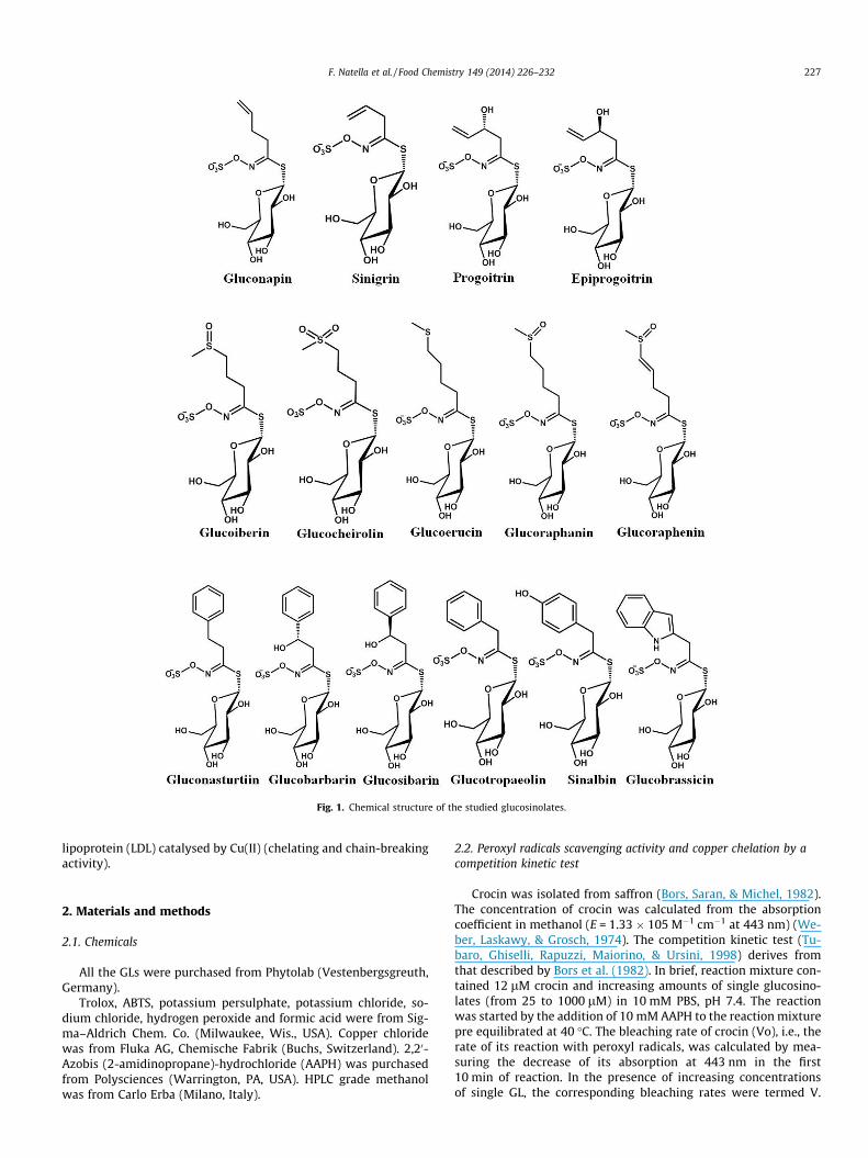
Fig. 1. Chemical structure of the studied glucosinolates.
F. Natella et al. / Food Chemistry 149 (2014) 226–232 227
lipoprotein (LDL) catalysed by Cu(II) (chelating and chain-breakingactivity).
2. Materials and methods
2.1. Chemicals
All the GLs were purchased from Phytolab (Vestenbergsgreuth,Germany).
Trolox, ABTS, potassium persulphate, potassium chloride, so-dium chloride, hydrogen peroxide and formic acid were from Sig-ma–Aldrich Chem. Co. (Milwaukee, Wis., USA). Copper chloridewas from Fluka AG, Chemische Fabrik (Buchs, Switzerland). 2,20-Azobis (2-amidinopropane)-hydrochloride (AAPH) was purchasedfrom Polysciences (Warrington, PA, USA). HPLC grade methanolwas from Carlo Erba (Milano, Italy).
2.2. Peroxyl radicals scavenging activity and copper chelation by acompetition kinetic test
Crocin was isolated from saffron (Bors, Saran, & Michel, 1982).The concentration of crocin was calculated from the absorptioncoefficient in methanol (E = 1.33 � 105 M�1 cm�1 at 443 nm) (We-ber, Laskawy, & Grosch, 1974). The competition kinetic test (Tu-baro, Ghiselli, Rapuzzi, Maiorino, & Ursini, 1998) derives fromthat described by Bors et al. (1982). In brief, reaction mixture con-tained 12 lM crocin and increasing amounts of single glucosino-lates (from 25 to 1000 lM) in 10 mM PBS, pH 7.4. The reactionwas started by the addition of 10 mM AAPH to the reaction mixturepre equilibrated at 40 �C. The bleaching rate of crocin (Vo), i.e., therate of its reaction with peroxyl radicals, was calculated by mea-suring the decrease of its absorption at 443 nm in the first10 min of reaction. In the presence of increasing concentrationsof single GL, the corresponding bleaching rates were termed V.

228 F. Natella et al. / Food Chemistry 149 (2014) 226–232
The slopes, calculated by linear regression analysis of the plot[Antioxidant]/[crocin] vs. Vo/V, indicate the relative capacity ofthe different molecules to interact with peroxyl radicals. The anti-oxidant capacity of the GLs, relative to the activity of trolox, wascalculated by dividing the slope of each compound by the slopeof trolox.
To study the capacity of GLs to complex copper, the method wasmodified by using a different initiating system in which the transi-tion metal ion Cu(II) (25 lM) decomposes dihydrogen peroxide(10 mM final concentration) to produce superoxide anion and hy-droxyl radical (Natella, Nardini, Di Felice, & Scaccini, 1999).
Fig. 2. Glucosinolates antioxidant capacities. (A) Peroxyl radical scavenging activity rsusceptibility of copper-catalysed LDL oxidation (LDL was oxidised in PBS at 37 �C with 5followed by continuously monitoring the absorbance at 234 nm. Delta of the lag time, elinear portion of the lag and propagation phases of conjugated dienes formation). Data aredifferent at p < 0.05 by one-way ANOVA followed by Tukey’s test.
2.3. ABTS�+ radical scavenging activity (TEAC assay)
The single compounds were tested by using the trolox equiva-lent antioxidant capacity (TEAC) assay. The TEAC value is basedon the ability of an antioxidant to scavenge the radical cation2,20-azino-bis (3-ethylbenzothiazoline-6-sulphonate) (ABTS�+) byspectrophotometric analysis (Re et al., 1999). The ABTS�+ cationradical was produced by the reaction between 7 mM ABTS inH2O and 2.45 mM potassium persulphate, stored in the dark atroom temperature for 12 h. The ABTS�+ solution was then dilutedwith PBS (pH 7.4) to obtain an absorbance of 0.70 at 734 nm and
elative to trolox. (B) Quenching of ABTS�+ relative to trolox (TEAC). (C) Effect onlM Cu(II) in presence of 100 lM GLs, the kinetic of conjugated dienes formation wasxpressed in minutes, was calculated from the intercept of lines drawn through thepresented as the mean ± SD (n = 3). Value with different letter (a–c) are significantly

Fig. 3. Inhibition of copper-catalysed LDL oxidation by gluconasturtin. LDL (50 lg/ml) was oxidised in PBS at 37 �C with 5 lM Cu(II) in presence of differentconcentration of gluconasturtin (from 5 to 100 lM), the kinetic of conjugateddienes formation was followed by continuously monitoring the absorbance at234 nm.
F. Natella et al. / Food Chemistry 149 (2014) 226–232 229
equilibrated at 30 �C. GLs were diluted with methanol to producesolutions of 0.3, 0.5, 1 and 1.5 mM.
The reaction was initiated by the addition of 1 ml of dilutedABTS to 10 ll of each sample solution. Determinations were re-peated three times for each sample solution. The percentage inhi-bition of absorbance at 734 nm was calculated for eachconcentration relative to a blank absorbance (methanol) and wasplotted as a function of concentration of compound or standard6-hydroxy-2,5,7,8-tetramethylchroman-2-carboxylic acid (Trolox).
The TEAC value is defined as the concentration of standard trol-ox with the same antioxidant capacity as a 1 mM concentration ofthe compound under investigation.
2.4. LDL preparation and oxidation
Human LDL (d 1.019–1.063 g/ml) was isolated from fastingplasma collected in EDTA (1 mg/ml) by sequential ultracentrifuga-tion in salt solutions, according to Havel, Eder, and Bragdon (1955),using a Beckman Optima Max-XP bench-top ultracentrifuge
Fig. 4. Effect of glucosinolates on Cu(II)/H2O2 induced bleaching of crocin. Crocin (12 lM500 lM glucosinolates (one representative experiment).
(T-100.3 rotor). LDL solution was flushed with N2, stored at 4 �Cand used within one week from the preparation. Protein was mea-sured by the method of Lowry, Rosebrough, Farr, and Randall(1951), using bovine serum albumin as standard.
LDL was dialysed in the dark for 24 h at 4 �C against twochanges of 1 L each of 0.01 M phosphate buffered saline (PBS),0.0027 M KCl and 0.138 M NaCl, pH 7.4. Dialysed LDL (50 lg pro-tein/ml) was oxidised in PBS at 37 �C for 4 h in the presence of5 lM CuCl2. The oxidation of LDL was performed in the presenceand in the absence of GLs at 5, 10, 50 and 100 lM. The kinetic ofconjugated diene formation (CD) was followed by continuouslymonitoring the absorbance at 234 nm, using a Beckman DU800spectrophotometer thermostated at 37 �C. The lag phase of CD for-mation was obtained by drawing a tangent to the slope of thepropagation phase and extrapolating it to the horizontal axis. Thelag time constitutes the interval from zero time to the intersectionpoint.
2.5. Electrospray mass spectrometry to study copper chelation bygluconasturtiin
Experiments were performed with a Mass Spectrometer (Ap-plied Biosystems API3200 Q-Trap; Foster City, CA, USA), equippedwith an electrospray ionisation source operated in positive ionmode, according to Fernandez (Fernandez, Mira, Florencio, & Jen-nings, 2002). The API 3200 ES source was tuned by infusing a glu-conasturtiin solution (1 lg ml�1 in methanol 50%) into the sourceat a flow rate of 10 ml min�1. The MS operated with an electro-spray voltage at +5500 V. Nitrogen was used as ion spray (GS1),and curtain gas at 20 and 20 arbitrary units, respectively. Thedeclustering potential (DP), and entrance potential (EP) were47.6 and 3.2, respectively. In brief, copper chloride was dissolvedin water and gluconasturtiin in water–methanol (1:1). Equal vol-umes of both solutions were mixed and then diluted 1:10 withmethanol water (1:1) with 0.1% of formic acid leading to equimolarsolutions of copper and gluconasturtiin, at 100 lM concentration.The solution was introduced using a syringe pump flowing at10 ll/min. Mass spectra were acquired by scanning from 400 to1000 m/z.
2.6. Statistical analysis
Data were expressed as mean ± standard deviation and ana-lysed by one-way ANOVA followed by Tukey’s test. Differenceswere considered significant at a value of p < 0.05.
The data were also analysed by Principal Component Analysis(PCA), performed on the data scaled by unit variance with the
) was oxidised in PBS at 40 �C with 25 lM Cu(II) and 10 mM H2O2 in presence of

Fig. 5. Electrospray mass spectrum of a solution of gluconasturtin and cupric chloride.
230 F. Natella et al. / Food Chemistry 149 (2014) 226–232
Factor MineR package of R 2.15.2 software. The results of the anal-ysis are presented in term of score and loading plots.
3. Results
The antioxidant capacity of 15 GLs was determined using differ-ent analytical approaches (Fig. 2).
The reactivity toward peroxyl radicals of the GLs (tested from25 to 1000 lM), relative to trolox, is shown in Fig. 2A; all the testedGLs possess a very low peroxyl radical scavenging capacity thatwas ten times lower than that of trolox. The ranking of activitywas: glucoraphenin P epiprogoitrin P glucobrassicin, gluconapin,sinalbin > gluconasturtiin > all the other. Fig. 2B shows the antiox-idant activities of the GLs toward ABTS�+ and relative to trolox.Generally, in this system, the antioxidant capacity of glucosinolatesis very weak. With the exception of sinalbin and glucobrassicin, infact, glucosinolates had an ABTS�+ quenching capacity ten timeslower than that of trolox. Glucobrassicin shows an ABTS�+ quench-ing capacity two times lower than that trolox, while sinalbin wasmore active (1.3 times) than trolox. The capacity of GLs (5, 10, 50and 100 lM) to inhibit copper-catalysed LDL oxidation was testedby measuring the increase in lag time of CD formation. Almost allGLs were ineffective at the lowest concentrations (data not
Fig. 6. Principal component analysis, (A
shown). Fig. 2C shows the activity measured at 100 lM GLs. Alsoat this concentration, almost all GLs failed to reduce CD formation,and some of them seem to have a weak pro-oxidant effect. Onlygluconasturtiin and sinalbin were able to significantly increase(p = 0.047 and p = 0.039, respectively) the lag time of CD formationin respect to test (copper oxidised LDL in absence of GL). The mostactive glucosinolate was gluconasturtiin, which was able to inhibitCu-catalysed LDL oxidation also at concentrations lower than100 lM and in a dose-dependent manner (Fig. 3). To further studythe effect of GLs in copper initiated oxidations, their activity wastested in the H2O2/Cu(II)-crocin system, in which copper decom-poses H2O2 yielding hydroxyl radicals that in turn bleach crocin.In this experiment, we tested the GLs at 500 lM concentration,in order to have the same stoichiometric ratio with copper(25 lM) used in LDL experiments. As shown in Fig. 4, almost allGLs failed to reduce the crocin bleaching, and most of them seemto have a weak pro-oxidant effect. Also in this system, the onlyglucosinolate that shows a clear antioxidant action was glu-conasturtiin. Then, the ability of gluconasturtiin to chelate copperions was studied by means of electrospray ionisation mass spec-trometry (ESI-MS). The MS spectra of gluconasturtiin with copperchloride (Fig. 5) indicated that gluconasturtiin forms twocomplexes with copper, having the stoichiometric metal:
) loadings plot and (B) score plots.

F. Natella et al. / Food Chemistry 149 (2014) 226–232 231
gluconasturtiin 1:1 and 1:2. These complexes were detectedthrough peaks at m/z 485 and 908 corresponding to[Cu(II)+(M�H)]+ and [Cu(II)+M+(M�H)]+.
A comprehensive view of the antioxidant capacity of the 15 GLscan be obtained from PCA. The computed model resulted able tocapture the 80% of the total observed variance with the first twoprincipal components (PCs). Data were represented by mean ofloading plots (Fig. 6A) estimating the correlation between the per-formed assays in the space identified by the principal components,and score plots (Fig. 6B) that describe the antioxidant behaviour ofGLs. The combined comparison of the two plots allowed evaluatingsimilarities and differences between the GLs in relation to theirantioxidant capability as measured by the performed assays.
The PC1 resulted able to separate a group of ten GLs (sinigrin,progoitrin, glucoiberin, glucocheirolin, glucoerucin, glucoraphanin,glucobarbarin, glucosibarin and glucotropaeolin) characterised byoverall low antioxidant capacity. The remaining five GLs are dis-criminated by the PC2 and are characterised by a moderate antiox-idant capacity. The PC2 also highlights the sinalbin as the GL withthe highest observed antioxidant capacity in TEAC assays. Differ-ently from epiprogoitrin, glucoraphenin and gluconasturtin whoseactivity is mainly evidenced in LDL and crocin assays.
4. Discussion
Our results show that in general GLs are not able to act as per-oxyl radical scavenger and chain breaking antioxidants (Fig. 2). Thelack of any chain breaking antioxidant activity was already ob-served for single GL, such as glucoerucin (Barillari et al., 2005)and glucoraphasatin (Papi et al., 2008).
Similar results were obtained when the antioxidant capacitywas measured by the TEAC assay, which measures the scavengingactivity of glucosinolates against the stable ABTS�+ radical. In thissystem, the greatest part of glucosinolates were not able to reactwith ABTS�+, confirming results already obtained for single GLs,such as gluconapin, glucoraphanin, glucoraphenin and progoitrin(Barillari et al., 2005; Cabello-Hurtado et al., 2012).
However, we observed that sinalbin and glucobrassicin showeda moderate capacity in quenching ABTS radical (Fig. 2), probablydue to the capability of phenolic and indolic groups to serve ashydrogen donors. Our results partially differ from those of Cabel-lo-Hurtado, who did not observe any ABTS�+ quenching activityfor glucobrassicin (Cabello-Hurtado et al., 2012). However, thesame author demonstrated a very high antioxidant capacity forglucobrassicin when measured by ORAC assay, suggesting an inter-esting radical scavenging activity for this indolic GL.
The antioxidant capacity of the 15 GLs was then studied on theLDL resistance to oxidative modification catalysed by copper. Inthis system copper decomposes pre-existing lipid hydroperoxidesinto LDL particle to give peroxyl and alkoxyl radicals (Esterbauer,Waeg, Puhl, Dieber-Rotheneder, & Tatzber, 1992); therefore, theinhibition of copper catalysed LDL oxidation represents a combina-tion of transition metal chelation and scavenging of different freeradical species. Also in this system, sinalbin showed a moderateantioxidant capacity and similar results were already obtained inthe NADPH/Fe induced lipid peroxidation of microsomes (Plumbet al., 1996).
Unexpectedly, in Cu(II)-catalysed LDL oxidation, gluconasturtiinresulted a very active antioxidant (Fig. 3). As no radical scavengingactivity seemed to be ascribable to gluconasturtiin, we supposethat gluconasturtiin could act as metal chelator. This hypothesiswas, initially, confirmed by the experiments carried out usingH2O2/Cu(II)-crocin system, in which copper decomposes H2O2
yielding to hydroxyl radical that in turn bleach crocin. In theseexperiments, the only glucosinolates able to inhibit the bleaching
of crocin was gluconasturtiin (Fig. 4). To further study the capacityof gluconasturtiin to chelate copper, we used ESI-MS that provideddirect evidence of chelation between copper and gluconasturtiin.We observed, in fact, two different complexes having the stoichi-ometric Cu(II):gluconasturtiin 1:1 and 1:2 (Fig. 5). A 1:2 metal-complex, involving the sulfur and oxygen atoms about the C@Nbond, has been already suggested for sinigrin, even if in that casethe complex was unstable (Youngs & Perlin, 1967).
The PCA analysis provided a comprehensive view of the antiox-idant capacity of glucosinolates (Fig. 6). The prominent indicationis that 10 out 15 tested GLs do not possess any antioxidant capac-ity; while the others possess some antioxidant activities, but theseactivities are related mainly to TEAC for glucobrassicin and sinalbinand to LDL and crocin for epiprogoitrin, glucoraphenin andgluconasturtiin.
5. Conclusions
Just few GLs possess an antioxidant capacity and this activity isquite specific (e.g. sinalbin in quenching ABTS radical, glu-conasturtiin in chelating copper); then it is very unlikely that GLsas such could contribute to the overall antioxidant potential ofBrassicaceae.
A last (obvious and more generic) indication that emerges fromour study is that only from a combination of antioxidant tests andmultivariate analysis it is possible to obtain a clear characterisationof the antioxidant properties of bioactive molecules.
Acknowledgements
This work was supported by Italian Ministry of Agriculture,Food & Forestry (MiPAAF) Grants ‘‘NUME’’ (DM 3688/7303/08)and ‘‘NUTRIGEA’’ (DM 30281 23/12/2009).
Kariklia Pascucci is acknowledged for her kind support in thedaily lab work.
References
Barillari, J., Canistro, D., Paolini, M., Ferroni, F., Pedulli, G. F., Iori, R., et al. (2005).Direct antioxidant activity of purified glucoerucin, the dietary secondarymetabolite contained in rocket (Eruca sativa Mill.) seeds and sprouts. Journalof Agricultural and Food Chemistry, 53(7), 2475–2482. http://dx.doi.org/10.1021/jf047945a.
Bheemreddy, R. M., & Jeffery, E. H. (2007). The metabolic fate of purifiedglucoraphanin in F344 rats. Journal of Agricultural and Food Chemistry, 55(8),2861–2866. http://dx.doi.org/10.1021/jf0633544.
Biovin, D., Lamy, S., Lord-Dufour, S., Jackson, J., Beaulieu, E., Cote, M., et al. (2009).Antiproliferative and antioxidant activities of common vegetables: Acomparative study. Food Chemistry, 112(2), 374–380.
Bors, W., Saran, M., & Michel, C. (1982). Radical intermediates involved in thebleaching of the carotenoid crocin. Hydroxyl radicals, superoxide anions andhydrated electrons. International Journal of Radiation Biology and Related Studiesin Physics, Chemistry, and Medicine, 41(5), 493–501.
Cabello-Hurtado, F., Gicquel, M., & Esnault, M. A. (2012). Evaluation of theantioxidant potential of cauliflower (Brassica oleracea) from a glucosinolatecontent perspective. Food Chemistry, 132(1003), 1003–1009.
Esterbauer, H., Waeg, G., Puhl, H., Dieber-Rotheneder, M., & Tatzber, F. (1992).Inhibition of LDL oxidation by antioxidants. Exs, 62, 145–157.
Fahey, J. W., & Talalay, P. (1999). Antioxidant functions of sulphoraphane: A potentinducer of phase II detoxication enzymes. Food and Chemical Toxicology: AnInternational Journal Published for the British Industrial Biological ResearchAssociation, 37(9–10), 973–979.
Fahey, J. W., Zalcmann, A. T., & Talalay, P. (2001). The chemical diversity anddistribution of glucosinolates and isothiocyanates among plants.Phytochemistry, 56(1), 5–51.
Fernandez, M. T., Mira, M. L., Florencio, M. H., & Jennings, K. R. (2002). Iron andcopper chelation by flavonoids: An electrospray mass spectrometry study.Journal of Inorganic Biochemistry, 92(2), 105–111.
Havel, R. J., Eder, H. A., & Bragdon, J. H. (1955). The distribution and chemicalcomposition of ultracentrifugally separated lipoproteins in human serum.Journal of Clinical Investigation, 34(9), 1345–1353. http://dx.doi.org/10.1172/JCI103182.
Jacob, C., Jamier, V., & Ba, L. A. (2011). Redox active secondary metabolites. CurrentOpinion in Chemical Biology, 15(1), 149–155. http://dx.doi.org/10.1016/j.cbpa.2010.10.015.
232 F. Natella et al. / Food Chemistry 149 (2014) 226–232
Kurilich, A. C., Jeffery, E. H., Juvik, J. A., Wallig, M. A., & Klein, B. P. (2002).Antioxidant capacity of different broccoli (Brassica oleracea) genotypes usingthe oxygen radical absorbance capacity (ORAC) assay. Journal of Agricultural andFood Chemistry, 50(18), 5053–5057.
Lowry, O. H., Rosebrough, N. J., Farr, A. L., & Randall, R. J. (1951). Proteinmeasurement with the Folin phenol reagent. Journal of Biological Chemistry,193(1), 265–275.
Natella, F., Nardini, M., Di Felice, M., & Scaccini, C. (1999). Benzoic and cinnamic acidderivatives as antioxidants: structure–activity relation. Journal of Agricultureand Food Chemistry, 47, 1453–1459.
Papi, A., Orlandi, M., Bartolini, G., Barillari, J., Iori, R., Paolini, M., et al. (2008).Cytotoxic and antioxidant activity of 4-methylthio-3-butenyl isothiocyanatefrom Raphanus sativus L. (Kaiware Daikon) sprouts. Journal of Agricultural andFood Chemistry, 56(3), 875–883. http://dx.doi.org/10.1021/jf073123c.
Pellegrini, N., Chiavaro, E., Gardana, C., Mazzeo, T., Contino, D., Gallo, M., et al.(2010). Effect of different cooking methods on color, phytochemicalconcentration, and antioxidant capacity of raw and frozen brassicavegetables. Journal of Agricultural and Food Chemistry, 58(7), 4310–4321.http://dx.doi.org/10.1021/jf904306r.
Plumb, G. W., Lambert, N., Chambers, S. J., Wanigatunga, S., Heaney, R. K., Plumb, J.A., et al. (1996). Are whole extracts and purified glucosinolates from cruciferousvegetables antioxidants? Free Radical Research, 25(1), 75–86.
Re, R., Pellegrini, N., Proteggente, A., Pannala, A., Yang, M., & Rice-Evans, C. (1999).Antioxidant activity applying an improved ABTS radical cation decolorisationassay. Free Radical Biology and Medicine, 26(9–10), 1231–1237.
Sandoval, M., Okuhama, N. N., Angeles, F. M., Melchor, V. V., Condezo, L. A., Lao, J.,et al. (2002). Antioxidant activity of the cruciferous vegetable Maca (Lepidiummeyenii). Food Chemistry, 79(2), 207–213.
Tubaro, F., Ghiselli, A., Rapuzzi, P., Maiorino, M., & Ursini, F. (1998). Analysis ofplasma antioxidant capacity by competition kinetics. Free Radical Biology andMedicine, 24(7–8), 1228–1234.
Valgimigli, L., & Iori, R. (2009). Antioxidant and pro-oxidant capacities of ITCs.Environmental and Molecular Mutagenesis, 50(3), 222–237. http://dx.doi.org/10.1002/em.20468.
Weber, F., Laskawy, G., & Grosch, W. (1974). Co-oxidation of carotene and crocin bysoybean lipoxygenase isoenzymes. Zeitschrift für Lebensmittel-Untersuchung undForschung, 155, 142–150.
Youngs, C. G., & Perlin, A. S. (1967). Fe(II)-catalysed decomposition of sinigrin andrelated thioglycosides. Canadian Journal of Chemistry, 45(1801), 1801–1803.
Zhang, Y., Cho, C. G., Posner, G. H., & Talalay, P. (1992). Spectroscopic quantitation oforganic isothiocyanates by cyclocondensation with vicinal dithiols. AnalyticalBiochemistry, 205(1), 100–107.