Thymidylate synthase gene amplification in human colon cancer cell lines resistant to 5-fluorouracil
Upload
independentCategory
view
0download
0
Biomedical Research and Therapy 2015, 2(2): 196-206 ISSN 2198-4093 www.bmrat.org
Drug Interactions of Methotrexate, Cyclophosphamide and 5-Fluorouracil
196
REVIEW
Molecular Basis of Drug Interactions of Methotrexate, Cyclophos-phamide and 5-Fluorouracil as Chemotherapeutic Agents in Cancer Amit Sarder1,2,*, Md. Golam Rabbani1,3, A. S. M. Homaun Kabir Chowdhury1, Mahbub-E-Sobhani1, 4
1Biotechnology and Genetic Engineering Discipline, Khulna University, Khulna, Bangladesh; 2Faculty of Life Sciences and Biotechnology, South Asian University, New Delhi, India; 3James P. Grant School of Public Health, BRAC University, Dhaka, Bangladesh; 4Department of Biochemistry and Molecular Biology, University of Dhaka, Dhaka, Bangladesh.
*Corresponding author: [email protected]
Received: 30 December 2014 / Accepted: 03 February 2015 / Published online: 19 February 2015 © The Author(s) 2015. This article is published with open access by BioMedPress (BMP).
Abstract— At present, chemotherapy is one of the principal methods of treatment of cancer. For many years, chem-otherapy is possibly the only way to control cancers that do not respond to either surgery or radiation. To date a good number of chemotherapeutic drugs have been developed which are effective in the treatment of human cancers. But, A few drugs have been known to be safe and promising. The most widely used chemotherapeutic drugs include meth-otrexate, cyclophosphamide, 5-fluorouracil etc. In this review, the molecular basis of drug interaction of methotrex-ate, cyclophosphamide and 5-fluorouracil has been studied. Understanding of the molecular basis of drug interaction of the chemotherapeutic agents is fundamental in order to enhance the clinical effectiveness of chemotherapy as well as to increase our knowledge of the cytotoxic effects of chemotherapeutic drugs. Increased understanding of the pharmacokinetics and the mechanism of action also help to develop biomodulating strategies. The boundary between the efficacy and toxicity of chemotherapeutic drugs is very narrow. So, novel approaches are needed to be formulated in order to enhance the efficacy and to minimize the toxic effects of the chemotherapeutic agents. Though a lot of in-formation is needed to be learned and a lot of task is needed to be accomplished, chemotherapy can be the ultimate and feasible way for controlling cancers if the toxic effects of chemotherapy can be reduced or minimized.
Keywords— Methotrexate, Cyclophosphamide, 5-Fluorouracil, Chemotherapy, Cancer.
INTRODUCTION
Methotrexate
Methotrexate (MTX), 4-amino-N10-
methylpteroglutamic acid is a folic acid antagonist
first developed for the treatment of malignancies. The
IUPAC name of MTX is 4-amino-4-deoxy-10-methyl-
pteroyl-glutamic acid and its chemical formula is
C20H22N8O5. The molecular weight of MTX is 454.45
(Genestier et al., 1998; Li, 1960; Saxena Ajit. K, 2009).
At present, it is most commonly used in the treatment
of rheumatoid arthritis (RA). It is now recognized as a
very useful disease-modifying anti-rheumatic drug. It
is now known that low dose MTX treatment is well-
tolerated and produces a quick clinical response in the
case of RA patients. It is also very much effective in
controlling RA refractory to more conventional treat-
ment (Louie and Lillington, 1986; Tariq and Tariq,
1993). Because of the immunosuppressive, cytostatic
and anti-inflammatory effects of MTX, it is used in the
systemic treatment of psoriasis. MTX selectively in-
duces apoptosis of activated but not resting lympho-
cytes, even after short-term exposure to MTX and sub-
sequent activation in drug-free medium. This event
DOI 10.7603/s40730-015-0005-1

Sarder et al., 2015 Biomed Res Ther 2015, 2(2): 196-206
Drug Interactions of Methotrexate, Cyclophosphamide and 5-Fluorouracil
197
provided the first evidence of immunosuppressive
activity of low-dose intermittent MTX administration
(Genestier et al., 1998; Kozub and Simaljakova, 2011;
Saleh Abdulla, 2010). It is also effective in the preven-
tion of acute graft-vs.-host disease (GVHD). But, the
combination therapy of MTX with cyclosporine (CSP)
has been found to demonstrate superior result than
the single MTX therapy (Ross et al., 1999; Storb et al.,
1989; von Bueltzingsloewen et al., 1993). Folic acid is
an important DNA precursor which undergoes
reduction to dihydrofalate and then tetrahydrofolate
by the enzyme dihydrofolate reductase (DHFR). MTX
and other folate analogues like raltitrexate and
pemetrexed act as folate antagonists and inhibit the de
novo systhesis of purines. DHFR inhibition by MTX
results in reduced folate pool. Reduced folate acts as a
cofactor for thymidylate synthase (TS) which
catalyzes the synthesis of pyrimidine converting
deoxyuridine monophosphate (dUMP) to
deoxythymidine monophosphate (dTMP).
Consequently, folate analogues inhibit the synthesis of
both purines and pyrimidines, although drugs within
this class differ in their relative specificities as
inhibitors of DHFR and TS function (Airley, 2009).
MTX was the first example of rational drug design
and it was mainly developed for the treatment of
acute lymphocytic leukaemia. MTX has also been re-
ported to cure choriocarcinoma and trophoblastic can-
cer (Agarwal et al., 2010). MTX can be given in low
doses (e.g. 20 mg/m2) in maintenance chemotherapy
and also in benign conditions. It has been found that
the use of low dose MTX is safe and well tolerated.
Because of its safety and efficacy, it is considered as a
first line therapy for the treatment of RA. But, MTX
can also be given in higher doses (e.g. 1,000 mg/m2 –
33,000 mg/m2) for the treatment of certain cancers. But,
high dose MTX therapy can cause significant toxicity
like nephrotoxicity, myelosuppression, mucositis,
hepatotoxicity etc. In severe cases, it may cause multi-
organ failure also (Cronstein, 2005; Rahiem Ahmed
YAA, 2013).
Cyclophosphamide
Cyclophosphamide (CP) is one of the most widely
used chemotherapeutic agents (Juma and Ogada,
1983). It is regarded as a major anticancer drug
(Rahiem Ahmed YAA, 2013). Though it is a very effec-
tive antineoplastic agent against various kinds of tu-
mors, previously it showed virtually no cytotoxic ac-
tivity against mammalian cell cultures (Arthur
Camerman, 1977). Basically, CPs are cytotoxic chemo-
therapy agents similar to mustard gas. It is nitrogen
mustard alkylating agent from the oxazophorines
group that attaches an alkyl group (CnH2n+1) to Deoxy-
ribonucleotide Acid (DNA). The molecular formula of
cyclophosphamide monohydrate is
C7H15Cl2N2O7P.H2O. The systematic name of cyclo-
phosphamide is 2-[bis(2-
Chloroethyl)amino]tetrahydro-2H-1,3,2-
oxazaphosphorine 2-oxide monohydrate. CP is mainly
used in the treatment of blood cancer, brain cancer,
leukemia and some solid tumors (Pavan K. V., 2013 ).
It is also used in the treatment of various non-
neoplastic diseases like nephritic syndrome, systemic
lupus erythematosus and rheumatoid arthritis (Morais
et al., 1999). As CP is used to treat both neoplastic and
non-neoplastic diseases, it is very important to assess
the long-term carcinogenic potential of this drug
(Travis et al., 1995). The side effects of CP are well
known in patients receiving this drug for the
treatment of lymphomas, leukemias or solid tumours.
Bone marrow toxicity with opportunistics infections,
haemorrhagic cystitis, temporary infertility, hair loss,
vomiting, nausea etc. are seen frequently with the
application of CP whereas pneumonitis, liver or
cardiac toxicity are very rare (Haubitz, 2007).
5-Fluorouracil
5-Fluorouracil (5-FU) is an example of a rationally de-
signed anticancer drug (Malet-Martino and Martino,
2002). This antimetabolite is widely used as a chemo-
therapeutic drug. Chemically, 5-FU is a dipodic acid
and highly polar in nature. 5-FU is a heterocyclic aro-
matic organic compound with a structure similar to
that of the pyrimidine molecules of DNA and RNA. It
is basically an analogue of uracil with a fluorine atom
at the C-5 position in place of hydrogen. It is one of
the most successful chemotherapeutic drugs used for
the treatment of solid tumors. It is frequently used as
chemotherapeutic agent during the simultaneous
chemoradiotrerapy. It is mainly applied in the neoad-
juvant and definitive chemoradiotherapy of gastroin-
testinal malignancies and also in the carcinoma of
head and neck. But, fluoropyrimidine therapy is asso-
ciated with cardiac toxicity. 5-FU induced cardiac tox-
icity is an underestimated problem in radiooncology.
Patients without history of cardiovascular risk factors
are often treated as out-patients without cardiac moni-
toring. Consequently asymptomatic and symptomatic
cardiac events may be overlooked. 5-FU is also a drug
Sarder et al., 2015 Biomed Res Ther 2015, 2(2): 196-206
Drug Interactions of Methotrexate, Cyclophosphamide and 5-Fluorouracil
198
of choice in oropharyngeal cancer, colorectal cancer,
stomach cancer and cervical cancer (Lamberti et al.,
2012; Pendekal and Tegginamat, 2012; Steger et al.,
2012). It has been found that infusional high dose 5-FU
is well-tolerated and effective treatment for gastric
cancer. Combination chemotherapy of 5-FU with
epirubicin and cisplatin has also resulted in significant
response rates and survival benefits for patients with
advanced gastric cancer (Wang Tso-Fu, 2006). But,
clinical studies suggest that the use of 5-FU is not also
free from toxicity and women experience more
toxicity than men by the 5-FU based chemotherapy.
Women not only exhibit a greater variety of toxicity
types but also experience them with greater average
severity. Common toxicity associated with 5-FU
includes stomatitis, leukopenia, alopecia, nausea,
vomiting, diarrhea etc.
MOLECULAR BASIS OF DRUG INTERACTION Molecular Basis of Drug Interaction of Methotrexate
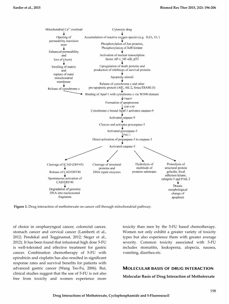
Figure 1. Drug interaction of methotrexate on cancer cell through mitochondrial pathway.

Sarder et al., 2015 Biomed Res Ther 2015, 2(2): 196-206
Drug Interactions of Methotrexate, Cyclophosphamide and 5-Fluorouracil
199
Tumors are complex tissues of various distinct types
of cells. Tumors are more than insular masses of pro-
liferating cancer cells. Tumors are complex tissues of
multiple distinct cell types that participate in hetero-
typic interactions with one another. Also, tumor stro-
ma represents some structural features that are differ-
ent from the normal tissues. Tumor cell-derived sig-
nals activate and recruit hosts cells like monocytes,
fibroblasts etc. Thus, the dynamic and reciprocal in-
teractions between tumor cells and the cells of the tu-
mor microenvironment lead to the tumor progression
and metastasis formation.
Figure 2. Drug interaction of methotrexate on cancer cell
through p53 dependent pathway.
The biology of tumors can no longer be understood by enumerating the traits of the cancer cells but instead must encompass the contributions of the tumor mi-croenvironment to tumorigenesis (Hanahan and Weinberg, 2011; Ungefroren et al., 2011). MTX can in-duce apoptosis through both mitochondrial pathway and p53 dependent pathway. These two major mech-anisms of action of MTX are discussed below.
Mitochondrial Pathway
Two major classical apoptosis signaling cascades have
been known. One major pathway for the initiation of
apoptosis is the receptor-mediated pathway or the
extrinsic pathway. Another major pathway for the in-
duction of apoptosis is the mitochondrial pathway or
the intrinsic pathway (Alonso et al., 2005; Putcha et al.,
2002). The mitochondrial pathway is a complex path-
way and acts as central gateway controllers. It has
been found that a variety of extracellular and intracel-
lular stress including MTX treatment and oxidative
stress can lead to the activation of mitochondrial
pathway (Chen et al., 2009; Lawen, 2003). Oxidative
stress can be defined as a state detrimental to the body
where oxidative forces exceed the antioxidant systems
because of the loss of balance between them. Some
atoms and molecules contain unpaired electrons orbit-
ing around the nucleus and these are called free radi-
cals (FR). Free radicals are highly reactive and unsta-
ble because the unpaired electrons of free radicals
tend to form pairs with other electrons. Oxygen (O2)
molecules can undergo 4-electron reduction when it is
metabolized in vivo. During this reduction, reactive
oxygen metabolites are produced by the excitation of
electrons or interaction with transition elements.
These reactive oxygen metabolites, also called active
oxygen species are highly reactive. Superoxide radical,
hydrogen peroxide, alkoxyl radical etc. are examples
of active oxygen species. For aerobic organisms, it is
very important to have a mechanism of removing
these active oxygen species in order to sustain their
life. Therefore, various antioxidant defense mecha-
nisms have been developed in the process of evolu-
tion. However, if active oxygen species or free radicals
are produced excessively, the balance between the
formation and removal of active oxygen species are
lost. This event results in the oxidative stress
(Yoshikawa Toshikazu, 2002). Mitochondria are the
most important source of reactive oxygen species
(ROS) because of the electron transport chain located
in the mitochondrial membrane. Again, some cyto-
chrome 450 enzymes can also produce ROS. There are
complex interactions between ROS generation, ROS

Sarder et al., 2015 Biomed Res Ther 2015, 2(2): 196-206
Drug Interactions of Methotrexate, Cyclophosphamide and 5-Fluorouracil
200
signaling, ROS-induced damage and carcinogenesis.
The intrinsic pathway or the mitochondrial pathway
can be triggered by many stimuli including ROS. So,
the accumulation of ROS can lead to the initiation of
apoptosis. As a cytotoxic drug, MTX induces ROS
including hydrogen peroxide and superoxide radical
and thus induce apoptosis. Hydrogen peroxide accu-
mulated by MTX can cause the release of cytochrome c
from the mitochondria into the cytosol. Moreover, it
may activate nuclear transcription factors such as acti-
vator protein (AP)-1, nuclear factor (NF)-κB and p53
which may lead to the up-regulation of death proteins
or production of inhibitors of survival proteins
(Dayem et al., 2010). AP-1 complex which consists of
either Jun homodimers or Fos/Jun heterodimers, bind
to a unique palindromic TPA-response element (TRE)
with the sequence TGA(C/G)TCA. It is suggested that
AP-1 activation is mediated by phosphorylation of Jun
proteins. The presence of ROS sensitive kinase cas-
cade, oxidative stress sensitive mitogen-activated pro-
tein (MAP) kinase and MAP kinase phosphate is also
evident in the activation of AP-1. On the other hand,
the activation of NF-κB is thought to be mediated by
the phosphorylation of IκB kinase (Chung et al., 2002;
Sen and Packer, 1996). Apoptotic signal from the MTX
or the change in the mitochondrial membrane poten-
tial ∆ release cytochrome c and other pro-
apoptotic proteins (e.g. adenylate kinase-2 (AK-2),
Smac/DIABLO, apoptosis inducing factor (AIF)) from
the mitochondrial intermembrane space into the cyto-
sol where cytochrome c binds to a mammalian CED-4
homologue called apoptotic protease activating factor-
1 (Apaf-1). Release of cytochrome c due to the apop-
totic signal from the MTX is considered a key step in
the apoptotic process. It has been found that the re-
lease of cytochrome c occurs by distinct mechanisms
that are either Ca2+-dependent or Ca2+-independent. In
the Ca2+-dependent mechanism, mitochondrial Ca2+
overload promotes the opening of the permeability
transition (PT) pore. The opening of this mega channel
is linked with the enhanced permeability and loss of
mitochondrial membrane potential ∆ . This in-
creased inner mitochondrial membrane permeability
leads to the swelling of the matrix, rupture of the out-
er mitochondrial membrane and finally the release of
cytochrome c. The release of cytochrome c by the Ca2+-
independent mechanism is thought to be governed by
different members of the Bcl-2 family of proteins. Ac-
cording to earlier evidences, opening of the mitochon-
drial permeability pore and loss of mitochondrial
membrane potential ∆ is needed for the release of
cytochrome c. But, recent data suggest that both
events are not needed for the release of apoptotic cy-
tochrome c in all the cases. Binding of cytochrome c to
Apaf-1 triggers the formation of the apoptosome
which catalyses the activation of caspases. This ∼ MDa
oligomeric complex contains 7 Apaf-1, 7 cytochrome c,
7 (d)ATP and 7 procaspase-9 molecules. Apaf-1 is ba-
sically a 3-domain protein with an N-terminal CARD
(caspase recruitment domain) followed by a putative
ATPase domain and C-terminal region containing 12
WD40 repeats. Caspase-9, an initiator caspase, is ca-
pable of self-processing upon binding with Apaf-1 and
it provides a complex for ensuring high local concen-
tration and protein conformation suitable for activa-
tion. For the activation of caspase-9, Apaf-1 binds cy-
tochrome c via its WD40 domains. Upon binding to
cytochrome c, Apaf-1 becomes competent to recruit
caspase-9 in the presence of ADP or ATP. Caspase-9,
being activated cleaves and activates pro-caspase-3
(Chen et al., 2009; Koya et al., 2000; Lawen, 2003; Ly et
al., 2003; Ott et al., 2002). It has been found that a
compound called PAC-1 activates procaspase-3 to
caspase-3. PAC-1 activates procaspase-3 by sequester-
ing inhibitory zinc ions and allows procaspase-3 to
auto-activate itself to caspase-3. Thus, PAC-1 induces
apoptosis via direct activation of procaspase-3. How-
ever, the precise mechanism by which PAC-1 activates
procaspase-3 is not yet known. But, it activates pro-
caspase-3 in a dose depended manner. Generally, pro-
caspase-3 levels are high in cancer cells. It suggests
that compounds like PAC-1 which stimulates the acti-
vation of procaspase-3 to caspase-3 can selectively in-
duce apoptosis. It has been found that the activation of
caspase-3 is required for the induction of apoptosis in
response to chemotherapeutic agents like MTX.
Caspase-3 is the key executioner caspase which cata-
lyzes hydrolysis of a multitude of protein substrate
within the cell. Caspase-3 cleaves a variety of cellular
substrate including structural proteins and DNA re-
pair enzymes. Caspase-3 mediates proteolysis of sev-
eral key substrate including the structural proteins
gelsolin, focal adhesion kinase, rabaptin-5 and PAK2
and contribute to the drastic morphological change of
apoptosis. Caspase-3 also activates an endonuclease
caspase-activated DNase (CAD) which causes the
fragmentation of DNA by specifically inactivating and
cleaving ICAD (DFF45), the inhibitor of CAD. A spe-
cific caspase-3 activated DNase, designated as DNA
fragmentation factor (DFF40) has been identified. This
CAD is revolutionarily conserved crossing rodents
and human. CAD/DFF40 exists normally as a nonac-

Sarder et al., 2015 Biomed Res Ther 2015, 2(2): 196-206
Drug Interactions of Methotrexate, Cyclophosphamide and 5-Fluorouracil
201
tive heterodimeric complex in the cell with its natural
inhibitor, ICAD (DFF45). Caspase-3 mediated cleavage
of ICAD allows the release of CAD/DFF40 from the
complex. It is then spontaneously activated. Activated
CAD/DFF40 then results in the degradation of ge-
nomic DNA into nucleosomal fragments (Fig. 1) (Cao
et al., 2001; Janicke et al., 1998; McIlroy et al., 2000;
O'Donovan et al., 2003; Peterson et al., 2009).
P53 Dependent Pathway
Methotrexate can also induce apoptosis through p53-
dependent pathway. The anticancer mechanism of
MTX acts through initiation of p53-dependent apopto-
sis. Methotrexate can induce p53 acetylation at
Lys373/382 and phosphorylation at Ser15/Ser392.
These events are correlated with the increase in DNA
damage and up-regulation of p53 target genes such as
p21, DR5, Puma and Noxa. Acetylation of p53 is corre-
lated with its nuclear accumulation and stability.
Phosphorylation of p53 also increases its stabilization
as well (Huang et al., 2011). Cellular stresses like
growth factor deprivation, DNA damage or oncogene
expression leads to the stabilization and activation of
the p53 tumor suppressor protein. This can result in
one of the two different outcomes: cell cycle arrest or
apoptosis. Cell death induced through p53 pathway is
executed by the caspase proteinases. Caspase activa-
tion by p53 occurs through the release of apoptogenic
factors from mitochondria including cytochrome c.
Release of cytochrome c and binding of cytochrome c
with the Apaf-1 allows the formation of apoptosome, a
high molecular weight complex consisting of Apaf-1
and caspase-9. Caspase-9 is activated following re-
cruitment into the apoptosome. Activated caspase-9
can then cleave and activate the effector caspases like
caspase-3 and caspase-7 (Schuler and Green, 2001;
Steele et al., 2008). The activation of these effector
caspases leads to the degradation of genomic DNA
into nucleosomal fragments, cleavage of structural
proteins and DNA repair enzymes, hydrolysis of mul-
titude of protein substrates, drastic morphological
change of apoptosis etc. (Fig. 2).
Molecular Basis of Drug Interaction of Cyclophos-
phamide
Figure 3. Drug interaction of cyclophosphamide on cancer cell.
Sarder et al., 2015 Biomed Res Ther 2015, 2(2): 196-206
Drug Interactions of Methotrexate, Cyclophosphamide and 5-Fluorouracil
202
As an alkylating drug, cyclophosphamide’s thera-
peutic mechanism of action induces DNA damage.
Treatment with CP has also been shown to cause oxi-
dative stress. Tripeptite glutathione (GSH) depletion
also induces apoptosis in some cell types in culture.
GSH depletion also sensitizes cells to proapoptotic
stimuli. CP itself is devoid of alkylating activity and
must undergo bio-activation by hepatic cytochrome
P450. CP activation is initiated by a cytochrome P450-
dependent 4-hydroxylation reaction. CP is metaboli-
cally activated in the liver to 4-
hydroxycyclophosphamide (4-HC) via oxidation by
cytochrome P450 enzymes like CYP2A6, CYP2B6,
CYP2C8, CYP2C9, CYP3A4 etc. The phenobarbital-
inducible rat P450 enzyme 2B1 and its human coun-
terpart 2B6 show high 4-hydroxylase activity com-
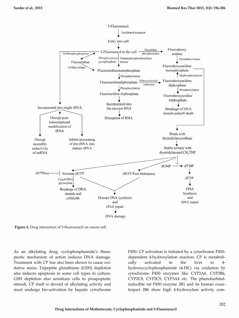
Figure 4. Drug interaction of 5-fluorouracil on cancer cell.

Sarder et al., 2015 Biomed Res Ther 2015, 2(2): 196-206
Drug Interactions of Methotrexate, Cyclophosphamide and 5-Fluorouracil
203
pared with other P450 forms. Initially, CP is hydrox-
ylated at C-4 by cytochrome P450. 4-HC then under-
goes ring opening to aldo-phosphamide. Aldo-
phosphamide then spontaneously eliminates acrolein
and decomposes to the reactive metabolite phospho-
ramide mustard. Acrolein covalently binds to and
there by inactivates several hepatic enzymes including
P450. On the other hand, phosphoramide mustard is
an active metabolite in terms of both toxicity and anti-
cancer activity. The cytotoxic effects of CP are the re-
sult of chemically reactive metabolites that alkylate
DNA and protein, producing cross-links. CP, phos-
phoramide mustard and other metabolites of CP are
detoxified by conjugation with GSH (Brummaier et al.,
2013; Cai et al., 1997; Chen et al., 2004; Clarke and
Waxman, 1989; Tsai-Turton et al., 2007). The activated
CP metabolites, phosphoramide mustard and acrolein
are transported into both tumor and healthy cells via
the bloodstream. CP and its isomer ifosfamide can
induce a caspase-9 dependent apoptotic pathway act-
ing via activated 4-hydroxy metabolites of CP and
ifosfamide (Fig. 3) (Schwartz and Waxman, 2001).
Molecular Basis of Drug Interaction of 5-
Fluorouracil
The mechanism of action of 5-FU is associated with
the inhibition of TS and incorporation of 5-FU into the
RNA and DNA. 5-FU rapidly enters into the cells us-
ing the facilitated transport mechanism. 5-FU is then
converted into three main active metabolites. The ac-
tive metabolites are fluorouridine triphosphate
(FUTP), fluorodeoxyuridine monophosphate
(FdUMP) and fluorodeoxyuridine triphosphate
(FdUTP) intracellularly. These three active metabolites
disrupt RNA synthesis and the action of TS. The main
mechanism of activation of 5-FU is the conversion to
fluorouridine monophosphate (FUMP) directly by
orotatephosphoribosyltransferase (OPRT) with phos-
phoribosyl pyrophosphate (PRPP) as the cofactor. This
conversion is also caused indirectly by fluorouridine
(FUR) in a two-step process involving the action of
uridinephosphorylase (UP) and uridine kinase (UK).
FUMP is then phosphorylated to fluorouridinedi-
phosphate (FUDP). FUDP can then be further phos-
phorylated to FUTP which can be incorporated into
nascent RNA. FUTP incorporates into the RNA and
disrupts the functions of RNA. When FUTP is mis-
incorporated into the RNA, it may interfere the nor-
mal nucleic acid metabolism. 5-FU may directly affect
the metabolism of RNA. After administration of 5-FU,
it can be readily incorporated into the long single
rRNa. The incorporation of 5-FU not only inhibit the
processing of pre-rRNA into mature rRNA, but also
disrupts the post transcriptional modification of
tRNAs. It also disrupts the assembly and activity of
snRNA/protein complex and thereby inhibits the
splicing of pre-mRNa. FUDP can also be converted
into the fluorodeoxyuridinediphosphate (FdUDP) by
the action of ribonucleotidereductase (RR). FdUDP
can then be phosphorylated to yield the active me-
tabolite FdUTP or it can be dephosphorylated to gen-
erate the active metabolite FdUMP. 5-FU can also be
converted in to the FdUMP in an alternative pathway.
5-FU is converted to the fluorodeoxyuridine (FUDR)
by the action of thymidine phosphorylase (TP) which
can then be phosphorylated to FdUMP by thymidine
kinase (TK). TS is a key enzyme for the de novo synthe-
sis and DNA synthesis. TS catalyses the reductive
methylation of dUMP to dTMP. Reduced folate 5, 10-
methylenetetrahydrofolate (CH2THF) acts here as the
methyl donor. This reaction provides the sole de novo
source of thymidylate, which is necessary for the DNA
repair and DNA replication. FdUMP, the active me-
tabolite of 5-FU binds to the nucleotide binding site of
TS and forms a stable ternary with TS and CH2THF.
Thus, it blocks the binding of dUMP and inhibits the
synthesis of dTMP. Depletion of dTMP results in the
depletion of deoxythymidine triphosphate (dTTP).
This event induces the puerturbations in the levels of
dATP, dGTP and dCTP also. These results in the de-
oxynucleotide (dNTP) pool imbalances and increases
the levels of dUTP both of which are thought to dis-
rupt DNA synthesis and repair severely leading to the
lethal DNA damage. Besides these, TS inhibition re-
sults in the depletion of dTTP and an increase in dUTP
followed by decreased DNA synthesis and DNA re-
pair. Both dUTP and FdUTP can be misincorporated
into DNA. Repair of uracil and 5-FU containing DNA
by the nucleotide excision repair enzyme uracil-DNA-
glycosylase (UDG) is futile in the presence of high
(F)dUTP/dTTP ratios and results in the false nucleo-
tide incorporation. These futile cycles of misincorpora-
tion, excision and repair leads to the breakage of DNA
strands and cell death. The extent of DNA damage is
dependent on the levels of pyrophosphatase dUTPase
and UDG. Thymidylate can be salvaged from thymi-
dine through the action of TK (Fig. 4) (Kaysen et al.,
1986; Longley et al., 2003; Mojardin et al., 2013; Roobol
et al., 1984; Van Triest et al., 2000; Zhao and Yu, 2007).
Sarder et al., 2015 Biomed Res Ther 2015, 2(2): 196-206
Drug Interactions of Methotrexate, Cyclophosphamide and 5-Fluorouracil
204
DISCUSSION
Cancer is a life threatening disease and chemotherapy
is possibly the only way to control cancers that do not
respond to either surgery or radiation. But, little in-
formation is available regarding the molecular basis of
drug interaction of chemotherapeutic drugs. Under-
standing of the molecular basis of drug interaction of
the commonly used chemotherapeutic drugs is fun-
damental in order to enhance the clinical effectiveness
of these drugs in chemotherapy as well as to increase
our knowledge of the cytotoxic effects of chemothera-
peutic drugs. Besides, understanding of the molecular
basis of drug interaction of the chemotherapeutic drug
will lead to the formulation of rationally designed
chemotherapeutic agent treatment combinations. Un-
derstanding of the mechanisms by which chemother-
apeutic drugs induce cell death and the mechanisms
by which tumors and cancerous cell become resistant
to chemotherapeutic drugs is very crucial in order to
overcome drug resistance. Increased understanding of
the pharmacokinetics and the mechanism of action
also helps to develop bio-modulating strategies. For
example, MTX is not only a good chemotherapeutic
agent but also a good disease modifying anti-
rheumatic drug. So, the knowledge of molecular basis
of drug interaction of MTX will help to use it in com-
bination with other disease modifying anti-rheumatic
drug or with newer therapies targeting cells or various
inflammatory cytokines. The boundary between the
efficacy and the toxicity of the chemotherapeutic
drugs is very narrow. So, novel approaches are need-
ed to be formulated in order to enhance the efficacy
and to minimize the toxic effects of the chemothera-
peutic agents. Construction of structural hybrids of
the members of the various gene families or targeting
certain active proteins, growth factors, receptors or
kinases can be novel approach to treat cancer. As the
targets are the cancer specific proteins, the drugs will
be comparatively less toxics to the healthy cells and
tissues. When chemotherapeutic drugs are adminis-
tered, the drugs not only interact with the tumor or
cancer cells but also it can affect the healthy cells of
the body and can cause severe cytotoxic effects. So, the
attempts to target drugs to specific sites of the body
can be a fruitful way to minimize the cytotoxic effects.
Although it is not an easy task to accomplish but the
concept of nano-systems in drug targeting can be an
option for this. Drug targeting can potentially reduce
the toxicity and increase the efficacy of new and the
existing drugs. Reducing the toxicity of the existing
chemotherapeutic drug is a challenge for us for safe-
guarding the human beings from cancer. There are
evidences that metronomic chemotherapy and combi-
nation of various chemotherapeutic drugs can en-
hance the efficacy of chemotherapeutic drugs. So, in-
tensive research regarding the metronomic chemo-
therapy and the combination chemotherapy is needed.
Besides, search for new drug should also be contin-
ued.
ACKNOWLEDGMENT
The authors would like to thank to Biotechnology and
Genetic Engineering Discipline, Khulna University,
Bangladesh to support this review work.
ABBREVIATIONS
MTX: Methotrexate, IUPAC: International Union of
Pure and Applied Chemistry, RA: Rheumatoid Arthri-
tis, GVHD: Graft-Vs.-Host Disease, CSP: Cyclosporine,
DHFR: Dihydrofolate Reductase, TS: Thymidylate
Synthase, dUMP: Deoxyuridine Monophosphate,
dTMP: Deoxythymidine Monophosphate, CP: Cyclo-
phosphamide, DNA: Deoxyribonucleotide Acid, 5-FU:
5-Fluorouracil, RNA: Ribonucleic Acid, FR: Free Radi-
cal, ROS: Reactive Oxygen Species, AP: Activator Pro-
tein, NF: Nuclear Factor, TRE: TPA-response Element,
MAP: Mitogen-activated Protein, AK-2: Adenylate
Kinase-2, Smac: Second mitochondria-derived activa-
tor of caspases, AIF: Apoptosis inducing Factor, Apaf-
1: Apoptotic Protease Activating Factor-1, PT: Permea-
bility Transition, ATP: Adenosine Triphosphate,
CARD: Caspase Recruitment Domain, ADP: Adeno-
sine Diphosphate, PAC-1: First Procaspase Activating
Compound, PAK2: p21 Activated Kinase 2, CAD:
Caspase-activated DNase, ICAD: Inhibitor of
CAD,DFF: DNA Fragmentation Factor, Lys: Lysine,
Ser: Serine, GSH: Glutathione, 4-HC: 4-
hydroxycyclophosphamide, FUTP: Fluorouridine Tri-
phosphate, FdUMP: Fluorodeoxyuridine Monophos-
phate, FdUTP: Fluorodeoxyuridine Triphosphate,
FUMP: Fluorouridine Monophosphate, OPRT: Oro-
tatePhosphoribosyltransferase, PRPP: Phosphoribosyl
Pyrophosphate, FUR: Fluorouridine, UP: Uri-
dinePhosphorylase, UK: Uridine Kinase, FUDP:
FluorouridineDiphosphate, snRNA: Small Nuclear
Sarder et al., 2015 Biomed Res Ther 2015, 2(2): 196-206
Drug Interactions of Methotrexate, Cyclophosphamide and 5-Fluorouracil
205
Ribonucleic Acid, FdUDP: FluorodeoxyuridineDi-
phosphate, RR: RibonucleotideReductase, FUDR:
Fluorodeoxyuridine, TP: Thymidine Phosphorylase,
TK: Thymidine Kinase, THF: Tetrahydrofolate,dTTP:
DeoxythymidineTriophosphate, dATP: Deoxyadeno-
sine Triphosphate, dGTP: Deoxyguanosine Triphos-
phate, dCTP: Deoxycytidine Triphosphate, dUTP: De-
oxyuridine Triphosphate, dNTP: Deoxynucleotide,
UDG: Uracil-DNA-Glycosylase.
Competing interests
The authors declare that they have no competing interests.
Open Access
This article is distributed under the terms of the Creative Com-
mons Attribution License (CC-BY 4.0) which permits any use,
distribution, and reproduction in any medium, provided the origi-
nal author(s) and the source are credited.
References Agarwal, N.K., Mueller, G.A., Mueller, C., Streich, J.H., Asif, A.R., and Dihazi, H. (2010). Expression proteomics of acute promyelocytic leukaemia cells treated with methotrexate. Biochim Biophys Acta 1804, 918-928.
Airley (2009). Cancer chemotherapy: basic science to the clinic ( Wiley-Blackwell, West Sussex. ).
Alonso, F.J., Segura, J.A., Lora, J., Lobo, C., Fernandez-Molina, B., Marquez, J., and Mates, J.M. (2005). Sensitisation of Ehrlich ascitic tumour cells to methotrexate by inhibiting glutaminase. Anticancer research 25, 3315-3320.
Arthur Camerman, W.S. (1977). Activated cyclophosphamide anticancer drugs: molecular structure of 4-hydroperoxycyclophosphamide. Acta Cryst B33, 678-683.
Brummaier, T., Pohanka, E., Studnicka-Benke, A., and Pieringer, H. (2013). Using cyclophosphamide in inflammatory rheumatic diseases. Eur J Intern Med 24, 590-596.
Cai, L., Hales, B.F., and Robaire, B. (1997). Induction of apoptosis in the germ cells of adult male rats after exposure to cyclophosphamide. Biol Reprod 56, 1490-1497.
Cao, G., Pei, W., Lan, J., Stetler, R.A., Luo, Y., Nagayama, T., Graham, S.H., Yin, X.M., Simon, R.P., and Chen, J. (2001). Caspase-activated DNase/DNA fragmentation factor 40 mediates apoptotic DNA fragmentation in transient cerebral ischemia and in neuronal cultures. J Neurosci 21, 4678-4690.
Chen, C.-S., Lin, J.T., Goss, K.A., He, Y.-a., Halpert, J.R., and Waxman, D.J. (2004). Activation of the anticancer prodrugs cyclophosphamide and ifosfamide: identification of cytochrome P450 2B enzymes and site-specific mutants with improved enzyme kinetics. Mol Pharmacol 65, 1278-1285.
Chen, Y.-X., Lv, W.-G., Chen, H.-Z., Ye, F., and Xie, X. (2009). Methotrexate induces apoptosis of human choriocarcinoma cell line JAR via a mitochondrial pathway. Eur J Obstet Gynecol Reprod Biol 143, 107-111.
Chung, Y.W., Jeong, D.W., Won, J.Y., Choi, E.J., Choi, Y.H., and Kim, I.Y. (2002). H(2)O(2)-induced AP-1 activation and its effect on p21(WAF1/CIP1)-mediated G2/M arrest in a p53-deficient human lung cancer cell. Biochem Biophys Res Commun 293, 1248-1253.
Clarke, L., and Waxman, D.J. (1989). Oxidative metabolism of cyclophosphamide: identification of the hepatic monooxygenase catalysts of drug activation. Cancer Res 49, 2344-2350.
Cronstein, B.N. (2005). Low-dose methotrexate: a mainstay in the treatment of rheumatoid arthritis. Pharmacol Rev 57, 163-172.
Dayem, A.A., Choi, H.Y., Kim, J.H., and Cho, S.G. (2010). Role of oxidative stress in stem, cancer, and cancer stem cells. Cancers (Basel) 2, 859-884.
Genestier, L., Paillot, R., Fournel, S., Ferraro, C., Miossec, P., and Revillard, J.P. (1998). Immunosuppressive properties of methotrexate: apoptosis and clonal deletion of activated peripheral T cells. J Clin Invest 102, 322-328.
Hanahan, D., and Weinberg, R.A. (2011). Hallmarks of cancer: the next generation. Cell 144, 646-674.
Haubitz (2007). Acute and long-term toxicity of cyclophosphamide. Tx Med 19, 26–31.
Huang, W.Y., Yang, P.M., Chang, Y.F., Marquez, V.E., and Chen, C.C. (2011). Methotrexate induces apoptosis through p53/p21-dependent pathway and increases E-cadherin expression through downregulation of HDAC/EZH2. Biochemical pharmacology 81, 510-517.
Janicke, R.U., Ng, P., Sprengart, M.L., and Porter, A.G. (1998). Caspase-3 is required for alpha-fodrin cleavage but dispensable for cleavage of other death substrates in apoptosis. J Biol Chem 273, 15540-15545.
Juma, F., and Ogada, T. (1983). Pharmacokinetics of cyclophosphamide in Kenyan Africans. British journal of clinical pharmacology 16, 61-63.
Kaysen, J., Spriggs, D., and Kufe, D. (1986). Incorporation of 5-fluorodeoxycytidine and metabolites into nucleic acids of human MCF-7 breast carcinoma cells. Cancer Res 46, 4534-4538.
Koya, R.C., Fujita, H., Shimizu, S., Ohtsu, M., Takimoto, M., Tsujimoto, Y., and Kuzumaki, N. (2000). Gelsolin inhibits apoptosis by blocking mitochondrial membrane potential loss and cytochrome c release. J Biol Chem 275, 15343-15349.
Kozub, P., and Simaljakova, M. (2011). Systemic therapy of psoriasis: methotrexate. Bratisl Lek Listy 112, 390-394.
Lamberti, M., Porto, S., Marra, M., Zappavigna, S., Grimaldi, A., Feola, D., Pesce, D., Naviglio, S., Spina, A., Sannolo, N., et al. (2012). 5-Fluorouracil induces apoptosis in rat cardiocytes through intracellular oxidative stress. J Exp Clin Cancer Res 31, 60-60.
Lawen, A. (2003). Apoptosis-an introduction. Bioessays 25, 888-896.
Li, M.C. (1960). Current status of cancer chemotherapy. J Natl Med Assoc 52, 315-320.
Longley, D.B., Harkin, D.P., and Johnston, P.G. (2003). 5-fluorouracil: mechanisms of action and clinical strategies. Nat Rev Cancer 3, 330-338.
Louie, S., and Lillington, G.A. (1986). Low dose methotrexate pneumonitis in rheumatoid arthritis. Thorax 41, 703-704.
Ly, J.D., Grubb, D.R., and Lawen, A. (2003). The mitochondrial membrane potential (deltapsi(m)) in apoptosis; an update. Apoptosis 8, 115-128.
Malet-Martino, M., and Martino, R. (2002). Clinical studies of three oral prodrugs of 5-fluorouracil (capecitabine, UFT, S-1): a review. Oncologist 7, 288-323.
McIlroy, D., Tanaka, M., Sakahira, H., Fukuyama, H., Suzuki, M., Yamamura, K., Ohsawa, Y., Uchiyama, Y., and Nagata, S. (2000). An

Sarder et al., 2015 Biomed Res Ther 2015, 2(2): 196-206
Drug Interactions of Methotrexate, Cyclophosphamide and 5-Fluorouracil
206
auxiliary mode of apoptotic DNA fragmentation provided by phagocytes. Genes & development 14, 549-558.
Mojardin, L., Botet, J., Quintales, L., Moreno, S., and Salas, M. (2013). New insights into the RNA-based mechanism of action of the anticancer drug 5'-fluorouracil in eukaryotic cells. PLoS One 8, e78172.
Morais, M.M., Belarmino-Filho, J.N., Brito, G.A., and Ribeiro, R.A. (1999). Pharmacological and histopathological study of cyclophosphamide-induced hemorrhagic cystitis - comparison of the effects of dexamethasone and Mesna. Braz J Med Biol Res 32, 1211-1215.
O'Donovan, N., Crown, J., Stunell, H., Hill, A.D., McDermott, E., O'Higgins, N., and Duffy, M.J. (2003). Caspase 3 in breast cancer. Clin Cancer Res 9, 738-742.
Ott, M., Robertson, J.D., Gogvadze, V., Zhivotovsky, B., and Orrenius, S. (2002). Cytochrome c release from mitochondria proceeds by a two-step process. Proc Natl Acad Sci U S A 99, 1259-1263.
Pavan K. V., M.A., A. Ravikiran, P. Kamaraj (2013 ). Sorption-Desorption Behavior and Characterization of Cyclophosphamide. Chem Sci Trans 2, 135-140
Pendekal, M.S., and Tegginamat, P.K. (2012). Development and characterization of chitosan-polycarbophil interpolyelectrolyte complex-based 5-fluorouracil formulations for buccal, vaginal and rectal application. Daru 20, 67.
Peterson, Q.P., Goode, D.R., West, D.C., Ramsey, K.N., Lee, J.J.Y., and Hergenrother, P.J. (2009). PAC-1 activates procaspase-3 in vitro through relief of zinc-mediated inhibition. J Mol Biol 388, 144-158.
Putcha, G.V., Harris, C.A., Moulder, K.L., Easton, R.M., Thompson, C.B., and Johnson, E.M., Jr. (2002). Intrinsic and extrinsic pathway signaling during neuronal apoptosis: lessons from the analysis of mutant mice. J Cell Biol 157, 441-453.
Rahiem Ahmed YAA, H.Y. (2013). Prevention and management of high dose methotrexate toxicity. J Cancer Sci Ther 5, 106-112.
Roobol, C., De Dobbeleer, G.B., and Bernheim, J.L. (1984). 5-fluorouracil and 5-fluoro-2'-deoxyuridine follow different metabolic pathways in the induction of cell lethality in L1210 leukaemia. Br J Cancer 49, 739-744.
Ross, M., Schmidt, G.M., Niland, J.C., Amylon, M.D., Dagis, A.C., Long, G.D., Nademanee, A.P., Negrin, R.S., O'Donnell, M.R., Parker, P.M., et al. (1999). Cyclosporine, methotrexate, and prednisone compared with cyclosporine and prednisone for prevention of acute graft-vs.-host disease: effect on chronic graft-vs.-host disease and long-term survival. Biology of blood and marrow transplantation : journal of the American Society for Blood and Marrow Transplantation 5, 285-291.
Saleh Abdulla, M.d.A., Bernard Cheung (2010). Methotrexate in psoriasis: from A to Z. J Turk Acad Dermatol 4, 1-13.
Saxena Ajit. K, D.S., Gajendra Singh (2009). Structural interaction between drug - DNA and protein- A novel approach for bioinformatics in medicine. Biomedical Research 20, 28-34.
Schuler, M., and Green, D.R. (2001). Mechanisms of p53-dependent apoptosis. Biochem Soc Trans 29, 684-688.
Schwartz, P.S., and Waxman, D.J. (2001). Cyclophosphamide induces caspase 9-dependent apoptosis in 9L tumor cells. Mol Pharmacol 60, 1268-1279.
Sen, C.K., and Packer, L. (1996). Antioxidant and redox regulation of gene transcription. FASEB journal : official publication of the Federation of American Societies for Experimental Biology 10, 709-720.
Steele, A.J., Prentice, A.G., Hoffbrand, A.V., Yogashangary, B.C., Hart, S.M., Nacheva, E.P., Howard-Reeves, J.D., Duke, V.M., Kottaridis, P.D., Cwynarski, K., et al. (2008). p53-mediated apoptosis of CLL cells: evidence for a transcription-independent mechanism. Blood 112, 3827-3834.
Steger, F., Hautmann, M.G., and Kolbl, O. (2012). 5-FU-induced cardiac toxicity--an underestimated problem in radiooncology? Radiat Oncol 7, 212.
Storb, R., Deeg, H.J., Pepe, M., Appelbaum, F., Anasetti, C., Beatty, P., Bensinger, W., Berenson, R., Buckner, C.D., and Clift, R. (1989). Methotrexate and cyclosporine versus cyclosporine alone for prophylaxis of graft-versus-host disease in patients given HLA-identical marrow grafts for leukemia: long-term follow-up of a controlled trial. Blood 73, 1729-1734.
Tariq, S., and Tariq, S.M. (1993). Methotrexate in rheumatoid arthritis: can current knowledge and experience justify its use as a first-line disease-modifying agent? Postgrad Med J 69, 775-780.
Travis, L.B., Curtis, R.E., Glimelius, B., Holowaty, E.J., Van Leeuwen, F.E., Lynch, C.F., Hagenbeek, A., Stovall, M., Banks, P.M., and Adami, J. (1995). Bladder and kidney cancer following cyclophosphamide therapy for non-Hodgkin's lymphoma. J Natl Cancer Inst 87, 524-530.
Tsai-Turton, M., Luong, B.T., Tan, Y., and Luderer, U. (2007). Cyclophosphamide-induced apoptosis in COV434 human granulosa cells involves oxidative stress and glutathione depletion. Toxicol Sci 98, 216-230.
Ungefroren, H., Sebens, S., Seidl, D., Lehnert, H., and Hass, R. (2011). Interaction of tumor cells with the microenvironment. Cell Communication and Signaling : CCS 9, 18-18.
Van Triest, B., Pinedo, H.M., Giaccone, G., and Peters, G.J. (2000). Downstream molecular determinants of response to 5-fluorouracil and antifolate thymidylate synthase inhibitors. Ann Oncol 11, 385-391.
von Bueltzingsloewen, A., Belanger, R., Perreault, C., Bonny, Y., Roy, D.C., Lalonde, Y., Boileau, J., Kassis, J., Lavallee, R., and Lacombe, M. (1993). Acute graft-versus-host disease prophylaxis with methotrexate and cyclosporine after busulfan and cyclophosphamide in patients with hematologic malignancies. Blood 81, 849-855.
Wang Tso-Fu, C.-C.L., Sung-Chao Chu, Chao-Yuan Yao, Ruey-Ho Kao (2006). Treatment of metastatic or recurrent gastric cancer with weekly 24-hour infusion of cisplatin and high-dose 5-fluorouracil/leucovorin in an outpatient setting. Tzu Chi Med J 18, 432-437.
Yoshikawa Toshikazu, Y.N. (2002). What is oxidative stress? JMAJ 45, 271-276.
Zhao, X., and Yu, Y.T. (2007). Incorporation of 5-fluorouracil into U2 snRNA blocks pseudouridylation and pre-mRNA splicing in vivo. Nucleic Acids Res 35, 550-558.
Cite this article as:
Sarder, A., Rabbani, M., Chowdhury, A., & Sobhani,
M. (2015). Molecular Basis of Drug Interactions of
Methotrexate, Cyclophosphamide and 5-Fluorouracil
as Chemotherapeutic Agents in Cancer. Biomedical Re-
search And Therapy, 2(2): 196-206.
Copyright © 2022 FDOKUMEN