Recruitment variability in North Atlantic cod and match-mismatch dynamics
Upload
independentCategory
view
3download
0
REVIEW
DNA mismatch repair (MMR)-dependent5-fluorouracil cytotoxicity and the potential for newtherapeutic targetsbph_423 679..692
Long Shan Li1, Julio C Morales1, Martina Veigl2, David Sedwick3, Sheldon Greer4, Mark Meyers1,Mark Wagner1, Richard Fishel5 and David A Boothman1
1Laboratory of Molecular Stress Responses, Program in Cell Stress and Cancer Nanomedicine, Simmons Comprehensive CancerCenter, University of Texas Southwestern Medical Center at Dallas, Dallas, TX, USA, 2Department of General MedicalSciences (Oncology), Case Western Reserve University, Cleveland, OH, USA, 3Department of Medicine, Case Western ReserveUniversity, Cleveland, OH, USA, 4Department of Microbiology and Immunology, University of Miami School of Medicine,Miami, FL, USA, and 5Molecular Virology, Immunology and Medical Genetics, Human Cancer Genetics, The Ohio StateUniversity School of Medicine and Public Health, Columbus, OH, USA
The metabolism and efficacy of 5-fluorouracil (FUra) and other fluorinated pyrimidine (FP) derivatives have been intensivelyinvestigated for over fifty years. FUra and its antimetabolites can be incorporated at RNA- and DNA-levels, with RNA levelincorporation provoking toxic responses in human normal tissue, and DNA-level antimetabolite formation and incorporationbelieved primarily responsible for tumour-selective responses. Attempts to direct FUra into DNA-level antimetabolites, based onmechanism-of-action studies, have led to gradual improvements in tumour therapy. These include the use of leukovorin tostabilize the inhibitory thymidylate synthase-5-fluoro-2′-deoxyuridine 5′ monophoshate (FdUMP)-5,10-methylene tetrahydro-folate (5,10-CH2FH4) trimeric complex. FUra incorporated into DNA also contributes to antitumour activity in preclinical andclinical studies. This review examines our current state of knowledge regarding the mechanistic aspects of FUra:Gua lesiondetection by DNA mismatch repair (MMR) machinery that ultimately results in lethality. MMR-dependent direct cell deathsignalling or futile cycle responses will be discussed. As 10–30% of sporadic colon and endometrial tumours display MMRdefects as a result of human MutL homologue-1 (hMLH1) promoter hypermethylation, we discuss the use and manipulationof the hypomethylating agent, 5-fluorodeoxycytidine (FdCyd), and our ability to manipulate its metabolism using the cytidineor deoxycytidylate (dCMP) deaminase inhibitors, tetrahydrouridine or deoxytetrahydrouridine, respectively, as a method forre-expression of hMLH1 and re-sensitization of tumours to FP therapy.British Journal of Pharmacology (2009) 158, 679–692; doi:10.1111/j.1476-5381.2009.00423.x; published online 23September 2009
Keywords: 5-fluorouracil; DNA mismatch repair; thymidylate synthase; hypermethylation; hMLH1; MMR/c-Abl/p73a/GADD45a signalling
Abbreviations: 5,10-CH2FH4, 5,10-methylene tetrahydrofolate; 5′-dRP, 5′-deoxyribose phosphate; AM, adaptors/mediators;AP, apyrimidinic/apurinic site; APE, apurinic/apyrimidinic endonuclease; ATM, ataxia telangiectasia mutated;ATR, ataxia telangiectasia-and-rad3-related; BER, base excision repair; CD, cytosine deaminase; dCMP, deoxy-cytidylate; dCMPD, deoxycytidylate deaminase; dH4Urd, deoxytetrahydrouridine; DS, damage sensors; DSBs,DNA double-strand breaks; dThyd, thymidine; ES, embryonic stem; FdCyd, 5-fluoro-2′-deoxycyticine; FdUDP,5-fluoro-2′-deoxyuridine 5′-diphosphate; FdUMP, 5-fluoro-2′-deoxyuridine 5′-monophosphate; FdUTP,5-fluoro-2′-deoxyuridine-5′-triphosphate; FdUrd, 5-fluoro-2′-deoxyuridine; FEN1, flap-endonuclease 1; FPs,fluorinated pyrimidines; FUDP, 5-fluorouridine 5′-diphosphate; FUra (or 5-FU), 5-fluorouracil; FUrd, fluorou-ridine; FUTP, 5-fluorouridine-5′-triphosphate; GADD45, growth arrest and DNA damage-inducible-45 gene/protein; H4Urd, 3,4,5,6-tetrahydrouridine; hMLH1, human MutL homologue-1; hMSH2, human MutShomologue-2; hMSH3, human MutS homologue-3; hMSH6, human MutS homologue-6; hPMS2, postmeioticsegregation increased 2; IDL, insertion/deletion loop-type; MBD4, methyl-CpG binding domain protein 4;MMR, mismatch repair; MNNG, N-methyl-N’-nitro-N-nitrosoguanidine; MNU, N-methyl-N-nitrosourea; PIKK,phosphotidylinositol-3′-kinase; Polß; polymerase-beta; rR, ribonucleotide reductase; SSB, single-stranded DNAbinding protein; ssDNA, single-stranded DNA; STI571, Gleevec™; Thy, thymine; TK, thymidine kinase; TP,thymidine phosphorylase; TS, thymidylate synthase; UDGs, Uracil-DNA glycosylases; Ura, uracil
British Journal of Pharmacology (2009), 158, 679–692© 2009 The AuthorsJournal compilation © 2009 The British Pharmacological Society All rights reserved 0007-1188/09www.brjpharmacol.org

The fluorinated pyrimidines (FPs) and theirmetabolism to DNA-level antimetabolites
5-Fluorouracil (FUra) was developed in 1957 as a potentialdrug for the treatment of advanced cancers (Heidelbergeret al., 1983). Investigation of its antimetabolites resulted inthe development of an entire class of fluorinated pyrimidines(FPs). This class of drugs, driven by the work of Dr. CharlesHeidelberger (Heidelberger et al., 1983) among many others,represented the first ‘mechanistically designed’ drugs for thetreatment of cancer. As enhanced utilization of uracil (Ura) asa precursor of DNA pyrimidines was observed in a series oftransplantable tumours, an antimetabolite that resembleduracil was devised. A fluorine atom was substituted for hydro-gen at the 5-position of Ura, creating FUra. As theorized, theresulting carbon-fluorine bond was far stronger than thecarbon-hydrogen bond, and was insensitive to thymidylatesynthase (TS) cleavage following the formation of the TS-5-fluoro-2′-deoxyuridine 5′-monophosphate (FdUMP)-5,10-methylene tetrahydrofolate (5,10-CH2FH4) trimeric inhibitorycomplex. Because FUra had significant antitumour activity,many related nucleosides were synthesized. One derivative,5-fluoro-2′-deoxyuridine (FdUrd), also showed considerableantitumour activity. In fact, FdUrd appeared more cytotoxicthan FUra in many cancer cell lines in vitro (Willmore andDurkacz, 1993). Moreover, FPs remain the drugs of choice forthe treatment of advanced colorectal cancer (Johnston et al.,1996; Sobrero et al., 1997; van Laar et al., 1998). FUra andFdUrd are inactive per se and must be metabolized to nucle-otide forms to be cytotoxic (reviewed in Santi, 1980; Heidel-berger et al., 1983; Boothman et al., 1989); salient features ofthis activation pathway are discussed below and demon-strated in Figure 1. Another FP-related antimetabolite,5-fluoro-2′-deoxycytidine (FdCyd) received much less atten-tion and was simultaneously developed by Greer et al.,(Mekras et al., 1984). This fluorodeoxycytidine derivativedepends on tumour-selective deamination for activation toFdU-related antimetabolites (Boothman et al., 1985; 1987a,b).Importantly, the metabolism of deoxycytidine, and therefore5-fluorodeoxycytidine antimetabolites, can be manipulatedfor improved cancer-selective uptake and anabolism usingspecific cytidine and dCMP deaminase inhibitors, tetrahy-drouridine (H4Urd) and deoxytetrahydrouridine (dH4Urd)respectively (Boothman et al., 1985; 1987a,b; 1989). Its use forthe treatment of well-defined sporadic MMR-deficient cancerswill be discussed below.
FUra and FdUrd can be converted to common mono-, di-,and tri-phosphate metabolites (Figure 1). FUra may beconverted to FdUrd by enzymatic sugar (deoxyribose-1-phosphate) exchange via thymidine phosphorylase (TP). Like-wise, TP can convert FdUrd to FUra, depending on theintracellular availability of ribo- or deoxyribo-nucleotidedonor pools.
In general, there are three major determinants of the cellu-lar response to FPs. FP exposure can lead to RNA-directedcytotoxicity via incorporation of 5-fluorouridine-5′-triphosphate (FUTP) into RNAs. FUra is converted to FUMP bypyrimidine phosphoribosyl transferase (or converted to fluo-rouridine (FUrd) by uridine phosphorylase, then to FUMPby uridine kinase), which can then be converted to5-fluorouridine 5′-diphosphate (FUDP) and ultimately, FUTP.FUTP is an excellent substrate for RNA polymerase, and itsincorporation can: (i) interfere with mRNA metabolism andexpression (van Laar et al., 1996; 1998); (ii) inhibit rRNAmaturation (Dolnick and Pink, 1983; 1985); (iii) interfere withtRNA function (Parker and Cheng, 1990); and (iv) possiblylead to the production of a non-functional RNA primer(Spiegelman et al., 1980a,b). Unfortunately, none of theseenzymes are typically elevated in tumour compared withnormal tissue. Therefore, such metabolism of FUra derivativesto RNA level antimetabolites leads to normal tissue cytotoxiccomplications, and not the more desirable efficacious antitu-mour activity.
Fluorinated pyrimidine exposure can also cause DNA-directed cytotoxicity via incorporation into DNA, and forma-tion of antimetabolites at this level elicits potent antitumour
Correspondence: David A Boothman, Department of Oncology, Program inCell Stress and Cancer Nanomedicine, 6001 Forest Drive, ND2.210K, UTSouthwestern Medical Center at Dallas, Dallas, TX 75390-8807, USA. E-mail:[email protected] 25 February 2009; revised 12 June 2009; accepted 24 June 2009
Figure 1 Metabolism of 5-fluorouracil (FUra) to DNA- and RNA-level metabolites. Developed in the late 1950s and studied intensivelyover the next 40 years, FUra is still a key chemotherapeutic agentused in the treatment of colon cancer, as well as in adjuvant therapiesfor a variety of other cancers. Upon entering the cell, FUra israpidly converted to both 5-fluorouridine (FUrd) and 5-fluoro-2’deoxyuridine (FdUrd) antimetabolites by phosphorylases that addon deoxyribose or ribose units, depending on available substrateribo- or deoxyribo-nucleosides. Once formed, FUrd or FdUrd arephosphorylated by uridine or thymidine kinases (UK or TK), respec-tively, to retain the antimetabolites in the cell. Basically, all of theFP-antimetabolites are better substrates than the normal metabolitesfor each enzymatic step. In general, metabolism of FUra to RNA-levelantimetabolites (level 1) leads to less antitumour activity and moregeneral toxicity to normal tissue, as the levels of enzymes thatmetabolize these RNA-level antimetabolites are not elevated intumour versus normal tissue. In contrast, enzymes [e.g. thymidinekinase (TK) and thymidylate synthase (TS)] that metabolize DNA-levelFUra antimetabolites are elevated in tumour above normal tissue(levels 2, 3). The contribution of FdUrd incorporated into DNA toantitumour activity has been misunderstood and greatly under-estimated. Enzyme abbreviations: UP, uridine phosphorylase; UK,uridine kinase; RNAP, RNA polymerase; rR, ribonucleotide reductase;DNAP, DNA polymerase; TP, thymidine phosphorylase; OPRT, oroticacid phosphoribosyl transferase. Adapted from Meyers et al. (2003).
MMR-dependent 5-fluorouracil cytotoxicity680 LS Li et al
British Journal of Pharmacology (2009) 158 679–692
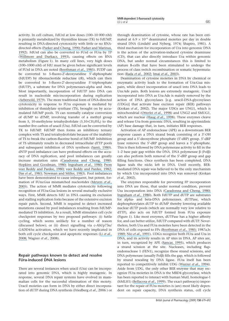
activity. In cell culture, FdUrd at low doses (100–10 000 nM)is primarily metabolized by thymidine kinase (TK) to FdUMP,resulting in DNA-directed cytotoxicity with little or no RNA-directed effects (Parker and Cheng, 1990; Parker and Marinus,1992). FdUrd can also be converted to FUrd or FUra by TP(Willmore and Durkacz, 1993), causing effects on RNAmetabolism (Figure 1). In many cell lines, very high doses(100–1000 nM) of FdU must be given before significant levelsof FUrd in DNA are noted (Boothman et al., 1985). FUDP canbe converted to 5-fluoro-2′-deoxyuridine 5′-diphosphate(FdUDP) by ribonucleotide reductase (rR), which can thenbe converted to 5-fluoro-2′-deoxyuridine 5′-triphosphate(FdUTP), a substrate for DNA polymerases-alpha and -beta.Most importantly, incorporation of FdUTP into DNA canresult in nucleotide mis-incorporation during replication(Aebersold, 1979). The more traditional form of DNA-directedcytotoxicity in response to FUra exposure is mediated byinhibition of thmidylate synthase (TS), brought on by accu-mulated FdUMP pools. TS can normally catalyse conversionof dUMP to dTMP, involving transfer of a methyl groupfrom 5, 10-methylene tetrahydrofolate (5,10-CH2FH4) to thenumber five carbon of uracil (Ura). FdUrd can be converted byTK to FdUMP. FdUMP then forms an inhibitory ternarycomplex with TS and tetrahydrofolate because of the inabilityof TS to break the carbon-fluorine bond of FdUMP. Inhibitionof TS ultimately results in decreased intracellular dTTP poolsand subsequent inhibition of DNA synthesis (Santi, 1980).dNTP pool imbalance can have profound effects on the accu-racy of DNA replication, and pool imbalances can greatlyincrease mutation rates (Caradonna and Cheng, 1980;Hopkins and Goodman, 1980; Ingraham et al., 1980; Premveer Reddy and Pardee, 1980; veer Reddy and Pardee, 1982;Das et al., 1983; Newman and Miller, 1983). Pool imbalanceshave been demonstrated to cause infrequent, but potent, for-mation of FUra:Gua mismatched nucleotides (Meyers et al.,2005). The action of MMR mediates cytotoxicity followingrecognition of FUra:Gua lesions in several mutually exclusiveways. First, MMR detects FdU in DNA causing its exclusionand stalling replication forks because of the extensive excisionrepair patch. Second, MMR is required to detect increasedmutations caused by pool imbalances resulting from FdUMP-mediated TS inhibition. As a result, MMR stimulates cell cyclecheckpoint responses by two proposed pathways: (i) futilecycling, which seems unlikely for a number of reason(discussed below); or (ii) direct signalling via c-Abl/p73a/GADD45a activation, which we have recently implicated inboth cell cycle checkpoint and apoptotic responses (Li et al.,2008; Wagner et al., 2008).
Repair pathways known to detect and resolveFUra-induced DNA lesions
There are several instances when uracil (Ura) can be incorpo-rated into genomic DNA, which is highly mutagenic. Inresponse, several DNA repair systems have evolved in mam-malian cells for the successful elimination of this moiety.Uracil moieties can form in DNA by either direct incorpora-tion of dUTP during DNA synthesis (Friedberg et al., 2006 ) or
through deamination of cytosine, whose rate has been esti-mated at 6.9 ¥ 10-8 deaminated moieties per day in doublestrand DNA (Lindahl and Nyberg, 1974; Shapiro, 1981). Athird mechanism for incorporation of Ura into genomic DNAis the action of the activation-induced cytosine deaminase(CD), that can also directly introduce Ura within genomicDNA, but under normal circumstances this is limited tomature B-cells that have been stimulated to undergo theprocess of class switch recombination or somatic hypermuta-tion (Rada et al., 2002; Imai et al., 2003).
Deamination of cytosine moieties in DNA by chemical orenzymatic activity leads to the formation of Ura:Gua mis-pairs, while direct incorporation of uracil into DNA leads toUra:Ade pairs. Both lesions are extremely mutagenic. Uracilincorporated into DNA as Ura:Ade is mainly removed by theaction of DNA glycosylases [e.g. uracil-DNA-glycosylases(UDGs)] that activate base excision repair (BER) pathways(Krokan et al., 2002). The major UDGs are UNG1, which ismitochondrial (Otterlei et al., 1998) and UNG2 and SMUG1,which are nuclear (Haug et al., 1998). These enzymes cleaveand release Ura from genomic DNA, resulting in apyrimidinic(AP) base damage that, in turn, initiates BER responses.
Activation of AP endonuclease (APE) as a downstream BERresponse causes a DNA strand break consisting of a 3′-OHgroup and a 5′-deoxyribose phosphate (5′-dRP) group. 5′-dRPlyase removes the 5′-dRP group and leaves a 5′-phosphate.This is then followed by DNA polymerase activity to fill in the1–2 base pair gap within the DNA. DNA polymerase b (Polb)can also perform both removal of the 5′-dRP group and gapfilling functions. Once synthesis has been completed, DNAligase seals the nicks left in DNA. Until recently, BER-mediated DNA repair was believed to be the only mechanismby which Ura incorporated into DNA was removed (Krokanet al., 2002).
The enzymes responsible for preventing FP incorporationinto DNA are those, that under normal condition, preventUra incorporation into DNA (Caradonna and Cheng, 1980;Ingraham et al., 1980). Both FdUTP and dUTP are substratesfor alpha- and beta-DNA polymerases. dUTPase, whichdephosphorylates dUTP to dUMP thereby lowering availablenuclear dUTP pools (which are normally very low relative todTTP), also acts on FdUTP formed from FUra exposure(Figure 1). Like most enzymes, dUTPase has a higher affinityfor, and can better utilize, FdUTP compared with dUTP. Never-theless, both Ura and FUra moieties have been detected in theDNA of cells exposed to FPs (Boothman et al., 1985; 1987a,b;1989; Nio et al., 1991). UDGs recognize both FUra and Ura inDNA, and its activity results in AP sites in DNA. AP sites are,in turn, recognized by APE (Sancar, 1995), which producesa strand scission at the site. Nucleases, including flap-endonuclease 1 (FEN1), recognize these strand scissions, andDNA polymerase (usually Polb) fills the gap, which is followedby strand resealing by DNA ligase. FUra itself has beenreported to competitively inhibit UDG (Wurzer et al., 1994).Aside from UDG, the only other BER enzyme that may rec-ognize FUra moieties in DNA is the MBD4 glycosylase, whichhas been reported to interact with human MutL homologue-1(hMLH1) (Bellacosa et al., 1999). The exact pathway(s) impor-tant for the repair of FUra moieties is (are) most likely depen-dent on repair capacity, DNA synthesis status, cell cycle
MMR-dependent 5-fluorouracil cytotoxicityLS Li et al 681
British Journal of Pharmacology (2009) 158 679–692

regulation, appropriate balanced deoxyribonucleotide andribonucleotide pools, as well as other possible tumourmicroenvironment factors. The contribution of MMR indetecting FUra moieties in DNA, and the resulting cytotoxicresponses of exposed cells are described below.
Treatment of mammalian cells with FPs can lead to dNTPpool imbalances. Decreases in dTTP pools because of FdUMPinhibition of TS removes negative feedback inhibition on rRand TK that result in greater levels of FdUTP, which then leadsto elevated incorporation of FUra into DNA. Additionally, TSinhibition will cause a build-up of both dUTP and FdUTPpools and eventually exhaust dUTPase. As dUTP and FdUTPaccumulate and dTTP levels fall, dUTP and FdUTP poolsreplace dTTP as substrates for DNA polymerase, resulting inever-increasing levels of FdUTP or dUTP incorporated intoDNA. Given these metabolic changes, it has been puzzlingwhy such low levels of FUra moieties in DNA have beendetected in most cancer cells after FP exposure.
DNA mismatch repair and DNA damage signalling
Primary tumours and tumour cell lines containing MMRdefects are resistant to a wide variety of commonly usedtherapeutic agents (see review: Irving and Hall, 2001). Theseinclude methylating agents [Temozolomide™; procabazine;N-methyl-N’-nitro-N-nitrosoguanidine (MNNG); N-methyl-N-nitrosourea (MNU)], antimetabolites (6-thioguanine; mer-captopurine), platinum compounds (cisplatin; carboplatin)and perhaps Topoisomerase II inhibitors that have additionaleffects on cellular redox reactions (doxorubicin; epirubicin).In the last several years, it has become apparent that the drugresistance in MMR-deficient cells was tied to reduced orabsent damage-induced G2 arrest and ultimately cell deathresponses (e.g. apoptosis) (Meyers et al., 1997; Davis et al.,1998; Gong et al., 1999; Hickman and Samson, 1999; Zhanget al., 1999).
Initiation of cellular responses to DNA damage caused by FPexposure requires DNA damage sensors (DS), adaptors/mediators (AM), as well as amplification responses involvingMMR-dependent c-Abl responses (Gong et al., 1999; Wagneret al., 2008), or MMR-independent PI-3-like kinases (PIKKs).For simplicity, only G2 arrest and apoptotic responses will beconsidered here, as these appear to be the primary cellularresponses to FP damage. A MMR-independent DS/AM/PIKKcomplex appears to activate at least two pathways that lead toG2 arrest by cascade phosphorylation of p53 mediated byChk1. Activation of Chk1, by phosphorylation, leads to theregulation of the Cdc25C phosphatase, by protein modifica-tion. During a normal cell cycle, Cdc25C dephosphorylatesCdc2, prior to entry into G2. Thus, inactivation ofCdc25C results in a de facto G2 arrest. Conversely, thephosphorylation-activation of p53 leads to significantup-regulation of 14-3-3s that, in turn, sequesters Cdc2/cyclinB leading to G2 arrest. These responses can be stimu-lated in MMR-deficient cells only by high doses of FPs and theresponses are more delayed (at least in response to alkylationdamage) than are MMR-dependent responses (Wagner et al.,2008).
In MMR-competent cells, another more potent and rapid G2
arrest and apoptotic stimulatory pathway is activated. These
responses are seen at >10-fold less doses of FPs or alkylatingagents in MMR-competent cells, compared with MMR-independent responses that are noted only after high doses ofdamaging agents. MMR-dependent sensing of alkylation or FPdamage stimulates the activation of c-Abl kinase, which canbe suppressed by Gleevec™ or by specific siRNA-c-Abl knock-down (Wagner et al., 2008). These ‘immediate-early’ (i.e., inminutes) G2 arrest responses provide the cellular frameworkfor the MMR signalling pathway. MMR-dependent persistentDNA lesion damage recognition, processing and signallingleads to combined ‘early’ (h) and ‘late’ (h/days) responses thatmay result in premature senescence, necrosis, or apoptoticcell death. MMR-dependent apoptosis is mediated by induc-tion and stimulated levels of GADD45a and p73a, but not byp53 (Meyers et al., 2001; 2003; 2005; Li et al., 2008). MMR-dependent apoptosis and G2 arrest were p53-independent, asloss of p53 because of E6 expression, somatic knockout, orstable siRNA-p53 knockdown had no affect on apoptoticresponses in MMR-dependent cell death (Li et al., 2008). Inter-estingly, our studies were able to separate MMR-dependentsignalling of G2 arrest from apoptotic responses, as siRNA-specific p73a knockdown resulted in loss of apoptosis but notG2 arrest. In contrast, specific knockdown of c-Abl orGADD45a prevented both apoptosis and G2 arrest responses.Loss of p53 in these cells did not affect MMR-dependentresponses (Li et al., 2008; Wagner et al., 2008).
Two opposing models have been proposed to account forthe MMR-dependent G2 arrest and apoptosis: (i) futile cyclingof repair; and (ii) direct MMR-dependent signalling. ‘Futilecycling’ of repair, was originally proposed to explain a similarMMR-dependent cell death effect in bacteria that containeda dam, DNA adenine methylase, mutation (Karran andMarinus, 1982; Fram et al., 1985). In the absence of Dammethylation, the MutH-dependent incision that initiatesMMR may occur on either DNA strand on either side of thelesion. Some of these incision events were proposed to lead tobidirectional degradation towards the lesion. This unregu-lated degradation could then lead to DNA double-strandbreaks (DSBs). Multiple DSBs induced by multiple MMR reac-tions were proposed to result in genetic catastrophe and celldeath. As DSBs are a well-known cause of G2 arrest and p53-induced apoptosis in mammalian cells (Kastan et al., 1991),such a mechanism seemed plausible in mammalian cells(Davis et al., 1998). However, there appears to be multipleproblems in adapting MMR-dependent ‘futile cycling’ mecha-nism to mammalian systems, among them the apparent lackof any requirement for p53 (for review see: Meyers et al., 2003;Wang and Edelmann, 2006).
The ‘direct signalling’ model was originally proposed (Daviset al., 1998; Fishel, 1999; 2001) to explain the MMR-dependent activation of G2 arrest and apoptosis, an apoptoticpathway that later appeared to include c-Abl (Gong et al.,1999), as well as the rapid induction of apoptosis followingover-expression of MMR genes (Zhang et al., 1999). Morerecent evidence suggests that a subset of the MMR proteins(i.e. hMSH2, hMSH6, hMLH1 and hPMS2) serve as sensors ofDNA damage (Wang and Edelmann, 2006). For example,Hsieh and colleagues (Yoshioka et al., 2006) have demon-strated that hMSH2-hMSH6 and hMLH1-hPMS2 bound toa methylation-damaged O6-MeG/T mismatched DNA
MMR-dependent 5-fluorouracil cytotoxicity682 LS Li et al
British Journal of Pharmacology (2009) 158 679–692
specifically interact with ataxia telangiectasia-and-rad3-related(ATR)-ATRIP (a PIKK family member) and Chk1. These studiessuggested the ATR/Chk1 pathway in at least part of the‘immediate early’ G2 arrest. However, most recently, our groupfound that MMR-dependent G2 arrest responses triggered byMNNG are dependent on a hMLH1/c-Abl/GADD45a signal-ling pathway, and that ataxia telangiectasia mutated (ATM)/Chk2, as well as ATR/Chk1, was clearly not involved in theMMR-dependent G2 arrest responses in response to alkylation(Wagner et al., 2008) or FP damage (Meyers et al., 2001; 2003).The activation of apoptosis following persistent DNA damagewas induced by hMLH1/c-Abl/p73a/GADD45a retrograde-signalling pathway, where ATM and p53 were not involved (Liet al., 2008). We also noted that MMR triggers apoptosis inresponse to MNNG-induced DNA lesions, which along withlong-term survival, was completely abrogated by the c-Ablkinase inhibitor, STI571 (Gleevec™). As a result, our datastrongly suggest that Gleevec™ may be ill-suited in conjunc-tion with temozolomide or cisplatin, or other clinically usedalkylating agents, for efficacious cancer therapy in tumoursthat are proficient in the MMR pathway (Li et al., 2008).
Remarkably, the introduction of the Msh2 (G674A) or Msh6(T1217D) mutation into mice resulted in an absence of MMRactivity but normal damage-induced apoptosis (Lin et al.,2004; Yang et al., 2004). Thus, dissociation of MMR-dependent multiple excision tracts required for ‘futile cycling’from a damage-induced apoptotic response would appear tosignificantly reduce the likelihood of the ‘futile cycling’mechanism. These functional dissociation mutations arelocated in separate, but proximal, highly conserved ATP/ADPprocessing domains of the Msh2-Msh6 heterodimer.
The mechanics of DNA mismatch repair andlesion recognition
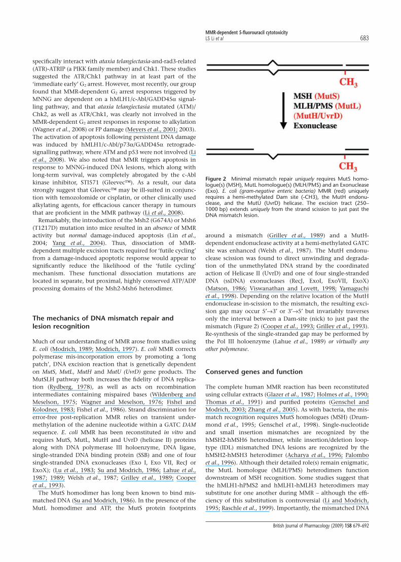
Much of our understanding of MMR arose from studies usingE. coli (Modrich, 1989; Modrich, 1997). E. coli MMR correctspolymerase mis-incorporation errors by promoting a ‘longpatch’, DNA excision reaction that is genetically dependenton MutS, MutL, MutH and MutU (UvrD) gene products. TheMutSLH pathway both increases the fidelity of DNA replica-tion (Rydberg, 1978), as well as acts on recombinationintermediates containing mispaired bases (Wildenberg andMeselson, 1975; Wagner and Meselson, 1976; Fishel andKolodner, 1983; Fishel et al., 1986). Strand discrimination forerror-free post-replication MMR relies on transient under-methylation of the adenine nucleotide within a GATC DAMsequence. E. coli MMR has been reconstituted in vitro andrequires MutS, MutL, MutH and UvrD (helicase II) proteinsalong with DNA polymerase III holoenzyme, DNA ligase,single-stranded DNA binding protein (SSB) and one of foursingle-stranded DNA exonucleases (Exo I, Exo VII, RecJ orExoX); (Lu et al., 1983; Su and Modrich, 1986; Lahue et al.,1987; 1989; Welsh et al., 1987; Grilley et al., 1989; Cooperet al., 1993).
The MutS homodimer has long been known to bind mis-matched DNA (Su and Modrich, 1986). In the presence of theMutL homodimer and ATP, the MutS protein footprints
around a mismatch (Grilley et al., 1989) and a MutH-dependent endonuclease activity at a hemi-methylated GATCsite was enhanced (Welsh et al., 1987). The MutH endonu-clease scission was found to direct unwinding and degrada-tion of the unmethylated DNA strand by the coordinatedaction of Helicase II (UvrD) and one of four single-strandedDNA (ssDNA) exonucleases (RecJ, ExoI, ExoVII, ExoX)(Matson, 1986; Viswanathan and Lovett, 1998; Yamaguchiet al., 1998). Depending on the relative location of the MutHendonuclease in-scission to the mismatch, the resulting exci-sion gap may occur 5′→3′ or 3′→5′ but invariably traversesonly the interval between a Dam-site (nick) to just past themismatch (Figure 2) (Cooper et al., 1993; Grilley et al., 1993).Re-synthesis of the single-stranded gap may be performed bythe Pol III holoenzyme (Lahue et al., 1989) or virtually anyother polymerase.
Conserved genes and function
The complete human MMR reaction has been reconstitutedusing cellular extracts (Glazer et al., 1987; Holmes et al., 1990;Thomas et al., 1991) and purified proteins (Genschel andModrich, 2003; Zhang et al., 2005). As with bacteria, the mis-match recognition requires MutS homologues (MSH) (Drum-mond et al., 1995; Genschel et al., 1998). Single-nucleotideand small insertion mismatches are recognized by thehMSH2-hMSH6 heterodimer, while insertion/deletion loop-type (IDL) mismatched DNA lesions are recognized by thehMSH2-hMSH3 heterodimer (Acharya et al., 1996; Palomboet al., 1996). Although their detailed role(s) remain enigmatic,the MutL homologue (MLH/PMS) heterodimers functiondownstream of MSH recognition. Some studies suggest thatthe hMLH1-hPMS2 and hMLH1-hMLH3 heterodimers maysubstitute for one another during MMR – although the effi-ciency of this substitution is controversial (Li and Modrich,1995; Raschle et al., 1999). Importantly, the mismatched DNA
Figure 2 Minimal mismatch repair uniquely requires MutS homo-logue(s) (MSH), MutL homologue(s) (MLH/PMS) and an Exonuclease(Exo). E. coli (gram-negative enteric bacteria) MMR (red) uniquelyrequires a hemi-methylated Dam site (-CH3), the MutH endonu-clease, and the MutU (UvrD) helicase. The excision tract (250–1000 bp) extends uniquely from the strand scission to just past theDNA mismatch lesion.
MMR-dependent 5-fluorouracil cytotoxicityLS Li et al 683
British Journal of Pharmacology (2009) 158 679–692
substrate must contain a pre-introduced single-stranded scis-sion (nick), either 5′- or 3′- of the DNA mismatch. Nucleaseactivity appears to be accomplished by a combination of a5′-exonuclease (ExoI) and an intrinsic ssDNA endonucleaseactivity found in some MLH family members (Kadyrov et al.,2006). The minimal 5′→3′ and 3′→5′ excision reactionrequires hMSH2-hMSH6 (or hMSH2-hMSH3), hMLH1-hPMS2, ExoI, RPA, PCNA, and RFC (Kadyrov et al., 2006).Re-synthesis of the single-stranded gap requires Pold, andligase I, and may be modestly enhanced with HMGB1 (Zhanget al., 2005).
Models for MMR
A detailed biophysical mechanism for MMR remains contro-versial and incomplete. Modrich and colleagues have pro-posed a Hydrolysis-Dependent Translocation Motor Model(Figure 3I). It posits the assembly of a MutS-MutL complex atthe mismatch (Figure 3IA and IB) (Modrich, 1989; Modrichand Lahue, 1996), which then uses ATP-hydrolysis to motorbi-directionally creating a looped structure (Figure 3IC) (Allenet al., 1997). This DNA tracking process was envisioned to linkMutS mismatch recognition to the MutH endonuclease inci-sion, as well as to provide the necessary directionality forsubsequent loading of Helicase II (UvrD) and one of thessDNA exonucleases (Figure 3ID). However, not all predic-tions arising from this model agree with the genetic or bio-chemical data (Gradia et al., 1997; 1999; 2000; Fishel, 1998;1999; Guerrette et al., 1998; 1999; Schmutte et al., 1998;Wilson et al., 1999; Berardini et al., 2000; Schmutte et al.,2001; Mazurek et al., 2002).
Our work with human MSH proteins led to the MolecularSwitch Model [Figure 3II; for review see: (Berardini et al.,2000)]. It is based on the observation that mismatched DNAstimulated the exchange bound ADP for ATP (ADP→ATPexchange) by the human MSH proteins (hMSH2-hMSH6 orhMSH2-hMSH3 (Figure 3IIA) (Gradia et al., 1997; Wilsonet al., 1999). ATP binding resulted in the formation of a MSHsliding clamp capable of hydrolysis-independent diffusion/tracking for several thousand nucleotides along the adjoiningDNA backbone (Gradia et al., 1999). Iterative loading of mul-tiple sliding clamps was suggested to provide a ‘thresholdsignal’ that distinguished the mismatch region and provideda ‘gradient’ of MSH proteins that proffered a directionalityalong the DNA duplex surrounding the mismatch site(Figure 3IIB) (Gradia et al., 1999; Heinen et al., 2002). ATPhydrolysis only occurred when the human MSH proteins dis-sociated from the DNA ends (Gradia et al., 1999); a rare con-dition in normal cells. These observations accounted for thelow ATPase activity (Haber and Walker, 1991; Gradia et al.,1997; Gradia et al., 2000; Hess et al., 2002) and identifiedADP→ATP exchange at the mismatch site as the rate-limitingstep. The process appeared remarkably similar to the controlof G protein molecular switches by GDP→GTP exchange(Sprang, 1997). Although their exact role in MMR remainsenigmatic, it appeared that the MLH/PMS proteins formed astable ternary complex with ATP-bound MSH sliding clamps(Figure 3IIB) (Acharya et al., 2003). The MSH-MLH/PMSternary complex was shown to interact with downstream
MMR components, such as MutH (Figure 3IIC), MutU (UvrD)helicase (not shown), or a ssDNA end (Figure 3IID). Incre-mental rearward diffusion, made irreversible by endo- or exo-nuclease digestion, was proposed to create a dynamic andredundant process as multiple MSH-MLH/PMS sliding clamps‘hand-off’ the excision reaction until it covers the mismatch(Figure 3IIE). At that point no further sliding clamps may beloaded and the minimal MMR reaction is complete. In thismodel, ATP hydrolysis is only required to recycle MSH slidingclamps. These studies led to a modification of the MotorModel by Modrich and colleagues; although it still requiredATP hydrolysis to move the MSH protein along the DNA(Blackwell et al., 1998).
A third mechanism, the Transactivation Model, arose frombacterial MutS, MutL, and MutH structures (Ban and Yang,1998; Ban et al., 1999; Lamers et al., 2000; Obmolova et al.,2000). However, recent experiments have shown that physi-cally blocking the intervening region between the mismatchand Dam site results in a near complete impairment of MMR(Pluciennik and Modrich, 2007). These observations appear torule-out this and any similar ‘trans’ models in favour of ‘cis’models similar to those considered above (Kolodner et al.,2007).
Recognition of mismatches and lesions byMSH proteins
MutS homologue proteins recognize a plethora of DNA mis-matches, lesions and structures. This is unusual comparedwith glycosylases, which often have overlapping recognitionwith MSH proteins but are highly lesion specific. Compari-son(s) of numerous mispair-bound MSH structures appearremarkably similar (Lamers et al., 2000; Obmolova et al., 2000;Natrajan et al., 2003; Warren et al., 2007). An important con-sequence of mismatch recognition by MSH proteins is theinsertion of a highly conserved phenylalanine residue 3′ of themismatch (Lamers et al., 2000; Obmolova et al., 2000). Usingnearest neighbor sequence contexts as a model, we recentlyfound that poorly recognized mismatches display an increasedstability of base pairs 3′ to the mismatch (Mazurek et al., 2009).This observation suggests that on at least one strand surround-ing a poorly recognized mismatch, the interrogation by MSHproteins may be significantly more difficult. NOE data alsosuggests that well-recognized mismatches have increasedlocalized dynamic flexibility (Mazurek et al., 2009). Together,these observations suggest that MSH proteins do not recognizethe mismatch or lesion but instead recognize the flexibility ofthe DNA that is induced by the mismatch or lesion. Theseresults have several important implications when consideringDNA metabolites that might be useful in provoking the MMRdamage signalling response for efficacy in cancer therapeutics.
Role of MMR in FP responses and antitumouractivity: a historical perspective
The MMR pathway recognizes all eight single nucleotidemismatches, as well as small insertion/deletion loop-type
MMR-dependent 5-fluorouracil cytotoxicity684 LS Li et al
British Journal of Pharmacology (2009) 158 679–692

mismatches. The majority of mismatched nucleotides arise asa result of polymerase mis-incorporation errors. As FPs (espe-cially thymidine analogs) could also be incorporated intoDNA across from Gua as a result of deoxynucleotide poolimbalances (e.g. after FUra or FdUrd exposures) and/orbecause of transition state alterations (e.g. BrdUrd), we inves-tigated the role of MMR in cellular responses to FUra and
FdUrd (Meyers et al., 2001). Our group was the first to reportthat MMR cells were resistant to FUra and FdUrd in anabstract submitted in 1996 (Meyers et al., 1996). We demon-strated that hMLH1-deficient HCT116 colon cancer cells were20-fold more resistant to FUra (continuous treatment for72 h) and 17-fold more resistant to FdUrd in clonogenicsurvival assays compared with genetically matched
Figure 3 Models for Mismatch Repair. See text for description.
MMR-dependent 5-fluorouracil cytotoxicityLS Li et al 685
British Journal of Pharmacology (2009) 158 679–692

hMLH1-proficient HCT116 3-6 cells. Likewise, murine MLH1-deficient CT-5 cells were threefold more resistant to a 2-hpulse of FdUrd than their MLH1-proficient ME-10 counter-parts. Synchronized MMR-proficient HCT116 3-6 cells treatedwith low doses of FPs had a twofold greater G2 cell cycle arrestresponse compared with MMR-deficient HCT116 cells. Asyn-chronous ME-10 cells demonstrated a fourfold greater G2
arrest after FdUrd treatment compared with CT-5 cells. G2 cellcycle arrest was not a result of mitotic arrest, but rather a trueG2 arrest as indicated by elevated cyclin B1 levels and a lack ofstaining with mitotic protein monoclonal antibody 2.Although p53 levels were induced in FdUrd-treated HCT1163-6 cells, cell death and G2 arrest responses were not depen-dent on the function of this tumour suppressor. FdUrd-mediated cytotoxicity was caused by DNA-directed and notRNA-directed effects, as administration of excess dThyd (andnot Urd) prevented cytotoxicity, cell cycle arrest and DSBformation. hMLH1-dependent responses to FP treatmentwere, therefore, predicted to have clinical relevance for theuse of DNA-directed FPs in the treatment of tumours withMMR deficiencies.
Clinical data suggest that patients withMMR-deficient cancers do not benefit fromFP therapies
Around 10~15% of sporadic colorectal cancers exhibit mis-match repair deficiencies because of hypermethylation ofhMLH1 (Li et al., 2003; Ribic et al., 2003; Jover et al., 2009).FUra has been used in cancer chemotherapy for more than 40years, and remains the standard of care as an adjuvant che-motherapeutic regimen for the treatment of colorectalcancer. Early studies reported that stages II and III colorectalcancer patients had improved overall survival from FUraadjuvant chemotherapy regardless of MSI status (Elsalehet al., 2000; Hemminki et al., 2000; Watanabe et al., 2001).However, these studies did not take into account patientswith MMR deficiencies that did not receive adjuvant chemo-therapy. These considerations decreased the accuracy of thestudy, as intrinsic overall survival of MMR-deficient colorec-tal cancer patients related to better prognosis than MMRcompetent patients. The other study from Elsaleh et al.(2000) was limited by non-randomized sample selection.Recently, both retrospective and prospective studies havedemonstrated that colorectal cancer patients with MMR defi-ciencies do not receive significant benefit from FUra-basedadjuvant chemotherapy (Ribic et al., 2003; Carethers et al.,2004; Jover et al., 2006; 2009). The Ribic et al.’s investigation(Ribic et al., 2003), a retrospective study based on largesample size and appropriate control groups, demonstratedthat patients with stages II and III colon cancer benefitedfrom FUra-based adjuvant chemotherapy only when theirtumours were MMR-competent. Patients in the same studywith tumours resulting from the lack of MMR activity, incontrast, received no benefit from FUra adjuvant therapies(Ribic et al., 2003). The most recent prospective studiesfurther confirmed the retrospective reports suggesting thatadjuvant FUra-based chemotherapy may not be useful in
stages II and III microsatellite-instable colorectal cancers(Jover et al., 2006; Jover et al., 2009). These clinical datafurther confirmed our previous findings that MMR-deficientcell lines were less responsive than MMR stable cell lines toFUra treatments (Meyers et al., 2001; 2005).
MSH2-deficient cells were resistant to FdUrd, butnot Tomudex™
We examined human colon cancer cells deficient in hMLH1expression, as well as both human and mouse cell lines defi-cient in MSH2 for resistance/sensitivity to FUra, FdUrd orTomudex™, a non-pyrimidine TS inhibitor. Whereas FdUrdhas two major DNA-directed mechanisms of cell killing (i.e.DNA incorporation and inhibition of TS), Tomudex™ specifi-cally inhibits TS. Thus, treatment with Tomudex allowed usto discriminate the relative contributions of DNA incorpora-tion versus TS inhibition in MMR-dependent, FdUrd-mediated cell killing. When corrected for differential TS levels(the isogenic cells used were not different in TS activities)near identical dose-response survival curves for HCT116versus HCT116 3-6 cells were noted in response to Tomudex,suggesting that incorporation of FUra into DNA accountedfor the differential survival noted between these cells (Meyerset al., 2005).
MSH2- cells have reduced G2 arrest after FdUrdor FUra
Restoring hMLH1 expression in HCT116 cells caused signifi-cantly more prolonged G2 arrest in response to 6-TG or FdUrd,as we reported (Davis et al., 1998; Meyers et al., 2001). Asimilar response was noted when examining MSH2-/- andMSH2+/+ murine embryonic stem (ES) cells (Meyers et al.,2005). For example, transient and prolonged G2 arrestresponses occurred at drug concentrations (e.g. 1.5 nM FdUrdin MSH2- ES cells) that caused no significant loss of survival.Similar to MLH1-deficient cells, MSH2-deficient cells showedan abrogated G2 arrest response to FdUrd or FUra treatments.Thus, G2 arrest in response to FP exposure also relied on anintact MMR system and was not merely dependent on MLH1expression. As noted above, no differences in G2 arrestresponses were noted after Tomudex™ exposure in isogeniccell lines expressing or lacking MLH1 or MSH2. Thymidine(dThyd) depletion in both cell systems as a consequence ofthe inhibition of TS activity, S-phase arrest independent ofMMR status was found (Meyers et al., 2005) as described(Orlandi et al., 1999; Yin et al., 1999).
In its role in post-replicative DNA repair, MMR detects DNAmispairs/lesions in the context of a newly synthesized DNAstrand. It identifies the incorrect base in a mispair (placedthere by a DNA polymerase error) because of its presence inthe daughter strand (Karran, 2001). FdUrd, formed after FUraexposure, relies on DNA replication for its incorporation intoDNA, whereby this pyrimidine analog is incorporated (as thebase FU) across from Ade or Gua. We examined the cell cyclearrest responses of HCT116 (hMLH1-, MMR-) and HCT116 3-6
MMR-dependent 5-fluorouracil cytotoxicity686 LS Li et al
British Journal of Pharmacology (2009) 158 679–692

(hMLH1+, MMR+) cells within the first cell cycle after treat-ment. While both cell lines responded with a strong G2 arrestby 20 h after FdUrd addition, only MMR+ HCT116 3-6 cellsresponded with a prolonged G2 arrest caused by MMR-dependent proof-reading. Identical G2 arrest responses werenoted in the first cell division in MSH2+ cells, whereas MSH2
-
cells did not arrest.
hMSH2-hMSH6 recognizes FUra:Gua lesions
To assess the ability of MMR to directly recognize FP-inducedlesions in DNA, we tested the ability of purified hMSH2-hMSH6 or hMSH2-hMSH3 heterodimers to recognize FPlesions (specifically FU base-paired with Ade or Gua) using41-mer oligonucleotide substrates. MMR activities using theseDNA substrates were assessed by ATPase activities (Mazureket al., 2002). A Thy:Gua base pair (as a positive control), butnot a Thy:Ade base pair (as a negative control), was able toactivate the ATPase (i.e. the ATPase velocity) of hMSH2-hMSH6. Interestingly, FUra:Gua and Ura:Gua, but notFUra:Ade or Ura:Ade base pairs, were able to activate MMRactivity. Importantly, MMR was not capable of recognizingthe dThyd analogs, Ura or FUra, when directly base-pairedwith Ade. Instead, MMR only detected FUra or Ura whenmispaired with Gua. Ade is the expected base-pairing partnerfor the dThyd/Urd analogs, Ura and FUra. We also examinedthe ability of hMSH2-hMSH3 complexes to recognize variousFUra or Ura substrates. The hMSH2-hMSH3 complex is pri-marily responsible for recognizing small insertion and dele-tion loops in DNA (Fishel, 1999). MMR ATPase activity fromthe hMSH2-hMSH3 complex was observed with the positivecontrol (the Cyt-Ade loop), but not the negative control(Thy:Ade). As expected, neither FUra:Ade nor FUra:Gua weresubstrates for MMR and, therefore, did not activate the ATPaseof the hMSH2-hMSH3 complex.
MMR-deficient cells incorporated higher FUralevels in their DNA
To determine if MMR status influenced the overall amount ofradio-labeled FP incorporated into DNA, MMR-deficient andMMR-proficient cells, lacking either MLH1 (HCT116) or MSH2(ES), were treated with various doses of FdUrd spiked with 20to 50 mCi·mL-1 [3H]FdUrd for 3 days, and genomic DNA puri-fied and assayed for antimetabolite-related, incorporatedradioactivity (Meyers et al., 2005). DNA of MLH1-deficientHCT116 or MSH2-/- ES cells treated with 2.5 mM FdUrd con-tained 2.6- and 2.3-fold greater incorporated radioactiveFdUrd, respectively, than their MMR-competent counterparts.Addition of excess dThyd, but not Urd, prevented incorpora-tion of FdUrd into DNA, consistent with the ability of dThydto rescue FdUrd-induced toxicity in these cells, as previouslyreported for MMR+ cells (Meyers et al., 2001).
FdUrd-treated MMR- cells selectively incorporatedlow, but significant levels of FUra:Gua in DNA
As the hMSH2-hMSH6 heterodimer selectively recognizedFUra:Gua lesions, we examined the frequency of these mis-
match bases in the DNA of FdUrd- or FUra-treated HCT116cells relative to FUra:Ade base pairs. To distinguish FUra:Adefrom FUra:Gua radiolabeled lesions, specific BER enzymes wereused. UDG removes Ura and FUra from DNA regardless of theirbase pairing partners; it can recognize Ura:Ade, FUra:Ade,Ura:Gua, and FUra:Gua, as well as other base pairings (Krokanet al., 2000; Meyers et al., 2003). In contrast, MBD4 (alsoknown as methyl-CpG-binding endonuclease I) only recog-nizes Ura:Gua or FUra:Gua, but not Ura:Ade or FUra:Ade, inDNA (Petronzelli et al., 2000; Meyers et al., 2003). Indeed, UDGrecognized both FUra:Ade and FUra:Gua in the same 41-meroligomer substrates, indicated by the generation of a cleavageproduct following hot alkali treatment. In contrast, MBD4only recognized DNA substrates containing FUra:Gua lesions.In fact, MBD4 recognized FUra:Gua regardless of Cyt methy-lation status (methylation in human DNA occurs on Cyt in aCpG context). This was interesting as MBD4 was once thoughtto be the human homologue of the bacterial MutH endonu-clease (Bellacosa et al., 1999; Bellacosa, 2001) that allowedMMR to direct repair to the newly synthesized strand based onits methylation status. In bacteria, the daughter strand (whichof course would contain the newly incorporated incorrectbase) is transiently unmethylated at Ade in a d(GATC) contextimmediately following replication (Modrich, 1991).
Using UDG and MBD4, MMR-deficient and -proficient cellswere analysed for FUra base-pair incorporation analyses.HCT116 and HCT116 3-6 cells were incubated with [3H]FdUrdfor 3–10 days and genomic DNA isolated (Meyers et al., 2005).DNA was then treated with UDG or MBD4 (both of whichdetect Urd or FUrd in DNA and release the free bases, Ura orFUra respectively) to investigate total FUra incorporationcompared with FUra incorporated into FUra:Gua lesions.Whereas levels of FUra incorporated across from Gua (asdetermined by the amount of [3H] released following MBD4treatment) were equivalent in MMR- HCT116 cells comparedwith MMR+ HCT116 3-6 cells after 3 days of treatment, therewas threefold more FUra:Gua in HCT116 DNA at day 10. Thisincorporation difference correlated well with lethality, wherea difference in cell survival was noted only following longer(i.e. 10 day) exposures to FdUrd (Meyers et al., 2001). In addi-tion, the fact that the amount of [3H] released from DNA afterUDG treatment was only modestly higher (in three of fourinstances) than that released after MBD4 treatment indicatesthat at least half of the FUra in DNA was paired with Gua. Torule out the possibility that MBD4 levels may be differentbetween MMR- and MMR+ cells, levels of MBD4 protein wereexamined and found equivalent in HCT116 and HCT116 3-6cells, as well as in the other cell systems used.
Can FPs be used to treat MSI+, hMLH1-
sporadic cancers?
Given that many colon and ovarian cancers are microsatelliteinstable due to the lack of hMLH1expression caused by hyper-methylation of the hMLH1 promoter, is there any hope oftreating these cancers using FPs? Recently, one FP, 5-fluoro-2′-deoxycytidine (FdCyd), has been shown to be a hypom-ethylating agent when incorporated into the DNA of exposed
MMR-dependent 5-fluorouracil cytotoxicityLS Li et al 687
British Journal of Pharmacology (2009) 158 679–692

cells (Smith et al., 1992; Chuang et al., 2005). The fluorinegroup in the 5-position of FdCyd resists methylation becauseof its carbon-fluorine bond. Indeed, exposing cells to FdCydalters the methylation pattern of exposed cells (Chuang et al.,2005).
Importantly, prior research from our laboratories has shownthat the metabolism of FdCyd can be manipulated to: (i)protect FdCyd from deamination using the dCMP and/orcytidine deaminase inhibitors, deoxytetrahydrouridine(dH4Urd) or tetrahydrouridine (H4Urd) respectively (Booth-man et al., 1985; 1987a; 1987b) (Figure 4); and (ii) directFdCyd to dramatically increase (~20-fold) incorporation intothe DNA of cells co-treated with dH4Urd, which results in thesimultaneous inhibition of both cytidine deaminase (asdH4Urd) and dCMP deaminaase (as dH4UMP) (Figure 4).Indeed, we have recently demonstrated that hMLH1- RKO6cells, which are normally resistant to FdUrd alone, becomesensitive to FdCyd (Figure 5) through the re-expression ofhMLH1 (not shown). A dramatic increase in G2 arrestresponses were noted when RKO6 cells were exposed toFdCyd. In contrast, RKO6 cells were completely resistant toFdUrd (Figure 5). Recently, Beumer et al. (2006; 2008) haveextended our prior mouse data (Boothman et al., 1987a,b) byexamining pharmacokinetics, metabolism, oral bioavailabil-ity, and cytotoxic metabolites of FdCyd in mice and patients.As we previously demonstrated, potential toxic metabolitesgenerated by CD were avoided by combing FdCyd in treat-ment with 3,4,5,6-tetrahydrouridine (H4Urd). Accompanyingplasma 5-FU and FdUrd concentrations were <10% of these
Figure 4 Potential use of 5-fluoro-2′-deoxycytidine (FdCyd) for treatment of hMLH1-, MMR-deficient sporadic cancers. FdCyd, which is nota substrate for RNA-level metabolism (e.g. dThyd or Urd phosphorylases), can be manipulated for increased incorporation into DNA by usingthe dCyd or dCMP deaminase (CD or dCMPD respectively) inhibitors, tetrahydrouridine (H4Urd) or deoxytetrahydrouridine (dH4Urd)respectively. Once incorporated, FdCyd can lead to hypomethylation of DNA and re-expression of hMLH1 in MSI+ sporadic cancers that aregenetically wild-type for hMLH1, but lack hMLH1 expression because of hypermethylation of its promoter, as described by Veigl et al., 1998.Adapted from Meyers et al. (2003). hMLH1, human MutL homologue-1.
Figure 5 Sensitization of MSI+, hMLH1- RKO6 cells using FdCyd.RKO6 cells are wild-type for the hMLH1 gene, yet are MSI+, and lackexpression of hMLH1 protein because of hypermethylation of itspromoter. RKO6 cells were synchronized by low serum medium andreleased by re-plating into 10% fetal calf serum-containing medium.Cells were then mock-treated (untreated cells) or exposed to 1 mMFdUrd or FdCyd for 62 h, past the p53 checkpoint as described(Meyers et al., 2001; 2005). Cells were then monitored for cell cyclecheckpoint responses and changes in the G2 cell population weregraphed over time (h) after release. Note that FdCyd treatmentcaused G2 arrest with corresponding hMLH1 expression (not shown).Dashed line, mock-treated synchronized cells; Open squares, FdUrd(1.0 mM, 4 h); Closed circles, FdCyd (1.0 mM, 4 h). Western blottingdemonstrated hMLH1 expression and hPMS2 stabilization afterFdCyd, but not after FdUrd, exposures. hMLH1, human MutLhomologue-1.
MMR-dependent 5-fluorouracil cytotoxicity688 LS Li et al
British Journal of Pharmacology (2009) 158 679–692

observed after therapeutic infusions of 5-FU or FdUrd, whileFdCyd levels were well above those required for the inhibitionof methylation in vitro. (Beumer et al., 2006; 2008).
We propose that FdCyd exposures can be used to cause there-expression of hMLH1, and thereby, convert resistant hyper-methylated hMLH1- colon or ovarian cancer cells to sensitivecells that re-express functional hMLH1, and therefore MMR.This is particularly true for colon cancer cells that haveelevated levels of dCyd kinase, whereby dH4Urd [that caninhibit both CD and deoxycytidylate deaminase (dCMPD),Figure 4)] can be effectively used to channel FdCyd into DNA(Boothman et al., 1985). After approximately 2–4 days orseveral cell divisions, reversal of cytidine and dCMP deami-nase inhibition (by removal of the reversible inhibitor,dH4Urd) followed by continuous exposure with FdCyd wouldallow its incorporation into DNA, hypomethlyation of thehMLH1 promoter, stimulated re-expression of hMLH1 proteinand restored MMR activity. Restoration of functional MMRwould, in turn, result in increased sensitivity of cells to FPs.Once dH4Urd is removed, the accumulated pools of FdCydand FdCMP then would be converted by deamination toFdUrd and FdUMP by CD and dCMPD (which are alsoelevated in colon cancers), resulting in enhanced incorpora-tion of FdUrd into DNA and formation of elevated levels ofFdUMP (Figure 4). Re-expression of hMLH1 then wouldincrease sensitivity of cells to the now converted FdUrd thatincorporates into DNA because of elevated levels of dThydkinase (TK). Thus, instead of trying to expose cells to aza-cytidine, a typical hypomethylating agent used to re-expressgenes for increased sensitivity (Compere and Palmiter, 1981),with FUra or FdUrd, one agent (FdCyd) can be used forhMLH1 re-expression and enhanced drug sensitivity of oth-erwise resistant cells.
Conclusion
Our studies have revealed that DNA mismatch repair (MMR),whose activities greatly affect the sensitivities of cells exposedto FPs. Understanding the mechanisms by which MMR medi-ates lethality to FPs has revealed several targets that could beexploited for enhanced sensitivity of cancer cells (particularlycolon and endometrial cancers that more commonly haveMMR deficiencies) to FPs. For example, our studies stronglysuggest that c-Abl inhibitors, such as Gleevec™, should not beused in conjunction with regimen that use cisplatin or Temo-zolomide™ for the treatment of MMR proficient cells. Over-coming hypomethylation of hMLH1 is one example, as notedabove. Other mechanisms include the signalling mechanismsthat arise after FUra:Gua moieties are detected and respondedto by MMR. Although incorporation of FUra:Gua moieties areformed rarely, unlike FUra:Ade lesions that are mutagenic,FUra:Gua (simulating G:T mismatches) become lethal in onecell division by MMR recognition and signalling. Althoughincreased DSBs in FdUrd-treated, MMR-competent cells werenoted (Meyers et al., 2001), it is clear that MMR-directed sig-nalling, through the c-Abl/p73a/GADD45a pathway of G2
arrest and apoptosis, plays a critical role in lethality responsesto FPs. Thus, activating the c-Abl kinase pathway indepen-dent of MMR function in cells devoid of this repair capacity
might allow given therapies to overcome this particular resis-tance mechanism to FPs.
Acknowledgements
This work was supported by Department of Energy (DE-FG-022179) and NIH/NCI (R01-CA-83196) grants to DAB, as wellas by NIH grants GM080176 and CA067007 to RF. This pub-lication is CSCN 021 from the Program in Cell Stress andCancer Nanomedicine, Simmons Comprehensive CancerCenter, UT Southwestern.
References
Acharya S, Wilson T, Gradia S, Kane MF, Guerrette S, Marsischky GTet al. (1996). hMSH2 forms specific mispair-binding complexes withhMSH3 and hMSH6. Proc Natl Acad Sci USA 93: 13629–13634.
Acharya S, Foster PL, Brooks P, Fishel R (2003). The coordinatedfunctions of the E. coli MutS and MutL proteins in mismatch repair.Mol Cell 12: 233–246.
Aebersold PM (1979). Mutation induction by 5-fluorodeoxyuridine insynchronous Chinese hamster cells. Cancer Res 39: 808–810.
Allen DJ, Makhov A, Grilley M, Taylor J, Thresher R, Modrich P et al.(1997). MutS mediates heteroduplex loop formation by a translo-cation mechanism. EMBO J 16: 4467–4476.
Ban C, Yang W (1998). Structural basis for MutH activation in E. colimismatch repair and relationship of MutH to restriction endonu-cleases. EMBO J 17: 1526–1534.
Ban C, Junop M, Yang W (1999). Transformation of MutL by ATPbinding and hydrolysis: a switch in DNA mismatch repair. Cell 97:85–97.
Bellacosa A (2001). Role of MED1 (MBD4) gene in DNA repair andhuman cancer. J Cell Physiol 187: 137–144.
Bellacosa A, Cicchillitti L, Schepis F, Riccio A, Yeung AT, Matsumoto Yet al. (1999). MED1, a novel human methyl-CpG-binding endonu-clease, interacts with DNA mismatch repair protein MLH1. Proc NatlAcad Sci USA 96: 3969–3974.
Berardini M, Mazurek A, Fishel R (2000). The effect ofO6-methylguanine DNA adducts on the adenosine nucleotideswitch functions of hMSH2-hMSH6 and hMSH2-hMSH3. J BiolChem 275: 27851–27857.
Beumer JH, Eiseman JL, Parise RA, Joseph E, Holleran JL, Covey JMet al. (2006). Pharmacokinetics, metabolism, and oral bioavail-ability of the DNA methyltransferase inhibitor 5-fluoro-2′-deoxycytidine in mice. Clin Cancer Res 12: 7483–7491.
Beumer JH, Parise RA, Newman EM, Doroshow JH, Synold TW, LenzHJ et al. (2008). Concentrations of the DNA methyltransferaseinhibitor 5-fluoro-2′-deoxycytidine (FdCyd) and its cytotoxicmetabolites in plasma of patients treated with FdCyd and tetrahy-drouridine (THU). Cancer Chemother Pharmacol 62: 363–368.
Blackwell LJ, Bjornson KP, Modrich P (1998). DNA-dependent activa-tion of the hMutSalpha ATPase. J Biol Chem 273: 32049–32054.
Boothman DA, Briggle TV, Greer S (1985). Metabolic channeling of5-fluoro-2′-deoxycytidine utilizing inhibitors of its deamination incell culture. Mol Pharmacol 27: 584–594.
Boothman DA, Briggle TV, Greer S (1987a). Protective, tumor-selectivedual pathway activation of 5-fluoro-2′-deoxycytidine provided bytetrahydrouridine in mice bearing mammary adenocarcinoma-755.Cancer Res 47: 2344–2353.
Boothman DA, Briggle TV, Greer S (1987b). Tumor-selective metabo-lism of 5-fluoro-2′-deoxycytidine coadministered with tetrahydrou-ridine compared to 5-fluorouracil in mice bearing Lewis lungcarcinoma. Cancer Res 47: 2354–2362.
MMR-dependent 5-fluorouracil cytotoxicityLS Li et al 689
British Journal of Pharmacology (2009) 158 679–692
Boothman DA, Briggle TV, Greer S (1989). Exploitation of elevatedpyrimidine deaminating enzymes for selective chemotherapy. Phar-macol Ther 42: 65–88.
Caradonna SJ, Cheng YC (1980). The role of deoxyuridine triphos-phate nucleotidohydrolase, uracil-DNA glycosylase, and DNA poly-merase alpha in the metabolism of FUdR in human tumor cells. MolPharmacol 18: 513–520.
Carethers JM, Smith EJ, Behling CA, Nguyen L, Tajima A, Doctolero RTet al. (2004). Use of 5-fluorouracil and survival in patients withmicrosatellite-unstable colorectal cancer. Gastroenterology 126: 394–401.
Chuang JC, Yoo CB, Kwan JM, Li TW, Liang G, Yang AS et al. (2005).Comparison of biological effects of non-nucleoside DNA methyla-tion inhibitors versus 5-aza-2′-deoxycytidine. Mol Cancer Ther 4:1515–1520.
Compere SJ, Palmiter RD (1981). DNA methylation controls theinducibility of the mouse metallothionein-I gene lymphoid cells.Cell 25: 233–240.
Cooper DL, Lahue RS, Modrich P (1993). Methyl-directed mismatchrepair is bidirectional. J Biol Chem 268: 11823–11829.
Das SK, Benditt EP, Loeb LA (1983). Rapid changes in deoxynucleosidetriphosphate pools in mammalian cells treated with mutagens.Biochem Biophys Res Commun 114: 458–464.
Davis TW, Wilson-Van Patten C, Meyers M, Kunugi KA, Cuthill S,Reznikoff C et al. (1998). Defective expression of the DNA mismatchrepair protein, MLH1, alters G2-M cell cycle checkpoint arrestfollowing ionizing radiation. Cancer Res 58: 767–778.
Dolnick BJ, Pink JJ (1983). 5-fluorouracil modulation of dihydrofolatereductase RNA levels in methotrexate-resistant KB cells. J Biol Chem258: 13299–13306.
Dolnick BJ, Pink JJ (1985). Effects of 5-fluorouracil on dihydrofolatereductase and dihydrofolate reductase mRNA from methotrexate-resistant KB cells. J Biol Chem 260: 3006–3014.
Drummond JT, Li GM, Longley MJ, Modrich P (1995). Isolation of anhMSH2-p160 heterodimer that restores DNA mismatch repair totumor cells. Science 268: 1909–1912.
Elsaleh H, Powell B, Soontrapornchai P, Joseph D, Goria F, Spry N et al.(2000). p53 gene mutation, microsatellite instability and adjuvantchemotherapy: impact on survival of 388 patients with Dukes’ Ccolon carcinoma. Oncology 58: 52–59.
Fishel R (1998). Mismatch repair, molecular switches, and signal trans-duction. Genes Dev 12: 2096–2101.
Fishel R (1999). Signaling mismatch repair in cancer. Nat Med 5:1239–1241.
Fishel R (2001). The selection for mismatch repair defects in hereditarynonpolyposis colorectal cancer: revising the mutator hypothesis.Cancer Res 61: 7369–7374.
Fishel RA, Kolodner R (1983). Gene conversion in Escherichia coli: theidentification of two repair pathways for mismatched nucleotides.UCLA Symp Mol Cell Biol New Series 11: 309–324.
Fishel RA, Siegel EC, Kolodner R (1986). Gene conversion in Escheri-chia coli. Resolution of heteroallelic mismatched nucleotides byco-repair. J Mol Biol 188: 147–157.
Fram RJ, Cusick PS, Wilson JM, Marinus MG (1985). Mismatch repairof cis-diamminedichloroplatinum(II)-induced DNA damage. MolPharmacol 28: 51–55.
Friedberg E, Walker G, Siede W, Wood R, Schultz R, Ellenberger T(2006). DNA Repair and Mutagenesis. ASM press: Washington DC.
Genschel J, Modrich P (2003). Mechanism of 5′-directed excision inhuman mismatch repair. Mol Cell 12: 1077–1086.
Genschel J, Littman SJ, Drummond JT, Modrich P (1998). Isolation ofMutSbeta from human cells and comparison of the mismatch repairspecificities of MutSbeta and MutSalpha. J Biol Chem 273: 19895–19901.
Glazer PM, Sarkar SN, Chisholm GE, Summers WC (1987). DNA mis-match repair detected in human cell extracts. Mol Cell Biol 7: 218–224.
Gong JG, Costanzo A, Yang HQ, Melino G, Kaelin WG Jr, Levrero JYet al. (1999). The tyrosine kinase c-Abl regulates p73 in apoptoticresponse to cisplatin-induced DNA damage. Nature 399: 806–809.
Gradia S, Acharya S, Fishel R (1997). The human mismatch recogni-tion complex hMSH2-hMSH6 functions as a novel molecularswitch. Cell 91: 995–1005.
Gradia S, Subramanian D, Wilson T, Acharya S, Makhov A, Griffith Jet al. (1999). hMSH2-hMSH6 forms a hydrolysis-independentsliding clamp on mismatched DNA. Mol Cell 3: 255–261.
Gradia S, Acharya S, Fishel R (2000). The role of mismatched nucle-otides in activating the hMSH2-hMSH6 molecular switch. J BiolChem 275: 3922–3930.
Grilley M, Welsh KM, Su SS, Modrich P (1989). Isolation and charac-terization of the Escherichia coli mutL gene product. J Biol Chem 264:1000–1004.
Grilley M, Griffith J, Modrich P (1993). Bidirectional excision inmethyl-directed mismatch repair. J Biol Chem 268: 11830–11837.
Guerrette S, Wilson T, Gradia S, Fishel R (1998). Interactions of humanhMSH2 with hMSH3 and hMSH2 with hMSH6: examination ofmutations found in hereditary nonpolyposis colorectal cancer. MolCell Biol 18: 6616–6623.
Guerrette S, Acharya S, Fishel R (1999). The interaction of the humanMutL homologues in hereditary nonpolyposis colon cancer. J BiolChem 274: 6336–6341.
Haber LT, Walker GC (1991). Altering the conserved nucleotidebinding motif in the Salmonella typhimurium MutS mismatchrepair protein affects both its ATPase and mismatch binding activi-ties. EMBO J 10: 2707–2715.
Haug T, Skorpen F, Aas PA, Malm V, Skjelbred C, Krokan HE (1998).Regulation of expression of nuclear and mitochondrial form ofhuman uracil-DNA glycosylase. Nucleic Acids Res 26: 1449–1457.
Heidelberger C, Danenberg PV, Moran RG (1983). Fluorinated pyrim-idines and their nucleosides. Adv Enzymol Relat Areas Mol Biol 54:58–119.
Heinen CD, Wilson T, Mazurek A, Berardini M, Butz C, Fishel R (2002).HNPCC mutations in hMSH2 result in reduced hMSH2-hMSH6molecular switch functions. Cancer Cell 1: 469–478.
Hemminki A, Mecklin JP, Jarvinen H, Aaltonen LA, Joensuu H (2000).Microsatellite instability is a favorable prognostic indicator inpatients with colorectal cancer receiving chemotherapy. Gastroen-terology 119: 921–928.
Hess MT, Gupta RD, Kolodner RD (2002). Dominant Saccharomycescerevisiae msh6 mutations cause increased mispair binding anddecreased dissociation from mispairs by Msh2-Msh6 in the presenceof ATP. J Biol Chem 277: 25545–25553.
Hickman MJ, Samson LD (1999). Role of DNA mismatch repair andp53 in signaling induction of apoptosis by alkylating agents. ProcNatl Acad Sci USA 96: 10764–10769.
Holmes J Jr, Clark S, Modrich P (1990). Strand-specific mismatchcorrection in nuclear extracts of human and Drosophila melano-gaster cell lines. Proc Natl Acad Sci USA 87: 5837–5841.
Hopkins RL, Goodman MF (1980). Deoxyribonucleotide pools, basepairing, and sequence configuration affecting bromodeoxyuridine-and 2-aminopurine-induced mutagenesis. Proc Natl Acad Sci USA 77:1801–1805.
Imai K, Slupphaug G, Lee W-I, Revy P, Nonoyama S, Catalan N et al.(2003). Human uracil-DNA glycosylase deficiency associated withprofoundly impaired immunoglobulin class-switch recombination.Nat Immunol 4: 1023–1028.
Ingraham HA, Tseng BY, Goulian M (1980). Mechanism for exclusionof 5-fluorouracil from DNA. Cancer Res 40: 998–1001.
Irving JA, Hall AG (2001). Mismatch repair defects as a cause ofresistance to cytotoxic drugs. Expert Rev Anticancer Ther 1: 149–158.
Johnston PG, Takimoto CH, Grem JL, Chabner BA, Allegra CJ, Chu E(1996). Antimetabolites. Cancer Chemother Biol Response Modif 16:1–27.
Jover R, Zapater P, Castells A, Llor X, Andreu M, Cubiella J et al. (2006).
MMR-dependent 5-fluorouracil cytotoxicity690 LS Li et al
British Journal of Pharmacology (2009) 158 679–692

Mismatch repair status in the prediction of benefit from adjuvantfluorouracil chemotherapy in colorectal cancer. Gut 55: 848–855.
Jover R, Zapater P, Castells A, Llor X, Andreu M, Cubiella J et al. (2009).The efficacy of adjuvant chemotherapy with 5-fluorouracil in col-orectal cancer depends on the mismatch repair status. Eur J Cancer45: 365–373.
Kadyrov FA, Dzantiev L, Constantin N, Modrich P (2006). Endonucle-olytic function of MutLalpha in human mismatch repair. Cell 126:297–308.
Karran P (2001). Mechanisms of tolerance to DNA damaging thera-peutic drugs. Carcinogenesis 22: 1931–1937.
Karran P, Marinus MG (1982). Mismatch correction atO6-methylguanine residues in E. coli DNA. Nature 296: 868–869.
Kastan MB, Onyekwere O, Sidransky D, Vogelstein B, Craig RW (1991).Participation of p53 protein in the cellular response to DNAdamage. Cancer Res 51: 6304–6311.
Kolodner RD, Mendillo ML, Putnam CD (2007). Coupling distant sitesin DNA during DNA mismatch repair. Proc Natl Acad Sci USA 104:12953–12954.
Krokan HE, Nilsen H, Skorpen F, Otterlei M, Slupphaug G (2000).Base excision repair of DNA in mammalian cells. FEBS Lett 476:73–77.
Krokan HE, Drablos F, Slupphaug G (2002). Uracil in DNA – occur-rence, consequences and repair. Oncogene 21: 8935–8948.
van Laar JA, van der Wilt CL, Rustum YM, Noordhuis P, Smid K,Pinedo HM et al. (1996). Therapeutic efficacy of fluoropyrimidinesdepends on the duration of thymidylate synthase inhibition in themurine colon 26-B carcinoma tumor model. Clin Cancer Res 2:1327–1333.
van Laar JA, Rustum YM, Ackland SP, van Groeningen CJ, Peters GJ(1998). Comparison of 5-fluoro-2′-deoxyuridine with 5-fluorouraciland their role in the treatment of colorectal cancer. Eur J Cancer 34:296–306.
Lahue RS, Su SS, Modrich P (1987). Requirement for d(GATC)sequences in Escherichia coli mutHLS mismatch correction. Proc NatlAcad Sci USA 84: 1482–1486.
Lahue RS, Au KG, Modrich P (1989). DNA mismatch correction in adefined system. Science 245: 160–164.
Lamers MH, Perrakis A, Enzlin JH, Winterwerp HH, de Wind N, SixmaTK (2000). The crystal structure of DNA mismatch repair proteinMutS binding to a G x T mismatch. Nature 407: 711–717.
Li GM, Modrich P (1995). Restoration of mismatch repair to nuclearextracts of H6 colorectal tumor cells by a heterodimer of humanMutL homologs. Proc Natl Acad Sci USA 92: 1950–1954.
Li LS, Kim NG, Kim SH, Park C, Kim H, Kang HJ et al. (2003). Chro-mosomal imbalances in the colorectal carcinomas with microsatel-lite instability. Am J Pathol 163: 1429–1436.
Li LS, Morales JC, Hwang A, Wagner MW, Boothman DA (2008). DNAmismatch repair-dependent activation of c-Abl/p73alpha/GADD45alpha-mediated apoptosis. J Biol Chem 283: 21394–21403.
Lin DP, Wang Y, Scherer SJ, Clark AB, Yang K, Avdievich E et al. (2004).An Msh2 point mutation uncouples DNA mismatch repair andapoptosis. Cancer Res 64: 517–522.
Lindahl T, Nyberg B (1974). Heat-induced deanimation of cytosineresidues in deoxyribonucleic acid. Biochemistry 13: 3405–3410.
Lu AL, Clark S, Modrich P (1983). Methyl-directed repair of DNAbase-pair mismatches in vitro. Proc Natl Acad Sci USA 80: 4639–4643.
Matson SW (1986). Escherichia coli helicase II (urvD gene product)translocates unidirectionally in a 3′ to 5′ direction. J Biol Chem 261:10169–10175.
Mazurek A, Berardini M, Fishel R (2002). Activation of human MutShomologs by 8-oxo-guanine DNA damage. J Biol Chem 277: 8260–8266.
Mazurek A, Johnson CN, Germann MW, Fishel R (2009). Sequencecontext effect for hMSH2-hMSH6 mismatch-dependent activation.Proc Natl Acad Sci USA 106: 4177–4182.
Mekras JA, Boothman DA, Perez LM, Greer S (1984). Use of5-fluorodeoxycytidine and tetrahydrouridine to exploit high levelsof deoxycytidylate deaminase in tumors to achieve DNA- andtarget-directed therapies. Cancer Res 44: 2551–2560.
Meyers M et al. (1996). Loss of DNA mismatch repair reveals resistanceto 5-fluorouracil. AACR Abstract.
Meyers M, Theodosiou M, Acharya S, Odegaard E, Wilson T, Lewis JEet al. (1997). Cell cycle regulation of the human DNA mismatchrepair genes hMSH2, hMLH1, and hPMS2. Cancer Res 57: 206–208.
Meyers M, Wagner MW, Hwang HS, Kinsella TJ, Boothman DA (2001).Role of the hMLH1 DNA mismatch repair protein influoropyrimidine-mediated cell death and cell cycle responses.Cancer Res 61: 5193–5201.
Meyers M, Hwang A, Wagner MW, Bruening AJ, Veigl ML, SedwickWD et al. (2003). A role for DNA mismatch repair in sensing andresponding to fluoropyrimidine damage. Oncogene 22: 7376–7388.
Meyers M, Wagner MW, Mazurek A, Schmutte C, Fishel R, BoothmanDA (2005). DNA mismatch repair-dependent response tofluoropyrimidine-generated damage. J Biol Chem 280: 5516–5526.
Modrich P (1989). Methyl-directed DNA mismatch correction. J BiolChem 264: 6597–6600.
Modrich P (1991). Mechanisms and biological effects of mismatchrepair. Annu Rev Genet 25: 229–253.
Modrich P (1997). Strand-specific mismatch repair in mammaliancells. J Biol Chem 272: 24727–24730.
Modrich P, Lahue R (1996). Mismatch repair in replication fidelity,genetic recombination, and cancer biology. Annu Rev Biochem 65:101–133.
Natrajan G, Lamers MH, Enzlin JH, Winterwerp HHK, Perrakis A,Sixma TK (2003). Structures of Escherichia coli DNA mismatch repairenzyme MutS in complex with different mismatches: a commonrecognition mode for diverse substrates. Nucleic Acids Res 31: 4814–4821.
Newman CN, Miller JH (1983). Mutagen-induced changes in cellulardeoxycytidine triphosphate and thymidine triphosphate inChinese hamster ovary cells. Biochem Biophys Res Commun 114:34–40.
Nio Y, Shiraishi T, Tsubono M, Morimoto H, Tseng CC, Kawabata Ket al. (1991). Relationship of in vivo antitumor activities of fluori-nated pyrimidines to thymidylate synthase activity and intratu-moral concentrations of 5-fluorouracil and uracil. Anticancer Res 11:607–612.
Obmolova G, Ban C, Hsieh P, Yang W (2000). Crystal structures ofmismatch repair protein MutS and its complex with a substrateDNA. Nature 407: 703–710.
Orlandi L, Bearzatto A, Abolafio G, De Marco C, Daidone MG, Zaf-faroni N (1999). Involvement of bcl-2 and p21waf1 proteins inresponse of human breast cancer cell clones to Tomudex. Br J Cancer81: 252–260.
Otterlei M, Haug T, Nagelhus TA, Slupphaug G, Lindmo T, Krokan HE(1998). Nuclear and mitochondrial forms of human uracil-DNAglycosylase contains complex nuclear localisation and a strong clas-sical mitochondrial localisation signal, respecticely. Nucleic AcidsRes 26: 4611–4617.
Palombo F, Iaccarino I, Nakajima E, Ikejima M, Shimada T, Jiricny J(1996). hMutSbeta, a heterodimer of hMSH2 and hMSH3, binds toinsertion/deletion loops in DNA. Curr Biol 6: 1181–1184.
Parker BO, Marinus MG (1992). Repair of DNA heteroduplexes con-taining small heterologous sequences in Escherichia coli. Proc NatlAcad Sci USA 89: 1730–1734.
Parker WB, Cheng YC (1990). Metabolism and mechanism of action of5-fluorouracil. Pharmacol Ther 48: 381–395.
Petronzelli F, Riccio A, Markham GD, Seeholzer SH, Stoerker J, Genu-ardi M et al. (2000). Biphasic kinetics of the human DNA repairprotein MED1 (MBD4), a mismatch-specific DNA N-glycosylase.J Biol Chem 275: 32422–32429.
Pluciennik A, Modrich P (2007). Protein roadblocks and helix
MMR-dependent 5-fluorouracil cytotoxicityLS Li et al 691
British Journal of Pharmacology (2009) 158 679–692

discontinuities are barriers to the initiation of mismatch repair. ProcNatl Acad Sci USA 104: 12709–12713.
Prem veer Reddy G, Pardee AB (1980). Multienzyme complex formetabolic channeling in mammalian DNA replication. Proc NatlAcad Sci USA 77: 3312–3316.
Rada C, Williams GT, Nilsen H, Barnes DE, Lindahl T, Neuberger MS(2002). Immunoglobulin isotype switching is inhibited and somatichypermutation perturbed in UNG-deficient mice. Curr Biol 12:1748–1755.
Raschle M, Marra G, Nystrom-Lahti M, Schar P, Jiricny J (1999).Identification of hMutLbeta, a heterodimer of hMLH1 and hPMS1.J Biol Chem 274: 32368–32375.
veer Reddy GP, Pardee AB (1982). Coupled ribonucleoside diphos-phate reduction, channeling, and incorporation into DNA of mam-malian cells. J Biol Chem 257: 12526–12531.
Ribic CM, Sargent DJ, Moore MJ, Thibodeau SN, French AJ, GoldbergRM et al. (2003). Tumor microsatellite-instability status as a predic-tor of benefit from fluorouracil-based adjuvant chemotherapy forcolon cancer. N Engl J Med 349: 247–257.
Rydberg B (1978). Bromouracil mutagenesis and mismatch repair inmutator strains of Escherichia coli. Mutat Res 52: 11–24.
Sancar A (1995). DNA repair in humans. Annu Rev Genet 29: 69–105.Santi DV (1980). Perspective on the design and biochemical pharma-
cology of inhibitors of thymidylate synthetase. J Med Chem 23:103–111.
Schmutte C, Marinescu RC, Sadoff MM, Guerrette S, Overhauser J,Fishel R (1998). Human exonuclease I interacts with the mismatchrepair protein hMSH2. Cancer Res 58: 4537–4542.
Schmutte C, Sadoff MM, Shim KS, Acharya S, Fishel R (2001). Theinteraction of DNA mismatch repair proteins with human exonu-clease I. J Biol Chem 276: 33011–33018.
Shapiro R (ed.) (1981) Damage to DNA Caused by Hydrolysis. PlatiumPress: New York.
Smith SS, Kaplan BE, Sowers LC, Newman EM (1992). Mechanism ofhuman methyl-directed DNA methyltransferase and the fidelity ofcytosine methylation. Proc Natl Acad Sci USA 89: 4744–4748.
Sobrero AF, Aschele C, Bertino JR (1997). Fluorouracil in colorectalcancer – a tale of two drugs: implications for biochemical modula-tion. J Clin Oncol 15: 368–381.
Spiegelman S, Nayak R, Sawyer R, Stolfi R, Martin D (1980a). Poten-tiation of the anti-tumor activity of 5FU by thymidine and itscorrelation with the formation of (5FU)RNA. Cancer 45: 1129–1134.
Spiegelman S, Sawyer R, Nayak R, Ritzi E, Stolfi R, Martin D (1980b).Improving the anti-tumor activity of 5-fluorouracil by increasing itsincorporation into RNA via metabolic modulation. Proc Natl AcadSci USA 77: 4966–4970.
Sprang SR (1997). G protein mechanisms: insights from structuralanalysis. Annu Rev Biochem 66: 639–678.
Su SS, Modrich P (1986). Escherichia coli mutS-encoded protein bindsto mismatched DNA base pairs. Proc Natl Acad Sci USA 83: 5057–5061.
Thomas DC, Roberts JD, Kunkel TA (1991). Heteroduplex repair inextracts of human HeLa cells. J Biol Chem 266: 3744–3751.
Veigl ML, Kasturi L, Olechnowicz J, Ma AH, Lutterbaugh JD,
Periyasamy S et al. (1998). Biallelic inactivation of hMLH1 by epi-genetic gene silencing, a novel mechanism causing human MSIcancers. Proc Natl Acad Sci USA 95: 8698–8702.
Viswanathan M, Lovett ST (1998). Single-strand DNA-specific exonu-cleases in Escherichia coli. Roles in repair and mutation avoidance.Genetics 149: 7–16.
Wagner MW, Li LS, Morales JC, Galindo CL, Garner HR, BornmannWG et al. (2008). Role of c-Abl kinase in DNA mismatch repair-dependent G2 cell cycle checkpoint arrest responses. J Biol Chem283: 21382–21393.
Wagner R Jr, Meselson M (1976). Repair tracts in mismatched DNAheteroduplexes. Proc Natl Acad Sci USA 73: 4135–4139.
Wang JY, Edelmann W (2006). Mismatch repair proteins as sensors ofalkylation DNA damage. Cancer Cell 9: 417–418.
Warren JJ, Pohlhaus TJ, Changela A, Iyer RR, Modrich PL, Beese LS(2007). Structure of the human MutSalpha DNA lesion recognitioncomplex. Mol Cell 26: 579–592.
Watanabe T, Wu TT, Catalano PJ, Ueki T, Satriano R, Haller DG et al.(2001). Molecular predictors of survival after adjuvant chemo-therapy for colon cancer. N Engl J Med 344: 1196–1206.
Welsh KM, Lu AL, Clark S, Modrich P (1987). Isolation and character-ization of the Escherichia coli mutH gene product. J Biol Chem 262:15624–15629.
Wildenberg J, Meselson M (1975). Mismatch repair in heteroduplexDNA. Proc Natl Acad Sci USA 72: 2202–2206.
Willmore E, Durkacz BW (1993). Cytotoxic mechanisms of5-fluoropyrimidines. Relationships with poly(ADP-ribose) poly-merase activity, DNA strand breakage and incorporation intonucleic acids. Biochem Pharmacol 46: 205–211.
Wilson T, Guerrette S, Fishel R (1999). Dissociation of mismatchrecognition and ATPase activity by hMSH2-hMSH3. J Biol Chem274: 21659–21664.
Wurzer JC, Tallarida RJ, Sirover MA (1994). New mechanism of actionof the cancer chemotherapeutic agent 5-fluorouracil in humancells. J Pharmacol Exp Ther 269: 39–43.
Yamaguchi M, Dao V, Modrich P (1998). MutS and MutL activate DNAhelicase II in a mismatch-dependent manner. J Biol Chem 273:9197–9201.
Yang G, Scherer SJ, Shell SS, Yang K, Kim M, Lipkin M et al. (2004).Dominant effects of an Msh6 missense mutation on DNA repairand cancer susceptibility. Cancer Cell 6: 139–150.
Yin MB, Guo B, Panadero A, Frank C, Wrzosek C, Slocum HK et al.(1999). Cyclin E-cdk2 activation is associated with cell cycle arrestand inhibition of DNA replication induced by the thymidylatesynthase inhibitor Tomudex. Exp Cell Res 247: 189–199.
Yoshioka K, Yoshioka Y, Hsieh P (2006). ATR kinase activation medi-ated by MutSalpha and MutLalpha in response to cytotoxicO6-methylguanine adducts. Mol Cell 22: 501–510.
Zhang H, Richards B, Wilson T, Lloyd M, Cranston A, Thorburn Aet al. (1999). Apoptosis induced by overexpression of hMSH2 orhMLH1. Cancer Res 59: 3021–3027.
Zhang Y, Yuan F, Presnell SR, Tian K, Gao Y, Tomkinson AE et al.(2005). Reconstitution of 5′-directed human mismatch repair in apurified system. Cell 122: 693–705.
MMR-dependent 5-fluorouracil cytotoxicity692 LS Li et al
British Journal of Pharmacology (2009) 158 679–692
Copyright © 2022 FDOKUMEN