Gene Amplification in Drosophila Ovarian Follicle Cells as a ...
Upload
independentCategory
view
2download
0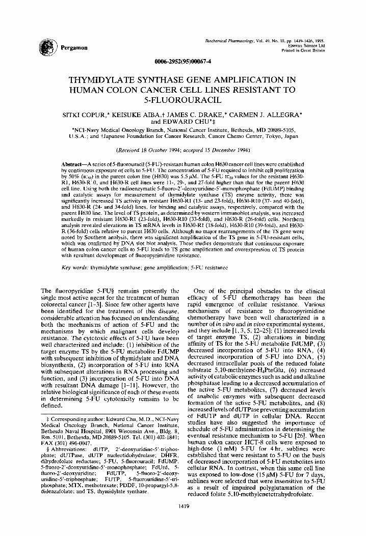
Pergamon Biochemical Pharmacology, Vol. 49. No. 10, pp. 1419-1426, 1995.
Elsevier Science Ltd Printed in Great Britain
0006-2952(95)00067-4
THYMIDYLATE SYNTHASE GENE AMPLIFICATION IN HUMAN COLON CANCER CELL LINES RESISTANT TO
5FLUOROURACIL
SITKI COPUR,* KEISUKE AIBA,t JAMES C. DRAKE,* CARMEN J. ALLEGRA* and EDWARD CHU*$
*NCI-Navy Medical Oncology Branch, National Cancer Institute, Bethesda, MD 20889-5105, U.S.A.; and tJapanese Foundation for Cancer Research, Cancer Chemo Center, Tokyo, Japan
(Received 18 October 1994; accepted 1.5 December 1994)
Abstract-A series of 5-fluorouracil(5-FU)-resistant human colon H630 cancer cell lines were established by continuous exposure of cells to 5-FU. The concentration of 5-FU required to inhibit cell proliferation by 50% (1~~~) in the parent colon line (H630) was 5.5 PM. The 5-FU tcso values for the resistant H630- Rl, H630-RIO, and H630-R cell lines were ll-, 29-, and 27-fold higher than that for the parent H630 cell line. Using both the radioenzymatic 5-fluoro-2’-deoxyuridine-5’-monophosphate (FdUMP) binding and catalytic assays for measurement of thymidylate synthase (TS) enzyme activity, there was significantly increased TS activity in resistant H630-Rl (13- and 23-fold), H630-RlO (37- and 40-fold), and H630-R (24- and 34-fold) lines, for binding and catalytic assays, respectively, compared with the parent H630 line. The level of TS protein, as determined by western immunoblot analysis, was increased markedly in resistant H630-Rl (!23-fold), H630-RlO (33:fold), and H630-R (26-foid) cells. Northern analvsis revealed elevations in TS mRNA levels in H630-Rl (18-fold). H630-RlO (39-fold). and H630- R (i6-fold) cells relative to parent H630 cells. Although no m‘ajor rearrangementsbf the fs gene were noted by Southern analysis, there was significant amplification of the TS gene in 5-FU-resistant cells, which was confirmed by DNA slot blot analysis. These studies demonstrate that continuous exposure of human colon cancer cells to 5-FU leads to TS gene amplification and overexpression of TS protein with resultant development of fluoropyrimidine resistance.
Key words: thymidylate synthase; gene amplification; 5-FU resistance
The fluoropyridine .5-FU$ remains presently the single most active agent for the treatment of human colorectal cancer [l-3]. Since few other agents have been identified for the treatment of this disease, considerable attentiscm has focused on understanding both the mechanisms of action of S-FU and the mechanisms by which malignant cells develop resistance. The cytc’toxic effects of 5-FU have been well characterized and include: (1) inhibition of the target enzyme TS by the S-FU metabolite FdUMP with subsequent inhibition of thymidylate and DNA biosynthesis, (2) incorporation of 5-FU into RNA with subsequent alterations in RNA processing and function, and (3) incorporation of 5-FU into DNA with resultant DNA damage [l-11]. However, the relative biological significance of each of these events in determining 5-FU cytotoxicity remains to be defined.
$ Corresponding author: Edward Chu, M.D., NCI-Navy Medical Oncology Branch, National Cancer Institute, Bethesda Naval Hospital, 8901 Wisconsin Ave., Bldg. 8, Rm. 5101, Bethesda, MD 20889-5105. Tel. (301) 402-1841; FAX (301) 496-0047.
8 Abbreviations: dUTP, 2’-deoxyuridine-S’-triphos- ohate: dUTPase. dUTP nucleotidohvdrolase: DHFR. hihydiofolate reductase; 5-FU, 5-fluoiouracil;’ FdUMP; 5-fluoro-2’-deoxyuridine-5’-monophosphate; FdUrd, 5- fluoro-2’-deoxyuridine; FdUTP, 5-fluoro-2’-deoxy- uridine-5’-triphosphate; FUTP, .5-fluorouridine-5’-tri- phosphate; MTX, methotrexate; PDDF, lo-propargyl-5,8- dideazafolate; and TS, thymidylate synthase.
One of the principal obstacles to the clinical efficacy of 5-FU chemotherapy has been the rapid emergence of cellular resistance. Various mechanisms of resistance to fluoropyrimidine chemotherapy have been well characterized in a number of in vitro and in vivo experimental systems, and they include [l, 3,5,12-251: (1) increased levels of target enzyme TS, (2) alterations in binding affinity of TS for the 5-FU metabolite FdUMP, (3) decreased incorporation of 5-FU into RNA, (4) decreased incorporation of 5-FU into DNA, (5) decreased intracellular pools of the reduced folate substrate 5,10-methylene-H4PteGlu, (6) increased activity of catabolic enzymes such as acid and alkaline phosphatase leading to a decreased accumulation of the active 5-FU metabolites, (7) decreased levels of anabolic enzymes with subsequent decreased formation of the active 5-FU metabolites, and (8) increased levels of dUTPase preventing accumulation of FdUTP and dUTP in cellular DNA. Recent studies have also suggested the importance of schedule of 5-FU administration in determining the eventual resistance mechanism to 5-FU [26]. When human colon cancer HCT-8 cells were exposed to high-dose (1 mM) 5-FU for 4 hr, sublines were established that were resistant to 5-FU on the basis of decreased incorporation of 5-FU metabolites into cellular RNA. In contrast, when this same cell line was exposed to low-dose (15 m) 5-FU for 7 days, sublines were selected that were insensitive to 5-FU as a result of impaired polyglutamation of the reduced folate 5,10-methylenetetrahydrofolate.
1419
1420 S. COPUR et al.
Gene amplification as a mechanism of drug resistance was first observed in Chinese hamster ovary cells and subsequently in murine and human leukemic cell lines following their exposure in vitro to increasing concentrations of MTX [27]. The gene encoding for DHFR was shown to be present at levels several hundred-fold higher than in the parent wild-type cells. Further investigation revealed that clones with amplification of the DHFR gene were highly resistant to MTX. More recently, this phenomenon of DHFR gene amplification has been observed in clinical specimens taken from patients treated with MTX [28,29]. TS gene amplification has been described in cultured neoplastic cells treated either with FdUrd [17,30] or with the specific antifolate TS inhibitor, PDDF [31,32]. Clark et al. [18] observed a 4- to 6-fold increase in the TS gene copy number in a tumor sample obtained from a patient with colon cancer following prolonged therapy with 5-FU. Although a pre-therapy biopsy specimen was unavailable for determination of the baseline TS gene copy number, the findings from this study suggested that the process of TS gene amplification may have direct clinical relevance in the development of resistance to 5FUchemotherapy.
In this study, we have established 5-FU-resistant human colon cancer H630 cell lines following continuous exposure to 5-FU. Our studies show that these cell lines are resistant to 5-FU due to a significant increase in the level of expression of TS protein. We present further evidence demonstrating that the increased level of TS protein expression results from amplification of the TS gene.
MATERIALS AND METHODS
Chemicals. S-FU, dextran (clinical grade), dUMP, bovine serum albumin (fraction V), and acid-washed activated charcoal were purchased from the Sigma Chemical Co. (St. Louis, MO). [5-3H]dUMP (20 Ci/ mmol), [6-3H]FdUMP (18 Ci/mmol), and [6-3H]5- FU (20 Ci/mmol) were purchased from Moravek Biochemicals (Brea, CA). 5’-[(u-32P]dCTP (3000 Ci/ mmol) and 5’-[(u-32P]dGTP (3000 Ci/mmol) were obtained from New England Nuclear (Boston, MA). All other chemicals were obtained from Sigma.
CelI culture. The origin and characteristics of the human colon cancer H630 cell line have been described previously [33]. Resistant sublines were selected in vitro for resistance to 5-FU by continuous exposure of the parental H630 cell line to medium supplemented with 5-FU. The resistant H630-Rl and H630-RlO colon cancer cells were grown and maintained in medium containing 1 and 1OpM 5- FU, respectively. The H630-R subline was derived from the 5-FU-resistant H630-RlO subline by maintaining the H630-RlO line in the absence of 5- FU. All cell lines were grown in 75-cm2 plastic tissue culture flasks (Falcon Labware, Oxnard, CA) in growth medium containing RPM1 1640 with 10% dialyzed fetal bovine serum and 2mM glutamine. Dialyzed fetal bovine serum was purchased from the Grand Island Biological Co. (Grand Island, NY). All other medium components were obtained from the Biofluids Co. (Rockville, MD). In all
experiments, resistant colon cancer cell lines were grown in the absence of 5-FU for a minimum of 2 weeks before study.
In vitro cytotoxicity studies. Plastic 25-cm2 tissue culture flasks (Falcon Labware) were seeded with 4.9 mL suspensions of 5 x 10⁴ce11s/mL of H630, H630-Rl, H630-RlO, and H630-R and were then incubated at 37”. After a 24-hr incubation, 0.1 mL of 5-FU, at various concentrations (10e4 to lo-* M) was added to each flask. Sterile water (0.1 mL) was added to control flasks. All experiments were carried out in duplicate. After a 72-hr incubation at 37”, cells were trypsinized and counted using a ZBI Coulter counter (Coulter Electronics Inc., Hialeah, FL). The concentration of drug that produced 50% inhibition of cell growth was determined from the plot of percent control growth (cell number) versus the logarithm of drug concentration.
The doubling times for the H630, H630-Rl, H630- RlO, and H630-R lines were 25, 32, 37, and 28 hr, respectively.
Measurement of DNA synthesis. Human colon cancer cells, in the logarithmic phase of growth, were incubated with 1 ,&i/mL [methyl-3H]thymidine (specific activity 60 Ci/mmol) for 30 min. The cells were then processed as previously described [22].
Incorporation of 5-FU into nucleic acids. Expo- nentially growing human colon cancer cells were treated with [3H]5-FU (1 ,uM; final specific activity 40 ,&i/mmol) for 4 hr at 37”. Cells were then washed three times with ice-cold PBS and fractionated for RNA and DNA, as previously described [22]. Cellular RNA was hydrolyzed in 0.2 M NaOH, and DNA was hydrolyzed in 1 M perchloric acid. The 200~PL samples of the hydrolysates were added to scintillation vials containing 10 mL of 3a70B counting fluid, and tritium radioactivity was measured in a Packard Tricarb Liquid scintillation counter.
7’S binding and catalytic assay. Human colon cancer cells, in the logarithmic phase of growth, were washed three times with ice-cold PBS, harvested, and resuspended in 0.1 M KH2P04, pH7.2. Cell lysis was accomplished by sonication using three 2- to 3-set bursts. The extracts were centrifuged at 10,OOOg for 30 min at 4”, and the supernatants were then assayed for both TS binding and catalytic activity, as previously described [22].
FdUMP binding affinity determination (Kd). Cell extracts from the H630 and H630-RlO cell lines were obtained as described above. The assay was performed in a final volume of 200 PL containing 75 PM 5,10-methylene-H,PteGlu, [6-3H]FdUMP at concentrations ranging from 40 pM to 40 nM, 100 PM 2-mercaptoethanol, 50 mM KH2P04, and aliquots of cytosolic extract (5OpL). The reaction mixtures were incubated for 30 min at 37”, enzyme-bound and free [6-3H]FdUMP were determined, and affinity calculations were performed as previously described by Lockshin and Danenberg [34].
Western immunoblot analysis. Human colon cancer cells were harvested and extracted as described above. Protein concentrations were determined by the Bio-Rad protein assay [35], and equivalent amounts of protein (200 pg) from each cell line were resolved by SDS-PAGE, using 12.5% acrylamide,
TS gene amplification 1421
Table 1. Growth-inhibitory effect (ICY) of 5-FU against human colon cancer cells
Cell line 1~~0 (PM)
H630 5.5 + 1.0 H630-Rl 61 * 8.5 H630-R10 160 k 10 H630-R 153 2 11
Human colon cancer cells in the logarithmic phase of growth were incubated with 5-FU (10e4 to lo-’ M) at 37” for 72 hr. The lcso values (concentration producing 50% growth inhibition) were determined as described in Materials and Methods. The H630 line represents the parent cell line, and the resistant H630-Rl and H630-R10 lines were maintained in 5-FU concentrations of 1 and 10 PM, respectively. The H630-R line is a subline of H630- RlO that was maintained in the absence of 5-FU. Results are the means 2 SEM from five separate experiments, each done in duplicate.
Table 2. Incorporatic’n of 5-FU into RNA and DNA of human colon cancer cells
5-FU incorporation
Cell line
H630 H630-RlO H630-R
RNA (pmcsl/mg RNA)
14 k 0.9 18 2 1.9 1.9 + 0.8
DNA (pmol/mg DNA)
0.8 + 0.05 1.1 k 0.10 1.4 + 0.20
Human colon cancer cells in the logarithmic phase of growth were incubated with [6-‘H]5-fU (1 PM; specific activitv 40 uCi/mL) at 37” for 4 hr. Cells were extracted * , and fractionated for RNA and DNA as described in Materials and Methods. Results are the means k SEM from at least three separate experiments.
according to the method of Laemmli [36]. The gels were then electroblotted onto nitrocellulose membranes (Schleicher & Schuell, Keene, NH). Antibody staining was performed with the use of a TS polyclonal primary antibody (l/1000 dilution) and horseradish peroxidase-conjugated secondary antibody (l/1000 dilution) (Bio-Rad Laboratories, Richmond, CA).
Isolation of total RNA and RNA blot hybridization (Northern) analysis. Human colon cancer cells were harvested from 150-cmL plastic tissue culture flasks and washed three times with ice-cold PBS. Total RNA was isolated according to the method of Chomczynski and :Sacchi [37]. After extraction, 30 pg/sample of total cellular RNA was denatured, resolved on a 1% formaldehyde-agarose gel, and transferred onto a Nytran filter membrane (Schleicher & Schuell). The membrane was hybridized to a 32P- radiolabeled human ‘TS cDNA insert probe after the human TS cDNA was labeled by the random primer method of Feinberg and Vogelstein [38]. The cDNA for human TS was a gift of Dr. T. Seno (Saitama Cancer Center Research Institute, Saitama-ken,
Japan). The filters were washed as previously described [22], and autoradiography was performed using Kodak XAR-5 film exposed for 3-5 days at -70”.
Extraction and analysis of DNA. Cellular DNA from human colon cancer cells was isolated as previously described [22]. Southern analysis was performed by digestion of 15 pg of the isolated genomic DNA with Hind111 restriction endonuclease, fractionation of the DNA fragments on a 0.5% agarose gel followed by transfer onto a Nytran filter membrane (Schleicher & Schuell). The membrane was hybridized to a 32P-radiolabeled human TS cDNA insert probe and then processed as previously described [22].
Slot blot hybridization analysis was performed according to previously described methods [39]. Serial dilutions of known concentrations of genomic DNA from each cell line were prepared and spotted onto nitrocellulose filters by means of a 24-well vacuum manifold (Bethesda Research Laboratories). The filters were air-dried and vacuum-baked at 80” for 2 hr, and then hybridized to a 32P-radiolabeled human TS cDNA probe as outlined above.
Densitometric analysis. Quantitation of signal intensities on autoradiographs and on Western blot filter membranes was performed by densitometry using a Scan Jet Plus scanner (Hewlett-Packard) and analyzed using NIH Image 1.36 software (Wayne Rasband, National Institute of Mental Health, Bethesda, MD).
RESULTS
In vitro cytotoxicity studies. The H630-Rl and H630-R10 human colon cancer cell lines were selected by in uitro exposure of the parent H630 cell line to stepwise increases in 5-FU concentration and were then maintained in medium containing 1 and 10 PM 5-FU, respectively. As shown in Table 1, the 5-FU lcso values for the H630-Rl and H630-RlO lines were 61 and 16OpM, respectively. These lines were ll- and 29-fold more resistant to 5-FU compared with the parent H630 line (rcso = 5.5 PM). H630-R cells, which were derived from the most resistant H630-RlO line, were still resistant to 5-FU (1~~0 = 153 PM) despite being grown in drug-free medium for up to 28 weeks. Moreover, the stability of the resistance phenotype was confirmed when H630-R cells passaged out of 5-FU for greater than 52 weeks were resistant to 5-FU to nearly the same degree as H630-RlO cells.
Additional growth experiments revealed that the presence of 10 ,uM thymidine completely protected against the growth-inhibitory activity of 5-FU in both the parent H630 and resistant H630-RlO cells (data not shown). These results suggest that inhibition of the target enzyme TS and/or involvement of a DNA- mediated event represent two possible mechanisms through which 5-FU exerts its cytotoxic effects in these cell lines.
Measurement of DNA synthesis. To measure the relative rates of DNA synthesis in each of the H630 cell lines, the level of thymidine incorporation was determined. Incorporation of [3H]thymidine into DNA was lower in each of the 5-FU-resistant cell
1422 S. COPUR et al.
Table 3. TS activity in human colon cancer cells
Cell line Binding assay
(pmol/mg protein) Fold increase Catalytic assay
(pmol/min/mg protein) Fold increase
H630 0.17 2 0.03 1 1.3 +- 0.2 1 H630-Rl 0.82 + 0.10 13 31 r5 23 H630-RlO 6.27 2 0.96 37 52 + 10 40 H630-R 4.10 ? 0.50 24 42 ? 5 34
Human colon cancer cells in the logarithmic phase of growth were harvested, and cytosolic extracts served as the source of enzyme. TS enzyme activity was determined using the FdUMP radioenzymatic binding and catalytic assays as described in Materials and Methods. Results are the means 2 SEM of at least five separate experiments. The H630-Rl (P < O.OOS), H630-RlO (P < 0.005) and H630-R (P < 0.005) lines were associated with a significantly increased level of TS, as determined by the binding and catalytic assays (two-sided Student’s f-test).
lines [H630-Rl(O.36 2 0.1 nmol/mg protein), H630- RlO (0.38 ? 0.3 nmol/mg protein), and H630-R (0.34 L 0.2 nmol/mg protein)] when compared with the parent H630 cell line (0.62 5 0.2 nmol/mg protein). These differences in thymidine incor- poration were not statistically significant as deter- mined by a two-sided Student’s f-test.
Incorporation into nucleic acids. Incorporation into RNA and DNA has been correlated with S-FU cytotoxicity in various in vitro and in vivo model systems. Given these observations, we determined the level of S-FU incorporation into each of these nucleic acid fractions. As shown in Table 2, there were no significant differences in the extent of incorporation of S-FU nucleotide metabolites into either RNA or DNA between parent H630 and the resistant H630-RlO and H630-R cell lines.
Measurement of thymidylate synthase. Increased expression of TS has been documented to be an important mechanism of resistance to 5-FU. Using the radioenzymatic FdUMP binding assay, TS activity was increased 13- and 37-fold, respectively, in the H630-Rl and H630-RlO lines when compared with the parent H630 line (Table 3). In the H630-R line, the level of TS enzyme remained 24-fold elevated even though these cells were grown in drug- free medium for 28 weeks (Table 3). TS activity, as determined by the catalytic assay, was similarly increased in the 5-FU-resistant human colon cancer cells. The H630-Rl and H630-R10 cell lines had 23- and 40-fold higher TS enzyme activity relative to the parent H630 line (Table 3). The H630-R line demonstrated a 34-fold increased TS catalytic activity relative to parent H630 cells (Table 3).
The tightness of binding of FdUMP to TS extracted from both sensitive H630 and 5-FU-resistant H630- RlO colon cancer cells was determined by measuring the respective dissociation constants, Kd, for FdUMP. There was no difference in TS binding affinity, inasmuch as the Kd values for FdUMP binding to TS of sensitive H630 and resistant H630-RlO cells were 0.7 + 0.4 and 0.6 -t 0.3 nM, respectively.
The level of expression of TS protein in each of these cell lines was subsequently analyzed by Western immunoblot to determine whether differences in TS enzyme activities corresponded to changes in the amount of immunoreactive protein. A representative
ABCDE
-TS
Fig. 1. Western immunoblot analysis of TS from parent and 5-FU-resistant H630 cells. Cytosolic extracts from human colon cancer cells were prepared as described in Materials and Methods. Equal amounts of protein (200 pg) were loaded onto each lane and resolved on a 12.5% SDS- polyacrylamide gel. Human recombinant TS protein (50 ng) was applied to the gel to verify the position of TS staining (lane A). Proteins were from parent H630 (lane B), H630- R (lane C), H630-RlO (lane D), and H630-Rl (lane E)
cells.
blot is presented in Fig. 1. Equal amounts of total cytosolic protein (2OOpg) were loaded onto each lane. The primary TS antiserum employed in this analysis was polyclonal, and, consequently, stained a number of proteins. However, the molecular weight of the denatured monomeric form of TS is 35,000, and the correct position of the TS band was confirmed by immunostaining of human recombinant TS protein (Fig. 1, lane A). Parent H630 cells displayed minimal staining for TS (Fig. 1, lane B). In contrast, resistant H630-RlO (Fig. 1, lane D), H630-R (Fig. 1, lane C), and H630-Rl (Fig. 1, lane E) cells all demonstrated significantly increased levels of TS. Densitometric scanning of the immunoblots revealed a 23-, 33-, and 26-fold increased expression of TS in H630-Rl, H630-RlO, and H630-R lines, respectively, when compared with the parent H630 line.
RNA blot hybridization analysis. Analysis of TS mRNA levels by Northern blot hybridization revealed a single 1.5-kb mRNA species that hybridized with the radiolabeled human TS cDNA
A El C D
TS gene amplification
AB CD
1423
18s - + TS
-BR,“V‘ i
18S- +- /3-Actin
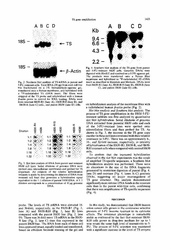
Fig. 2. Northern blot analysis of TS mRNA in parent and 5-FU-resistant cells. Total RNA (30 pg) from each cell line was fractionated on a 1% formaldehyde-agarose gel, transferred onto a Nytran membrane, and hybridized with a 32P-radiolabeled TS cDNA insert. The filters were stripped of the TS probe and rehybridized with a human &a&n probe to control for RNA loading. RNAs were from resistant H630-RI (lane A), H630-RlO (lane B), and
H630-R (lane C) cellj, and parent H630 (lane D) cells.
I:1
I:2
I:4
I:8
1:16
I:32
1:l
1:2
1:4
1:8
I:16
I:32
I:1
1:2
I:4
I:8
1:16
1:32
Fig. 3. Slot blot analysi!; of DNA from parent and resistant H630 cell lines. Serial dilutions of genomic DNA were spotted onto nitrocellulose membranes and probed for TS sequences. An estimate of the relative hybridization intensity is made by determining the dilution of DNA from resistant cell lines that generates a hybridization signal equivalent to the 1: 1 dilution of parent H630 DNA. A 1: 1 dilution corresponds to a concentration of 15 pg

1424 S. COPUR et al.
activity and TS protein expression, as determined by biochemical techniques, including the radio- enzymatic FdUMP binding and TS catalytic assays, and a Western immunoblotting analysis. Of note, the relative differences in TS expression in parent and resistant human colon cancer cell lines were equivalent using any of these biological assays. Scatchard analyses of TS protein from parent, wild- type H630 and resistant H630-RlO cells revealed no alteration in binding affinity of TS for FdUMP. These studies, taken together, suggest that TS protein in 5-FU-resistant cell lines is qualitatively identical to native TS protein expressed in wild-type H630 cells.
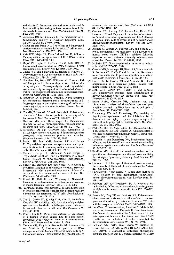
The increase in immunoreactive TS in .5-FU- resistant colon cancer cells was associated with corresponding changes in the steady-state levels of both the TS gene and TS mRNA. Southern blot analysis revealed significant amplification (approxi- mately 32-fold) of TS-specific genomic sequences without any apparent rearrangement of the amplified gene. Moreover, the elevation in TS gene copy number remained stable given that there was, at most, a 2-fold decrease in the level of TS gene amplification despite resistant H630-RlO cells being maintained in drug-free medium for nearly 7 months. Previous studies characterizing amplification of the DHFR gene demonstrated that the amplified gene can be localized to either homogeneously staining regions (HSRs) or to double-minute chromosomes [27]. In the absence of the selective pressure of drug, HSR-mediated gene amplification confers stable resistance in contrast to amplification resulting from double-minute chromosomes whereby resistance is unstable with rapid reversion to the original genotype [27]. Our results are similar to those of Imam et al. [31], who identified TS gene amplification in murine leukemic L1210 cells resistant to the antifolate TS inhibitor CB3717. When CB3717-resistant L1210 cells were grown in the absence of the antifolate inhibitor for 375 generations, drug resistance remained stable with only a minimal 2- to 3-fold decrease in the TS gene copy number.
Given the multiple sites of 5-FU cytotoxic action, it was important to rule out alternative mechanisms that might contribute to the development of cellular resistance to S-FU in these 5-FU-resistant colon cancer cells. Earlier studies have shown that resistance to 5-FU can develop as a result of decreased incorporation of 5-FU metabolites into either total cellular RNA or DNA [5,23,26]. Thus, we determined the level of 5-FU incorporation in the specific RNA and DNA fractions of parent H630 and resistant H630-RlO cells, and, as shown in Table 2, there were no significant quantitative differences between parent and resistant cells. Although the levels of the individual 5-FU nucleotide metabolites such as FUTP or FdUTP were not measured, the fact that RNA and DNA incorporation, the target endpoints of these respective metabolites, remained unchanged suggests that direct measurement of these metabolites may not be of further value. Recently, a novel resistance mechanism has been identified whereby resistance to 5-FU is mediated by enhanced DNA repair enzyme activity [23-251. Increased DNA repair activity has been associated with
decreased incorporation of 5-FU-specific metabolites and/or dUTP into cellular DNA. Our observation that incorporation of 5-FU into DNA is nearly identical in wild-type parent H630 and resistant H630-RlO cells suggests that there can be no biologically significant difference in levels of DNA enzyme repair activity such as dUTPase in either of these cell lines.
Earlier studies described elevations in TS in cells resistant to CB3717 [31,32], an antifolate analog that directly targets TS, and FdUrd [17,30], a fluoropyrimidine analog that exerts its cytotoxic action through inhibition of TS by the metabolite FdUMP. Selection with either agent resulted in an increase in TS enzyme levels that was associated with amplification of the TS gene. A recent study reported TS gene amplification in 5-FU-resistant HT-29 human colon cancer grown in the selective pressure of 5-FU [40]. However, this report did not attempt to examine other mechanisms through which resistance to 5-FU might be mediated. For this reason, the present study is the first to specifically identify increased expression of TS with TS gene amplification as the principal mechanism by which human colon cancer cells chronically exposed to 5- FU develop fluoropyrimidine resistance.
Significant research efforts are now ongoing to identify novel antifolate and/or non-antifolate-based inhibitors of TS. ZD1694 [41,42] and LY231514 [43,44] are two antifolate-based compounds that are presently undergoing clinical testing. One issue raised by this study is whether cancer patients previously treated with a 5-FU-based regimen can be treated subsequently with a TS-directed antineoplastic agent. Our findings suggest that this patient population would be refractory to such therapy and that alternative therapeutic approaches should be considered. Further work using tumor biopsy specimens from patients will be required to determine the clinical relevance of TS gene amplification and TS overexpression as a mechanism of 5-FU resistance. Such studies may identify patients who will most benefit from non-TS-directed therapeutic approaches.
Acknowledgemenfs-The authors would like to thank Sydelle Zinn for her technical help and Janet Edds for her editorial assistance in the preparation of this manuscript.
REFERENCES
1. Pinedo HM and Peters GFJ, Fluorouracil: Biochemistry and pharmacology. J C[in 0~016: 1653-1664, 1988.
2. Heidelberger C, Fluorinated pyrimidines and their nucleosides. In: Antineoplastic and Immunosuppressive Agents (Eds. Sartorelli A and Johns D), pp. 193-231. Springer, New York, 1975.
3. Heidelberger C, Danenberg PV and Moran RG, Fluorinated pyrimidines and their nucleosides. Adv Enzymol Relat Areas Mol Biol 54: 57-119, 1989.
4. Santi DV, McHenry CS and Sommer H, Mechanism of interaction of thymidine synthetase with 5- fluorodeoxyuridylate. Biochemistry 13: 471-481, 1974.
5. Ardalan B, Cooney DA, Jayaram HN, Carrico CK, Glazar RI, Macdonald J and Schein PS, Mechanisms of sensitivity and resistance of murine tumors to 5- fluorouracil. Cancer Res 40: 1431-1437, 1980.
6. Spiegelman S, Sawyer R, Nayak R, Ritzi E, Stolfi R
TS gene amplification 1425
and Martin D, Improving the antitumor activity of 5- fluorouracil by inc:reasing its incorporation into RNA via metabolic modulation. Proc Nat1 Acad Sci USA 77: 25. 4966-4970, 1980.
7. Wilkinson DS and Crumley J, The mechanism of 5- fluorouridine toxicity in Novikoff hepatoma cells. Cancer Res 36: 4032-4038. 1976.
8. Glazar RI and Peale AL, ‘The effect of 5-fluorouracil
9.
10.
11.
12.
13.
14.
15.
16.
17.
18.
19.
20.
21.
22.
on the synthesis of nuclear RNA in L1210 cells in uitro. Mol Pharmacol 16 270-277, 1979. Kufe DW, Major F’P, Egan EM and Loh E, 5-Fluoro- 2’-deoxyuridine incorporation in L1210 DNA. J Biol Chem 256: 8885-8888, 1981. Maior PP, Egan .E, Herrick D and Kufe DW, 5- Fluorouracil incorporation in DNA of human breast carcinoma cells. Ccrncer Res 42: 3005-3009. 1982. Cheng Y-C and Nakayama K, Effects of %fluoro-2’- deoxyuridine on DNA metabolism in HeLa cells. Mol Pharmacol23: 171-174, 1983. Houghton JA, We& KD, Williams LG, Torrance PM and Houghton PJ, Relationship between 5-fluoro-2’- deoxyuridylate, 2’-deoxyuridylate and thymidylate synthase activity subsequent to 5-fluorouracil adminis- tration, in xenografts of human colon adenocarcinomas. Biochem Pharmacol35: 1351-1358. 1986. Houghton JA, Maroda SJ, Phillips’JO and Houghton PJ, Biochemical determinants of responsiveness to 5- fluorouracil and its derivatives in xenografts of human colorectal adenocarcinomas in mice. Cancer Res 41: 144-149, 1981. Yin MB, Zakrzewski SF and Hakala MT, Relationship of cellular folate cofactor pools to the activity of 5- fluorouracil. Mol P.Sarmacol23: 190-197, 1983. Mulkins MA and Heidelberger C, Biochemical characterization of fluoropyrimidine-resistant murine leukemic cell lines. Cancer Res 42: 965-973, 1982. Fernandes DJ and Crawford SK, Resistance of CCRF-CEM cloned sublines to 5-fluorodeoxyuridine associated with enhanced phosphatase activities. Biochem Pharmaco.’ 34: 125-132, 1985. Berger SH, Jenh C-H, Johnson LF and Berger F, Thymidylate synthase overproduction and gene amplification in fluorodeoxyuridine-resistant human cells. Mol Pharmacol28: 461-467, 1985. Clark JL, Berger SH, Mittleman A and Berger F, Thymidylate synthase gene amplification in a colon tumor resistant to fluoropyrimidine chemotherapy. Cancer Treat Rep 711: 261-265, 1987. Berger SH, Barbour KW and Berger F, A naturally occurring variation in thymidylate synthase structure is associated with a reduced response to 5-fluoro-2’- deoxyuridine in a human colon tumor cell line. Mol Pharmacol34: 480-484, 1988. Kessel D, Hall TC and Wodinsky I, Nucleotide formation as a determinant of 5-fluorouracil response in mouse leukemia. Science 154: 911-913, 1966. Scanlon KJ and Kashani-Saabet M, Elevated expression of thymidylate synthase cycle genes in cisplatin-resistant human ovarian carcinoma A2780 cells. Proc Nat1 Acad Sci USA 8.5: 650-653, 1988. Chu E, Drake JC, Koeller DM, Zinn S, Jamis-Dow CA, Yeh GC and Allegra CJ, Induction of thymidylate synthase associated with multidrug resistance in human breast and colon cancer cell lines. Mol Pharmacol39: 136-143. 1991.
23.
24
Chu E, Lai G-M, Zinn S and Allegra CJ, Resistance of a human ovarian cancer line to 5-fluorouracil associated with decreased levels of 5-fluorouracil in DNA. Mol Pharmacol38: 410-417, 1990. Canman CE, Tang H-Y, Normolle DP, Lawrence TS 41 and Maybaum J, Variations in patterns of DNA damage induced in human colorectal tumor cells by 5- fluorodeoxyuridine: Implications for mechanisms of
BP 49110-E
resistance and cytotoxicity. Proc Nat1 Acad Sci USA 89: 10474-10478, 1992. Canman CE, Radany EH, Parsels LA, Davis MA, Lawrence TS and Maybaum J, Induction of resistance to fluorodeoxyuridine cytotoxicity and DNA damage in human tumor cells by expression of Escherichia coli deoxyuridinetriphosphatase. Cancer Res 54: 2296- 2298, 1994.
26. Aschele C, Sobrero A, Faderan MA and Bertino JR, Novel mechanism(s) of resistance to 5-fluorouracil in human colon cancer (HCT-8) sublines following exposure to two different clinically relevant dose schedules. Cancer Res 52: 1855-1864, 1992.
27.
28.
29.
30
31
32
33
34
35
36
37.
38.
39.
40.
Schimke RT, Gene amplification in cultured animal cells. Cell 37: 705-713, 1984. Carman MD, Schornagel JH, Rivest RS, Srimatkandada S, Portlock CS, Duffy T and Bertino JR, Resistance to methotrexate due to gene amplification in a patient with acute leukemia. J Clin Oncol2: 1620, 1984. Horns CR Jr, Dower WJ and Schimke RT, Gene amplification in a leukemic patient treated with methotrexate. J Clin Oncol 2: 2-7, 1984. Jenh C-H, Geyer PK, Baskin F and Johnson LF, Thymidylate synthase gene amplification in fluorodeoxyuridine-resistant mouse cell lines. Mol Pharmacol28: 8&85, 1985. Imam AMA, Crossley PH, Jackman AL and Little PFR, Analysis of thymidylate synthase gene amplification and of mRNA levels in the cell cycle. J Biol Chem 262: 7268-7373, 1987. Danenberg KD and Danenberg PV, Activity of thymidylate synthetase and its inhibition by 5- Ruorouracil in highly enzyme-overproducing cells resistant to lo-propargyl-5,8-dideazafolate. Mol Phar- macol36: 219-223, 1989. Park J-G, Oie HK, Sugarbaker PH, Henslee JG, Chen T-R, Johnson BE and Gazdar A, Characteristics of cell lines established from human colorectal carcinoma. Cancer Res 47: 6710-6718, 1987. Lockshin A and Danenberg PV, Biochemical factors affecting the tightness of 5-fluorodeoxyuridylate binding to human thymidylate synthetase. Biochem Pharmacol 30: 247-257, 1981. Bradford MM, A rapid and sensitive method for the quantitation of microgram quantities of protein utilizing the principle of protein-dye binding. Anal Biochem 72: 248-254, 1976. Laemmli UK, Cleavage of structural proteins during the assembly of the head of bacteriophage T.+ Nature 227: 680-685, 1970. Chomczynski P and Sacchi N, Single-step method of RNA isolation by acid guanidinium thiocyanate- phenol-chloroform extraction. Anal Biochem 162: 156- 159, 1987. Feinberg AP and Vogelstein B, A technique for radiolabeling DNA restriction endonuclease fragments to high specific activity. Anal Biochem 137: 266-267, 1984. Brown PC, Tlsty TD and Schimke RT, Enhancement of methotrexate resistance and dihydrofolate reductase gene amplification by treatment of mouse 3T6 cells with hydroxyurea. Mol Cell Bio13: 1097-1107, 1983. Lesuffleur T, Kornowski A, Luccioni C, Muleris M, Barbat A, Beaumatin J, Dussaulx E, Dutrillaux B and Zweibaum A, Adaptation to 5fluorouracil of the heterogeneous human colon tumor cell line HT-29 results in the selection of cells committed to differentiation. Znt J Cancer 49: 721-730, 1991. Jackman AL, Taylor GA, Gibson W, Kimbell R, Brown M, Calvert AH, Judson IR and Hughes LR, ICI Dl694, a quinazoline antifolate thymidylate synthase inhibitor that is a potent inhibitor of L1210

1426 S. COPUR er al.
42.
43.
tumor cell growth in vitro and in ho: A new agent for clinical study. Cancer Res 51: 5579-5586, 1991. Cunningham D, Zalcberg J, Francois E, Van Cutsem E, Schornagel JH, Adenis A, Green M, Starkhammer H, Hanrahan A, Ellis P and Azab M, Tomudex (ZD1694) a new thymidylate synthase inhibitor with good antitumor activity in advanced colorectal cancer (ACC). Proc Am Sot Clin Oncol 13: 199, 1994. Grindey GB, Shih C, Barnett CJ, Pearce HL, Engelhardt JA, Todd GC, Rinzel SM, Worzalla JF,
44.
Gossett LS, Everson TP, Wilson TM, Kobierski ME, Winter MA, Bewley JR, Kuhnt D, Taylor EC and Moran RG, LY231514, a novel pyrrolopyrimidine antifolate that inhibits thymidylate synthase (TS). Proc Am Assoc Cancer Res 33: 411, 1992. Rinaldi DA, Burris HA, Forr FA, Nelson J, Fields SM, Kuhn JG, Eckardt JR, Lu P, Woodworth JR, Corso SW and Von Hoff DD, A phase I evaluation of the novel thymidylate synthase inhibitor, LY231514, in patients with advanced solid tumors. Proc. Am Sot Clin Oncol 13: 159, 1994.
Copyright © 2022 FDOKUMEN