Transcriptional Activation and Cell Cycle Block Are the Keys for 5-Fluorouracil Induced...
11
Transcriptional Activation and Cell Cycle Block Are the Keys for 5-Fluorouracil Induced Up-Regulation of Human Thymidylate Synthase Expression Alessio Ligabue 1,2 , Gaetano Marverti 3 , Ursula Liebl 1,2 , Hannu Myllykallio 1,2 * 1 INSERM U696, Palaiseau, France, 2 Laboratoire d’Optique et Biosciences, CNRS, Ecole Polytechnique, Palaiseau, France, 3 Dipartimento di Scienze Biomediche, Sezione di Chimica Biologica, University of Modena and Reggio Emilia, Modena, Italy Abstract Background: 5-fluorouracil, a commonly used chemotherapeutic agent, up-regulates expression of human thymidylate synthase (hTS). Several different regulatory mechanisms have been proposed to mediate this up-regulation in distinct cell lines, but their specific contributions in a single cell line have not been investigated to date. We have established the relative contributions of these previously proposed regulatory mechanisms in the ovarian cancer cell line 2008 and the corresponding cisplatin-resistant and 5-FU cross-resistant-subline C13*. Methodology/Principal Findings: Using RNA polymerase II inhibitor DRB treated cell cultures, we showed that 70–80% of up-regulation of hTS results from transcriptional activation of TYMS mRNA. Moreover, we report that 5-FU compromises the cell cycle by blocking the 2008 and C13* cell lines in the S phase. As previous work has established that TYMS mRNA is synthesized in the S and G 1 phase and hTS is localized in the nuclei during S and G 2 -M phase, the observed cell cycle changes are also expected to affect the intracellular regulation of hTS. Our data also suggest that the inhibition of the catalytic activity of hTS and the up-regulation of the hTS protein level are not causally linked, as the inactivated ternary complex, formed by hTS, deoxyuridine monophosphate and methylenetetrahydrofolate, was detected already 3 hours after 5-FU exposure, whereas substantial increase in global TS levels was detected only after 24 hours. Conclusions/Significance: Altogether, our data indicate that constitutive TYMS mRNA transcription, cell cycle-induced hTS regulation and hTS enzyme stability are the three key mechanisms responsible for 5-fluorouracil induced up-regulation of human thymidylate synthase expression in the two ovarian cancer cell lines studied. As these three independent regulatory phenomena occur in a precise order, our work provides a feasible rationale for earlier observed synergistic combinations of 5-FU with other drugs and may suggest novel therapeutic strategies. Citation: Ligabue A, Marverti G, Liebl U, Myllykallio H (2012) Transcriptional Activation and Cell Cycle Block Are the Keys for 5-Fluorouracil Induced Up-Regulation of Human Thymidylate Synthase Expression. PLoS ONE 7(10): e47318. doi:10.1371/journal.pone.0047318 Editor: Kapil Mehta, University of Texas MD Anderson Cancer Center, United States of America Received February 17, 2012; Accepted September 14, 2012; Published October 9, 2012 Copyright: ß 2012 Ligabue et al. This is an open-access article distributed under the terms of the Creative Commons Attribution License, which permits unrestricted use, distribution, and reproduction in any medium, provided the original author and source are credited. Funding: This work was supported by THYMET and RETIDYNA grants from ‘‘Agence nationale de la recherche ´’’. G.M. acknowledges support from the fund AIRC IG 10474. The funders had no role in study design, data collection and analysis, decision to publish, or preparation of the manuscript. Competing Interests: The authors have declared that no competing interests exist. * E-mail: [email protected] Introduction Human thymidylate synthase of the ThyA family [hTS (EC 2.1.1.45), encoded by the gene TYMS] is a folate-dependent enzyme that converts 29-deoxyuridine-59-monophosphate (dUMP) and N 5 -N 10 -methylenetetrahydrofolate (mTHF) to dihydrofolate and 29-deoxythymidine-59-monophosphate (dTMP). Recent pa- pers demonstrated that hTS is localized not only in the cytoplasm, but also in the nuclei and in the mitochondria. Nuclear hTS is associated with proliferating cell nuclear antigen (PCNA) and other components of the DNA replication machinery, suggesting that de novo thymidylate biosynthesis occurs at replication forks [1]. On the other hand, mitochondrial hTS prevents uracil accumu- lation in mitochondrial DNA and is essential for mtDNA integrity [2]. Human cells do not possess the flavin-dependent thymidylate synthase ThyX that is found in many free living microbes [3]. Consequently, hTS provides the only de novo pathway for thymidylate synthesis in human cells and represents an essential target enzyme for cancer chemotherapy [4]. Several inhibitors that prevent the catalytic activity of human thymidylate synthase through binding to dUMP and/or mTHF binding pockets have been identified. For instance, the uracil- analog 5-fluorouracil (5- FU), after metabolic conversion to 5-FdUMP, is a well character- ized active-site inhibitor of hTS that has been widely used in chemotherapy since 1957 [5]. FdUMP forms a covalent ternary complex with hTS and mTHF, resulting in the irreversible inhibition of the catalytic activity of hTS. Inhibition of hTS provokes an increase of the intracellular dUMP concentration [6,7] and causes depletion of deoxythymidine triphosphate (dTTP) [8]. The imbalance of intracellular deoxynucleotide pools disrupts DNA replication and triggers cell death [9,10]. In addition to direct inhibition of hTS, the 5-FU metabolites 5-fluorouridine-59- triphosphate (F-UTP) and 5-fluoro-29-deoxyuridine-59-triphos- phate (FdUTP) cause cell death through incorporation into RNA and DNA, respectively (for a review see [11]). To prevent PLOS ONE | www.plosone.org 1 October 2012 | Volume 7 | Issue 10 | e47318
-
Upload
independent -
Category
Documents
-
view
3 -
download
0
Transcript of Transcriptional Activation and Cell Cycle Block Are the Keys for 5-Fluorouracil Induced...
Transcriptional Activation and Cell Cycle Block Are theKeys for 5-Fluorouracil Induced Up-Regulation of HumanThymidylate Synthase ExpressionAlessio Ligabue1,2, Gaetano Marverti3, Ursula Liebl1,2, Hannu Myllykallio1,2*
1 INSERM U696, Palaiseau, France, 2 Laboratoire d’Optique et Biosciences, CNRS, Ecole Polytechnique, Palaiseau, France, 3 Dipartimento di Scienze Biomediche, Sezione di
Chimica Biologica, University of Modena and Reggio Emilia, Modena, Italy
Abstract
Background: 5-fluorouracil, a commonly used chemotherapeutic agent, up-regulates expression of human thymidylatesynthase (hTS). Several different regulatory mechanisms have been proposed to mediate this up-regulation in distinct celllines, but their specific contributions in a single cell line have not been investigated to date. We have established the relativecontributions of these previously proposed regulatory mechanisms in the ovarian cancer cell line 2008 and thecorresponding cisplatin-resistant and 5-FU cross-resistant-subline C13*.
Methodology/Principal Findings: Using RNA polymerase II inhibitor DRB treated cell cultures, we showed that 70–80% ofup-regulation of hTS results from transcriptional activation of TYMS mRNA. Moreover, we report that 5-FU compromises thecell cycle by blocking the 2008 and C13* cell lines in the S phase. As previous work has established that TYMS mRNA issynthesized in the S and G1 phase and hTS is localized in the nuclei during S and G2-M phase, the observed cell cyclechanges are also expected to affect the intracellular regulation of hTS. Our data also suggest that the inhibition of thecatalytic activity of hTS and the up-regulation of the hTS protein level are not causally linked, as the inactivated ternarycomplex, formed by hTS, deoxyuridine monophosphate and methylenetetrahydrofolate, was detected already 3 hours after5-FU exposure, whereas substantial increase in global TS levels was detected only after 24 hours.
Conclusions/Significance: Altogether, our data indicate that constitutive TYMS mRNA transcription, cell cycle-induced hTSregulation and hTS enzyme stability are the three key mechanisms responsible for 5-fluorouracil induced up-regulation ofhuman thymidylate synthase expression in the two ovarian cancer cell lines studied. As these three independent regulatoryphenomena occur in a precise order, our work provides a feasible rationale for earlier observed synergistic combinations of5-FU with other drugs and may suggest novel therapeutic strategies.
Citation: Ligabue A, Marverti G, Liebl U, Myllykallio H (2012) Transcriptional Activation and Cell Cycle Block Are the Keys for 5-Fluorouracil Induced Up-Regulationof Human Thymidylate Synthase Expression. PLoS ONE 7(10): e47318. doi:10.1371/journal.pone.0047318
Editor: Kapil Mehta, University of Texas MD Anderson Cancer Center, United States of America
Received February 17, 2012; Accepted September 14, 2012; Published October 9, 2012
Copyright: � 2012 Ligabue et al. This is an open-access article distributed under the terms of the Creative Commons Attribution License, which permitsunrestricted use, distribution, and reproduction in any medium, provided the original author and source are credited.
Funding: This work was supported by THYMET and RETIDYNA grants from ‘‘Agence nationale de la recherche’’. G.M. acknowledges support from the fund AIRCIG 10474. The funders had no role in study design, data collection and analysis, decision to publish, or preparation of the manuscript.
Competing Interests: The authors have declared that no competing interests exist.
* E-mail: [email protected]
Introduction
Human thymidylate synthase of the ThyA family [hTS (EC
2.1.1.45), encoded by the gene TYMS] is a folate-dependent
enzyme that converts 29-deoxyuridine-59-monophosphate (dUMP)
and N5-N10-methylenetetrahydrofolate (mTHF) to dihydrofolate
and 29-deoxythymidine-59-monophosphate (dTMP). Recent pa-
pers demonstrated that hTS is localized not only in the cytoplasm,
but also in the nuclei and in the mitochondria. Nuclear hTS is
associated with proliferating cell nuclear antigen (PCNA) and
other components of the DNA replication machinery, suggesting
that de novo thymidylate biosynthesis occurs at replication forks [1].
On the other hand, mitochondrial hTS prevents uracil accumu-
lation in mitochondrial DNA and is essential for mtDNA integrity
[2]. Human cells do not possess the flavin-dependent thymidylate
synthase ThyX that is found in many free living microbes [3].
Consequently, hTS provides the only de novo pathway for
thymidylate synthesis in human cells and represents an essential
target enzyme for cancer chemotherapy [4]. Several inhibitors that
prevent the catalytic activity of human thymidylate synthase
through binding to dUMP and/or mTHF binding pockets have
been identified. For instance, the uracil- analog 5-fluorouracil (5-
FU), after metabolic conversion to 5-FdUMP, is a well character-
ized active-site inhibitor of hTS that has been widely used in
chemotherapy since 1957 [5]. FdUMP forms a covalent ternary
complex with hTS and mTHF, resulting in the irreversible
inhibition of the catalytic activity of hTS. Inhibition of hTS
provokes an increase of the intracellular dUMP concentration
[6,7] and causes depletion of deoxythymidine triphosphate (dTTP)
[8]. The imbalance of intracellular deoxynucleotide pools disrupts
DNA replication and triggers cell death [9,10]. In addition to
direct inhibition of hTS, the 5-FU metabolites 5-fluorouridine-59-
triphosphate (F-UTP) and 5-fluoro-29-deoxyuridine-59-triphos-
phate (FdUTP) cause cell death through incorporation into
RNA and DNA, respectively (for a review see [11]). To prevent
PLOS ONE | www.plosone.org 1 October 2012 | Volume 7 | Issue 10 | e47318
formation of resistant cell populations and to improve the response
rate of treatment, 5-FU is usually given in combination with other
drugs in clinical settings. For instance, a combination of 5-FU with
irinotecan and oxaliplatin has increased the response rate to
treatment for advanced colorectal cancer from 10%–15% to 40%–
50% [12,13], and histone deacetylase (HDAC) inhibitors have
shown synergistic effects in combination with 5-FU [14,15].
Although the reliability of thymidylate synthase expression as a
clinical predictor of the response to 5-FU remains controversial
[16,17], it should be noted that the nuclear to cytosolic expression
ratio of hTS predicts the outcome of 5-FU treatment better than
the overall expression level [18]. It is well established that 5-FU
administration increases the steady-state expression level of hTS in
tissues and cell lines (for a review see [19]). Different regulatory
mechanisms contributing to this phenomenon have been described
in distinct human cell lines. For instance, in human gastrointestinal
cell lines (Hutu 80, HT-29 and WIDR), as well as in human
ovarian carcinoma cell lines (2008 and C13*), the ternary complex
5-FdUMP-MTF-hTS has increased stability as compared with the
non-complexed enzyme, thus increasing up to 6-fold the steady-
state expression level of hTS [20,21]. The increase in protein
stability is controlled by the amino-terminus of hTS that contains
an intrinsically disordered region essential for ubiquitin-indepen-
dent degradation by the proteasome and which may be partially
buried in the ternary complex [22]. Kitchens [23] proposed the
observed enzyme stabilization to be the primary mechanism that
contributes to increased expression levels of hTS in human colon
cancer cell lines (HTC15 and HTC15/200). In human hepato-
cellular carcinoma cell lines (Hep-AEG-1-14 and QGY-7703),
over-expression of the transcription factor LSF, involved in G1/S
phase transition of the cell cycle [24], increases the expression
levels of hTS and 5-FU catabolic enzymes, thus partially
conferring resistance to 5-FU [25]. It has also been proposed that
in human colon cancer cells (H630 and H630R10), hTS binds to
its own mRNA, resulting in translational repression [26,27,28]. In
the presence of hTS ligands, including 5-FU and other active site
inhibitors, the negative regulatory function of hTS as RNA
binding protein is lost, resulting in increased expression. Finally,
another mechanism of post-transcriptional regulation has been
proposed in colorectal cancer cell lines (RKO, LoVo, DLD1 and
SW620), where miRNA-192 and miRNA-195 modulate the
expression levels of the TS protein without decreasing TYMS
mRNA levels [29]. As far as we are aware, the possibility that
incorporation of 5-FU into RNA may inhibit synthesis, stability
and/or splicing of mature TYMS mRNA has not been addressed
to date.
In this study we have investigated the relevance of these
proposed mechanisms for hTS regulation and assayed their
kinetics during 5-FU treatment in the ovarian cancer cell line 2008
and the corresponding cisplatin resistant- and 5-FU cross-resistant
subline C13* that shows higher steady-state expression level of the
enzymes of the folate cycle [20]. Despite the constitutively higher
levels of TYMS in the C13* cell line, we found no obvious effect
on splicing or maturation of TYMS pre-mRNA. Our findings
support that in these cell lines a combination of increased protein
stability and TYMS mRNA transcription (i.e. constitutive and 5-
FU induced) is sufficient to increase hTS expression levels during
5-FU treatment. Our data addresses for the first time the relative
contributions of these mechanisms involved in TS regulation in
two distinct cell lines and may help to predict the observed
synergistic effects between 5-FU and other drugs acting either on
cell cycle regulation or on the stability of hTS.
Materials and Methods
Cell linesThe 2008 cell line was established from a patient with serious
cystadenocarcinoma of the ovary and the cDDP-resistant C13*
subline, about 15-fold resistant to cDDP and 2.5-fold cross-
resistant to 5-FU, was derived from the parent 2008 cell line by 13
monthly selections where the cells were exposed chronically to
cDDP starting at 0.25 mM (first month) and incrementally
increased to 5.25 mM (last month) [30]. These human ovarian
cell lines were grown as monolayers in RPMI 1640 medium
containing 10% heat-inactivated fetal bovine serum and 50 mg/ml
gentamycin sulfate. Cultures were equilibrated with humidified
5% CO2 in air at 37uC. Protein content in the various assays was
calculated by the method of Bradford [31].
Volume size determination and intracellularconcentration of hTS
Cells were harvested and placed on a Burker cell counter and
100 randomly selected cells were examined using an Axioscope 40
epifluorescence microscope (Zeiss, Germany). The diameters were
quantified at a 400-fold magnification by image analysis software
(Axiovision 3.1 from Zeiss). The volume of a single cell was
determined using the volume formula of the sphere. The
intracellular concentration of mRNAs was established using retro
transcription real-time PCR (RT-PCR) accounting for the
aqueous phase volume recovered during tri-reagent extraction
(80%). The real-time PCR amplification efficiency (100%) was
determined according to the ‘‘Guide to Performing Relative
Quantification of Gene Expression Using Real–Time quantitative
PCR’’ (Applied Biosystems) and retro transcription efficiency
(34%) was evaluated by comparing our condition with the
condition already reported by Stahlberg et al. [32].
Cell cycle analysisQuantitative measures of the cell cycle phase distribution were
performed by flow cytometry [33]. Cells were incubated with
10 mM BrdU for 1 h at 37uC and labeled with monoclonal anti-5-
bromodeoxyuridine (Clone MoBu-1,Sigma) in conjunction with a
goat anti mouse IgG-FICT (Fab*specific, Sigma). Subsequently,
cells were suspended in 0.5 ml of hypotonic fluorochrome solution
(50 mg/ml PI, 0.1% sodium citrate, 0.1% Triton X-100). The
samples were kept at 4C in the dark for at least 30 min, dispersed
by repeated pipetting before flow cytometry analysis in a FACS-
Coulter Epics XL flow cytometer equipped with a single 488 nm
argon laser. The percentage of nuclei in the different phases of the
cell cycle (G0–G1, S and G2-M) was calculated with DNA cell cycle
analysis software (Cell-Fit, Becton Dickinson). A minimum of
104 cells/sample was analyzed for each sample.
Western blottingThe intracellular concentration of TS protein was determined
by Western blotting and immunodetection assuming 100%
efficiency of both protein extraction and blotting (table 1). Western
blot analysis was conducted as previously described [34]. Cells
were harvested and washed twice in ice-cold 16PBS, and
resuspended in 20 mM Tris–HCl (pH 7.4), 150 mM NaCl,
1 mM EDTA (pH 8.0), 1% Triton X-100, and 0.1% SDS. Cells
were lysed by freeze-thawing three times followed by sonication
using three 2-to-3-s bursts. The insoluble debris was removed by
centrifugation at 15,0006g for 30 min. 10 mg of each sample was
resolved by SDS-PAGE (12%). The gels were electroblotted onto
100% pure nitrocellulose membranes (Amersham HybondTM-
ECLTM, GE Healthcare Bio-Science). Antibody staining was
Human Thymidylate Synthase Regulation
PLOS ONE | www.plosone.org 2 October 2012 | Volume 7 | Issue 10 | e47318
performed with an Infrared Dye detection system (LI-CORHIRDyeH, LI-CORH Biosciences), using a 1:500 dilution of the anti-
human TS mouse TS106 monoclonal primary antibody (Abcam)
in conjunction with a 1:5000 dilution of IRDyeH 800CW
Conjugated Goat (polyclonal) Anti-Mouse IgG, highly cross
adsorbed (LI-CORH Biosciences). Red Ponceau staining of the
membrane prior immune-detection was used as loading control
and to ensure equal efficiency of Western transfer (figure S3).
Quantification of signal intensity was performed using LI-COR
software (LI-CORH Biosciences). To determine the intracellular
concentration of hTS, 105 cells instead of 10 mg were resolved on
SDS-PAGE and the absolute amount of TS protein was
established using the standard curve obtained with purified TS
protein (figure S1).
Reverse transcription and real-time PCR analysesTotal RNA was extracted from the cultured cells using TRI
reagent (Sigma-Aldrich). Reverse transcription was performed
with 2 mg of total RNA using random primers (Promega) and M-
MLV reverse transcriptase (Promega). Real time RT–PCR was
performed with 10 ng of cDNA using Power SYBRH Green PCR
Master Mix (Eurogentec) and a Mini-OpticonTM (Bio-Rad),
followed by dissociation curve analysis and subsequent agarose
gel electrophoresis to confirm specificity of amplification. The
following primer sets were used: TYMS [Genbank:
NM_001071.1], forward: 59-CAGATTATTCAGGACAGG-
GAGTT-39, reverse: 59-CATCAGAGGAAGATCTCTTG-
GATT-39; GAPDH [Genbank: NM_002046.3], forward: 59-
CAAGGTCATCCATGACAACTTTG-39, reverse: 59-
GGGCCATCCACAGTCTTCTG-39; TYMS pre-mRNA [Gen-
bank: NT_010859.14], forward: 59-CCCTTCAGCTCTGATG-
GAAG-39, reverse: 59-GTTTCTGCAGGTGTCCATT-39; p53
[Genbank: NM_001126112.1, NM_001126113.1,
NM_001126114.1, NM_001126115.1, NM_001126116.1,
NM_001126117.1, NM_000546.4] forward: 59-CCCCAGG-
GAGCACTAAGCGAGCACT-39,reverse: 59-TCGAAGCGCT-
CACGCCCACGGA-39. The amount of target, normalized to an
endogenous reference (GADPH) and relative to a calibrator (2008
cell line or untreated sample), was given by 22DDCt calculation
[35]. To determine the intracellular concentration, the cDNA
derived from 250 cells instead of 10 ng was quantified by Real
Time PCR and the absolute amount of TYMS mRNA was
established using a TYMS cDNA standard curve [36]. All
experiments were carried out in triplicate; amplification plots
were analyzed using the CFX Manager Software v 1.6 (Bio-Rad).
Immunoprecipitation assayImmunoprecipitation of TS-RNP complexes was performed as
described by Peritz at al. [37] using protein A and TS monoclonal
antibody (TS 106), which were recently used to investigate
Zebrafish TS protein interaction with its own mRNA [38]. In
brief, 56106 cells were harvested and washed twice in PBS. The
cells were subsequently lysed in polysome lysis buffer (100 mM
KCl, 5 mM MgCl2, 10 mM HEPES, pH 7.0, 0.5% Nonidet P-40,
1 mM DTT, 100 U ml21 RNasin RNase inhibitor (Promega),
2 mM vanadyl ribonucleoside complex solution (Sigma-Aldrich),
25 ml ml21 protease inhibitor cocktail for mammalian tissues
(Sigma-Aldrich) and pre-cleared by two 1 h washes with protein A
agarose beads 12.5% (GE healthcare Bio-Science) at 4uC. The
cleared extract was then incubated with TS monoclonal antibody
(TS 106) overnight at 4uC and the day after protein A agarose
beads 12.5% were added for 6 h at 4uC. Immunoprecipitates were
centrifuged at 250 g for 5 min and then washed four times with
polysome lysis buffer. The pellets were subsequently solubilized in
glycine 0.1 M pH 3 and used for both Western immunoblotting
and RT-real time PCR analysis. The percentages of TYMS
mRNA bound to TS protein in cell free extract and immunopre-
cipitates are reported where indicated. GAPDH mRNA and b-
tubulin antibody (Abcam) were used to estimate the non-specific
binding. The supernatant fraction after immunoprecipitation was
used to exclude any effect due to the TYMS mRNA stability under
the experimental conditions used.
Drug interaction analysesThe effects of drug combinations were quantified by a
synergism quotient (SQ) [39,40]. The synergism quotient was
defined as the net growth inhibitory effect of the drug combination
divided by the sum of the net individual analogue effects on
growth inhibition. A quotient of .1 indicates a synergistic effect,
while a quotient of ,1 indicates an antagonistic effect and a
quotient close to 1 indicates an additive effect.
Results
Effect of 5-FU on TS expressionWe first investigated the effect of 5-FU on both the TS protein
and the TS mRNA levels in the cisplatin/5-FU-resistant (C13*)
and sensitive (2008) cell lines. We confirmed that the basal levels of
both, TYMS mRNA and hTS protein are significantly higher in
C13* cells compared to the 2008 parental cell line (p,0.05, n = 5,
figure 1). Our time-course study indicated that treatment with 5-
FU increased the TS mRNA level in the 2008 cells by 1.5-fold
72 hours after addition of 5-FU (p,0.05, n = 3, figure 1A). A
modulation of the mRNA level was not observed in the sensitive
cells after 24 h- and 48 h treatment (figure 1A); although the
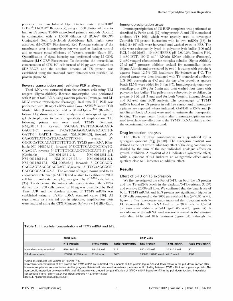
Table 1. Intracellular concentrations of TYMS mRNA and hTS.
2008 cells C13* cells
hTS Protein TYMS mRNA Ratio Prot/mRNA hTS Protein TYMS mRNA Ratio Prot/mRNA
Intracellular concentration* 4306140 nM 3.660.9 nM 119 9306300 nM 10.362.6 nM 90
Pull down amount 139500642000 amol 2366 amol 6065 135000637000 amol 4565 amol 3000
*Using an estimated cell volume of 1.86*10212 L.Intracellular concentrations of hTS protein and TYMS mRNA are indicated. The amounts of hTS protein (figure S2) and TYMS mRNA in the pull down fraction afterimmunoprecipitation are also shown. Antibody against Beta-tubulin was used to evaluate the non-specific binding between TYMS mRNA and a generic protein. Thenon-specific interaction between mRNAs and hTS protein was checked by quantification of GAPDH mRNA bound to hTS in the pull down fraction. (Intracellularconcentration: n = 5, error = 6S.D. Pull down amount: n = 2, error = 6S.E.)doi:10.1371/journal.pone.0047318.t001
Human Thymidylate Synthase Regulation
PLOS ONE | www.plosone.org 3 October 2012 | Volume 7 | Issue 10 | e47318
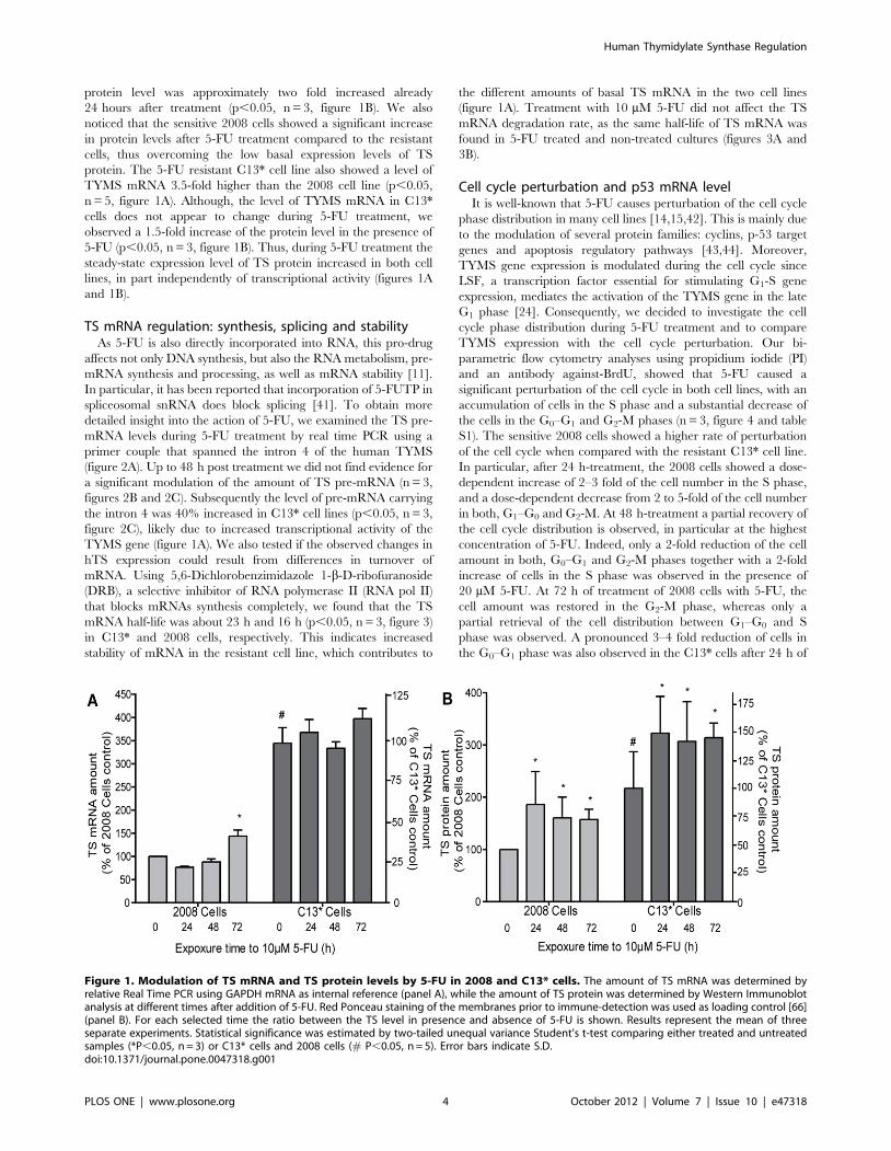
protein level was approximately two fold increased already
24 hours after treatment (p,0.05, n = 3, figure 1B). We also
noticed that the sensitive 2008 cells showed a significant increase
in protein levels after 5-FU treatment compared to the resistant
cells, thus overcoming the low basal expression levels of TS
protein. The 5-FU resistant C13* cell line also showed a level of
TYMS mRNA 3.5-fold higher than the 2008 cell line (p,0.05,
n = 5, figure 1A). Although, the level of TYMS mRNA in C13*
cells does not appear to change during 5-FU treatment, we
observed a 1.5-fold increase of the protein level in the presence of
5-FU (p,0.05, n = 3, figure 1B). Thus, during 5-FU treatment the
steady-state expression level of TS protein increased in both cell
lines, in part independently of transcriptional activity (figures 1A
and 1B).
TS mRNA regulation: synthesis, splicing and stabilityAs 5-FU is also directly incorporated into RNA, this pro-drug
affects not only DNA synthesis, but also the RNA metabolism, pre-
mRNA synthesis and processing, as well as mRNA stability [11].
In particular, it has been reported that incorporation of 5-FUTP in
spliceosomal snRNA does block splicing [41]. To obtain more
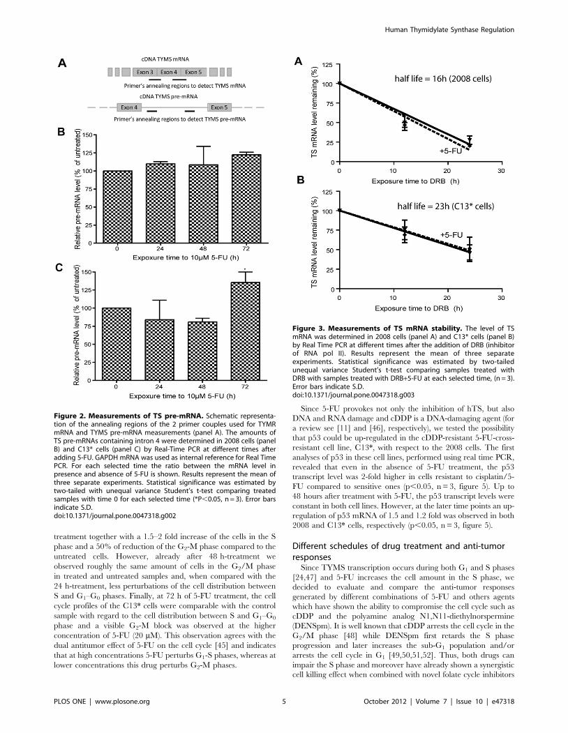
detailed insight into the action of 5-FU, we examined the TS pre-
mRNA levels during 5-FU treatment by real time PCR using a
primer couple that spanned the intron 4 of the human TYMS
(figure 2A). Up to 48 h post treatment we did not find evidence for
a significant modulation of the amount of TS pre-mRNA (n = 3,
figures 2B and 2C). Subsequently the level of pre-mRNA carrying
the intron 4 was 40% increased in C13* cell lines (p,0.05, n = 3,
figure 2C), likely due to increased transcriptional activity of the
TYMS gene (figure 1A). We also tested if the observed changes in
hTS expression could result from differences in turnover of
mRNA. Using 5,6-Dichlorobenzimidazole 1-b-D-ribofuranoside
(DRB), a selective inhibitor of RNA polymerase II (RNA pol II)
that blocks mRNAs synthesis completely, we found that the TS
mRNA half-life was about 23 h and 16 h (p,0.05, n = 3, figure 3)
in C13* and 2008 cells, respectively. This indicates increased
stability of mRNA in the resistant cell line, which contributes to
the different amounts of basal TS mRNA in the two cell lines
(figure 1A). Treatment with 10 mM 5-FU did not affect the TS
mRNA degradation rate, as the same half-life of TS mRNA was
found in 5-FU treated and non-treated cultures (figures 3A and
3B).
Cell cycle perturbation and p53 mRNA levelIt is well-known that 5-FU causes perturbation of the cell cycle
phase distribution in many cell lines [14,15,42]. This is mainly due
to the modulation of several protein families: cyclins, p-53 target
genes and apoptosis regulatory pathways [43,44]. Moreover,
TYMS gene expression is modulated during the cell cycle since
LSF, a transcription factor essential for stimulating G1-S gene
expression, mediates the activation of the TYMS gene in the late
G1 phase [24]. Consequently, we decided to investigate the cell
cycle phase distribution during 5-FU treatment and to compare
TYMS expression with the cell cycle perturbation. Our bi-
parametric flow cytometry analyses using propidium iodide (PI)
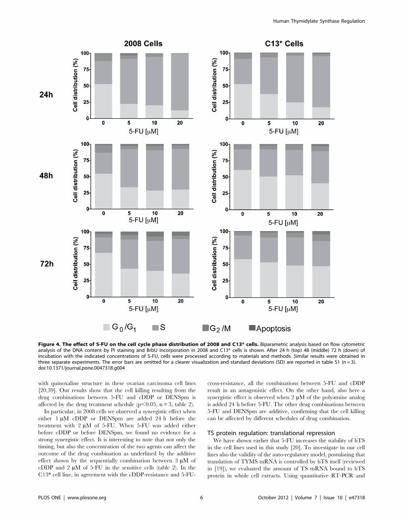
and an antibody against-BrdU, showed that 5-FU caused a
significant perturbation of the cell cycle in both cell lines, with an
accumulation of cells in the S phase and a substantial decrease of
the cells in the G0–G1 and G2-M phases (n = 3, figure 4 and table
S1). The sensitive 2008 cells showed a higher rate of perturbation
of the cell cycle when compared with the resistant C13* cell line.
In particular, after 24 h-treatment, the 2008 cells showed a dose-
dependent increase of 2–3 fold of the cell number in the S phase,
and a dose-dependent decrease from 2 to 5-fold of the cell number
in both, G1–G0 and G2-M. At 48 h-treatment a partial recovery of
the cell cycle distribution is observed, in particular at the highest
concentration of 5-FU. Indeed, only a 2-fold reduction of the cell
amount in both, G0–G1 and G2-M phases together with a 2-fold
increase of cells in the S phase was observed in the presence of
20 mM 5-FU. At 72 h of treatment of 2008 cells with 5-FU, the
cell amount was restored in the G2-M phase, whereas only a
partial retrieval of the cell distribution between G1–G0 and S
phase was observed. A pronounced 3–4 fold reduction of cells in
the G0–G1 phase was also observed in the C13* cells after 24 h of
Figure 1. Modulation of TS mRNA and TS protein levels by 5-FU in 2008 and C13* cells. The amount of TS mRNA was determined byrelative Real Time PCR using GAPDH mRNA as internal reference (panel A), while the amount of TS protein was determined by Western Immunoblotanalysis at different times after addition of 5-FU. Red Ponceau staining of the membranes prior to immune-detection was used as loading control [66](panel B). For each selected time the ratio between the TS level in presence and absence of 5-FU is shown. Results represent the mean of threeseparate experiments. Statistical significance was estimated by two-tailed unequal variance Student’s t-test comparing either treated and untreatedsamples (*P,0.05, n = 3) or C13* cells and 2008 cells (# P,0.05, n = 5). Error bars indicate S.D.doi:10.1371/journal.pone.0047318.g001
Human Thymidylate Synthase Regulation
PLOS ONE | www.plosone.org 4 October 2012 | Volume 7 | Issue 10 | e47318
treatment together with a 1.5–2 fold increase of the cells in the S
phase and a 50% of reduction of the G2-M phase compared to the
untreated cells. However, already after 48 h-treatment we
observed roughly the same amount of cells in the G2/M phase
in treated and untreated samples and, when compared with the
24 h-treatment, less perturbations of the cell distribution between
S and G1–G0 phases. Finally, at 72 h of 5-FU treatment, the cell
cycle profiles of the C13* cells were comparable with the control
sample with regard to the cell distribution between S and G1–G0
phase and a visible G2-M block was observed at the higher
concentration of 5-FU (20 mM). This observation agrees with the
dual antitumor effect of 5-FU on the cell cycle [45] and indicates
that at high concentrations 5-FU perturbs G1-S phases, whereas at
lower concentrations this drug perturbs G2-M phases.
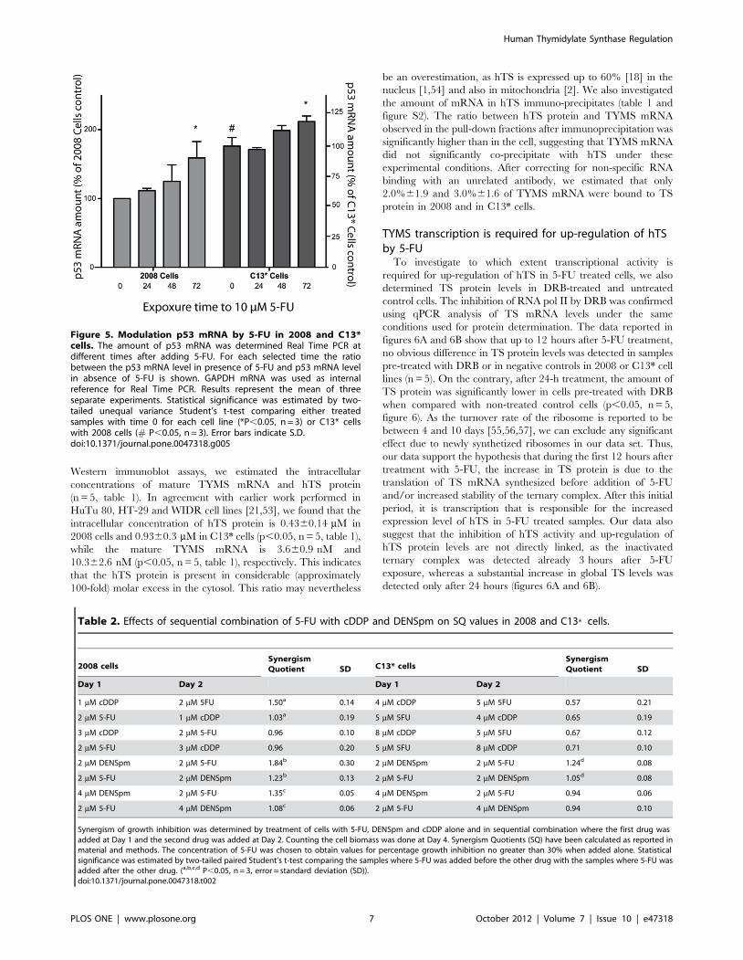
Since 5-FU provokes not only the inhibition of hTS, but also
DNA and RNA damage and cDDP is a DNA-damaging agent (for
a review see [11] and [46], respectively), we tested the possibility
that p53 could be up-regulated in the cDDP-resistant 5-FU-cross-
resistant cell line, C13*, with respect to the 2008 cells. The first
analyses of p53 in these cell lines, performed using real time PCR,
revealed that even in the absence of 5-FU treatment, the p53
transcript level was 2-fold higher in cells resistant to cisplatin/5-
FU compared to sensitive ones (p,0.05, n = 3, figure 5). Up to
48 hours after treatment with 5-FU, the p53 transcript levels were
constant in both cell lines. However, at the later time points an up-
regulation of p53 mRNA of 1.5 and 1.2 fold was observed in both
2008 and C13* cells, respectively (p,0.05, n = 3, figure 5).
Different schedules of drug treatment and anti-tumorresponses
Since TYMS transcription occurs during both G1 and S phases
[24,47] and 5-FU increases the cell amount in the S phase, we
decided to evaluate and compare the anti-tumor responses
generated by different combinations of 5-FU and others agents
which have shown the ability to compromise the cell cycle such as
cDDP and the polyamine analog N1,N11-diethylnorspermine
(DENSpm). It is well known that cDDP arrests the cell cycle in the
G2/M phase [48] while DENSpm first retards the S phase
progression and later increases the sub-G1 population and/or
arrests the cell cycle in G1 [49,50,51,52]. Thus, both drugs can
impair the S phase and moreover have already shown a synergistic
cell killing effect when combined with novel folate cycle inhibitors
Figure 2. Measurements of TS pre-mRNA. Schematic representa-tion of the annealing regions of the 2 primer couples used for TYMRmRNA and TYMS pre-mRNA measurements (panel A). The amounts ofTS pre-mRNAs containing intron 4 were determined in 2008 cells (panelB) and C13* cells (panel C) by Real-Time PCR at different times afteradding 5-FU. GAPDH mRNA was used as internal reference for Real TimePCR. For each selected time the ratio between the mRNA level inpresence and absence of 5-FU is shown. Results represent the mean ofthree separate experiments. Statistical significance was estimated bytwo-tailed with unequal variance Student’s t-test comparing treatedsamples with time 0 for each selected time (*P,0.05, n = 3). Error barsindicate S.D.doi:10.1371/journal.pone.0047318.g002
Figure 3. Measurements of TS mRNA stability. The level of TSmRNA was determined in 2008 cells (panel A) and C13* cells (panel B)by Real Time PCR at different times after the addition of DRB (inhibitorof RNA pol II). Results represent the mean of three separateexperiments. Statistical significance was estimated by two-tailedunequal variance Student’s t-test comparing samples treated withDRB with samples treated with DRB+5-FU at each selected time, (n = 3).Error bars indicate S.D.doi:10.1371/journal.pone.0047318.g003
Human Thymidylate Synthase Regulation
PLOS ONE | www.plosone.org 5 October 2012 | Volume 7 | Issue 10 | e47318
with quinoxaline structure in these ovarian carcinoma cell lines
[20,39]. Our results show that the cell killing resulting from the
drug combinations between 5-FU and cDDP or DENSpm is
affected by the drug treatment schedule (p,0.05, n = 3, table 2).
In particular, in 2008 cells we observed a synergistic effect when
either 1 mM cDDP or DENSpm are added 24 h before the
treatment with 2 mM of 5-FU. When 5-FU was added either
before cDDP or before DENSpm, we found no evidence for a
strong synergistic effect. It is interesting to note that not only the
timing, but also the concentration of the two agents can affect the
outcome of the drug combination as underlined by the additive
effect shown by the sequentially combination between 3 mM of
cDDP and 2 mM of 5-FU in the sensitive cells (table 2). In the
C13* cell line, in agreement with the cDDP-resistance and 5-FU-
cross-resistance, all the combinations between 5-FU and cDDP
result in an antagonistic effect. On the other hand, also here a
synergistic effect is observed when 2 mM of the polyamine analog
is added 24 h before 5-FU. The other drug combinations between
5-FU and DENSpm are additive, confirming that the cell killing
can be affected by different schedules of drug combination.
TS protein regulation: translational repressionWe have shown earlier that 5-FU increases the stability of hTS
in the cell lines used in this study [20]. To investigate in our cell
lines also the validity of the auto-regulatory model, postulating that
translation of TYMS mRNA is controlled by hTS itself (reviewed
in [19]), we evaluated the amount of TS mRNA bound to hTS
protein in whole cell extracts. Using quantitative RT-PCR and
Figure 4. The effect of 5-FU on the cell cycle phase distribution of 2008 and C13* cells. Biparametric analysis based on flow cytometricanalysis of the DNA content by PI staining and BrbU incorporation in 2008 and C13* cells is shown. After 24 h (top) 48 (middle) 72 h (down) ofincubation with the indicated concentrations of 5-FU, cells were processed according to materials and methods. Similar results were obtained inthree separate experiments. The error bars are omitted for a clearer visualization and standard deviations (SD) are reported in table S1 (n = 3).doi:10.1371/journal.pone.0047318.g004
Human Thymidylate Synthase Regulation
PLOS ONE | www.plosone.org 6 October 2012 | Volume 7 | Issue 10 | e47318
Western immunoblot assays, we estimated the intracellular
concentrations of mature TYMS mRNA and hTS protein
(n = 5, table 1). In agreement with earlier work performed in
HuTu 80, HT-29 and WIDR cell lines [21,53], we found that the
intracellular concentration of hTS protein is 0.4360.14 mM in
2008 cells and 0.9360.3 mM in C13* cells (p,0.05, n = 5, table 1),
while the mature TYMS mRNA is 3.660.9 nM and
10.362.6 nM (p,0.05, n = 5, table 1), respectively. This indicates
that the hTS protein is present in considerable (approximately
100-fold) molar excess in the cytosol. This ratio may nevertheless
be an overestimation, as hTS is expressed up to 60% [18] in the
nucleus [1,54] and also in mitochondria [2]. We also investigated
the amount of mRNA in hTS immuno-precipitates (table 1 and
figure S2). The ratio between hTS protein and TYMS mRNA
observed in the pull-down fractions after immunoprecipitation was
significantly higher than in the cell, suggesting that TYMS mRNA
did not significantly co-precipitate with hTS under these
experimental conditions. After correcting for non-specific RNA
binding with an unrelated antibody, we estimated that only
2.0%61.9 and 3.0%61.6 of TYMS mRNA were bound to TS
protein in 2008 and in C13* cells.
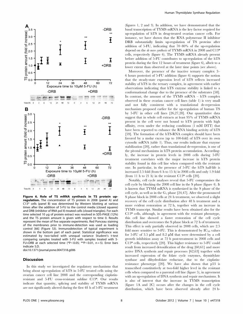
TYMS transcription is required for up-regulation of hTSby 5-FU
To investigate to which extent transcriptional activity is
required for up-regulation of hTS in 5-FU treated cells, we also
determined TS protein levels in DRB-treated and untreated
control cells. The inhibition of RNA pol II by DRB was confirmed
using qPCR analysis of TS mRNA levels under the same
conditions used for protein determination. The data reported in
figures 6A and 6B show that up to 12 hours after 5-FU treatment,
no obvious difference in TS protein levels was detected in samples
pre-treated with DRB or in negative controls in 2008 or C13* cell
lines (n = 5). On the contrary, after 24-h treatment, the amount of
TS protein was significantly lower in cells pre-treated with DRB
when compared with non-treated control cells (p,0.05, n = 5,
figure 6). As the turnover rate of the ribosome is reported to be
between 4 and 10 days [55,56,57], we can exclude any significant
effect due to newly synthetized ribosomes in our data set. Thus,
our data support the hypothesis that during the first 12 hours after
treatment with 5-FU, the increase in TS protein is due to the
translation of TS mRNA synthesized before addition of 5-FU
and/or increased stability of the ternary complex. After this initial
period, it is transcription that is responsible for the increased
expression level of hTS in 5-FU treated samples. Our data also
suggest that the inhibition of hTS activity and up-regulation of
hTS protein levels are not directly linked, as the inactivated
ternary complex was detected already 3 hours after 5-FU
exposure, whereas a substantial increase in global TS levels was
detected only after 24 hours (figures 6A and 6B).
Figure 5. Modulation p53 mRNA by 5-FU in 2008 and C13*cells. The amount of p53 mRNA was determined Real Time PCR atdifferent times after adding 5-FU. For each selected time the ratiobetween the p53 mRNA level in presence of 5-FU and p53 mRNA levelin absence of 5-FU is shown. GAPDH mRNA was used as internalreference for Real Time PCR. Results represent the mean of threeseparate experiments. Statistical significance was estimated by two-tailed unequal variance Student’s t-test comparing either treatedsamples with time 0 for each cell line (*P,0.05, n = 3) or C13* cellswith 2008 cells (# P,0.05, n = 3). Error bars indicate S.D.doi:10.1371/journal.pone.0047318.g005
Table 2. Effects of sequential combination of 5-FU with cDDP and DENSpm on SQ values in 2008 and C13* cells.
2008 cellsSynergismQuotient SD C13* cells
SynergismQuotient SD
Day 1 Day 2 Day 1 Day 2
1 mM cDDP 2 mM 5FU 1.50a 0.14 4 mM cDDP 5 mM 5FU 0.57 0.21
2 mM 5-FU 1 mM cDDP 1.03a 0.19 5 mM 5FU 4 mM cDDP 0.65 0.19
3 mM cDDP 2 mM 5-FU 0.96 0.10 8 mM cDDP 5 mM 5FU 0.67 0.12
2 mM 5-FU 3 mM cDDP 0.96 0.20 5 mM 5FU 8 mM cDDP 0.71 0.10
2 mM DENSpm 2 mM 5-FU 1.84b 0.30 2 mM DENSpm 2 mM 5-FU 1.24d 0.08
2 mM 5-FU 2 mM DENSpm 1.23b 0.13 2 mM 5-FU 2 mM DENSpm 1.05d 0.08
4 mM DENSpm 2 mM 5-FU 1.35c 0.05 4 mM DENSpm 2 mM 5-FU 0.94 0.06
2 mM 5-FU 4 mM DENSpm 1.08c 0.06 2 mM 5-FU 4 mM DENSpm 0.94 0.10
Synergism of growth inhibition was determined by treatment of cells with 5-FU, DENSpm and cDDP alone and in sequential combination where the first drug wasadded at Day 1 and the second drug was added at Day 2. Counting the cell biomass was done at Day 4. Synergism Quotients (SQ) have been calculated as reported inmaterial and methods. The concentration of 5-FU was chosen to obtain values for percentage growth inhibition no greater than 30% when added alone. Statisticalsignificance was estimated by two-tailed paired Student’s t-test comparing the samples where 5-FU was added before the other drug with the samples where 5-FU wasadded after the other drug. (a,b,c,d P,0.05, n = 3, error = standard deviation (SD)).doi:10.1371/journal.pone.0047318.t002
Human Thymidylate Synthase Regulation
PLOS ONE | www.plosone.org 7 October 2012 | Volume 7 | Issue 10 | e47318
Discussion
In this study we investigated the regulatory mechanisms that
bring about up-regulation of hTS in 5-FU treated cells using the
ovarian cancer cell line 2008 and the corresponding cisplatin-
resistant and 5-FU cross-resistant subline C13*. Our results
indicate that quantity, splicing and stability of TYMS mRNA
are not significantly altered during the first 48 h of 5-FU treatment
(figures 1, 2 and 3). In addition, we have demonstrated that the
basal transcription of TYMS mRNA is the key factor required for
up-regulation of hTS in drug-treated ovarian cancer cells. For
instance, we have shown that the RNA polymerase II inhibitor
DRB substantially limits up-regulation of TS proteins after
addition of 5-FU, indicating that 70–80% of the up-regulation
depend on the de novo synthesis of TYMS mRNA in 2008 and C13*
cells, respectively (figure 6). The TYMS mRNA already present
before addition of 5-FU contributes to up-regulation of the hTS
protein during the first 12 hours of treatment (figure 6), albeit to a
lower extent than observed at the later time points (see above).
Moreover, the presence of the inactive ternary complex 3–
6 hours posteriori of 5-FU addition (figure 6) supports the notion
that the steady-state expression level of hTS reflects increased
stability of hTS in the ternary complex, in agreement with earlier
observations indicating that hTS enzyme stability is linked to a
conformational change due to the presence of the substrates [58].
In contrast, the amount of the TYMS mRNA – hTS complex
observed in these ovarian cancer cell lines (table 1) is very small
and not fully consistent with a translational de-repression
mechanism proposed earlier for the up-regulation of human TS
by 5-FU in other cell lines [26,27,28]. Our quantitative data
suggest that in whole cell extracts at least 95% of TYMS mRNA
present in the cell were not bound to hTS protein with high
affinity, even under the reducing conditions (1 mM DTT) that
have been reported to enhance the RNA binding activity of hTS
[59]. The formation of the hTS-RNA complex should have been
favored by a molar excess (up to 100-fold) of hTS over its own
cytosolic mRNA (table 1). Thus, our results indicate that enzyme
stabilization [20], rather than translational de-repression, is one of
the pivotal mechanisms in hTS protein accumulation. According-
ly, the increase in protein levels in 2008 cells during 5-FU
treatment correlates with the major increase in hTS protein
stability found in this cell line when compared with the resistant
one. In particular, in the presence of 5-FU the hTS half-life is
increased 2.5 fold (from 6 h to 15 h) in 2008 cells and only 1.9 fold
(from 11 h to 21 h) in the resistant C13* cells [20].
Notably, cell cycle analyses reveal that 5-FU compromises the
cell cycle by blocking the 2008 cell line in the S phase (figure 4). It
is known that TYSM mRNA is synthesized in the S phase of the
cell cycle, as well as in the G1 phase [24,47]. After the pronounced
S phase block in 2008 cells at 24 h treatment, we observed a slight
recovery of the cell cycle distribution after 48 h treatment and a
more evident restoration at 72 h, together with an increase in
TYMS transcript. Similar results have been obtained also for the
C13* cells, although, in agreement with the resistant phenotype,
this cell line showed a faster restoration of the cell cycle
distribution and overcomes the S phase block at 72 h of treatment.
This effect is only partially observed in 2008 cells, which are 2.5
fold more sensitive to 5-FU. This is demonstrated by IC50 values
for 5-FU of 3.5 mM and 8.2 mM that were determined by a cell
growth inhibition assay at 72 h post-treatment in 2008 cells and
C13* cells, respectively [20]. This higher resistance to 5-FU could
result from increased detoxification of the drug [60,61] and more
active DNA synthesis and repair processes [62,63] together with
increased expression of the folate cycle enzymes, thymidylate
synthase and dihydrofolate reductase, due to the cisplatin-
resistance phenotype [20]. We have also shown that p53 is
transcribed constitutively at two-fold higher level in the resistant
cells when compared to a parental cell line (figure 5), in agreement
with an up-regulation of DNA synthesis and repair mechanisms. It
is also of interest that the increase in TYMS transcription
(figure 1A and 2C) occurs after the changes in the cell cycle
distribution, which have been observed already after 24 h-
Figure 6. Role of TS mRNA synthesis in TS protein up-regulation. The concentration of TS protein in 2008 (panel A) andC13* cells (panel B) was determined by Western blotting at varioustimes after the addition of 5-FU to the control media (closed squares)and DRB (inhibitor of RNA pol II)-treated cells (closed triangles). For eachtime selected 10 mg of protein extract was resolved in SDS-PAGE (12%)and the TS protein amount is given with respect to time 0. Resultsrepresent the mean of five separate experiments. Red Ponceau stainingof the membranes prior to immune-detection was used as loadingcontrol [66] (Figure S3). Immunodetection of typical experiment isshown in the bottom part of each panel. Statistical significance wasestimated by two-tailed with unequal variance Student’s t-testcomparing samples treated with 5-FU with samples treated with 5-FU+DRB at each selected time (*P,0.05; **P,0.01, n = 5). Error barsindicate S.D.doi:10.1371/journal.pone.0047318.g006
Human Thymidylate Synthase Regulation
PLOS ONE | www.plosone.org 8 October 2012 | Volume 7 | Issue 10 | e47318
treatment (figure 4), further indicating that increase in TYMS
transcription is a relatively late cellular response to 5-FU. This
enhanced transcription of TYMS could be part of a more complex
pathway involving p53, as transcriptional up-regulation of p53 and
TYMS are at least partially temporally linked (figures 1A, 2C and
5).
The fact that 5-FU has multiple cellular targets makes it difficult
to fully establish causal links between the multiple and complex
regulatory mechanisms affecting TYMS up-regulation. Neverthe-
less, our data has clearly established that the inhibition of hTS
itself, 3 h-6 h after 5-FU addition, is not directly involved in the
transcriptional activation, that occurs 72 hours after treatment,
and only a later stimulus, characterized also by the increase of p53
mRNA levels, seems to be the starting point of the transcriptional
activation. Since p53 is mainly involved in the pathways of DNA
damage/repair, we hypothesize that the DNA damage could be
one of the causes of this increase of the transcription of the TYMS
gene. Moreover, it is of particular interest that recent reports have
shown that the sub-cellular localization of hTS is cell cycle-
dependent. In particular, in the S and G2-M phases, hTS is
present in the nucleus to enable nuclear de novo synthesis of
thymidylate during DNA replication and repair [1] and to prevent
uracil accumulation in nuclear DNA [64] avoiding DNA damage.
Our results suggest a direct link between TYMS transcription and
cell cycle perturbation resulting from 5-FU treatment, as the
TYMS gene is transcribed in the S phase where hTS is in the
nucleus to overcome the cytotoxic effects provoked by metabolites
of 5-FU. Thus, the S phase-block could promote cell survival in
the presence of 5-FU through an increase in the level of hTS in the
nucleus due to both, enhanced transcription and intracellular
localization induced by the cell cycle.
Our findings also suggest that a combination of 5-FU together
with cell cycle modulators may result in beneficial synergic drug
effects. We showed that distinct anti-tumor responses are
generated by different schedules of drug combination (p,0.05,
n = 3, table 2). Pre-treatment with both cDDP and DENSpm is
known to reduce or impair the S phase [48,49,50,51,52] and may
explain the observed synergistic effects of these drugs with 5-FU in
our cell models. Since we have observed a synergistic effect
primarily when the cell cycle modulator (i.e. cDDP and DENSpm)
is added 24 h before 5-FU, we suggest that a reduced or impaired
S phase could increase the cytotoxic effect of the following
administration of 5-FU. However, the cell cycle modulation of
DENSpm is strongly time-dependent [52], possibly explaining why
an antagonist effect between DENSpm and 5-FU was found when
5-FU was added 48 h after DENSpm in HCT116 colon cancer
cells [51]. In any event, our combined findings suggest that the
effects of the drug combinations between 5-FU and both
DENSpm and cDDP are closely related to the drug treatment
schedule.
Moreover, combinations of 5-FU and other drugs that have
been reported in the literature, such as RPR-115135, a
farnesyltransferase inhibitor, show a synergic effect on growth
inhibition only in human colon cancer cell lines. Here the drug
combination drastically reduces the amount of cells either in the
G0–G1 or the S phase (HCT-116, LoVo, RKO, DLD-1, Colo-
320), whereas an antagonism or a non-significant effect is observed
when both G0–G1 and S phases are slightly modulated or
increased (SW-620, HT-29, HCT- 15, KM-12) [42]. Moreover,
the co-treatments with 5-FU and two inhibitors of histone
deacetylase (HDAC), either Vorinostat or LBH589, decrease
either the amount of cells in the G1 or S phase together with a
inhibition of TYMS gene expression in colon cancer cells (HCT-
116 and HT-29), therefore enhancing the effect of 5-FU [14].
Besides these synergic drug combinations acting on cell cycle
distribution, it has been reported that Trichostatin A, another
HDAC inhibitor synergic with 5-FU, affects the protein stability of
hTS [65], confirming the important role of this mechanism in TS
protein regulation. We also note that the cDDP/5-FU-resistant
cells, which have high basal levels of both, TYMS mRNA and
hTS protein, showed a behavior similar to the cDDP/5-FU-
sensitive cell line. This suggests that key mechanisms regulating
hTS expression are maintained even under conditions of high
steady-state expression levels of hTS.
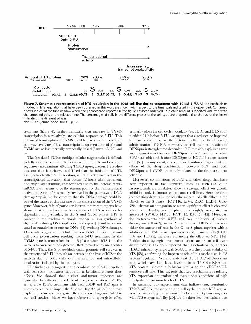
In summary, our experimental data indicate that, constitutive
TYMS mRNA transcription and cell cycle-induced hTS regula-
tion (i.e. increasing the amount of cells in the S phase) together
with hTS enzyme stability [20], are the three key mechanisms that
Figure 7. Schematic representation of hTS regulation in the 2008 cell line during treatment with 10 mM 5-FU. All the mechanismsinvolved in hTS regulation that have been observed in this work are shown with respect to the time scale indicated in the upper part. Continuedarrows represent the time window where the phenomenon reported in the figure has been observed. TS protein amount is reported with respect tothe untreated cells at the selected time. The percentages of cells in the different phases of the cell cycle are proportional to the size of the lettersindicating the different phases.doi:10.1371/journal.pone.0047318.g007
Human Thymidylate Synthase Regulation
PLOS ONE | www.plosone.org 9 October 2012 | Volume 7 | Issue 10 | e47318
mediate 5-fluorouracil induced up-regulation of human thymidyl-
ate synthase expression in the two ovarian cancer cell lines studied.
We have also established that these three regulatory phenomena
have a precise order (figure 7), suggesting the possibility of new
therapeutic strategies based upon our findings.
Supporting Information
Figure S1 hTS protein standard curve for absolutequantification. hTS protein standard curve has been obtained
by serial dilution of purified hTS followed by SDS-PAGE,
electroblotting, antibody staining (panel B) and quantization of
signal intensity using LI-COR (panel A) as described in the
material and method. The correlation coefficient is also shown
(panel A).
(DOCX)
Figure S2 hTS detection in the immunoprecipitationanalysis. The amounts of hTS protein after the immunoprecip-
itation (IP) assay were quantified by Western blot as described
under materials and methods. Lane 1: supernatant of IP using hTS
antibody in 2008 cells. Lane 2: pull down fraction of IP using hTS
antibody in 2008 cells. Lane 3: pull down fraction of IP using Beta-
tubulin antibody. Lane 4: supernatant of IP using hTS antibody in
C13* cells. Lane 5: pull down fraction of IP using hTS antibody in
C13* cells. Lane 6: pull down fraction of IP using Beta-tubulin
antibody in C13 cells.
(DOCX)
Figure S3 Red Ponceau staining of the western blotreported in figure 6. Red ponceau staining prior immunode-
tection in 2008 cells (panel A) and C13* cells (panel B) was used as
loading control and to confirm equal efficiency during Western
transfer.
(DOCX)
Table S1 Standard deviations (SD) of the cell-cycle phase
distribution (%). Standard deviations of the cell cycle distributions
are shown for every phase of each sample reported in figure 4
(n = 3).
(DOCX)
Acknowledgments
We thank members of the LIGHTS consortium for discussions.
Author Contributions
Conceived and designed the experiments: AL GM HM. Performed the
experiments: AL GM. Analyzed the data: AL GM UL HM. Contributed
reagents/materials/analysis tools: AL GM UL HM. Wrote the paper: AL
UL HM.
References
1. Anderson D, Woeller C, Chiang E-P, Shane B, Stover PJ (2012) Serine
Hydroxymethyltransferase anchors the de novo thymidylate synthesis pathway
to the nuclear lamina for DNA synthesis. Journal of Biological Chemistry.
2. Anderson DD, Quintero CM, Stover PJ (2011) Identification of a de novo
thymidylate biosynthesis pathway in mammalian mitochondria. Proceedings of
the National Academy of Sciences 108: 15163–15168.
3. Myllykallio H, Lipowski G, Leduc D, Filee J, Forterre P, et al. (2002) An
Alternative Flavin-Dependent Mechanism for Thymidylate Synthesis. Science
297: 105–107.
4. Garg D, Henrich S, Salo-Ahen OMH, Myllykallio H, Costi MP, et al. (2010)
Novel Approaches for Targeting Thymidylate Synthase To Overcome the
Resistance and Toxicity of Anticancer Drugs. Journal of Medicinal Chemistry
53: 6539–6549.
5. Peters G, Kohne C (1999) Fluoropyrimidines as antifolate drugs; (ed.) JJ, editor.
Totowa, NJ: Humana Press
6. Aherne GW, Hardcastle A, Raynaud F, Jackman AL (1996) Immunoreactive
dUMP and TTP pools as an index of thymidylate synthase inhibition; effect of
tomudex (ZD1694) and a nonpolyglutamated quinazoline antifolate (CB30900)
in L1210 mouse leukaemia cells. Biochemical Pharmacology 51: 1293–1301.
7. Mitrovski B, Pressacco J, Mandelbaum S, Erlichman C (1994) Biochemical
effects of folate-based inhibitors of thymidylate synthase in MGH-U1 cells.
Cancer Chemother Pharmacol 35: 109–114.
8. Jackson RC, Grindley GB (1984) The Biochemical Basis for Methotrexate
Cytotoxicity; Accademic, editor. New york: Sirotnak, FM, Burchall, JJ,
Ensminger, WD, Montgomery, JA. 289–315 p.
9. Houghton JA, Tillman DM, Harwood FG (1995) Ratio of 29-deoxyadenosine-
59-triphosphate/thymidine-59-triphosphate influences the commitment of hu-
man colon carcinoma cells to thymineless death. Clin Cancer Res 1: 723–730.
10. Yoshioka A (1987) Deoxyribonucleoside triphosphate imbalance. 5-Fluorodeox-
yuridine-induced DNA double strand breaks in mouse FM3A cells and the
mechanism of cell death. J Biol Chem 262: 8235–8241.
11. Longley DB, Harkin DP, Johnston PG (2003) 5-Fluorouracil: mechanisms of
action and clinical strategies. Nat Rev Cancer 3: 330–338.
12. Giacchetti S (2000) Phase III multicenter randomized trial of oxaliplatin added
to chronomodulated fluorouracil-leucovorin as first-line treatment of metastatic
colorectal cancer. J Clin Oncol 18: 136–147.
13. Douillard JY (2000) Irinotecan combined with fluorouracil compared with
fluorouracil alone as first-line treatment for metastatic colorectal cancer: a
multicentre randomised trial. Lancet 355: 1041–1047.
14. Fazzone W, Wilson PM, LaBonte MJ, Lenz H-J, Ladner RD (2009) Histone
deacetylase inhibitors suppress thymidylate synthase gene expression and
synergize with the fluoropyrimidines in colon cancer cells. International Journal
of Cancer 125: 463–473.
15. Di Gennaro E, Bruzzese F, Pepe S, Leone A, Delrio P, et al. (2009) Modulation
of thymidilate synthase and p53 expression by HDAC inhibitor vorinostat
resulted in synergistic antitumor effect in combination with 5FU or Raltitrexed.
Cancer Biology & Therapy 8: 782–791.
16. Showalter SL, Showalter TN, Witkiewicz A, Havens R, Kennedy EP, et al.
(2008) Evaluating the drug-target relationship between thymidylate synthase
expression and tumor response to 5-fluorouracil: Is it time to move forward?
Cancer Biology & Therapy 7: 986–994.
17. Backus H, Pinedo H, Wouters D, Kuiper C, Jansen G, et al. (2000) Differences
in the induction of DNA damage, cell cycle arrest, and cell death by 5-
fluorouracil and antifolates. Oncology Research 12: 8.
18. Gustavson MD, Molinaro AM, Tedeschi G, Camp RL, Rimm DL (2008)
AQUA Analysis of Thymidylate Synthase Reveals Localization to be a Key
Prognostic Biomarker in 2 Large Cohorts of Colorectal Carcinoma. Archives of
Pathology & Laboratory Medicine 132: 1746–1752.
19. Berg RW, Ferguson PJ, DeMoor JM, Vincent MD, Koropatnick J (2002) The
means to an end of tumor cell resistance to chemotherapeutic drugs targeting
thymidylate synthase: Shoot the messenger. Current Drug Targets 3: 297–309.
20. Marverti G, Ligabue A, Paglietti G, Corona P, Piras S, et al. (2009) Collateral
sensitivity to novel thymidylate synthase inhibitors correlates with folate cycle
enzymes impairment in cisplatin-resistant human ovarian cancer cells. European
Journal of Pharmacology 615: 17–26.
21. Washtien WL (1984) Increased levels of thymidylate synthetase in cells exposed
to 5-fluorouracil. Molecular Pharmacology 25: 171–177.
22. Melo SP, Barbour KW, Berger FG (2011) Cooperation between an Intrinsically
Disordered Region and a Helical Segment Is Required for Ubiquitin-
independent Degradation by the Proteasome. Journal of Biological Chemistry
286: 36559–36567.
23. Kitchens ME, Forsthoefel AM, Rafique Z, Spencer HT, Berger FG (1999)
Ligand-mediated Induction of Thymidylate Synthase Occurs by Enzyme
Stabilization. Journal of Biological Chemistry 274: 12544–12547.
24. Hansen U, Owens L, Saxena U (2009) Transcription factors LSF and E2Fs:
Tandem cyclists driving G0 to S? Cell Cycle 8: 2146–2151.
25. Yoo BK, Gredler R, Vozhilla N, Su Z-z, Chen D, et al. (2009) Identification of
genes conferring resistance to 5-fluorouracil. Proceedings of the National
Academy of Sciences 106: 12938–12943.
26. Chu E, Koeller DM, Casey JL, Drake JC, Chabner BA, et al. (1991)
Autoregulation of human thymidylate synthase messenger RNA translation by
thymidylate synthase. Proceedings of the National Academy of Sciences 88:
8977–8981.
27. Chu E, Koeller DM, Johnston PG, Zinn S, Allegra CJ (1993) Regulation of
thymidylate synthase in human colon cancer cells treated with 5-fluorouracil and
interferon-gamma. Mol Pharmacol 43: 527–533.
28. Chu E, Cogliati T, Copur SM, Borre A, Voeller DM, et al. (1996) Identification
of in Vivo Target RNA Sequences Bound by Thymidylate Synthase. Nucleic
Acids Research 24: 3222–3228.
29. Boni V, Bitarte N, Cristobal I, Zarate R, Rodriguez J, et al. (2010) miR-192/
miR-215 Influence 5-Fluorouracil Resistance through Cell Cycle-Mediated
Mechanisms Complementary to Its Post-transcriptional Thymidilate Synthase
Regulation. Molecular Cancer Therapeutics 9: 2265–2275.
Human Thymidylate Synthase Regulation
PLOS ONE | www.plosone.org 10 October 2012 | Volume 7 | Issue 10 | e47318
30. Andrews PA, Albright KD (1992) Mitochondrial Defects in cis-Diamminedi-
chloroplatinum(II)-resistant Human Ovarian Carcinoma Cells. Cancer Re-
search 52: 1895–1901.
31. Bradford MM (1976) A rapid and sensitive method for the quantitation of
microgram quantities of protein utilizing the principle of protein-dye binding.
Analytical biochemistry 72: 248–254.
32. Stahlberg A, Kubista M, Pfaffl M (2004) Comparison of reverse transcriptases in
gene expression analysis. Clinical Chemistry 50: 1678–1680.
33. Dolbeare F, Gratzner H, Pallavicini MG, Gray JW (1983) Flow cytometric
measurement of total DNA content and incorporated bromodeoxyuridine.
Proceedings of the National Academy of Sciences 80: 5573–5577.
34. Longley DB, Ferguson PR, Boyer J, Latif T, Lynch M, et al. (2001)
Characterization of a Thymidylate Synthase (TS)-inducible Cell Line. Clinical
Cancer Research 7: 3533–3539.
35. Arocho A, Chen BY, Ladanyi M, Pan QL (2006) Validation of the 2(-Delta
Delta Ct) calculation as an alternate method of data analysis for quantitative
PCR of BCR-ABL P210 transcripts. Diagnostic Molecular Pathology 15: 56–61.
36. Bengtsson M, Hemberg M, Rorsman P, Stahlberg A (2008) Quantification of
mRNA in single cells and modelling of RT-qPCR induced noise. BMC
Molecular Biology 9: 63.
37. Peritz T, Zeng F, Kannanayakal TJ, Kilk K, Eiriksdottir E, et al. (2006)
Immunoprecipitation of mRNA-protein complexes. Nat Protocols 1: 577–580.
38. Zhang Y, Yang S, Liu M, Song C, Wu N, et al (2010). Interaction between
Thymidylate Synthase and Its Cognate mRNA in Zebrafish Embryos. PLoS
ONE 5: e10618.
39. Marverti G, Ligabue A, Guerrieri D, Paglietti G, Piras S, et al. (2010)
Spermidine/spermine N1-acetyltranferase modulation by novel folate cycle
inhibitors in cisplatin-sensitive and -resistant human ovarian cancer cell lines.
Gynecologic Oncology 117: 202–210.
40. Cho YS, Cho-Chung YS (2003) Antisense Protein Kinase A RIa Acts
Synergistically with Hydroxycamptothecin to Inhibit Growth and Induce
Apoptosis in Human Cancer Cells. Clinical Cancer Research 9: 1171–1178.
41. Zhao X, Yu Y-T (2007) Incorporation of 5-fluorouracil into U2 snRNA blocks
pseudouridylation and pre-mRNA splicing in vivo. Nucleic Acids Research 35:
550–558.
42. Russo P, Malacarne D, Falugi C, Trombino S, O’Connor PM (2002) RPR-
115135, a farnesyltransferase inhibitor, increases 5-FU- cytotoxicity in ten
human colon cancer cell lines: Role of p53. International Journal of Cancer 100:
266–275.
43. De Angelis P, Svendsrud D, Kravik K, Stokke T (2006) Cellular response to 5-
fluorouracil (5-FU) in 5-FU-resistant colon cancer cell lines during treatment and
recovery. Molecular Cancer 5: 20.
44. Guo X, Goessl E, Jin G, Collie-Duguid ESR, Cassidy J, et al. (2008) Cell Cycle
Perturbation and Acquired 5-Fluorouracil Chemoresistance. Anticancer Re-
search 28: 9–14.
45. Yoshikawa R, Kusunoki M, Yanagi H, Noda M, Furuyama J-i, et al. (2001) Dual
Antitumor Effects of 5-Fluorouracil on the Cell Cycle in Colorectal Carcinoma
Cells: A Novel Target Mechanism Concept for Pharmacokinetic Modulating
Chemotherapy. Cancer Research 61: 1029–1037.
46. Jamieson ER, Lippard SJ (1999) Structure, Recognition, and Processing of
Cisplatin DNA Adducts. Chemical Reviews 99: 2467–2498.
47. Le Francois BG, Maroun JA, Birnboim HC (2007) Expression of thymidylate
synthase in human cells is an early G1 event regulated by CDK4 and
p16INK4A but not E2F. Br J Cancer 97: 1242–1250.
48. Montopoli M, Ragazzi E, Froldi G, Caparrotta L (2009) Cell-cycle inhibitionand apoptosis induced by curcumin and cisplatin or oxaliplatin in human
ovarian carcinoma cells. Cell Proliferation 42: 195–206.
49. Kramer DL, Fogel-Petrovic M, Diegelman P, Cooley JM, Bernacki RJ, et al.
(1997) Effects of Novel Spermine Analogues on Cell Cycle Progression andApoptosis in MALME-3M Human Melanoma Cells. Cancer Research 57:
5521–5527.
50. Kramer DL, Vujcic S, Diegelman P, Alderfer J, Miller JT, et al. (1999)Polyamine Analogue Induction of the p53–p21WAF1/CIP1-Rb Pathway and
G1 Arrest in Human Melanoma Cells. Cancer Research 59: 1278–1286.51. Choi W, Gerner EW, Ramdas L, Dupart J, Carew J, et al. (2005) Combination
of 5-Fluorouracil and N1,N11-Diethylnorspermine Markedly Activates Spermi-
dine/Spermine N1-Acetyltransferase Expression, Depletes Polyamines, andSynergistically Induces Apoptosis in Colon Carcinoma Cells. Journal of
Biological Chemistry 280: 3295–3304.52. Alm K, Berntsson PSH, Kramer DL, Porter CW, Oredsson SM (2000)
Treatment of cells with the polyamine analog N1,N11-diethylnorspermineretards S phase progression within one cell cycle. European Journal of
Biochemistry 267: 4157–4164.
53. Dolnick B, Zhang Z, Hines J, Rustum Y (1992) Quantitation of dihydrofolatereductase and thymidylate synthase mRNAs in vivo and in vitro by polymerase
chain reaction. Oncology Research 4: 8.54. Anderson DD, Woeller CF, Stover PJ (2007) Small ubiquitin-like modifier-1
(SUMO-1) modification of thymidylate synthase and dihydrofolate reductase.
Clinical Chemistry and Laboratory Medicine: De Gruyter. pp. 1760–1763.55. Retz KC, Steele WJ (1980) Ribosome turnover in rat brain and liver. Life
Sciences 27: 2601–2604.56. Hirsch CA, Hiatt HH (1966) Turnover of Liver Ribosomes in Fed and in Fasted
Rats. Journal of Biological Chemistry 241: 5936–5940.57. Nikolov EN, Dabeva MD, Nikolov TK (1983) Turnover of ribosomes in
regenerating rat liver. International Journal of Biochemistry 15: 1255–1260.
58. Mohsen AW, Aull JL, Payne DM, Daron HH (1995) Ligand-inducedconformational changes of thymidylate synthase detected by limited proteolysis.
Biochemistry 34: 9.59. Chu E, Voeller DM, Morrison PF, Jones KL, Takechi T, et al. (1994) The effect
of reducing reagents on binding of thymidylate synthase protein to thymidylate
synthase messenger RNA. Journal of Biological Chemistry 269: 20289–20293.60. Marverti G, Cusumano M, Ligabue A, Di Pietro ML, Vainiglia PA, et al. (2008)
Studies on the anti-proliferative effects of novel DNA-intercalating bipyridyl-thiourea-Pt(II) complexes against cisplatin-sensitive and -resistant human
ovarian cancer cells. Journal of Inorganic Biochemistry 102: 699–712.61. Mann SC, Andrews PA, Howell SB (1990) Short-term cis-diamminedichlor-
oplatinum(II) accumulation in sensitive and resistant human ovarian carcinoma
cells. Cancer Chemotherapy and Pharmacology 25: 236–240.62. Zhen W, Link CJ, O’Connor PM, Reed E, Parker R, et al. (1992) Increased
gene-specific repair of cisplatin interstrand cross-links in cisplatin-resistanthuman ovarian cancer cell lines. Molecular and Cellular Biology 12: 3689–3698.
63. Marverti G, Ligabue A, Montanari M, Guerrieri D, Cusumano M, et al. (2011)
Characterization of the cell growth inhibitory effects of a novel DNA-intercalating bipyridyl-thiourea-Pt(II) complex in cisplatin-sensitive and -
resistant human ovarian cancer cells. Investigational New Drugs 29: 73–86.64. MacFarlane AJ, Anderson DD, Flodby P, Perry CA, Allan RH, et al. (2011)
Nuclear localization of the De Novo thymidylate biosynthesis pathway isrequired to prevent uracil accumulation in DNA. Journal of Biological
Chemistry 286: 44015–44022.
65. Lee J-H, Park J-H, Jung Y, Kim J-H, Jong H-S, et al. (2006) Histone deacetylaseinhibitor enhances 5-fluorouracil cytotoxicity by down-regulating thymidylate
synthase in human cancer cells. Molecular Cancer Therapeutics 5: 3085–3095.66. Romero-Calvo I, Ocon B, Martinez-Moya P, Suarez MD, Zarzuelo A, et al.
(2010) Reversible Ponceau staining as a loading control alternative to actin in
Western blots. Analytical biochemistry 401: 318–320.
Human Thymidylate Synthase Regulation
PLOS ONE | www.plosone.org 11 October 2012 | Volume 7 | Issue 10 | e47318