MMT-PLGA NANOCOMPOSITES AS AN ORAL AND CONTROLLED RELEASE CARRIER FOR 5-FLUOROURACIL: A NOVEL...
10
Research Article MMT-PLGA NANOCOMPOSITES AS AN ORAL AND CONTROLLED RELEASE CARRIER FOR 5-FLUOROURACIL: A NOVEL APPROACH SEEMA AND MONIKA DATTA* Analytical Research Laboratory, Department of Chemistry, University of Delhi, Delhi 110007, India, Email: [email protected] Received: 19 Jan 2013, Revised and Accepted: 31 Mar 2013 ABSTRACT Objective of the present study is to develop MMT based PLGA nanocomposites of oral and controlled release carrier of 5-Fluorouracil. Double emulsion solvent evaporation method was used for nanocomposite synthesis and effect of medical clay MMT on encapsulation efficiency has been evaluated. The synthesized 5FU-PLGA-MMT nanocomposites were compared with respect to morphology, particle size, physical status of 5FU and MMT, and in vitro drug release to the corresponding biodegradable PLGA nanoparticles. Intercalation of polymer-drug moiety within the MMT layers has been supported by XRD data. Particle size of synthesized nanocomposites was found in the range of 100-200 nm, a favorable size for cellular uptake. In vitro drug release profiles of 5FU-PLGA-MMT nanocomposites in simulated gastrointestinal fluid, indicates a controlled release of 5-Fluorouracil as compared to 5FU-PLGA nanoparticles and pure 5FU for a period of 8 hours. The delivery of 5FU by MMT based PLGA nanocomposites suggest the possibility of designing the oral and controlled release formulations to minimize the drug administration frequency and hence improve the patient compliance. Keywords: Controlled release, 5 Fluorouracil, Montmorillonite, Nanocomposites, Oral chemotherapy INTRODUCTION 5 Fluorouracil (5FU) is an analogue of the naturally occurring pyrimidine, uracil, with fluorine substitution in the 5 position. It is widely used in clinical for the treatment of a variety of carcinomas especially those involving the breast, colorectal region, pancreatic neck, head and skin [1]. The unpredictable bioavailability related to first pass metabolism, upon oral administration 5 FU is usually administered by intravenous bolus or continuous infusion [2]. 5FU has very short biological half life of 10-15 minutes as it is quickly metabolized in the body [3]. Therefore the maintenance of high serum concentrations of this drug to improve its therapeutic activity is needed. The maintenance of these serum concentrations requires continuous/multiple administrations, but 5-FU shows severe toxic effects [4], and of course, the multiple dosing of 5 FU is more probable to reach and or exceed the toxic concentration. Therefore, an efficient oral drug delivery system is desired to overcome the drawbacks associated with 5FU and improve the clinical therapy which is capable of maintaining the therapeutic concentration over a longer period of time and provide a long time exposure to the cancer cells. A novel oral drug delivery system with controlled release chatracterstics can be prove more patient. Compliant by providing a Preferred mode of treatment over present intermittant chemotherapy by injection or infusion. The carriers used for control drug release are mainly biodegradable polymers [5] and porous inorganic matrix [6,7]. In recent years, drug intercalated smectite, especially Montmorillonite (MMT) a pharmaceutical grade clay have attracted great interest from researchers [8,9]. MMT is a 2:1 smectite layered clay mineral composed of two SiO4 tetrahedral layers sandwiching a central Al octahedral layer as shown in Figure (1, a). The isomorphous substitution of Al 3+ for Si 4+ in the tetrahedral layer and Mg 2+ for Al 3+ in the octahedral layer results in a net negative surface charge which is balanced by inorganic exchangeable cations (Na + ,Ca 2+, etc.) Chemically it is hydrated sodium calcium aluminum magnesium silicate hydroxide (Na,Ca)0.33(Al,Mg)2(Si4O10)(OH)2.nH2O. MMT has large specific surface area, good adsorption ability, cation exchange capacity, and drug-carrying capability. It, posses’ hydrophilicity, high dispersability in water and along the (001) axis MMT can encapsulate various protonated and hydrophilic organic molecules, which can be released in controlled manner by replacement with other kind of cations in the release media [10-15] . Therefore the MMT minerals are suggested to be good delivery carries of the hydrophilic drugs. MMT is a potent detoxifier with excellent adsorbent properties due to its high aspect ratio. It can adsorb excess water from feces and thus act as anti-diarrhoeic. MMT can also provide mucoadhesive capability for the nanoparticles to cross the gastrointestinal barrier [16-18]. Although certain literature has already been reported for controlled release of 5FU using MMT [19, 20] but to the best of our knowledge no report has been available for the combination of biodegradable polymer PLGA and MMT for controlled release of 5FU. This novel formulation of MMT-PLGA nanocomposites are expecting to combine the advantages of FDA approved biodegradable and biocompatible polymer PLGA with non toxic GRAS grade clay MMT. The objective of the present work is to develop and characterize 5Fluorouracil loaded MMT based PLGA nanocomposites for oral and controlled delivery so as to minimizing the administration frequency and side effects resulting in enhanced patient compliance by promoting the concept of “oral chemotherapy”. MATERIALS AND METHODS Montmorillonite KSF, PLGA 50:50 (molecular weight 40,000- 75,000), 5Fluorouracil (purity 99.8%) and FDA approved nonionic surface active agent Pluronic F-68 (PF-68) were obtained from Sigma Aldrich USA. PF-68 is a poly (polyethylene oxide)– poly (polypropylene oxide) – poly (polyethylene oxide) triblock copolymer (Fig. 1, c) with a molecular weight equal to 8500 Dalton and was selected for this study because of its advantageous effect on cancerous cells [21]. Analytical grade dichloromethane, HCl, KCl, NaOH, Potassium dihydrogen phosphate were ordered by MERCK (Germany). HPLC grade methanol and water were used for drug estimation by HPLC technique. All other reagents whether specified or not were of analytical grade. Water used in the experiments was deionized and filtered (Milli-Q Academic, Millipore, France). Synthesis of 5FU-PLGA Nanoparticles In this study, the modified water/oil/water (w/o/w) double emulsion solvent evaporation method [22] has been selected to encapsulate hydrophilic drug 5FU in nanoparticles. 5FU-PLGA nanoparticles were synthesized in two steps. First, 5FU was dissolved in 1 ml of water and emulsified in a solution of methylene chloride containing PLGA under sonication for 5 min. In the second step, the primary w/o-emulsion was emulsified in the external aqueous phase of Pluronic F68 to form a w/o/w-emulsion by homogenization at 16,000 rpm for 5 min. The middle organic phase separated the internal water droplets from each other as well as from the external aqueous continuous phase. After solvent evaporation the nanoparticles were isolated by centrifugation for 30 minute at 25,000 rpm (Sigma, Sartorius, 3K30) and washed with double distilled water before freeze-drying (Virtis, benchtop K). The powder samples thus obtained were used for further investigations. International Journal of Pharmacy and Pharmaceutical Sciences ISSN- 0975-1491 Vol 5, Issue 2, 2013 A A c c a a d d e e m mi i c c S Sc c i i e e n n c c e e s s
Transcript of MMT-PLGA NANOCOMPOSITES AS AN ORAL AND CONTROLLED RELEASE CARRIER FOR 5-FLUOROURACIL: A NOVEL...
Research Article
MMT-PLGA NANOCOMPOSITES AS AN ORAL AND CONTROLLED RELEASE CARRIER FOR 5-FLUOROURACIL: A NOVEL APPROACH
SEEMA AND MONIKA DATTA*
Analytical Research Laboratory, Department of Chemistry, University of Delhi, Delhi 110007, India, Email: [email protected]
Received: 19 Jan 2013, Revised and Accepted: 31 Mar 2013
ABSTRACT
Objective of the present study is to develop MMT based PLGA nanocomposites of oral and controlled release carrier of 5-Fluorouracil. Double emulsion solvent evaporation method was used for nanocomposite synthesis and effect of medical clay MMT on encapsulation efficiency has been evaluated. The synthesized 5FU-PLGA-MMT nanocomposites were compared with respect to morphology, particle size, physical status of 5FU and MMT, and in vitro drug release to the corresponding biodegradable PLGA nanoparticles. Intercalation of polymer-drug moiety within the MMT layers has been supported by XRD data. Particle size of synthesized nanocomposites was found in the range of 100-200 nm, a favorable size for cellular uptake. In vitro drug release profiles of 5FU-PLGA-MMT nanocomposites in simulated gastrointestinal fluid, indicates a controlled release of 5-Fluorouracil as compared to 5FU-PLGA nanoparticles and pure 5FU for a period of 8 hours.
The delivery of 5FU by MMT based PLGA nanocomposites suggest the possibility of designing the oral and controlled release formulations to minimize the drug administration frequency and hence improve the patient compliance.
Keywords: Controlled release, 5 Fluorouracil, Montmorillonite, Nanocomposites, Oral chemotherapy [
INTRODUCTION
5 Fluorouracil (5FU) is an analogue of the naturally occurring pyrimidine, uracil, with fluorine substitution in the 5 position. It is widely used in clinical for the treatment of a variety of carcinomas especially those involving the breast, colorectal region, pancreatic neck, head and skin [1]. The unpredictable bioavailability related to first pass metabolism, upon oral administration 5 FU is usually administered by intravenous bolus or continuous infusion [2]. 5FU has very short biological half life of 10-15 minutes as it is quickly metabolized in the body [3]. Therefore the maintenance of high serum concentrations of this drug to improve its therapeutic activity is needed. The maintenance of these serum concentrations requires continuous/multiple administrations, but 5-FU shows severe toxic effects [4], and of course, the multiple dosing of 5 FU is more probable to reach and or exceed the toxic concentration.
Therefore, an efficient oral drug delivery system is desired to overcome the drawbacks associated with 5FU and improve the clinical therapy which is capable of maintaining the therapeutic concentration over a longer period of time and provide a long time exposure to the cancer cells. A novel oral drug delivery system with controlled release chatracterstics can be prove more patient. Compliant by providing a Preferred mode of treatment over present intermittant chemotherapy by injection or infusion. The carriers used for control drug release are mainly biodegradable polymers [5] and porous inorganic matrix [6,7].
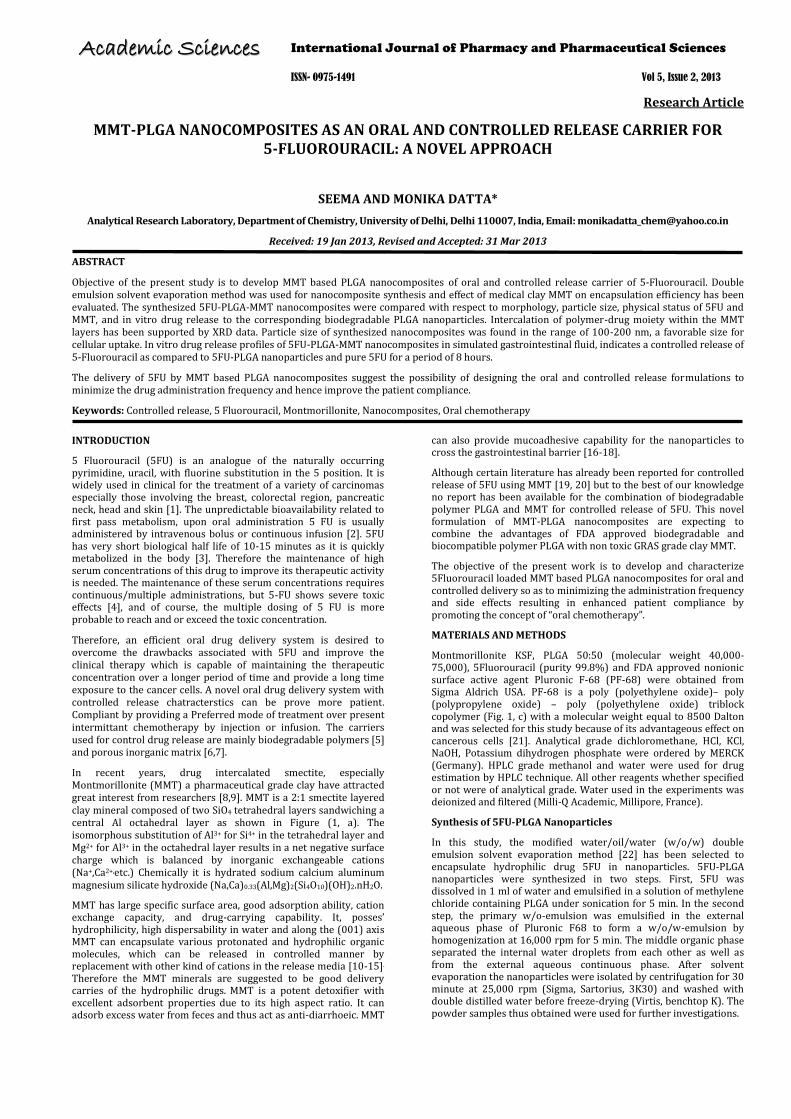
In recent years, drug intercalated smectite, especially Montmorillonite (MMT) a pharmaceutical grade clay have attracted great interest from researchers [8,9]. MMT is a 2:1 smectite layered clay mineral composed of two SiO4 tetrahedral layers sandwiching a central Al octahedral layer as shown in Figure (1, a). The isomorphous substitution of Al3+ for Si4+ in the tetrahedral layer and Mg2+ for Al3+ in the octahedral layer results in a net negative surface charge which is balanced by inorganic exchangeable cations (Na+,Ca2+,etc.) Chemically it is hydrated sodium calcium aluminum magnesium silicate hydroxide (Na,Ca)0.33(Al,Mg)2(Si4O10)(OH)2.nH2O.
MMT has large specific surface area, good adsorption ability, cation exchange capacity, and drug-carrying capability. It, posses’ hydrophilicity, high dispersability in water and along the (001) axis MMT can encapsulate various protonated and hydrophilic organic molecules, which can be released in controlled manner by replacement with other kind of cations in the release media [10-15]. Therefore the MMT minerals are suggested to be good delivery carries of the hydrophilic drugs. MMT is a potent detoxifier with excellent adsorbent properties due to its high aspect ratio. It can adsorb excess water from feces and thus act as anti-diarrhoeic. MMT
can also provide mucoadhesive capability for the nanoparticles to cross the gastrointestinal barrier [16-18].
Although certain literature has already been reported for controlled release of 5FU using MMT [19, 20] but to the best of our knowledge no report has been available for the combination of biodegradable polymer PLGA and MMT for controlled release of 5FU. This novel formulation of MMT-PLGA nanocomposites are expecting to combine the advantages of FDA approved biodegradable and biocompatible polymer PLGA with non toxic GRAS grade clay MMT.
The objective of the present work is to develop and characterize 5Fluorouracil loaded MMT based PLGA nanocomposites for oral and controlled delivery so as to minimizing the administration frequency and side effects resulting in enhanced patient compliance by promoting the concept of “oral chemotherapy”.
MATERIALS AND METHODS
Montmorillonite KSF, PLGA 50:50 (molecular weight 40,000-75,000), 5Fluorouracil (purity 99.8%) and FDA approved nonionic surface active agent Pluronic F-68 (PF-68) were obtained from Sigma Aldrich USA. PF-68 is a poly (polyethylene oxide)– poly (polypropylene oxide) – poly (polyethylene oxide) triblock copolymer (Fig. 1, c) with a molecular weight equal to 8500 Dalton and was selected for this study because of its advantageous effect on cancerous cells [21]. Analytical grade dichloromethane, HCl, KCl, NaOH, Potassium dihydrogen phosphate were ordered by MERCK (Germany). HPLC grade methanol and water were used for drug estimation by HPLC technique. All other reagents whether specified or not were of analytical grade. Water used in the experiments was deionized and filtered (Milli-Q Academic, Millipore, France).
Synthesis of 5FU-PLGA Nanoparticles
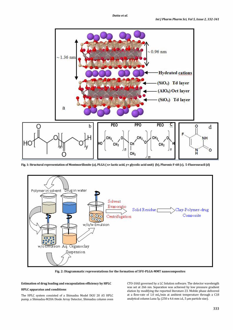
In this study, the modified water/oil/water (w/o/w) double emulsion solvent evaporation method [22] has been selected to encapsulate hydrophilic drug 5FU in nanoparticles. 5FU-PLGA nanoparticles were synthesized in two steps. First, 5FU was dissolved in 1 ml of water and emulsified in a solution of methylene chloride containing PLGA under sonication for 5 min. In the second step, the primary w/o-emulsion was emulsified in the external aqueous phase of Pluronic F68 to form a w/o/w-emulsion by homogenization at 16,000 rpm for 5 min. The middle organic phase separated the internal water droplets from each other as well as from the external aqueous continuous phase. After solvent evaporation the nanoparticles were isolated by centrifugation for 30 minute at 25,000 rpm (Sigma, Sartorius, 3K30) and washed with double distilled water before freeze-drying (Virtis, benchtop K). The powder samples thus obtained were used for further investigations.
International Journal of Pharmacy and Pharmaceutical Sciences
ISSN- 0975-1491 Vol 5, Issue 2, 2013
AAccaaddeemmiicc SScciieenncceess
Datta et al. Int J Pharm Pharm Sci, Vol 5, Issue 2, 332-341
333
Fig. 1: Structural representation of Montmorillonite (a), PLGA ( x= lactic acid, y= glycolic acid unit) (b), Pluronic F-68 (c), 5-Fluorouracil (d)
Fig. 2: Diagrammatic representations for the formation of 5FU-PLGA-MMT nanocomposites
Estimation of drug loading and encapsulation efficiency by HPLC
HPLC apparatus and conditions
The HPLC system consisted of a Shimadzu Model DGU 20 A5 HPLC pump, a Shimadzu-M20A Diode Array Detector, Shimadzu column oven
CTO-10AS governed by a LC Solution software. The detector wavelength was set at 266 nm. Separation was achieved by low pressure gradient elution by modifying the reported literature 23. Mobile phase delivered at a flow-rate of 1.0 mL/min at ambient temperature through a C18
analytical column Luna 5 (250 x 4.6 mm i.d., 5 μm particle size).
Datta et al. Int J Pharm Pharm Sci, Vol 5, Issue 2, 332-341
334
Stock solutions and standards
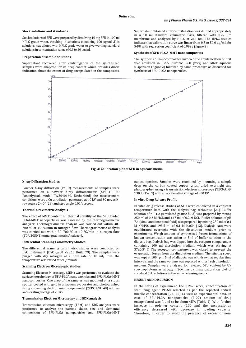
Stock solutions of 5FU were prepared by dissolving 10 mg 5FU in 100 ml HPLC grade water, resulting in solutions containing 100 μg/ml .This solutions was diluted with HPLC grade water to give working standard solutions in concentration range of 0.5 to 50 μg/ml.
Preparation of sample solutions
Supernatant recovered after centrifugation of the synthesized samples were analyzed for its drug content which provides direct indication about the extent of drug encapsulated in the composites.
Supernatant obtained after centrifugation was diluted appropriately in a 10 ml standard volumetric flask, filtered with 0.22 μm membrane and analyzed by HPLC at 266 nm. The HPLC studies indicate that calibration curve was linear from 0.5 to 50.0 μg/mL for 5-FU with regression coefficient of 0.9998 (figure 3)
Synthesis of 5FU-PLGA-MMT nanocomposites
The synthesis of nanocomposites involved the emulsification of first w/o emulsion in 0.2% Pluronic F-68 (w/v) and MMT aqueous suspension (figure 2) followed by same procedure as discussed for synthesis of 5FU-PLGA nanoparticles.
Fig. 3: Calibration plot of 5FU in aqueous media
X-ray Diffraction Studies
Powder X-ray diffraction (PXRD) measurements of samples were performed on a powder X-ray diffractometer (XPERT PRO Pananlytical, model PW3040160, Netherland) the measurement conditions were a Cu α radiation generated at 40 kV and 30 mA as X-ray source 2-40° (2θ) and step angle 0.01°/second.
Thermal Gravimetric Analysis
The effect of MMT content on thermal stability of the 5FU loaded PLGA-MMT nanoparticles was assessed by the thermogravimetric analyzer. Thermogravimetric analysis was carried out within 30–700 °C at 10 °C/min in nitrogen flow Thermogravimetric analysis was carried out within 30–700 °C at 10 °C/min in nitrogen flow (TGA 2050 Thermal gravimetric Analyzer).
Differential Scanning Calorimetry Studies
The differential scanning calorimetric studies were conducted on DSC instrument (DSC Q200 V23.10 Build 79). The samples were purged with dry nitrogen at a flow rate of 10 ml/ min. the temperature was raised at 5°C/ minute.
Scanning Electron Microscopic Studies
Scanning Electron Microscopy (SEM) was performed to evaluate the surface morphology of 5FU-PLGA nanoparticles and 5FU-PLGA-MMT nanocomposites. One drop of the samples was mounted on a stubs; sputter coated with gold in a vacuum evaporator and photographed using a scanning electron microscope model (ZEISS EVO 40) with an accelerating voltage of 20 KV.
Transmission Electron Microscopy and EDX analysis
Transmission electron microscopy (TEM) and EDX analysis were performed to analyze the particle shape, size and elemental composition of 5FU-PLGA nanoparticles and 5FU-PLGA-MMT
nanocomposites. Samples were examined by mounting a sample drop on the carbon coated copper grids, dried overnight and photographed using a transmission electron microscope (TECNAI G2 T30, U-TWIN) with an accelerating voltage of 300 KV.
In vitro Drug Release Profile
In vitro drug release studies of 5FU were conducted in a constant temperature bath with the dialysis bag technique [23]. Buffer solution of pH 1.2 (simulated gastric fluid) was prepared by mixing 250 ml of 0.2 M HCL and 147 ml of 0.2 M KCL. Buffer solution of pH 7.4 (simulated intestinal fluid) was prepared by mixing 250 ml of 0.1 M KH2PO4 and 195.5 ml of 0.1 M NaOH [12]. Dialysis sacs were equilibrated overnight with the dissolution medium prior to experiments. Weigh amount of synthesized frozen formulations of known concentration was taken in 5ml of buffer solution in the dialysis bag. Dialysis bag was dipped into the receptor compartment containing 100 ml dissolution medium, which was stirring at 37±0.5° C. The receptor compartment was closed to prevent the evaporation losses from the dissolution medium. The stirring speed was kept at 100 rpm. 5 ml of aliquots was withdrawn at regular time intervals and the same volume was replaced with a fresh dissolution medium. Samples were analyzed for released 5FU content by UV spectrophotometer at λmax = 266 nm by using calibration plot of standard 5FU solutions in the same releasing media.
RESULT AND DISCUSSION
In the series of experiment, the 0.2% (wt/v) concentration of stabilizing agent PF-68 selected as per the reported critical micelle concentration [24, 25] as well as experimental data. In case of 5FU-PLGA nanoparticles (F-02) amount of drug encapsulated was found to be about 45% (Table 1). With further increase in polymer content (100 mg) the encapsulation efficiency decreased with decrease in loading capacity. Therefore, in order to avoid the presence of excess of non-
Datta et al. Int J Pharm Pharm Sci, Vol 5, Issue 2, 332-341
335
emulsified polymer the amount of PLGA was fixed at 50 mg for all formulations. It has also been observed that in case of 5FU-PLGA-MMT nanocomposites, maximum amount of drug (F-09) retained was 73%. Variation in composition of 5FU-PLGA-MMT nanocomposites were further studied in detail.
Effect of PF-68, MMT and 5FU content on drug loading and encapsulation efficiency
The drug loading (DL) and extent of drug encapsulation efficiency (EE) in 5FU-PLGA nanoparticles and 5FU-PLGA-MMT
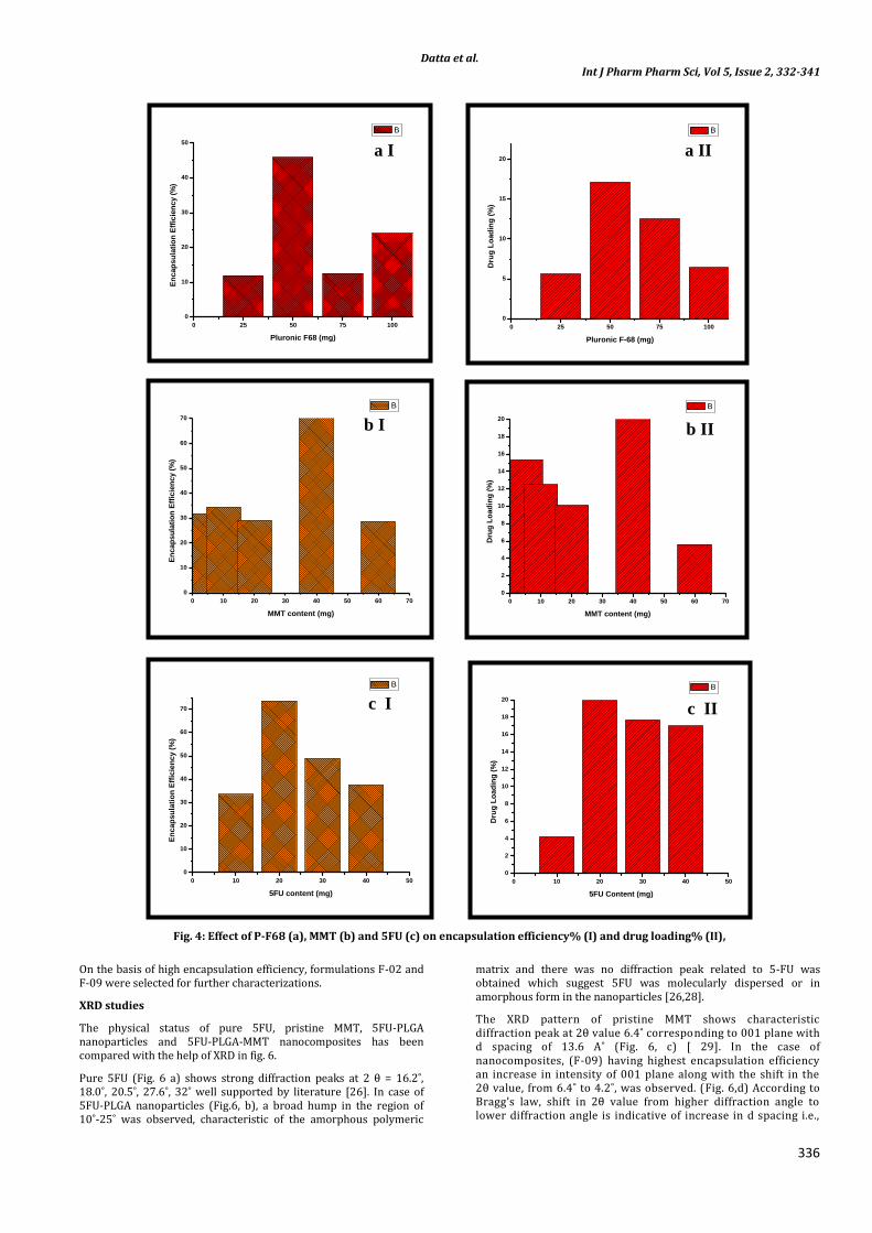
nanocomposites as function of P-F68, MMT and 5FU content was calculated with the help of equation 1 and 2 respectively [ 26]. The amount of 5FU encapsulated in the PLGA nanoparticles increase from 12 to 45% (Table 1 and fig. 4a,I) with increase in surfactant ratio from 0.1% (cmc) to 0.2 (% w/v) attributed to the formation of more stable emulsion. With increase in surfactant. However further increase in surfactant upto 0.4% (w/v), does not show any significant change in the encapsulation efficiency %. Therefore, in order to avoid the presence of excess surfactant the amount of PF-68 was fixed at 0.2% (w/v) for all formulations.
DL (%)= Drug weight in nanoparticles/weight of nanoparticles obtained …… Equation (1)
EE (%) = Drug weight in nanoparticles/weight of drug added …...Equation (2)
Table 1: Composition of formulations
Code MMT (mg)
PLGA (mg)
PF-68 (mg)
5FU (mg)
Yield ( %)
DL* (%)
EE* (%)
F01 ---- 50 25 20 44.6 5.60 11.93 F02 --- 50 50 20 43.69 17.18 44.25 F03 ---- 50 75 20 ----- 12.19 12.54 F04 --- 50 100 20 44.23 6.40 24.43 F05 --- 100 50 20 69.62 5.88 34.83 F06 5 50 50 20 33.54 15.33 31.37 F07 10 50 50 20 35.82 12.48 34.37 F08 20 50 50 20 41.20 13.55 39.85 F09 40 50 50 20 45.13 20.23 73.13 F10 40 50 50 10 53.06 4.24 33.47 F11 40 50 50 30 48.57 17.66 48.63 F12 40 50 50 40 48.85 16.99 37.34
(Note: * DL% = drug loading %, EE% = Encapsulation Efficiency %)
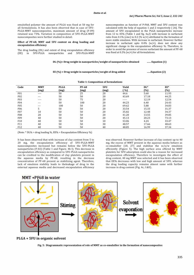
It has been observed that with increase of clay content from 5 to 20 mg, the encapsulation efficiency of 5FU-PLGA-MMT nanocomposites increased but remains below the 5FU-PLGA nanoparticles (F-02) (Table 1 and Figure. 4b I). This decrease in encapsulation efficiency as compared to 5FU-PLGA nanoparticles can be credited to the modification of clay platelets present in the aqueous media by PF-68, resulting in the decrease concentration of PF-68 present as stabilizing agent. Therefore, lack of emulsion stability leads to theleakage of drug to the external aqueous media and decreased encapsulation efficiency
was observed. However further increase of clay content up to 40 mg, the excess of MMT present in the aqueous media behave as co-emulsifier [18, 27] and stabilize the w/o/w emulsion efficiently (Figure 5). The high surface area offered by MMT platelets for 5FU adsorption could also be a reason for increased encapsulation efficiency. Therefore to investigate the effect of drug content, 40 mg MMT was selected and it has been observed that EE% decreases with low and high amount of 5FU, whereas the drug loading capacity remains almost same with further increase in drug content (Fig. 4c, I &II).
Fig. 5: Diagrammatic representation of role of MMT as co-emulsifier in the formation of nanocomposites
PLGA + 5FU in organic solvent
Datta et al. Int J Pharm Pharm Sci, Vol 5, Issue 2, 332-341
336
Fig. 4: Effect of P-F68 (a), MMT (b) and 5FU (c) on encapsulation efficiency% (I) and drug loading% (II),
On the basis of high encapsulation efficiency, formulations F-02 and F-09 were selected for further characterizations.
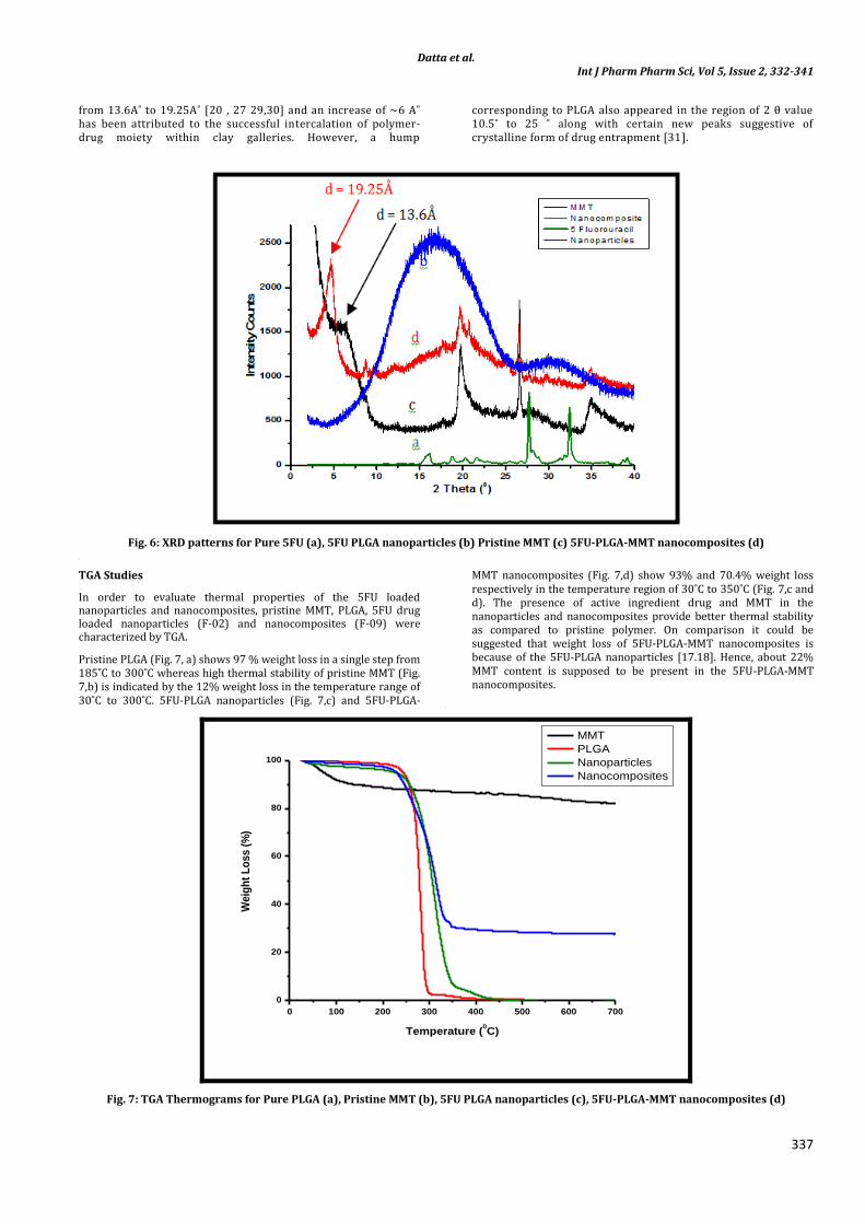
XRD studies
The physical status of pure 5FU, pristine MMT, 5FU-PLGA nanoparticles and 5FU-PLGA-MMT nanocomposites has been compared with the help of XRD in fig. 6.
Pure 5FU (Fig. 6 a) shows strong diffraction peaks at 2 θ = 16.2˚, 18.0˚, 20.5˚, 27.6˚, 32˚ well supported by literature [26]. In case of 5FU-PLGA nanoparticles (Fig.6, b), a broad hump in the region of 10˚-25˚ was observed, characteristic of the amorphous polymeric
matrix and there was no diffraction peak related to 5-FU was obtained which suggest 5FU was molecularly dispersed or in amorphous form in the nanoparticles [26,28].
The XRD pattern of pristine MMT shows characteristic diffraction peak at 2θ value 6.4˚ corresponding to 001 plane with d spacing of 13.6 A˚ (Fig. 6, c) [ 29]. In the case of nanocomposites, (F-09) having highest encapsulation efficiency an increase in intensity of 001 plane along with the shift in the 2θ value, from 6.4˚ to 4.2˚, was observed. (Fig. 6,d) According to Bragg’s law, shift in 2θ value from higher diffraction angle to lower diffraction angle is indicative of increase in d spacing i.e.,
0 10 20 30 40 50 60 70
0
10
20
30
40
50
60
70
En
cap
su
lati
on
Eff
icie
ncy (
%)
MMT content (mg)
B
0 10 20 30 40 50 60 70
0
2
4
6
8
10
12
14
16
18
20
Dru
g L
oad
ing
(%
)
MMT content (mg)
B
0 25 50 75 100
0
10
20
30
40
50
En
cap
su
lati
on
Eff
icie
ncy (
%)
Pluronic F68 (mg)
B
0 25 50 75 100
0
5
10
15
20
Dru
g L
oad
ing
(%
)
Pluronic F-68 (mg)
B
0 10 20 30 40 50
0
10
20
30
40
50
60
70
En
cap
su
lati
on
Eff
icie
ncy (
%)
5FU content (mg)
B
0 10 20 30 40 50
0
2
4
6
8
10
12
14
16
18
20
Dru
g L
oad
ing
(%
)
5FU Content (mg)
B
a I a II
b I b II
c I c II
Datta et al. Int J Pharm Pharm Sci, Vol 5, Issue 2, 332-341
337
from 13.6A˚ to 19.25A˚ [20 , 27 29,30] and an increase of ~6 A˚ has been attributed to the successful intercalation of polymer-drug moiety within clay galleries. However, a hump
corresponding to PLGA also appeared in the region of 2 θ value 10.5˚ to 25 ˚ along with certain new peaks suggestive of crystalline form of drug entrapment [31].
Fig. 6: XRD patterns for Pure 5FU (a), 5FU PLGA nanoparticles (b) Pristine MMT (c) 5FU-PLGA-MMT nanocomposites (d) [
TGA Studies
In order to evaluate thermal properties of the 5FU loaded nanoparticles and nanocomposites, pristine MMT, PLGA, 5FU drug loaded nanoparticles (F-02) and nanocomposites (F-09) were characterized by TGA.
Pristine PLGA (Fig. 7, a) shows 97 % weight loss in a single step from 185˚C to 300˚C whereas high thermal stability of pristine MMT (Fig. 7,b) is indicated by the 12% weight loss in the temperature range of 30˚C to 300˚C. 5FU-PLGA nanoparticles (Fig. 7,c) and 5FU-PLGA-
MMT nanocomposites (Fig. 7,d) show 93% and 70.4% weight loss respectively in the temperature region of 30˚C to 350˚C (Fig. 7,c and d). The presence of active ingredient drug and MMT in the nanoparticles and nanocomposites provide better thermal stability as compared to pristine polymer. On comparison it could be suggested that weight loss of 5FU-PLGA-MMT nanocomposites is because of the 5FU-PLGA nanoparticles [17.18]. Hence, about 22% MMT content is supposed to be present in the 5FU-PLGA-MMT nanocomposites.
[
0 100 200 300 400 500 600 700
0
20
40
60
80
100
Weig
ht
Lo
ss (
%)
Temperature (0C)
MMT
PLGA
Nanoparticles
Nanocomposites
Fig. 7: TGA Thermograms for Pure PLGA (a), Pristine MMT (b), 5FU PLGA nanoparticles (c), 5FU-PLGA-MMT nanocomposites (d)
Datta et al. Int J Pharm Pharm Sci, Vol 5, Issue 2, 332-341
338
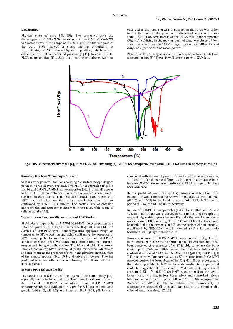
DSC Studies
Physical state of pure 5FU (Fig. 8,c) compared with the thermograms of 5FU-PLGA nanoparticles and 5FU-PLGA-MMT nanocomposites in the range of 0°C to 450°C.The thermogram of the pure 5-FU showed a sharp melting endotherm at approximately 282°C followed by decomposition, which was in agreement with those reported previously [31]. In case of 5FU-PLGA nanoparticles, (Fig. 8,d), drug melting endotherm was not
observed in the region of 282°C, suggesting that drug was either totally dissolved in the polymer or dispersed as an amorphous solid [22,32]. However, In case of 5FU-PLGA-MMT nanocomposites (Fig. 8,e) a shifting in the melting peak of drug was observed by a small but sharp peak at 224˚C suggesting the crystalline form of drug entrapped within nanocomposites.
Physical status of drug observed in both nanoparticles (F-02) and nanocomposites (F-09) was in well correlation with XRD data.
Fig. 8: DSC curves for Pure MMT (a), Pure PLGA (b), Pure drug (c), 5FU PLGA nanoparticles (d) and 5FU-PLGA-MMT nanocomposites (e)
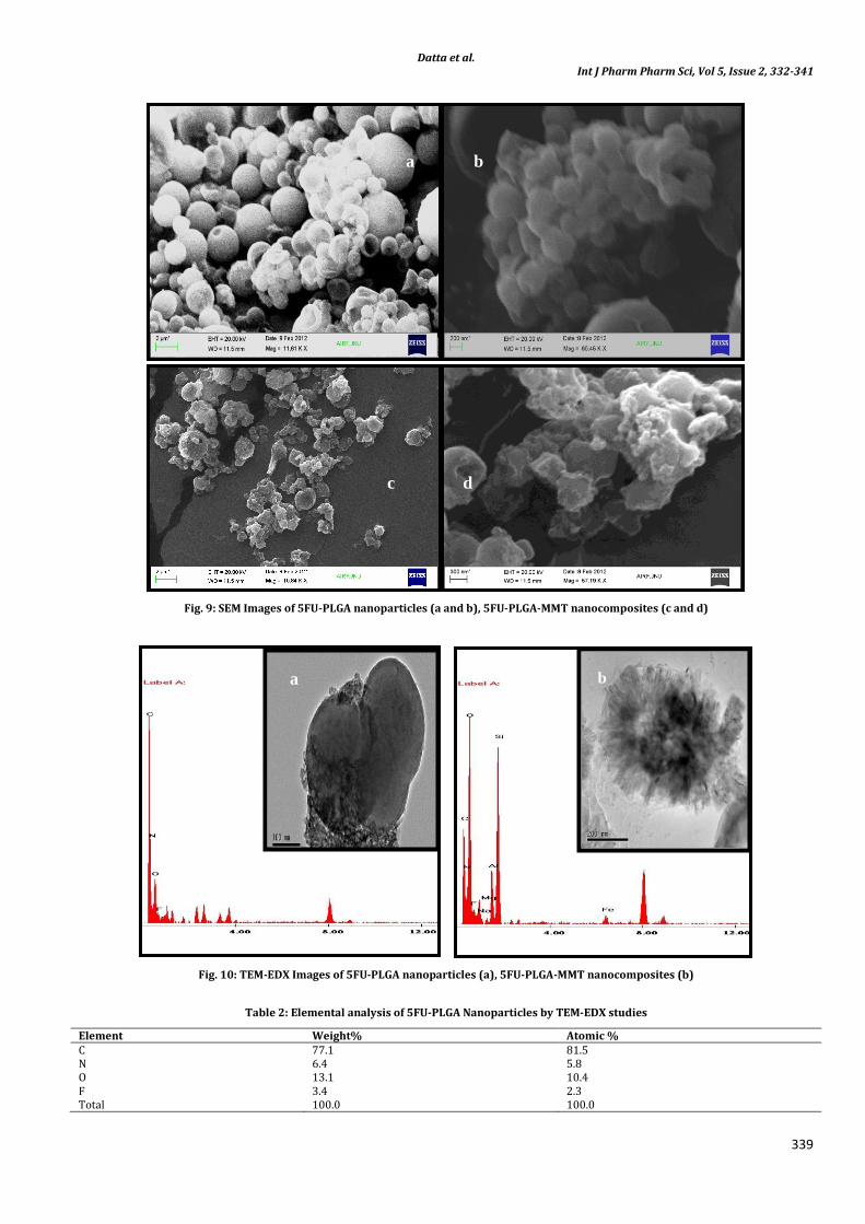
Scanning Electron Microscopic Studies
SEM is a very powerful tool for analyzing the surface morphology of polymeric drug delivery systems. 5FU-PLGA nanoparticles (Fig. 9 a and b) and 5FU-PLGA-MMT nanocomposites (Fig. 9, c and d) appear to be 100 - 300 nm spherical particles, the earlier has a smooth surface and the latter has rough surface because of the presence of MMT nano platelets on the surface which has been further confirmed by TEM – EDX studies. The particle size of obtained nanoparticles and nanocomposites was in the favourable range of cellular uptake [ 33].
Transmission Electron Microscopic and EDX Studies
5FU-PLGA nanoparticles and 5FU-PLGA-MMT nanocomposites are spherical particles of 100-200 nm in size (Fig. 10, a and b). The surface of 5FU-PLGA/MMT nanocomposites appeared rough as compared to 5FU-PLGA nanoparticles confirming the presence of MMT nano platelets on the surface. In case of 5FU-PLGA nanoparticles, the TEM EDX studies indicates high content of carbon, oxygen and nitrogen on the surface (Fig. 10, a and table 2) whereas, samples containing MMT, additional peaks for Silicon, Aluminum and Iron confirms the presence of MMT nano platelets on the surface of the nanocomposites (Fig. 10 b and table 3). However Fluorine peak is observed in both the cases confirming the 5FU content on the particle surface.
In Vitro Drug Release Profile
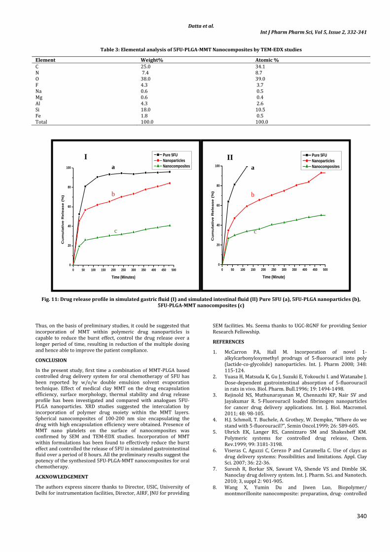
The target sites of 5-FU are all the organs of the human body [34], especially the gastrointestinal tract. Therefore the release profile of the selected 5FU-PLGA nanoparticles and 5FU-PLGA-MMT nanocomposites was evaluated in vitro for 8 hours, in simulated gastric fluid (HCl, pH 1.2) and intestinal fluid (PBS, pH 7.4) and
compared with release of pure 5-FU under similar conditions (Fig. 11, I and II). Considerable differences in the release characteristics between MMT-PLGA nanocomposites and PLGA nanoparticles have been observed.
Release profile of pure 5FU (Fig.11 a) shows a rapid burst of ~80% in initial 1 h which approach to 94.6% in simulated gastric fluid (HCl, pH 1.2) and 100% in simulated intestinal fluid (PBS, pH 7.4) over a period of 4 hours and 2 hours respectively.
In case of 5FU-PLGA nanoparticles (F-02), burst effect of 56% and 47% in initial 1 hour was observed in HCl (pH 1.2) and PBS (pH 7.4) respectively, which approaches to 84% and 93% cumulative release over a period of 8 hours (Fig. 11, b). The initial burst release could be attributed to the presence of 5FU on the surface of nanoparticles (confirmed by TEM-EDX) which released swiftly in the media because of its high hydrophilic nature.
However, in case of 5FU-PLGA-MMT nanocomposites (Fig. 11, c) a more controlled release over a period of 8 hours was obtained. It has been observed that presence of MMT is able to reduce the burst effect up to 25% and 30% during the first hour followed by controlled release of 40.6% and 50.2% in HCl (pH 1.2) and PBS (pH 7.4) respectively. Comparatively, less 5FU release from PLGA-MMT nanocomposites has been obtained in HCl (pH 1.2) corresponding to the stability provided by MMT in the acidic media. On comparison it could be suggested that presence of MMT allowed migration of entrapped 5FU from5FU-PLGA-MMT nanocomposites through a longer path, resulting in less burst effect and controlled release behavior as compared to pure 5FU and 5FU-PLGA nanoparticles. Presence of MMT is able to enhance the permeability of nanoparticles through GI tract and can reduce the common side effect of anticancer drug [17, 18].
Datta et al. Int J Pharm Pharm Sci, Vol 5, Issue 2, 332-341
339
Fig. 9: SEM Images of 5FU-PLGA nanoparticles (a and b), 5FU-PLGA-MMT nanocomposites (c and d)
Fig. 10: TEM-EDX Images of 5FU-PLGA nanoparticles (a), 5FU-PLGA-MMT nanocomposites (b)
Table 2: Elemental analysis of 5FU-PLGA Nanoparticles by TEM-EDX studies
Element Weight% Atomic % C 77.1 81.5 N 6.4 5.8 O 13.1 10.4 F 3.4 2.3 Total 100.0 100.0
a b
c d
kev kev
a b
Datta et al. Int J Pharm Pharm Sci, Vol 5, Issue 2, 332-341
340
Table 3: Elemental analysis of 5FU-PLGA-MMT Nanocomposites by TEM-EDX studies
Element Weight% Atomic % C 25.0 34.1 N 7.4 8.7 O 38.0 39.0 F 4.3 3.7 Na 0.6 0.5 Mg 0.6 0.4 Al 4.3 2.6 Si 18.0 10.5 Fe 1.8 0.5 Total 100.0 100.0
Fig. 11: Drug release profile in simulated gastric fluid (I) and simulated intestinal fluid (II) Pure 5FU (a), 5FU-PLGA nanoparticles (b), 5FU-PLGA-MMT nanocomposites (c)
Thus, on the basis of preliminary studies, it could be suggested that incorporation of MMT within polymeric drug nanoparticles is capable to reduce the burst effect, control the drug release over a longer period of time, resulting in reduction of the multiple dosing and hence able to improve the patient compliance.
CONCLUSION
In the present study, first time a combination of MMT-PLGA based controlled drug delivery system for oral chemotherapy of 5FU has been reported by w/o/w double emulsion solvent evaporation technique. Effect of medical clay MMT on the drug encapsulation efficiency, surface morphology, thermal stability and drug release profile has been investigated and compared with analogues 5FU-PLGA nanoparticles. XRD studies suggested the intercalation by incorporation of polymer drug moiety within the MMT layers. Spherical nanocomposites of 100-200 nm size encapsulating the drug with high encapsulation efficiency were obtained. Presence of MMT nano platelets on the surface of nanocomposites was confirmed by SEM and TEM-EDX studies. Incorporation of MMT within formulations has been found to effectively reduce the burst effect and controlled the release of 5FU in simulated gastrointestinal fluid over a period of 8 hours. All the preliminary results suggest the potency of the synthesized 5FU-PLGA-MMT nanocomposites for oral chemotherapy.
ACKNOWLEDGEMENT
The authors express sincere thanks to Director, USIC, University of Delhi for instrumentation facilities, Director, AIRF, JNU for providing
SEM facilities. Ms. Seema thanks to UGC-RGNF for providing Senior Research Fellowship.
REFERENCES
1. McCarron PA, Hall M. Incorporation of novel 1-alkylcarbonyloxymethyl prodrugs of 5-fluorouracil into poly (lactide-co-glycolide) nanoparticles. Int. J. Pharm 2008; 348: 115-124.
2. Yuasa H, Matsuda K, Gu J, Suzuki E, Yokouchi I. and Watanabe J. Dose-dependent gastrointestinal absorption of 5-fluorouracil in rats in vivo. Biol. Pharm. Bull.1996; 19: 1494-1498.
3. Rejinold NS, Muthunarayanan M, Chennazhi KP, Nair SV and Jayakumar R. 5-Fluorouracil loaded fibrinogen nanoparticles for cancer drug delivery applications. Int. J. Biol. Macromol. 2011; 48: 98-105.
4. H.J. Schmoll, T. Buchele, A. Grothey, W. Dempke, “Where do we stand with 5-fluorouracil?”, Semin Oncol.1999; 26: 589-605.
5. Uhrich EK, Langer RS, Cannizzaro SM and Shakesheff KM. Polymeric systems for controlled drug release, Chem. Rev.1999; 99: 3181-3198.
6. Viseras C, Aguzzi C, Cerezo P and Caramella C. Use of clays as drug delivery systems: Possibilities and limitations. Appl. Clay Sci. 2007; 36: 22-36.
7. Suresh R, Borkar SN, Sawant VA, Shende VS and Dimble SK. Nanoclay drug delivery system. Int. J. Pharm. Sci. and Nanotech. 2010; 3, suppl 2: 901-905.
8. Wang X, Yumin Du and Jiwen Luo, Biopolymer/ montmorillonite nanocomposite: preparation, drug- controlled
0 50 100 150 200 250 300 350 400 450 500
0
20
40
60
80
100
Cu
mu
lati
ve R
ele
ase (
%)
Time (Minutes)
Pure 5FU
Nanoparticles
Nanocomposites
0 50 100 150 200 250 300 350 400 450 500
0
20
40
60
80
100
Pure 5FU
Nanoparticles
Nanocomposites
Cu
mu
lati
ve R
ele
ase (
%)
Time (Minute)
I II
a
b
c
a
b
c
Datta et al. Int J Pharm Pharm Sci, Vol 5, Issue 2, 332-341
341
release property and cytotoxicity. Nanotechnology. 2008; 19: 06570.
9. Depan D, Kumar AP and Singh RP. Cell proliferation and controlled drug release studies of nanohybrids based on chitosan-g-lactic acid and montmorillonite. Acta Biomaterialia. 2009; 5: 93-100.
10. Iliescu RI, Andronescu E, Voicu G, Ficai A and Covaliu CI. Hybrid materials based on montmorillonite and citostatic drugs: Preparation and characterization. Appl. Clay Sci.2011; 52: 62-68.
11. Chen Y, Zhou A, Bo Liu and Jun Liang. Tramadol hydrochloride/montmorillonite composite: Preparation and controlled drug release. Appl. Clay Sci. 2010; 49: suppl 3:108-112.
12. Nunes CD, Vaz PD, Fernandes AC, Ferreira P, Romao CC and Calhorda M.J. Loading and delivery of sertraline using inorganic micro and mesoporous materials. Eur. J. Pharm. Biopharm 2007; 66:357-365.
13. Joshi GV, Patel HA, Kevadiya BD and Bajaj HC. Montmorillonite intercalated with vitamin B1 as drug carrier. Appl. Clay Sci. 2009; 45: 248-253.
14. Seki Y and Yurdakoc K. Equilibrium, kinetics and thermodynamic aspects of Promethazine hydrochloride sorption by iron rich smectite. Colloids and Surfaces A: Physicochem. Eng. Aspects. 2009; 340:143-148.
15. Madurai SL, Joseph SW, Mandal AB, Tsibouklis J and Reddy B.S. Intestine-Specific, Oral delivery of captopril/montmorillonite: formulation and release kinetics. Nanoscale Research Letters, 2011; 6 :suppl 15.
16. Lee YH, Kuo TF, Chen BY, Yi-Kai Feng, Yu- Ren Wen,Wu-Ching Lin and Lin FH, Toxicity assessment of montmorillonite as a drug carrier for pharmaceutical applications: yeast and rats model. Biomedical Engineering Applications, Basis & Communications, 2005; 17: 72-78.
17. Feng SS, Mei L, Anitha P, Gan CW and Zhou W. Poly(lactide)–vitamin E derivative/montmorillonite nanoparticle formulations for the oral delivery of Docetaxel. Biomaterials, 2009; 30:3297-3306.
18. Dong Y and Feng SS. Poly (D,L-lactide-co-glycolide)/montmorillonite nanoparticles fororal delivery of anticancer drugs. Biomaterials , 2005; 26: 6068-6076.
19. Kevadiya BD, Patel TA, Thumbar RP, Pandya MP and Bajaj HC. Layered inorganic nanocomposites: A promising carrier for 5-fluorouracil, . Eur. J. Pharm. Biopharm .2012; 81: 91-101.
20. Lin FH, Lee YH, Jian CH, Jau-Min Wong, Ming-Jium Shieh, Cheng-Yi Wang, A study of purified montmorillonite intercalated with 5-fluorouracil as drug carrier. Biomaterials. 2002, 23: 1981- 87.
21. Huang L, Mei L, Yan F, Zhang C and Zheng Y , The Effect of Poloxamer 188 on nanoparticle Morphology, Size, Cancer Cell
Uptake, and Cytotoxicity. Nanotechnology, Biology and Medicine, 2010; 6:170-178.
22. Nair LK, Jagadeeshan S, Nair SA, Kumar GSV. Biological evaluation of 5-fluorouracil nanoparticles for cancer chemotherapy and its dependence on the carrier, PLGA. Int. J . Nanomedicine.2011; 6: 1685-1697.
23. Nagarwal RC, Singh PN, Shri Kant, Maiti P, and Pandit JK. Chitosan nanoparticles of 5-fluorouracil for ophthalmic delivery: characterization, in-vitro and in-vivo study. Chem. Pharm. Bull. 2011, 59 suppl 2: 272-278.
24. Schmolka IR. A review of block polymer surfactants. J. of American Oil Chemists' society, 1977; 54: 110-116.
25. Saski W, Shah SG. Availability of drugs in the presence of surface-active agents I critical micelle concentrations of some oxyethylene oxypropylene polymer , J. Pharm. Sci.. 1965; 54 suppl 1: 71-74.
26. Zhang Y, Li J , Lang M , Tang X , Li L and Shen X. Folate-functionalized nanoparticles for controlled 5-Fluorouracil delivery. J. Colloid Interface Sci.. 2011; 354: 202–209.
27. Das PR, Nanda RM, Behara A and Nayak PL. Gelatin blended with nanoparticle cloisite30B (MMT)for control drug delivery of anticancer drug paclitaxel. Int. Res. J. Biochem. and Bioinform. 2011; 1 suppl 2: 35-42.
28. Xie J, Tan JC, and Wang CH. Biodegradable films developed by electrospray deposition for sustained drug delivery. J. Pharm. Sci.; 2008, 97suppl 8: 3109-3122.
29. Chapter 7: Ali Olad. Polymer/Clay Nanocomposites, Dr. Boreddy S.R. Reddy (Editior), Advances in Diverse Industrial Applications of Nanocomposites. 1st ed.India: Intech, 2011, p. 113-138 ISBN: 978-953-307-202-9,
30. Zampori L, Stampino PG, Cristiani C, Cazzola P and Dotelli G. Intercalation of poly(ethylene-oxides) in montmorillonite: tailor-made nanocontainers for drug delivery systems. Appl. Clay Sci. 2010;50:, 266-270.
31. Lee JS, Chae GS, Kun An T, and Khang G. Preparation of 5-fluorouracil-loaded poly(l-lactide-co-glycolide) wafer and evaluation of in vitro release behaviour. Macromol. Res.; 2003, 11 suppl 3: 183-188.
32. Kalaria DR, Sharma G, Beniwal V and Ravi Kumar MNV. Design of biodegradable nanoparticles for oral delivery of doxorubicin: in vivo pharmacokinetics and toxicity studies in rats. Pharm. Res.; 2009, 26 suppl 3: 492-501.
33. Win KY, Feng SS. Effects of particle size and surface coating on cellular uptake of polymeric nanoparticles for oral delivery of anticancer drugs. Biomaterial; 2005; 26: 2713–2722.
34. Puoci F, Iemma F, Cirillo G, Picci N, Matricardi P and Alhaique F. Molecularly Imprinted Polymers for 5-Fluorouracil Release in Biological Fluids, Molecules; 2007, 12: 805-814.
![[Mạng máy tính] Chương 1: MMT & các khái niệm cơ bản](https://static.fdokumen.com/doc/165x107/63317aaaf008040551040327/mang-may-tinh-chuong-1-mmt-cac-khai-niem-co-ban.jpg)