Micropatterned methacrylate polymers direct spiral ganglion neurite and Schwann cell growth
Upload
independentCategory
view
6download
0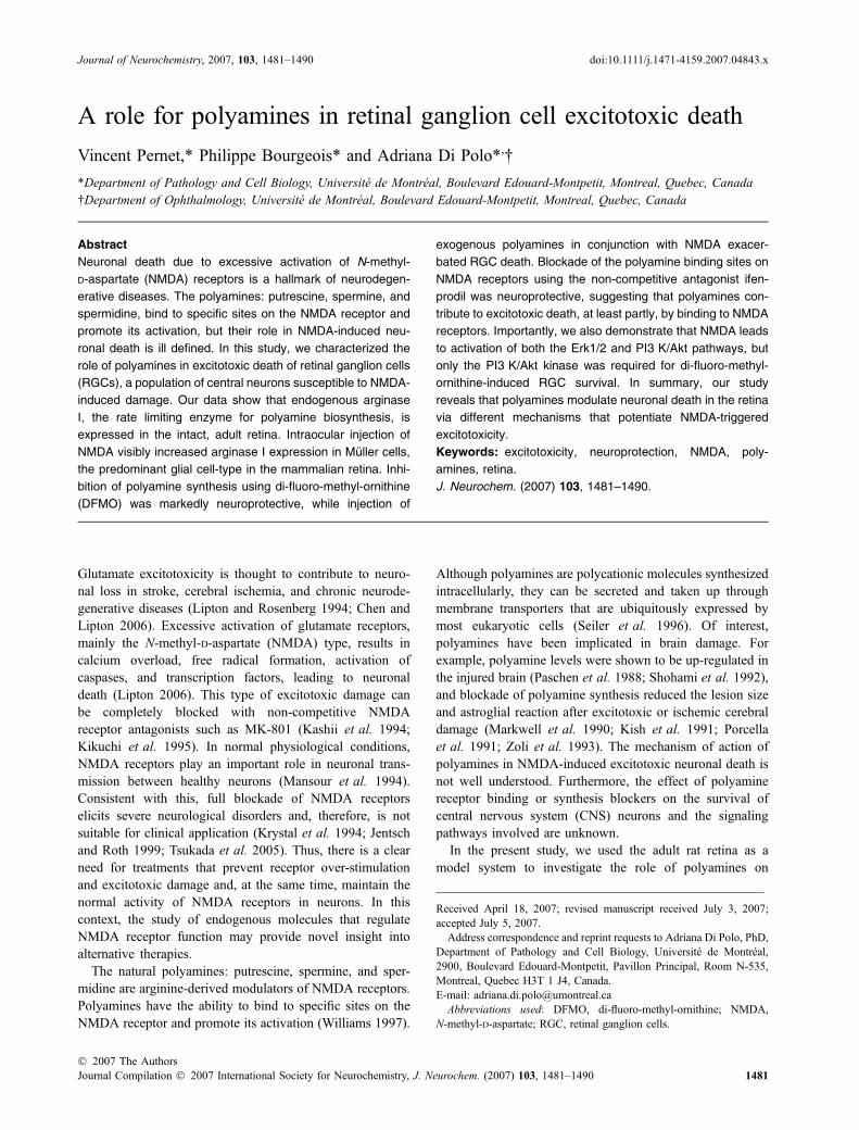
A role for polyamines in retinal ganglion cell excitotoxic death
Vincent Pernet,* Philippe Bourgeois* and Adriana Di Polo*,�
*Department of Pathology and Cell Biology, Universite de Montreal, Boulevard Edouard-Montpetit, Montreal, Quebec, Canada
�Department of Ophthalmology, Universite de Montreal, Boulevard Edouard-Montpetit, Montreal, Quebec, Canada
Abstract
Neuronal death due to excessive activation of N-methyl-
D-aspartate (NMDA) receptors is a hallmark of neurodegen-
erative diseases. The polyamines: putrescine, spermine, and
spermidine, bind to specific sites on the NMDA receptor and
promote its activation, but their role in NMDA-induced neu-
ronal death is ill defined. In this study, we characterized the
role of polyamines in excitotoxic death of retinal ganglion cells
(RGCs), a population of central neurons susceptible to NMDA-
induced damage. Our data show that endogenous arginase
I, the rate limiting enzyme for polyamine biosynthesis, is
expressed in the intact, adult retina. Intraocular injection of
NMDA visibly increased arginase I expression in Muller cells,
the predominant glial cell-type in the mammalian retina. Inhi-
bition of polyamine synthesis using di-fluoro-methyl-ornithine
(DFMO) was markedly neuroprotective, while injection of
exogenous polyamines in conjunction with NMDA exacer-
bated RGC death. Blockade of the polyamine binding sites on
NMDA receptors using the non-competitive antagonist ifen-
prodil was neuroprotective, suggesting that polyamines con-
tribute to excitotoxic death, at least partly, by binding to NMDA
receptors. Importantly, we also demonstrate that NMDA leads
to activation of both the Erk1/2 and PI3 K/Akt pathways, but
only the PI3 K/Akt kinase was required for di-fluoro-methyl-
ornithine-induced RGC survival. In summary, our study
reveals that polyamines modulate neuronal death in the retina
via different mechanisms that potentiate NMDA-triggered
excitotoxicity.
Keywords: excitotoxicity, neuroprotection, NMDA, poly-
amines, retina.
J. Neurochem. (2007) 103, 1481–1490.
Glutamate excitotoxicity is thought to contribute to neuro-nal loss in stroke, cerebral ischemia, and chronic neurode-generative diseases (Lipton and Rosenberg 1994; Chen andLipton 2006). Excessive activation of glutamate receptors,mainly the N-methyl-D-aspartate (NMDA) type, results incalcium overload, free radical formation, activation ofcaspases, and transcription factors, leading to neuronaldeath (Lipton 2006). This type of excitotoxic damage canbe completely blocked with non-competitive NMDAreceptor antagonists such as MK-801 (Kashii et al. 1994;Kikuchi et al. 1995). In normal physiological conditions,NMDA receptors play an important role in neuronal trans-mission between healthy neurons (Mansour et al. 1994).Consistent with this, full blockade of NMDA receptorselicits severe neurological disorders and, therefore, is notsuitable for clinical application (Krystal et al. 1994; Jentschand Roth 1999; Tsukada et al. 2005). Thus, there is a clearneed for treatments that prevent receptor over-stimulationand excitotoxic damage and, at the same time, maintain thenormal activity of NMDA receptors in neurons. In thiscontext, the study of endogenous molecules that regulateNMDA receptor function may provide novel insight intoalternative therapies.
The natural polyamines: putrescine, spermine, and sper-midine are arginine-derived modulators of NMDA receptors.Polyamines have the ability to bind to specific sites on theNMDA receptor and promote its activation (Williams 1997).
Although polyamines are polycationic molecules synthesizedintracellularly, they can be secreted and taken up throughmembrane transporters that are ubiquitously expressed bymost eukaryotic cells (Seiler et al. 1996). Of interest,polyamines have been implicated in brain damage. Forexample, polyamine levels were shown to be up-regulated inthe injured brain (Paschen et al. 1988; Shohami et al. 1992),and blockade of polyamine synthesis reduced the lesion sizeand astroglial reaction after excitotoxic or ischemic cerebraldamage (Markwell et al. 1990; Kish et al. 1991; Porcellaet al. 1991; Zoli et al. 1993). The mechanism of action ofpolyamines in NMDA-induced excitotoxic neuronal death isnot well understood. Furthermore, the effect of polyaminereceptor binding or synthesis blockers on the survival ofcentral nervous system (CNS) neurons and the signalingpathways involved are unknown.
In the present study, we used the adult rat retina as amodel system to investigate the role of polyamines on
Received April 18, 2007; revised manuscript received July 3, 2007;accepted July 5, 2007.Address correspondence and reprint requests to Adriana Di Polo, PhD,
Department of Pathology and Cell Biology, Universite de Montreal,2900, Boulevard Edouard-Montpetit, Pavillon Principal, Room N-535,Montreal, Quebec H3T 1 J4, Canada.E-mail: [email protected] used: DFMO, di-fluoro-methyl-ornithine; NMDA,
N-methyl-D-aspartate; RGC, retinal ganglion cells.
Journal of Neurochemistry, 2007, 103, 1481–1490 doi:10.1111/j.1471-4159.2007.04843.x
� 2007 The AuthorsJournal Compilation � 2007 International Society for Neurochemistry, J. Neurochem. (2007) 103, 1481–1490 1481

NMDA-induced excitotoxicity of central neurons in vivo. Wefocused on retinal ganglion cells (RGCs) because theseneurons express NMDA receptors (Guenther et al. 2004;Lamas et al. 2005; Zhang and Diamond 2006) and aresusceptible to NMDA-induced damage (Siliprandi et al.1992; Kikuchi et al. 2000). Moreover, RGCs are exposed tothe vitreous humour, the liquid that fills the eye; henceintraocular injection of pharmacological reagents can readilydiffuse to reach RGC bodies. In this study, we show thatco-administration of NMDA with polyamines increasedRGC excitotoxic death. In contrast, blockade of polyaminesynthesis with di-fluoro-methyl-ornithine (DFMO), or block-ade of the polyamine binding sites on NMDA receptors usingifenprodil, were neuroprotective. Furthermore, we demon-strate that the phosphatidylinositol 3-kinase (PI3 K)/Aktpathway, but not the extracellular signal-regulated kinases 1/2 (Erk1/2), was required for di-fluoro-methyl-ornithine(DFMO)-induced RGC survival against NMDA excitotoxic-ity. Our data reveal that polyamines are important modulatorsof excitotoxic neuronal death in the retina and, as such, mayplay a role in the pathological events associated withexcessive NMDA receptor activation in prevalent neurode-generative diseases.
Experimental methods
Surgical procedures
Animal procedures were performed in accordance with the
guidelines of the Canadian Council on Animal Care for the use of
experimental animals (http://www.ccac.ca/). All surgeries were
carried out in adult, female Sprague–Dawley rats (180–200 g)
under general anesthesia (2% Isoflurane, 0.8 L/min). RGCs were
labeled by application of the retrograde tracer FluoroGold (2%,
Fluorochrome, Englewood, CO, USA) in the superior colliculus as
described by us (Cheng et al. 2002; Sapieha et al. 2003; Pernetet al. 2005; Pernet and Di Polo 2006). One week later, animals
received a single intraocular injection of N-methyl-D-aspartate
(NMDA, 5 mmol/L, Sigma, Oakville, ON, Canada), polyamines
or inhibitors as described below. Intraocular injections were
performed by infusing each compound, or combination of com-
pounds (total injection volume: 5 lL), into the vitreous chamber of
the left eye using a 10-lL Hamilton syringe adapted with a
�32-gauge glass needle. The needle tip was inserted into the
superior hemisphere of the eye, at the level of the pars plana, at a
45� angle through the sclera into the vitreous body. This route of
administration avoided retinal detachment or injury to eye struc-
tures, including the lens and the iris, which release factors that
induce RGC regeneration and survival (Mansour-Robaey et al.1994; Leon et al. 2000). The injection was performed over a period
of 2 min and the needle was kept in place for an additional 2 min,
after which it was gently removed. Surgical glue (Indermill, Tyco
Health Care, Mansfield, MA, USA) was used to seal the site of
injection. Retinal fundus examination was performed to check the
integrity of the retinal circulation after surgery, and animals showing
signs of compromised blood supply were excluded from the study.
Treatment with polyamines and pharmacological inhibitors
The polyamines putrescine (10 mmol/L, Sigma) or spermine
(0.1 mmol/L, Calbiochem, La Jolla, CA, USA) were either injected
alone (n = 3/each polyamine) or co-injected with NMDA (NMDA +
putrescine, n = 5; NMDA + spermine, n = 3) into the vitreous
chamber, as described above. Control animals received a co-
injection of NMDA and saline (0.9% NaCl) (n = 3). Intraocular
injection of di-fluoro-methyl-ornithine (DFMO, 10 mmol/L, n = 6;
Sigma), or Ifenprodil (2 mmol/L, n = 4; Sigma) was performed
24 h before NMDA treatment. In these experiments, control animals
received an injection of saline 24 h prior to injection of NMDA
(n = 3). The NMDA receptor antagonist MK-801 (1 mmol/L, n = 3;
Sigma) was co-injected with NMDA. To test the role of Erk1/2 and
PI3 K activation on RGC survival, animals received an intravitreal
injection of the PI3 K inhibitor LY294002 (800 lmol/L, Sigma), or
the MEK1 inhibitor PD98059 (200 lmol/L, New England Biolabs,
Mississauga, ON, Canada). NMDA and DFMO were co-adminis-
tered with LY294002 (n = 3) or PD98059 (n = 4) and survival was
assessed as described below. Control animals received NMDA,
DFMO and saline (n = 3). In addition, contralateral eyes or non-
operated eyes served as intact controls (n = 15) for all of these
experiments.
Quantification of neuronal survival
Experimental animals were perfused transcardially with 4% para-
formaldehyde between 3 h and 7 days after NMDA injection. The
left retinas (treated) and the right retinas (untreated controls) were
dissected out, fixed for an additional 30 min and flat-mounted vitreal
side up on a glass slide for examination of the ganglion cell layer.
RGCs backfilled with FluoroGold were counted in 12 standard
retinal areas as previously described by us (Cheng et al. 2002;
Sapieha et al. 2003). Microglia that may have incorporated
FluoroGold after phagocytosis of dying RGCs were distinguished
by their morphology and excluded from our quantitative analyses.
Data analyses and statistics were performed using the GraphPad
Instat software (GraphPad Software Inc., San Diego, CA, USA) by
Student’s t-test or one-way analysis of variance (ANOVA) followed by
a Newman–Keuls multiple comparisons post-test.
Retinal immunohistochemistry
Animals were perfused with 4% paraformaldehyde and the eyes
were rapidly dissected out. The cornea and the lens were removed,
and the eye cup was incubated in the same fixative for 2 h at 4�C.Tissue was further incubated in 30% sucrose overnight, and then
frozen in optimal cutting temperature (O.C.T.) compound (Tissue-
Tek, Miles Laboratories, Elkhart, IN, USA). Retinal cryosections
(16 lm) were collected onto gelatin-coated slides and primary
antibody incubation was performed in 5% bovine serum albumin,
3% normal serum, 0.3% Triton X-100 overnight at 4�C. Sectionswere then incubated with the appropriate secondary antibody for 1 h
at 23�C, and mounted with anti-fade reagent (SlowFade, Molecular
Probes, Eugene, OR, USA). Primary antibodies were: arginase I
(0.25 lg/mL, BD Biosciences, Mississauga, ON, Canada) and
cellular retinaldehyde-binding protein (CRALBP, 1 : 1000, gift
from Dr J. C. Saari, University of Washington, Seattle, WA, USA).
Fluorescent staining was examined using a Zeiss Axioskop 2 Plus
microscope (Carl Zeiss Canada, Kirkland, QC, Canada) and pictures
captured with a CCD camera (Retiga, Qimaging, Burnaby, BC,
1482 V. Pernet et al.
Journal Compilation � 2007 International Society for Neurochemistry, J. Neurochem. (2007) 103, 1481–1490� 2007 The Authors

Canada) and analyzed with Northern Eclipse software (Empix
Imaging, Mississauga, ON, Canada).
Western blot analysis
Retinas were quickly extracted and homogenized with an electric
pestle (Kontes, Vineland, NJ, USA) in lysis buffer: 20 mmol/L Tris,
pH 8.0, 135 mmol/L NaCl, 1% NP-40, 0.1% sodium dodecyl sulfate
and 10% glycerol supplemented with protease inhibitors at 4�C.Retinal lysates were incubated for 30 min on ice prior to centrifu-
gation at 18 000 g for 5 min and the supernatant was collected. The
protein concentration of retinal extracts was determined by the Lowry
method (Bio-Rad Life Science, Mississauga, ON, Canada). Samples
(100 lg of protein) were separated by electrophoresis on 10% sodium
dodecyl sulfate–polyacrylamide gels and transferred to nitrocellulose
membranes (Bio-Rad Life Science). The following antibodies were
incubated with the blots: phospho-Erk1/2 (10 lg/mL), total Erk1/2
(2 lg/mL, Invitrogen Canada Inc., Burlington, ON, Canada),
phospho-Akt (0.6 lg/mL, Cell Signaling, Beverly, MA, USA), or
total Akt (0.1 lg/mL, Cell Signaling), followed by incubation with
anti-mouse or anti-rabbit peroxidase-linked secondary antibody
(0.5 lg/mL, Amersham Pharmacia, Baie d’Urfe, QC, Canada).
Protein signals were detected using a chemiluminescence reagent
(ECL, Amersham Biosciences) followed by exposure of blots to X-
OMAT (Kodak) imaging films. Densitometric analysis was per-
formed using BioDoc Analyze 1.0 software (Biometra, Gottingen,
Germany) on scanned autoradiographic films obtained from a series
of three independent western blots each carried out using retinal
samples from distinct experimental groups. The densitometric values
obtained for the phosphorylated (active) proteins were normalized
with respect to their loading controls (non-phosphorylated proteins)
in the same blot to obtain the final phosphorylated/total protein ratios.
These relative densitometric values were expressed as % of values in
intact, non-treated retinas.
Results
NMDA promotes rapid RGC death in vivo
First, we established the time-course of RGC loss afterintraocular injection of NMDA in vivo. For this purpose,RGCs were retrogradely labeled with FluoroGold and, oneweek later, a single intravitreal injection of NMDA(5 mmol/L = 25 nmoles) was performed. The volume ofthe vitreous fluid in the adult rat eye has been estimated tobe 56 ± 2 lL (Berkowitz et al. 1998), thus the finalintravitreal concentration of NMDA used here was�450 lmol/L. The time-course of RGC death was quanti-tatively analyzed by counting surviving RGCs (FluoroGold-positive) in whole-mounted retinas. Neuronal death was firstdetected at 6 h after NMDA administration, and markedlyincreased between 6 and 12 h post-injection (Fig. 1). Thevast majority of RGCs (�78%) were lost by 3 days afterinjection of NMDA, however, cell death did not increasefurther at later times suggesting a stabilization of neuronalloss. Based on this, we chose to carry out the analysis ofRGC survival at 3 days after NMDA injection. The cell
death kinetic that we report here following exogenousadministration of NMDA is consistent with a previousreport (Manabe and Lipton 2003).
RGCs and Muller glia express arginase I in
NMDA-treated retinas
Changes in the expression of arginase I, the rate limitingenzyme for polyamine synthesis, have been well correlatedwith variable polyamine levels in neurons (Cai et al. 2002).Thus, we examined the endogenous expression of arginase Iin intact and NMDA-treated retinas by immunohistochem-istry. In control retinas, injected with saline, arginase I wasdetected in the inner nuclear and ganglion cell layers(Fig. 2a). FluoroGold-positive cells in the ganglion cell layer(Fig. 2d) were found to contain arginase I labeling (Fig. 2e),indicating that this enzyme is expressed by adult, rat RGCsin vivo (Fig. 2f). This finding is consistent with a previousstudy showing immunodetection of polyamines in develop-ing, rabbit RGCs (Withrow et al. 2002). As early as 6 hafter NMDA injection, there was a visible reduction of theinner retina thickness caused by NMDA-induced neuronalloss (Fig. 2b). The reduction of the inner nuclear layer islikely due to loss of amacrine cells, which have beensuggested to precede that of RGCs following NMDAtreatment (Ullian et al. 2004). Interestingly, at 24 h afterNMDA injection, arginase I signal was stronger in the inner
Fig. 1 Time course of NMDA-induced RGC death in vivo. RGC death
was induced by a single intravitreal injection of NMDA (5 mmol/L, 5 lL
total volume). The density of surviving RGCs at different times after
NMDA administration was established by counting FluoroGold-labeled
neurons on flat-mounted retinas (n = 4–6 per time point). Neuronal
death was first detected at 6 h after NMDA administration, and in-
creased dramatically between 6 and 12 h post-injection. The vast
majority of RGCs were lost by 3 days after injection of NMDA. Neu-
ronal death did not increase further at later times. Data is presented
as the number of RGCs per mm2 of retina (mean ± SEM) and as %
of neuronal survival with respect to intact, control retinas (2180 ±
64 RGCs/mm2, n = 15).
Polyamines regulate excitotoxic death of retinal neurons 1483
� 2007 The AuthorsJournal Compilation � 2007 International Society for Neurochemistry, J. Neurochem. (2007) 103, 1481–1490

nuclear layer and clearly appeared in radial processescharacteristic of Muller cells (Fig. 2c). To confirm thatMuller cells up-regulated arginase I during excitotoxicdamage, we used an antibody against cellular retinalde-hyde-binding protein (CRALBP), a Muller specific marker.The superimposition of CRALBP and arginase I immuno-staining (Fig. 2g–i) revealed that Muller cell bodies andprocesses expressed this enzyme following intraocularNMDA injection. Taken together, these data demonstratethat endogenous arginase I expression is increased in Mullerglia in response to excitotoxic damage.
Polyamines potentiate NMDA-induced RGC death
To study the role of polyamines in NMDA-induced RGCdeath, we performed two sets of experiments: one in whichthe endogenous synthesis of polyamines was inhibited, andanother in which polyamines were administered into the eye.The polyamine biosynthetic pathway begins when the aminoacid arginine is transformed by the rate-limiting enzyme
arginase I into ornithine and urea. The enzyme ornithinedecarboxylase then converts ornithine into putrescine, whichis the primary substrate for the synthesis of spermidine andspermine. To study the role of endogenous polyamines onexcitotoxic neuronal death, we first carried out experimentsin which animals received an intraocular injection of theornithine decarboxylase inhibitor DFMO (10 mmol/L)administered 24 h prior to NMDA injection. At this dose,DFMO was previously shown to effectively inhibit ornithinedecarboxylase and polyamine synthesis in vitro and was nottoxic for neurons (Markwell et al. 1990; Trout et al. 1993;Ientile et al. 1999; Withrow et al. 2002). Retinas wereexamined histologically at 3 days after NMDA injection todetermine the density of surviving RGCs. Treatment withDFMO markedly increased the number of RGCs thatremained alive following excitotoxic damage, and the overallmorphology of these neurons was better preserved in thepresence of this inhibitor than in control retinas treated withNMDA and saline (Fig. 3a and b).
(a) (b) (c)
(d)
(g)
(e) (f)
(h) (i)
Fig. 2 Arginase I is up-regulated in Muller glial cells following NMDA
injection. Arginase I immunohistochemistry revealed the presence of
this enzyme in the inner layers of the intact (non-injured) retina, par-
ticularly in the ganglion cell layer (GCL) and fiber layer (FL) (a).
Arginase I staining did not change early after NMDA injection (6 h) (b),
but appeared stronger in the inner nuclear layer (INL) and in pre-
sumptive Muller cell radial processes at 24 h post-injection (c). RGCs,
identified following retrograde labeling with FluoroGold (FG) (d),
express arginase I in the intact retina as demonstrated by the
superimposition of arginase I (e) and FG staining (f). Higher magnifi-
cation inset in 2f shows arginase I-positive RGCs. Muller glial cells,
identified by the cell-specific marker cellular retinaldehyde binding
protein (CRALBP) (g), display robust arginase I expression in their
somas and processes following NMDA administration (h and i). PS:
photoreceptor segments; ONL: outer nuclear layer; OPL: outer plexi-
form layer; INL: inner nuclear layer; IPL: inner plexiform layer; GCL:
ganglion cell layer; FL: fiber layer. Scale bar = 50 lm.
1484 V. Pernet et al.
Journal Compilation � 2007 International Society for Neurochemistry, J. Neurochem. (2007) 103, 1481–1490� 2007 The Authors

The effect of exogenously applied polyamines on RGCdeath was examined by co-injecting either putrescine orspermine with NMDA. Putrescine, at the concentration usedhere (10 mmol/L), was previously shown to stimulate Ca2+
influx in cultured retinal cells in the presence of NMDA andwas not toxic by itself (Trout et al. 1993). Spermine wasadministered at a much lower concentration (0.1 mmol/L)than putrescine because of its higher affinity for theextracellular polyamine binding site on NMDA receptors(Williams 1997; Sharif and Xu 1999). Examination of flat-mounted retinas, demonstrated that neuronal survival wasreduced when putrescine (Fig. 3c) or spermine (Fig. 3d)were co-injected with NMDA compared to control retinastreated with NMDA and saline. Quantitative analysis of RGCdensities confirmed that DFMO protected RGCs fromNMDA-induced death, increasing RGC survival by 105%(905 ± 103 RGCs/mm2, mean ± SEM) (Fig. 4a). In contrast,putrescine or spermine worsened the cytotoxic effect ofNMDA, decreasing RGC survival by 29% (347 ± 39 RGCs/mm2) or 20% (388 ± 48 RGCs/mm2), respectively, relativeto control eyes (Fig. 4b). We found that while the effect ofNMDA combined with putrescine was significantly differentfrom that of NMDA (Student’s t-test, p < 0.05), the effect ofNMDA and spermine was not (p > 0.05). The effect ofexogenous polyamines on NMDA-induced RGC death maybe limited by the high levels of endogenous polyamines
present in the normal retina. This idea is supported by ourfindings that inhibition of endogenous polyamine synthesiswith DFMO is markedly neuroprotective (Student’s t-test,p < 0.01). Intraocular injection of polyamines by themselvesdid not affect RGC survival (Fig. 4c), confirming that theyare not toxic in the absence of NMDA. Collectively, theseresults indicate that polyamines exacerbate NMDA-inducedRGC excitotoxicity in vivo.
Polyamines enhance neuronal death by binding to
NMDA receptors
To test the hypothesis that polyamines potentiate excitotox-icity by binding to specific sites on the NMDA receptor, wecarried out experiments using ifenprodil (2 mmol/L), a non-competitive NMDA receptor antagonist that is highly
NMDA + Putrescine NMDA + Spermine
NMDA + Saline NMDA + DFMO(a) (b)
(c) (d)
Fig. 3 Polyamines potentiate NMDA-induced RGC death. The role of
polyamines was investigated by pre-treating animals with the ornithine
decarboxylase inhibitor, DFMO, or by co-injecting putrescine or
spermine with NMDA. RGC survival was examined on whole-mounted
retinas at 3 days after NMDA injection. Treatment with DFMO (b,
n = 6) markedly increased the number of RGCs that remained alive
following excitotoxic damage. The overall morphology of RGCs was
better preserved in the presence of DFMO than in control retinas
treated with saline (a, n = 3). In contrast, neuronal death was in-
creased when putrescine (c, n = 5) or spermine (d, n = 3) were co-
administered with NMDA. Scale bar = 50 lm.
(a) (b)
(c)
Fig. 4 Quantitative analysis of RGC densities. (a) Intraocular injection
of the polyamine synthesis inhibitor DFMO increased RGC survival by
105% following NMDA treatment (n = 6) compared to control retinas
treated with saline and NMDA (n = 3) (Student’s t-test, *p < 0.01). (b) In
contrast, putrescine (n = 5) or spermine (n = 3) worsened the cytotoxic
effect of NMDA, decreasing RGC survival by 29% and 20%, respec-
tively, relative to saline-treated control eyes (n = 3). (c) Intraocular
injection of polyamines by themselves did not affect RGC survival
(putrescine, n = 3; spermine, n = 3) with respect to intact, non-treated
retinas (n = 15).
Polyamines regulate excitotoxic death of retinal neurons 1485
� 2007 The AuthorsJournal Compilation � 2007 International Society for Neurochemistry, J. Neurochem. (2007) 103, 1481–1490

selective for the polyamine binding sites on NR2B subunits(Perin-Dureau et al. 2002). Our data show that intraocularinjection of NMDA and ifenprodil increased RGC survivalby 59% (703 ± 47 RGCs/mm2) compared to control retinastreated with NMDA (487 ± 4 RGCs/mm2) and saline(441 ± 40 RGCs/mm2) (Fig. 5). To better characterize theeffect of ifenprodil on RGC neuroprotection, we compared itwith the effect of DFMO or MK-801 (1 mmol/L), a non-competitive, complete blocker of NMDA receptors (Sunet al. 2001). The neuroprotective effect of ifenprodil wasvisibly less, but not significantly different (Student’s t-test,p = 0.168), from that observed when endogenous polyaminesynthesis was inhibited with DFMO. As expected, MK-801exerted a strong neuroprotective effect, enhancing RGCsurvival by 2.2-fold (1,575 ± 0.6 RGCs/mm2) compared toifenprodil-treated retinas (703 ± 47 RGCs/mm2). Collec-tively, these results indicate that polyamines exacerbate theexcitotoxic effect of NMDA, at least partly, by binding toNMDA receptors.
DFMO-induced survival depends on activation of the
PI3 K/Akt pathway
Our data show that blocking polyamine synthesis with DFMOconfers robust protection against NMDA-induced RGC death.Next, we sought to elucidate the intracellular signalingpathways involved in NMDA excitotoxicity and DFMO
neuroprotection in the retina. Previous studies, including ours,have demonstrated that activation of the Erk1/2 and/or PI3 K/Akt pathways are required for RGC survival induced by avariety of factors (Cheng et al. 2002; Isenmann et al. 2003;Park et al. 2004; Pernet et al. 2005; Kilic et al. 2006).Therefore, we examined the stimulation of these signalingcomponents by western blot analysis using antibodies thatspecifically recognized the phosphorylated forms of Erk1/2 orAkt kinases. Retinal samples obtained at 6 h after NMDAinjection, with or without DFMO, were analyzed. Treatmentwith NMDA alone, or combination of NMDA and DFMO, ledto strong up-regulation of phospho-Erk1/2 representing a6.5-fold or 6-fold increase, respectively, relative to intact orsaline-treated control retinas (Fig. 6a). Phospho-Akt wassimilarly up-regulated by NMDA or the combination ofNMDA and DFMO at 4.7-fold or 4.5-fold, respectively,compared with control samples (Fig. 6b). Intraocular injectionof DFMO by itself did not activate Erk1/2 or Akt (data notshown). Our data is consistent with previous studies showingthat NMDA stimulates Erk1/2 and Akt in the rat retina in vivo(Manabe and Lipton 2003; Munemasa et al. 2005). Further-more, we demonstrate that NMDA is a potent activator ofErk1/2 andAkt prior toRGCdeath (6 h afterNMDA injection)and independently of polyamine inhibition with DFMO.
To determine whether activation of Erk1/2 or Akt wasrequired for DFMO-induced neuronal survival, we usedselective pharmacological inhibitors for components of eachof these pathways that were injected 1 h prior to NMDA.PD98059 selectively inhibits MEK (Dudley et al. 1995), theobligate upstream activator of Erk1/2, while LY294002 is aselective inhibitor of PI3 K (Weber et al. 1997), an upstreamactivator of Akt. We have previously shown that, at the dosesused here, PD98059 (200 lmol/L)or LY294002 (800 lmol/L)specifically inhibited Erk1/2 or Akt activation, respectively, inthe rat retina without any cytotoxic effects in vivo (Cheng et al.2002; Sapieha et al. 2006). In our present study, quantitativeanalysis of RGC densities demonstrated that administration ofLY294002 resulted in almost complete inhibition of thesurvival effect produced by DFMO. In this situation, RGCdensitieswere significantly reducedcompared to retinas treatedwith DFMO (ANOVA, p < 0.05) to levels similar to those foundin control retinas treated onlywithNMDAand saline (Fig. 6c).In contrast, blockade of Erk1/2 activation with PD98059 didnot significantly change the robust neuroprotective effect ofDFMO (ANOVA, p > 0.05). These results demonstrate thatwhile the PI3 K pathway is essential for DFMO-mediatedsurvival ofRGCs in vivo, the Erk1/2 pathway has no role in thiseffect.
Discussion
In this study, we tested the hypothesis that polyaminesregulate NMDA-induced excitotoxicity of adult RGCsin vivo. Our data demonstrate that inhibition of endogenous
Fig. 5 Potentiation of NMDA toxicity by endogenous polyamines is
mediated by receptor facilitation. Injection of ifenprodil (n = 4), a non-
competitive NMDA receptor antagonist that is highly selective for the
polyamine binding sites on the NR2B subunits, enhanced RGC sur-
vival by 59% with respect to saline-treated controls (n = 3) (Student’s
t-test *p < 0.05). The neuroprotective effect of ifenprodil was less, but
not significantly different (p > 0.05), than that observed with DFMO
(n = 6). As expected, the full NMDA receptor blocker MK-801 pro-
moted robust RGC survival (n = 3).
1486 V. Pernet et al.
Journal Compilation � 2007 International Society for Neurochemistry, J. Neurochem. (2007) 103, 1481–1490� 2007 The Authors

polyamine synthesis with DFMO markedly protected RGCsfrom excitotoxic damage, while exogenous administration ofpolyamines increased NMDA-induced neuronal death.Putrescine or spermine by themselves did not affect RGCsurvival, but worsened RGC death only when co-adminis-tered with NMDA. This is consistent with a previous studyshowing that polyamines are not toxic for embryonic retinalcells (Ientile et al. 1999). Therefore, polyamines appear to bemodulators of NMDA excitotoxicity rather than inducers ofneuronal death.
Polyamine levels in neurons have been shown to correlatewell with the expression of arginase I (Cai et al. 2002), therate limiting enzyme for polyamine synthesis. Our immuno-
cytochemical analysis of retinal arginase I demonstrated thatthis enzyme is abundantly expressed by adult RGCs,suggesting that polyamines are endogenously produced inthese neurons. Previous studies showed marked elevation ofpolyamine levels in the brain following ischemia or contu-sion injury (Kish et al. 1991; Shohami et al. 1992; Zoliet al. 1993). Of interest, our results show that Muller cells,the predominant glial cell-type in the retina, markedlyup-regulated arginase I after NMDA administration. Thisfinding suggests that Muller cells synthesize polyaminesin vivo and is consistent with a previous report that detectedspermine and spermidine in cultured Muller glia (Bieder-mann et al. 1998). Polyamines are polycationic molecules
(a) (b)
(c)
Fig. 6 DFMO-induced survival requires Akt
activation. Activation of Erk1/2 (a) or Akt (b)
was examined by western blot analysis of
retinal samples using antibodies that spe-
cifically recognized the phosphorylated
forms of Erk1/2 or Akt kinases. Treatment
with NMDA alone, or combination of NMDA
and DFMO, led to strong up-regulation of
phospho-Erk1/2 relative to intact or saline-
treated control retinas (a, n = 3). Phospho-
Akt was similarly up-regulated by NMDA or
the combination of NMDA and DFMO
compared with control samples (b, n = 3).
Bottom panels in (a) and (b) show the same
blots in the corresponding upper panels
probed with an antibody against total Erk1/2
or total Akt, respectively. (c) The density of
RGCs that survived following NMDA and
DFMO treatment (n = 6) was significantly
reduced by specific inhibition of the PI3 K/
Akt pathway with a single intraocular injec-
tion of LY294002 (800 lmol/L) (n = 3,
ANOVA Newman–Keuls post-test, *p < 0.05)
to a level similar to that observed in saline-
treated controls. In contrast, neuronal sur-
vival was not significantly changed by
injection of PD98059 (200 lmol/L) (n = 4,
ANOVA Newman–Keuls post-test, p > 0.05).
Polyamines regulate excitotoxic death of retinal neurons 1487
� 2007 The AuthorsJournal Compilation � 2007 International Society for Neurochemistry, J. Neurochem. (2007) 103, 1481–1490

that can be secreted and, once in the extracellular milieu, theycan bind to specific sites on NMDA receptors or are taken upthrough membrane transporters ubiquitously present in cells(Seiler et al. 1996). Thus, release of polyamines by Mullercells may influence RGC death during excitotoxicity. DFMOis a potent inhibitor of ornithine decarboxylase that effec-tively blocks the transformation of ornithine into putrescine;therefore the marked neuroprotective effect of DFMO maybe, at least partly, due to polyamine synthesis inhibition inMuller glia. Of interest, despite robust up-regulation ofarginase I in Muller glia, these cells were invulnerable toNMDA excitotoxicity. A possible explanation is that thedifferential composition of NMDA receptor subunitsbetween Muller cells and retinal neurons (Lamas et al.2005) influences the degree of NMDA receptor activationand the signaling pathways responsible for cell death.
Due to the rapid time-course of RGC and amacrine celldeath after NMDA treatment, we could not detect a clearincrease of arginase I expression in these neurons. However,at present we cannot rule out the possibility that this enzymeis also up-regulated within RGCs and displaced amacrinecells. It has been proposed that while low doses of NMDApromote apoptotic neuronal death, exposure to high concen-trations of NMDA leads to necrosis (Bonfoco et al. 1995).Previous studies have demonstrated the presence of apopto-tic, TUNEL-positive retinal cells following excitotoxicdamage using concentrations of NMDA in the same rangeas that used in our study (25 nmoles) (Manabe and Lipton2003; Nakazawa et al. 2005). However, the rapid pattern ofRGC death and the absence of active microglia reported heresuggest that necrosis may also contribute to neuronal loss inthis model. Of interest, necrotic cell death cannot be rescuedusing anti-apoptotic factors. For example, the potent anti-apoptotic agent brain-derived neurotrophic factor exerts littleneuroprotection of RGCs, if any, following NMDA-induceddamage (Kido et al. 2000; Pernet and Di Polo, unpublisheddata).
To elucidate the mechanism of action of polyamines inNMDA damage, we tested the effect of ifenprodil, a selectiveantagonist of polyamine binding sites on NMDA receptors.We found that ifenprodil significantly increased RGCsurvival after NMDA injection, indicating that polyaminespotentiate excitotoxicity by binding to NMDA receptors.However, the neuroprotective effect promoted by ifenprodilwas significantly less than that produced by MK-801. Aprevious study showed that ifenprodil was equally effectiveas MK-801 in protecting embryonic retinal cells in vitroexposed to glutamate (Zhang et al. 2000), suggesting thatexcitotoxicity was mostly mediated by polyamine binding toNMDA receptors. It has also been reported that the extent ofNMDA damage is more severe in adult than in immatureretinas (Izumi et al. 1995). It is tempting to speculate that thedifferences observed between immature and adult retinasoccurs as a result of the developmental changes in the
expression of NR2B subunits, which contain the high affinitybinding sites for polyamines and ifenprodil. This is unlikely,however, because the expression of NR2B subunits in RGCsdoes not change during postnatal development, and ifenpro-dil has been shown to block NMDA receptor ionic currentswith similar efficacy in both developing and adult RGCs(Guenther et al. 2004). Collectively, these results suggestthat polyamine binding to NMDA receptors may play a lessprominent role in the modulation of excitotoxic death in theadult retina compared to the developing retina. Our data alsosupport the idea that, in addition to binding to NMDAreceptors, polyamines may exacerbate neuronal toxicity byother molecular mechanisms.
To further explore how endogenous polyamines regulateneuronal death, we examined the activity of two intracellularsignaling components known to mediate RGC survivalinduced by different stimuli: the Erk1/2 and the PI3 K/Aktkinases (Cheng et al. 2002; Isenmann et al. 2003; Park et al.2004; Pernet et al. 2005; Kilic et al. 2006). Our datademonstrate that although both Erk1/2 and PI3 K/Aktpathways were activated following NMDA and DFMOadministration, only selective inhibition of PI3 K/Aktblocked DFMO-induced survival while Erk1/2 inhibitionhad no effect. This finding suggests that the PI3 K/Aktpathway, but not the Erk1/2 pathway, is necessary forDFMO-induced RGC survival during excitotoxic damage.Our data, and that of others, show that NMDA by itself canstimulate the PI3 K/Akt pathway (Manabe and Lipton 2003).In spite of this endogenous activation, massive RGC lossoccurs during NMDA damage indicating that the pro-survival effect of PI3 K/Akt is severely limited. Our resultsdemonstrate that only when polyamine synthesis is inhibitedwith DFMO, can PI3 K/Akt exert a neuroprotective role, aneffect that is fully inhibited by the selective PI3 K inhibitorLY294002. These data suggest that polyamines exert anegative regulatory role on the PI3 K/Akt pathway that isremoved by DFMO.
There may be additional levels of regulation by whichendogenous polyamines modulate the excitotoxic cascadeinitiated by NMDA. For example, nitric oxide release is acentral mediator of NMDA excitotoxicity in the embryonicand adult nervous system (Choi and Lipton 2000; Nelsonet al. 2003). In the retina, inhibition of nitric oxide synthe-tase using L-NAME effectively protected inner retinal cellsfrom NMDA-induced death (Morizane et al. 1997). Ofinterest, depletion of intracellular polyamines with DFMOhas been shown to prevent nitric oxide synthesis followingNMDA administration in embryonic chick retinal cellsin vitro (Ientile et al. 1999). Therefore, polyamines mayregulate the intracellular production of nitric oxide in adultRGCs to influence their survival.
In summary, we showed that endogenously producedpolyamines modulate NMDA-induced RGC death in vivo.NMDA increased the expression of arginase I, a critical
1488 V. Pernet et al.
Journal Compilation � 2007 International Society for Neurochemistry, J. Neurochem. (2007) 103, 1481–1490� 2007 The Authors

enzyme for polyamine synthesis, in Muller glia suggestingthat polyamine secretion by these cells may contribute toRGC excitotoxicity. Importantly, we demonstrate that therole of polyamines in the control of NMDA-inducedneuronal death is not restricted to the regulation of NMDAreceptor activity by binding to specific receptor sites. Instead,our data indicate that polyamines also influence excitotoxicdamage via complex molecular interactions including inhi-bition of the pro-survival PI3 K/Akt pathway. These mech-anisms are likely to cooperate in the potentiation ofexcitotoxic death and may have implications in neuropatho-logical processes including retinal ischemia and glaucoma.
Acknowledgments
This work was supported by grants to ADP from the Canadian
Institutes of Health Research (CIHR). We thank Drs Timothy
Kennedy and Johanne Bertrand for comments on the manuscript.
ADP is a scholar of Fonds de recherche en sante du Quebec(FRSQ).
References
Berkowitz B. A., Lukaszew R. A., Mullins C. M. and Penn J. S. (1998)Impaired hyaloidal circulation function and uncoordinated oculargrowth patterns in experimental retinopathy of prematurity. Invest.Ophthalmol. Vis. Sci. 39, 391–396.
Biedermann B., Skatchkov S. N., Brunk I., Bringmann A., Pannicke T.,Bernstein H. G., Faude F., Germer A., Veh R. and Reichenbach A.(1998) Spermine/spermidine is expressed by retinal glial (Muller)cells and controls distinct K+ channels of their membrane. Glia 23,209–220.
Bonfoco E., Krainc D., Ankarcrona M., Nicotera P. and Lipton S. A.(1995) Apoptosis and necrosis: two distinct events induced,respectively, by mild and intense insults with N-methyl-D-aspartateor nitric oxide/superoxide in cortical cell cultures. Proc. Natl Acad.Sci. USA 92, 7162–7166.
Cai D., Deng K., Mellado W., Lee J., Ratan R. R. and Filbin M. T.(2002) Arginase I and polyamines act downstream from cyclicAMP in overcoming inhibition of axonal growth MAG and myelinin vitro. Neuron 35, 711–719.
Chen H. S. and Lipton S. A. (2006) The chemical biology of clinicallytolerated NMDA receptor antagonists. J. Neurochem. 97,1611–1626.
Cheng L., Sapieha P., Kittlerova P., Hauswirth W. W. and Di Polo A.(2002) TrkB gene transfer protects retinal ganglion cells from ax-otomy-induced death in vivo. J. Neurosci. 22, 3977–3986.
Choi Y. B. and Lipton S. A. (2000) Redox modulation of the NMDAreceptor. Cell. Mol. Life Sci. 57, 1535–1541.
Dudley D. T., Pang L., Decker S. J., Bridges A. J. and Saltiel A. R.(1995) A synthetic inhibitor of the mitogen-activated proteinkinase cascade. Proc. Natl Acad. Sci. USA 92, 7686–7689.
Guenther E., Schmid S., Wheeler-Schilling T., Albach G., Grunder T.,Fauser S. and Kohler K.(2004) Developmental plasticity of NMDAreceptor function in the retina and the influence of light. FASEB J.18, 1433–1435. Epub 2004 Jul 1439.
Ientile R., Pedale S., Ginoprelli T., Cannavo L. and Macaione S. (1999)Intracellular polyamine levels are involved in NMDA-evoked nitricoxide production in chick retina cells. J. Neurochem. 72, 1744–1749.
Isenmann S., Kretz A. and Cellerino A. (2003) Molecular determinantsof retinal ganglion cell development, survival, and regeneration.Prog. Retin. Eye Res. 22, 483–543.
Izumi Y., Kirby-Sharkey C. O., Benz A. M., Labruyere J., Price M. T.,Wozniak D. F., Zorumski C. F. and Olney J. W. (1995) Agedependent sensitivity of the rat retina to the excitotoxic action ofN-methyl-D-aspartate. Neurobiol. Dis. 2, 139–144.
Jentsch J. D. and Roth R. H. (1999) The neuropsychopharmacology ofphencyclidine: from NMDA receptor hypofunction to the dopa-mine hypothesis of schizophrenia. Neuropsychopharmacology 20,201–225.
Kashii S., Takahashi M., Mandai M., Shimizu H., Honda Y., Sasa M.,Ujihara H., Tamura Y., Yokota T. and Akaike A. (1994) Protectiveaction of dopamine against glutamate neurotoxicity in the retina.Invest. Ophthalmol. Vis. Sci. 35, 685–695.
Kido N., Tanihara H., Honjo M., Inatani M., Tatsuno T., Nakayama C.and Honda Y. (2000) Neuroprotective effects of brain-derivedneurotrophic factor in eyes with NMDA-induced neuronal death.Brain Res. 884, 59–67.
Kikuchi M., Kashii S., Honda Y., Ujihara H., Sasa M., Tamura Y. andAkaike A. (1995) Protective action of zinc against glutamateneurotoxicity in cultured retinal neurons. Invest. Ophthalmol. Vis.Sci. 36, 2048–2053.
Kikuchi M., Tenneti L. and Lipton S. A. (2000) Role of p38 mitogen-activated protein kinase in axotomy-induced apoptosis of rat retinalganglion cells. J. Neurosci. 20, 5037–5044.
Kilic U., Kilic E., Jarve A., Guo Z., Spudich A., Bieber K., Barzena U.,Bassetti C. L., Marti H. H. and Hermann D. M. (2006) Humanvascular endothelial growth factor protects axotomized retinalganglion cells in vivo by activating ERK-1/2 and Akt pathways.J. Neurosci. 26, 12439–12446.
Kish S. J., Wilson J. M. and Fletcher P. J. (1991) The polyamine syn-thesis inhibitor alpha-difluoromethylornithine is neuroprotectiveagainst N-methyl-D-aspartate-induced brain damage in vivo. Eur. J.Pharmacol. 209, 101–103.
Krystal J. H., Karper L. P., Seibyl J. P., Freeman G. K., Delaney R.,Bremner J. D., Heninger G. R., Bowers Jr M. B. and Charney D. S.(1994) Subanesthetic effects of the noncompetitive NMDAantagonist, ketamine, in humans. Psychotomimetic, perceptual,cognitive, and neuroendocrine responses. Arch. Gen. Psychiatry51, 199–214.
Lamas M., Lee-Rivera I. and Lopez-Colome A. M. (2005) Cell-specificexpression of N-methyl-D-aspartate receptor subunits in Muller gliaand neurons from the chick retina. Invest. Ophthalmol. Vis. Sci. 46,3570–3577.
Leon S., Yin Y., Nguyen J., Irwin N. and Benowitz L. I. (2000) Lensinjury stimulates axon regeneration in the mature rat optic nerve.J. Neurosci. 20, 4615–4626.
Lipton S. A. (2006) Paradigm shift in neuroprotection by NMDAreceptor blockade: memantine and beyond. Nature 5, 160–170.
Lipton S. A. and Rosenberg P. A. (1994) Excitatory amino acids as afinal common pathway for neurologic disorders. N. Engl. J. Med.330, 613–622.
Manabe S. and Lipton S. A. (2003) Divergent NMDA signals leading toproapoptotic and antiapoptotic pathways in the rat retina. Invest.Ophthalmol. Vis. Sci. 44, 385–392.
Mansour S. J., Resing K. A., Candi J. M., Hermann A. S., Gloor J. W.,Herskind K. R., Wartmann M., Davis R. J. and Ahn N. G. (1994)Mitogen-activated protein (MAP) kinase phosphorylation of MAPkinase kinase: determination of phosphorylation sites by massspectrometry and site-directed mutagenesis. J. Biochem. (Tokyo)116, 304–314.
Mansour-Robaey S., Clarke D. B., Wang Y.-C., Bray G. M. and AguayoA. J. (1994) Effects of ocular injury and administration of brain-
Polyamines regulate excitotoxic death of retinal neurons 1489
� 2007 The AuthorsJournal Compilation � 2007 International Society for Neurochemistry, J. Neurochem. (2007) 103, 1481–1490

derived neurotrophic factor on survival and regrowth of axotomizedretinal ganglion cells. Proc. Natl Acad. Sci. USA 91, 1632–1636.
Markwell M. A., Berger S. P. and Paul S. M. (1990) The polyaminesynthesis inhibitor alpha-difluoromethylornithine blocks NMDA-induced neurotoxicity. Eur. J. Pharmacol. 182, 607–609.
Morizane C., Adachi K., Furutani I., Fujita Y., Akaike A., Kashii S. andHonda Y. (1997) N(omega)-nitro-L-arginine methyl ester protectsretinal neurons against N-methyl-D-aspartate-induced neurotoxicityin vivo. Eur. J. Pharmacol. 328, 45–49.
Munemasa Y., Ohtani-Kaneko R., Kitaoka Y. et al.(2005) Contribution ofmitogen-activated protein kinases to NMDA-induced neurotoxicityin the rat retina. Brain Res. 1044, 227–240. Epub 2005 Apr 2019.
Nakazawa T., Shimura M., Endo S., Takahashi H., Mori N. and TamaiM. (2005) N-Methyl-D-aspartic acid suppresses Akt activitythrough protein phosphatase in retinal ganglion cells. Mol. Vis. 11,1173–1182.
Nelson E. J., Connolly J. and McArthur P. (2003) Nitric oxide and S-nitrosylation: excitotoxic and cell signaling mechanism. Biol. Cell95, 3–8.
Park K., Luo J. M., Hisheh S., Harvey A. R. and Cui Q. (2004) Cellularmechanisms associated with spontaneous and ciliary neurotrophicfactor-cAMP-induced survival and axonal regeneration of adultretinal ganglion cells. J. Neurosci. 24, 10806–10815.
Paschen W., Rohn G., Meese C. O., Djuricic B. and Schmidt-Kastner R.(1988) Polyamine metabolism in reversible cerebral ischemia:effect of alpha-difluoromethylornithine. Brain Res. 453, 9–16.
Perin-Dureau F., Rachline J., Neyton J. and Paoletti P. (2002) Mappingthe binding site of the neuroprotectant ifenprodil on NMDAreceptors. J. Neurosci. 22, 5955–5965.
Pernet V. and Di Polo A. (2006) Synergistic action of brain-derivedneurotrophic factor and lens injury promotes retinal ganglion cellsurvival, but leads to optic nerve dystrophy in vivo. Brain 17, 17.
Pernet V., Hauswirth W. W. and Di Polo A. (2005) Extracellular signal-regulated kinase 1/2 mediates survival, but not axon regeneration,of adult injured central nervous system neurons in vivo. J. Neu-rochem. 93, 72–83.
Porcella A., Fage D., Voltz C., Carter C., Scatton B. and Bartholini G.(1991) Difluoromethyl ornithine protects against the neurotoxiceffects of intrastriatally administered N-methyl-D-aspartate in vivo.Eur. J. Pharmacol. 199, 267–269.
Sapieha P. S., Peltier M., Rendahl K. G., Manning W. C. and Di Polo A.(2003) Fibroblast growth factor-2 gene delivery stimulates axongrowth by adult retinal ganglion cells after acute optic nerve injury.Mol. Cell. Neurosci. 24, 656–672.
Sapieha P. S., Hauswirth W. W. and Di Polo A. (2006) Extracellularsignal-regulated kinases 1/2 are required for adult retinal ganglioncell axon regeneration induced by fibroblast growth factor-2.J. Neurosci. Res. 21, 21.
Seiler N., Delcros J. G. and Moulinoux J. P. (1996) Polyamine transportin mammalian cells. An update. Int. J. Biochem. Cell Biol. 28,843–861.
Sharif N. A. and Xu S. X. (1999) Human retina contains polyaminesensitive [3H]-ifenprodil binding sites: implications for neuropro-tection? Br. J. Ophthalmol. 83, 236–240.
Shohami E., Nates J. L., Glantz L., Trembovler V., Shapira Y. andBachrach U. (1992) Changes in brain polyamine levels followinghead injury. Exp. Neurol. 117, 189–195.
Siliprandi R., Canella R., Carmignoto G., Schiavo N., Zanellato A.,Zanoni R. and Vantini G. (1992) N-methyl-D-aspartate-inducedneurotoxicity in the adult rat retina. Vis. Neurosci. 8, 567–573.
Sun Q., Ooi V. E. and Chan S. O. (2001) N-methyl-D-aspartate-inducedexcitotoxicity in adult rat retina is antagonized by single systemicinjection of MK-801. Exp. Brain Res. 138, 37–45.
Trout J. J., Koenig H., Goldstone A. D., Iqbal Z., Lu C. Y. and SiddiquiF. (1993) N-methyl-D-aspartate receptor excitotoxicity involvesactivation of polyamine synthesis: protection by alpha-difluorom-ethylornithine. J. Neurochem. 60, 352–355.
Tsukada H., Nishiyama S., Fukumoto D., Sato K., Kakiuchi T. andDomino E. F. (2005) Chronic NMDA antagonism impairs workingmemory, decreases extracellular dopamine, and increases D1receptor binding in prefrontal cortex of conscious monkeys.Neuropsychopharmacology 30, 1861–1869.
Ullian E. M., Barkis W. B., Chen S., Diamond J. S. and Barres B. A.(2004) Invulnerability of retinal ganglion cells to NMDA excito-toxicity. Mol. Cell. Neurosci. 26, 544–557.
Weber G., Shen F., Prajda N., Yang H., Li W., Yeh A., Csokay B., OlahE. and Look K. Y. (1997) Regulation of the signal transductionprogram by drugs. Adv. Enzyme Regul. 37, 35–55.
Williams K. (1997) Modulation and block of ion channels: a new biol-ogy of polyamines. Cell. Signal. 9, 1–13.
Withrow C., Ashraf S., O’Leary T., Johnson L. R., Fitzgerald M. E. andJohnson D. A. (2002) Effect of polyamine depletion on conephotoreceptors of the developing rabbit retina. Invest. Ophthalmol.Vis. Sci. 43, 3081–3090.
Zhang J. and Diamond J. S. (2006) Distinct perisynaptic and synapticlocalization of NMDA and AMPA receptors on ganglion cells inrat retina. J. Comp. Neurol. 498, 810–820.
Zhang S., Kashii S., Yasuyoshi H., Kikuchi M., Honda Y., Kaneda K.,Sato S. and Akaike A. (2000) Protective effects of ifenprodilagainst glutamate-induced neurotoxicity in cultured retinal neu-rons. Graefes Arch. Clin. Exp. Ophthalmol. 238, 846–852.
Zoli M., Zini I., Grimaldi R., Biagini G. and Agnati L. F. (1993) Effectsof polyamine synthesis blockade on neuronal loss and astroglialreaction after transient forebrain ischemia. Int. J. Dev. Neurosci. 11,175–187.
1490 V. Pernet et al.
Journal Compilation � 2007 International Society for Neurochemistry, J. Neurochem. (2007) 103, 1481–1490� 2007 The Authors
Copyright © 2022 FDOKUMEN