Human Immunodeficiency Virus Type 1 Nef Potently Induces Apoptosis in Primary Human Brain...
14
10.1128/JVI.79.7.4257-4269.2005. 2005, 79(7):4257. DOI: J. Virol. Pomerantz Mehrnush Kalayeh, Muhammad Mukhtar and Roger J. Edward A. Acheampong, Zahida Parveen, Lois W. Muthoga, Cells via the Activation of Caspases Human Brain Microvascular Endothelial Potently Induces Apoptosis in Primary Human Immunodeficiency Virus Type 1 Nef http://jvi.asm.org/content/79/7/4257 Updated information and services can be found at: These include: REFERENCES http://jvi.asm.org/content/79/7/4257#ref-list-1 at: This article cites 115 articles, 36 of which can be accessed free CONTENT ALERTS more» articles cite this article), Receive: RSS Feeds, eTOCs, free email alerts (when new http://journals.asm.org/site/misc/reprints.xhtml Information about commercial reprint orders: http://journals.asm.org/site/subscriptions/ To subscribe to to another ASM Journal go to: on February 3, 2014 by guest http://jvi.asm.org/ Downloaded from on February 3, 2014 by guest http://jvi.asm.org/ Downloaded from
-
Upload
independent -
Category
Documents
-
view
0 -
download
0
Transcript of Human Immunodeficiency Virus Type 1 Nef Potently Induces Apoptosis in Primary Human Brain...

10.1128/JVI.79.7.4257-4269.2005.
2005, 79(7):4257. DOI:J. Virol. PomerantzMehrnush Kalayeh, Muhammad Mukhtar and Roger J. Edward A. Acheampong, Zahida Parveen, Lois W. Muthoga, Cells via the Activation of CaspasesHuman Brain Microvascular EndothelialPotently Induces Apoptosis in Primary Human Immunodeficiency Virus Type 1 Nef
http://jvi.asm.org/content/79/7/4257Updated information and services can be found at:
These include:
REFERENCEShttp://jvi.asm.org/content/79/7/4257#ref-list-1at:
This article cites 115 articles, 36 of which can be accessed free
CONTENT ALERTS more»articles cite this article),
Receive: RSS Feeds, eTOCs, free email alerts (when new
http://journals.asm.org/site/misc/reprints.xhtmlInformation about commercial reprint orders: http://journals.asm.org/site/subscriptions/To subscribe to to another ASM Journal go to:
on February 3, 2014 by guest
http://jvi.asm.org/
Dow
nloaded from
on February 3, 2014 by guest
http://jvi.asm.org/
Dow
nloaded from

JOURNAL OF VIROLOGY, Apr. 2005, p. 4257–4269 Vol. 79, No. 70022-538X/05/$08.00�0 doi:10.1128/JVI.79.7.4257–4269.2005Copyright © 2005, American Society for Microbiology. All Rights Reserved.
Human Immunodeficiency Virus Type 1 Nef Potently InducesApoptosis in Primary Human Brain MicrovascularEndothelial Cells via the Activation of Caspases
Edward A. Acheampong, Zahida Parveen, Lois W. Muthoga, Mehrnush Kalayeh,Muhammad Mukhtar, and Roger J. Pomerantz*
The Dorrance H. Hamilton Laboratories, Center for Human Virology and Biodefense, Division of InfectiousDiseases and Environmental Medicine, Department of Medicine, Jefferson Medical College,
Thomas Jefferson University, Philadelphia, Pennsylvania
Received 24 September 2004/Accepted 3 November 2004
The lentiviral protein Nef plays a major role in the pathogenesis of human immunodeficiency virus type I(HIV-1) infection. Although the exact mechanisms of its actions are not fully understood, Nef has been shownto be essential for the maintenance of high-titer viral replication and disease pathogenesis in in vivo modelsof simian immunodeficiency virus infection of monkeys. Nef has also been suggested to play a pivotal role inthe depletion of T cells by promoting apoptosis in bystander cells. In this context, we investigated the abilityof extracellular and endogenously expressed HIV-1 Nef to induce apoptosis in primary human brain micro-vascular endothelial cells (MVECs). Human brain MVECs were exposed to baculovirus-expressed HIV-1 Nefprotein, an HIV-1-based vector expressing Nef, spleen necrosis virus (SNV)-Nef virus (i.e., SNV vector ex-pressing HIV-1 Nef as a transgene), and the HIV-1 strain ADA and its Nef deletion mutant, ADA�Nef. Weobserved that ADA Nef, the HIV-1 vector expressing Nef, and SNV-Nef were able to induce apoptosis in adose-dependent manner. The mutant virus with a deletion in Nef was able to induce apoptosis in MVECs tomodest levels, but the effects were not as pronounced as with the wild-type HIV-1 strain, ADA, the HIV-1-basedvector expressing Nef, or SNV-Nef viruses. We also demonstrated that relatively high concentrations ofexogenous HIV-1 Nef protein were able to induce apoptosis in MVECs. Gene microarray analyses showedincreases in the expression of several specific proapoptotic genes. Western blot analyses revealed that thevarious caspases involved with Nef-induced apoptosis are processed into cleavage products, which occur onlyduring programmed cell death. The results of this study demonstrate that Nef likely contributes to theneuroinvasion and neuropathogenesis of HIV-1, through its effects on select cellular processes, including variousapoptotic cascades.
Nef (negative factor) is a 27-kDa accessory protein that playsa major role in the pathogenesis of HIV-1 infections (36, 40).Although the exact mechanisms of its actions are not fullyunderstood, Nef has been shown to be essential for the main-tenance of high-titer viral replication and disease pathogenesisin in vivo models of simian immunodeficiency virus (SIV) in-fection of monkeys (54). Nef has also been suggested to play apivotal role in the depletion of T cells during HIV-1 infection(5). It is of interest that macaques infected with SIV with adeletion in nef do not rapidly develop AIDS-like symptoms,and this inability of SIV with a deletion in Nef to induceAIDS-like symptoms in these animals has been attributed to adrastic reduction in apoptotic death of cytotoxic T lymphocytes(CTLs) and CD4� T cells (54). Actually, careful analyses ofprevious reports suggest that pathogenesis of SIV with a dele-tion in nef occurs but is quite slow in progression (45).
Nef in vitro has been shown to deplete T cells during HIV-1infection and also enhances virion infectivity via interference
with signal transduction pathways and down-modulation ofmajor histocompatibility complex class I molecules and CD4receptors (76, 98). In addition, Nef accelerates rapid endocy-tosis and degradation of major histocompatibility complexclass I molecules, resulting in a reduction of epitope densityand evasion of CTL lysis (17). In a recent study, Sol-Foulon etal. (97) reported that HIV-1 Nef induces the up-regulation ofthe expression of DC-SIGN, a dendritic cell (DC)-specific lec-tin which modulates clustering of DCs with T lymphocytes forthe initiation of immune responses (97). It was shown in thatstudy that the up-regulation of DC-SIGN expression signifi-cantly increased clustering of DCs to T cells. It was suggestedthat this phenomenon could affect the potency of DCs toactivate lymphocytes, thereby leading to increased viral repli-cation (97).
The activities of this lentiviral protein, as observed in vitro,have led to the suggestion that Nef allows HIV-1 to avoidimmune surveillance, via either active or passive mechanisms(17). These mechanisms become especially important in exam-ining the neuropathological abnormalities of the brains ofHIV-1-infected individuals. These abnormalities include neu-ronal apoptosis and dropout, which most likely result in thecentral nervous system (CNS) damage associated with AIDSdementia complex (3). More than 50% of untreated HIV-1-infected patients develop CNS disorders, including neoplasms,
* Corresponding author. Mailing address: The Dorrance H. Hamil-ton Laboratories, Center for Human Virology and Biodefense, Divi-sion of Infectious Diseases and Environmental Medicine, Departmentof Medicine, Jefferson Medical College, Thomas Jefferson University,Philadelphia, PA 19107. Phone: (215) 503-8575. Fax: (215) 503-2624.E-mail: [email protected].
4257
on February 3, 2014 by guest
http://jvi.asm.org/
Dow
nloaded from

chronic HIV-1 encephalopathy, and opportunistic infections(84). Blood-brain barrier (BBB) leakage has been observedmore frequently in neurologically symptomatic than in asymp-tomatic HIV-infected patients, although the exact pathophys-iological mechanisms causing the altered BBB permeabilityduring HIV-1 infection are not yet fully understood (93, 94).
In this context, we examined the role of Nef in the inductionof apoptosis in primary human brain microvascular endothelialcells (MVECs). We utilized, in a complementary fashion, bothextracellular and endogenously expressed HIV-1 Nef to induceapoptosis in MVECs. Most of the previous studies utilizingextracellular Nef have employed bacterially expressed Nef pro-tein, which lacks the N-terminal myristoylation (7, 48). Since ithas been suggested that the cytopathic effects of Nef are due toits being targeted to the plasma membrane and other cellularmembranes via the N-terminal myristoyl group (18, 19), weused baculovirally expressed Nef protein, which is known to bemyristoylated. In these experiments, complementary treatmentprotocols for Nef were developed and utilized. Thus, the pres-ent studies evaluated the effects of baculovirally expressedHIV-1 Nef protein, HIV-1-based Nef virus (i.e., an HIV-1-based vector expressing Nef), spleen necrosis virus (SNV)-Nefvirus (i.e., a SNV vector expressing HIV-1 Nef as a transgene),and the HIV-1 strains ADA and its Nef deletion mutant,ADA�Nef, in an in vitro culture system of primary humanbrain MVECs. As MVECs are a major cellular component ofthe human BBB, it was hypothesized that HIV-1 Nef may alterthe integrity of the BBB during infection.
MATERIALS AND METHODS
Primary human brain MVECs. Primary human brain MVECs were purchasedfrom Cell Systems (Kirkland, Wash.). The cells were initially seeded into 75-cm2
flasks in Endothelial Cell Basal Medium-2 supplemented with endotheliumgrowth factors (Biowhittaker, Walkersville, Md.). Cells were incubated at 37°Cwith 5% CO2 in a humidified environment. The purity of the cells was assessedby immunostaining for von Willebrand’s factor (factor VIII), as described pre-viously (70, 71).
Recombinant HIV-1 Nef protein. Baculovirally expressed Nef protein wasgenerated in our laboratory, as reported previously (1). Briefly, a consensusNef-coding sequence, isolated from pLconsNef (National Institutes of HealthAIDS Reagent Program), was cloned into a baculovirus transfer vector, pAcGHLT-A (BD PharMingen, San Diego, Calif.) at the EcoRI site to produce a glutathi-one S-transferase (GST)–Nef fusion product. The recombinant vector was co-transfected into Sf9 insect cells with linearized baculovirus DNA to generate therecombinant virus. The viral supernatant was collected, amplified, and used toinfect Sf9 cells for Nef protein expression. Protein purification was carried out byapplying soluble protein extracted from infected Sf9 cells to glutathione agarosebeads (Amersham Pharmacia, Piscataway, N.J.), which were equilibrated with1� phosphate-buffered saline (PBS). After extensive washing with PBS, the Nefprotein was cleaved from the fused GST. The fractions containing Nef proteinwere pooled and further purified by passage through benzamidine Sepharose.This step removed the bovine thrombin from the recombinant protein. Thepurity of Nef protein was confirmed by Western blotting with Nef monoclonalantibody. As a negative control, recombinant GST was utilized, after productionvia the same protein expression system as was used to create Nef.
Construction of Nef-expressing vectors. To generate an HIV-1-based vectorcontaining the Nef consensus sequence gene, the Nef fragment was cloned intothe transfer vector of a three-plasmid HIV-1 expression system (73). The Neffragment was excised from pLconsNef with EcoRI restriction endonuclease. Thefragment was subcloned into the EcoRI site of the expression vector pBluescriptSK I (Stratagene Corporation). The Nef fragment was then generated by digest-ing the pBluescript with the restriction endonucleases BamHI and XhoI. Thevector, pHR�CMVNef, was generated by cloning this fragment into the transfervector pHR�CMVLacZ after removal of the LacZ fragment with BamHI andXhoI.
The SNV-Nef vector was generated by cloning the nef open reading frame into
the SNV transfer vector pZP35 via blunt-ended ligation (78, 79). The Neffragment was generated as indicated above, and the pZP35 was linearized withSmaI restriction endonuclease. The 3� and 5� ends of both the pZP35 and the Neffragment were blunt ended by using the DNA polymerase I large fragmentminikit (Promega, Madison, Wis.). The Nef fragment was subsequently ligatedinto the pZP35 vector to generate the SNV-Nef vector. As negative controls, bothHIV-1 and SNV vectors expressing LacZ as a transgene were utilized (69, 78).
HIV-1- and SNV-based viral vectors and HIV-1 virions. The various compo-nents of the HIV-1-based vector system, as well as the SNV-Nef vector, used inthis study are depicted in Fig. 1. The plasmids for HIV-1 strains ADA andADA�Nef were kindly provided by Mario Stevenson (University of Massachu-setts). The HIV-1-derived vectors were obtained by transient transfection of293T cells with plasmids encoding viral proteins, pCMV�R 8.2, the gene transfervector pHR�CMVNef, and an envelope-encoding vesicular stomatitis virus(VSV) glycoprotein plasmid (pMD.G). Lentiviral particles, harvested after trans-fection, were utilized for infection experiments. SNV-Nef retroviral particleswere generated by transient transfection of 293T cells with packaging constructpZP33, the gene transfer vector pZP35-Nef, and the VSV envelope glycoprotein-encoding plasmid (pMD.G) (78). The HIV-1 ADA and ADA�Nef viral particleswere also obtained by transient transfection, as described above.
Exposure of primary human brain MVECs to HIV-1 Nef protein and virions.Low-passage primary human brain MVECs were trypsinized and seeded intofour-well chamber slides. The cells were allowed to attach overnight before beingexposed to recombinant HIV-1 Nef protein at concentrations ranging from 1 to1,000 ng/ml. The quantities of HIV-1-based Nef virus, HIV-1 strain ADA, and itsdeletion mutant ADA�Nef utilized ranged from 1 to 10 ng of p24 antigenequivalents per ml. The amount of SNV-Nef virus used ranged from 10 to 1,000CFU/ml. Following treatment with the protein and viruses, the cells were incu-bated for 48 h at 37°C with 5% CO2 in a humidified environment.
FIG. 1. Schematic representations of the triple-vector plasmid sys-tems employed for the recombinant HIV-1 Nef, SNV-Nef, and LacZviruses. Replication-competent retroviral particles were generated bytriple transfection of transfer vector (A) pHR�CMVNef (HIV-1-basedvector expressing Nef) or (B) SNV-Nef (SNV-based vector expressingNef) or pHR�CMVLacZ, the packaging construct pCMV�R8.2, andthe VSV envelope-encoding plasmid pMD.G.
4258 ACHEAMPONG ET AL. J. VIROL.
on February 3, 2014 by guest
http://jvi.asm.org/
Dow
nloaded from

TUNEL assay. To study apoptotic cell death, terminal deoxynucleotidyltrans-ferase-mediated dUTP-biotin nick end labeling (TUNEL) assays were per-formed on treated and untreated cells with the in situ cell death detection system,TMR Red (Roche Diagnostics, Mannheim, Germany), according to the manu-facturer’s instructions. Briefly, cells were washed twice with 1� PBS and fixed for10 min with acetone at room temperature. The fixed cells were washed threetimes with 1� PBS, and 50 �l of TUNEL reaction mixture was added to eachwell. Cells treated with the TUNEL mixture were incubated at 37°C in a humid-ified environment for 1 h. On completion of incubation, the treated cells werewashed four times with blocking buffer (PBS, 0.1% Triton X-100, 0.5% bovineserum albumin) and evaluated by fluorescence microscopy. As positive controls,MVECs were treated with DNase (Life Technologies, Gaithersburg, Md.) at aconcentration of 1.0 �g/ml and incubated for 10 min at room temperature toinduce DNA strand breaks. Untreated cells, GST-treated cells, and cells trans-duced with LacZ-expressing vectors were used as negative apoptosis controls.
Semiquantitative apoptosis determinations. For semiquantitative measure-ments of apoptosis, images generated with a charge-coupled device array camera(RT Color; Diagnostic Instruments, Inc., Sterling Heights, Mich.) were subjectedto fluorescence brightness value determinations on a monochromatic scale inlight of red, green, and blue values (ASCII numbers 0 to 255). Apoptosis was de-fined based on a red monochromatic scale in the range of 0 to 255. Blank valueswere subtracted from the averages of seven random values from different cells.
Human apoptosis gene microarrays. The Clontech Atlas cDNA ExpressionHuman Apoptosis Array was used in this study. Total cellular RNA was ex-tracted from third-passage human brain MVECs with an Ultraspec RNA isola-tion kit (Biotecx Laboratories, Inc., Houston, Tex.), according to the manufac-turer’s instructions. After the RNA extraction, cDNA probe mixtures weresynthesized by reverse transcribing the respective RNA with the cDNA synthesisprimer mix provided by the manufacturer and [�-32P]dATP (Perkin-Elmer Sci-ences, Inc, Boston, Mass.). Each radioactively labeled probe mix was then hy-bridized to separate Atlas arrays overnight at 68°C. After high-stringency washes,as suggested by the manufacturer, the hybridization patterns were analyzed byautoradiography and quantified with a phosphorimager (Molecular Dynamics).The relative expression levels of a given cDNA from RNA obtained fromMVECs treated with recombinant HIV-1 Nef protein or the HIV-1 vectorexpressing Nef were assessed by comparing the signal obtained with a probe ofRNA from treated MVECs to that obtained with a probe of control RNA.
Protein assays and Western blot analyses. MVECs were analyzed for theactivation of various caspases and poly(ADP-ribose) polymerase (PARP). Cellswere either exposed to recombinant HIV-1 Nef protein or GST or infected withthe HIV-1 vector expressing Nef or LacZ and incubated for 48 h at 37°C with 5%CO2 in a humidified environment. Cells were then harvested and disrupted withmammalian cell lysis buffer (Pierce Biotechnologies, Rockford, Ill.). Proteinconcentrations were determined with the bicinchoninic acid protein assay kit(Pierce Biotechnologies). Approximately 25 �g of each protein preparation wasresolved on sodium dodecyl sulfate–12% polyacrylamide gels (Bio-Rad) andtransferred to polyvinylidene difluoride membranes (Amersham Biosciences) byelectroblotting. Membranes were washed in PBS containing 0.01% Tween 20(Sigma-Aldrich, St. Louis, Mo.) and then blocked for nonspecific proteins withPBS-based blocking buffer (Pierce Biotechnologies). The membranes wereprobed with specific monoclonal antibodies against caspase-3 (Oncogene, SanDiego, Calif.), caspase-9 (Cell Signaling Technologies, Beverly, Mass.), andPARP (BD PharMingen) as primary antibodies and horseradish peroxidase-labeled goat anti-rabbit or anti-mouse immunoglobulin G (heavy plus lightchains) as secondary antibodies. All three antibodies recognized both the pro-and active forms of the respective proteins. The protein-antibody complexeswere visualized by incubating the membranes with the Supersignal West FermtoWestern blotting detection system (Pierce Biotechnologies) and subsequentlyexposing them to BioMax MS autoradiographic film (Kodak, Rochester, N.Y.).
RESULTS
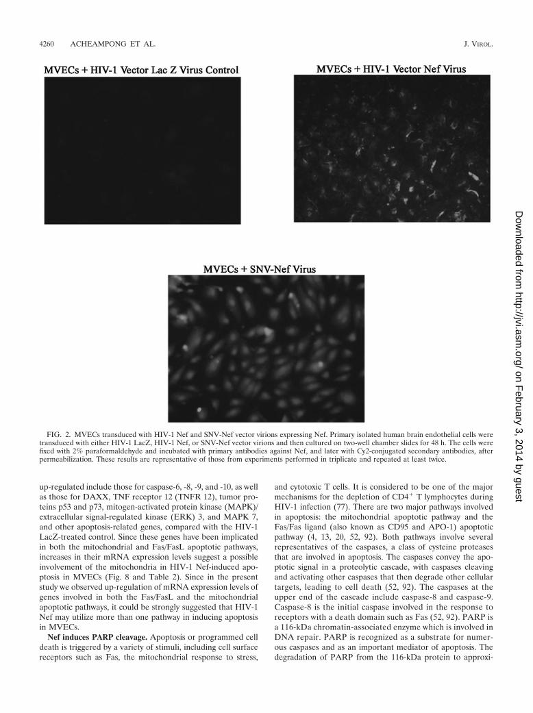
HIV-1 Nef virions express Nef intracellularly in MVECs.Human brain MVECs transduced with recombinant HIV-1-based Nef virus and SNV-Nef virus (i.e., HIV-1- and SNV-based vectors expressing HIV-1 Nef as a transgene, respec-tively) were immunostained for the intracellular expression ofNef. The results are depicted in Fig. 2. It was observed thatNef, as a transgene, was efficiently integrated and expressed inthe cells by both vectors, although the levels of transduction
and expression may be modestly less with HIV-1 compared toSNV vectors.
HIV-1 Nef induces apoptosis in human brain MVECs. HIV-1 Nef has been reported to inhibit apoptosis processes byinteracting with cellular kinases and thereby repressing pro-apoptotic signaling in HIV-1-infected cells (76). For example,it has been reported that Nef protects HIV-1-infected cellsagainst CD95 and tumor necrosis factor alpha (TNF-�) recep-tor-mediated death signaling by inhibiting the apoptosis signal-regulating kinase-1 (34). Furthermore, Nef has also been report-ed to repress signaling by Bad, a proapoptotic member of theBcl-2 protein family whose expression is induced by HIV-1(105).
In this study, we investigated the effects of recombinantHIV-1 Nef protein and intracellular Nef delivered throughlentiviral and oncoretroviral delivery systems into MVECs. Itwas observed that treatment of MVECs with extracellular Nefat low concentrations (i.e., 10 ng/ml) resulted in a moderateamount of programmed cell death; however, at higher concen-trations (i.e., 50 and 100 ng/ml), there was substantial induc-tion of apoptosis, as illustrated in Fig. 3. Similarly, TUNELassays demonstrated that MVECs transduced with recombi-nant HIV-1-based Nef and SNV-Nef viral vectors exhibiteddose-dependent programmed cell death, as depicted in Fig. 4and 5, respectively.
To further confirm the proapoptotic potential of Nef, weinfected MVECs with the HIV-1 strain ADA (CCR5-tropic)and the mutant with a deletion in Nef, ADA�Nef. The resultsof the TUNEL assays are depicted in Fig. 6 and 7. As illus-trated (Fig. 6), HIV-1 ADA at 10 ng of p24 antigen equivalentsper ml induced substantial levels of apoptosis in MVECs,whereas the quantity of apoptosis induced by its ADA�Nef,was quite low (Fig. 7). However, at higher levels of ADA�Nef(i.e., 100 ng of p24 antigen equivalents of virus per ml), therewas a substantial increase in the number of apoptotic cells (Fig.7). These results show that both endogenously expressed andextracellular HIV-1 Nef induced apoptosis in MVECs. Theresults also reveal that Nef may play an important but not solerole in the induction of programmed cell death by HIV-1 inMVECs.
Strict quantitation is difficult in these assay systems, but inevaluating multiple assays they can be reasonably describedsemiquantitatively; HIV-1-based vector Nef virus was the mostpotent in inducing apoptosis, followed by recombinant Nefprotein and then SNV-Nef (Table 1). Similarly, HIV-1 ADAalso induced substantial levels of apoptosis in MVECs. Thus,cell-free and intracellularly expressed HIV-1 proteins may dis-rupt the BBB via induction of programmed cell death.
Analyses of MVECs transduced with HIV-1 Nef by humanapoptosis gene microarrays. Induction of apoptosis by HIV-1Nef occurs through unknown mechanisms. Therefore, to elu-cidate the molecular mechanisms involved in Nef-induced apo-ptosis in MVECs, we used targeted gene microarrays to inves-tigate the up-regulation of apoptosis-related genes in MVECstransduced with HIV-1-based vector Nef virus and baculovi-rally expressed recombinant HIV-1 Nef protein. The humanapoptosis gene microarray generated with MVECs transducedwith HIV-1-based Nef virus (i.e., a HIV-1-based vector ex-pressing Nef as a transgene) revealed a substantial up-regula-tion of the mRNA levels of proapoptotic genes. The genes
VOL. 79, 2005 HIV-1 Nef AND APOPTOSIS 4259
on February 3, 2014 by guest
http://jvi.asm.org/
Dow
nloaded from
up-regulated include those for caspase-6, -8, -9, and -10, as wellas those for DAXX, TNF receptor 12 (TNFR 12), tumor pro-teins p53 and p73, mitogen-activated protein kinase (MAPK)/extracellular signal-regulated kinase (ERK) 3, and MAPK 7,and other apoptosis-related genes, compared with the HIV-1LacZ-treated control. Since these genes have been implicatedin both the mitochondrial and Fas/FasL apoptotic pathways,increases in their mRNA expression levels suggest a possibleinvolvement of the mitochondria in HIV-1 Nef-induced apo-ptosis in MVECs (Fig. 8 and Table 2). Since in the presentstudy we observed up-regulation of mRNA expression levels ofgenes involved in both the Fas/FasL and the mitochondrialapoptotic pathways, it could be strongly suggested that HIV-1Nef may utilize more than one pathway in inducing apoptosisin MVECs.
Nef induces PARP cleavage. Apoptosis or programmed celldeath is triggered by a variety of stimuli, including cell surfacereceptors such as Fas, the mitochondrial response to stress,
and cytotoxic T cells. It is considered to be one of the majormechanisms for the depletion of CD4� T lymphocytes duringHIV-1 infection (77). There are two major pathways involvedin apoptosis: the mitochondrial apoptotic pathway and theFas/Fas ligand (also known as CD95 and APO-1) apoptoticpathway (4, 13, 20, 52, 92). Both pathways involve severalrepresentatives of the caspases, a class of cysteine proteasesthat are involved in apoptosis. The caspases convey the apo-ptotic signal in a proteolytic cascade, with caspases cleavingand activating other caspases that then degrade other cellulartargets, leading to cell death (52, 92). The caspases at theupper end of the cascade include caspase-8 and caspase-9.Caspase-8 is the initial caspase involved in the response toreceptors with a death domain such as Fas (52, 92). PARP isa 116-kDa chromatin-associated enzyme which is involved inDNA repair. PARP is recognized as a substrate for numer-ous caspases and as an important mediator of apoptosis. Thedegradation of PARP from the 116-kDa protein to approxi-
FIG. 2. MVECs transduced with HIV-1 Nef and SNV-Nef vector virions expressing Nef. Primary isolated human brain endothelial cells weretransduced with either HIV-1 LacZ, HIV-1 Nef, or SNV-Nef vector virions and then cultured on two-well chamber slides for 48 h. The cells werefixed with 2% paraformaldehyde and incubated with primary antibodies against Nef, and later with Cy2-conjugated secondary antibodies, afterpermeabilization. These results are representative of those from experiments performed in triplicate and repeated at least twice.
4260 ACHEAMPONG ET AL. J. VIROL.
on February 3, 2014 by guest
http://jvi.asm.org/
Dow
nloaded from

mately 89- and 24-kDa cleavage products is one of the classicalindicators of apoptosis (60).
To test whether the caspase mRNAs detected via the mi-croarrays act at the translational level to induce apoptosis inMVECs, we analyzed apoptosis by using PARP cleavage viaWestern blot analyses. It was observed that MVECs trans-duced with the HIV-1 vector expressing Nef resulted in thecleavage of the PARP protein into the 116-kDa fragment anda smaller 85-kDa fragment, which were not detected in theLacZ virus-treated controls (Fig. 9A). Similar results wereobtained when cells were exposed to extracellular soluble Nefprotein (Fig. 9A). Thus, Nef-induced apoptosis correlated withthe biochemical cleavage of PARP, a known substrate of cas-pase-3.
Nef induces caspase activation in MVECs. Since PARP isknown to be cleaved by caspase-3 and -9 (52, 92) and themicroarray data in this study showed a substantial up-regula-tion of the expression of caspase-9 (Table 2) and a modestincrease in the level of caspase-3 (results not illustrated), weinvestigated whether these caspases undergo cleavage.
After either exposure extracellular soluble Nef or transduc-tion with HIV-1 Nef-expressing virions, cells were analyzed forcaspase activity. Western blot analyses utilizing specific anti-bodies revealed the expression of caspase-3, which was cleavedinto 17- to 19-kDa fragments when the cells were either ex-posed to extracellular Nef or transduced with Nef-expressingHIV-1 vector particles (Fig. 9B). Similar results were obtainedfor caspase-9 (Fig. 9C), which was cleaved into 48- and 37-kDacleavage products. Pretreatment of cells with specific caspase-3and -9 inhibitors (Ac-DEVD-CHO and Z-LEHD-FMK, re-spectively) completely eliminated the cleavage of both caspases(Fig. 9B and C). Thus, the Western blot analyses corroboratedthe microarray data, and both studies showed the involvementof caspase-3 and -9 in HIV-1 Nef-induced apoptosis in humanbrain MVECs.
DISCUSSION
One of the hallmarks of HIV-1 infection of the CNS is thedevelopment of AIDS dementia complex, which is character-ized by diffuse motor, sensory, and cognitive dysfunctions (6,10). It has been shown that monocytes/macrophages are theCNS-based-cells that are most commonly and productively in-fected with HIV-1 (25, 33, 55a, 65, 68, 104). However, certainstudies have demonstrated that low-level viral replication oc-curs in MVECs, astrocytes, and neurons (8, 11, 42, 69–71).HIV-1 infection also leads to a decline in the number of CD4�
T lymphocytes, achieved either through direct cytopathic ef-fects (103) or the activation of apoptotic cascades (110).
The molecular mechanisms involved in apoptosis of CNS-FIG. 3. Recombinant HIV-1 Nef protein-mediated apoptosis of
human brain MVECs. Representative TUNEL staining of MVECsexposed to GST- and baculovirally expressed HIV-1 Nef protein isshown. MVECs were exposed to either GST protein or HIV-1 Nefprotein expressed from Sf9 insect cells for 72 h. The concentrations ofprotein used were 10, 50, and 100 ng/ml. As a positive control, MVECswere exposed to DNase at a concentration of 10 �g/ml for 10 min atroom temperature. TUNEL assays were performed with the in situ celldeath detection kit, TMR Red (Roche Diagnostics). The cells wereanalyzed by fluorescence microscopy (Olympus System microscope,model BX60, with fluorescence attachment BX-FLA). These resultsare representative of those from experiments performed in triplicateand repeated at least twice.
FIG. 4. Nef expressed from the retroviral vector pHR�CMVNef issufficient to induce apoptosis in MVECs in a dose-dependent manner.Representative TUNEL staining of MVECs infected with HIV-1-based vectors expressing either Nef or LacZ for 72 h, after which cellswere subjected to TUNEL staining, is shown. The amount of virusused varied from 1 to 10 ng of HIV-1 p24 antigen equivalents per ml.TUNEL assays were performed with the in situ cell death detection kit,TMR Red (Roche Diagnostics). The cells were analyzed by fluores-cence microscopy (Olympus System microscope, model BX60, withfluorescence attachment BX-FLA). These results are representative ofthose from experiments performed in triplicate and repeated at leasttwice.
VOL. 79, 2005 HIV-1 Nef AND APOPTOSIS 4261
on February 3, 2014 by guest
http://jvi.asm.org/
Dow
nloaded from

based cells are not completely understood. During HIV-1 in-fection, cells can release specific viral products (82, 107, 113),which may induce apoptosis in bystander cells. In CNS-basedcells, it has been shown that HIV-1 infection can lead to theproduction and release of proinflammatory cytokines (9, 26,83, 114), which are neurotoxic. It is believed that induction ofapoptosis in the CNS occurs through the direct cytopathiceffects of viral proteins and/or indirectly via the release ofsoluble cellular factors (35). It has been shown in primary braincultures that HIV-1 infection induces apoptosis of neurons andastrocytes at 1 to 2 weeks after peak virus production (91).Also, the fact that in the brain tissues of HIV-1-infected indi-viduals most apoptotic cells are not localized in close proximityto HIV-1 infected cells (91) suggests that soluble cellular fac-tors and/or free viral proteins may induce apoptosis in thosecells.
Several HIV-1 proteins, such as Tat, gp120, Vpr, Vpu, andintact viral particles (55, 80, 81, 109), have been shown toinduce apoptosis in both infected and uninfected cells. Therole of Nef in inducing apoptosis is controversial for certaincell types (2, 34, 57, 75, 85). For example, Nef has been re-ported to favor cell survival through the down-modulation of
HLA-I molecules that allow CTLs to detect viral peptides, butat the same time preserving HLA-I molecules that inhibits cellkilling by natural killer (NK) cells. Furthermore, Nef has alsobeen reported to protect infected cells by repressing deathsignaling from “within” by the proapoptotic Bcl-2 family of pro-teins (34, 105) and by up-regulating FasL expression in infect-ed cells, which induces apoptosis in attacking CTLs. Neverthe-less, it has been recently reported that Nef induces apoptosisin CD4� T cells (50). We also reported recently that Nef incombination with ethanol induced apoptosis in MVECs (1). Inthe present study, we wished to assess the ability of Nef to in-duce apoptosis in CNS-based cells in detail, by studying mo-lecular mechanisms involved in Nef-induced apoptosis in CNS-based cells.
We investigated the potency of Nef in inducing apoptosis inthe context of the virus by using both lentiviral and oncoret-roviral delivery systems, where Nef was expressed from thetransfer vector, packaged in the virion vectors, and delivered
FIG. 5. HIV-1 Nef expressed from the spleen necrosis viral vector,SNV-Nef, induces apoptosis in MVECs in a dose-dependent manner.Representative TUNEL staining of SNV-LacZ and SNV-Nef� vectorvirion-treated MVECs, infected for 72 h, is shown. The quantities ofvirions used to infect cells were 10, 100, 500, and 1,000 CFU/ml. As apositive control, MVECs were exposed to DNase at a concentration of10 �g/ml for 10 min at room temperature. TUNEL assays were per-formed with the in situ cell death detection kit, TMR Red (RocheDiagnostics). The cells were analyzed by fluorescence microscopy(Olympus System microscope, model BX60, with fluorescence attach-ment BX-FLA). These results are representative of those from exper-iments performed in triplicate and repeated at least twice.
FIG. 6. HIV-1 R5 strain ADA induces apoptosis in MVECs in adose-dependent manner. Representative TUNEL staining of HIV-1ADA-treated MVECs is shown. Cells were infected with HIV-1 ADAfor 72 h, after which cells were subjected to TUNEL staining. Theamount of virus used ranged from 1 to 100 ng of HIV-1 p24 antigenequivalents per ml. As a positive control, MVECs were exposed toDNase at a concentration of 10 �g/ml for 10 min at room temperature.TUNEL assays were performed with the in situ cell death detection kit,TMR Red (Roche Diagnostics). The cells were analyzed by fluores-cence microscopy (Olympus System microscope, model BX60, withfluorescence attachment BX-FLA). These results are representative ofthose from experiments performed in triplicate and repeated at leasttwice.
4262 ACHEAMPONG ET AL. J. VIROL.
on February 3, 2014 by guest
http://jvi.asm.org/
Dow
nloaded from

into the cellular genome for integration. The results indicatedthe efficient expression of Nef protein within MVECs. We alsoinvestigated the ability of the extracellular recombinant Nefprotein expressed in and purified from Sf9 insect cells to in-duce apoptosis in MVECs.
Apoptosis assays utilizing TUNEL staining indicated thatintracellularly expressed Nef induced significant levels of apo-ptosis in a dose-dependent manner. Similarly, dose-responseexperiments with baculovirally expressed extracellular Nefshowed that this cell-free lentiviral protein could induce con-siderable apoptosis in MVECs. Cells exposed to the averagelevel of soluble Nef protein (10 ng/ml) normally detected in thesera of HIV-1-infected patients (31) showed substantial levelsof apoptosis. Furthermore, the TUNEL staining clearly dem-onstrated that MVECs exposed to Nef protein and those trans-duced with HIV-1-based vector Nef virus revealed cytoplasmicshrinkage and nuclear fragmentation (results not illustrated),with higher doses of Nef producing elevated levels of nuclearfragmentation and apoptotic bodies. It is not clear preciselyhow extracellular Nef induces programmed cell death in brain
MVECs, whether by entering cells or at the membrane level. Inaddition, it was observed that there was an increase in trypanblue dye uptake by the treated cells, confirming the increasingcell death observed in primary isolated MVECs exposed ortransduced with HIV-1 Nef or SNV-Nef viral vectors (notillustrated). As such, Nef induces apoptosis in primary humanbrain MVECs in a dose-dependent manner. We also observedthat infection of cells with the HIV-1 strain ADA resulted insubstantial induction of apoptosis. In contrast, exposure ofcells to the mutant strain with a deletion in Nef, ADA�Nef,resulted in markedly reduced apoptosis. These results clearlydemonstrate that both extracellular and intracellularly ex-pressed Nef are capable of killing MVECs through apoptosis.This is consistent with other reports which suggest that HIV-1proteins, such as Nef, gp120, Tat, and Vpr, can initiate apo-ptotic cascades in CNS-based cells (1, 80, 100, 109).
Nef inhibits (i.e., protects infected cells) and induces apo-ptosis in bystander cells (34, 105). The N terminus of the Nefprotein has been implicated in this bystander cell death (48).Studies by Okada et al. (75) have shown that soluble Nefprotein, in conjunction with Nef antibodies, causes the death ofa wide range of uninfected lymphoid tissues (74, 75). Similarly,transgenic animal models developed to study the in vivo ex-pression of HIV-1 proteins in the host systems (21–23, 37, 43,51, 99) were found to undergo lymphocyte depletion, as well asmost of the organ dysfunction commonly found in AIDS pa-tients. It is important to note that these animals required onlyexpressed Nef in the target cells in order to manifest these
FIG. 7. An HIV-1 R5 strain with a deletion in the Nef gene(ADA�Nef) induces apoptosis in MVECs at lower levels than wild-typeHIV-1 ADA. Representative TUNEL staining of HIV-1 ADA�Nef-treated MVECs is shown. Cells were infected with HIV-1 ADA�Neffor 72 h, after which they were subjected to TUNEL staining. The amountof virus used ranged from 1 to 100 ng of HIV-1 Gag p24 equivalentsper ml. As a positive control, MVECs were exposed to DNase at aconcentration of 10 �g/ml for 10 min at room temperature. TUNELassays were performed with the in situ cell death detection kit, TMRRed (Roche Diagnostics). The cells were analyzed with fluorescencemicroscopy (Olympus System microscope, model BX60, with fluores-cence attachment BX-FLA). These results are representative of thosefrom experiments performed in triplicate and repeated at least twice.
TABLE 1. Semiquantitative determination of HIV-1 Nef-inducedapoptosis in primary human brain MVECs
Treatment Red-green-bluevalues
Nef protein (ng/ml)10 60.2550 142.0100 207.0
HIV-1-based vector Nef virus (ng of p24 antigenequivalents per ml)
1 134.55 185.310 233.5
SNV vector-based Nef virus (CFU/ml)10 22.8100 44.0500 49.01,000 97.0
HIV-1 strain ADA (ng of p24 antigenequivalents per ml)
1 8.010 66.850 151.8100 189.5
HIV-1 strain ADA�Nef (ng of p24 antigenequivalents per ml)
1 3.010 3.550 18.0100 86.50
VOL. 79, 2005 HIV-1 Nef AND APOPTOSIS 4263
on February 3, 2014 by guest
http://jvi.asm.org/
Dow
nloaded from

changes. Therefore, the observation in the present study thatextracellular soluble Nef induced apoptosis in MVECs is con-sistent with the bystander effect hypothesis (27), which impli-cates a viral protein or virally stimulated cellular factors re-leased into the medium as mediators of apoptosis (24, 72).Since Nef is synthesized early during initial viral infection andit has been shown that Nef produced by recombinant plasmidsis secreted into culture medium (31, 50, 61), it is possible thatintracellular expression of Nef in this study resulted in therelease of the protein into the culture, which in turn may havecaused the bystander effects. Hence, it could also be said thatthe induction of apoptosis in MVECs by intracellular Nef isalso consistent with the bystander effect hypothesis (27).
Although the TUNEL assays in the present study demon-strated clearly that Nef induces apoptosis in MVECs, the exact
mechanisms were not known. Therefore, to begin to elucidatethe molecular mechanisms involved, targeted gene microarraysand Western blotting were employed to analyze the regulationof apoptotic genes in MVECs exposed to soluble Nef proteinand also those transduced with an HIV-1 viral vector con-taining the Nef transgene alone. These were compared withMVECs exposed to GST and LacZ virus (i.e., a HIV-1 vectorexpressing LacZ), respectively. We observed that Nef inducedapoptosis in MVECs by up-regulating the expression of thecomponents of both the Fas/Fas ligand (also known as CD95and APO-1), and the TNF/TNFR 1 signaling pathways, plusthe mitochondrial apoptotic pathways.
The components of the Fas/Fas ligand that were found up-regulated included DAXX, TNFR-12 (also known as TRAMP,LARD, APO3, DR3, and TNFR 25), and caspase-8 and -10.
FIG. 8. Human apoptosis gene microarrays of MVECs transduced with HIV-1 LacZ virus (top panel) and HIV-1-based vector Nef� virus(bottom panel). These results are representative of those from two independent experiments.
4264 ACHEAMPONG ET AL. J. VIROL.
on February 3, 2014 by guest
http://jvi.asm.org/
Dow
nloaded from

Receptors in the TNF receptor family are associated with theinduction of apoptosis, as well as inflammatory signaling. Bind-ing of FAS to oligomerized FasL on another cell leads to theformation of a death-induced signal complex (DISC) com-posed of Fas, Fas-associated death domain protein, and cas-pase-8 or -10 (12). Recruitment of procaspase-8 or -10 to DISCresults in its activation, and it then cleaves and activates down-stream caspases and a variety of cellular substrates, includingPARP, leading to cell death (49, 52, 92). TNFR 12 or DR3shows close sequence similarity to TNFR 1 (15). Also, the re-sponses that it triggers are similar to those of TNFR 1, namely,nuclear factor �B (NF-�B) activation and apoptosis. TNFR 12triggers apoptosis through TNFR1-associated death domain(TRADD), Fas-associated death domain protein, and caspase8 and activates NF-�B through TRADD, TNF receptor-asso-ciated factor 2, and receptor interacting protein (15, 46).
In addition to the caspase activation cascade, induction ofthe Fas/FasL pathway may also involve the activation of c-JunN-terminal kinase/stress activated protein kinase (JNK/SAPK)(112). After Fas/FasL ligation, an adaptor protein calledDAXX is recruited by Fas and then interacts with apopto-sis signal-regulating kinase 1, activating JNK/SAPK and p38MAPK by phosphorylation (14, 47). This eventually results inthe activation of caspases and subsequent apoptosis.
Our results demonstrate up-regulation in the expression ofDAXX, TNFR 12 (DR3), and caspase-8 and -10. Since thesegenes have been implicated in the Fas/FasL and the TNF/TNFR apoptotic pathways (12, 15, 46), the results suggest thatboth extracellular and intracellularly expressed HIV-1 Nef in-duces apoptosis in human brain MVECs, most likely throughcomponents of the Fas/FasL and the TNF/TNFR apoptoticpathways. These results are fully consistent with the results ofother studies which have shown that Nef increases the expres-sion of Fas ligand, resulting in Fas signaling and apoptosis inbystander cells (28–31, 85, 108). However, some studies havedemonstrated that extracellular Nef protein induces apoptoticcytolysis in both lymphoid and myeloid cells of murine andhuman origins independently of the Fas/FasL apoptotic path-way (74–76). The apparent discrepancy between our study andthose studies may be explained by the fact that different cell
FIG. 9. Western blotting analyses of PARP (A), caspase-3 (B), andcaspase-9 (C) during HIV-1 Nef-induced apoptosis in MVECs. Cellswere either left untreated, exposed to 100 ng of GST protein per ml,transduced with 10 ng of p24 antigen equivalents of HIV-1-basedvector LacZ virus per ml, treated with 100 ng of recombinant Nefprotein per ml, transduced with 10 ng of p24 antigen equivalents ofHIV-1-based vector Nef virus per ml, or pretreated with 100 nMspecific caspase inhibitor and then transduced with HIV-1-based vec-tor Nef virus. Twenty-five micrograms of total protein extract wasloaded in each lane and separated by sodium dodecyl sulfate-polyac-rylamide gel electrophoresis. Caspase-3 and -9 and PARP were ana-lyzed by Western blotting with specific caspase-3, caspase-9 and PARPmonoclonal antibodies. These results are representative of those fromexperiments repeated at least twice. (A) PARP. Lanes: M, proteinmarker; 1, MVECs treated with 100 ng of recombinant Nef protein perml; 2, MVECs transduced with 10 ng of p24 antigen equivalents ofHIV-1-based vector Nef virus per ml; 3, untreated MVECs; 4, MVECsexposed to 100 ng of GST protein per ml; 5, MVECs pretreated with100 nM broad-spectrum caspase inhibitor Z-VAD-FMK (BD PharMingen) and then transduced with HIV-1-based vector Nef virus; 6,MVECs transduced with 10 ng of p24 antigen equivalents of HIV-1-based vector LacZ virus per ml. The upper arrow depicts 116-kDaPARP, and the lower arrow depicts approximately the 85-kDa cleavedPARP. (B) Caspase-3. Lanes: 1, untreated MVECs; 2, MVECs ex-posed to 100 ng of GST protein per ml; 3, MVECs transduced with 10ng of p24 antigen equivalents of HIV-1-based vector LacZ virus perml; 4, MVECs treated with 100 ng of recombinant Nef protein per ml;5, MVECs transduced with 10 ng of p24 antigen equivalents of HIV-1-based vector Nef virus per ml; 6, MVECs pretreated with 100 nM ofAc-DEVD-CHO (BD PharMingen), a specific caspase 3 inhibitor, andthen transduced with HIV-1-based Nef virus. The top arrow depicts35-kDa intact caspase-3, and the bottom arrow depicts the 17- to19-kDa cleaved caspase-3 fragment. (C) Caspase-9. Lanes: M, proteinmarker; 1, untreated MVECs; 2, MVECs exposed to 100 ng of GSTprotein per ml; 3, MVECs transduced with 10 ng of p24 antigenequivalents of HIV-1-based vector LacZ virus per ml; 4, MVECspretreated with 100 nM Z-LEHD-FMK, a specific caspase 9 inhibitor(BD PharMingen), and then transduced with HIV-1-based Nef virus;5, MVECs treated with 100 ng of recombinant Nef protein per ml; 6,MVECs transduced with 10 ng of p24 antigen equivalents of HIV-1-based vector Nef virus per ml. The top arrow depicts 45-kDa pro-cas-pase-9, and the bottom arrow depicts 35- and 37-kDa cleaved cas-pase-9 fragments.
TABLE 2. cDNA microarray analyses of human brain MVECstransduced with HIV-1 vector expressing Nef
Up-regulated gene product Foldincrease
CDC-like kinase (CLK 3).......................................................................... 2.0PICTAIRE protein kinase 2 (apoptosis related).................................... 1.5Mitogen-activated protein kinase 7 .......................................................... 1.7MAPK/ERK 3 ............................................................................................. 2.0Tumor protein p73 ..................................................................................... 2.5Tumor protein p53 ..................................................................................... 1.5Glutathione S-transferase-like protein..................................................... 1.5Caspase-6 ..................................................................................................... 2.5Caspase-8 ..................................................................................................... 1.5Caspase-9 ..................................................................................................... 3.0Caspase-10 ................................................................................................... 2.0Death-associated protein 6 (DAXX) ....................................................... 2.5Secreted apoptosis-related protein 2........................................................ 2.5Death-associated protein kinase 1............................................................ 1.8Tumor necrosis factor receptor superfamily, member 12 ..................... 2.5Insulin-like growth factor 1 ....................................................................... 1.7Insulin-like growth factor binding protein 6 ........................................... 1.6
VOL. 79, 2005 HIV-1 Nef AND APOPTOSIS 4265
on February 3, 2014 by guest
http://jvi.asm.org/
Dow
nloaded from

types were studied. In addition, whereas our study employedendogenously expressed Nef and N-terminal myristoylated ex-tracellular Nef, Okada et al. (74–76) utilized nonmyristoylatedNef. Nonetheless, our results indicate that apoptosis via trig-gering of the Fas/FasL pathway may not be common to all celltypes that are made to undergo Nef expression and could verywell be dependent on the cell type, culture conditions, andnature of the stimulus.
Other apoptosis-related genes found to be up-regulatedupon exposure of MVECs to HIV-1 Nef were those for cas-pase-3, -6, and -9. Western blot analyses revealed the cleavageproducts of caspase-3 and -9 and PARP, which are detectedonly in cells undergoing apoptosis. Caspase-9 (also known asICE-LAP6, Mch-6, or Apaf-3) is a 45-kDa protein that is in-volved in the mitochondrial apoptotic pathway (52, 92). Uponapoptotic stimulation, cytochrome c released from mitochon-dria associates with procaspase-9 and Apaf-1 in the presence ofdATP to form a proapoptotic complex. This complex promotesthe self-cleavage of procaspase-9 into a large active subunit (35or 17 kDa) and a small 10-kDa subunit (56, 58). Once activat-ed, caspase-9 initiates a caspase cascade involving caspase-3,-6, and -7, which act by themselves to cleave cellular targets suchas PARP, leading to cell death (16, 63, 67, 86). As well, caspase-8 activated via the Fas/FasL and the TNF/TNFR apoptoticpathways can stimulate proteolytic cleavage of Bcl-2-interact-ing protein, which initiates cytochrome c release in the mito-chondria (38), leading to the activation of caspase-9 and -3.
In addition to the above-described proteins, our studies alsorevealed prominent up-regulation of the mRNA expression ofMAPK 7, ERK, MAPK, and the tumor suppressor proteinsp53 and p73. The tumor suppressor protein p53 is involved inthe mitochondrial apoptotic pathway (88) and plays a role inapoptosis by down-modulating the mitochondrial membranepotential and promoting the release of cytochrome c (90). p53also leads to cell cycle arrest and induces apoptosis by regu-lating at the transcriptional level several genes, including pro-apoptotic Bax of the Bcl-2 family (59, 66, 102), the gene for thecell cycle inhibitor p21, and the DNA repair gene GADD45(53). The transcriptional regulation by p53 of Bax and theother proapoptotic genes, according to Soengas et al., is de-pendent on Apaf-1/caspase-9 activation involved in the mito-chondrial pathway (96). p53 can also induce apoptosis bymeans of a direct signaling pathway involving the expression ofp53AIP1, which has been reported to regulate the mitochon-drial apoptotic pathway (64). Thus, the up-regulation of p53mRNA levels in this study was important in that it showed thatNef-induced apoptosis in MVECs most likely occurred viaspecific mitochondrial elements associated with cell death.
Unlike p53, MAPK and MAPK/ERK have not been directlylinked to the mitochondrial apoptotic pathway. Also, the rela-tionship between the activation of MAPK cascades and induc-tion of apoptosis is still not clear and may vary among differentcell types (58, 101, 106). However, it has been reported thatoverexpression of MAPK results in increased expression andactivity of p53, causing a permanent growth arrest and apopto-sis (32).
Nef possesses the capacity to regulate numerous transcrip-tional activities by targeting and controlling various src familykinase, MAPK, and p53 activities. However, most reports re-garding Nef-mediated transcriptional activation of p53 and
MAPK pathways have been with T cells, and these analyseshave been less than conclusive. Most of those studies suggestthat Nef has the capacities to both inhibit and enhance T-cellactivity (62, 95). From work with MOLT-4 cells, Greenway etal. (41) reported that Nef binds to p53 to protect the cellsagainst p53-mediated apoptosis. Similarly, other studies (39,44) have suggested that Nef binds to p53, Raf-1, and MAPK todestabilize these proteins and inhibit their transcriptional ac-tivation. Nef has been shown to bind and inhibit Lck andMAPK activity (39). In contrast, Schrager et al. (89) demon-strated in primary CD4� T cells that the expression of Nefspecifically increases activity of the ERK/MAP kinase cascade.Similarly, Robichaud and Poulin (87), using the U251MG hu-man astrocytoma cell line, reported that Nef preferentiallyenhanced MAPK and JNK activation. Furthermore, it hasbeen demonstrated that HIV-1 Nef, Rev, and Tat could bepotential substrates that may activate MAPK cascades (111).Thus, it appears that the regulation of the transcriptional ac-tivities of various kinases and p53 by Nef is quite complex andthat whether or not Nef inhibits or enhances these activitiesmay depend on the cell type and possibly on nature of the Nefprotein involved. Nonetheless, there is evolving support forNef as a positive regulator for p53 and MAPK.
Based on the evidence presented regarding the involvementof caspase-3 and -9, p53, p73, and MAPK cascades, it is clearthat Nef-induced apoptosis in human brain MVECs involvescertain components of the mitochondrial apoptotic pathways.In summary, the present studies strongly suggest that theHIV-1 protein Nef can potently induce apoptosis in humanbrain MVECs. As well, these data demonstrate, for the firsttime, that Nef-induced apoptosis in MVECs may result from acombination of several intracellular signaling pathways.
ACKNOWLEDGMENTS
We thank Rita M. Victor and Brenda O. Gordon for excellentsecretarial assistance. HIV-1 Nef antibodies and the plasmid contain-ing the Nef consensus sequence were obtained from the AIDS Re-search and Reference Reagent Program, Division of AIDS, NIAID,NIH.
This work was supported in part by Public Health Service grantsNS41864, NS044513, and AA13849 to R.J.P. and AI46149 to M.M.
REFERENCES
1. Acheampong, E., M. Mukhtar, Z. Parveen, N. Ngoubilly, N. Ahmad, C.Patel, and R. J. Pomerantz. 2002. Ethanol strongly potentiates apoptosisinduced by HIV-1 proteins in primary human brain microvascular endo-thelial cells. Virology 304:222–234.
2. Adachi, K., X. Li, H. Kimura, H. Amemiya, and S. Suzuki. 2001. HIV-1 Nefprotein prolonged recipient survival in rat liver allografting. TransplantProc. 33:284.
3. Adle-Biassette, H., Y. Levy, M. Colombel, F. Poron, S. Natchev, C. Keo-hane, and F. Gray. 1995. Neuronal apoptosis in HIV infection in adults.Neuropathol. Appl. Neurobiol. 21:218–227.
4. Alderson, M. R., T. W. Tough, T. Davis-Smith, S. Braddy, B. Falk, K. A.Schooley, R. G. Goodwin, C. A. Smith, F. Ramsdell, and D. H. Lynch. 1995.Fas ligand mediates activation-induced cell death in human T lymphocytes.J. Exp. Med. 181:71–77.
5. Alimonti, J. B., T. B. Ball, and K. R. Fowke. 2003. Mechanisms of CD4� Tlymphocyte cell death in human immunodeficiency virus infection andAIDS. J. Gen. Virol. 84:1649–1661.
6. Arnoult, D., F. Petit, J. D. Lelievie, D. Lecossier, A. Hance, V. Monceaux, R.Ho Tsong Fang, B. Huntrel, J. C. Ameisen, and J. Estaquier. 2003. Caspase-dependent and -independent T-cell death pathways in pathogenic simianimmunodeficiency virus infection: relationship to disease progression. CellDeath Differ. 10:1240–1252.
7. Azad, A., S. B. Lall, and S. Mittra. 2001. Effect of N-acetylcysteine andL-NAME on aluminium phosphide induced cardiovascular toxicity in rats.Acta Pharmacol. Sin. 22:298–304.
4266 ACHEAMPONG ET AL. J. VIROL.
on February 3, 2014 by guest
http://jvi.asm.org/
Dow
nloaded from

8. Bagasra, O., E. Lavi, L. Bobroski, K. Khalili, J. P. Pestaner, R. Tawadros,and R. J. Pomerantz. 1996. Cellular reservoirs of HIV-1 in the centralnervous system of infected individuals: identification by the combination ofin situ polymerase chain reaction and immunohistochemistry. AIDS 10:573–585.
9. Barak, O., I. Goshen, T. Ben-Hur, J. Weidenfeld, A. N. Taylor, and R.Yirmiya. 2002. Involvement of brain cytokines in the neurobehavioral dis-turbances induced by HIV-1 glycoprotein120. Brain Res. 933:98–108.
10. Bell, D. J., and D. H. Dockrell. 2003. Apoptosis in HIV-1 infection. J. Eur.Acad. Dermatol. Venereol. 17:178–183.
11. Bobardt, M. D., P. Salmon, L. Wang, J. D. Esko, D. Gabuzda, M. Fiala, D.Trono, B. Van der Schueren, G. David, and P. A. Gallay. 2004. Contributionof proteoglycans to human immunodeficiency virus type 1 brain invasion.J. Virol. 78:6567–6584.
12. Boldin, M. P., E. E. Varfolomeev, Z. Pancer, I. L. Mett, J. H. Camonis, andD. Wallach. 1995. A novel protein that interacts with the death domain ofFas/APO1 contains a sequence motif related to the death domain. J. Biol.Chem. 270:7795–7798.
13. Brunner, T., R. J. Mogil, D. LaFace, N. J. Yoo, A. Mahboubi, F. Echeverri,S. J. Martin, W. R. Force, D. H. Lynch, C. F. Ware, et al. 1995. Cell-autonomous Fas (CD95)/Fas-ligand interaction mediates activation-in-duced apoptosis in T-cell hybridomas. Nature 373:441–444.
14. Chang, H. Y., H. Nishitoh, X. Yang, H. Ichijo, and D. Baltimore. 1998.Activation of apoptosis signal-regulating kinase 1 (ASK1) by the adapterprotein Daxx. Science 281:1860–1863.
15. Chinnaiyan, A. M., K. O’Rourke, G. L. Yu, R. H. Lyons, M. Garg, D. R.Duan, L. Xing, R. Gentz, J. Ni, and V. M. Dixit. 1996. Signal transductionby DR3, a death domain-containing receptor related to TNFR-1 and CD95.Science 274:990–992.
16. Cline, A. M., and M. Z. Radic. 2004. Apoptosis, subcellular particles, andautoimmunity. Clin. Immunol. 112:175–182.
17. Collins, K. L., B. K. Chen, S. A. Kalams, B. D. Walker, and D. Baltimore.1998. HIV-1 Nef protein protects infected primary cells against killing bycytotoxic T lymphocytes. Nature 391:397–401.
18. Curtain, C. C., M. G. Lowe, C. K. Arunagiri, P. W. Mobley, I. G. Macreadie,and A. A. Azad. 1997. Cytotoxic activity of the amino-terminal region ofHIV type 1 Nef protein. AIDS Res. Hum. Retroviruses 13:1213–1220.
19. Curtain, C. C., M. G. Lowe, I. G. Macreadie, I. R. Gentle, G. A. Lawrie, andA. A. Azad. 1998. Structural requirements for the cytotoxicity of the N-terminal region of HIV type 1 Nef. AIDS Res. Hum. Retroviruses 14:1543–1551.
20. Dhein, J., H. Walczak, M. O. Westendorp, C. Baumler, K. Stricker, R.Frank, K. M. Debatin, and P. H. Krammer. 1995. Molecular mechanisms ofAPO-1/Fas(CD95)-mediated apoptosis in tolerance and AIDS. BehringInst. Mitt. 9:13–20.
21. Dickie, P. 1996. HIV type 1 Nef perturbs eye lens development in trans-genic mice. AIDS Res. Hum. Retroviruses 12:177–189.
22. Dickie, P. 2000. Nef modulation of HIV type 1 gene expression and cyto-pathicity in tissues of HIV transgenic mice. AIDS Res. Hum Retroviruses16:777–790.
23. Dickie, P., F. Ramsdell, A. L. Notkins, and S. Venkatesan. 1993. Sponta-neous and inducible epidermal hyperplasia in transgenic mice expressingHIV-1 Nef. Virology 197:431–438.
24. Ensoli, B., G. Barillari, S. Z. Salahuddin, R. C. Gallo, and F. Wong-Staal.1990. Tat protein of HIV-1 stimulates growth of cells derived from Kaposi’ssarcoma lesions of AIDS patients. Nature 345:84–86.
25. Epstein, L. G., and H. E. Gendelman. 1993. Human immunodeficiency virustype 1 infection of the nervous system: pathogenetic mechanisms. Ann.Neurol. 33:429–436.
26. Feuerstein, G. Z., T. Liu, and F. C. Barone. 1994. Cytokines, inflammation,and brain injury: role of tumor necrosis factor-alpha. Cerebrovasc. BrainMetab. Rev. 6:341–360.
27. Finkel, T. H., G. Tudor-Williams, N. K. Banda, M. F. Cotton, T. Curiel, C.Monks, T. W. Baba, R. M. Ruprecht, and A. Kupfer. 1995. Apoptosis occurspredominantly in bystander cells and not in productively infected cells ofHIV- and SIV-infected lymph nodes. Nat. Med. 1:129–134.
28. Fujii, Y., K. Otake, Y. Fujita, N. Yamamoto, Y. Nagai, M. Tashiro, and A.Adachi. 1996. Clustered localization of oligomeric Nef protein of humanimmunodeficiency virus type 1 on the cell surface. FEBS Lett. 395:257–261.
29. Fujii, Y., K. Otake, M. Tashiro, and A. Adachi. 1996. Human immunode-ficiency virus type 1 Nef protein on the cell surface is cytocidal for humanCD4� T cells. FEBS Lett. 393:105–108.
30. Fujii, Y., K. Otake, M. Tashiro, and A. Adachi. 1996. In vitro cytocidaleffects of human immunodeficiency virus type 1 Nef on unprimed humanCD4� T cells without MHC restriction. J. Gen. Virol. 77:2943–2951.
31. Fujii, Y., K. Otake, M. Tashiro, and A. Adachi. 1996. Soluble Nef antigenof HIV-1 is cytotoxic for human CD4� T cells. FEBS Lett. 393:93–96.
32. Fukasawa, K., and G. F. Vande Woude. 1997. Synergy between the Mos/mitogen-activated protein kinase pathway and loss of p53 function in trans-formation and chromosome instability. Mol. Cell. Biol. 17:506–518.
33. Garden, G. A. 2002. Microglia in human immunodeficiency virus-associatedneurodegeneration. Glia 40:240–251.
34. Geleziunas, R., W. Xu, K. Takeda, H. Ichijo, and W. C. Greene. 2001.HIV-1 Nef inhibits ASK1-dependent death signalling providing a potentialmechanism for protecting the infected host cell. Nature 410:834–838.
35. Gendelman, H. E., S. A. Lipton, M. Tardieu, M. I. Bukrinsky, and H. S.Nottet. 1994. The neuropathogenesis of HIV-1 infection. J. Leukoc. Biol.56:389–398.
36. Geyer, M., O. T. Fackler, and B. M. Peterlin. 2001. Structure–functionrelationships in HIV-1 Nef. EMBO Rep. 2:580–585.
37. Goudreau, G., S. Carpenter, N. Beaulieu, and P. Jolicoeur. 1996. Vacuolarmyelopathy in transgenic mice expressing human immunodeficiency virustype 1 proteins under the regulation of the myelin basic protein genepromoter. Nat. Med. 2:655–661.
38. Green, D. R., and G. Kroemer. 2004. The pathophysiology of mitochondrialcell death. Science 305:626–629.
39. Greenway, A., A. Azad, J. Mills, and D. McPhee. 1996. Human immuno-deficiency virus type 1 Nef binds directly to Lck and mitogen-activatedprotein kinase, inhibiting kinase activity. J. Virol. 70:6701–6708.
40. Greenway, A. L., G. Holloway, and D. A. McPhee. 2000. HIV-1 Nef: acritical factor in viral-induced pathogenesis. Adv. Pharmacol. 48:299–343.
41. Greenway, A. L., D. A. McPhee, K. Allen, R. Johnstone, G. Holloway, J.Mills, A. Azad, S. Sankovich, and P. Lambert. 2002. Human immunodefi-ciency virus type 1 Nef binds to tumor suppressor p53 and protects cellsagainst p53-mediated apoptosis. J. Virol. 76:2692–2702.
42. Gujuluva, C., A. R. Burns, T. Pushkarsky, W. Popik, O. Berger, M. Bukrin-sky, M. C. Graves, and M. Fiala. 2001. HIV-1 penetrates coronary arteryendothelial cells by transcytosis. Mol. Med. 7:169–176.
43. Hanna, Z., D. G. Kay, N. Rebai, A. Guimond, S. Jothy, and P. Jolicoeur.1998. Nef harbors a major determinant of pathogenicity for an AIDS-likedisease induced by HIV-1 in transgenic mice. Cell 95:163–175.
44. Hodge, D. R., K. J. Dunn, G. K. Pei, M. K. Chakrabarty, G. Heidecker, J. A.Lautenberger, and K. P. Samuel. 1998. Binding of c-Raf1 kinase to aconserved acidic sequence within the carboxyl-terminal region of the HIV-1Nef protein. J. Biol. Chem. 273:15727–15733.
45. Hofmann-Lehmann, R., J. Vlasak, A. L. Williams, A. L. Chenine, H. M.McClure, D. C. Anderson, S. O’Neil, and R. M. Ruprecht. 2003. Liveattenuated, nef-deleted SIV is pathogenic in most adult macaques afterprolonged observation. AIDS 17:157–166.
46. Hsu, H., H. B. Shu, M. G. Pan, and D. V. Goeddel. 1996. TRADD-TRAF2and TRADD-FADD interactions define two distinct TNF receptor 1 signaltransduction pathways. Cell 84:299–308.
47. Ichijo, H. 1999. From receptors to stress-activated MAP kinases. Oncogene18:6087–6093.
48. Ikuta, K., M. Kameoka, and R. B. Luftig. 1997. AIDS pathogenesis: the roleof accessory gene mutations, leading to formation of long-lived persistentlyinfected cells and/or apoptosis-inducing HIV-1 particles. Virus Res. 52:145–156.
49. Imai, Y., T. Kimura, A. Murakami, N. Yajima, K. Sakamaki, and S. Yone-hara. 1999. The CED-4-homologous protein FLASH is involved in Fas-mediated activation of caspase-8 during apoptosis. Nature 398:777–785.
50. James, C. O., M. B. Huang, M. Khan, M. Garcia-Barrio, M. D. Powell, andV. C. Bond. 2004. Extracellular Nef protein targets CD4� T cells for apo-ptosis by interacting with CXCR4 surface receptors. J. Virol. 78:3099–3109.
51. Jolicoeur, P., D. G. Kay, M. Cool, S. Jothy, N. Rebai, and Z. Hanna. 1999.A novel mouse model of HIV-1 disease. Leukemia 13(Suppl. 1):S78–S80.
52. Joza, N., S. A. Susin, E. Daugas, W. L. Stanford, S. K. Cho, C. Y. Li, T.Sasaki, A. J. Elia, H. Y. Cheng, L. Ravagnan, K. F. Ferri, N. Zamzami, A.Wakeham, R. Hakem, H. Yoshida, Y. Y. Kong, T. W. Mak, J. C. Zuniga-Pflucker, G. Kroemer, and J. M. Penninger. 2001. Essential role of themitochondrial apoptosis-inducing factor in programmed cell death. Nature410:549–554.
53. Kastan, M. B. 1993. P53: a determinant of the cell cycle response to DNAdamage. Adv. Exp. Med. Biol. 339:291–293.
54. Kestler, H. W., III, D. J. Ringler, K. Mori, D. L. Panicali, P. K. Sehgal,M. D. Daniel, and R. C. Desrosiers. 1991. Importance of the nef gene formaintenance of high virus loads and for development of AIDS. Cell 65:651–662.
55. Kim, T.-A., H. K. Avraham, Y.-H. Koh, S. Jiang, I.-W. Park, and S. Avra-ham. 2003. HIV-1 tat mediated apoptosis in human brain microvascularendothelial cells. J. Immunol. 170:2629–2637.
55a.Kim, W. K., S. Corey, X. Alvarez, and K. Williams. 2003. Monocyte/mac-rophage traffic in HIV and SIV encephalitis. J. Leukoc. Biol. 74:650–656.
56. Li, P., H. Allen, S. Banerjee, and T. Seshadri. 1997. Characterization ofmice deficient in interleukin-1 beta converting enzyme. J. Cell Biochem. 64:27–32.
57. Li, X., M. C. Multon, Y. Henin, F. Schweighoffer, C. Venot, J. LaVecchio, J.Josef, P. Stuckert, A. Mhashilkar, B. Tocque, and W. A. Marasco. 2000.Upregulation of the apoptosis-associated protein Grb3-3 in HIV-1-infectedhuman CD4(�) lymphocytes. Biochem. Biophys. Res. Commun. 276:362–370.
58. Liu, X., C. N. Kim, J. Pohl, and X. Wang. 1996. Purification and charac-terization of an interleukin-1 beta-converting enzyme family protease thatactivates cysteine protease P32 (CPP32). J. Biol. Chem. 271:13371–13376.
VOL. 79, 2005 HIV-1 Nef AND APOPTOSIS 4267
on February 3, 2014 by guest
http://jvi.asm.org/
Dow
nloaded from

59. Lowe, S. W. 1995. Cancer therapy and p53. Curr. Opin. Oncol. 7:547–553.60. MacFarlane, M., K. Cain, X. M. Sun, E. S. Alnemri, and G. M. Cohen.
1997. Processing/activation of at least four interleukin-1beta convertingenzyme-like proteases occurs during the execution phase of apoptosis inhuman monocytic tumor cells. J. Cell Biol. 137:469–479.
61. Macreadie, I. G., A. C. Ward, P. Failla, E. Grgacic, D. McPhee, and A. A.Azad. 1993. Expression of HIV-1 nef in yeast: the 27 kDa Nef protein ismyristylated and fractionates with the nucleus. Yeast 9:565–573.
62. Marsh, J. W. 1999. The numerous effector functions of Nef. Arch. Biochem.Biophys. 365:192–198.
63. Martin, D. A., and K. B. Elkon. 2004. Mechanisms of apoptosis. Rheum.Dis. Clin. North Am. 30:441–454, vii.
64. Matsuda, K., K. Yoshida, Y. Taya, K. Nakamura, Y. Nakamura, and H.Arakawa. 2002. p53AIP1 regulates the mitochondrial apoptotic pathway.Cancer Res. 62:2883–2889.
65. Minagar, A., P. Shapshak, R. Fujimura, R. Ownby, M. Heyes, and C.Eisdorfer. 2002. The role of macrophage/microglia and astrocytes in thepathogenesis of three neurologic disorders: HIV-associated dementia, Alz-heimer disease, and multiple sclerosis. J. Neurol. Sci. 202:13–23.
66. Miyashita, T., and J. C. Reed. 1995. Tumor suppressor p53 is a directtranscriptional activator of the human bax gene. Cell 80:293–299.
67. Mori, I., Y. Nishiyama, T. Yokochi, and Y. Kimura. 2004. Virus-inducedneuronal apoptosis as pathological and protective responses of the host.Rev. Med. Virol. 14:209–216.
68. Morner, A., J. A. Thomas, E. Bjorling, P. J. Munson, S. B. Lucas, and A.McKnight. 2003. Productive HIV-2 infection in the brain is restricted tomacrophages/microglia. AIDS 17:1451–1455.
69. Mukhtar, M., H. Duke, M. BouHamdan, and R. J. Pomerantz. 2000. Anti-human immunodeficiency virus type 1 gene therapy in human central ner-vous system-based cells: an initial approach against a potential viral reser-voir. Hum. Gene Ther. 11:347–359.
70. Mukhtar, M., S. Harley, P. Chen, M. BouHamdan, C. Patel, E. Acheam-pong, and R. J. Pomerantz. 2002. Primary isolated human brain microvas-cular endothelial cells express diverse HIV/SIV-associated chemokine co-receptors and DC-SIGN and L-SIGN. Virology 297:78–88.
71. Mukhtar, M., and R. J. Pomerantz. 2000. Development of an in vitroblood-brain barrier model to study molecular neuropathogenesis and neu-rovirologic disorders induced by human immunodeficiency virus type 1infection. J. Hum. Virol. 3:324–334.
72. Munis, J. R., R. S. Kornbluth, J. C. Guatelli, and D. D. Richman. 1992.Ordered appearance of human immunodeficiency virus type 1 nucleic acidsfollowing high multiplicity infection of macrophages. J. Gen. Virol. 73:1899–1906.
73. Naldini, L., U. Blomer, P. Gallay, D. Ory, R. Mulligan, F. H. Gage, I. M.Verma, and D. Trono. 1996. In vivo gene delivery and stable transduction ofnondividing cells by a lentiviral vector. Science 272:263–267.
74. Okada, H., S. Morikawa, and M. Tashiro. 1998. HIV-1 Nef binding proteinexpressed on the surface of murine blood cells. Med. Microbiol. Immunol.(Berlin) 186:201–207.
75. Okada, H., R. Takei, and M. Tashiro. 1997. HIV-1 Nef protein-inducedapoptotic cytolysis of a broad spectrum of uninfected human blood cellsindependently of CD95 (Fas). FEBS Lett. 414:603–606.
76. Okada, H., R. Takei, and M. Tashiro. 1997. Nef protein of HIV-1 inducesapoptotic cytolysis of murine lymphoid cells independently of CD95 (Fas)and its suppression by serine/threonine protein kinase inhibitors. FEBSLett. 417:61–64.
77. Oyaizu, N., T. W. McCloskey, M. Coronesi, N. Chirmule, V. S. Kalyanara-man, and S. Pahwa. 1993. Accelerated apoptosis in peripheral blood mono-nuclear cells (PBMCs) from human immunodeficiency virus type-1 infectedpatients and in CD4 cross-linked PBMCs from normal individuals. Blood82:3392–3400.
78. Parveen, Z., A. Krupetsky, M. Engelstadter, K. Cichutek, R. J. Pomerantz,and R. Dornburg. 2000. Spleen necrosis virus-derived C-type retroviralvectors for gene transfer to quiescent cells. Nat. Biotechnol. 18:623–629.
79. Parveen, Z., M. Mukhtar, M. Rafi, D. A. Wenger, K. M. Siddiqui, C. A.Siler, B. Dietzschold, R. J. Pomerantz, M. J. Schnell, and R. Dornburg.2003. Cell-type-specific gene delivery into neuronal cells in vitro and in vivo.Virology 314:74–83.
80. Patel, C. A., M. Mukhtar, S. Harley, J. Kulkosky, and R. J. Pomerantz.2002. Lentiviral expression of HIV-1 Vpr induces apoptosis in humanneurons. J. Neurovirol. 8:86–99.
81. Patel, C. A., M. Mukhtar, and R. J. Pomerantz. 2000. Human immunode-ficiency virus type 1 Vpr induces apoptosis in human neuronal cells. J. Vi-rol. 74:9717–9726.
82. Persidsky, Y., J. Limoges, R. McComb, P. Bock, T. Baldwin, W. Tyor, A.Patil, H. S. Nottet, L. Epstein, H. Gelbard, E. Flanagan, J. Reinhard, S. J.Pirruccello, and H. E. Gendelman. 1996. Human immunodeficiency virusencephalitis in SCID mice. Am. J. Pathol. 149:1027–1053.
83. Peterson, K. E., S. J. Robertson, J. L. Portis, and B. Chesebro. 2001.Differences in cytokine and chemokine responses during neurological dis-ease induced by polytropic murine retroviruses Map to separate regions ofthe viral envelope gene. J. Virol. 75:2848–2856.
84. Price, R. W. 1996. Neurological complications of HIV infection. Lancet 348:445–452.
85. Rasola, A., D. Gramaglia, C. Boccaccio, and P. M. Comoglio. 2001. Apo-ptosis enhancement by the HIV-1 Nef protein. J. Immunol. 166:81–88.
86. Reed, J. C., K. S. Doctor, and A. Godzik. 2004. The domains of apoptosis:a genomics perspective. Sci. STKE 2004:re9.
87. Robichaud, G. A., and L. Poulin. 2000. HIV type 1 nef gene inhibits tumornecrosis factor alpha-induced apoptosis and promotes cell proliferationthrough the action of MAPK and JNK in human glial cells. AIDS Res.Hum. Retroviruses 16:1959–1965.
88. Schmitt, O., C. Schubert, T. Feyerabend, T. Hellwig-Burgel, C. Weiss, andW. Kuhnel. 2002. Preferential topography of proteins regulating vascular-ization and apoptosis in a MX1 xenotransplant after treatment with hyp-oxia, hyperthermia, ifosfamide, and irradiation. Am. J. Clin. Oncol. 25:325–336.
89. Schrager, J. A., V. Der Minassian, and J. W. Marsh. 2002. HIV Nefincreases T cell ERK MAP kinase activity. J. Biol. Chem. 277:6137–6142.
90. Schuler, M., E. Bossy-Wetzel, J. C. Goldstein, P. Fitzgerald, and D. R.Green. 2000. p53 induces apoptosis by caspase activation through mito-chondrial cytochrome c release. J. Biol. Chem. 275:7337–7342.
91. Shi, B., U. De Girolami, J. He, S. Wang, A. Lorenzo, J. Busciglio, and D.Gabuzda. 1996. Apoptosis induced by HIV-1 infection of the central ner-vous system. J. Clin. Investig. 98:1979–1990.
92. Shi, Y. 2001. A structural view of mitochondria-mediated apoptosis. Nat.Struct. Biol. 8:394–401.
93. Singer, E. J., K. Syndulko, B. Fahy-Chandon, P. Schmid, A. Conrad, andW. W. Tourtellotte. 1994. Intrathecal IgG synthesis and albumin leakage areincreased in subjects with HIV-1 neurologic disease. J. Acquir. ImmuneDefic. Syndr. 7:265–271.
94. Singer, E. J., K. Syndulko, B. N. Fahy-Chandon, P. Shapshak, L. Resnick,P. Schmid, A. J. Conrad, and W. W. Tourtellotte. 1994. Cerebrospinal fluidp24 antigen levels and intrathecal immunoglobulin G synthesis are associ-ated with cognitive disease severity in HIV-1. AIDS 8:197–204.
95. Skowronski, J., M. E. Greenberg, M. Lock, R. Mariani, S. Salghetti, T.Swigut, and A. J. Iafrate. 1999. HIV and SIV Nef modulate signal trans-duction and protein sorting in T cells. Cold Spring Harbor Symp. Quant.Biol. 64:453–463.
96. Soengas, M. S., R. M. Alarcon, H. Yoshida, A. J. Giaccia, R. Hakem, T. W.Mak, and S. W. Lowe. 1999. Apaf-1 and caspase-9 in p53-dependent apo-ptosis and tumor inhibition. Science 284:156–159.
97. Sol-Foulon, N., A. Moris, C. Nobile, C. Boccaccio, A. Engering, J. P. Abas-tado, J. M. Heard, Y. van Kooyk, and O. Schwartz. 2002. HIV-1 Nef-induced upregulation of DC-SIGN in dendritic cells promotes lymphocyteclustering and viral spread. Immunity 16:145–155.
98. Stove, V., E. Naessens, C. Stove, T. Swigut, J. Plum, and B. Verhasselt.2003. Signaling but not trafficking function of HIV-1 protein Nef is essentialfor Nef-induced defects in human intrathymic T-cell development. Blood102:2925–2932.
99. Thomas, F. P., C. Chalk, R. Lalonde, Y. Robitaille, and P. Jolicoeur. 1994.Expression of human immunodeficiency virus type 1 in the nervous systemof transgenic mice leads to neurological disease. J. Virol. 68:7099–7107.
100. Trillo-Pazos, G., E. McFarlane-Abdulla, I. C. Campbell, G. J. Pilkington,and I. P. Everall. 2000. Recombinant nef HIV-IIIB protein is toxic tohuman neurons in culture. Brain Res. 864:315–326.
101. Verheij, M., R. Bose, X. H. Lin, B. Yao, W. D. Jarvis, S. Grant, M. J. Birrer,E. Szabo, L. I. Zon, J. M. Kyriakis, A. Haimovitz-Friedman, Z. Fuks, andR. N. Kolesnick. 1996. Requirement for ceramide-initiated SAPK/JNKsignalling in stress-induced apoptosis. Nature 380:75–79.
102. Vousden, K. H. 2002. Switching from life to death: the Miz-ing link betweenMyc and p53. Cancer Cell 2:351–352.
103. Wei, X., S. K. Ghosh, M. E. Taylor, V. A. Johnson, E. A. Emini, P. Deutsch,J. D. Lifson, S. Bonhoeffer, M. A. Nowak, B. H. Hahn, et al. 1995. Viraldynamics in human immunodeficiency virus type 1 infection. Nature 373:117–122.
104. Williams, K. C., and W. F. Hickey. 2002. Central nervous system damage,monocytes and macrophages, and neurological disorders in AIDS. Annu.Rev. Neurosci. 25:537–562.
105. Wolf, D., V. Witte, B. Laffert, K. Blume, E. Stromer, S. Trapp, P. d’Aloja,A. Schurmann, and A. S. Baur. 2001. HIV-1 Nef associated PAK andPI3-kinases stimulate Akt-independent Bad-phosphorylation to induce anti-apoptotic signals. Nat. Med. 7:1217–1224.
106. Xia, Z., M. Dickens, J. Raingeaud, R. J. Davis, and M. E. Greenberg. 1995.Opposing effects of ERK and JNK-p38 MAP kinases on apoptosis. Science270:1326–1331.
107. Xiong, H., J. Zheng, M. Thylin, and H. E. Gendelman. 1999. Unraveling themechanisms of neurotoxicity in HIV type 1-associated dementia: inhibitionof neuronal synaptic transmission by macrophage secretory products. AIDSRes. Hum. Retroviruses 15:57–63.
108. Xu, X. N., G. R. Screaton, F. M. Gotch, T. Dong, R. Tan, N. Almond, B.
4268 ACHEAMPONG ET AL. J. VIROL.
on February 3, 2014 by guest
http://jvi.asm.org/
Dow
nloaded from

Walker, R. Stebbings, K. Kent, S. Nagata, J. E. Stott, and A. J. McMichael.1997. Evasion of cytotoxic T lymphocyte (CTL) responses by nef-dependentinduction of Fas ligand (CD95L) expression on simian immunodeficiencyvirus-infected cells. J. Exp Med. 186:7–16.
109. Xu, Y., J. Kulkosky, E. Acheampong, G. Nunnari, J. Sullivan, and R. J.Pomerantz. 2004. HIV-1-mediated apoptosis of neuronal cells: proximalmolecular mechanisms of HIV-1-induced encephalopathy. Proc. Natl.Acad. Sci. USA 101:7070–7075.
110. Yang, O. O., A. C. Tran, S. A. Kalams, R. P. Johnson, M. R. Roberts, andB. D. Walker. 1997. Lysis of HIV-1-infected cells and inhibition of viralreplication by universal receptor T cells. Proc. Natl. Acad. Sci. USA 94:11478–11483.
111. Yang, X., and D. Gabuzda. 1999. Regulation of human immunodeficiency
virus type 1 infectivity by the ERK mitogen-activated protein kinase sig-naling pathway. J. Virol. 73:3460–3466.
112. Yang, X., R. Khosravi-Far, H. Y. Chang, and D. Baltimore. 1997. Daxx,a novel Fas-binding protein that activates JNK and apoptosis. Cell 89:1067–1076.
113. Zheng, J., M. R. Thylin, A. Ghorpade, H. Xiong, Y. Persidsky, R. Cotter, D.Niemann, M. Che, Y. C. Zeng, H. A. Gelbard, R. B. Shepard, J. M. Swartz,and H. E. Gendelman. 1999. Intracellular CXCR4 signaling, neuronalapoptosis and neuropathogenic mechanisms of HIV-1-associated dementia.J. Neuroimmunol. 98:185–200.
114. Zujovic, V., J. Benavides, X. Vige, C. Carter, and V. Taupin. 2000. Frac-talkine modulates TNF-alpha secretion and neurotoxicity induced by mi-croglial activation. Glia 29:305–315.
VOL. 79, 2005 HIV-1 Nef AND APOPTOSIS 4269
on February 3, 2014 by guest
http://jvi.asm.org/
Dow
nloaded from