Calpain activation is upstream of caspases in radiation-induced apoptosis
11
Calpain activation is upstream of caspases in radiation- induced apoptosis Nigel J. Waterhouse 1,2 , Debra M. Finucane 3 , Douglas R. Green 3 , John S. Elce 4 , Sharad Kumar 5 , Emad S. Alnemri 6 , Gerald Litwack 6 , KumKum Khanna 1 , Martin F. Lavin 1,2 and Dianne J. Watters 1,7 1 Queensland Cancer Fund Research Unit, Queensland Institute of Medical Research, P.O. Royal Brisbane Hospital, Herston, Queensland, 4029, Australia 2 Department of Surgery, University of Queensland, St. Lucia, Queensland, 4072, Australia 3 La Jolla Institute of Allergy and Immunology, 10355 Science Center Drive, San Diego, California 92121, USA 4 Department of Biochemistry, Queen’s University, Kingston, Ontario, Canada K7L 3N6 5 The Hanson Centre for Cancer Research, P.O. Box 14, Rundle Mall, Adelaide, South Australia 5000, Australia 6 Department of Pharmacology and the Jefferson Cancer Institute, Thomas Jefferson University, Philadelphia, Pennsylvania 19107, USA 7 corresponding author: Queensland Cancer Fund Research Unit, Queensland Institute of Medical Research, P.O. Royal Brisbane Hospital, Herston, Queensland 4029, Australia. tel: 61-7-3362 0335; fax: 61-7-3362 0106; email: [email protected] Received 9.3.98; revised 14.5.98; accepted 2.6.98 Edited by S.J. Martin Abstract The molecular events involved in apoptosis induced by ionizing radiation remain unresolved. In this paper we show that the cleavage of fodrin to a 150 kDa fragment is an early proteolytic event in radiation-induced apoptosis in the Burkitts’ Lymphoma cell line BL30A and requires 100 mM zVAD-fmk for inhibition. Caspases-1, -3, -6 and -7 were shown to cleave fodrin to the 150 kDa fragment in vitro and all were inhibited by 10 mM zVAD-fmk. We also show that the in vitro cleavage of fodrin by calpain is inhibited by 100 mM zVAD-fmk as was the calpain-mediated hydrolysis of casein. We demonstrate that calpain is activated within 15 min after radiation exposure, concomitant with the cleavage of fodrin to the 150 kDa fragment whereas caspase-3 is activated at 2 h correlating with the cleavage of fodrin to the 120 kDa fragment. These results support a role for calpain in the early phases of the radiation-induced apoptosis pathway, upstream of the caspases. Keywords: calpain, caspases, radiation, fodrin Abbreviations: ICE, interleukin 1-b converting enzyme; hnRNP, heteronuclear ribonucleoprotein; Gy, Gray; PARP, poly(ADP- ribose)polymerase; U1-70 kDa, 70 kDa protein component of the U1 small ribonucleoprotein; DNA-PKcs, DNA dependent protein kinase catalytic subunit; D4-GDI, D4 GDP dissociation inhibitor; DTT, dithiothreitol; HEPES, N-2-hydroxyethyl piperazine-N-2- ethane sulphonic acid; TBE, Tris-Borate-EDTA; PS, phosphatidyl- serine; PAGE, polyacrylamide gel electrophoresis; DEVD-CHO, Ac-Asp-Glu-Val-Asp aldehyde; zVAD-fmk, Z-Val-Ala-Asp-CH 2 F, FLICE, FADD like ICE; Mch, mammalian Ced-3 homologue; CPP32, cysteine protease protein of molecular mass 32 kDa; Caspase, cysteine protease cleaving at the carboxy terminal of aspartic acid Introduction Apoptosis is accepted as a form of cell death distinct from necrosis in that it is an active process in which the required machinery is already present in the cell (Vaux and Strasser, 1996). Although there are many stimuli and many pathways that can lead to apoptosis (Lazebnik et al, 1995; Wertz and Hanley, 1996), it is believed that these pathways have a point of convergence leading to the manifestation of events that are common to most if not all forms of apoptosis. These include activation of proteases, chromatin condensation, nuclear fragmentation, deregulation of cellular function and ultimately the formation of apoptotic bodies which are then rapidly phagocytosed, preventing the leakage of potentially fatal cytoplasmic material (Kerr et al, 1972; Earnshaw, 1995). Previous studies on apoptosis identified three genes, ced-3, ced-4 and ced-9 as essential in the apoptotic process in C. elegans (Hengartner and Horvitz, 1994a). Interleukin 1b-converting enzyme (ICE) 1 was the first identified mammalian homologue of Ced-3 (Yuan et al, 1993) and a group of cysteine proteases homologous to ICE have now been characterised. These proteases have recently been termed caspases, since they cleave at the C- terminal of an aspartic acid residue (Alnemri et al, 1996). The caspases have been further categorised on the basis of their similarity to caspase-1 (ICE), caspase-3 (CPP32) or caspase-2 (Nedd2), (Kumar and Lavin, 1996). The Bcl-2 family of proteins have been shown to be mammalian homologues of the ced-9 gene product (Hengartner and Horvitz, 1994b) and they act upstream of the caspases (Perry et al, 1997). It is still not known how Bcl-2 protects cells from apoptosis. Ced-9 is known to interact with Ced-4 and it has been suggested that Bcl-X L can complex with certain caspases removing them from their site of action. (Chinnaiyan et al, 1997). Different theories also suggest that Bcl-2 may act as an antioxidant, as a mitochondrio- tropic agent, or as a regulator of intracellular ion fluxes (Kroemer, 1997; Reed, 1997). Several proteins have been shown to be cleaved by caspases during apoptosis. These include PARP (Lazebnik et al, 1994; Gu et al, 1995), hnRNP C1/C2 (Waterhouse et al, 1996), DNA-PKcs (Casciola-Rosen et al, 1995; Song et al, 1996), D4-GDI (Songqing et al, 1996), U1-70 kDa (Casciola-Rosen et al, 1994) and nuclear lamin (Lazebnik et al, 1995). Although it is possible that the caspases have overlapping functions it is generally believed that they are Cell Death and Differentiation (1998) 5, 1051 – 1061 ª 1998 Stockton Press All rights reserved 13509047/98 $12.00 http://www.stockton-press.co.uk/cdd
-
Upload
independent -
Category
Documents
-
view
1 -
download
0
Transcript of Calpain activation is upstream of caspases in radiation-induced apoptosis

Calpain activation is upstream of caspases in radiation-induced apoptosis
Nigel J. Waterhouse1,2, Debra M. Finucane3,
Douglas R. Green3, John S. Elce4, Sharad Kumar5,
Emad S. Alnemri6, Gerald Litwack6, KumKum Khanna1,
Martin F. Lavin1,2 and Dianne J. Watters1,7
1 Queensland Cancer Fund Research Unit, Queensland Institute of MedicalResearch, P.O. Royal Brisbane Hospital, Herston, Queensland, 4029, Australia
2 Department of Surgery, University of Queensland, St. Lucia, Queensland,4072, Australia
3 La Jolla Institute of Allergy and Immunology, 10355 Science Center Drive,San Diego, California 92121, USA
4 Department of Biochemistry, Queen's University, Kingston, Ontario, CanadaK7L 3N6
5 The Hanson Centre for Cancer Research, P.O. Box 14, Rundle Mall, Adelaide,South Australia 5000, Australia
6 Department of Pharmacology and the Jefferson Cancer Institute, ThomasJefferson University, Philadelphia, Pennsylvania 19107, USA
7 corresponding author: Queensland Cancer Fund Research Unit, QueenslandInstitute of Medical Research, P.O. Royal Brisbane Hospital, Herston,Queensland 4029, Australia. tel: 61-7-3362 0335; fax: 61-7-3362 0106;email: [email protected]
Received 9.3.98; revised 14.5.98; accepted 2.6.98Edited by S.J. Martin
AbstractThe molecular events involved in apoptosis induced byionizing radiation remain unresolved. In this paper we showthat the cleavage of fodrin to a 150 kDa fragment is an earlyproteolytic event in radiation-induced apoptosis in theBurkitts' Lymphoma cell line BL30A and requires 100 mMzVAD-fmk for inhibition. Caspases-1, -3, -6 and -7 were shownto cleave fodrin to the 150 kDa fragment in vitro and all wereinhibited by 10 mM zVAD-fmk. We also show that the in vitrocleavage of fodrin by calpain is inhibited by 100 mM zVAD-fmkas was the calpain-mediated hydrolysis of casein. Wedemonstrate that calpain is activated within 15 min afterradiation exposure, concomitant with the cleavage of fodrin tothe 150 kDa fragment whereas caspase-3 is activated at 2 hcorrelatingwith thecleavageof fodrin to the120 kDafragment.These results support a role for calpain in the early phases ofthe radiation-induced apoptosis pathway, upstream of thecaspases.
Keywords: calpain, caspases, radiation, fodrin
Abbreviations: ICE, interleukin 1-b converting enzyme; hnRNP,heteronuclear ribonucleoprotein; Gy, Gray; PARP, poly(ADP-ribose)polymerase; U1-70 kDa, 70 kDa protein component of theU1 small ribonucleoprotein; DNA-PKcs, DNA dependent proteinkinase catalytic subunit; D4-GDI, D4 GDP dissociation inhibitor;DTT, dithiothreitol; HEPES, N-2-hydroxyethyl piperazine-N-2-
ethane sulphonic acid; TBE, Tris-Borate-EDTA; PS, phosphatidyl-serine; PAGE, polyacrylamide gel electrophoresis; DEVD-CHO,Ac-Asp-Glu-Val-Asp aldehyde; zVAD-fmk, Z-Val-Ala-Asp-CH2F,FLICE, FADD like ICE; Mch, mammalian Ced-3 homologue;CPP32, cysteine protease protein of molecular mass 32 kDa;Caspase, cysteine protease cleaving at the carboxy terminal ofaspartic acid
Introduction
Apoptosis is accepted as a form of cell death distinct fromnecrosis in that it is an active process in which the requiredmachinery is already present in the cell (Vaux and Strasser,1996). Although there are many stimuli and many pathwaysthat can lead to apoptosis (Lazebnik et al, 1995; Wertz andHanley, 1996), it is believed that these pathways have a pointof convergence leading to the manifestation of events that arecommon to most if not all forms of apoptosis. These includeactivation of proteases, chromatin condensation, nuclearfragmentation, deregulation of cellular function and ultimatelythe formation of apoptotic bodies which are then rapidlyphagocytosed, preventing the leakage of potentially fatalcytoplasmic material (Kerr et al, 1972; Earnshaw, 1995).
Previous studies on apoptosis identified three genes,ced-3, ced-4 and ced-9 as essential in the apoptoticprocess in C. elegans (Hengartner and Horvitz, 1994a).Interleukin 1b-converting enzyme (ICE)1 was the firstidentified mammalian homologue of Ced-3 (Yuan et al,1993) and a group of cysteine proteases homologous toICE have now been characterised. These proteases haverecently been termed caspases, since they cleave at the C-terminal of an aspartic acid residue (Alnemri et al, 1996).The caspases have been further categorised on the basisof their similarity to caspase-1 (ICE), caspase-3 (CPP32) orcaspase-2 (Nedd2), (Kumar and Lavin, 1996). The Bcl-2family of proteins have been shown to be mammalianhomologues of the ced-9 gene product (Hengartner andHorvitz, 1994b) and they act upstream of the caspases(Perry et al, 1997). It is still not known how Bcl-2 protectscells from apoptosis. Ced-9 is known to interact with Ced-4and it has been suggested that Bcl-XL can complex withcertain caspases removing them from their site of action.(Chinnaiyan et al, 1997). Different theories also suggestthat Bcl-2 may act as an antioxidant, as a mitochondrio-tropic agent, or as a regulator of intracellular ion fluxes(Kroemer, 1997; Reed, 1997).
Several proteins have been shown to be cleaved bycaspases during apoptosis. These include PARP (Lazebniket al, 1994; Gu et al, 1995), hnRNP C1/C2 (Waterhouse etal, 1996), DNA-PKcs (Casciola-Rosen et al, 1995; Song etal, 1996), D4-GDI (Songqing et al, 1996), U1-70 kDa(Casciola-Rosen et al, 1994) and nuclear lamin (Lazebniket al, 1995). Although it is possible that the caspases haveoverlapping functions it is generally believed that they are
Cell Death and Differentiation (1998) 5, 1051 ± 1061ã 1998 Stockton Press All rights reserved 13509047/98 $12.00
http://www.stockton-press.co.uk/cdd
effective as a highly regulated proteolytic cascade cleavingspecific proteins at specific times during the apoptoticprocess (Kumar and Harvey, 1995). For example caspase-6 has been shown to specifically cleave lamin resulting innuclear disintegration characteristic of apoptosis (Takaha-shi et al, 1996; Orth et al, 1996). The method of regulationof the caspases and the order in which they are activatedremains unclear.
Peptide inhibitors of the caspases, based on thecleavage sequences in PARP and ICE (Thornberry et al,1994), have been developed in an effort to betterunderstand the role of these enzymes during apoptosis.Using these inhibitors and Fas-induced apoptosis as amodel, three levels of proteolysis were predicted(Greidinger et al, 1995). In that model, the Fas receptorforms a trimer to which FADD and caspase-8 (MACH/FLICE/Mch5a) are recruited (Boldin et al, 1996; Fer-nandes-Alnemri et al, 1996; Muzio et al, 1996; Nagata,1997). Caspase-8 is activated at this level and isresponsible for the activation of the death pathway.zVAD-fmk inhibits apoptosis by preventing the processingof caspase-3 to its active form (Slee et al, 1996). In thecase of Fas-induced apoptosis this inhibition may be at thelevel of caspase-8. In other forms of apoptosis, such asthat induced by c-myc (McCarthy et al, 1997), zVAD-fmkcan inhibit the appearance of the nuclear morphology ofapoptosis but the cells eventually die by a processinvolving cytoplasmic blebbing. It is thus unclear as tohow many levels of proteolysis exist during apoptosisinduced by stimuli other than Fas. The understanding ofthese early events in non-Fas-induced apoptosis will helpus understand the signaling pathways leading to theapoptotic phenotype.
Another protease, calpain, has been implicated inapoptosis in response to hypoxia in hepatocytes (Bronkand Gores, 1993), in neuronal degeneration (Saito et al,1993) and in irradiation and dexamethasone treatment ofmurine thymocytes (Squier et al, 1994). Calpain is acalcium-dependent neutral protease with two isozymeforms, m-calpain and m-calpain distinguished by their invitro calcium requirements (Murachi, 1989; Croall and DeMartino, 1991). Calpain stimulators such as ONO-3403(Hiwasa, 1996) have been shown to cause apoptosis.Calpain inhibitors I and II (analogues of leupeptin) areknown to protect against apoptosis in irradiated murinethymocytes and metamyelocytes (Squier et al, 1994) and inactivation-induced apoptosis in HIV+ donors (Sarin et al,1994). However, these same inhibitors induce apoptosis incultured Molt 4 and L5178Y cells (Shinohara et al, 1996)and in BL30A cells (unpublished data). Calpain inhibitors Iand II may inhibit other proteases such as cathepsin B/Land the proteasome (Squier et al, 1994), thus it is difficult topredict calpain involvement in apoptosis using theseinhibitors. However it has recently been reported that theinhibitor PD 150606, which binds to the calcium binding siteof calpain and is not a proteasome inhibitor, is effective ininhibiting dexamethasone-induced apoptosis in thymocytes(Squier and Cohen, 1997). The importance of calpain in celldeath has been reviewed recently (Squier and Cohen,1996), however, several aspects of the specific role of
calpain remain unanswered, in particular its position in theproteolytic cascade and its specific substrates. It is also notknown whether calpain is involved in all forms of apoptosis.
One potential substrate for calpain during apoptosis isfodrin (Martin et al, 1995). Only the caspases seem to beinvolved in fodrin cleavage during Fas-mediated apoptosis(Cryns et al, 1996; Vanags et al, 1996), while both calpainand caspases appear to be involved in staurosporine andmaitotoxin-induced apoptosis in neuronal cells (Nath et al,1996) and in TNF-induced apoptosis in U937 cells 1996(Vanags et al, 1996). In the latter paper it was suggestedthat calpain plays an important role in the later eventsinduced by TNF in U937 cells.
In this paper we show that the early proteases involvedin radiation-induced apoptosis are different from thoseinvolved in Fas-induced apoptosis. We also provideevidence that calpain is responsible for the initial cleavageof fodrin to the 150 kDa fragment and that calpain isupstream of caspases in radiation-induced apoptosis.
Results
Cleavage of fodrin to a 150 kDa fragment is the®rst proteolytic event in radiation-inducedapoptosis
We have previously shown that 80 ± 90% of BL30A cellsexposed to 20 Gy of ionizing radiation die by apoptosis within8 h (Waterhouse et al, 1996). In order to investigate this
A
B
C
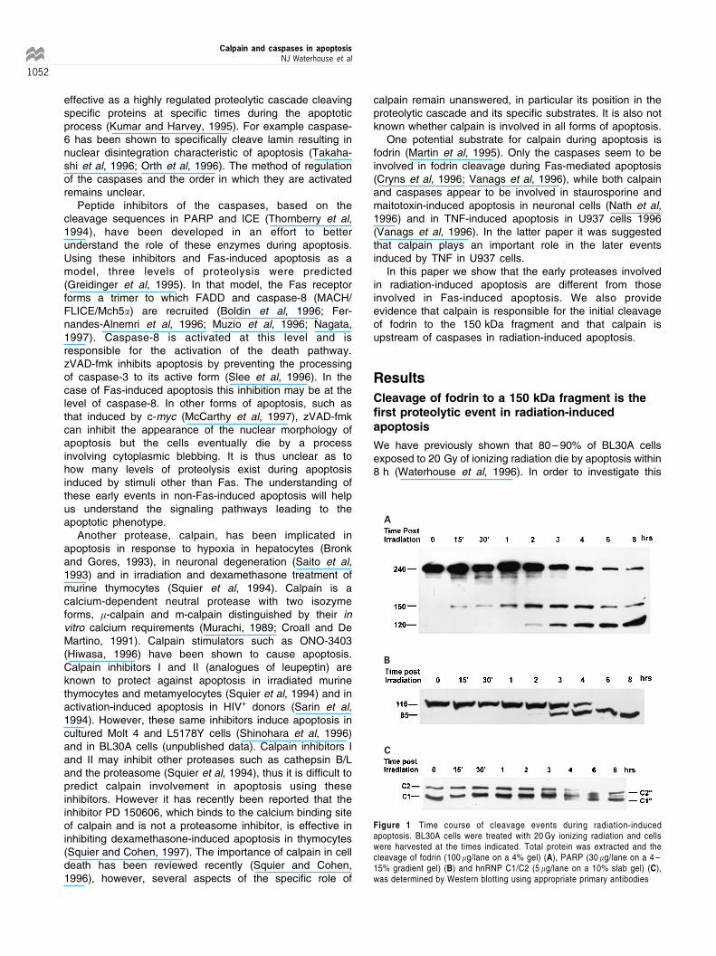
Figure 1 Time course of cleavage events during radiation-inducedapoptosis. BL30A cells were treated with 20 Gy ionizing radiation and cellswere harvested at the times indicated. Total protein was extracted and thecleavage of fodrin (100 mg/lane on a 4% gel) (A), PARP (30 mg/lane on a 4 ±15% gradient gel) (B) and hnRNP C1/C2 (5 mg/lane on a 10% slab gel) (C),was determined by Western blotting using appropriate primary antibodies
Calpain and caspases in apoptosisNJ Waterhouse et al
1052

process in more detail, the time course of the cleavage ofseveral proteins degraded during apoptosis was investigated.These experiments revealed the earliest cleavage event to bethe degradation of fodrin to a 150 kDa fragment commencingwithin 15 min (Figure 1A), which precedes the cleavage of thisprotein to a 120 kDa fragment, first apparent by 2 h post-irradiation (Figure 1A). Cleavage fragments of PARP (Figure1B) and the hnRNP C proteins (Figure 1C) also appear at 2 ±4 h in response to radiation damage.
The requirement of 100 mM zVAD-fmk to inhibit thecleavage of fodrin to a 150 kDa fragment is afeature distinguishing it from other proteolyticevents in radiation-induced apoptosis
It has been shown that 10 mM zVAD-fmk is sufficient to inhibitthe cleavage of fodrin during Fas-induced apoptosis in Jurkatcells (Greidinger et al, 1995). Since BL30A cells do notundergo Fas-induced apoptosis due to the absence of Fas-receptor (data not shown), we employed an EBV-transformedlymphoblastoid cell line, AT1ABR, to demonstrate theinhibition of fodrin cleavage by 10 mM zVAD-fmk during Fas-induced apoptosis (Figure 2A). In radiation-induced apoptosisin BL30A cells, 10 mM zVAD-fmk was sufficient to inhibit thecleavage of fodrin to a 120 kDa fragment, however ten timesthe concentration of zVAD-fmk was required to completely
inhibit the cleavage of fodrin to a 150 kDa fragment (Figure2B). This inhibition profile was also seen when BL30A cellswere treated with 40 mM etoposide (Figure 2C) and whenmurine thymocytes were treated with 10 Gy radiation (Figure2D).
Nuclear fragmentation as determined by DNA ladderingon an agarose gel (Figure 3A), cleavage of proteinsknown to be degraded by caspases during apoptosis,PARP (Figure 3B) and hnRNP C1/C2 (Figure 3C),phosphatidylserine exposure as determined by annexinV binding (Figure 3D) and morphological changes asdetermined by electron microscopy (Figure 3E), were allprevented by the addition of 10 mM zVAD-fmk. Thissuggests that the protease responsible for the initialcleavage of fodrin to the 150 kDa fragment was unique inthat 100 mM zVAD-fmk was required to fully inhibit thereaction.
Caspases -1, -3, -6, -7 and calpain cleave fodrin invitro to a 150 kDa fragment
Calpain has previously been shown to cleave murine fodrinbetween the tyrosine and glycine residues to create a150 kDa fragment (Harris et al, 1988). There are also manypotential caspase cleavage sites and one of these (DETD/S) is in close proximity to the calpain cleavage site. A
A B
C D
Figure 2 Inhibition of fodrin cleavage by zVAD-fmk. All cells were pre-treated for 15 min with the concentrations of zVAD-fmk indicated. The cells were treatedwith the apoptotic stimuli, incubated for 8 h and total protein was extracted. By this time the majority of cells in the treated populations without inhibitors wereapoptotic. AT1ABr cells were treated with Fas antibody (A), BL30A cells were treated with 20 Gy ionizing radiation (B), BL30A cells were treated with 40 mMetoposide (C) and murine thymocytes were treated with 10 Gy ionizing radiation (D). 100 mg of protein was loaded per lane of a 4% SDS ± PAGE gel and Westernblotted using anti-fodrin primary antibodies
Calpain and caspases in apoptosisNJ Waterhouse et al
1053

A B
C
D
E
Figure 3 Inhibition of the hallmarks of apoptosis by zVAD-fmk. BL30A cells were pre-treated for 15 min with the concentrations of zVAD-fmk indicated followed bytreatment with 20 Gy ionizing radiation. The cells were harvested after 8 h when 80 ± 90% irradiated cells, with no inhibitor, were apoptotic. The DNA from 56105
cells per sample was subjected to agarose gel electrophoresis and visualised by ethidium bromide staining (A), Total cellular protein was extracted and thecleavage of caspase specific substrates, PARP (30 mg of total protein per lane resolved on a 4 ± 15% gel) (B) and hnRNP C1/C2 (5 mg of total protein resolved on a10% slab gel) (C) was determined. The population of cells was stained with Annexin V and 5000 cells from each sample were assayed by flow cytometry todetermine the extent of phosphatidylserine exposure (D) and the morphological changes associated with apoptosis as viewed by electron microscopy were shown(E). 10 mM of zVAD-fmk was sufficient to inhibit all of these apoptotic events
Calpain and caspases in apoptosisNJ Waterhouse et al
1054

similar situation exits in human fodrin (Stabach et al, 1997)(Figure 4). Thus there has been much debate as to whethercaspases rather than calpain are responsible for thecleavage of fodrin during apoptosis in vivo (Greidinger etal, 1995; Cryns et al, 1996; Nath et al, 1996; Vanags et al,1996). Since 100 mM zVAD-fmk can inhibit the cleavage offodrin in radiation-induced apoptosis, we investigated whichproteases caused cleavage of fodrin to a 150 kDa fragmentin vitro. A panel of caspases was incubated with total cellextracts and assayed by Western blot for their ability tomediate fodrin cleavage. Of the caspases used, onlycaspases -1, -3, -6 and -7 had the ability to generate the150 kDa fragment (Figure 5A). These reactions were allsensitive to inhibition by zVAD-fmk, caspases -1 and -6
Figure 4 The amino acid sequence of human fodrin, residues 1171 ± 1210(Stabach et al, 1997) shows the calpain cleavage site and potential caspasecleavage site adjacent to the calmodulin binding domain. The position of the18 potential caspase cleavage sites is also shown (not to scale)
A
B i
B ii
Figure 5 Caspases-1, -3, -6 and -7 can cleave fodrin to a 150 kDa fragment in vitro. Total cell lysates (100 mg) from BL30A cells were incubated at 378C for 3 h withequal amounts of caspase activity (caspases-1, -8 and -10) as determined by assay of fluorogenic substrates. The samples were subjected to Western blottingusing 4% SDS ± PAGE gels and anti-fodrin antibodies (A). The cleavage of fodrin by recombinant caspases -1 and -3 (B i and caspases -6 and -7 (B ii) wasinhibited by adding 10 mM zVAD-fmk to similar digests as described in Figure 5A
Calpain and caspases in apoptosisNJ Waterhouse et al
1055

being inhibited by 1 mM zVAD-fmk and caspases -3 and -7being inhibited by 10 mM zVAD-fmk Figure 5B i and ii). It isnoted that there is a trace amount of caspase-3 activityremaining in the presence of 10 mM zVAD-fmk.m-Calpain added to cell extracts also mediates fodrin
cleavage to the 150 kDa fragment (Figure 6). Since thecleavage of fodrin during radiation-induced apoptosis isinhibited by 100 mM zVAD-fmk (Figure 2B) we testedwhether calpain could be inhibited by zVAD-fmk. We showthat 10 mM zVAD-fmk is not sufficient to inhibit the in vitrocleavage of fodrin by calpain, but 100 mM zVAD-fmkinhibits this cleavage completely (Figure 6). Since, in thisreaction, it is possible that the inhibitor is blocking acalpain-activated caspase, we showed that zVAD-fmk alsoblocks the hydrolysis of casein by purified calpain in an invitro assay, demonstrating that zVAD-fmk directly inhibitscalpain (Table 1).
Calpain mediates the cleavage of fodrin to the150 kDa fragment in radiation-induced apoptosisin vivo
We have shown that five proteases, m-calpain andcaspases-1, -3, -6 and -7 are capable of cleaving fodrin invitro. Calpain and caspase-3 are less sensitive to inhibitionby zVAD-fmk and as such, are possible candidates for thecleavage of fodrin to the 150 kDa fragment in vivo. Todetermine which enzyme is involved in vivo, we analyzedthe time course of activation as evidenced by cleavage totheir active fragments after 20 Gy radiation. Caspase-3 isonly activated 2 h after irradiation in BL30A cells (Figure 7A)whereas calpain activation occurs within 15 min (Figure 7B).We also show that there is almost negligible activation of
Figure 6 The m-calpain cleavage of fodrin to a 150 kDa fragment in vitro isinhibited by zVAD-fmk. Calpain (0.256 U) was incubated with total cell lysatefrom BL30A cells and the indicated concentration of zVAD-fmk at 378C for20 min. The samples were Western blotted using 4% SDS ± PAGE gels andanti-fodrin antibodies. Calpain can cleave fodrin to a 150 kDa fragment in vitroand this cleavage was completely inhibited by 100 mM zVAD-fmk
Table 1 Inhibition of calpain activity by zVAD-fmk
[zVAD-fmk] mM Rate (dA/min)
00.115
1050
100
0.1050.1040.1050.0660.0410.0050.003
Calpain activity was measured as described (Jiang et al, 1997). 0.256 U ofcalpain was incubated with 1 mg/ml casein and the indicated concentration ofzVAD-fmk at room temperature. The rate of hydrolysis was calculated for variouszVAD-fmk concentrations. The experiment was performed several times withsimilar results each time. The standard deviation was between 5% and 15% inall cases. This shows that calpain is directly inhibited by zVAD-fmk
A B
C i C ii
Figure 7 Time course of protease activation in BL30A and BL30K cells treated with 20 Gy ionizing radiation. The cells were treated with 20 Gy ionizing radiationand harvested at the times indicated. Total cell lysates from BL30A cells were extracted and caspase-3 activation was determined by Western blotting, 300 mg/laneon a 15% SDS ± PAGE gel using anti-caspase-3 primary antibody (A). Calpain activation was determined by Western blotting. 10 mg/lane was loaded on an 8%SDS ± PAGE gel and detected using anti-calpain antibodies (B). 10 mg/lane of BL30K lysates were run on an 8% SDS ± PAGE gel and detected by Western blottingusing anti-calpain primary antibodies. This shows that calpain is not activated in the radiation resistant cell line (C i). 100 mg/lane of the BL30K lysates were run on a4% gel and analyzed by Western blotting to show that fodrin is not cleaved after radiation treatment in BL30K cells (C ii)
Calpain and caspases in apoptosisNJ Waterhouse et al
1056

calpain in BL30K cells (Figure 7C i), nor is fodrin cleaved(Figure 7C ii), correlating with the relative resistance ofthese cells to radiation-induced apoptosis. The strongcorrelation between the time of activation of calpain(Figure 7B) and the time of the initial cleavage of fodrin toa 150 kDa fragment (Figure 1A) coupled with the inhibitordata suggests that, in radiation-induced apoptosis, calpainis responsible for the initial cleavage of fodrin to the150 kDa fragment. The time course of activation ofcaspase-3 also correlates with the time of cleavage offodrin to a 120 kDa fragment and inhibitor data suggeststhat caspase-3 is responsible for the second cleavage to the120 kDa fragment. Caspase-2 (Nedd-2/ICH-1) did notcleave fodrin in our experiments, however it has beenreported that caspase-2 cleaved fodrin to a 150 kDafragment in vitro (Nath et al 1996). Caspase-2 was onlyactivated in BL30A cells 2 h after radiation treatment (datanot shown), suggesting that even if caspase-2 can cleavefodrin to a 150 kDa fragment in vitro, it does not mediatethis cleavage in vivo in radiation-induced apoptosis.
Cleavage of caspase-3 to its active form isinhibited by 100 mM zVAD-fmk but not by 10 mMzVAD-fmk
In order to determine whether calpain activation is instru-mental in the activation of the caspases or whether it lies onanother pathway, we investigated the proteolytic cleavage(activation) of caspase-3 by Western blotting after irradiationin the presence of 10 mM zVAD-fmk. This concentrationshould have minimal effect on calpain activity but willcompletely block caspase activity. The results of thisexperiment are shown in Figure 8. Caspase-3 is stillproteolytically processed in the presence of the 10 mMzVAD-fmk thus the apoptotic signal from radiation throughcalpain to caspase processing is still functional, only thedownstream events relying on activity of caspases areinhibited (Figure 3). Caspase-3 is not processed when theconcentration of zVAD-fmk is increased to 100 mM, theconcentration required to inhibit calpain. This provides strongsupport for calpain being placed upstream of the caspases inradiation-induced apoptosis.
Discussion
Our results show that the first proteolytic event in radiation-induced apoptosis of BL30A cells is the cleavage of fodrin to a150 kDa fragment appearing within 15 min, followed byfurther cleavage to a 120 kDa fragment which is evident by2 ± 4 h, concomitant with the cleavage of PARP and thehnRNP C proteins (Figure 1). In human and murine fodrinthere is a caspase cleavage site within 1 kDa of the calpaincleavage site (Figure 4) (Harris et al, 1988). Both caspasesand calpain have been shown to cleave fodrin to a 150 kDafragment in vitro, however it is still debatable as to whichenzyme is responsible for cleavage in vivo (Cryns et al, 1996;Nath et al, 1996; Vanags et al, 1996).
It has been reported that in Fas-induced apoptosis ofJurkat cells, both cleavage events in fodrin are inhibited by10 mM zVAD-fmk (Greidinger et al, 1995). This is also truein our hands in AT1ABR cells (Figure 2A). The level atwhich zVAD-fmk (10 mM) inhibits Fas-induced apoptosismay be caspase-8, which has been linked directly to theFas-initiated death-inducing complex. In radiation- andetoposide-induced apoptosis of BL30A cells and inradiation-induced apoptosis of murine thymocytes, theinitial cleavage of fodrin to a 150 kDa fragment is mediatedby a protease inhibited by high concentrations of zVAD-fmk(100 mM) whereas the protease responsible for thecleavage of fodrin to the 120 kDa fragment is moresensitive to inhibition by zVAD-fmk (10 mM) (Figure 2D).
zVAD-fmk is categorised as a caspase inhibitor and islikely to have different specificity for different caspases,however its inhibitory profile for individual caspases is notknown. In radiation-induced apoptosis, 10 mM zVAD-fmk issufficient to inhibit all the hallmarks of apoptosis:- nuclearfragmentation, cleavage of PARP, cleavage of hnRNP C1/C2, phosphatidylserine exposure, apoptotic morphology(Figure 3) and the formation of the 120 kDa fragment offodrin, but not the appearance of the 150 kDa band (Figure2B). Thus zVAD-fmk at 10 mM stops the proteolyticcascade at a point further downstream in the radiationpathway. While it delays apoptosis for up to 8 h (Figure 3),the cells have lost their ability to proliferate (data notshown) and may eventually die.
In all systems studied in this paper, a single 150 kDafragment of fodrin was observed, in contrast to the SH-SY5Y neuronal model (Nath et al, 1996) where a doubletwas observed at 150/145 kDa. In irradiated murinethymocytes, but not in irradiated human lymphoma cells,we observed a doublet at 120 kDa. This may indicate thatdifferent proteases are responsible for the cleavage offodrin to the two fragments in different models of apoptosis.
In order to determine which enzyme could beresponsible for the cleavage of fodrin to the 150 kDa and120 kDa fragments in radiation-induced cell death, we useda panel of recombinant caspases and m-calpain in vitro.The only proteases with the ability to form similar fragmentsto those seen in vivo were caspases -1, -3, -6, -7 and m-calpain (Figures 5A and 6). In our experiments, caspases-1 and -6 were inhibited by 1 mM zVAD-fmk (Figure 5B).This suggests that caspases -1 and -6 are not involved in,or upstream of any of the apoptotic events assayed here
Figure 8 Cleavage of caspase-3 to its active form is inhibited by 100 mMzVAD-fmk but not by 10 mM zVAD-fmk. BL30A cells were pre-treated for 15 minwith the concentrations of zVAD-fmk indicated, followed by treatment with20 Gy g-radiation. The cells were harvested after 8 h when 80 ± 90% of theirradiated cells were apoptotic in the absence of inhibitor. Total cell lysateswere extracted and 300 mg/lane of each sample was run on a 15% SDS ±PAGE gel. Caspase-3 activation was determined by Western blotting usinganti-caspase-3 primary antibody. The 17 kDa band represents the activefragment
Calpain and caspases in apoptosisNJ Waterhouse et al
1057

(Figure 3) as fodrin was still fully cleaved and all apoptoticevents were completed in the presence of 1 mM zVAD-fmk.Caspase-6 has been implicated in the cleavage of lamin,which has been shown to be a late event in Fas-inducedapoptosis (Greidinger et al, 1995). Caspase-7 is inhibitedby 10 mM zVAD-fmk and therefore cannot be responsiblefor the cleavage of fodrin to the 150 kDa fragment duringradiation-induced apoptosis, however it may be involved inthe cleavage of fodrin to the 120 kDa fragment. Caspase-3is also inhibited by 10 mM zVAD-fmk, although trace activityis still observed when treated with this concentration ofinhibitor (Figure 5B i). Nevertheless, it is unlikely thatcaspase-3 cleaves fodrin to the 150 kDa fragment in vivo,since it is not activated in BL30A cells until 2 h aftertreatment with 20 Gy ionizing radiation (Figure 7A) and the150 kDa fragment is evident at least 1.5 h prior to thisactivation.
During Fas-induced apoptosis, 100 mM of the specificcaspase-3 inhibitor, DEVD-CHO, inhibits the cleavage offodrin to the 120 kDa fragment but not to the 150 kDafragment (Greidinger et al, 1995). This is also evident inradiation-induced apoptosis (data not shown). Caspase-3 ismost likely to be responsible for the cleavage of fodrin to the120 kDa fragment since DEVD-CHO inhibits this cleavage invivo and since in vitro, caspase-3 appears to cleave fodrin tothe 120 kDa fragment more efficiently than caspase-7(Figure 5A). There are 18 potential caspase cleavage sitesin fodrin (Figure 4) however the actual cleavage giving riseto the 120 kDa fragment is not yet known.
Since none of the caspases tested appeared to beresponsible for the cleavage of fodrin to the 150 kDafragment, this suggests that either calpain activates acaspase as yet undescribed in the literature, or thatzVAD-fmk is not a specific caspase inhibitor and caninhibit calpain. There is no evidence to suggest thatcalpain can activate the caspases in a cell free system.The use of peptides has revealed no definite recognitionsequence for calpain as reviewed in (Takahashi, 1990).Amino acids with an aromatic or large aliphatic sidechain are preferred in positions P1, P2 and P3 andbasic or large aliphatic amino acids in the P'1 position.Nonetheless, we observed that 100 mM zVAD-fmk couldinhibit the cleavage of fodrin by calpain in vitro (Figure6). We have also demonstrated that zVAD-fmk candirectly inhibit the hydrolysis of casein by calpain in anenvironment free of caspases (Table 1). The data showsthat 100 mM zVAD-fmk was sufficient to inhibit thereaction completely, whereas 10 mM was only sufficientto reduce the rate of hydrolysis. This inhibition profilecorrelates strongly with the inhibition profile for thecalpain cleavage of fodrin in vitro and the cleavage offodrin to a 150 kDa fragment in vivo. zVAD-fmk issupplied with an OMe block on the Asp-b-carbonyl toenhance cell permeability (Enzyme Product SystemsCatalogue). The Asp-OMe may sterically appear like aleucine and in this way act as a calpain inhibitor(anonymous reviewer's comments). Our results providethe first direct evidence that zVAD-fmk inhibits calpain aswell as caspases. Since zVAD-fmk also inhibitsapoptosis inducing factor (Susin et al, 1996), conclu-
sions from experiments using zVAD-fmk as a caspasespecific inhibitor should be made with caution.
The time course of activation of calpain (Figure 7B) alsocorrelates with the time course of cleavage of fodrin to a150 kDa fragment (Figure 1A). This combined with thestrong correlation between the inhibition profiles of calpainby zVAD-fmk in vitro and the inhibition profile of thecleavage of fodrin to the 150 kDa fragment in vivo and alsothe fact that none of the caspases assayed can beresponsible for fodrin cleavage in vivo, is compellingevidence for the involvement of calpain in the cleavage offodrin to a 150 kDa fragment during radiation-inducedapoptosis in BL30A cells.
The reasons for the two step cleavage of fodrin are stillunclear. It has been suggested that the cleavage of fodrinmay result in phosphatidylserine exposure (Martin et al,1995, 1996; Vanags et al, 1996). We show that inradiation-induced apoptosis, since 10 mM zVAD-fmk issufficient to inhibit phosphatidylserine exposure, thecleavage of fodrin to a 150 kDa fragment is not likely toinduce this event. Since all of the other assayed apoptoticevents are also inhibited by 10 mM zVAD-fmk, it is alsoprobable that the cleavage of fodrin to a 150 kDa fragmentdoes not result in any of these phenomena. The initialcleavage of fodrin to a 150 kDa fragment may just be aconsequence of calpain activation, however this appearsunlikely since two distinct proteolytic pathways, one inFas-induced apoptosis and one in radiation-inducedapoptosis, involve the cleavage of fodrin to a 150 kDafragment. It is possible that the first cleavage makes fodrinmore susceptible to the second caspase-mediated
Figure 9 A schematic representation of the possible molecular ordering ofthe apoptotic pathway in radiation-induced cell death
Calpain and caspases in apoptosisNJ Waterhouse et al
1058

cleavage giving rise to the 120 kDa fragment. However,this remains to be established.
The evidence provided in Figure 8, places calpainupstream of the caspases, however the mode of activationof calpain is presently unknown. Studies in our laboratoryhave shown that levels of ceramide increase after ionizingradiation in the sensitive BL30A cell line, but not in theresistant BL30K cell line (Michael et al, 1997). It is alsoknown that ceramide can lead to activation of calpain indifferentiated PC12 cells (Xie and Johnson, 1997). Tofurther address the role of calpain as an importantcomponent of the radiation-signalling pathway leading toapoptosis, it will be important to determine the signalsleading to calpain activation and the subsequent stepsleading to activation of the caspases.
We propose a model (Figure 9) for the proteolyticcascade in radiation-induced apoptosis. The data pre-sented in this study predict that calpain is responsible forthe cleavage of fodrin to a 150 kDa fragment and thatcalpain is activated upstream of the caspases.
Materials and Methods
Reagents
Etoposide, b-glycerophosphate and Nonidet P-40 (NP-40) werepurchased from Sigma, (St Louis MO, USA). Leupeptin, aprotininand EGTA were from ICN, (Costa Mesa, CA, USA). Calpain-I andTriton X-100 were from Calbiochem (La Jolla, CA, USA) andPepstatin was from Auspep (Parkville, Australia). z-Val-Ala-Asp-CH2F (zVAD-fmk) was obtained from Enzyme Products Systems,(Dublin, CA, USA). Complete inhibitor, Proteinase K, DNAse freeRNAse A and PARP antibody were from Boehringer MannheimGmBH. Annexin V was from Biowhittaker, (Walkersville, MD, USA).Caspase-3 (CPP32) antibody was from Santa Cruz (CA, USA). Themonoclonal antibody to hnRNP C (4F4) has been describedpreviously (Choi and Dreyfuss, 1984) and was a kind gift from Dr.Gideon Dreyfuss (Philadelphia, USA). The antibody to humancalpain-I has been previously described (Samis et al, 1987).Monoclonal antibody to Fas (clone CH11) was from Immunex(Seattle, Washington, USA.). Electrophoresis materials were fromBIO-RAD and all other reagents were analytical grade.
Cell culture
The isogenic Burkitt's lymphoma cell lines used in this study, (BL30A,radiation sensitive and BL30K, radiation resistant) have beendescribed previously (Waterhouse et al, 1996). AT1ABr, a lympho-blastoid cell line from a patient with ataxia-telangiectasia, has alsobeen previously described (Watters et al, 1997). Murine thymocyteswere removed from 4 ± 6 week old outbred mice (Quackenbush). Cellswere maintained at 378C in Roswell Park Memorial Institute(RPMI)1640 medium supplemented with 10% heat-inactivated foetalcalf serum (FCS) in a 5% CO2 atmosphere. BL30A cells weremaintained under similar conditions using 20% FCS. For the inductionof apoptosis, BL30A cells were irradiated with 20 Gy ionizing radiationusing a 137Cs source at 3 Gy/min or exposed to 40 mM etoposide.AT1ABr cells were treated with anti-Fas antibody (250 ng/ml).Thymocytes were exposed to 10 Gy ionizing radiation. Inhibitorswere added 15 min prior to the apoptotic stimulus.
Protein isolation for Western blot, caspasedigestion and calpain digestion
Cells were pelleted and washed twice in cold PBS. The pellet was thenlysed in modified Universal Immunoprecipitation Buffer (50 mM Tris-HCl pH 7.4, 150 mM NaCl, 2 mM EDTA, 2 mM EGTA, 25 mM NaF,25 mM b-glycerophosphate, 0.2% Triton X-100, 0.3% NP40 and16complete inhibitorTM) and rocked at 48C for 10 min. This was thenmicrofuged at 10 500 g for 10 min and the supernatant was stored at7708C. If lysates were to be digested by caspases or calpain, thecomplete inhibitorTM was not added. For calpain digestion, EDTA andEGTA were also left out of the lysis buffer and 0.2 mM CaCl2 and5 mM b-mercaptoethanol were added.
Western blotting
For Western blotting, equal quantities of protein were resolved bystandard Laemmli SDS-polyacrylamide gel electrophoresis. The gelwas electroblotted to Hybond-C nitrocellulose using Towbins Buffer(125 mM Tris, 95 mM glycine, 0.02% SDS, 20% methanol). Themembrane was washed in PBS for 1 h and then blocked in 5% skimmilk powder in PBS (SMP-PBS) for 1 h. Primary antibody in 5% SMP-PBS was added and rocked gently overnight at 48C. The membranewas washed in PBS (3620 min), the horseradish peroxidase-conjugated secondary antibody in 5% SMP-PBS was added andincubation continued for 2 h at room temperature. The membrane waswashed again in PBS (3620 min). Proteins were detected using theDuPont RennaissanceTM chemiluminescence kit with ReflectionTM
film.
Detection of DNA fragmentation
56105 cells were microfuged for 3 min and the pellet was lysed in25 ml lysis buffer (0.5% SDS, 10 mM EDTA, 50 mM Tris-HCI pH 8.0,50 mg/ml Proteinase K) and incubated at 508C for 1 h. 10 ml of DNAse-free RNAse A was added and incubation continued for a further hour at508C. Samples were heated in loading buffer (10 mM EDTA, 0.25%bromophenol blue, 40% sucrose, 1% agarose (low melting point) to708C and were resolved on a 2% agarose TBE gel.
Annexin V binding
The cells were pelleted by centrifugation at 5006g. The media wasaspirated and the cells were resuspended in 50 ml of 0.5 mg/mlAnnexin V in HEPES buffer (10 mM HEPES-NaOH pH 7.4, 150 mMNaCl, 5 mM KCl, 1 mM MgCl2, 1.8 mM CaCl2) as described (Koopmanet al, 1994). This was incubated at room temperature for 5 min and thevolume was made up to 200 ml with HEPES buffer. Propidium iodidewas added to a final concentration of 10 mg/ml before analysis. Theannexin V binding was determined by flow cytometry.
Caspase digestion
Caspases were expressed in E. coli as previously described (Song etal, 1996). Activities of various caspases in bacterial extracts wereestimated using fluorogenic peptide substrates, YVAD-AMC forcaspase-1, -4 and -5 and DEVD-AMC for all others. Aliquotscontaining equivelent molar amounts of active caspases (usually in2 ± 10 ml volume) were incubated with 100 mg of total cell lysate fromBL30A cells at 378C for 3 h. The digestion reactions were carried out ina total of 20 ml, consisting of 2 ml of total cell lysate (50 mg/ml) fromBL30A cells, 2 ml of 106caspase assay buffer [250 mM HEPES, pH7.5, 50 mM EDTA, 20 mM DTT and 1% CHAPS] and the caspase. The
Calpain and caspases in apoptosisNJ Waterhouse et al
1059

reaction was terminated by boiling the samples in 56SDS ± PAGEloading dye for 5 min. The samples were then subjected to Westernblot analysis from a 4% SDS ± PAGE gel using anti-fodrin antibodies.
Calpain Digestion
100 mg of total protein extracted from BL30A cells was placed in eachof several microfuge tubes. 5 ml of the appropriate inhibitor was addedto each tube followed by 2 ml (0.256 U) of calpain. The sample volumewas made to 20 ml with lysis buffer and incubated at 378C for 20 min.The reactions were terminated by adding 5 ml of 56SDS ± PAGEloading dye and boiling for 5 min.
Calpain activity assays
In order to determine the effect of zVAD-fmk on calpain activity, aspectrophotometric assay (Jiang et al, 1997) was used. Briefly, calpain(0.256 U) and the appropriate concentration of zVAD-fmk were dilutedto 95 ml in casein/imidazole buffer (50 mM imidazole-HCl (pH 7.5),10 mM b-mercaptoethanol, 1 mM sodium azide and 1 mg/ml casein)and incubated for 5 min at room temperature. Calcium chloride wasthen added to a final concentration of 5 mM to initiate the calpainhydrolysis reaction. The spectrophotometer was immediately blankedand the absorbance at 500 nm was measured at 30 s intervals. Sincethe hydrolysis reaction was sigmoidal, the rates were measured at thesteepest point of the curve.
AcknowledgementsWe thank Wen Yu and Deborah Stenzel for Electron Microscopy andGideon Dreyfuss for hnRNP C antibodies. This study was supported bygrants from the Queensland Cancer Fund and the University ofQueensland, Cancer Research Fund.
References
Alnemri ES, Livingston DJ, Nicholson DW, Salvesen G, Thornberry NA, Wong WW
and Yuan J (1996) Human ICE/CED-3 protease nomenclature. Cell 87: 171Boldin MP, Goncharov TM, Goltsev YV and Wallach D (1996) Involvement of MACH,
a novel MORT1/FADD-interacting protease, in Fas/APO-1- and TNF receptor-
induced cell death. Cell 85: 803 ± 815
Bronk SF and Gores GJ (1993) pH-dependent nonlysosomal proteolysis contributes
to lethal anoxic injury of rat hepatocytes. Am. J. Physiol. 264: G744 ± G751
Casciola-Rosen LA, Anhalt GJ and Rosen A (1995) DNA-dependent protein kinase is
one of a subset of autoantigens specifically cleaved early during apoptosis. J.
Exp. Med. 183: 1625 ± 1634
Casciola-Rosen LA, Miller DK, Anhalt GJ and Rosen A (1994) Specific cleavage of
the 70-kDa protein component of the U1 small nuclear ribonucleoprotein is a
characteristic biochemical feature of apoptotic cell death. J. Biol. Chem. 269:
30757 ± 30760
Chinnaiyan AM, O'Rourke K, Lane BR and Dixit VM (1997) Interaction of CED-4 with
CED-3 and CED-9: a molecular framework for cell death. Science 275: 1122 ±
1126
Choi YD and Dreyfuss G (1984) Monoclonal antibody characterization of the C
proteins of heterogeneous nuclear ribonucleoprotein complexes in vertebrate
cells. J. Cell. Biol. 99: 1997 ± 2004Croall DE and De Martino GN (1991) Calcium-activated neutral protease (calpain)
system: structure, function, and regulation. Physiol. Rev. 71: 813 ± 847
Cryns VL,Bergeron L,Zhu H,LiH andYuan J (1996)Specificcleavage ofalpha-fodrin
during Fas- and tumor necrosis factor-induced apoptosis is mediated by an
interleukin-1beta-converting enzyme/Ced-3 protease distinct from the
poly(ADP-ribose) polymerase protease. J. Biol. Chem. 271: 31277 ± 31282
Earnshaw WC (1995) Nuclear changes in apoptosis. Curr. Opin. Cell Biol. 7: 337 ±
343
Fernandes-Alnemri T, Armstrong RC, Krebs J, Srinivasula SM, Wang L, Bullrich F,
Fritz LC, Trapani J, Tomaselli KJ, Litwack G and Alnemri ES (1996) In vitro
activation of CPP32 and Mch3 by Mch4, a novel human apoptotic cysteine
protease containing two FADD-like domains. Proc. Natl. Acad. Sci. USA. 93:
7464 ± 7469
Greidinger EL, Miller DK, Yamin TT, Casciola-Rosen L, Rosen A (1995) Sequential
activation of three distinct ICE-like activities in Fas-ligated Jurkat cells. FEBS
Letts 390: 299 ± 303Gu Y, Sarnecki C, Aldape RA, Livingston DJ and Su MS-S (1995) Cleavage of
poly(ADP-ribose) polymerase by interleukin-1 beta converting enzyme and its
homologs TX and Nedd-2. J. Biol. Chem. 270: 18715 ± 18718
Harris AS, Croall DE and Morrow JS (1988) The calmodulin-binding site in alpha-
fodrin is near the calcium-dependent protease-1 cleavage site. J. Biol. Chem.
263: 15754 ± 15761
Hengartner MO and Horvitz HR (1994a) C. elegans cell survival gene ced-9 encodes
a functional hormolog of the mammalian proto-oncogene bcl-2. Cell 76: 665 ±
676
Hengartner MO and Horvitz HR (1994b) Programmed cell death in Caenorhabditis
elegans. Curr. Opin. Genet. Dev. 4: 581 ± 586
Hiwasa T (1996) Induction of apoptosis by a calpain stimulator, ONO-3403.
Apoptosis 1: 75 ± 80
Jiang S-T, Wang J-H, Chang T and Chen C-S (1997) A continuous method for
measuring calpain activity. Anal. Biochem. 244: 233 ± 238
Kerr JFR, Wyllie AH and Currie AR (1972) Apoptosis: a basic biological phenomenon
with wide-ranging implications in tissue kinetics. Br. J. Cancer 26: 239 ± 257
Koopman G, Reutelingsperger CPM, Kuijten GAM, Keehnen RMJ, Pals ST and vanOers MHJ (1994) Annexin V for flow cytometric detection of phosphatidylserine
expression on B cells undergoing apoptosis. Blood 84: 1415 ± 1420
Kroemer G (1997) The proto-oncogene Bcl-2 and its role in regulating apoptosis.
Nature Medicine 3: 614 ± 620
Kumar S and Lavin MF (1996) The ICE family of cysteine proteases as effectors of cell
death. Cell Death Differ. 3: 255 ± 267
Kumar S and Harvey NL (1995) Role of multiple cellular proteases in the execution of
programmed cell death. FEBS Letts. 375: 169 ± 173
Lazebnik YA, Kaufmann SH, Desnoyers S, Poirier GG and Earnshaw WC (1994)
Cleavage of poly(ADP-ribose) polymerase by a proteinase with properties like
ICE. Nature 371: 346 ± 347
Lazebnik YA, Takahashi A, Moir RD, Goldman RD, Poirier GG, Kaufmann SH and
Earnshaw WC (1995) Studies of the lamin proteinase reveal multiple parallel
biochemical pathways during apoptotic execution. Proc. Natl. Acad. Sci. USA 92:
9042 ± 9048
Martin SJ, Finucane DM, Amarante-Mendes GP, O'Brien GA and Green D (1996)
Phosphatidylserine externalization during CD95-induced apoptosis of cells and
cytoplasts requires ICE/CED-3 protease activity. J. Biol. Chem. 271: 31075 ±31085
Martin SJ, O'Brien GA, Nishioka WK, McGahon AJ, Mahboubi A, Saido TK and Green
DG (1995) Proteolysis of fodrin (non-erythroid spectrin) during apoptosis. J. Biol.
Chem. 270: 6425 ± 6428
McCarthy NJ, Whyte MKB, Gilbert CS and Evan GI (1997) Inhibition of Ced-3/ICE-
related proteases does not prevent cell death induced by oncogenes, DNA
damage, or the Bcl-2 homologue Bak. J. Cell. Biol. 136: 215 ± 227
Michael JM, Lavin MF and Watters DJ (1997) Resistance to radiation-induced
apoptosis in Burkitt's lymphoma cells is associated with defective ceramide
signaling. Cancer Res. 57: 3600 ± 3605
Murachi T (1989) Intracellular regulatory system involving calpain and calpastatin.
Biochem. Int. 18: 263 ± 294
Muzio M, Chinnaiyan AM, Kischkel FC, O'Rourke K, Shevchenko A, Ni J, Scaffidi C,
Bretz JD, Zhang M, Gentz R, Mann M, Krammer PH, Peter ME and Dixit VM
(1996) FLICE, a novel FADD-homologous ICE/CED-3-like protease, is recruited
to the CD95 (Fas/APO-1) death-inducing signaling complex. Cell 85: 817 ± 827
Nagata S (1997) Apoptosis by death factor. Cell 88: 355 ± 365
Nath R, Raser JK, Stafford D, Hajimohammadreza I, Posner A, Allen H, Talanian RV,
Yuen P, Gilbertsen RB and Wang KKW (1996) Non-erythroid alpha-spectrinbreakdown by calpain and interleukin 1 beta-converting-enzyme-like
protease(s) in apoptotic cells: contributory roles of both protease families in
neuronal apoptosis. Biochem. J. 319: 683 ± 690
Orth K, Chinnaiyan AM, Garg M, Froelich CJ and Dixit VM (1996) The CED-3/ICE-like
protease Mch2 is activated during apoptosis and cleaves the death substrate
lamin A. J. Biol. Chem. 271: 16443 ± 16446
Calpain and caspases in apoptosisNJ Waterhouse et al
1060

Perry DK, Smyth MJ, Wang H, Reed JC, Duriez P, Poirier GG, Obeid LM and Hannun
YA (1997) Bcl-2 acts upstream of the PARP protease and prevents its activation.
Cell Death Differ. 4: 29 ± 33
Reed JC (1997) Double identity for proteins of the Bcl-2 family. Nature 387: 773 ± 776
Saito K, Elce JS, Hamos JE and Nixon RA (1993) Widespread activation of calcium-
activated neutral proteinase (calpain) in the brain in Alzheimer disease: a
potential molecular basis for neuronal degeneration. Proc. Natl. Acad. Sci. USA.
90: 2628 ± 2632Samis JA, Aboril G and Elce JS (1987) Calpain I remains intact and intracellular
during platelet activation. Immunochemical measurements with monoclonal and
polyclonal antibodies. Biochem. J. 246: 481 ± 488
Sarin A, Clerici M, Blatt SP, Hendrix CW, Shearer GM and Henkart PA (1994)
Inhibition of activation-induced programmed cell death and restoration of
defective immune responses of HIV+ donors by cysteine protease inhibitors. J.
Immunol. 153: 862 ± 872
Shinohara K, Tomioka M, Nakano H, Tone S, Ito H and Kawashima S (1996)
Apoptosis induction resulting from proteasome inhibition. Biochem. J. 317: 385 ±
388
Slee EA, Zhu H, Chow SC, Mac Farlane M, Nicholson DW and Cohen GM (1996)
Benzyloxycarbonyl-Val-Ala-Asp(OMe) fluoromethylketone (Z-VAD.FMK) inhi-
bits apoptosis by blocking the processing of CPP32. Biochem. J. 319: 21 ± 24
Song Q, Lees-Miller SP, Kumar S, Zhang N, Chen DW, Smith GCM, Jackson SP,
Alnemri AS, Litwack G, Khanna KK and Lavin MF (1996) DNA-dependent protein
kinase catalytic subunit: a target for an ICE-like protease in apoptosis. EMBO J.
15: 3238 ± 3246
Songqing N, Chuang TH, Cunningham A, Turi TG, Hanke JH, Bokoch GM and DanleyDE (1996) D4-GDI, a substrate of CPP32, is proteolyzed during Fas-induced
apoptosis. J. Biol. Chem. 271: 11209 ± 11213
Squier MKT and Cohen JJ (1996) Calpain and cell death. Cell Death Differ. 3: 275 ±
283
Squier MKT and Cohen JJ (1997) Calpain, an upstream regulator of thymocyte
apoptosis. J. Immunol. 158: 3690 ± 3697
Squier MKT, Miller ACK, Malkinson AM and Cohen JJ (1994) Calpain activation in
apoptosis. J. Cell Physiol. 159: 229 ± 237
Stabach PR, Cianci CD, Glantz SB, Zhang Z and Morrow JS (1997) Site-directed
mutagenesis of alpha II spectrin at codon 1175 modulates its mu-calpain
susceptibility. Biochemistry 36: 57 ± 65
Susin SA, Zamzami N, Castedo M, Hirsch T, Marchetti P, Macho A, Daugas E,
Geuskens M and Kroemer G (1996) Bcl-2 inhibits the mitochondrial release of an
apoptogenic protease. J. Exp. Med. 184: 1331 ± 1341
Takahashi A, Alnemri ES, Lazebnik YA, Fernandes-Alnemri T, Litwack G, Moir RD,
Goldman RD, Poirier GG, Kaufmann SH and Earnshaw WC (1996) Cleavage of
lamin A by Mch2 alpha but not CPP32: multiple interleukin 1 beta-converting
enzyme-related proteases with distinct substrate recognition properties are
active in apopotosis. Proc. Natl. Acad. Sci. USA 93: 8395 ± 8400Takahashi K (1990) in Intracellular Calcium-dependent proteolysis. Mellgren RL and
Murachi T eds (Boca Raton, R.L.; CRC Press Inc) 55 ± 74
Thornberry NA, Peterson EP, Zhao JJ, Howard AD, Griffin PR and Chapman KT
(1994) Inactivation of interleukin-1 beta converting enzyme by peptide
(acyloxy)methyl ketones. Biochemistry 33: 3934 ± 3940
Vanags DM, Porn-Ares IM, Coppola S, Burgess DH and Orrenius S (1996) Protease
involvement in fodrin cleavage and phosphatidylserine exposure in apoptosis. J.
Biol. Chem. 271: 31075 ± 31085
Vaux DL and Strasser A (1996) The molecular biology of apoptosis. Proc. Natl. Acad.
Sci. USA 93: 2239 ± 2244
Waterhouse N, Kumar S, Song Q, Strike P, Sparrow L, Dreyfuss G, Alnemri E, Litwack
G, Lavin M and Watters D (1996) Heteronuclear ribonucleoproteins C1 and C2,
components of the spliceosome, are specific targets of interleukin 1beta-
converting enzyme-like proteases in apoptosis. J. Biol. Chem. 271: 29335 ±
29341
Watters D, Khanna KK, Beamish H, Birrell G, Spring K, Kedar P, Gatei M, Stenzel D,
Hobson K, Kozlov S, Zhang N, Farrell A, Ramsay J, Gatti R and Lavin M (1997)
Cellular localisation of the ataxia-telangiectasia (ATM) gene product anddiscrimination between mutated and normal forms. Oncogene 14: 1911 ± 1921
Wertz IE and Hanley MR (1996) Diverse molecular provocation of programmed cell
death. Trends in Biochem. Sci. 21: 359 ± 364
Xie H and Johnson GV (1997) Ceramide selectively decreases tau levels in
differentiated PC12 cells through modulation of calpain I. J. Neurochem. 69:
1020 ± 1030
Yuan J, Shaham S, Ledoux S, Ellis HM and Horvitz HR (1993) The C. elegans cell
death gene ced-3 encodes a protein similar to mammalian interleukin-1b-
converting enzyme. Cell 75: 641 ± 652
Calpain and caspases in apoptosisNJ Waterhouse et al
1061