Membrane tension and peripheral protein density mediate membrane shape transitions
Upload
independentCategory
view
0download
0
Investigations into the Membrane Interactions of m-CalpainDomain V
Sarah R. Dennison,* Silvia Dante,y Thomas Hauß,y Klaus Brandenburg,z Frederick Harris,* andDavid A. Phoenix§
*Department of Forensic and Investigative Science, University of Central Lancashire, Preston, United Kingdom; yBerlin NeutronScattering Centre, Hahn-Meitner-Institut, Berlin, Germany; zDivision of Biophysics, Forschunginstitut, Borstel, Germany; and§Dean’s Office, Faculty of Science, University of Central Lancashire, Preston, United Kingdom
ABSTRACT m-Calpain is a calcium-dependent heterodimeric protease implicated in a number of pathological conditions. Theactivation of m-calpain appears to bemodulated bymembrane interaction, which has been predicted to involve oblique-orientateda-helix formation by aGTAMRILGGVI segment located in domain V of the protein’s small subunit. Here, we have investigated thisprediction. Fourier transform infrared conformational analysis showed that VP1, a peptide homolog of this segment, exhibiteda-helicity of ;45% in the presence of dimyristoylphosphatidylcholine/dimyristoylphosphatidylserine (DMPS) vesicles. The levelof helicity was unaffected over a 1- to 8-mM concentration range and did not alter when the anionic lipid composition of thesevesicles was varied between 1% and 10% DMPS. Similar levels of a-helicity were observed in trifluoroethanol and the peptideappeared to adopt a-helical structure at an air/water interface with a molecular area of 164 A2 at the monolayer collapse pressure.VP1 was found to penetrate dimyristoylphosphatidylcholine/DMPS monolayers, and at an initial surface pressure of 30 mN m�1,the peptide induced surface pressure changes in these monolayers that correlated strongly with their anionic lipid content(maximal at 4 mNm�1 in the presence of 10%DMPS). Neutron diffraction studies showed VP1 to be localized at the hydrophobiccore of model palmitoyloleylphosphatidylcholine/palmitoyloleylphosphatidylserine (10:1 molar ratio) bilayer structures and, incombination, these results are consistent with the oblique membrane penetration predicted for the peptide. It would also appearthat although not needed for structural stabilization anionic lipid was required for membrane penetration.
INTRODUCTION
Calpains (EC 3.4.22.17) are a superfamily of Ca21-dependent
intracellular cysteine proteases that are ubiquitously distrib-
uted in mammalian cells (Perrin and Huttenlocher, 2002;
Sorimachi and Suzuki, 2001). Despite intensive study, the
physiological functions of these enzymes are not wholly clear
but they are believed to play important roles in an array of
processes, including: embryonic development, cytoskeletal
remodeling, cell differentiation, apoptosis, and signal trans-
duction (Carafoli and Molinari, 1998; Ono et al., 1998; Sato
and Kawashima, 2001). Calpains are of medical importance,
having been implicated in a range of pathological conditions
as diverse as cataract formation, type 2 diabetes, muscular
dystrophy, rheumatoid arthritis, ischemic tissue damage/
reperfusion, and neurodegenerative conditions (Huang and
Wang 2001; Branca 2004). These disorders show a com-
mon characteristic of altered Ca21 homeostasis, and it has
been suggested that the resulting deregulated activation of
calpains may play a role in their development (Perrin and
Huttenlocher, 2002).
The most studied member of the calpain superfamily is
m-calpain (calpain 2). This enzyme is heterodimeric and, upon
activation, undergoes autolytic conversion to give its mature
proteolytic form. The larger subunit of m-calpain is 80 kDa
and organized into four domains (I–IV). Domain II houses
the enzyme’s active site, which is a papainlike cysteine pro-
tease domain (Tompa, 2001), whereas domain I forms an
N-terminal extension of domain II and is associated with
m-calpain autolysis. Domain III appears to be a linker region
between the catalytic domain and domain IV, which contains
a calmodulinlike Ca21-binding domain with multiple EF-
hands. The smaller subunit of m-calpain is 30 kDa and
possesses two domains (V and VI). Domain VI possesses a
calmodulinlike Ca21 binding domain and associates with
domain IV to facilitate dimerization of the m-calpain subunits,
whereas domain V is largely cleaved from m-calpain during
autolysis (Goll et al., 2003).
It is well established that for in vitro activation m-calpain
requires millimolar levels of Ca21 that are far in excess of
those found in vivo, clearly suggesting that activation of the
enzyme involves other factors (Johnson and Guttmann, 1997;
Hosfield et al., 2004). A membrane-associated form of
m-calpain is known and a number of studies have shown
that the presence of lipid can strongly reduce the Ca21 con-
centrations required for activation of the enzyme (Imajoh
et al., 1986; Melloni and Pontremoli, 1989; Saido et al., 1992)
to within range of those found in physiological systems
(Chakrabarti et al., 1996). These results have led to the
suggestion that lipid/membrane interactions could play a role
in the activation of m-calpain (Johnson and Guttmann, 1997),
and based on in vitro studies, two regions of the enzyme have
been separately proposed to facilitate such interactions.
Submitted July 16, 2004, and accepted for publication January 11, 2005.
Address reprint requests to D. A. Phoenix, Dean’s Office, Faculty of Science,
University of Central Lancashire, Preston PR1 2HE, UK. Tel.: 1772-893481;
Fax: 1772-894981; E-mail: [email protected].
� 2005 by the Biophysical Society
0006-3495/05/04/3008/10 $2.00 doi: 10.1529/biophysj.104.049957
3008 Biophysical Journal Volume 88 April 2005 3008–3017
Recent studies have shown that domain III of m-calpain
binds lipid in a Ca21 dependent manner (Tompa, 2001) and
forms an antiparallel b-sheet structure that possesses distant
structural resemblances to C2 domains (Hosfield et al., 1999,
2004; Strobl et al., 2000). Such domains are believed to
modulate enzyme activity via Ca21-regulated lipid binding
(Rizo and Sudhof, 1998; Verdaguer et al., 1999) and it has
been proposed that a similar mechanism may allow domain III
to modulate the activation of m-calpain (Reverter et al., 2001).
Previous studies have suggested that interactions between
domain V of m-calpain and lipid are able to lower the enzyme’s
Ca21 requirement for activation (Arthur and Crawford, 1996;
Crawford et al., 1990). More recently, theoretical analyses of
domain V from a number of mammalian m-calpains identified
a common sequence, GTAMRILGGVI, with the potential to
form a lipid interactive a-helix. This was confirmed by experi-
mental results and further theoretical analysis predicted that
the a-helix possessed structural amphiphilicity and a gradient
in hydrophobicity similar to those of known oblique-orientated
a-helices (Brandenburg et al., 2002). Based on these struc-
tural similarities, it was proposed that the GTAMRILGGVI
a-helix might utilize a similar mechanism of membrane
interaction, penetrating the membrane hydrophobic core re-
gion in an oblique orientation (Daman et al., 2001).
Here, we have investigated this prediction and studied the
ability of VP1, a peptide homolog of the GTAMRILGGVI
segment, to penetrate lipid assemblies, mimetic of naturally
occurring membranes. Fourier transform infrared spectros-
copy (FTIR) conformational analysis and lipid monolayer
studies showed that VP1 strongly penetrates phosphatidyl-
choline/phosphatidylserine monolayers via a-helix forma-
tion. Moreover, neutron diffraction showed that VP1 localizes
at the hydrophobic core region of model bilayers with a similar
lipid composition. We discuss our results in relation to the
oblique form of membrane penetration predicted for the
GTAMRILGGVI segment of m-calpain.
MATERIALS AND METHODS
Reagents
The peptide VP1 was supplied by Albachem (Gladsmuir, UK), synthesized by
solid-state synthesis and purified by high-performance liquid chromatography
to purity .99%, confirmed by matrix-assisted laser desorption/ionization
mass spectrometry. Buffers and solutions for monolayer experiments were
prepared from Milli-Q water. Palmitoyloleylphosphatidylcholine (POPC) and
palmitoyloleylphosphatidylserine (POPS) were purchased from Avanti Polar
Lipids (Alabaster, AL). Dioleoylphosphatidylcholine (DOPC), dioleoylphos-
phatidylserine (DOPS), dimyristoylphosphatidylcholine (DMPC), and dimyr-
istoylphosphatidylserine (DMPS) were purchased from Alexis (Bingham,
UK). All other reagents were purchased from Sigma (Bracknell, UK).
Monolayer studies
Langmuir-Blodgett studies
Monolayer studies were conducted at a constant temperature of 21.0 6 1�Cusing Langmuir-Blodgett equipment supplied by NIMA technology
(Canley, UK). Studies were conducted using a Teflon trough with surface
area dimensions of 5 3 15 cm and holding a volume of 80 ml. The trough
was equipped with moveable barriers, the position of which could be
adjusted at a rate of 6 cm2 min�1. For all experiments, a buffer subphase of
10 mM Tris buffer, pH 7.5, was utilized and, unless indicated otherwise,
VP1 was introduced into the subphase via an injection port to give desired
final concentrations. The subphase was continuously stirred by a magnetic
bar (5 rpm) and surface tension was monitored by the Wilhelmy method
using a paper plate (Whatman’s Ch1) in conjunction with a microbalance, as
described by Brandenburg et al. (2002). Changes in monolayer surface
pressure/area were recorded as graphic output on a PC using NIMA
software, which interfaces with the Langmuir-Blodgett microbalance.
VP1 surface activity
The barriers of the Langmuir-Blodgett trough were adjusted to their
maximum separation (surface area 86 cm2) and this position was maintained.
VP1 was then introduced into the buffer subphase to give final concentra-
tions ranging between 1.0 and 25.0 mM, and at each peptide concentration
changes in surface pressure at the air/water interface were monitored. The
maximal values of these surface pressure changes were then plotted as a
function of VP1 final subphase concentration (Fig. 1). From these results the
surface excess, G, was calculated by means of the Gibbs adsorption isotherm,
which is given by Eq. 1 (Birdi, 1999):
G ¼ � 1
RT
Dp
D ln c; (1)
where R is 8.314 J mol�1 K�1, T ¼ 294 �K, p is the interfacial pressure
increase (mN m�1), and c is the molar concentration of peptide in the
subphase (M). These values of G were then used to determine values of the
interfacial surface area per VP1 molecule (A) according to the equation
A ¼ 1
NG; (2)
where N is Avogadro’s number (Table 1).
In addition, the ability of VP1 to spread on an aqueous surface and to
form a stable monolayer was investigated. The barriers of the Langmuir-
Blodgett trough were adjusted to their maximum separation (surface area
86 cm2) and this position was maintained. A 10-ml aliquot of VP1 in
chloroform (0.8 mM) was spread onto a buffer subphase and allowed to
equilibrate for 1 h. The resulting peptide monolayer was compressed using
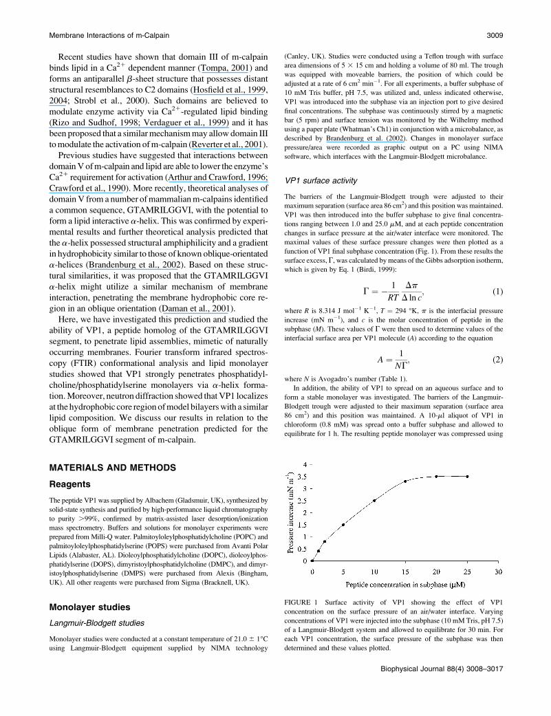
FIGURE 1 Surface activity of VP1 showing the effect of VP1
concentration on the surface pressure of an air/water interface. Varying
concentrations of VP1 were injected into the subphase (10 mM Tris, pH 7.5)
of a Langmuir-Blodgett system and allowed to equilibrate for 30 min. For
each VP1 concentration, the surface pressure of the subphase was then
determined and these values plotted.
Membrane Interactions of m-Calpain 3009
Biophysical Journal 88(4) 3008–3017
the moveable barriers of the trough to produce a pressure/area isotherm,
which was converted by NIMA software to an output plot of surface pressure
versus monolayer surface area per VP1 molecule (Fig. 2).
Insertion of VP1 into lipid monolayers at constant area
The ability of VP1 to penetrate lipid monolayers at constant area was studied.
Monolayers were formed by spreading lipid mixtures (5 mM) in chloroform
of either DMPC/DMPS or DOPC/DOPS (each at a 10:1 molar ratio) onto
a buffer subphase. The solvent was allowed to evaporate off over 30 min and
then the monolayer was compressed at a velocity of 6 cm2 min�1 to give
a surface pressure of 30 mN m�1. The lipid monolayer was then maintained at
the area corresponding to this pressure throughout the experiment. VP1 was
introduced into the subphase to give a final peptide concentration of 20 mM
and interactions of the peptide with lipid monolayers were monitored as
changes in monolayer surface pressure versus time (Fig. 3).
The ability of VP1 to penetrate DMPC/DMPS monolayers at different
molar ratios was studied. Chloroform solutions (5 mM) of DMPC/DMPS at
molar ratios of either 100:1, 50:1, 20:1, or 10:1 were spread onto a buffer
subphase and compressed to a lateral pressure of 30 mN m�1, as described
above. The lipid monolayer was then maintained at the area corresponding to
this pressure throughout the experiment. VP1 was introduced into the
subphase to give a final peptide concentration of 20 mM and maximal
changes in monolayer surface pressure were plotted versus the anionic lipid
content of the monolayer (Fig. 4).
Interaction of VP1 with lipid monolayers at constant pressure
The ability of VP1 to penetrate lipid monolayers at constant pressure was
studied. Chloroform solutions (5 mM) of DMPC/DMPS at molar ratios of
either 100:1, 50:1, 20:1, or 10:1 were spread onto a buffer subphase. The
solvent was allowed to evaporate off for 30 min and the monolayer com-
pressed at a velocity of 6 cm2 min�1 to give a surface pressure of 30 mN m�1.
The lipid monolayer was then maintained at this pressure throughout the
experiment. The monolayer was allowed to equilibrate for 10 min and then
VP1 was introduced into the subphase to give a final peptide concentration
of 20 mM. Interactions of the peptide with lipid monolayers were monitored
by NIMA software, which provided an output plot of changes in monolayer
surface area per lipid molecule versus time (Fig. 5).
FTIR spectroscopy
Sample preparation
Small unilamellar vesicles (SUVs) were prepared according to Keller et al.,
(1992). Essentially, DMPC/DMPS mixtures in chloroform were prepared,
dried with nitrogen, and rehydrated with aqueous N-[2-hydroxyethylpiper-
azine-N9-[2-ethanesulphonic acid] (HEPES) at pH 7.5 to give desired final
lipid molar ratios. The resulting cloudy suspensions were sonicated at 4�Cwith a Soniprep (Foster City, CA) 150 sonicator until clear suspensions
resulted (30 cycles of 30 s) which were then centrifuged (15 min, 3000 3 g,
4�C).
The effect of varying vesicle lipid composition on levels of VP1
secondary structure was studied. VP1 (final concentration 8 mM) was
solubilized in vesicle suspensions prepared as described above, where the
overall lipid/VP1 ratio was maintained at 100:1 but the DMPC/DMPS ratio
ranged between 10:1 and 100:1 (Fig. 6 A, Table 2). The effect of varying the
total lipid/peptide ratio on levels of VP1 secondary structure was also
studied. VP1 (final concentration 8 mM) was solubilized in vesicle
suspensions prepared as described above, where the DMPC/DMPS ratio
was maintained at 10:1 but the overall lipid to VP1 ratio was varied between
100:1 and 10:1 (Fig. 6 B). Additionally, the effect of final VP1 concentration
on levels of secondary structure shown by the peptide in the presence of lipid
vesicles was studied. VP1 (final concentration ranging between 8 mM and 1
mM) was solubilized in vesicle suspensions prepared as described above,
where the overall lipid to VP1 ratio was maintained at 100:1 and the DMPC/
DMPS ratio was maintained at 10:1 (Table 3).
FTIR conformational analyses of VP1 in the presence of lipid
All samples prepared as described above were individually spread on a CaF2
crystal, and the free excess water evaporated at room temperature. The single
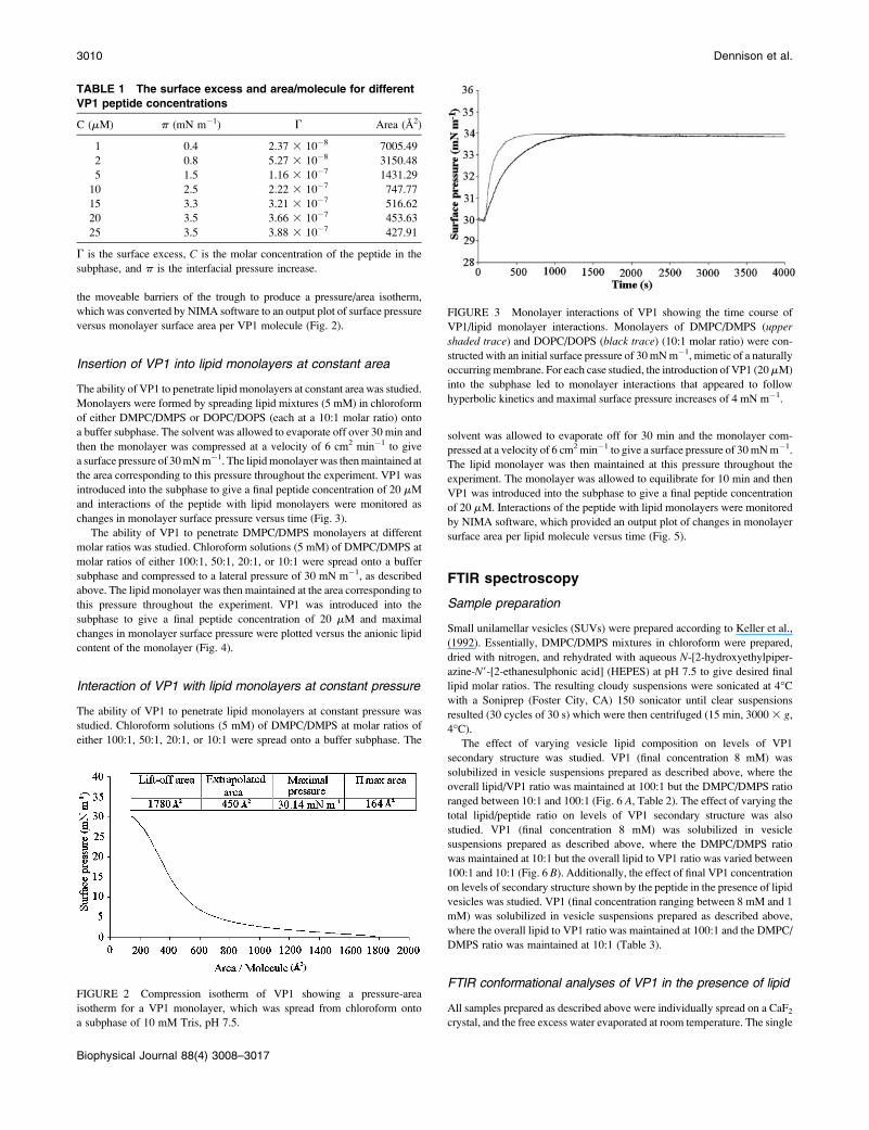
TABLE 1 The surface excess and area/molecule for different
VP1 peptide concentrations
C (mM) p (mN m�1) G Area (A2)
1 0.4 2.37 3 10�8 7005.49
2 0.8 5.27 3 10�8 3150.48
5 1.5 1.16 3 10�7 1431.29
10 2.5 2.22 3 10�7 747.77
15 3.3 3.21 3 10�7 516.62
20 3.5 3.66 3 10�7 453.63
25 3.5 3.88 3 10�7 427.91
G is the surface excess, C is the molar concentration of the peptide in the
subphase, and p is the interfacial pressure increase.
FIGURE 2 Compression isotherm of VP1 showing a pressure-area
isotherm for a VP1 monolayer, which was spread from chloroform onto
a subphase of 10 mM Tris, pH 7.5.
FIGURE 3 Monolayer interactions of VP1 showing the time course of
VP1/lipid monolayer interactions. Monolayers of DMPC/DMPS (upper
shaded trace) and DOPC/DOPS (black trace) (10:1 molar ratio) were con-
structed with an initial surface pressure of 30 mN m�1, mimetic of a naturally
occurring membrane. For each case studied, the introduction of VP1 (20mM)
into the subphase led to monolayer interactions that appeared to follow
hyperbolic kinetics and maximal surface pressure increases of 4 mN m�1.
3010 Dennison et al.
Biophysical Journal 88(4) 3008–3017
band components of the VP1 amide I vibrational band (predominantly C¼O
stretch) was monitored using an FTIR 5DX spectrometer (Nicolet Instru-
ments, Madison, WI) and absorbance spectra were produced for each
sample. For these spectra, water bands were subtracted and the evaluation of
peptide band parameters (peak position, band width, and intensity) was
performed. Curve fitting was applied to overlapping bands using a modified
version of the CURFIT procedure written by D. Moffat (National Research
Council, Ottawa, Canada). The band shapes of the single components are
superpositions of Gaussian and Lorentzian band shapes. Best fits were
obtained by assuming a Gauss fraction of 0.55–0.6. The CURFIT procedure
measures the peak areas of single-band components and, after statistical
evaluation, determines the relative percentages of primary structure involved
in secondary structure formation.
Neutron diffraction studies
Neutron diffraction experiments were conducted using the V1 membrane
diffractometer at BENSC and analytical techniques previously described
(Bradshaw et al., 1997, 1998, 2000; Duff et al., 1994; Haub et al., 2002).
Essentially: solutions of POPC/POPS (10:1 molar ratio) in chloroform were
prepared. Aliquots (1 ml) of these lipid solutions, and these solutions
containing VP1 (8 mM), were evenly deposited onto separate quartz
microscope slides using an artist’s airbrush (Aerobrush pro281, Hansa,
Norderstedt, Germany). These treated slides were placed in a vacuum
desiccator for 12 h to remove all traces of chloroform. Each slide was then
individually placed in an aluminium thermostat sample can and rehydrated
for 24 h at 25�C with relative humidity maintained at 98% using Teflon
water baths containing saturated potassium sulfate solution. Potassium
sulfate water solutions at each of three isotopic compositions, 50%, 20%,
and 8% 2H2O, were used to assist with phase assignment. Under these
conditions, lipid samples self-assemble to form multilamellar structures that
may be considered as stacked membrane bilayers. After each completed
neutron diffraction measurement, a given quartz sample slide was rehydrated
with water at the next isotopic composition, with at least 24 h allowed for
equilibration. The scanning procedure consisted of sequential u scans around
the predicted Bragg angle for each order. The rocking scans covered the
Bragg position u for the angle u 6 2�. Diffraction patterns of the prepared
samples were measured with up to five orders detected for each sample.
The raw data from the two dimensional detector were summed to
intensity versus 2u using the V1 instrumental software. The commercial
software IGOR Pro (version 4) was used for all further data treatment. The
lamella (bilayer) spacing d for each sample, was calculated by least-square
fitting of the observed 2u values to the Bragg equation:
Nl ¼ 2d sin u; (3)
where n is the diffraction order and l is the neutron wavelength (4.52 A).
The integrated intensities were calculated based upon the Gaussian fit of
the experimental Bragg reflections. Absorption correction and Lorentz factor
were applied and their intensities square-rooted to produce structure-factor
amplitudes. The phase assignment of each order and the relative scaling of
the different datasets were determined by least-squares fitting to straight-line
functions (Dante et al., 2002). The scattering length density profiles r(z)
were then calculated for each sample using the formula
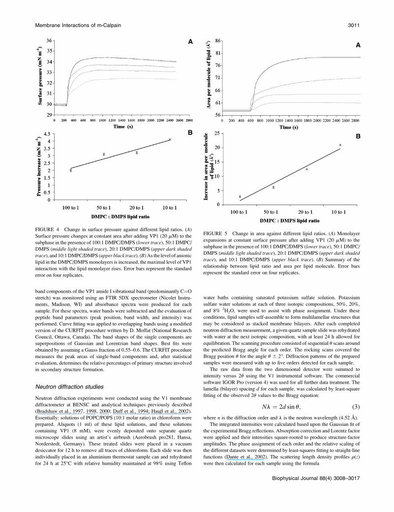
FIGURE 5 Change in area against different lipid ratios. (A) Monolayer
expansions at constant surface pressure after adding VP1 (20 mM) to the
subphase in the presence of 100:1 DMPC/DMPS (lower trace), 50:1 DMPC/
DMPS (middle light shaded trace), 20:1 DMPC/DMPS (upper dark shaded
trace), and 10:1 DMPC/DMPS (upper black trace). (B) Summary of the
relationship between lipid ratio and area per lipid molecule. Error bars
represent the standard error on four replicates.
FIGURE 4 Change in surface pressure against different lipid ratios. (A)
Surface pressure changes at constant area after adding VP1 (20 mM) to the
subphase in the presence of 100:1 DMPC/DMPS (lower trace), 50:1 DMPC/
DMPS (middle light shaded trace), 20:1 DMPC/DMPS (upper dark shaded
trace), and 10:1 DMPC/DMPS (upper black trace). (B) As the level of anionic
lipid in the DMPC/DMPS monolayers is increased, the maximal level of VP1
interaction with the lipid monolayer rises. Error bars represent the standard
error on four replicates.
Membrane Interactions of m-Calpain 3011
Biophysical Journal 88(4) 3008–3017
rðzÞ ¼ r0 1 2=dSf ðhÞ cosðð2phzÞ=d; (4)
where r0(z) is the integral density per unit length of the bilayer, f(h) are the
scaled structural factors, and the second term describes the distribution in the
scattering lengths across the bilayer.
RESULTS
Monolayer studies
VP1 surface activity
VP1 was introduced into the buffer subphase of the
Langmuir-Blodgett trough to give a range of final peptide
concentrations. In each case, changes in the interfacial surface
pressure were monitored and maximal values of these
pressure changes were plotted against the corresponding final
peptide concentrations (Fig. 1). It can be seen from Fig. 1 that
these pressure changes increase in an approximately linear
manner with rising VP1 concentration until at 20 mM pep-
tide, the maximal value is observed. Above this peptide con-
centration, surface pressures are effectively constant. Thus
20 mM VP1 was the minimum bulk concentration required
to saturate the air/water interface with the peptide under these
experimental conditions and was used as the VP1 subphase
concentration in all subsequent monolayer studies. Addition-
ally, these data were used to determine the corresponding
interfacial surface area per VP1 molecule (Table 1) and for
20mM peptide this can expand to 453 A2 which is comparable
to that found for other a-helical peptides (Ambroggio et al.,
2004).
The ability of VP1 to spread on an aqueous surface and to
form a stable monolayer was investigated. VP1 in chloroform
was spread on to a buffer subphase and a plot of surface
pressure versus monolayer surface area per VP1 molecule
generated (Fig. 2). Fig. 2 shows that the collapse pressure of
the isotherm starts around 30 mN m�1, indicating the presence
of a well-ordered monolayer (Alminana et al., 2003). Fig. 2
also shows that the area per VP1 molecule corresponding to
this collapse pressure was 164 A2, which is consistent with the
presence of a-helical structure in VP1 (Fidelio et al., 1986).
The extrapolated area at n ¼ 0 mN m�1 for the isotherm
provides a measure of the mean monolayer surface area per
VP1 molecule (Sospedra et al., 1999). This area was 450 A2
per VP1 molecule and is comparable to the value of 453 A2
calculated above using Eqs. 1 and 2 (Table 1).
Insertion of VP1 into lipid monolayers at constant area
The ability of VP1 to penetrate lipid monolayers at constant
area was studied. Monolayers of either: DMPC/DMPS or
DOPC/DOPS (each at a 10:1 molar ratio) were spread onto
a buffer subphase and compressed to give an initial surface
pressure of 30 mN m�1. With constant monolayer area
maintained, VP1 was introduced into the subphase to give
a final peptide concentration of 20 mM and interactions of the
peptide with lipid monolayers were monitored as changes in
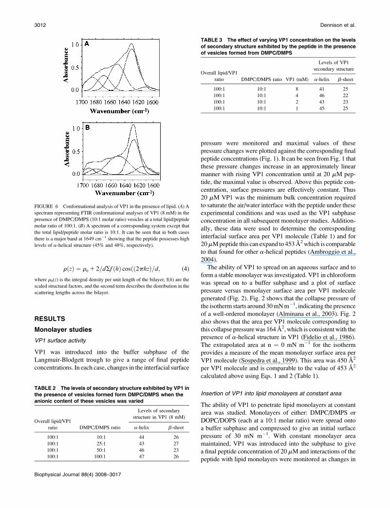
TABLE 3 The effect of varying VP1 concentration on the levels
of secondary structure exhibited by the peptide in the presence
of vesicles formed from DMPC/DMPS
Overall lipid/VP1
ratio
Levels of VP1
secondary structure
DMPC/DMPS ratio VP1 (mM) a-helix b-sheet
100:1 10:1 8 41 25
100:1 10:1 4 46 22
100:1 10:1 2 43 23
100:1 10:1 1 45 25
TABLE 2 The levels of secondary structure exhibited by VP1 in
the presence of vesicles formed form DMPC/DMPS when the
anionic content of these vesicles was varied
Overall lipid/VP1
ratio
Levels of secondary
structure in VP1 (8 mM)
DMPC/DMPS ratio a-helix b-sheet
100:1 10:1 44 26
100:1 25:1 43 27
100:1 50:1 46 23
100:1 100:1 47 26
FIGURE 6 Conformational analysis of VP1 in the presence of lipid. (A) A
spectrum representing FTIR conformational analyses of VP1 (8 mM) in the
presence of DMPC/DMPS (10:1 molar ratio) vesicles at a total lipid/peptide
molar ratio of 100:1. (B) A spectrum of a corresponding system except that
the total lipid/peptide molar ratio is 10:1. It can be seen that in both cases
there is a major band at 1649 cm�1 showing that the peptide possesses high
levels of a-helical structure (45% and 48%, respectively).
3012 Dennison et al.
Biophysical Journal 88(4) 3008–3017
monolayer surface pressure versus time (Fig. 3). It can be seen
from Fig. 3 that for each of the monolayers studied, the
insertion of VP1 followed hyperbolic kinetics. In each case,
the final surface pressure change induced by VP1 was;4 mN
m�1 although the time taken to induce these changes showed
significant variation (300 s and 1500 s, respectively).
The ability of VP1 to penetrate DMPC/DMPS monolayers
at different molar ratios was studied. Monolayers of DMPC/
DMPS at molar ratios of either 100:1, 50:1, 20:1, or 10:1 were
spread onto a buffer subphase and compressed to an initial
surface pressure of 30 mN m�1. With constant monolayer area
maintained, VP1 was introduced into the subphase to give a
final peptide concentration of 20 mM and monolayer surface
pressure was plotted versus time (Fig. 4A). Fig. 4A shows that
in each case VP1 induced an increase in surface pressure,
which followed a hyperbolic time course. Maximal changes in
monolayer surface pressure were then plotted against the
anionic lipid content of the monolayer (Fig. 4 B). The
regression analysis in Fig. 4 B indicates a strong correlation
between these parameters (R2 ¼ 0.97). As the level of anionic
lipid in the DMPC/DMPS monolayer is increased, the
maximal level of VP1 interaction with the lipid monolayer
rises.
Interaction of VP1 with lipid monolayers atconstant pressure
The ability of VP1 to penetrate lipid monolayers at constant
pressure was studied. Monolayers of DMPC/DMPS at molar
ratios of either 100:1, 50:1, 20:1, or 10:1 were spread onto
a buffer subphase and compressed to give a surface pressure
of 30 mN m�1, giving rise to an area of 58.4 A2 per molecule
of pure lipid. With constant pressure maintained, VP1 was
introduced into the subphase and output indicating changes in
monolayer surface area per lipid molecule versus time was
monitored (Fig. 5 A). Fig. 5 A shows that in each case VP1
induced significant expansion of monolayers, which followed
a hyperbolic time course but with differing kinetics. Fig. 5 Ashows that the maximum increase in monolayer surface area
per molecule of lipid was 79.4 A2 for DMPC/DMPS at a molar
ratio of 10:1. The level of these area changes decreased with
decreasing anionic content of monolayers until, in the
presence of DMPC/DMPS at a molar ratio of 100:1, the
monolayer expanded to 61.2 A2 per molecule of lipid,
representing a 22% decrease in area compared to the 10:1 lipid
ratio. The regression analysis in Fig. 5 B shows a strong
correlation (R2 ¼ 0.96) between these parameters. Here the
area decrease is inversely proportional to the anionic content
of the monolayer.
FTIR spectroscopy
The effect of varying lipid vesicle composition on levels of
VP1 secondary structure was studied. VP1 (final concentra-
tion 8 mM) was solubilized in vesicle suspensions where the
overall lipid/VP1 ratio was maintained at 100:1 but the DMPC/
DMPS ratio was varied between 10:1 and 100:1. Levels of
VP1 a-helicity and b-sheet structure were then determined
(Fig. 6A and Table 2). Table 2 shows that decreasing the level
of DMPS in these vesicles from 10% to 1% led to no
significant variation in levels of VP1 a-helicity or b-sheet
structure, which were ;45% and 25%, respectively.
The effect of varying the total lipid/peptide ratio on levels
of VP1 secondary structure was also studied. VP1 (final
concentration 8 mM) was solubilized in vesicle suspensions
where the DMPC/DMPS ratio was maintained at 10:1 but the
overall lipid/VP1 ratio was varied between 10:1 and 100:1.
Levels of VP1 a-helicity were then determined and were
found to be 48% in the presence of vesicle suspensions at a
10:1 total lipid/peptide ratio (Fig. 6 B) and 45% in the
presence of vesicle suspensions at a 100:1 total lipid/peptide
ratio (Fig. 6 A).
The effect of final VP1 concentration on levels of
secondary structure shown by the peptide in the presence of
lipid vesicles was studied. VP1 (final concentration ranging
between 8 mM and 1 mM) was solubilized in vesicles where
the overall lipid/VP1 ratio was maintained at 100:1 and the
DMPC/DMPS ratio was maintained at 10:1. Levels of VP1
a-helicity and b-sheet structure were then determined (Table
3). Table 3 shows that between 1 mM and 8 mM of VP1, no
significant variations in levels of either a-helicity or b-sheet
structure (;45% and 25%, respectively) were observed.
Neutron diffraction studies
The interactions of VP1 with POPC/POPS bilayers was
studied. The d-spacings obtained in the absence and presence
of peptide were 55.0 6 0.2 A indicating that the membrane
thickness and hydration were not affected by the peptide. The
structure factors from Table 4 were used to determine the
neutron diffraction density profiles using an 8% 2H2O con-
trast. At this H2O/2H2O ratio, the mean neutron scattering
length of the water mixture is zero (King and White, 1986).
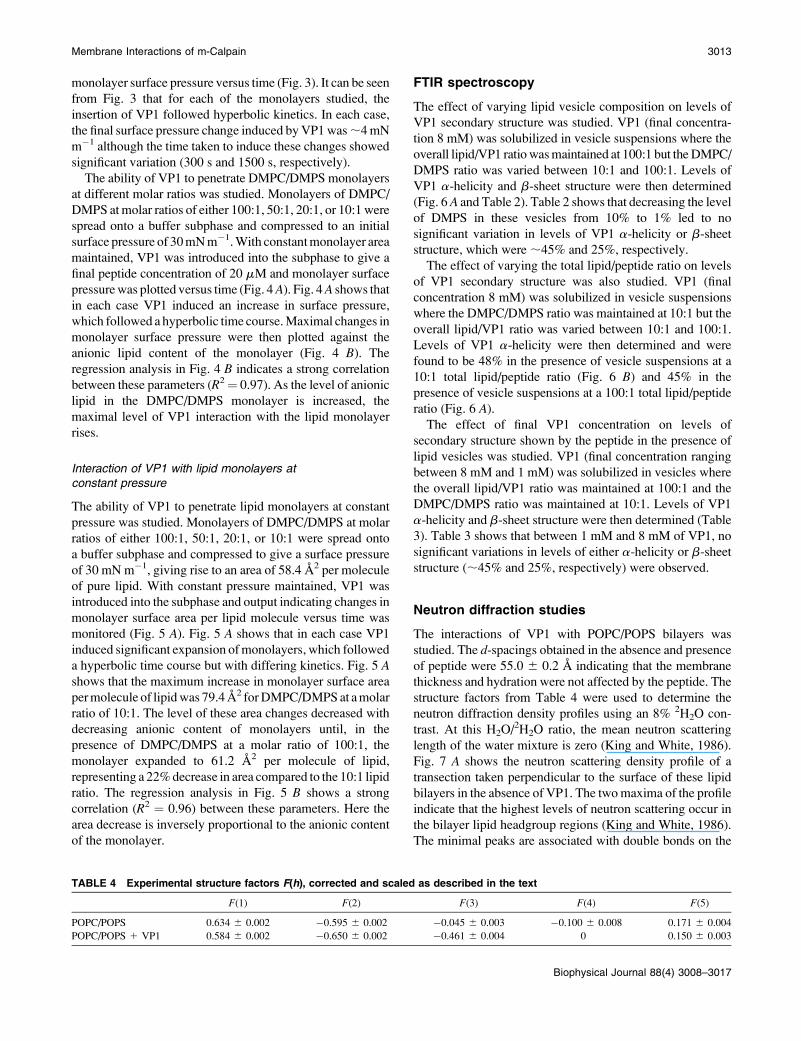
Fig. 7 A shows the neutron scattering density profile of a
transection taken perpendicular to the surface of these lipid
bilayers in the absence of VP1. The two maxima of the profile
indicate that the highest levels of neutron scattering occur in
the bilayer lipid headgroup regions (King and White, 1986).
The minimal peaks are associated with double bonds on the
TABLE 4 Experimental structure factors F(h), corrected and scaled as described in the text
F(1) F(2) F(3) F(4) F(5)
POPC/POPS 0.634 6 0.002 �0.595 6 0.002 �0.045 6 0.003 �0.100 6 0.008 0.171 6 0.004
POPC/POPS 1 VP1 0.584 6 0.002 �0.650 6 0.002 �0.461 6 0.004 0 0.150 6 0.003
Membrane Interactions of m-Calpain 3013
Biophysical Journal 88(4) 3008–3017
acyl chain that have a larger scattering length than the adjacent
methylenes because of fewer hydrogens, which indicates that
the lowest levels are produced by lipid terminal methyl
groups, demarking the center of the bilayer hydrophobic core
(King and White, 1986). The extra hydrogen on the terminal
methyl group creates the central trough that corresponds to
zero on the x axis. The scattering density profiles (Fig. 8 A) in
the presence of the peptide are thought to be typical of protein
penetration into an anionic POPC/POPS membrane (Dante
et al., 2002; Haub et al., 2002; King and White, 1986).
To find a more quantitative description of the changes
introduced by VP1 into the membrane structure, the experi-
mental structure factors were fitted to a model membrane in
reciprocal space. This was preformed using the reciprocal
representation of Gaussian distributions as the fit function:
FðhÞ ¼ scale � +n
i¼1
areaðiÞwidth
� e�ð2p�width�hÞ2=2
� cosð2p � positionðiÞ � hÞ; (5)
where some constant factors are included in scale and the sum
is taken over the number of Gaussian distributions used. The
pure lipid bilayer from Fig. 7 A was partitioned into three
Gaussian distributions corresponding to the methyl groups
(1), alkyl chains (2), and polar headgroups, which also include
the glycerol backbone (3). The parameters of the Gaussians
were amplitude, position, and width. The width is a single
parameter for all three Gaussians. This is a minimal model,
obtained by parsing the lipid molecules in only three mole-
cular groups to keep the number of parameters as low as
possible. The model was fitted to the five structure factors of
the lipid membrane keeping the areaA, which is the calculated
scattering length of the molecular groups, and the position of
the methyl groups (z¼ 0) as fixed parameters. The positions of
the headgroups and alkyl chains, and the width of the Gauss
functions were therefore free parameters of the fit. To scale the
absolute scattering length to the experimental data an appro-
priate scaling factor was used as the fourth fitting parameter.
Fig. 7 A shows the experimental curve (solid line) and the
fitted profile (dotted line) follow a similar trend. The fitted
profile is decomposed into the individual Gaussian distribu-
tions in Fig. 7 B, and the final parameters are listed in Table 5.
To obtain a good fit to the structure factors of the membrane
sample with VP1 peptide, we had to introduce a fourth
Gaussian distribution to account for the additional scattering
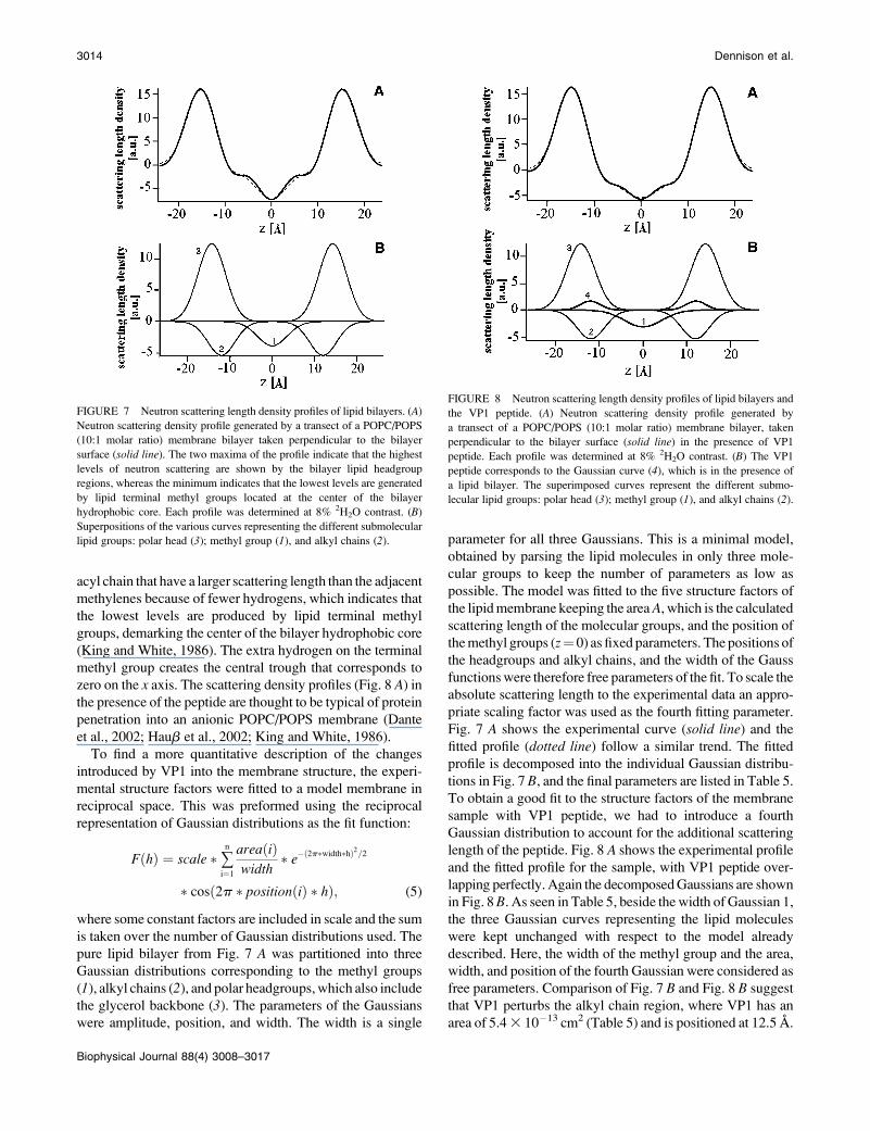
length of the peptide. Fig. 8 A shows the experimental profile
and the fitted profile for the sample, with VP1 peptide over-
lapping perfectly. Again the decomposed Gaussians are shown
in Fig. 8B. As seen in Table 5, beside the width of Gaussian 1,
the three Gaussian curves representing the lipid molecules
were kept unchanged with respect to the model already
described. Here, the width of the methyl group and the area,
width, and position of the fourth Gaussian were considered as
free parameters. Comparison of Fig. 7 B and Fig. 8 B suggest
that VP1 perturbs the alkyl chain region, where VP1 has an
area of 5.4 3 10�13 cm2 (Table 5) and is positioned at 12.5 A.
FIGURE 7 Neutron scattering length density profiles of lipid bilayers. (A)
Neutron scattering density profile generated by a transect of a POPC/POPS
(10:1 molar ratio) membrane bilayer taken perpendicular to the bilayer
surface (solid line). The two maxima of the profile indicate that the highest
levels of neutron scattering are shown by the bilayer lipid headgroup
regions, whereas the minimum indicates that the lowest levels are generated
by lipid terminal methyl groups located at the center of the bilayer
hydrophobic core. Each profile was determined at 8% 2H2O contrast. (B)
Superpositions of the various curves representing the different submolecular
lipid groups: polar head (3); methyl group (1), and alkyl chains (2).
FIGURE 8 Neutron scattering length density profiles of lipid bilayers and
the VP1 peptide. (A) Neutron scattering density profile generated by
a transect of a POPC/POPS (10:1 molar ratio) membrane bilayer, taken
perpendicular to the bilayer surface (solid line) in the presence of VP1
peptide. Each profile was determined at 8% 2H2O contrast. (B) The VP1
peptide corresponds to the Gaussian curve (4), which is in the presence of
a lipid bilayer. The superimposed curves represent the different submo-
lecular lipid groups: polar head (3); methyl group (1), and alkyl chains (2).
3014 Dennison et al.
Biophysical Journal 88(4) 3008–3017
This value is in very good agreement with the scattering
length density of VP1 calculated from its chemical structure,
and in a peptide/lipid ratio of 1:33, which is 6.043 10�13 cm2.
It should be noted that the width of the Gaussian curve for the
methyl group in Fig. 8 B (6.4 A) is larger than in Fig. 7 B (3.7
A). This would suggest that the peptide perturbs the middle of
the bilayer because the methyl region is enlarged. An attempt
to fit the peptide sample with only three Gaussian distributions
as in the case of the pure lipid sample failed. Even the liber-
ation of the width in the methyl group distribution resulted in
a bad fit. This is a strong indication that the additional
scattering length of the peptide is really being measured in the
neutron diffraction experiment.
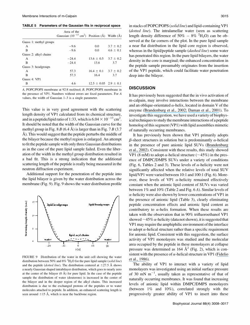
Additional support for the penetration of the peptide into
the lipid bilayer is given by the water distribution across the
membrane (Fig. 9). Fig. 9 shows the water distribution profile
in stacks of POPC/POPS (solid line) and lipid-containing VP1
(dotted line). The intralamellar water (seen as scattering
length density difference of 50% � 8% 2H2O) can be ob-
served at the far corners of the plot. In the pure lipid sample
a near flat distribution in the lipid core region is observed,
whereas in the lipid/peptide sample (dashed line) some water
has penetrated this region. In the pure lipid bilayers, the water
density in the core is marginal; the enhanced concentration in
the peptide sample presumably originates from the insertion
of the VP1 peptide, which could facilitate water penetration
deep into the bilayer.
DISCUSSION
It has previously been suggested that the in vivo activation of
m-calpain, may involve interactions between the membrane
and an oblique-orientated a-helix, located in domain V of the
enzyme (Brandenburg et al., 2002; Daman et al., 2001). To
investigate this suggestion, we have used a variety of biophys-
ical techniques to study the membrane interactions of a peptide
homolog of this segment (VP1) with lipid assemblies mimetic
of naturally occurring membranes.
It has previously been shown that VP1 primarily adopts
b-sheet structures in solution but is predominantly a-helical
in the presence of pure anionic lipid SUVs (Brandenburg
et al., 2002). Consistent with these results, this study showed
VP1 (8 mM) to adopt a-helical structure (;45%) in the pres-
ence of DMPC/DMPS SUVs under a variety of conditions
(Fig. 6, Tables 2 and 3). These levels of a-helicity were not
significantly affected when the relative levels of total SUV
lipid/VP1 were varied between 10:1 and 100:1 (Fig. 6). More-
over, these levels of VP1 a-helicity remained effectively
constant when the anionic lipid content of SUVs was varied
between 1% and 10% (Table 2 and Fig. 6 A). Similar levels of
a-helicity were also shown by lower concentrations of VP1 in
the presence of anionic lipid (Table 3), clearly eliminating
peptide concentration effects and anionic lipid content as
contributory to a-helix formation. When these results are
taken with the observation that in 90% trifluoroethanol VP1
showed;45%a-helicity (data not shown), it is suggested that
VP1 may require the amphiphilic environment of the interface
to adopt a-helical structure rather than a specific requirement
for anionic lipid. Consistent with this suggestion, the surface
activity of VP1 monolayers was studied and the molecular
area occupied by the peptide in these monolayers at collapse
pressure was determined as 164 A2 (Fig. 2), which is con-
sistent with the presence ofa-helical structure in VP1 (Fidelio
et al., 1986).
The ability of VP1 to interact with a variety of lipid
monolayers was investigated using an initial surface pressure
of 30 mN m�1, usually taken as representative of that of
naturally occurring membranes. It was found that increasing
levels of anionic lipid within DMPC/DMPS monolayers
(between 1% and 10%), correlated strongly with the
progressively greater ability of VP1 to insert into these
TABLE 5 Parameters of the Gaussian fits in reciprocal space
Area of the
Gaussian (10�13 cm2) Position (A) Width (A)
Gauss 1: methyl groups
A �9.6 0.0 3.7 6 0.2
B �9.6 0.0 4.6 6 0.1
Gauss 2: alkyl chains
A �24.4 13.6 6 0.5 3.7 6 0.2
B �24.4 13.6 3.7
Gauss 3: headgroups
A 57.3 16.4 6 0.1 3.7 6 0.2
B 57.3 16.4 3.7
Gauss 4: VP1
B 4.6 12.5 6 0.05 2.9 6 0.1
A, POPC/POPS membrane at 92:8 mol/mol; B, POPC/POPS membrane in
the presence of VP1. Numbers without errors are fixed parameters. For Avalues, the width of Gaussian 1–3 is a single parameter.
FIGURE 9 Distribution of the water in the unit cell showing the water
distribution between 50% and 8% 2H2O for the pure lipid sample (solid line)
and the peptide (dotted line). The distribution centered at 627.5 A shows
a nearly Gaussian shaped intrabilayer distribution, which goes to nearly zero
at the center of the bilayer (0 A) for pure lipid. In the case of the peptide
sample the distribution of water (deuterons) is increased in the center of
the bilayer and in the deeper region of the alkyl chains. This increased
distribution is due to the exchanged protons of the peptides or to water
molecules attached to peptide. In addition, an enhanced scattering length is
seen around 615 A, which is near the backbone region.
Membrane Interactions of m-Calpain 3015
Biophysical Journal 88(4) 3008–3017
membranes (Figs. 4 and 5). Repeated experiments at a fixed
pressure of 30 mNm�1 gave similar levels of insertion,
implying that the hyperbolic curves observed in Figs. 4 A and
5 A do represent saturation of the system rather than
pressure-induced inhibition of further insertion by the pep-
tide. Although the monolayer may therefore support maximal
a-helix formation by VP1, insertion by the peptide would
appear to have a specific requirement for anionic lipid, pos-
sibly supporting the involvement of a snorkelling effect to
accommodate the sole arginine residue of VP1.
Neutron diffraction studies were used to investigate the
interactions of VP1 with bilayers formed from physiologi-
cally relevant lipids. It was found that the peptide was able
to localize to the central core region of POPC/POPS (10:1
molar ratio) bilayers. Although our results do not directly
demonstrate oblique-angled membrane penetration by the
GTAMRILGGVI segment, they would be consistent with a
mechanism of membrane penetration that involves anionic
lipid and snorkelling by the segment’s arginine residue, al-
lowing the strongly hydrophobic C-terminal region of the
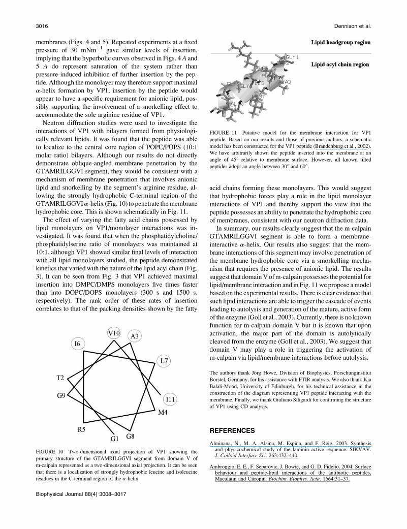
GTAMRILGGVIa-helix (Fig. 10) to penetrate the membrane
hydrophobic core. This is shown schematically in Fig. 11.
The effect of varying the fatty acid chains possessed by
lipid monolayers on VP1/monolayer interactions was in-
vestigated. It was found that when the phosphatidylcholine/
phosphatidylserine ratio of monolayers was maintained at
10:1, although VP1 showed similar final levels of interaction
with all lipid monolayers studied, the peptide demonstrated
kinetics that varied with the nature of the lipid acyl chain (Fig.
3). It can be seen from Fig. 3 that VP1 achieved maximal
insertion into DMPC/DMPS monolayers five times faster
than into DOPC/DOPS monolayers (300 s and 1500 s,
respectively). The rank order of these rates of insertion
correlates to that of the packing densities shown by the fatty
acid chains forming these monolayers. This would suggest
that hydrophobic forces play a role in the lipid monolayer
interactions of VP1 and thereby support the view that the
peptide possesses an ability to penetrate the hydrophobic core
of membranes, consistent with our neutron diffraction data.
In summary, our results clearly suggest that the m-calpain
GTAMRILGGVI segment is able to form a membrane-
interactive a-helix. Our results also suggest that the mem-
brane interactions of this segment may involve penetration of
the membrane hydrophobic core via a smorkelling mecha-
nism that requires the presence of anionic lipid. The results
suggest that domain V of m-calpain possesses the potential for
lipid/membrane interaction and in Fig. 11 we propose a model
based on the experimental results. There is clear evidence that
such lipid interactions are able to trigger the cascade of events
leading to autolysis and generation of the mature, active form
of the enzyme (Goll et al., 2003). Currently, there is no known
function for m-calpain domain V but it is known that upon
activation, the major part of the domain is autolytically
cleaved from the enzyme (Goll et al., 2003). We suggest that
domain V may play a role in triggering the activation of
m-calpain via lipid/membrane interactions before autolysis.
The authors thank Jorg Howe, Division of Biophysics, Forschunginstitut
Borstel, Germany, for his assistance with FTIR analysis. We also thank Kia
Balali-Mood, University of Edinburgh, for his technical assistance in the
construction of the diagram representing VP1 peptide interacting with the
membrane. Finally, we thank Giuliano Siligardi for confirming the structure
of VP1 using CD analysis.
REFERENCES
Alminana, N., M. A. Alsina, M. Espina, and F. Reig. 2003. Synthesisand physicochemical study of the laminin active sequence: SIKVAV.J. Colloid Interface Sci. 263:432–440.
Ambroggio, E. E., F. Separovic, J. Bowie, and G. D. Fidelio. 2004. Surfacebehaviour and peptide-lipid interactions of the antibiotic peptides,Maculatin and Citropin. Biochim. Biophys. Acta. 1664:31–37.
FIGURE 11 Putative model for the membrane interaction for VP1
peptide. Based on our results and those of previous authors, a schematic
model has been constructed for the VP1 peptide (Brandenburg et al., 2002).
We have arbitrarily shown the peptide inserted into the membrane at an
angle of 45� relative to membrane surface. However, all known tilted
peptides adopt an angle between 30� and 60�.
FIGURE 10 Two-dimensional axial projection of VP1 showing the
primary structure of the GTAMRILGGVI segment from domain V of
m-calpain represented as a two-dimensional axial projection. It can be seen
that there is a localization of strongly hydrophobic leucine and isoleucine
residues in the C-terminal region of the a-helix.
3016 Dennison et al.
Biophysical Journal 88(4) 3008–3017
Arthur, J. S., and C. Crawford. 1996. Investigation of the interaction ofm-calpain with phospholipids: calpain-phospholipid interactions. Biochim.Biophys. Acta. 1293:201–206.
Branca, D. 2004. Calpain-related diseases. Biochem. Biophys. Res.Commun. 322:1098–1104.
Birdi, K. S. 1999. Self-Assembly Monolayer Structures of Lipids andMacromolecules at Interfaces. Kluwer Academic, Dordrecht, TheNetherlands.
Bradshaw, J. P., R. J. Bushby, C. C. Giles, M. R. Saunders, and A. Saxena.1997. The headgroup orientation of dimyristoylphosphatidylinositol-4-phosphate in mixed lipid bilayers: a neutron diffraction study. Biochim.Biophys. Acta. 1329:124–138.
Bradshaw, J. P., M. J. Darkes, T. A. Harroun, J. Katsaras, and R. M. Epand.2000. Oblique membrane insertion of viral fusion peptide probed byneutron diffraction. Biochemistry. 39:6581–6585.
Bradshaw, J. P., S. M. Davies, and T. Haub. 1998. Interaction of substanceP with phospholipid bilayers: a neutron diffraction study. Biophys. J. 75:889–895.
Brandenburg, K., F. Harris, S. Dennison, U. Seydel, and D. Phoenix. 2002.Domain V of m-calpain shows the potential to form an oblique-orientatedalpha-helix, which may modulate the enzyme’s activity via interactionswith anionic lipid. Eur. J. Biochem. 269:5414–5422.
Carafoli, E., and M. Molinari. 1998. Calpain: a protease in search ofa function? Biochem. Biophys. Res. Commun. 247:193–203.
Chakrabarti, A. K., S. Dasgupta, R. H. Gadsden, E. L. Hogan, and N. L.Banik. 1996. Regulation of brain m calpain Ca21 sensitivity by mixturesof membrane lipids: activation at intracellular Ca21 level. J. Neurosci.Res. 44:374–380.
Crawford, C., N. R. Brown, and A. C. Willis. 1990. Investigation of thestructural basis of the interaction of calpain II with phospholipid and withcarbohydrate. Biochem. J. 265:575–579.
Daman, A., F. Harris, S. Biswas, J. Wallace, and D. A. Phoenix. 2001. Atheoretical investigation into the lipid interactions of m-calpain. Mol.Cell. Biochem. 223:159–163.
Dante, S., T. Haub, and N. A. Dencher. 2002. Beta-amyloid 25 to 35 isintercalated in anionic and zwitterionic lipid membranes to differentextents. Biophys. J. 83:2610–2616.
Duff, K. C., P. R. Gilchrist, A. M. Saxena, and J. P. Bradshaw. 1994.Neutron diffraction reveals the site of amantadine blockade in theinfluenza A M2 ion channel. Virology. 202:287–293.
Fidelio, G. D., B. M. Austen, D. Chapman, and J. A. Lucy. 1986. Propertiesof signal-sequence peptides at an air-water interface. Biochem. J. 238:301–304.
Goll, D. E., V. F. Thompson. H. Li, W. Wei, and J. Cong. 2003. Thecalpain system. Physiol. Rev. 83:731–801.
Haub, T., S. Dante, N. A. Dencher, and T. H. Haines. 2002. Squalane is inthe midplane of the lipid bilayer: implications for its function as a protonpermeability barrier. Biochim. Biophys. Acta. 1556:149–154.
Hosfield, C. M., J. S. Elce, P. L. Davies, and Z. C. Jia. 1999. Crystalstructure of calpain reveals the structural basis for Ca21-dependentprotease activity and a novel mode of enzyme activation. EMBO J. 18:6880–6889.
Hosfield, C. M., J. S. Elce, and Z. Jia. 2004. Activation of calpain by Ca21:roles of the large subunit N-terminal and domain III–IV linker peptides.J. Mol. Biol. 343:1049–1053.
Huang, Y., and K. K. Wang. 2001. The calpain family and human disease.Trends Mol. Med. 7:355–362.
Imajoh, S., H. Kawasaki, and K. Suzuki. 1986. The amino-terminalhydrophobic region of the small subunit of calcium-activated neutralprotease (Canp) is essential for its activation by phosphatidylinositol.J. Biochem. (Tokyo). 99:1281–1284.
Johnson, G. V., and R. P. Guttmann. 1997. Calpains: intact and active?Bioessays. 19:1011–1018.
Keller, R. C., J. A. Killian, and B. Kruijff. 1992. Anionic phospholipids areessential for alpha-helix formation of the signal peptide of prePhoE uponinteraction with phospholipid vesicles. Biochemistry. 31:1672–1677.
King, G., and S. White. 1986. Determining bilayer hydrocarbon thicknessfrom neutron diffraction measurements using strip-function models.Biophys. J. 49:1047–1054.
Melloni, E., and S. Pontremoli. 1989. The calpains. Trends Neurosci. 12:438–444.
Ono, Y., H. Sorimachi, and K. Suzuki. 1998. Structure and physiology ofcalpain, an enigmatic protease. Biochem. Biophys. Res. Commun. 245:289–294.
Perrin, B. J., and A. Huttenlocher. 2002. Calpain. Int. J. Biochem. Cell Biol.34:722–725.
Reverter, D., S. Strobl, C. Fernandez-Catalan, H. Sorimachi, K. Suzuki, andW. Bode. 2001. Structural basis for possible calcium-induced activationmechanisms of calpains. Biol. Chem. 382:753–766.
Rizo, J., and T. C. Sudhof. 1998. C-2-domains, structure and function ofa universal Ca21-binding domain. Biol. Chem. 273:15879–15882.
Saido, T. C., M. Shibata, T. Takenawa, H. Murofushi, and K. Suzuki. 1992.Positive regulation of mu-calpain action by polyphosphoinositides. Biol.Chem. 267:24585–24590.
Sato, K., and S. Kawashima. 2001. Calpain function in the modulation ofsignal transduction molecules. Biol. Chem. 382:743–751.
Sorimachi, H., and K. Suzuki. 2001. The structure of calpain. J. Biochem.(Tokyo). 129:653–664.
Sospedra, P., I. Haro, M. A. Alsina, F. Reig, and C. Mestres. 1999.Physicochemical interaction of a lipophilic derivative of HAV antigenVP3 (110–121) with lipid monolayers. Mater. Sci. Eng. C. 8–9:543–549.
Strobl, S., C. Fernandez-Catalan, M. Braun, R. Huber, H. Masumoto, K.Nakagawa, A. Irie, H. Sorimachi, G. Bourenkow, H. Bartunik, K.Suzuki, and W. Bode. 2000. The crystal structure of calcium-free humanm-calpain suggests an electrostatic switch mechanism for activation bycalcium. Proc. Natl. Acad. Sci. USA. 97:588–592.
Tompa, M. 2001. Identifying functional elements by comparative DNAsequence analysis. Genome Res. 11:1143–1144.
Verdaguer, N., S. Corbalan-Garcia, W. F. Ochoa, I. Fita, and J. C. Gomez-Fernandez. 1999. Ca21 bridges the C2 membrane-binding domain ofprotein kinase C alpha directly to phosphatidylserine. EMBO J. 18:6329–6338.
Membrane Interactions of m-Calpain 3017
Biophysical Journal 88(4) 3008–3017
Copyright © 2022 FDOKUMEN