Endocannabinoids mediate muscarine-induced synaptic depression at the vertebrate neuromuscular...
12
Endocannabinoids mediate muscarine-induced synaptic depression at the vertebrate neuromuscular junction Zachary Newman, Priya Malik, Tse-Yu Wu, Christopher Ochoa, Nayantara Watsa and Clark Lindgren Department of Biology, Grinnell College, Grinnell, IA 50112, USA Keywords: Anolis carolinensis, endocannabinoids, muscarinic, neuromuscular junction, synaptic depression OnlineOpen: This article is available free online at www.blackwell-synergy.com Abstract Endocannabinoids (eCBs) inhibit neurotransmitter release throughout the central nervous system. Using the Ceratomandibularis muscle from the lizard Anolis carolinensis we asked whether eCBs play a similar role at the vertebrate neuromuscular junction. We report here that the CB 1 cannabinoid receptor is concentrated on motor terminals and that eCBs mediate the inhibition of neurotransmitter release induced by the activation of M 3 muscarinic acetylcholine (ACh) receptors. N-(piperidin-1-yl)-5-(4- iodophenyl)-1-(2,4-dichlorophenyl)-4-methyl-1H-pyrazole-3-carboxamide, a CB 1 antagonist, prevents muscarine from inhibiting release and arachidonylcyclopropylamide (ACPA), a CB 1 receptor agonist, mimics M 3 activation and occludes the effect of muscarine. As for its mechanism of action, ACPA reduces the action-potential-evoked calcium transient in the nerve terminal and this decrease is more than sufficient to account for the observed inhibition of neurotransmitter release. Similar to muscarine, the inhibition of synaptic transmission by ACPA requires nitric oxide, acting via the synthesis of cGMP and the activation of cGMP-dependent protein kinase. 2-Arachidonoylglycerol (2-AG) is responsible for the majority of the effects of eCB as inhibitors of phospholipase C and diacylglycerol lipase, two enzymes responsible for synthesis of 2-AG, significantly limit muscarine-induced inhibition of neurotransmitter release. Lastly, the injection of (5Z,8Z,11Z,14Z)-N-(4-hydroxy-2-methylphenyl)-5,8,11,14-eicosatetraenamide (an inhibitor of eCB transport) into the muscle prevents muscarine, but not ACPA, from inhibiting ACh release. These results collectively lead to a model of the vertebrate neuromuscular junction whereby 2-AG mediates the muscarine-induced inhibition of ACh release. To demonstrate the physiological relevance of this model we show that the CB 1 antagonist N-(piperidin-1-yl)-5-(4-iodophenyl)-1-(2,4- dichlorophenyl)-4-methyl-1H-pyrazole-3-carboxamide prevents synaptic inhibition induced by 20 min of 1-Hz stimulation. Introduction Cannabinoids, the active ingredients found in the marijuana plant Cannabis sativa (Ameri, 1999; Martin et al., 1999), produce their biological effects through binding to specific G-protein-coupled receptors (Howlett et al., 2002). The term endocannabinoid (eCB) refers to endogenously released compounds that alter function by binding to these receptors (Devane et al., 1992; Evans et al., 1992). The eCBs are synthesized de novo from membrane phospholipids and are released through an unknown mechanism (Freund et al., 2003; but see Ronesi et al., 2004). Recently, eCBs have been shown to act as retrograde signalling molecules in several areas of the central nervous system (for reviews see Kreitzer & Regehr, 2002; Wilson & Nicoll, 2002). Depolarization of the postsynaptic neurone and the resulting elevation of intracellular Ca 2+ triggers eCB release (Ohno-Shosaku et al., 2001; Wilson & Nicoll, 2001; Brenowitz & Regehr, 2003). The activation of muscarinic acetylcholine (ACh) receptors (mAChRs) (Kim et al., 2002; Ohno-Shosaku et al., 2003; Fukudome et al., 2004) can also trigger the release of eCBs. The eCBs released in neural tissue usually bind to the CB 1 receptor subtype and inhibit the release of neurotransmitter from the presynaptic terminal (Kreitzer & Regehr, 2001, 2002; Maejima et al., 2001; Ohno-Shosaku et al., 2001; Wilson & Nicoll, 2001; Diana et al., 2002; Yoshida et al., 2002; but see Van Sickle et al., 2005). The inhibition of neurotransmitter release via the activation of mAChRs has been observed throughout both the central and peripheral nervous systems (for reviews, see Starke et al., 1989; Caulfield, 1993; Boehm & Huck, 1997). It has been well established that activation of mAChRs at the vertebrate neuromuscular junction (NMJ) modulates the release of the neurotransmitter ACh (Ganguly & Das, 1979; Duncan & Publicover, 1979; Michaelson et al., 1979; Standaert, 1982; Wali et al., 1988; Slutsky et al., 1999, 2001; Minic et al., 2002). In particular, activation of the M 1 subtype of the mAChR enhances ACh release (Slutsky et al., 1999; Graves et al., 2004), whereas activation of the M 2 and ⁄ or M 3 subtype inhibits release (Slutsky et al., 1999, 2001; Graves et al., 2004). Recently, the M 1 -mediated enhancement and the M 3 -mediated inhibition of neuro- transmitter release at the lizard NMJ have been shown to require the synthesis and extracellular diffusion of nitric oxide (NO) (Graves et al., 2004). As eCBs mediate the suppression of neurotransmitter release induced by M 1 and M 3 receptor activation in the hippocampus (Fukudome et al., 2004), we searched for a similar involvement of eCBs at the lizard NMJ. Using immunofluorescence, we localized CB 1 receptors to the NMJ and, using physiological and pharmacological approaches, discovered that eCBs [primarily 2-arachidonoylglycerol (2-AG)] do indeed mediate the depression of neurotransmitter release induced by the activation of M 3 mAChRs. Furthermore, this Correspondence: Clark A. Lindgren, as above. E-mail: [email protected] Re-use of this article is permitted in accordance with the Creative Commons Deed, Attribution 2.5, which does not permit commercial exploitation. Received 5 July 2006, revised 16 December 2006, accepted 18 January 2007 European Journal of Neuroscience, Vol. 25, pp. 1619–1630, 2007 doi:10.1111/j.1460-9568.2007.05422.x ª The Authors (2007). Journal Compilation ª Federation of European Neuroscience Societies and Blackwell Publishing Ltd
-
Upload
independent -
Category
Documents
-
view
1 -
download
0
Transcript of Endocannabinoids mediate muscarine-induced synaptic depression at the vertebrate neuromuscular...

Endocannabinoids mediate muscarine-induced synapticdepression at the vertebrate neuromuscular junction
Zachary Newman, Priya Malik, Tse-Yu Wu, Christopher Ochoa, Nayantara Watsa and Clark LindgrenDepartment of Biology, Grinnell College, Grinnell, IA 50112, USA
Keywords: Anolis carolinensis, endocannabinoids, muscarinic, neuromuscular junction, synaptic depression
OnlineOpen: This article is available free online at www.blackwell-synergy.com
Abstract
Endocannabinoids (eCBs) inhibit neurotransmitter release throughout the central nervous system. Using the Ceratomandibularismuscle from the lizard Anolis carolinensis we asked whether eCBs play a similar role at the vertebrate neuromuscular junction. Wereport here that the CB1 cannabinoid receptor is concentrated on motor terminals and that eCBs mediate the inhibition ofneurotransmitter release induced by the activation of M3 muscarinic acetylcholine (ACh) receptors. N-(piperidin-1-yl)-5-(4-iodophenyl)-1-(2,4-dichlorophenyl)-4-methyl-1H-pyrazole-3-carboxamide, a CB1 antagonist, prevents muscarine from inhibitingrelease and arachidonylcyclopropylamide (ACPA), a CB1 receptor agonist, mimics M3 activation and occludes the effect ofmuscarine. As for its mechanism of action, ACPA reduces the action-potential-evoked calcium transient in the nerve terminal and thisdecrease is more than sufficient to account for the observed inhibition of neurotransmitter release. Similar to muscarine, the inhibitionof synaptic transmission by ACPA requires nitric oxide, acting via the synthesis of cGMP and the activation of cGMP-dependentprotein kinase. 2-Arachidonoylglycerol (2-AG) is responsible for the majority of the effects of eCB as inhibitors of phospholipase Cand diacylglycerol lipase, two enzymes responsible for synthesis of 2-AG, significantly limit muscarine-induced inhibition ofneurotransmitter release. Lastly, the injection of (5Z,8Z,11Z,14Z)-N-(4-hydroxy-2-methylphenyl)-5,8,11,14-eicosatetraenamide (aninhibitor of eCB transport) into the muscle prevents muscarine, but not ACPA, from inhibiting ACh release. These results collectivelylead to a model of the vertebrate neuromuscular junction whereby 2-AG mediates the muscarine-induced inhibition of ACh release.To demonstrate the physiological relevance of this model we show that the CB1 antagonist N-(piperidin-1-yl)-5-(4-iodophenyl)-1-(2,4-dichlorophenyl)-4-methyl-1H-pyrazole-3-carboxamide prevents synaptic inhibition induced by 20 min of 1-Hz stimulation.
Introduction
Cannabinoids, the active ingredients found in the marijuana plantCannabis sativa (Ameri, 1999; Martin et al., 1999), produce theirbiological effects through binding to specific G-protein-coupledreceptors (Howlett et al., 2002). The term endocannabinoid (eCB)refers to endogenously released compounds that alter function bybinding to these receptors (Devane et al., 1992; Evans et al., 1992).The eCBs are synthesized de novo from membrane phospholipids andare released through an unknown mechanism (Freund et al., 2003; butsee Ronesi et al., 2004). Recently, eCBs have been shown to act asretrograde signalling molecules in several areas of the central nervoussystem (for reviews see Kreitzer & Regehr, 2002; Wilson & Nicoll,2002). Depolarization of the postsynaptic neurone and the resultingelevation of intracellular Ca2+ triggers eCB release (Ohno-Shosakuet al., 2001; Wilson & Nicoll, 2001; Brenowitz & Regehr, 2003). Theactivation of muscarinic acetylcholine (ACh) receptors (mAChRs)(Kim et al., 2002; Ohno-Shosaku et al., 2003; Fukudome et al., 2004)can also trigger the release of eCBs. The eCBs released in neural tissueusually bind to the CB1 receptor subtype and inhibit the release ofneurotransmitter from the presynaptic terminal (Kreitzer & Regehr,2001, 2002; Maejima et al., 2001; Ohno-Shosaku et al., 2001; Wilson
& Nicoll, 2001; Diana et al., 2002; Yoshida et al., 2002; but seeVan Sickle et al., 2005).The inhibition of neurotransmitter release via the activation of
mAChRs has been observed throughout both the central andperipheral nervous systems (for reviews, see Starke et al., 1989;Caulfield, 1993; Boehm & Huck, 1997). It has been well establishedthat activation of mAChRs at the vertebrate neuromuscular junction(NMJ) modulates the release of the neurotransmitter ACh (Ganguly &Das, 1979; Duncan & Publicover, 1979; Michaelson et al., 1979;Standaert, 1982; Wali et al., 1988; Slutsky et al., 1999, 2001; Minicet al., 2002). In particular, activation of the M1 subtype of the mAChRenhances ACh release (Slutsky et al., 1999; Graves et al., 2004),whereas activation of the M2 and ⁄ or M3 subtype inhibits release(Slutsky et al., 1999, 2001; Graves et al., 2004). Recently, theM1-mediated enhancement and the M3-mediated inhibition of neuro-transmitter release at the lizard NMJ have been shown to require thesynthesis and extracellular diffusion of nitric oxide (NO) (Graveset al., 2004).As eCBs mediate the suppression of neurotransmitter release
induced by M1 and M3 receptor activation in the hippocampus(Fukudome et al., 2004), we searched for a similar involvement ofeCBs at the lizard NMJ. Using immunofluorescence, we localized CB1
receptors to the NMJ and, using physiological and pharmacologicalapproaches, discovered that eCBs [primarily 2-arachidonoylglycerol(2-AG)] do indeed mediate the depression of neurotransmitter releaseinduced by the activation of M3 mAChRs. Furthermore, this
Correspondence: Clark A. Lindgren, as above.
E-mail: [email protected]
Re-use of this article is permitted in accordance with the Creative Commons Deed,Attribution 2.5, which does not permit commercial exploitation.
Received 5 July 2006, revised 16 December 2006, accepted 18 January 2007
European Journal of Neuroscience, Vol. 25, pp. 1619–1630, 2007 doi:10.1111/j.1460-9568.2007.05422.x
ª The Authors (2007). Journal Compilation ª Federation of European Neuroscience Societies and Blackwell Publishing Ltd
depression requires NO, acting via cGMP and cGMP-dependentprotein kinase, involves a decrease in the size of the calcium transientin the presynaptic nerve terminal, and requires an eCB transporter inthe muscle membrane. Lastly, we demonstrate the physiologicalrelevance of eCBs by showing that a form of long-term synapticdepression requires functional CB1 receptors.
Materials and methods
Experimental preparation and solutions
Prior to being pithed, lizards (Anolis carolinensis; Carolina BiologicalSupply Co.) were placed at 7–10 �C for 8–10 min to facilitate thequick and accurate ablation of the forebrain. The ceratomandibularismuscle (and its associated nerve) was isolated from small lizards asdescribed by Lindgren & Moore (1989) and pinned down in aSylgard�-coated chamber containing fresh physiological salinesolution composed of 158 mm NaCl, 2 mm KCl, 2 mm MgCl2,5 mm HEPES, 2 mm CaCl2 and 2 g ⁄ L dextrose (pH adjusted to 7.3using 1 m NaOH). Evoked end-plate potentials (EPPs) were reducedbelow the action potential threshold of the muscle by applying 10 lm
d-tubocurarine chloride. For experiments indicated in Fig. 8, 2.5 lm
d-tubocurarine chloride was used together with 1 lg ⁄ mL tetraethylr-hodamine-a-bungarotoxin The procedures described above wereapproved by the Institutional Animal Use and Care Committee atGrinnell College.In all of the experiments except the one described in Fig. 8, drugs
were administered via the physiological saline solution bathing thepreparation. Unless indicated otherwise, concentrated stock solutionsof the various drugs were prepared in advance and frozen at )20 �C.On the day of the experiment, aliquots were diluted in physiologicalsaline solution to their final concentrations. In the case of arachido-nylcyclopropylamide (ACPA) or (5Z,8Z,11Z,14Z)-N-(4-hydroxy-2-methylphenyl)-5,8,11,14-eicosatetraenamide (VDM 11), the drug wasobtained in Tocrisolve� (a soy oil and water emulsion) and diluteddirectly into physiological saline. In experiments where ACPA orVDM 11 was applied, the control solution contained Tocrisolve� atthe same concentration as in the experimental solution.For the experiments depicted in Fig. 8, muscarine or ACPA was
applied locally to an identified NMJ through a glass pipette with adiameter of approximately 1 lm via back pressure applied with apneumatic pico pump (PV 830; World Precision Instruments, Sarasota,FL, USA). Between one and six 2- and 5-s pressure pulses (10 s apart)were applied at 10–15 p.s.i. The electrode was filled with 20 lm
muscarine or ACPA and either rhodamine B or fluorescein. The latterwere used to track the dispersion of the pipette contents. The electrodewas positioned within 100–200 lm of nerve terminals on the topsurface of the muscle and the dispersion of the dye always envelopedthe NMJ. Although we do not know the local concentration ofmuscarine or ACPA at the synapse, the concentrations used producedchanges similar to bath application of either chemical.2-(4-carboxyphenyl)-4,4,5,5-tetramethylimidazoline-1-oxyl-3-oxide
potassium salt (carboxy-PTIO) was purchased from MolecularProbes (Eugene, OR, USA). ACPA, N-(piperidin-1-yl)-5-(4-iodo-phenyl)-1-(2,4-dichlorophenyl)-4-methyl-1H-pyrazole-3-carboxamide(AM 281), 1-[6-[[(17b)-3-methoxyestra-1,3,4(10)-trien-17-yl]amino]hexyl]-1H-pyrrole-2,5-dione (U-73122), 1H-[1,2,4]oxadiazolo[4,3-a]quinoxalin-1-one (ODQ) and VDM 11 were purchased from TocrisCookson (Ellisville, MO, USA). 1,6-Bis-(cyclohexyloximinocarbonyl-amino)-hexane (RHC-80267) was purchased from Biomol (PlymouthMeeting, PA, USA). All other drugs, including 4-diphenylacetoxy-N-methylpiperidine methiodide, Rp-b-phenyl-1,N2-etheno-8-bromo-
guanosine 3¢,5¢-cyclic monophosphorothioate (Rp-8-Br-PET-cGMPS),diethylamine ⁄ NO complex and Nx-nitro-l-arginine methyl ester(L-NAME), were purchased from Sigma-Aldrich (St Louis, MO, USA).
Immunofluorescence
Muscles were fixed in 3% paraformaldehyde for 1 h at 4 �C, rinsed for1 h in physiological saline, permeablized for 30 min at 37 �C in 0.3%Triton X-100, preincubated for 15 min at room temperature (22–24 �C)in blocking solution (0.01% Triton X-100, 1% bovine serum albumin)and incubated in primary antibody (10 lg ⁄ mL of rabbit anti-humanCB1 IgG no. 1; Alpha Diagnostic International, San Antonio, TX,USA) for 4 h at room temperature and then 12 h at 4 �C. Muscleswere rinsed for 1 h in blocking solution, incubated with fluorescein-conjugated goat anti-rabbit IgG secondary antibody (5 lg ⁄ mL;American Qualex, San Clemente, CA, USA) for 2 h at 37 �C, rinsedin blocking solution for 30 min and mounted on slides with 20%glycerol in Slowfade Antifade� solution (Sigma-Aldrich). The antigenused to create the primary antibody was a 14-amino-acid peptide,referred to as CB11-P, which is found near the extracellularN-terminus of human CB1 (CB11-A; Alpha Diagnostic International).No punctate staining was observed when the secondary antibody wasapplied without the primary antibody.To visualize the perisynaptic Schwann cells (PSCs), preparations
were incubated for 15 min at room temperature with 1 lm POPO�-3iodide nucleic acid stain (Molecular Probes) following the wash of thesecondary antibody and then washed for 30 min in blocking solution.To visualize nerve terminals, the ends of cut nerve axons were loadedwith Texas red dextran (Molecular Probes; 3000 MW, made in 10 mm
HEPES, pH 7.2). Immediately following isolation of the Ceratoman-dibularis muscle and its associated nerve, the cut end of the nerve axonwas placed into a small (1–2 lL) well containing 20 mm Texas reddextran. The Texas red dextran was allowed to load through antero-grade transport at 9 �C for 16–18 h and then at 4 �C for an additional2–3 h. After the nerve terminals had been filled with Texas red dextran,the tissue was processed for immunofluorescence as described above.After being stained, NMJs were observed with a laser scanning
confocal microscope manufactured by Prairie Technologies (Middle-ton, WI, USA) connected to a Nikon inverted microscope with a 60·oil immersion objective (1.4 numerical aperture). Images weremanipulated and displayed using metamorph
� software (v6.3,Universal Imaging, Downingtown, PA, USA).
Electrophysiology
End-plate potentials were evoked by stimulating the motor nerve axonwith a continuous train of depolarizing square pulses of 1–10 V,0.04 ms duration, at 0.25 Hz (or, for the conditioning stimuli used inFig. 10, 1 Hz). EPPs were measured using glass micropipettes filledwith 3 m KCl (20–40 MW). Membrane potentials were amplified witha Cell Explorer (Dagan Instruments, Minneapolis, MN, USA) andcollected with a MacLab data acquisition system (AD Instruments,Colorado Springs, CO, USA). For the experiments depicted in Figs 2,5, 6, 7 and 10, EPPs were recorded from randomly selected musclefibers. Each trial (n) represents the mean EPP amplitude recorded atfive to eight locations (i.e. NMJs) in a single preparation. Theelectrode was inserted in each muscle cell only long enough to recordbetween four and 16 EPPs that were filtered to reject direct current (i.e.16–64 s), which were averaged online and the maximum amplitudemeasured offline. For the experiment depicted in Fig. 3, theintracellular recording electrode was inserted into a single muscle
1620 Z. Newman et al.
ª The Authors (2007). Journal Compilation ª Federation of European Neuroscience Societies and Blackwell Publishing LtdEuropean Journal of Neuroscience, 25, 1619–1630

fibre and left in place long enough to record at least 100 miniatureEPPs (MEPPs) before and during the application of ACPA (approx.10 min). For the experiments depicted in Fig. 8, the intracellularelectrode was filled with 1.5 m KCl (rather than 3 m KCl) and insertedinto a muscle cell just long enough to record between four and eightEPPs (i.e. 8–32 s). The electrode was carefully retracted until it wasjust outside the muscle. The electrode was then reinserted at the samespot after waiting at least 1 min. This process was repeated up to 10times. In these experiments, n refers to the number of muscle cells (i.e.NMJs). Student’s t-test (two-sample assuming equal variance) wasused to evaluate the significance of all electrophysiological data.
Intracellular injection
VDM 11 (dissolved in Tocrisolve�) or Tocrisolve� itself was injectedinto muscle cells within 100 lm of the end plate through a glassmicropipette with a tip diameter less than 0.1 lm. The electrode wasfilled with 7 lmVDM 11 (or the corresponding volume of Tocrisolve�)and rhodamine B to monitor the progress of the injection. Between 10and 20 5-s pulses (30 p.s.i.) were applied via a pneumatic pico pump(PV 830; World Precision Instruments). When VDM 11 was applieddirectly to the bathing solution, its final concentration was at least 7 lm.
Calcium imaging
Wide-field epifluorescence microscopy was used to measure calciumtransients in motor nerve terminals loaded with the fluorescent Ca2+
indicator calcium green-1 dextran (Molecular Probes). Calcium green-1dextran (3000 or 10 000 MW) was back-loaded into nerve terminalsusing the same technique described previously for loading Texas reddextran. The imaging was performed on a Nikon Eclipse (TE2000-E)inverted microscope with a 60· water immersion objective (numericalaperture 1.0) with an additional 1.5· magnification for a finalmagnification of 90·. A standard filter cube optimized for fluorescinisothiocyeuate (FITC) was used. The camera for the imaging experi-ments was a Cascade 512B cooled charge-coupled device (CCD)camera (Photometrics, Tucson, AZ, USA) that utilizes impact-ioniza-tion for low-noise signal gain. Images were acquired as a time lapse (50or 100 10-ms exposures with a delay of 25 ms between exposures dueto the internal memory transfer and buffering of the camera) and thencompiled into a stack (metamorph v6.3). After collecting five to siximages, which were used to establish baseline fluorescence, a stimulus(a suprathreshold 1–10-V square pulse 0.04 ms in duration) wasdelivered to the nerve. The stimulus was synchronized with the imagecapture by using trigger pulses (generated by metamorph v6.3) andtwo Macintosh-driven PowerLab instruments (400 and 4SP, ADInstruments). The speed of the image acquisition and proper synchron-ization allowed for the recording of the complete time-course of thecalcium signal. The data were analysed by selecting a single bouton as aregion of interest and measuring the average light intensity. Aftersubtracting the average light intensity from a region not associated withthe nerve terminal (i.e. background), the average light intensity wasplotted as a function of time (see Fig. 4C).
Results
CB1 receptors are concentrated on the presynaptic nerveterminal
Most of the biological effects of cannabinoids are mediated throughspecific membrane receptors. Of the two subtypes that have been
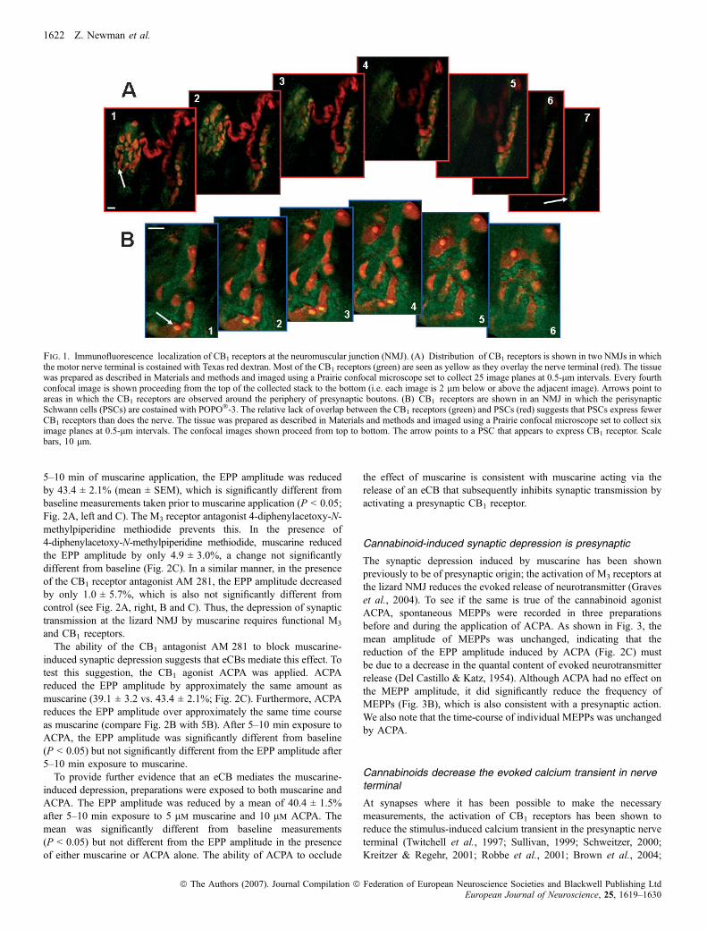
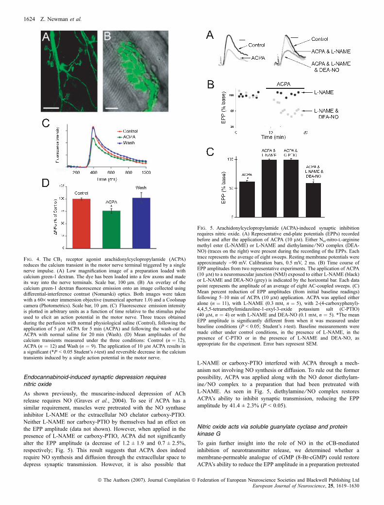
discovered and cloned, the CB1 receptor exists primarily in the nervoussystem with the CB2 receptor located mainly in immune tissue(Howlett et al., 2002; but see Van Sickle et al., 2005). We usedimmunofluorescence to determine whether the CB1 receptor is presentat the lizard NMJ. As seen in Fig. 1, we detected considerable stainingwith antibodies to the CB1 receptor. To determine specifically wherethe receptors are located at the NMJ, we back-filled the nerve terminalswith Texas red dextran (see Materials and methods) and processed thetissue for immunofluorescence, using fluorescein-labelled secondaryantibodies to reveal the CB1 receptors. The results, shown in Fig. 1A,indicate that CB1 receptors are found mostly on the nerve terminals. Wedid observe some CB1 receptor staining that did not colocalize with thenerve terminal and therefore appears green but most was clearly on thenerve terminal and therefore appears yellow because it overlays Texasred dextran. Careful examination of 0.5-lm confocal planes revealedthat most of the CB1 receptors were located along the periphery of thenerve terminal branches and boutons, presumably associated with thecell membrane (see arrows in Fig. 1A). As a control, two preparationswere exposed to the secondary antibody without the primary anti-CB1
antibody. No fluorescence could be detected in these preparations.To further establish the localization of the CB1 receptors, PSCs,
glial cells that closely envelope the nerve terminals, were stained usingthe nucleic acid stain POPO�-3 iodide (Molecular Probes). As thenerve terminals do not contain nucleic acids, POPO�-3 uniquelyidentifies the PSCs. The tissue was also processed for immunofluo-rescence, using fluorescein-labelled secondary antibodies to locate theCB1 receptors. Using confocal microscopy, we determined that a smallamount of CB1 receptors are present on the PSCs. Figure 1B shows sixconfocal images collected at 0.5-lm intervals. Although there is asmall amount of overlap between the green (CB1 receptor) and red(PSCs) signals, a close examination of the individual confocal imagesreveals that most of the CB1 receptors do not overlap with the PSCsbut are located above, below or between the PSCs. Our observationsof 12 preparations costained with CB1 antibodies and either POPO�-3or Texas red dextran (to identify PSCs or nerve terminals, respect-ively) allow us to conclude that the CB1 receptors are concentrated incell membranes of the motor nerve terminals at the lizard NMJ.However, our observations do not allow us to exclude the possibilitythat some CB1 receptors are expressed on the PSCs. We did notobserve any CB1 immunofluorescence associated with the muscle cells(data not shown).
M3 and CB1 antagonists block muscarine-induced synapticdepression
In previous work muscarine has been shown to modulate synaptictransmission at the lizard NMJ in a biphasic manner, first depressingand then enhancing neurotransmitter release (Graves et al., 2004). Inaddition to being temporally separable, the depression and enhance-ment are also pharmacologically distinct; the M3 mAChR subtypemediates the depression and the M1 subtype mediates the enhance-ment (Graves et al., 2004). To determine whether the CB1 receptorplays a role in either (or both) phase(s), the CB1 antagonist AM 281was applied. Although AM 281 had no effect when applied by itself(data not shown), when muscarine was applied in the presence ofAM 281 the first phase (depression) was precluded whereas thesecond phase (enhancement) was unaffected. Thus, we focused theremainder of our investigation on the depression of neurotransmissiontriggered by muscarine.Figure 2 depicts the unique pharmacological sensitivity of the first
phase of muscarine’s influence on synaptic transmission. Following
Endocannabinoids mediate depression 1621
ª The Authors (2007). Journal Compilation ª Federation of European Neuroscience Societies and Blackwell Publishing LtdEuropean Journal of Neuroscience, 25, 1619–1630
5–10 min of muscarine application, the EPP amplitude was reducedby 43.4 ± 2.1% (mean ± SEM), which is significantly different frombaseline measurements taken prior to muscarine application (P < 0.05;Fig. 2A, left and C). The M3 receptor antagonist 4-diphenylacetoxy-N-methylpiperidine methiodide prevents this. In the presence of4-diphenylacetoxy-N-methylpiperidine methiodide, muscarine reducedthe EPP amplitude by only 4.9 ± 3.0%, a change not significantlydifferent from baseline (Fig. 2C). In a similar manner, in the presenceof the CB1 receptor antagonist AM 281, the EPP amplitude decreasedby only 1.0 ± 5.7%, which is also not significantly different fromcontrol (see Fig. 2A, right, B and C). Thus, the depression of synaptictransmission at the lizard NMJ by muscarine requires functional M3
and CB1 receptors.The ability of the CB1 antagonist AM 281 to block muscarine-
induced synaptic depression suggests that eCBs mediate this effect. Totest this suggestion, the CB1 agonist ACPA was applied. ACPAreduced the EPP amplitude by approximately the same amount asmuscarine (39.1 ± 3.2 vs. 43.4 ± 2.1%; Fig. 2C). Furthermore, ACPAreduces the EPP amplitude over approximately the same time courseas muscarine (compare Fig. 2B with 5B). After 5–10 min exposure toACPA, the EPP amplitude was significantly different from baseline(P < 0.05) but not significantly different from the EPP amplitude after5–10 min exposure to muscarine.To provide further evidence that an eCB mediates the muscarine-
induced depression, preparations were exposed to both muscarine andACPA. The EPP amplitude was reduced by a mean of 40.4 ± 1.5%after 5–10 min exposure to 5 lm muscarine and 10 lm ACPA. Themean was significantly different from baseline measurements(P < 0.05) but not different from the EPP amplitude in the presenceof either muscarine or ACPA alone. The ability of ACPA to occlude
the effect of muscarine is consistent with muscarine acting via therelease of an eCB that subsequently inhibits synaptic transmission byactivating a presynaptic CB1 receptor.
Cannabinoid-induced synaptic depression is presynaptic
The synaptic depression induced by muscarine has been shownpreviously to be of presynaptic origin; the activation of M3 receptors atthe lizard NMJ reduces the evoked release of neurotransmitter (Graveset al., 2004). To see if the same is true of the cannabinoid agonistACPA, spontaneous MEPPs were recorded in three preparationsbefore and during the application of ACPA. As shown in Fig. 3, themean amplitude of MEPPs was unchanged, indicating that thereduction of the EPP amplitude induced by ACPA (Fig. 2C) mustbe due to a decrease in the quantal content of evoked neurotransmitterrelease (Del Castillo & Katz, 1954). Although ACPA had no effect onthe MEPP amplitude, it did significantly reduce the frequency ofMEPPs (Fig. 3B), which is also consistent with a presynaptic action.We also note that the time-course of individual MEPPs was unchangedby ACPA.
Cannabinoids decrease the evoked calcium transient in nerveterminal
At synapses where it has been possible to make the necessarymeasurements, the activation of CB1 receptors has been shown toreduce the stimulus-induced calcium transient in the presynaptic nerveterminal (Twitchell et al., 1997; Sullivan, 1999; Schweitzer, 2000;Kreitzer & Regehr, 2001; Robbe et al., 2001; Brown et al., 2004;
Fig. 1. Immunofluorescence localization of CB1 receptors at the neuromuscular junction (NMJ). (A) Distribution of CB1 receptors is shown in two NMJs in whichthe motor nerve terminal is costained with Texas red dextran. Most of the CB1 receptors (green) are seen as yellow as they overlay the nerve terminal (red). The tissuewas prepared as described in Materials and methods and imaged using a Prairie confocal microscope set to collect 25 image planes at 0.5-lm intervals. Every fourthconfocal image is shown proceeding from the top of the collected stack to the bottom (i.e. each image is 2 lm below or above the adjacent image). Arrows point toareas in which the CB1 receptors are observed around the periphery of presynaptic boutons. (B) CB1 receptors are shown in an NMJ in which the perisynapticSchwann cells (PSCs) are costained with POPO�-3. The relative lack of overlap between the CB1 receptors (green) and PSCs (red) suggests that PSCs express fewerCB1 receptors than does the nerve. The tissue was prepared as described in Materials and methods and imaged using a Prairie confocal microscope set to collect siximage planes at 0.5-lm intervals. The confocal images shown proceed from top to bottom. The arrow points to a PSC that appears to express CB1 receptor. Scalebars, 10 lm.
1622 Z. Newman et al.
ª The Authors (2007). Journal Compilation ª Federation of European Neuroscience Societies and Blackwell Publishing LtdEuropean Journal of Neuroscience, 25, 1619–1630
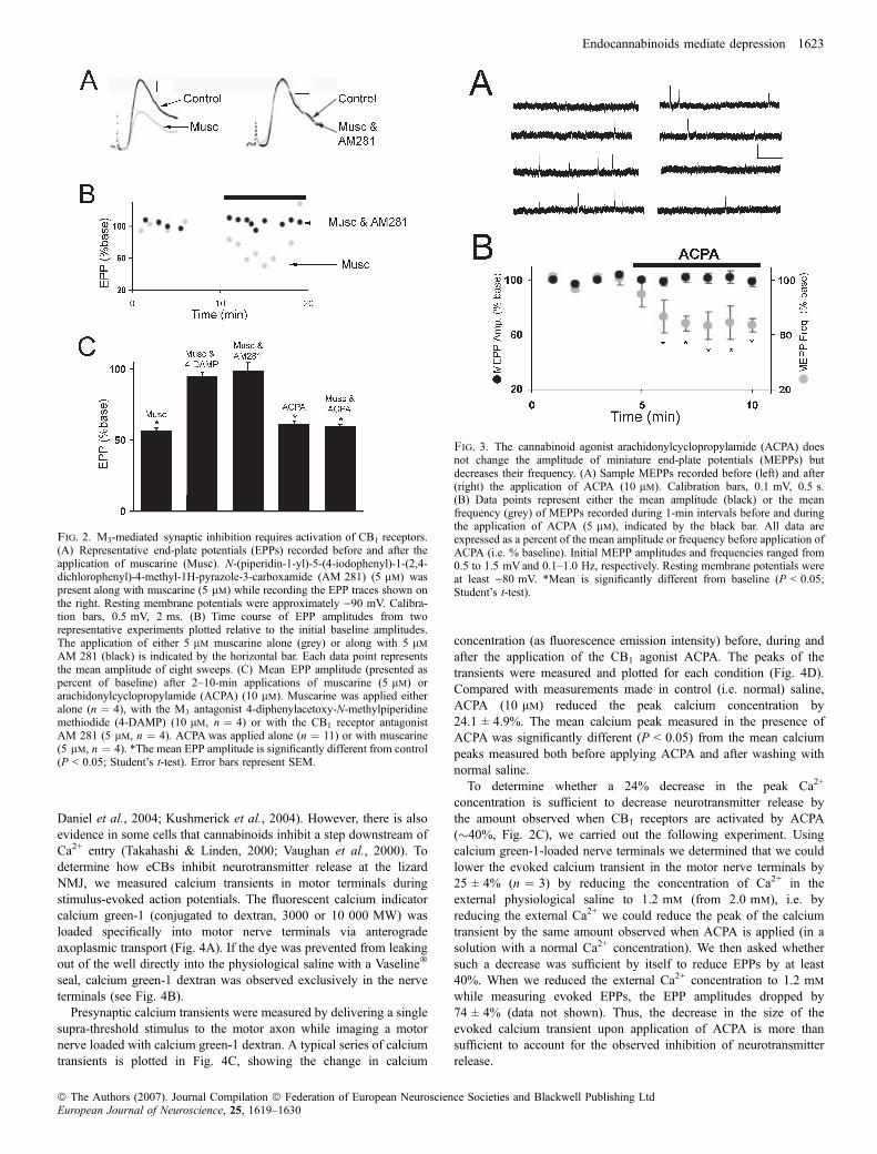
Daniel et al., 2004; Kushmerick et al., 2004). However, there is alsoevidence in some cells that cannabinoids inhibit a step downstream ofCa2+ entry (Takahashi & Linden, 2000; Vaughan et al., 2000). Todetermine how eCBs inhibit neurotransmitter release at the lizardNMJ, we measured calcium transients in motor terminals duringstimulus-evoked action potentials. The fluorescent calcium indicatorcalcium green-1 (conjugated to dextran, 3000 or 10 000 MW) wasloaded specifically into motor nerve terminals via anterogradeaxoplasmic transport (Fig. 4A). If the dye was prevented from leakingout of the well directly into the physiological saline with a Vaseline�
seal, calcium green-1 dextran was observed exclusively in the nerveterminals (see Fig. 4B).
Presynaptic calcium transients were measured by delivering a singlesupra-threshold stimulus to the motor axon while imaging a motornerve loaded with calcium green-1 dextran. A typical series of calciumtransients is plotted in Fig. 4C, showing the change in calcium
concentration (as fluorescence emission intensity) before, during andafter the application of the CB1 agonist ACPA. The peaks of thetransients were measured and plotted for each condition (Fig. 4D).Compared with measurements made in control (i.e. normal) saline,ACPA (10 lm) reduced the peak calcium concentration by24.1 ± 4.9%. The mean calcium peak measured in the presence ofACPA was significantly different (P < 0.05) from the mean calciumpeaks measured both before applying ACPA and after washing withnormal saline.To determine whether a 24% decrease in the peak Ca2+
concentration is sufficient to decrease neurotransmitter release bythe amount observed when CB1 receptors are activated by ACPA(�40%, Fig. 2C), we carried out the following experiment. Usingcalcium green-1-loaded nerve terminals we determined that we couldlower the evoked calcium transient in the motor nerve terminals by25 ± 4% (n ¼ 3) by reducing the concentration of Ca2+ in theexternal physiological saline to 1.2 mm (from 2.0 mm), i.e. byreducing the external Ca2+ we could reduce the peak of the calciumtransient by the same amount observed when ACPA is applied (in asolution with a normal Ca2+ concentration). We then asked whethersuch a decrease was sufficient by itself to reduce EPPs by at least40%. When we reduced the external Ca2+ concentration to 1.2 mm
while measuring evoked EPPs, the EPP amplitudes dropped by74 ± 4% (data not shown). Thus, the decrease in the size of theevoked calcium transient upon application of ACPA is more thansufficient to account for the observed inhibition of neurotransmitterrelease.
Fig. 2. M3-mediated synaptic inhibition requires activation of CB1 receptors.(A) Representative end-plate potentials (EPPs) recorded before and after theapplication of muscarine (Musc). N-(piperidin-1-yl)-5-(4-iodophenyl)-1-(2,4-dichlorophenyl)-4-methyl-1H-pyrazole-3-carboxamide (AM 281) (5 lm) waspresent along with muscarine (5 lm) while recording the EPP traces shown onthe right. Resting membrane potentials were approximately )90 mV. Calibra-tion bars, 0.5 mV, 2 ms. (B) Time course of EPP amplitudes from tworepresentative experiments plotted relative to the initial baseline amplitudes.The application of either 5 lm muscarine alone (grey) or along with 5 lm
AM 281 (black) is indicated by the horizontal bar. Each data point representsthe mean amplitude of eight sweeps. (C) Mean EPP amplitude (presented aspercent of baseline) after 2–10-min applications of muscarine (5 lm) orarachidonylcyclopropylamide (ACPA) (10 lm). Muscarine was applied eitheralone (n ¼ 4), with the M3 antagonist 4-diphenylacetoxy-N-methylpiperidinemethiodide (4-DAMP) (10 lm, n ¼ 4) or with the CB1 receptor antagonistAM 281 (5 lm, n ¼ 4). ACPA was applied alone (n ¼ 11) or with muscarine(5 lm, n ¼ 4). *The mean EPP amplitude is significantly different from control(P < 0.05; Student’s t-test). Error bars represent SEM.
Fig. 3. The cannabinoid agonist arachidonylcyclopropylamide (ACPA) doesnot change the amplitude of miniature end-plate potentials (MEPPs) butdecreases their frequency. (A) Sample MEPPs recorded before (left) and after(right) the application of ACPA (10 lm). Calibration bars, 0.1 mV, 0.5 s.(B) Data points represent either the mean amplitude (black) or the meanfrequency (grey) of MEPPs recorded during 1-min intervals before and duringthe application of ACPA (5 lm), indicated by the black bar. All data areexpressed as a percent of the mean amplitude or frequency before application ofACPA (i.e. % baseline). Initial MEPP amplitudes and frequencies ranged from0.5 to 1.5 mV and 0.1–1.0 Hz, respectively. Resting membrane potentials wereat least )80 mV. *Mean is significantly different from baseline (P < 0.05;Student’s t-test).
Endocannabinoids mediate depression 1623
ª The Authors (2007). Journal Compilation ª Federation of European Neuroscience Societies and Blackwell Publishing LtdEuropean Journal of Neuroscience, 25, 1619–1630
Endocannabinoid-induced synaptic depression requiresnitric oxide
As shown previously, the muscarine-induced depression of AChrelease requires NO (Graves et al., 2004). To see if ACPA has asimilar requirement, muscles were pretreated with the NO synthaseinhibitor L-NAME or the extracellular NO chelator carboxy-PTIO.Neither L-NAME nor carboxy-PTIO by themselves had an effect onthe EPP amplitude (data not shown). However, when applied in thepresence of L-NAME or carboxy-PTIO, ACPA did not significantlyalter the EPP amplitude (a decrease of 1.2 ± 1.9 and 0.7 ± 2.5%,respectively; Fig. 5). This result suggests that ACPA does indeedrequire NO synthesis and diffusion through the extracellular space todepress synaptic transmission. However, it is also possible that
L-NAME or carboxy-PTIO interfered with ACPA through a mech-anism not involving NO synthesis or diffusion. To rule out the formerpossibility, ACPA was applied along with the NO donor diethylam-ine ⁄ NO complex to a preparation that had been pretreated withL-NAME. As seen in Fig. 5, diethylamine ⁄ NO complex restoresACPA’s ability to inhibit synaptic transmission, reducing the EPPamplitude by 41.4 ± 2.3% (P < 0.05).
Nitric oxide acts via soluble guanylate cyclase and proteinkinase G
To gain further insight into the role of NO in the eCB-mediatedinhibition of neurotransmitter release, we determined whether amembrane-permeable analogue of cGMP (8-Br-cGMP) could restoreACPA’s ability to reduce the EPP amplitude in a preparation pretreated
Fig. 4. The CB1 receptor agonist arachidonylcyclopropylamide (ACPA)reduces the calcium transient in the motor nerve terminal triggered by a singlenerve impulse. (A) Low magnification image of a preparation loaded withcalcium green-1 dextran. The dye has been loaded into a few axons and madeits way into the nerve terminals. Scale bar, 100 lm. (B) An overlay of thecalcium green-1 dextran fluorescence emission onto an image collected usingdifferential-interference contrast (Nomarski) optics. Both images were takenwith a 60· water immersion objective (numerical aperture 1.0) and a Coolsnapcamera (Photometrics). Scale bar, 10 lm. (C) Fluorescence emission intensityis plotted in arbitrary units as a function of time relative to the stimulus pulseused to elicit an action potential in the motor nerve. Three traces obtainedduring the perfusion with normal physiological saline (Control), following theapplication of 5 lm ACPA for 5 min (ACPA) and following the wash-out ofACPA with normal saline for 20 min (Wash). (D) Mean amplitudes of thecalcium transients measured under the three conditions: Control (n ¼ 12),ACPA (n ¼ 12) and Wash (n ¼ 9). The application of 10 lm ACPA results ina significant (*P < 0.05 Student’s t-test) and reversible decrease in the calciumtransients induced by a single action potential in the motor nerve.
Fig. 5. Arachidonylcyclopropylamide (ACPA)-induced synaptic inhibitionrequires nitric oxide. (A) Representative end-plate potentials (EPPs) recordedbefore and after the application of ACPA (10 lm). Either Nx-nitro-l-argininemethyl ester (L-NAME) or L-NAME and diethylamine ⁄ NO complex (DEA-NO) (traces on the right) were present during the recording of the EPPs. Eachtrace represents the average of eight sweeps. Resting membrane potentials wereapproximately )90 mV. Calibration bars, 0.5 mV, 2 ms. (B) Time course ofEPP amplitudes from two representative experiments. The application of ACPA(10 lm) to a neuromuscular junction (NMJ) exposed to either L-NAME (black)or L-NAME and DEA-NO (grey) is indicated by the horizontal bar. Each datapoint represents the amplitude of an average of eight AC-coupled sweeps. (C)Mean percent reduction of EPP amplitudes (from initial baseline readings)following 5–10 min of ACPA (10 lm) application. ACPA was applied eitheralone (n ¼ 11), with L-NAME (0.3 mm, n ¼ 5), with 2-(4-carboxyphenyl)-4,4,5,5-tetramethylimidazoline-1-oxyl-3-oxide potassium salt (C-PTIO)(40 lm, n ¼ 4) or with L-NAME and DEA-NO (0.1 mm, n ¼ 5). *The meanEPP amplitude is significantly different from when it was measured underbaseline conditions (P < 0.05; Student’s t-test). Baseline measurements weremade either under control conditions, in the presence of L-NAME, in thepresence of C-PTIO or in the presence of L-NAME and DEA-NO, asappropriate for the experiment. Error bars represent SEM.
1624 Z. Newman et al.
ª The Authors (2007). Journal Compilation ª Federation of European Neuroscience Societies and Blackwell Publishing LtdEuropean Journal of Neuroscience, 25, 1619–1630
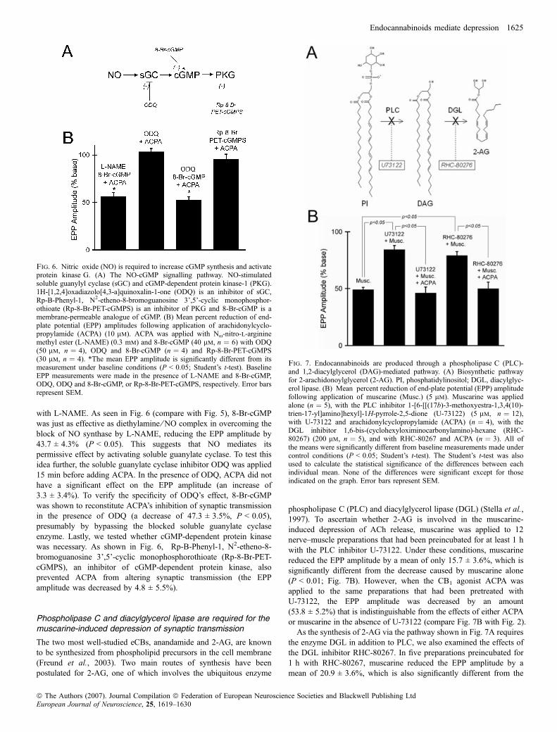
with L-NAME. As seen in Fig. 6 (compare with Fig. 5), 8-Br-cGMPwas just as effective as diethylamine ⁄ NO complex in overcoming theblock of NO synthase by L-NAME, reducing the EPP amplitude by43.7 ± 4.3% (P < 0.05). This suggests that NO mediates itspermissive effect by activating soluble guanylate cyclase. To test thisidea further, the soluble guanylate cyclase inhibitor ODQ was applied15 min before adding ACPA. In the presence of ODQ, ACPA did nothave a significant effect on the EPP amplitude (an increase of3.3 ± 3.4%). To verify the specificity of ODQ’s effect, 8-Br-cGMPwas shown to reconstitute ACPA’s inhibition of synaptic transmissionin the presence of ODQ (a decrease of 47.3 ± 3.5%, P < 0.05),presumably by bypassing the blocked soluble guanylate cyclaseenzyme. Lastly, we tested whether cGMP-dependent protein kinasewas necessary. As shown in Fig. 6, Rp-B-Phenyl-1, N2-etheno-8-bromoguanosine 3’,5’-cyclic monophosphorothioate (Rp-8-Br-PET-cGMPS), an inhibitor of cGMP-dependent protein kinase, alsoprevented ACPA from altering synaptic transmission (the EPPamplitude was decreased by 4.8 ± 5.5%).
Phospholipase C and diacylglycerol lipase are required for themuscarine-induced depression of synaptic transmission
The two most well-studied eCBs, anandamide and 2-AG, are knownto be synthesized from phospholipid precursors in the cell membrane(Freund et al., 2003). Two main routes of synthesis have beenpostulated for 2-AG, one of which involves the ubiquitous enzyme
phospholipase C (PLC) and diacylglycerol lipase (DGL) (Stella et al.,1997). To ascertain whether 2-AG is involved in the muscarine-induced depression of ACh release, muscarine was applied to 12nerve–muscle preparations that had been preincubated for at least 1 hwith the PLC inhibitor U-73122. Under these conditions, muscarinereduced the EPP amplitude by a mean of only 15.7 ± 3.6%, which issignificantly different from the decrease caused by muscarine alone(P < 0.01; Fig. 7B). However, when the CB1 agonist ACPA wasapplied to the same preparations that had been pretreated withU-73122, the EPP amplitude was decreased by an amount(53.8 ± 5.2%) that is indistinguishable from the effects of either ACPAor muscarine in the absence of U-73122 (compare Fig. 7B with Fig. 2).As the synthesis of 2-AG via the pathway shown in Fig. 7A requires
the enzyme DGL in addition to PLC, we also examined the effects ofthe DGL inhibitor RHC-80267. In five preparations preincubated for1 h with RHC-80267, muscarine reduced the EPP amplitude by amean of 20.9 ± 3.6%, which is also significantly different from the
Fig. 6. Nitric oxide (NO) is required to increase cGMP synthesis and activateprotein kinase G. (A) The NO-cGMP signalling pathway. NO-stimulatedsoluble guanylyl cyclase (sGC) and cGMP-dependent protein kinase-1 (PKG).1H-[1,2,4]oxadiazolo[4,3-a]quinoxalin-1-one (ODQ) is an inhibitor of sGC,Rp-B-Phenyl-1, N2-etheno-8-bromoguanosine 3’,5’-cyclic monophosphor-othioate (Rp-8-Br-PET-cGMPS) is an inhibitor of PKG and 8-Br-cGMP is amembrane-permeable analogue of cGMP. (B) Mean percent reduction of end-plate potential (EPP) amplitudes following application of arachidonylcyclo-propylamide (ACPA) (10 lm). ACPA was applied with Nx-nitro-l-argininemethyl ester (L-NAME) (0.3 mm) and 8-Br-cGMP (40 lm, n ¼ 6) with ODQ(50 lm, n ¼ 4), ODQ and 8-Br-cGMP (n ¼ 4) and Rp-8-Br-PET-cGMPS(30 lm, n ¼ 4). *The mean EPP amplitude is significantly different from itsmeasurement under baseline conditions (P < 0.05; Student’s t-test). BaselineEPP measurements were made in the presence of L-NAME and 8-Br-cGMP,ODQ, ODQ and 8-Br-cGMP, or Rp-8-Br-PET-cGMPS, respectively. Error barsrepresent SEM.
Fig. 7. Endocannabinoids are produced through a phospholipase C (PLC)-and 1,2-diacylglycerol (DAG)-mediated pathway. (A) Biosynthetic pathwayfor 2-arachidonoylglycerol (2-AG). PI, phosphatidylinositol; DGL, diacylglyc-erol lipase. (B) Mean percent reduction of end-plate potential (EPP) amplitudefollowing application of muscarine (Musc.) (5 lm). Muscarine was appliedalone (n ¼ 5), with the PLC inhibitor 1-[6-[[(17b)-3-methoxyestra-1,3,4(10)-trien-17-yl]amino]hexyl]-1H-pyrrole-2,5-dione (U-73122) (5 lm, n ¼ 12),with U-73122 and arachidonylcyclopropylamide (ACPA) (n ¼ 4), with theDGL inhibitor 1,6-bis-(cyclohexyloximinocarbonylamino)-hexane (RHC-80267) (200 lm, n ¼ 5), and with RHC-80267 and ACPA (n ¼ 3). All ofthe means were significantly different from baseline measurements made undercontrol conditions (P < 0.05; Student’s t-test). The Student’s t-test was alsoused to calculate the statistical significance of the differences between eachindividual mean. None of the differences were significant except for thoseindicated on the graph. Error bars represent SEM.
Endocannabinoids mediate depression 1625
ª The Authors (2007). Journal Compilation ª Federation of European Neuroscience Societies and Blackwell Publishing LtdEuropean Journal of Neuroscience, 25, 1619–1630

decrease caused by muscarine alone (Fig. 7B; P < 0.01). Thelikelihood that the action of RHC-80267 was due specifically to itsinhibition of DGL was demonstrated by applying 10 lm ACPA topreparations that had been pretreated with RHC-80267 (and musca-rine) and showing that ACPA still inhibited the EPP amplitude by itsnormal amount (i.e. 50.1 ± 5.9%).Although the comparisons described above indicate that the
enzymes PLC and DGL are responsible for a statistically significantcomponent of the muscarine-inducued depression, it is noteworthythat muscarine still significantly reduced (P < 0.05) the EPPamplitude in the presence of either U-73122 or RHC-80267 (seeFig. 7).
Endocannabinoids are released from the muscle via anendocannabinoid membrane transporter
Evidence has recently been presented that eCBs are released fromstriatal neurones in the rat brain by a membrane transporter that actsvia facilitated diffusion (Ronesi et al., 2004). To determine whetherthe same mechanism might be responsible for eCB release at thelizard NMJ and to establish the cellular source of the eCBs, weinjected individual muscle cells with VDM 11, an inhibitor of eCB-facilitated diffusion (De Petrocellis et al., 2000). Shortly afterinjecting VDM 11, EPPs were recorded from the same muscle fibrebefore and after the local application of muscarine. The average EPPamplitude was not significantly changed under these conditions(Fig. 8). In contrast, when muscles were injected with Tocrisolve�,the solvent in which VDM 11 was dissolved, or if VDM 11 wasadded to the bathing solution, muscarine significantly inhibited theEPP amplitude (by 58.1 ± 1.2 and 49.6 ± 8.2%, respectively; Fig. 8).In contrast to muscarine, when the CB1 receptor agonist ACPA wasapplied to muscle cells injected with VDM 11, the EPP amplitudewas significantly depressed (45.8 ± 8.9%; Fig. 8). The only condi-tion under which the EPP amplitude was not significantly inhibitedwas when muscarine was applied to muscles that had been injectedwith VDM 11. The small reduction of the EPP amplitude(10.9 ± 3.7%) was significantly different from each of the otherconditions (P < 0.01; Fig. 8). These results collectively support thesuggestion that eCBs are released from the postsynaptic muscle cellsvia facilitated diffusion (Ronesi et al., 2004). These results alsoindicate that the muscle is the source of eCBs released at thevertebrate NMJ following the activation of M3 mAChRs.
Endocannabinoids are involved in synaptic depression
The preceding experiments point to an essential role for eCBs in theinhibition of synaptic transmission following the activation of M3
mAChRs. To determine the physiological context under which eCBsmight become deployed, we looked for an effect of the CB1 receptorantagonist AM 281 on a form of synaptic depression that mostclosely resembles the magnitude and time-course of the inhibitioninduced by either muscarine or ACPA. The results are shown inFig. 10. Continuous stimulation of the motor nerve at 1 Hz for20 min depresses the EPP amplitude by 38.2 ± 9.2%, a reductionthat is not significantly different from that produced by eithermuscarine or ACPA (see Fig. 2). In the presence of AM 281,however, stimulation of the motor nerve (20 min, 1 Hz) failed todepress synaptic transmission, resulting in a mean EPP amplitudethat is 120.7 ± 15.3% of baseline, an amplitude that is significantlydifferent from that produced in the absence of AM 281 (P < 0.05;Fig. 10).
Discussion
To our knowledge, this is the first report of cannabinoid receptors at avertebrate striated NMJ. Although the CB1 receptor has beenidentified throughout the central and peripheral nervous systems inseveral species (see Howlett et al., 2002) and its effects have beendetected at the frog NMJ (Turkanis & Karler, 1986; Van der Kloot,1994), the receptor has never been localized specifically at the NMJ ofany species. Our observation that CB1 receptors are concentrated onthe motor nerve terminals is consistent with its preferential expressionon presynaptic nerve terminals elsewhere in the nervous system (seeKreitzer & Regehr, 2002; Wilson & Nicoll, 2002). However, ourimmunofluorescence studies also suggest that CB1 receptors may beon the closely associated PSCs, albeit at a lower density (Fig. 1B). Thesignificance of this observation has yet to be explored.
Fig. 8. Intracellular injection of cannabinoid transport inhibitor(5Z,8Z,11Z,14Z)-N-(4-hydroxy-2-methylphenyl)-5,8,11,14-eicosatetraenamide(VDM 11) into the muscle blocks muscarine-induced depression. (A) Micro-graphs showing the injection of VDM 11 into a muscle fibre near an end plate.The image on the left was collected during the pressure injection of VDM 11and rhodamine B. The neuromuscular junction (NMJ) was stained withtetraethylrhodamine-a-bungarotoxin and can be seen just below and to the rightof the injection electrode (arrow). A faint image of the recording electrode canalso be seen approaching from the right (highlighted by dashed lines). Theimage on the right was collected 5 min after injecting the muscle fibre. Theinjection electrode has been removed from the field of view and theextracellular pipette used to apply muscarine has been moved into positionjust below the neuromuscular junction. (B) Mean percent reduction of end-platepotential (EPP) amplitudes following the local application of muscarine orarachidonylcyclopropylamide (ACPA). Muscarine was applied to NMJsinjected with the solvent Tocrisolve� (n ¼ 3) or with VDM 11 dissolved inTocrisolve� (n ¼ 6). ACPA was applied to NMJs injected with VDM 11(n ¼ 8). Muscarine was also applied to NMJs bathed in VDM 11 (n ¼ 4). TheStudent’s t-test was used to calculate the statistical significance of thedifferences between the indicated pairs of means.
1626 Z. Newman et al.
ª The Authors (2007). Journal Compilation ª Federation of European Neuroscience Societies and Blackwell Publishing LtdEuropean Journal of Neuroscience, 25, 1619–1630

We carried out this investigation at the NMJ of the lizard becauseprevious work had shown that: (i) activation of M3 mAChRs depressesneurotransmitter release at this synapse (Graves et al., 2004) and (ii)eCBs mediate a similar suppression of neurotransmitter releaseinduced by M3 receptor activation in the hippocampus (Fukudomeet al., 2004). The results reported here suggest that eCBs play a similarrole at these two synapses. The model presented in Fig. 9 summarizesthe role of eCBs at the vertebrate NMJ suggested by the experimentsdescribed in this work. Activation of M3 receptors on the muscle celltriggers the synthesis of eCBs, probably 2-AG, that are released via atransporter in the muscle membrane. Once in the synaptic cleft, 2-AGbinds to CB1 receptors on the presynaptic nerve terminal, reduces theaction-potential-induced Ca2+ transient and thereby reduces neuro-transmitter release. NO, produced in either the muscle or PSCs, isrequired for one or more of the steps depicted in Fig. 9.
Mechanism of action of endocannabinoids
Previous studies are consistent with our conclusion that eCBs suppresssynaptic transmission presynaptically (Kreitzer & Regehr, 2002;Wilson & Nicoll, 2002) and by decreasing the calcium transient in the
presynaptic nerve terminal. In some neurones, cannabinoids have beenshown to inhibit presynaptic voltage-dependent Ca2+ channels (e.g.see Kushmerick et al., 2004). In other neurones, cannabinoids havebeen found to activate presynaptic K+ channels (e.g. Schweitzer, 2000;Robbe et al., 2001; Daniel et al., 2004). In either case, cannabinoidsreduce the depolarization-induced Ca2+ transient in the presynapticterminal (see Kreitzer & Regehr, 2001) and thereby decrease therelease of neurotransmitter. Our results are consistent with eitherpossibility; future work will be necessary to elucidate the specificmechanism at the NMJ.Our observation that the cannabinoid agonist ACPA reduces the
frequency ofMEPPs (see Fig. 3) has been reported by others (Takahashi& Linden, 2000; Vaughan et al., 2000; Gerdeman & Lovinger, 2001).Such observations have been used to support a mechanism of action forcannabinoids that is downstream of Ca2+ influx, i.e. a direct action onthe secretory machinery. This appears to conflict with most investiga-tions, including the present one (Fig. 4C and D), which have reportedthat activation of CB1 receptors causes a significant reduction inpresynaptic Ca2+ influx (Brown et al., 2004; Twitchell et al., 1997;Sullivan, 1999; Schweitzer, 2000; Kreitzer & Regehr, 2001; Robbeet al., 2001; Daniel et al., 2004; Kushmerick et al., 2004). These
Fig. 9. Proposed model summarizing the role played by endocannabinoids (eCBs) at the vertebrate neuromuscular junction. This represents our current workingmodel for explaining the signalling pathways involved in muscarine-induced synaptic depression at the vertebrate neuromuscular junction. Block arrows representthe diffusion or movement of a signalling molecule. Curved block arrows indicate an enzymatic conversion. Solid black arrows depict steps that have beenexperimentally verified, whereas dashed arrows reveal steps that contain unknown details. All chemicals in italics and their respective arrows are meant to show thevarious targets of each of the experimental reagents used (see text for details). We are not sure whether nitric oxide (NO) is produced in the muscle fibers or theperisynaptic Schwann cells so we have included each possibility and noted both with an asterisk. NO, acting via cGMP-dependent protein kinase (PKG), is necessarybut not sufficient to modulate neurotransmitter release and we have noted this with a dashed line and & symbol. We do not yet know the specific target of PKG.D[Ca2+]i, intracellular calcium transient; ACh, acetylcholine; ACPA, arachidonylcyclopropylamide; 2-AG, 2-arachidonylglycerol; AM 281, N-(piperidin-1-yl)-5-(4-iodophenyl)-1-(2,4-dichlorophenyl)-4-methyl-1H-pyrazole-3-carboxamide; CB1, cannabinoid receptor subtype 1; cGMP, cyclic guanosine monophosphate; DAG,diacylglycerol; 4-DAMP, 4-diphenylacetoxy-N-methylpiperidine methiodide; DGL, diacylglycerol lipase; G, G-protein; GTP, guanosine triphosphate; L-NAME,Nx-nitro-l-arginine methyl ester; M3, muscarinic acetylcholine receptor subtype 3; nAChR, nicotinic acetylcholine receptor; NOS, NO synthase; ODQ,1H-[1,2,4]oxadiazolo[4,3-a]quinoxalin-1-one; PI, phosphatidylinositol or its phosphorylated derivatives; PLC, phospholipase C; RHC-80267, 1,6-bis-(cyclohexyl-oximinocarbonylamino)-hexane; Rp-8-Br-PET-cGMPS; sGC, soluble guanylate cyclase; U-73122, 1-[6-[[(17b)-3-methoxyestra-1,3,4(10)-trien-17-yl]amino]hexyl]-1H-pyrrole-2,5-dione; VDM 11, (5Z,8Z,11Z,14Z)-N-(4-hydroxy-2-methylphenyl)-5,8,11,14-eicosatetraenamide.
Endocannabinoids mediate depression 1627
ª The Authors (2007). Journal Compilation ª Federation of European Neuroscience Societies and Blackwell Publishing LtdEuropean Journal of Neuroscience, 25, 1619–1630

apparent discrepancies may reflect different mechanisms of action foreCBs. Alternatively, they may reflect different degrees of couplingbetween calcium channels and the presynaptic vesicle release complex(e.g. see Spafford & Zamponi, 2003). We have not yet investigated thisfurther; however, the vertebrate NMJ is ideal for answering suchmechanistic questions and we eagerly anticipate using this preparationto clarify such questions related to eCB-mediated synaptic modulation.
Requirement for nitric oxide
In addition to being the first description of a physiological role for eCBsat the vertebrate NMJ, this is also the first time that the mechanism ofaction of eCBs has been directly shown to depend on NO (see Fig. 5).There have been numerous reports suggesting a linkage between eCBsand NO (e.g. see Randall & Kendall, 1998;Waksman et al., 1999; Azadet al., 2001; Namiki et al., 2005); however, to our knowledge, thecellular mechanism of action of eCBs in the nervous system has neverbeen linked to an absolute requirement for NO. The dependence ofeCBs on NO reported in this work may be unique to the NMJ.Alternatively, it may be a more general phenomenon that has not beenconsidered elsewhere. Our results indicate that eCB-mediated synapticmodulation requires NO; however, as shown previously for muscarine-induced synaptic modulation (Graves et al., 2004), NO is necessary butnot sufficient. The observation that NO synthase is present in all threecellular components at the NMJ, the nerve terminal, muscle and PSC(Graves et al., 2004), makes it difficult to determine the essential sourceof NO. However, we do know that NO must be synthesized in adifferent cell than its target as chelating extracellular NO with carboxy-PTIO abolishes synaptic modulation (Fig. 5). Therefore, we postulatethat NO is synthesized in the muscle or PSC and diffuses to the nerveterminal where it activates soluble guanylate cyclase (see Fig. 9).Evidence obtained at the amphibian NMJ supports the suggestion thatthe muscle is the source of NO, which is generated either tonically(Thomas & Robitaille, 2001) or in response to indirect low-frequencystimulation (Etherington & Everett, 2004).
Evidence that 2-arachidonoylglycerol is responsible formuscarine-induced depression
Our results implicate 2-AG as the eCB at the vertebrate NMJ (Fig. 7).It is worth noting that we were unable to completely abolishmuscarine-induced synaptic depression by inhibiting PLC or DGL
(compare Figs 2 and 7). This either means that the inhibitors that weused, U-73122 and RHC-80276, did not fully eliminate the activity ofPLC and DGL or that there is another pathway for synthesizing eCBsat the lizard NMJ. In addition to using PLC and DGL, phosphatidy-linositol can also be converted to 2-AG via phospholipase A1 andlyso-PLC (Freund et al., 2003). Thus, the residual synaptic depressionobserved in Fig. 7 may have been due to the synthesis of 2-AG via thislatter pathway (not shown). It is also possible that another cannabi-noid, such as anandamide, is also released by the activation of M3
receptors at the NMJ. Additional experiments are needed to distin-guish between these possibilities.
Mechanism of endocannabinoid release at the vertebrateneuromuscular junction
Regardless of whether the eCB at the vertebrate NMJ is exclusively2-AG or is 2-AG and anandamide, it is clear from this study that theeCB is synthesized in the muscle and is released by a membranetransporter (see Fig. 8). The idea of injecting VDM 11 in the musclewas inspired by Ronesi et al. (2004) who had shown that injection ofVDM 11 into postsynaptic cells in corticostriatal brain slices from therat abolished a long-term depression known to involve eCBs. As notedby Ronesi et al. (2004), the transporter previously shown to beresponsible for the uptake of anandamide and 2-AG via the process offacilitated diffusion (Beltramo et al., 1997; Hillard et al., 1997) wouldalso be able to transport either eCB out of the cell. The net direction ofthe movement would simply depend on the relative concentration ofeCB across the membrane. Thus, in a cell such as a striatal neurone or avertebrate muscle in which eCBs are rapidly synthesized, thetransporter will function to release the eCBs into the surrounding synapticgap. We believe that this is what is happening at the vertebrate NMJ.
Physiological relevance of endocannabinoids at the vertebrateneuromuscular junction
Under physiological conditions the muscarinic receptors at thevertebrate NMJ are presumably activated by ACh released from themotor nerve terminal during the process of synaptic transmission. Theresults presented here suggest that 20 min of 1-Hz stimulation issufficient to activate the release of eCBs, which then depress synaptictransmission by activating CB1 receptors (Fig. 10). The simplest
Control
AM 281
0
40
80
120
160
)enilesa
B fo
%( ed
utilp
mA
PP
E
0
20
40
60
80
100
120
140
0 5 10 15 20 25 30 35
Time (min)
ControlAM 281
A B
Fig. 10. Synaptic depression requires functional CB1 receptors. (A) Time course of end-plate potential (EPP) amplitudes from two representative experiments inwhich the motor nerve was stimulated for 20 min at 1 Hz (indicated by the horizontal hatched bar). The experiment was performed either under control conditions(black) or in the presence of N-(piperidin-1-yl)-5-(4-iodophenyl)-1-(2,4-dichlorophenyl)-4-methyl-1H-pyrazole-3-carboxamide (AM 281) (grey). Each data pointrepresents the amplitude of an average of eight sweeps. (B) Mean percent reduction of EPP amplitudes (from initial baseline readings) following 20 min ofcontinuous 1-Hz stimulation of the motor nerve. Stimulation was delivered either under control conditions (n ¼ 11) or in the presence of AM 281 (1 lm; n ¼ 7).The mean EPP amplitudes under these two conditions are significantly different from each other (P < 0.05; Student’s t-test). Baseline measurements were eithermade under control conditions or in the presence of AM 281, as appropriate for the experiment. Error bars represent SEM.
1628 Z. Newman et al.
ª The Authors (2007). Journal Compilation ª Federation of European Neuroscience Societies and Blackwell Publishing LtdEuropean Journal of Neuroscience, 25, 1619–1630

explanation, and the one that is most consistent with the pharmaco-logical results presented in this work, is that ACh activates M3
mAChRs, which then elicits the synthesis (Fig. 7) and release (Fig. 8)of eCBs. However, there are other possibilities. Glutamate, which hasbeen shown to be involved in synaptic depression at the frog NMJ(Pinard et al., 2003), may be responsible for the release of eCBs underphysiological conditions. Under certain circumstances, glutamate mayact either with or without ACh. Further experiments are needed toresolve these possibilities.
Conclusion
Given the apparent high density of CB1 receptors on motor nerveterminals (e.g. Fig. 1A) and the relatively robust physiological effectsof CB1 agonists and antagonists (e.g. Figs 2, 3 and 5), we find itsurprising that it has taken so long for a role to be discovered for eCBsat the vertebrate NMJ. This is even more surprising given the fact thatit has been known for many years that exogenous cannabinoids reducemotor function in rats (Compton et al., 1996; Romero et al., 1996) andhumans (Ashton, 1999). We hope that the results presented in thiswork will stimulate further investigation into the role of eCBs at theNMJ. In addition to providing a greater appreciation of the diversity ofroles for eCBs, the results reported here indicate that the vertebrateNMJ, which is an excellent model synapse for investigating thecellular and molecular details of synaptic transmission, may beemployed to great advantage in studying details of eCB physiology.
Acknowledgements
We would like to thank the Howard Hughes Medical Institute’s UndergraduateScience Education Program and Grinnell College for their financial support ofthis research. We also thank Profs Charles Sullivan and Mark Levandoski fortheir assistance with the preparation of the manuscript. This article iscontribution no. 4 from the Barr Laboratory of Developmental Biology andCellular Neuroscience at Grinnell College.
Abbreviations
ACh, acetylcholine; ACPA, arachidonylcyclopropylamide; 2-AG, 2-arachido-noylglycerol; AM 281, N-(piperidin-1-yl)-5-(4-iodophenyl)-1-(2,4-dichloro-phenyl)-4-methyl-1H-pyrazole-3-carboxamide; carboxy-PTIO, 2-(4-carboxy-phenyl)-4,4,5,5-tetramethylimidazoline-1-oxyl-3-oxide potassium salt; DGL,diacylglycerol lipase; eCB, endocannabinoid; EPP, end-plate potential;L-NAME, Nx-nitro-l-arginine methyl ester; mAChR, muscarinic acetylcholinereceptor; MEPP, miniature end-plate potential; NMJ, neuromuscular junction;NO, nitric oxide; ODQ, 1H-[1,2,4]oxadiazolo[4,3-a]quinoxalin-1-one; PLC,phospholipase C; PSC, perisynaptic Schwann cell; RHC-80267, 1,6-bis-(cyclohexyloximinocarbonylamino)-hexane; U-73122, 1-[6-[[(17b)-3-meth-oxyestra-1,3,4(10)-trien-17-yl]amino]hexyl]-1H-pyrrole-2,5-dione; VDM 11,(5Z,8Z,11Z,14Z)-N-(4-hydroxy-2-methylphenyl)-5,8,11,14-eicosatetraenamide.
References
Ameri, A. (1999) The effects of cannabinoids on the brain. Prog. Neurobiol.,58, 315–348.
Ashton, C.H. (1999) Adverse effects of cannabis and cannabinoids. Br. J.Anaesth., 83, 637–649.
Azad, S.C., Marsicano, G., Eberlein, I., Putzke, J., Zieglgansberger, W.,Spanagel, R. & Lutz, B. (2001) Differential role of the nitric oxide pathwayon delta(9)-THC-induced central nervous system effects in the mouse. Eur. J.Neurosci., 13, 561–568.
Beltramo, M., Stella, N., Calignano, A., Lin, S.Y., Makriyannis, A. & Piomelli,D. (1997) Functional role of high-affinity anandamide transport, as revealedby selective inhibition. Science, 277, 1094–1097.
Boehm, S. & Huck, S. (1997) Receptors controlling transmitter release fromsympathetic neurons in vitro. Prog. Neurobiol., 51, 225–242.
Brenowitz, S.D. & Regehr, W.G. (2003) Calcium dependence of retrogradeinhibition by endocannabinoids at synapses onto Purkinje cells. J. Neurosci.,23, 6373–6384.
Brown, S.P., Safo, P.K. & Regehr, W.G. (2004) Endocannabinoids inhibittransmission at granule cell to Purkinje cell synapses by modulating threetypes of presynaptic calcium channels. J. Neurosci., 24, 5623–5631.
Caulfield, M.P. (1993) Muscarinic receptors ) characterization, coupling andfunction. Pharmacol. Ther., 58, 319–379.
Compton, D.R., Aceto, M.D., Lowe, J. & Martin, B.R. (1996) In vivocharacterization of a specific cannabinoid receptor antagonist (SR141716A):inhibition of delta 9-tetrahydrocannabinol-induced responses and apparentagonist activity. J. Pharmacol. Exp. Ther., 277, 586–594.
Daniel, H., Rancillac, A. & Crepel, F. (2004) Mechanisms underlyingcannabinoid inhibition of presynaptic Ca2+ influx at parallel fibre synapsesof the rat cerebellum. J. Physiol., 557, 159–174.
Del Castillo, J. & Katz, B. (1954) Quantal components of the end-platepotential. J. Physiol., 124, 560–573.
De Petrocellis, L., Bisogno, T., Davis, J.B., Pertwee, R.G. & DiMarzo, V.(2000) Overlap between the ligand recognition properties of the anandamidetransport and the VR1 vanilloid receptor: inhibitors of anandamide uptakewith negligible capsaicin-like activity. FEBS Lett., 483, 52–56.
Devane, W.A., Hanus, L., Breuer, A., Pertwee, R.G., Stevenson, L.A., Griffin,G., Gibson, D., Mandelbaum, A., Etinger, A. & Mechoulam, R. (1992)Isolation and structure of a brain constituent that binds to the cannabinoidreceptor. Science, 258, 1946–1949.
Diana, M.A., Levenes, C., Mackie, K. & Marty, A. (2002) Short-termretrograde inhibition of GABAergic synaptic currents in rat Purkinje cells ismediated by endogenous cannabinoids. J. Neurosci., 22, 200–208.
Duncan, C.J. & Publicover, S.J. (1979) Inhibitory effects of cholinergic agentson the release of transmitter at the frog neuromuscular junction. J. Physiol.,294, 91–103.
Etherington, S.J. & Everett, A.W. (2004) Postsynaptic production of nitricoxide implicated in long-term depression at the mature amphibian (Bufomarinus) neuromuscular junction. J. Physiol., 559, 507–517.
Evans, D.M., Johnson, M.R. & Howlett, A.C. (1992) Ca2+-dependent releasefrom rat brain of cannabinoid receptor binding activity. J. Neurochem., 58,780–782.
Freund, T.F., Katona, I. & Piomelli, D. (2003) Role of endogenouscannabinoids in synaptic signaling. Physiol. Rev., 83, 1017–1066.
Fukudome, Y., Ohno-Shosaku, T., Matsui, M., Omori, Y., Fukaya, M.,Tsubokawa, H., Taketo, M.M., Watanabe, M., Manabe, T. & Kano, M.(2004) Two distinct classes of muscarinic action on hippocampal inhibitorysynapses: M2-mediated direct suppression and M1 ⁄ M3-mediated indirectsuppression through endocannabinoid signaling. Eur. J. Neurosci., 19, 2682–2692.
Ganguly, D.K. & Das, M. (1979) Effects of oxotremorine demonstratepresynaptic muscarinic and dopaminergic receptors on motor nerveterminals. Nature, 278, 645–646.
Gerdeman, G. & Lovinger, D.M. (2001) CB1 cannabinoid receptor inhibitssynaptic release of glutamate in rat dorsolateral striatum. J. Neurophysiol.,85, 468–471.
Graves, A.R., Lewin, K.A. & Lindgren, C.A. (2004) Nitric oxide, cAMP andthe biphasic muscarinic modulation of ACh release at the lizardneuromuscular junction. J. Physiol., 559, 423–432.
Hillard, C.J., Edgemond, W.S., Jarrahian, A. & Campbell, W.B. (1997)Accumulation of N-arachidonoylethanolamine (anandamide) intocerebellar granule cells occurs via facilitated diffusion. J. Neurochem., 69,631–638.
Howlett, A.C., Barth, F., Bonner, T.I., Cabral, G., Casellas, P., Devane, W.A.,Felder, C.C., Herkenham, M., Mackie, K., Martin, B.R., Mechoulam, R. &Pertwee, R.G. (2002) International Union of Pharmacology. XXVII.Classification of cannabinoid receptors. Pharmacol. Rev., 54, 161–202.
Kim, J., Isokawa, M., Ledent, C. & Alger, B.E. (2002) Activation of muscarinicacetylcholine receptors enhances the release of endogenous cannabinoids inthe hippocampus. J. Neurosci., 22, 10 182–10 191.
Kreitzer, A.C. & Regehr, W.G. (2001) Retrograde inhibition of presynapticcalcium influx by endogenous cannabinoids at excitatory synapses ontoPurkinje cells. Neuron, 29, 717–727.
Kreitzer, A.C. & Regehr, W.G. (2002) Retrograde signaling by endocannabi-noids. Curr. Opin. Neurobiol., 12, 324–330.
Kushmerick, C., Price, G.D., Taschenberger, J., Puente, N., Renden, R.,Wadiche, J.I., Duvoisin, R.M., Grandes, P. & von Gersdorff, H. (2004)Retroinhibition of presynaptic Ca2+ currents by endocannabinoids released
Endocannabinoids mediate depression 1629
ª The Authors (2007). Journal Compilation ª Federation of European Neuroscience Societies and Blackwell Publishing LtdEuropean Journal of Neuroscience, 25, 1619–1630

via postsynaptic mGluR activation at a Calyx Synapse. J. Neurosci., 24,5955–5965.
Lindgren, C.A. & Moore, J.W. (1989) Identification of ionic currents atpresynaptic nerve endings of the lizard. J. Physiol., 414, 201–222.
Maejima, T., Hashimoto, K., Yoshida, T., Aiba, A. & Kano, M. (2001)Presynaptic inhibition caused by retrograde signal from metabotropicglutamate to cannabinoid receptors. Neuron, 31, 463–475.
Martin, B.R., Mechoulam, R. & Razdan, R.K. (1999) Discovery andcharacterization of endogenous cannabinoids. Life Sci., 65, 573–595.
Michaelson, D.M., Avissar, S., Kloog, Y. & Sokolovsky, M. (1979) Mechanismof acetylcholine release: possible involvement of presynaptic muscarinicreceptors in regulation of acetylcholine release and protein phosphorylation.Proc. Natl Acad. Sci. U.S.A., 76, 6336–6340.
Minic, J., Molgo, J., Karlsson, E. &Krejci, E. (2002) Regulation of acetylcholinerelease by muscarinic receptors at the mouse neuromuscular junction dependson the activity of acetylcholinesterase. Eur. J. Neurosci., 15, 439–448.
Namiki, S., Kakizawa, S., Hirose, K. & Iino, M. (2005) NO signaling decodesfrequency of neuronal activity and generates synapse-specific plasticity inmouse cerebellum. J. Physiol., 566, 849–863.
Ohno-Shosaku, T., Maejima, T. & Kano, M. (2001) Endogenous cannabinoidsmediate retrograde signals from depolarized postsynaptic neurons topresynaptic terminals. Neuron, 29, 729–738.
Ohno-Shosaku, T., Matsui, M., Fukudome, Y., Shosaku, J., Tsubokawa, H.,Taketo, M.M., Manabe, T. & Kano, M. (2003) Postsynaptic M1 and M3
receptors are responsible for the muscarinic enhancement of retrograde endo-cannabinoid signaling in the hippocampus. Eur. J. Neurosci., 18, 109–116.
Pinard, A., Levesque, S., Vallee, J. & Robitaille, R. (2003) Glutamatergicmodulation of synaptic plasticity at a PHS vertebrate cholinergic synapse.Eur. J. Neurosci., 18, 3241–3250.
Randall, M.D. & Kendall, D.A. (1998) Endocannabinoids: a new class ofvasoactive substances. Trends Pharmacol. Sci., 19, 55–58.
Robbe, D., Alonso, G., Duchamp, F., Bockaert, J. & Manzoni, O.J. (2001)Localization and mechanisms of action of cannabinoid receptors at theglutamatergic syanapses of the mouse nucleus accumbens. J. Neurosci., 21,109–116.
Romero, J., Garcia-Palomero, D., Lin, S.Y., Ramos, J.A., Makriyannis, A. &Fernandez-Ruiz, J.J. (1996) Extrapyramidal effects of methanandamide, ananalog of anandamide, the endogenous CB1 receptor ligand. Life Sci., 58,1249–1257.
Ronesi, J., Gerdeman, G.L. & Lovinger, D.M. (2004) Disruption of endocanna-binoid release and striatal long-term depression by postsynaptic blockade ofendocannabinoid membrane transport. J. Neurosci., 24, 1673–1679.
Schweitzer, P. (2000) Cannabinoids decrease the K+ M-current in hippocampalCA1 neurons. J. Neurosci., 20, 51–58.
Slutsky, I., Parnas, H. & Parnas, I. (1999) Presynaptic effects of muscarine onACh release at the frog neuromuscular junction. J. Physiol., 514, 769–782.
Slutsky, I., Silman, I., Parnas, I. & Parnas, H. (2001) Presynaptic M2
muscarinic receptors are involved in controlling the kinetics of ACh releaseat the frog neuromuscular junction. J. Physiol., 536, 717–725.
Spafford, J.D. & Zamponi, G.W. (2003) Functional interactions betweenpresynaptic calcium channels and the neurotransmitter release machinery.Curr. Opin. Neurobiol., 13, 1–7.
Standaert, F.G. (1982) Release of transmitter at the neuromuscular junction. Br.J. Anaesth., 54, 131–145.
Starke, K., Gothert, M. & Kilbinger, H. (1989) Modulation of neurotransmitterrelease by presynaptic autoreceptors. Physiol. Rev., 69, 864–989.
Stella, N., Schweitzer, P. & Piomelli, D. (1997) A second endogenouscannabinoid that modulates long-term potentiation. Nature, 388, 773–778.
Sullivan, J.M. (1999) Mechanisms of cannabinoid-receptor-mediated inhibitionof synaptic transmission in cultured hippocampal pyramidal neurons.J. Neurophysiol., 82, 1286–1294.
Takahashi, K.A. & Linden, D.J. (2000) Cannabinoid receptor modulation ofsynapses received by cerebellar Purkinje cells. J. Neurophysiol., 83, 1167–1180.
Thomas, S. & Robitaille, R. (2001) Differential frequency-dependent regulationof transmitter release by endogenous nitric oxide at the amphibianneuromuscular synapse. J. Neurosci., 21, 1087–1095.
Turkanis, S.A. & Karler, R. (1986) Effects of delta-9-tetrahydrocannabinol, 11-hydroxy-delta-9-tetrahydrocannabinol and cannabidol on neuromusculartransmission in the frog. Neuropharmacology, 25, 1273–1278.
Twitchell, W., Brown, S. & Mackie, K. (1997) Cannabinoids Inhibit N- andP/Q-type Calcium Channels in cultured Rat Hippocampal neurons.J. Neurophysiol, 78, 43–50.
Van der Kloot, W. (1994) Anandamide, a naturally-occurring agonist of thecannabinoid receptor, blocks adenylate cyclase at the frog neuromuscularjunction. Brain Res., 649, 181–184.
Van Sickle, M.D., Duncan, M., Kingsley, P.J., Mouihate, A., Urbani, P.,Mackie, K., Stella, N., Makriyannis, A., Piomelli, D., Davison, J.S., Marnett,L.J., Di Marzo, V., Pittman, Q.J., Patel, K.D. & Sharkey, K.A. (2005)Identification and functional characterization of brainstem cannabinoid CB2
receptors. Science, 310, 329–332.Vaughan, C.W., Connor, M., Bagley, E.E. & Christie, M.J. (2000) Actions of
cannabinoids on membrane properties and synaptic transmission in ratperiaqueductal gray neurons in vitro. Mol. Pharmacol., 57, 288–295.
Waksman, Y., Olson, J.M., Carliscl, S.J. & Cabral, G.A. (1999) The centralcannabinoid receptor (CB1) mediates inhibition of nitric oxide production byrat microglial cells. J. Pharm. Exp. Ther., 288, 1357–1366.
Wali, F.A., Suer, A.H., McAteer, E., Dark, C.H. & Jones, C.J. (1988) Effects ofatropine and glycopyrrolate on neuromuscular transmission in the rat phrenicnerve-diaphragm preparation. Gen. Pharmacol., 19, 285–290.
Wilson, R.I. & Nicoll, R.A. (2001) Endogenous cannabinoids mediateretrograde signaling at hippocampal synapses. Nature, 410, 588–592.
Wilson, R.I. & Nicoll, R.A. (2002) Endocannabinoid signaling in the brain.Science, 296, 678–682.
Yoshida, T., Hashimoto, K., Zimmer, A., Maejima, T., Araishi, K. & Kano, M.(2002) The cannabinoid CB1 receptor mediates retrograde signals fordepolarization-induced suppression of inhibition in cerebellar Purkinje cells.J. Neurosci., 22, 1690–1697.
1630 Z. Newman et al.
ª The Authors (2007). Journal Compilation ª Federation of European Neuroscience Societies and Blackwell Publishing LtdEuropean Journal of Neuroscience, 25, 1619–1630