Inhibition of human and mouse plasma membrane bound NTPDases by P2 receptor antagonists

Chemokines are small, mainly basic proteins involved in host defense that summonimmune cells in response to invasion by pathogenic organisms (1). They consist of 340–380 amino acid residues, which play key roles in immunomodulation and host defensemechanisms (2). Chemokine receptors are G protein-coupled receptors containing 7
287
Acta Pharm. 62 (2012) 287–304 Original research paperDOI: 10.2478/v10007-012-0029-7
Structural investigations of CXCR2 receptor antagonistsby CoMFA, CoMSIA and flexible docking studies
SHRAVAN KUMAR GUNDAROHITH KUMAR ANUGOLU*SRI RAMYA TATASAIKH MAHMOOD
Bioinformatics Division, OsmaniaUniversity, Hyderabad-500007Andhra Pradesh, India
Accepted July 17, 2012
Three-dimensional quantitative structure activity rela-tionship (3D QSAR) analysis was carried out on a set of56 N,N’-diarylsquaramides, N,N’-diarylureas and diami-nocyclobutenediones in order to understand their antago-nistic activities against CXCR2. The studies includedcomparative molecular field analysis (CoMFA) and com-parative molecular similarity indices analysis (CoMSIA).Models with good predictive abilities were generatedwith CoMFA q2 = 0.709, r2 (non-cross-validated square ofcorrelation coefficient) = 0.951, F value = 139.903, r2 bs =0.978 with five components, standard error of estimate =0.144 and the CoMSIA q2 = 0.592, r2 = 0.955, F value =122.399, r2 bs = 0.973 with six components, standard er-ror of estimate = 0.141. In addition, a homology model ofCXCR2 was used for docking based alignment of thecompounds. The most active compound then served as atemplate for alignment of the remaining structures. Fur-ther, mapping of contours onto the active site validatedeach other in terms of residues involved with referenceto the respective contours. This integrated moleculardocking based alignment followed by 3D QSAR studiesprovided a further insight to support the structure-baseddesign of CXCR2 antagonistic agents with improved ac-tivity profiles. Furthermore, in silico screening was adap-ted to the QSAR model in order to predict the structuresof new, potentially active compounds.
Keywords: CXCR2, homology modeling, CoMFA, CoMSIA,PLS, docking
* Correspondence; e-mail: [email protected]
trans-membrane domains with three intracellular and thre extracellular hydrophilicloops, and an intracellular C-terminus containing serine and threonine residues. Che-mokines, an expanding family of small cytokines, have an important role in the chemo-taxis of several leucocyte subsets. Chemokines comprise a large protein family that canbe divided into subfamilies on the bases of structural motifs. These receptors are dividedinto four subgroups: CC (proteins have two adjacent cysteines), C (protein has only twocysteines), CX3C (proteins have three amino acids between the two cysteines) and CXC(proteins have two N-terminal cysteines, which are separated by one amino acid) che-mokine ligands, based upon the presence and positioning of the first two of the fourconserved cysteine residues (3).
CXC chemokines are characteristically heparin binding proteins. The candidateCXC chemokine receptors that mediate angiogenic activity are CXCR1 and/or CXCR2.Only IL-8 and GCP-2 specifically bind to CXCR1, whereas all ELR+ CXC chemokinesbind to CXCR2 (4). CXCR2 is expressed by a wide range of cell types, for example, neu-trophils, mast cells, T cells, keratinocytes, and cerebellar neurons. Increased levels ofCXCR2 and its ligand IL-8 have been observed in humans with diseases such as arthri-tis, asthma, rheumatoid arthritis, psoriasis, reperfusion injury, and chronic obstructivepulmonary disease (5). This suggests that the CXCR2 receptor and IL-8 may play a piv-otal role in these inflammatory disorders. An immune response is characterized by theactivation of leukocytes and migration of hematopoietic cells into secondary lymphoidorgans or to the site of inflammation. Under inflammatory conditions, the expression ofspecific chemokines, a class of small proteins with potent chemotactic activity, is up-regu-lated (6). In the light of these findings, small molecule antagonists of the CXCR2 recep-tor are attractive biological targets for molecular drug discovery.
Quantitative structure-activity relationship (QSAR) enables the investigators to es-tablish reliable quantitative structure-activity and structure-property relationships to de-rive in silico QSAR models to predict the activity of novel molecules prior to their syn-thesis. Comparative molecular field analysis (CoMFA) (7) and comparative molecularsimilarity indices analysis (CoMSIA) (8) have been performed to better understand thestructure-activity relationship. In the present work, the ligand guided approach and themolecular modeling studies of CXCR2 antagonists were carried out using docking andthree-dimensional quantitative structure-activity relationship (3D-QSAR) analyses.
EXPERIMENTAL
Data set
Biological and chemical data of 56 CXCR2 antagonists, taken from the literature (9,10) were selected for the QSAR study. The half maximal inhibitory concentration is ameasure of the effectiveness of a compound in inhibiting a biological or biochemicalfunction, which is usually represented as IC50 in mmol L�1. Six molecules, whose IC50values were reported as > 270 mmol L�1 and > 2000 mmol L�1, respectively, were notconsidered in this study. Reported IC50 values of molecules were converted into pIC50values (negative log of molar concentration value) and used as dependent variables inCoMFA and CoMSIA analyses. The 3D QSAR models were generated using a training
288
S. Kumar Gunda et al.: Structural investigations of CXCR2 receptor antagonists by CoMFA, CoMSIA and flexible docking studies,Acta Pharm. 62 (2012) 287–304.
set of 42 molecules. Predictive power of the resulting models was evaluated using a testset of 14 molecules. The test set compounds were selected manually so that the struc-tural diversity and wide range of activity in the data set were included.
Molecular modeling
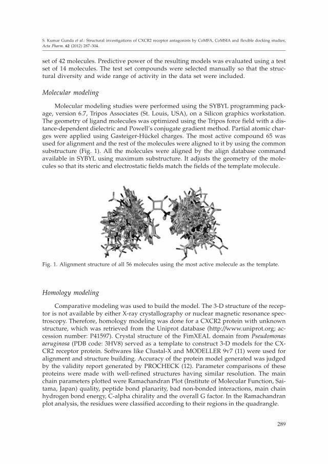
Molecular modeling studies were performed using the SYBYL programming pack-age, version 6.7, Tripos Associates (St. Louis, USA), on a Silicon graphics workstation.The geometry of ligand molecules was optimized using the Tripos force field with a dis-tance-dependent dielectric and Powell’s conjugate gradient method. Partial atomic char-ges were applied using Gasteiger-Hückel charges. The most active compound 65 wasused for alignment and the rest of the molecules were aligned to it by using the commonsubstructure (Fig. 1). All the molecules were aligned by the align database commandavailable in SYBYL using maximum substructure. It adjusts the geometry of the mole-cules so that its steric and electrostatic fields match the fields of the template molecule.
Homology modeling
Comparative modeling was used to build the model. The 3-D structure of the recep-tor is not available by either X-ray crystallography or nuclear magnetic resonance spec-troscopy. Therefore, homology modeling was done for a CXCR2 protein with unknownstructure, which was retrieved from the Uniprot database (http://www.uniprot.org; ac-cession number: P41597). Crystal structure of the FimXEAL domain from Pseudomonasaeruginosa (PDB code: 3HV8) served as a template to construct 3-D models for the CX-CR2 receptor protein. Softwares like Clustal-X and MODELLER 9v7 (11) were used foralignment and structure building. Accuracy of the protein model generated was judgedby the validity report generated by PROCHECK (12). Parameter comparisons of theseproteins were made with well-refined structures having similar resolution. The mainchain parameters plotted were Ramachandran Plot (Institute of Molecular Function, Sai-tama, Japan) quality, peptide bond planarity, bad non-bonded interactions, main chainhydrogen bond energy, C-alpha chirality and the overall G factor. In the Ramachandranplot analysis, the residues were classified according to their regions in the quadrangle.
289
S. Kumar Gunda et al.: Structural investigations of CXCR2 receptor antagonists by CoMFA, CoMSIA and flexible docking studies,Acta Pharm. 62 (2012) 287–304.
Fig. 1. Alignment structure of all 56 molecules using the most active molecule as the template.

Docking studies
Molecular docking studies were carried out using the FlexX (13) program interfacedwith SYBYL 6.7. In this automated docking program, the flexibility of ligands is conside-red while the protein or biomolecule is considered as a rigid structure. The ligand isbuilt in an incremental fashion, where each new fragment is added in all possible posi-tions and conformations to a pre-placed base fragment inside the active site. All the mole-cules for docking were sketched in the SYBYL and all the charges were removed. The 3Dcoordinates of active sites were taken from the X-ray crystal structure of the FimXEALdomain protein for docking. All water molecules were removed and the protein wasused to dock the inhibitor (14). The ligand was pre-processed before docking calcula-tions by giving charges according to the Gasteiger-Hückel method, followed by energyminimization with 10,000 iterations of conjugate gradient algorithm using the Triposforce field. The compounds were docked into the binding site using FlexX, which uses arobust incremental construction algorithm in which the ligand is decomposed into pie-ces and then flexibly is built up in the active site, using a variety of placement strategies.The active site is defined within the 6.5 × 10�10 m radius around the co-crystallized li-gand. This ligand-based study was performed using the pharmacophore based programin SYBYL 6.7, and the receptor-guided study was performed using molecular dockingwith FlexX. The receptor structure of CXCR2 was modeled by using the crystal structureof the FimXEAL domain protein (PDB code: 3HV8) as a template. A linear method suchas PLS is used to find quantitative relationships between the structures of CXCR2 antag-onists and their biological activities, and the results obtained by this method are com-pared.
CoMFA studies
Steric and electrostatic interactions were calculated using the Tripos force field with adistance-dependent dielectric constant in all interactions in a regularly spaced (2 × 10�10
m) grid taking a sp3 carbon atom as steric probe and a +1 charge as electrostatic probe.The cut-off was set to 30 kcal mol�1. With standard options for the scaling of variables,the regression analysis was carried out using the full cross-validated partial leastsquares (PLS) method (leave-one-out) (15). The minimum sigma (column filtering) wasset to 2.0 kcal mol�1 to improve the signal-to-noise ratio by omitting those lattice pointswhose energy variation was below this threshold. The final model, non-cross-validatedconventional analysis, was developed with the optimum number of components to yielda non-cross-validated r2 value.
CoMSIA studies
In CoMSIA, a distance-dependent Gaussian-type physicochemical property wasadopted to avoid singularities at the atomic positions and dramatic changes to potentialenergy for those grids in the proximity of the surface. With the standard parameters andno arbitrary cut-off limits, five physicochemical properties, namely, steric, electrostatic,hydrophobic, hydrogen bond donor and hydrogen bond acceptor fields, were calcu-lated. The properties were calculated using a C+ probe atom with a radius of 1 × 10�10 mplaced at a regular grid spacing of 2 × 10�10 m to enclose all the binding conformations
290
S. Kumar Gunda et al.: Structural investigations of CXCR2 receptor antagonists by CoMFA, CoMSIA and flexible docking studies,Acta Pharm. 62 (2012) 287–304.

of the inhibitors. In general, the similarity index AF,K between the compounds of interestwas computed by placing a probe atom at the intersections of lattice points using Eq. (1):
A j W W ear
i
n
F,Kq
probe,k ikiq( ) ��
�
�
�2
1
(1)
where q represents a grid point, i is the summation index over all atoms of molecule junder computation, Wik is the actual value of the physicochemical property k of atom i,and Wprobe,k is the value of the probe atom. In the present study, we used a probe atom(Wprobe,k) with charge +1, radius 10�10 m, hydrophobicity +1, and attenuation factor (á)of 0.3 for the Gaussian type distance (16). Statistical evaluation for the CoMSIA analysiswas performed in the same way as described for CoMFA.
Partial least squares analysis
In 3D-QSAR, the CoMFA and CoMSIA descriptors were used as independent vari-ables and pIC50 as the dependent variable. To quantify the relationship between thesestructural parameters (CoMFA and CoMSIA interaction energies) and biological activities,the PLS algorithm was used. The CoMFA descriptors were used as independent variables,and pIC50 values as dependent variables in partial least square regression analysis.Cross-validation partial least square method of leave-one-out (LOO) was performed toobtain the optimal number of components used in subsequent analyses. The minimumsigma (column filtering) was set to 2.0 kcal mol�1 to improve the signal-to-noise ratio. Theoptimum number of principle components in the final non-cross-validated QSAR equa-tions was determined to be that leading to the highest coefficient (r2) and the lowest stan-dard error in the LOO cros-validated predictions. The non-cross-validation was used inthe analysis of CoMFA results and prediction of the model. The same method was alsoused for CoMSIA; thereafter a full PLS was run using column filtering of 1.0 kcal mol�1
(17). Auto scaling was applied to all CoMSIA analysis. The cross-validated correlation co-efficient (q2), which resulted in optimum number of components and lowest standard er-ror of prediction, was considered for further analysis and calculated using Eq. (2):
q2 = 1 –( )
( )
g g
g gg
g
pred actual
actual mean
�
���
2
2(2)
where ãpred, ãactual, and ãmean are the predicted, actual, and mean values of the targetproperty (pIC50), respectively.
The predicted ability of generated 3D-QSAR models was determined using a test set of14 molecules that were separated during model generation. Test set molecules were chosenrandomly considering a wide range of activity. Energy minimization and alignment of these14 molecules were the same as the training set molecules described previously, and their ac-tivities were predicted using the model generated from the training set. The predicted corre-lation coefficient (r2
pred) based on test set molecules, was computed using Eq. (3).
r2pred =
SD PRESS
SD
�(3)
291
S. Kumar Gunda et al.: Structural investigations of CXCR2 receptor antagonists by CoMFA, CoMSIA and flexible docking studies,Acta Pharm. 62 (2012) 287–304.

where SD is the sum of squared deviations between the biological activities of the testset and mean activities of the training molecules and PRESS is the sum of squared devi-ations between the predicted and actual activities of the test set molecules.
RESULTS AND DISCUSSION
Homology modeling and molecular dynamic simulation
The sequence was aligned based on the structure. Based on sequence similarity ana-lysis between the target and other proteins, known structures showed that the template(3HV8) had 63 % of amino acid sequence identity with our predicted model C-C chemo-kine receptor type 2 protein. Practically, at this level of sequence identity, it is satisfac-tory enough to use the crystallographic structure of 3HV8 as a template in order to ob-tain high quality alignment for structure prediction by homology modeling. In thisstudy, ö and ø torsion angles were also checked using the Ramachandran plots. Com-parison of the results shows that one of the models generated by the MODELLER9v7program maintained by Ben Webb (San Francisco, USA) is more acceptable. The bestmodel predicted by MODELLER was used for further analysis by PROCHECK (Euro-pean Bioinformatics Institute, Cambridge, UK). The overall general similarities and sub-tle differences between the 3D structure of template 3HV8 and the predicted model canbe seen from the backbone superposition. PROCHECK statistics indicated that approxi-mately 98 % of residues in the CXCR2 model were either in the most favored or in theadditionally allowed regions; 1.5 % of residues were in the generously allowed regions,and 0.3 % of the residues were in the disallowed regions. As evident from superposition,the general folding topology of the structure is similar; however, some structural differ-ences appear between the predicted model and the template. These differences aremainly due to insertions and deletions in different loop regions (18). The CXCR2 mod-eled protein interacting with the most active compound 65 is shown in Fig. 2.
292
S. Kumar Gunda et al.: Structural investigations of CXCR2 receptor antagonists by CoMFA, CoMSIA and flexible docking studies,Acta Pharm. 62 (2012) 287–304.
Fig. 2. The CXCR2 homology modeled protein interacting with the most active compound 65.

Molecular docking
The most active compound 65 was docked into the receptor site along with the re-maining fifty five molecules by using FlexX. The crystal structure (PDB ID: 3HV8) wasused for docking. The ligand with all water molecules was deleted and Gasteiger-Hü-ckel charges were assigned. The structure was then minimized using the conjugate gra-dient algorithm for 5,000 steps with no initial optimization, using the Tripos force field.The non-bonding cut-off was set to 15 × 10�10 m and a distance dependent dielectricconstant was applied. All atoms of the protein were treated as aggregates, with the ex-ception of those within the 15 × 10�10 m radius of the bound ligand. Based on rigid-bodydocking by FlexX, protein and ligand were analyzed for shape complementarity, hydro-phobic effects resulting from a decrease in the solvent accessible surface, and electro-static interactions. Docking studies yielded crucial information concerning the orienta-tion of inhibitors in the binding pocket of the receptor. The key amino acid residueswithin the docking complex model involved in the interaction between the predictedCXCR2 and the three compounds (most active, least active and highest dock score) wereTHR149, VAL161, ASN179 and ASN182. Out of all the compounds, docking interactionswith three compounds, 65 (most active compound), 14 (least active compound) and 24(highest dock score compound) are shown in the Figs. 3a–c, respectively.
293
S. Kumar Gunda et al.: Structural investigations of CXCR2 receptor antagonists by CoMFA, CoMSIA and flexible docking studies,Acta Pharm. 62 (2012) 287–304.
a) b)
Fig. 3. Active site of the crystal structure ofFimXEAL (PDB code: 3HV8) with importantamino acid residues (shown in stick model) andthe docked ligand of: a) the most active com-pound 65, b) the least active compound 14 andc) the highest dock score compound 24, wherethese compounds are shown in the capped stickmodel.
c)

3D-QSAR analysis
CoMFA analysis. � Forty-two compounds out of fifty six derivatives were used in thetraining set and fourteen compounds were used as the test set. PLS analysis was carriedout for the training set and a cross-validated q2 of 0.709 for five components was ob-tained. The non-cross-validated PLS analysis with the optimum components revealedr2 = 0.951, F value = 139.903 and standard error of estimate (SEE) 0.144. Bootstrap analy-sis for 100 runs was then carried out for further validation of the model by statisticalsampling of the original dataset to create new datasets.
Thus, the difference in the parameters calculated from the original data and the av-erage of the parameters calculated from the N (=100) runs of bootstrapping samplingwas a measure of the bias of the original calculation. This yielded higher r2
bootstrap value0.978 for CoMFA with a standard error value of 0.096. This further supports the statisti-cal validity of the developed models. The structures, predicted activities for the deriva-tives vs. their experimental values are listed in Table I (N,N’-diarylsquaramide deriva-tives) and Table II (3,4-diaminocyclobutene derivatives) and the correlation between thepredicted activities and experimental values is depicted in Fig. 4a. Illustrated predictedactivities using the CoMFA model are in good agreement with the experimental data,suggesting that the CoMFA model should have a satisfactory predictive ability.
294
S. Kumar Gunda et al.: Structural investigations of CXCR2 receptor antagonists by CoMFA, CoMSIA and flexible docking studies,Acta Pharm. 62 (2012) 287–304.
Table I. Structure, experimental and predicted activity along with the docking scores ofN,N’-diarylsquaramides derivatives (series-1)
Compd.No.
R R1 IC50 pIC50 CoMFApredicted
CoMFAresidual
CoMSIApredicted
CoMSIAresidual
Dockingscore(mmol L�1)
1 H H 235 6.63 6.67 �0.04 6.70 –0.07 –23.8
2a 3-CN H 32 7.50 6.94 0.56 7.21 0.29 –23.2
4a 4-CN H 15 7.82 6.98 0.84 7.18 0.64 –22.3
6 4-Me H 205 6.69 7.09 –0.40 7.09 –0.40 –24.7
8 4-NO2 H 22 7.66 7.27 0.39 7.16 0.50 –24.8
10 4-NO2, 5-Cl H 52 7.28 7.50 –0.21 7.42 –0.14 –25.0
12 5-Me H 308 6.51 6.79 –0.27 7.01 –0.50 –24.4
13 5-Cl H 54 7.27 6.88 0.39 6.59 0.68 –23.6
14 6-Me H 1720 5.76 6.68 –0.92 6.86 –1.10 –22.4
15 6-NO2 H 358 6.45 5.86 0.58 6.04 0.41 –22.5
16 H Br 51 7.29 6.96 0.33 7.34 –0.05 –22.0
17 4-CN Br 36 7.44 7.47 –0.03 7.20 0.22 –21.2
19 4-NO2 Br 37 7.43 7.60 –0.17 7.98 –0.51 –24.2

CoMSIA analysis. � CoMSIA analyses were performed using five descriptor fields:steric, electrostatic, hydrophobic, hydrogen bond donor and acceptor, where the best q2
was found by using all five different descriptor variables. This demonstrates that thesevariables are necessary to describe the interaction mode of the N,N’-diarylsquaramides,N,N’-diarylureas and diaminocyclobutenediones inhibitors with CXCR2, as well as thefield properties around the inhibitors. The predicted binding affinities derived from Co-
295
S. Kumar Gunda et al.: Structural investigations of CXCR2 receptor antagonists by CoMFA, CoMSIA and flexible docking studies,Acta Pharm. 62 (2012) 287–304.
Compd.No.
R R1 R2 IC50 pIC50 CoMFApredicted
CoMFAresidual
CoMSIApredicted
CoMSIAresidual
Dockingscore(mmol L�1)
21 Cl H Br 32 7.45 7.56 �0.06 7.37 0.13 –20.7
22a CN H Br 15 7.82 7.46 0.36 7.39 0.43 –27.8
23 Me SO2NMe2 Me 15 7.83 7.45 0.37 7.28 0.54 –21.9
24 Cl SO2NH(CH2)2OMe H 8 8.10 8.20 –0.10 8.22 –0.12 –29.3
25 F Br 13 7.89 7.79 0.09 8.02 –0.13 –22.5
26 Cl n-Pr 8 8.10 7.87 0.23 7.99 0.11 –20.7
28 Cl Br 10 8.00 7.76 0.24 7.90 0.103 –24.1
29a Cl Cl 20 7.70 8.01 –0.31 8.13 –0.43 –25.8
30a Cl H 18 7.74 7.23 0.51 7.35 0.39 –22.5
31 Cl CONH2 H 14 7.85 7.78 0.08 7.72 0.13 –26.1
35 Cl Br 28 7.55 7.42 0.13 7.57 –0.02 –28.0
36a H H 27 7.57 8.08 –0.51 7.95 –0.38 –24.9
37 H H 44 7.36 7.51 –0.16 7.24 0.11 –27.2
a – Test set compounds.

296
S. Kumar Gunda et al.: Structural investigations of CXCR2 receptor antagonists by CoMFA, CoMSIA and flexible docking studies,Acta Pharm. 62 (2012) 287–304.
Table II. Structure, experimental and predicted activity along with the docking scores of3,4-diaminocyclobutenediones (series-2)
Compd.No.
Structure IC50
(mmol L–1)pIC50 CoMFA
predictedCoMFAresidual
CoMSIApredicted
CoMSIAresidual
Dockingscore
38 5 8.30 8.30 0.04 8.2 0.08 –21.1
39 12 7.92 7.91 0.007 8.16 –0.24 –21.8
40a 25 7.60 7.72 –0.12 8.11 –0.51 –22.4
42 22 7.66 7.63 0.027 7.50 0.20 –19.0
Compd.No.
R IC50(mmol L–1)
pIC50 CoMFApredicted
CoMFAresidual
CoMSIApredicted
CoMSIAresidual
Dockingscore
46a 28 7.56 7.72 –0.29 7.95 –0.39 –21.3
47 219 6.66 6.82 –0.16 6.45 0.21 –21.3
48 34 7.47 7.60 –0.17 7.77 –0.30 –20.9
49a 4.4 8.36 7.91 0.45 7.50 0.86 –20.8
50 11 7.96 7.77 0.19 7.80 0.16 –21.5
51 9 8.04 7.90 0.14 8.21 –0.16 –18.4
52a 10 8.00 7.64 0.36 7.74 0.26 –21.1
53 12 7.92 7.65 0.27 7.44 0.45 –21.7
54 20 7.70 7.61 0.09 7.81 –0.11 –21.7

297
S. Kumar Gunda et al.: Structural investigations of CXCR2 receptor antagonists by CoMFA, CoMSIA and flexible docking studies,Acta Pharm. 62 (2012) 287–304.
55 18 7.74 7.76 –0.02 7.90 –0.19 –20.5
56a 20 7.70 7.20 0.50 7.31 0.39 –18.7
57a 5 8.30 7.78 0.52 7.91 0.39 –25.0
58 27 7.59 7.72 –0.15 7.76 –0.19 –21.5
59 20 7.70 7.77 –0.07 7.67 0.03 –22.0
Compd.No.
R IC50
(mmol L–1)pIC50 CoMFA
predictedCoMFAresidual
CoMSIApredicted
CoMSIAresidual
Dockingscore
60a H 4.4 8.36 7.88 0.48 8.19 0.55 –21.1
61 4-bromo 4 8.40 7.98 0.42 7.77 0.63 –23.4
62 5-methyl 203 6.70 6.90 –0.22 6.90 –0.20 –21.3
63a 4-phenyl 17 7.77 8.03 –0.26 8.19 –0.42 –20.0
64 4-(2-pyridyl) 15 7.80 7.89 –0.06 7.94 –0.11 –22.5
65 5-cyano 3 8.52 7.92 0.60 7.95 0.57 –18.7
67 6-methyl 4.5 8.35 8.04 0.31 8.19 0.16 –20.1
Compd.No.
R IC50
(mmol L–1)pIC50 CoMFA
predictedCoMFAresidual
CoMSIApredicted
CoMSIAresidual
Dockingscore
68 H 5 8.30 7.87 0.43 8.08 0.23 –20.2
69 cyano 5 8.30 8.06 0.24 7.96 0.34 –17.3
70 chloro 50 7.30 7.80 –0.55 7.61 –0.30 –21.1
71 bromo 61 7.20 7.80 –0.60 7.86 –0.64 –20.3
72 methyl 76 7.12 7.48 –0.36 7. 07 –0.58 –20.6
a – Test set compounds.

298
S. Kumar Gunda et al.: Structural investigations of CXCR2 receptor antagonists by CoMFA, CoMSIA and flexible docking studies,Acta Pharm. 62 (2012) 287–304.
Table III. Statistical results of CoMFA and CoMSIA models
Component CoMFA CoMSIAq2 0.709 0.592r2 0.951 0.955N 5 6F-value 139.903 122.399SEE 0.144 0.141CV 0.681 0.641Boot-strap mean std. dev. mean std. dev.SEE 0.096 0.063 0.0102 0.062r2 0.978 0.011 0.973 0.010
q2 � LOO-cross-validated correlation coefficient; r2 � non-cross-validated correlation coefficient; N � number ofcomponents used in the PLS analysis; SEE � standard error of estimation; F-value � F-statistic for the analysis
Fig. 4. a) Plot of predicted vs. ac-tual pIC50 values of training setmolecules for the CoMFA model,b) plot of predicted vs. actualpIC50 values of training set mole-cules for the CoMSIA model.
a)
b)

MSIA analysis are also listed in Tables I and II and the correlation between the predictedactivities and experimental values is depicted in Fig. 4b. The CoMSIA study revealedleave-one-out q2 = 0.592, non-cross-validated r2 = 0.955, SEE 0.141 and F = 122.399. ThePLS analysis results of CoMFA and CoMSIA are summarized in Table III.
CoMFA and CoMSIA contour analysis
The CoMFA steric and electrostatic fields from the final non-cross-validated analy-sis were plotted as 3-D contour maps with the field energies at each lattice point calcu-lated as the scalar results of the coefficient and the standard deviation associated with aparticular column of the data table (SD*coeff), always plotted as the percentages of theCoMFA equation contribution. These maps show regions where differences in molecularfields are associated with the differences in biological activity. The CoMFA contours forsteric and electrostatic fields are shown in Figs. 5a,b, while those of CoMSIA steric, elec-trostatic, hydrophobic, hydrogen bond donor and hydrogen bond acceptor fields areshown in Figs. 6a–e, respectively.
The most active compound 65 is shown superimposed with CoMFA steric and elec-trostatic contour maps in Figs. 5a,b. Two colored isopleths were present, in which a largeregion was present near the R substituted phenyl ring. Here the steric counter is presentaround the C8, C9, C10, C11, C12, C13 carbons, indicating the favorability of the stericallybulky group for activity and accounting for the better activity of compounds 61, 67, 68and 69 compared to that of the other molecules. On the other hand, regions indicated byisopleths correspond to regions for unfavorable presence of steric groups and should beavoided for better activity of the molecule. Four isopleths were observed around thecompound, indicating the removal of bulky groups for enhancing the activity of thiscompound. For the electrostatic contour map, a few isopleths were observed near the Rsubstituted phenyl ring indicating that the electronegative substitution was favorablefor activity. This is consistent with that of the steric contour, providing confirmation thata bulky electronegative substituent is highly favorable. The contour around the phenylring indicates that the electropositive environment is desirable at this position and hencethe electropositive environment -NH group occupying this position is present in all po-tent compounds.
The CoMSIA steric, electrostatic, hydrophobic, hydrogen bond donor and acceptorcontour map for the highly active compound 65 is shown in Fig. 6. The CoMSIA stericfield is more or less similar to that of the corresponding CoMFA fields. In Fig. 6a, large
299
S. Kumar Gunda et al.: Structural investigations of CXCR2 receptor antagonists by CoMFA, CoMSIA and flexible docking studies,Acta Pharm. 62 (2012) 287–304.
a) b)
Fig. 5. CoMFA contour maps: a) steric field distribution for the highly active compound, b) electro-static field distribution for the highly active compound.
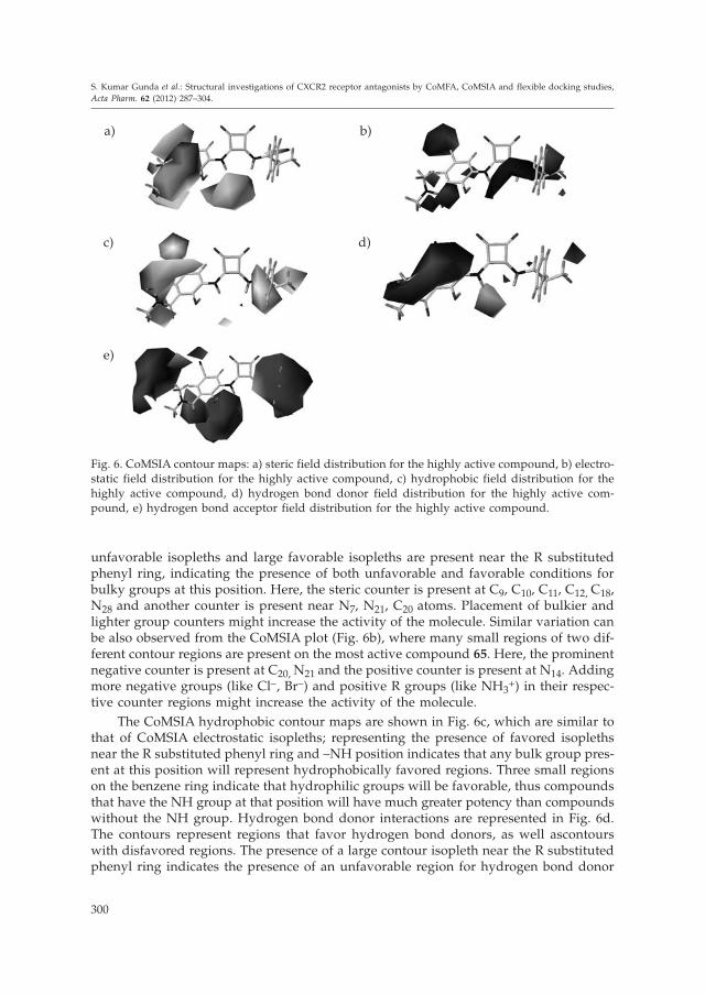
unfavorable isopleths and large favorable isopleths are present near the R substitutedphenyl ring, indicating the presence of both unfavorable and favorable conditions forbulky groups at this position. Here, the steric counter is present at C9, C10, C11, C12, C18,N28 and another counter is present near N7, N21, C20 atoms. Placement of bulkier andlighter group counters might increase the activity of the molecule. Similar variation canbe also observed from the CoMSIA plot (Fig. 6b), where many small regions of two dif-ferent contour regions are present on the most active compound 65. Here, the prominentnegative counter is present at C20, N21 and the positive counter is present at N14. Addingmore negative groups (like Cl�, Br�) and positive R groups (like NH3
+) in their respec-tive counter regions might increase the activity of the molecule.
The CoMSIA hydrophobic contour maps are shown in Fig. 6c, which are similar tothat of CoMSIA electrostatic isopleths; representing the presence of favored isoplethsnear the R substituted phenyl ring and –NH position indicates that any bulk group pres-ent at this position will represent hydrophobically favored regions. Three small regionson the benzene ring indicate that hydrophilic groups will be favorable, thus compoundsthat have the NH group at that position will have much greater potency than compoundswithout the NH group. Hydrogen bond donor interactions are represented in Fig. 6d.The contours represent regions that favor hydrogen bond donors, as well ascontourswith disfavored regions. The presence of a large contour isopleth near the R substitutedphenyl ring indicates the presence of an unfavorable region for hydrogen bond donor
300
S. Kumar Gunda et al.: Structural investigations of CXCR2 receptor antagonists by CoMFA, CoMSIA and flexible docking studies,Acta Pharm. 62 (2012) 287–304.
Fig. 6. CoMSIA contour maps: a) steric field distribution for the highly active compound, b) electro-static field distribution for the highly active compound, c) hydrophobic field distribution for thehighly active compound, d) hydrogen bond donor field distribution for the highly active com-pound, e) hydrogen bond acceptor field distribution for the highly active compound.
a) b)
c) d)
e)

groups. There is only a small contour isopleth present near the –NH group, favorable forthe donor group around this region. Hydrogen-bond donor and acceptor contour mapsfrom CoMSIA are shown in Fig. 6e. The contour isopleths favor the positioning of thehydrogen bond acceptor group in these regions whereas a few isopleths disfavored thepresence of H-bond acceptor groups. The hydrogen-bond acceptor contour map isshown with three large contours in which the hydrogen-bond acceptor is not favored.Contour regions are mainly positioned at the –NH group and also near the R substitutedphenyl ring. Only a small contour region is present near the phenyl ring, which favorsthe H-bond acceptor group around this region.
In silico screening
In silico methods can help identify drug targets via bioinformatics tools. They can bealso used to analyze the target structures for possible binding active sites, generate can-didate molecules, check their drug likeness, dock these molecules with the target, rankthem according to their binding affinities, further optimize molecules to improve theirbinding characteristics. Use of computers and computational methods permeates all as-pects of drug discovery today and forms the core of structure-based drug design. Use ofcomplementary experimental and informatics techniques increases the chance of successin many stages of the discovery process, from the identification of novel targets and elu-cidation of their functions to the discovery and development of lead compounds withdesired properties. Thus, the in silico procedure can be applied as a physicochemical fil-ter to reduce the number of compounds to be tested experimentally for hit/lead genera-tion. In other words, the in silico procedure minimizes the time and cost associated withidentifying new leads. Virtual screening was performed by insertion, deletion and sub-stitution of different substituents on the original molecules and the effects of structuralmodifications on the biological activity were investigated (19). Then, the domain ofQSAR model application was defined for using the model for screening new com-pounds. The newly designed molecule showed higher activity than the most active mol-ecule, as shown in Fig. 7. The designed molecule contains a negatively charged Br– atC23 and a bulky pentane ring instead of two methyl groups near N28. Adding these twogroups to the most active molecule helped in increasing the antagonistic activity of themolecule.
301
S. Kumar Gunda et al.: Structural investigations of CXCR2 receptor antagonists by CoMFA, CoMSIA and flexible docking studies,Acta Pharm. 62 (2012) 287–304.
Newly designed molecule (pIC50 = 8.57) Most active molecule (pIC50 = 8.52)
Fig. 7. Comparison of the newly designed molecule with the most active molecule.

CONCLUSIONS
A receptor independent 3D-QSAR has been established for CXCR2 antagonistic mo-lecules employing the most widely used techniques CoMFA and CoMSIA. The presentstudies highlight the importance of ligand orientation and selection of the training setmolecules in the development of statistically significant QSAR models. CoMSIA modelsprovided better statistical models than CoMFA, which points to the significance of thehydrogen bond donor and hydrophobic fields in the selectivity and activity of these lig-ands in addition to steric and electrostatic fields. Statistical significance and robustnessof the generated 3D-QSAR models were confirmed using an external set of molecules.The structural requirements identified in the present study can be utilized strategicallyin designing novel, potent CXCR2 antagonistic molecules.
REFERENCES
1. A. Zlotnik and O. Yoshie, Chemokines: a new classification system and their role in immunity,Immunity 12 (2000) 121–127; DOI: 10.1016/S1074-7613(00)80165-X.
2. L. Yang, C. Zhou, L. Guo, G. Morriello, G. Butora, A. Pasternak, W. H. Parsons, S. G. Mills, M.MacCoss, P. P. Vicario, H. Zweerink, J. M. Avala, S. Goval, W. A. Hanlon, M. A. Cascieri and M.S. Springer, Discovery of 3,5-bis(trifluoromethyl) benzyl L-arylglycinamide based potent CCR2antagonists, Bioorg. Med. Chem. Lett. 16 (2006) 3735–3739; DOI: 10.1016/j.bmcl.2006.04.045.
3. J. A. Belperio, M. P. Keane, D. A. Arenberg, C. L. Addison, J. E. Ehlert, M. D. Burdick and R. M.Strieter, CXC chemokines in angiogenesis, J. Leukocyte Biol. 68 (2000) 1–8.
4. P. Loetscher, M. Seitz, I. Clark-Lewis, M. Baggiolini and B. Moser, Both interleukin-8 receptorsindependently mediate chemotaxis: Jurkat cells transfected with IL-8R1 or IL-8R2 migrate in re-sponse to IL-8, GROá and NAP-2, FEBS Lett. 341 (1994) 187–192; DOI: 10.1016/0014-5793(94)80454-0.
5. J. C. Acosta, A. O’Loghlen, A. Banito, M. V. Guijarro, A. Augert, S. Raguz, M. Fumagalli, M. DaCosta, C. Brown, N. Popov, Y. Takatsu, J. Melamed, F. d’Adda di Fagagna, D. Bernard, E. Her-nando and J. Gil, Chemokine signaling via the CXCR2 receptor reinforces senescence, Cell 133(2008) 1006–1018; DOI: 10.1016/j.cell.2008.03.038.
6. J. Busch-Petersen, Small molecule antagonists of the CXCR2 and CXCR1 chemokine receptorsas therapeutic agents for the treatment of inflammatory diseases, Curr. Med. Chem. 6 (2006)1345–1352.
7. R. D. Cramer III, D. E. Patterson and J. D. Bunce, Comparative molecular-field analysis(COMFA). 1. Effect of shape on binding of steroids to carrier proteins, J. Am. Chem. Soc. 110(1988) 5959–5967; DOI: 10.1021/ja00226a005.
8. G. Klebe, U. Abraham and T. Mietzner, Molecular similarity indexes in a comparative-analysis(COMSIA) of drug molecules to correlate and predict their biological-activity, J. Med. Chem. 37(1994) 4130–4146; DOI: 10.1021/jm00050a010.
9. B. W. McCleland, R. S. Davis, M. R. Palovich, K. L. Widdowson, M. L. Werner, M. Burman, J. J.Foley, D. B. Schmidt, H. M. Sarau, M. Rogers, K. L. Salyers, P. D.Gorycki, T. J. Roethke, G. J.Stelman, L. M. Azzarano, K. W. Ward and J. Busch-Petersen, Comparison of N,N’-diarylsqua-ramides and N,N’-diarylureas as antagonists of the CXCR2 chemokine receptor, Bioorg. Med.Chem. Lett. 17 (2007) 1713–1717; DOI: 10.1016/j.bmcl.2006.12.067.
302
S. Kumar Gunda et al.: Structural investigations of CXCR2 receptor antagonists by CoMFA, CoMSIA and flexible docking studies,Acta Pharm. 62 (2012) 287–304.

10. C. Aki, J. Chao, J. A. Ferreira, M. P. Dwyer, Y. Yu, J. Chao, R. J. Merritt, G. Lai, M. Wu, R. W.Hipkin, X. Fan, W. Gonsiorek, J. Fosseta, D. Rindgen, J. Fine, D. Lundell, A. G. Taveras and P.Biju, Diaminocyclobutenediones as potent and orally bioavailable CXCR2 receptor antagonists:SAR in the phenolic amide region, Bioorg. Med. Chem. Lett. 19 (2009) 4446–4449; DOI: 10.1016/j.bmcl.2009.05.049.
11. A. Fiser, R. K. Do and A. Sali, Modeling of loops in protein structures, Protein Sci. 9 (2000) 1753–1773; DOI: 10.1110/ps.9.9.1753.
12. R. A. Laskowski, J. A. Rullmannn, M. W. MacArthur, R. Kaptein and J. M. Thornton, AQUA andPROCHECK-NMR: programs for checking the quality of protein structures solved by NMR,J. Biomol. NMR 8 (1996) 477–486; DOI: 10.1007/BF00228148.
13. M. Rarey, B. Kramer, G. Metz, T. Lengauer and G. Klebe, A fast flexible docking method usingan incremental construction algorithm, J. Mol. Biol. 261 (1996) 470–489; DOI: 10.1006/jmbi.1996.0477.
14. K. M. Gilbert, W. J. Skawinski, M. Misra, K. A. Paris, R. A. Buono, H. M. Deutsch and C. A.Venanzi, Conformational analysis of methylphenidate: comparison of molecular orbital and mo-lecular mechanics methods, J. Comput. Aided Mol. Des. 18 (2004) 719–738; DOI: 10.1007/s10822--004-7610-1.
15. S. Wold, Cross-validatory estimation of the number of components in factor and principal com-ponents models, Technometrics 20 (1978) 397–405.
16. G. Klebe, The use of composite crystal-field environments in molecular recognition and the denovo design of protein ligands, J. Mol. Biol. 237 (1994) 212–235; DOI: 10.1006/jmbi.1994.1223.
17. R. D. Cramer III, J. D. Bunce, D. E. Patterson and I. S. Frank, Crossvalidation, bootstrapping andpartial least squares compared with multiple regression in conventional QSAR studies, Quant.Struct.-Act. Rel. 7 (1988) 18–25; DOI: 10.1002/qsar.19880070105.
18. Y. Zhang and J. Skolnick, Scoring function for automated assessment of protein structure tem-plate quality, Proteins 57 (2004) 702–710; DOI: 10.1002/prot.20264.
19. G. Melagraki, A. Afantitis, H. Sarimveis, P. A. Koutentis, J. Markopoulos and O. Igglessi-Mar-kopoulou, Optimization of biarylpiperidine and 4-amino-2-biarylurea MCH1 receptor antago-nists using QSAR modeling, classification techniques and virtual screening, J. Comput. AidedMol. Des. 21 (2007) 251–267; DOI: 10.1007/s10822-007-9112-4.
S A @ E T A K
Strukturno istra`ivanje antagonista CXCR2 receptora pomo}u CoMFA,CoMSIA i fleksibilnih doking studija
SHRAVAN KUMAR GUNDA, ROHITH KUMAR ANUGOLU, SRI RAMYA TATA i SAIKH MAHMOOD
U radu je opisano trodimenzijsko ispitivanje kvantitativnog odnosa strukture i anta-gonisti~kog djelovanja na CXCR2 receptore (3D QSAR) u seriji od 56 N,N’-diarilskvara-mida, N,N’-diarilurea i diaminociklobutendiona. Provedene studije uklju~uju kompara-tivnu analizu molekulskih polja (CoMFA) i komparativnu analizu indeksa molekulskesli~nosti (CoMSIA). Modeli s dobrom predvidljivo{}u izvedeni su pomo}u CoMFA q2 =0,709, r2 (neunakrsno-validirani kvadrat koeficijenta korelacije) = 0,951, F = 139,903, r2 bs= 0,978 s pet komponenata, standardnom pogre{kom procjene 0,144 i CoMSIA q2 = 0,592,r2 = 0,955, F = 122,399, r2 bs = 0,973 sa {est komponenata, standardnom pogre{kom pro-
303
S. Kumar Gunda et al.: Structural investigations of CXCR2 receptor antagonists by CoMFA, CoMSIA and flexible docking studies,Acta Pharm. 62 (2012) 287–304.

cjene 0,141. Osim toga, model homologije CXCR2 receptora upotrijebljen je za svrstava-nje spojeva doking metodom. Najaktivniji spoj poslu`io je kao predlo`ak za svrstavanjepreostalih struktura. Nadalje, mapiranjem kontura na aktivno mjesto spojevi su se uza-jamno vrednovali kroz ostatke s obzirom na svoje konture. Ovo integrirano svrstavanjena temelju molekulskog dokinga i naknadnih 3D QSAR studija pomoglo je dizajniranjuCXCR2 antagonista s pobolj{anom aktivnosti. In silico pretra`ivanje prilago|eno je QSARmodelu sa svrhom predvi|anja strukture novih, potencijalno aktivnih spojeva.
Klju~ne rije~i: CXCR2, model homologije, CoMFA, CoMSIA, PLS, doking
Bioinformatics Division, Osmania University, Hyderabad-500007, Andhra Pradesh, India
304
S. Kumar Gunda et al.: Structural investigations of CXCR2 receptor antagonists by CoMFA, CoMSIA and flexible docking studies,Acta Pharm. 62 (2012) 287–304.
Copyright © 2022 FDOKUMEN