Synthesis and characterization of selective dopamine D 2 receptor antagonists
0892-6638/89/0003-2019/$01.50. © FASEB 2019
Antagonists of B2 bradykinin receptorsLARRY R. STERANKA, STEPHEN G. FARMER, AND RONALD M. BURCH
Nova Pharmaceutical Corporation, Baltimore, Maryland 21224, USA
ABSTRACT
Bradykinin and its active metabolites, produced by kal-
likreins at their sites of action, potently elicit a variety ofbiological effects: hypotension, bronchoconstriction, gutand uterine contraction, epithelial secretion in airway,
gut, and exocrine glands, vascular permeability, pain,
connective tissue proliferation, cytokine release, andeicosanoid formation. These effects are mediated by atleast two broad classes of receptors. The most common isthe B2 subtype. The availability of competitive antag-onists of B2 receptors has provided powerful tools for thestudy of bradykinin’s actions. The significance of kininsin certain human diseases is being explored by usingthese agents as potential therapeutic agents. Humanclinical trials are under way to test the usefulness ofbradykinin receptor antagonists to treat symptoms ofthe common cold and the pain associated with severeburns. Trials are also being comtemplated for use intreatment of asthma.---STE1tsNi, L. R.; FARMER,
S. G.; BURCH, R. M. Antagonists of B2 bradykinin
receptors. FASEBJ 3: 2019-2025; 1989.
Key Words: bradylcinin plasma /callikrein B2 receptor
inflammation endotoxic shock
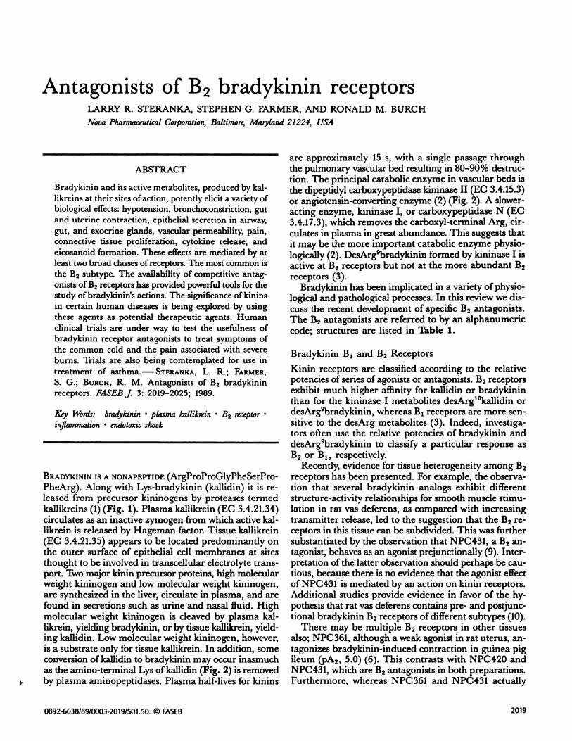
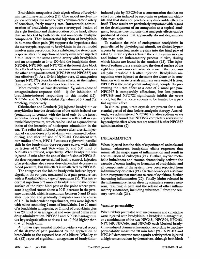
BRADYKININ IS A NONAPEPTIDE (ArgProProGlyPheSerPro-PheArg). Along with Lys-bradykinin (kallidin) it is re-leased from precursor kininogens by proteases termedkallikreins (1) (Fig. 1). Plasma kallikrein (EC 3.4.21.34)circulates as an inactive zymogen from which active kal-likrein is released by Hageman factor. Tissue kallikrein(EC 3.4.21.35) appears to be located predominantly onthe outer surface of epithelial cell membranes at sitesthought to be involved in transcellular electrolyte trans-port. Two major kinin precursor proteins, high molecularweight kininogen and low molecular weight kininogen,are synthesized in the liver, circulate in plasma, and arefound in secretions such as urine and nasal fluid. Highmolecular weight kininogen is cleaved by plasma kal-likrein, yielding bradykinin, or by tissue kallikrein, yield-ing kallidin. Low molecular weight kininogen, however,is a substrate only for tissue kallikrein. In addition, someconversion of kallidin to bradykinin may occur inasmuchas the amino-terminal Lys of kallidin (Fig. 2) is removedby plasma aminopeptidases. Plasma half-lives for kinins
are approximately 15 s, with a single passage throughthe pulmonary vascular bed resulting in 80-90% destruc-tion. The principal catabolic enzyme in vascular beds isthe dipeptidyl carboxypeptidase kininase II (EC 3.4.15.3)or angiotensin-converting enzyme (2) (Fig. 2). A slower-acting enzyme, kininase I, or carboxypeptidase N (EC3.4.17.3), which removes the carboxyl-terminal Arg, cir-culates in plasma in great abundance. This suggests thatit may be the more important catabolic enzyme physio-logically (2). DesArg9bradykinin formed by kininase I isactive at B1 receptors but not at the more abundant B2receptors (3).
Bradykinin has been implicated in a variety of physio-logical and pathological processes. In this review we dis-cuss the recent development of specific B2 antagonists.The B2 antagonists are referred to by an alphanumericcode; structures are listed in Table 1.
Bradykinin B1 and B2 Receptors
Kinin receptors are classified according to the relativepotencies of series of agonists or antagonists. B2 receptorsexhibit much higher affinity for kallidin or bradykininthan for the kininase I metabolites desArg’#{176}kallidin ordesArg#{176}bradykinin, whereas B1 receptors are more sen-sitive to the desArg metabolites (3). Indeed, investiga-tors often use the relative potencies of bradykinin anddesArg#{176}bradykinin to classify a particular response asB2 or B1, respectively.
Recently, evidence for tissue heterogeneity among B2receptors has been presented. For example, the observa-tion that several bradykinin analogs exhibit differentstructure-activity relationships for smooth muscle stimu-lation in rat vas deferens, as compared with increasingtransmitter release, led to the suggestion that the B2 re-ceptors in this tissue can be subdivided. This was furthersubstantiated by the observation that NPC431, a B2 an-tagonist, behaves as an agonist prejunctionally (9). Inter-pretation of the latter observation should perhaps be cau-
tious, because there is no evidence that the agonist effect
of NPC431 is mediated by an action on kinin receptors.Additional studies provide evidence in favor of the hy-pothesis that rat vas deferens contains pre- and postjunc-tional bradykinin B2 receptors of different subtypes (10).
There may be multiple B2 receptors in other tissuesalso; NPC361, although a weak agonist in rat uterus, an-tagonizes bradykinin-induced contraction in guinea pigileum (pA2, 5.0) (6). This contrasts with NPC42O andNPC431, which are B2 antagonists in both preparations.Furthermore, whereas NPC361 and NPC431 actually

Negatively charged surfaces (collagen, urate)pH changesTemperature changes
Hageman ActivatedFactor Hageman
Factor
Prekallikrein Kallikrein(plasma)
HMW Kininogen
LMW Kininogen
Figure 1. Kinin formation.
stimulate phosphoinositide turnover in N1E-115 neuro-blastoma cells, NPC348, NPC349, NPC394, and NPC420 inhibit bradykinin-induced phosphoinositide turn-over (11). However, even though these bradykinin ana-logs bind with varying nanomolar affinities to ileal orN1E-115 cell membranes, there is no direct evidence thatthe agonist effects of NPC361 and NPC431 are mediated
1’Kallikrein
(tissue)
Kininase I (Carboxypeplidase N)
Carboxypeptldase B
desArg9 bradykinin
ArgProProGlyPheSerPro + PheArg
Figure 2. Metabolism of kinins.
2020 Vol. 3 July 1989 The FASEB Journal STERANKA ET AL.
via bradykinin receptors. It is interesting that NPC567elicits phasic contractions in rat uterus in addition to itswell-known B2 receptor antagonist effect (12), althoughwhether the stimulation of uterine smooth muscle reflectsa receptor mechanism remains to be determined.
In contrast to the apparent ileum selectivity of NPC 361as a B2 antagonist, NPC573, a [DArg1]-substituted ana-
log, inhibits bradykinin-induced contractions in ratuterus (pA2, 5.6) but not in guinea pig ileum (5). More-over, this analog does not significantly displace [3Hbrady-kinin binding from ileal membranes. Therefore, NPC573is not a B2 receptor antagonist in guinea pig ileum. Simi-larly, NPC573, unlike several other analogs (NPC349,___________________NPC414 NPC566, NPC567 and NPC 722) has no effect
Bradykinin . . .
on bradykinin-induced vascular pain in vlvo. The ap-parent uterus selectivity of NPC573 resulted in the pro-posal that B2 receptors in ileum are different from those
Kallidin in uterus (5).However, more recent studies have revealed that 11
[DArg1 ]-substituted bradykinin analogs, includingNPC573, are either inactive or very weak displacers of[3H]bradykinin binding in ileum or uterus (12). NPC573
also inhibits vasopressin-induced uterine contractionswithout competing for vasopressin binding (12). Thusthe inhibitory action of NPC573 on bradykinin effects in
uterus is probably not mediated by bradykinin receptors.
In guinea pig tracheal smooth muscle precontractedwith histamine, bradykirtin causes concentration-depen-dent relaxations, whereas desArg9bradykinin is inactive.Moreover, bradykinin is not blocked by the antagonist
desArg9[Leu#{176}]bradykinin, NPC431, or [Thi69DPhe8}kal-lidin (13), which indicates that tracheal relaxation is not
mediated by B1 or B2 receptors. In guinea pigs, NPC567
LysArgProProclyPheSerProPheArg LysArgProProGlyPheSerProPhe + Arg
KalIldin desArg9 katlidn
Aminopeptidase
Kininase I (Carboxypeptidase N)
Lys + ArgProProGlyPheSerProPheArg ArgProProGlyPheSerProPhe + Arg
Brzdykinln
Kinnaae II (Angiolensin-Converting Enzyme) Kininase II
Neutral Uetalloendopeptidase (Enkephalinase)
ArgProProGlyPhe + SerProPhe
/own enzymes wn enzymes
ArgProPro + GIy + Phe Ser + Pro + Phe

TABLE 1. Structure-activity relations among bradykinin B2 receptor antagonists’
BRADYKININ RECEPTORS 2021
NPC No. Structure pA2, GPI pA2, RUT
Bradykinin
348
ArgProProGlyPheSerProPheArg
DArg[Hyp23Thi58DPhe7}BK fi#{216}4t 6.946349 DArg[Hyp3Thi58DPhe7]BK 6.0 6.9
358 [DPal7]BK 534 (0.1)
361 [DPhe7]BK 5.1,6 597 (0.4),6 (4.0)394 [Hyp3Thi58DPhe7]BK 574 594
414 LysLys[Hyp23Thi58DPhe7]BK 5.6 (0.05)
420 LysLys[Thi58DPhe7 ]BK 5. 6 (0.4)431 [Thi5tDPhe7]BK 6.36 6.56
566 DArg[Hyp2DPhe7]BK 6.3 (2)567
722
DArg[Hyp3DPhe7JBK
[DNaI’Thi58DPhe7]BK[Gly6Leu58DPhe7]BK
595
IA.8595
6.5
5.68(0.2)
809 [FDF7]BK 547 (3)7
813 DArg[FDF7]BK 6.l (2)
‘Antagonist potency is expressed in terms of the pA2 value for inhibiting bradykinin-induced contraction. Analogs that produced a bradykinin-likecontraction by themselves were assayed as agonists; the potencies are expressed in parentheses as a percentage of the potency of bradykinin. GPI, guineapig ileum; RUT, rat uterus; IA., inactive; Thi, -(2-thienyl)-Ala; Hyp, 4-hydroxy-Pro; FDF, p-fluoro-D-Phe; Nal, fl-(2-naphthyl)-Ala. bReferences
for data are shown as superscripts in last two columns.
inhibits bradykinin-induced bronchoconstriction onlypartially in vivo and is a very weak or inactive antagonistin vitro (12, 14). DesArg9bradykinin has no effect onguinea pig tracheal tone. Guinea pig lung and trachealsmooth muscle contain saturable, specific binding sites forbradykinin but not for desArg9bradykinin (11). Neither
desArg#{176}[Leu8]bradykinin nor NPC567 displaces bradyki-nm from the tracheal binding site, and in lung mem-branes NPC567 displaced only 68% of total specificallybound bradykinin. These studies suggest that guinea pigpulmonary tissue contains a novel bradykinin receptor(14). Preliminary data indicate that sheep trachea andlung, in addition to B2 receptors, also contain a novelbradykinin binding site from which NPC567 displaces30 and 70% of [3H]bradykinin binding, respectively (14).
Structure-activity considerations among B2 receptor
antagonists
Since the sequence of bradykinin was published in 1960,hundreds of analogs have been synthesized in an effort todefine structure-activity relationships and in the search forreceptor antagonists. In 1985, Vavrek and Stewart (6) re-ported the first sequence-related competitive bradykininantagonists. The essential substitution is D-phenylalaninefor L-proline in position 7 (Table 1); [DPhe7jbradykinin(NPC361) is the first described competitive bradykinin
antagonist. Many additional antagonist sequences havebeen synthesized and nearly all contain the DPhe7 sub-stitution (Table 1) (see ref 15 for review).
Isolated tissue studies
Although these are many reports on the effects of kininson a diversity of isolated tissues, less is known about theeffects of specific bradykinin antagonists in these types ofstudies. Studies of the effects of bradykinin antagonistson isolated tissues have recently been catalogued (15).
The purpose of this section is to provide a summary ofthe pharmacology of kinin antagonists in isolated tissues.
Guinea pig ileal smooth muscle has been used exten-sively to characterize B2 bradykinin receptors (15). Brady-kinin-induced contractions in guinea pig ileum are antag-onized by NPC361 (pA2, 5.85), NPC431 (pA2, 5.90), and[Thi69DPhe8]kallidin (pA2, 5.93) (16), although the latteranalog also exhibited weak stimulatoiy effects in this tissue.The three peptides are specific for bradykinin and haveno effect on responses to substance P and angiotensin11(7, 16). Recent studies also confirm that the brady-kinin-induced mucosal electrolyte secretion in rat colonand guinea pig ileum are mediated by B2 receptors.NPC361, but not desArg9[Leu8]bradykinin, inhibits theeffect of bradykinin in these preparations in a competi-tive fashion (17).
Regoli et al. (16) demonstrated that bradykinin-in-duced relaxation of the canine carotid artery and contrac-tion of rabbit jugular vein and canine bladder smoothmuscle are inhibited by NPC431 or [Thi69DPhe8]kalli-din, which confirms the presence of B2 receptors. More-over, the agonist desArg9bradykinin is inactive in thesepreparations. There are very few reports on the effects ofkinins on human isolated vascular preparations (15) and,to our knowledge, there has been only one study that usedbradykinin antagonists. In human basilar artery, bradyki-nm is considerably more potent than desArg9bradykinin(which is essentially inactive) as a relaxant (18). Further,bradykinin-induced relaxation is inhibited by NPC431,and it was concluded that bradykinin-induced relaxationof human basilar artery is mediated by B2 receptors.
Pain and inflammatory hyperalgesia
Although the algesic properties of bradykinin (19) com-bined with its presence in damaged tissue (20) suggest thatbradykinin is a physiological mediator of pain and inflam-matory hyperalgesia, the availability of specific antago-nists is permitting the first direct tests of this hypothesis.
2022 Vol. 3 July 1989 The FASEB Journal STERANKA ET AL.
Bradykinin antagonists block algesic effects of bradyki-nm itself in several models (15). One model involves in-jection of bradykinin into the right common carotid arteryof conscious, freely moving rats. Intracarotid admini-stration of bradykinin produces stereotyped flexion ofthe right forelimb and dextrorotation of the head, effectsthat are blocked by both opiate and non-opiate analgesiccompounds. That intraarterial injection of bradykininin humans elicits pain (19) supports the hypothesis thatthe stereotypic response to bradykinin in the rat modelinvolves pain perception. Rats exhibiting the stereotypicresponse after the injection of bradykinin were injected5 mm later in the same carotid artery with bradykininand an antagonist at 1- to 100-fold the bradykinin dose.NPC414, NPC566, and NPC722 at the lowest dose blockthe effects of bradykinin in all or most animals, whereasthe other antagonists tested (NPC349 and NPC567) areless effective (5). At a 10-fold higher dose, all antagonists(except NPC573) block bradykinin effects. At the highestdose tested, NPC414 becomes less effective.
More recently, we have determined KB values (dose ofantagonist/dose-response shift - 1) for inhibition ofbradykinin-induced responding in this model (15).NPC567 and NPC414 exhibit KB values of 6.7 and 7.2nmol/kg, respectively.
Griesbacher and Lembeck (21) injected bradykinin oracetylcholine into the circulation of the rabbit isolated ear(remaining in contact with the head only by the intactauricular nerve). Both agents cause a reflex fall in sys-temic blood pressure, which can be used as a quantitativeindex of the intensity of nociceptor stimulation in theear. The reflex fall in blood pressure after arterial injec-tion of various doses of bradykinin was measured before,during, and after infusion of NPC415. Consistent withour studies of rats, NPC415 causes a rightward, parallelshift in the bradykinin dose-response curve, with shiftsby factors of 8.7 and 26.4 when 50 and 500 nmol ofNPC4I5 are infused, respectively. When bradykinin wasinjected 15 mm after the end of the infusion of NPC415,the dose-response curves shifted back to control. Injectionof acetylcholine also causes dose-dependent decreases inblood pressure, but this effect is unaffected by NPC4I5.
The antagonists also inhibit bradykinin-induced hyper-algesia in the rat paw, measured by a paw pressure testwith a Randall-Selitto type of apparatus (5). The intra-
dermal injection of 2 nmol of bradykinin into the dorsalsurface of the right hind paw at the point where pres-sure is applied causes about a 50% decrease in the pres-sure threshold, which is maximum between 5 and 15 mmafter injection and gradually dissipates over the courseof 1 h. In independent experiments, rats were injectedwith saline containing 2 nmol of bradykinin, 2 or 20 nmolof a bradykinin antagonist, or 2 nmol of bradykinin plus2 or 20 nmol of an antagonist and were tested 5 mm afterdrug administration. NPC567 and NPC349 antagonizethe hyperalgesic effect at doses 1- to 10-fold higher thanthat of bradykinin.
A human experimental model provides a verbal reportof the degree of pain produced by the application ofbradykinin to the exposed base of a blister. Whalley etal. (22) reported significant antagonism of bradykinin-
induced pain by NPC349 at a concentration that has noeffect on pain induced by serotonin or potassium chlor-ide and that does not produce any discernible effect byitself. These results are particularly important with regardto the development of an antagonist as a topical anal-
gesic, because they indicate that analgesic effects can beproduced at doses that apparently do not degranulateskin mast cells.
To evaluate the role of endogenous bradykinin inpain elicited by physiological stimuli, we elicited hyper-algesia by injecting urate crystals into the hind paw ofrats (5). Urate crystals activate the kinin system in vitroand induce an inflammatory process in vivo, duringwhich kinins are found in the exudate (23). The injec-tion of sodium urate crystals into the dorsal surface of theright hind paw causes a marked decrease in the mechani-cal pain threshold 4 h after injection. Bradykinin an-tagonists were injected at the same site alone or in com-bination with urate crystals and rats were tested 4 h later.NPC349 is the most potent antagonist, completely pre-
venting the urate effect at a dose of 2 nmol per paw.NPC567 is comparably efficacious, but less potent.NPC414 and NPC 722 significantly inhibit the urateeffect, but their efficacy appears to be limited by a par-tial agonist effect.
In clinical gout, urate crystals are present for a sub-stantial period of time before analgesic therapy. Accord-ingly, we administered NPC567 2 h after sodium uratecrystals and found that NPC567 completely reverses thehyperalgesic effect when rats are tested 2 h after NPC567administration (5).
INFLAMMATION
When injected into the skin of experimental animals andhuman volunteers, bradykinin elicits responses thatmimic all the major signs of inflammation and result inaccumulation of leukocytes (24). Further, myriad meta-bolic imbalances and trauma dramatically activate thecascade of events leading to formation of bradykinin, and
all components of the system have been reported frominflammatory exudates (24). Certain leukocytes also havekinin receptors that mediate release of cytokines, furtherincreasing inflammation (25). Finally, kinins released inthe inflammatory lesion directly stimulate sensory neu-rons, resulting in pain and the release of other inflam-matory substances, including substance P from the sen-sory terminals (24).
Vascular permeability
When rabbits pretreated with mepyramine and cimetidinewere injected with bradykinin, a bradykinin antagonist,or a combination of the two, NPC431, NPC394, NPC413,NPC349, NPC414, and NPC415 each blocked brady-
kinin-induced plasma extravasation according to capifiarypermeability measured 20 mm later (21). NPC4I3 andNPC349 demonstrate some agonist activity when injectedat high concentrations by themselves, although both block
BRADYKININ RECEPTORS 2023
bradykinin (21). Similarly, when rabbits receive NPC431,NPC394, NPC349, NPC42O, or NPC419, bradykinin-induced plasma extravasation is blocked (26). However,when rats receive the same compounds, no blockadeof bradykinin is observed, and NPC349 augments brady-kinin’s action.
Recently, Steranka et al. (27) resolved the apparentdifferences among species. When rats were pretreatedwith cyproheptadine, a mixed serotonin and histamineantagonist, the apparent agonist effects of the bradykininanalogs were abolished, unmasking the antagonism ofbradykinin-induced plasma extravasation by NPC567.Thus, the recognized ability of these bradykinin analogsto cause histamine release (discussed later) obscured theantagonism of bradykinin effects in the original study.
Rhinovirus symptoms
Proud and Kaplan (28) have amassed a large body ofevidence that kinins play roles in the symptomatology ofseveral diseases characterized by rhinitis. Kallikrein andkininogen leakage as well as kinin formation occur during
the immediate response to allergen along with the pro-duction of other mediators, including histamine, PGD2,
and leukotrienes; the appearance of all the mediatorscorrelates with the onset of symptoms. The kinins in thenasal lavage fluid consist of bradykinin and kallidin inroughly equal proportion.
In rhinitis secondary to rhinovirus (a virus responsiblefor a significant fraction of common colds), a role forkinins in symptomatology is even more prominent. Un-like allergic rhinitis, rhinitis caused by rhinoviruschallenge is associated with bradykinin in the absenceof large increases in other mediators (28). Subjects whodo not manifest symptoms of rhinitis have no kinins intheir lavages. However, in symptomatic subjects, kininlevels correlate well with symptom scores. Intranasal ad-
ministration of bradykinin in normal subjects induces
nasal obstruction and rhinorrhea as well as sore throat,another common accompaniment of rhinovirus infection(29). This action may be caused by kinin reaching thepharynx and stimulating pain receptors (29).
The presence of bradykinin in nasal lavages of subjectswith symptomatic rhinovirus infection, coupled with thedemonstration that bradykinin administration can elicitthe relevant symptoms, suggests that kinins are promi-nent mediators of rhinovirus symptoms. This has
prompted NOVA Pharmaceutical Corporation to con-duct clinical trials of NPC567 for treatment of the symp-toms of the common cold.
CARDIOVASCULAR FUNCTION
Regulation of blood pressure
Since bradykinin is a potent vasodilator, many laboratoryand clinical studies have examined whether kinins play aphysiological role in blood pressure regulation. Severalstudies (2) have shown that urinary kallikrein excretion is
low in hypertensive patients, which suggests that kininsmay play a role in maintaining normotension. Severalstudies have failed to find any effect of the peptide brady-kinin antagonists NPC349 and NPC394 on blood pressurein normal rats after administration at doses that blockedthe blood pressure-lowering effect of bradykinin (15).
In one interesting study, NPC349 at an i.v. dose of 400tg (a dose that was shown to block the reduction in bloodpressure elicited by bradykinin) was administered to con-scious, normotensive rats with no effect on blood pres-sure. However, if the rats are given nonpressor doses ofangiotensin II or methoxamine or pressor doses of vaso-pressin or methoxamine, NPC349 significantly increasesblood pressure by 9-16 mm Hg (30), which suggests thatbradykinin may play a regulatory role in counteractingthe hypertensive effects of pressor hormones.
It has been suggested that a component of the hypoten-sive effect of angiotensin-converting enzyme inhibitorsmight be mediated by elevated bradykinin concentration,and several studies have attempted to address this hy-pothesis through the use of bradykinin antagonists (re-viewed in ref 15). Results with the antagonists have beenvariable, suggesting that from none (31) to one-third (32)of the effect of angiotensin-converting enzyme inhibitorsis mediated by bradykinin (15).
NPC392 infused intraarterially (500 gig. min . kg’)elicits a rise in blood pressure of 15 mm Hg within I mm,an effect that increases gradually by another 10 mm Hgover the next 4 mm. Blood pressure is also increased byNPC392 in nephrectomized animals, but the rise is onlyabout one-half that observed in the intact animals. Be-cause the hypertensive effect of NPC392 was found tobe mediated by release of epinephrine from the adrenalgland (33), the relation of the effect of NPC392 to brady-kinin antagonism is questionable.
Thus, although studies with bradykinin receptorantagonists have suggested that bradykinin does not playan active role in the regulation of blood pressure in nor-
mal rats, this peptide may act to blunt the hypertensiveeffects of a variety of pressor hormones. Because some ofthe antagonists may have actions other than those atbradykinin receptors (including release of adrenal epine-phrine), results obtained with the antagonists, especiallywhen they are administered at high doses, should be in-terpreted with caution.
Endotoxic shock
Endotoxic shock is caused by the interaction of lipopoly-saccharides (LPS, endotoxins) of certain bacterial cellwalls with cells of the reticuloendothelial system. An earlyeffect of LPS administration is a transient, profound fallin mean arterial pressure within a few minutes, whichfalls again as the animals approach death. A large bodyof literature, from 1960 to the middle 1970s, suggests thatthe kallikrein-kinin system is activated in human sepsisand experimental endotoxic shock (34). However, withoutthe availability of kinin receptor antagonists, studies cor-related only metabolic and physiological changes withchanges in components of the kallikrein-kinin system.

2024 Vol. 3 July 1989 The FASEB Journal STERANKA ET AL.
We have studied the role of bradykinin in endotoxicshock (35) when jugular vein infusion of NPC567 intorats was initiated 30 mm before LPS, continued alongwith a 10-mm LPS infusion, and infused alone for anadditional 90 mm. In this model, LPS causes a biphasicfall in blood pressure, the first phase reaching its nadir at1 h, recovering gradually by 2.5 h, and falling again untilthe animals die. LPS causes a 38% fall in mean arterialpressure at 1 h, an effect that is inhibited approximately60% by NPC567. During the second phase of hypoten-sion, the effects of NPC567 were monitored to 4 h afterLPS, at which time NPC567 completely blocks the hypo-tensive effect of LPS, even though infusion of the an-tagonist was discontinued 2.5 h previously. Even morestriking than the effects of NPC567 on blood pressure isits effects on mortality. At 24 h, 100% of animals receiv-ing LPS had died, compared with only 50% of theanimals that received NPC567 (35).
Airway function
The possible involvement of kinins in inflammatory air-way conditions such as asthma has been a subject of muchspeculation since the early 1960s. Bradykinin has beenimplicated in the pathogenesis of asthma in that broncho-alveolar kinin levels increase after allergen challenge inallergic asthmatic subjects (36). Moreover, bradykinin isa very potent bronchoconstrictor in asthmatic but notin nonasthmatic subjects (37). In addition, elevatedkinin levels have been detected in plasma of asthmaticpatients (38).
Allergic sheep respond in a manner analogous to asth-matic patients to inhalation of a specific antigen (Ascaris
suum), with acute anaphylactic bronchoconstriction fol-lowed 6-8 h later by a late phase of increased pulmo-nary resistance. In addition, these animals develop non-specific airway hyperreactivity 2 and 24 h after challengewith Ascaris (see ref 15). NPC567 given 30 mm beforeand coadministered with inhaled antigen has no effecton the acute bronchoconstriction, but completely inhibitsthe onset of airway hyperresponsiveness at 2 h (39). Inaddition, the bradykinin antagonist prevents the broncho-alveolar increase in eosinophils and neutrophils afterAscaris. These data imply an important role for bradyki-nm in antigen-induced hyperreactivity and inflamma-tion in the airways. It is suggested that endogenousbradykinin modifies reactivity and the inflammatoryresponse of the airways after bronchial anaphylaxis (39).A hallmark of asthma is the nonspecific airway hyper-reactivity, which manifests itself as an extreme sensitivityof the asthmatic airways to many stimuli. Preliminarystudies also indicate that NPC567 inhibits the onset ofthe late airway response 6-8 h after antigen challenge(W. Abraham, personal communication). Thus, bradyki-nm antagonists represent a potentially novel approachfor the prophylaxis of bronchial asthma.
Nonreceptor mediated actions
Several DPhe7 bradykinin antagonists have a number ofeffects not mediated by bradykinin receptors (reviewed
in ref 15). Among these are the release of epinephrinefrom the adrenal medulla discussed above, release ofhistamine from certain mast cells (40), inhibition of tissuekallikrein (41), and inhibition of kininase 11(42). We havealso found that several of the analogs have spasmogenicactivity in isolated smooth muscle. Thus, caution must beexercised, especially in in vivo studies, in interpreting aneffect of the antagonists as a result of receptor blockade.
The authors wish to acknowledge Cheryl Sowards for her assistance
in preparing the manuscript.
REFERENCES
1. Miller, D. M., and Margolius, H. S. (1988) Kallikrein-kininogen-kinin system. 2nd Ed, Endocrinology (DeGroot, L. J., and Cahill,G. F., Jr., eds) Grune and Stratton, Orlando, Florida
2. Erd#{246}s,E. G. (1979) Bradykinin, Kallidin and Kallikrein, Handbook ofExperimental Pharmacology, (Erd#{246}s,E. G., ed) Vol. XXV, Suppl.,pp. 428-488, Springer-Verlag, New York
3. Regoli, D., and Barab#{233},J. (1980) Pharmacology of bradykininand related kinins. Pharmacol. Rev. 32, 1-46
4. Stewart, J. M., and Vavrek, R. J. (1987) Bradykinin competitiveantagonists: design and activities. Enzyme Inhibitors (Barth, A.,and Schrower, R. L., eds) pp. 73-80, Pergamon, New York
5. Steranka, L. R., Manning, D. C., DeHaas, C. J., Ferkany, J. W.,Borosky, S. A., Connor,J. R., Vavrek, R.J., Stewart,J. M., andSnyder, S. H. (1988) Bradykinin as a pain mediator: receptorsare localized to sensory neurons, and antagonists have analgesiceffects. Proc. Natl. Acad. Sci. USA 85, 3245-3249
6. Vavrek, R. J., and Stewart, J. M. (1987) Bradykinin antagonists
containing hydroxyproline. Peptides 1986 (Theodoropoulos, D.,ed) pp. 287-290, W. de Gruyter, Berlin
7. Vavrek, R. J., and Stewart, J. M. (1985) Competitive antagonistsof bradykinin. Peptides 6, 161-164
8. Vavrek, R. J., and Stewart, J. M. (In press) Development of
bradykinin antagonists: structure-activity relationships for newcategories of antagonist sequences. Kinins V (Moriya, H., ed)Plenum, New York
9. Llona, I., Vavrek, R., Stewart, J. M., and Huidobro-Toro, J. P.(1987) Identification of pre- and postsynaptic bradykinin receptorsites in the vas deferens: evidence for different structural prerequi-sites. j Pharmacol. Exp. Ther. 241, 608-614
10. Rifo, J., Pourrat, M., Vavrek, R. J., Stewart, J. M., and Huidobro-Toro, J. P. (1987) Bradykinin receptor antagonists used to charac-terize the heterogeneity of bradykinin-induced responses in ratvas deferens. Eur. j Pharmacol. 142, 305-312
11. Braas, K. M., Manning, D. C., Perr#{231}D. C., and Snyder, S. H.(1988) Bradykinin analogues: differential agonist and antagonistactivities suggesting multiple receptors. Br.]. PharmacoL 94, 3-5
12. Farmer, S. G., Burch, R. M., Det-Iaas, C. J., Togo, J., andSteranka, L. R. (1989) [Arg1DPhe7]-substituted bradykinin ana-logs inhibit bradykinin- and vasopressin-induced uterus contrac-tion. j Pharmacol. Exp. Ther. 248, 677-681
13. Rhaleb, N. E., Dion, S., D’Orl#{233}ans-Juste, P., Drapeau, G.,Regoli, D., and Browne, R. G. (1988) Bradykinin antagonism:differentiation between peptide antagonists and anti-inflamma-
tory agents. Eur. j P/zarmacol. 151, 275-27914. Farmer, S. G., Burch, R. M., Meeker, S. A., and Wilkins, D. E.
(In press) Evidence for a pulmonary B3bradykinin receptor. Mol.PharmacoL
15. Burch, R. M., Farmer, S. G., and Steranka, L. R. (In press) B2bradykinin receptor antagonists. Med. Res. Rev.
16. Regoli, D., Drapeau, G., Rovero, P., Dion, S., IYOrleans-Juste, P.,and Barabb, J. (1986) The actions of kinin antagonists on B1 andB2 receptor systems. Eur. j Phar,nacol. 123, 61-65
17. Gaginella, T. S., and Kachur, J. F. (1988) Bradykinin as a medi-ator of mucosal secretion in IBD. Inflammatory Bowel Disease: CunEntStatus and Future Approach (MacDermott, R. P., ed) pp. 383-390,Elsevier Science Publishers B.V., England

BRADYKININ RECEPTORS 2025
18. Whalley, E. T., Amure, Y. 0., and Lye, R. H. (1987) Analysis ofthe mechanism of action of bradykinin on human basilar arteryin vitro. Naunyn Schmiedeberg’s Arch. Pharmacol. 335, 433-437
19. Clark, W. G. (1979) Kinins and the peripheral and central nervoussystems. In Bradykinin, Kallidin and Kallikrein, Handbook of Ex-perimental Pharmacology (Erdos, E. G., ed) Vol. XXV, Suppl., pp.312-356, Springer-Verlag, New York
20. Kellermeyer, R. W., and Graham, R. C. (1968) Kinins-possiblephysiologic and pathologic roles in man. N EngL j Med. 279,859-866
21. Gnesbacher, T, and Lembeck, F (1987) Effect of bradykinin an-tagonists on bradykinm-induced plasma extravasation, venocon-striction, prostaglandin E2 release, nociceptor stimulation andcontraction of the iris sphincter muscle in the rabbit. Br. j Phar-macol. 92, 333-340
22. Whalley, E. T, Clegg, S., Stewart, J. M., and Vavrek, R. J. (1987)The effect of kinin agonists and antagonists on the pain responseof the human blister base. Naunyn-Schmeideberg’s Arch. PharmacoL336, 652-655
23. Melmon, K. L., Webster, M. E., Goldfinger, S. E., and Seeg-miller, J. E. (1967) The presence of a kinin in inflammatorysynovial effusion from arthritides of varying etiologies. Arthritis
Rheum. 10, 13-20
24. Marceau, F, Lussier, A., Regoli, D., and Giroud, J. P. (1983)Pharmacology of the kinins: their relevance to tissue injury andinflammation. Gen. Pharmacol. 14, 209-229
25. Burch, R. M., and Tiffany, C. W. (1989) Bradykinin stimulatesthe production of tumor necrosis factor and interleukin 1 frommacrophages. Clin. Res. 37, 406A
26. Whalley, E. T., Nwator, I. A., Stewart, J. M., and Vavrek, R. J.(1987) Analysis of the receptors mediating vascular actions ofbradykinin. Naunyn-Schmeideberg’s Arch. PharmacoL 336, 430-433
27. Steranka, L. R., Rodiguez, R. A., DeHaas, C. J. (1988) Effects ofbradykinin antagonists on vascular permeability in rat skin. Phar-macologist 30, A30
28. Proud, D., and Kaplan, A. P. (1988) Kinin formation: mechan-isms and role in inflammatory disorders. Annu. Rev. ImmunoL 6,49-83
29. Proud, D., Reynolds, C. J., LaCapra, S., Kagey-Sobotka, A.,Lichtenstein, L. M., and Naclerio, R. M. (1988) Nasal provoca-tion with bradykinin induces symptoms of rhinitis and a sorethroat. Am. Rev. Respir. Dis. 137, 613-616
30. Aubes-t, J. I., Waeber, B., Nussberger, J., Vavrek, R. J., Stewart,J. M., and Brunner, H. R. (1988) Influence of endogenous brady-kinin on acute blood pressure response to vasopressors in normo-
tensive rats assessed with a bradykinin antagonist. j Cardiovasc.P/zarmacol. 11, 51-55
31. Aubert, J. F., Waeber, B., Nussberger, J., Vavrek, R. J., Stewart,J. M., and Brunner, H. R. (1987) Lack of a role of circulatingbradykinin in the blood pressure response to acute angiotensinconverting enzyme inhibition in rats. Agents Actions (SuppL 22)349, 349-351
32. Gavras, I., and Gavras, H. (1988) Anti-hormones and blood pres-sure: bradykinin antagonists in blood pressure regulation. KidneyJut. (SuppL 26)34, S60-S62
33. Mulinari, R., Benetos, A., Gavras, I., and Gavras, H. (1988) Vas-cular and sympathoadrenal responses to bradykinin and abradykinin analogue. Hypertension 11, 754-757
34. Colman, R. W., and Wong, P. Y. (1979) Kallikrein-kinin systemin pathologic conditions. Bradykinin, Kallidin and Kallikrein, Hand-book of Experimental Pharmacology (Erd#{246}s,E. G., ed) Vol. XXV,Suppl., pp. 584-586, Springer-Verlag, New York
35. Wilson, D. D., de Garavilla, L., Kuhn, W., Togo, J., Burch,R. M., and Steranka, L. R. (1989) D-Arg[Hyp3t)Phe7Brady-kinin, a bradykinin antagonist, decreases mortality in a rat modelof endotoxic shock. Girt. Shock. 27, 93-101
36. Chnstiansen, S. C., Proud, D., and Cochrane, C. G. (1987) De-tection of tissue kallikrein in the bronchoalveolar lavage fluid ofasthmatic subjects. J. Clin. Invest. 79, 188-197
37. Fuller, R. W., and Barnes, P. J. (1988) Kinins. Asthma: BasicMechanisms and Clinical Management, (Barnes, P. J., Rodger, I. W.,Thompson, N. C., eds) pp. 259-272, Academic Press Ltd.,London
38. Abe, K., Watanabe, N., Kumagai, N., Mouri, T., Seki, T., andYoshinaga, K. (1967) Circulating plasma in patients with bron-chial asthma. Experientia 23, 626-627
39. Soler, M., Sielczak, M. W., Abraham, W. M. (1989) A bradyki-nm antagonist modifies antigen-induced airway hyperresponsive-ness and airway inflammation in allergic sheep. Am. Rev. Respir.Dis. 139, A327
40. Lawrence, I. D., Warner, J. A., Cohan, V. L., Lichtenstem, L. M.,Kagey-Sobotka, A., Vavrek, R. J., and Stewart, J. M. (1989) In-duction of histamine release from human skin mast cells bybradykinin analogs. Biochem. Pharmacol. 38, 227-233
41. Spragg, J., Vavrek, R. J., and Stewart, J. M. (1988) The inhibi-tion of glandular kallikrein by peptide analog antagonists ofbradykinin. Peptides 9, 203-206
42. Togo, J., Burch, R. M., DeHaas, C. J., Connor, J. R., andSteranka, L. R. (1989) D-Phe’-substituted bradykinin analogsare not substrates for kininase II (angiotensin converting enzyme).Peptides 10, 109-112
Copyright © 2022 FDOKUMEN