A Population of CD4+CD8+ Double-Positive T Cells ... - MDPI
14
Citation: Yu, E.D.; Wang, H.; da Silva Antunes, R.; Tian, Y.; Tippalagama, R.; Alahakoon, S.U.; Premawansa, G.; Wijewickrama, A.; Premawansa, S.; De Silva, A.D.; et al. A Population of CD4 + CD8 + Double-Positive T Cells Associated with Risk of Plasma Leakage in Dengue Viral Infection. Viruses 2022, 14, 90. https://doi.org/10.3390/ v14010090 Academic Editors: Célia F. Rodrigues and Natália Cruz-Martins Received: 2 December 2021 Accepted: 3 January 2022 Published: 5 January 2022 Publisher’s Note: MDPI stays neutral with regard to jurisdictional claims in published maps and institutional affil- iations. Copyright: © 2022 by the authors. Licensee MDPI, Basel, Switzerland. This article is an open access article distributed under the terms and conditions of the Creative Commons Attribution (CC BY) license (https:// creativecommons.org/licenses/by/ 4.0/). viruses Article A Population of CD4 + CD8 + Double-Positive T Cells Associated with Risk of Plasma Leakage in Dengue Viral Infection Esther Dawen Yu 1 , Hao Wang 2 , Ricardo da Silva Antunes 1 , Yuan Tian 1 , Rashmi Tippalagama 1 , Shakila U. Alahakoon 3 , Gayani Premawansa 4 , Ananda Wijewickrama 5 , Sunil Premawansa 6 , Aruna Dharshan De Silva 1,3,7 , April Frazier 1 , Alba Grifoni 1 , Alessandro Sette 1,8, * ,† and Daniela Weiskopf 1,8, * ,† 1 Center for Infectious Disease and Vaccine Research, La Jolla Institute for Immunology, La Jolla, CA 92037, USA; [email protected] (E.D.Y.); [email protected] (R.d.S.A.); [email protected] (Y.T.); [email protected] (R.T.); [email protected] (A.D.D.S.); [email protected] (A.F.); [email protected] (A.G.) 2 School of Medicine, University of California San Diego, La Jolla, CA 92037, USA; [email protected] 3 Genetech Research Institute, Colombo 00800, Sri Lanka; [email protected] 4 Colombo North Teaching Hospital, Ragama 11010, Sri Lanka; [email protected] 5 National Institute of Infectious Diseases, Angoda 10620, Sri Lanka; [email protected] 6 Department of Zoology and Environment Sciences, University of Colombo, Colombo 00700, Sri Lanka; [email protected] 7 Department of Paraclinical Sciences, Faculty of Medicine, General Sir John Kotelawala Defence University, Mount Lavinia 10390, Sri Lanka 8 Department of Medicine, Division of Infectious Diseases and Global Public Health, University of California, La Jolla, CA 92037, USA * Correspondence: [email protected] (A.S.); [email protected] (D.W.) † These authors contributed equally to this work. Abstract: According to the WHO 2009 classification, dengue with warning signs is at the risk of developing severe form of dengue disease. One of the most important warning signs is plasma leakage, which can be a serious complication associated with higher morbidity and mortality. We report that the frequency of CD4 + CD8 + double-positive (DP) T cells is significantly increased in patients at risk of developing plasma leakage. Transcriptomic analysis demonstrated that CD4 + CD8 + DP cells were distinct from CD4 + Single Positive (SP) T cells but co-clustered with CD8 + SP cells, indicating a largely similar transcriptional profile. Twenty significant differentially expressed (DE) genes were identified between CD4 + CD8 + DP and CD8 + SP cells. These genes encode OX40 and CCR4 proteins as well as other molecules associated with cell signaling on the cell surface (NT5E, MXRA8, and PTPRK). While comparing the profile of gene expression in CD4 + CD8 + DP cells from patients with and without warning signs of plasma leakage, similar expression profile was observed, implying a role of CD4 + CD8 + DP cells in plasma leakage through a quantitative increase rather than functional alteration. This study provided novel insight into the host immune response during the acute febrile phase of DENV infection and the role of CD4 + CD8 + DP T cells in the pathogenesis of plasma leakage. Keywords: CD4 + ; CD8 + ; double positive; T cells; infectious diseases; dengue; plasma leakage; transcriptomic analysis 1. Introduction Dengue fever (DF) is one of the major re-emerging infectious diseases and the most prevalent arthropod-borne viral disease in humans [1]. The Center for Diseases Control and Prevention (CDC) estimates that around 400 million people get infected annually, and approximately 3 billion of the world’s population live in dengue-endemic areas [2]. The 1997 WHO classification [3] distinguished four main categories of dengue disease: non- classical DF, classical DF, dengue hemorrhagic fever (DHF), and dengue shock syndrome Viruses 2022, 14, 90. https://doi.org/10.3390/v14010090 https://www.mdpi.com/journal/viruses
-
Upload
khangminh22 -
Category
Documents
-
view
1 -
download
0
Transcript of A Population of CD4+CD8+ Double-Positive T Cells ... - MDPI
�����������������
Citation: Yu, E.D.; Wang, H.;
da Silva Antunes, R.; Tian, Y.;
Tippalagama, R.; Alahakoon, S.U.;
Premawansa, G.; Wijewickrama, A.;
Premawansa, S.; De Silva, A.D.; et al.
A Population of CD4+CD8+
Double-Positive T Cells Associated
with Risk of Plasma Leakage in
Dengue Viral Infection. Viruses 2022,
14, 90. https://doi.org/10.3390/
v14010090
Academic Editors: Célia F. Rodrigues
and Natália Cruz-Martins
Received: 2 December 2021
Accepted: 3 January 2022
Published: 5 January 2022
Publisher’s Note: MDPI stays neutral
with regard to jurisdictional claims in
published maps and institutional affil-
iations.
Copyright: © 2022 by the authors.
Licensee MDPI, Basel, Switzerland.
This article is an open access article
distributed under the terms and
conditions of the Creative Commons
Attribution (CC BY) license (https://
creativecommons.org/licenses/by/
4.0/).
viruses
Article
A Population of CD4+CD8+ Double-Positive T Cells Associatedwith Risk of Plasma Leakage in Dengue Viral InfectionEsther Dawen Yu 1 , Hao Wang 2 , Ricardo da Silva Antunes 1 , Yuan Tian 1, Rashmi Tippalagama 1,Shakila U. Alahakoon 3, Gayani Premawansa 4, Ananda Wijewickrama 5, Sunil Premawansa 6,Aruna Dharshan De Silva 1,3,7 , April Frazier 1 , Alba Grifoni 1, Alessandro Sette 1,8,*,†
and Daniela Weiskopf 1,8,*,†
1 Center for Infectious Disease and Vaccine Research, La Jolla Institute for Immunology,La Jolla, CA 92037, USA; [email protected] (E.D.Y.); [email protected] (R.d.S.A.); [email protected] (Y.T.);[email protected] (R.T.); [email protected] (A.D.D.S.); [email protected] (A.F.); [email protected] (A.G.)
2 School of Medicine, University of California San Diego, La Jolla, CA 92037, USA; [email protected] Genetech Research Institute, Colombo 00800, Sri Lanka; [email protected] Colombo North Teaching Hospital, Ragama 11010, Sri Lanka; [email protected] National Institute of Infectious Diseases, Angoda 10620, Sri Lanka; [email protected] Department of Zoology and Environment Sciences, University of Colombo, Colombo 00700, Sri Lanka;
[email protected] Department of Paraclinical Sciences, Faculty of Medicine, General Sir John Kotelawala Defence University,
Mount Lavinia 10390, Sri Lanka8 Department of Medicine, Division of Infectious Diseases and Global Public Health, University of California,
La Jolla, CA 92037, USA* Correspondence: [email protected] (A.S.); [email protected] (D.W.)† These authors contributed equally to this work.
Abstract: According to the WHO 2009 classification, dengue with warning signs is at the risk ofdeveloping severe form of dengue disease. One of the most important warning signs is plasmaleakage, which can be a serious complication associated with higher morbidity and mortality. Wereport that the frequency of CD4+CD8+ double-positive (DP) T cells is significantly increased inpatients at risk of developing plasma leakage. Transcriptomic analysis demonstrated that CD4+CD8+
DP cells were distinct from CD4+ Single Positive (SP) T cells but co-clustered with CD8+ SP cells,indicating a largely similar transcriptional profile. Twenty significant differentially expressed (DE)genes were identified between CD4+CD8+ DP and CD8+ SP cells. These genes encode OX40 andCCR4 proteins as well as other molecules associated with cell signaling on the cell surface (NT5E,MXRA8, and PTPRK). While comparing the profile of gene expression in CD4+CD8+ DP cells frompatients with and without warning signs of plasma leakage, similar expression profile was observed,implying a role of CD4+CD8+ DP cells in plasma leakage through a quantitative increase rather thanfunctional alteration. This study provided novel insight into the host immune response during theacute febrile phase of DENV infection and the role of CD4+CD8+ DP T cells in the pathogenesis ofplasma leakage.
Keywords: CD4+; CD8+; double positive; T cells; infectious diseases; dengue; plasma leakage;transcriptomic analysis
1. Introduction
Dengue fever (DF) is one of the major re-emerging infectious diseases and the mostprevalent arthropod-borne viral disease in humans [1]. The Center for Diseases Controland Prevention (CDC) estimates that around 400 million people get infected annually, andapproximately 3 billion of the world’s population live in dengue-endemic areas [2]. The1997 WHO classification [3] distinguished four main categories of dengue disease: non-classical DF, classical DF, dengue hemorrhagic fever (DHF), and dengue shock syndrome
Viruses 2022, 14, 90. https://doi.org/10.3390/v14010090 https://www.mdpi.com/journal/viruses
Viruses 2022, 14, 90 2 of 14
(DSS). The diagnosis of DHF required the presence of four criteria: fever, thrombocytopenia(<100,000 platelets/mm3), as well as both hemorrhagic and plasma leakage manifestations.Several studies reported that this classification did not fully correlate with disease sever-ity [3–5] and had limited sensitivity in detecting severe dengue cases that require advancedmedical care [6,7]. As a consequence, a new WHO classification scheme was released [1]in 2009 that divides dengue into two categories: non-severe and severe dengue, withnon-severe dengue further subcategorized as dengue without warning signs and denguewith warning signs. Plasma leakage is one of the most important warning signs, and it canbe defined as abnormal extravascular accumulation of body fluids diagnosed clinically orradiologically and/or hemoconcentration (rise in hematocrit (HCT) ≥ 20% of the patient’sbaseline level or a drop in HCT ≥ 20% of the baseline level following rehydration) [1].
The most dangerous complication of dengue infection is the dengue shock syndrome,a consequence of severe intravascular volume depletion from plasma leakage due toincreased vascular permeability and decreased intravascular osmolarity. The severity ofplasma leakage varies among patients, and failure to promptly identify and treat thiswarning sign of dengue is related to high mortality [8]. Thus, it is important to identifyrisk factors for the shock syndrome and to predict progression of dengue to more severedisease, such as plasma leakage or shock during the acute febrile phase.
Studies reported that female, infant, elderly patients, and those with comorbiditiesare prone to have more severe infection/shock syndrome [9,10]. Virus serotype, secondaryinfections, and laboratory tests, such as platelet count, serum albumin, aspartate amino-transferase (AST), and alanine aminotransferase (ALT) levels, may be also related andshould be monitored during the febrile phase of illness [10]. However, these risk factorsare not sensitive enough to be used in a clinical setting to predict severe disease, and morefactors are yet to be determined. Thus, while several studies reported symptomatologicassociations, the molecular mechanisms involved in plasma leakage progression to shockare still not well defined.
Here, we examined whether we could define changes in T-cell subset composition as apotential marker of plasma leakage progression to shock and also as a way to probe theassociated underlying molecular mechanisms. We report that a population of CD4+CD8+
double-positive (DP) T cells detectable in the peripheral blood is associated with risk ofplasma leakage in dengue disease. We further investigated the specific gene expressionprofiles associated with this subpopulation compared to other T cell subsets and as afunction of disease severity.
2. Materials and Methods2.1. Study Cohort and PBMC Isolation
The aim of this study was to investigate the association of the gene expression profilesand phenotypic attributes of CD4+CD8+ double-positive (DP) cells with plasma leakage indengue diseases by RNA-sequencing and flow cytometry. The blood samples from patientswith acute dengue virus (DENV) infection were collected upon diagnosis or admissionwith a median of 4 days after onset of fever (interquartile range (IQR): 4.0–6.0 days),in the North Colombo Teaching Hospital, Ragama in Gampaha District, Sri Lanka, andthe National Institute of Infectious Diseases, Gothatuwa, Angoda, Sri Lanka, between2010 and 2016. This study was approved by the appropriate local Ethics committees (seebelow for details). Each participant provided informed consent and was assigned a studyidentification number with clinical information recorded. The diagnosis of dengue infectionwas confirmed if at least one of the following criteria was met in acute phase serum:(1) positive reverse transcription polymerase chain reaction (RT-PCR) of DENV RNA,(2) positive serology for dengue IgM, or (3) positive dengue-specific non-structural antigen-1(NS1). All patients were screened to ensure no history of anemia, HIV/HBV/HCV infection,or significant systemic disease. One group comprises patients that were diagnosed asdengue without warning sign of plasma leakage (D-L) cases, while the other group wasclassified as dengue with warning sign of plasma leakage (D+L) cases upon discharge.
Viruses 2022, 14, 90 3 of 14
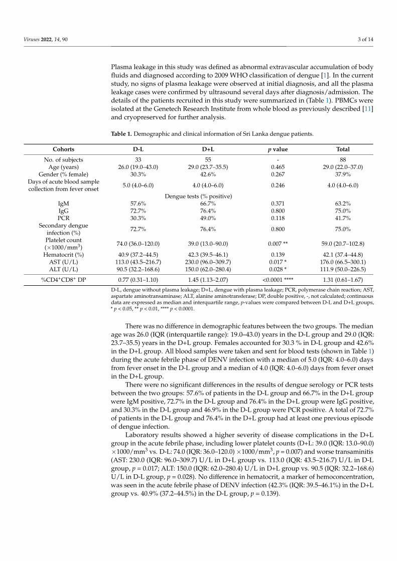
Plasma leakage in this study was defined as abnormal extravascular accumulation of bodyfluids and diagnosed according to 2009 WHO classification of dengue [1]. In the currentstudy, no signs of plasma leakage were observed at initial diagnosis, and all the plasmaleakage cases were confirmed by ultrasound several days after diagnosis/admission. Thedetails of the patients recruited in this study were summarized in (Table 1). PBMCs wereisolated at the Genetech Research Institute from whole blood as previously described [11]and cryopreserved for further analysis.
Table 1. Demographic and clinical information of Sri Lanka dengue patients.
Cohorts D-L D+L p value Total
No. of subjects 33 55 - 88Age (years) 26.0 (19.0–43.0) 29.0 (23.7–35.5) 0.465 29.0 (22.0–37.0)
Gender (% female) 30.3% 42.6% 0.267 37.9%Days of acute blood samplecollection from fever onset 5.0 (4.0–6.0) 4.0 (4.0–6.0) 0.246 4.0 (4.0–6.0)
Dengue tests (% positive)IgM 57.6% 66.7% 0.371 63.2%IgG 72.7% 76.4% 0.800 75.0%PCR 30.3% 49.0% 0.118 41.7%
Secondary dengueinfection (%) 72.7% 76.4% 0.800 75.0%
Platelet count(×1000/mm3) 74.0 (36.0–120.0) 39.0 (13.0–90.0) 0.007 ** 59.0 (20.7–102.8)
Hematocrit (%) 40.9 (37.2–44.5) 42.3 (39.5–46.1) 0.139 42.1 (37.4–44.8)AST (U/L) 113.0 (43.5–216.7) 230.0 (96.0–309.7) 0.017 * 176.0 (66.5–300.1)ALT (U/L) 90.5 (32.2–168.6) 150.0 (62.0–280.4) 0.028 * 111.9 (50.0–226.5)
%CD4+CD8+ DP 0.77 (0.31–1.10) 1.45 (1.13–2.07) <0.0001 **** 1.31 (0.61–1.67)
D-L, dengue without plasma leakage; D+L, dengue with plasma leakage; PCR, polymerase chain reaction; AST,aspartate aminotransaminase; ALT, alanine aminotransferase; DP, double positive, -, not calculated; continuousdata are expressed as median and interquartile range, p-values were compared between D-L and D+L groups,* p < 0.05, ** p < 0.01, **** p < 0.0001.
There was no difference in demographic features between the two groups. The medianage was 26.0 (IQR (interquartile range): 19.0–43.0) years in the D-L group and 29.0 (IQR:23.7–35.5) years in the D+L group. Females accounted for 30.3 % in D-L group and 42.6%in the D+L group. All blood samples were taken and sent for blood tests (shown in Table 1)during the acute febrile phase of DENV infection with a median of 5.0 (IQR: 4.0–6.0) daysfrom fever onset in the D-L group and a median of 4.0 (IQR: 4.0–6.0) days from fever onsetin the D+L group.
There were no significant differences in the results of dengue serology or PCR testsbetween the two groups: 57.6% of patients in the D-L group and 66.7% in the D+L groupwere IgM positive, 72.7% in the D-L group and 76.4% in the D+L group were IgG positive,and 30.3% in the D-L group and 46.9% in the D-L group were PCR positive. A total of 72.7%of patients in the D-L group and 76.4% in the D+L group had at least one previous episodeof dengue infection.
Laboratory results showed a higher severity of disease complications in the D+Lgroup in the acute febrile phase, including lower platelet counts (D+L: 39.0 (IQR: 13.0–90.0)×1000/mm3 vs. D-L: 74.0 (IQR: 36.0–120.0)×1000/mm3, p = 0.007) and worse transaminitis(AST: 230.0 (IQR: 96.0–309.7) U/L in D+L group vs. 113.0 (IQR: 43.5–216.7) U/L in D-Lgroup, p = 0.017; ALT: 150.0 (IQR: 62.0–280.4) U/L in D+L group vs. 90.5 (IQR: 32.2–168.6)U/L in D-L group, p = 0.028). No difference in hematocrit, a marker of hemoconcentration,was seen in the acute febrile phase of DENV infection (42.3% (IQR: 39.5–46.1%) in the D+Lgroup vs. 40.9% (37.2–44.5%) in the D-L group, p = 0.139).
Viruses 2022, 14, 90 4 of 14
2.2. Dengue Serology and PCR Tests
DENV serology tests were performed with anti-DENV IgG and IgM ELISA as pre-viously described [12]. Reverse transcription-PCR was performed by using the DV1 andDV3 primer set [13] and the ALD 1 and ALD 2 primer set [14] in one reaction with detailsspecified previously [15].
2.3. Flow Cytometry Analysis and Cell Sorting for RNA Sequencing
PBMCs were stained with anti-human CD3, CD4, CD8, CD14, CD19, CD56, andlive/dead viability antibodies (see Table S1 for antibody details). Subsequently, live CD4+
single positive (SP) (CD14−CD19−CD56−CD3+CD4+CD8−), CD8+ SP (CD14−CD19−CD56−CD3+CD4−CD8+), and CD4+CD8+ DP (CD14−CD19−CD56−CD3+CD4+CD8+) cells weresorted into TRIzol LS Reagent (Invitrogen, Carlsbad, CA, USA) using a BD FACSAria cellsorter (BD Biosciences, San Jose, CA, USA). Flow cytometry data were analyzed by FlowJoX Software (version 10, Tree Star, Ashland, OR, USA). The gating strategy utilized wereshown in Figure 1A,B, and results from representative donors from both D-L and D+Lgroups are shown in Figure 1B. To note that in this study, B cells, monocytes, and NKcells were removed by flowcytometry using CD19, CD14, and CD56 antibodies, and onlyCD3+ T cells were analyzed. However, the roles of these subsets of immune cells may beimportant as well and warrant further investigation because several relevant factors (e.g.,NF-kB) have been associated with plasma leakage [16].
2.4. RNA Sequencing
Total RNA was purified and quantified, as previously described [17]. Purified totalRNA (5 ng) was amplified following the Smart-seq2 protocol [18]. cDNA was purifiedusing AMPure XP beads (1:1 ratio; Beckman Coulter, Carlsbad, CA, USA). From thisstep, 1 ng cDNA was used to prepare a standard Nextera XT sequencing library (NexteraXT DNA library preparation kit and index kit (set B and C, respectively), Illumina, SanDiego, CA, USA). Samples were sequenced in 3 batches using a NovaSeq 6000 system(Illumina, San Diego, CA, USA) to obtain 50-bp paired-end reads. Both whole transcriptomeamplification and sequencing library preparations were performed in a 96-well formatto reduce assay-to-assay variability. Quality control steps were included to determinetotal RNA quality and quantity, the optimal number of PCR pre-amplification cycles, andfragment size selection using the Agilent 2100 Bioanalyzer system (Agilent, Santa Clara,CA, USA). Samples that failed quality control were eliminated from further downstreamsteps. Barcoded Illumina sequencing libraries (Nextera, Illumina, San Diego, CA, USA)were generated utilizing the automated platform (Biomek FXp, Beckman Coulter, Carlsbad,CA, USA). Libraries were sequenced on the NovaSeq 6000 Illumina platform to obtain50-bp paired-end reads using the NovaSeq 6000 S4 Reagent kit v1.5 (Illumina, San Diego,CA, USA), generating a median of 25.4 million mapped reads per sample.
2.5. Transcriptomic Analysis
Interventionary studies involving animals or humans and other studies that requireethical approval must list the authority that provided approval and the correspondingethical approval code.
The single-end reads that passed Illumina filters were filtered for reads aligning totRNA, rRNA, adapter sequences, and spike-in controls. The reads were then aligned toUCSC hg19 reference genome using TopHat (v 1.4.1) [19]. DUST scores were calculatedwith PRINSEQ Lite (v 0.20.3) [20], and low-complexity reads (DUST > 4) were removedfrom the BAM files. The alignment results were parsed via the SAMtools [21] to generateSAM files. Read counts to each genomic feature were obtained with the HTseq-countprogram (v 0.6.0) [22] using the “union” option.
Viruses 2022, 14, 90 5 of 14
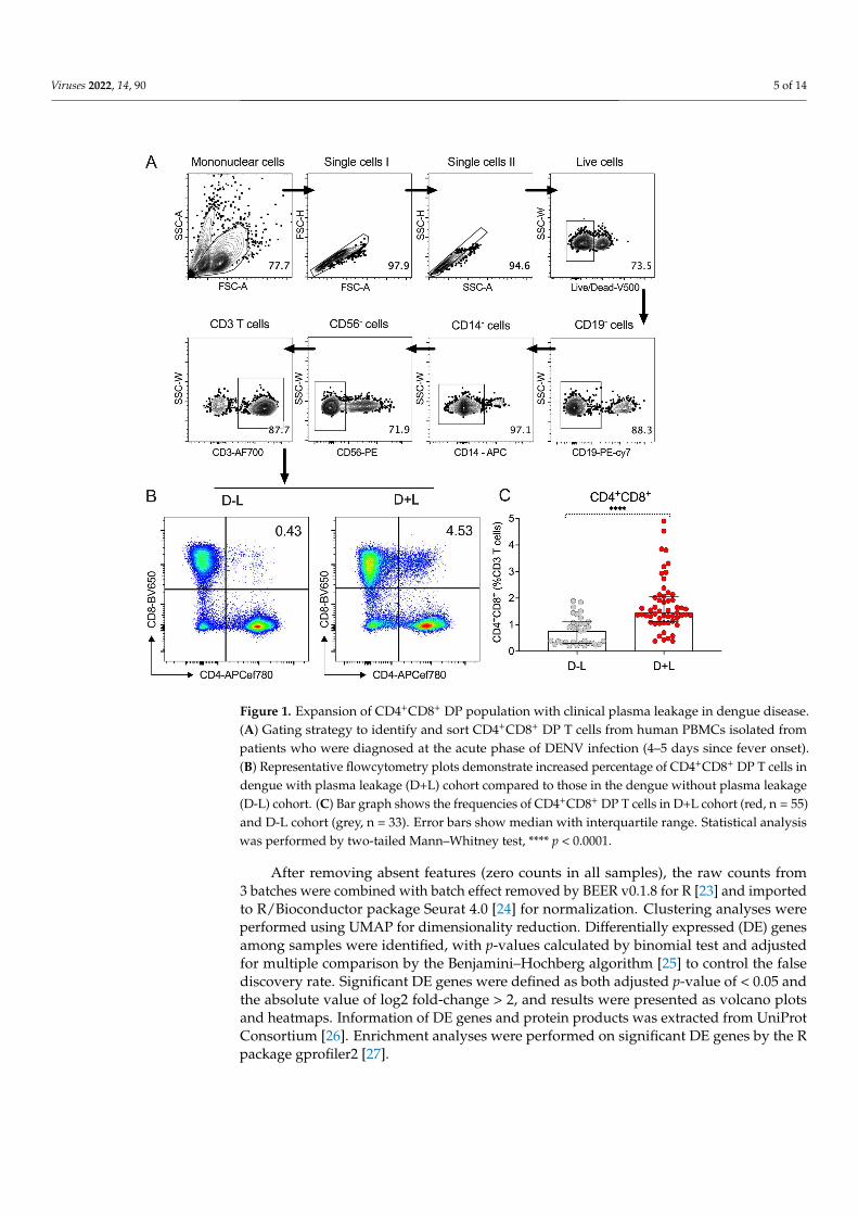
Figure 1. Expansion of CD4+CD8+ DP population with clinical plasma leakage in dengue disease.(A) Gating strategy to identify and sort CD4+CD8+ DP T cells from human PBMCs isolated frompatients who were diagnosed at the acute phase of DENV infection (4–5 days since fever onset).(B) Representative flowcytometry plots demonstrate increased percentage of CD4+CD8+ DP T cells indengue with plasma leakage (D+L) cohort compared to those in the dengue without plasma leakage(D-L) cohort. (C) Bar graph shows the frequencies of CD4+CD8+ DP T cells in D+L cohort (red, n = 55)and D-L cohort (grey, n = 33). Error bars show median with interquartile range. Statistical analysiswas performed by two-tailed Mann–Whitney test, **** p < 0.0001.
After removing absent features (zero counts in all samples), the raw counts from3 batches were combined with batch effect removed by BEER v0.1.8 for R [23] and importedto R/Bioconductor package Seurat 4.0 [24] for normalization. Clustering analyses wereperformed using UMAP for dimensionality reduction. Differentially expressed (DE) genesamong samples were identified, with p-values calculated by binomial test and adjustedfor multiple comparison by the Benjamini–Hochberg algorithm [25] to control the falsediscovery rate. Significant DE genes were defined as both adjusted p-value of < 0.05 andthe absolute value of log2 fold-change > 2, and results were presented as volcano plotsand heatmaps. Information of DE genes and protein products was extracted from UniProtConsortium [26]. Enrichment analyses were performed on significant DE genes by the Rpackage gprofiler2 [27].
Viruses 2022, 14, 90 6 of 14
2.6. Statistical Analysis
Flow cytometry and clinical data were analyzed by GraphPad Prism Version 8 (La Jolla,CA, USA). The statistical details of the experiments are provided in the respective figurelegends. Normality of distribution was assessed by Shapiro–Wilk test. Comparison ofthe non-parametric continuous data between D+L and D-L cohorts was performed bytwo-tailed Mann–Whitney test, and categorical data were compared with Fisher’s exacttest. Multivariate logistic regression was performed in R (version 4.0.2) (R DevelopmentCore Team, Vienna, Austria). Non-parametric data in this study are represented as medianwith interquartile range. p-Values < 0.05 (after adjustment if indicated) were consideredstatistically significant.
3. Results3.1. Expansion of CD4+CD8+ DP T Cells Is Associated with Risk of Plasma Leakage in Dengue
A total of 88 dengue patients, including 55 with plasma leakage (D+L) and 33 withoutplasma leakage (D-L) were enrolled in the study and recruited from the North ColomboTeaching Hospital, Ragama in Gampaha District, Sri Lanka and the National Institute ofInfectious Diseases, Gothatuwa, Angoda, Sri Lanka between 2010 and 2016. Demographicand clinical information of patient groups included in the analysis are summarized inTable 1 and described in details in methods. It is important to emphasize that the samplesstudied corresponded to blood draws obtained before any plasma leakage was detectableclinically or radiologically.
Initial phenotyping of peripheral blood mononuclear cells (PBMC) from the twogroups (D+L and D-L) was determined by flow cytometry. Specifically, the proportionsof CD4+CD8+ double-positive (DP), CD4+ single-positive (SP), and CD8+ SP T cells inacute DENV infection was assessed, using the gating strategy illustrated in Figure 1A.Representative donors from both D-L and D+L group are illustrated in Figure 1B. As shownin Figure 1C, we observed a significant higher proportion (p < 0.0001) of CD4+CD8+ DP Tcells in the D+L group (median 1.45%, interquartile range (IQR): 1.13–2.07%) compared tothe D-L group (median 0.77%, IQR: 0.31–1.10%), with no differences detected for CD4+ orCD8+ SP T cells (Figure S1). The detailed values of median and IQR for CD4+CD8+ DP Tcells from two cohorts are summarized in Table 1.
To further evaluate the association between the proportion of CD4+CD8+ DP cellsand plasma leakage, we conducted a multivariate logistic regression analysis accountingfor other confounding factors, including demographic features (e.g., age, gender), previ-ous DENV infection, as well as hematological and biochemical parameters (e.g., plateletcount, hematocrit, AST, and ALT). As shown in Table 2, the percentage of CD4+CD8+ DPcells was the only significant factor (p = 0.002), positively correlated with plasma leakage(log (OR) = 1.86). These results suggested that the CD4+CD8+ DP T cell population is anovel and relevant marker of development of plasma leakage.
Table 2. Multivariate analysis of risk factors associated with plasma leakage in dengue disease by alogistic regression model.
Risk Factors Estimate (β) Standard Error Z Score padj Value
%CD4+CD8+ DP 1.8632 0.5946 3.133 0.002 **Age (years) 0.0151 0.0222 0.682 0.495
Gender (M/F) −0.7616 0.8402 −0.907 0.365Secondary dengue (Y/N) −0.1705 0.8201 −0.208 0.835
Platelet count(×1000/mm3) −0.0105 0.0082 −1.280 0.200
Hematocrit (%) 0.0510 0.0766 0.666 0.505AST (U/L) 0.0011 0.0031 0.369 0.712ALT (U/L) 0.0001 0.0029 0.042 0.966
DP, double positive; M/F, male/female; Y/N, yes/no; AST, aspartate aminotransaminase; ALT, alanine amino-transferase, padj value, adjusted p value; Akaike information criterion (AIC): 81.106, ** p < 0.01.
Viruses 2022, 14, 90 7 of 14
3.2. The Gene Expression Profile of CD4+CD8+ DP T Cells Reveals a Higher Similarity to CD8+ TCells as Opposed to CD4+ T Cells
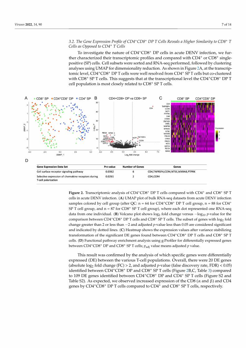
To investigate the nature of CD4+CD8+ DP cells in acute DENV infection, we fur-ther characterized their transcriptomic profiles and compared with CD4+ or CD8+ single-positive (SP) cells. Cell subsets were sorted and RNA-seq performed, followed by clusteringanalyses using UMAP for dimensionality reduction. As shown in Figure 2A, at the transcrip-tomic level, CD4+CD8+ DP T cells were well resolved from CD4+ SP T cells but co-clusteredwith CD8+ SP T cells. This suggests that at the transcriptional level the CD4+CD8+ DP Tcell population is most closely related to CD8+ SP T cells.
Figure 2. Transcriptomic analysis of CD4+CD8+ DP T cells compared with CD4+ and CD8+ SP Tcells in acute DENV infection. (A) UMAP plot of bulk RNA-seq datasets from acute DENV infectionsamples colored by cell group (after QC: n = 64 for CD4+CD8+ DP T cell group, n = 88 for CD4+
SP T cell group, and n = 87 for CD8+ SP T cell group), where each dot represented one RNA-seqdata from one individual. (B) Volcano plot shows log2 fold change versus −log10 p-value for thecomparison between CD4+CD8+ DP T cells and CD8+ SP T cells. The subset of genes with log2 foldchange greater than 2 or less than −2 and adjusted p-value less than 0.05 are considered significantand indicated by dotted lines. (C) Heatmap shows the expression values after variance stabilizingtransformation of the significant DE genes found between CD4+CD8+ DP T cells and CD8+ SP Tcells. (D) Functional pathway enrichment analysis using g:Profiler for differentially expressed genesbetween CD4+CD8+ DP and CD8+ SP T cells; padj value means adjusted p value.
This result was confirmed by the analysis of which specific genes were differentiallyexpressed (DE) between the various T-cell populations. Overall, there were 20 DE genes(absolute log2 fold change (FC) > 2, and adjusted p-value (false discovery rate, FDR) < 0.05)identified between CD4+CD8+ DP and CD8+ SP T cells (Figure 2B,C, Table 3) comparedto 109 DE genes identified between CD4+CD8+ DP and CD4+ SP T cells (Figure S2 andTable S2). As expected, we observed increased expression of the CD8 (α and β) and CD4genes by CD4+CD8+ DP T cells compared to CD4+ and CD8+ SP T cells, respectively.
Viruses 2022, 14, 90 8 of 14
Table 3. List of DE genes found between CD4+CD8+ DP and CD8+ SP cells.
Gene Symbol Encoded Protein p-Value Log2FC FDR Gene Product Function *
Higher expression in CD4+CD8+ DP cells
CD4 T-cell surface glycoprotein CD4 4.11E-46 6.42 2.40E-41 Cell differentiation antigen CD4, MHCclass II receptor
TNFRSF4 Tumor necrosis factor receptorsuperfamily member 4 (OX40) 6.24E-11 3.19 3.64E-06 Costimulatory molecule implicated in
long-term T-cell immunity
CCR4 C-C chemokine receptor type 4(CCR4) 5.80E-08 3.16 3.38E-03
High-affinity binding for basophilchemoattractant, G protein coupled
receptor superfamily, specific receptor forthymus and activation-regulated
chemokine
NCF1 Neutrophil cytosol factor 1 7.09E-10 2.08 4.14E-05Activate NADPH oxidase for superoxide
production and oxygen dependentmechanism of phagocytosis
Reduced expression in CD4+CD8+ DP cells
PTPRK Receptor-type tyrosine-proteinphosphatase kappa 9.03E-08 −2.06 5.27E-03
Transmembrane receptor protein tyrosinephosphatase activity, regulation of
processes involving cell contact andadhesion
CA6 Carbonic anhydrase 6 1.58E-10 −2.22 9.20E-06 Carbonate dehydratase activity, proteinbinding
S100B S100 Calcium-Binding Protein B 6.52E-09 −2.33 3.81E-04 Calcium ion binding, proteinhomodimerization activity
ZNF433 Zinc finger protein 433 3.47E-07 −2.42 2.03E-02Transcriptional regulation, RNApolymerase II regulatory regionsequence-specific DNA binding
DSEL Dermatan-sulfateepimerase-like protein 7.12E-08 −2.45 4.15E-03 Membrane proteins, sulfotransferase
activity, isomerase activity
MMP28 Matrix metalloproteinase-28 1.62E-07 −2.50 9.48E-03 Tissues homeostasis and repair,metalloendopeptidase activity
SPINK2 Serine protease inhibitorKazal-type 2 5.22E-07 −2.67 3.04E-02 Serine-type endopeptidase inhibitor
activity
NT5E 5′-nucleotidase 9.12E-15 −2.73 5.32E-10 Hydrolyzes extracellular nucleotides intomembrane permeable nucleosides
MXRA8 Matrix remodeling-associatedprotein 8 1.56E-08 −2.87 9.13E-04
Transmembrane protein which canmodulate activity of various signaling
pathways
FBLN2 Fibulin-2 2.67E-07 −2.95 1.56E-02Extracellular matrix protein, expressed inelastic tissues including intima of blood
vessels
CNN3 Calponin-3 1.82E-11 −2.98 1.06E-06
Cytoskeletal protein involved in cell-celladhesion, downregulated during
dedifferentiation of vascular smoothmuscle cell
PRCD Photoreceptor disk component 6.21E-09 −3.15 3.62E-04 Opsin binding
NOG Noggin 7.99E-13 −3.26 4.66E-08 Cytokine binding, proteinhomodimerization activity
GIPC3 PDZ domain-containing protein 5.15E-08 −3.30 3.00E-03 Protein binding
REG4 Regenerating islet-derivedprotein 4 3.15E-15 −3.48 1.84E-10 Carbohydrate binding, signaling receptor
activitySDK2 Protein sidekick-2 4.31E-08 −3.62 2.52E-03 Adhesion molecule
Log2FC, log2 fold change; FDR, false-discovery rate; DE genes cut off: FDR < 0.05 and Log2FC > 2, * Gene proteinproducts with functional annotation information was extracted from UniProt Consortium [26].
3.3. Molecular Pathways Associated with CD4+CD8+ DP T Cells
The analysis above suggested that CD4+CD8+ DP T cells are more closely related toCD8+ SP T cells in terms of their transcription profile. We next set out to examine whichgenes might be differentially expressed between the two T-cell compartments and mighttherefore define the specific gene expression pattern associated with CD4+CD8+ DP Tcells. Compared to CD8+ SP T cells, we mainly observed in CD4+CD8+ DP T cells a higherexpression of genes involved in T-cell activation (TNFRSF4, Tumor necrosis factor receptorsuperfamily member 4 or OX40), immune cell recruitment (CCR4, C-C chemokine receptortype 4), and oxygen dependent phagocytosis (NCF1, Neutrophil cytosol factor 1).
Viruses 2022, 14, 90 9 of 14
Significantly down-regulated DE genes in CD4+CD8+ DP T cells (Figure 2B,C) in-cluded PTPRK (Receptor-type tyrosine-protein phosphatase kappa), CA6 (Carbonic an-hydrase 6), S100B (S100 Calcium-Binding Protein B), ZNF433 (Zinc finger protein 433),DSEL (Dermatan-sulfate epimerase-like protein), MMP28 (Matrix metalloproteinase-28),SPINK2 (Serine protease inhibitor Kazal-type 2), NT5E (5′-nucleotidase), MXRA8 (Matrixremodeling-associated protein 8), FBLN2 (Fibulin-2), CNN3 (Calponin-3), PRCD (Photore-ceptor disk component), NOG (Noggin), GIPC3 (PDZ domain-containing protein), REG4(Regenerating islet-derived protein 4), and SDK2 (Protein sidekick-2), representing intricateprocesses of cell differentiation and signaling in CD4+CD8+ cells (Table 3).
The functional pathway enrichment analysis using g:Profiler revealed that those DEgenes were enriched in two pathways involved in cell surface receptor signaling pathway(CD4, TNFRSF4, CCR4, NT5E, MXRA8, PTPRK) and selective expression of chemokinereceptors during T-cell polarization (CD4, CCR4) (Figure 2D). These results suggest that thetranscriptomic profile of CD4+CD8+ DP T cells is more closely related CD8+ SP T cells withsome differences detected.
3.4. Paucity of Transcriptomic Changes in CD4+CD8+ DP T Cells between Patients with andwithout Plasma Leakage in Dengue
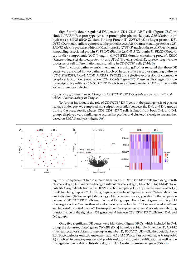
To further investigate the role of CD4+CD8+ DP T cells in the pathogenesis of plasmaleakage in dengue, we compared transcriptomic profiles between the D+L and D-L groupsduring the acute febrile phase. CD4+CD8+ DP T cells isolated from both D+L and D-Lgroups displayed very similar gene expression profiles and clustered closely to one anotherbased on UMAP analysis (Figure 3A).
Figure 3. Comparison of transcriptomic signatures of CD4+CD8+ DP T cells from dengue withplasma leakage (D+L) cohort and dengue without plasma leakage (D-L) cohort. (A) UMAP plot ofbulk RNA-seq datasets from acute DENV infection samples colored by disease groups (after QC:n = 41 for D+L group, n = 23 for D-L group), where each dot represented one RNA-seq data fromone individual. (B) Volcano plot shows log2 fold change versus −log10 p-value for the comparisonbetween CD4+CD8+ DP T cells from D+L and D-L groups. The subset of genes with log2 foldchange greater than 2 or less than −2 and adjusted p-value less than 0.05 are considered significantand indicated by dotted lines. (C) Heatmap shows the expression values after variance stabilizingtransformation of the significant DE genes found between CD4+CD8+ DP T cells from D+L andD-L groups.
Only five significant DE genes were identified (Figure 3B,C), which included in D+Lgroup the down-regulated genes DNAJB1 (DnaJ homolog subfamily B member 1), NR4A2(Nuclear receptor subfamily 4 group A member 2), B3GNT7 (UDP-GlcNAc:betaGal beta-1,3-N-acetylglucosaminyltransferase), and SLC45A1 (Proton-associated sugar transporterA) involved in gene expression and post-translational protein modification as well as theup-regulated gene ABO (Histo-blood group ABO system transferase) gene (Table 4).

Viruses 2022, 14, 90 10 of 14
Table 4. List of DE genes found between CD4+CD8+ DP cells of D+L and D-L cohorts.
Gene Symbol Encoded Protein p-Value Log2FC FDR Gene Product Function *
Reduced expression in CD4+CD8+ DP cells of D+L cohort
DNAJB1 DnaJ homolog subfamily Bmember 1 1.36E-07 −2.12 7.96E-03
ATPase activator activity, transcriptioncorepressor activity, Hsp70 protein
binding, involved in protein folding
NR4A2 Nuclear receptor subfamily 4group A member 2 1.49E-07 −3.08 8.72E-03 RNA polymerase II regulatory region
sequence-specific DNA binding
B3GNT7UDP-GlcNAc:betaGal
beta-1,3-N-acetylglucosaminyltransferase
1.31E-07 −5.48 7.66E-03 Acetylglucosaminyltransferase activity,protein binding
SLC45A1 Proton-associated sugartransporter A 6.06E-07 −5.39 3.53E-02 Sucrose:proton symporter activity
Higher expression in CD4+CD8+ DP cells of D+L cohort
ABO Histo-blood group ABO systemtransferase 3.11E-10 4.62 1.82E-05 Glycosyltransferase activity
Log2FC, log2 fold change; FDR, false-discovery rate; DE genes cut off: FDR < 0.05 and Log2FC > 2, * Gene proteinproducts with functional annotation information was extracted from UniProt Consortium [26].
4. Discussion
This study provided several novel observations on the host immune response dur-ing the acute phase of DENV infection. First, the report that the frequency of a novelCD4+CD8+ DP T cell subset was increased in D+L versus D-L, and it is thus associatedwith risk of plasma leakage in dengue disease. Second, on the basis of their transcriptomicprofile, CD4+CD8+ DP T cells were most closely related to CD8+ SP T cells, but still, wewere able to define 20 DE genes enriched in two functional pathways representative ofselective expression of chemokine receptors and signaling on the cell surface during T-cellpolarization. Third, while the transcriptomic profile of the CD4+CD8+ DP T cells was rathersimilar in D+L and D-L patients, five DE genes were identified.
CD4+CD8+ DP T cells have been described in several pathological conditions in com-parison to normal individuals [28]; for example, expansion of DP cells has been observedin autoimmune diseases (e.g., Myasthenia gravis, Rheumatoid arthritis, multiple sclerosis,Kawasaki diseases, and systemic sclerosis), infectious diseases (e.g., HIV and EBV), andcancers (e.g., melanoma, lymphoma) [28]. However, their phenotype and role in DENVinfection have not been reported. Peripheral CD4+CD8+ DP T cells are a phenotypically andfunctionally heterogeneous population depending on their origin and pathologic context,and their roles in the pathogenesis of autoimmune diseases, viral infections, and cancers areunder ongoing debate [29]. While CD4+CD8+ DP T cells appear to function at peripheralsites as potent immune suppressors [30,31] or cells with high cytotoxic potential [32,33],a more thorough examination of their contribution to the adaptive immunity in humandiseases is warranted [29].
Here, we observed that an increased frequency of CD4+CD8+ DP T cells duringacute DENV infection was associated with development of plasma leakage in denguedisease. Furthermore, CD4+CD8+ DP T cells appear to be the most significant factorassociated with development of plasma leakage even after adjusting for other reported riskfactors, such as age, gender, platelet count, hematocrit, AST, ALT levels, and secondarydengue infection [10]. These results indicate that CD4+CD8+ DP T cells are a novel factorthat warrants further investigation, especially taking into consideration of the potentialimplication in clinical settings and pathophysiology studies of plasma leakage (both onsetand severity) in DENV infection by collecting bloods at various time points in future. Inparticular, the current study did not address the viral load in the blood nor the antigenspecificity of the CD4+CD8+ DP T cells, and future studies will have to address theircorrelation with the immune response and what fraction of these CD4+CD8+ DP T cells isactually DENV specific.
In addition, the origin and function of peripheral CD4+CD8+ DP T cells in vari-ous pathological conditions are not fully understood to date. Parrot et al. reported
Viruses 2022, 14, 90 11 of 14
that intra-melanoma DP T cells were transcriptome-wise closer to CD8+ SP T cells [34],while Mucida et al. found mature CD4+ T helper cells could undergo transcriptomic re-programming and generate cytotoxic CD4+CD8+ DP T cells after persistent ovalbuminantigen stimulation [35]. However, those studies only investigated limited genes by microar-ray, and there was no systemic RNA-sequencing (RNA-seq) data comparing transcriptomicprofiles of CD4+CD8+ DP with CD4+ or CD8+ SP T cells.
Our study reports the first set of RNA-seq data of CD4+CD8+ DP T cells, and thefinding that their transcriptomic profile is most closely related to CD8+ SP T cells in acuteDENV infection. The principal transcripts distinguishing CD4+CD8+ DP T cells from CD8+
SP T cells encoded OX40 and CCR4, which were found up-regulated in CD4+CD8+ DPcompared to CD8+ SP T cells. Interestingly, OX40 has been established as a crucial signalingmolecule during persistent viral infection [36] and implicated in their pathology throughmaintenance of higher numbers of long-lived CD8+ effector T cells [37]. In addition, adifferential regulation of the expression of the OX40 signaling pathway has been observedin asymptomatic dengue cases compared with clinical cases [38] and associated withdengue disease severity [39]. Moreover, clinical studies in endemic areas have describedthe associations between the levels of CC chemokines and dengue disease outcome [40,41].In particular, the CCR4 receptor has been shown to contribute to the pathogenesis of severeconditions [42,43], including dengue [44]. Collectively, our results indicate that CD4+CD8+
DP T cells are effector T cells that are transcriptome-wise closer to CD8+ SP T cells duringacute DENV infection.
While the gene expression profile of CD4+CD8+ DP T in D+L and D-L was relativelysimilar, DE genes were identified. Interestingly, the ABO gene was the only up-regulatedgene, associated with plasma leakage during acute DENV infection. This gene encodesA and B antigens of human blood group and is autosomal dominant [45]. Studies haveshown that ABO blood group plays a role in viral infection and host susceptibility, such asnorovirus, rotavirus, HIV, influenza viruses, as well as SARS coronavirus (SARS-CoV) [45].Kalayanarooj et al. found that blood group AB was associated with increased risk ofsevere dengue disease with plasma leakage in secondary infections [46], consistent withour findings. These differentially expressed genes, together with those discriminatingCD4+CD8+ DP T cells from CD8+ SP T cells, could have clinical utility, perhaps as PCR-based markers.
Finally, it can be noted that the results presented herein do not support the previouspostulate of original antigenic sin and altered responses [47,48] as key drivers of DENVimmunopathology. Rather, they can be interpreted in a growing body of studies [49–52]that indicate that the T-cell responses and T-cell subsets are modulated in DENV diseaseand that differential disease severity is mostly associated with differences in magnitude ofresponses rather than phenotypic differences of different T-cell compartments modulatedin the course of infection and disease. That is, severe disease is characterized by an infectedcell mass that is larger than that of mild disease, and an enhanced antigen mass stimulatesa greater magnitude of T-cell responses.
5. Conclusions
Plasma leakage in dengue diseases is a warning complication which requires earlydetection and clinical intervention. In this study, we observed increased frequency ofa novel CD4+CD8+ double-positive (DP) T-cell subset in the acute febrile phase of theDENV infection is associated with risk of developing plasma leakage later in denguedisease. Further transcriptomic analysis suggested this T-cell subset is closest to CD8+
single-positive (SP) T cells. Nonetheless, the transcriptomic profile of the CD4+CD8+ DPT cells was largely similar in patients with and without plasma leakage, implying a roleof CD4+CD8+ DP cells in plasma leakage through a quantitative increase rather thanfunctional alteration. These data provide new insight into the host immune response inDENV infection and the role of CD4+CD8+ DP T cells in the pathogenesis of plasma leakage.
Viruses 2022, 14, 90 12 of 14
Supplementary Materials: The following are available online at https://www.mdpi.com/article/10.3390/v14010090/s1, Figure S1: Distribution of CD4+ and CD8+ single positive (SP) in D-L and D+Lcohorts; Figure S2: Transcriptomic analysis of CD4+CD8+ DP T cells compared with CD4+ SP T cellsin acute DENV infection; Table S1: List of antibodies used in the flowcytometry study; Table S2: Listof DE genes found between CD4+CD8+ DP and CD4+ SP cells.
Author Contributions: Designing research studies, A.S., D.W. and E.D.Y.; investigation, E.D.Y., H.W.,R.d.S.A., Y.T. and A.G.; data analysis, E.D.Y., H.W. and Y.T.; resources, R.T., S.U.A., G.P., A.W., S.P.and A.D.D.S.; manuscript writing, E.D.Y., H.W., R.d.S.A. and A.S.; supervision, A.S. and D.W.; projectadministration, A.F.; funding acquisition, A.S. and D.W. All authors have read and agreed to thepublished version of the manuscript.
Funding: Research reported in this publication was supported by the National Institute of Allergyand Infectious Diseases of the National Institutes of Health under Award Number U19AI118626,HHSN272201400045C, and 75N93019C00065. The content is solely the responsibility of the authorsand does not necessarily represent the official views of the National Institutes of Health.
Institutional Review Board Statement: This study was approved by the Human Subjects ProtectionPrograms of the La Jolla Institute for Immunology (Protocols VD-085 and VD-101), the Universityof Colombo (IRB Protocols EC-15-002 and EC-15-095), the Nicaragua Ministry of Health (ProtocolCIRE-01/10/06-13.Ver14), and the University of California Berkeley (Protocol 2010-06-1649).
Informed Consent Statement: Informed consent was obtained from all subjects involved in the study.
Data Availability Statement: The RNA-Seq data were deposited in the NCBI’s Gene ExpressionOmnibus (GEO) database under the accession code GSE178240. The other datasets generated and/oranalyzed during the current study are available from the corresponding author on reasonable request.
Acknowledgments: We wish to acknowledge all subjects for their participation and for donatingtheir blood and time for this study.
Conflicts of Interest: The authors have declared that no conflict of interest exists.
References1. World Health Organization. Dengue: Guidelines for Diagnosis, Treatment, Prevention and Control: New Edition; World Health
Organization: Geneva, Switzerland, 2009.2. Centre for Disease Control and Prevention. About Dengue: What You Need to Know. Available online: https://www.cdc.gov/
dengue/about/index.html (accessed on 15 April 2021).3. Deen, J.L.; Harris, E.; Wills, B.; Balmaseda, A.; Hammond, S.N.; Rocha, C.; Dung, N.M.; Hung, N.T.; Hien, T.T.; Farrar, J.J. The
WHO dengue classification and case definitions: Time for a reassessment. Lancet 2006, 368, 170–173. [CrossRef]4. Phuong, C.X.; Nhan, N.T.; Kneen, R.; Thuy, P.T.; van Thien, C.; Nga, N.T.; Thuy, T.T.; Solomon, T.; Stepniewska, K.; Wills,
B.; et al. Clinical diagnosis and assessment of severity of confirmed dengue infections in Vietnamese children: Is the world healthorganization classification system helpful? Am. J. Trop. Med. Hyg. 2004, 70, 172–179. [CrossRef]
5. Grifoni, A.; Voic, H.; Mateus, J.; Fung, K.M.Y.; Wang, A.; Seumois, G.; De Silva, A.D.; Tennekon, R.; Premawansa, S.; Premawansa,G.; et al. Transcriptomics of acute DENV-specific CD8+ T cells does not support qualitative differences as drivers of diseaseseverity. medRxiv 2021. [CrossRef]
6. Alexander, N.; Balmaseda, A.; Coelho, I.C.; Dimaano, E.; Hien, T.T.; Hung, N.T.; Janisch, T.; Kroeger, A.; Lum, L.C.; Martinez,E.; et al. Multicentre prospective study on dengue classification in four South-east Asian and three Latin American countries.Trop. Med. Int. Health 2011, 16, 936–948. [CrossRef] [PubMed]
7. Rigau-Perez, J.G. Severe dengue: The need for new case definitions. Lancet Infect. Dis. 2006, 6, 297–302. [CrossRef]8. Rajapakse, S. Dengue shock. J. Emerg. Trauma Shock 2011, 4, 120–127. [CrossRef]9. Lee, I.K.; Hsieh, C.J.; Lee, C.T.; Liu, J.W. Diabetic patients suffering dengue are at risk for development of dengue shock
syndrome/severe dengue: Emphasizing the impacts of co-existing comorbidity(ies) and glycemic control on dengue severity. J.Microbiol. Immunol. Infect. 2020, 53, 69–78. [CrossRef]
10. Sangkaew, S.; Ming, D.; Boonyasiri, A.; Honeyford, K.; Kalayanarooj, S.; Yacoub, S.; Dorigatti, I.; Holmes, A. Risk predictors ofprogression to severe disease during the febrile phase of dengue: A systematic review and meta-analysis. Lancet Infect. Dis. 2021,21, 1014–1026. [CrossRef]
11. Weiskopf, D.; Angelo, M.A.; de Azeredo, E.L.; Sidney, J.; Greenbaum, J.A.; Fernando, A.N.; Broadwater, A.; Kolla, R.V.; De Silva,A.D.; de Silva, A.M.; et al. Comprehensive analysis of dengue virus-specific responses supports an HLA-linked protective role forCD8+ T cells. Proc. Natl. Acad. Sci. USA 2013, 110, E2046–E2053. [CrossRef]
12. Fernandez, R.J.; Vazquez, S. Serological diagnosis of dengue by an ELISA inhibition method (EIM). Mem. Inst. Oswaldo Cruz 1990,85, 347–351. [CrossRef]
Viruses 2022, 14, 90 13 of 14
13. Seah, C.L.; Chow, V.T.; Tan, H.C.; Can, Y.C. Rapid, single-step RT-PCR typing of dengue viruses using five NS3 gene primers. J.Virol. Methods 1995, 51, 193–200. [CrossRef]
14. Sudiro, T.M.; Ishiko, H.; Green, S.; Vaughn, D.W.; Nisalak, A.; Kalayanarooj, S.; Rothman, A.L.; Raengsakulrach, B.; Janus, J.;Kurane, I.; et al. Rapid diagnosis of dengue viremia by reverse transcriptase-polymerase chain reaction using 3′-noncodingregion universal primers. Am. J. Trop. Med. Hyg. 1997, 56, 424–429. [CrossRef]
15. Kanakaratne, N.; Wahala, W.M.; Messer, W.B.; Tissera, H.A.; Shahani, A.; Abeysinghe, N.; de-Silva, A.M.; Gunasekera, M. Severedengue epidemics in Sri Lanka, 2003–2006. Emerg. Infect. Dis. 2009, 15, 192–199. [CrossRef] [PubMed]
16. Iuvone, T.; D’Acquisto, F.; Van Osselaer, N.; Di Rosa, M.; Carnuccio, R.; Herman, A.G. Evidence that inducible nitric oxidesynthase is involved in LPS-induced plasma leakage in rat skin through the activation of nuclear factor-kappaB. Br. J. Pharmacol.1998, 123, 1325–1330. [CrossRef] [PubMed]
17. Seumois, G.; Vijayanand, P.; Eisley, C.J.; Omran, N.; Kalinke, L.; North, M.; Ganesan, A.P.; Simpson, L.J.; Hunkapiller, N.;Moltzahn, F.; et al. An integrated nano-scale approach to profile miRNAs in limited clinical samples. Am. J. Clin. Exp. Immunol.2012, 1, 70–89.
18. Picelli, S.; Faridani, O.R.; Bjorklund, A.K.; Winberg, G.; Sagasser, S.; Sandberg, R. Full-length RNA-seq from single cells usingSmart-seq2. Nat. Protoc. 2014, 9, 171–181. [CrossRef]
19. Trapnell, C.; Pachter, L.; Salzberg, S.L. TopHat: Discovering splice junctions with RNA-Seq. Bioinformatics 2009, 25, 1105–1111.[CrossRef]
20. Schmieder, R.; Edwards, R. Quality control and preprocessing of metagenomic datasets. Bioinformatics 2011, 27, 863–864.[CrossRef]
21. Li, H.; Handsaker, B.; Wysoker, A.; Fennell, T.; Ruan, J.; Homer, N.; Marth, G.; Abecasis, G.; Durbin, R.; Genome Project DataProcessing Subgroup. The Sequence Alignment/Map format and SAMtools. Bioinformatics 2009, 25, 2078–2079. [CrossRef]
22. Anders, S.; Pyl, P.T.; Huber, W. HTSeq–a Python framework to work with high-throughput sequencing data. Bioinformatics 2015,31, 166–169. [CrossRef]
23. Zhang, F.; Wu, Y.; Tian, W. A novel approach to remove the batch effect of single-cell data. Cell Discov. 2019, 5, 46. [CrossRef]24. Love, M.I.; Huber, W.; Anders, S. Moderated estimation of fold change and dispersion for RNA-seq data with DESeq2. Genome
Biol. 2014, 15, 550. [CrossRef]25. Benjamini, Y.; Drai, D.; Elmer, G.; Kafkafi, N.; Golani, I. Controlling the false discovery rate in behavior genetics research. Behav.
Brain Res. 2001, 125, 279–284. [CrossRef]26. UniProt, C. UniProt: The universal protein knowledgebase in 2021. Nucleic Acids Res. 2021, 49, D480–D489. [CrossRef]27. Reimand, J.; Kull, M.; Peterson, H.; Hansen, J.; Vilo, J. g:Profiler–a web-based toolset for functional profiling of gene lists from
large-scale experiments. Nucleic Acids Res. 2007, 35, W193–W200. [CrossRef] [PubMed]28. Parel, Y.; Chizzolini, C. CD4+ CD8+ double positive (DP) T cells in health and disease. Autoimmun. Rev. 2004, 3, 215–220.
[CrossRef] [PubMed]29. Overgaard, N.H.; Jung, J.W.; Steptoe, R.J.; Wells, J.W. CD4+/CD8+ double-positive T cells: More than just a developmental stage?
J. Leukoc. Biol. 2015, 97, 31–38. [CrossRef] [PubMed]30. Das, G.; Augustine, M.M.; Das, J.; Bottomly, K.; Ray, P.; Ray, A. An important regulatory role for CD4+CD8 alpha alpha T cells in
the intestinal epithelial layer in the prevention of inflammatory bowel disease. Proc. Natl. Acad. Sci. USA 2003, 100, 5324–5329.[CrossRef]
31. Szczepanik, M.; Bryniarski, K.; Tutaj, M.; Ptak, M.; Skrzeczynska, J.; Askenase, P.W.; Ptak, W. Epicutaneous immunization inducesalphabeta T-cell receptor CD4 CD8 double-positive non-specific suppressor T cells that inhibit contact sensitivity via transforminggrowth factor-beta. Immunology 2005, 115, 42–54. [CrossRef]
32. Kitchen, S.G.; Jones, N.R.; LaForge, S.; Whitmire, J.K.; Vu, B.A.; Galic, Z.; Brooks, D.G.; Brown, S.J.; Kitchen, C.M.; Zack, J.A.CD4 on CD8(+) T cells directly enhances effector function and is a target for HIV infection. Proc. Natl. Acad. Sci. USA 2004, 101,8727–8732. [CrossRef]
33. Kitchen, S.G.; Whitmire, J.K.; Jones, N.R.; Galic, Z.; Kitchen, C.M.; Ahmed, R.; Zack, J.A. The CD4 molecule on CD8+ Tlymphocytes directly enhances the immune response to viral and cellular antigens. Proc. Natl. Acad. Sci. USA 2005, 102,3794–3799. [CrossRef]
34. Parrot, T.; Oger, R.; Allard, M.; Desfrancois, J.; de la Raingeard Bletiere, D.; Coutolleau, A.; Preisser, L.; Khammari, A.; Dreno,B.; Delneste, Y.; et al. Transcriptomic features of tumour-infiltrating CD4(low)CD8(high) double positive alphabeta T cells inmelanoma. Sci. Rep. 2020, 10, 5900. [CrossRef]
35. Mucida, D.; Husain, M.M.; Muroi, S.; van Wijk, F.; Shinnakasu, R.; Naoe, Y.; Reis, B.S.; Huang, Y.; Lambolez, F.; Docherty,M.; et al. Transcriptional reprogramming of mature CD4(+) helper T cells generates distinct MHC class II-restricted cytotoxic Tlymphocytes. Nat. Immunol. 2013, 14, 281–289. [CrossRef] [PubMed]
36. Boettler, T.; Moeckel, F.; Cheng, Y.; Heeg, M.; Salek-Ardakani, S.; Crotty, S.; Croft, M.; von Herrath, M.G. OX40 facilitates controlof a persistent virus infection. PLoS Pathog. 2012, 8, e1002913. [CrossRef]
37. Humphreys, I.R.; Walzl, G.; Edwards, L.; Rae, A.; Hill, S.; Hussell, T. A critical role for OX40 in T cell-mediated immunopathologyduring lung viral infection. J. Exp. Med. 2003, 198, 1237–1242. [CrossRef] [PubMed]
Viruses 2022, 14, 90 14 of 14
38. Simon-Loriere, E.; Duong, V.; Tawfik, A.; Ung, S.; Ly, S.; Casademont, I.; Prot, M.; Courtejoie, N.; Bleakley, K.; Buchy, P.; et al.Increased adaptive immune responses and proper feedback regulation protect against clinical dengue. Sci. Transl. Med. 2017, 9.[CrossRef]
39. Nikolayeva, I.; Bost, P.; Casademont, I.; Duong, V.; Koeth, F.; Prot, M.; Czerwinska, U.; Ly, S.; Bleakley, K.; Cantaert, T.; et al. ABlood RNA Signature Detecting Severe Disease in Young Dengue Patients at Hospital Arrival. J. Infect. Dis. 2018, 217, 1690–1698.[CrossRef] [PubMed]
40. Bozza, F.A.; Cruz, O.G.; Zagne, S.M.; Azeredo, E.L.; Nogueira, R.M.; Assis, E.F.; Bozza, P.T.; Kubelka, C.F. Multiplex cytokineprofile from dengue patients: MIP-1beta and IFN-gamma as predictive factors for severity. BMC Infect. Dis. 2008, 8, 86. [CrossRef]
41. Spain-Santana, T.A.; Marglin, S.; Ennis, F.A.; Rothman, A.L. MIP-1 alpha and MIP-1 beta induction by dengue virus. J. Med. Virol.2001, 65, 324–330. [CrossRef] [PubMed]
42. Vijayanand, P.; Durkin, K.; Hartmann, G.; Morjaria, J.; Seumois, G.; Staples, K.J.; Hall, D.; Bessant, C.; Bartholomew, M.; Howarth,P.H.; et al. Chemokine receptor 4 plays a key role in T cell recruitment into the airways of asthmatic patients. J. Immunol. 2010,184, 4568–4574. [CrossRef]
43. Yuan, Q.; Bromley, S.K.; Means, T.K.; Jones, K.J.; Hayashi, F.; Bhan, A.K.; Luster, A.D. CCR4-dependent regulatory T cell functionin inflammatory bowel disease. J. Exp. Med. 2007, 204, 1327–1334. [CrossRef]
44. Guabiraba, R.; Marques, R.E.; Besnard, A.G.; Fagundes, C.T.; Souza, D.G.; Ryffel, B.; Teixeira, M.M. Role of the chemokinereceptors CCR1, CCR2 and CCR4 in the pathogenesis of experimental dengue infection in mice. PLoS ONE 2010, 5, e15680.[CrossRef]
45. Cooling, L. Blood Groups in Infection and Host Susceptibility. Clin. Microbiol. Rev. 2015, 28, 801–870. [CrossRef]46. Kalayanarooj, S.; Gibbons, R.V.; Vaughn, D.; Green, S.; Nisalak, A.; Jarman, R.G.; Mammen, M.P., Jr.; Perng, G.C. Blood group AB
is associated with increased risk for severe dengue disease in secondary infections. J. Infect. Dis. 2007, 195, 1014–1017. [CrossRef]47. Mongkolsapaya, J.; Dejnirattisai, W.; Xu, X.N.; Vasanawathana, S.; Tangthawornchaikul, N.; Chairunsri, A.; Sawasdivorn,
S.; Duangchinda, T.; Dong, T.; Rowland-Jones, S.; et al. Original antigenic sin and apoptosis in the pathogenesis of denguehemorrhagic fever. Nat. Med. 2003, 9, 921–927. [CrossRef] [PubMed]
48. Rothman, A.L. Immunity to dengue virus: A tale of original antigenic sin and tropical cytokine storms. Nat. Rev. Immunol. 2011,11, 532–543. [CrossRef] [PubMed]
49. Tian, Y.; Babor, M.; Lane, J.; Schulten, V.; Patil, V.S.; Seumois, G.; Rosales, S.L.; Fu, Z.; Picarda, G.; Burel, J.; et al. Uniquephenotypes and clonal expansions of human CD4 effector memory T cells re-expressing CD45RA. Nat. Commun. 2017, 8, 1473.[CrossRef] [PubMed]
50. Tian, Y.; Babor, M.; Lane, J.; Seumois, G.; Liang, S.; Goonawardhana, N.D.S.; De Silva, A.D.; Phillips, E.J.; Mallal, S.A.;da Silva Antunes, R.; et al. Dengue-specific CD8+ T cell subsets display specialized transcriptomic and TCR profiles. J. Clin.Investig. 2019, 129, 1727–1741. [CrossRef] [PubMed]
51. Tian, Y.; Sette, A.; Weiskopf, D. Cytotoxic CD4 T Cells: Differentiation, Function, and Application to Dengue Virus Infection.Front. Immunol. 2016, 7, 531. [CrossRef]
52. Tian, Y.; Seumois, G.; De-Oliveira-Pinto, L.M.; Mateus, J.; Herrera-de la Mata, S.; Kim, C.; Hinz, D.; Goonawardhana, N.D.S.;de Silva, A.D.; Premawansa, S.; et al. Molecular Signatures of Dengue Virus-Specific IL-10/IFN-gamma Co-producing CD4 TCells and Their Association with Dengue Disease. Cell Rep. 2019, 29, 4482–4495.e4. [CrossRef] [PubMed]